1 BCMB 3100 – Lipids (Text Chapters 11, 12, 13) •Definition •Major classes •Fatty acids •Triacylglycerol •Glycerophospholipids •Sphingolipids •Cholesterol ___________: water insoluble organic compounds in living organisms Lipids are hydrophobic or amphipathic In BCMB3100 we will emphasize *phospholipids *glycolipids *cholesterol (steroid) _________________: main lipids in most biological membranes _______________: 2nd most abundant lipid in membranes (abundant in CNS) from animals and plants Structural relationships of major lipid classes Structure and nomenclature of fatty acids

Welcome message from author
This document is posted to help you gain knowledge. Please leave a comment to let me know what you think about it! Share it to your friends and learn new things together.
Transcript

1
BCMB 3100 – Lipids(Text Chapters 11, 12, 13)
•Definition
•Major classes
•Fatty acids
•Triacylglycerol
•Glycerophospholipids
•Sphingolipids
•Cholesterol
___________: water insoluble organic compounds in living organisms
Lipids are hydrophobic or amphipathic
In BCMB3100 we will emphasize
*phospholipids
*glycolipids
*cholesterol (steroid)
_________________: main lipids in most biological membranes
_______________: 2nd most abundant lipid in membranes (abundant in CNS) from animals and plants
Structural relationships of major lipid classes Structure and nomenclature of fatty acids

2
*
*
**
*
*
common name IUPAC name18:0 stearate Octadecanoate18:1 oleate cis-9-Octadecenoate18:2 linoleate cis,cis-9,12-Octadecadienoate
You must be able to draw the structure of those marked *See Table 11.1
Saturated FA - no C-C double bonds
Unsaturated FA -at least one C-C
double bond
Fig. 11.1, Page 191Pg. 192

3
Structural relationships of major lipid classes Structure of a triacylglycerol
(a) Glycerol backbone
(b)Triacylglycerol(a)
(b) ________________ are a neutral storage form of fatty acids
See Pg. 193
A triglyceride molecule. Left: glycerol; right (top to bottom): palmitic acid, oleic acid, alpha-linolenic acid
An EXAMPLE
Note: the types of fatty acyl groups present in any given triacylglycerol may vary.
Figure 1 Adipose tissue fatty acid composition in 4258 and 3096 healthy men and women from 19 studies. Data are shown for individual studies (panels A–L) and collated values are shown in the histogram (panel M). Data are expressed as mean (mol%) and error bars represent SD.
Leanne Hodson , C. Murray Skeaff , Barbara A. Fielding
Fatty acid composition of adipose tissue and blood in humans and its use as a biomarker of dietary intake
Progress in Lipid Research Volume 47, Issue 5 2008 348 - 380 http://dx.doi.org/10.1016/j.plipres.2008.03.003 12

4
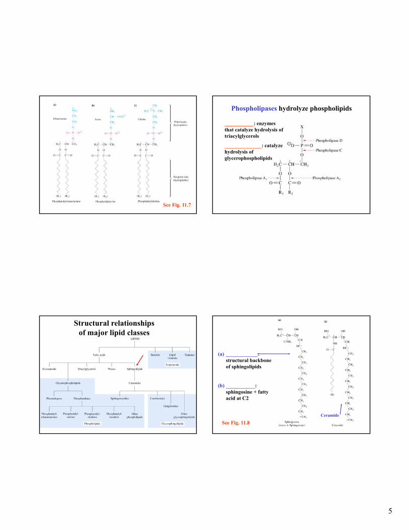
MEMBRANE LIPIDS: 3 major types = phospholipids, glycolipids, & cholesterol
_________________: most abundant class of lipids in membranes (note: triacylglycerols most abundant on mass basis in mammals); derived from glycerol or sphingosine
*lipids from glycerol = phosphoglycerides (also called glycerophospholipids)
* phosphoglycerides consist
of glycerol backbone,
two fatty acids & a
phosphorylated alcohol
Fatty acid
Fatty acid
GLYCEROL
Phosphatealcohol
See Fig. 11.5
Fatty acid chains (long aliphatic tails) in phospholipids & glycolipids contain even # of carbons (12-20) with 16 and 18 being most common
Fatty acids can be ____________ or ______________
Under physiological conditions fatty acids are ionized (pKa 4.5-5.0)
Fatty acids in biological organisms
Fig 9.1 Structural relationships of major lipid classes (a) Glycerol 3-P and (b) phosphatidate
See Fig. 11.6
5
See Fig. 11.7
Phospholipases hydrolyze phospholipids
___________: enzymes that catalyze hydrolysis of triacylglycerols
______________: catalyze hydrolysis of glycerophospholipids
Structural relationships of major lipid classes
(a) ____________: structural backbone of sphingolipids
(b) ___________: sphingosine + fatty acid at C2
Ceramide
See Fig. 11.8

6
(c) Sphingomyelin:present in plasma membrane & myelin sheath around neurons
See Fig. 11.8
• Structure of a galactocerebroside
Example of a Cerebroside:abundant in nerves
Sugar-Sphingosine
Fatty acid
See Pg. 196
Example of a Ganglioside Ganglioside GM2
(NeuNAc in blue)
Hexosaminidase A cleaves here
Mutation Tay-Sachs disease
Abundant in the brain and nervous system; Cell surface, cell-cell interactions (e.g. blood group antigens)
Structural relationships of major lipid classes

7
Structure of the steroid cholesterol.Steroids are polyprenyl compounds
In eukaryotes but NOT in most prokaryotes
Other steroids: steroid hormones (estrogen →estradiol, testosterone, corticosteriods), bile salts, sterols in plants, yeast, fungi
Synthesized from isoprene
See Pg. 197
____________
• Cholesterol modulates the fluidity of mammalian cell membranes
• It is also a precursor of the steroid hormones and bile salts
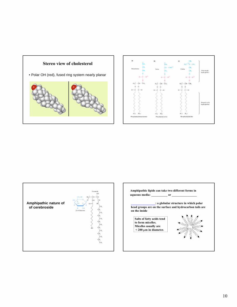
Stereo view of cholesterol
• Polar OH (red), fused ring system nearly planar
Waxes: esters of long-chain monohydroxylic alcohols and long-chain fatty acids (nonpolar)
Waxes are very water insoluble and high melting point
They are widely distributed in nature as protective waterproof coatings on leaves, fruits, animal skin, fur, feathers and exoskeletons
Myricyl palmitate, a wax
Palmitate portion Myricyl alcohol portion

8
Eicosanoids: oxygenated derivatives of C20 polyunsaturated fatty acids (e.g. arachidonic acid)
Arachidonic acid and three eicosanoids
can cause constriction of blood vessels
(aspirin inhibits prostaglandin synthesis)
involved in blood clot formation
mediator of smooth-muscle contraction and bronchial constriction seen in asthmatics
Some vitamins are Lipid Vitamins
• Four lipid vitamins: A, D, E, K
• (All contain rings and long, aliphatic side chains
• All are highly hydrophobic
• The lipid vitamins differ widely in their functions
* Examples of isoprenoids
* * * *
See Fig. 15.19 for structures
Fig. 15.19
BCMB 3100 - Lipids
•Biological Membranes
•Micelles
•Lipid Bilayer
•Peripheral membrane proteins
•Integral membrane proteins
•Lipid-anchored
•Transport across membranes
•Signal Transduction

9
Structure of a typical eukaryotic plasma membrane
See Fig. 12.1; 12.8
______________
• Highly selective permeability barriers that surround cells & cellular compartments
•Sheetlike structures of ~60-100Å
•Consists mostly of lipids & proteins in ratio of 1:4 to 4:1 (typical 40% lipid; 60% protein). Lipids & proteins may be glycosylated.
•Lipids in biological membranes are ______________: hydrophilic (polar) head group & hydrophobic tail. Spontaneously form bilayers in aqueous solution.
Fig. 12.1Membrane lipid and bilayer
Lipids in biological membranes include phospholipids, sphingolipids, cholesterol (in some eukaryotes)
10
Stereo view of cholesterol
• Polar OH (red), fused ring system nearly planar
Amphipathic nature of of cerebroside
Amphipathic lipids can take two different forms in aqueous media: _________ or ______________
____________: a globular structure in which polar head groups are on the surface and hydrocarbon tails are on the inside
Salts of fatty acids tend to form micelles. Micelles usually are< 200 m in diameter.

11
Structure and nomenclature of fatty acids _______________: favored structure for
phospholipids & sphingolipids since lipids with two fatty acyl chains are too large to fit into the center of a micelle. Bilayers can have large dimensions (107 Å, 1mm) (recall diameter = ~60-100Å)
Lipid bilayers self-assemble due to hydrophobic interactions between hydrocarbon tails (main force), van der Waals attractive forces between hydrocarabon tails, & elecrostatic & H-bonding forces between polar head groups and water
Biolayers are extensive, closed, and self-sealing
A liposome Fig. 12.2Preparation of liposomes Fig. 12.3
12
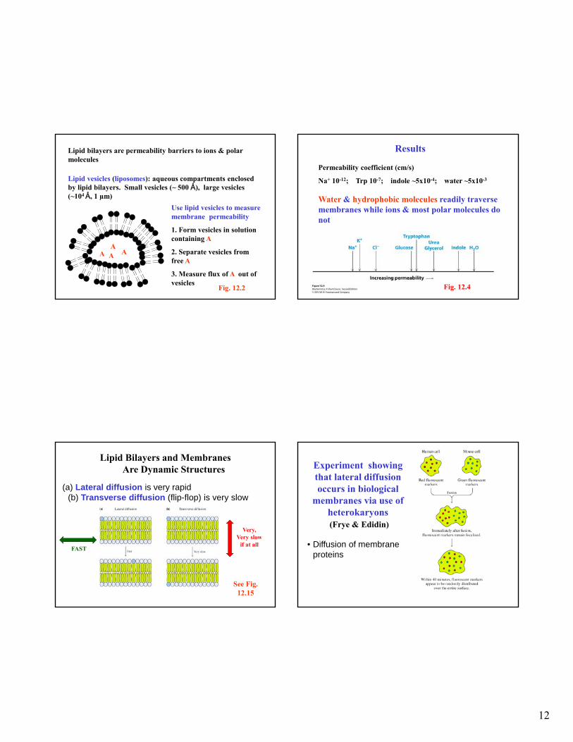
Lipid bilayers are permeability barriers to ions & polar molecules
Lipid vesicles (liposomes): aqueous compartments enclosed by lipid bilayers. Small vesicles (~ 500 Å), large vesicles (~104 Å, 1 µm)
Use lipid vesicles to measure membrane permeability
1. Form vesicles in solution containing A
2. Separate vesicles from free A
3. Measure flux of A out of vesicles
A A AA
Fig. 12.2
Results
Permeability coefficient (cm/s)
Na+ 10-12; Trp 10-7; indole ~5x10⁻⁴; water ~5x10⁻³
Water & hydrophobic molecules readily traverse membranes while ions & most polar molecules do not
Fig. 12.4
Lipid Bilayers and Membranes Are Dynamic Structures
(a) Lateral diffusion is very rapid(b) Transverse diffusion (flip-flop) is very slow
See Fig. 12.15
FAST
Very, Very slow
if at all • Diffusion of membrane proteins
Experiment showing that lateral diffusion occurs in biological
membranes via use of heterokaryons(Frye & Edidin)
13
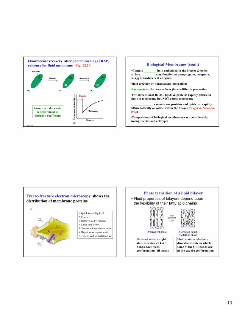
Fluorescence recovery after photobleaching (FRAP) evidence for fluid membrane. Fig. 12.14
From such data rate is determined as
diffusion coefficient
Biological Membranes (cont.)
• Contain ________ both embedded in the bilayer & on its surface. ________ may function as pumps, gates, receptors, energy transducers & enzymes.
•Held together by noncovalent interactions
•Asymmetric: the two surfaces (faces) differ in properties
•Two dimensional fluids - lipids & proteins rapidly diffuse in plane of membrane but NOT across membrane
•______________ - membrane proteins and lipids can rapidly diffuse laterally or rotate within the bilayer (Singer & Nicolson, 1972)
•Compositions of biological membranes vary considerably among species and cell types
Freeze-fracture electron microscopy, shows the distribution of membrane proteins
1. Quick freeze liquid N
2. Fracture
3. Remove ice by vacuum
4. Layer thin layer C
5. Shadow with platinum vapor
6. Digest away organic matter
7. TEM of carbon-metal replica
Phase transition of a lipid bilayer• Fluid properties of bilayers depend upon
the flexibility of their fatty acid chains
Ordered state: a rigid state in which all C-C bonds have trans conformation (all trans)
Fluid state: a relatively disordered state in which some of the C-C bonds are in the gauche conformation

14
Phase transition of a lipid bilayer
Fig 12.5
Transition from rigid to partly fluid state occurs at TM, the _______________
TM depends on _______ of fatty acyl chains & on degree of ___________
Rigid state favored by saturated fatty acyl chains
Disordered state favored by cis double bound(s) (i.e. TM is lowered)
Prokaryotes regulate membrane fluidity by varying # of double bonds & length of fatty acyl chains . As temperature changes from 42ºC to 27ºC ratio of saturated:unsaturated changes from 1.6 to 1
Packing of fatty acid chains in membrane is disrupted by double bounds and lowers Tm.
Fig. 12.6
Effect of cholesterol on phase transition (TM) of membranes
In eukaryotes membrane fluidity is largely regulated by _________. Cholesterol moderates the fluidity of membranes (prevents tight packing of fatty acyl chains & blocks large motions
Pure phospholipid bilayer has a sharp phase transition
Addition of 20 mol% cholesterol broadens phase transition

15
Fig 12.7
Cholesterol modulates fluidity of the membranes. Also, association with sphingolipids leads to cholesterol-rich regions called lipid rafts
that may effect specific membrane-protein function.
http://en.wikipedia.org/wiki/Lipid_raft
Lipid raft PM side
ECM side
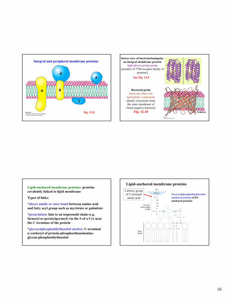
Structure of a typical eukaryotic plasma membrane Three types membrane associated proteins
______________________: loosely bound to membrane by H-bonds or electrostatic forces, generally water soluble once released from membrane using high salt or pH. Often bound to integral membrane proteins
_____________________: proteins firmly bound to membrane by hydrophobic interactions. Solubilized with detergents. Most have one or more membrane spanning domains (e.g. -helix with ~20 amino acids).
16
Integral and peripheral membrane proteins
Fig. 12.8
Stereo view of bacteriorhodopsin: an integral membrane protein
light-driven proton pump[member of 7TM receptor family of
proteins]
See Fig. 12.9
Bacterial porinmolecular filters for
hydrophilic compounds[family of proteins from the outer membrane of
Gram-negative bacteria]Fig. 12.10
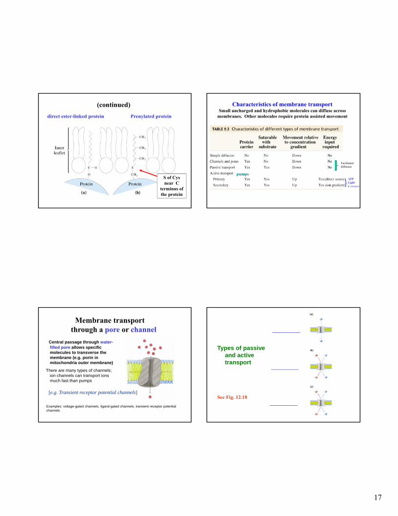
Lipid-anchored membrane proteins: proteins covalently linked to lipid membrane
Types of links:
*direct amide or ester bond between amino acid and fatty acyl group such as myristate or palmitate
*prenylation: link to an isoprenoid chain (e.g. farnesyl or geranylgeranyl) via the S of a Cys near the C terminus of the protein
*glycosylphosphatidylinositol anchor: C-terminal -carboxyl of protein-phosphoethanolamine-glycan-phosphatidylinositol
Lipid-anchored membrane proteins
Glycosylphosphatidylinositol-anchored protein (GPI-anchored protein)
Carboxy group of C-terminal amino acid
17
(continued)
Prenylated proteindirect ester-linked protein
S of Cys near C
terminus of the protein
Characteristics of membrane transport Small uncharged and hydrophobic molecules can diffuse across
membranes. Other molecules require protein assisted movement
ATPLighte- transport
Facilitated diffusion
pumps
Membrane transport through a pore or channel
Central passage through water-filled pore allows specific molecules to transverse the membrane (e.g. porin in mitochondria outer membrane)
There are many types of channels; ion channels can transport ions much fast than pumps
Examples: voltage-gated channels, ligand-gated channels, transient receptor potential channels
[e.g. Transient receptor potential channels]
Types of passive and active transport
__________
__________
__________ See Fig. 12.18
18
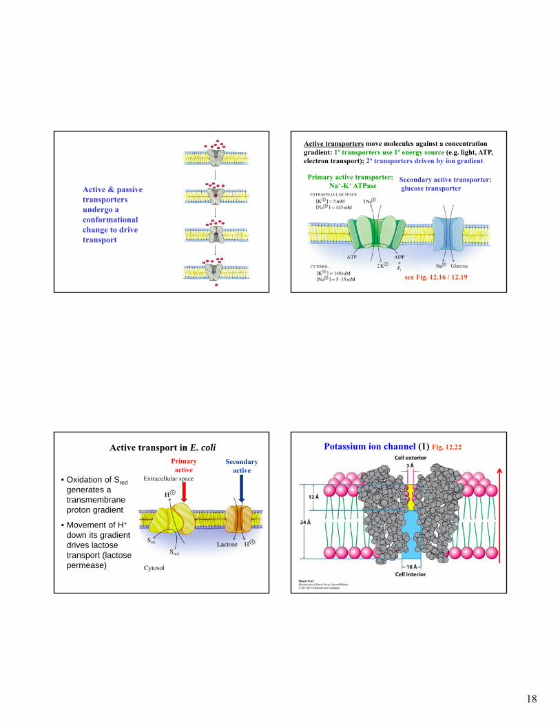
Active & passive transporters undergo a conformational change to drive transport
Primary active transporter:Na+-K+ ATPase
Secondary active transporter:glucose transporter
Active transporters move molecules against a concentration gradient: 1º transporters use 1º energy source (e.g. light, ATP, electron transport); 2º transporters driven by ion gradient
see Fig. 12.16 / 12.19
Active transport in E. coli
• Oxidation of Sred
generates a transmembrane proton gradient
• Movement of H+
down its gradient drives lactose transport (lactose permease)
Secondaryactive
Primary active
Potassium ion channel (1) Fig. 12.22

19
Potassium ion channel (2) Fig. 12.23Selectivity filter of K+ ion channel
Potassium ion channel (3) Fig. 12.24Energetic basis of ion selectivity in K+ ion channel
Potassium ion channel (4) Fig. 12.24Energetic basis of ion selectivity in K+ ion channel.
Potassium ion channel (5) Fig. 12.25Rapid rate of K+ movement due to structure of
channel and electrostatic repulsion of incoming K+

20
Molecules and complexes that are too
large to be transported via transport
proteins are transported in lipid vesicles
out of the cell via exocytosis, and into
the cell via endocytosis. We will not
cover these processes in this course.
Signal transduction through a membrane
Fig. 13.1
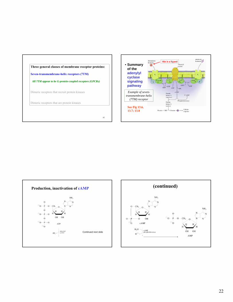
Three general classes of membrane receptor proteins:
• seven-transmembrane-helix receptors
• Dimeric receptors that recruit protein kinases
• Dimeric receptors that are protein kinases
79
General mechanism of signal transductionacross a membrane
(e.g. hormones)
e.g. *G proteins
Tyrosine kinase
*Adenylate cyclase
*Phospholipase C
*detect*amplify signal external
To generate responsee.g. changes in gene expression, enzyme activity, ion channel, etc.

21
Common secondary messengers Fig. 13.2 G-protein cycle
• G proteins are activated by binding to a receptor-ligand complex
• G-proteins are inactivated slowly by their own GTPase activity(kcat about 3/min)
α: fatty acyl anchored
gamma: prenyl-anchored
Understanding G Proteins:Hydrolysis of GTP to GDP and Pi
84
Three general classes of membrane receptor proteins:
Seven-transmembrane-helix receptors (7TM)
Dimeric receptors that recruit protein kinases
Dimeric receptors that are protein kinases
22
85
Three general classes of membrane receptor proteins:
Seven-transmembrane-helix receptors (7TM)
All 7TM appear to be G-protein-coupled receptors (GPCRs)
Dimeric receptors that recruit protein kinases
Dimeric receptors that are protein kinases
• Summary of the adenylyl cyclase signaling pathway
See Fig 13.6, 13.7; 13.8
Example of seven-transmembrane-helix
(7TM) receptor
this is a ligand
Production, inactivation of cAMP
Continued next slide
(continued)

23
• Activation of protein kinase A by cAMP
See Fig. 13.7
Caffeine & theophylline inhibit cAMP phosphodiesterase
• Inhibition of cAMP phosphodiesterases prolongs the effects of cAMP
• This increases the intensity and duration of stimulatory hormones
• Inositol-phospholipid signaling pathway
See Fig. 13.11, 13.12
Phospholipase C Phosphatidylinositol 4,5-bis phosphate
Phospholipases hydrolyze phospholipids
Lipases: enzymes that catalyze hydrolysis of triacylglycerols
Phospholipases: catalyze hydrolysis of glycerophospholipids
92

24
Phosphatidylinositol 4,5-bisphosphate (PIP2) produces IP3 and diacylglycerol
See Fig. 13.11
(continued)
• Activation of receptor tyrosine kinases by ligand-induced dimerization
See Fig. 13.15
Three general classes of membrane receptor proteins:
Seven-transmembrane-helix receptors
Dimeric receptors that recruit protein kinases
*Dimeric receptors that are protein kinases
(continued)
• Phosphorylated dimer phosphorylates cellular target proteins

25
(continued)
• Each domain catalyzes phosphorylation of its partner
Insulin receptor and tyrosine kinase activity
• Insulin binds to 2 extracellular -chains
• Transmembrane -chains then autophosphorylate
• Tyrosine kinase domains then phosphorylate insulin-receptor substrates (IRSs) (which are proteins)
Insulin-stimulated formation of PIP3
*phosphatidyl-inositol 3,4,5-trisphosphate
*
See Figs. 13.17 / 13.21
Fig. 13.13
A different type of tyrosine kinase signal transduction: Growth hormone receptor for which binding brings together
associated proteins with tyrosine kinase domains
Three general classes of membrane receptor proteins:
Seven-transmembrane-helix receptors
*Dimeric receptors that recruit protein kinases
Dimeric receptors that are protein kinases
26
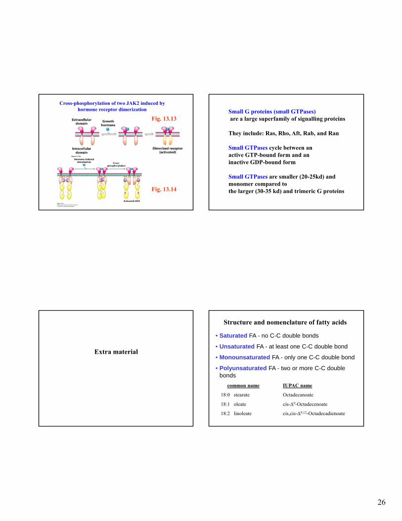
Cross-phosphorylation of two JAK2 induced by hormone receptor dimerization
Fig. 13.13
Fig. 13.14
Small G proteins (small GTPases)are a large superfamily of signalling proteins
They include: Ras, Rho, Aft, Rab, and Ran
Small GTPases cycle between an active GTP-bound form and aninactive GDP-bound form
Small GTPases are smaller (20-25kd) and monomer compared tothe larger (30-35 kd) and trimeric G proteins
Extra material
Structure and nomenclature of fatty acids
• Saturated FA - no C-C double bonds
• Unsaturated FA - at least one C-C double bond
• Monounsaturated FA - only one C-C double bond
• Polyunsaturated FA - two or more C-C double bonds
common name IUPAC name
18:0 stearate Octadecanoate
18:1 oleate cis-9-Octadecenoate
18:2 linoleate cis,cis-9,12-Octadecadienoate

27
Another method to measure membrane permeability
Mueller & Rudin: dip paint brush into lipid membrane solution & paint across 1 mm diameter hole partitioned between two aqueous media. macroscopic bilayer membrane
Measure electrical conductance from one media to the other
Related Documents