Molecular Microbiology (2004) 54 (3), 647–664 doi:10.1111/j.1365-2958.2004.04307.x © 2004 Blackwell Publishing Ltd Blackwell Science, LtdOxford, UKMMIMolecular Microbiology0950-382XBlackwell Publishing Ltd, 2004 ? 2004 54 3647664 Original Article Pseudopilin PulGR. Köhler et al. Accepted 12 July, 2004. *For correspondence. E-mail [email protected]; Tel. ( + 33) 145688494; Fax ( + 33) 145688960. † These authors contributed equally to this work. Structure and assembly of the pseudopilin PulG Rolf Köhler, 1† Karsten Schäfer, 2† Shirley Müller, 3† Guillaume Vignon, 1 Kay Diederichs, 2 Ansgar Philippsen, 3 Philippe Ringler, 3 Anthony P. Pugsley, 1 * Andreas Engel 3 and Wolfram Welte 2 1 Molecular Genetics Unit, Institut Pasteur, 25, rue du Dr Roux, 75724 Paris Cedex 15, France. 2 Department of Biology, University of Konstanz, M656, D-78457 Konstanz, Germany. 3 M. E. Müller Institute for Structural Biology, Biozentrum, University of Basel, CH-4056, Basel, Switzerland. Summary The pseudopilin PulG is one of several essential com- ponents of the type II pullulanase secretion machin- ery (the Pul secreton) of the Gram-negative bacterium Klebsiella oxytoca . The sequence of the N-terminal 25 amino acids of the PulG precursor is hydrophobic and very similar to the corresponding region of type IV pilins. The structure of a truncated PulG (lacking the homologous region), as determined by X-ray crystallography, was found to include part of the long N-terminal a -helix and the four internal anti-parallel b - strands that characterize type IV pilins, but PulG lacks the highly variable loop region with a disulphide bond that is found in the latter. When overproduced, PulG forms flexible pili whose structural features, as visu- alized by electron microscopy, are similar to those of bacterial type IV pili. The average helical repeat com- prises 17 PulG subunits and four helical turns. Elec- tron microscopy and molecular modelling show that PulG probably assembles into left-handed helical pili with the long N-terminal a -helix tightly packed in the centre of the pilus. As in the type IV pilins, the hydro- phobic N-terminal part of the PulG a -helix is neces- sary for its assembly. Subtle sequence variations within this highly conserved segment seem to deter- mine whether or not a type IV pilin can be assembled into pili by the Pul secreton. Introduction The type II protein secretion (T2S) machinery, or secreton, of Gram-negative bacteria transports proteins from the periplasm across the outer membrane (Pugsley, 1993a; Sandkvist, 2001). It is composed of up to 14 different proteins, many of which share significant sequence simi- larities with components of the type IV pilus biogenesis pathway, also of Gram-negative bacteria (Pugsley, 1993a; Nunn, 1999; Sandkvist, 2001). The best-characterized component of both pathways is the secretin, an integral outer membrane protein that forms a multimeric channel complex (Bitter et al ., 1998; Collins et al ., 2001; 2003) through which the pilus (Wolfgang et al ., 2000) or folded secreted proteins (Bitter et al ., 1998; Brok et al ., 1999; Nouwen et al ., 1999; 2000) probably cross the outer membrane. The 30 N-terminal amino acids of five secreton compo- nents, the pseudopilins, share substantial sequence simi- larity with the corresponding regions of type IV pilins (Pugsley, 1993a; Nunn, 1999; Sandkvist, 2001). This observation led to the idea that the pseudopilins might form a pilus-like structure (the pseudopilus) that functions either as a periplasmic scaffold on which other secreton compo- nents are assembled or as a piston-like motor to drive secretion across the outer membrane (Hobbs and Mattick, 1993; Pugsley, 1993a; Nunn, 1999; Sandkvist, 2001). Early studies with the pullulanase secreton from Klebsiella oxytoca failed to provide any evidence for such a pseudo- pilus, probably because only a small proportion of the pseudopilins are assembled at any given moment (Pugsley and Possot, 1993; Pugsley, 1996). The most abundant pseudopilin, PulG, was subsequently shown to assemble into long, bundled pili when expression of its structural gene was increased and the bacteria were grown on agar (Sauvonnet et al ., 2000a; Pugsley et al ., 2001; Vignon et al ., 2003). These long pili were considered to result from the uncontrolled elongation of a normally shorter, periplas- mic pseudopilus (Sauvonnet et al ., 2000a; Vignon et al ., 2003). This idea is supported by studies in Xanthomonas campestris showing that a PulG homologue, XpsG, forms intraperiplasmic complexes (Hu et al ., 2002) and by the fact that similar phenomena have been reported for other T2S secretons (Durand et al ., 2003; Vignon et al ., 2003). X-ray diffraction and NMR analyses of four full-length or N-terminally truncated type IV pilins (PilE from Neisseria gonorrhoeae , PilA from two strains of Pseudomonas aeruginosa and TcpA from Vibrio cholerae ) revealed a considerable degree of structural conservation, despite the fact that major sequence similarities are restricted to the extreme N-terminal regions (Parge et al ., 1995; Hazes et al ., 2000; Keizer et al ., 2001; Craig et al ., 2003). The

Welcome message from author
This document is posted to help you gain knowledge. Please leave a comment to let me know what you think about it! Share it to your friends and learn new things together.
Transcript
Molecular Microbiology (2004)
54
(3), 647–664 doi:10.1111/j.1365-2958.2004.04307.x
© 2004 Blackwell Publishing Ltd
Blackwell Science, LtdOxford, UKMMIMolecular Microbiology0950-382XBlackwell Publishing Ltd, 2004
? 2004
54
3647664
Original Article
Pseudopilin PulGR. Köhler
et al.
Accepted 12 July, 2004. *For correspondence. [email protected]; Tel. (
+
33) 145688494; Fax (
+
33) 145688960.
†
These authors contributed equally to this work.
Structure and assembly of the pseudopilin PulG
Rolf Köhler,
1†
Karsten Schäfer,
2†
Shirley Müller,
3†
Guillaume Vignon,
1
Kay Diederichs,
2
Ansgar Philippsen,
3
Philippe Ringler,
3
Anthony P. Pugsley,
1
* Andreas Engel
3
and Wolfram Welte
2
1
Molecular Genetics Unit, Institut Pasteur, 25, rue du Dr Roux, 75724 Paris Cedex 15, France.
2
Department of Biology, University of Konstanz, M656, D-78457 Konstanz, Germany.
3
M. E. Müller Institute for Structural Biology, Biozentrum, University of Basel, CH-4056, Basel, Switzerland.
Summary
The pseudopilin PulG is one of several essential com-ponents of the type II pullulanase secretion machin-ery (the Pul secreton) of the Gram-negative bacterium
Klebsiella oxytoca
. The sequence of the N-terminal 25amino acids of the PulG precursor is hydrophobic andvery similar to the corresponding region of type IVpilins. The structure of a truncated PulG (lackingthe homologous region), as determined by X-raycrystallography, was found to include part of the longN-terminal
aaaa
-helix and the four internal anti-parallel
bbbb
-strands that characterize type IV pilins, but PulG lacksthe highly variable loop region with a disulphide bondthat is found in the latter. When overproduced, PulGforms flexible pili whose structural features, as visu-alized by electron microscopy, are similar to those ofbacterial type IV pili. The average helical repeat com-prises 17 PulG subunits and four helical turns. Elec-tron microscopy and molecular modelling show thatPulG probably assembles into left-handed helical piliwith the long N-terminal
aaaa
-helix tightly packed in thecentre of the pilus. As in the type IV pilins, the hydro-phobic N-terminal part of the PulG
aaaa
-helix is neces-sary for its assembly. Subtle sequence variationswithin this highly conserved segment seem to deter-mine whether or not a type IV pilin can be assembledinto pili by the Pul secreton.
Introduction
The type II protein secretion (T2S) machinery, or secreton,of Gram-negative bacteria transports proteins from the
periplasm across the outer membrane (Pugsley, 1993a;Sandkvist, 2001). It is composed of up to 14 differentproteins, many of which share significant sequence simi-larities with components of the type IV pilus biogenesispathway, also of Gram-negative bacteria (Pugsley, 1993a;Nunn, 1999; Sandkvist, 2001). The best-characterizedcomponent of both pathways is the secretin, an integralouter membrane protein that forms a multimeric channelcomplex (Bitter
et al
., 1998; Collins
et al
., 2001; 2003)through which the pilus (Wolfgang
et al
., 2000) or foldedsecreted proteins (Bitter
et al
., 1998; Brok
et al
., 1999;Nouwen
et al
., 1999; 2000) probably cross the outermembrane.
The 30 N-terminal amino acids of five secreton compo-nents, the pseudopilins, share substantial sequence simi-larity with the corresponding regions of type IV pilins(Pugsley, 1993a; Nunn, 1999; Sandkvist, 2001). Thisobservation led to the idea that the pseudopilins might forma pilus-like structure (the pseudopilus) that functions eitheras a periplasmic scaffold on which other secreton compo-nents are assembled or as a piston-like motor to drivesecretion across the outer membrane (Hobbs and Mattick,1993; Pugsley, 1993a; Nunn, 1999; Sandkvist, 2001).Early studies with the pullulanase secreton from
Klebsiellaoxytoca
failed to provide any evidence for such a pseudo-pilus, probably because only a small proportion of thepseudopilins are assembled at any given moment (Pugsleyand Possot, 1993; Pugsley, 1996). The most abundantpseudopilin, PulG, was subsequently shown to assembleinto long, bundled pili when expression of its structuralgene was increased and the bacteria were grown on agar(Sauvonnet
et al
., 2000a; Pugsley
et al
., 2001; Vignon
et al
., 2003). These long pili were considered to result fromthe uncontrolled elongation of a normally shorter, periplas-mic pseudopilus (Sauvonnet
et al
., 2000a; Vignon
et al
.,2003). This idea is supported by studies in
Xanthomonascampestris
showing that a PulG homologue, XpsG, formsintraperiplasmic complexes (Hu
et al
., 2002) and by thefact that similar phenomena have been reported for otherT2S secretons (Durand
et al
., 2003; Vignon
et al
., 2003).X-ray diffraction and NMR analyses of four full-length or
N-terminally truncated type IV pilins (PilE from
Neisseriagonorrhoeae
, PilA from two strains of
Pseudomonasaeruginosa
and TcpA from
Vibrio cholerae
) revealed aconsiderable degree of structural conservation, despitethe fact that major sequence similarities are restricted tothe extreme N-terminal regions (Parge
et al
., 1995; Hazes
et al
., 2000; Keizer
et al
., 2001; Craig
et al
., 2003). The
648
R. Köhler
et al.
© 2004 Blackwell Publishing Ltd,
Molecular Microbiology
,
54
, 647–664
conserved structural features include a long, N-terminalhelix, the first, highly conserved part of which is hydropho-bic, followed by loop structure and then several anti-par-allel
b
-strands and a disulphide bridge near the C-terminus. The hydrophobic N-terminal helix is proposed tobe required for pilin export across the plasma membrane,for retention of the unassembled pilin in this membraneand for the interactions that promote pilus assembly.Model building predicts that the pilins are arranged in ahelical manner, with the hydrophobic N-terminal helixpacked in the central core of the pilus, although the detailsof the models vary according to the particular pilin (Parge
et al
., 1995; Keizer
et al
., 2001; Craig
et al
., 2003; 2004).Here we analyse the structure of PulG filaments by
electron microscopy, present the structure of a truncatedPulG determined by X-ray crystallography, and use this tobuild an atomic model. To gain further insight into pilusassembly and protein secretion, we analysed chimericproteins formed by exchanging the N-terminal hydropho-bic region between pseudopilins and pilins.
Results
Characterization of the PulG(His)
6
helix
PulG(His)
6
is identical to natural PulG except that the C-terminal sequence G
132
K
133
K
134
(numbers refer to positionin the mature PulG polypeptide chain) has been extendedto G
132
FHHHHHHK to incorporate a hexahistidine tag(Pugsley, 1993b). The protein is fully functional in secre-tion (Pugsley, 1993b) and forms pili when it is overpro-duced (Pugsley
et al
., 2001; Vignon
et al
., 2003). Incontrast to pili formed by untagged PulG, pili formed byPulG(His)
6
do not form bundles, which facilitates theiranalysis (Vignon
et al
., 2003).PulG(His)
6
pili were purified by shearing of agar-grown
Escherichia coli
K-12 PAP9001(pCHAP7009) (seeTables 1 and 2 for details of strains and plasmids) andcobalt affinity chromatography essentially as describedpreviously (Vignon
et al
., 2003). The strain used herelacks the
pulA
gene coding for the secreted enzyme pul-lulanase that contaminated earlier samples of PulG(His)
6
Table 2.
Plasmids.
PlasmidVector/replication origin/resistance
Cloned genes and/or chimeras
Reference and/or source of cloned fragment
pCHAP231 pBR322/pColE1/Ap pBR322
::
(
pulS pulA-B pulC-O
) d’Enfert
et al
. (1987)pCHAP1216 pBR322/pColE1/Ap pCHAP231
pulB::kan-1
D
pulG
Possot
et al
. (2000)pCHAP1362 pSU19/p15/Cm
lacZ
p-
pulG::His
6
Pugsley (1993b)pCHAP3100 pSU18/p15/Cm
lacZ
p-
ppdD
Sauvonnet
et al
. (2000a)pCHAP1379 pSU18/p15/Cm
lacZ
p-
pilA
This studypCHAP1404 pSU18/p15/Cm
lacZ
p-
pilE
This studypCHAP1418 pSU19/p15/Cm
lacZ
p
-tcpA
This studypCHAP4260 pUC18/pColE1/Ap
lacZ-pelB
¢
O. FranceticpCHAP7003 pSU19/p15/Cm
lacZ
p-
ppdD
,
pulG::His
6
This studypCHAP7007 pUC19/pColE1/Ap
pulB
::
kan-1-pulF
This studypCHAP7008 pUC19/pColE1/Ap
pulB
::
kan-1-
D
pulA-pulF
This studypCHAP7009 pBR322/pColE1/Ap pCHAP231
pulB
::
kan-1
D
pulG
,
D
pulA
This studypCHAP7010 pUC18/pColE1/Ap
pelBss::pulG(25-134)::His
6
This studypCHAP7012 pSU18/p15/Cm
pelBss::pulG(25-134)::His
6
This studypCHAP7013 pT7-5/PColE1/Ap T7p-
pelBss::pulG(25-134)::His
6
This studypCHAP7015 pSU18/p15/Cm
lacZ
p-(
ppdD17::pulG::His
6
) This studypCHAP7016 pSU18/p15/Cm
lacZ
p-(
pilE17::pulG::His
6
) This studypCHAP7017 pSU18/p15/Cm
lacZ
p-(
pulG17::pilE
) This studypCHAP7019 pSU18/p15/Cm
lacZ
p-(
pulG21::ppdD
) This studypCHAP7023 pSU18/p15/Cm
lacZ
p-(
pulG17::ppdD
) This studypCHAP7024 pSU18/p15/Cm
lacZ
p-(
ppdD21::pulG::His
6
) This studypCHAP7026 pSU18/p15/Cm
lacZ
p-(
pulG17::pilA
) This studypCHAP7027 pSU18/p15/Cm
lacZ
p-(
ppdD21::pilA
) This study
Ap, ampicillin; Cm, chloramphenicol.
Table 1.
E. coli
strains.
Strain Relevant characteristics Source/reference
PAP105
D
(lac-proAB)
F
¢
lacI
Q
Tn
10 Laboratory collectionPAP7500BG araD139 (argF-lac)U169 rpsL150 relA1 flbB5301 deoC1 ptsF25 malG501
F¢ lacI q1 Tn10 malP::(pulS pulA-pulB::kan1 pulCO)pulB::kan1 and DpulG (pul genes integrated into chromosome; lacks functional pulG gene)
Pugsley (1993b); Sauvonnet et al. (2000a)
PAP9001 araD139 (argF-lac)U169 rpsL150 relA1 flbB5301 deoC1 ptsF25 malE44 malG510 fimAB::kan sacB (lacks type I pili)
Laboratory collection
PAP7501 PAP9001 F¢ lacI Q Tn10 Laboratory collection
Pseudopilin PulG 649
© 2004 Blackwell Publishing Ltd, Molecular Microbiology, 54, 647–664
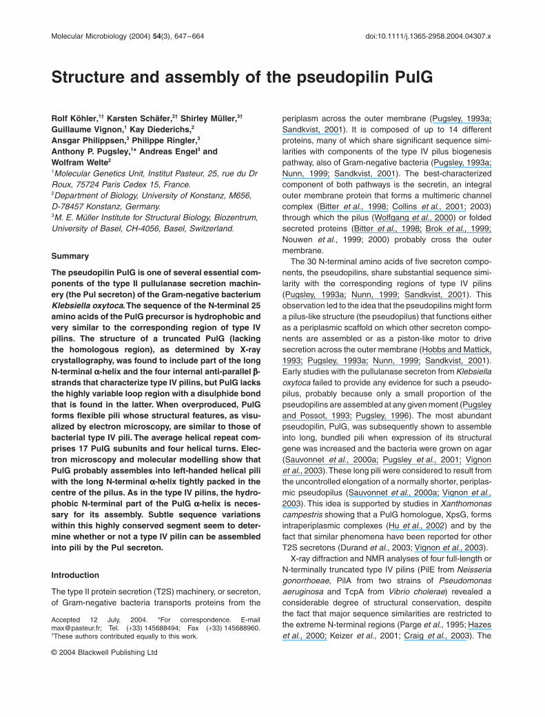
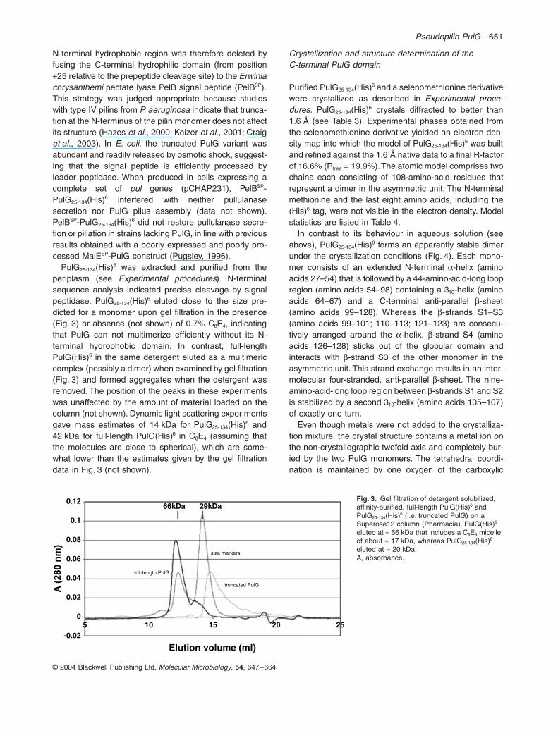
Fig. 1. Appearance of the PulG(His)6 pili.A and B. Negatively stained pili imaged by the STEM. (A) Single and intertwined twisting pili. (B) The periodic helical structure is distinct at higher magnification, even in the bent regions of PulG(His)6 pili. A stained filled central cavity could not be detected.C. PulG(His)6 pili imaged by transmission electron microscope after shadowing with W/Ta. A left-handed helical twist is clearly visible when viewing at a glancing angle in the direction indicated by black arrows. The T4 phage tail also present displays the expected right-handed helical symmetry. The direction of shadowing is indicated (white arrow).Scale bars: 50 nm in (A) and (C), 20 nm in (B).
pili (Vignon et al., 2003). Images of uranyl acetate stainedPulG(His)6 pili recorded in a scanning transmission elec-tron microscope (STEM) revealed mainly long filamentswith pronounced flexibility (Fig. 1A). Distinct repeatingprotrusions, suggestive of a helical structure, became vis-ible at higher magnification (Fig. 1B). Although the nega-tive stain penetrated deeply into the grooves between theprotrusions, there was no indication of a central, stain-filled cavity. The handedness of the pilus helix was deter-mined by metal shadowing experiments, which revealeda left-handed twist (Fig. 1C).
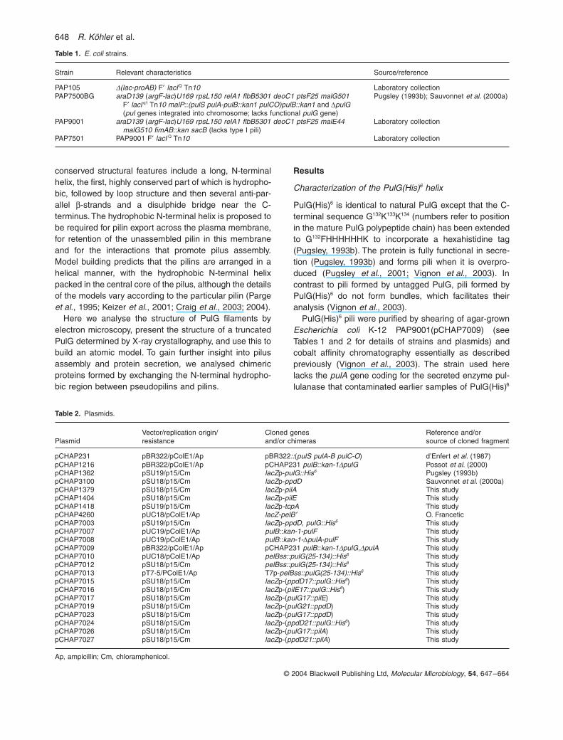
Twenty-seven negatively stained PulG(His)6 pili in theSTEM images were analysed for structural information.Selected segments were first computationally straight-ened (Fig. 2A) and the average profile perpendicular tothe filament axis was calculated to determine their outerdiameter (8.2 ± 0.3 nm). The full-width-half-maximum(FWHM) diameter measured on freeze-dried, unstainedfilaments was 6.9 ± 1.0 nm (see below). Most diffractionpatterns from straightened rods exhibited the x-patterntypical of helical symmetry (Fig. 2B) but many were notsymmetrical with respect to the meridian, indicatinguneven staining and unsatisfactory structural preserva-tion. The strongest layer lines of all 27 filaments were(4.38 ± 0.12 nm)-1 from the equator, reflecting the pitch ofthe genetic helix (i.e. the lowest pitch one-start helixdescribing the helical symmetry of the structure; seeDeRosier and Klug, 1968). Amplitude and phase plots ofthis layer line calculated from the pilus segment shown inFig. 2A exhibit a minor asymmetry in the amplitude(Fig. 2C) and a phase difference of p between the left andright side (Fig. 2D). Because the distance between themajor maxima corresponded to (5.4 nm)-1, and the fila-ment radius was 3.5–4.1 nm, this layer line is bestdescribed by a first-order Bessel function (J1), suggestingthat the pilus is a one-start helix.
In an attempt to identify the helical repeat, PulG(His)6
pilus stretches of up to 157 nm length were examinedusing a ‘linear Markham’ superposition algorithm (Hahnet al., 2000). Repeat lengths, ranging from 7 pixels up tothe largest possible were tested (i.e. 238 pixels, 78 nm,for the longest pilus when two stretches were averaged).The enhancement resulting from superposition and aver-aging was quantified by comparing the root mean square(rms) deviation of the average with that of the unproc-essed segment. Plots of the rms quotient against therepeat length exhibited series of maxima for all 27 pili. Therepeats were 4.37 ± 0.85 nm apart for the six best-preserved pilus rods. This separation corresponds to thepitch of the genetic helix, in good agreement with theposition of the prominent layer line in Fig. 2B. Averagingthe Markham superposition curves for 7 well-preservedpili yielded a distinct maximum of the rms quotient at17.5 nm, the peak height of which is more than 10%
650 R. Köhler et al.
© 2004 Blackwell Publishing Ltd, Molecular Microbiology, 54, 647–664
above all of the other maxima, disregarding the first rathernoisy maximum of almost equal height (Fig. 2E). Thus, thehelical repeat of the PulG(His)6 pilus appears to containfour turns.
To establish the number of subunits per helical repeat,a series of low-dose dark-field images was recorded fromfreeze-dried, unstained PulG(His)6 samples by STEM,and the mass-per-length (MPL) of the PulG(His)6 rodsdetermined (Müller et al., 1992). The pooled MPL datafrom 331 pilus segments yielded a histogram that isdescribed by a single Gauss curve peaking at15.5 ± 1.3 kDa nm-1 after correction for mass loss [stan-dard error (SE) = ±0.07 kDa nm-1; Fig. 2F]. The overalluncertainty of the measurement is ±0.8 kDa nm-1 consid-ering the SE of the result and the 5% calibrationuncertainty of the microscope. The average FWHM dia-meter of the unstained PulG(His)6 pili (see above) was6.9 ± 1.0 nm (see Experimental procedures). Becausethe PulG(His)6 pili are homopolymers of the 15.4 kDaprotein PulG(His)6 (Pugsley, 1993b), the number of pro-tein monomers per helical repeat can be directly calcu-lated from the MPL. Accordingly, 17.64 ± 0.92 subunits
would assemble into a full helical repeat comprising fourhelical turns.
Taking together the layer line analysis, which suggeststhe PulG(His)6 pilus to be single started, the MPL andMarkham superposition results, a compatible helicalselection rule is l = 4n + 17m, where l is the layer linenumber (l = 0, ±1, ±2 . . .), m the meridian branch number(m = 0, ±1, ±2 . . .) and n the Bessel order sampled onlayer line l (n = 0, ±1, ±2 . . .). Several helical selectionrules were explored because the PulG(His)6 pili exhibitedpronounced flexibility. The simplest rules delineating therange tested were l = 1n + 4m (y = 90∞) and l = 1n + 5m(y = 72∞), where the screw angle y is the angle by whichadjacent subunits are rotated relative to each other. Tosample this screw angle interval (i.e. 72∞-90∞), all 33helical selection rules from 1n to 10n with screw anglesbetween 72∞ and 90∞ were applied to the 14 bestPulG(His)6 pili. Their two-dimensional (2D) Fourier trans-forms were calculated and the power transmitted (PT) bythe D(Z,k) filter derived from the respective helical selec-tion rule was determined (Smith and Aebi, 1974). The PTwas smaller for the longest helical repeats (9n and 10n)than for the shorter helical repeats (4n, 6n, 7n and 8n)over the entire screw angle range tested, indicating vari-ations of the helical structure along the rod axis. All helicalselection rules explored having screw angles between 80∞and 90∞ yielded higher PT values than those between 72∞and 80∞, with a global flat maximum around 85∞. Thismaximum is within the restriction dictated by the MPLvalue (15.5 ± 0.8 kDa nm-1), which limits the range ofscrew angles to 83∞-86∞.
As a result of their inherent flexibility (see Fig. 1), thebest helical selection rule describing the PulG(His)6 pilivaried depending on the pilus stretch tested, the variationcomplying to the screw angle range 83∞-86∞. Accordingly,the pilus structure should be described by the rulethat corresponds to the average situation, i.e. to a screwangle of ª 84.5∞. This requirement is met by the rulel = 4n + 17m (y = 84.7∞), which was accordingly used toD(Z,k)-filter the Fourier transform of the pilus shown inFig. 2A. The three-dimensional (3D) helical reconstructionwas then produced from the filtered layer lines 0 to ±8,including 12 Bessel orders, which corresponds to a reso-lution of (2.3 nm)-1. A 3D map thus reconstructed from thenegatively stained PulG(His)6 pilus was isocontoured toinclude 100% of the mass (see Experimental procedures).This envelope served as a guide to build an atomic modelof the PulG(His)6 pilus (see below).
Construction and purification of a soluble, truncated PulG variant for structural analysis
Attempts to crystallize full-length PulG(His)6 obtained bydissociating purified pili in the detergent C8E4 failed. The
Fig. 2. Analysis of straightened pilus stretches.A. Distinct regular peripheral projections indicate the helical architec-ture (scale bar 10 nm).B. The diffraction pattern reveals the characteristic x-pattern for a helical structure, with the strong layer line at (4.37 nm)-1.C and D. Amplitude (C) and phase plots (D) of the strong layer line indicate a first-order Bessel function (see text).E. Markham superposition yields a regular repeat of 4.38 nm, corre-sponding to the pitch of the genetic helix. The plot shown is the average (solid line) from seven pili, with dotted lines marking the standard error. The peak at 17.5 nm stands out and indicates that the pili have four turns per helical repeat.F. The mass-per-length histogram comprises measurements of 331 pilus stretches of ª 80 nm length.Scale bars: 10 nm in (A) and 5 nm in (B).
A
B
E
C
D
F
1.0
0.5
0.0
p
0
(2.5 nm)–1
15.5±1.3kDa nm–1
100
80
60
40
20
0
Fre
quen
cy
6 14 22 30 38Mass-per-length (kDa nm–1)Repeat length averaged
0 10 20 30 40 50 nm
0.88
0.87
0.86
0.85rms a
vera
ge /r
ms i
nitia
l
Pseudopilin PulG 651
© 2004 Blackwell Publishing Ltd, Molecular Microbiology, 54, 647–664
N-terminal hydrophobic region was therefore deleted byfusing the C-terminal hydrophilic domain (from position+25 relative to the prepeptide cleavage site) to the Erwiniachrysanthemi pectate lyase PelB signal peptide (PelBSP).This strategy was judged appropriate because studieswith type IV pilins from P. aeruginosa indicate that trunca-tion at the N-terminus of the pilin monomer does not affectits structure (Hazes et al., 2000; Keizer et al., 2001; Craiget al., 2003). In E. coli, the truncated PulG variant wasabundant and readily released by osmotic shock, suggest-ing that the signal peptide is efficiently processed byleader peptidase. When produced in cells expressing acomplete set of pul genes (pCHAP231), PelBSP-PulG25-134(His)6 interfered with neither pullulanasesecretion nor PulG pilus assembly (data not shown).PelBSP-PulG25-134(His)6 did not restore pullulanase secre-tion or piliation in strains lacking PulG, in line with previousresults obtained with a poorly expressed and poorly pro-cessed MalESP-PulG construct (Pugsley, 1996).
PulG25-134(His)6 was extracted and purified from theperiplasm (see Experimental procedures). N-terminalsequence analysis indicated precise cleavage by signalpeptidase. PulG25-134(His)6 eluted close to the size pre-dicted for a monomer upon gel filtration in the presence(Fig. 3) or absence (not shown) of 0.7% C8E4, indicatingthat PulG can not multimerize efficiently without its N-terminal hydrophobic domain. In contrast, full-lengthPulG(His)6 in the same detergent eluted as a multimericcomplex (possibly a dimer) when examined by gel filtration(Fig. 3) and formed aggregates when the detergent wasremoved. The position of the peaks in these experimentswas unaffected by the amount of material loaded on thecolumn (not shown). Dynamic light scattering experimentsgave mass estimates of 14 kDa for PulG25-134(His)6 and42 kDa for full-length PulG(His)6 in C8E4 (assuming thatthe molecules are close to spherical), which are some-what lower than the estimates given by the gel filtrationdata in Fig. 3 (not shown).
Crystallization and structure determination of the C-terminal PulG domain
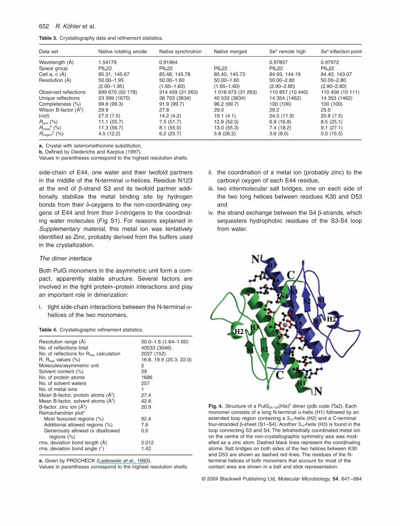
Purified PulG25-134(His)6 and a selenomethionine derivativewere crystallized as described in Experimental proce-dures. PulG25-134(His)6 crystals diffracted to better than1.6 Å (see Table 3). Experimental phases obtained fromthe selenomethionine derivative yielded an electron den-sity map into which the model of PulG25-134(His)6 was builtand refined against the 1.6 Å native data to a final R-factorof 16.6% (Rfree = 19.9%). The atomic model comprises twochains each consisting of 108-amino-acid residues thatrepresent a dimer in the asymmetric unit. The N-terminalmethionine and the last eight amino acids, including the(His)6 tag, were not visible in the electron density. Modelstatistics are listed in Table 4.
In contrast to its behaviour in aqueous solution (seeabove), PulG25-134(His)6 forms an apparently stable dimerunder the crystallization conditions (Fig. 4). Each mono-mer consists of an extended N-terminal a-helix (aminoacids 27–54) that is followed by a 44-amino-acid-long loopregion (amino acids 54–98) containing a 310-helix (aminoacids 64–67) and a C-terminal anti-parallel b-sheet(amino acids 99–128). Whereas the b-strands S1–S3(amino acids 99–101; 110–113; 121–123) are consecu-tively arranged around the a-helix, b-strand S4 (aminoacids 126–128) sticks out of the globular domain andinteracts with b-strand S3 of the other monomer in theasymmetric unit. This strand exchange results in an inter-molecular four-stranded, anti-parallel b-sheet. The nine-amino-acid-long loop region between b-strands S1 and S2is stabilized by a second 310-helix (amino acids 105–107)of exactly one turn.
Even though metals were not added to the crystalliza-tion mixture, the crystal structure contains a metal ion onthe non-crystallographic twofold axis and completely bur-ied by the two PulG monomers. The tetrahedral coordi-nation is maintained by one oxygen of the carboxylic
full-length PulG
66kDa 29kDa
Elution volume (ml)
0.12
0.1
0.08
0.06
0.04
0.02
A (
280
nm
)
0
-0.025 10 15 20 25
size markers
truncated PulG
Fig. 3. Gel filtration of detergent solubilized, affinity-purified, full-length PulG(His)6 and PulG25-134(His)6 (i.e. truncated PulG) on a Superose12 column (Pharmacia). PulG(His)6 eluted at ª 66 kDa that includes a C8E4 micelle of about ª 17 kDa, whereas PulG25-134(His)6 eluted at ª 20 kDa.A, absorbance.
652 R. Köhler et al.
© 2004 Blackwell Publishing Ltd, Molecular Microbiology, 54, 647–664
Fig. 4. Structure of a PulG25-134(His)6 dimer (pdb code ITa2). Each monomer consists of a long N-terminal a-helix (H1) followed by an extended loop region containing a 310-helix (H2) and a C-terminal four-stranded b-sheet (S1–S4). Another 310-helix (H3) is found in the loop connecting S3 and S4. The tetrahedrally coordinated metal ion on the centre of the non-crystallographic symmetry axis was mod-elled as a zinc atom. Dashed black lines represent the coordinating atoms. Salt bridges on both sides of the two helices between K30 and D53 are shown as dashed red lines. The residues of the N-terminal helices of both monomers that account for most of the contact area are shown in a ball and stick representation.
side-chain of E44, one water and their twofold partnersin the middle of the N-terminal a-helices. Residue N123at the end of b-strand S3 and its twofold partner addi-tionally stabilize the metal binding site by hydrogenbonds from their d-oxygens to the non-coordinating oxy-gens of E44 and from their d-nitrogens to the coordinat-ing water molecules (Fig. S1). For reasons explained inSupplementary material, this metal ion was tentativelyidentified as Zinc, probably derived from the buffers usedin the crystallization.
The dimer interface
Both PulG monomers in the asymmetric unit form a com-pact, apparently stable structure. Several factors areinvolved in the tight protein–protein interactions and playan important role in dimerization:
i. tight side-chain interactions between the N-terminal a-helices of the two monomers,
ii. the coordination of a metal ion (probably zinc) to thecarboxyl oxygen of each E44 residue,
iii. two intermolecular salt bridges, one on each side ofthe two long helices between residues K30 and D53and
iv. the strand exchange between the S4 b-strands, whichsequesters hydrophobic residues of the S3-S4 loopfrom water.
Table 3. Crystallography data and refinement statistics.
Data set Native rotating anode Native synchrotron Native merged Sea remote high Sea inflection point
Wavelength (Å) 1.54179 0.91964 0.97857 0.97972Space group P6522 P6522 P6522 P6522 P6522Cell a, c (Å) 85.31, 145.67 85.48, 145.78 85.40, 145.73 84.93, 144.19 84.40, 143.07Resolution (Å) 50.00–1.95
(2.00–1.95)50.00–1.60 (1.65–1.60)
50.00–1.60 (1.65–1.60)
50.00–2.80 (2.90–2.80)
50.00–2.80 (2.90–2.80)
Observed reflections 699 670 (50 178) 314 459 (31 263) 1 018 973 (31 263) 110 857 (10 440) 110 456 (10 111)Unique reflections 23 399 (1670) 38 703 (3634) 40 533 (3634) 14 354 (1462) 14 353 (1462)Completeness (%) 99.8 (99.3) 91.9 (99.7) 96.2 (99.7) 100 (100) 100 (100)Wilson B-factor (Å2) 29.9 27.8 29.0 29.2 25.0I/s(I) 27.0 (7.5) 14.2 (4.2) 19.1 (4.1) 24.0 (11.9) 20.8 (7.5)Rsym (%) 11.1 (55.7) 7.5 (51.7) 12.8 (52.0) 6.9 (16.8) 8.5 (25.1)Rmeas
b (%) 11.3 (56.7) 8.1 (55.0) 13.0 (55.3) 7.4 (18.2) 9.1 (27.1)Rmrgd-F
b (%) 4.5 (12.2) 6.2 (25.7) 5.8 (26.2) 3.6 (9.0) 5.0 (15.5)
a. Crystal with selenomethionine substitution.b. Defined by Diederichs and Karplus (1997).Values in parentheses correspond to the highest resolution shells.
Table 4. Crystallographic refinement statistics.
Resolution range (Å) 50.0–1.6 (1.64–1.60)No. of reflections total 40533 (3046)No. of reflections for Rfree calculation 2037 (152)R, Rfree values (%) 16.8, 19.9 (20.3, 22.0)Molecules/asymmetric unit 2Solvent content (%) 59No. of protein atoms 1686No. of solvent waters 257No. of metal ions 1Mean B-factor, protein atoms (Å2) 27.4Mean B-factor, solvent atoms (Å2) 42.8B-factor, zinc ion (Å2) 20.9Ramachandran plota
Most favoured regions (%) 92.4Additional allowed regions (%) 7.6Generously allowed or disallowed
regions (%)0.0
rms. deviation bond length (Å) 0.012rms. deviation bond angle (∞) 1.42
a. Given by PROCHECK (Laskowski et al., 1993).Values in parentheses correspond to the highest resolution shells.
Pseudopilin PulG 653
© 2004 Blackwell Publishing Ltd, Molecular Microbiology, 54, 647–664
The net result is a large contact area of 1094 Å2 (calcu-lated with GRASP; Nicholls et al., 1991) that is buriedbetween both monomers. Most residues in the contactarea are contributed by the N-terminal helices and by b-strands S3 and S4.
Comparison of the PulG structure with those of type IV pilins
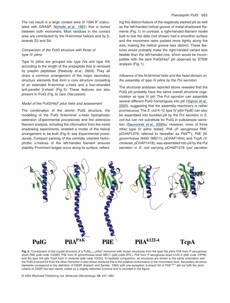
Type IV pilins are grouped into type IVa and type IVbaccording to the length of the prepeptide that is removedby prepilin peptidase (Peabody et al., 2003). They allshare a common arrangement of the major secondarystructure elements that form a core structure consistingof an extended N-terminal a-helix and a four-strandedanti-parallel b-sheet (Fig. 5). These features are alsopresent in PulG (Fig. 5) (see Discussion).
Model of the PulG(His)6 pilus helix and assessment
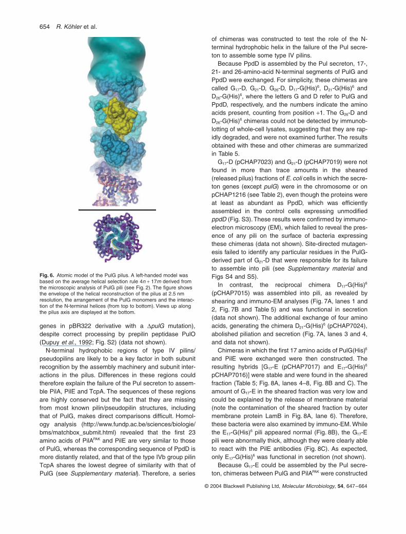
The combination of the atomic PulG structure, themodelling of the PulG N-terminal a-helix hydrophobicextension (Experimental procedures) and the extensivefilament analysis, including the information from the metalshadowing experiments, enabled a model of the helicalarrangement to be built (Fig. 6; see Experimental proce-dures). Compact packing of the centrally oriented hydro-phobic a-helices of the left-handed filament ensuresstability. Prominent bulges occur along its surface, reflect-
ing this distinct feature of the negatively stained pili as wellas the left-handed helical groove of metal-shadowed fila-ments (Fig. 1). In contrast, a right-handed filament modelbuilt to test the data (not shown) had a smoother surfaceand the monomers were packed more tightly along theaxis, making the helical groove less distinct. These fea-tures would probably make the right-handed variant lessflexible than the left-handed one, which would be incom-patible with the bent PulG(His)6 pili observed by STEManalysis (Fig. 1).
Influence of the N-terminal helix and the head domain on the assembly of type IV pilins by the Pul secreton
The structural analyses reported above revealed that thePulG pili probably have the same overall structural orga-nization as type IV pili. The Pul secreton can assembleseveral different PulG homologues into pili (Vignon et al.,2003), suggesting that the assembly machinery is ratherpromiscuous. The E. coli K-12 type IV pilin PpdD can alsobe assembled into bundled pili by the Pul secreton in E.coli but can not substitute for PulG in pullulanase secre-tion (Sauvonnet et al., 2000a). However, none of threeother type IV pilins tested, PilA (P. aeruginosa PAK;pCHAP1379; referred to hereafter as PilAPAK), PilE [N.gonorrhoeae N400 (MS11); pCHAP1404] and TcpA (V.cholerae, pCHAP1418), was assembled into pili by the Pulsecreton in E. coli carrying pCHAP1216 (pul secreton
Fig. 5. Comparison of the crystal structure of a PulG25-134(His)6 monomer with known structures from the type IVa pilins PilA from P. aeruginosa strain PAK (pdb code 1OQW), PilE from N. gonorrhoeae strain MS11 (pdb code 2PIL), PilA from P. aeruginosa strain k122-4 (pdb code 1HPW) and the type IVb pilin TcpA from V. cholerae (pdb cade 1OQV). To facilitate comparison, all structures are shown in the same orientation and the PulG b-strand S4 from the other monomer is also shown because this is the putative conformation in the monomeric form. Secondary structure elements correspond to the definition of DSSP (Kabsch and Sander, 1983) with one exception: b-strand S4 of PilAk122-4 did not fulfil the strict criteria of DSSP but was clearly visible as a slightly distorted b-strand and is included in the figure.
654 R. Köhler et al.
© 2004 Blackwell Publishing Ltd, Molecular Microbiology, 54, 647–664
genes in pBR322 derivative with a DpulG mutation),despite correct processing by prepilin peptidase PulO(Dupuy et al., 1992; Fig. S2) (data not shown).
N-terminal hydrophobic regions of type IV pilins/pseudopilins are likely to be a key factor in both subunitrecognition by the assembly machinery and subunit inter-actions in the pilus. Differences in these regions couldtherefore explain the failure of the Pul secreton to assem-ble PilA, PilE and TcpA. The sequences of these regionsare highly conserved but the fact that they are missingfrom most known pilin/pseudopilin structures, includingthat of PulG, makes direct comparisons difficult. Homol-ogy analysis (http://www.fundp.ac.be/sciences/biologie/bms/matchbox_submit.html) revealed that the first 23amino acids of PilAPAK and PilE are very similar to thoseof PulG, whereas the corresponding sequence of PpdD ismore distantly related, and that of the type IVb group pilinTcpA shares the lowest degree of similarity with that ofPulG (see Supplementary material). Therefore, a series
of chimeras was constructed to test the role of the N-terminal hydrophobic helix in the failure of the Pul secre-ton to assemble some type IV pilins.
Because PpdD is assembled by the Pul secreton, 17-,21- and 26-amino-acid N-terminal segments of PulG andPpdD were exchanged. For simplicity, these chimeras arecalled G17-D, G21-D, G26-D, D17-G(His)6, D21-G(His)6 andD26-G(His)6, where the letters G and D refer to PulG andPpdD, respectively, and the numbers indicate the aminoacids present, counting from position +1. The G26-D andD26-G(His)6 chimeras could not be detected by immunob-lotting of whole-cell lysates, suggesting that they are rap-idly degraded, and were not examined further. The resultsobtained with these and other chimeras are summarizedin Table 5.
G17-D (pCHAP7023) and G21-D (pCHAP7019) were notfound in more than trace amounts in the sheared(released pilus) fractions of E. coli cells in which the secre-ton genes (except pulG) were in the chromosome or onpCHAP1216 (see Table 2), even though the proteins wereat least as abundant as PpdD, which was efficientlyassembled in the control cells expressing unmodifiedppdD (Fig. S3). These results were confirmed by immuno-electron microscopy (EM), which failed to reveal the pres-ence of any pili on the surface of bacteria expressingthese chimeras (data not shown). Site-directed mutagen-esis failed to identify any particular residues in the PulG-derived part of G21-D that were responsible for its failureto assemble into pili (see Supplementary material andFigs S4 and S5).
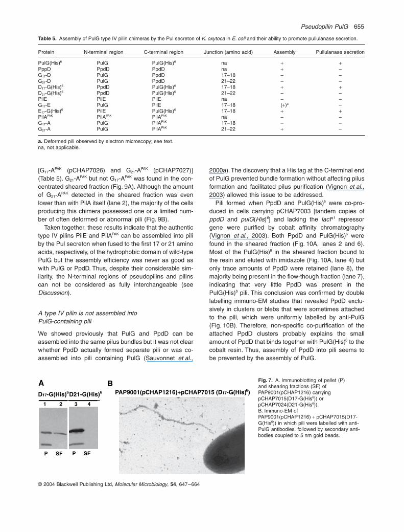
In contrast, the reciprocal chimera D17-G(His)6
(pCHAP7015) was assembled into pili, as revealed byshearing and immuno-EM analyses (Fig. 7A, lanes 1 and2, Fig. 7B and Table 5) and was functional in secretion(data not shown). The additional exchange of four aminoacids, generating the chimera D21-G(His)6 (pCHAP7024),abolished piliation and secretion (Fig. 7A, lanes 3 and 4,and data not shown).
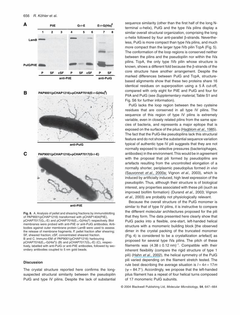
Chimeras in which the first 17 amino acids of PulG(His)6
and PilE were exchanged were then constructed. Theresulting hybrids [G17-E (pCHAP7017) and E17-G(His)6
pCHAP7016)] were stable and were found in the shearedfraction (Table 5; Fig. 8A, lanes 4–8, Fig. 8B and C). Theamount of G17-E in the sheared fraction was very low andcould be explained by the release of membrane material(note the contamination of the sheared fraction by outermembrane protein LamB in Fig. 8A, lane 6). Therefore,these bacteria were also examined by immuno-EM. Whilethe E17-G(His)6 pili appeared normal (Fig. 8B), the G17-Epili were abnormally thick, although they were clearly ableto react with the PilE antibodies (Fig. 8C). As expected,only E17-G(His)6 was functional in secretion (not shown).
Because G17-E could be assembled by the Pul secre-ton, chimeras between PulG and PilAPAK were constructed
Fig. 6. Atomic model of the PulG pilus. A left-handed model was based on the average helical selection rule 4n + 17m derived from the microscopic analysis of PulG pili (see Fig. 2). The figure shows the envelope of the helical reconstruction of the pilus at 2.5 nm resolution, the arrangement of the PulG monomers and the interac-tion of the N-terminal helices (from top to bottom). Views up along the pilus axis are displayed at the bottom.
Pseudopilin PulG 655
© 2004 Blackwell Publishing Ltd, Molecular Microbiology, 54, 647–664
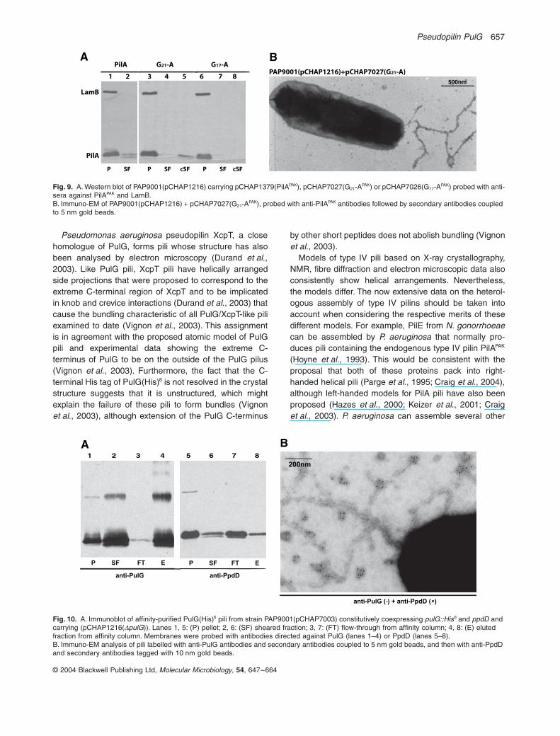
[G17-APAK (pCHAP7026) and G21-APAK (pCHAP7027)](Table 5). G21-APAK but not G17-APAK was found in the con-centrated sheared fraction (Fig. 9A). Although the amountof G21-APAK detected in the sheared fraction was evenlower than with PilA itself (lane 2), the majority of the cellsproducing this chimera possessed one or a limited num-ber of often deformed or abnormal pili (Fig. 9B).
Taken together, these results indicate that the authentictype IV pilins PilE and PilAPAK can be assembled into piliby the Pul secreton when fused to the first 17 or 21 aminoacids, respectively, of the hydrophobic domain of wild-typePulG but the assembly efficiency was never as good aswith PulG or PpdD. Thus, despite their considerable sim-ilarity, the N-terminal regions of pseudopilins and pilinscan not be considered as fully interchangeable (seeDiscussion).
A type IV pilin is not assembled into PulG-containing pili
We showed previously that PulG and PpdD can beassembled into the same pilus bundles but it was not clearwhether PpdD actually formed separate pili or was co-assembled into pili containing PulG (Sauvonnet et al.,
2000a). The discovery that a His tag at the C-terminal endof PulG prevented bundle formation without affecting pilusformation and facilitated pilus purification (Vignon et al.,2003) allowed this issue to be addressed.
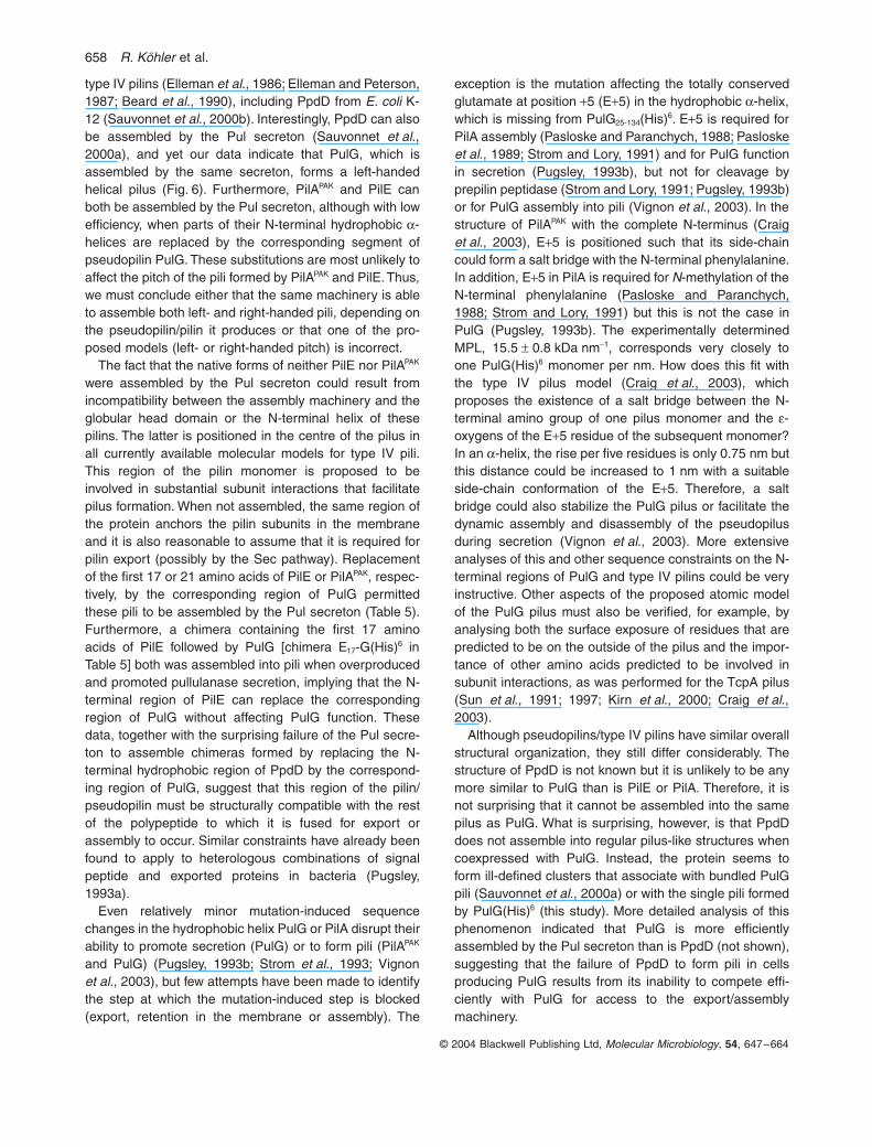
Pili formed when PpdD and PulG(His)6 were co-pro-duced in cells carrying pCHAP7003 [tandem copies ofppdD and pulG(His)6] and lacking the lacIq1 repressorgene were purified by cobalt affinity chromatography(Vignon et al., 2003). Both PpdD and PulG(His)6 werefound in the sheared fraction (Fig. 10A, lanes 2 and 6).Most of the PulG(His)6 in the sheared fraction bound tothe resin and eluted with imidazole (Fig. 10A, lane 4) butonly trace amounts of PpdD were retained (lane 8), themajority being present in the flow-though fraction (lane 7),indicating that very little PpdD was present in thePulG(His)6 pili. This conclusion was confirmed by doublelabelling immuno-EM studies that revealed PpdD exclu-sively in clusters or blebs that were sometimes attachedto the pili, which were uniformly labelled by anti-PulG(Fig. 10B). Therefore, non-specific co-purification of theattached PpdD clusters probably explains the smallamount of PpdD that binds together with PulG(His)6 to thecobalt resin. Thus, assembly of PpdD into pili seems tobe prevented by the assembly of PulG.
Table 5. Assembly of PulG type IV pilin chimeras by the Pul secreton of K. oxytoca in E. coli and their ability to promote pullulanase secretion.
Protein N-terminal region C-terminal region Junction (amino acid) Assembly Pullulanase secretion
PulG(His)6 PulG PulG(His)6 na + +PppD PpdD PpdD na + –G17-D PulG PpdD 17–18 – –G21-D PulG PpdD 21–22 – –D17-G(His)6 PpdD PulG(His)6 17–18 + +D21-G(His)6 PpdD PulG(His)6 21–22 – –PilE PilE PilE na – –G17-E PulG PilE 17–18 (+)a –E17-G(His)6 PilE PulG(His)6 17–18 + +PilAPAK PilAPAK PilAPAK na – –G17-A PulG PilAPAK 17–18 – –G21-A PulG PilAPAK 21–22 + –
a. Deformed pili observed by electron microscopy; see text.na, not applicable.
D21-G(His)6
BPAP9001(pCHAP1216)+pCHAP7015 (D17-G(His) )D17-G(His)6
A
1 2 3 4
P SF P SF
6
Fig. 7. A. Immunoblotting of pellet (P) and shearing fractions (SF) of PAP9001(pCHAP1216) carrying pCHAP7015(D17-G(His6)) or pCHAP7024(D21-G(His6)).B. Immuno-EM of PAP9001(pCHAP1216) + pCHAP7015(D17-G(His6)) in which pili were labelled with anti-PulG antibodies, followed by secondary anti-bodies coupled to 5 nm gold beads.
656 R. Köhler et al.
© 2004 Blackwell Publishing Ltd, Molecular Microbiology, 54, 647–664
Discussion
The crystal structure reported here confirms the long-suspected structural similarity between the pseudopilinPulG and type IV pilins. Despite the lack of substantial
sequence similarity (other than the first half of the long N-terminal a-helix), PulG and the type IVa pilins display asimilar overall structural organization, comprising the longa-helix followed by four anti-parallel b-strands. Neverthe-less, PulG is more compact than type IVa pilins, and muchmore compact than the larger type IVb pilin TcpA (Fig. 5).The conformation of the loop regions is conserved neitherbetween the pilins and the pseudopilin nor within the IVapilins. TcpA, the only type IVb pilin whose structure isknown, shows a different fold because the b-strands of thecore structure have another arrangement. Despite themarked differences between PulG and TcpA, structure-based alignments show that these two proteins share 16identical residues on superposition using a 5 Å cut-off,compared with only eight for PilE and PulG and four forPilA and PulG (see Supplementary material, Table S1 andFig. S6 for further information).
PulG lacks the loop region between the two cysteineresidues that are conserved in all type IV pilins. Thesequence of this region of type IV pilins is extremelyvariable, even in closely related pilins from the same spe-cies of bacteria, and represents a major epitope that isexposed on the surface of the pilus (Hagblom et al., 1985).The fact that the PulG-like pseudopilins lack this structuralfeature and do not show the substantial sequence variationtypical of authentic type IV pili suggests that they are notnormally exposed to selective pressures (bacteriophages,antibodies) in the environment. This would be in agreementwith the proposal that pili formed by pseudopilins areartefacts resulting from the uncontrolled elongation of anormally shorter, periplasmic pseudopilus formed in vivo(Sauvonnet et al., 2000a; Vignon et al., 2003), which isinduced by artificially induced, high level expression of thepseudopilin. Thus, although their structure is of biologicalinterest, any properties associated with these pili (such asimproved biofilm formation) (Durand et al., 2003; Vignonet al., 2003) are probably not physiologically relevant.
Because the overall structure of the PulG monomer issimilar to that of type IV pilins, it is instructive to comparethe different molecular architectures proposed for the pilithat they form. The data presented here clearly show thatPulG packs into a flexible, one-start, left-handed helicalstructure with a monomeric building block [the observeddimer in the crystal packing of the truncated monomer(Fig. 4) is considered to be a crystallization artefact] asproposed for several type IVa pilins. The pitch of thesefilaments was (4.38 ± 0.12 nm)-1. Compatible with theirinherent flexibility (compare the rigid structure of type 1pili) (Hahn et al., 2002), the helical symmetry of the PulGpili varied depending on the filament stretch tested. Therule best describing the average situation is l = 4n + 17m(y = 84.7∞). Accordingly, we propose that the left-handedpilus filament has a repeat of four helical turns composedof 17 monomeric PulG subunits.
Fig. 8. A. Analysis of pellet and shearing fractions by immunoblotting of PAP9001(pCHAP1216) transformed with pCHAP1404(PilE), pCHAP7017(G17-E) and pCHAP7016(E17-G(His)6) respectively. Blot membranes were probed with anti-PilE or anti-PulG antibodies. Anti-bodies against outer membrane protein LamB were used to assess the release of membrane fragments. P, pellet fraction after shearing; SF, sheared fraction; cSF, concentrated sheared fraction.B and C. Immuno-EM of PAP9001(pCHAP1216) harbouring pCHAP7016(E17-G(His6)) (B) and pCHAP7017(G17-E) (C), respec-tively, labelled with anti-PulG or anti-PilE antibodies, followed by sec-ondary antibodies coupled to 5 nm gold beads.
PAP9001(pCHAP1216)+pCHAP7017(G17-E)
PAP9001(pCHAP1216)+pCHAP7016(E17-G(His) )
P SF cSF P SF cSF
anti-PilE
P SF
anti-PulG
G17-E PilE E17-G(His)
1 2 3 4 5 6 7 8
anti-PilE
anti-PulG
LamB
PulG/PilE
B
C
A 6
6
Pseudopilin PulG 657
© 2004 Blackwell Publishing Ltd, Molecular Microbiology, 54, 647–664
Pseudomonas aeruginosa pseudopilin XcpT, a closehomologue of PulG, forms pili whose structure has alsobeen analysed by electron microscopy (Durand et al.,2003). Like PulG pili, XcpT pili have helically arrangedside projections that were proposed to correspond to theextreme C-terminal region of XcpT and to be implicatedin knob and crevice interactions (Durand et al., 2003) thatcause the bundling characteristic of all PulG/XcpT-like piliexamined to date (Vignon et al., 2003). This assignmentis in agreement with the proposed atomic model of PulGpili and experimental data showing the extreme C-terminus of PulG to be on the outside of the PulG pilus(Vignon et al., 2003). Furthermore, the fact that the C-terminal His tag of PulG(His)6 is not resolved in the crystalstructure suggests that it is unstructured, which mightexplain the failure of these pili to form bundles (Vignonet al., 2003), although extension of the PulG C-terminus
by other short peptides does not abolish bundling (Vignonet al., 2003).
Models of type IV pili based on X-ray crystallography,NMR, fibre diffraction and electron microscopic data alsoconsistently show helical arrangements. Nevertheless,the models differ. The now extensive data on the heterol-ogous assembly of type IV pilins should be taken intoaccount when considering the respective merits of thesedifferent models. For example, PilE from N. gonorrhoeaecan be assembled by P. aeruginosa that normally pro-duces pili containing the endogenous type IV pilin PilAPAK
(Hoyne et al., 1993). This would be consistent with theproposal that both of these proteins pack into right-handed helical pili (Parge et al., 1995; Craig et al., 2004),although left-handed models for PilA pili have also beenproposed (Hazes et al., 2000; Keizer et al., 2001; Craiget al., 2003). P. aeruginosa can assemble several other
Fig. 9. A. Western blot of PAP9001(pCHAP1216) carrying pCHAP1379(PilAPAK), pCHAP7027(G21-APAK) or pCHAP7026(G17-APAK) probed with anti-sera against PilAPAK and LamB.B. Immuno-EM of PAP9001(pCHAP1216) + pCHAP7027(G21-APAK), probed with anti-PilAPAK antibodies followed by secondary antibodies coupled to 5 nm gold beads.
500nm
PAP9001(pCHAP1216)+pCHAP7027(G21-A)PilA
P SF P SF cSF P SF cSF
G21-A G17-A
LamB
PilA
B
1 2 3 4 5 6 7 8
A
Fig. 10. A. Immunoblot of affinity-purified PulG(His)6 pili from strain PAP9001(pCHAP7003) constitutively coexpressing pulG::His6 and ppdD and carrying (pCHAP1216(DpulG)). Lanes 1, 5: (P) pellet; 2, 6: (SF) sheared fraction; 3, 7: (FT) flow-through from affinity column; 4, 8: (E) eluted fraction from affinity column. Membranes were probed with antibodies directed against PulG (lanes 1–4) or PpdD (lanes 5–8).B. Immuno-EM analysis of pili labelled with anti-PulG antibodies and secondary antibodies coupled to 5 nm gold beads, and then with anti-PpdD and secondary antibodies tagged with 10 nm gold beads.
P SF FT E
anti-PulG anti-PpdD
P SF FT E
200nm
anti-PulG ( ) + anti-PpdD ( )
A B1 2 3 4 5 6 7 8
658 R. Köhler et al.
© 2004 Blackwell Publishing Ltd, Molecular Microbiology, 54, 647–664
type IV pilins (Elleman et al., 1986; Elleman and Peterson,1987; Beard et al., 1990), including PpdD from E. coli K-12 (Sauvonnet et al., 2000b). Interestingly, PpdD can alsobe assembled by the Pul secreton (Sauvonnet et al.,2000a), and yet our data indicate that PulG, which isassembled by the same secreton, forms a left-handedhelical pilus (Fig. 6). Furthermore, PilAPAK and PilE canboth be assembled by the Pul secreton, although with lowefficiency, when parts of their N-terminal hydrophobic a-helices are replaced by the corresponding segment ofpseudopilin PulG. These substitutions are most unlikely toaffect the pitch of the pili formed by PilAPAK and PilE. Thus,we must conclude either that the same machinery is ableto assemble both left- and right-handed pili, depending onthe pseudopilin/pilin it produces or that one of the pro-posed models (left- or right-handed pitch) is incorrect.
The fact that the native forms of neither PilE nor PilAPAK
were assembled by the Pul secreton could result fromincompatibility between the assembly machinery and theglobular head domain or the N-terminal helix of thesepilins. The latter is positioned in the centre of the pilus inall currently available molecular models for type IV pili.This region of the pilin monomer is proposed to beinvolved in substantial subunit interactions that facilitatepilus formation. When not assembled, the same region ofthe protein anchors the pilin subunits in the membraneand it is also reasonable to assume that it is required forpilin export (possibly by the Sec pathway). Replacementof the first 17 or 21 amino acids of PilE or PilAPAK, respec-tively, by the corresponding region of PulG permittedthese pili to be assembled by the Pul secreton (Table 5).Furthermore, a chimera containing the first 17 aminoacids of PilE followed by PulG [chimera E17-G(His)6 inTable 5] both was assembled into pili when overproducedand promoted pullulanase secretion, implying that the N-terminal region of PilE can replace the correspondingregion of PulG without affecting PulG function. Thesedata, together with the surprising failure of the Pul secre-ton to assemble chimeras formed by replacing the N-terminal hydrophobic region of PpdD by the correspond-ing region of PulG, suggest that this region of the pilin/pseudopilin must be structurally compatible with the restof the polypeptide to which it is fused for export orassembly to occur. Similar constraints have already beenfound to apply to heterologous combinations of signalpeptide and exported proteins in bacteria (Pugsley,1993a).
Even relatively minor mutation-induced sequencechanges in the hydrophobic helix PulG or PilA disrupt theirability to promote secretion (PulG) or to form pili (PilAPAK
and PulG) (Pugsley, 1993b; Strom et al., 1993; Vignonet al., 2003), but few attempts have been made to identifythe step at which the mutation-induced step is blocked(export, retention in the membrane or assembly). The
exception is the mutation affecting the totally conservedglutamate at position +5 (E+5) in the hydrophobic a-helix,which is missing from PulG25-134(His)6. E+5 is required forPilA assembly (Pasloske and Paranchych, 1988; Pasloskeet al., 1989; Strom and Lory, 1991) and for PulG functionin secretion (Pugsley, 1993b), but not for cleavage byprepilin peptidase (Strom and Lory, 1991; Pugsley, 1993b)or for PulG assembly into pili (Vignon et al., 2003). In thestructure of PilAPAK with the complete N-terminus (Craiget al., 2003), E+5 is positioned such that its side-chaincould form a salt bridge with the N-terminal phenylalanine.In addition, E+5 in PilA is required for N-methylation of theN-terminal phenylalanine (Pasloske and Paranchych,1988; Strom and Lory, 1991) but this is not the case inPulG (Pugsley, 1993b). The experimentally determinedMPL, 15.5 ± 0.8 kDa nm-1, corresponds very closely toone PulG(His)6 monomer per nm. How does this fit withthe type IV pilus model (Craig et al., 2003), whichproposes the existence of a salt bridge between the N-terminal amino group of one pilus monomer and the e-oxygens of the E+5 residue of the subsequent monomer?In an a-helix, the rise per five residues is only 0.75 nm butthis distance could be increased to 1 nm with a suitableside-chain conformation of the E+5. Therefore, a saltbridge could also stabilize the PulG pilus or facilitate thedynamic assembly and disassembly of the pseudopilusduring secretion (Vignon et al., 2003). More extensiveanalyses of this and other sequence constraints on the N-terminal regions of PulG and type IV pilins could be veryinstructive. Other aspects of the proposed atomic modelof the PulG pilus must also be verified, for example, byanalysing both the surface exposure of residues that arepredicted to be on the outside of the pilus and the impor-tance of other amino acids predicted to be involved insubunit interactions, as was performed for the TcpA pilus(Sun et al., 1991; 1997; Kirn et al., 2000; Craig et al.,2003).
Although pseudopilins/type IV pilins have similar overallstructural organization, they still differ considerably. Thestructure of PpdD is not known but it is unlikely to be anymore similar to PulG than is PilE or PilA. Therefore, it isnot surprising that it cannot be assembled into the samepilus as PulG. What is surprising, however, is that PpdDdoes not assemble into regular pilus-like structures whencoexpressed with PulG. Instead, the protein seems toform ill-defined clusters that associate with bundled PulGpili (Sauvonnet et al., 2000a) or with the single pili formedby PulG(His)6 (this study). More detailed analysis of thisphenomenon indicated that PulG is more efficientlyassembled by the Pul secreton than is PpdD (not shown),suggesting that the failure of PpdD to form pili in cellsproducing PulG results from its inability to compete effi-ciently with PulG for access to the export/assemblymachinery.
Pseudopilin PulG 659
© 2004 Blackwell Publishing Ltd, Molecular Microbiology, 54, 647–664
The studies reported here represent a structural basisfor exploring the role of PulG in secretion. The pilusformed by pseudopilins is proposed to represent an aber-rant variant of a pseudopilus that is usually restricted tothe periplasm and might function as a dynamic motor orpiston that pushes secreted proteins through the secretinchannel in the outer membrane. The validation of thishypothesis will require analysis of the way pilus growthand retraction are promoted and regulated, of how theminor pseudopilin PulI promotes pilus PulG assembly(Sauvonnet et al., 2000a), of how PulK performs its pro-posed role in the control of (pseudo)pilus length (Vignonet al., 2003) and of the specific role of PulJ in secretion.Structure analysis of these proteins and studies of theirability to interact with the PulG pilus that are currentlyunderway will provide valuable insight into this fascinatingsecretion machine.
Experimental procedures
Strains and growth conditions
Escherichia coli strains are listed in Table 1. Bacteria weregrown at 30∞C in Luria–Bertani (LB) broth (Miller, 1992) oron LB agar plates supplemented, where appropriate, withmaltose (0.4%), chloramphenicol (25 mg ml-1) or ampicillin(50 or 100 mg ml-1).
DNA manipulations and plasmids
The sequence encoding for the N-terminal truncated PulGgene from position +25 relative to the prepeptide cleavagesite was amplified by standard polymerase chain reaction(PCR) reactions using pCHAP1362 as template. Primersused for amplification were 5¢PulG(NcoI) 5¢-CCC AAC CCCATG GGC AAC AAG GAA-3¢ and 3¢PulG(XbaI) 5¢-CAT CTCTAG AAA CGT AAA GCC GCG-3¢. The restriction sites usedto fuse the fragment to the signal peptide of E. chrysanthemipectate lyase PelB (PelBSP) in pCHAP4260 (Apr) are under-lined and resulted in pCHAP7010. The entire fragmentencoding PelBSP-PulG25-134(His)6 was subcloned intopSU18(Cmr) using EcoRI and HindIII, giving pCHAP7012that was used to test complementation and transdominanteffects in a pullulanase secretion assay (Michaelis et al.,1985). For higher level production, the same fragment wassubcloned into the overexpression vector pT7-5 (Tabor andRichardson, 1985), giving pCHAP7013.
Plasmid pCHAP7009 (DpulG, DpulA) was constructed inorder to purify full-length PulG without co-purifying PulA.pCHAP1216, a DpulG variant of pCHAP231, was digestedusing EcoRI and HindIII to release a ~12 kb fragment carry-ing all pul genes between pulB and pulF. The fragment wassubcloned into pUC19, giving plasmid pCHAP7007. BamHIdigestion was used to release two internal pulA fragmentsfrom pCHAP7007. The remaining vector sequence encodingpulB-DpulA-pulF was gel purified and religated givingpCHAP7008. Finally, the entire fragment was released frompCHAP7008 by digestion with EcoRI and HindIII and reinte-
grated into pCHAP1216 cut with EcoRI and HindIII (pulF-pulO), resulting in pCHAP7009.
pCHAP1404 and pCHAP1418 (pilE and tcpA respectively)were constructed by amplifying DNA from plasmidspPilEN400 (M. Koomey) and pRT198 (R. Taylor), respec-tively, using primers that flanked the pilin gene and that intro-duced sites (EcoRI and HindIII and XbaI and HindIIIrespectively) that permitted their oriented insertion into thecorresponding sites of pSU18 or pSU19 (Bartolomé et al.,1991). In both cases, the 5¢ oligonucleotide was designed topermit in frame fusion to the first few codons of the lacZ genein the vector to ensure efficient translation initiation in E. coli(Table S2). The resulting proteins were readily detectablewith antibodies against PilE and TcpA respectively.pCHAP1379 (pilA) was obtained by direct subcloning of a1 kb HindIII fragment from pAW103 (J. Mattick) into pSU18so that the gene was under lacZp control.
Polymerase chain reactions with overlapping primer pairs(Table S3) were used to generate gene fusions encoding forhybrids of which the N-terminal a-helix up to and includingpositions +17, +21 or +26 (relative to the prepeptide cleav-age site) and the hydrophilic region of PulG, PpdD, PilE andPilAPAK (downstream from position +18, +22 and +27) wereexchanged. Sequences encoding chimeras were first ampli-fied in two fragments using either oligonucleotides 1 and 2or oligonucleotides 3 and 4 and the appropriate templateDNA (Table S3). Fragments were then amplified using stan-dard PCRs with Herculase (Stratagene) and purified usingQIAquick columns (Qiagen). Fragments to be fused wereheated for 2 min to 92∞C and then held at room temperatureto allow hybridization. These samples were used as tem-plates in a nested PCR to amplify the entire fusion fragmentusing only the outer 5¢ (1) and 3¢ (4) primers. Subsequently,the PCR product was gel purified and digested with restric-tion enzymes cleaving cognate sequences introduced by theoligonucleotides, and the fragments were cloned intopSU18. The domain exchanges were confirmed by DNAsequencing.
Periplasmic extraction and purification of PelBSP-PulG25-134(His)6
To produce PelBSP-PulG25-134(His)6, E. coli BL21(DE3) (F–
ompT lon hsdSB, lDE3; carries phage T7 DNA polymerasegene under lacZp control) transformed with pCHAP7013 wasgrown overnight in LB medium at 30∞C. All subsequent prep-aration and incubation steps were carried out on ice andcentrifugation was carried out at 4∞C. The cells were resus-pended in 10 mM Tris/HCl (pH 7.4) containing 20% sucroseand 20 mM EDTA and incubated for 10 min. Bacteria werecentrifuged (20 000 g for 5 min), resuspended in 25 mMMgCl2 and held for another 10 min. The cells were thencentrifuged (20 000 g for 5 min). PelBSP-PulG25-134(His)6 in thesupernatant was affinity purified (Vignon et al., 2003) andeluted fractions were pooled and dialysed overnight twiceagainst 10 mM Hepes (pH 7.4) using Spectra/Por mem-branes with a size cut-off of 3.5 kDa. After dialysis, the pro-tein solution was concentrated to 4 mg ml-1 using Centriprepand Centricon devises (YM-10) with a size cut-off of 10 kDa.Selenomethionine-labelled PelBSP-PulG25-134(His)6 was pre-pared from the same strain grown in minimal medium con-
660 R. Köhler et al.
© 2004 Blackwell Publishing Ltd, Molecular Microbiology, 54, 647–664
taining M63 stock salts (Miller, 1992) with glucose (0.2%) andamino acids (4 mg ml-1) except methionine, which was addedin the form of selenomethionine to a final concentration of4 mg ml-1.
Shearing and immunoblotting
Bacteria were harvested from the plates and resuspended inphosphate-buffered saline to an optical density of 1.0 (SF) or2.0 (cSF) at 600 nm. The suspension was centrifuged twiceat 13 000 g in a microcentrifuge for 5 min (each time) toseparate bacteria (the pellet fraction) from the pilus-enrichedsupernatant (sheared fraction). Fractions were precipitatedwith 10% trichloroacetic acid and loaded onto sodium dode-cyl sulphate (SDS)-12% polyacrylamide gels for immunoblot-ting. Proteins were separated by SDS-polyacrylamide gelelectrophoresis (PAGE), electroblotted onto nitrocellulosemembranes, and incubated first with specific anti-serum[polyclonal anti-PulG at 1/6000, anti-PpdD at 1/1000, anti-PilE at 1/4000 (M. Koomey), anti-PilAPAK at 1/1000 (S. Lory)and anti-LamB at 1/5000] and then with horseradishperoxidase-coupled anti-rabbit or anti-mouse immunoglobu-lin G (1/10 000; Amersham). The membranes were devel-oped by enhanced chemiluminescence (Amersham).Procedures for transmission electron microscopy wereessentially as used previously (Sauvonnet et al., 2000a),with specific antibodies diluted to 1:100 and with 10 nm or5 nm gold beads coupled to the secondary antibodies. Spec-imens were analysed as described elsewhere (Vignon et al.,2003).
Gel filtration
The assembly state of full-length PulG and of soluble trun-cated PulG were determined by FPLC size-exclusion chro-matography with a Superose12 (24 ml) column (Pharmacia)equilibrated with 50 mM phosphate buffer (pH 7.0) containing150 mM NaCl and 0.7% C8E4. Molecular sizes were esti-mated from standards purchased from Sigma (bovine serumalbumen, 66 kDa, and carbonic anhydrase, 29 kDa).
Freeze drying and metal shadowing
Five microlitre aliquots of the sample were adsorbed to glow-discharged carbon-coated collodion films on 400 mesh/inchcopper grids, washed on four drops of water and, afterremoval of excess liquid, frozen by plunging into liquidnitrogen. Freeze drying was performed in a BAF 300 freeze-fracture device (Balzers AG, Liechtenstein) for 1 h at a sam-ple temperature of -80∞C followed by 1 h at -35∞C, and apressure of £5 ¥ 10-7 Torr. Finally, the grids were unidirection-ally metal shadowed with tungsten/tantalum (W/Ta) at anelevation angle of 45∞ to yield an average metal film thicknessof ª 1 nm, and reinforced with carbon to avoid rehydratationand oxidation of the metal film.
The grids were examined and photographed in a HitachiH-7000 transmission electron microscope operating at100 kV. Electron micrographs were recorded at ¥30 000nominal magnification on Kodak SO163 sheet film.
STEM
A Vacuum Generators (East Grinstead) HB-5 STEM, inter-faced to a modular computer system (Tietz Video and ImageProcessing Systems), was used. All samples were preparedon 200-mesh-per-inch gold-plated copper grids as describedpreviously (Müller et al., 1992). Isolated tobacco mosaic virusparticles (kindly supplied by R. Diaz-Avalos) adsorbed to aseparate grid and air-dried, served as mass standard for themeasurements.
For structural analysis, digital 512 ¥ 512 pixel dark-fieldimages were recorded from the isolated negativelystained pili at an acceleration voltage of 100 kV and anominal magnification of 500 000¥ using doses of(7.6 ± 1.2) ¥ 103 electrons nm-2. The pixel size is dependenton the focus conditions for the STEM (Müller et al., 1992) andwas 0.330 ± 0.004 nm for the images analysed.
For mass determination 512 ¥ 512 pixel dark-field imageswere recorded from unstained samples of the isolated pili atan acceleration voltage of 80 kV and a nominal magnificationof 200 000¥ using doses of 414 ± 36 electrons nm-2. In addi-tion, repeated low-dose scans were recorded from some gridregions to assess beam-induced mass loss. The imageswere evaluated with the program package IMPSYS asdescribed previously (Müller et al., 1992). Accordingly, pilussegments were defined by square boxes, tracked and theirlength and mass profile calculated. The total scattering withinan integration box matched to their width was then deter-mined, the background scattering of the carbon support filmsubtracted and the average MPL calculated. Furthermore,the FWHM was determined from the mass profile. The beam-induced mass loss was calculated as described previously(Müller et al., 1992). The experimental MPL results were thenmultiplied by the correction factor calculated for the recordingdose used, scaled according to the MPL measured for TMVparticles. Finally, the MPL and FWHM values were binnedinto histograms and fitted with Gauss curves. The overallexperimental uncertainty of the MPL average was estimatedfrom the corresponding SE (SE = SD/÷n) and the 5% uncer-tainty in the calibration of the instrument by calculating thesquare root of the sum of the squares.
Digital image processing
Using the SEMPER program package (Saxton et al., 1979),suitable negatively stained pilus stretches were computation-ally unbent using a cubic spline interpolation scheme asdescribed previously (Hahn et al., 2000). To estimate theouter pilus diameter, selected unbent pili were averagedalong their axis and the resulting intensity profile was plotted.The distance between the two outer minima of this profilewas taken as the outer diameter. Power spectra were calcu-lated for the unbent pilus segments and the altitude of themost prominent layer line was determined. Layer lines wereextracted from the 2D Fourier transforms of the unbent rodsto inspect the amplitudes and phases.
Further processing was performed using the ModularMicrograph Data Processing Program (MDPP; Smith andGottesman, 1996). First, the pitch of well-preserved pili wasalso determined in real space using the linear Markhamsuperposition procedure described previously (Hahn et al.,
Pseudopilin PulG 661
© 2004 Blackwell Publishing Ltd, Molecular Microbiology, 54, 647–664
2000). Averaging runs were made over two, three and fourrepeat lengths respectively. In each case, the repeat lengthwas incremented in steps of one pixel until the maximumpossible length had been attained (limited by the piluslength). The degree of enhancement on superposition andaveraging was quantified by comparing the resulting rmsdeviation with that of the segment used, whereby maximumvalues of the quotient correspond to minimal ‘blurring’ of theaverage image, i.e. minimum power loss and, thus, to theoptimal enhancement of the true repeat. The rms quotientwas then plotted for each repeat length and the local maximaassessed graphically.
All helical selection rules falling within the range compati-ble with helical repeats less than or equal to 10 times thepitch and including screw angles within the range of 72∞-90∞ were compared in batches. For this, the straightenedpilus segments were trimmed to an integer multiple of thehelical repeat and interpolated onto the sampling grid calcu-lated to give four pixels per axial rise. The width of the fila-ment array was trimmed to minimize the background effectof the negative stain, and the six marginal pixels on eitherside of the array parallel to the pilus axis were floated to themean density value. The 2D Fourier transform was calcu-lated and D(Z,k) filtered as described (Smith and Aebi,1974). At this stage 16 Bessel coefficients and the numberof layer lines indicated by the rule under test were used.The rules within a batch were tested on exactly the samestretch of pilus, allowing the results to be compared, e.g.the rule l = 1n + 5m was in the same batch as the rule10n + 43m and the total filament length tested was 260 pix-els. Thus for the first rule there were 20 helical repeats andfor the second only two.
A stack of 200 sections of 52 ¥ 52 pixels and spaced by0.26 nm was calculated from the D(Z,k)-filtered layer linesresulting from nine helical repeats containing 153 subunits ofone well-preserved pilus rod by back-transformation. Thisreconstruction comprised 50 axial repeats. It was isocon-toured to include 100% of the nominal mass taking a densityof 810 Da nm-3 into account.
Crystallization and data collection
Small hexagonal bipyramidal crystals of N-terminal trun-cated PulG25-134(His)6 (50 mm ¥ 50 mm ¥ 70 mm) were ob-tained by the hanging drop vapour diffusion method. Twomicrolitres of 20 mg ml-1 purified PulG25-134(His)6 in 10 mMHepes pH 7.2, 0.02% (w/v) sodium azide were mixed with2 ml of reservoir solution containing 1.7 M ammonium sul-phate and 5% (v/v) isopropanol as precipitants. These crys-tals could be enlarged by macro seeding to a final size of200 mm ¥ 200 mm ¥ 300 mm by transferring them into a newdrop composed of 2 ml of protein solution (30 mg ml-1) and2 ml of reservoir [1.6 mM ammonium sulphate, 5% (v/v) iso-propanol]. The space group was found to be P6522 with celldimensions of 85.4 Å for the a/b-axis and 145.7 Å for thec-axis.
Crystals of selenomethionine-containing PulG25-134(His)6
were grown and analysed to resolve the crystallographicphase problem. Well-diffracting derivative crystals of thesame space group grew in hanging drops containing2 ml of selenomethionine PulG25-134(His)6 15 mg ml-1 in
10 mM Hepes pH 7.2, 0.02% (w/v sodium azide) and 2 mlof the reservoir [1.9 M ammonium sulphate, 6.5% (v/v)isopropanol].
Diffraction data were collected under cryogenic condi-tions. The crystals were flash frozen in liquid nitrogen inbuffer corresponding to the equilibrated crystallization dropsplus 25% (v/v) glycerol as a cryoprotectant. Two data setsfrom one native PulG25-134(His)6 crystal were collected: oneon a rotating anode X-ray source equipped with a Mar345detector and one high resolution data set at the Swiss-Light-Source (SLS, beamline X06SA) provided with aMarCCD detector. Both data sets were processed usingXDS2002 (Kabsch, 1993) and subsequently merged inXSCALE2002.
Data from one derivative crystal were collected at the SLSat two different wavelengths, corresponding to the remotehigh value and the inflection point of the anomalous signalfrom the covalently bound selenium atoms. These data setswere also reduced using XDS2002. More details of the datacollection statistics are shown in Table 3.
Crystal structure determination and refinement
The crystallographic phase problem was solved by multipleanomalous dispersion (MAD). Two strong and a further fourless occupied heavy atom positions were found in the deriv-ative crystal using the program SHELXD (Schneider andSheldrick, 2002). All of them were taken as selenium atomsfor experimental phase determination. The calculated phaseswere combined with the intensities from the merged nativedata set and extended to 1.8 Å in SHELXE. This resulted inan interpretable electron density map (final weighted contrast0.71, connectivity 0.95 as given by SHELXE).
Model building and refinement was done with programs ofthe CCP4-suite (Collaborative Computational Project, 1994)and the graphical model building program ‘O’ (Jones et al.,1991) for visible inspection and manual improvement of themodel. A starting model consisting of 199 peptides distrib-uted over 6 separated chains was obtained by automaticmodel building with the program ARPWARP (Perrakis et al.,1999) using the extended experimental phases and all nativedata. It clearly corresponds to a dimer in the asymmetric unit.Side-chain atoms as well as other missing parts of the struc-ture were added manually, followed by alternating refinementcycles including non-crystallographic symmetry restraintsand anisotropic B-factor refinement in REFMAC5 (Murshu-dov et al., 1997; 1999). Solvent atoms were added automat-ically by the program ARPWARP (Lamzin and Wilson, 1993).Model statistics are summarized in Table 4.
Modelling of the PulG pilus
The monomer derived from the crystal structure was pre-pared for helical modelling as follows: (i) the C-terminal sec-tion (amino acids N128-G132) including b-strand S4 whichforms the interdomain contact in the crystallographic dimerwas relocated into its monomer by removing the secondmonomer of the asymmetric unit, (ii) the flexible hingeregion composed of I126 and G127 was modelled with theSWISS-MODEL server (http://www.expasy.org/swissmod/SWISS-MODEL.html) and (iii) the N-terminal a-helix was
662 R. Köhler et al.
© 2004 Blackwell Publishing Ltd, Molecular Microbiology, 54, 647–664
elongated. For this, the backbone of the 24 missing residueswere taken from the N-terminal helix of the PulG25–134(His)6
structure and manually moved to the corresponding positionin the graphical model building software ‘O’. Side-chain posi-tions were taken as the standard conformations proposed bythe program ‘O’.
Modelling was performed using the visualization tool DINO(http://www.dino3d.org), which allows the display of helicalsymmetry mates as well as their synchronous real-timeupdate upon rotation and translation of the reference mono-mer. Constraints applied during the modelling procedurewere: (i) helical symmetry according to the performed analy-sis: rotation angle of ±84.71 degree per subunit and an axialrise of 1.04 nm, (ii) the long a-helix was located within thecore of the pilus helix, both axes quasi-parallel, (iii) the C-terminus was exposed to the outside medium and (iv) com-pact packing was ensured visually, avoiding steric clashesand cavities within the pilus core.
Acknowledgements
We would like to thank Marie-Christine Prévost for her guid-ance and permission to use the facilities at the ElectronMicroscope Station at the Institut Pasteur, Michael Koomey,Steve Lory, John Mattick and Ron Taylor for plasmids and/orantibodies and Vesna Olivieri for the STEM microscopy. Wealso thank Erik Hahn, Michel Steinmetz and Ueli Aebi for theirhelp in the use of the helical processing programs. The Euro-pean Commission (Contract No. HPRN-2000-00075), theMaurice E. Müller Foundation of Switzerland and the SwissNational Foundation contributed financially to this work.
Supplementary material
The following material is available from http://www.blackwellpublishing.com/products/journals/suppmat/mmi/mmi4307/mmi4307sm.htmAppendix A1. The supplementary material includes struc-tural and functional data on the PulG pilus and control dataon the failure of the Pul secreton to assemble the TcpA pilus.Fig. S1. The metal binding site in the dimeric crystal form ofPulG25-134(His)6. Fig. S2. Processing of preTcpA in E. coli strain carrying theprepilin peptidase structural gene pulO. Fig. S3. Analysis of pellet and sheared fractions ofE. coli stains PAP7000BG, PAP7501(pCHAP1216) andPAP9001(pCHAP1216) carrying pCHAP3100 (ppdD),pCHAP7023 and pCHAP7019 (encoding G17-D and G21-Drespectively) by immunoblotting. Fig. S4. Multiple sequence alignment of the first 23 N-terminal amino acids of PulG, PilE, PilAPAK and PpdD. Fig. S5. Western blot analysis of bacterial pellet, shearedfractions and concentrated sheared fractions from E. coliK-12 strain PAP7500BG(DpulG) harbouring pCHAP3100(encoding PpdD) (lanes 1–3) and pCHAP7028 (encodingG21(modified)-D) (lanes 4–6) respectively. Fig. S6. A structural alignment of the C-terminal globulardomain of the pseudopilin PulG with known type IV pilinstructures.
Table S1. Similarity of atomic structures of type IV pilins andPulG. The structures were superimposed with the programLSQMAN as described in Fig. S2.Table S2. Oligonucleotide primers used to amplify pilE andtcpA genes.Table S3. Oligonucleotide primers used to construct pilin/pseudopilin chimeras in which the N-terminal domains wereinterchanged.
References
Bartolomé, B., Jubete, Y., Martinez, E., and de la Cruz, F.(1991) Construction and properties of a family ofpACYC184-derived cloning vectors compatible withpBR322 and its derivatives. Gene 102: 75–78.
Beard, M.K., Mattick, J.S., Moor, M.R., Marrs, C.F., and Eger-ton, J.R. (1990) Morphogenic expression of Moraxellabovis fimbriae (pili) in Pseudomonas aeruginosa. J Bacte-riol 172: 2601–2607.
Bitter, W., Koster, M., Latijnhouwers, M., de Cock, H., andTommassen, J. (1998) Formation of oligomeric rings byXcpQ and PilQ, which are involved in protein transportacross the outer membrane of Pseudomonas aeruginosa.Mol Microbiol 27: 209–219.
Brok, R., Van Gelder, P., Winterhalter, M., Ziese, U., Koster,A.J., de Cock, H., et al. (1999) The C-terminal domain ofthe Pseudomonas secretin XcpQ forms oligomeric ringswith pore activity. J Mol Biol 294: 1169–1179.
Collaborative Computational Project (1994) The CCP4 suite:programs for protein crystallography. Acta Cryst D50: 760–763.
Collins, R.F., Davidsen, L., Derrick, J.P., Ford, R.C., andTonjum, T. (2001) Analysis of the PilQ secretin from Neis-seria meningitidis by transmission electron microscopyreveals a dodecameric quaternary structure. J Bacteriol183: 3825–3832.
Collins, R.F., Ford, R.C., Kitmitto, A., Olsen, R.O., Tonjum,T., and Derrick, J.P. (2003) Three-dimensional structure ofthe Neisseria meningitidis secretin PilQ determined fromnegative-stain transmission electron microscopy. J Bacte-riol 185: 2611–2617.
Craig, L., Taylor, R., Pique, M.E., Adair, B.D., Arvai, A.S.,Singh, M., et al. (2003) Type IV pilin structure and assem-bly. X-ray and EM analyses of Vibrio cholerae toxin-coregulated pilus and Pseudomonas aeruginosa PAK pilin.Mol Cell 11: 1139–1150.
Craig, L., Pique, M.E., and Tainer, J.A. (2004) Type IV pilusstructure and bacterial pathogenicity. Nature Rev Microbiol2: 363–378.
DeRosier, D.J., and Klug, A. (1968) Reconstitution of three-dimensional structures from electron micrographs. Nature217: 130–134.
Diederichs, K., and Karplus, P.A. (1997) Improved R-factorsfor diffraction data analysis in macromolecular crystallog-raphy. Nature Struct Biol 4: 269–275.
Dupuy, B., Taha, M.-K., Possot, O., Marchal, C., and Pugsley,A.P. (1992) PulO, a component of the pullulanase secre-tion pathway of Klebsiella oxytoca, correctly and efficientlyprocesses gonococcal type IV prepilin in Escherichia coli.Mol Microbiol 6: 1887–1894.
Durand, E., Bernadac, A., Ball, G., Lazdunski, A., Sturgis,
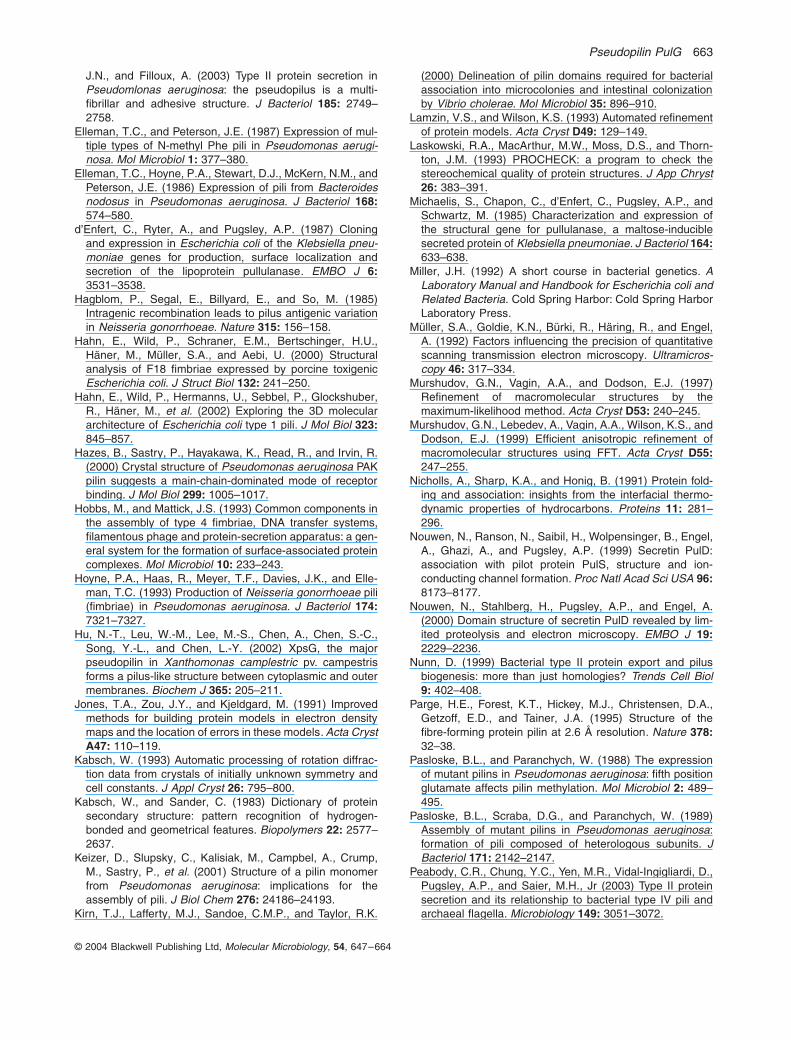
Pseudopilin PulG 663
© 2004 Blackwell Publishing Ltd, Molecular Microbiology, 54, 647–664
J.N., and Filloux, A. (2003) Type II protein secretion inPseudomlonas aeruginosa: the pseudopilus is a multi-fibrillar and adhesive structure. J Bacteriol 185: 2749–2758.
Elleman, T.C., and Peterson, J.E. (1987) Expression of mul-tiple types of N-methyl Phe pili in Pseudomonas aerugi-nosa. Mol Microbiol 1: 377–380.
Elleman, T.C., Hoyne, P.A., Stewart, D.J., McKern, N.M., andPeterson, J.E. (1986) Expression of pili from Bacteroidesnodosus in Pseudomonas aeruginosa. J Bacteriol 168:574–580.
d’Enfert, C., Ryter, A., and Pugsley, A.P. (1987) Cloningand expression in Escherichia coli of the Klebsiella pneu-moniae genes for production, surface localization andsecretion of the lipoprotein pullulanase. EMBO J 6:3531–3538.
Hagblom, P., Segal, E., Billyard, E., and So, M. (1985)Intragenic recombination leads to pilus antigenic variationin Neisseria gonorrhoeae. Nature 315: 156–158.
Hahn, E., Wild, P., Schraner, E.M., Bertschinger, H.U.,Häner, M., Müller, S.A., and Aebi, U. (2000) Structuralanalysis of F18 fimbriae expressed by porcine toxigenicEscherichia coli. J Struct Biol 132: 241–250.
Hahn, E., Wild, P., Hermanns, U., Sebbel, P., Glockshuber,R., Häner, M., et al. (2002) Exploring the 3D moleculararchitecture of Escherichia coli type 1 pili. J Mol Biol 323:845–857.
Hazes, B., Sastry, P., Hayakawa, K., Read, R., and Irvin, R.(2000) Crystal structure of Pseudomonas aeruginosa PAKpilin suggests a main-chain-dominated mode of receptorbinding. J Mol Biol 299: 1005–1017.
Hobbs, M., and Mattick, J.S. (1993) Common components inthe assembly of type 4 fimbriae, DNA transfer systems,filamentous phage and protein-secretion apparatus: a gen-eral system for the formation of surface-associated proteincomplexes. Mol Microbiol 10: 233–243.
Hoyne, P.A., Haas, R., Meyer, T.F., Davies, J.K., and Elle-man, T.C. (1993) Production of Neisseria gonorrhoeae pili(fimbriae) in Pseudomonas aeruginosa. J Bacteriol 174:7321–7327.
Hu, N.-T., Leu, W.-M., Lee, M.-S., Chen, A., Chen, S.-C.,Song, Y.-L., and Chen, L.-Y. (2002) XpsG, the majorpseudopilin in Xanthomonas camplestric pv. campestrisforms a pilus-like structure between cytoplasmic and outermembranes. Biochem J 365: 205–211.
Jones, T.A., Zou, J.Y., and Kjeldgard, M. (1991) Improvedmethods for building protein models in electron densitymaps and the location of errors in these models. Acta CrystA47: 110–119.
Kabsch, W. (1993) Automatic processing of rotation diffrac-tion data from crystals of initially unknown symmetry andcell constants. J Appl Cryst 26: 795–800.
Kabsch, W., and Sander, C. (1983) Dictionary of proteinsecondary structure: pattern recognition of hydrogen-bonded and geometrical features. Biopolymers 22: 2577–2637.
Keizer, D., Slupsky, C., Kalisiak, M., Campbel, A., Crump,M., Sastry, P., et al. (2001) Structure of a pilin monomerfrom Pseudomonas aeruginosa: implications for theassembly of pili. J Biol Chem 276: 24186–24193.
Kirn, T.J., Lafferty, M.J., Sandoe, C.M.P., and Taylor, R.K.
(2000) Delineation of pilin domains required for bacterialassociation into microcolonies and intestinal colonizationby Vibrio cholerae. Mol Microbiol 35: 896–910.
Lamzin, V.S., and Wilson, K.S. (1993) Automated refinementof protein models. Acta Cryst D49: 129–149.
Laskowski, R.A., MacArthur, M.W., Moss, D.S., and Thorn-ton, J.M. (1993) PROCHECK: a program to check thestereochemical quality of protein structures. J App Chryst26: 383–391.
Michaelis, S., Chapon, C., d’Enfert, C., Pugsley, A.P., andSchwartz, M. (1985) Characterization and expression ofthe structural gene for pullulanase, a maltose-induciblesecreted protein of Klebsiella pneumoniae. J Bacteriol 164:633–638.
Miller, J.H. (1992) A short course in bacterial genetics. ALaboratory Manual and Handbook for Escherichia coli andRelated Bacteria. Cold Spring Harbor: Cold Spring HarborLaboratory Press.
Müller, S.A., Goldie, K.N., Bürki, R., Häring, R., and Engel,A. (1992) Factors influencing the precision of quantitativescanning transmission electron microscopy. Ultramicros-copy 46: 317–334.
Murshudov, G.N., Vagin, A.A., and Dodson, E.J. (1997)Refinement of macromolecular structures by themaximum-likelihood method. Acta Cryst D53: 240–245.
Murshudov, G.N., Lebedev, A., Vagin, A.A., Wilson, K.S., andDodson, E.J. (1999) Efficient anisotropic refinement ofmacromolecular structures using FFT. Acta Cryst D55:247–255.
Nicholls, A., Sharp, K.A., and Honig, B. (1991) Protein fold-ing and association: insights from the interfacial thermo-dynamic properties of hydrocarbons. Proteins 11: 281–296.
Nouwen, N., Ranson, N., Saibil, H., Wolpensinger, B., Engel,A., Ghazi, A., and Pugsley, A.P. (1999) Secretin PulD:association with pilot protein PulS, structure and ion-conducting channel formation. Proc Natl Acad Sci USA 96:8173–8177.
Nouwen, N., Stahlberg, H., Pugsley, A.P., and Engel, A.(2000) Domain structure of secretin PulD revealed by lim-ited proteolysis and electron microscopy. EMBO J 19:2229–2236.
Nunn, D. (1999) Bacterial type II protein export and pilusbiogenesis: more than just homologies? Trends Cell Biol9: 402–408.
Parge, H.E., Forest, K.T., Hickey, M.J., Christensen, D.A.,Getzoff, E.D., and Tainer, J.A. (1995) Structure of thefibre-forming protein pilin at 2.6 Å resolution. Nature 378:32–38.
Pasloske, B.L., and Paranchych, W. (1988) The expressionof mutant pilins in Pseudomonas aeruginosa: fifth positionglutamate affects pilin methylation. Mol Microbiol 2: 489–495.
Pasloske, B.L., Scraba, D.G., and Paranchych, W. (1989)Assembly of mutant pilins in Pseudomonas aeruginosa:formation of pili composed of heterologous subunits. JBacteriol 171: 2142–2147.
Peabody, C.R., Chung, Y.C., Yen, M.R., Vidal-Ingigliardi, D.,Pugsley, A.P., and Saier, M.H., Jr (2003) Type II proteinsecretion and its relationship to bacterial type IV pili andarchaeal flagella. Microbiology 149: 3051–3072.
664 R. Köhler et al.
© 2004 Blackwell Publishing Ltd, Molecular Microbiology, 54, 647–664
Perrakis, A., Morris, R., and Lamzin, V.S. (1999) Automatedprotein model building combined with iterative structurerefinement. Nature Struct Biol 6: 458–463.
Possot, O., Vignon, G., Bomchil, N., Ebel, F., and Pugsley,A.P. (2000) Multiple interactions between pullulanasesecreton components involved in stabilization and cyto-plasmic membrane association of PulE. J Bacteriol 182:2142–2152.
Pugsley, A.P. (1993a) The complete general secretory path-way in gram-negative bacteria. Microbiol Rev 57: 50–108.
Pugsley, A.P. (1993b) Processing and methylation of PulG,a pilin-like component of the general secretory pathway ofKlebsiella oxytoca. Mol Microbiol 9: 295–308.
Pugsley, A.P. (1996) Multimers of the precursor of a type IVpilin-like component of the general secretory pathway areunrelated to pili. Mol Microbiol 20: 1235–1245.
Pugsley, A.P., and Possot, O. (1993) The general secretorypathway of Klebsiella oxytoca: no evidence for relocaliza-tion or assembly of pilin-like PulG protein into a multiproteincomplex. Mol Microbiol 10: 665–674.
Pugsley, A.P., Bayan, N., and Sauvonnet, N. (2001) Disul-phide bond formation in secreton component PulK pro-vides a possible explanation for the role of DsbA inpullulanase secretion. J Bacteriol 183: 1312–1319.
Sandkvist, M. (2001) Biology of type II secretion. Mol Micro-biol 40: 271–283.
Sauvonnet, N., Vignon, G., Pugsley, A.P., and Gounon, P.(2000a) Pilus formation and protein secretion by thesame machinery in Escherichia coli. EMBO J 19: 2221–2228.
Sauvonnet, N., Gounon, P., and Pugsley, A.P. (2000b)PpdD type IV pilin of Escherichia coli K-12 can be assem-bled into pili in Pseudomonas aeruginosa. J Bacteriol 182:848–854.
Saxton, W.O., Pitt, T.J., and Horner, M. (1979) Digital imageprocessing: the Semper system. Ultramicroscopy 4: 343–354.
Schneider, T.R., and Sheldrick, G.M. (2002) Substructuresolution with SHELXD. Acta Cryst D58: 1772–1779.
Smith, P.R., and Aebi, U. (1974) Computer-generatedFourier transforms of helical particles. J Phys 7: 1627–1633.
Smith, P.R., and Gottesman, S.M. (1996) The micrographdata processing program. J Struct Biol 116: 35–40.
Strom, M.S., and Lory, S. (1991) Amino acid substitutions inpilin of Pseudomonas aeruginosa. Effect on leader peptidecleavage, amino-terminal methylation, and pilus assembly.J Biol Chem 266: 1656–1664.
Strom, M.S., Bergman, P., and Lory, S. (1993) Identificationof active-site cysteines in the conserved domain ofPilD, the bifunctional type IV pilin leader peptidase/N-methyltransferase of Pseudomonas aeruginosa. J BiolChem 268: 15788–15794.
Sun, D., Seyer, J.M., Kovari, I., Siumrada, R.A., and Taylor,R.K. (1991) Localization of protective epitopes within pilinsubunit of Vibrio cholerae toxin-coregulated pilus. InfectImmun 59: 114–118.
Sun, D., Lafferty, M.J., Pek, J.A., and Taylor, R.K. (1997)Domains within the Vibrio cholerae toxin coregulated pilinsububnit that mediate bacterial colonization. Gene 192:79–85.
Tabor, S., and Richardson, C.C. (1985) A bacteriophage T7RNA polymerase/promoter system for controlled exclusiveexpression of specific genes. Proc Natl Acad Sci USA 82:1074–1078.
Vignon, G., Köhler, R., Larquet, E., Giroux, S., Prévost,M.-C., Roux, P., and Pugsley, A.P. (2003) Type IV-like piliformed by the type II secreton: specificity, composition,bundling, polar localization and surface presentation ofpeptides. J Bacteriol 185: 3416–3428.
Wolfgang, M., van Putten, J.P.M., Hayes, S.F., Dorward, D.,and Koomey, M. (2000) Components and dynamics of fiberformation define a ubiquitous biogenesis pathway for bac-terial pili. EMBO J 19: 6408–6418.
Related Documents