CERAMICS INTERNATIONAL Available online at www.sciencedirect.com Ceramics International 40 (2014) 12855–12860 Structural, vibrational and dielectric study of Ni doped spinel Co ferrites: Co 1 x Ni x Fe 2 O 4 (x ¼ 0.0, 0.5, 1.0) Ashwini Kumar, Poorva Sharma, Dinesh Varshney n Materials Science Laboratory, School of Physics, Vigyan Bhawan, Devi Ahilya University, Khandwa Road Campus, Indore 452001, India Received 22 February 2014; received in revised form 25 April 2014; accepted 25 April 2014 Available online 4 May 2014 Abstract Bulk samples of spinel ferrites with the compositional formula, Co 1 x Ni x Fe 2 O 4 (x ¼ 0.0, 0.5, 1.0) were synthesized by a solid-state reaction route to discuss doping effect on the structural, vibrational and dielectric properties. The crystal structure and cell parameters were refined by the Rietveld analysis, infers that ceramics crystallized in single-phase cubic structure with Fd-3m space group. The variation in cell parameters with Ni substitution confirmed the partial substitution of the Co 3 þ by Ni 3 þ into the spinel CoFe 2 O 4 structure. A blue shift in Ni doped CoFe 2 O 4 ferrite has been observed as compared to parent CoFe 2 O 4 from Raman scattering measurements. The dielectric constant (ε 0 ) and loss tangent (tan δ) have been investigated as a function of composition and frequency in the frequency range 10 Hz–1 MHz. Porous ceramics are useful for microelectronics with lower value of dielectric constant. & 2014 Elsevier Ltd and Techna Group S.r.l. All rights reserved. Keywords: D. Dielectric properties; Ceramics; X-ray diffraction; Raman spectroscopy 1. Introduction Ferrites with spinel structure AB 2 O 4 have been the subject of numerous investigations for many years due to its application in spintronics, optoelectronics, magneto-electronics, electrochemical science and technology and biotechnology [1–4]. Spinel cubic structured ferrites, MFe 2 O 4 (M=Fe 2 þ , Mn 2 þ , Co 2 þ , Ni 2 þ , Zn 2 þ , etc.) exhibit interesting magnetic properties, high electrical resistivity, mechanical hardness, and chemical stability. Herein, oxygen forms FCC closed packing and M 2 þ and Fe 3 þ occupy either tetrahedral (A) or octahedral (B) interstitial sites [5]. The unit cell of spinel cubic structures contains 32 oxygen atoms with 8 tetrahedral (A) and 16 octahedral (B) occupied sites [6]. Usually, Co and Fe ions accommodate both at A and B sites while Ni ion occupy B sites. Cobalt ferrite is a hard magnetic inverse spinel material having moderate magnetization, and is a promising candidate for the magnetic recording applications [7]. Nickel ferrite (NiFe 2 O 4 ) is a soft ferrimagnetic material with spinel structure with tetrahedral A-sites (8a) are occupied by half of the Fe 3 þ cations, and rest of the Fe 3 þ as well Ni 2 þ cations are distributed over the octahedral B-sites (16d). The cation distribution between A- and B- site depends on the ionic radii, the type of bonding and the preparation method. Substitutions at Fe sites by magnetic and non-magnetic ion influence the magnetic and dielectric properties of doped ferrites. For magnetic doped ions ferrites, it is reported that the dielectric loss of nanocrystalline NiFe 2 O 4 prepared by ball milling is two orders of magnitude smaller than bulk NiFe 2 O 4 as prepared by the conventional ceramic method [8]. Cobalt-doped samples (CoFe 2 O 4 ) synthesized using the polymeric precursor method structures are cubic. The Raman spectrum reveals that Co 0.25 Cu 0.75 Fe 2 O 4 , Co 0.5 Cu 0.5 Fe 2 O 4 and Co 0.75 Cu 0.25 Fe 2 O 4 locally assume tetragonal structures. Such discrepancy is explained by the localized nature of Raman spectroscopy in contrast to the XRD results. Besides, no significative wavenum- ber mode dependence was observed with Co substitution. Raman and infrared spectroscopies results showed that there is a tetragonal local distortion into the mixed sample [9]. Moreover, the dielectric permittivity of Ni–Zn ferrite pre- pared by the citrate precursor method is one to two orders of magnitude less as compared to that prepared by the www.elsevier.com/locate/ceramint http://dx.doi.org/10.1016/j.ceramint.2014.04.140 0272-8842/& 2014 Elsevier Ltd and Techna Group S.r.l. All rights reserved. n Corresponding author. Tel./fax: þ 91 731 2467028. E-mail address: [email protected] (D. Varshney).

Welcome message from author
This document is posted to help you gain knowledge. Please leave a comment to let me know what you think about it! Share it to your friends and learn new things together.
Transcript
CERAMICSINTERNATIONAL
Available online at www.sciencedirect.com
http://dx.doi.org/0272-8842/& 20
nCorrespondinE-mail addre
(2014) 12855–12860
Ceramics International 40 www.elsevier.com/locate/ceramintStructural, vibrational and dielectric study of Ni doped spinel Co ferrites:Co1�xNixFe2O4 (x¼0.0, 0.5, 1.0)
Ashwini Kumar, Poorva Sharma, Dinesh Varshneyn
Materials Science Laboratory, School of Physics, Vigyan Bhawan, Devi Ahilya University, Khandwa Road Campus, Indore 452001, India
Received 22 February 2014; received in revised form 25 April 2014; accepted 25 April 2014Available online 4 May 2014
Abstract
Bulk samples of spinel ferrites with the compositional formula, Co1�xNixFe2O4 (x¼0.0, 0.5, 1.0) were synthesized by a solid-state reactionroute to discuss doping effect on the structural, vibrational and dielectric properties. The crystal structure and cell parameters were refined by theRietveld analysis, infers that ceramics crystallized in single-phase cubic structure with Fd-3m space group. The variation in cell parameters withNi substitution confirmed the partial substitution of the Co3þ by Ni3þ into the spinel CoFe2O4 structure. A blue shift in Ni doped CoFe2O4
ferrite has been observed as compared to parent CoFe2O4 from Raman scattering measurements. The dielectric constant (ε0) and loss tangent(tan δ) have been investigated as a function of composition and frequency in the frequency range 10 Hz–1 MHz. Porous ceramics are useful formicroelectronics with lower value of dielectric constant.& 2014 Elsevier Ltd and Techna Group S.r.l. All rights reserved.
Keywords: D. Dielectric properties; Ceramics; X-ray diffraction; Raman spectroscopy
1. Introduction
Ferrites with spinel structure AB2O4 have been the subject ofnumerous investigations for many years due to its application inspintronics, optoelectronics, magneto-electronics, electrochemicalscience and technology and biotechnology [1–4]. Spinel cubicstructured ferrites, MFe2O4 (M=Fe2þ , Mn2þ , Co2þ , Ni2þ ,Zn2þ , etc.) exhibit interesting magnetic properties, high electricalresistivity, mechanical hardness, and chemical stability. Herein,oxygen forms FCC closed packing and M2þ and Fe3þ occupyeither tetrahedral (A) or octahedral (B) interstitial sites [5].
The unit cell of spinel cubic structures contains 32 oxygenatoms with 8 tetrahedral (A) and 16 octahedral (B) occupied sites[6]. Usually, Co and Fe ions accommodate both at A and B siteswhile Ni ion occupy B sites. Cobalt ferrite is a hard magneticinverse spinel material having moderate magnetization, and is apromising candidate for the magnetic recording applications [7].Nickel ferrite (NiFe2O4) is a soft ferrimagnetic material withspinel structure with tetrahedral A-sites (8a) are occupied by half
10.1016/j.ceramint.2014.04.14014 Elsevier Ltd and Techna Group S.r.l. All rights reserved.
g author. Tel./fax: þ91 731 2467028.ss: [email protected] (D. Varshney).
of the Fe3þ cations, and rest of the Fe3þ as well Ni2þ cationsare distributed over the octahedral B-sites (16d). The cationdistribution between A- and B- site depends on the ionic radii, thetype of bonding and the preparation method.Substitutions at Fe sites by magnetic and non-magnetic ion
influence the magnetic and dielectric properties of doped ferrites.For magnetic doped ions ferrites, it is reported that the dielectricloss of nanocrystalline NiFe2O4 prepared by ball milling is twoorders of magnitude smaller than bulk NiFe2O4 as prepared bythe conventional ceramic method [8]. Cobalt-doped samples(CoFe2O4) synthesized using the polymeric precursor methodstructures are cubic. The Raman spectrum reveals thatCo0.25Cu0.75Fe2O4, Co0.5Cu0.5Fe2O4 and Co0.75Cu0.25Fe2O4
locally assume tetragonal structures. Such discrepancy isexplained by the localized nature of Raman spectroscopy incontrast to the XRD results. Besides, no significative wavenum-ber mode dependence was observed with Co substitution. Ramanand infrared spectroscopies results showed that there is atetragonal local distortion into the mixed sample [9].Moreover, the dielectric permittivity of Ni–Zn ferrite pre-
pared by the citrate precursor method is one to two orders ofmagnitude less as compared to that prepared by the
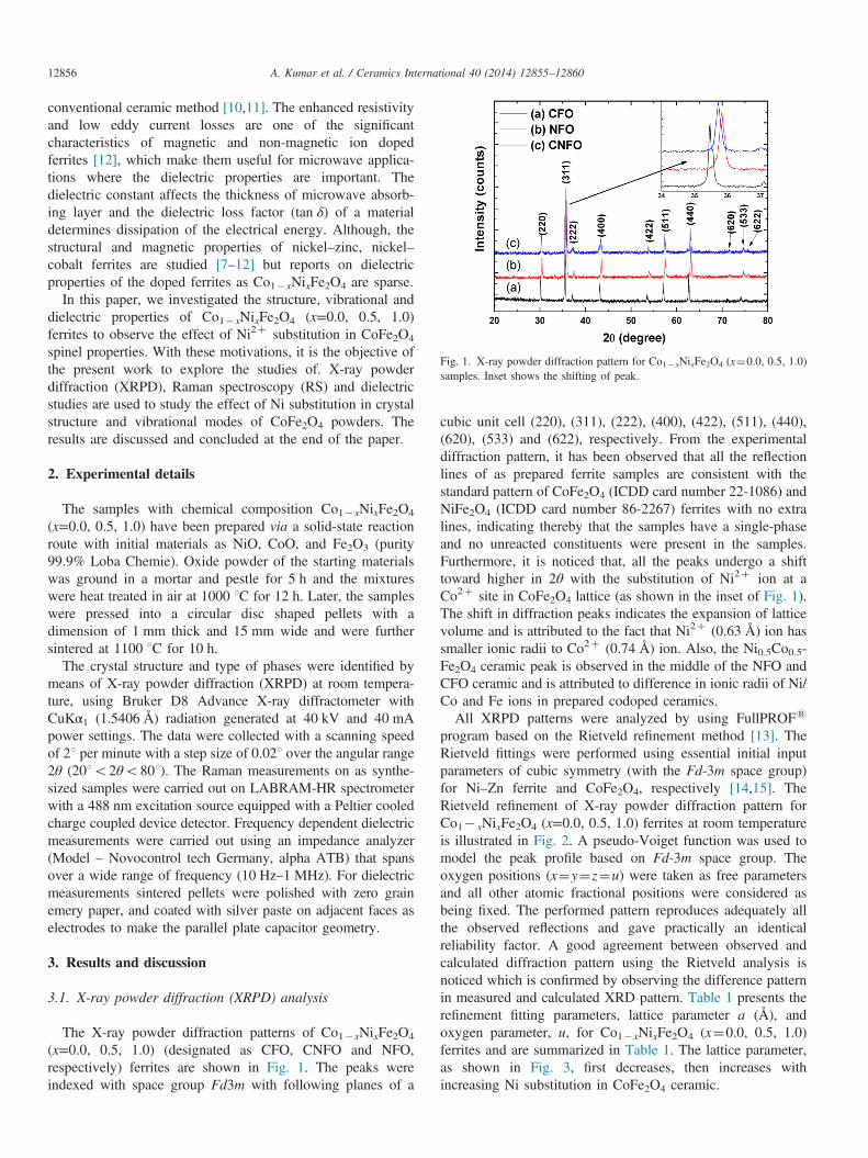
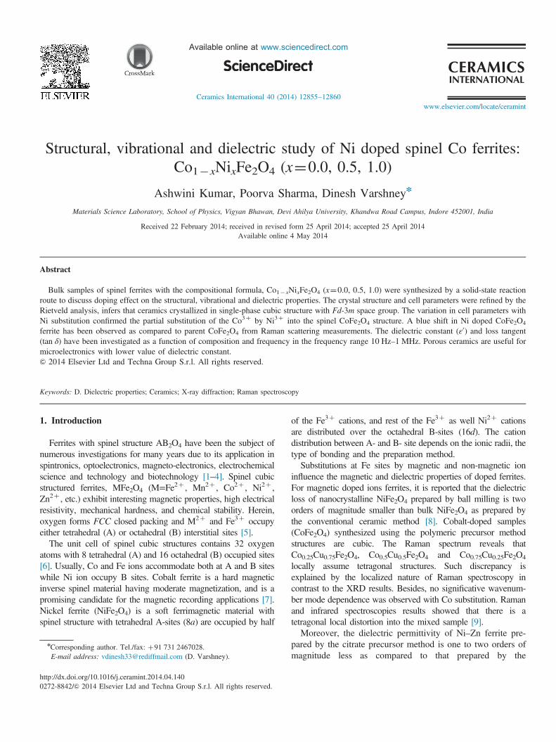
Fig. 1. X-ray powder diffraction pattern for Co1�xNixFe2O4 (x¼0.0, 0.5, 1.0)samples. Inset shows the shifting of peak.
A. Kumar et al. / Ceramics International 40 (2014) 12855–1286012856
conventional ceramic method [10,11]. The enhanced resistivityand low eddy current losses are one of the significantcharacteristics of magnetic and non-magnetic ion dopedferrites [12], which make them useful for microwave applica-tions where the dielectric properties are important. Thedielectric constant affects the thickness of microwave absorb-ing layer and the dielectric loss factor (tan δ) of a materialdetermines dissipation of the electrical energy. Although, thestructural and magnetic properties of nickel–zinc, nickel–cobalt ferrites are studied [7–12] but reports on dielectricproperties of the doped ferrites as Co1�xNixFe2O4 are sparse.
In this paper, we investigated the structure, vibrational anddielectric properties of Co1�xNixFe2O4 (x=0.0, 0.5, 1.0)ferrites to observe the effect of Ni2þ substitution in CoFe2O4
spinel properties. With these motivations, it is the objective ofthe present work to explore the studies of. X-ray powderdiffraction (XRPD), Raman spectroscopy (RS) and dielectricstudies are used to study the effect of Ni substitution in crystalstructure and vibrational modes of CoFe2O4 powders. Theresults are discussed and concluded at the end of the paper.
2. Experimental details
The samples with chemical composition Co1�xNixFe2O4
(x=0.0, 0.5, 1.0) have been prepared via a solid-state reactionroute with initial materials as NiO, CoO, and Fe2O3 (purity99.9% Loba Chemie). Oxide powder of the starting materialswas ground in a mortar and pestle for 5 h and the mixtureswere heat treated in air at 1000 1C for 12 h. Later, the sampleswere pressed into a circular disc shaped pellets with adimension of 1 mm thick and 15 mm wide and were furthersintered at 1100 1C for 10 h.
The crystal structure and type of phases were identified bymeans of X-ray powder diffraction (XRPD) at room tempera-ture, using Bruker D8 Advance X-ray diffractometer withCuKα1 (1.5406 Å) radiation generated at 40 kV and 40 mApower settings. The data were collected with a scanning speedof 21 per minute with a step size of 0.021 over the angular range2θ (201o2θo801). The Raman measurements on as synthe-sized samples were carried out on LABRAM-HR spectrometerwith a 488 nm excitation source equipped with a Peltier cooledcharge coupled device detector. Frequency dependent dielectricmeasurements were carried out using an impedance analyzer(Model – Novocontrol tech Germany, alpha ATB) that spansover a wide range of frequency (10 Hz–1 MHz). For dielectricmeasurements sintered pellets were polished with zero grainemery paper, and coated with silver paste on adjacent faces aselectrodes to make the parallel plate capacitor geometry.
3. Results and discussion
3.1. X-ray powder diffraction (XRPD) analysis
The X-ray powder diffraction patterns of Co1�xNixFe2O4
(x=0.0, 0.5, 1.0) (designated as CFO, CNFO and NFO,respectively) ferrites are shown in Fig. 1. The peaks wereindexed with space group Fd3m with following planes of a
cubic unit cell (220), (311), (222), (400), (422), (511), (440),(620), (533) and (622), respectively. From the experimentaldiffraction pattern, it has been observed that all the reflectionlines of as prepared ferrite samples are consistent with thestandard pattern of CoFe2O4 (ICDD card number 22-1086) andNiFe2O4 (ICDD card number 86-2267) ferrites with no extralines, indicating thereby that the samples have a single-phaseand no unreacted constituents were present in the samples.Furthermore, it is noticed that, all the peaks undergo a shifttoward higher in 2θ with the substitution of Ni2þ ion at aCo2þ site in CoFe2O4 lattice (as shown in the inset of Fig. 1).The shift in diffraction peaks indicates the expansion of latticevolume and is attributed to the fact that Ni2þ (0.63 Å) ion hassmaller ionic radii to Co2þ (0.74 Å) ion. Also, the Ni0.5Co0.5-Fe2O4 ceramic peak is observed in the middle of the NFO andCFO ceramic and is attributed to difference in ionic radii of Ni/Co and Fe ions in prepared codoped ceramics.All XRPD patterns were analyzed by using FullPROFs
program based on the Rietveld refinement method [13]. TheRietveld fittings were performed using essential initial inputparameters of cubic symmetry (with the Fd-3m space group)for Ni–Zn ferrite and CoFe2O4, respectively [14,15]. TheRietveld refinement of X-ray powder diffraction pattern forCo1� xNixFe2O4 (x=0.0, 0.5, 1.0) ferrites at room temperatureis illustrated in Fig. 2. A pseudo-Voiget function was used tomodel the peak profile based on Fd-3m space group. Theoxygen positions (x¼y¼z¼u) were taken as free parametersand all other atomic fractional positions were considered asbeing fixed. The performed pattern reproduces adequately allthe observed reflections and gave practically an identicalreliability factor. A good agreement between observed andcalculated diffraction pattern using the Rietveld analysis isnoticed which is confirmed by observing the difference patternin measured and calculated XRD pattern. Table 1 presents therefinement fitting parameters, lattice parameter a (Å), andoxygen parameter, u, for Co1�xNixFe2O4 (x¼0.0, 0.5, 1.0)ferrites and are summarized in Table 1. The lattice parameter,as shown in Fig. 3, first decreases, then increases withincreasing Ni substitution in CoFe2O4 ceramic.
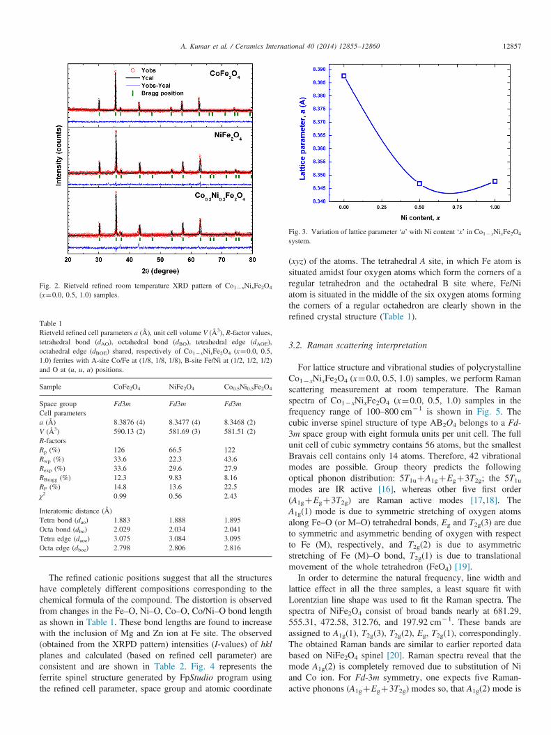
Table 1Rietveld refined cell parameters a (Å), unit cell volume V (Å3), R-factor values,tetrahedral bond (dAO), octahedral bond (dBO), tetrahedral edge (dAOE),octahedral edge (dBOE) shared, respectively of Co1�xNixFe2O4 (x¼0.0, 0.5,1.0) ferrites with A-site Co/Fe at (1/8, 1/8, 1/8), B-site Fe/Ni at (1/2, 1/2, 1/2)and O at (u, u, u) positions.
Sample CoFe2O4 NiFe2O4 Co0.5Ni0.5Fe2O4
Space group Fd3m Fd3m Fd3mCell parametersa (Å) 8.3876 (4) 8.3477 (4) 8.3468 (2)V (Å3) 590.13 (2) 581.69 (3) 581.51 (2)R-factorsRp (%) 126 66.5 122Rwp (%) 33.6 22.3 43.6Rexp (%) 33.6 29.6 27.9RBragg (%) 12.3 9.83 8.16RF (%) 14.8 13.6 22.5χ2 0.99 0.56 2.43
Interatomic distance (Å)Tetra bond (dao) 1.883 1.888 1.895Octa bond (dbo) 2.029 2.034 2.041Tetra edge (daoe) 3.075 3.084 3.095Octa edge (dboe) 2.798 2.806 2.816
Fig. 2. Rietveld refined room temperature XRD pattern of Co1�xNixFe2O4
(x¼0.0, 0.5, 1.0) samples.
Fig. 3. Variation of lattice parameter ‘a’ with Ni content ‘x’ in Co1�xNixFe2O4
system.
A. Kumar et al. / Ceramics International 40 (2014) 12855–12860 12857
The refined cationic positions suggest that all the structureshave completely different compositions corresponding to thechemical formula of the compound. The distortion is observedfrom changes in the Fe–O, Ni–O, Co–O, Co/Ni–O bond lengthas shown in Table 1. These bond lengths are found to increasewith the inclusion of Mg and Zn ion at Fe site. The observed(obtained from the XRPD pattern) intensities (I-values) of hklplanes and calculated (based on refined cell parameter) areconsistent and are shown in Table 2. Fig. 4 represents theferrite spinel structure generated by FpStudio program usingthe refined cell parameter, space group and atomic coordinate
(xyz) of the atoms. The tetrahedral A site, in which Fe atom issituated amidst four oxygen atoms which form the corners of aregular tetrahedron and the octahedral B site where, Fe/Niatom is situated in the middle of the six oxygen atoms formingthe corners of a regular octahedron are clearly shown in therefined crystal structure (Table 1).
3.2. Raman scattering interpretation
For lattice structure and vibrational studies of polycrystallineCo1�xNixFe2O4 (x¼0.0, 0.5, 1.0) samples, we perform Ramanscattering measurement at room temperature. The Ramanspectra of Co1�xNixFe2O4 (x¼0.0, 0.5, 1.0) samples in thefrequency range of 100–800 cm�1 is shown in Fig. 5. Thecubic inverse spinel structure of type AB2O4 belongs to a Fd-3m space group with eight formula units per unit cell. The fullunit cell of cubic symmetry contains 56 atoms, but the smallestBravais cell contains only 14 atoms. Therefore, 42 vibrationalmodes are possible. Group theory predicts the followingoptical phonon distribution: 5T1uþA1gþEgþ3T2g; the 5T1umodes are IR active [16], whereas other five first order(A1gþEgþ3T2g) are Raman active modes [17,18]. TheA1g(1) mode is due to symmetric stretching of oxygen atomsalong Fe–O (or M–O) tetrahedral bonds, Eg and T2g(3) are dueto symmetric and asymmetric bending of oxygen with respectto Fe (M), respectively, and T2g(2) is due to asymmetricstretching of Fe (M)–O bond, T2g(1) is due to translationalmovement of the whole tetrahedron (FeO4) [19].In order to determine the natural frequency, line width and
lattice effect in all the three samples, a least square fit withLorentzian line shape was used to fit the Raman spectra. Thespectra of NiFe2O4 consist of broad bands nearly at 681.29,555.31, 472.58, 312.76, and 197.92 cm�1. These bands areassigned to A1g(1), T2g(3), T2g(2), Eg, T2g(1), correspondingly.The obtained Raman bands are similar to earlier reported databased on NiFe2O4 spinel [20]. Raman spectra reveal that themode A1g(2) is completely removed due to substitution of Niand Co ion. For Fd-3m symmetry, one expects five Raman-active phonons (A1gþEgþ3T2g) modes so, that A1g(2) mode is
Fig. 4. Schematic represents the unit cell of the ferrite spinel structure.
Table 3Raman parameters of Co1�xNixFe2O4 (x¼0.0, 0.5, 1.0) samples.
Assignment Raman modes (cm�1)
CoFe2O4 [9] CoFe2O4 NiFe2O4 Co0.5Ni0.5Fe2O4
A1g(1) 680 675.65 681.29 686.93T2g(3) 596 583.52 555.31 592.92T2g(2) 458 461.3 472.58 472.58Eg 296 293.95 312.76 312.76T2g(1) 179 190.0 197.29 202.0
Table 2Comparison between some of the observed and calculated I-values for Co1�xNixFe2O4 (x¼0.0, 0.5, 1.0) ferrites after Rietveld refinement of XRD pattern.
CoFe2O4 NiFe2O4 Co0.5Ni0.5Fe2O4 hkl
I-obs I-cal I-obs I-cal I-obs I-cal
119.0 106.5 150.8 137.2 127.4 106.5 220453.2 449.8 584.9 583.9 468.5 449.8 31132.8 36.2 45.1 62.1 36.4 63.7 222102.5 117.3 152.1 172.3 120.0 214.0 40042.9 43.8 52.2 50.2 43.9 30.5 42231.4 27.6 39.2 34.4 119.8 106.9 511193.8 185.1 259.3 281.3 212.5 229.3 44015.2 4.0 20.0 30.7 18.6 3.7 62038.4 42.9 49.0 54.0 40.6 53.0 53317.4 13.5 24.9 22.1 21.4 27.7 622
A. Kumar et al. / Ceramics International 40 (2014) 12855–1286012858
not present in Co1�xNixFe2O4 (x¼0.0, 1.0) ceramics [19]. Inthe Co/Ni ion doped ceramic Fe3O4 the very small presence ofshoulder like feature at lower wave number side(686.93 cm�1) against the reported single band to that ofFe3O4 in the range of 650–750 cm-1 [17]. These bands areassigned to A1g(1) and A1g(2) modes reflecting the stretchingvibration of Fe3þ and O2� ions in tetrahedral site. The otherlow frequency modes are assigned to T2g and Eg modesreflecting the vibration of that site.
The very small doublet like feature in Ni0.5Co0.5Fe2O4 isattributed to the distribution in bond distances. In Fe3O4,whole of the tetrahedral and octahedral sites are occupied byFe ions while in Co1�xNixFe2O4 most of the octahedral site areoccupied by Ni, Co and Fe ions and tetrahedral site areoccupied by only Fe ion [21]. Both Fe3O4 and doped
Co1�xNixFe2O4 posses similar crystal structures but localstructures are different. Needless to mention, Raman spectro-scopy is a sensitive probe to detect the local structure veryeffectively. This result in a distortion free structure anduniform Fe–O bond distances in pristine Fe3O4. Due todifference in ionic radii of Ni/Co and Fe ions in preparedco-doped ceramics, the respective bond distance (Fe/Ni/Co–O)distribution results in doublet like structure. This distributionin bond distances probably results in double peak like structurein NiFe2O4, one peak representing the unit cell with all Fe ionsand the other one to the unit cell with mixed Fe and Ni ions[20,21].
3.3. Dielectric response and its analysis
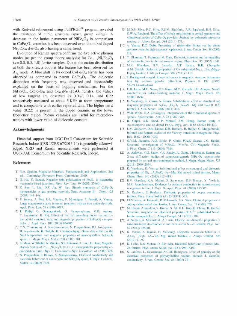
Fig. 6(a) shows the variations of frequency dependence ofdielectric constant at room temperature for Co1�xNixFe2O4
(x¼0.0, 0.5, 1.0). It has been observed that all samples exhibitthe dielectric dispersion where ε0 decreases as the frequencyincreases from 10 Hz to 1 MHz. Usually, the dipolar, inter-facial, ionic, and electronic polarizations contributes to thedielectric constant of any material. At low frequencies, it is thedipolar and interfacial polarizations are effective to thedielectric constant. However, at higher frequencies the electro-nic polarization is an effective and dipolar contributionbecomes insignificant. The decrease in dielectric constant with
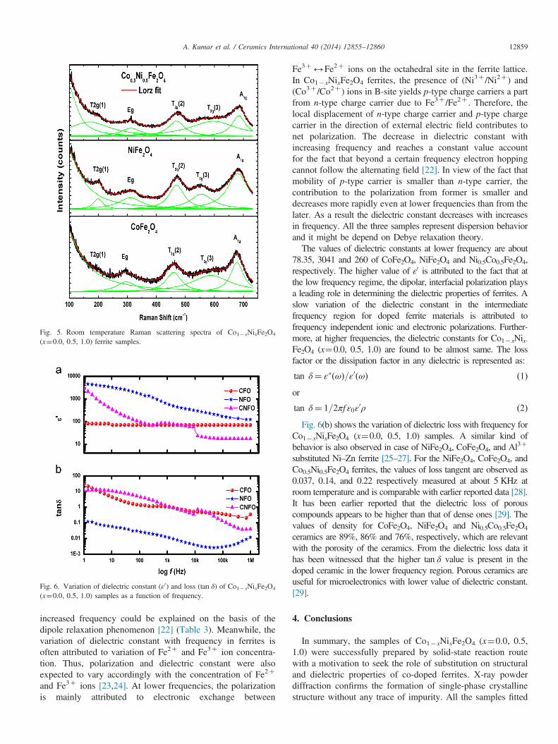
Fig. 5. Room temperature Raman scattering spectra of Co1�xNixFe2O4
(x¼0.0, 0.5, 1.0) ferrite samples.
Fig. 6. Variation of dielectric constant (ε0) and loss (tan δ) of Co1�xNixFe2O4
(x¼0.0, 0.5, 1.0) samples as a function of frequency.
A. Kumar et al. / Ceramics International 40 (2014) 12855–12860 12859
increased frequency could be explained on the basis of thedipole relaxation phenomenon [22] (Table 3). Meanwhile, thevariation of dielectric constant with frequency in ferrites isoften attributed to variation of Fe2þ and Fe3þ ion concentra-tion. Thus, polarization and dielectric constant were alsoexpected to vary accordingly with the concentration of Fe2þ
and Fe3þ ions [23,24]. At lower frequencies, the polarizationis mainly attributed to electronic exchange between
Fe3þ2Fe2þ ions on the octahedral site in the ferrite lattice.In Co1�xNixFe2O4 ferrites, the presence of (Ni3þ /Ni2þ ) and(Co3þ /Co2þ ) ions in B-site yields p-type charge carriers a partfrom n-type charge carrier due to Fe3þ /Fe2þ . Therefore, thelocal displacement of n-type charge carrier and p-type chargecarrier in the direction of external electric field contributes tonet polarization. The decrease in dielectric constant withincreasing frequency and reaches a constant value accountfor the fact that beyond a certain frequency electron hoppingcannot follow the alternating field [22]. In view of the fact thatmobility of p-type carrier is smaller than n-type carrier, thecontribution to the polarization from former is smaller anddecreases more rapidly even at lower frequencies than from thelater. As a result the dielectric constant decreases with increasesin frequency. All the three samples represent dispersion behaviorand it might be depend on Debye relaxation theory.The values of dielectric constants at lower frequency are about
78.35, 3041 and 260 of CoFe2O4, NiFe2O4 and Ni0.5Co0.5Fe2O4,respectively. The higher value of ε0 is attributed to the fact that atthe low frequency regime, the dipolar, interfacial polarization playsa leading role in determining the dielectric properties of ferrites. Aslow variation of the dielectric constant in the intermediatefrequency region for doped ferrite materials is attributed tofrequency independent ionic and electronic polarizations. Further-more, at higher frequencies, the dielectric constants for Co1�xNix-Fe2O4 (x¼0.0, 0.5, 1.0) are found to be almost same. The lossfactor or the dissipation factor in any dielectric is represented as:
tan δ¼ ε″ðωÞ=ε0ðωÞ ð1Þor
tan δ¼ 1=2πf ε0ε0ρ ð2Þ
Fig. 6(b) shows the variation of dielectric loss with frequency forCo1�xNixFe2O4 (x¼0.0, 0.5, 1.0) samples. A similar kind ofbehavior is also observed in case of NiFe2O4, CoFe2O4, and Al3þ
substituted Ni–Zn ferrite [25–27]. For the NiFe2O4, CoFe2O4, andCo0.5Ni0.5Fe2O4 ferrites, the values of loss tangent are observed as0.037, 0.14, and 0.22 respectively measured at about 5 KHz atroom temperature and is comparable with earlier reported data [28].It has been earlier reported that the dielectric loss of porouscompounds appears to be higher than that of dense ones [29]. Thevalues of density for CoFe2O4, NiFe2O4 and Ni0.5Co0.5Fe2O4
ceramics are 89%, 86% and 76%, respectively, which are relevantwith the porosity of the ceramics. From the dielectric loss data ithas been witnessed that the higher tan δ value is present in thedoped ceramic in the lower frequency region. Porous ceramics areuseful for microelectronics with lower value of dielectric constant.[29].
4. Conclusions
In summary, the samples of Co1�xNixFe2O4 (x¼0.0, 0.5,1.0) were successfully prepared by solid-state reaction routewith a motivation to seek the role of substitution on structuraland dielectric properties of co-doped ferrites. X-ray powderdiffraction confirms the formation of single-phase crystallinestructure without any trace of impurity. All the samples fitted
A. Kumar et al. / Ceramics International 40 (2014) 12855–1286012860
with Rietveld refinement using FullPROFs program revealedthe existence of cubic structure (space group Fd3m). Adecrease in the lattice parameter of NiFe2O4 in comparisonto CoFe2O4 ceramics has been observed even the mixed dopedNi0.5Co0.5Fe2O4 also having a same trend.
Evolution of Raman spectra confirms the five active phononmodes (as per the group theory analysis) for Co1�xNixFe2O4
(x¼0.0, 0.5, 1.0) ferrite samples. Due to the cation distributionat both the sites, a doublet like feature has been observed forA1g mode. A blue shift in Ni doped CoFe2O4 ferrite has beenobserved as compared to parent CoFe2O4. The dielectricdispersion with frequency was observed and successfullyexplained on the basis of hopping mechanism. For theNiFe2O4, CoFe2O4, and Co0.5Ni0.5Fe2O4 ferrites, the valuesof loss tangent are observed as 0.037, 0.14, and 0.22respectively measured at about 5 KHz at room temperatureand is comparable with earlier reported data. The higher tan δvalue (0.22) is present in the doped ceramic in the lowerfrequency region. Porous ceramics are useful for microelec-tronics with lower value of dielectric constant.
Acknowledgments
Financial support from UGC-DAE Consortium for ScientificResearch, Indore (CSR-I/CRS-67/2013-14) is gratefully acknowl-edged. XRD and Raman measurements were performed atUGC-DAE Consortium for Scientific Research, Indore.
References
[1] N.A. Spaldin, Magnetic Materials: Fundamentals and Applications, 2nded., Cambridge University Press, Cambridge, 2010.
[2] G. Hu, Y. Suzuki, Negative spin polarization of Fe3O4 in magnetite/manganite-based junctions, Phys. Rev. Lett. 89 (2002) 276601.
[3] Z. Sun, L. Liu, D.Z. Jia, W. Pan, Simple synthesis of CuFe2O4
nanoparticles as gas-sensing materials, Sens. Actuators B – Chem. 125(2007) 144–148.
[4] P. Seneor, A. Fert, J.-L. Maurice, F. Montaigne, F. Petroff, A. Vaures,Large magnetoresistance in tunnel junctions with an iron oxide electrode,Appl. Phys. Lett. 74 (1999) 4017.
[5] J. Philip, G. Gnanaprakash, G. Panneerselvam, M.P. Antony,T. Jayakumar, B. Raj, Effect of thermal annealing under vacuum onthe crystal structure, size, and magnetic properties of ZnFe2O4 nanopar-ticles, J. Appl. Phys. 102 (2005) 054305.
[6] C.N. Chinnasamy, A. Narayanasamya, N. Ponpandiana, R.J. Joseyphusa,B. Jeyadevanb, K. Tohjib, K. Chattopadhyay, Grain size effect on theNéel temperature and magnetic properties of nanocrystalline NiFe2O4
spinel, J. Magn. Magn. Mater. 238 (2002) 281.[7] K. Maaz, W. Khalid, A. Mumtaz, S.K. Hasanain, J. Liu, J.L. Duan, Magnetic
characterization of Co1�xNixFe2O4 (0rxr1) nanoparticles prepared by co-precipitation route, Phys. E: Low-dimens. Syst. Nanostruct. 41 (2009) 593.
[8] N. Ponpandian, P. Balaya, A. Narayanasamy, Electrical conductivity anddielectric behaviour of nanocrystalline NiFe2O4 spinel, J. Phys.: Condens.Matter 14 (2002) 3221.
[9] M.D.P. Silva, F.C. Silva, F.S.M. Sinfrônio, A.R. Paschoal, E.N. Silva,C.W.A. Paschoal, The effect of cobalt substitution in crystal structure andvibrational modes of CuFe2O4 powders obtained by polymeric precursormethod, J. Alloys Compd. 584 (2014) 573.
[10] A. Verma, D.C. Dube, Processing of nickel–zinc ferrites via the citrateprecursor route for high-frequency applications, J. Am. Ceram. Soc. 88 (2005)519.
[11] T. Okamura, T. Fujimura, M. Date, Dielectric constant and permeabilityof various ferrites in the microwave region, Phys. Rev. 85 (1952) 1041.
[12] M.R. Bhandare, H.V. Jamadar, A.T. Pathan, B.K. Chougule,A.M. Shaikh, Dielectric properties of Cu substituted Ni0.5�xZn0.3Mg0.2-Fe2O4 ferrites, J. Alloys Compd. 509 (2011) L113.
[13] J. Rodriguez-Carvajal, Recent advances in magnetic structure determina-tion by neutron powder diffraction, Physica B 192 (1993)55–69 (Amsterdam).
[14] U.R. Lima, M.C. Nasar, R.S. Nasar, M.C. Rezende, J.H. Araujoc, Ni–Znnanoferrite for radar-absorbing material, J. Magn. Magn. Mater. 320(2008) 1666.
[15] D. Varshney, K. Verma, A. Kumar, Substitutional effect on structural andmagnetic properties of AxCo1�xFe2O4 (A¼Zn, Mg and x¼0.0, 0.5)ferrites, J. Mol. Struct. 1006 (2011) 447.
[16] W.B. White, B.A. DeAngelis, Interpretation of the vibrational spectra ofspinels, Spectrochim. Acta A 23 (1967) 985.
[17] R. Gupta, A.K. Sood, P. Metcalf, J.M. Honig, Raman study ofstoichiometric and Zn-doped Fe3O4, Phys. Rev. B 65 (2002) 104430.
[18] L.V. Gasparov, D.B. Tanner, D.B. Romero, H. Berger, G. Margaritondo,Infrared and Raman studies of the Verwey transition in magnetite, Phys.Rev. B 62 (2000) 7939.
[19] G.V.M. Jacintho, A.G. Brolo, P. Corio, P.A.Z. Suarez, J.C. Rubim,Structural investigation of MFe2O4 (M¼Fe, Co) Magnetic Fluids,J. Phys. Chem. C 113 (2009) 7684.
[20] A. Ahlawat, V.G. Sathe, V.R. Reddy, A. Gupta, Mossbauer, Raman andX-ray diffraction studies of superparamagnetic NiFe2O4 nanoparticlesprepared by sol–gel auto-combustion method, J. Magn. Magn. Mater. 323(2011) 2049–2054.
[21] D. Varshney, K. Verma, Substitutional effect on structural and dielectricproperties of Ni1�xAxFe2O4 (A¼Mg, Zn) mixed spinel ferrites, Mater.Chem. Phys. 140 (2013) 412–418.
[22] E.V. Gopalan, K.A. Malini, S. Saravanan, D.S. Kumar, Y. Yoshida,M.R. Anantharaman, Evidence for polaron conduction in nanostructuredmanganese ferrite, J. Phys. D: Appl. Phys. 41 (2008) 185005.
[23] N. Rezlescu, E. Rezlescu, Dielectric properties of copper containingferrites, Phys. Status Solidi (A) 23 (1974) 575.
[24] J.T.S. Irvine, A. Huanosta, R. Velenzuela, A.R. West, Electrical properties ofpolycrystalline nickel zinc ferrites, J. Am. Ceram. Soc. 73 (1990) 729.
[25] M. Hasim, Alimuddin, S. Kumar, S. Ali, B.H. Koo, H. Chung, R. Kumar,Structural, magnetic and electrical properties of Al3þ substituted Ni–Znferrite nanoparticles, J. Alloys Compd. 511 (2012) 107.
[26] A. Sutka1, G. Mezinskis1, A. Lusis, Electric and dielectric properties ofnanostructured stoichiometric and excess-iron Ni–Zn ferrites, Phys. Scr.87 (2013) 025601.
[27] K. Verma, A. Kumar, D. Varshney, Dielectric relaxation behavior ofAxCo1�xFe2O4 (A¼Zn, Mg) mixed ferrites, J. Alloys Compd. 526(2012) 91–97.
[28] K. Latha, K.S. Mohan, D. Ravinder, Dielectric behaviour of mixed Mn–Zn ferrites, Phys. Status Solidi (A) 142 (1994) K103.
[29] S. Lanfredi, L. Dessemond, A.C.M. Rodrigues, Effect of porosity on theelectrical properties of polycrystalline sodium niobate: I, electricalconductivity, J. Am. Ceram. Soc. 86 (2003) 291.
Related Documents