Citation: Liu, H.; Gui, X.; Chen, S.; Fu, W.; Li, X.; Xiao, T.; Hou, J.; Jiang, T. Structural Variability of Lipoarabinomannan Modulates Innate Immune Responses within Infected Alveolar Epithelial Cells. Cells 2022, 11, 361. https://doi.org/ 10.3390/cells11030361 Academic Editor: Vladimir V. Yeremeev Received: 16 December 2021 Accepted: 18 January 2022 Published: 21 January 2022 Publisher’s Note: MDPI stays neutral with regard to jurisdictional claims in published maps and institutional affil- iations. Copyright: © 2022 by the authors. Licensee MDPI, Basel, Switzerland. This article is an open access article distributed under the terms and conditions of the Creative Commons Attribution (CC BY) license (https:// creativecommons.org/licenses/by/ 4.0/). cells Article Structural Variability of Lipoarabinomannan Modulates Innate Immune Responses within Infected Alveolar Epithelial Cells Hanrui Liu 1,† , Xuwen Gui 1,† , Shixing Chen 2 , Weizhe Fu 1 , Xiang Li 1 , Tingyuan Xiao 1 , Jie Hou 1 and Tao Jiang 1, * 1 Department of Biotechnology, The College of Basic Medical Science, Dalian Medical University, Dalian 116044, China; [email protected] (H.L.); [email protected] (X.G.); [email protected] (W.F.); [email protected] (X.L.); [email protected] (T.X.); [email protected] (J.H.) 2 Key Laboratory of Science and Technology on Microsystem, Shanghai Institute of Microsystem and Information Technology, Chinese Academy of Sciences, Shanghai 200050, China; [email protected] * Correspondence: [email protected]; Tel.: +86-411-8611-0350 † These authors contributed equally to this work. Abstract: Mycobacterium tuberculosis (M. tb) is an intracellular pathogen persisting in phagosomes that has the ability to escape host immune surveillance causing tuberculosis (TB). Lipoarabinomannan (LAM), as a glycolipid, is one of the complex outermost components of the mycobacterial cell envelope and plays a critical role in modulating host responses during M. tb infection. Different species within the Mycobacterium genus exhibit distinct LAM structures and elicit diverse innate immune responses. However, little is known about the mechanisms. In this study, we first constructed a LAM-truncated mutant with fewer arabinofuranose (Araf ) residues named M. sm-ΔM_6387 (Mycobacterium smegmatis arabinosyltransferase EmbC gene knockout strain). It exhibited some prominent cell wall defects, including tardiness of mycobacterial migration, loss of acid-fast staining, and increased cell wall permeability. Within alveolar epithelial cells (A549) infected by M. sm-ΔM_6387, the uptake rate was lower, phagosomes with bacterial degradation appeared, and microtubule-associated protein light chain 3 (LC3) recruitment was enhanced compared to wild type Mycobacterium smegmatis (M. smegmatis). We further confirmed that the variability in the removal capability of M. sm-ΔM_6387 resulted from host cell responses rather than the changes in the mycobacterial cell envelope. Moreover, we found that M. sm-ΔM_6387 or its glycolipid extracts significantly induced expression changes in some genes related to innate immune responses, including Toll-like receptor 2 (TLR2), class A scavenger receptor (SR-A), Rubicon, LC3, tumor necrosis factor alpha (TNF-α), Bcl-2, and Bax. Therefore, our studies suggest that nonpathogenic M. smegmatis can deposit LC3 on phagosomal membranes, and the decrease in the quantity of Araf residues for LAM molecules not only impacts mycobacterial cell wall integrity but also enhances host defense responses against the intracellular pathogens and decreases phagocytosis of host cells. Keywords: Mycobacterium smegmatis; EmbC; lipoarabinomannan; LC3-associated phagocytosis; alveolar epithelial cells 1. Introduction Tuberculosis (TB) leads to millions of deaths each year [1,2]. Its prevalence largely depends on the ability of Mycobacterium tuberculosis (M. tb) to escape host immune surveil- lance [3]. M. tb, as an intracellular pathogen, interacts with phagocytes to trigger innate immune responses, whereas mycobacterial virulence factors can widely contribute to avoiding the protective immune responses [4–7]. Recently, many researchers have made efforts to develop host-directed therapy (HDT), which can better control TB by modulating host responses with or without additional antibodies [8–10]. We tried to explore a molecule that can evoke host defense responses Cells 2022, 11, 361. https://doi.org/10.3390/cells11030361 https://www.mdpi.com/journal/cells

Welcome message from author
This document is posted to help you gain knowledge. Please leave a comment to let me know what you think about it! Share it to your friends and learn new things together.
Transcript

Citation: Liu, H.; Gui, X.; Chen, S.;
Fu, W.; Li, X.; Xiao, T.; Hou, J.;
Jiang, T. Structural Variability of
Lipoarabinomannan Modulates
Innate Immune Responses within
Infected Alveolar Epithelial Cells.
Cells 2022, 11, 361. https://doi.org/
10.3390/cells11030361
Academic Editor: Vladimir V. Yeremeev
Received: 16 December 2021
Accepted: 18 January 2022
Published: 21 January 2022
Publisher’s Note: MDPI stays neutral
with regard to jurisdictional claims in
published maps and institutional affil-
iations.
Copyright: © 2022 by the authors.
Licensee MDPI, Basel, Switzerland.
This article is an open access article
distributed under the terms and
conditions of the Creative Commons
Attribution (CC BY) license (https://
creativecommons.org/licenses/by/
4.0/).
cells
Article
Structural Variability of Lipoarabinomannan Modulates InnateImmune Responses within Infected Alveolar Epithelial CellsHanrui Liu 1,† , Xuwen Gui 1,†, Shixing Chen 2, Weizhe Fu 1, Xiang Li 1, Tingyuan Xiao 1, Jie Hou 1
and Tao Jiang 1,*
1 Department of Biotechnology, The College of Basic Medical Science, Dalian Medical University,Dalian 116044, China; [email protected] (H.L.); [email protected] (X.G.); [email protected] (W.F.);[email protected] (X.L.); [email protected] (T.X.); [email protected] (J.H.)
2 Key Laboratory of Science and Technology on Microsystem, Shanghai Institute of Microsystem andInformation Technology, Chinese Academy of Sciences, Shanghai 200050, China; [email protected]
* Correspondence: [email protected]; Tel.: +86-411-8611-0350† These authors contributed equally to this work.
Abstract: Mycobacterium tuberculosis (M. tb) is an intracellular pathogen persisting in phagosomes thathas the ability to escape host immune surveillance causing tuberculosis (TB). Lipoarabinomannan(LAM), as a glycolipid, is one of the complex outermost components of the mycobacterial cell envelopeand plays a critical role in modulating host responses during M. tb infection. Different species withinthe Mycobacterium genus exhibit distinct LAM structures and elicit diverse innate immune responses.However, little is known about the mechanisms. In this study, we first constructed a LAM-truncatedmutant with fewer arabinofuranose (Araf ) residues named M. sm-∆M_6387 (Mycobacterium smegmatisarabinosyltransferase EmbC gene knockout strain). It exhibited some prominent cell wall defects,including tardiness of mycobacterial migration, loss of acid-fast staining, and increased cell wallpermeability. Within alveolar epithelial cells (A549) infected by M. sm-∆M_6387, the uptake ratewas lower, phagosomes with bacterial degradation appeared, and microtubule-associated proteinlight chain 3 (LC3) recruitment was enhanced compared to wild type Mycobacterium smegmatis(M. smegmatis). We further confirmed that the variability in the removal capability of M. sm-∆M_6387resulted from host cell responses rather than the changes in the mycobacterial cell envelope. Moreover,we found that M. sm-∆M_6387 or its glycolipid extracts significantly induced expression changesin some genes related to innate immune responses, including Toll-like receptor 2 (TLR2), classA scavenger receptor (SR-A), Rubicon, LC3, tumor necrosis factor alpha (TNF-α), Bcl-2, and Bax.Therefore, our studies suggest that nonpathogenic M. smegmatis can deposit LC3 on phagosomalmembranes, and the decrease in the quantity of Araf residues for LAM molecules not only impactsmycobacterial cell wall integrity but also enhances host defense responses against the intracellularpathogens and decreases phagocytosis of host cells.
Keywords: Mycobacterium smegmatis; EmbC; lipoarabinomannan; LC3-associated phagocytosis;alveolar epithelial cells
1. Introduction
Tuberculosis (TB) leads to millions of deaths each year [1,2]. Its prevalence largelydepends on the ability of Mycobacterium tuberculosis (M. tb) to escape host immune surveil-lance [3]. M. tb, as an intracellular pathogen, interacts with phagocytes to trigger innateimmune responses, whereas mycobacterial virulence factors can widely contribute toavoiding the protective immune responses [4–7].
Recently, many researchers have made efforts to develop host-directed therapy (HDT),which can better control TB by modulating host responses with or without additionalantibodies [8–10]. We tried to explore a molecule that can evoke host defense responses
Cells 2022, 11, 361. https://doi.org/10.3390/cells11030361 https://www.mdpi.com/journal/cells

Cells 2022, 11, 361 2 of 21
against the pathogens, which are mainly determined by phagolysosomal pathways, includ-ing canonical autophagy and noncanonical autophagy [11,12]. The canonical autophagypathway is characterized by autophagosome formation with a double-membrane, initi-ating microtubule-associated protein light chain 3 phospholipid conjugates (LC3-II) andphagophore formation in the vicinity of pathogens [13]. The noncanonical autophagy path-way includes LC3-associated phagocytosis (LAP), which is typically initiated by signalingthrough pathogen-recognition receptors (PPRs), such as Toll-like receptor 2 (TLR2), topromote LC3-II recruitment to single-membrane phagosomes [14–16]. LAP requires nicoti-namide adenine dinucleotide phosphate (NADPH) oxidase and the phosphatidylinositol(PI)3-phosphate-kinase generated by the Rubicon–Beclin–Vps34 complex, which generatesreactive oxygen species (ROS) and PI3-P to acquire LC3 on the phagosomes [17,18]. Rubi-con (RUN domain protein as Beclin-interacting and cysteine-rich-containing), identifiedas a Beclin-1 binding partner, negatively regulates canonical autophagy and the endocyticpathway [19]. Importantly, it is required for LAP, located in the late phagosomes but notthe autophagosomes [20,21]. M. tb can activate PPRs but does not robustly trigger LAP toeliminate mycobacteria within phagosomes [11,15]. Therefore, there is a close relationshipbetween autophagy and phagocytosis. In contrast to autophagy, which contributes to theremoval of intracellular pathogens, phagocytosis engulfs extracellular pathogens and playsa critical role in innate immune responses [22,23]. Recent studies revealed that phagocyto-sis was enhanced in autophagy-deficient macrophages, and autophagy might modulatephagocytosis [22,24,25].
The mycobacterial cell wall comprises peptidoglycan, arabinogalactan, and mycolicacids, along with lipopolysaccharides, polysaccharides, and proteins [26]. The lipopolysac-charides mainly consist of lipoarabinomannan (LAM), lipomannan (LM), and phosphatidyli-nositol mannosides (PIMs) [27]. LAM, located on the cell envelope, is a crucial immunomod-ulatory compound that participates in host–pathogen interactions [28,29]. There are threekinds of arabinosyltransferase in mycobacteria, namely, EmbA, EmbB, and EmbC, whichcatalyze the transfer of arabinofuranose (Araf ) residues to different substrate molecules.EmbC controls arabinan chain synthesis on an average of 55–70 Araf residues in LAM [30–32].Importantly, the Mycobacterium genus represents a complex group of more than 100 species,some of which exhibit distinct LAM structures and elicit diverse host cell innate immuneresponses [28,33]. LAMs are mainly divided into two types—mannose-capped arabino-mannan (manLAM) and PI-LAM. The manLAM from M. tb features mannanon at theterminus, and the PI-LAM without mannan at the terminus comes mainly from rapidlygrowing nonpathogenic strains such as Mycobacterium smegmatis (M. smegmatis) [33]. Duringhost–pathogen interactions, LAMs exhibit a wide array of immunomodulatory activities,including cytokine effects, PPR interactions, and apoptosis induction [34]. The different ter-minal structures of the two LAM variants profoundly influence their biological activity [35].ManLAM can block macrophage apoptosis, inhibit tumor necrosis factor alpha (TNF-α)and interleukin (IL)-12 production, induce IL-10 secretion, and limit TLR-mediated activa-tion to weaken the cellular immune responses [36–39]. In contrast, PI-LAM may induceautophagy-related processes by TLR2 and activate the expression of pro-inflammatorycytokines [40,41]. Moreover, ManLAM suppresses the accumulation of LC3-II in phago-somes, whereas PI-LAM has no such effects [35]. Therefore, LAM diversity is responsiblefor innate immunity differences.
Some previous results suggest that EmbC gene knockout produces LAM-truncatedvariants with fewer Araf residues [32,42]. Reports indicated that LAM-truncated moleculeswith the shorter arabinan domains elicited stronger cytokine production responses, whichare likely to be autophagy activators [38,43]. However, little is known about the effects ofLAM-truncated variants on mycobacterial endocytosis, LC3 accumulation, phagocytosis,especially LAP. In this study, we first constructed a LAM-truncated mutant with fewerAraf residues named M. sm-∆M_6387, and wild type M. smegmatis as its parent strain; wefurther tested whether the structural variability in PI-LAM modulates autophagy-relatedprocesses, including phagocytosis and LAP.
Cells 2022, 11, 361 3 of 21
2. Materials and Methods2.1. Preparation of M. smegmatis EmbC Gene Knockout Strain
The M. smegmatis EmbC gene (MSMEG_6387), with its downstream sequence, was am-plified using the forward primer (5′-GACTAGTCGGTCCGCATGCAGCGGGTGGCAGC-3′,SpeI) and the reverse primer (5′-TAGCGGCCGCTCAGCCGCTCAACTCAGCCGCAG-3′,NotI), using M. smegmatis mc2155 genomic DNA as a template. The target gene wasamplified by PCR, and the product was cloned into the pMD18T vector to generatepMD18_M_6387. After DNA sequencing, pMD18_M_6387 was digested by NcoI to makeblunt ends using Klenow DNA polymerase. Digested pUC4K by BamHI was used toobtain the kanamycin resistance cassette (KanR, 1264 bp), and the KanR fragment wasalso filled in the blunt ends. The MSMEG_6387 gene was disrupted through the inser-tion of KanR to generate the pMD18_M_6387::KanR and pMD18_M_6387-kanR
i plasmids,and the pMD18_M_6387::KanR plasmid digested by SpeI and NotI was required to producethe M_6281::KanR fragment, and then the DNA fragment was cloned into pPR27-xylE togenerate a conditional replication plasmid pPR27-M_6387::KanR (pJYI) [44].
The pJYI was electroporated into 100 µL M. smegmatis-competent cells under thecondition of 2.5 Kv 1000 Ω with an electroporator 2510 (Eppendorf, Hamburg, Germany),and then the cells were grown in 2 mL Luria-Bertani (LB) medium with 0.05% Tween80using 120 rpm shaking at 37 C for 4 h. The cells were grown on LB agar plates containingKanR and GenR at 30 C for approximately 4–5 d. Several clones were grown and pickedfrom the LB agar plate containing KanR at 42 C. When the first single-crossover eventoccurred, the mutants were stained yellow with 1% catechol. The positive clones wereidentified through PCR [45]. Transformants were grown on LB agar plates containing25 µg/mL Kan and 10% sucrose at 37 C to release the vector’s sequence by providingsurvival pressure. M. sm-∆M_6387 produced with the second single-crossover event werethen selected by PCR and verified by DNA sequencing. Furthermore, the expression levelsof adjacent genes were identified through RT-PCR analysis, and sequences and conditionsof the corresponding primers were showed in Table 1.
Table 1. Sequences and conditions of the mentioned primers related to M. smegmatis.
Gene Primer Sequences (5′–3′) AnnealingTemperature
ProductLength (bp)
MSMEG_6387 F: CCGACCCTGCTGAAACTGCT 58 494R: AGCCAGAACGCCAGGAACAG
MSMEG_6388 F: GTCGGTGCGCATCAAGTACG 58 680R: CCTTGACCATCGAGCCGAGT
MSMEG_6389 F: CGATCGTGTCGACCGTCATC 58 292R: ATGAGCGCCAGCACGTTGTA
MSMEG_6390 F: CTGGCTGGTGCTCGAACTCA 58 773R: CCAGCACACATCCGTTGAGG
gap F: GGAAAGCTGTGGCGTGATGG 54 399R: GTAGGCCATGAGGTCCACCA
2.2. Extraction and Analysis of LAM/LM Mixture
Both wild type M. smegmatis and M. sm-∆M_6387 were grown in 3 mL LB with 0.05%Tween80 (LBT) medium until the late logarithmic growth phase. Cells were harvested,washed with phosphate-buffered salt (PBS), and treated in 100 µL CHCL3:MeOH:H2O(20:20:3, v:v:v) at 55 C for 30 min. The lysates were centrifuged at 14,000× g for 5 min,and the pellets were incubated with 200 µL Tris-saturated phenol: H2O (1:1, v:v) for2 h at 80 C. The samples were centrifuged at 14,000× g for 15 min after being mixedwith 100 µL CHCL3, and the aqueous layer was transferred to a new Eppendorf (EP)tube to obtain the LAM/LM mixture [46]. The samples were analyzed on 15% SDS-PAGE through periodic acid-Schiff (PAS) staining. Western blotting was performed bytransferring the antigens (LAM/LM) on SDS-PAGE gels to polyvinylidene difluoride
Cells 2022, 11, 361 4 of 21
(PVDF) membranes, and then the membrane was blocked and blotted with Concanavalinfrom Canavalia ensiformis peroxidase conjugate (Sigma-Aldrich, St. Louis, MO, USA),following visualization by Enhanced ECL (efficient chemiluminescence) kit (Wanleibio,Shenyang, China). The total glucose concentrations in the LAM/LM extraction weredetermined using the sulfate–phenol method.
2.3. Identification of Biological Characteristics
The mycobacterial growth curve was determined through the colony-forming units(CFUs). Briefly, M. smegmatis and M. sm-∆M_6387 were collected at 6 h intervals and werediluted in PBS to generate 10-fold serial dilutions. Ten microliters of each diluted solutionwas spotted onto LB agar plates, repeated at least three times for all samples [46]. To observethe bacterial migration rates, M. smegmatis and M. sm-∆M_6387 were (independently)spotted onto LB or LBT (LB with 0.05% tween 80) solid media and incubated at 37 C until abacterial ring was generated, and its diameter was measured [47]. To identify the integrityof the mycobacterial cell wall, M. smegmatis and M. sm-∆M_6387 were cultured untilreaching the logarithmic phase, and then acid-fast staining analysis was performed usingthe Ziehl–Neelsen method [46]. To identify cell wall permeability on chemical compounds,M. smegmatis and M. sm-∆M_6387 at the logarithmic growth phase were serially diluted andtreated with 0.5 µg/mL crystal violet and 0.005% SDS in triplicate, and then the CFU/mLvalues were determined [46]. Further electron microscopy analysis was also carried outaccording to the methods outlined previously [45].
2.4. Uptake and Survival of Intracellular Pathogens
A549 cells (Human type II alveolar epithelial cells) were grown in Dulbecco’s ModifiedEagle Medium (DMEM) and supplemented with a 10% fetal bovine serum (FBS) and1% penicillin–streptomycin mixture. A549 cells at a density of 8 × 10³ cells/mL wereseeded in 96-well plates for 24 h. After being washed with PBS, the cells were infected byM. smegmatis or M. sm-∆M_6387 strains, washed with PBST at a multiplicity of infection(MOI) value of 100, and cultured further using DMEM medium with 2% FBS at 37 C for6 h. Following incubation, cells were washed three times with PBS per well and treatedby 100 µg/mL gentamicin for 1 h to remove extracellular bacteria [48]. Then, the cellswere cultured and collected at specified time points (0, 6, 12, 24 h) to determine CFUaiming to measure the mycobacterial survival capability in A549 cells. A549 cells were alsoinfected by M. smegmatis-FITC and M. sm-∆M_6387-FITC at an MOI value of 100 for 6 hand removed extracellular bacteria according to the methods mentioned above; they werecultured further using DMEM medium with 10% FBS at 37 C for 0, 6, and 24 h; and thenwashed and fixed with 4% paraformaldehyde for fluorescence microscopy. A549 cellswere inoculated at a density of 1 × 10⁵ cells /well on 24-well plates and pre-treated with100 µg/mL LAM/LM mixtures from M. smegmatis or M. sm-∆M_6387 for 12 h, and theninfected by M. smegmatis with a pSUM-kan-EGFP plasmid at an MOI of 100 for 6 h [49].After removing extracellular bacteria and washing with PBST, the cells were incubated withrabbit polyclonal anti-TLR2 antibody (Abcam, Cambridge, UK) and a secondary antibodyof Alexa Fluor 594-conjugated goat anti-rabbit IgG (Proteintech, Wuhan, China) and stainedwith DAPI for 5 min to observe the cells under a fluorescence microscope (SDPTOP ICX41,Ningbo, China).
2.5. Transmission Electron Microscopy (TEM)
All A549 cells treated or infected were centrifuged at 1200 rpm for 10 min, washedtwice in PBS, and fixed by 2.5% glutaraldehyde overnight, followed by dehydration withgradient alcohol. Cells were embedded with epon and cut into ultrathin sections [45].The sections were stained with 2% uranyl acetate for 30 min and lead citrate for 20 min,and the mitochondrial and phagosome structures within the A549 cells were then observedwith a JEM-2000EX transmission electron microscope (JEOL, Tokyo, Japan). Finally, both
Cells 2022, 11, 361 5 of 21
mitochondria and phagosomes in the images were measured and counted using Image J(Version 2.6.1) software (NIH, Bethesda, MD, USA).
2.6. Detection of ROS Release in A549 Cells
To identify the release of ROS (reactive oxygen species) of A549 cells, the cells weretreated by 100 µg/mL LAM/LM mixtures from M. smegmatis or M. sm-∆M_6387 for 6, 12,or 24 h, and incubated with 20 µM DCFH-DA as a probe for 1 h for observation undera fluorescence microscope and measure relative fluorescence unit at 485/525 using ROSassay kit (Wanleibio, Shenyang, China).
2.7. Assessing LC3 Recruitments with a Confocal Fluorescence Microscope
To more directly measure LC-3 activation, we used a lentiviral vector with tandem-tagged RFP-GFP-LC3 (Genechem, Shanghai, China) to obtain stably transfected A549cells (A549+LC3). Furthermore, A549+LC3 cells were infected by M. smegmatis, M. sm-∆M_6387, BCG, or M. smegmatis::EGFP at an MOI of 100 for 6 h; cultured further usingDMEM medium with 10% FBS at 37 C for 1, 2, 3, or 48 h; and after washing, fixed with4% paraformaldehyde for observation using a confocal fluorescence microscope(LeicaTCS SP5II, wetzlar, Germany). The images were processed and analyzed with Image J(version 2.6.1)software(NIH, Bethesda, MD, USA) [50].
2.8. The Expression Changes of Genes Related to Pathogen Recognition and Immune Responses
A549 cells were infected by mycobacteria or treated by LAM/LM mixtures and thencollected at determined time point. Cell lysates were prepared using RIPA kit (WENLEIBiotech). Proteins were separated through 12% SDS-PAGE and transferred to PVDF mem-brane. The membrane was blocked with 5% skim milk powder and then incubated witha primary antibody including rabbit antibody against Beclin (Proteintech), Rubicon (CellSignaling Tech., Danvers, IL, USA), LC3 (Proteintech, Wuhan, China), TLR2 (Abcam, Cam-bridge, UK), Bcl-2 (Proteintech, Wuhan, China), Bax (Proteintech, Wuhan, China), interferongamma(IFN-γ) (Bioss, Beijing, China), and β-actin (Proteintech, Wuhan, China) at 4 Covernight and a secondary antibody(anti-rabbit antibody, HRP conjugated, Proteintech) for2 h. The bound probe was visualized by Enhanced ECL kit (WENLEI Biotech), and imageswere captured using a digital gel image processing system (Tanon 1600, Shanghai, China).
We picked up some genes related to pathogenic recognition and inflammatory re-sponses and further detected their expression changes through qRT-PCR and RT-PCR.The corresponding primer sequences are listed in Table 2. Total RNA was extracted usingTrizol (Sangon Tech, Shanghai, China.) A total of 1 µg RNA was first reversed into cDNAusing RT kit (TaKaRa, Beijing, China), and then PCR was performed by correspondingcDNA as template; the ratios of the objected genes including TLR2, TLR4, Class A scav-enger receptor (SR-A), Rubicon, IFN-γ, TNF-α, and IL-1β to internal control gene (β-actin)were used to assess the differences in gene expression levels.
Table 2. Sequences and conditions of the mentioned primers related to A549 cells.
Gene Primer Sequences (5′–3′) Product Length (bp)
TLR-2 F: GGAATCGGTGAGGTCCTGTCCTG 296R: GGCGTCACATGCAGAAAGCCC
TLR-4 F: AAGTGTCTGAACTCCCTCCAGG 278R: ATGGTCTTATTCATCTGACAGGTGATA
SR-A F: ATGTCCGTTCAGCGTCTT 380R: TAGGTCCTGATGCTTCTTTA
Rubicon R: AGTGGGTTACTTGGGAGTG 310F: CTTTGGCTAATAGTTCTGC
IFN-γ F: TCGGTAACTGACTTGAATGTCCA 93R: TCGCTTCCCTGTTTTAGCTGC
IL-1β F: GGACAAGCTGAGGAAGATGC 360R: TCTTTCAACACGCAGGACAG
Cells 2022, 11, 361 6 of 21
Table 2. Cont.
Gene Primer Sequences (5′–3′) Product Length (bp)
TNF-α F: TGCTTGTTCCTCAGCCTCTT 514R: GGAAGACCCCTCCCAGATAG
β-actin F: CATGGATGATGATATCGCCGCG 371R: ACATGATCTGGGTCATCTTCTCG
2.9. Statistical Analysis
Data are presented as the mean ± standard error of the mean (SEM) of at least threeindependent experiments. All data were plotted and analyzed using GraphPad Prismversion 8 (GraphPad software Inc., La Jolla, CA, USA) or Image J (version 2.6.1) software(NIH, USA). p < 0.05 was considered statistically significant (unpaired Student’s t-test).
3. Results3.1. Construction of an EmbC Gene Knockout Strain
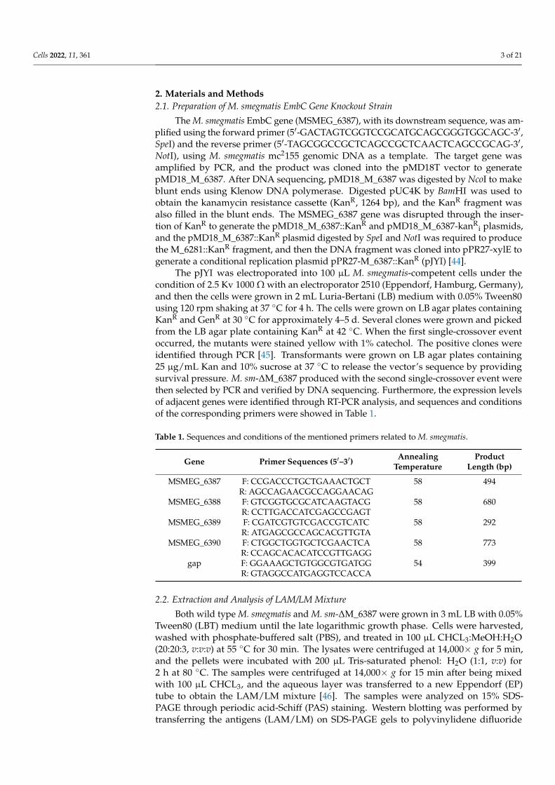
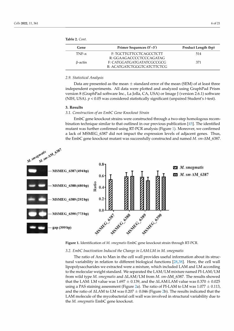
EmbC gene knockout strains were constructed through a two-step homologous recom-bination technique similar to that outlined in our previous publication [45]. The identifiedmutant was further confirmed using RT-PCR analysis (Figure 1). Moreover, we confirmeda lack of MSMEG_6387 did not impact the expression levels of adjacent genes. Thus,the EmbC gene knockout mutant was successfully constructed and named M. sm-∆M_6387.
Cells 2022, 11, x FOR PEER REVIEW 6 of 21
SR-A F: ATGTCCGTTCAGCGTCTT 380
R: TAGGTCCTGATGCTTCTTTA
Rubicon R: AGTGGGTTACTTGGGAGTG 310
F: CTTTGGCTAATAGTTCTGC
IFN-γ F: TCGGTAACTGACTTGAATGTCCA 93
R: TCGCTTCCCTGTTTTAGCTGC
IL-1β F: GGACAAGCTGAGGAAGATGC 360
R: TCTTTCAACACGCAGGACAG
TNF-α F: TGCTTGTTCCTCAGCCTCTT 514
R: GGAAGACCCCTCCCAGATAG
β-actin F: CATGGATGATGATATCGCCGCG 371
R: ACATGATCTGGGTCATCTTCTCG
2.9. Statistical Analysis
Data are presented as the mean ± standard error of the mean (SEM) of at least three
independent experiments. All data were plotted and analyzed using GraphPad Prism ver-
sion 8 (GraphPad software Inc., La Jolla, CA, USA) or Image J (version 2.6.1) software
(NIH, USA) . p < 0.05 was considered statistically significant (unpaired Student’s t-test).
3. Results
3.1. Construction of an EmbC Gene Knockout Strain
EmbC gene knockout strains were constructed through a two-step homologous re-
combination technique similar to that outlined in our previous publication [45]. The iden-
tified mutant was further confirmed using RT-PCR analysis (Figure 1). Moreover, we con-
firmed a lack of MSMEG_6387 did not impact the expression levels of adjacent genes.
Thus, the EmbC gene knockout mutant was successfully constructed and named M. sm-
ΔM_6387.
Figure 1. Identification of M. smegmatis EmbC gene knockout strain through RT-PCR.
3.2. EmbC Inactivation Induced the Change in LAM:LM in M. smegmatis
The ratio of Ara to Man in the cell wall provides useful information about its struc-
tural variability in relation to different biological functions [28,38]. Here, the cell wall lip-
opolysaccharides we extracted were a mixture, which included LAM and LM according
to the molecular weight standard. We separated the LAM/LM mixture named PI-
LAM/LM from wild type M. smegmatis and ΔLAM/LM from M. sm-ΔM_6387. The results
Figure 1. Identification of M. smegmatis EmbC gene knockout strain through RT-PCR.
3.2. EmbC Inactivation Induced the Change in LAM:LM in M. smegmatis
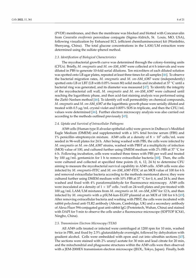
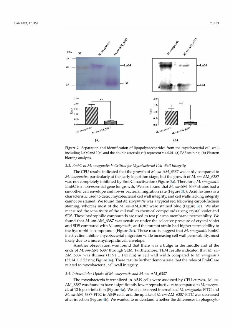
The ratio of Ara to Man in the cell wall provides useful information about its struc-tural variability in relation to different biological functions [28,38]. Here, the cell walllipopolysaccharides we extracted were a mixture, which included LAM and LM accordingto the molecular weight standard. We separated the LAM/LM mixture named PI-LAM/LMfrom wild type M. smegmatis and ∆LAM/LM from M. sm-∆M_6387. The results showedthat the LAM: LM value was 1.697 ± 0.139, and the ∆LAM:LAM value was 0.370 ± 0.025using a PAS staining assessment (Figure 2a). The ratio of PI-LAM to LM was 1.077 ± 0.113,and the ratio of ∆LAM to LM was 0.207 ± 0.046 (Figure 2b). The results indicated that theLAM molecule of the mycobacterial cell wall was involved in structural variability due tothe M. smegmatis EmbC gene knockout.
Cells 2022, 11, 361 7 of 21
Cells 2022, 11, x FOR PEER REVIEW 7 of 21
showed that the LAM: LM value was 1.697 ± 0.139, and the ΔLAM:LAM value was 0.370
± 0.025 using a PAS staining assessment (Figure 2a). The ratio of PI-LAM to LM was 1.077
± 0.113, and the ratio of ΔLAM to LM was 0.207 ± 0.046 (Figure 2b). The results indicated
that the LAM molecule of the mycobacterial cell wall was involved in structural variability
due to the M. smegmatis EmbC gene knockout.
Figure 2. Separation and identification of lipopolysaccharides from the mycobacterial cell wall, in-
cluding LAM and LM, and the double asterisks (**) represent p < 0.01. (a) PAS staining. (b) Western
blotting analysis.
3.3. EmbC in M. smegmatis Is Critical for Mycobacterial Cell Wall Integrity
The CFU results indicated that the growth of M. sm-ΔM_6387 was tardy compared
to M. smegmatis, particularly at the early logarithm stage, but the growth of M. sm-
ΔM_6387 was not completely inhibited by EmbC inactivation (Figure 3a). Therefore, M.
smegmatis EmbC is a non-essential gene for growth. We also found that M. sm-ΔM_6387
strains had a smoother cell envelope and lower bacterial migration rate (Figure 3b). Acid
fastness is a characteristic used to detect mycobacterial cell wall integrity, and cell walls
lacking integrity cannot be stained. We found that M. smegmatis was a typical red follow-
ing carbol-fuchsin staining, whereas most of the M. sm-ΔM_6387 were stained blue (Fig-
ure 3c). We also measured the sensitivity of the cell wall to chemical compounds using
crystal violet and SDS. These hydrophilic compounds are used to test plasma membrane
permeability. We found that M. sm-ΔM_6387 was sensitive under the selective pressure
of crystal violet and SDS compared with M. smegmatis, and the mutant strain had higher
permeability to the hydrophilic compounds (Figure 3d). These results suggest that M.
smegmatis EmbC inactivation inhibits mycobacterial migration while increasing cell wall
permeability, most likely due to a more hydrophilic cell envelope.
Another observation was found that there was a bulge in the middle and at the ends
of M. sm-ΔM_6387 through SEM. Furthermore, TEM results indicated that M. sm-
ΔM_6387 was thinner (13.91 ± 1.85 nm) in cell wall width compared to M. smegmatis (32.14
± 3.52 nm; Figure 3e). These results further demonstrate that the roles of EmbC are related
to mycobacterial cell wall integrity.
Figure 2. Separation and identification of lipopolysaccharides from the mycobacterial cell wall,including LAM and LM, and the double asterisks (**) represent p < 0.01. (a) PAS staining. (b) Westernblotting analysis.
3.3. EmbC in M. smegmatis Is Critical for Mycobacterial Cell Wall Integrity
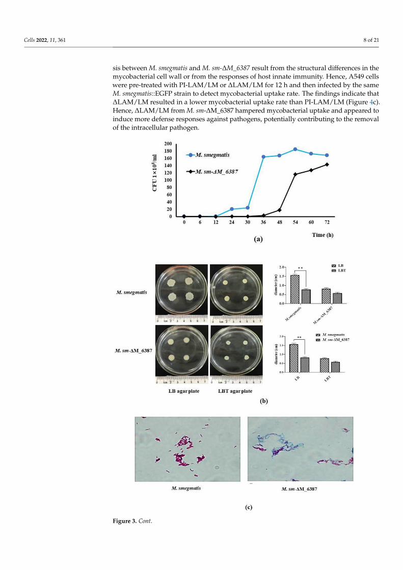
The CFU results indicated that the growth of M. sm-∆M_6387 was tardy compared toM. smegmatis, particularly at the early logarithm stage, but the growth of M. sm-∆M_6387was not completely inhibited by EmbC inactivation (Figure 3a). Therefore, M. smegmatisEmbC is a non-essential gene for growth. We also found that M. sm-∆M_6387 strains had asmoother cell envelope and lower bacterial migration rate (Figure 3b). Acid fastness is acharacteristic used to detect mycobacterial cell wall integrity, and cell walls lacking integritycannot be stained. We found that M. smegmatis was a typical red following carbol-fuchsinstaining, whereas most of the M. sm-∆M_6387 were stained blue (Figure 3c). We alsomeasured the sensitivity of the cell wall to chemical compounds using crystal violet andSDS. These hydrophilic compounds are used to test plasma membrane permeability. Wefound that M. sm-∆M_6387 was sensitive under the selective pressure of crystal violetand SDS compared with M. smegmatis, and the mutant strain had higher permeability tothe hydrophilic compounds (Figure 3d). These results suggest that M. smegmatis EmbCinactivation inhibits mycobacterial migration while increasing cell wall permeability, mostlikely due to a more hydrophilic cell envelope.
Another observation was found that there was a bulge in the middle and at theends of M. sm-∆M_6387 through SEM. Furthermore, TEM results indicated that M. sm-∆M_6387 was thinner (13.91 ± 1.85 nm) in cell wall width compared to M. smegmatis(32.14 ± 3.52 nm; Figure 3e). These results further demonstrate that the roles of EmbC arerelated to mycobacterial cell wall integrity.
3.4. Intracellular Uptake of M. smegmatis and M. sm-∆M_6387
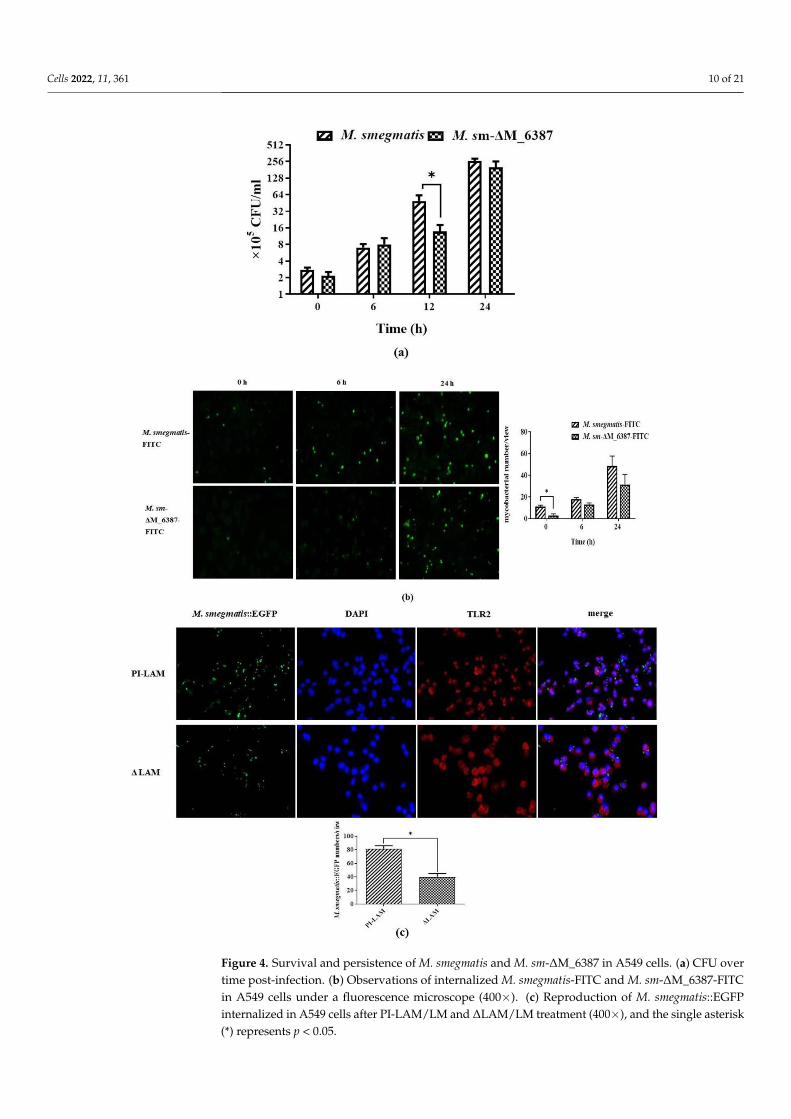
The mycobacteria internalized in A549 cells were assessed by CFU curves. M. sm-∆M_6387 was found to have a significantly lower reproductive rate compared to M. smegma-tis at 12 h post-infection (Figure 4a). We also observed internalized M. smegmatis-FITC andM. sm-∆M_6387-FITC in A549 cells, and the uptake of M. sm-∆M_6387-FITC was decreasedafter infection (Figure 4b). We wanted to understand whether the differences in phagocyto-
Cells 2022, 11, 361 8 of 21
sis between M. smegmatis and M. sm-∆M_6387 result from the structural differences in themycobacterial cell wall or from the responses of host innate immunity. Hence, A549 cellswere pre-treated with PI-LAM/LM or ∆LAM/LM for 12 h and then infected by the sameM. smegmatis::EGFP strain to detect mycobacterial uptake rate. The findings indicate that∆LAM/LM resulted in a lower mycobacterial uptake rate than PI-LAM/LM (Figure 4c).Hence, ∆LAM/LM from M. sm-∆M_6387 hampered mycobacterial uptake and appeared toinduce more defense responses against pathogens, potentially contributing to the removalof the intracellular pathogen.
Cells 2022, 11, x FOR PEER REVIEW 8 of 21
Figure 3. Cont.

Cells 2022, 11, 361 9 of 21Cells 2022, 11, x FOR PEER REVIEW 9 of 21
Figure 3. Assessment of the biological characteristics of M. smegmatis and M. sm-ΔM_6387. (a)
Growth curves of M. smegmatis and M. sm-ΔM_6387. (b) Measurement of the migration rate through
colony diameter on different LB agar plates. (c) Acid-fast staining analysis using the Ziehl–Neelsen
method. (d) Detection of cell wall permeability through SDS and crystal violet. (e) Morphology and
cell wall structures of M. smegmatis and M. sm-ΔM_6387 were observed by SEM (5000×) and TEM
(8000×), respectively. The double asterisks (**) represent p < 0.01.
3.4. Intracellular Uptake of M. smegmatis and M. sm-ΔM_6387
The mycobacteria internalized in A549 cells were assessed by CFU curves. M. sm-
ΔM_6387 was found to have a significantly lower reproductive rate compared to M. smeg-
matis at 12 h post-infection (Figure 4a). We also observed internalized M. smegmatis-FITC
and M. sm-ΔM_6387-FITC in A549 cells, and the uptake of M. sm-ΔM_6387-FITC was de-
creased after infection (Figure 4b). We wanted to understand whether the differences in
phagocytosis between M. smegmatis and M. sm-ΔM_6387 result from the structural differ-
ences in the mycobacterial cell wall or from the responses of host innate immunity. Hence,
Figure 3. Assessment of the biological characteristics of M. smegmatis and M. sm-∆M_6387. (a) Growthcurves of M. smegmatis and M. sm-∆M_6387. (b) Measurement of the migration rate through colonydiameter on different LB agar plates. (c) Acid-fast staining analysis using the Ziehl–Neelsen method.(d) Detection of cell wall permeability through SDS and crystal violet. (e) Morphology and cell wallstructures of M. smegmatis and M. sm-∆M_6387 were observed by SEM (5000×) and TEM (8000×),respectively. The double asterisks (**) represent p < 0.01.
Cells 2022, 11, 361 10 of 21
Cells 2022, 11, x FOR PEER REVIEW 10 of 21
A549 cells were pre-treated with PI-LAM/LM or ΔLAM/LM for 12 h and then infected by
the same M. smegmatis::EGFP strain to detect mycobacterial uptake rate. The findings in-
dicate that ΔLAM/LM resulted in a lower mycobacterial uptake rate than PI-LAM/LM
(Figure 4c). Hence, ΔLAM/LM from M. sm-ΔM_6387 hampered mycobacterial uptake and
appeared to induce more defense responses against pathogens, potentially contributing
to the removal of the intracellular pathogen.
Cells 2022, 11, x FOR PEER REVIEW 11 of 21
Figure 4. Survival and persistence of M. smegmatis and M. sm-ΔM_6387 in A549 cells. (a) CFU over
time post-infection. (b) Observations of internalized M. smegmatis-FITC and M. sm-ΔM_6387-FITC
in A549 cells under a fluorescence microscope (400×). (c) Reproduction of M. smegmatis::EGFP inter-
nalized in A549 cells after PI-LAM/LM and ΔLAM/LM treatment (400×), and the single asterisk (*)
represents p < 0.05.
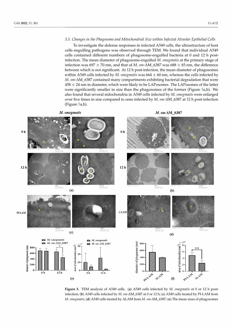
3.5. Changes in the Phagosome and Mitochondrial Size within Infected Alveolar Epithelial Cells
To investigate the defense responses in infected A549 cells, the ultrastructure of host
cells engulfing pathogens was observed through TEM. We found that individual A549
cells contained different numbers of phagosome-engulfed bacteria at 0 and 12 h post-in-
fection. The mean diameter of phagosome-engulfed M. smegmatis at the primary stage of
infection was 697 ± 70 nm, and that of M. sm-ΔM_6387 was 688 ± 83 nm, the difference
between which is not significant. At 12 h post-infection, the mean diameter of phagosomes
within A549 cells infected by M. smegmatis was 664 ± 60 nm, whereas the cells infected by
M. sm-ΔM_6387 contained many compartments exhibiting bacterial degradation that
were 458 ± 24 nm in diameter, which were likely to be LAPosomes. The LAPosomes of the
latter were significantly smaller in size than the phagosomes of the former (Figure 5a,b).
We also found that several mitochondria in A549 cells infected by M. smegmatis were en-
larged over five times in size compared to ones infected by M. sm-ΔM_6387 at 12 h post-
infection (Figure 5a,b).
To further illustrate whether LAM molecules directly affect the role of mitochondria,
LAM extraction was used to treat A549 cells. Our results indicate that PI-LAM from M.
smegmatis also elicited larger-sized mitochondria than ΔLAM/LM extraction from M. sm-
ΔM_6387 (Figure 5c,d). These results suggest that both M. smegmatis and M. sm-ΔM_6387
can trigger the maturation of phagosomes, but M. sm-ΔM_6387 appeared to have poorer
survival due to degradation in the observed cells. In addition, PI-LAM from nonpatho-
genic M. smegmatis might induce stronger reactions in mitochondria compared to ΔLAM
from M. sm-ΔM_6387.
Figure 4. Survival and persistence of M. smegmatis and M. sm-∆M_6387 in A549 cells. (a) CFU overtime post-infection. (b) Observations of internalized M. smegmatis-FITC and M. sm-∆M_6387-FITCin A549 cells under a fluorescence microscope (400×). (c) Reproduction of M. smegmatis::EGFPinternalized in A549 cells after PI-LAM/LM and ∆LAM/LM treatment (400×), and the single asterisk(*) represents p < 0.05.
Cells 2022, 11, 361 11 of 21
3.5. Changes in the Phagosome and Mitochondrial Size within Infected Alveolar Epithelial Cells
To investigate the defense responses in infected A549 cells, the ultrastructure of hostcells engulfing pathogens was observed through TEM. We found that individual A549cells contained different numbers of phagosome-engulfed bacteria at 0 and 12 h post-infection. The mean diameter of phagosome-engulfed M. smegmatis at the primary stage ofinfection was 697 ± 70 nm, and that of M. sm-∆M_6387 was 688 ± 83 nm, the differencebetween which is not significant. At 12 h post-infection, the mean diameter of phagosomeswithin A549 cells infected by M. smegmatis was 664 ± 60 nm, whereas the cells infected byM. sm-∆M_6387 contained many compartments exhibiting bacterial degradation that were458 ± 24 nm in diameter, which were likely to be LAPosomes. The LAPosomes of the latterwere significantly smaller in size than the phagosomes of the former (Figure 5a,b). Wealso found that several mitochondria in A549 cells infected by M. smegmatis were enlargedover five times in size compared to ones infected by M. sm-∆M_6387 at 12 h post-infection(Figure 5a,b).
Cells 2022, 11, x FOR PEER REVIEW 12 of 21
Figure 5. TEM analysis of A549 cells. (a) A549 cells infected by M. smegmatis at 0 or 12 h post-infec-
tion; (b) A549 cells infected by M. sm-ΔM_6387 at 0 or 12 h; (c) A549 cells treated by PI-LAM from
M. smegmatis; (d) A549 cells treated by ΔLAM from M. sm-ΔM_6387; (e) The mean sizes of phago-
somes and mitochondria in infected A549 were respectively analyzed, and the asterisks (*) repre-
sents p < 0.05; (f) The mean sizes of phagosomes and mitochondria in A549 cells pre-treated by LAM
mixture were showed, and the double asterisks (**) represent p < 0.01. Red arrows point to all kinds
of mitochondria, and the yellow asterisks are exhibited within phagosomes untaked bacteria.
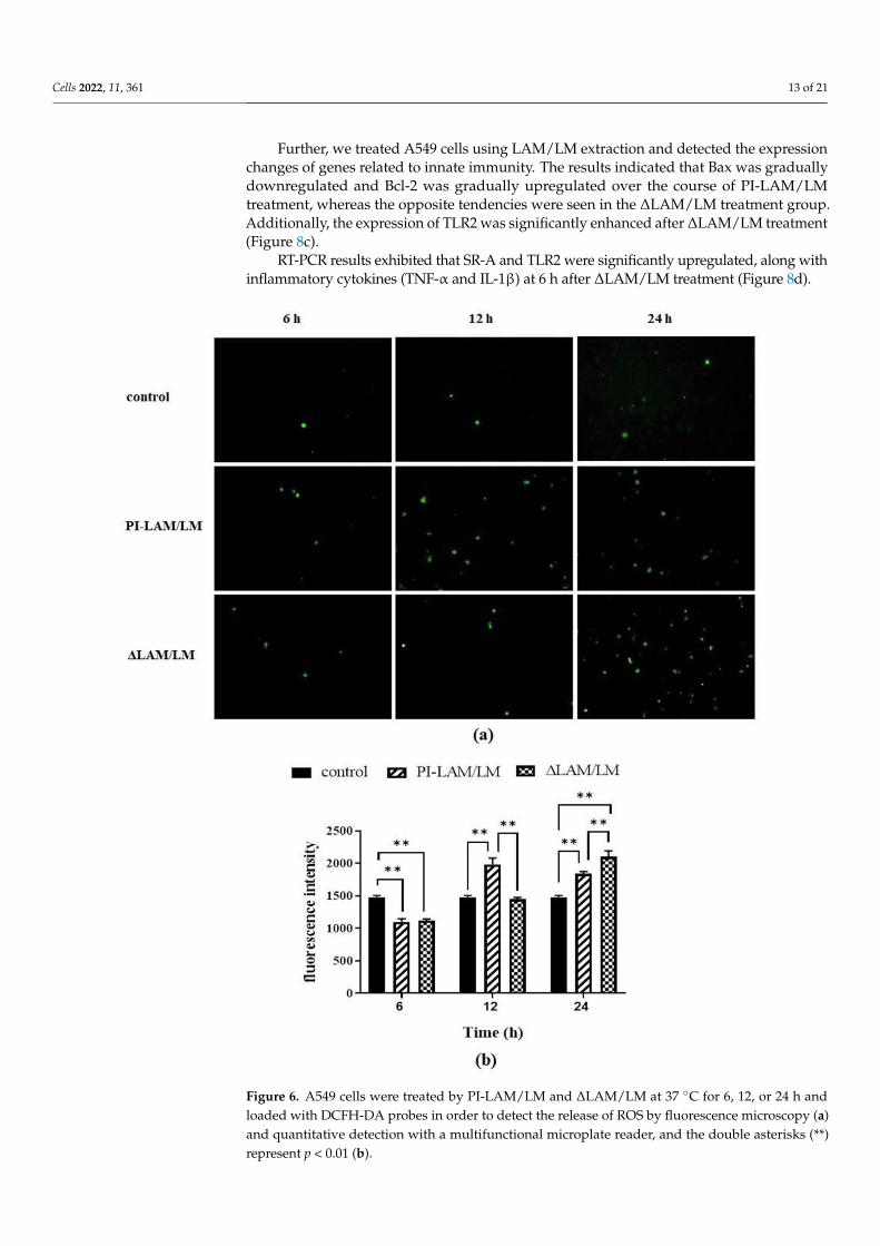
3.6. LAM/LM Molecules Affected the Release of ROS
ROS can promote the permeability of the mitochondrial membrane and increase the
release of Ca2+, and so they play a critical role in the induction of apoptosis mediated by
the mitochondrial pathway [51]. In addition, LAP depends on ROS generated by NADPH
oxidase, which can directly kill bacteria [18]. Our results indicated that ROS released from
the two LAM/LM treatment groups gradually increased with the duration of LAM/LM
treatment. PI-LAM/LM treatment induced ROS release at 12 h, whereas ΔLAM/LM treat-
ment activated ROS release at 24 h (Figure 6). Thus, LAM from M. smegmatis can enhance
ROS release, which is probably a critical factor to induce intracellular pathogen clearance,
and a decrease in Araf residues in LAM molecules may modulate ROS release.
Figure 5. TEM analysis of A549 cells. (a) A549 cells infected by M. smegmatis at 0 or 12 h post-infection; (b) A549 cells infected by M. sm-∆M_6387 at 0 or 12 h; (c) A549 cells treated by PI-LAM fromM. smegmatis; (d) A549 cells treated by ∆LAM from M. sm-∆M_6387; (e) The mean sizes of phagosomes

Cells 2022, 11, 361 12 of 21
and mitochondria in infected A549 were respectively analyzed, and the asterisks (*) representsp < 0.05; (f) The mean sizes of phagosomes and mitochondria in A549 cells pre-treated by LAMmixture were showed, and the double asterisks (**) represent p < 0.01. Red arrows point to all kindsof mitochondria, and the yellow asterisks are exhibited within phagosomes untaked bacteria.
To further illustrate whether LAM molecules directly affect the role of mitochondria,LAM extraction was used to treat A549 cells. Our results indicate that PI-LAM fromM. smegmatis also elicited larger-sized mitochondria than ∆LAM/LM extraction fromM. sm-∆M_6387 (Figure 5c,d). These results suggest that both M. smegmatis and M. sm-∆M_6387 can trigger the maturation of phagosomes, but M. sm-∆M_6387 appeared tohave poorer survival due to degradation in the observed cells. In addition, PI-LAM fromnonpathogenic M. smegmatis might induce stronger reactions in mitochondria compared to∆LAM from M. sm-∆M_6387.
3.6. LAM/LM Molecules Affected the Release of ROS
ROS can promote the permeability of the mitochondrial membrane and increase therelease of Ca2+, and so they play a critical role in the induction of apoptosis mediated bythe mitochondrial pathway [51]. In addition, LAP depends on ROS generated by NADPHoxidase, which can directly kill bacteria [18]. Our results indicated that ROS released fromthe two LAM/LM treatment groups gradually increased with the duration of LAM/LMtreatment. PI-LAM/LM treatment induced ROS release at 12 h, whereas ∆LAM/LMtreatment activated ROS release at 24 h (Figure 6). Thus, LAM from M. smegmatis canenhance ROS release, which is probably a critical factor to induce intracellular pathogenclearance, and a decrease in Araf residues in LAM molecules may modulate ROS release.
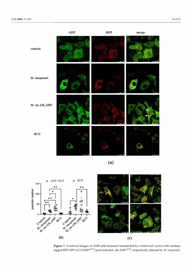
3.7. Recruitment and Localization of LC3 Molecules within Infected Alveolar Epithelial Cells
To further evaluate LC3-mediated phagocytosis compartments, we used a tandem-tagged RFP-GFP-LC3 vector to construct A549+LC3 cells. Due to the differential pH sen-sitivity of GFP and RFP, non-acidic phagocytosis compartments are labeled with bothGFP and RFP; however, the phagolysosome, after fusing with a lysosome, becomes acidic,and GFP fluorescence is quenched, and only RFP remains [41]. Here, we found that bothM. smegmatis and M. sm-∆M_6387 caused a significant increase in non-acidic yellow spots(RFP + GFP) 2 h post-infection. M sm-∆M_6387 had a particularly large number of spots;however, BCG with man-LAM did not cause a specific change in yellow spot number(Figure 7a,b and Figure S1). Moreover, EGFP molecules seemed to be embraced on thecell membrane, while LC3 recruitment was obvious, which might have resulted from theleakage of the M. smegmatis::EGFP strains being killed (Figure 7c). The data indicate thatboth M. smegmatis and M sm-∆M_6387 are able to induce LC3 recruitment, but BCG strainsdo not.
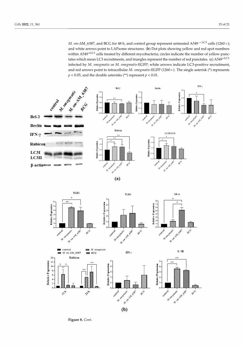
3.8. LAM/LM Molecules Regulated Gene Expression Related to Pathogenic Recognition andImmune Responses
A549 cells were collected at 12 h post-infection to detect the expression changes ofgenes related to LAP, including Bcl-2, Beclin, Rubicon, IFN-γ, and LC3, through Westernblotting. The results indicated that Rubicon was upregulated in both M sm-∆M_6387 andM. smegmatis compared with the control group; the lipidated LC3 II was also upregulated inthe two strains. In addition, we also found that M sm-∆M_6387 significantly downregulatedthe expression of IFN-γ compared with the control group (Figure 8a). The decrease in IFN-γexpression is likely to relate to a lesser intercellular mycobacterial load. The QRT-PCRresults exhibited that M sm-∆M_6387 upregulated the expression of SR-A, Rubicon, andIL-1β. In particular, Rubicon expression was obviously increased at 24 h post-infection(Figure 8b). It is necessary to clarify the mechanisms of the expression changes of IFN-γand SR-A in the future.
Cells 2022, 11, 361 13 of 21
Further, we treated A549 cells using LAM/LM extraction and detected the expressionchanges of genes related to innate immunity. The results indicated that Bax was graduallydownregulated and Bcl-2 was gradually upregulated over the course of PI-LAM/LMtreatment, whereas the opposite tendencies were seen in the ∆LAM/LM treatment group.Additionally, the expression of TLR2 was significantly enhanced after ∆LAM/LM treatment(Figure 8c).
RT-PCR results exhibited that SR-A and TLR2 were significantly upregulated, along withinflammatory cytokines (TNF-α and IL-1β) at 6 h after ∆LAM/LM treatment (Figure 8d).
Cells 2022, 11, x FOR PEER REVIEW 13 of 21
Figure 6. A549 cells were treated by PI-LAM/LM and ΔLAM/LM at 37 °C for 6, 12, or 24 h and
loaded with DCFH-DA probes in order to detect the release of ROS by fluorescence microscopy (a)
and quantitative detection with a multifunctional microplate reader, and the double asterisks (**)
represent p < 0.01 (b).
3.7. Recruitment and Localization of LC3 Molecules within Infected Alveolar Epithelial Cells
To further evaluate LC3-mediated phagocytosis compartments, we used a tandem-
tagged RFP-GFP-LC3 vector to construct A549+LC3 cells. Due to the differential pH sensi-
tivity of GFP and RFP, non-acidic phagocytosis compartments are labeled with both GFP
and RFP; however, the phagolysosome, after fusing with a lysosome, becomes acidic, and
GFP fluorescence is quenched, and only RFP remains [41]. Here, we found that both M.
smegmatis and M. sm-ΔM_6387 caused a significant increase in non-acidic yellow spots
(RFP + GFP) 2 h post-infection. M sm-ΔM_6387 had a particularly large number of spots;
however, BCG with man-LAM did not cause a specific change in yellow spot number
(Figures 7a,b and S1). Moreover, EGFP molecules seemed to be embraced on the cell mem-
brane, while LC3 recruitment was obvious, which might have resulted from the leakage
of the M. smegmatis::EGFP strains being killed (Figure 7c). The data indicate that both M.
smegmatis and M sm-ΔM_6387 are able to induce LC3 recruitment, but BCG strains do not.
Figure 6. A549 cells were treated by PI-LAM/LM and ∆LAM/LM at 37 C for 6, 12, or 24 h andloaded with DCFH-DA probes in order to detect the release of ROS by fluorescence microscopy (a)and quantitative detection with a multifunctional microplate reader, and the double asterisks (**)represent p < 0.01 (b).
Cells 2022, 11, 361 14 of 21
Cells 2022, 11, x FOR PEER REVIEW 14 of 21
Figure 7. Confocal images of A549 cells transient transfected by a lentiviral vector with tandem-tagged RFP-GFP-LC3 (A549+LC3) post-infection. (a) A549+LC3, respectively, infected by M. smegmatis,
Cells 2022, 11, 361 15 of 21
M. sm-∆M_6387, and BCG for 48 h, and control group represent untreated A549 + LC3 cells (1260×);and white arrows point to LAPsome structures. (b) Dot plots showing yellow and red spot numberswithin A549+LC3 cells treated by different mycobacteria; circles indicate the number of yellow punc-tates which mean LC3 recruitments, and triangles represent the number of red punctates. (c) A549+LC3
infected by M. smegmatis or M. smegmatis-EGFP; white arrows indicate LC3-positive recruitment,and red arrows point to intracellular M. smegmatis-EGFP (1260×). The single asterisk (*) representsp < 0.05, and the double asterisks (**) represent p < 0.01.
Cells 2022, 11, x FOR PEER REVIEW 16 of 21
Figure 8. Cont.

Cells 2022, 11, 361 16 of 21Cells 2022, 11, x FOR PEER REVIEW 17 of 21
Figure 8. The identification of the expression levels of proteins or genes related to LAP and inflammatory responses through Western blotting and RT-PCR. (a) A549 cells were infected by M. smegmatis, M. sm-ΔM_6387, or BCG for 6 h, and then were collected 12 h post-infection for the identification of protein expression changes through Western blotting. The proteins analyzed included Bcl-2, Beclin, Rubicon, IFN-γ, and LC3, and the internal reference, β-actin. The significant differences in expression are marked. (b) qRT-PCR was used to detect mRNA expression, and the genes analyzed included TLR2, TLR4, SR-A, Rubicon, IFN-γ, and IL-1β. (c) A549 cells were treated PI-LAM/LM or ΔLAM/LM and were incubated at 37 °C for 6, 12, or 24 h. The expression changes of three genes, Bax, Bcl-2, and TLR2, were identified through Western blotting. (d) The expression changes of four genes, TLR2, SR-A, TNF-α, and IL-1β, were identified through RT-PCR anal-ysis. The single asterisk (*) represents p value < 0.05, and statistical difference, and the double asterisks (**) represent p value < 0.01, and significance difference.
4. Discussion After M. tb infection, LAM can modulate the innate immunity of phagocytes, such as
alveolar epithelial cells, macrophages, and dendritic cells [52,53]. The innate immune re-sponses against mycobacteria are mainly involved in inflammatory factors secretion, phagocytosis, autophagy, and bactericidal activity [20,54]. LAM is recognized through mannose receptor (MR), dendritic cell-specific intercellular adhesion molecule-3-grabbing non-integrin (DC-SIGN), and TLRs [55]. SR-A has been demonstrated to modulate TLRs signaling [56]. LAM possesses diversity receptor affinity and heterogeneous immunogen-icity due to the differences in LAM structures [57].
This study first exhibited that M. sm-ΔM_6387 with LAM-truncated molecules had some prominent cell wall defects and migration inhibition. The changes suggest that the cell envelope of M. sm-ΔM_6387 tends to have a higher permeability compared with wild type M. smegmatis. When carrying out the studies on the phagocytosis of alveolar epithe-lial cells (A549), we found that M. sm-ΔM_6387 had lower intracellular uptake and
Figure 8. The identification of the expression levels of proteins or genes related to LAP and inflamma-tory responses through Western blotting and RT-PCR. (a) A549 cells were infected by M. smegmatis,M. sm-∆M_6387, or BCG for 6 h, and then were collected 12 h post-infection for the identificationof protein expression changes through Western blotting. The proteins analyzed included Bcl-2,Beclin, Rubicon, IFN-γ, and LC3, and the internal reference, β-actin. The significant differences inexpression are marked. (b) qRT-PCR was used to detect mRNA expression, and the genes analyzedincluded TLR2, TLR4, SR-A, Rubicon, IFN-γ, and IL-1β. (c) A549 cells were treated PI-LAM/LMor ∆LAM/LM and were incubated at 37 C for 6, 12, or 24 h. The expression changes of threegenes, Bax, Bcl-2, and TLR2, were identified through Western blotting. (d) The expression changesof four genes, TLR2, SR-A, TNF-α, and IL-1β, were identified through RT-PCR analysis. The singleasterisk (*) represents p value < 0.05, and statistical difference, and the double asterisks (**) representp value < 0.01, and significance difference.
4. Discussion
After M. tb infection, LAM can modulate the innate immunity of phagocytes, suchas alveolar epithelial cells, macrophages, and dendritic cells [52,53]. The innate immuneresponses against mycobacteria are mainly involved in inflammatory factors secretion,phagocytosis, autophagy, and bactericidal activity [20,54]. LAM is recognized throughmannose receptor (MR), dendritic cell-specific intercellular adhesion molecule-3-grabbingnon-integrin (DC-SIGN), and TLRs [55]. SR-A has been demonstrated to modulate TLRs sig-naling [56]. LAM possesses diversity receptor affinity and heterogeneous immunogenicitydue to the differences in LAM structures [57].
This study first exhibited that M. sm-∆M_6387 with LAM-truncated molecules hadsome prominent cell wall defects and migration inhibition. The changes suggest thatthe cell envelope of M. sm-∆M_6387 tends to have a higher permeability compared withwild type M. smegmatis. When carrying out the studies on the phagocytosis of alveolar
Cells 2022, 11, 361 17 of 21
epithelial cells (A549), we found that M. sm-∆M_6387 had lower intracellular uptake andretarded growth compared to M. smegmatis. We wondered whether the consequenceswere determined by mycobacterial biological characteristics such as the cell envelopepermeability and mycobacterial migration or different immunity of alveolar epithelial cells.Here, we confirmed that the effects did not result from mycobacteria but the divergentreactions of host cells through the studies on the same mycobacteria to challenge A549 cellspre-treated by LAM.
Further studies found that both M. smegmatis and its variant M. sm-∆M_6387 depositedLC3 on phagosomal membranes. Moreover, the levels of their LC3 recruitment were signif-icantly different. We observed that M. sm-∆M_6387 increased mycobacterial degradation inphagosomes in TEM images. The results suggest that M. sm-∆M_6387 induces a strongerremoval ability of intracellular bacteria, which is likely due to the regulation of LAP.
Rubicon is an essential and positive regulator of the NADPH oxidase complex, whichactivates LAP [18,19,22]. Our results indicated that both M. smegmatis and M. sm-∆M_6387upregulated Rubicon expression; moreover, M. sm-∆M_6387 induced stronger RubiconmRNA expression at 24 h post-infection. In addition, the two strains also enhanced,transiently, and mRNA expression of genes related to protective immunity, includingTLR2, SR-A, and IL-1β. Interestingly, we also found that ∆LAM/LM from M. sm-∆M_6387can strongly upregulate mRNA expression of TLR2, SR-A, TNF-α at 6h, and enhanceROS release at 24 h. Therefore, the findings suggest that the LAM–truncated moleculesdifferently influence innate immune responses against intracellular bacteria, which is likelyto be related to LAP.
The studies indicated that the clearance ability of host cells for a pathogen was promi-nently determined by autophagy [16,58]. Previous studies also found that nonpathogenicM. smegmatis within alveolar epithelial cells may be eliminated or replicated, mainlydepending on the intracellular mycobacterial load and the effectiveness of the immuneresponses [11,59–61]. On the basis of TLR2 activation, Rubicon interacts with one subunit ofthe NADPH oxidase complex, enhancing phagosomal trafficking to induce ROS release andthe expression of inflammatory cytokines to defend against invaders [18,19]. Our resultsdemonstrated that nonpathogenic M. smegmatis could activate the expression of genesrelated to the LAP pathway, and the arabinose domain of PI-LAM modulates the activation.
In addition, we found that ∆LAM/LM from M. sm-∆M_6387 decreased mycobacterialuptake in our experimental conditions. Another interesting observation is that individualphagocyte has distinct uptake ability in TEM and confocal images, being likely to elicitdivergent phagocytosis. Moreover, mycobacterial uptake ability is contrary to LC3 acti-vation. The findings led us to hypothesize that LAP inhibits mycobacterial phagocytosisand intracellular survival. SR-A, with a wide range of ligand targets, is expressed onphagocytes. In the lungs, SR-A mediates uptake and pathogenic clearance and has criticalroles in the capture and degradation of pathogens [25,62]. These results demonstrate thatthe LAM can better activate SR-A mRNA expression. Furthermore, the time-dependentexpression changes of Bcl-2 caused by ∆LAM/LM and PI-LAM/LM were opposite. Aswe know, Beclin-1 is essential for LAP, and the Beclin-1/VPS34 complex is repressed byBcl-2 [20]. It is an interesting question whether the expression changes of Bcl-2 affect theactivation of Beclin-1 during LAM treatment. However, we did not check the statisticaldifference of Beclin-1 expression in this study.
Taken together, nonpathogenic M. smegmatis can deposit LC3 on phagosomal mem-branes, and the decrease in the quantity of Araf residues of LAM molecules not onlyimpacts mycobacterial cell wall integrity but also enhances host defense responses againstdevastating pathogens. Given LAM-truncated molecules could be used as a tool for thedevelopment of HDTs, it is important to resolve how circulating LAM in serum enters thehost cell cytoplasm [43,63]. Our results indicate that both PI-LAM and the variant ∆LAMare of interest to the modulation of innate immune responses. These results provide a clueto uncovering the relationship between the structure of LAM and innate immune responses.

Cells 2022, 11, 361 18 of 21
On the basis of these findings, it is necessary to further explore the molecular mechanismsin detail.
Supplementary Materials: The following supporting information can be downloaded at: https://www.mdpi.com/article/10.3390/cells11030361/s1. Figure S1: Confocal image of A549 cellstransiently transfected by a lentiviral vector with tandem-tagged RFP-GFP-LC3 (A549+LC3) post-infection. (a) A549+LC3 infected by M. smegmatis, M. sm-∆M_6387, or BCG for 1, 2, and 3 h, respectively(630×); white arrows point to LAPsome structures. (b–d) Dot plots showing yellow and red spotnumbers within A549+LC3 cells treated by different mycobacteria; circles indicate the number ofyellow punctates, and triangles point to red spots; the panels of b, c and d showed respectively 1, 2,and 3 h post-infection; the asterisks including *, **, *** and **** represents p < 0.05 or lower.
Author Contributions: Methodology, S.C. and J.H.; software, T.X. and X.L.; validation, T.J.; formalanalysis, W.F.; investigation, H.L. and W.F.; data curation, X.G. and W.F.; writing—original draftpreparation, X.G.; writing—review and editing, H.L.; supervision, T.J.; project administration, T.J.;funding acquisition, T.J. All authors have read and agreed to the published version of the manuscript.
Funding: This research was funded by the National Natural Science Foundation of China, grantnumbers 31300672 and 31900937; the Natural Science Foundation of Liaoning Province, grant number20180550231; the Technology Innovation Foundation of Dalian, grant number 2020JJ26SN051.
Institutional Review Board Statement: Not applicable.
Informed Consent Statement: Not applicable.
Data Availability Statement: The A549 cell line was provided by national collection of authenticatedcell cultures. The plasmids relating to the construction of M. smefmatis geneknock out strain wereobtained from Prof. Yufang Ma, and the pSUM-kan-EGFP reporter plasmid was obtained from Dr.Nicolai S.C. van Oers.
Acknowledgments: I would like to thanks Yufang Ma and Oers for providing plasmids, thanksBiaojie Qin for providing technical support, thanks the program supporting Young faculty of thecolloge of Basic Medical Science of Dalian Medical University for providing funding, as well thanksfor grants from the National Natural Science Foundation of China (31300672, 31900937), the NaturalScience Foundation of Liaoning Province (20180550231), and the Technology Innovation Foundationof Dalian (2020JJ26SN051).
Conflicts of Interest: The authors declare no conflict of interest.
References1. World Health Organization. Global Tuberculosis Report 2021; World Health Organization: Geneva, Switzerland, 2021.2. Chakaya, J.; Khan, M.; Ntoumi, F.; Aklillu, E.; Fatima, R.; Mwaba, P.; Kapata, N.; Mfinanga, S.; Hasnain, S.E.; Katoto, P.D.M.C.;
et al. Global Tuberculosis Report 2020—Reflections on the Global TB burden, treatment and prevention efforts. Int. J. Infect. Dis.2021, 113, S7–S12. [CrossRef]
3. Grayfer, L.; Hodgkinson, J.W.; Belosevic, M. Antimicrobial responses of teleost phagocytes and innate immune evasion strategiesof intracellular bacteria. Dev. Comp. Immunol. 2014, 43, 223–242. [CrossRef] [PubMed]
4. Ryndak, M.B.; Laal, S. Mycobacterium tuberculosis Primary Infection and Dissemination: A Critical Role for Alveolar EpithelialCells. Front. Cell Infect. Microbiol. 2019, 9, 299. [CrossRef]
5. Scordo, J.M.; Knoell, D.L.; Torrelles, J.B. Alveolar Epithelial Cells in Mycobacterium tuberculosis Infection: Active Players orInnocent Bystanders? J. Innate. Immun. 2016, 8, 3–14. [CrossRef]
6. Paik, S.; Kim, J.K.; Chung, C.; Jo, E.K. Autophagy: A new strategy for host-directed therapy of tuberculosis. Virulence 2019, 10,448–459. [CrossRef] [PubMed]
7. Kramarska, E.; Squeglia, F.; de Maio, F.; Delogu, G.; Berisio, R. PE_PGRS33, an Important Virulence Factor of Mycobacteriumtuberculosis and Potential Target of Host Humoral Immune Response. Cells 2021, 10, 161. [CrossRef] [PubMed]
8. Ahmed, S.; Raqib, R.; Guðmundsson, G.H.; Bergman, P.; Agerberth, B.; Rekha, R.S. Host-Directed Therapy as a Novel TreatmentStrategy to Overcome Tuberculosis: Targeting Immune Modulation. Antibiotics 2020, 9, 21. [CrossRef] [PubMed]
9. Bhatt, K.; Bhagavathula, M.; Verma, S.; Timmins, G.S.; Deretic, V.P.; Ellner, J.J.; Salgame, P. Rapamycin modulates pulmonarypathology in a murine model of Mycobacterium tuberculosis infection. Dis. Model. Mech. 2021, 14, dmm049018. [CrossRef][PubMed]
10. Young, C.; Walzl, G.; Plessis, N.D. Therapeutic host-directed strategies to improve outcome in tuberculosis. Mucosal. Immunol.2020, 13, 190–204. [CrossRef]
Cells 2022, 11, 361 19 of 21
11. Gutierrez, M.G.; Master, S.S.; Singh, S.B.; Taylor, G.A.; Colombo, M.I.; Deretic, V. Autophagy is a defense mechanism inhibitingBCG and Mycobacterium tuberculosis survival in infected macrophages. Cell 2004, 119, 753–766. [CrossRef]
12. Scarlatti, F.; Maffei, R.; Beau, I.; Ghidoni, R.; Codogno, P. Non-canonical autophagy: An exception or an underestimated form ofautophagy? Autophagy 2008, 4, 1083–1085. [CrossRef] [PubMed]
13. Xie, Z.; Klionsky, D.J. Autophagosome formation: Core machinery and adaptations. Nat. Cell Biol. 2007, 9, 1102–1109. [CrossRef]14. Inomata, M.; Xu, S. Macrophage LC3-associated phagocytosis is an immune defense against Streptococcus pneumoniae that
diminishes with host aging. Proc. Natl. Acad. Sci. USA 2020, 117, 33561–33569. [CrossRef] [PubMed]15. Köster, S.; Upadhyay, S.; Chandra, P.; Papavinasasundaram, K.; Yang, G.; Hassan, A.; Grigsby, S.J.; Mittal, E.; Park, H.S.; Jones, V.;
et al. Mycobacterium tuberculosis is protected from NADPH oxidase and LC3-associated phagocytosis by the LCP protein CpsA.Proc. Natl. Acad. Sci. USA 2017, 114, E8711–E8720. [CrossRef]
16. Upadhyay, S.; Philips, J.A. LC3-associated phagocytosis: Host defense and microbial response. Curr. Opin. Immunol. 2019, 60,81–90. [CrossRef]
17. Castillo, E.F.; Dekonenko, A.; Arko-Mensah, J.; Mandell, M.A.; Dupont, N.; Jiang, S.; Delgado-Vargas, M.; Timmins, G.S.;Bhattacharya, D.; Yang, H.; et al. Autophagy protects against active tuberculosis by suppressing bacterial burden and inflamma-tion. Proc. Natl. Acad. Sci. USA 2012, 109, E3168–E3176. [CrossRef] [PubMed]
18. Yang, C.-S.; Lee, J.-S.; Rodgers, M.; Min, C.-K.; Lee, J.-Y.; Kim, H.J.; Lee, K.-H.; Kim, C.-J.; Oh, B.; Zandi, E.; et al. Autophagyprotein Rubicon mediates phagocytic NADPH oxidase activation in response to microbial infection or TLR stimulation. Cell Host.Microbe. 2012, 11, 264–276. [CrossRef] [PubMed]
19. Vergne, I.; Gilleron, M.; Nigou, J. Manipulation of the endocytic pathway and phagocyte functions by Mycobacterium tuberculosislipoarabinomannan. Front. Cell Infect. Microbiol. 2014, 4, 187. [CrossRef] [PubMed]
20. Matsunaga, K.; Saitoh, T.; Tabata, K.; Omori, H.; Satoh, T.; Kurotori, N.; Maejima, I.; Shirahama-Noda, K.; Ichimura, T.; Isobe, T.;et al. Two Beclin 1-binding proteins, Atg14L and Rubicon, reciprocally regulate autophagy at different stages. Nat. Cell Biol. 2009,11, 385–396. [CrossRef]
21. Ogawa, M.; Takada, N.; Shizukuishi, S.; Tomokiyo, M.; Chang, B.; Yoshida, M.; Kakuta, S.; Tanida, I.; Ryo, A.; Guan, J.; et al.Streptococcus pneumoniae triggers hierarchical autophagy through reprogramming of LAPosome-like vesicles via NDP52-delocalization. Commun. Biol. 2020, 3, 25. [CrossRef]
22. Köster, S.; Upadhyay, S.; Philips, J.A. Why macrophages cannot LAP up TB. Autophagy 2018, 14, 552–554. [CrossRef]23. Sanjuan, M.A.; Dillon, C.P.; Tait, S.W.G.; Moshiach, S.; Dorsey, F.; Connell, S.; Komatsu, M.; Tanaka, K.; Cleveland, J.L.; Withoff, S.;
et al. Toll-like receptor signalling in macrophages links the autophagy pathway to phagocytosis. Nature 2007, 450, 1253–1257.[CrossRef]
24. Cadwell, K.; Philips, J.A. Autophagy meets phagocytosis. Immunity 2013, 39, 425–427. [CrossRef]25. Bonilla, D.L.; Bhattacharya, A.; Sha, Y.; Xu, Y.; Xiang, Q.; Kan, A.; Jagannath, C.; Komatsu, M.; Eissa, N.T. Autophagy regulates
phagocytosis by modulating the expression of scavenger receptors. Immunity 2013, 39, 537–547. [CrossRef]26. Barry, C.E.; Crick, D.C.; McNeil, M.R. Targeting the formation of the cell wall core of M. tuberculosis. Infect. Disord. Drug Targets
2007, 7, 182–202. [CrossRef] [PubMed]27. Chatterjee, D. The mycobacterial cell wall: Structure, biosynthesis and sites of drug action. Curr. Opin. Chem. Biol. 1997, 1,
579–588. [CrossRef]28. Turner, J.; Torrelles, J.B. Mannose-capped lipoarabinomannan in Mycobacterium tuberculosis pathogenesis. Pathog. Dis. 2018,
76, fty026. [CrossRef]29. Feng, X.; Lu, J.; He, Z.; Wang, Y.; Qi, F.; Pi, R.; Zhang, G. Mycobacterium smegmatis Induces Neurite Outgrowth and Differentiation
in an Autophagy-Independent Manner in PC12 and C17.2 Cells. Front. Cell Infect. Microbiol. 2018, 8, 201. [CrossRef]30. Berg, S.; Kaur, D.; Jackson, M.; Brennan, P.J. The glycosyltransferases of Mycobacterium tuberculosis-roles in the synthesis of
arabinogalactan, lipoarabinomannan, and other glycoconjugates. Glycobiology 2007, 17, 35R–56R. [CrossRef] [PubMed]31. Cui, Z.; Li, Y.; Cheng, S.; Yang, H.; Lu, J.; Hu, Z.; Ge, B. Mutations in the embC-embA intergenic region contribute to Mycobac-
terium tuberculosis resistance to ethambutol. Antimicrob. Agents Chemother. 2014, 58, 6837–6843. [CrossRef] [PubMed]32. Korkegian, A.; Roberts, D.M.; Blair, R.; Parish, T. Mutations in the essential arabinosyltransferase EmbC lead to alterations in
Mycobacterium tuberculosis lipoarabinomannan. J. Biol. Chem. 2014, 289, 35172–35181. [CrossRef]33. Torrelles, J.B.; Khoo, K.H.; Sieling, P.A.; Modlin, R.L.; Zhang, N.; Marques, A.M.; Treumann, A.; Rithner, C.D.; Brennan, P.J.;
Chatterjee, D. Truncated structural variants of lipoarabinomannan in Mycobacterium leprae and an ethambutol-resistant strain ofMycobacterium tuberculosis. J. Biol. Chem. 2004, 279, 41227–41239. [CrossRef] [PubMed]
34. Aliprantis, A.O.; Yang, R.B.; Mark, M.R.; Suggett, S.; Devaux, B.; Radolf, J.D.; Klimpel, G.R.; Godowski, P.; Zychlinsky, A. Cellactivation and apoptosis by bacterial lipoproteins through toll-like receptor-2. Science 1999, 285, 736–739. [CrossRef] [PubMed]
35. Shui, W.; Petzold, C.J.; Redding, A.; Liu, J.; Pitcher, A.; Sheu, L.; Hsieh, T.-y.; Keasling, J.D.; Bertozzi, C.R. Organelle membraneproteomics reveals differential influence of mycobacterial lipoglycans on macrophage phagosome maturation and autophagosomeaccumulation. J. Proteome. Res. 2011, 10, 339–348. [CrossRef]
36. Dao, D.N.; Kremer, L.; Guérardel, Y.; Molano, A.; Jacobs, W.R., Jr.; Porcelli, S.A.; Briken, V. Mycobacterium tuberculosislipomannan induces apoptosis and interleukin-12 production in macrophages. Infect. Immun. 2004, 72, 2067–2074. [CrossRef]

Cells 2022, 11, 361 20 of 21
37. Roca, F.J.; Whitworth, L.J.; Redmond, S.; Jones, A.A.; Ramakrishnan, L. TNF Induces Pathogenic Programmed MacrophageNecrosis in Tuberculosis through a Mitochondrial-Lysosomal-Endoplasmic Reticulum Circuit. Cell 2019, 178, 1344–1361.e11.[CrossRef] [PubMed]
38. Vignal, C.; Guérardel, Y.; Kremer, L.; Masson, M.; Legrand, D.; Mazurier, J.; Elass, E. Lipomannans, but not lipoarabinomannans,purified from Mycobacterium chelonae and Mycobacterium kansasii induce TNF-alpha and IL-8 secretion by a CD14-toll-likereceptor 2-dependent mechanism. J. Immunol. 2003, 171, 2014–2023. [CrossRef] [PubMed]
39. Yuan, C.; Qu, Z.L.; Tang, X.L.; Liu, Q.; Luo, W.; Huang, C.; Pan, Q.; Zhang, X.L. Mycobacterium tuberculosis Mannose-CappedLipoarabinomannan Induces IL-10-Producing B Cells and Hinders CD4(+)Th1 Immunity. iScience 2019, 11, 13–30. [CrossRef]
40. O’Connor, R.A.; Wittmer, S.; Dalton, D.K. Infection-induced apoptosis deletes bystander CD4+ T cells: A mechanism forsuppression of autoimmunity during BCG infection. J. Autoimmun. 2005, 24, 93–100. [CrossRef]
41. Paik, S.; Jo, E.K. An Interplay Between Autophagy and Immunometabolism for Host Defense Against Mycobacterial Infection.Front. Immunol. 2020, 11, 603951. [CrossRef]
42. Shi, L.; Berg, S.; Lee, A.; Spencer, J.S.; Zhang, J.; Vissa, V.; McNeil, M.R.; Khoo, K.-H.; Chatterjee, D. The carboxy terminus ofEmbC from Mycobacterium smegmatis mediates chain length extension of the arabinan in lipoarabinomannan. J. Biol. Chem.2006, 281, 19512–19526. [CrossRef]
43. Augenstreich, J.; Briken, V. Host Cell Targets of Released Lipid and Secreted Protein Effectors of Mycobacterium tuberculosis.Front. Cell Infect. Microbiol. 2020, 10, 595029. [CrossRef]
44. Li, W.; Xin, Y.; McNeil, M.R.; Ma, Y. rmlB and rmlC genes are essential for growth of mycobacteria. Biochem. Biophys. Res. Commun.2006, 342, 170–178. [CrossRef]
45. Jiang, T.; He, L.; Zhan, Y.; Zang, S.; Ma, Y.; Zhao, X.; Zhang, C.; Xin, Y. The effect of MSMEG_6402 gene disruption on the cell wallstructure of Mycobacterium smegmatis. Microb. Pathog. 2011, 51, 156–160. [CrossRef]
46. Banaei, N.; Kincaid, E.Z.; Lin, S.-G.; Desmond, E.; William, R.J., Jr.; Ernst, J.D. Lipoprotein processing is essential for resistance ofMycobacterium tuberculosis to malachite green. Antimicrob. Agents Chemother. 2009, 53, 3799–3802. [CrossRef] [PubMed]
47. Fukuda, T.; Matsumura, T.; Ato, M.; Hamasaki, M.; Nishiuchi, Y.; Murakami, Y.; Maeda, Y.; Yoshimori, T.; Matsumoto, S.;Kobayashi, K.; et al. Critical roles for lipomannan and lipoarabinomannan in cell wall integrity of mycobacteria and pathogenesisof tuberculosis. mBio 2013, 4, e00472-12. [CrossRef] [PubMed]
48. Küssau, T.; van Wyk, N.; Johansen, M.D.; Alsarraf, H.M.A.B.; Neyret, A.; Hamela, C.; Sørensen, K.K.; Thygesen, M.B.;Beauvineau, C.; Kremer, L.; et al. Functional Characterization of the N-Acetylmuramyl-l-Alanine Amidase, Ami1, from Mycobac-terium abscessus. Cell 2020, 9, 2410. [CrossRef] [PubMed]
49. Eitson, J.L.; Medeiros, J.J.; Hoover, A.R.; Srivastava, S.; Roybal, K.T.; Aínsa, J.A.; Hansen, E.J.; Gumbo, T.; van Oers, N.S.C.Mycobacterial shuttle vectors designed for high-level protein expression in infected macrophages. Appl. Environ. Microbiol. 2012,78, 6829–6837. [CrossRef]
50. Sentürk, M.; Lin, G.; Zuo, Z.; Mao, D.; Watson, E.; Mikos, A.G.; Bellen, H.J. Ubiquilins regulate autophagic flux through mTORsignalling and lysosomal acidification. Nat. Cell Biol. 2019, 21, 384–396. [CrossRef] [PubMed]
51. Zhang, J.; Wang, X.; Vikash, V.; Ye, Q.; Wu, D.; Liu, Y.; Dong, W. ROS and ROS-Mediated Cellular Signaling. Oxid. Med. Cell.Longev. 2016, 2016, 4350965. [CrossRef] [PubMed]
52. Correia-Neves, M.; Sundling, C.; Cooper, A.; Källenius, G. Lipoarabinomannan in Active and Passive Protection againstTuberculosis. Front. Immunol. 2019, 10, 1968. [CrossRef]
53. Yan, Z.H.; Zhao, B.; Pang, Y.; Wang, X.J.; Yi, L.; Wang, H.L.; Yang, B.; Wei, P.J.; Jia, H.Y.; Li, S.P.; et al. Generation of mycobacteriallipoarabinomannan-specific monoclonal antibodies and their ability to identify mycobacterium isolates. J. Microbiol. Immunol.Infect. 2021, 54, 437–446. [CrossRef]
54. Rodrigues, T.S.; Conti, B.J.; Fraga-Silva, T.F.d.; Almeida, F.; Bonato, V.L.D. Interplay between alveolar epithelial and dendritic cellsand Mycobacterium tuberculosis. J. Leukoc. Biol. 2020, 108, 1139–1156. [CrossRef] [PubMed]
55. Nigou, J.; Gilleron, M.; Rojas, M.; García, L.F.; Thurnher, M.; Puzo, G. Mycobacterial lipoarabinomannans: Modulators of dendriticcell function and the apoptotic response. Microbe. Infect. 2002, 4, 945–953. [CrossRef]
56. Wu, M.Y.; Lu, J.H. Autophagy and Macrophage Functions: Inflammatory Response and Phagocytosis. Cells 2019, 9, 70. [CrossRef][PubMed]
57. Zhang, S.; Wu, Q.; Lei, H.; Zheng, H.; Zhou, F.; Sun, Z.; Zhao, J.; Yu, X.; Zhang, S. Mannosylated structures of mycobacteriallipoarabinomannans facilitate the maturation and activation of dendritic cells. Cell Immunol. 2019, 335, 85–92. [CrossRef][PubMed]
58. Huang, J.; Brumell, J.H. Bacteria-autophagy interplay: A battle for survival. Nat. Rev. Microbiol. 2014, 12, 101–114. [CrossRef]59. Sharma, M.; Sharma, S.; Roy, S.; Varma, S.; Bose, M. Pulmonary epithelial cells are a source of interferon-gamma in response to
Mycobacterium tuberculosis infection. Immunol. Cell Biol. 2007, 85, 229–237. [CrossRef]60. Miralda, I.; Klaes, C.K.; Graham, J.E.; Uriarte, S.M. Human Neutrophil Granule Exocytosis in Response to Mycobacterium
smegmatis. Pathogens 2020, 9, 123. [CrossRef]61. García-Pérez, B.E.; Hernández-González, J.C.; García-Nieto, S.; Luna-Herrera, J. Internalization of a non-pathogenic mycobacteria
by macropinocytosis in human alveolar epithelial A549 cells. Microb. Pathog. 2008, 45, 1–6. [CrossRef]

Cells 2022, 11, 361 21 of 21
62. Bowdish, D.M.E.; Sakamoto, K.; Kim, M.-J.; Kroos, M.; Mukhopadhyay, S.; Leifer, C.A.; Tryggvason, K.; Gordon, S.; Russell, D.G.MARCO, TLR2, and CD14 are required for macrophage cytokine responses to mycobacterial trehalose dimycolate and Mycobac-terium tuberculosis. PLoS Pathog. 2009, 5, e1000474. [CrossRef] [PubMed]
63. Inoue, M.; Niki, M.; Ozeki, Y.; Nagi, S.; Chadeka, E.A.; Yamaguchi, T.; Osada-Oka, M.; Ono, K.; Oda, T.; Mwende, F.; et al.High-density lipoprotein suppresses tumor necrosis factor alpha production by mycobacteria-infected human macrophages. Sci.Rep. 2018, 8, 6736. [CrossRef] [PubMed]
Related Documents