Laleh Ghasemi-Mobarakeh, Elham Shamirzaei-Jeshvaghani, Department of Textile Engineering, Isfahan University of Technology, Isfahan, 84156-83111, Iran Molamma P Prabhakaran, Lingling Tian, Seeram Ramakrishna, Department of Mechanical Engineering, Faculty of Engineering, 2 Engineering Drive 3, National University of Singapore, Singapore 117576, Singapore Leila Dehghani, Department of Medical Science, Islamic Azad University, Najafabad Branch, Isfahan,81595158, Iran Leila Dehghani, Isfahan Neurosciences Research Center, AlZahra Hospital, Isfahan University of Medical Sciences, Isfahan, 8174675731, Iran Author contributions: All the authors solely contributed to this paper. Conflict-of-interest: None to declare. Open-Access: This article is an open-access article which was selected by an in-house editor and fully peer-reviewed by external reviewers. It is distributed in accordance with the Creative Commons Attribution Non Commercial (CC BY-NC 4.0) license, which permits others to distribute, remix, adapt, build upon this work non-commercially, and license their derivative works on different terms, provided the original work is properly cited and the use is non-commercial. See: http://creativecommons.org/ licenses/by-nc/4.0/ Correspondence to: Laleh Ghasemi-Mobarakeh, PhD, Department of Textile Engineering, Isfahan University of Technology, Daneshgah e Sanati HW, Isfahan 84156-83111, Iran. [email protected] Telephone: +98-31-33911092 Fax: +98-31-33912444 Received: October 21, 2014 Peer-review started: October 22, 2014 First decision: November 27, 2014 Revised: December 18, 2014 Accepted: March 4, 2015 Article in press: March 5, 2015 Published online: May 26, 2015 Abstract Tissue engineering is a multidisciplinary field that applies the principles of engineering and life-sciences for regeneration of damaged tissues. Stem cells have attracted much interest in tissue engineering as a cell source due to their ability to proliferate in an undi- fferentiated state for prolonged time and capability of differentiating to different cell types after induction. Scaffolds play an important role in tissue engineering as a substrate that can mimic the native extracellular matrix and the properties of scaffolds have been shown to affect the cell behavior such as the cell attachment, proliferation and differentiation. Here, we focus on the recent reports that investigated the various aspects of scaffolds including the materials used for scaffold fabrication, surface modification of scaffolds, topography and mechanical properties of scaffolds towards stem cells differentiation effect. We will present a more detailed overview on the effect of mechanical properties of scaffolds on stem cells fate. Key words: Stem cells; Tissue engineering; Differen- tiation; Mechanical properties; Surface modification; Topography © The Author(s) 2015. Published by Baishideng Publishing Group Inc. All rights reserved. Core tip: Application of stem cells in tissue engineering and regenerative medicine has attracted many resear- chers and it is considered as a hot topic of research. The research was mostly aimed towards the fabrication of a suitable scaffold with appropriate physical, chemical, biological and mechanical properties to guide the differentiation of stem cells to specific lineages of interest. This paper reviews the previous researches on effect of chemical structure, surface modification, topography and mechanical properties of scaffolds on stem cells behavior. Fabrication of scaffolds with desirable properties can open new strategies for cell therapy using tissue engineering technique. Ghasemi-Mobarakeh L, Prabhakaran MP, Tian L, Shamirzaei- 728 May 26, 2015|Volume 7|Issue 4| WJSC|www.wjgnet.com REVIEW Submit a Manuscript: http://www.wjgnet.com/esps/ Help Desk: http://www.wjgnet.com/esps/helpdesk.aspx DOI: 10.4252/wjsc.v7.i4.728 World J Stem Cells 2015 May 26; 7(4): 728-744 ISSN 1948-0210 (online) © 2015 Baishideng Publishing Group Inc. All rights reserved. Structural properties of scaffolds: Crucial parameters towards stem cells differentiation Laleh Ghasemi-Mobarakeh, Molamma P Prabhakaran, Lingling Tian, Elham Shamirzaei-Jeshvaghani, Leila Dehghani, Seeram Ramakrishna

Welcome message from author
This document is posted to help you gain knowledge. Please leave a comment to let me know what you think about it! Share it to your friends and learn new things together.
Transcript
Laleh Ghasemi-Mobarakeh, Elham Shamirzaei-Jeshvaghani, Department of Textile Engineering, Isfahan University of Technology, Isfahan, 84156-83111, IranMolamma P Prabhakaran, Lingling Tian, Seeram Ramakrishna, Department of Mechanical Engineering, Faculty of Engineering, 2 Engineering Drive 3, National University of Singapore, Singapore 117576, SingaporeLeila Dehghani, Department of Medical Science, Islamic Azad University, Najafabad Branch, Isfahan,81595158, IranLeila Dehghani, Isfahan Neurosciences Research Center, AlZahra Hospital, Isfahan University of Medical Sciences, Isfahan, 8174675731, IranAuthor contributions: All the authors solely contributed to this paper.Conflict-of-interest: None to declare.Open-Access: This article is an open-access article which was selected by an in-house editor and fully peer-reviewed by external reviewers. It is distributed in accordance with the Creative Commons Attribution Non Commercial (CC BY-NC 4.0) license, which permits others to distribute, remix, adapt, build upon this work non-commercially, and license their derivative works on different terms, provided the original work is properly cited and the use is non-commercial. See: http://creativecommons.org/licenses/by-nc/4.0/Correspondence to: Laleh Ghasemi-Mobarakeh, PhD, Department of Textile Engineering, Isfahan University of Technology, Daneshgah e Sanati HW, Isfahan 84156-83111, Iran. [email protected]: +98-31-33911092Fax: +98-31-33912444Received: October 21, 2014Peer-review started: October 22, 2014First decision: November 27, 2014Revised: December 18, 2014Accepted: March 4, 2015Article in press: March 5, 2015Published online: May 26, 2015
AbstractTissue engineering is a multidisciplinary field that applies the principles of engineering and life-sciences
for regeneration of damaged tissues. Stem cells have attracted much interest in tissue engineering as a cell source due to their ability to proliferate in an undi-fferentiated state for prolonged time and capability of differentiating to different cell types after induction. Scaffolds play an important role in tissue engineering as a substrate that can mimic the native extracellular matrix and the properties of scaffolds have been shown to affect the cell behavior such as the cell attachment, proliferation and differentiation. Here, we focus on the recent reports that investigated the various aspects of scaffolds including the materials used for scaffold fabrication, surface modification of scaffolds, topography and mechanical properties of scaffolds towards stem cells differentiation effect. We will present a more detailed overview on the effect of mechanical properties of scaffolds on stem cells fate.
Key words: Stem cells; Tissue engineering; Differen-tiation; Mechanical properties; Surface modification; Topography
© The Author(s) 2015. Published by Baishideng Publishing Group Inc. All rights reserved.
Core tip: Application of stem cells in tissue engineering and regenerative medicine has attracted many resear-chers and it is considered as a hot topic of research. The research was mostly aimed towards the fabrication of a suitable scaffold with appropriate physical, chemical, biological and mechanical properties to guide the differentiation of stem cells to specific lineages of interest. This paper reviews the previous researches on effect of chemical structure, surface modification, topography and mechanical properties of scaffolds on stem cells behavior. Fabrication of scaffolds with desirable properties can open new strategies for cell therapy using tissue engineering technique.
Ghasemi-Mobarakeh L, Prabhakaran MP, Tian L, Shamirzaei-
728 May 26, 2015|Volume 7|Issue 4|WJSC|www.wjgnet.com
REVIEW
Submit a Manuscript: http://www.wjgnet.com/esps/Help Desk: http://www.wjgnet.com/esps/helpdesk.aspxDOI: 10.4252/wjsc.v7.i4.728
World J Stem Cells 2015 May 26; 7(4): 728-744ISSN 1948-0210 (online)
© 2015 Baishideng Publishing Group Inc. All rights reserved.
Structural properties of scaffolds: Crucial parameters towards stem cells differentiation
Laleh Ghasemi-Mobarakeh, Molamma P Prabhakaran, Lingling Tian, Elham Shamirzaei-Jeshvaghani, Leila Dehghani, Seeram Ramakrishna
Jeshvaghani E, Dehghani L, Ramakrishna S. Structural properties of scaffolds: Crucial parameters towards stem cells differentiation. World J Stem Cells 2015; 7(4): 728-744 Available from: URL: http://www.wjgnet.com/1948-0210/full/v7/i4/728.htm DOI: http://dx.doi.org/10.4252/wjsc.v7.i4.728
INTRODUCTIONAutologous cells obtained from adult patients might deliver a less difficult route to regenerative-cell therapies. In the past, many tissues were assumed incapable of self-regeneration upon its damage, mainly because they do not possess any endogenous stem cells. However, recent discoveries show that more adult tissues harbor cells with capacity for regenerative repair[1]. Tissue engineering comprises of suitable cells, scaffolds and appropriate biochemical/physical factors, such that it function as a strategy towards the engineering of tissues and cellular delivery. Encapsulating stem cells within biocompa-tible scaffolds provide a matrix which result in cell adhesion, migration and differentiation suitable for transplantation and tissue regeneration[2-4].
Stem cells have uncovered a new perspective as therapeutic tools in regenerative medicine. These cells with the ability to proliferate indefinitely (in vitro) and the capacity to differentiate into any somatic cell type are a potential cell source. Stem cells are originated in the early embryo, the fetus, placenta, umbilical cord, and it occurs in many different tissues of the body. Also they have been engineered from somatic cells, termed as induced pluripotent stem cells (iPSCs). iPSCs give rise to different cell types including the neural cell types (neurons, oligodendrocytes, and glia), cardiomyocytes, osteoblasts, hepatocytes, and haemopoietic proge-nitors cells[5-8]. The two main stem cell types are the embryonic stem cells (ESCs) and the adult stem cells. ESCs are derived from fetal tissue or from the inner cell mass of the blastocytes with ability for unlimited growth in culture that could be associated to a high risk of teratoma formation. Several researches have been done on ESCs pluripotency to obtain specific cell lineages for tissue engineering and regenerative medicine, as well as for therapeutic treatments[8-13]. Adult stem cells are specialized cells including the mesenchymal stem cells (MSCs), hematopoietic stem cells (HSCs), and neural stem cells (NSCs)[9]. MSCs are self-replicating cells that are capable of differentiating in multidirectional pathways, resulting in cell lineages such as the osteoblasts, chondrocytes, myocytes, marrow stromal cells, tendon-ligament fibroblasts, adipocytes, neural cells, oligodendrocytes and haemopoietic cells[9,14,15]. These cells have been used for the engineering of different tissues such as the cardiac[16] , tendon[17] , cartilage[18-20] , vascular[11,21] Meniscus[22], bone[23], Ligament[24], myocardia[25], fat[26], and neural tissues[5,16,17,27].
While a routine source of human MSCs is the bone marrow, they have been also derived from multiple adult tissues comprising of adipose tissue (ADSCs), umbilical cord blood, placenta, thymus, and dental pulp[28]. In comparison to MSCs derived from the bone marrow[29], ADSCs have several advantages such that they are easy to obtain after minor donor site morbidity, possess high proliferating capacity and can be preserved for longer period of time in cell banks[30]. Under appropriate culture conditions, ADSCs can differentiate to classical mesenchymal lineages, which include adipogenesis, chondrogenesis, osteogenesis, and myogenesis[31,32]. Moreover, the ability of ADSCs differentiation to numerous lineages when seeded into polymeric scaffolds has been well documented in several literatures[33,34].
The sources of HSCs are bone marrow, peripheral blood, umbilical cord blood, fetal hematopoietic sys-tem[35], ESCs and embryonic germ cells[11]. These cells are also a suitable cell source for blood vessel engineering[12]. Avoidance of anaesthesia without the need for hospitalization or blood transfusion, and low risk of serious adverse events are the major advantages of the peripheral stem cells, which make them the favourable source of stem cells worldwide.On the other hand, NSCs are defined as self-renewing multipotent progenitors existing in the developing and adult CNS. Generally, they are considered by their capacity to symmetrically self-renew and their ability to discriminate into neurons, oligodendrocytes, and astrocytes through asymmetrical fate-committed division[27]. Neural stem or progenitor cells can be obtained in different approaches such as direct isolation of these cells from embryonic or adult brain tissue[36,37]. NSCs have been used for tissue engineering of different tissues such as multiple cell types of the adult nervous system[5,38]. It was shown in several studies that stem cells behavior such as their adherence, proliferation and differentiation on scaffolds, depends on the material itself. Additionally, the surface chemistry, surface geometry/micro architecture of scaffolds, and the mechanical properties of scaffolds[39-42] play a significant role in determining the cellular responses after culture of stem cells on them[43].
Therefore, recent studies have focused on the selection of an appropriate biomaterial, whit focusing on the surface modification of the scaffolds, creating appropriate microstructures and controlling the mech-anical properties of scaffolds to recapitulate the in vivo microenvironment suitable for regenerating tissues or for the differentiation stem cells to specific cell lineages[44].
This review will discuss on the influence of different aspects of scaffolds including scaffold composition, surface modification, micro-nano architecture of scaffolds and mechanical properties of scaffolds per-taining to stem cells differentiation. An emphasis is also given to the effect of mechanical properties of
Ghasemi-Mobarakeh L et al . Scaffolds and stem cells differentiation
729 May 26, 2015|Volume 7|Issue 4|WJSC|www.wjgnet.com

scaffolds towards stem cells differentiation.
EFFECT OF SCAFFOLD COMPOSITION ON STEM CELLS DIFFERENTIATIONThe interaction of stem cells with their surrounding microenvironment is fundamental to multiple pro-cesses such as cell migration, proliferation, lineage specificity, and tissue morphogenesis[45]. Biomaterials play an important role in directing tissue growth and chemical properties of the scaffolds have been shown to influence the behavior of stem cells whereas the scaffold composition has an significant role in stem cells differentiation towards preferred lineages[42,43,45]. The ability to selectively guide stem cells differentiation by merely changing the properties of an underlying biomaterial scaffold is a smart approach in tissue engineering, which can help compliment or potentially eliminate the use of exogenous differentiation inducers like the viral gene vectors and small molecule drugs[46].
Natural polymers such as the collagen, fibrinogen, hyaluronic acid, glycosaminoglycans, cellulose, chi-tosan, silk fibroin, etc., consist of components found in the native extracellular matrix, such that they can assist cells to attach to cell surface receptors and provide a physical environment to control cell function compared to synthetic materials[40,47]. However these materials suffer from fast degradation rates, difficulty in sterilization and purification, high variability, high contamination potential, and induce immune response upon implantation, while synthetic materials offer the potential for improved control, repeatability and safety[40,47]. Scaffolds fabricated by synthetic materials can be chemically modified by natural materials to carefully regulate the differentiation of stem cells[47]. Immune response is an important concern in synthetic biomaterials used for scaffold fabrication and it can be reduced by selection of materials which are inert inherently or by modification of scaffolds to avoid detection by the immune system. Hydrophobic materials usually tend to increase monocyte adhesion compared to hydrophilic materials leading to a local immune reaction at the scaffold site[48].
Awad et al[34] compared the chondrogenic diffe-rentiation of human adipose-derived adult stem cells seeded in alginate, agarose and gelatin scaffolds and their findings revealed that the growth and differentiation potential of adult stem cells seeded on the scaffolds varied and the cells on gelatin scaffolds showed better proliferation and differentiation. Collagen has attracted much interest as a biomaterial for fabrication of scaffolds in tissue engineering as a key structural protein found in the extracellular matrix of many connective tissues[49,50]. In spite of the several advantages of collagen as a biodegradable, biocompatible, safe and multifunctional material, its potential to evoke immune response has raised concerns as an animal-derived biomaterial[51].
Meinel et al[52] investigated the osteogenic differen-tiation of human MSCs on different protein substrates including unmodified collagen with fast degradation rate, cross-linked collagen with slow degradation rate and silk; in both static and dynamic culture conditions. Under dynamic condition, MSCs on cross-linked collagen and silk scaffolds deposited more calcium and had a higher alkaline phosphatase activity than MSCs on unmodified collagen scaffolds indicating the importance of scaffold properties in causing osteogenesis of the cultured MSCs[52].
Jäger et al[39] cultivated MSCs on the D,D,L,L-polylactide (PLLA), collagen I/III, and polygalactin-910/polydioxanone (PGPD) scaffolds and their results showed significant differences in the proliferation, differentiation, and Ca2+ accumulation of MSCs on different scaffolds. Cell adhesion on PGPD was lower compared to the cell adhesion on PLLA and collagen I/III scaffolds, which was related to the significant biodegradation rate of PGPD scaffolds compared to other scaffolds. They found significantly higher cell numbers on collagen I/III membrane compared to that for PLLA scaffolds after 4 h cell culture while the differences were less significant after 28 d of cell seeding. Lower cell number on PLLA scaffolds compared to collagen I/III after 4 h could be attributed to the hydrophobic nature of PLLA while an increasing deposition of serum proteins from culture media on the PLLA scaffold may reduce the effect of its hydrophobic nature and decreased the quantitative differences in cell attachment between collagen I/III and PLLA scaffolds after 28 d cell seeding[39].
PEG is a relatively inert ,biocompatible and hydrophilic polymer[53]. Hwang et al[41] encapsulated MSCs in PEG based hydrogels containing type I collagen, type II collagen and hyaluronic acids (HA) which are the main components of musculoskeletal tissue matrix. Chondrogenic differentiation of MSCs was slightly enhanced in collagen-matrix-based hydrogels, whereas osteogenic differentiation was induced in HA-containing hydrogels suggesting the potential of ECM components to modulate the fate of MSCs differentiation[41].
Ravichandran et al[54] fabricated a composite scaffold containing PLLA and Poly-benzyl-L-glutamate (PBLG), a polymer of glutamic acid in which the carboxyl groups have been benzoylated, by electro-spinning. Nanohydroxyapatite (n-HA) was also deposited on the surface of the nanofibers by calcium-phosphate dipping method. Their results proved that the incorporation of PBLG along with n-HA deposition on the surface of scaffolds promoted greater osteo-genic differentiation of ADSCs in the absence of an induction medium highlighting the chemical compo-sition of the scaffold as a determinant factor towards ADSCs differentiation[54].
Shah et al[46] reported selective differentiation of NSCs into oligodendrocytes by seeding NSCs in PCL nanofibrous scaffolds coated by graphene oxide (GO).
730 May 26, 2015|Volume 7|Issue 4|WJSC|www.wjgnet.com
Ghasemi-Mobarakeh L et al . Scaffolds and stem cells differentiation
EFFECT OF SURFACE MODIFICATION OF SCAFFOLDS ON STEM CELLS DIFFERENTIATIONCell-biomaterial interactions has a significant role on the growth and differentiation of stem cells within scaffolds[34]. It is well known that the surface properties of scaffolds have an important role in stem cells- biomaterials interactions, ultimately controlling the stem cell adhesion, proliferation, and differentiation after its attachment on the surface[59,60]. Stem cells differentiation can be regulated by presenting suitable biological or chemical signals within the structure of the scaffolds[47]. Therefore the chemical and biological modification of the scaffolds can affect stem cells behavior which is also defined by the substrate properties and their degradation rate, eventually manipulating the signal transduction pathways in stem cells[47]. It can be concluded that the surface chemistry of materials can affect protein adsorption and the binding of different integrins and influence the cell behavior[59]. Surface modification of scaffolds is therefore an important issue in tissue engineering in order to control the cellular behavior[59]. More recently different methods have been established for the modification of scaffolds thorough incorporation of biomolecules within the scaffold structure by method like simple physical adsorption or by covalent conjugation[47,61].
Ayala et al[45] demonstrated the influence of interfacial matrix hydrophobicity on stem cells behavior. They fabricated tunable synthetic scaffolds with control over the hydrophobicity of the scaffolds, by copolymerizing acrylamide with acryloyl amino acids and their results showed that the small changes in matrix hydrophobicity can dramatically alter the cell-matrix interaction and can influence various cellular behaviors[45].
Chua et al[62] investigated the effect of surface-functionalized polyethersulfone (PES) scaffolds including the hydroxylated, carboxylated and aminated PES nanofibers and films and their results showed that the surface-bound amino groups highly influenced the proliferation and differentiation of hematopoietic stem/progenitor cells. The effect of coating of polycaprolactone-co-lactide scaffolds with collagen I (coll I) and coll I/chondroitin sulfate (CS) on osteogenic differentiation potential of ovine bone marrow MSCs has been investigated. The results showed surface modification of scaffolds using coll I/CS induce os-teogenic differentiation of cells, without using any differentiation supplements such as dexamethasone, revealing the osteoinductive characteristics of the modified scaffolds[63].
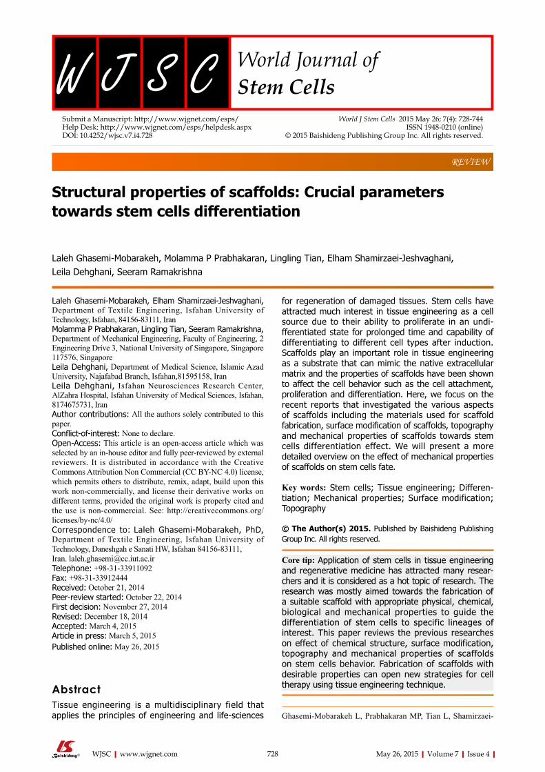
Rim et al[64] fabricated electrospun PLLA nanofibrous scaffolds and coated scaffolds with polydopamine through immersion in an aqueous solution of dopamine with mild shaking for 1 h. Their results showed more
Higher concentration of GO was found to promote NSCs differentiation into mature oligodendrocytes without introducing differentiation inducers in the culture media. Their results showed that the presence of GO in the structure of nanofibrous scaffolds was a crucial factor that determined the stem cell-scaffold interactions in directing NSCs towards oligodendrocyte differentiation[46].
One of the major challenges in cartilage and disc-tissue engineering using MSCs is the rapid expression of type X collagen as a marker of chondrocyte hyper-trophy associated with endochondral ossification. Nelea et al[55] used polypropylene and nylon-6 polymers for the fabrication of scaffolds and modified the surface of scaffolds by glow discharge plasma treatment in ammonia gas to investigate the potential of each polymer in its abilityto inhibit the expression of type X collagen. They cultured MSCs on modified and un-modified polypropylene and nylon-6 and evaluated the chondrogenic differentiation pathway using aggrecan and types I, II, and X collagens. Their results showed that MSCs did not adhere to unmodified PP while they observed cell adhesion on modified PP, nylon 6 and unmodified nylon 6 and concluded that the nature of the surface should be vitalfor the interaction of MSCs. Both modified and un-modified nylon-6 suppressed the expression of type X collagen, while the modified polypropylene scaffold almost completely inhibited its expression indicating the impact of polymer type and surface characteristics on the fate of stem cells[55].
Hosseinkhani et al[56] encapsulated DNA nano-particles into collagen sponge reinforced with poly (glycolic acid) fiber for bone tissue engineering. More osteogenic differentiation of MSCs and formation of homogeneous bone upon implantation of scaffolds containing DNA nanoparticles was observed by these researchers[56].
In yet in another study, semi-interpenetrating polymer networks made of collagen type I fibers combined with fibronectin or laminin were fabricated to investigate the effect of matrix composition towards ESCs differentiation. Results showed that scaffold composition has an important role in ESCs development and differentiation whereas the presence of fibronectin in collagen scaffolds strongly induces endothelial cell differentiation and vascularization. On the contrary, laminin stimulated ESCs to differentiate into beating cardiomyocytes[57]. Costa-Pinto et al[58] prepared chitosan-poly (butylene succinate) scaffolds in different weight ratios of chitosan (50%, 25%, and 0%) and compared the potential of osteogenic differentiation of human bone marrow stromal cells on these scaffolds. Their results revealed higher cell viability, adhesion, proliferation, and osteogenic differentiation on scaffolds with more ratios of chitosan[58]. Overall, it was clear that the scaffold material and its properties played a critical role in directing the stem cells towards differentiation, with proteins and peptides had a positive influence towards this effect.
731 May 26, 2015|Volume 7|Issue 4|WJSC|www.wjgnet.com
Ghasemi-Mobarakeh L et al . Scaffolds and stem cells differentiation
adhesion and proliferation of MSCs cultured on surface modified nanofibrous scaffolds with dopamine relative to cells seeded on unmodified scaffolds (Figure 1). More osteogenic differentiation as well as angiogenesis was also observed for MSCs seeded on modified scaffolds compared to unmodified scaffolds[64].
In yet another study, electrospun PCL nanofibers were coated using polydopamine by simple immersion of substrates in an alkaline dopamine solution. RE-1 silencing transcription factor (REST) was then absorbed onto PCL nanofibers coated with polydopamine to induce scaffold-mediated gene knockdown for enhanced neuronal differentiation of neural stem/progenitor cells. The results showed significant enhanced neuronal commitment and decreased glial cells differentiation due to presence of the silencing of REST[44]. Controlled release of insulin-like growth factor I (IGF-I) from silk fibroin scaffolds for chondrogenic differentiation of human MSCs has been studied by Uebersax et al[65] and the results showed that IGF-I loaded silk fibroin scaffolds have the potential to provide chondrogenic stimuli to human MSCs, thus beneficial for cartilage repair.
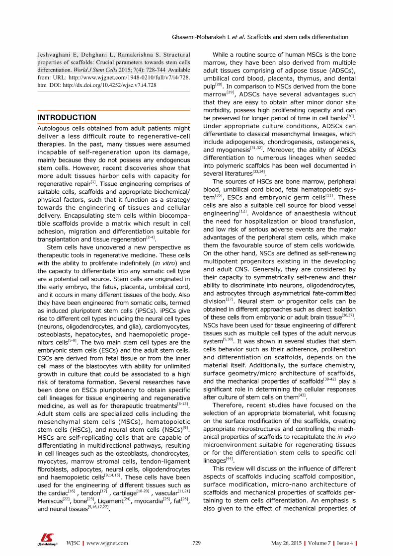
In yet another study, copolyester of 3-hydroxy-butyrate-cohydroxyhexanoate (PHBHHx) scaffolds were fabricated and coated with PHA granule-
associated proteins (PhaP) and PHA granule-associated proteins PhaP fused with RGD peptide (PhaP-RGD) for investigating the effect of surface modification towards chondrogenic differentiation of human bone marrow mesenchymal stem cells (hBMSCs). Their results showed that the surface modification of scaffolds with PhaP-RGD promote chondrogenic differentiation of hBMSCs compared to PhaP coated or uncoated scaffolds even without the presence ofchondrogenic induction medium (Figure 2). Hyaline cartilage rege-neration, and inhibited fibrocartilage formation in hBMSCs derived chondrocytes was also observed on PhaP-RGD coated PHBHHx scaffolds indicating suitability of this substrate for cartilage tissue engi-neering compared to the uncoated ones[61].
In yet another study, poly (ethylene glycol) (PEG) hydrogels were functionalized with heparin and osteogenic differentiation of human MSCs was evalu-ated. The results through increased ALP production and gene expression of osteopontin and collagen type I showed that functionalization of hydrogel with heparin induce osteogenic differentiation which is likely due to improvement of cell-scaffold interactions due to the presence of heparin[66]. Surface modification of poly-(lactic-co-glycolic acid) (PLGA) (75:25) scaf-folds with RGD peptides also enhanced the human
732 May 26, 2015|Volume 7|Issue 4|WJSC|www.wjgnet.com
PLLA PD-PLLA PLLA PD-PLLA
Rela
tive
cell
num
ber
Rela
tive
spre
adin
g ar
ea
3
2
1
0
3
2
1
0
a
a
Figure 1 Quantitative analysis of initial cell adhesion from human mesenchymal stem cells cultured on the fibers. Relative adherent cell numbers and spreading area of human mesenchymal stem cells (hMSCs) cultured on PLLA and PD-PLLA fibers were analyzed after 12 h of culture. aP < 0.05, PD-PLLA vs PLLA group. Adherent morphology of hMSCs on PLLA and PD-PLLA fibers was observed by confocal microscopy. Scale bars represent 100 µm. Reproduced with permission from Rim et al[64]. PLLA: Poly(l-lactide); PD-PLLA: Poly(l-lactide) (PLLA) fibers coated with polydopamine.
PD-P
LLA
P
LLA
f-actin nuclei Paxillin Merged
Ghasemi-Mobarakeh L et al . Scaffolds and stem cells differentiation
100 µm
100 µm

osteoprogenitor differentiation and osteogenesis[67]. While RGD peptide immobilized macroporous alginate scaffolds showed enhanced chondrogenesis properties of human MSCs compared to the un-modified ones[68].
In vitro osteogenic differentiation of human MSCs was also observed on RGD peptide functionalized PLLA nanofibersby Paletta et al[69]. Their results showed more osteogenic differentiation of human MSCs on modified scaffolds revealing the osteoinductive effect of the scaffolds functionalized with RGD[69].
Kuo et al[70] fabricated PLGA /chitosan scaffolds and functionalized it with type I collagen, whereby these researchers were able to improve the cell adhesion and viability on PLGA/chitosan/collagen scaffolds. Moreover, MSCs differentiated towards osteoblasts in the modified scaffolds without induction procedures, while neural differentiation was observed on the scaffolds by the induction MSCs with neuron growth factor (NGF)[70].
In another study Yang et al[71] fabricated porous poly l-lactide-co-ε-caprolactone (PLCL) and did surface modification via crosslinking of chitosan on the surface of scaffold. Their finding showed elongated morphology of MSCs on modified scaffolds while cells on unmodified scaffolds showed more spherical morphology with lower spreading. Moreover, the surface modified scaffolds provide surfaces for early differentiation of MSCs with more in vitro cartilage tissue formation revealing more condrogenic differ-
entiation of MSCs on modified scaffolds compared to the unmodified PLCL scaffold[71].
In yet another study, Budiraharjo et al[72] obtained carboxymethyl chitosan scaffolds (CMCS), coated with hydroxyapatite and compared the behavior of osteoblasts and human MSCs on both modified and un-modified scaffolds. Coating the scaffold with HA substantially enhanced the osteogenic differentiation of the human MSCs.
Surface modification using plasma has been frequen-tly used to improve surface properties of scaffolds fabricated with synthetic polymeric materials through formation of functional groups on the surface[73]. Lin et al[74] investigated the effect of modification of PLLA scaffolds by gas plasma towards the differentiation of ADSCs and their results showed that the cells seeded on modified scaffolds displayed significantly increased differentiation into endothelial cells[74].
Wang et al[60] fabricated porous nanocrystalline HA/chitosan scaffolds using a lyophilization technique, further treated them with cold atmospheric plasma as a simple, quick, and inexpensive method to modify the surface of scaffolds. Their results showed enhanced stem cell growth and osteogenic differentiation on modified scaffolds, which might be due to higher amounts of protein adsorption on the scaffold surfaces due to improves surface roughness and wettability after its modification[60].
An ideal bone implant should recruit osteoblasts or
733 May 26, 2015|Volume 7|Issue 4|WJSC|www.wjgnet.com
Uncoated PhaP PhaP-RGD
72 h
4 h
Figure 2 Confocal microscopic imaging of human bone marrow mesenchymal stem cells grown in uncoated, PhaP and PhaP-RGD coated 3-hydroxybutyrate-cohydroxyhexanoate scaffolds after 4 or 72 h of incubation, respectively. Phalloidin-fluorescein isothiocyanate was used to F-actin of cells grown in the scaffolds (Green). Reproduced with permission from You et al[61]. PhaP-RGD: PhaP binding protein fused with arginyl-glycyl-aspartic acid.
Ghasemi-Mobarakeh L et al . Scaffolds and stem cells differentiation

progenitor cells from the adjacent native tissue and induce cell proliferation and osteogenic differentiation for new bone formation. Hu et al[75] modified Ti6Al4V (TC4) implants using biofunctional multilayer coating of gelatin, chitosan, gelatin and human recombinant BMP2. In vitro results showed that such modifications of the implant surface stimulated the adhesion and osteogenic differentiation of MSCs and in vivo results showed improved induction of new bone formation at the interface of bone and implant and the integration of the implant within the surrounding living tissues[75] .
EFFECT OF SCAFFOLD TOPOGRAPHY ON STEM CELLS DIFFERENTIATION The topography of the extracellular matrix is known to influence cell attachment, morphology, proliferation and differentiation[76]. Topography can also enhance differentiation of progenitor cells into their programmed pathways[77].
Topographic surfaces with micro and nano scale struc-tures have been shown to stimulate the cell alignment, polarization, elongation, migration, proliferation, gene expression, etc., while the mammalian cells have been shown to react to nanoscale structures on a synthetic surface[77,78]. Nano scale topographies are obtained by different fabrication methods such as electrospinning, polymer phase separation, photolithography, chemical vapor deposition, electron beam lithography, etc.[2]. The similarity of electrospun nanofibers to the morphology of native extracellular matrix has attracted interest in the application of these scaffolds as a substrate for stem cell attachment and differentiation[2].
Smith et al[79] compared the in vitro osteogenic differentiation of ESCs on nanofibrous scaffolds and traditional scaffolds with two and three dimensional structure without the nanofibrous features. The results showed that the nanofibrous features enhanced the osteogenic differentiation and mineralization of ESCs compared to traditional substrates. Moreover, the osteogenic differentiation was observed on nanofibrous scaffolds without osteogenic supplements, while cells on other scaffolds required osteogenic supplements and growth factors for osteogenic differentiation[79].
Ragetly et al[80] evaluated the chondrogenic differentiation of MSCs on chitosan microfibrous scaf-folds and chitosan sponges. More chondrogenesis differentiation was observed on chitosan microfibers compared to that on sponges indicating the effect of scaffold microstructure towards the cell behavior[80].
Christopherson et al[81] evaluated the effect of topographical features of nanofibrous scaffolds in view point of fiber diameter on rat adult neural stem/progenitor cell (rNSC) differentiation and proliferation. They fabricated different nanofibrous scaffolds with average fiber diameters of 283 ± 45 nm, 749 ± 153 nm and 1452 ± 312 nm and their findings showed that the fiber diameter of PES nanofibrous scaffolds significantly
influenced the differentiation of rNSCs, whereas by decreasing of fiber diameter, more cell spreading and proliferation was observed. Additionally, the differentiation of stem cells towards oligodendrocytes was observed on nanofibrous scaffolds with fiber diameter of 283 ± 45 nm while morphology similar to neuronal progenitors was observed on nanofibers with fiber diameter of 749 ± 153 nm. Although, the number of cells on nanofibers with fiber diameter of 1452 ± 312 nm was found to be low which may be due to the difficulty of adhesion and migration of rNSCs on fibers with large diameter. However, the cells present within these nanofibrous scaffolds showed the morphology of neurons or neuronal progenitors[81].
In yet another study, aligned PLLA nanofibrous scaffolds with a mean diameter ranging from 307 to 917 nm and random PLLA nanofibrous scaffolds with a mean diameter ranging from 327 to 1150 nm were fabricated and the behavior of NSCs were evaluated on these scaffolds. The results showed that the depending on the fiber diameter and pattern, NSCs behaved differently revealing that the fiber alignment and diameter had significant effects on cellular behavior[82].
Yin et al[76] examined the effect of alignment of PLLA nanofibrous scaffolds on differentiation of stem cells from human tendon andthe differentiation of the cells seeded on aligned nanofibrous scaffolds was found to be higher with either normal or osteogenic media than on the randomly-oriented nanofibers[76].
Subramony et al[83] also evaluated the effect of PLGA nanofiber alignment and mechanical stimulation towards MSCs differentiation. Their results showed fibroblastic differentiation of MSCs in the absence of chemical induction factors on aligned nanofibers together with mechanical stimulation under tensile loading. No fibroblastic differentiation was observed on the non-aligned scaffolds even after the application of mechanical stimulation, indicating the effect of topography of scaffold towards stem cells differentiation[83].
Bakeine et al[84] investigated the effect of nanoscale surface topography on adhesion, proliferation and neural differentiation of mouse ESCs by fabrication of thin films of gold with different root mean square (RMS) surface roughness of 10, 21, 30 nm and 50 nm. Their results showed that surface topography influencedthe neural differentiation whereas highest differentiation was observed on gold films with RMS surface roughness of 21 nm after five days of cell seeding without addition of the traditional soluble neurotrophic factors[84].
The effect of topography with different sizes (groove width: 350 nm/2 µm/5 µm, depth = 300 nm) on the differentiation of human induced pluripotent stem cells (hiPSCs) towards neuronal lineage was studied by Pan et al[78]. Their results showed the obvious effect of topography on directing differentiation of hiPSCs towards neuronal lineage with noticeable up-regulation of neuronal marker expression on surfaces with nanos-tructured topography[78].
Yim et al[77] studied the proliferation and diffe-
734 May 26, 2015|Volume 7|Issue 4|WJSC|www.wjgnet.com
Ghasemi-Mobarakeh L et al . Scaffolds and stem cells differentiation
rentiation of human MSCs on nano-gratings of 1 µm, 10 µm, 350 nm width and their results showed the alignment of cytoskeleton and nuclei of human MSCs along the nano-gratings. Significant up-regulation of neuronal markers was also observed on patterned surface compared to un-patterned and micro-patterned controls[77].
Chaubey et al[85] evaluated the effect of the surface topography on the differentiation of multipotent mouse bone marrow stromal precursors (D1 cells) to adipocytes. They compared the differentiation potential of D1 cells on patterned PLLA films, plain PLLA films and tissue culture-plate and their results showed the impact of patterned surfaces on cell differentiation[85].
735 May 26, 2015|Volume 7|Issue 4|WJSC|www.wjgnet.com
A B C
Mer
ged
N
ucle
us
Vin
clin
F
-act
in
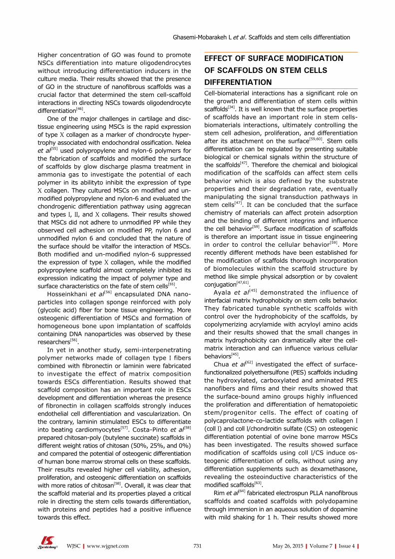
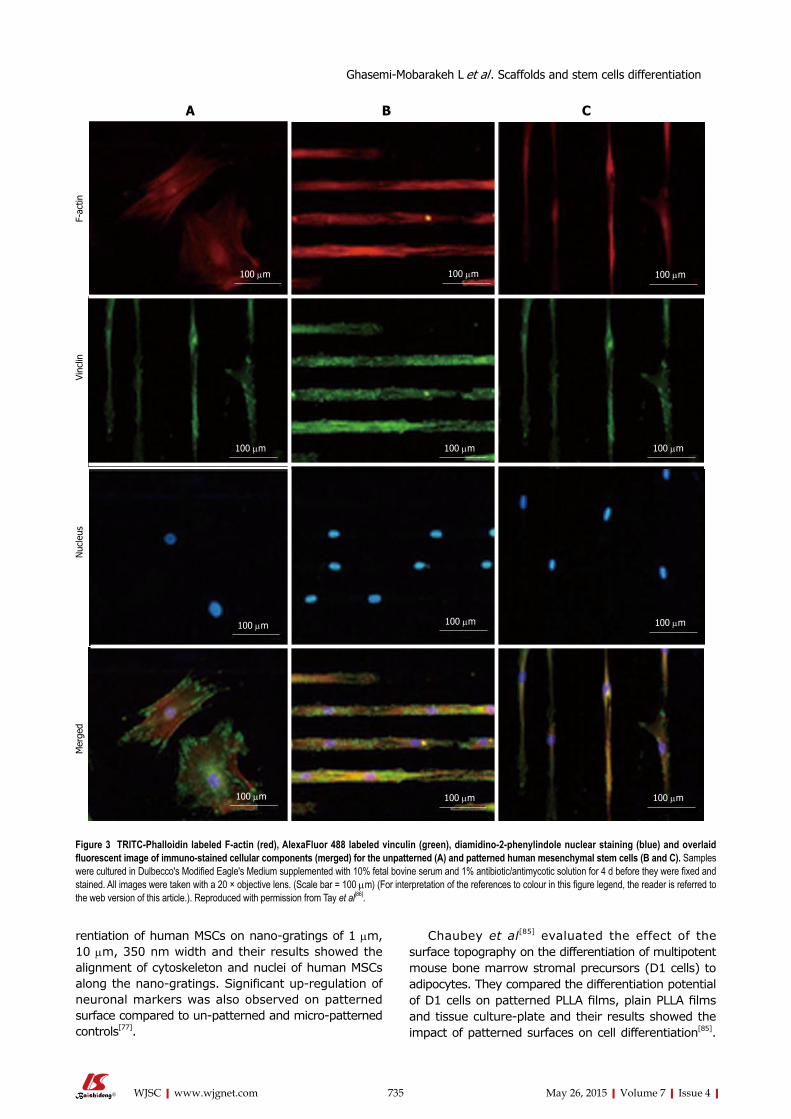
Figure 3 TRITC-Phalloidin labeled F-actin (red), AlexaFluor 488 labeled vinculin (green), diamidino-2-phenylindole nuclear staining (blue) and overlaid fluorescent image of immuno-stained cellular components (merged) for the unpatterned (A) and patterned human mesenchymal stem cells (B and C). Samples were cultured in Dulbecco's Modified Eagle's Medium supplemented with 10% fetal bovine serum and 1% antibiotic/antimycotic solution for 4 d before they were fixed and stained. All images were taken with a 20 × objective lens. (Scale bar = 100 µm) (For interpretation of the references to colour in this figure legend, the reader is referred to the web version of this article.). Reproduced with permission from Tay et al[86].
Ghasemi-Mobarakeh L et al . Scaffolds and stem cells differentiation
100 µm 100 µm 100 µm
100 µm 100 µm 100 µm
100 µm 100 µm 100 µm
100 µm 100 µm 100 µm

Tay et al[86] printed fibronectin on PLGA thin films and investigated the differentiation of human MSCs on patterned and un-patterned scaffolds, where they revealed the differentiation of human mesenchymal stem cells (hMSCs) into myocyte like cells on the micropatterned films (Figure 3).
Shi et al[87] fabricated two nano-grating substrates with a period of 250 and 500 nm and with a depth of 120 nm on quartz, and investigated the effect of substrate nanotopography on differentiation of ADSCs into endothelial cells which play an important role in vascularization. Decreased cell proliferation along with enhanced endothelial gene expression of the ADSCs and in vitro angiogenesis was observed on nanograting ssuggested the role of nanotopography on ADSCs differentiation to endothelial cells[87]. Oh et al[88] fabricated PCL cylindrical scaffolds with different pore size (90-400 µm range) along the longitudinal direction; and investigated the effect of pore size on the chondrogenic differentiation of ADSCs. Their results showed that the pore size of 370-400 µm provided a more suitable environment for chondrogenic differen-tiation than other pore size groups[88].
Teo et al[89] applied nanotopography technique along with NGF controlled release from polydimethylsiloxane substrate for neuronal tissue engineering and their results showed enhanced expression of neuronal genes on nanopatterned substrate combined with NGF delivery[89].
The influence on nanotopography, whether in the form of fibers, porous microstructures or pits and grates, was studied extensively by various researchers. However, comparison of the results between such studies was difficult due to the differences in various factors involved in each study, where some researchers used mechanical stimulation, while others used a specific biomolecule for the differentiation to take place. A systematic evaluation is therefore necessary, to clearly define the fiber length or the pore size that
might specifically cause the differentiation of stem cells to nerve, osteolineages or endothelial cells.
EFFECT OF CULTURE ENVIRONMENT ON STEM CELLS DIFFERENTIATIONBioreactors have been used in tissue engineering to overcome the drawbacks of static culturing conditions and to obtain uniform distribution of cells within the scaffolds and provide sufficient levels of oxygen, nutrients, cytokines, and growth factors, and to expose the cultured cells to mechanical stimuli[90]. Dynamic condition of cell culture has also been shown to influence stem cells differentiation.
Gomes et al[90] evaluated the effect of static and dynamic culturing conditions on the proliferation and osteogenic differentiation of rat bone marrow stromal cells seeded on two starch-based scaffolds. Calcium content analysis, VonKossa-stained sections, tetracycline fluorescence and histological analysis revealed the enhancement of osteogenic differentiation under dynamic condition created by flow perfusion bioreactor[90]. Mauney et al[91] also showed increased levels of alkaline phosphatase activity, bone-specific protein transcript levels, and mineralized matrix pro-duction by human bone marrow stromal cells under dynamic culture condition indicating increase inosteo-genic differentiation of human BMSCs using mechanical stimulation applied by bioreactor. However, the influence of mechanical stimulation towards stem cells differentiation is not studied highly and more investigations are required.
EFFECT MATRIX MECHANICAL PROPERTIES TOWARDS STEM CELL DIFFERENTIATIONMatrix stiffness varies with respect to different organs
736 May 26, 2015|Volume 7|Issue 4|WJSC|www.wjgnet.com
Brain FatMuscle
Cartilage
Precalcified Bone
0.1 1 10 100
Tissue elasticity, E (kPa)Fibrosis
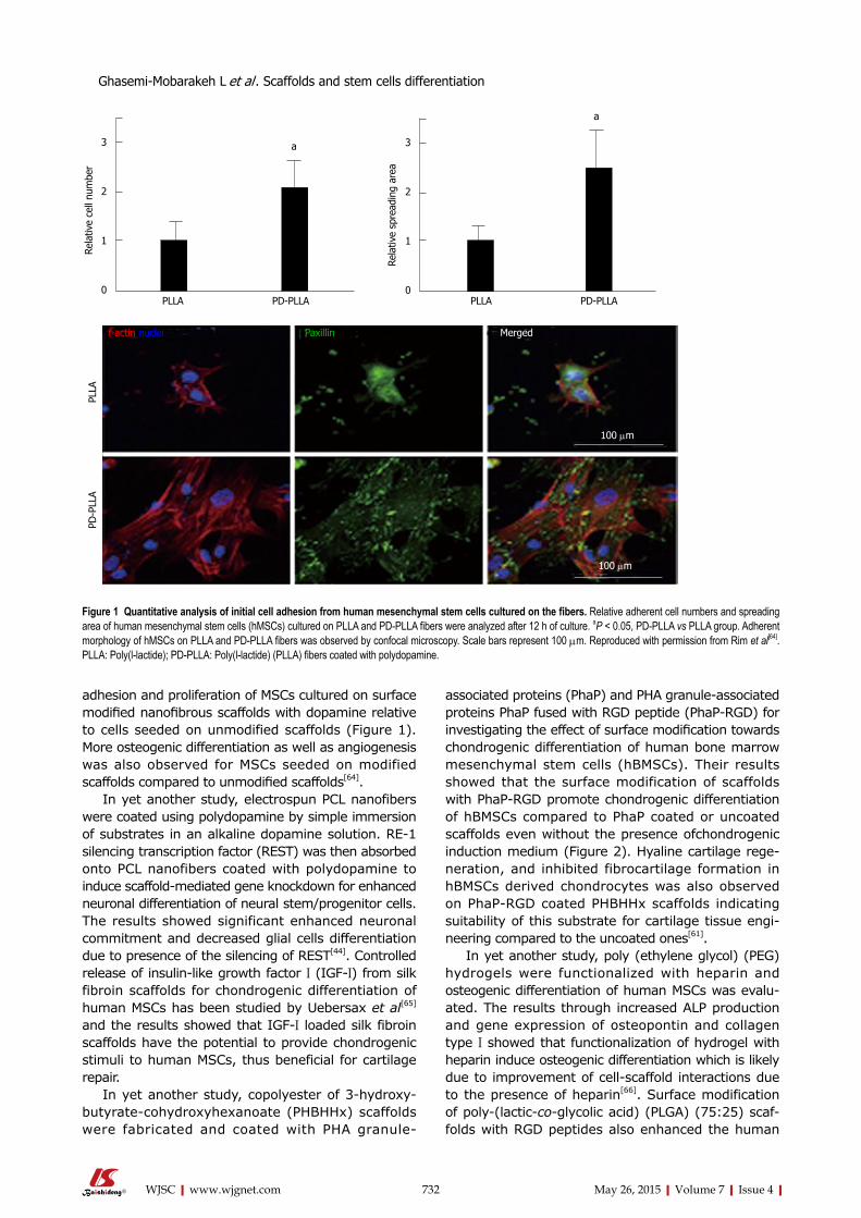
Figure 4 Schematic depicting the normal variation in elasticity of the indicated tissue[92].
Ghasemi-Mobarakeh L et al . Scaffolds and stem cells differentiation

present in the human body, and Figure 4 shows the difference in matrix stiffness of a few organs, as depicted by Vincent et al[92]. It explains how the matrix stiffness varies throughout the body: the brain is the softest tissue with an elasticity ranging between 0.2 to 1 kPa, while the precalcified bone is the hardest tissue with an elasticity value of more than 30 kPa[87], explaining the differences in the tissue matrix stiffness. Cells of different lineages survive in distinct natural environment, and they possess different stiffness. This is also the main reason why we believe that the stiffness have a higher influence towards the differentiation of stem cells. For tissue engineering, the most considered factors are the structure and the components used for scaffold fabrication, which determine the physical, chemical and biological properties of the scaffolds, and these properties will furthermore affect the differ-entiationof stem cells.
BoneVarious strategies has been acknowledged and selec-tively chosen to alter the biomaterial surfaces or to create micro-environment to establish both mechanical and biochemical cues for the differentiation of stem cells, especially for application in regenerative medicine. Chemical factors have been individually studied, suggesting the ability of BMP-2 to promote pro-osteo-genesis or IGF to promote tenogenic phenotype[93,94]. However, aligning the mechanical properties of the scaffolds with the specific tissue of interest was an interesting strategy applied to hydrogels, nanofibers and ceramics, in order to advance the field of stem cell differentiation on bioengineered substrates. Among these, injectable biodegradable hydrogels eliminate the need for surgical interventions or they allow for minimally invasive procedures causing minimal harm to the patients. However, physically crosslinked hydrogels lack strength and stability, while the chemically cross-linked hydrogels are more appreciated due to their high mechanical strength and stiffness. Hydrogels are useful substrates that assist to determine the interplay between physical cues/stiffness and their relationship with the fate of stem cell differentiation. A thiolene chemistry was utilized by Gandavarapu et al[95] to functionalize poly(ethylene glycol) hydrogels with a pendant peptide moiety, c(RRETAWA) and gels with Young’s Modulus (YM) of 2 kPa (soft) and 25 kPa (Stiff) were prepared. The stiffer substrate was found to give 3.5 fold higher hMSCs attachment. The ALP activity of hMSCs cultured on stiff gels containing 0.1 mmol/L and 1 mmol/L c(RRETAWA) increased by 2.5 and 3.5 fold, respectively after 14 d of cell culture. At the same time, the expression of osteopontin and collagen-1a for cells cultured on stiffer gels were higher, suggesting the application of high substrate stiffness gels for osteogenic differentiation of MSCs. In order to adjust the matrix stiffness, crosslinking of the thiol functionalized hyaluronic acid (HA-SH) and thiol functionalized recombinant human gelatin (Gtn-
SH), were performed by using varied amounts of poly (ethylene glycol) tetra-acrylate (PEGTA)[96]. Further hMSCs were seeded on the hydrogels to assess their potential towards osteo-lineage differentiation. MSCs grown on gels with 1.5 kPa stiffness, was found to express the early bone protein, namely osteopontin, while the cells on stiffer (4 kPa) gel expressed late osteogenic gene or the bone sialoprotein (BSP). This highlights the potential of higher osteogenic differen-tiation on stiffer hydrogels, mainly because the focal adhesion structures might have increased and F-actin might have got organized on such substrates, supporting the differentiation of MSCs to osteo lineages.
In order to study the role of substrate stiffness towards cell differentiation, Sun et al[97] prepared crosslinked gelatin scaffolds, where by the influence of scaffold strength was demonstrated in vitro and in vivo during this study. Crosslinking of gelatin scaffolds was performed by these researchers, using 1-Ethyl-3-[3- dimethylaminopropyl] carbodiimide hydrochloride (EDC) to prepare three dimension gelatin scaffolds with high stiffness properties. The elastic modulus of the EDC crosslinked scaffolds (2.5 kP) was significantly higher than those of the non-crosslinked scaffolds (0.6 kP). High mechanical strength of cross-linked scaffolds was suggested to promote stem cell mediated bone regeneration via endochondral ossification process, than the non-crosslinked scaffolds. In vivo studies using Inbred C57BJL/6 mice, showed significantly high trabecular bone formation for the crosslinked gelatin implanted animals, by micro-computed to-mography and histology analysis[92]. Wang et al[98] prepared gelatin-hydroxyphenylpropionic acid-tyramine (Gtn-HPA-Tyr) conjugates to simulate osteogenic differentiation of hMSCs, by conjugation of Tyr to Gtn-HPA conjugate by means of carbodiimide/active ester mediated coupling reaction. Oxidative coupling of phenol moieties were further carried out by hydrogen peroxide (H2O2) and horseradish peroxidase (HRP), whereby the concentration of H2O2 was varied produ-cing Gtn-HPA-Tyr hydrogels of varying storage modulus (G0). Hydrogels with G0 > 20000 was found suitable for the osteo-specific differentiation of MSCs, with osteocalcinupregulation and high calcium accumulation was observed on these gels compared to hydrogels with G0 of 13500. Stiffer hydrogels were suggested beneficial for the repair of bone defects. In yet another attempt, the ability of cells to differentiate to osteogenic cells were studied on transglutaminase cross-linked gelatin (TG-Gel) of varying rigidity and these researchers found that thesofter matrix promote cell proliferation, while stiffer matrix promote osteogenesis[99]. Osteogenic differentiation was found to occur in 6% and 9% TG-Gels with rigidity of 13.51-2.13 kPa and 32.32-1.9 kPa, respectively than using 3% TG-Gels. However, little or no effect of BMP-2 was found in the case of stiffer (6% and 9%) TG-Gelstowards MSC differentiation. The 9% TG-Gels with and without BMP-2 demonstrated high
737 May 26, 2015|Volume 7|Issue 4|WJSC|www.wjgnet.com
Ghasemi-Mobarakeh L et al . Scaffolds and stem cells differentiation

calcium deposition with expression of osteogenic markers during in vitro studies. On the other hand, the ALP activity and early markers of osteogenic differentiation were observed in 6% TG-Gel and these cells differentiated to osteoblast-like cells at a later time point. Stiffer substrates are also demonstrated to direct the osteogenic differentiation of ADSCs, even in the absence of any growth factors[100]. PDGF and BMP-2 were covalently immobilized to collagen-Glycosoaminoglycan (CG) membrane in defined patterns, by combined photolithography-carbodiimide crosslinking strategy and the elastic modulus (stiffness) of the matrix was orthogonally manipulated. These researchers explained that by increasing the ratios of EDC:NHS:COOH, the stiffness of the CG got increased. Photoimmobilized BMP-2 was demonstrated to have a little effect on lineage-specific gene expression of ADSCs during this study, while the matrix stiffness had a higher impact. In yet another study, using BMP-2 modified poly(acrylamide-co-acrylic acid) hydrogels, BMP-2 was found to have little or no effect towards stem cell differentiation if grafted on softer gels[101]. Hence the mechanical properties of the substrate dictated the potential for stem cell differentiation than the biochemical reagents, explaining the diversity of stem cell behaviors. Efforts to leverage approaches to explore a wider range of combinatorial environments and biochemical pathways, in a systematic manner is necessary, such that the results obtained by different kinds of material systems can be compared. In yet another study, CG scaffolds were prepared by cross-linking treatment via dehydrothermal and 1-ethyl-3-3-dimethyl aminopropylcarbodiimide methods, with varying stiffness of 0.5 to 1.5 kPa[102]. MSCs diffe-rentiation studies on these scaffolds, showed higher level of RUNX2 expression on the stiffer scaffolds (1.5 kPa) indicating MSCs differentiation to osteogenic lineage. Chondroitin sulphate and hyaluronic acid was found to have a positive influence towards the MSCs differentiation towards osteogenic and chondrogenic lineages, respectively.
Actin filaments of cells are structures for force transmission in response to the substrate with which it comes in contact[103,104]. Increased cell spreading in response to geometric features of the substrate, could result in enhanced actomyosin contractility associated with osteogenic differentiation[103]. The expression of osteogenic markers was demonstrated as stiffness dependent, with maximum osteogenesis observed for fibronectin coated micropatterned polyacrylamide gels with stiffness of 30 kPa.
Studies on the rate of proliferation of mouse ESCs suggest higher proliferation on stiffer substrates of polyacrylamide hydrogels compared to the proliferation on soft substrates, favoring mESC colonization with high levels of Oct3/4 and the Nanog biomarker production[105]. In maintaining the stemness of mESCs on soft hydrogels with varying topography, no signi-ficant advantages were observed, suggesting the
insensitiveness of topography effect towards mESC stemness. However, surface topography might have a role on substrates with hexagonal or pillar shapes topology or even on stiffer surfaces. It is also believed that either stiffness or topography might play an independent decision factor towards colony formation on 3D substrates. Geometric control of osteogenesis was also studied on polyacrylamide gels prepared by microcontact printing of adhesion proteins where cell spreading and cell geometry are suggested to have some influence towards MSCs differentiation[106]. Stiffer matrices can assist to increase cell spreading, thus promote osteogenesis through enhanced actomyosin contractility and this was observed up to a stiffness of 30 kPa. However, the density of seeded cells were described to over-ride the effect of gel stiffness to cause osteogenic differentiation, according to Xue et al[107]. However, this effect was not pronounced on soft gel, where chondrogenic differentiation was induced. Overall, their study highlights the importance of cell-matrix and cell-cell interactions in causing MSCs differentiation. Substrate stiffness was also found to play a predominant role in regulating the growth of rat BM-MSCs, while substrate topography manipulate the cell morphology and spreading, with spherically shaped cells were observed on pillar substrates but not on grooved substrate[108].
In general, studies using mechano-variant polyacry-lamide substrates showed that the osteogenic differen-tiation reduced on substrates with low stiffness and ligand density. However, compared to fibronectin, BMSCs were demonstrated to have a higher binding affinity towards an equivalent bulk of collagen (Figure 5). Such comparisons were further delineated by Sharma et al[109], and they found the occurrence of osteogenic differentiation on fibronectin substrates, while tenoblast gene expression was only found on collagen substrates. Two entirely different microsystems namely, sponges and hydrogels were compared by Zhang et al[110] where they studied the behavior of MSCs and they found fast cell proliferation in sponges than in hydrogels. Sponges might have encouraged cell-cell and cell-matrix interaction and upon inducing them with media containing TGF-β3, the chondrogenic differentiation of MSCs were better compared to those in hydrogels.
NerveFrom Figure 4 and the abovementioned researches, it can be indicated that for neural differentiation, scaffolds with relatively small stiffness is required. The fate of MSCs towards neural or glial lineages could be manipulated by adjusting the substrate stiffness. Three dimensional collagen-HA scaffolds (fabricated by lyophilization) with higher elasticity (E approximately 1 kPa) induced MSCs into neuronal differentiation, while those with less elasticity (E approximately 10 kPa) induced MSCs into glial differentiation. The elasticity was adjusted by using different concentration
738 May 26, 2015|Volume 7|Issue 4|WJSC|www.wjgnet.com
Ghasemi-Mobarakeh L et al . Scaffolds and stem cells differentiation

of the crosslinking agent, namely 1-ethyl-3 (3-dimethylaminopropyl) carbodiimide (EDC)[111]. Tuning the modulus of hydrogels affects neurite outgrowth and ESCs differentiation; with higher modulus gels offered better substrate for neural differentiation and neurite outgrowth, while less modulus enhanced astrocyte formation and cell migration. Thus, ESCs differentiation into neural and glial lineages is influenced by the type, composition, architecture and stiffness of the matrix and the type and concentration of signaling molecules comprehensively[112]. Neuronal differentiation was favored on softer surfaces such as the photopolymerizablemethacrylamide chitosan hydrogel with Young’s modulus less than 1000 Pa as confirmed by both immunohistochemistry and qRT-PCR. Oligodendrocyte differentiation was favored on stiffer scaffolds with Young’s modulus more than 7000 Pa. However, myelin oligodendrocyte glycoprotein gene expression suggested that oligodendrocyte maturation and myelination was best on scaffolds with modulus less than 1000 Pa where more mature neurons were present. Astrocyte differentiation was only observed on scaffolds with modulus less than 1000 Pa and 3500 Pa and represented less than 2% of the total cell population[113]. Elastic moduli, even in the range of neural tissue, would have a great impact on adult neural stem cell differentiation. The adult NSCs expressed a high level of the neuronal marker,
β-tubulin III, on variable moduli interpenetrating polymer networks of 500 Pa, which is similar to the physiological stiffness of brain tissue. Furthermore, under mixed differentiation conditions with serum, softer gels (approximately 100-500 Pa) greatly favored neurons, whereas harder gels (approximately 1000-10000 Pa) promoted glial cultures[114].
To achieve the scaffold with favorable stiffness for nerve tissue regeneration, the fabrication method and the materials should be carefully considered. Hydrogels possess low modulus, and they are suitable as substrates for neuronal differentiation of stem cells. Alternatively, polymers with low modulus might be appropriate for this purpose. Within the category of the neuronal tissue structures, glial differentiation of stem cells might be gained on stiffer scaffolds, and neural differentiation can be achieved on softer scaffolds. By fine tuning the stiffness of the matrix, there is a great possibility to control the differentiation of stem cells into appropriate nerve cells.
Cardio-vascularLineage commitment of MSCs towards specific vascular cells (MSCs to ECs or MSCs to SMCs) on electrospun scaffolds could be manipulated by controlling the substrate modulus, from 2 to 15 kPa. Around 95% of MSCs seeded on soft scaffolds (3 kPa) showed Flk-1 endothelial markers within 24 h, while only 20% of
739 May 26, 2015|Volume 7|Issue 4|WJSC|www.wjgnet.com
70%
60%
50%
40%
30%
20%
10%
0%20 40 60 80
Perc
ent of
cel
ls a
ttac
hed
Substrate compliance (kPa)
Fibronectin 10 µg /mL
Fibronectin 50 µg /mL
Collagen 10 µg /mL
Collagen 50 µg /mL
20 kPa 60 kPa 80 kPa
Fibr
onec
tin
Colla
gen
A
B
Figure 5 (A) As the rigidity increased, so did cell attachment. Collagen elicited a greater degree of cell attachment, and increased bulk density of the ligand further enhanced this (B) Morphology on functionalized substrates at 8000 cells/cm2 for 24 h. astatistical difference compared to fibronectin at 10 mg/mL, bdifference compared to collagen at 10 mg/mL, cdifference compared to preceding substrate compliance. Cells were stained with FITC-phalloidin. (Scale bar represents 50 µm). Reproduced with permission from Sharma et al[109].
Ghasemi-Mobarakeh L et al . Scaffolds and stem cells differentiation
50 µm
b,c
b,cb
b
aa,c
a,c
a,c
a,cc

MSCs seeded on the rigid scaffolds (> 8 kPa) showed Flk-1 marker. In contrast, about 80% of MSCs seeded on rigid scaffolds (> 8 kPa) showed smooth muscle α-actin marker within 24 h, while fewer than 10% of MSCs seeded on soft scaffold (< 5 kPa) showed α-actin markers[115]. Besides, the synergistic effect of scaffold elasticity and growth factors has been studied for the differentiation of MSCs. In one such study, MSCs were seeded on soft nanofibrousmatrices with or without VEGF, and in Petri dishes with or without VEGF, where these researchers found that MSCs in soft matrices with VEGF showed significant increase in the expression of endothelia markers (vWF, eNOS, Flt-1, and Flk-1), with faster up-regulation of the endothelial markers. The results indicate that it is critical to control both mechanical factors and biochemical factors to regulate vascular endothelial regeneration of MSCs[116]. MSCs gained very low rate of cardiogenic differentia-tion after transplantation to infarcted heart, partly because stiffer scar tissue lack the capacity to support cardiogenic differentiation. Thermosensitive and injectable hydrogels with different moduli (16, 45 and 65 kPa) were achieved by controlling the concentration of the hydrogel solution. After 14 d, more than 76% MSCs differentiated into cardiac cells in gels with 45 and 65 kPa, while MSCs in the 65 kPa gel had the highest differentiation efficiency[117]. But the effect of stiffness towards the differentiation of stem cells might be time-dependent. Human embryonic stem cells are sensitive to substrate stiffness during early mesodermal specification, but not late fate choices during cardiac differentiation, when they are seeded on polyacrylamide hydrogels. An intermediate stiffness was most beneficial for cardiomyogenic differentiation than soft or stiff substrates[118].
CONCLUSION Selection of appropriate biomaterials for scaffold fabrication and their modification by incorporation of biological constitutes, or by patterning, thus altering the topography and mechanical properties of the scaffolds, might have a significant effect on stem cells behavior. Major literature review shows that MSCs are in favor of soft substrate to differentiate to neuronal cells, with moderately stiffer materials promote myo-genic differentiation and more stiffer substrates support osteogenic differentiation, suggesting the effect of substrate mechanical properties in influencing the differentiation of stem cells toward specific lineages. The stiffness of mature tendon range between 500-1000 MPa and bone falls in the range of 20 GPa and this might be a reason for such specific behaviors of stem cells on substrates with proportionate mech-anical properties. Prioritizing the stiffness of the substrate alone might not serve as a smart approach, but the presence of specific ligands along with the matrix mechanical compliance, might assist for more specific stem cell differentiations. There exists great
difficulty in comparing the results obtained by different research groups, mainly because the parameters such as the ligand type, their concentrations, surface topology or matrix type and their mechanics, and even the cell numbers differ between studies. Hence more uniform and systematically organized studies, are required for a thorough understanding of the stem cell differentiation effect. The extent of differentiation or the % of differentiated cells is another parameter that deserves great attention, such that the cell-substrate can be used for its maximum potential.
The cells on a 2-D platform might be less sensitive to mechanical properties than when they are in 3-D environment. The 3-D structures might provide an additional dimension to mechanical properties, such that the cell related factors such as the integrin ligation, cell contraction, and associated intracellular signaling might be influenced.
REFERENCES1 Preston SL, Alison MR, Forbes SJ, Direkze NC, Poulsom R,
Wright NA. The new stem cell biology: something for everyone. Mol Pathol 2003; 56: 86-96 [PMID: 12665626]
2 Prabhakaran MP, Venugopal J, Ghasemi-Mobarakeh L, Kai D, Jin G, Ramakrishna S. Stem cells and nanostructures for advanced tissue regeneration. Biomedical applications of polymeric nanofibers. Berlin: Springer, 2012: 21-62 [DOI: 10.1007/12_2011_113]
3 Li Y, Rodrigues J, Tomás H. Injectable and biodegradable hydrogels: gelation, biodegradation and biomedical applications. Chem Soc Rev 2012; 41: 2193-2221 [PMID: 22116474 DOI: 10.1039/C1CS15203C]
4 Grayson WL, Martens TP, Eng GM, Radisic M, Vunjak-Novakovic G. Biomimetic approach to tissue engineering Proceedings of the Seminars in cell and developmental biology. Amsterdam: Elsevier, 2009: 665-673 [DOI: 10.1016/j.semcdb.2008.12.008]
5 Wang A, Tang Z, Park IH, Zhu Y, Patel S, Daley GQ, Li S. Induced pluripotent stem cells for neural tissue engineering. Biomaterials 2011; 32: 5023-5032 [PMID: 21514663 DOI: 10.1016/j.biomaterials.2011.03.070]
6 Dehghani L, Farokhpour M, Karbalaie K, Nematollahi M, Tanhaie S, Hayati-Rodbari N, Kiani-Esfahani A, Hescheler J, Nasr-Esfahani MH, Baharvand H. The influence of dexamethasone administration on the protection against doxorubicin-induced cardiotoxicity in purified embryonic stem cell-derived cardiomyocytes. Tissue Cell 2013; 45: 101-106 [PMID: 23141520 DOI: 10.1016/j.tice.2012.09.009]
7 Dehghani L, Hashemi-Beni B, Poorazizi E, Khorvash F, Shaygannejad V, Sedghi M, Vesal S, Meamar R. Evaluation of neural gene expression in serum treated embryonic stem cells in Alzheimer’s patients. J Res Med Sci 2013; 18: S20-S23 [PMID: 23961278]
8 Tabar V, Studer L. Pluripotent stem cells in regenerative medicine: challenges and recent progress. Nat Rev Genet 2014; 15: 82-92 [PMID: 24434846 DOI: 10.1038/nrg3563]
9 Eberli D, Atala A. Tissue engineering using adult stem cells. Methods in enzymology 2006; 420: 287-302 [DOI: 10.1016/S0076-6879(06)20013-2]
10 Polak JM, Bishop AE. Stem cells and tissue engineering: past, present, and future. Ann N Y Acad Sci 2006; 1068: 352-366 [PMID: 16831937 DOI: 10.1196/annals.1346.001]
11 Rammal H, Harmouch C, Lataillade JJ, Laurent-Maquin D, Labrude P, Menu P, Kerdjoudj H. Stem cells: a promising source for vascular regenerative medicine. Stem Cells Dev 2014; 23: 2931-2949 [PMID: 25167472 DOI: 10.1089/scd.2014.0132]
12 Riha GM, Lin PH, Lumsden AB, Yao Q, Chen C. Review: application of stem cells for vascular tissue engineering. Tissue
740 May 26, 2015|Volume 7|Issue 4|WJSC|www.wjgnet.com
Ghasemi-Mobarakeh L et al . Scaffolds and stem cells differentiation
Eng 2005; 11: 1535-1552 [PMID: 16259608 DOI: 10.1089/ten.2005.11.1535]
13 Yamzon JL, Kokorowski P, Koh CJ. Stem cells and tissue engineering applications of the genitourinary tract. Pediatr Res 2008; 63: 472-477 [PMID: 18427290 DOI: 10.1203/PDR.0b013e31816a704a]
14 Yamout B, Hourani R, Salti H, Barada W, El-Hajj T, Al-Kutoubi A, Herlopian A, Baz EK, Mahfouz R, Khalil-Hamdan R, Kreidieh NM, El-Sabban M, Bazarbachi A. Bone marrow mesenchymal stem cell transplantation in patients with multiple sclerosis: a pilot study. J Neuroimmunol 2010; 227: 185-189 [PMID: 20728948 DOI: 10.1016/j.jneuroim.2010.07.013]
15 Cohen JA. Mesenchymal stem cell transplantation in multiple sclerosis. J Neurol Sci 2013; 333: 43-49 [PMID: 23294498 DOI: 10.1016/j.jns.2012.12.009]
16 Curtis MW, Russell B. Cardiac tissue engineering. The J of cardiovascular nursing 2009; 24: 87 [DOI: 10.1097/01.JCN.0000343562.06614.49]
17 Butler DL, Juncosa-Melvin N, Boivin GP, Galloway MT, Shearn JT, Gooch C, Awad H. Functional tissue engineering for tendon repair: A multidisciplinary strategy using mesenchymal stem cells, bioscaffolds, and mechanical stimulation. J Orthop Res 2008; 26: 1-9 [PMID: 17676628 DOI: 10.1002/jor.20456]
18 Caplan AI. The mesengenic process. Clin Plast Surg 1994; 21: 429-435 [PMID: 7924141]
19 Raghunath J, Salacinski HJ, Sales KM, Butler PE, Seifalian AM. Advancing cartilage tissue engineering: the application of stem cell technology. Curr Opin Biotechnol 2005; 16: 503-509 [PMID: 16153817 DOI: 10.1016/j.copbio.2005.08.004]
20 Tuan RS. Stemming cartilage degeneration: adult mesenchymal stem cells as a cell source for articular cartilage tissue engineering. Arthritis Rheum 2006; 54: 3075-3078 [PMID: 17009225 DOI: 10.1002/art.22148]
21 Campbell GR, Campbell JH. Development of tissue engineered vascular grafts. Curr Pharm Biotechnol 2007; 8: 43-50 [PMID: 17311552 DOI: 10.2174/138920107779941426]
22 Khan WS, Longo UG, Adesida A, Denaro V. Stem cell and tissue engineering applications in orthopaedics and musculoskeletal medicine. Stem Cells Int 2012; 2012: 403170 [PMID: 22550506 DOI: 10.1155/2012/403170]
23 Goshima J, Goldberg VM, Caplan AI. The origin of bone formed in composite grafts of porous calcium phosphate ceramic loaded with marrow cells. Clin Orthop Relat Res 1991; (269): 274-283 [PMID: 1650657]
24 Hui JH, Ouyang HW, Hutmacher DW, Goh JC, Lee EH. Mesenchymal stem cells in musculoskeletal tissue engineering: a review of recent advances in National University of Singapore. Ann Acad Med Singapore 2005; 34: 206-212 [PMID: 15827669]
25 Stamm C, Westphal B, Kleine HD, Petzsch M, Kittner C, Klinge H, Schümichen C, Nienaber CA, Freund M, Steinhoff G. Autologous bone-marrow stem-cell transplantation for myocardial regeneration. Lancet 2003; 361: 45-46 [PMID: 12517467 DOI: 10.1016/S0140-6736(03)12110-1]
26 Neubauer M, Hacker M, Bauer-Kreisel P, Weiser B, Fischbach C, Schulz MB, Goepferich A, Blunk T. Adipose tissue engineering based on mesenchymal stem cells and basic fibroblast growth factor in vitro. Tissue Eng 2005; 11: 1840-1851 [PMID: 16411830 DOI: 10.1089/ten.2005.11.1840]
27 Reekmans K, Praet J, Daans J, Reumers V, Pauwels P, Van der Linden A, Berneman ZN, Ponsaerts P. Current challenges for the advancement of neural stem cell biology and transplantation research. Stem Cell Rev 2012; 8: 262-278 [PMID: 21537994 DOI: 10.1007/s12015-011-9266-2]
28 Harris VK, Yan QJ, Vyshkina T, Sahabi S, Liu X, Sadiq SA. Clinical and pathological effects of intrathecal injection of mesenchymal stem cell-derived neural progenitors in an experimental model of multiple sclerosis. J Neurol Sci 2012; 313: 167-177 [PMID: 21962795 DOI: 10.1016/j.jns.2011.08.036]
29 Kassem M, Kristiansen M, Abdallah BM. Mesenchymal stem cells: cell biology and potential use in therapy. Basic Clin Pharmacol
Toxicol 2004; 95: 209-214 [PMID: 15546474 DOI: 10.1111/j.1742-7843.2004.pto950502.x]
30 Sterodimas A, de Faria J, Nicaretta B, Pitanguy I. Tissue engineering with adipose-derived stem cells (ADSCs): current and future applications. J Plast Reconstr Aesthet Surg 2010; 63: 1886-1892 [PMID: 19969517 DOI: 10.1016/j.bjps.2009.10.028]
31 Rodriguez AM, Elabd C, Amri EZ, Ailhaud G, Dani C. The human adipose tissue is a source of multipotent stem cells. Biochimie 2005; 87: 125-128 [PMID: 15733747 DOI: 10.1016/j.biochi.2004.11.007]
32 Chai C, Leong KW. Biomaterials approach to expand and direct differentiation of stem cells. Mol Ther 2007; 15: 467-480 [PMID: 17264853 DOI: 10.1038/sj.mt.6300084]
33 Santiago LY, Nowak RW, Peter Rubin J, Marra KG. Peptide-surface modification of poly(caprolactone) with laminin-derived sequences for adipose-derived stem cell applications. Biomaterials 2006; 27: 2962-2969 [PMID: 16445976 DOI: 10.1016/j.biomaterials.2006.01.011]
34 Awad HA, Wickham MQ, Leddy HA, Gimble JM, Guilak F. Chondrogenic differentiation of adipose-derived adult stem cells in agarose, alginate, and gelatin scaffolds. Biomaterials 2004; 25: 3211-3222 [PMID: 14980416 DOI: 10.1016/j.biomaterials.2003.10.045]
35 Atkins HL, Freedman MS. Hematopoietic stem cell therapy for multiple sclerosis: top 10 lessons learned. Neurotherapeutics 2013; 10: 68-76 [PMID: 23192675 DOI: 10.1007/s13311-012-0162-5]
36 Richards LJ, Kilpatrick TJ, Bartlett PF. De novo generation of neuronal cells from the adult mouse brain. Proc Natl Acad Sci USA 1992; 89: 8591-8595 [PMID: 1528866 DOI: 10.1073/pnas.89.18.8591]
37 Reynolds BA, Weiss S. Generation of neurons and astrocytes from isolated cells of the adult mammalian central nervous system. Science 1992; 255: 1707-1710 [PMID: 1553558 DOI: 10.1126/science.1553558]
38 Robertson MJ, Gip P, Schaffer DV. Neural stem cell engineering: directed differentiation of adult and embryonic stem cells into neurons. Front Biosci 2008; 13: 21-50 [PMID: 17981526]
39 Jäger M, Feser T, Denck H, Krauspe R. Proliferation and osteogenic differentiation of mesenchymal stem cells cultured onto three different polymers in vitro. Ann Biomed Eng 2005; 33: 1319-1332 [PMID: 16240081 DOI: 10.1007/s10439-005-5889-2]
40 Saha K, Pollock JF, Schaffer DV, Healy KE. Designing synthetic materials to control stem cell phenotype. Curr Opin Chem Biol 2007; 11: 381-387 [PMID: 17669680 DOI: 10.1016/j.cbpa.2007.05.030]
41 Hwang NS, Varghese S, Li H, Elisseeff J. Regulation of osteogenic and chondrogenic differentiation of mesenchymal stem cells in PEG-ECM hydrogels. Cell Tissue Res 2011; 344: 499-509 [PMID: 21503601 DOI: 10.1007/s00441-011-1153-2]
42 Elisseeff J, Ferran A, Hwang S, Varghese S, Zhang Z. The role of biomaterials in stem cell differentiation: applications in the musculoskeletal system. Stem Cells Dev 2006; 15: 295-303 [PMID: 16846368 DOI: 10.1089/scd.2006.15.295]
43 Hwang NS, Varghese S, Elisseeff J. Controlled differentiation of stem cells. Advanced drug delivery reviews 2008; 60: 199-214 [DOI: 10.1016/j.addr.2007.08.036]
44 Low WC, Rujitanaroj PO, Lee DK, Messersmith PB, Stanton LW, Goh E, Chew SY. Nanofibrous scaffold-mediated REST knockdown to enhance neuronal differentiation of stem cells. Biomaterials 2013; 34: 3581-3590 [PMID: 23415645 DOI: 10.1016/j.biomaterials.2013.01.093]
45 Ayala R, Zhang C, Yang D, Hwang Y, Aung A, Shroff SS, Arce FT, Lal R, Arya G, Varghese S. Engineering the cell-material interface for controlling stem cell adhesion, migration, and differentiation. Biomaterials 2011; 32: 3700-3711 [PMID: 21396708 DOI: 10.1016/j.biomaterials.2011.02.004]
46 Shah S, Yin PT, Uehara TM, Chueng ST, Yang L, Lee KB. Guiding stem cell differentiation into oligodendrocytes using graphene-nanofiber hybrid scaffolds. Adv Mater 2014; 26: 3673-3680 [PMID: 24668911 DOI: 10.1002/adma.201400523]
47 Dawson E, Mapili G, Erickson K, Taqvi S, Roy K. Biomaterials for
741 May 26, 2015|Volume 7|Issue 4|WJSC|www.wjgnet.com
Ghasemi-Mobarakeh L et al . Scaffolds and stem cells differentiation
stem cell differentiation. Advanced drug delivery reviews 2008; 60: 215-228 [DOI: 10.1016/j.addr.2007.08.037]
48 Boehler RM, Graham JG, Shea LD. Tissue engineering tools for modulation of the immune response. Biotechniques 2011; 51: 239-240, 242, 244 passim [PMID: 21988690 DOI: 10.2144/000113754]
49 Parenteau-Bareil R, Gauvin R, Berthod F. Collagen-based biomaterials for tissue engineering applications. Materials 2010; 3: 1863-1887 [DOI: 10.3390/ma3031863]
50 Gorgieva S, Kokol V. Collagen-vs. Gelatine-based biomaterials and their biocompatibility: Review and perspectives. Biomaterials Applications for Nanomedicine 2011: 17-58 [DOI: 10.5772/24118]
51 Lynn AK, Yannas IV, Bonfield W. Antigenicity and immunogenicity of collagen. J Biomed Mater Res B Appl Biomater 2004; 71: 343-354 [PMID: 15386396 DOI: 10.1002/jbm.b.30096]
52 Meinel L, Karageorgiou V, Fajardo R, Snyder B, Shinde-Patil V, Zichner L, Kaplan D, Langer R, Vunjak-Novakovic G. Bone tissue engineering using human mesenchymal stem cells: effects of scaffold material and medium flow. Ann Biomed Eng 2004; 32: 112-122 [PMID: 14964727]
53 Koch TG, Moroni L, Leysi-Derilou Y, Berg LC. Joint cartilage tissue engineering and pre-clinical safety and efficacy testing. InTech, 2011 [DOI: 10.5772/24096]
54 Ravichandran R, Venugopal JR, Sundarrajan S, Mukherjee S, Ramakrishna S. Precipitation of nanohydroxyapatite on PLLA/PBLG/Collagen nanofibrous structures for the differentiation of adipose derived stem cells to osteogenic lineage. Biomaterials 2012; 33: 846-855 [PMID: 22048006 DOI: 10.1016/j.biomaterials.2011.10.030]
55 Nelea V, Luo L, Demers CN, Antoniou J, Petit A, Lerouge S, R Wertheimer M, Mwale F. Selective inhibition of type X collagen expression in human mesenchymal stem cell differentiation on polymer substrates surface-modified by glow discharge plasma. J Biomed Mater Res A 2005; 75: 216-223 [PMID: 16044417 DOI: 10.1002/jbm.a.30402]
56 Hosseinkhani H, Hosseinkhani M, Gabrielson NP, Pack DW, Khademhosseini A, Kobayashi H. DNA nanoparticles encapsulated in 3D tissue-engineered scaffolds enhance osteogenic differentiation of mesenchymal stem cells. J Biomed Mater Res A 2008; 85: 47-60 [PMID: 17688252 DOI: 10.1002/jbm.a.31327]
57 Battista S, Guarnieri D, Borselli C, Zeppetelli S, Borzacchiello A, Mayol L, Gerbasio D, Keene DR, Ambrosio L, Netti PA. The effect of matrix composition of 3D constructs on embryonic stem cell differentiation. Biomaterials 2005; 26: 6194-6207 [PMID: 15921736 DOI: 10.1016/j.biomaterials.2005.04.003]
58 Costa-Pinto AR, Vargel I, Tuzlakoglu K, Correlo VM, Sol PC, Faria S, Piskin E, Reis RL, Neves NM. Influence of scaffold composition over in vitro osteogenic differentiation of hBMSCs and in vivo inflammatory response. J Biomater Appl 2014; 28: 1430-1442 [PMID: 24255003 DOI: 10.1177/0885328213512385]
59 Liu X, Feng Q, Bachhuka A, Vasilev K. Surface modification by allylamine plasma polymerization promotes osteogenic differentiation of human adipose-derived stem cells. ACS Appl Mater Interfaces 2014; 6: 9733-9741 [PMID: 24893152 DOI: 10.1021/am502170s]
60 Wang M, Cheng X, Zhu W, Holmes B, Keidar M, Zhang LG. Design of biomimetic and bioactive cold plasma-modified nanostructured scaffolds for enhanced osteogenic differentiation of bone marrow-derived mesenchymal stem cells. Tissue Eng Part A 2014; 20: 1060-1071 [PMID: 24219622 DOI: 10.1089/ten.TEA.2013.0235]
61 You M, Peng G, Li J, Ma P, Wang Z, Shu W, Peng S, Chen GQ. Chondrogenic differentiation of human bone marrow mesenchymal stem cells on polyhydroxyalkanoate (PHA) scaffolds coated with PHA granule binding protein PhaP fused with RGD peptide. Biomaterials 2011; 32: 2305-2313 [PMID: 21190731 DOI: 10.1016/j.biomaterials.2010.12.009]
62 Chua KN, Chai C, Lee PC, Tang YN, Ramakrishna S, Leong KW, Mao HQ. Surface-aminated electrospun nanofibers enhance adhesion and expansion of human umbilical cord blood hematopoietic stem/progenitor cells. Biomaterials 2006; 27:
6043-6051 [PMID: 16854459 DOI: 10.1016/j.biomaterials.2006.06.017]
63 Rentsch C, Hess R, Rentsch B, Hofmann A, Manthey S, Scharnweber D, Biewener A, Zwipp H. Ovine bone marrow mesenchymal stem cells: isolation and characterization of the cells and their osteogenic differentiation potential on embroidered and surface-modified polycaprolactone-co-lactide scaffolds. In Vitro Cell Dev Biol Anim 2010; 46: 624-634 [PMID: 20490706 DOI: 10.1007/s11626-010-9316-0]
64 Rim NG, Kim SJ, Shin YM, Jun I, Lim DW, Park JH, Shin H. Mussel-inspired surface modification of poly(L-lactide) electrospun fibers for modulation of osteogenic differentiation of human mesenchymal stem cells. Colloids Surf B Biointerfaces 2012; 91: 189-197 [PMID: 22118890 DOI: 10.1016/j.colsurfb.2011.10.057]
65 Uebersax L, Merkle HP, Meinel L. Insulin-like growth factor I releasing silk fibroin scaffolds induce chondrogenic differentiation of human mesenchymal stem cells. J Control Release 2008; 127: 12-21 [PMID: 18280603 DOI: 10.1016/j.jconrel.2007.11.006]
66 Benoit DS, Durney AR, Anseth KS. The effect of heparin-functionalized PEG hydrogels on three-dimensional human mesenchymal stem cell osteogenic differentiation. Biomaterials 2007; 28: 66-77 [PMID: 16963119 DOI: 10.1016/j.biomaterials.2006.08.033]
67 Yang XB, Roach HI, Clarke NM, Howdle SM, Quirk R, Shakesheff KM, Oreffo RO. Human osteoprogenitor growth and differentiation on synthetic biodegradable structures after surface modification. Bone 2001; 29: 523-531 [PMID: 11728922 DOI: 10.1016/S8756-3282(01)00617-2]
68 Re’em T, Tsur-Gang O, Cohen S. The effect of immobilized RGD peptide in macroporous alginate scaffolds on TGFbeta1-induced chondrogenesis of human mesenchymal stem cells. Biomaterials 2010; 31: 6746-6755 [PMID: 20542332 DOI: 10.1016/j.biomaterials.2010.05.025]
69 Paletta JR, Bockelmann S, Walz A, Theisen C, Wendorff JH, Greiner A, Fuchs-Winkelmann S, Schofer MD. RGD-functionalisation of PLLA nanofibers by surface coupling using plasma treatment: influence on stem cell differentiation. J Mater Sci Mater Med 2010; 21: 1363-1369 [PMID: 19943087 DOI: 10.1007/s10856-009-3947-2]
70 Kuo YC, Yeh CF. Effect of surface-modified collagen on the adhesion, biocompatibility and differentiation of bone marrow stromal cells in poly(lactide-co-glycolide)/chitosan scaffolds. Colloids Surf B Biointerfaces 2011; 82: 624-631 [PMID: 21074381 DOI: 10.1016/j.colsurfb.2010.10.032]
71 Yang Z, Wu Y, Li C, Zhang T, Zou Y, Hui JH, Ge Z, Lee EH. Improved mesenchymal stem cells attachment and in vitro cartilage tissue formation on chitosan-modified poly(L-lactide-co-epsilon-caprolactone) scaffold. Tissue Eng Part A 2012; 18: 242-251 [PMID: 21902611 DOI: 10.1089/ten.TEA.2011.0315]
72 Budiraharjo R, Neoh KG, Kang ET. Hydroxyapatite-coated carboxymethyl chitosan scaffolds for promoting osteoblast and stem cell differentiation. J Colloid Interface Sci 2012; 366: 224-232 [PMID: 22024371 DOI: 10.1016/j.jcis.2011.09.072]
73 Zandén C, Hellström Erkenstam N, Padel T, Wittgenstein J, Liu J, Kuhn HG. Stem cell responses to plasma surface modified electrospun polyurethane scaffolds. Nanomedicine 2014; 10: 949-958 [PMID: 24524929 DOI: 10.1016/j.nano.2014.01.010]
74 Lin J, Lindsey ML, Zhu B, Agrawal CM, Bailey SR. Effects of surface-modified scaffolds on the growth and differentiation of mouse adipose-derived stromal cells. J Tissue Eng Regen Med 2007; 1: 211-217 [PMID: 18038413]
75 Hu Y, Cai K, Luo Z, Zhang Y, Li L, Lai M, Hou Y, Huang Y, Li J, Ding X, Zhang B, Sung KL. Regulation of the differentiation of mesenchymal stem cells in vitro and osteogenesis in vivo by microenvironmental modification of titanium alloy surfaces. Biomaterials 2012; 33: 3515-3528 [PMID: 22333987 DOI: 10.1016/j.biomaterials.2012.01.040]
76 Yin Z, Chen X, Chen JL, Shen WL, Hieu Nguyen TM, Gao L, Ouyang HW. The regulation of tendon stem cell differentiation by the alignment of nanofibers. Biomaterials 2010; 31: 2163-2175
742 May 26, 2015|Volume 7|Issue 4|WJSC|www.wjgnet.com
Ghasemi-Mobarakeh L et al . Scaffolds and stem cells differentiation
[PMID: 19995669 DOI: 10.1016/j.biomaterials.2009.11.083]77 Yim EK, Pang SW, Leong KW. Synthetic nanostructures inducing
differentiation of human mesenchymal stem cells into neuronal lineage. Exp Cell Res 2007; 313: 1820-1829 [PMID: 17428465 DOI: 10.1016/j.yexcr.2007.02.031]
78 Pan F, Zhang M, Wu G, Lai Y, Greber B, Schöler HR, Chi L. Topographic effect on human induced pluripotent stem cells differentiation towards neuronal lineage. Biomaterials 2013; 34: 8131-8139 [PMID: 23891397 DOI: 10.1016/j.biomaterials.2013.07.025]
79 Smith LA, Liu X, Hu J, Ma PX. The influence of three-dimensional nanofibrous scaffolds on the osteogenic differentiation of embryonic stem cells. Biomaterials 2009; 30: 2516-2522 [PMID: 19176243 DOI: 10.1016/j.biomaterials.2009.01.009]
80 Ragetly GR, Griffon DJ, Lee HB, Fredericks LP, Gordon-Evans W, Chung YS. Effect of chitosan scaffold microstructure on mesenchymal stem cell chondrogenesis. Acta Biomater 2010; 6: 1430-1436 [PMID: 19861178 DOI: 10.1016/j.actbio.2009.10.040]
81 Christopherson GT, Song H, Mao HQ. The influence of fiber diameter of electrospun substrates on neural stem cell differentiation and proliferation. Biomaterials 2009; 30: 556-564 [PMID: 18977025 DOI: 10.1016/j.biomaterials.2008.10.004]
82 He L, Liao S, Quan D, Ma K, Chan C, Ramakrishna S, Lu J. Synergistic effects of electrospun PLLA fiber dimension and pattern on neonatal mouse cerebellum C17.2 stem cells. Acta Biomater 2010; 6: 2960-2969 [PMID: 20193781 DOI: 10.1016/j.actbio.2010.02.039]
83 Subramony SD, Dargis BR, Castillo M, Azeloglu EU, Tracey MS, Su A, Lu HH. The guidance of stem cell differentiation by substrate alignment and mechanical stimulation. Biomaterials 2013; 34: 1942-1953 [PMID: 23245926 DOI: 10.1016/j.biomaterials.2012.11.012]
84 Bakeine GJ, Ban J, Grenci G, Pozzato A, Zilio SD, Prasciolu M, Businaro L, Tormen M, Ruaro ME. Design, fabrication and evaluation of nanoscale surface topography as a tool in directing differentiation and organisation of embryonic stem-cell-derived neural precursors. Microelectronic Engineering 2009; 86: 1435-1438 [DOI: 10.1016/j.mee.2009.01.032]
85 Chaubey A, Ross KJ, Leadbetter RM, Burg KJ. Surface patterning: tool to modulate stem cell differentiation in an adipose system. J Biomed Mater Res B Appl Biomater 2008; 84: 70-78 [PMID: 17455278 DOI: 10.1002/jbm.b.30846]
86 Tay CY, Yu H, Pal M, Leong WS, Tan NS, Ng KW, Leong DT, Tan LP. Micropatterned matrix directs differentiation of human mesenchymal stem cells towards myocardial lineage. Exp Cell Res 2010; 316: 1159-1168 [PMID: 20156435 DOI: 10.1016/j.yexcr.2010.02.010]
87 Shi Z, Neoh KG, Kang ET, Poh CK, Wang W. Enhanced endothelial differentiation of adipose-derived stem cells by substrate nanotopography. J Tissue Eng Regen Med 2014; 8: 50-58 [PMID: 22628362 DOI: 10.1002/term.1496]
88 Oh SH, Kim TH, Im GI, Lee JH. Investigation of pore size effect on chondrogenic differentiation of adipose stem cells using a pore size gradient scaffold. Biomacromolecules 2010; 11: 1948-1955 [PMID: 20690707 DOI: 10.1021/bm100199m]
89 Teo BK , Tan GD, Yim EK. The synergis t ic effec t of nanotopography and sustained dual release of hydrophobic and hydrophilic neurotrophic factors on human mesenchymal stem cell neuronal lineage commitment. Tissue Eng Part A 2014; 20: 2151-2161 [PMID: 24932660 DOI: 10.1089/ten.tea.2013.0382]
90 Gomes ME, Sikavitsas VI, Behravesh E, Reis RL, Mikos AG. Effect of flow perfusion on the osteogenic differentiation of bone marrow stromal cells cultured on starch-based three-dimensional scaffolds. J Biomed Mater Res A 2003; 67: 87-95 [PMID: 14517865 DOI: 10.1002/jbm.a.10075]
91 Mauney JR, Sjostorm S, Blumberg J, Horan R, O’Leary JP, Vunjak-Novakovic G, Volloch V, Kaplan DL. Mechanical stimulation promotes osteogenic differentiation of human bone marrow stromal cells on 3-D partially demineralized bone scaffolds in vitro. Calcif Tissue Int 2004; 74: 458-468 [PMID: 14961210
DOI: 10.1007/s00223-003-0104-7]92 Vincent L, Engler AJ. 5.504 - effect of substrate modulus on
cell function and differentiation. In: Ducheyne P Comprehensive biomaterials. Oxford: Elsevier, 2011: 51-63
93 Bhakta G, Lim ZX, Rai B, Lin T, Hui JH, Prestwich GD, van Wijnen AJ, Nurcombe V, Cool SM. The influence of collagen and hyaluronan matrices on the delivery and bioactivity of bone morphogenetic protein-2 and ectopic bone formation. Acta Biomater 2013; 9: 9098-9106 [PMID: 23871940 DOI: 10.1016/j.actbio.2013.07.008]
94 Abrahamsson SO, Lundborg G, Lohmander LS. Recombinant human insulin-like growth factor-I stimulates in vitro matrix synthesis and cell proliferation in rabbit flexor tendon. J Orthop Res 1991; 9: 495-502 [PMID: 2045976 DOI: 10.1002/jor.1100090405]
95 Gandavarapu NR, Alge DL, Anseth KS. Osteogenic differentiation of human mesenchymal stem cells on α5 integrin binding peptide hydrogels is dependent on substrate elasticity. Biomater Sci 2014; 2: 352-361 [PMID: 24660057 DOI: 10.1039/C3BM60149H]
96 Zhao W, Li X, Liu X, Zhang N, Wen X. Effects of substrate stiffness on adipogenic and osteogenic differentiation of human mesenchymal stem cells. Mater Sci Eng C Mater Biol Appl 2014; 40: 316-323 [PMID: 24857499 DOI: 10.1016/j.msec.2014.03.048]
97 Sun H, Zhu F, Hu Q, Krebsbach PH. Controlling stem cell-mediated bone regeneration through tailored mechanical properties of collagen scaffolds. Biomaterials 2014; 35: 1176-1184 [PMID: 24211076 DOI: 10.1016/j.biomaterials.2013.10.054]
98 Wang LS, Du C, Chung JE, Kurisawa M. Enzymatically cross-linked gelatin-phenol hydrogels with a broader stiffness range for osteogenic differentiation of human mesenchymal stem cells. Acta Biomater 2012; 8: 1826-1837 [PMID: 22343003 DOI: 10.1016/j.actbio.2012.02.002]
99 Tan S, Fang JY, Yang Z, Nimni ME, Han B. The synergetic effect of hydrogel stiffness and growth factor on osteogenic differentiation. Biomaterials 2014; 35: 5294-5306 [PMID: 24703716 DOI: 10.1016/j.biomaterials.2014.02.040]
100 Banks JM, Mozdzen LC, Harley BA, Bailey RC. The combined effects of matrix stiffness and growth factor immobilization on the bioactivity and differentiation capabilities of adipose-derived stem cells. Biomaterials 2014; 35: 8951-8959 [PMID: 25085859 DOI: 10.1016/j.biomaterials.2014.07.012]
101 Zouani OF, Kalisky J, Ibarboure E, Durrieu MC. Effect of BMP-2 from matrices of different stiffnesses for the modulation of stem cell fate. Biomaterials 2013; 34: 2157-2166 [PMID: 23290467 DOI: 10.1016/j.biomaterials.2012.12.007]
102 Murphy CM, Matsiko A, Haugh MG, Gleeson JP, O’Brien FJ. Mesenchymal stem cell fate is regulated by the composition and mechanical properties of collagen-glycosaminoglycan scaffolds. J Mech Behav Biomed Mater 2012; 11: 53-62 [PMID: 22658154 DOI: 10.1016/j.jmbbm.2011.11.009]
103 Kilian KA, Bugarija B, Lahn BT, Mrksich M. Geometric cues for directing the differentiation of mesenchymal stem cells. Proc Natl Acad Sci USA 2010; 107: 4872-4877 [PMID: 20194780 DOI: 10.1073/pnas.0903269107]
104 Yim EK, Darling EM, Kulangara K, Guilak F, Leong KW. Nanotopography-induced changes in focal adhesions, cytoskeletal organization, and mechanical properties of human mesenchymal stem cells. Biomaterials 2010; 31: 1299-1306 [PMID: 19879643 DOI: 10.1016/j.biomaterials.2009.10.037]
105 Lü D, Luo C, Zhang C, Li Z, Long M. Differential regulation of morphology and stemness of mouse embryonic stem cells by substrate stiffness and topography. Biomaterials 2014; 35: 3945-3955 [PMID: 24529627 DOI: 10.1016/j.biomaterials.2014.01.066]
106 Lee J, Abdeen AA, Huang TH, Kilian KA. Controlling cell geometry on substrates of variable stiffness can tune the degree of osteogenesis in human mesenchymal stem cells. J Mech Behav Biomed Mater 2014; 38: 209-218 [PMID: 24556045 DOI: 10.1016/j.jmbbm.2014.01.009]
107 Xue R, Li JY, Yeh Y, Yang L, Chien S. Effects of matrix elasticity and cell density on human mesenchymal stem cells differentiation. J Orthop Res 2013; 31: 1360-1365 [PMID: 23606500 DOI: 10.1002/
743 May 26, 2015|Volume 7|Issue 4|WJSC|www.wjgnet.com
Ghasemi-Mobarakeh L et al . Scaffolds and stem cells differentiation

jor.22374]108 Li Z, Gong Y, Sun S, Du Y, Lü D, Liu X, Long M. Differential
regulation of stiffness, topography, and dimension of substrates in rat mesenchymal stem cells. Biomaterials 2013; 34: 7616-7625 [PMID: 23863454 DOI: 10.1016/j.biomaterials.2013.06.059]
109 Sharma RI, Snedeker JG. Biochemical and biomechanical gradients for directed bone marrow stromal cell differentiation toward tendon and bone. Biomaterials 2010; 31: 7695-7704 [PMID: 20656345 DOI: 10.1016/j.biomaterials.2010.06.046]
110 Zhang J, Wu Y, Thote T, Lee EH, Ge Z, Yang Z. The influence of scaffold microstructure on chondrogenic differentiation of mesenchymal stem cells. Biomed Mater 2014; 9: 035011 [PMID: 24818859 DOI: 10.1088/1748-6041/9/3/035011]
111 Her GJ, Wu HC, Chen MH, Chen MY, Chang SC, Wang TW. Control of three-dimensional substrate stiffness to manipulate mesenchymal stem cell fate toward neuronal or glial lineages. Acta Biomater 2013; 9: 5170-5180 [PMID: 23079022 DOI: 10.1016/j.actbio.2012.10.012]
112 Kothapalli CR, Kamm RD. 3D matrix microenvironment for targeted differentiation of embryonic stem cells into neural and glial lineages. Biomaterials 2013; 34: 5995-6007 [PMID: 23694902 DOI: 10.1016/j.biomaterials.2013.04.042]
113 Leipzig ND, Shoichet MS. The effect of substrate stiffness on adult
neural stem cell behavior. Biomaterials 2009; 30: 6867-6878 [PMID: 19775749 DOI: 10.1016/j.biomaterials.2009.09.002]
114 Saha K, Keung AJ, Irwin EF, Li Y, Little L, Schaffer DV, Healy KE. Substrate modulus directs neural stem cell behavior. Biophys J 2008; 95: 4426-4438 [PMID: 18658232 DOI: 10.1529/biophysj.108.132217]
115 Wingate K, Bonani W, Tan Y, Bryant SJ, Tan W. Compressive elasticity of three-dimensional nanofiber matrix directs mesenchymal stem cell differentiation to vascular cells with endothelial or smooth muscle cell markers. Acta Biomater 2012; 8: 1440-1449 [PMID: 22266031 DOI: 10.1016/j.actbio.2011.12.032]
116 Wingate K, Floren M, Tan Y, Tseng PO, Tan W. Synergism of matrix stiffness and vascular endothelial growth factor on mesenchymal stem cells for vascular endothelial regeneration. Tissue Eng Part A 2014; 20: 2503-2512 [PMID: 24702044 DOI: 10.1089/ten.TEA.2013.0249]
117 Li Z, Guo X, Palmer AF, Das H, Guan J. High-efficiency matrix modulus-induced cardiac differentiation of human mesenchymal stem cells inside a thermosensitive hydrogel. Acta Biomater 2012; 8: 3586-3595 [PMID: 22729021 DOI: 10.1016/j.actbio.2012.06.024]
118 Hazeltine LB, Badur MG, Lian X, Das A, Han W, Palecek SP. Temporal impact of substrate mechanics on differentiation of human embryonic stem cells to cardiomyocytes. Acta Biomater 2014; 10: 604-612 [PMID: 24200714 DOI: 10.1016/j.actbio.2013.10.033]
P- Reviewer: Tanabe S S- Editor: Song XX L- Editor: A E- Editor: Wu HL
744 May 26, 2015|Volume 7|Issue 4|WJSC|www.wjgnet.com
Ghasemi-Mobarakeh L et al . Scaffolds and stem cells differentiation

© 2015 Baishideng Publishing Group Inc. All rights reserved.
Published by Baishideng Publishing Group Inc8226 Regency Drive, Pleasanton, CA 94588, USA
Telephone: +1-925-223-8242Fax: +1-925-223-8243
E-mail: [email protected] Desk: http://www.wjgnet.com/esps/helpdesk.aspx
http://www.wjgnet.com
Related Documents