Structural magnetic resonance imaging predictors of responsiveness to cognitive behaviour therapy in psychosis Preethi Premkumar a , Dominic Fannon a , Elizabeth Kuipers a,b , Emmanuelle R. Peters a,b , Ananatha P.P. Anilkumar c , Andrew Simmons b,d , and Veena Kumari a,b,⁎ a Department of Psychology, Institute of Psychiatry, King's College London, London, UK. b NIHR Biomedical Research Centre for Mental Health, South London and Maudsley NHS Trust, London, London, UK. c South London and Maudsley NHS Trust, London, UK. d Centre for Neuroimaging Sciences, Institute of Psychiatry, King's College London, London, UK. Abstract Background—Responsiveness to cognitive behaviour therapy (CBT) in psychosis may have a neurological basis. This study aimed to determine whether improvement in symptoms following CBT for psychosis (CBTp) in people with schizophrenia is positively associated with pre-therapy grey matter volume in brain regions involved in cognitive processing. Methods—Sixty outpatients stable on medication with at least one distressing symptom of schizophrenia and willing to receive CBTp in addition to their standard care (SC), and 25 healthy participants underwent magnetic resonance imaging. Subsequently, 30 patients received CBTp (CBTp+SC; 25 completers) for 6–8 months and 30 continued with their standard care (SC; 19 completers). Symptoms in all patients were assessed (blindly) at entry and follow-up. Results—The CBTp+SC and SC groups did not differ clinically at baseline, and only the CBTp +SC group showed improved symptoms at follow-up. Severity of baseline symptoms was not associated with CBTp responsiveness. Reduction with CBTp in positive symptoms was associated with greater right cerebellum (lobule VII) grey matter volume, in negative symptoms with left precentral gyrus and right inferior parietal lobule grey matter volumes, and in general psychopathology with greater right superior temporal gyrus, cuneus and cerebellum (Crus I) grey matter volumes. Grey matter volume in these brain areas did not correlate with the severity of baseline symptoms. Conclusion—Grey matter volume of the frontal, temporal, parietal and cerebellar areas that are known to be involved in the co-ordination of mental activity, cognitive flexibility, and verbal learning and memory predict responsiveness to CBTp in patients with psychosis. © 2009 Elsevier B.V. This document may be redistributed and reused, subject to certain conditions. ⁎Corresponding author. Department of Psychology, PO78, Institute of Psychiatry, De Crespigny Park, London SE5 8AF, UK. Tel.: + 44 207 848 0233; fax: + 44 207 848 0860. [email protected]. This document was posted here by permission of the publisher. At the time of deposit, it included all changes made during peer review, copyediting, and publishing. The U.S. National Library of Medicine is responsible for all links within the document and for incorporating any publisher-supplied amendments or retractions issued subsequently. The published journal article, guaranteed to be such by Elsevier, is available for free, on ScienceDirect. Sponsored document from Schizophrenia Research Published as: Schizophr Res. 2009 December ; 115(2-3): 146–155. Sponsored Document Sponsored Document Sponsored Document

Welcome message from author
This document is posted to help you gain knowledge. Please leave a comment to let me know what you think about it! Share it to your friends and learn new things together.
Transcript

Structural magnetic resonance imaging predictors ofresponsiveness to cognitive behaviour therapy in psychosis
Preethi Premkumara, Dominic Fannona, Elizabeth Kuipersa,b, Emmanuelle R. Petersa,b,Ananatha P.P. Anilkumarc, Andrew Simmonsb,d, and Veena Kumaria,b,⁎aDepartment of Psychology, Institute of Psychiatry, King's College London, London, UK.bNIHR Biomedical Research Centre for Mental Health, South London and Maudsley NHS Trust,London, London, UK.cSouth London and Maudsley NHS Trust, London, UK.dCentre for Neuroimaging Sciences, Institute of Psychiatry, King's College London, London, UK.
AbstractBackground—Responsiveness to cognitive behaviour therapy (CBT) in psychosis may have aneurological basis. This study aimed to determine whether improvement in symptoms following CBTfor psychosis (CBTp) in people with schizophrenia is positively associated with pre-therapy greymatter volume in brain regions involved in cognitive processing.
Methods—Sixty outpatients stable on medication with at least one distressing symptom ofschizophrenia and willing to receive CBTp in addition to their standard care (SC), and 25 healthyparticipants underwent magnetic resonance imaging. Subsequently, 30 patients received CBTp(CBTp+SC; 25 completers) for 6–8 months and 30 continued with their standard care (SC; 19completers). Symptoms in all patients were assessed (blindly) at entry and follow-up.
Results—The CBTp+SC and SC groups did not differ clinically at baseline, and only the CBTp+SC group showed improved symptoms at follow-up. Severity of baseline symptoms was notassociated with CBTp responsiveness. Reduction with CBTp in positive symptoms was associatedwith greater right cerebellum (lobule VII) grey matter volume, in negative symptoms with leftprecentral gyrus and right inferior parietal lobule grey matter volumes, and in generalpsychopathology with greater right superior temporal gyrus, cuneus and cerebellum (Crus I) greymatter volumes. Grey matter volume in these brain areas did not correlate with the severity of baselinesymptoms.
Conclusion—Grey matter volume of the frontal, temporal, parietal and cerebellar areas that areknown to be involved in the co-ordination of mental activity, cognitive flexibility, and verbal learningand memory predict responsiveness to CBTp in patients with psychosis.
© 2009 Elsevier B.V.This document may be redistributed and reused, subject to certain conditions.
⁎Corresponding author. Department of Psychology, PO78, Institute of Psychiatry, De Crespigny Park, London SE5 8AF, UK. Tel.: + 44207 848 0233; fax: + 44 207 848 0860. [email protected] document was posted here by permission of the publisher. At the time of deposit, it included all changes made during peer review,copyediting, and publishing. The U.S. National Library of Medicine is responsible for all links within the document and for incorporatingany publisher-supplied amendments or retractions issued subsequently. The published journal article, guaranteed to be such by Elsevier,is available for free, on ScienceDirect.
Sponsored document fromSchizophrenia Research
Published as: Schizophr Res. 2009 December ; 115(2-3): 146–155.
Sponsored Docum
ent Sponsored D
ocument
Sponsored Docum
ent
KeywordsGrey matter; Voxel-based morphometry; Cerebellum; Cognitive flexibility; Memory; Schizophrenia
1 IntroductionA number of randomised-controlled trials have established that cognitive-behaviour therapy(CBT) for psychosis is efficacious (Wykes et al., 2008). Efficacy is clearest for patients whoare antipsychotic medication-resistant (Kuipers et al., 1997; Tarrier et al., 1998), with acutepatients not benefiting as much from therapy (Garety et al., 2008). Most studies have targetedpositive symptoms of psychosis, but benefits have also been found in a number of differentareas, such as negative symptoms, depression and social functioning (Wykes et al., 2008). Ameaningful clinical response to CBTp, however, is seen in only about 50% of patients withpsychosis who receive it (Pfammatter et al., 2006; Wykes et al., 2008).
Predictors of response to CBT for psychosis (CBTp) remain unclear. In one study, cognitiveflexibility about delusions predicted the effect of CBTp on delusional thinking (Garety et al.,1997). CBTp requires patients with psychosis to generate alternative explanations of theirpsychotic experiences (Kuipers et al., 2006). Patients whose cognitive flexibility is challengedmay not be able to consider new ways of thinking or coping. It is thought that CBT operatesat the level of the cortex and explicit processing of meaning (Gorman et al., 1989; van derGaag, 2006). CBTp responsiveness thus may be influenced by the integrity of specific brainregions that are involved in complex top-down processing of information (Kumari, 2006; vander Gaag, 2006).
This study aimed to determine whether improvement in symptoms following CBTp in peoplewith psychosis when added to their standard care (SC) is positively associated with pre-therapygrey matter volume (GMV). A number of brain regions are found to be structurally deficient,on average, in people with schizophrenia compared to healthy people (Honea et al., 2005;Shenton et al., 2001) and these deficits in turn relate to impairment in a range of cognitivedomains, including cognitive flexibility, in schizophrenia (Antonova et al., 2004).
We hypothesized that greater improvement in symptoms following CBTp+SC would beassociated with greater GMV at baseline (pre-CBTp) in the frontal lobes, given previouslyreported positive associations between frontal lobe GMV and executive functioning, includingcognitive flexibility, in schizophrenia (Antonova et al., 2004). However, given the dearth ofstudies on both cognitive and MRI predictors of CBTp, we explored positive associationsbetween GMV and CBTp responsiveness across the whole brain. We also explored negativeassociations between GMV across the whole brain and CBTp+SC responsiveness and did nothypothesize for GMV in any specific regions to show a negative association.
2 Materials and methods2.1 Participants and design
Participants were 60 outpatients with a DSM-IV diagnosis of schizophrenia or schizoaffectivedisorder who were willing to receive CBTp in addition to their usual care, and 25 healthyparticipants. Of these, 30 patients received CBTp+SC (25 completers) and 30 patients receivedSC (19 completers). The groups were matched on average for age, years in education and sex(Table 1). No participant had a history of neurological or organic illness or head injury.
Patients were recruited from the South London and Maudsley (SLAM) NHS Foundation Trust,were on stable doses of antipsychotic medication for at least two years and on the current
Premkumar et al. Page 2
Published as: Schizophr Res. 2009 December ; 115(2-3): 146–155.
Sponsored Docum
ent Sponsored D
ocument
Sponsored Docum
ent

antipsychotic drug for at least three months prior to taking part (86% on atypicalantipsychotics), and had a score of ≥ 60 on the Positive and Negative Syndrome Scale (PANSS,Kay et al., 1987) and at least one persistent positive symptom (a score of 3 or above on at leastone of the positive symptoms items of the PANSS, which they experienced as distressing).The patients in the CBTp+SC and SC groups were recruited from the same geographical area,were identified by local psychiatric consultants as suitable for CBTp, and wished to receiveCBTp in addition to their usual care. Patients who were referred to and accepted for CBTp bythe Psychological Interventions Clinic for Outpatients with Psychosis (PICuP), SLAM NHSFoundation Trust went into the CBTp+SC group. The researchers did not have a say in whichof the patients might receive CBT at this specialist clinic. With the resources available to theSLAM NHS Foundation Trust at the time of this investigation, only a small number (about10%) of eligible patients were offered CBT for psychosis. Others who matcheddemographically and clinically as much as possible those accepted for CBTp by the PICuPwere studied as part of the SC group over the same interval as the CBTp+SC group patients.SC consisted of case management offered by the case management team for a particulargeographical area. The team includes a dedicated care coordinator who sees the patient on aregular basis. Six-monthly care plan assessment reviews are carried out with a focus onrecovery. There was no change in antipsychotic dosage over the follow-up period in eithergroup.
The study was approved by the local ethics committee. All participants provided writteninformed consent to their participation and were compensated for their time and travel.
2.2 Cognitive behaviour therapyTherapy followed the procedure developed by Fowler and colleagues (1995). Therapy wasdelivered weekly or fortnightly, as preferred by the patient, over an average of 16 individualone-hour sessions. Therapy lasted for 6–9 months, according to NICE guidelines (NICE,2008). It was based on individualized formulations, and aimed to reduce distress andinterference arising from psychotic symptoms, reduce depression, anxiety and hopelessness,and modify dysfunctional schemas when appropriate. Initial sessions focused on facilitatingengagement in therapy. The therapist endeavored to build and maintain a good therapeuticrelationship by taking a flexible approach that focused on the patient's needs (Kuipers et al.,1997). The therapists were qualified CBT practitioners, and were supervised by experiencedclinical psychologists (EK, EP) with expertise in CBTp.
2.3 Clinical assessmentsClinical diagnosis was confirmed by the Structured Clinical Interview for DSM-IV (First etal., 2002). PANSS assessments (Kay et al., 1987) were performed on all patients before andafter CBTp+SC by an experienced psychiatrist (DF) who was blind to whether or not a patientreceived CBTp in addition to their usual treatment. This psychiatrist had no role in patientrecruitment or clinical management of any of the patients included in this investigation.Appointments for these assessments were made by another member of the research team. Wehave no evidence that blindness was broken during these assessments.
2.4 Magnetic resonance imaging acquisitionAll participants underwent structural magnetic resonance imaging (MRI) at baseline. MRIscans were acquired using a 1.5 Tesla GE NV/i Signa system (General Electric, MilwaukeeWI, USA) at the Maudsley Hospital, London. Initially, a series of sagittal fast gradient echoscout images were acquired. A 3-D inversion recovery prepared fast spoiled GRASS sequencewas applied to the whole brain to obtain T1-weighted images in the axial plane with 1.5 mmcontiguous sections (TR = 18 ms, TI = 450 ms, TE = 5.1 ms, flip angle = 20° with one data
Premkumar et al. Page 3
Published as: Schizophr Res. 2009 December ; 115(2-3): 146–155.
Sponsored Docum
ent Sponsored D
ocument
Sponsored Docum
ent

average and a 256 × 256 × 128 voxel matrix). Acquisition parameters were chosen using asophisticated image contrast simulation (Simmons et al., 1996).
2.5 MRI pre-processingStructural images were converted into ANALYZE format (ANALYZE software, BRU, MayoFoundation, Rochester, MN) and pre-processed using Statistical Parametric Mapping (SPM2;http://www.fil.ion.ucl.ac.uk/spm), running in MATLAB 2006a (MathWorks, Natick, MA) aspreviously described (Premkumar et al., 2008). Customised T1-weighted templates of thewhole brain, grey matter, white matter and cerebro-spinal fluid (CSF) were created for patientand healthy participant groups separately, and also for the whole study sample (patients andhealthy participants combined). For analyses concerning GMV correlates of symptomimprovement in the CBTp+SC group, templates used were derived from the patient groupalone. For comparisons between CBTp+SC responders, CBTp+SC non-responders and healthyparticipant groups, templates created for the whole study sample were utilized. Using a separate‘patients only’ template for the analyses concerning GMV correlates of CBTp+SC allowed usto determine CBTp specific effects, without confounding for GMV-averaged effects of healthyparticipants.
2.6 Statistical analysis2.6.1 Baseline demographic and clinical characteristics and total brainvolumes—Analyses of variance (ANOVA) were performed to examine baseline groupdifferences (as applicable) in age, years in education, age of illness onset, duration of illnessand medication. Gender distribution was examined using chi-square test.
The total grey matter, white matter and CSF volumes for each individual were calculated fromthe unsmoothed modulated segmented images using SPM2 and the values representing thevolume in milliliters extracted. Intracranial volume was calculated as the sum of the greymatter, white matter and CSF volumes. Group differences in total grey matter, white matterand CSF volumes were examined in SPSS (v15) by means of ANOVA.
2.6.2 Symptom change following CBTp—Repeated-measures ANOVAs wereperformed to determine symptom change from baseline to follow-up, with time (baseline,follow-up) as the within-subject factor and group (CBTp+SC and SC) as the between-subjectsfactor. Following significant symptom improvement at follow-up in the CBTp+SC group butnot the SC group (see Results section), potential associations between symptom improvementand age, duration of illness, medication dosage (chlorpromazine equivalents) and baselinesymptoms (total PANSS; positive, negative and general psychopathology subscale scores)were examined in the CBTp+SC group using Pearson's r. Such analyses were not performedin the SC group since this group did not show clinical change between the baseline and follow-up.
2.6.3 MRI correlates of symptom change following CBTp—Analyses of GMVcorrelates of CBTp responsiveness were performed in SPM5 (5–1782). For the purpose of thisanalysis, treatment outcome in the CBTp+SC group, was estimated as the residual change inPANSS symptoms (Siegle et al., 2006). To examine the associations between baseline (pre-CBTp) GMV and symptom improvement in the CBTp+SC group, residual symptom changescores on the PANSS (total and three subscales) were regressed at each voxel across the wholebrain. The resultant SPM correlation maps were thresholded at p < 0.005 uncorrected. Giventhe exploratory nature of this study and the suggestion that the procedure for correction formultiple comparisons in SPM (originally designed for the analysis of functional data) is overlystrict when applied to structural data (Sowell et al., 1999), relationships between CBTp+SCresponsiveness and brain regional volumes where clusters were larger than 300 voxels with a
Premkumar et al. Page 4
Published as: Schizophr Res. 2009 December ; 115(2-3): 146–155.
Sponsored Docum
ent Sponsored D
ocument
Sponsored Docum
ent

local maxima reaching T > 2.81 (p < 0.005) were treated as being of interest and a small volumecorrection (SVC; 10 mm radius sphere centred on the maxima voxel) was applied to determinewhether a cluster is significant (p = 0.05, family-wise error corrected) after correcting formultiple comparisons within a locally defined volume.
We then extracted the values representing the percentage of total grey/white matter volumeunder a smoothing kernel relative to the total grey/white matter volume for each participant inthe patient group at the maxima voxels of all the regions where GM volumes were associatedwith symptom change and/or baseline symptoms in the CBTp+SC group. Partial correlationswere performed between these values and symptom change controlling for intracranial volume.To determine whether these associations might also be present in the SC group, the GMintensities at each significant voxel were also extracted for the SC group and correlationsperformed between these values and symptom change. Finally, scatterplots were used toexplore whether GMV correlates of symptom improvement in CBTp+SC patients receivingclozapine (n = 6) differed from those receiving other types of antipsychotics.
2.6.4 Group differences in regional brain volumes—Group differences in GMVwere also examined in SPM5 (5–1782). We compared regional GMVs across the whole brainof CBTp+SC responders (patients who showed a 20% improvement from baseline on PANSStotal symptoms) with that of CBTp+SC non-responders and healthy participants usingindependent sample t-tests and applied the same significance criteria as noted earlier forassociation with responsiveness to CBTp. Following the observation of group differences inhippocampal GMV, group differences were re-evaluated using masks for left and righthippocampus from the PickAtlas software (Maldjian et al., 2003).
3 Results3.1 Baseline demographic and clinical characteristics and total brain volumes
The demographic and clinical characteristics and total grey matter, white matter and CSFvolumes of the patients and healthy participants are presented in Table 1. The CBTp+SC andSC patient groups did not differ in demographic characteristics, symptoms, medication level,or brain variables at baseline.
3.2 Symptom change following CBTpSymptom severity in the CBTp+SC group was reduced at follow-up relative to the SC group(Table 1). CBTp+SC patients showed an improvement, on average, of 14.5% (s.d. = 16.9%)on PANSS total symptoms following CBTp+SC, while SC patients showed no change. Post-CBTp improvement in symptoms was not associated significantly with any baseline clinicalcharacteristic.
3.3 MRI correlates of symptom improvement following CBTpIn the CBTp+SC group, greater GMV in the right cerebellum was associated with improvementin positive symptoms (Table 2 and Fig. 1). Greater GMV in the left precentral gyrus and rightinferior parietal lobule was associated with improvement in negative symptoms. Greater GMVin the right STG, cuneus and cerebellum was associated with improvement in generalpsychopathology. Greater GMV in the right cuneus and right STG was positively associatedwith improvement in total symptoms. These associations in the CBTp+SC group remainedsignificant after controlling for intracranial volume, and were not found in the SC group(Table 3). There were no negative associations between GMV in any brain areas and symptomimprovement.
Premkumar et al. Page 5
Published as: Schizophr Res. 2009 December ; 115(2-3): 146–155.
Sponsored Docum
ent Sponsored D
ocument
Sponsored Docum
ent

The GMV correlates of post-CBTp symptom improvement appeared to be similarly present inpatients receiving clozapine and those receiving other antipsychotics (Fig. 2).
3.4 Group differences in regional brain volumesCBTp+SC responders (n = 11) had greater left and right hippocampal volumes relative tohealthy participants and greater left hippocampal volumes relative to CBTp+SC non-responders (Table 4 and Fig. 3). No other group differences met the significance criteria.
4 DiscussionThis study examined structural MRI predictors of symptom change following CBTp in patientswith psychosis and found that the patients who received CBTp added to their usual care (SC),on average, showed improved symptoms at follow-up; SC patients showed the same level ofsymptoms at study entry and follow-up. In the CBTp+SC group, greater symptom improvementwas associated with greater GMV as follows: (a) positive symptoms with right cerebellum(lobule VII), (b) negative symptoms with left precentral gyrus and right inferior parietal lobule,(c) general psychopathology with right STG, right cuneus and right cerebellum (Crus I), and(d) total symptoms with right STG and right cuneus. These associations were absent in the SCgroup. CBTp+SC responders (n = 11/25) had greater posterior hippocampal volume bilaterallyrelative to healthy participants and greater left posterior hippocampal volume than CBTp+SCnon-responders.
4.1 MRI correlates of symptom improvement following CBTpContrary to our expectation, no significant association was found between CBTpresponsiveness and prefrontal GMV. However, GMVs in a number of other regions asdiscussed below were positively associated with CBTp responsiveness.
We found that greater GMV in the right cerebellum (lobule VII and Crus I) was associatedwith improvement in positive symptoms and general psychopathology. Although thecerebellum, traditionally, has been implicated in motor control, recent data demonstratecerebellar contributions to higher order cognitive functions, especially the task managementand multi-tasking components of executive processing (Andreasen and Pierson, 2008),manipulation of information held in verbal working memory [in a manner similar to thatobserved for the prefrontal cortex (Hayter et al., 2007)] and co-ordination of mental activity(Thach, 2007), all of which are likely to be pertinent to effective CBTp. Patients with greatercerebellar GMVs may have been more capable of reasoning and relational processing (poolingtogether and comparing decision-relevant information) and benefited most from CBT.According to Andreasen and colleagues (Andreasen et al., 1999; Andreasen and Pierson,2008), disruption in the cortico-cerebellar-thalamo-cortical circuitry results in deficientprocessing, prioritising, retrieval, coordination, and responding to information inschizophrenia. It is likely that functions of this circuitry are also relevant to CBTpresponsiveness in schizophrenia as suggested previously (Kumari et al., 2009).
GMVs in the left precentral gyrus (BA6) and right inferior parietal lobule (BA40) werepositively associated with improvement in negative symptoms. The precentral gyrus respondswhen information must be continuously updated and memory for temporal order maintainedin working memory (Wager and Smith, 2003), while the parietal lobe is involved in bothworking and episodic memory (Olson and Berryhill, 2009). Functional imaging studies alsoreveal inferior parietal lobule involvement in sensory integration, body image, concept of self,and executive function in schizophrenia (Torrey, 2007). The association between larger fronto-parietal GMV and greater responsiveness to CBTp in this study could be mediated by thecognitive functions associated with these brain regions.
Premkumar et al. Page 6
Published as: Schizophr Res. 2009 December ; 115(2-3): 146–155.
Sponsored Docum
ent Sponsored D
ocument
Sponsored Docum
ent

Greater GMV in the right cuneus (BA17) was associated with post-CBTp improvement inoverall symptoms and general psychopathology. The cuneus is also part of the cortico-cerebellar-thalamic-cortical circuit proposed (mentioned earlier) by Andreasen and colleagues(1999) within the context of cognitive dysmetria in schizophrenia and shows activity changesduring recall of practiced verbal material (Crespo-Facorro et al., 1999). Greater availability ofcuneal GMV in patients may benefit better recall of discussions during therapy and in turn theirability to carry out home assignments.
Greater GMV in the right posterior STG was associated with CBTp-led improvement in totalsymptoms and general psychopathology. Greater bilateral posterior STG GMV has beenassociated with better abstraction (Nestor et al., 1993), and right posterior STG with fewerperseverative errors on the Wisconsin Card Sorting test (Nestor et al., 2007b) and good insightin schizophrenia (Cooke et al., 2008). Greater right posterior STG GMV thus may enable bettercognitive flexibility and insight into their symptoms, and in turn facilitate CBTpresponsiveness.
It is important to highlight that GMV alterations in certain brain areas in schizophrenia areassociated with both low level of certain cognitive functions (Antonova et al., 2004) andseverity of symptoms (review, Kumari and Cooke, 2006). We chose to consider the GMVcorrelates of CBTp responsiveness in terms of their neuropsychological significance because(a) there is no evidence to our knowledge (from any previous study or this study) that baselinesymptom severity moderates the effects of CBT for psychosis, and (b) the GMVs associatedwith CBTp-led improvement were also not associated with baseline symptoms in this study.Furthermore, for the purpose of detecting relationships between pre-therapy GMVs and CBTp-led improvement, CBTp responsiveness was defined as residual change scores (estimated frombaseline symptom severity-predicted change scores) following the methods proposed for suchstudies by Siegle and colleagues (2006). Although at present there are very limited data onpredictors of CBTp in schizophrenia, studies in other psychiatric disorders, such as depressionand anxiety, have highlighted the importance of pretherapy level of cognitive functions asmoderators of responsiveness to CBT (Julian and Mohr, 2006; Mohlman and Gorman, 2005)and this may be even more salient in patients with schizophrenia who, on average, showdeficient performance across a range of neuropsychological tasks (Reichenberg and Harvey,2007).
4.2 Group differences in hippocampal volumeCBTp+SC responders showed greater posterior hippocampal volume compared to healthyparticipants and CBTp+SC non-responders. The hippocampal volume is positively associatedwith verbal learning and memory in psychosis (Antonova et al., 2004; Nestor et al., 2007a;Rametti et al., 2007). Although hippocampal volume is generally smaller in patients withschizophrenia (Honea et al., 2005; Shenton et al., 2001), a recent study (Thoma et al., 2009)reported smaller anterior, but not posterior, hippocampal volumes in schizophrenia patients,compared to controls; this study further showed that the anterior hippocampal volumecorrelated negatively, while the posterior hippocampal volume correlated positively, withmemory measures in the schizophrenia group. Based on these previous findings, it can besuggested that greater posterior hippocampal volume facilitated memory for verbal materialsand thus was conducive for treatment response in CBTp+SC responders. In a recent study,MacQueen and colleagues (2008) found greater bilateral hippocampal volume at baseline inpatients with major depression who remitted, compared to those who did not remit, after 8-weeks antidepressant therapy (MacQueen et al., 2008). They suggested that greaterhippocampal volume may afford better cognitive (Leal-Galicia et al., 2008; Wais, 2008) andemotion processing (Dolcos et al., 2005; LaBar and Cabeza, 2006; Phelps, 2004; Viveros etal., 2007) in psychiatric patients and enable them to reformulate their experiences (symptoms)
Premkumar et al. Page 7
Published as: Schizophr Res. 2009 December ; 115(2-3): 146–155.
Sponsored Docum
ent Sponsored D
ocument
Sponsored Docum
ent

resulting in improved symptoms following treatment. It is also possible that CBTp respondersof our study had other characteristics that were not measured but may be associated with bothgreater-than-normal posterior hippocampal GMVs and a beneficial response to CBTp. Forexample, the right hippocampus is also enlarged in people who meditate (Holzel et al., 2008;Luders et al., 2009). However, we had not expected to find a greater-than-normal posteriorhippocampal GMV in CBTp responders; this finding needs to be replicated before being takenseriously.
4.3 LimitationsA limitation of the study is that although some specific predictions were made about the neuralnetworks associated with symptom change in response to CBTp+SC, the analysis wasexploratory in nature as this is the first study to our knowledge to examine the brain structuralpredictors of CBTp+SC and our findings are based on a small sample. It is possible that theGMV correlates of CBTp responsiveness were confounded by antipsychotic drug treatment.We, however, did not find any differences in the distribution of GMV correlates betweenpatients receiving clozapine (n = 6) and patients receiving other antipsychotic drugs. We werenot able to determine the effect of mood stabilizers on the GMV-symptom improvementassociations due to the small number of CBTp+SC patients on mood stabilizers (n = 4). Finally,in the absence of pre-CBTp assessment of relevant cognitive functions, the proposedneuropsychological significance of MRI predictors of CBTp remains speculative.
4.4 ConclusionsIn conclusion, greater GMV of the brain areas that sub-serve the co-ordination of mentalactivity, cognitive flexibility, and verbal learning and memory may predict a beneficialresponse to CBTp in patients with psychosis. These findings need to be replicated in relativelylarger samples and supplemented with pre-CBTp assessment of relevant cognitive functionsusing sophisticated neuropsychological tests along with MRI to confirm theneuropsychological significance of MRI predictors in a direct manner.
Role of funding sourceThe sponsors had no role in study design; in the collection, analysis and interpretation of data;in the writing of the report; or in the decision to submit the paper for publication.
ContributorsVeena Kumari, Elizabeth Kuipers and Preethi Premkumar designed the study. Dominic Fannonand Ananatha PP Anilkumar performed the clinical diagnostic interviews and symptom ratings.Emmanuelle R Peters and Elizabeth Kuipers supervised cognitive behaviour therapy forpsychosis. Andrew Simmons contributed to development of MRI protocol. Preethi Premkumarassisted with data collection, scored and data-based clinical measures, undertook statisticalanalysis, and wrote the first draft of the manuscript.
Conflict of interestThe authors declare no conflict of interest.
AcknowledgmentsV.K. and P.P. are supported by a Wellcome Trust Senior Research Fellowship to Veena Kumari (067427/z/02/z). Thestudy was also supported by funds from the Wellcome Trust, UK (067427/z/02/z.).
Premkumar et al. Page 8
Published as: Schizophr Res. 2009 December ; 115(2-3): 146–155.
Sponsored Docum
ent Sponsored D
ocument
Sponsored Docum
ent

ReferencesAndreasen N.C. Pierson R. The role of the cerebellum in schizophrenia. Biol. Psychiatry 2008;64(2):81–
88. [PubMed: 18395701]Andreasen N.C. Nopoulos P. O'Leary D.S. Miller D.D. Wassink T. Flaum M. Defining the phenotype of
schizophrenia: cognitive dysmetria and its neural mechanisms. Biol. Psychiatry 1999;46(7):908–920.[PubMed: 10509174]
Antonova E. Sharma T. Morris R. Kumari V. The relationship between brain structure and neurocognitionin schizophrenia: a selective review. Schizophr. Res. 2004;70(2–3):117–145. [PubMed: 15329292]
Cooke M.A. Fannon D. Kuipers E. Peters E. Williams S.C. Kumari V. Neurological basis of poor insightin psychosis: a voxel-based MRI study. Schizophr. Res. 2008;103(1–3):40–51. [PubMed: 18539438]
Crespo-Facorro B. Paradiso S. Andreasen N.C. O'Leary D.S. Watkins G.L. Boles Ponto L.L. HichwaR.D. Recalling word lists reveals “cognitive dysmetria” in schizophrenia: a positron emissiontomography study. Am. J. Psychiatry 1999;156(3):386–392. [PubMed: 10080553]
Dolcos F. LaBar K.S. Cabeza R. Remembering one year later: role of the amygdala and the medialtemporal lobe memory system in retrieving emotional memories. Proc. Natl. Acad. Sci. U. S. A.2005;102(7):2626–2631. [PubMed: 15703295]
First, M.B.; Spitzer, R.L.; Gibbon, M.; Williams, J.B.W. Biometrics Research Department New York;NY: 2002. Structured clinical interview for DSM-IV-TR Axis I disorders — patient edition (SCID-I/P, 11/2002 revision).
Fowler, D.; Garety, P.; Kuipers, E. Cognitive behaviour therapy for psychosis: theory and practice. Wiley,Chichester; 1995.
Garety P.A. Fowler D. Kuipers E. Freeman D. Dunn G. Bebbington P. Hadley C. Jones S. London-EastAnglia randomised controlled trial of cognitive-behavioural therapy for psychosis. II: predictors ofoutcome. Br. J. Psychiatry 1997;171:420–426. [PubMed: 9463599]
Garety P.A. Fowler D.G. Freeman D. Bebbington P. Dunn G. Kuipers E. Cognitive-behavioural therapyand family intervention for relapse prevention and symptom reduction in psychosis: randomisedcontrolled trial. Br. J. Psychiatry 2008;192(6):412–423. [PubMed: 18515890]
Gorman J.M. Liebowitz M.R. Fyer A.J. Stein J. A neuroanatomical hypothesis for panic disorder. Am.J. Psychiatry 1989;146(2):148–161. [PubMed: 2643361]
Hayter A.L. Langdon D.W. Ramnani N. Cerebellar contributions to working memory. Neuroimage2007;36(3):943–954. [PubMed: 17468013]
Holzel B.K. Ott U. Gard T. Hempel H. Weygandt M. Morgen K. Vaitl D. Investigation of mindfulnessmeditation practitioners with voxel-based morphometry. Soc. Cogn. Affect. Neurosci. 2008;3(1):55–61. [PubMed: 19015095]
Honea R. Crow T.J. Passingham D. Mackay C.E. Regional deficits in brain volume in schizophrenia: ameta-analysis of voxel-based morphometry studies. Am. J. Psychiatry 2005;162(12):2233–2245.[PubMed: 16330585]
Julian L.J. Mohr D.C. Cognitive predictors of response to treatment for depression in multiple sclerosis.J. Neuropsychiatry Clin. Neurosci. 2006;18(3):356–363. [PubMed: 16963585]
Kay S.R. Fiszbein A. Opier L.A. The Positive and Negative Syndrome Scale (PANSS) for schizophrenia.Schizophr. Bull. 1987;13:261–276. [PubMed: 3616518]
Kuipers E. Garety P. Fowler D. Dunn G. Bebbington P. Freeman D. Hadley C. London-East Angliarandomised controlled trial of cognitive-behavioural therapy for psychosis. I: effects of the treatmentphase. Br. J. Psychiatry 1997;171:319–327. [PubMed: 9373419]
Kuipers E. Garety P. Fowler D. Freeman D. Dunn G. Bebbington P. Cognitive, emotional, and socialprocesses in psychosis: refining cognitive behavioral therapy for persistent positive symptoms.Schizophr. Bull. 2006;32(Suppl 1):S24–S31. [PubMed: 16885206]
Kumari V. Do psychotherapies produce neurobiological effects? Acta Neuropscyhiatrica 2006;18:61–70.
Kumari V. Cooke M. Use of magnetic resonance imaging in tracking the course and treatment ofschizophrenia. Expert. Rev. Neurother. 2006;6(7):1005–1016. [PubMed: 16831115]
Premkumar et al. Page 9
Published as: Schizophr Res. 2009 December ; 115(2-3): 146–155.
Sponsored Docum
ent Sponsored D
ocument
Sponsored Docum
ent

Kumari V. Peters E.R. Fannon D. Antonova E. Premkumar P. Anilkumar A.P. Williams S.C. Kuipers E.Dorsolateral prefrontal cortex activity predicts responsiveness to cognitive-behavioral therapy inschizophrenia. Biol. Psychiatry. 2009;66(6):594–602. [PubMed: 19560121]
LaBar K.S. Cabeza R. Cognitive neuroscience of emotional memory. Nat. Rev. Neurosci. 2006;7(1):54–64. [PubMed: 16371950]
Leal-Galicia P. Castaneda-Bueno M. Quiroz-Baez R. Arias C. Long-term exposure to environmentalenrichment since youth prevents recognition memory decline and increases synaptic plasticitymarkers in aging. Neurobiol. Learn. Mem. 2008;90(3):511–518. [PubMed: 18675926]
Luders E. Toga A.W. Lepore N. Gaser C. The underlying anatomical correlates of long-term meditation:larger hippocampal and frontal volumes of gray matter. Neuroimage. 2009;45(3):672–678. [PubMed:19280691]
MacQueen G.M. Yucel K. Taylor V.H. Macdonald K. Joffe R. Posterior hippocampal volumes areassociated with remission rates in patients with major depressive disorder. Biol. Psychiatry 2008;64(10):880–883. [PubMed: 18722590]
Maldjian J.A. Laurienti P.J. Kraft R.A. Burdette J.H. An automated method for neuroanatomic andcytoarchitectonic atlas-based interrogation of fMRI data sets. Neuroimage 2003;19(3):1233–1239.[PubMed: 12880848]
Mohlman J. Gorman J.M. The role of executive functioning in CBT: a pilot study with anxious olderadults. Behav. Res. Ther. 2005;43(4):447–465. [PubMed: 15701356]
Nestor P.G. Shenton M.E. McCarley R.W. Haimson J. Smith R.S. O'Donnell B. Kimble M. Kikinis R.Jolesz F.A. Neuropsychological correlates of MRI temporal lobe abnormalities in schizophrenia.Am. J. Psychiatry 1993;150(12):1849–1855. [PubMed: 8238641]
Nestor P.G. Kubicki M. Kuroki N. Gurrera R.J. Niznikiewicz M. Shenton M.E. McCarley R.W. Episodicmemory and neuroimaging of hippocampus and fornix in chronic schizophrenia. Psychiatry Res.2007;155(1):21–28. [PubMed: 17395435]
Nestor P.G. Onitsuka T. Gurrera R.J. Niznikiewicz M. Frumin M. Shenton M.E. McCarley R.W.Dissociable contributions of MRI volume reductions of superior temporal and fusiform gyri tosymptoms and neuropsychology in schizophrenia. Schizophr. Res. 2007;91(1–3):103–106.[PubMed: 17229550]
NICE. Gaskell Press London; UK: 2008. Schizophrenia: core interventions in the treatment andmanagement of schizophrenia in primary and secondary care (update).
Olson I.R. Berryhill M. Some surprising findings on the involvement of the parietal lobe in humanmemory. Neurobiol. Learn. Mem. 2009;91(2):155–165. [PubMed: 18848635]
Pfammatter M. Junghan U.M. Brenner H.D. Efficacy of psychological therapy in schizophrenia:conclusions from meta-analyses. Schizophr. Bull. 2006;32(Suppl 1):S64–80. [PubMed: 16905634]
Phelps E.A. Human emotion and memory: interactions of the amygdala and hippocampal complex. Curr.Opin. Neurobiol. 2004;14(2):198–202. [PubMed: 15082325]
Premkumar P. Fannon D. Kuipers E. Simmons A. Frangou S. Kumari V. Emotional decision-making andits dissociable components in schizophrenia and schizoaffective disorder: a behavioural and MRIinvestigation. Neuropsychologia 2008;46(7):2002–2012. [PubMed: 18329673]
Rametti G. Segarra N. Junque C. Bargallo N. Caldu X. Ibarretxe N. Bernardo M. Left posteriorhippocampal density reduction using VBM and stereological MRI procedures in schizophrenia.Schizophr. Res. 2007;96(1–3):62–71. [PubMed: 17604968]
Reichenberg A. Harvey P.D. Neuropsychological impairments in schizophrenia: integration ofperformance-based and brain imaging findings. Psychol. Bull. 2007;133(5):833–858. [PubMed:17723032]
Shenton M.E. Dickey C.C. Frumin M. McCarley R.W. A review of MRI findings in schizophrenia.Schizophr. Res. 2001;49(1–2):1–52. [PubMed: 11343862]
Siegle G.J. Carter C.S. Thase M.E. Use of FMRI to predict recovery from unipolar depression withcognitive behavior therapy. Am. J. Psychiatry 2006;163(4):735–738. [PubMed: 16585452]
Simmons A. Arridge S.R. Barker G.J. Williams S.C. Simulation of MRI cluster plots and application toneurological segmentation. Magn. Reson. Imaging 1996;14(1):73–92. [PubMed: 8656992]
Premkumar et al. Page 10
Published as: Schizophr Res. 2009 December ; 115(2-3): 146–155.
Sponsored Docum
ent Sponsored D
ocument
Sponsored Docum
ent
Sowell E.R. Thompson P.M. Holmes C.J. Batth R. Jernigan T.L. Toga A.W. Localizing age-relatedchanges in brain structure between childhood and adolescence using statistical parametric mapping.Neuroimage 1999;9(6 Pt 1):587–597. [PubMed: 10334902]
Tarrier N. Yusupoff L. Kinney C. McCarthy E. Gledhill A. Haddock G. Morris J. Randomised controlledtrial of intensive cognitive behaviour therapy for patients with chronic schizophrenia. BMJ 1998;317(7154):303–307. [PubMed: 9685273]
Thach W.T. On the mechanism of cerebellar contributions to cognition. Cerebellum 2007;6(3):163–167.[PubMed: 17786811]
Thoma R.J. Monnig M. Hanlon F.M. Miller G.A. Petropoulos H. Mayer A.R. Yeo R. Euler M. Lysne P.Moses S.N. Canive J.M. Hippocampus volume and episodic memory in schizophrenia. J. Int.Neuropsychol. Soc. 2009;15(2):182–195. [PubMed: 19203430]
Torrey E.F. Schizophrenia and the inferior parietal lobule. Schizophr. Res. 2007;97(1–3):215–225.[PubMed: 17851044]
van der Gaag M. A neuropsychiatric model of biological and psychological processes in the remissionof delusions and auditory hallucinations. Schizophr. Bull. 2006;32(Suppl 1):S113–S122. [PubMed:16905635]
Viveros M.P. Marco E.M. Llorente R. Lamota L. The role of the hippocampus in mediating emotionalresponses to nicotine and cannabinoids: a possible neural substrate for functional interactions. Behav.Pharmacol. 2007;18(5–6):375–389. [PubMed: 17762508]
Wager T.D. Smith E.E. Neuroimaging studies of working memory: a meta-analysis. Cogn. Affect. Behav.Neurosci. 2003;3(4):255–274. [PubMed: 15040547]
Wais P.E. FMRI signals associated with memory strength in the medial temporal lobes: a meta-analysis.Neuropsychologia 2008;46(14):3185–3196. [PubMed: 18817791]
Wykes T. Steel C. Everitt B. Tarrier N. Cognitive behavior therapy for schizophrenia: effect sizes, clinicalmodels, and methodological rigor. Schizophr. Bull. 2008;34(3):523–537. [PubMed: 17962231]
Premkumar et al. Page 11
Published as: Schizophr Res. 2009 December ; 115(2-3): 146–155.
Sponsored Docum
ent Sponsored D
ocument
Sponsored Docum
ent
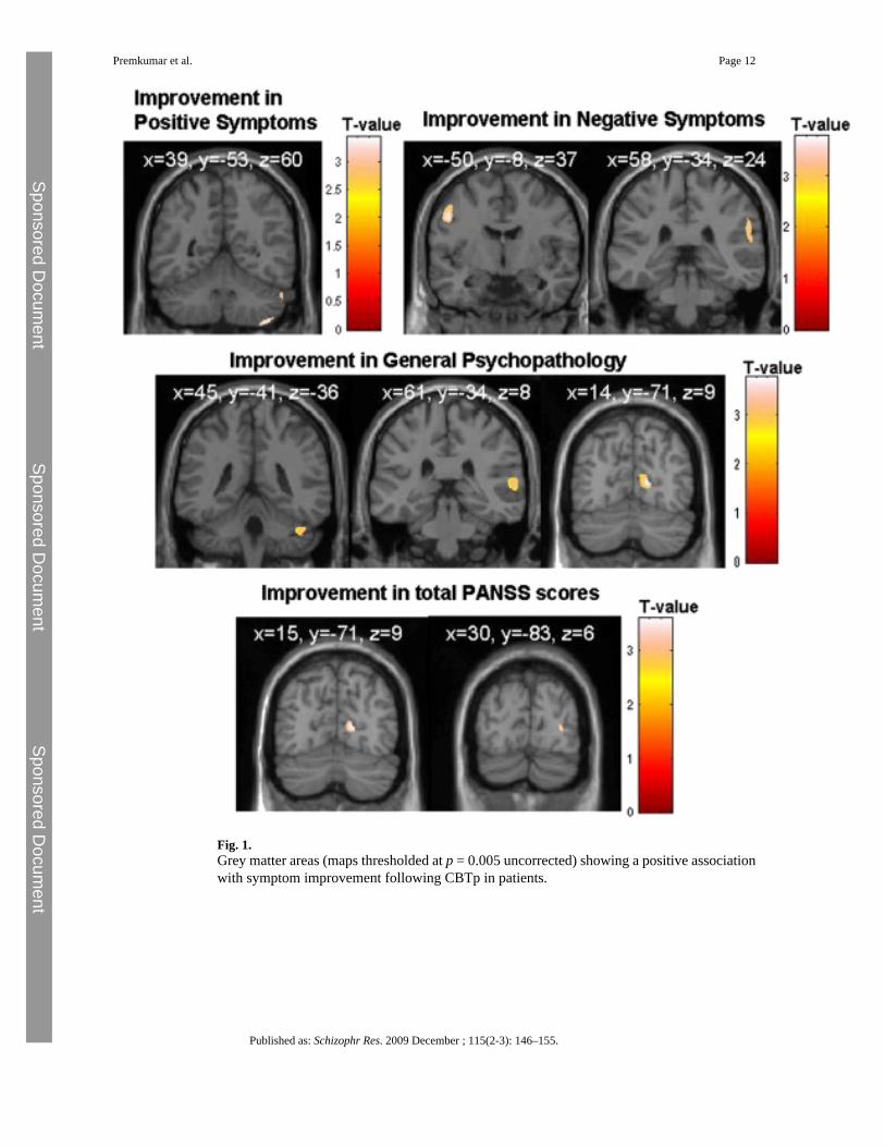
Fig. 1.Grey matter areas (maps thresholded at p = 0.005 uncorrected) showing a positive associationwith symptom improvement following CBTp in patients.
Premkumar et al. Page 12
Published as: Schizophr Res. 2009 December ; 115(2-3): 146–155.
Sponsored Docum
ent Sponsored D
ocument
Sponsored Docum
ent

Fig. 2.Scatter plots showing the relationship between the percentage grey matter volumes (relativeto group mean) at the maxima voxel of all the regions (y-axis) showing an association withsymptom change (x-axis) for CBTp+SC patients receiving clozapine or other antipsychotics.
Premkumar et al. Page 13
Published as: Schizophr Res. 2009 December ; 115(2-3): 146–155.
Sponsored Docum
ent Sponsored D
ocument
Sponsored Docum
ent

Fig. 3.Greater posterior hippocampal volume (maps thresholded at p = 0.05 uncorrected) in CBTp+SC responders relative to healthy participants and CBTp+SC non-responders.
Premkumar et al. Page 14
Published as: Schizophr Res. 2009 December ; 115(2-3): 146–155.
Sponsored Docum
ent Sponsored D
ocument
Sponsored Docum
ent

Sponsored Docum
ent Sponsored D
ocument
Sponsored Docum
ent
Premkumar et al. Page 15
Tabl
e 1
Dem
ogra
phic
and
clin
ical
cha
ract
eris
tics o
f stu
dy g
roup
s.
Cha
ract
eris
ticC
BT
p+SC
pat
ient
s (n
= 25
)SC
pat
ient
s (n
= 19
)H
ealth
y pa
rtic
ipan
ts (n
= 2
5)M
odel
χ2 (d
f)p
Sex:
mal
e/fe
mal
e (n
)17
/815
/417
/8G
roup
0.80
(2)
0.67
2
F (d
f)
Age
in y
ears
− m
ean,
s.d.
35.6
, 8.1
40.6
, 9.8
33.7
, 12.
6G
roup
2.48
(2,6
6)0.
090
Yea
rs in
edu
catio
n −
mea
n, s.
d.13
.9, 3
.113
.5, 1
.615
.1, 2
.5G
roup
2.53
(2,6
6)0.
093
Dia
gnos
is S
chiz
ophr
enia
/sch
izoa
ffec
tive
diso
rder
24/1
17/2
–G
roup
0.72
(1,4
4)0.
395
Dur
atio
n of
illn
ess a
, yea
rs −
mea
n, s.
d.11
.1, 7
.915
.2, 1
1.5
–G
roup
1.94
(1,4
2)0.
170
Age
at i
llnes
s ons
et, y
ears
− m
ean,
s.d.
24.4
, 8.0
25.4
, 8.2
–G
roup
0.16
(1,4
2)0.
691
Sym
ptom
s (PA
NSS
b) −
mea
n, s.
d.B
asel
ine
Follo
w-u
pB
asel
ine
Follo
w-u
p
Pos
itive
18.1
, 4.8
▼14
.8, 4
.218
.4, 3
.418
.0, 3
.8–
Tim
e ×
Gro
up5.
84 (1
,42)
0.02
4
Neg
ativ
e17
.9, 4
.4▼
15.5
, 4.2
19.2
, 3.9
20.1
, 4.6
–Ti
me
× G
roup
6.67
(1,4
2)0.
016
Gen
eral
psy
chop
atho
logy
33.2
, 7.2
▼28
.3, 7
.234
.6, 4
.634
.3, 6
.9–
Tim
e ×
Gro
up4.
37 (1
,42)
0.04
2
Tot
al69
.1, 1
3.6
▼58
.6, 1
4.5
72.2
, 9.0
72.3
, 13.
1–
Tim
e ×
Gro
up7.
93 (1
,42)
0.00
7
Med
icat
ion
leve
lc −
mea
n do
se, s
.d.
520.
7, 3
80.7
484.
7, 3
45.0
–G
roup
0.11
(2,6
6)0.
748
Tota
l gre
y m
atte
r vol
ume
(ml) −
mea
n, s.
d.70
2.4,
72.
069
7.5,
68.
473
6.4,
72.
5G
roup
2.08
(2,6
6)0.
133
Tota
l whi
te m
atte
r vol
ume
(ml) −
mea
n, s.
d.41
0.1,
47.
541
5.3,
41.
741
7.8,
48.
9G
roup
0.18
(2,6
6)0.
838
Tota
l cer
ebro
-spi
nal f
luid
(ml) −
mea
n, s.
d.52
5.6,
87.
252
7.9,
61.
553
1.0,
82.
7G
roup
0.03
(2,6
6)0.
971
▼Lo
wer
sym
ptom
scor
es (p
< 0
.01)
at f
ollo
w-u
p in
the
CB
Tp +
SC
, but
not
in th
e SC
, gro
up.
a Diff
eren
ce b
etw
een
the
age
at o
nset
of p
sych
otic
sym
ptom
s (as
repo
rted
by th
e pa
tient
and
, whe
re p
ossi
ble,
con
firm
ed w
ith c
linic
al re
cord
s and
oth
er so
urce
s) a
nd c
urre
nt a
ge.
b PAN
SS: P
ositi
ve a
nd N
egat
ive
Synd
rom
e Sc
ale.
c Med
icat
ion
leve
l exp
ress
ed in
chl
orpr
omaz
ine
equi
vale
nts.
Published as: Schizophr Res. 2009 December ; 115(2-3): 146–155.

Sponsored Docum
ent Sponsored D
ocument
Sponsored Docum
ent
Premkumar et al. Page 16
Table 2
GMV correlates (positive associations) of symptom improvement following CBTp(n = 25) (height threshold p = 0.005 for all voxels; T > 2.81; family-wise error corrected p < 0.05).
Brain region Side BAa MNIb coordinates Voxel T value Number ofcontiguousvoxels
Voxel equiv Z
x y Z
PANSS positive symptoms improvement
Cerebellum (lobule VII) Right 39 −52 − 59 3.21 313 2.89
PANSS negative symptoms improvement
Precentral gyrus Left 6 − 50 − 8 37 3.75 1244 3.29
Inferior parietal lobule Right 40 58 − 34 24 3.72 1100 3.26
PANSS general psychopathology improvement
Superior temporal gyrus Right 22 61 − 34 8 3.8 1747 3.31
Cuneus Right 17 14 − 71 9 3.86 708 3.35
Cerebellum (Crus I) Right 45 − 41 − 36 3.83 955 3.33
PANSS total symptoms improvement
Cuneus Right 17 16 − 71 9 3.44 319 3.06
Superior temporal gyrus Right 22 60 − 33 13 3.57 1662 3.15
aBroadmann area.
bMontreal Neurological Institute.
Published as: Schizophr Res. 2009 December ; 115(2-3): 146–155.

Sponsored Docum
ent Sponsored D
ocument
Sponsored Docum
ent
Premkumar et al. Page 17
Table 3
Correlations between localized GMVs (with positive associations with CBTp response within SPM5) andsymptom improvement in CBTp+SC and SC groups.
Brain region-symptom improvementcorrelate
CBTp+SC CBTp+SCcontrolling forintracranial volume
SC
r (p) Partial r (p) r (p)
Right cerebellum-positive symptoms 0.557 (0.004) 0.581 (0.003) 0.421 (0.073)
Left precentral gyrus-negative symptoms 0.615 (0.001) 0.653 (0.001) − 0.315 (0.189)
Right inferior parietal lobule-negativesymptoms
0.613 (0.001) 0.628 (0.001) 0.002 (0.994)
Right superior temporal gyrus-generalpsychopathology
0.621 (0.001) 0.676 (<0.001) − 0.264 (0.275)
Right cuneus-general psychopathology 0.627 (0.001) 0.700 (<0.001) 0.216 (0.375)
Right cerebellum-general psychopathology 0.435 (0.030) 0.627 (0.001) 0.226 (0.352)
Right cuneus-total symptoms 0.583 (0.002) 0.618 (0.001) 0.267 (0.270)
Right superior temporal gyrus-total symptoms 0.597 (0.002) 0.654 (0.007) − 0.288 (0.232)
Published as: Schizophr Res. 2009 December ; 115(2-3): 146–155.

Sponsored Docum
ent Sponsored D
ocument
Sponsored Docum
ent
Premkumar et al. Page 18
Table 4
Differences in the posterior hippocampus GMVs between CBTp+SC responders (n = 11), CBTp+SC non-responders (n = 14) and healthy participants (n = 25) (height threshold p = 0.005, cluster size > 10 voxels).
Side MNIa coordinates Voxel T value Number ofcontiguous voxels
Voxel equiv Z
x y z
CBTp+SC responders > healthy participants
Right 28 − 28 − 5 3.33 132 3.07
Left − 25 − 30 − 5 4.36 797 3.86
CBTp+SC responders > CBTp+SC non-responders
Left − 27 − 36 − 5 2.89 11 2.64
aMontreal Neurological Institute.
Published as: Schizophr Res. 2009 December ; 115(2-3): 146–155.
Related Documents