ARTICLE Structural genomic variation in childhood epilepsies with complex phenotypes Ingo Helbig* ,1 , Marielle EM Swinkels 2,3 , Emmelien Aten 4 , Almuth Caliebe 5 , Ruben van ‘t Slot 2 , Rainer Boor 1 , Sarah von Spiczak 1 , Hiltrud Muhle 1 , Johanna A Ja ¨hn 1 , Ellen van Binsbergen 2 , Onno van Nieuwenhuizen 6 , Floor E Jansen 6 , Kees PJ Braun 6 , Gerrit-Jan de Haan 3 , Niels Tommerup 7 , Ulrich Stephani 1 , Helle Hjalgrim 8,9 , Martin Poot 2 , Dick Lindhout 2,3 , Eva H Brilstra 2 , Rikke S Møller 7,8 and Bobby PC Koeleman 2 A genetic contribution to a broad range of epilepsies has been postulated, and particularly copy number variations (CNVs) have emerged as significant genetic risk factors. However, the role of CNVs in patients with epilepsies with complex phenotypes is not known. Therefore, we investigated the role of CNVs in patients with unclassified epilepsies and complex phenotypes. A total of 222 patients from three European countries, including patients with structural lesions on magnetic resonance imaging (MRI), dysmorphic features, and multiple congenital anomalies, were clinically evaluated and screened for CNVs. MRI findings including acquired or developmental lesions and patient characteristics were subdivided and analyzed in subgroups. MRI data were available for 88.3% of patients, of whom 41.6% had abnormal MRI findings. Eighty-eight rare CNVs were discovered in 71 out of 222 patients (31.9%). Segregation of all identified variants could be assessed in 42 patients, 11 of which were de novo. The frequency of all structural variants and de novo variants was not statistically different between patients with or without MRI abnormalities or MRI subcategories. Patients with dysmorphic features were more likely to carry a rare CNV. Genome-wide screening methods for rare CNVs may provide clues for the genetic etiology in patients with a broader range of epilepsies than previously anticipated, including in patients with various brain anomalies detectable by MRI. Performing genome-wide screens for rare CNVs can be a valuable contribution to the routine diagnostic workup in patients with a broad range of childhood epilepsies. European Journal of Human Genetics advance online publication, 27 November 2013; doi:10.1038/ejhg.2013.262 Keywords: CNV; structural genomic variation; childhood epilepsies; epileptic encephalopathies INTRODUCTION Epilepsies are frequent neurological disorders with a strong genetic component. 1 Decades of intense research have led to the discovery of several epilepsy genes, for which genetic testing can provide early diagnosis and guide optimal treatment. However, the genetic basis for the majority of refractory and childhood-onset epilepsies remains elusive. Many patients have epilepsies with complex phenotypes that are difficult to classify, sometimes with definite or questionable magnetic resonance imaging (MRI) abnormalities ranging from nonspecific findings to clear developmental abnormalities, some degree of dysmorphic features, mild-to-severe intellectual disability or psychiatric comorbidities. Various classifications can be used to describe the epilepsy phenotypes in these patients, including the 1989 and 2010 classifica- tion of the International League Against Epilepsy (ILAE). 2,3 However, the rare complex phenotypes including possible dysmorphic features, intellectual disability, and questionable or nonspecific MRI findings are sometimes not fully captured in either classification. We used existing 1989 ILAE categories for patients if possible, acknowledging that a large fraction of patients were difficult to classify. For some syndromes, we also use the current 2010 ILAE classification. We refer to the overall cohort as epilepsies with complex phenotypes. Although this patient group represents a significant fraction of patients referred for genetic counseling, causative genetic alterations in known genes for monogenic seizure disorders are only identified in a small subset. Also, patients with these complex phenotypes are usually not included in current large-scale genetic studies. Accord- ingly, these epilepsies represent ‘forgotten phenotypes’, and gene discovery in this group of patients is pressing and of major importance. Copy number variations (CNVs) including microdeletions and microduplications have emerged as a new pathogenic principle in a range of neurological and psychiatric disorders in recent years. Genome-wide screens of focal and generalized epilepsy have identified recurrent microdeletions in up to 3% of patients with idiopathic generalized epilepsies (also genetic generalized epilepsies) and in 1% of focal epilepsies. 4,5 Microdeletions at the chromosomal regions 15q13.3 and 16p13.11 are the most frequently identified variants, 1 Department of Neuropediatrics, University Medical Center Schleswig-Holstein (UKSH), Kiel, Germany; 2 Department of Medical Genetics, University Medical Center Utrecht, Utrecht, The Netherlands; 3 SEIN Epilepsy Institute in the Netherlands Foundation, Hoofddorp, The Netherlands; 4 Department of Medical Genetics, Leiden University Medical Center, Leiden, The Netherlands; 5 Department of Human Genetics, University Medical Center Schleswig-Holstein (UKSH), Kiel, Germany; 6 Department of Child Neurology, Rudolf Magnus Institute of Neurosciences, University Medical Center Utrecht, The Netherlands; 7 Wilhelm Johannsen Centre for Functional Genome Research, Copenhagen, Denmark; 8 Danish Epilepsy Centre, Dianalund, Denmark; 9 Institute of Regional Health Services Research, University of Southern Denmark, Odense, Denmark *Correspondence: Professor I Helbig, Department of Neuropediatrics, University Medical Center Schleswig-Holstein (UKSH), Building 9, Arnold-Heller-Street 3, D-24105 Kiel, Germany. Tel: +49 (0) 431 597 1622; Fax: +49 (0) 431 597 1769; E-mail: [email protected] Received 27 January 2013; revised 1 October 2013; accepted 18 October 2013 European Journal of Human Genetics (2013), 1–6 & 2013 Macmillan Publishers Limited All rights reserved 1018-4813/13 www.nature.com/ejhg

Welcome message from author
This document is posted to help you gain knowledge. Please leave a comment to let me know what you think about it! Share it to your friends and learn new things together.
Transcript

ARTICLE
Structural genomic variation in childhood epilepsieswith complex phenotypes
Ingo Helbig*,1, Marielle EM Swinkels2,3, Emmelien Aten4, Almuth Caliebe5, Ruben van ‘t Slot2, Rainer Boor1,Sarah von Spiczak1, Hiltrud Muhle1, Johanna A Jahn1, Ellen van Binsbergen2, Onno van Nieuwenhuizen6,Floor E Jansen6, Kees PJ Braun6, Gerrit-Jan de Haan3, Niels Tommerup7, Ulrich Stephani1, Helle Hjalgrim8,9,Martin Poot2, Dick Lindhout2,3, Eva H Brilstra2, Rikke S Møller7,8 and Bobby PC Koeleman2
A genetic contribution to a broad range of epilepsies has been postulated, and particularly copy number variations (CNVs)have emerged as significant genetic risk factors. However, the role of CNVs in patients with epilepsies with complex phenotypesis not known. Therefore, we investigated the role of CNVs in patients with unclassified epilepsies and complex phenotypes.A total of 222 patients from three European countries, including patients with structural lesions on magnetic resonanceimaging (MRI), dysmorphic features, and multiple congenital anomalies, were clinically evaluated and screened for CNVs.MRI findings including acquired or developmental lesions and patient characteristics were subdivided and analyzed insubgroups. MRI data were available for 88.3% of patients, of whom 41.6% had abnormal MRI findings. Eighty-eight rareCNVs were discovered in 71 out of 222 patients (31.9%). Segregation of all identified variants could be assessed in 42patients, 11 of which were de novo. The frequency of all structural variants and de novo variants was not statistically differentbetween patients with or without MRI abnormalities or MRI subcategories. Patients with dysmorphic features were more likelyto carry a rare CNV. Genome-wide screening methods for rare CNVs may provide clues for the genetic etiology in patients witha broader range of epilepsies than previously anticipated, including in patients with various brain anomalies detectable by MRI.Performing genome-wide screens for rare CNVs can be a valuable contribution to the routine diagnostic workup in patients witha broad range of childhood epilepsies.European Journal of Human Genetics advance online publication, 27 November 2013; doi:10.1038/ejhg.2013.262
Keywords: CNV; structural genomic variation; childhood epilepsies; epileptic encephalopathies
INTRODUCTIONEpilepsies are frequent neurological disorders with a strong geneticcomponent.1 Decades of intense research have led to the discovery ofseveral epilepsy genes, for which genetic testing can provide earlydiagnosis and guide optimal treatment. However, the genetic basis forthe majority of refractory and childhood-onset epilepsies remainselusive. Many patients have epilepsies with complex phenotypes thatare difficult to classify, sometimes with definite or questionablemagnetic resonance imaging (MRI) abnormalities ranging fromnonspecific findings to clear developmental abnormalities, somedegree of dysmorphic features, mild-to-severe intellectual disabilityor psychiatric comorbidities.
Various classifications can be used to describe the epilepsyphenotypes in these patients, including the 1989 and 2010 classifica-tion of the International League Against Epilepsy (ILAE).2,3 However,the rare complex phenotypes including possible dysmorphic features,intellectual disability, and questionable or nonspecific MRI findingsare sometimes not fully captured in either classification. We usedexisting 1989 ILAE categories for patients if possible, acknowledging
that a large fraction of patients were difficult to classify. For somesyndromes, we also use the current 2010 ILAE classification. We referto the overall cohort as epilepsies with complex phenotypes.
Although this patient group represents a significant fraction ofpatients referred for genetic counseling, causative genetic alterationsin known genes for monogenic seizure disorders are only identified ina small subset. Also, patients with these complex phenotypes areusually not included in current large-scale genetic studies. Accord-ingly, these epilepsies represent ‘forgotten phenotypes’, and genediscovery in this group of patients is pressing and of majorimportance.
Copy number variations (CNVs) including microdeletions andmicroduplications have emerged as a new pathogenic principle in arange of neurological and psychiatric disorders in recent years.Genome-wide screens of focal and generalized epilepsy have identifiedrecurrent microdeletions in up to 3% of patients with idiopathicgeneralized epilepsies (also genetic generalized epilepsies) and in 1%of focal epilepsies.4,5 Microdeletions at the chromosomal regions15q13.3 and 16p13.11 are the most frequently identified variants,
1Department of Neuropediatrics, University Medical Center Schleswig-Holstein (UKSH), Kiel, Germany; 2Department of Medical Genetics, University Medical Center Utrecht,Utrecht, The Netherlands; 3SEIN Epilepsy Institute in the Netherlands Foundation, Hoofddorp, The Netherlands; 4Department of Medical Genetics, Leiden University MedicalCenter, Leiden, The Netherlands; 5Department of Human Genetics, University Medical Center Schleswig-Holstein (UKSH), Kiel, Germany; 6Department of Child Neurology,Rudolf Magnus Institute of Neurosciences, University Medical Center Utrecht, The Netherlands; 7Wilhelm Johannsen Centre for Functional Genome Research, Copenhagen,Denmark; 8Danish Epilepsy Centre, Dianalund, Denmark; 9Institute of Regional Health Services Research, University of Southern Denmark, Odense, Denmark*Correspondence: Professor I Helbig, Department of Neuropediatrics, University Medical Center Schleswig-Holstein (UKSH), Building 9, Arnold-Heller-Street 3, D-24105 Kiel,Germany. Tel: +49 (0) 431 597 1622; Fax: +49 (0) 431 597 1769; E-mail: [email protected] 27 January 2013; revised 1 October 2013; accepted 18 October 2013
European Journal of Human Genetics (2013), 1–6& 2013 Macmillan Publishers Limited All rights reserved 1018-4813/13
www.nature.com/ejhg

which are clearly established genetic risk factors for epilepsy. However,contribution to disease is modest, and carriers may present withvariable phenotypes including a broad spectrum of differentepilepsy subtypes, various neurodevelopmental disorders, and severesyndromes with multiple congenital anomalies (MCAs). Furthermore,these microdeletions are often transmitted from a healthy parent,illustrating the complexity of the underlying genetic architecture anddisease mechanism.5–7 In addition to recurrent CNVs, up to 10%of patients with various epilepsies carry a unique and possiblypathogenic CNV.4 Although many of these variants may representbenign variants, the overall CNV burden in this group is clearlyelevated as compared with control populations,8 indicating that atleast some of these variants are genetic risk factors.
The role of microdeletions and microduplications in seizuredisorders extending beyond the group of well-classified non-lesionaland non-syndromal epilepsies remains unclear. We therefore soughtto investigate the rate of rare and recurrent CNVs in a large sample ofcases with a complex epilepsy phenotype, including patients with orwithout intellectual disability, dysmorphic features, or MRI abnorm-alities. Most of these patients would have been classified as havingsymptomatic epilepsy in the 1989 ILAE classification.
MATERIALS AND METHODSClinical phenotypingPatients were recruited from three centers, including two epilepsy centers (Kiel,Dianalund) and one clinical genetics center (Utrecht). In accordance with theoverall aim of the study, the inclusion criteria were designed to allow for aselection of a highly diverse patient population with rare epilepsy phenotypes,which are usually not included in genetic studies either because of thephenotype or owing to the presence of abnormalities on MRI.
Patients for this study were selected on the basis of the following criteria:(1) rare epilepsies, which were not easily classified into existing commonepilepsy syndromes such as Idiopathic/Genetic Generalized Epilepsy or BenignRolandic Epilepsy, even though atypical presentations of common epilepsieswere included; and (2) availability of MRI data on patients. The presence ofapparently acquired lesions was not used as an exclusion criterion, as long as agenetic component was considered by the referring physicians – for example,when the seizure disorder was too severe or not typical for the type of lesion,or the pathology of the lesion was not clear. The study was approved by thelocal ethics boards at the University of Kiel, the Danish Epilepsy CenterDianalund, and the University of Utrecht.
MRI classificationAvailable information on MRI was included. MRI findings were divided intoacquired or developmental lesions or nonspecific abnormalities. The develop-mental lesions were further subdivided into malformations of corticaldevelopment9 and other lesions.
Genetic analysisPatients from the center at Utrecht were genotyped using an Agilent 105K/180K comparative genomic hybridization (CGH) array and analyzed with thestandard software provided by the manufacturer (Agilent Technologies, Inc.,Santa Clara, CA, USA). Patients from the centers at Dianalund and Kiel weregenotyped with Affymetrix 6.0 arrays and analyzed with the AffymetrixGenotyping Console (Affymetrix, Illumina, San Diego, CA, USA). The structuralgenomic variations for the entire sample were included in the analysis if thevariant (1) was larger than 100 kb, (2) overlapped with a known gene, and (3a)did not overlap with a benign CNV in the Database of Genomic Variants or (3b)overlapped with previously reported pathogenic CNVs. For a subset of patients,parents were available for segregation analysis. CNVs were classified as pathogenicvariants, likely pathogenic variants or as variants of unknown significanceaccording to established guidelines.10 In brief, CNVs were consideredpathogenic if they represented de novo deletions or known deletions associatedwith human epilepsies. Likely pathogenic variants were de novo duplications and
CNVs larger than 1 Mb that were inherited or of unknown inheritance. All otherCNVs were considered of unknown significance.
Variants were annotated in hg19 as a reference sequence. Some arrays wereanalyzed based on hg18, which is indicated in the overall variant list(Supplementary 1 and 2).
Statistical analysisStatistical analysis was performed with the R Statistical Package (http://www.r-project.org). Fisher’s exact test was applied when appropriate. All tests weretwo-sided; confidence intervals and odds ratios were reported as appropriate.
RESULTSCohortsThe patient sample included 223 patients (129 male and 94 femalepatients) from the three participating centers (Utrecht n! 155,Dianalund n! 39, Kiel n! 29). The epilepsy phenotypes of thepatients are shown in Table 1. A total of 137 patients were classified ashaving lesional or presumably lesional or structural epilepsy accordingto the most recent classification by the ILAE.10 The frequencies ofMRI abnormalities, dysmorphic features, multiple congenitalabnormalities, intellectual disability, and other neurodevelopmentaldisorders are outlined in the sections below.
Overall frequency of rare structural variantsWe identified 88 rare CNVs exceeding 100 kb in 71 patients (31.8%,Supplementary Table 1). A female patient with monosomy of the Xchromosome was excluded from further analysis of rare structuralvariants (patient NL69, see below ‘other genetic findings’).An additional 19 variants smaller than 100 kb in size were detectedin 19 patients (including six patients with additional larger variants)using the higher resolution of the array-CGH platform from theUtrecht center (Supplementary Table 2). Only one of these variantsrepresenting a deletion (patient NL8) was de novo (see below ‘CNVswith additional mutations’). We further focused on the variants largerthan 100 kb to allow for comparability between platforms. A total of18 out of 88 variants were considered pathogenic including 10 de novodeletions, whereas 15 out of 88 variants were considered likelypathogenic and 55 out of 88 variants were of unknown significance,including 17 deletions and 38 duplications (ratioB1:2). The 88variants were distributed in the 71 patients with identified variants asfollows: 18 out of 71 patients carried at least a single pathogenicvariant, 12 out of 71 patients carried at least a single likely pathogenicvariant, and 41 out of 71 patients carried at least a single variant ofunknown significance; 57 out of 71 patients had a single rare variant(15 pathogenic, 7 likely pathogenic, 35 unknown significance), 11 outof 71 patients had two variants and 3 patients had three variants.The median size of all variants was 410 kb (range 102 kb to 12.7 Mb).The size of 25 variants was between 100 and 200 kb (28.4%), 27variants were between 200 and 500 kb (30.6%), 13 variants rangedbetween 500 kb and 1 Mb (14.8%), 19 variants were between 1 and3 Mb (21.6%) and 4 variants were larger than 3 Mb (4.6%). All fourvariants larger than 3 Mb were de novo. Segregation of all identifiedvariants could be assessed in 42 out of 71 patients, and 11 of thesepatients had at least a single de novo variant (26.1%, Table 2).
CNVs in genomic hotspotsCNVs in genomic hot-spot regions of the human genome11 wereidentified in 11 out of 223 patients (4.9%) and were consideredpathogenic (Table 3). Duplications at 1q21.1 and 16p13.11 werefound in one patient each. Similarly, microdeletions at 15q13.3 and22q11.2 were detected in a single patient each. Microdeletions at
CNVs in childhood epilepsiesI Helbig et al
2
European Journal of Human Genetics

15q11.2,6 16p13.11,12 and 16p11.213 were observed in two patientseach. Finally, one patient with unclassified focal epilepsy had a de novo17p11.2 deletion consistent with the Smith–Magenis syndrome(SMS).
CNVs with additional mutationsFive patients with an additional monogenic cause for their epilepsywere identified in the cohort. All five patients carried a candidateCNV in addition to the disease-related monogenic variant, includingone pathogenic variant and four variants of unknown significance.Two patients (NL9, NL23) had Dravet syndrome with mutations inSCN1A and carried duplications of unknown significance. Early in thecourse of the disease, patient NL9 did not show the typicalpresentation of Dravet syndrome, which prompted array-CGHanalysis in addition to SCN1A testing. A previously unknownduplication at 14q22.1q22.2 spanning the GNG2 gene was identifiedthrough array-CGH. In patient NL23, array-CGH showing a pater-nally inherited duplication at 2q21.3 was performed because ofmicrocephaly, a cleft lip and cleft palate in addition to clinicalfeatures of Dravet syndrome. Furthermore, patient NL60 with amaternally inherited duplication at 2q32.1 (unknown significance)was diagnosed with mitochondrial encephalomyopathy, lactic acido-sis, and stroke-like episodes (MELAS) after array-CGH, and was seento be carrying a common MELAS-related mutation in the mitochon-drial DNA (m.3243A4G). Patient NL62 had familial benign neonatal
seizures with a deletion of KCNQ2; array-CGH was performed toassess the size of the deletion and an additional 15q11.2 deletion wasidentified (pathogenic variant). In patient NL18, the diagnosis ofTuberous Sclerosis Complex was made after array-CGH had beenperformed, which showed a maternally inherited duplication at4q32.1 (unknown significance). In all five patients, array-CGH wouldnot have been performed if the positive genetic finding had beendetected before array-CGH. This suggests that array-CGH may beconsidered a part of the diagnostic workup in patients with knownmutations if atypical clinical findings are present. Finally, two malepatients NL41 and D16 carried inherited deletions of the DMD genecompatible with Duchenne muscular dystrophy. Although consideredexplanatory for the muscular symptoms, the association with theepilepsy phenotype is uncertain. Patient NL69 had monosomy of theX chromosome compatible with Turner syndrome and was excludedfrom the overall analysis.
MRI findingsInformation on MRI was available for 196 patients and is shown inTable 4. Of them, 82 were reported to have some abnormality onimaging. In these patients, 26 had lesions that were considered to beacquired, 38 had lesions classified as developmental, and 18 hadnonspecific or unclassified lesions. The developmental lesionsincluded 23 malformations of cortical development. Significantdifferences between subgroups with respect to the CNV burden werenot found (Supplementary Table 3).
Dysmorphic features, MCAsIn 207 patients the presence of dysmorphic features could be assessed,and were present in 72 patients. Information on the presence ofMCAs was available for the same 207 patients; MCA was present in 19patients. Out of 222 patients, 13 had microcephaly and 5 hadmacrocephaly. Patients with dysmorphic features had a significantlyhigher CNV burden (Supplementary Table 3). However, this differ-ence was due to CNVs of unknown significance and likely pathogenicCNVs. All other differences between subgroups were not significant.
Neurodevelopmental phenotypesInformation on neurological examination was available for 168patients, of whom 91 were considered neurologically abnormal.Information on speech development was available for 201 patients,of whom 131 were considered abnormal. Gross motor developmentcould be assessed in 206 patients, of whom 110 had abnormal motordevelopment. Information on intellectual functioning and intellectualdisability was available for 172 patients. A total of 140 of thesepatients had at least moderate intellectual disability. Information onbehavioral phenotypes was available for 81 individuals, of whom 38were considered to have normal behavior. The subgroup forbehavioral phenotypes was small owing to the difficulties in assessingdistinct behavioral phenotypes in patients with intellectual disability.The comparison of CNV frequency between subgroups and differentclasses of CNVs did not show significant differences, except for asignificant excess of CNVs of unknown significance in patientswithout ID or behavioral problems. This result is possibly due tothe small number of patients investigated.
Novel recurrent CNVsNo novel recurrent CNVs larger than 100 kb were detected. However,inclusion of CNVs smaller than 100 kb revealed two patients (NL54and NL55) with an overlapping duplication at 7q36.3, including partof the PTPRN2 gene (Supplementary Figure 1). However, as one
Table 1 Epilepsy phenotypes
Epilepsy syndrome n
Genetic findings
before inclusion in study
Distinct rare epilepsy syndromes or epileptic encephalopathies
Ohtahara syndrome 2
BFNS 1 KCNQ2 del
MMPSI 1
West Syndrome 30 DMD dela (n!1)
LGS 9
Dravet syndrome 3 SCN1A (n!2)
ABPE (Pseudo-Lennox syndrome) 3
CSWS 13
Landau–Kleffner syndrome 1
Generalized epilepsies
GGEb 13
MAE 5
PME 5
Lesional epilepsies (symptomatic focal and generalized epilepsies)
Symptomatic focal epilepsy 75
Symptomatic generalized epilepsyc 2
Symptomatic epilepsy, unclassifiedd and/or
presumably lesional
60 m.3243A4Ge (n!1)DMD
dela (n!1)
Abbreviations: ABPE, atypical benign partial epilepsy; BFNS, benign familial neonatal seizures;CSWS, continuous spikes and waves during slow sleep; GGE, genetic generalized epilepsies;LGS, Lennox–Gastaut syndrome; MAE, myoclonic astatic epilepsy; MMPSI, malignant migratingpartial seizures of infancy; PME, progressive myoclonus epilepsy.aPatients NL41 and D16 carried a maternally transmitted deletion of DMD, which wasconsidered unrelated to the epilepsy phenotype.bPatients with apparent idiopathic generalized epilepsy phenotypes, but additional imagingfindings, dysmorphic features, or abnormal clinical examination.cEpilepsies not easily classified into distinct electroclinical syndromes such as West Syndromeor LGS.dIncluding patient NL69 with a monosomy of the X-chromosome compatible with TurnerSyndrome.eMitochondrial mutation related to MELAS (mitochondirial encephalomyelopathy, lacticacidosis, and stroke-like episodes).
CNVs in childhood epilepsiesI Helbig et al
3
European Journal of Human Genetics
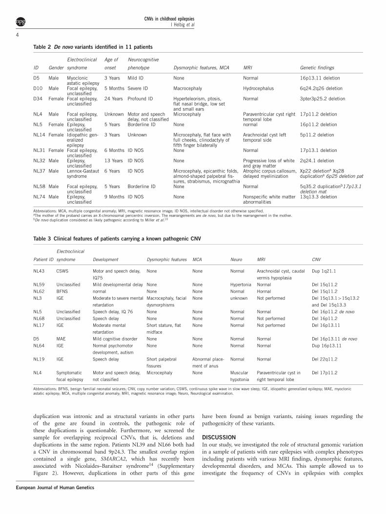
duplication was intronic and as structural variants in other partsof the gene are found in controls, the pathogenic role ofthese duplications is questionable. Furthermore, we screened thesample for overlapping reciprocal CNVs, that is, deletions andduplications in the same region. Patients NL39 and NL66 both hada CNV in chromosomal band 9p24.3. The smallest overlap regioncontained a single gene, SMARCA2, which has recently beenassociated with Nicolaides–Baraitser syndrome14 (SupplementaryFigure 2). However, duplications in other parts of this gene
have been found as benign variants, raising issues regarding thepathogenicity of these variants.
DISCUSSIONIn our study, we investigated the role of structural genomic variationin a sample of patients with rare epilepsies with complex phenotypesincluding patients with various MRI findings, dysmorphic features,developmental disorders, and MCAs. This sample allowed us toinvestigate the frequency of CNVs in epilepsies with complex
Table 2 De novo variants identified in 11 patients
ID Gender
Electroclinical
syndrome
Age of
onset
Neurocognitive
phenotype Dysmorphic features, MCA MRI Genetic findings
D5 Male Myoclonicastatic epilepsy
3 Years Mild ID None Normal 16p13.11 deletion
D10 Male Focal epilepsy,unclassified
5 Months Severe ID Macrocephaly Hydrocephalus 6q24.2q26 deletion
D34 Female Focal epilepsy,unclassified
24 Years Profound ID Hyperteleorism, ptosis,flat nasal bridge, low setand small ears
Normal 3pter3p25.2 deletion
NL4 Male Focal epilepsy,unclassified
Unknown Motor and speechdelay, not classified
Microcephaly Paraventricular cyst righttemporal lobe
17p11.2 deletion
NL5 Female Epilepsy,unclassified
5 Years Borderline ID None normal 16p11.2 deletion
NL14 Female Idiopathic gen-eralizedepilepsy
3 Years Unknown Microcephaly, flat face withfull cheeks, clinodactyly offifth finger bilaterally
Arachnoidal cyst lefttemporal side
5p11.2 deletion
NL31 Female Focal epilepsy,unclassified
6 Months ID NOS None Normal 17p13.1 deletion
NL32 Male Epilepsy,unclassified
13 Years ID NOS None Progressive loss of whiteand gray matter
2q24.1 deletion
NL37 Male Lennox-Gastautsyndrome
6 Years ID NOS Microcephaly, epicanthic folds,almond-shaped palpebral fis-sures, strabismus, micrognathia
Atrophic corpus callosum,delayed myelinization
Xp22 deletiona Xq28duplicationa 6p25 deletion pat
NL58 Male Focal epilepsy,unclassified
5 Years Borderline ID None Normal 5q35.2 duplicationb17p13.1deletion mat
NL74 Male Epilepsy,unclassified
9 Months ID NOS None Nonspecific white matterabnormalities
13q13.3 deletion
Abbreviations: MCA, multiple congenital anomaly; MRI, magnetic resonance image; ID NOS, intellectual disorder not otherwise specified.aThe mother of the proband carries an X-chromosomal pericentric inversion. The rearrangements are de novo, but due to the rearrangement in the mother.bDe novo duplication considered as likely pathogenic according to Miller et al.10
Table 3 Clinical features of patients carrying a known pathogenic CNV
Patient ID
Electroclinical
syndrome Development Dysmorphic features MCA Neuro MRI CNV
NL43 CSWS Motor and speech delay,
IQ75
None None Normal Arachnoidal cyst, caudal
vermis hypoplasia
Dup 1q21.1
NL59 Unclassified Mild developmental delay None None Hypertonia Normal Del 15q11.2
NL62 BFNS normal None None Normal Hormal Del 15q11.2
NL3 IGE Moderate to severe mental
retardation
Macrocephaly, facial
dysmorphisms
None unknown Not performed Del 15q13.1415q13.2
and Del 15q13.3
NL5 Unclassified Speech delay, IQ 76 None None Normal Normal Del 16p11.2 de novo
NL68 Unclassified Speech delay None None Normal Not performed Del 16p11.2
NL17 IGE Moderate mental
retardation
Short stature, flat
midface
None Normal Not performed Del 16p13.11
D5 MAE Mild cognitive disorder None None Normal Normal Del 16p13.11 de novo
NL64 IGE Normal psychomotor
development, autism
None None Normal Normal Dup 16p13.11
NL19 IGE Speech delay Short palpebral
fissures
Abnormal place-
ment of anus
Normal Normal Del 22q11.2
NL4 Symptomatic
focal epilepsy
Motor and speech delay,
not classified
Microcephaly None Muscular
hypotonia
Paraventricular cyst in
right temporal lobe
Del 17p11.2
Abbreviations: BFNS, benign familial neonatal seizures; CNV, copy number variation; CSWS, continuous spike wave in slow wave sleep; IGE, idiopathic generalized epilepsy; MAE, myoclonicastatic epilepsy; MCA, multiple congenital anomaly; MRI, magnetic resonance image; Neuro, Neurological examination.
CNVs in childhood epilepsiesI Helbig et al
4
European Journal of Human Genetics

phenotypes with and without additional features. We found a highfrequency of rare CNVs in approximately 30% of patients with orwithout abnormal MRI findings, indicating that the overall frequencyof rare CNVs does not depend on the presence of MRI findings.These results also apply when different classes of CNVs (unknownsignificance, likely pathogenic, pathogenic, de novo) are taken intoconsideration. This observation suggests that structural genomicvariations have a role in a larger group of epilepsies than previouslyanticipated, including seizure disorders with various degrees ofimaging abnormalities. We observed a higher frequency of CNVs inpatients with dysmorphic features and/or MCAs.
The observed rate of rare CNVs in patients with epilepsies andcomplex phenotypes is slightly lower as compared with thoseobserved in patients with intellectual disability and autism, but ishigher than expected in control cohorts. Cooper et al. reported afrequency of rare CNVs Z400 kb in 25.7% of 15 767 patients withintellectual disability compared with 11.5% in 8329 controls,15
a difference that was even more pronounced with increasing CNVsize – for example, 11.3% in cases versus 0.6% in controls for CNVsZ1.5 Mb. In our sample, 17.5% of patients had CNVs larger than400 kb with 6.3% of patients carrying CNVs larger than 1.5 Mb. Thedifferences in the frequencies of CNVs in our sample and publishedcontrols are significant both at the 400 kb level (P! 0.007) and at the1.5 Mb level (Po0.001). This comparison suggests an attributablerisk of 6.2% for CNVs 4400 kb and 5.7% for CNVs 41.5 Mb.This finding is intriguing, implying that the attributable risk is largelydue to the influence of larger CNVs.
In a significant number of patients a de novo variant could beconfirmed. Three patients had de novo deletions consistent withknown genomic disorders, including a microdeletion in 16p13.11(D5), 16p11.2 (NL5), and 17p11.2 (NL4), the region implicated inSMS. In addition, two patients had terminal deletion syndromesincluding a terminal deletion of 6q (D10) and a terminal deletion of3p (D34). Both deletions were the two largest deletions in our sample,exceeding 10 Mb. Epilepsy in 6q terminal deletion syndrome is a well-described feature,16,17 whereas intellectual disability is usually themost prominent feature in 3p deletion syndrome.18,19 As parents werenot available for a significant proportion of patients, the frequency ofde novo variants might be an underestimation.
In five patients with a rare CNV, a monogenic cause of the epilepsywas also present. This illustrates that the presence of a CNV does notpreclude the possibility of a monogenic cause of the epilepsy. Inchildren with epilepsy and developmental delay, array-CGH is oftenperformed early in the process of etiological evaluation. When CNVsare found that may explain the epilepsy, monogenic causes can easilybe overlooked.20
We found three regions with overlapping CNVs in more than onepatient, at 16p13.11, 7q36.3, and 9p23. The CNVs at 16p13.11 furtherlimit the epilepsy candidate genes in this region to ABCC1 andABCC6. Both genes are members of the MRP subfamily and have arole in multi-drug resistance.
The two other overlapping copy number variants at 7q36.3 and9p23 identified in our sample did not delineate novel disease genes, asboth candidate genes (PTPRN2 and SMARCA2) also contained alarge number of benign duplications and deletions.
We did not observe significant group differences between patientswith or without MRI findings, independent of the class of CNVinvestigated. In particular, several patients with known MRI abnorm-alities had genetic findings, which might contribute to the overallphenotype. In patient NL4, MRI revealed a paraventricular cyst in theright temporal lobe that was considered causal for underlying focalepilepsy by the treating physician. However, genetic analysis identifieda 17p11.2 microdeletion, consistent with SMS. Epilepsy is a well-described feature of SMS. Although we are unable to clearly attributethe patient’s epilepsy to the developmental MRI abnormality or to thede novo microdeletion, this case illustrates that even the presence ofcausally implicated MRI findings should not preclude genetic analysis.Similar arguments have been raised in patients with temporal lobeepilepsy, in which a significant subset of patients with resectivesurgery and good postoperative outcome carry large CNVs.21
The patient sample included in our study is a mixed sample fromthree different centers with different backgrounds and is not popula-tion-based. In fact, it was the impetus of the investigation to assessCNV frequencies in patients not otherwise included in other well-defined epilepsy cohorts. This inclusion strategy might have con-founded risk estimation and CNV frequencies. However, as baselinecharacteristics, for example, the overall CNV frequency, are in linewith published data, we assume that the effect of our inclusionstrategy would not have significantly biased the results.
Recommendations for clinical practiceWe describe the frequency and the ‘genetic landscape’15 of CNVs in adiverse sample of patients with rare epilepsies and complexphenotypes, including in patients with a diverse phenotypicspectrum. The high frequency of CNVs irrespective of clinicalphenotype indicates that screening for CNVs has the potential toidentify relevant etiological genetic factors across a wide rangeof diverse epilepsies, independent of preexisting abnormalities.
Table 4 Classification of MRI findings
Type of lesion n
Acquired lesions 26
MTS 9
PVL 5
Gliosis after asphyxia 3
Rasmussen’s encephalitis 2
Other lesions classified as acquired 7
Developmental lesions 38
Malformations of cortical development 23
Focal cortical dysplasia unilateral 8
Polymicrogyria 4
Hemimegalencephaly 2
Transmantle dysplasia 2
Hippocampal malrotation 2
Lissencephaly 1
Schizencephaly 1
Holoprosencephaly 1
Subependymal noduli 1
Megalencephaly 1
Other developmental abnormalities 15
Delay in myelinisation/hypomyelinisation 5
Atrophy 3
Paraventricular or arachnoidal cysts 3
Cerebellar hypoplasia, Chiari-II-malformations 2
Other 2
Non-specific, unclassified findings 18
Nonspecific ventricular enlargement 5
Nonspecific white matter abnormalities 5
Other 8
Abbreviations: MRI, magnetic resonance image; MTS, mesial temporal sclerosis; PVL,periventricular leukomalacia.
CNVs in childhood epilepsiesI Helbig et al
5
European Journal of Human Genetics

This suggests that array-CGH or SNP arrays may represent a powerfulfirst-line screening tool in diverse epilepsy syndromes and should notbe limited to epilepsies that are commonly assumed to be genetic.Array-CGH may also identify additional possibly pathogenic variantsin known monogenic epilepsies, particularly if the phenotype hasadditional clinical features.
The structural genomic variants described in this publication havebeen submitted to the Database of Genomic Variants Archive (http://www.ebi.ac.uk/dgva/, accession number estd208).
CONFLICT OF INTERESTThe authors declare no conflict of interest.
ACKNOWLEDGEMENTSWe thank the patients and their parents for participation in this study. Thisproject was supported by the EuroEPINOMICS projects within theEUROCORES framework of the European Science Foundation and funds ofthe German Research Foundation (DFG HE 5413/3-1).
1 Helbig I, Scheffer IE, Mulley JC, Berkovic SF: Navigating the channels andbeyond: unravelling the genetics of the epilepsies. Lancet Neurol 2008; 7:231–245.
2 Proposal for revised classification of epilepsies and epileptic syndromes. Commissionon Classification and Terminology of the International League Against Epilepsy.Epilepsia 1989; 30: 389–399.
3 Berg AT, Berkovic SF, Brodie MJ et al: Revised terminology and concepts fororganization of seizures and epilepsies: report of the ILAE Commission on Classifi-cation and Terminology, 2005-2009. Epilepsia 2010; 51: 676–685.
4 Mefford HC, Muhle H, Ostertag P et al: Genome-wide copy number variation inepilepsy: novel susceptibility loci in idiopathic generalized and focal epilepsies.PLoS Genet 2010; 6: e1000962.
5 Heinzen EL, Radtke RA, Urban TJ et al: Rare deletions at 16p13.11 predisposeto a diverse spectrum of sporadic epilepsy syndromes. Am J Hum Genet 2010; 86:707–718.
6 de Kovel CG, Trucks H, Helbig I et al: Recurrent microdeletions at 15q11.2and 16p13.11 predispose to idiopathic generalized epilepsies. Brain 2010; 133:23–32.
7 Helbig I, Mefford HC, Sharp AJ et al: 15q13.3 microdeletions increase risk ofidiopathic generalized epilepsy. Nat Genet 2009; 41: 160–162.
8 Itsara A, Cooper GM, Baker C et al: Population analysis of large copy numbervariants and hotspots of human genetic disease. Am J Hum Genet 2009; 84:148–161.
9 Barkovich AJ, Kuzniecky RI, Jackson GD, Guerrini R, Dobyns WB: A developmentaland genetic classification for malformations of cortical development. Neurology 2005;65: 1873–1887.
10 Miller DT, Adam MP, Aradhya S et al: Consensus statement: chromosomal microarrayis a first-tier clinical diagnostic test for individuals with developmental disabilities orcongenital anomalies. Am J Hum Genet 2010; 86: 749–764.
11 Mefford HC, Eichler EE: Duplication hotspots, rare genomic disorders, and commondisease. Curr Opin Genet Dev 2009; 19: 196–204.
12 Hannes FD, Sharp AJ, Mefford HC et al: Recurrent reciprocal deletions andduplications of 16p13.11: the deletion is a risk factor for MR/MCA while theduplication may be a rare benign variant. J Med Genet 2009; 46: 223–232.
13 Weiss LA, Shen Y, Korn JM et al: Association between microdeletion andmicroduplication at 16p11.2 and autism. N Engl J Med 2008; 358: 667–675.
14 Van Houdt JK, Nowakowska BA, Sousa SB et al: Heterozygous missense mutations inSMARCA2 cause Nicolaides-Baraitser syndrome. Nat Genet 2012; 44: 445–449.
15 Cooper GM, Coe BP, Girirajan S et al: A copy number variation morbidity map ofdevelopmental delay. Nat Genet 2011; 43: 838–846.
16 Striano P, Malacarne M, Cavani S et al: Clinical phenotype and molecular characte-rization of 6q terminal deletion syndrome: Five new cases. Am J Med Genet A 2006;140: 1944–1949.
17 Elia M, Striano P, Fichera M et al: 6q terminal deletion syndrome associatedwith a distinctive EEG and clinical pattern: a report of five cases. Epilepsia 2006;47: 830–838.
18 Malmgren H, Sahlen S, Wide K, Lundvall M, Blennow E: Distal 3p deletion syndrome:detailed molecular cytogenetic and clinical characterization of three small distaldeletions and review. Am J Med Genet A 2007; 143A: 2143–2149.
19 Fernandez TV, Garcia-Gonzalez IJ, Mason CE et al: Molecular characterization of apatient with 3p deletion syndrome and a review of the literature. Am J Med Genet A2008; 146A: 2746–2752.
20 Poot M, van der Smagt JJ, Brilstra EH, Bourgeron T: Disentangling the myriadgenomics of complex disorders, specifically focusing on autism, epilepsy, andschizophrenia. Cytogenet Genome Res 2011; 135: 228–240.
21 Catarino CB, Kasperaviciute D, Thom M et al: Genomic microdeletions associatedwith epilepsy: not a contraindication to resective surgery. Epilepsia 2011; 52:1388–1392.
Supplementary Information accompanies this paper on European Journal of Human Genetics website (http://www.nature.com/ejhg)
CNVs in childhood epilepsiesI Helbig et al
6
European Journal of Human Genetics
Related Documents