Structural Determinants of Substrate Recognition in the HAD Superfamily Member D-Glycero-D-manno-Heptose 1,7- bisphosphate Phosphatase, GmhB † Henry Nguyen 1 , Liangbing Wang 2,# , Hua Huang 2 , Ezra Peisach 3 , Debra Dunaway- Mariano 2,* , and Karen N. Allen 1,3,* 1 Department of Physiology and Biophysics, Boston University School of Medicine, Boston, MA 02118-2394, USA 2 Department of Chemistry and Chemical Biology, University of New Mexico, Albuquerque, NM 87131, USA 3 Department of Chemistry, Boston University, Boston, MA 02215, USA Abstract The Haloalkanoic Acid Dehalogenase (HAD)1 enzyme superfamily is the largest family of phosphohydrolases. In HAD members, the structural elements that provide the binding interactions that support substrate specificity are separated from those that orchestrate catalysis. For most HAD phosphatases a cap domain functions in substrate recognition. However, for the HAD phosphatases which lack a cap domain, an alternate strategy for substrate selection must be operative. One such HAD phosphatase, GmhB of the HisB subfamily was selected for structure-function analysis. Herein, the X-ray crystallographic structures of E. coli GmhB in the apo form (1.6 Å resolution), complexed with Mg 2+ and orthophosphate (1.8 Å resolution), and with Mg 2+ and Dglycero-D-manno- heptose-1β,7-bisphosphate (2.2 Å resolution) were determined, in addition to the structure of B. bronchiseptica GmhB bound to Mg 2+ and orthophosphate (1.7 Å resolution). The structures show that in place of a cap domain, the GmhB catalytic site is elaborated by three peptide inserts or loops that pack to form a concave, semicircular surface around the substrate leaving group. Structure- † This work was supported by NIH Grant GM61099 to K.N.A and D.D.-M. Financial support for beamlines X12B, X25C and X29 at Brookhaven National Laboratory comes principally from the Offices of Biological and Environmental Research and of Basic Energy Sciences of the US Department of Energy and the National Center for Research Resources of the National Institutes of Health. 1 Abbreviations used are: H1β,7bisP; -D-glycero-D-manno-heptose-1 β,7-bisphosphate, H1α,7bisP; D-glycero-D-manno-heptose-1α,7- bisphosphate; D-glycero-D-manno-heptose 1,7-bisphosphate phosphatase, GmhB; Haloalkanoic Acid Dehalogenase, HAD; bifunctional histidinol-phosphate phosphatase/ imidazole-glycerol-phosphate dehydratase, HisB selenomethionine, SeMet; 2-keto-3-deoxy- octulosonate-8-phosphate, KDOP-8-P; 2-keto-3-deoxy-D-glycero-D-galactonononate-9-phosphate, KDN-9-P; DTT, dithiothreitol. *Address correspondence to Debra Dunaway-Mariano [email protected], phone: 505-277-3383, fax: 505-277-2609 and Karen N. Allen, phone: 617-358-5544, fax: 617-358-5554, [email protected]. # Current Address: Chengdu Di'Ao Pharmaceutical Company, Institute of Medicine, Sichuan, China. ‡ Coordinates for the X-ray structures of Bordatella bronchiseptica D-glycero-D-manno-heptose 1,7-bisphosphate phosphatase complexed with Mg 2+ and phosphate, and for E. coli D-glycero-D-manno-heptose 1,7-bisphosphate phosphatase in the apo form and complexed with Mg 2+ plus phosphate or D-glycero-D-manno-heptose 1β,7-bisphosphate have been deposited in the Protein Data Bank with accession codes 3L8H, 3L8E, 3L8F, and 3L8G, respectively. 3 At first glance this conclusion might appear to be inconsistent with the observed reduction in catalytic efficiency in the E. coli GmhB Zn 2+ binding loop mutants (Table 2). However, this is not the case as the alteration of a structural motif by mutation is frequently destabilizing to the native conformation enzyme and often manifested in decreased catalytic efficiency. In this case, removal of one of the four Zn 2+ ligands might result in misfolding (as appears to be the case for the H94A mutant which is insoluble) or destabilization of the native conformation as the Zn 2+ ion incorporates a fourth ligand from solvent, or elsewhere in the protein. Supporting Information Available Alignment of representative GmhB and HisB sequences. This material is available free of charge via the Internet at ://pubs.acs.org. NIH Public Access Author Manuscript Biochemistry. Author manuscript; available in PMC 2011 February 16. Published in final edited form as: Biochemistry. 2010 February 16; 49(6): 1082–1092. doi:10.1021/bi902019q. NIH-PA Author Manuscript NIH-PA Author Manuscript NIH-PA Author Manuscript

Welcome message from author
This document is posted to help you gain knowledge. Please leave a comment to let me know what you think about it! Share it to your friends and learn new things together.
Transcript
Structural Determinants of Substrate Recognition in the HADSuperfamily Member D-Glycero-D-manno-Heptose 1,7-bisphosphate Phosphatase, GmhB†
Henry Nguyen1, Liangbing Wang2,#, Hua Huang2, Ezra Peisach3, Debra Dunaway-Mariano2,*, and Karen N. Allen1,3,*1Department of Physiology and Biophysics, Boston University School of Medicine, Boston, MA02118-2394, USA2Department of Chemistry and Chemical Biology, University of New Mexico, Albuquerque, NM87131, USA3Department of Chemistry, Boston University, Boston, MA 02215, USA
AbstractThe Haloalkanoic Acid Dehalogenase (HAD)1 enzyme superfamily is the largest family ofphosphohydrolases. In HAD members, the structural elements that provide the binding interactionsthat support substrate specificity are separated from those that orchestrate catalysis. For most HADphosphatases a cap domain functions in substrate recognition. However, for the HAD phosphataseswhich lack a cap domain, an alternate strategy for substrate selection must be operative. One suchHAD phosphatase, GmhB of the HisB subfamily was selected for structure-function analysis. Herein,the X-ray crystallographic structures of E. coli GmhB in the apo form (1.6 Å resolution), complexedwith Mg2+ and orthophosphate (1.8 Å resolution), and with Mg2+ and Dglycero-D-manno-heptose-1β,7-bisphosphate (2.2 Å resolution) were determined, in addition to the structure of B.bronchiseptica GmhB bound to Mg2+ and orthophosphate (1.7 Å resolution). The structures showthat in place of a cap domain, the GmhB catalytic site is elaborated by three peptide inserts or loopsthat pack to form a concave, semicircular surface around the substrate leaving group. Structure-
†This work was supported by NIH Grant GM61099 to K.N.A and D.D.-M. Financial support for beamlines X12B, X25C and X29 atBrookhaven National Laboratory comes principally from the Offices of Biological and Environmental Research and of Basic EnergySciences of the US Department of Energy and the National Center for Research Resources of the National Institutes of Health.1Abbreviations used are: H1β,7bisP; -D-glycero-D-manno-heptose-1 β,7-bisphosphate, H1α,7bisP; D-glycero-D-manno-heptose-1α,7-bisphosphate; D-glycero-D-manno-heptose 1,7-bisphosphate phosphatase, GmhB; Haloalkanoic Acid Dehalogenase, HAD; bifunctionalhistidinol-phosphate phosphatase/ imidazole-glycerol-phosphate dehydratase, HisB selenomethionine, SeMet; 2-keto-3-deoxy-octulosonate-8-phosphate, KDOP-8-P; 2-keto-3-deoxy-D-glycero-D-galactonononate-9-phosphate, KDN-9-P; DTT, dithiothreitol.*Address correspondence to Debra Dunaway-Mariano [email protected], phone: 505-277-3383, fax: 505-277-2609 and Karen N. Allen,phone: 617-358-5544, fax: 617-358-5554, [email protected].#Current Address: Chengdu Di'Ao Pharmaceutical Company, Institute of Medicine, Sichuan, China.‡Coordinates for the X-ray structures of Bordatella bronchiseptica D-glycero-D-manno-heptose 1,7-bisphosphate phosphatasecomplexed with Mg2+ and phosphate, and for E. coli D-glycero-D-manno-heptose 1,7-bisphosphate phosphatase in the apo form andcomplexed with Mg2+ plus phosphate or D-glycero-D-manno-heptose 1β,7-bisphosphate have been deposited in the Protein Data Bankwith accession codes 3L8H, 3L8E, 3L8F, and 3L8G, respectively.3At first glance this conclusion might appear to be inconsistent with the observed reduction in catalytic efficiency in the E. coli GmhBZn2+ binding loop mutants (Table 2). However, this is not the case as the alteration of a structural motif by mutation is frequentlydestabilizing to the native conformation enzyme and often manifested in decreased catalytic efficiency. In this case, removal of one ofthe four Zn2+ ligands might result in misfolding (as appears to be the case for the H94A mutant which is insoluble) or destabilization ofthe native conformation as the Zn2+ ion incorporates a fourth ligand from solvent, or elsewhere in the protein.Supporting Information AvailableAlignment of representative GmhB and HisB sequences. This material is available free of charge via the Internet at ://pubs.acs.org.
NIH Public AccessAuthor ManuscriptBiochemistry. Author manuscript; available in PMC 2011 February 16.
Published in final edited form as:Biochemistry. 2010 February 16; 49(6): 1082–1092. doi:10.1021/bi902019q.
NIH
-PA Author Manuscript
NIH
-PA Author Manuscript
NIH
-PA Author Manuscript

guided kinetic analysis of site-directed mutants was carried out in parallel with a bioinformatics studyof sequence diversification within the HisB subfamily to identify loop residues that serve as substraterecognition elements and that distinguish GmhB from its subfamily counterpart, the histdinol-phosphate phosphatase domain of HisB. We show that GmhB and the histidinol-phosphatephosphatase domain use the same design of three substrate-recognition loops inserted into the capdomain, yet through selective residue usage on the loops, have achieved unique substrate specificityand thus novel biochemical function.
KeywordsGmhB; D-glycero-D-manno-heptose-1,7-bisphosphate phosphatase; histidinol-phosphatephosphatase; HisB; phosphoryl transfer; substrate specificity; HAD superfamily; biochemicalfunction; divergent evolution
The Haloalkanoic Acid Dehalogenase (HAD)1 enzyme superfamily(7–8) is Nature’s majorsupplier of organophosphate metabolite phosphohydrolases (phosphatases). The HADphosphatase catalytic site is housed in the Rossmann-like fold core domain and is comprisedby a Mg2+ cofactor, an Asp nucleophile, an Asp acid/base and two conserved hydrogen bonddonors (Thr/Ser and Lys/Arg) which function in collaboration with main-chain amide units tobind the substrate phosphoryl group (Fig. 1)(9). The phosphoryl group is transferred to the Aspnucleophile to form an aspartylphosphate intermediate, which is subsequentlydephosphorylated by attack of an activated water molecule (Scheme 1). The catalytic siteprovides only weak binding interaction with the substrate phosphoryl group in the ground stateand reserves strong interaction for stabilization of the trigonal bipyramidal phosphorane-liketransition states(10–12). With the exception of the catalytic Asp acid residue, which donatesa hydrogen bond to the oxygen atom that bridges the organic unit of the leaving group to thephosphorus atom, there is virtually no interaction between the leaving group and the residuesthat form the catalytic pocket (Fig. 1). Thus, the structural elements that provide the additionalbinding interaction necessary to secure the physiological substrate are separate from thestructural elements of catalysis. Consequently, changes needed for adaptation to a novelsubstrate can be made to the constellation of residues that interact with the substrate-leavinggroup (i.e., the substrate specificity residues) without perturbing the catalytic machinery. Theautonomy of the structural elements of substrate recognition has allowed the successfuladaptation of the HAD phosphatases to a wide variety of biochemical niches(13).
Inspection of the numerous HAD family sequences and structures reveals a multiplicity ofways in which the basic Rossmann-like scaffold has been elaborated, through the course ofevolution, with sequence inserts that function in substrate recognition(14–15). For the vastmajority of the HAD phosphatases the added sequence forms a separate folding domain knownas the cap domain. The cap domain provides a large surface area for interaction with the leavinggroup of the phosphorylated metabolite, which in turn contributes to the association of the capdomain and catalytic domain in preparation for catalytic turnover (Fig. 2)(16–18). The“capless” HAD phosphatases on the other hand, are well suited to function in the removal ofphosphoryl groups from phosphorylated macromolecules because such substrates require afully accessible active site(19–21). Yet, remarkably, some capless HAD phosphatases areknown to be selective for small substrates(22–23). Structure-function analysis of these HAD
1Abbreviations used are: H1β,7bisP; -D-glycero-D-manno-heptose-1 β,7-bisphosphate, H1α,7bisP; D-glycero-D-manno-heptose-1α,7-bisphosphate; D-glycero-D-manno-heptose 1,7-bisphosphate phosphatase, GmhB; Haloalkanoic Acid Dehalogenase, HAD; bifunctionalhistidinol-phosphate phosphatase/ imidazole-glycerol-phosphate dehydratase, HisB selenomethionine, SeMet; 2-keto-3-deoxy-octulosonate-8-phosphate, KDOP-8-P; 2-keto-3-deoxy-D-glycero-D-galactonononate-9-phosphate, KDN-9-P; DTT, dithiothreitol.
Nguyen et al. Page 2
Biochemistry. Author manuscript; available in PMC 2011 February 16.
NIH
-PA Author Manuscript
NIH
-PA Author Manuscript
NIH
-PA Author Manuscript

members are underway in our laboratories in order to determine their mechanisms of substraterecognition.
D-glycero-D-manno-heptose-1,7-bisphosphate phosphatase (GmhB) is a member of the HADHisB subfamily. The subfamily is named after the N-terminal domain of the bifunctionalhistidine biosynthetic pathway enzyme histidinol-phosphate phosphatase/imidazole-glycerol-phosphate dehydratase known as HisB. The structure of the E. coli histidinol-phosphatephosphatase domain of HisB shows that it is capless and that it possesses a novel Zn2+-bindingloop (Fig. 2)(24). However, because the structure is that of one single domain of a two-domainprotein, the interactions between the two domains are not known and it is not certain if or howthe C-terminal domain may interact with the phosphatase active site.
The fact that GmhB is capless and monomeric, and shows high catalytic efficiency (kcat/Km ~1 × 10⁷ M−1 s−1) and substrate specificity (see companion paper(25)), suggested that it mightemploy a unique strategy in substrate binding compared to members of other subfamilies ofcapless HAD phosphatases. Herein, we report the apo and liganded structures of GmhB fromE. coli K-12 and Bordetella bronchiseptica and present the results from a probe of the structuralelements of substrate recognition via site-directed mutagenesis. These findings, together withinformation derived from bioinformatics analysis, shed new light on the structural basis for thebiochemical divergence of GmhB biochemical function among the HAD phosphatases.
EXPERIMENTAL METHODSX-ray Structure Determination
The B. bronchiseptica GmhB (Swiss Prot accession #Q7WG29) was prepared as described inthe companion paper(25). The selenomethionine-substituted E. coli K-12 GmhB (Swiss Protaccession #P63288) was prepared using a methionine auxotroph of E. coli (B-834)) grown ina defined medium containing 40 mg/L selenomethionine and purified using the same protocolas used for the native enzyme(25). The crystallization conditions were screened at 25 °C usingthe Hampton Index Screen via the hanging-drop vapor-diffusion method. The optimalconditions found for B. bronchiseptica GmhB crystallization are 10 mg/ml protein, 10 mMfructose 1,6-bisphosphate, 0.05 M magnesium formate, 100 mM sarcosine, 5 % dioxane and25 % PEG 3350 (25 °C). The crystal was protected with paratone-N before flash freezing in astream of N2 gas at 100 K. The optimal conditions found for E. coli GmhB crystallization are25 mg/ml of protein, 0.1 M Tris (pH 7.5), 5 mM MgCl2, and 25% PEG 3350 (25 °C). For theapo data set, the GmhB crystal was transferred to a solution consisting of 0.1 M Tris (pH 7.5),5 mM MgCl2, 35% PEG 3350, and 10% glycerol for 5 min and then transferred to the motherliquor plus 20% glycerol for 5 min before flash-freezing in a gaseous stream of N2 at 100 K.For E. coli GmhB complexed with orthophosphate the crystal of the apo enzyme wastransferred to a cryo-protecting solution containing 0.1 M of Tris (pH 7.5), 5 mM MgCl2, 5mM imidodiphosphate, 25% PEG 3350, and 20% glycerol for 5 min, and flash-frozen in astream of N2 gas at 100 K. The complex of GmhB with D-glycero-manno-heptose-1β,7-bisphosphate (GmhB-Mg2+-H1β,7bisP) was obtained by soaking crystals of the apo enzymein a 10 µl drop consisting of 0.1 M Tris (pH 7.5), 5 mM MgCl2, 35% PEG 3350, 30 mM D-glycero-D-manno-heptose-1β-phosphate and 30 mM D-glycero-D-manno-heptose-1β,7-bisphosphate for 15 min. The crystal was protected with paratone-N before flash freezing in astream of N2 gas at 100 K.
Diffraction data were collected at the National Synchrotron Light Source at the BrookhavenNational Laboratory, New York. The diffraction data set was collected on a Rigaku RU300generator equipped with a Raxis IV++ area detector. All diffraction data were processed in theDENZO/SCALEPACK program package (26). The data set for the E. coli apo GmhB wasphased using one subunit of histidinol-phosphate phosphatase (24) (PDB accession code 2FPR;
Nguyen et al. Page 3
Biochemistry. Author manuscript; available in PMC 2011 February 16.
NIH
-PA Author Manuscript
NIH
-PA Author Manuscript
NIH
-PA Author Manuscript
28% sequence identity with GmhB) as the search model for molecular replacement in theprogram MOLREP (27) of the CCP4 program package. (28). The initial model was refined bymanual rebuilding in COOT (29) with alternating rounds of refinement in CNS (30). Thequality of the final model was assessed by MOLPROBITY (31). The E. coli apo GmhBstructure was used as the search model for molecular replacement to phase the liganded E.coli GmhB structures (which crystallized in a different space group) as well as the B.bronchiseptica GmhB structure. Refinement of the B. bronchiseptica GmhB data set wasdebiased by including the phases from selenomethionine. The data collection and refinementstatistics are summarized in Table 1.
Native Molecular Weight DeterminationThe molecular masses of E. coli and B. bronchiseptica GmhB were determined using gravity-flow gel-filtration chromatography. Specifically, the enzyme was chromatographed at 4 °C ona calibrated (Pharmacia Gel Filtration Calibration Kit) 1.5 × 180 cm Sephacryl S-200 column(Pharmacia) using 50 mM K+HEPES/5 mM MgCl2/ 1 mM DTT (pH 7.0 at 25 °C), at a flowrate of 1 mL/min, as eluant. The molecular weight was determined from a plot of log(molecularweight) of a standard protein vs. elution volume as reference.
Preparation of Site Directed Mutants of E. coli GmhBMutagenesis was carried out using a PCR based strategy with commercial primers and thewild-type GmhB/pET-23b plasmid serving as template. The purified PCR products were usedto transform to BL21(DE3) competent cells. The mutant gene sequences were verified by DNAsequencing. The mutant proteins were prepared in the same manner as used for wild-typeGmhB(25).
Ligand Docking ModelsParameters for bond-length, angles and van der Waals interactions for the α- and β- anomersof D-glycero-D-manno-heptose-1,7-bisphosphate substrates were generated based on idealmodels. Substrates were placed in the active site such that the phosphoryl group overlayed withthe phosphoryl group position in the GmhB complex. Active-site waters were removed fromthe model. Energy minimization of the complex was carried out in CNS(30), utilizing onlybond parameterization and van der Waals contacts.
DNA Binding AssayDNA binding was assessed via gel-shift assays carried out according to known procedures(32). Solutions containing 0.5–1.0 mM GmhB and 1 molar equivalent of calf thymus DNA(Invitrogen) in 50 mM Tris/100 mM NaCl/2 mM MgCl2 (pH 7.5) were incubated at 25 °C for30 min. Loading buffer (5X) (50% glycerol, 30 mM Tris pH 6.8) was added and the mixturewas subjected to chromatography on a 12% polyacrylamide gel using Tris/glycine runningbuffer (192 mM glycine, 25 mM Tris, pH 8.3) for 1 h at 25 °C. The gels were stained withR250 coomassie-blue stain in order to visualize the protein.
BioinformaticsSequence homology searches against nonredundant sequence data banks were carried out usingPSI Blast (http://blast.ncbi.nlm). Multiple alignments were carried out using COBALT(http://www.ncbi.nlm.nih.gov/tools/cobalt/).
Steady-state KineticsSteady-state kinetic constant determinations were carried out at 25 °C using reaction solutionsinitially containing GmhB, varying concentrations of D-glycero-D-manno-heptose-1,7-
Nguyen et al. Page 4
Biochemistry. Author manuscript; available in PMC 2011 February 16.
NIH
-PA Author Manuscript
NIH
-PA Author Manuscript
NIH
-PA Author Manuscript

bisphosphate (0.5–5 Km), 1 mM MgCl2, 0.2 mM 2-amino-6-mercapto-7-methylpurineribonucleoside, 1U purine nucleoside phosphorylase and 50 mM Tris-HCl (pH 7.5). Reactionswere continuously monitored for phosphate formation at 360 nm (Δε = 11 mM−1 cm−1). Initialvelocity data were fitted to the equation: V0 = Vmax [S] / (Km + [S]) with the KinetAsyst Iprogram. The kcat value was calculated from Vmax and [E] according to the equation kcat =Vmax /[E], where [E] is the enzyme concentration.
RESULTS AND DISCUSSIONThe Structure of GmhB
The E. coli and B. bronchiseptica GmhB were shown by size-exclusion columnchromatography to have native molecular masses of ~16 kDa and ~20 kDa, respectively, whichby comparison to the respective calculated masses of 21, 294 Da and 19,035 Da, indicate thatboth enzymes exist as monomers in solution.
X-ray structures were determined for E. coli GmhB in the apo form (no Mg2+ bound despitethe inclusion of 5 mM MgCl2 in the crystallization solution) at 1.6 Å resolution, complexedwith Mg2+ and orthophosphate at 1.8 Å resolution, and complexed with Mg2+ and D-glycero-D-manno-heptose-1β,7-bisphosphate at 2.2 Å resolution. In addition, the structure of B.bronchiseptica GmhB bound to Mg2+ and orthophosphate was determined to 1.7 Å resolution.Data collection and refinement statistics for each of the structures are reported in Table 1. Allfour structures reveal a Zn2+ bound at the predicted Zn2+-binding loop as evidenced by strongX-ray fluorescence at the peak wavelength (Kedge) of Zn2+. The Zn2+ apparently co-purifiedwith the protein as no Zn2+ was added at any stage of the purification or during thecrystallization procedure.
The crystalline E. coli GmhB/Mg2+/phosphate complex was formed by soaking crystals of theapo enzyme in a buffered solution of MgCl2 and imidodiphosphate. Activity assays revealedthat E. coli GmhB hydrolyzes imidodiphosphate at a slow but detectable rate (estimatedturnover rate is ~ 1 × 10⁻⁴ s−1). Thus, the phosphate ligand observed in the X-ray crystalstructure was generated in situ. In a similar fashion, the crystalline B. bronchiseptica GmhB/Mg2+/phosphate complex was obtained by co-crystallization of the enzyme with MgCl2 andfructose 1,6-bisphosphate. The fructose 1,6-bisphosphate is a slow substrate (kcat = 0.55 ± 0.01s−1, Km = 680 ± 10 µM) and thus the phosphate was generated by catalyzed hydrolysis of thefructose 1,6-bisphosphate. The crystalline E. coli GmhB/Mg2+/D-glycero-D-manno-heptose-1β,7-bisphosphate complex was formed by soaking crystals of the apo enzyme withMgCl2 plus a 1:1 mixture of D-glycero-D-manno-heptose-1β,7-bisphosphate and D-glycero-D-manno-heptose-1β-phosphate (Fig. 3).2
The E. coli and B. bronchiseptica GmhB structures reveal a single subunit protein, consistentwith the molecular mass determined for the native enzymes in solution. The GmhB orthologs(Fig. 4) possess the HAD enzyme superfamily catalytic domain and, as was predicted from the
2Structures of enzymes bound to substrates Michaelis complexes have been frequently observed by X-ray crystallography (1–2). Thisis seemingly in conflict with data showing that enzymes are active in the crystalline form (3–4). However, these data can be reconciledby considering the fact that the internal equilibria may differ in the crystalline enzyme versus enzyme in the solution. Becuase crystalstructures generally do not detect species in low abundance (<20%) a shift in substrate : product equilibrium to 5:1 would lead toobservation of a substrate bound structure. Alternatively, dynamics of the protein are expected to be constrained in the crystal latticecompared to solution. To the extent that the enzyme transition state is formed by the instantaneous and optimal alignment of functionalgroups at the catalytic site during protein dynamic motion (5) the crystalline enzyme may not have sufficient mobility to attain the catalyticconformation (may not achieve sufficient energy of activation). The resulting complex does not represent an "off path" state but ratherone on the reaction coordinate where collapse to the transition state may not proceed. In covalent catalysis as is observed in HAD members,this may also include the case where the nucleophile does not reach the near attack conformation (6). Notably, this scenario is not similarto one in which a large conformational change such as loop closure or domain swiveling is precluded by the crystal lattice becuase theactive site in such a complex would not be that of the active form in terms of bulk solvent exposure and/or amino acid composition.
Nguyen et al. Page 5
Biochemistry. Author manuscript; available in PMC 2011 February 16.
NIH
-PA Author Manuscript
NIH
-PA Author Manuscript
NIH
-PA Author Manuscript

sequence alignment data, they lack a cap domain. The GmhB backbone trace closely resemblesthat of the E. coli HisB histidinol-phosphate phosphatase domain (Fig. 2) with an rmsd betweencommon Cα carbons of 1.86 Å. Superposition of the apo E. coli GmhB structure with theliganded structures (not shown) showed that ligand binding does not induce global or localconformational changes. The only significant change observed is the rotomer conformation ofthe Asp 11 side chain, which in the liganded structure, coordinates to the Mg2+ cofactor.
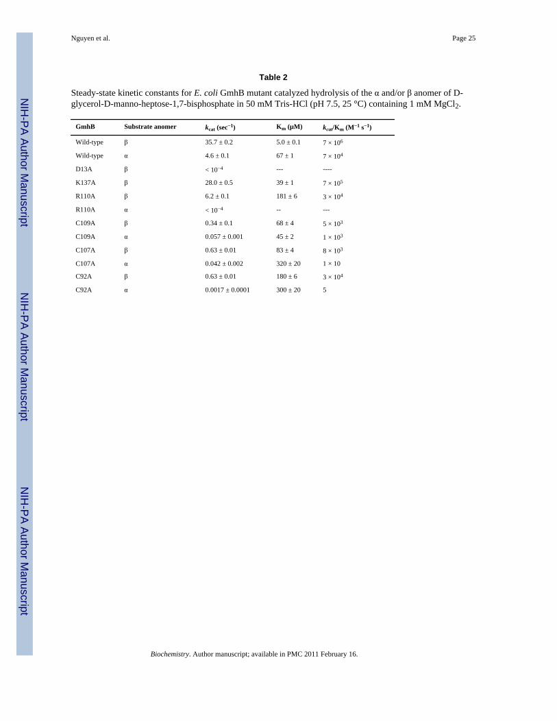
The Catalytic ScaffoldThe catalytic scaffolds observed in the structures of the E. coli and B. bronchiseptica GmhB/Mg2+/phosphate product complexes (Fig. 5) comprise the Asp nucleophile (Asp11 and Asp7,in E. coli and B. bronchiseptica respectively), Asp acid/base (Asp13 and Asp9), Thr and Lysphosphoryl-binding groups (Thr53 and Thr50; Lys111 and Lys101), and the Asp ligand to theMg2+ cofactor (Asp136 and Asp126). Together, this conserved constellation of residuescomprises the phosphatase catalytic scaffold for the stabilization of the trigonal bipyramidaltransition state (compare to Fig. 1). In addition, the residue which performs the role ofpositioning the Asp acid/base in all HAD phosphatases is present in the two GmhB orthologsas Asn17 (2.7 Å from Asp13) and Asn13 (2.9 Å from Asp9) (Fig. 5). The acid/base catalysthas been identified as critical for catalysis in HAD phosphatases(12). The essential role of thisresidue is also supported in GmhB as demonstrated by the finding that the E. coli GmhB D13Amutant is devoid of detectable activity toward the physiological substrate D-glycero-D-manno-heptose-1β,7-bisphosphate (Table 2).
The phosphate ligand in the E. coli GmhB/Mg2+/phosphate complex forms a coordination bondwith Mg2+ (2.1 Å) and engages in hydrogen-bond interaction with the side chains of Thr53(3.5 Å), Lys111 (2.5 Å) and Asp13 (3.4 Å) as well as the backbone amide NH of Asp13 (3.1Å). The phosphate ligand of the B. bronchiseptica GmhB/Mg2+/phosphate complex forms acoordination bond with the Mg2+ (2.1 Å) and engages in hydrogen bond interaction with theside chains of Thr50 (3.6 Å) and the backbone amide NH of Asp9 (2.8 Å). The side chains ofLys101 (4.3 Å) and Asp9 (4.0 Å) do not form the expected hydrogen bonds with the phosphateligand due to subtle differences in position of both the phosphate and side chains.
In phosphate complexes of HAD phosphatases(33–34), the six coordination sites of theMg2+ cofactor are typically filled by two water ligands, one oxygen atom of the phosphateligand, one oxygen atom from the Asp nucleophile carboxylate group, the backbone C=O ofthe Asp acid/base, and one oxygen atom from the carboxylate group of an Asp/Glu residuelocated on the Mg2+-binding loop. The structure of the Mg2+ center of the B. bronchisepticaGmhB/Mg2+/phosphate complex conforms to this model. Aside from the phosphate ligand,there are two water ligands (2.1 and 2.2 Å), the Asp7 carboxylate oxygen atom (2.2 Å), theAsp11 backbone carbonyl oxygen (2.1 Å) and the Asp126 carboxylate oxygen atom (2.0 Å)(Fig. 5). The Mg2+ center of the E. coli GmhB/Mg2+/phosphate complex on the other hand, isunusual in that one coordination position is occupied by the side chain nitrogen atom (2.4 Å)of a Lys residue (Lys137) adjacent to the Asp 136 (2.3 Å) ligand of the Mg2+-binding loop.The five coordination sites that remain are filled by a phosphate oxygen atom (2.0 Å), the Asp11 carboxylate oxygen atom (2.1 Å), the Asp 13 backbone carbonyl oxygen (2.3 Å), and awater molecule (2.5 Å). The Lys137 residue is not stringently conserved among GmhBorthologs. In B. bronchiseptica GmhB, the corresponding residue is Ser127. As shown in Fig.5, the Ser127 forms a hydrogen bond with the Mg2+ water ligand that assumes the samecoordination position as the Lys137 in the E. coli GmhB/Mg2+/phosphate complex. In addition,the Lys137 does not coordinate to the Mg2+ in the E. coli GmhB Michaelis complex (videinfra), nor does it make a significant contribution to catalysis (Table 2).
Nguyen et al. Page 6
Biochemistry. Author manuscript; available in PMC 2011 February 16.
NIH
-PA Author Manuscript
NIH
-PA Author Manuscript
NIH
-PA Author Manuscript

Binding Site for the Substrate Leaving Group: Collaboration between Three Loop ExtensionsThe GmhB catalytic site is elaborated by three extended peptide inserts, which pack to form aconcave, semicircular surface around the substrate leaving group (Fig. 6A). Together, with thebulky substrate-leaving group, the three inserts shield the catalytic site from solvent (asdemonstrated by using the program Voidoo(35)). All three inserts project from the C-terminalend of the Rossmann-like fold. The first is an elongated loop, which follows the signature DxDcatalytic motif containing the Asp nucleophile and Asp general acid/base residue. In the typeC1 HAD phosphatases an α-helical cap domain is inserted at this position. The second insertis a helix-turn motif that follows the conserved Thr/Ser located at the terminus of β-strand 2.The third insert is the Zn2+-binding loop, which precedes the conserved catalytic Lys. In theC2 HAD phosphatases an α/β-fold cap domain is inserted at this position. Because the capdomains inserted in these positions provide substrate-binding residues(15), it is notunreasonable to expect that the three loops in GmhB might do the same.
The Zn2-binding loop—The Zn2+-binding loops (Fig. 6B–D) of E. coli and B.bronchiseptica GmhB possesses a classical CxH(x)nCxC motif that coordinates the Zn2+ withsquare planar geometry. The structure of the E. coli GmhB/Mg2+/D-glycero-D-manno-heptose-1β,7-bisphosphate complex shows that the Zn2+ does not interact with the substrateand therefore, that it does not directly participate in catalysis. Our first impression was that theZn2+ functions to stabilize the loop conformation. Consistent with this model was the findingthat Ala replacement of any one of the four Zn2+ ligands of E. coli GmhB is detrimental. TheHis94Ala mutant proved to be insoluble, and the three Cys mutants which were soluble,displayed reduced catalytic efficiencies towards catalyzed hydrolysis of the α and β-anomersof D-glycero-l-D-manno-heptose-1,7-bisphosphate (Table 2).
Examination of the multiple alignment of GmhB sequences (Fig. SI1), however revealed theunexpected; the Zn2+-binding loop sequence is not conserved. Moreover, for a significantfraction of the GmhB orthologs (~10 %) two or more of the Zn2+ ligands of the CxH(x)nCxC consensus sequence are absent, which is likely to eliminate Zn2+ binding to the loop.In addition, the number of residues that separate the two pairs of Zn2+ ligands varies from 4for the “short loop” to 12–14 for the long loop (Fig. 6). The E. coli GmhB provides an exampleof the CxH(x)nCxC long loop (n = 12), the B. bronchiseptica GmhB and Bacteriodesthetaioatamicron GmhB provide examples of the CxH(x)nCxC short loop (n = 4), whereasthe Aneurinibacillus thermoaerophilus GmhB (CxP(x)12SxD) and the Mesorhizobium lotiGmhB (CxY(x)14HxM) provide examples of the long loop, both of which have lost the capacityfor Zn2+ binding. Each of these GmhB orthologs has been shown by in vitro kinetic analysisto be an effective catalysts of D-glycero-D-manno-heptose-1,7-bisphosphate hydrolysis. Thestructure of the M. loti GmhB (PDB ID 2O2X) provides a snapshot of the “empty” loop (Fig.6). The loop is ordered and in fact, the B-factors for loop residues do not differ (relative to theaverage B-factors of the core) from those of the E. coli and B. bronchiseptica GmhB loopresidues. Furthermore, the conformation of the M. loti GmhB loop is similar to that of the long,Zn2+-bound loop of the E. coli GmhB (Fig. 6). Examination of the structure of the M. lotiGmhB demonstrated a possible structural feature for stabilization of the loop in the absence ofthe Zn2+ binding motif, namely, the presence of an N-terminal extension (residues 15–21)which forms an additional β-strand antiparallel to that from which the insert extends. However,inspection of the alignment of GmhB orthologs (Fig. SI1) revealed that no correlation existsbetween the presence and absence of an extended N-terminus and the presence or absence ofa Zn2+-binding site. Thus, the structural significance of the presence of Zn2+ in some GhmB(or HisB, vide infra) orthologs, and its absence in others, is not known.
The finding that the GmhB loop exists in either a “no Zn2+ or a “Zn2+-bound” form promptedthe examination of aligned HisB sequences (Fig. SI2). Whereas the HisB orthologs in
Nguyen et al. Page 7
Biochemistry. Author manuscript; available in PMC 2011 February 16.
NIH
-PA Author Manuscript
NIH
-PA Author Manuscript
NIH
-PA Author Manuscript

Proteobacteria (the E. coli HisB is pictured in Fig. 2) contain the Zn2+-binding site, those inBacterioidetes do not. Thus, both loop types are present in both subfamilies.
Given that Zn2+-binding is not a determinant of catalysis, an alternate role that the Zn2+ mightperform was considered, leading to the experimental interrogation of DNA complexation bythe E. coli and B. bronchiseptica GmhB. The Zn2+-finger is a common DNA binding motifutilized by transcriptional factors. Although the GmhB Zn2+-binding loop bares limitedresemblance to the Zn2+ finger, it nevertheless seemed prudent to rule out DNA binding as apossible function. A standard DNA gel-shift assay (described in Materials and Methods) using“generic” calf-thymus DNA showed no indication of DNA binding. At this point in time theorigin and function (if any) of the Zn2+-binding motif remains undefined.
The Zn2+-binding loop, with or without Zn2+, contributes to the sequestration of the substrate.In addition, a stringently conserved Arg residue (Fig SI1) is positioned at the base of the loop,in between the CxH(x)nCxC motif and the catalytic Lys (viz. CxH(x)nCxCRK, Fig. 6). Thestructure of the E. coli GmhB/Mg2+/D-glycero-D-manno-heptose-1β,7-bisphosphate complexsuggests that the Arg contributes to substrate recognition by binding the C(1)phosphate (Fig.3 and 7). Replacement of the E. coli GmhB Arg110 with Ala resulted in a significant reductionin the catalytic efficiency of hydrolysis of the physiological substrate D-glycerol-D-manno-heptose-1β,7-bisphosphate, reducing kcat 6-fold and increasing Km 36-fold, such that kcat/Km was reduced by 200-fold (Table 2). On the other hand, the Arg110Ala mutant showed nodetectable activity towards D-glycerol-D-manno-heptose-1α,7-bisphosphate, which reveals adramatic reduction in catalytic efficiency compared to the activity of the wild-type enzyme(kcat/Km = 7 × 10⁴ M−1 s−1; Table 2). This finding suggests that the Arg plays a critical rolein preserving catalytic activity towards the α-anomer. This residue might be especiallyimportant for the GmhB orthologs that function in the D-glycero-D-manno-heptose-1α-GDPpathway, because their physiological substrate is the α-anomer. In principle, the orientation ofthe Arg can be directed by the backbone conformation, and it by the adjacent Zn2+-bindingsite. This raised the possibility that the Zn2+-binding site acts as anomeric specificity switch.However this model was eliminated by the finding that there is no correlation between anomericspecificity of the GmhB ortholog and whether or not it possesses a Zn2+-binding site (Fig. 6E).
Loops 1 and 2—Loops 1 (residues 18–30; Fig. 7) and 2 (residues 55–64; Fig. 7) areextensions from the core Rossmann-like fold that, as with the Zn2+ binding loop, are expectedto provide the residues to bind the substrate leaving group. The structure of the E. coli GmhB/Mg2+/D-glycero-D-manno-heptose-1β,7-bisphosphate complex shows that the loop 1 Asp19makes a hydrogen bond to the heptose C(3)OH (3.2 Å) and C(6) OH (3.6 Å). This residue isconserved in ~80% of all GmhB sequences and is conservatively replaced by Glu in all otherGmhB orthologs. Thus, the function of this residue in binding the substrate-leaving group isconserved. Tyr22 makes a hydrogen bond to a water molecule which forms a hydrogen bondto the substrate C1 phosphoryl group. Moreover, the Tyr22 ring sits over the substrate leavinggroup, shielding the substrate leaving group. Tyr22 is conserved in ~80% of all GmhBsequences and in the remaining sequences it is replaced by a Phe. This finding is consistentwith the interpretation that the major role of this Tyr/Phe and the adjoining Loop 1 residues isthe shielding of the substrate leaving group and core active site from bulk solvent. There areno additional conserved residues on loop 1.
In loop 2, Ser 56 is found in the motif TNQS wherein the Thr53 is part of the HAD catalyticscaffold (vide infra). Ser56 makes two hydrogen bonds with the substrate C(1) phosphate (3.3and 3.5 Å), while the adjacent Gln55 in the motif forms a hydrogen bond with the C1(O). Boththe Ser56 and Gln55 are stringently conserved.
Nguyen et al. Page 8
Biochemistry. Author manuscript; available in PMC 2011 February 16.
NIH
-PA Author Manuscript
NIH
-PA Author Manuscript
NIH
-PA Author Manuscript

The Mg2+-binding loop—Although technically a component of the catalytic scaffold, theE. coli GmhB Mg2+-binding loop (Fig. 6) does contribute Lys137 that forms a hydrogen bondwith to substrate C(1)phosphate (Fig. 7). However, the residue is not stringently conserved(Fig. 7; Fig. SI1), and furthermore, its replacement with Ala results in only a small reductionin catalytic efficiency (Table 2).
Substrate SpecificityE. coli GmhB is highly specific for D-glycero-D-manno-heptose-1,7-bisphosphate, it shows a100 : 1 preference for the β-anomer over the α-anomer, it removes the C(7)phosphate and notthe C(1)phosphate, and it does not hydrolyze the reaction product, D-glycero-D-manno-heptose-1-phosphate(25). In order to gain insight into the structural basis for this specificity,the structure of the E. coli GmhB/Mg2+/D-glycero-D-manno-heptose-1β,7-bisphosphatecomplex was used to generate a model in which the D-glycero-D-manno-heptose-1β,7-bisphosphate ligand is docked into the active site, in the reverse orientation, such that the C(1)phosphate rather than the C(7)phosphate is positioned for Mg2+ coordination and in-line attackby the Asp nucleophile (Fig. 8). In general, models are not sufficiently accurate to predict thesmall changes in orientations in interacting groups that can result in large changes in transitionstate structure and energy. However, in the present case, the comparison of the experimentallyobserved substrate complex (Fig. 8) vs. the substrate docked in the reverse orientation (Fig. 8)clearly identifies a major effect that, independent of the more subtle orientation effects, is likelyto preclude hydrolysis of the C(1) phosphate. Specifically, in the reverse orientation, the leavinggroup does not have the correct shape to seal the active site entrance, and thus the electrostaticstabilization of the transition state is likely to be impaired by water molecules that can solvatethe catalytic site. In contrast, the model of the D-glycero-D-manno-heptose-1α,7-bisphosphatedocked in the catalytic orientation (Fig. 8) predicts that the solvent seal is in place. In addition,an alteration in ring pucker brings the C(1)phosphate of the α-anomer into position to form asalt bridge with Arg110. The 100 : 1 anomeric preference observed for the E. coli GmhB islikely to be determined by subtle orientation effects, which the model cannot predict.
Divergence of the HisB Histidinol-Phosphate PhosphataseWe have shown(25) that histidinol-phosphate is not a substrate for E. coli GmhB. Althoughthe GmhB substrate, D-glycero-D-manno-heptose 1,7-bisphosphate, was not tested forsubstrate activity with E. coli HisB, the reported absence of activity towards other phosphateester metabolites indicates that the histidinol-phosphate phosphatase is not promiscuous(24).Thus, whereas the two sequences share 34% identity, they do not share substrates. In order toidentify the structural elements for the substrate specialization that define the respectivebiochemical functions of GmhB and histidinolphosphate phosphatase, the structures of thesetwo enzymes were compared, within the context of the residue conservation defined by multiplesequence alignments. Accordingly, the E. coli histidinol-phosphate phosphatase domain of theHisB fusion protein was used as query in a Blast search of the nonredundant protein sequencedatabase. The sequences were aligned and curated to remove sequences that did not encodeHisB (Fig. SI2). An analogous procedure was carried out to create the multiple sequencealignment of the GhmB homologs identified by Blast searches with the E. coli and M. lotiGmhB sequences serving as query (Fig. SI1). No evidence for a “stand alone” HAD familyhistidinol-phosphate phosphatase was found. Likewise, there was no indication that a homologpossessing a novel catalytic activity is found among the GmhB sequences.
The respective sequence families provide insight into the biological range of HisB and GmhB,which agrees with and expands upon an earlier report(36). The HisB is primarily found inbacterial species of the gamma subdivision of Proteobacter, yet it is also found in numerousspecies of the Phylum Bacteroidetes/Chlorobi. HisB is believed to have evolved within thegamma subdivision of Proteobacter following its split from the beta division, and is thus a
Nguyen et al. Page 9
Biochemistry. Author manuscript; available in PMC 2011 February 16.
NIH
-PA Author Manuscript
NIH
-PA Author Manuscript
NIH
-PA Author Manuscript

relatively new enzyme(36). A likely scenario is that the GmhB ancestor gene underwentduplication within a gamma subdivision bacterium, and the extra copy evolved to the HisBhistidinol-phosphate phosphatase gene. The HisB gene was most probably acquired byBacteroides via horizontal gene transfer. GmhB has a much greater biological range, whichincludes a variety of species from each Phylum of Bacteria as well as some species of Archaea.The HisB histidinol-phosphate phosphatase might thus be viewed as an adaptation of the GmhBto a novel biochemical niche (viz. the histidine biosynthetic pathway) within in a smallbiological sector.
The GmhB sequences have diverged to ~30% similarity and the HisB histidinol-phosphatephosphatase sequences have diverged to ~40% similarity. The two alignments are thereforereliable for use in distinguishing stringently conserved residues. First, it is noteworthy that theArg110 residue in the E. coli GmhB Zn2+-binding loop, which plays an important role insubstrate recognition (vide supra), is stringently conserved among the histidinol-phosphatephosphatase domains of HisB. In E. coli HisB, the Arg105 residue does not bind the substrate,rather it forms a hydrogen bond with an Asp residue of loop 2 (Fig. 7). The E. coli HisB Asp58residue is stringently conserved in the position that corresponds to the E. coli GmhB Ser56(Fig. 7) that, like the Arg residue, binds the substrate C(1)phosphate. The counterpart to thestringently conserved sequence motif of GmhB “TNQS” is the HisB TNQD motif (where T isthe catalytic Thr53 in E. coli GmhB that is conservatively substituted with Ser in some GmhBorthologs (Fig 5) and Q is the Gln55 in E. coli that forms a long hydrogen bond (3.9 Å) to thesubstrate C(1)O (not shown). The switch from Ser to Asp allows a clear distinction to be madebetween GmhB and histidinol-phosphate phosphatase sequences. In the E. coli HisB structurethe Asp58 of the TNQD motif forms a long hydrogen bond to the N1H of the histidinol-phosphate ligand and an ion pair with the Zn2+ binding loop Arg105 (Fig. 7).
In the E. coli HisB structure, the stringently conserved residues Gln24 and Glu18 (of loop 1)engage the N3 and C(2)NH3
+ of the histidinol ligand in hydrogen bond formation. Thehomologous residues in E. coli GmhB, Tyr22 and Asp13 function in the respective roles ofsubstrate leaving group desolvation and substrate binding (Fig. 7). The HisB Glu18 forms anion pair with the catalytic scaffold Arg132, yet neither it, nor its E. coli GmhB counterpart,Lys137 (vide supra) are fully conserved.
Summary and ConclusionHistorically, two of the first capless members of the HAD family to be examined structurallywere T4 polynucleotide kinase(21) and magnesium dependent phosphatase-1(19). Althoughthese proteins have active sites that are completely solvent exposed, their large macromolecularsubstrates (t-RNA and protein, respectively) are expected to act like a cap by binding andprotecting the phosphoryl transfer site from bulk solvent. Later, the capless HAD phosphatases,2-keto-3-deoxy-octulosonate-8-phosphate (KDO-8-P) phosphatase(37–38) and 2-keto-3-deoxy-D-glycero-D-galactonononate-9-phosphate (KDN-9-P) phosphatase(23) werediscovered to efficiently and specifically hydrolyze their respective (small molecule) substratesby recruiting a second subunit to act as a “cap” to the active site of the first subunit. Thetetramerization of these two enzymes is mediated by a short peptide insert to the Rossmann-like fold at the position corresponding to cap domain of the C1 HADS (Fig. 1).
GmhB provides a unique solution to the problem of substrate discrimination in the caplessHAD phosphatases. The GmhB structures show that the catalytic site is elaborated by threeloops that form a concave, semicircular surface around the substrate-leaving group. One of thethree loops binds Zn2+ in some but not all GmhB orthologs. Each of the three loops positionstringently conserved residues that engage in hydrogen bond interaction with the substrate-leaving group, D-glycero-D-manno-heptose 1-phosphate. GmhB and the histidinol-phosphate
Nguyen et al. Page 10
Biochemistry. Author manuscript; available in PMC 2011 February 16.
NIH
-PA Author Manuscript
NIH
-PA Author Manuscript
NIH
-PA Author Manuscript
phosphatase domain of HisB share the same design, yet through selective residue usage on thethree substrate-recognition loops, they have achieved unique substrate specificity, and thusnovel biochemical function.
Supplementary MaterialRefer to Web version on PubMed Central for supplementary material.
AcknowledgmentsWe thank Dr. Mirek Cygler and Dr. Steven Almo for providing the coordinates of HisB and Q8A5V9, respectively,prior to publication.
References1. Heine A, DeSantis G, Luz JG, Mitchell M, Wong CH, Wilson IA. Observation of covalent intermediates
in an enzyme mechanism at atomic resolution. Science 2001;294:369–374. [PubMed: 11598300]2. Dalby A, Dauter Z, Littlechild JA. Crystal structure of human muscle aldolase complexed with fructose
1,6-bisphosphate: mechanistic implications. Protein Sci 1999;8:291–297. [PubMed: 10048322]3. Sygusch J, Beaudry D. Catalytic activity of rabbit skeletal muscle aldolase in the crystalline state. J
Biol Chem 1984;259:10222–10227. [PubMed: 6469960]4. Schlichting I, Chu K. Trapping intermediates in the crystal: ligand binding to myoglobin. Curr Opin
Struct Biol 2000;10:744–752. [PubMed: 11114513]5. Schramm VL. Enzymatic transition states: thermodynamics, dynamics and analogue design. Arch
Biochem Biophys 2005;433:13–26. [PubMed: 15581562]6. Petrek M, Otyepka M, Banas P, Kosinova P, Koca J, Damborsky J. CAVER: a new tool to explore
routes from protein clefts, pockets and cavities. BMC Bioinformatics 2006;7:316. [PubMed:16792811]
7. Collet JF, van Schaftingen E, Stroobant V. A new family of phosphotransferases related to P-typeATPases. Trends Biochem Sci 1998;23:284. [PubMed: 9757826]
8. Koonin EV, Tatusov RL. Computer Analysis of Bacterial Haloacid Dehalogenases Defines a LargeSuperfamily of Hydrolases with Diverse Specificity: Application of an Iterative Approach to DatabaseSearch. Journal of Molecular Biology 1994;244:125–132. [PubMed: 7966317]
9. Allen KN, Dunaway-Mariano D. Phosphoryl group transfer: evolution of a catalytic scaffold. TrendsBiochem Sci 2004;29:495–503. [PubMed: 15337123]
10. Smith CA, Rayment I. X-ray structure of the magnesium(II).ADP.vanadate complex of theDictyostelium discoideum myosin motor domain to 1.9 A resolution. Biochemistry 1996;35:5404–5417. [PubMed: 8611530]
11. Wang W, Cho HS, Kim R, Jancarik J, Yokota H, Nguyen HH, Grigoriev IV, Wemmer DE, Kim SH.Structural characterization of the reaction pathway in phosphoserine phosphatase: crystallographic"snapshots" of intermediate states. J Mol Biol 2002;319:421–431. [PubMed: 12051918]
12. Lu Z, Dunaway-Mariano D, Allen KN. The catalytic scaffold of the haloalkanoic acid dehalogenaseenzyme superfamily acts as a mold for the trigonal bipyramidal transition state. Proc Natl Acad SciU S A 2008;105:5687–5692. [PubMed: 18398008]
13. Allen KN, Dunaway-Mariano D. Markers of fitness in a successful enzyme superfamily. Curr OpinStruct Biol in press. 2009
14. Burroughs AM, Allen KN, Dunaway-Mariano D, Aravind L. Evolutionary genomics of the HADsuperfamily: understanding the structural adaptations and catalytic diversity in a superfamily ofphosphoesterases and allied enzymes. J Mol Biol 2006;361:1003–1034. [PubMed: 16889794]
15. Lahiri SD, Zhang G, Dai J, Dunaway-Mariano D, Allen KN. Analysis of the substrate specificity loopof the HAD superfamily cap domain. Biochemistry 2004;43:2812–2820. [PubMed: 15005616]
16. Zhang G, Dai J, Wang L, Dunaway-Mariano D, Tremblay LW, Allen KN. Catalytic cycling in beta-phosphoglucomutase: a kinetic and structural analysis. Biochemistry 2005;44:9404–9416. [PubMed:15996095]
Nguyen et al. Page 11
Biochemistry. Author manuscript; available in PMC 2011 February 16.
NIH
-PA Author Manuscript
NIH
-PA Author Manuscript
NIH
-PA Author Manuscript

17. Kim HY, Heo YS, Kim JH, Park MH, Moon J, Kim E, Kwon D, Yoon J, Shin D, Jeong EJ, Park SY,Lee TG, Jeon YH, Ro S, Cho JM, Hwang KY. Molecular basis for the local conformationalrearrangement of human phosphoserine phosphatase. J Biol Chem 2002;277:46651–46658.[PubMed: 12213811]
18. Dai J, Finci L, Zhang C, Lahiri S, Zhang G, Peisach E, Allen KN, Dunaway-Mariano D. Analysis ofthe structural determinants underlying discrimination between substrate and solvent in beta-phosphoglucomutase catalysis. Biochemistry 2009;48:1984–1995. [PubMed: 19154134]
19. Peisach E, Selengut J, Dunaway-Mariano D, Allen KN. Structure of the Magnesium-DependentProtein Tyrosine Phosphatase, MDP-1. Biochemistry 2004;43:12770–12779. [PubMed: 15461449]
20. Peisach E, Wang L, Burroughs AM, Aravind L, Dunaway-Mariano D, Allen KN. The X-raycrystallographic structure and activity analysis of a Pseudomonas-specific subfamily of the HADenzyme superfamily evidences a novel biochemical function. Proteins 2008;70:197–207. [PubMed:17654544]
21. Galburt EA, Pelletier J, Wilson G, Stoddard BL. Structure of a tRNA repair enzyme and molecularbiology workhorse: T4 polynucleotide kinase. Structure (Camb) 2002;10:1249–1260. [PubMed:12220496]
22. Wu J, Woodard RW. Escherichia coli YrbI is 3-deoxy-D-mannooctulosonate 8-phosphatephosphatase. J Biol Chem 2003;278:18117–18123. [PubMed: 12639950]
23. Lu Z, Wang L, Dunaway-Mariano D, Allen KN. Structure-function analysis of 2-keto-3-deoxy-D-glycero-D-galactonononate-9-phosphate phosphatase defines specificity elements in type C0haloalkanoate dehalogenase family members. J Biol Chem 2009;284:1224–1233. [PubMed:18986982]
24. Rangarajan ES, Proteau A, Wagner J, Hung MN, Matte A, Cygler M. Structural snapshots ofEscherichia coli histidinol phosphate phosphatase along the reaction pathway. J Biol Chem2006;281:37930–37941. [PubMed: 16966333]
25. Wang L, Huang H, Nguyen H, Allen KN, Mariano PS, Dunaway-Mariano D. Divergence ofBiochemical Function in the HAD Superfamily: D-Glycero-D-manno-heptose 1,7-bisphosphatePhosphatase, GmhB. Biochemistry companion manuscript. 2010
26. Otwinowski Z, Minor W. Processing of X-ray Diffraction Data Collected in Oscillation Mode.Methods Enzymol 1997;276:307–326.
27. Vagin A, Teplyakov A. An approach to multi-copy search in molecular replacement. Acta CrystallogrD Biol Crystallogr 56 Pt 12 2000:1622–1624.
28. Collaborative Computational Project Number 4. Collaborative Computational Project Number 4 ActaCryst 1994;D50:760–768.
29. Emsley P, Cowtan K. Coot: model-building tools for molecular graphics. Acta Crystallogr D BiolCrystallogr 60 2004:2126–2132.
30. Brünger AT, Adams PD, Clore GM, DeLano WL, Gros P, Grosse-Kunstleve RW, Jiang JS, KuszewskiJ, Nilges M, Pannu NS, Read RJ, Rice LM, Simonson T, Warren GL. Crystallography & NMRsystem: A new software suite for macromolecular structure determination. Acta Crystallogr D BiolCrystallogr 1998;54(Pt 5):905–921. [PubMed: 9757107]
31. Davis IW, Murray LW, Richardson JS, Richardson DC. MOLPROBITY: structure validation andall-atom contact analysis for nucleic acids and their complexes. Nucleic Acids Res 2004;32:W615–W619. [PubMed: 15215462]
32. Kerr LD. Electrophoretic mobility shift assay. Methods Enzymol 1995;254:619–632. [PubMed:8531719]
33. Wang W, Kim R, Jancarik J, Yokota H, Kim SH. Crystal structure of phosphoserine phosphatasefrom Methanococcus jannaschii, a hyperthermophile, at 1.8 Å resolution. Structure (Camb)2001;9:65–71. [PubMed: 11342136]
34. Rinaldo-Matthis A, Rampazzo C, Reichard P, Bianchi V, Nordlund P. Crystal structure of a humanmitochondrial deoxyribonucleotidase. Nat. Struct. Biol 2002;10:779–787. [PubMed: 12352955]
35. Kleywegt GJ, Jones TA. Detection, delineation, measurement and display of cavities inmacromolecular structures. Acta Crystallogr D Biol Crystallogr 1994;50:178–185. [PubMed:15299456]
Nguyen et al. Page 12
Biochemistry. Author manuscript; available in PMC 2011 February 16.
NIH
-PA Author Manuscript
NIH
-PA Author Manuscript
NIH
-PA Author Manuscript

36. Brilli M, Fani R. Molecular evolution of hisB genes. J Mol Evol 2004;58:225–237. [PubMed:15042344]
37. Parsons JF, Lim K, Tempczyk A, Krajewski W, Eisenstein E, Herzberg O. From structure to function:YrbI from Haemophilus influenzae (HI1679) is a phosphatase. Proteins 2002;46:393–404. [PubMed:11835514]
38. Biswas T, Yi L, Aggarwal P, Wu J, Rubin JR, Stuckey JA, Woodard RW, Tsodikov OV. The tail ofKDSC: Conformational changes control the activity of a haloacid dehalogenase superfamilyphosphatase. J Biol Chem. 2009
39. Lu Z, Dunaway-Mariano D, Allen KN. HAD superfamily phosphotransferase substratediversification: structure and function analysis of HAD subclass IIB sugar phosphatase BT4131.Biochemistry 2005;44:8684–8696. [PubMed: 15952775]
Nguyen et al. Page 13
Biochemistry. Author manuscript; available in PMC 2011 February 16.
NIH
-PA Author Manuscript
NIH
-PA Author Manuscript
NIH
-PA Author Manuscript

Figure 1.A) Diagram of the HAD catalytic template depicting the binding interaction between substrate(black), Mg2+ cofactor (cyan), and the conserved catalytic residues of the HAD phosphataseactive site. B) The structure of 2-keto-3-deoxy-D-glycero-Dgalactonononate-9-phosphatephosphatase monomer bound with Mg2+ (cyan), vanadate and 2-keto-3-deoxy-5-acetylamine-D-glycero-D-galactonononate-9-phosphate (black) (23). The conserved catalytic residues arecolored as in A. C) The tetramer of 2-keto-3-deoxy-D-glycero-D-galactonononate-9-phosphate phosphatase tetramer depicted as ribbons. The subunits are individually colored andligands colored as in B.
Nguyen et al. Page 14
Biochemistry. Author manuscript; available in PMC 2011 February 16.
NIH
-PA Author Manuscript
NIH
-PA Author Manuscript
NIH
-PA Author Manuscript

Figure 2.A) Ribbon diagram of C1 HAD phosphatase Q8A5V9 (Steven Almo, Albert Einstein Collegeof Medicine, unpublished data). The Rossmann fold catalytic domain is colored gray,conserved catalytic residues are shown as sticks and colored as in Fig 1A. The Mg2+ is a cyansphere and the cap domain is colored magenta. B) The structure of the C2 HAD phosphataseBT4131(39). C) The structure of the histidinol-phosphate phosphatase domain of E. coli HisB(24). The histidinol ligand (shown as sticks) is colored cyan. The Zn2+ and Mg2+ are depictedas black and cyan spheres. The Zn2+ binding loop (blue), loop 1 (magenta), loop 2 (green) andthe Mg2+-binding loop (yellow) are colored to coordinate with the structures shown in Figure4 and Figure 7.
Nguyen et al. Page 15
Biochemistry. Author manuscript; available in PMC 2011 February 16.
NIH
-PA Author Manuscript
NIH
-PA Author Manuscript
NIH
-PA Author Manuscript
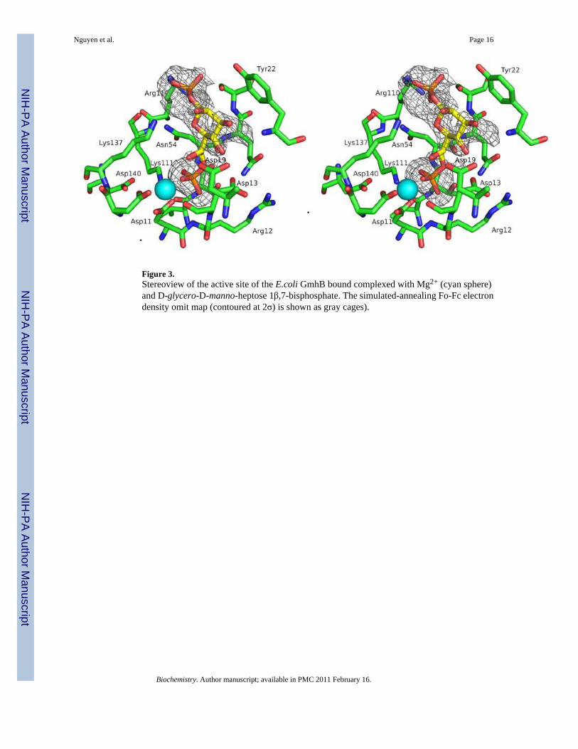
Figure 3.Stereoview of the active site of the E.coli GmhB bound complexed with Mg2+ (cyan sphere)and D-glycero-D-manno-heptose 1β,7-bisphosphate. The simulated-annealing Fo-Fc electrondensity omit map (contoured at 2σ) is shown as gray cages).
Nguyen et al. Page 16
Biochemistry. Author manuscript; available in PMC 2011 February 16.
NIH
-PA Author Manuscript
NIH
-PA Author Manuscript
NIH
-PA Author Manuscript

Figure 4.A) Ribbon diagram of E. coli GmhB bound to Zn2+ and Mg2+ (black and cyan spheres) andthe substrate and D-glycero-D-manno-heptose 1β,7-bisphosphate. The Zn2+-binding loop iscolored blue, loop 2 green, loop 1 magenta and the Mg2+-binding loop yellow. B) Ribbondiagram of E. coli GmhB bound to Zn2+ and Mg2+ and the product orthophosphate. The loopsare colored the same as Figure 2.
Nguyen et al. Page 17
Biochemistry. Author manuscript; available in PMC 2011 February 16.
NIH
-PA Author Manuscript
NIH
-PA Author Manuscript
NIH
-PA Author Manuscript

Figure 5.The active sites from the structures of E. coli GmhB/Mg2+/phosphate (A) and B.bronchiseptica GmhB/Mg2+/phosphate complexes (B). Metal coordination (black), hydrogenbonds (within 3.5 Å; green), and long hydrogen bonds (3.6–3.9 Å; olive) are represented bydashed lines. The Mg2+ is a cyan sphere and water ligand(s) are red spheres. The paired yellowand salmon residues occupy homologous positions in the two active sites.
Nguyen et al. Page 18
Biochemistry. Author manuscript; available in PMC 2011 February 16.
NIH
-PA Author Manuscript
NIH
-PA Author Manuscript
NIH
-PA Author Manuscript

Figure 6.A. The structure of the E. coli GmhB/Mg2+/D-glycero-D-manno-heptose 1β,7-bisphosphatecomplex. The Rossmann catalytic unit is depicted as ribbon (grey) with space-filling modelsfor the Zn2+-binding loop (blue), loop 2 (green) and loop 1 (magenta). B, C, and D TheZn2+ (black sphere) bound loops of E. coli, B. bronchiseptica and the homologous, unligandedloop of M. loti GmhB. The Zn2+ ligands are shown as stick. The stringently conserved Arg isalso depicted. E) Representative GmhB Zn2+-binding loops. Listed are the species (E. coli(Ec), M. loti (Ml), A. thermoaerophilus (At), B. bronchiseptica (Bb), B. thetaioatamicron (Bt)),preferred anomer (α or β) of D-glycero-D-manno-heptose 1,7-bisphosphate and the presenceor absence of a Zn2+-binding motif. The first pair and second pairs of Zn2+ ligands are colored
Nguyen et al. Page 19
Biochemistry. Author manuscript; available in PMC 2011 February 16.
NIH
-PA Author Manuscript
NIH
-PA Author Manuscript
NIH
-PA Author Manuscript

red and magenta, respectively, and the stringently conserved C(1) phosphate binding Arg andcatalytic Lys residues are colored blue.
Nguyen et al. Page 20
Biochemistry. Author manuscript; available in PMC 2011 February 16.
NIH
-PA Author Manuscript
NIH
-PA Author Manuscript
NIH
-PA Author Manuscript
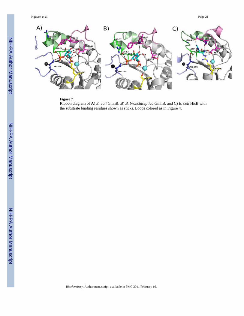
Figure 7.Ribbon diagram of A) E. coli GmhB, B) B. bronchiseptica GmhB, and C) E. coli HisB withthe substrate binding residues shown as sticks. Loops colored as in Figure 4.
Nguyen et al. Page 21
Biochemistry. Author manuscript; available in PMC 2011 February 16.
NIH
-PA Author Manuscript
NIH
-PA Author Manuscript
NIH
-PA Author Manuscript
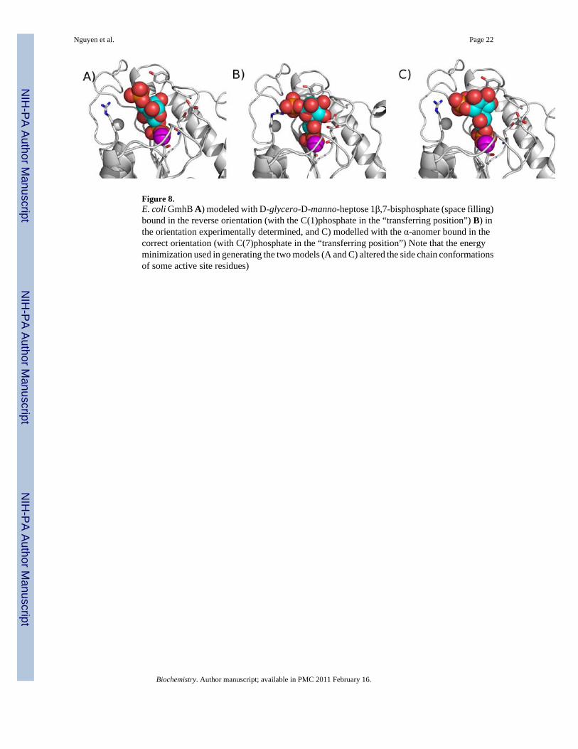
Figure 8.E. coli GmhB A) modeled with D-glycero-D-manno-heptose 1β,7-bisphosphate (space filling)bound in the reverse orientation (with the C(1)phosphate in the “transferring position”) B) inthe orientation experimentally determined, and C) modelled with the α-anomer bound in thecorrect orientation (with C(7)phosphate in the “transferring position”) Note that the energyminimization used in generating the two models (A and C) altered the side chain conformationsof some active site residues)
Nguyen et al. Page 22
Biochemistry. Author manuscript; available in PMC 2011 February 16.
NIH
-PA Author Manuscript
NIH
-PA Author Manuscript
NIH
-PA Author Manuscript
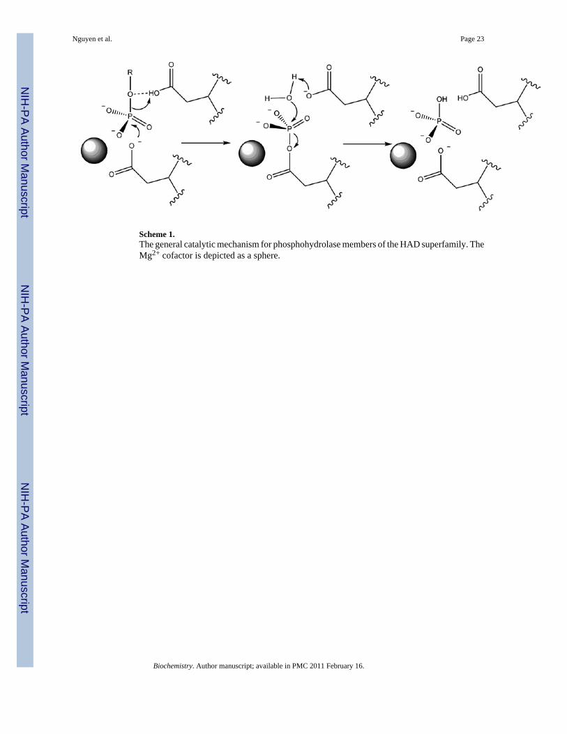
Scheme 1.The general catalytic mechanism for phosphohydrolase members of the HAD superfamily. TheMg2+ cofactor is depicted as a sphere.
Nguyen et al. Page 23
Biochemistry. Author manuscript; available in PMC 2011 February 16.
NIH
-PA Author Manuscript
NIH
-PA Author Manuscript
NIH
-PA Author Manuscript

NIH
-PA Author Manuscript
NIH
-PA Author Manuscript
NIH
-PA Author Manuscript
Nguyen et al. Page 24
Table 1
Data collection statistics. Values in parentheses are for the highest resolution shell.
Crystal Ec GmhBapo
Ec GmhBMg2+/Pi
Ec GmhB/Mg2+/H1β,7bisP
Bb GmhBMg2+/Pi
Space group P212121 P21212 P21212 P1
Unit cell dimensions (Å)a = 51.77b = 63.97c = 103.20
a = 64.48b = 50.00c = 51.88
a = 63.67b = 51.57c = 52.19
a = 38.1b = 58.4c = 85.7
X-ray source X12B, NSLS X25C, NSLS R AxisIV++ X29, NSLS
Wavelength (Å) 0.9786 1.1000 1.5418 1.0000
Resolution (Å) 1.64 (1.64–1.70) 1.8 (1.79–1.85) 2.2 (2.18–2.26) 1.7 (1.68–1.74)
Total / unique reflections 207847/42543 109143/16358 32412/9183 79124/6443
Completeness (%) 99.4 (98.4) 99.9 (99.6) 97.2 (99.0) 94.6 (77.3)
I/σ (I) 16.7 (2.5) 33.5 (3.7) 10.1 (1.9) 18.4 (3.2)
Rmerge 0.072 (0.491) 0.048 (0.501) 0.073 (0.427) 0.059 (0.253)
No. mol/ asymmetric unit 2 1 1 4
No. ordered/total residues 372/382 182/191 182/191 716/716
No. of non-solvent atoms perasymmetric unit
2888 1433 1457 5373
No. of reflections (work/free) 76862/7540 26258/2844 8332/420 147262/14317
Rwork/Rfree 0.17 / 0.21 0.19 / 0.23 0.18/ 0.25 0.17/0.20
Total waters 378 110 79 666
No. Zn2+/Mg2+ 2/0 1/1 1/1 4/4
No./ligand 2/acetates none 1/H1β,7bisP 5/Pi1/fructose
Average B-factor (Å2)
Amino acid residues 15.8 31.3 31.1 19.4
Mg2+ ions - 35.2 21.5 14.4
Zn2+ ions 12.6 34.6 33.8 19.9
water 26.0 41.2 33.6 26.3
ligands 12.7 39.2 42.9 20.0
rms deviation
Bond lengths (Å) 0.006 0.028 0.010 0.008
Bond angles (deg) 1.069 2.258 1.268 1.122
Ramachandran favored/allowed (%) 98.1/100 97.2/100 96.7/100 97.6/100
Biochemistry. Author manuscript; available in PMC 2011 February 16.
NIH
-PA Author Manuscript
NIH
-PA Author Manuscript
NIH
-PA Author Manuscript
Nguyen et al. Page 25
Table 2
Steady-state kinetic constants for E. coli GmhB mutant catalyzed hydrolysis of the α and/or β anomer of D-glycerol-D-manno-heptose-1,7-bisphosphate in 50 mM Tris-HCl (pH 7.5, 25 °C) containing 1 mM MgCl2.
GmhB Substrate anomer kcat (sec−1) Km (µM) kcat/Km (M−1 s−1)
Wild-type β 35.7 ± 0.2 5.0 ± 0.1 7 × 10⁶
Wild-type α 4.6 ± 0.1 67 ± 1 7 × 10⁴
D13A β < 10−4 --- ----
K137A β 28.0 ± 0.5 39 ± 1 7 × 10⁵
R110A β 6.2 ± 0.1 181 ± 6 3 × 10⁴
R110A α < 10−4 -- ---
C109A β 0.34 ± 0.1 68 ± 4 5 × 10³
C109A α 0.057 ± 0.001 45 ± 2 1 × 10³
C107A β 0.63 ± 0.01 83 ± 4 8 × 10³
C107A α 0.042 ± 0.002 320 ± 20 1 × 10
C92A β 0.63 ± 0.01 180 ± 6 3 × 10⁴
C92A α 0.0017 ± 0.0001 300 ± 20 5
Biochemistry. Author manuscript; available in PMC 2011 February 16.
Related Documents