Structural Contributions to the Intracellular Targeting Strategies of Antimicrobial Peptides Yun Lan, Yan Ye, Justyna Kozlowska, Jenny K.W. Lam, Alex F. Drake, and A. James Mason King’s College London, Pharmaceutical Science Division, 150 Stamford Street, London, SE1 9NH, UK Abstract The interactions of cationic amphipathic antimicrobial peptides (AMPs) with anionic biological membranes have been the focus of much research aimed at improving the activity of such compounds in the search for therapeutic leads. However, many of these peptides are thought to have other polyanions, such as DNA or RNA, as their ultimate target. Here a combination of fluorescence and circular dichroism (CD) spectroscopies has been used to assess the structural properties of amidated versions of buforin II, pleurocidin and magainin 2 that support their varying abilities to translocate through bacterial membranes and bind to double stranded DNA. Unlike magainin 2 amide, a prototypical membrane disruptive AMP, buforin II amide adopts a poorly helical structure in membranes closely mimicking the composition of Gram negative bacteria, such as Escherichia coli, and binds to a short duplex DNA sequence with high affinity, ultimately forming peptide-DNA condensates. The binding affinities of the peptides to duplex DNA are shown to be related to the structural changes that they induce. Furthermore, CD also reveals the conformation of the bound peptide buforin II amide. In contrast with a synthetic peptide, designed to adopt a perfect amphipathic α-helix, buforin II amide adopts an extended or polyproline II conformation when bound to DNA. These results show that an α-helix structure is not required for the DNA binding and condensation activity of buforin II amide. Keywords peptide antibiotics; CD spectroscopy; buforin II; magainin 2; pleurocidin; DNA binding 1. Introduction The wide spectrum and rapid bactericidal activity of antimicrobial peptides has made them the focus of much research, aiming to add to the current arsenal of antibiotics whose effectiveness is being severely eroded by the increased prevalence of bacteria resistant to one or more classical antibiotics [1-4]. Considerable focus has been placed on the membrane disruptive properties of cationic amphipathic peptides, although it is recognized that many peptides may have alternative bactericidal mechanisms requiring them to penetrate bacteria and interfere with metabolic processes [5, 6]. One such peptide, buforin II derived from the Asian toad Bufo gargarizans, has been shown to penetrate bacteria, inhibiting cellular functions [7]. Considering the demonstrated therapeutic efficacy of buforin II in a rat model of Acinetobacter baumannii sepsis [8], understanding the bactericidal strategy of buforin II and the underpinning structural contributions will offer a new avenue in the pursuit of therapeutically relevant peptide based antibiotics. In comparison with magainin 2, a cationic Address correspondence to: A. James Mason, King’s College London, Pharmaceutical Science Division, 150 Stamford Street, London, SE1 9NH, UK; Tel: + 44 207 848 4813; Fax: + 44 207 848 4800; [email protected]. Europe PMC Funders Group Author Manuscript Biochim Biophys Acta. Author manuscript; available in PMC 2012 March 21. Published in final edited form as: Biochim Biophys Acta. 2010 October ; 1798(10): 1934–1943. doi:10.1016/j.bbamem.2010.07.003. Europe PMC Funders Author Manuscripts Europe PMC Funders Author Manuscripts

Welcome message from author
This document is posted to help you gain knowledge. Please leave a comment to let me know what you think about it! Share it to your friends and learn new things together.
Transcript
Structural Contributions to the Intracellular Targeting Strategiesof Antimicrobial Peptides
Yun Lan, Yan Ye, Justyna Kozlowska, Jenny K.W. Lam, Alex F. Drake, and A. James MasonKing’s College London, Pharmaceutical Science Division, 150 Stamford Street, London, SE19NH, UK
AbstractThe interactions of cationic amphipathic antimicrobial peptides (AMPs) with anionic biologicalmembranes have been the focus of much research aimed at improving the activity of suchcompounds in the search for therapeutic leads. However, many of these peptides are thought tohave other polyanions, such as DNA or RNA, as their ultimate target. Here a combination offluorescence and circular dichroism (CD) spectroscopies has been used to assess the structuralproperties of amidated versions of buforin II, pleurocidin and magainin 2 that support theirvarying abilities to translocate through bacterial membranes and bind to double stranded DNA.Unlike magainin 2 amide, a prototypical membrane disruptive AMP, buforin II amide adopts apoorly helical structure in membranes closely mimicking the composition of Gram negativebacteria, such as Escherichia coli, and binds to a short duplex DNA sequence with high affinity,ultimately forming peptide-DNA condensates. The binding affinities of the peptides to duplexDNA are shown to be related to the structural changes that they induce. Furthermore, CD alsoreveals the conformation of the bound peptide buforin II amide. In contrast with a syntheticpeptide, designed to adopt a perfect amphipathic α-helix, buforin II amide adopts an extended orpolyproline II conformation when bound to DNA. These results show that an α-helix structure isnot required for the DNA binding and condensation activity of buforin II amide.
Keywordspeptide antibiotics; CD spectroscopy; buforin II; magainin 2; pleurocidin; DNA binding
1. IntroductionThe wide spectrum and rapid bactericidal activity of antimicrobial peptides has made themthe focus of much research, aiming to add to the current arsenal of antibiotics whoseeffectiveness is being severely eroded by the increased prevalence of bacteria resistant toone or more classical antibiotics [1-4]. Considerable focus has been placed on the membranedisruptive properties of cationic amphipathic peptides, although it is recognized that manypeptides may have alternative bactericidal mechanisms requiring them to penetrate bacteriaand interfere with metabolic processes [5, 6]. One such peptide, buforin II derived from theAsian toad Bufo gargarizans, has been shown to penetrate bacteria, inhibiting cellularfunctions [7]. Considering the demonstrated therapeutic efficacy of buforin II in a rat modelof Acinetobacter baumannii sepsis [8], understanding the bactericidal strategy of buforin IIand the underpinning structural contributions will offer a new avenue in the pursuit oftherapeutically relevant peptide based antibiotics. In comparison with magainin 2, a cationic
Address correspondence to: A. James Mason, King’s College London, Pharmaceutical Science Division, 150 Stamford Street,London, SE1 9NH, UK; Tel: + 44 207 848 4813; Fax: + 44 207 848 4800; [email protected].
Europe PMC Funders GroupAuthor ManuscriptBiochim Biophys Acta. Author manuscript; available in PMC 2012 March 21.
Published in final edited form as:Biochim Biophys Acta. 2010 October ; 1798(10): 1934–1943. doi:10.1016/j.bbamem.2010.07.003.
Europe PM
C Funders A
uthor Manuscripts
Europe PM
C Funders A
uthor Manuscripts
amphipathic peptide with well characterized membrane disruptive properties, buforin IIbinds DNA and RNA from Escherichia coli with a much greater affinity [7]. However, thenature of the peptide-nucleic acid interactions and the mechanisms that promote peptidebinding are poorly understood.
To gain an improved understanding of these processes, we have used a combined circulardichroism (CD) and fluorescence approach to characterize the binding of buforin II and C-terminal amidated versions of buforin II, pleurocidin, magainin 2 and two tryptophancontaining analogues (buforin F10W, magainin F5W) to mixed anionic lipid membranes aswell as a short 15 base pair stretch of duplex DNA which is identical to that used in a recentmolecular dynamics simulation study [9]. Cationic AMPs are often amidated at the C-terminus to increase activity and in the present study we have studied amidated versions ofbuforin II and magainin 2, comparing like with like, but have also examined the binding ofthe non-amidated form of buforin II to assess the contribution of this modification. Incontrast with magainin 2 amide and pleurocidin amide, buforin II amide does not adoptsignificant α-helix conformation in model membranes mimicking those of Gram negativebacteria. Buforin II amide was observed to bind to DNA more readily than magainin 2amide, as expected, and ψ condensates were indicated by the presence of circular intensitydifferential light scattering (CIDS). A sigmoidal response was observed in thiazole orangefluorescence intercalator displacement (FID) assays for buforin II amide but not formagainin 2 amide unless the phenylalanine at position 5 in magainin 2 amide wassubstituted by tryptophan (magainin F5W amide). Finally, the conformation of buforin IIamide bound to DNA was shown to be extended (probably PII), not α-helical as suggestedby the molecular dynamics simulation study [9]. The fundamentally different structuralproperties of buforin II amide, pleurocidin amide and magainin 2 amide can therefore beunderstood to be crucial in underpinning their distinct antibacterial strategies.
2. Materials and methods2.1 Peptides, lipids, DNA and E. coli
Peptides (Table 1) were purchased from either EZBiolab (Carmel, IN) or Pepceuticals Ltd(Nottingham, UK) as desalted grade. Further HPLC purification was performed usingmethanol/water gradients. The lipids 1-palmitoyl-2-oleoyl-sn-glycero-3-phosphatidylcholine(POPC), 1-palmitoyl-2-oleoyl-sn-glycero-3-phosphatidylglycerol (POPG), 1-palmitoyl-2-oleoyl-sn-glycero-3-phosphatidylethanolamine (POPE), dimyristoyl-sn-glycero-3-phosphatidylcholine (DMPC) and dimyristoyl-sn-glycero-3-phosphatidylglycerol (DMPG)were obtained from Avanti Polar Lipids, Inc. (Alabaster, AL) and used without furtherpurification. DNA duplex, as described previously [9], (AAA TAC ACT TTT GGT andcomplement, Mw duplex 9141.1, ε = 233,145.9 L.mole−1.cm−1) was from Integrated DNATechnologies (Coralville, IA). E. coli (NCTC 9001) and E. coli TOP10 were gifts from K.DBruce (King’s College London) and C. Junkes (FMP, Berlin) respectively. All otherreagents were analytical grade or better.
2.2 Liposome preparationSamples with different lipid compositions were prepared (molar ratios in brackets): DMPC/DMPG (80:20), POPC/POPG (80:20) and POPE/POPG (80:20). For the binary lipidmixtures a total of around 7 mg lipids per sample were dissolved and mixed in chloroformand dried under rotor-evaporation at room temperature. In order to remove all organicsolvent, the lipid films were exposed to vacuum overnight. The films were then rehydratedwith 2 ml of 5 mM Tris-amine buffer at pH 7.0 at room temperature. Samples were thenextruded, passaged eleven times through a 100 nm filter at room temperature. Extrusionrendered the liposomes optically transparent while the liposome sizes, measured on a
Lan et al. Page 2
Biochim Biophys Acta. Author manuscript; available in PMC 2012 March 21.
Europe PM
C Funders A
uthor Manuscripts
Europe PM
C Funders A
uthor Manuscripts

Zetaplus (Brookhaven Instruments Corp., Long Island, NY), were typically in the region of127 nm (DMPC/DMPG), 100 nm (POPC/POPG) and 128 nm (POPE/POPG) withpolydispersity between 0.08 and 0.1.
2.3 Circular DichroismSpectra were acquired on a Chirascan spectrometer (Applied Photophysics, Leatherhead,UK). Liposome samples were maintained at 37°C while DNA binding experiments wereperformed at room temperature. For liposome experiments, spectra were recorded from 260to 185 nm for liposomes composed of lipids with saturated acyl chains or from 260 to 195nm when mono-unsaturated acyl chains were present. Lipid suspension was added to a 0.5mm cuvette at a final concentration of 4.8 mM and then a few μl of a concentrated peptidesolution were added and thoroughly mixed to give a final peptide concentration of 24 μMand a peptide-to-lipid molar ratio of 1:200. In processing, a spectrum of the peptide freesuspension or solution was subtracted and Savitsky-Golay smoothing with a convolutionwidth of 5 points applied. Secondary structure analyses were performed using CDPro [10].For DNA measurements both near and far-UV CD spectra were obtained. Near-UVmeasurements were performed in a 10 mm cell, recording from 340 to 220 nm; far-UVmeasurements were performed on the same sample in 1 mm cells recording from 260 to 185nm. DNA was prepared at a concentration of 1.66 μM and was titrated through the additionof small volumes (1-2 μl) of 5 mg/ml peptide stock solutions. UV absorbance spectra,acquired simultaneously with the CD measurements, are presented without correction in thesupplementary materials to show the effects of light scattering and here, after being zeroedat 340 nm, to allow the absorbance at 260 nm to be followed more readily. The CDintensities at 272 nm were plotted as a function of the change in peptide to DNA molarratios. The slope quoted for each peptide (Table 3) is the average of two independentlyrepeated experiments.
2.4 Thiazole orange fluorescence intercalator displacement (FID)Emission spectra of thiazole orange loaded onto the 15 base pair DNA duplex were acquiredusing a Cary Eclipse fluorimeter using an excitation wavelength of 519 nm and scanningfrom 520 nm to 600 nm. 1 ml of a 0.41 μM DNA solution loaded with thiazole orange in a 4mm × 10 mm cuvette was titrated with small volumes of peptide. Normalized fluorescenceintensity at 527 nm was plotted against the change in peptide/DNA molar ratio and fittedusing the nonlinear curve fitting regime in Origin 8 (OriginLab Corporation, Northampton,MA) in order to extract an EC50, defined as the peptide to DNA molar ratio that is requiredfor half the available binding sites on the DNA to be occupied. The EC50 is used here as aqualitative comparison of the binding affinities of the peptides to the duplex DNA. Whilethe binding response curves mostly displayed a strong sigmoidal character, the response ofmagainin 2 amide only could be described in terms of a single exponential. Bindingcharacteristics extracted in this way are quoted (Table 3) as the average of two independentrepeats of the assay.
2.5 Broth micro-dilution assayThe activities of the peptides against two strains of E. coli were assessed in planktonicsuspension in polypropylene 96 well plates (Greiner Bio-one, Frickhausen, Germany)according to a modified broth dilution assay [11]. E. coli (NCTC 9001), competent E. coliTOP10 or P. aeruginosa (PAO1) were grown without shaking in 50 ml Mueller-Hinton(MH) broth at 37°C. Peptides were tested in duplicates with two rows allocated for eachpeptide. In each of columns 2-11, 50 μl of MH broth was added under sterile conditions. Inthe first row, 50 μl of 256 μg/ml stock peptide solutions prepared in distilled water wereadded and then the broth from the second row was pipette into the first row and thoroughlymixed before being deposited again in the second row. This process was repeated
Lan et al. Page 3
Biochim Biophys Acta. Author manuscript; available in PMC 2012 March 21.
Europe PM
C Funders A
uthor Manuscripts
Europe PM
C Funders A
uthor Manuscripts
throughout the tray providing a twofold dilution of peptide with each row. Bacteria with anOD620 of 0.0001 were then added in volumes of 50 μl giving a further twofold dilution anda final volume of 100 μl per well. The final column was used either as sterility control (100μl broth) or negative control (no peptide). Plates were incubated overnight at 37°C and theOD620 read. Growth curves prepared from duplicates were fitted to determine the peptideconcentration required to inhibit growth by 50% (MIC50). The MIC50 quoted for eachpeptide (Table 1) is an average value from four independent repeats.
3. Results3.1 Circular Dichroism – membrane binding
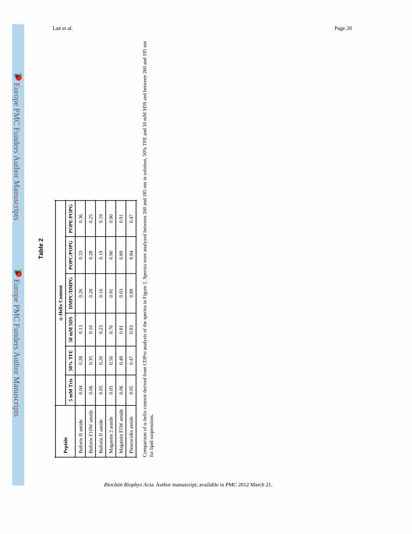
Circular Dichroism (CD) measurements of the peptides in the presence of membranemimetic media and lipid vesicles of differing composition provide information on theaverage secondary structure adopted by each peptide in each differing environment. BuforinII amide, magainin 2 amide and pleurocidin amide are all cationic peptides but each has anumber of uncharged polar or hydrophobic residues. At lower concentrations, magainin 2 isknown to adopt an amphipathic α-helical conformation in membranes with the helix longaxis aligned parallel to the membrane surface [12-16]. This enables charged residues tointeract with the polar or charged lipid headgroups while hydrophobic residues are orientedtowards the hydrophobic core of the membrane. CD measurements of magainin 2 amide(Fig. 1B) and its tryptophan analogue (Supp. Fig. 1B) confirm that in aqueous solution bothpeptides adopt a disordered structure. As expected, in the presence of 50% TFE, whichexcludes water to promote intra-molecular hydrogen bonding, CD spectra are obtained thatare consistent with a considerable ordered α-helical content. CD spectra indicating an evengreater α-helical content are obtained in the presence of micelles formed by the anionicdetergent SDS. Analyses of the spectra using CDPro provides a numerical estimate of thehelical content (Table 2). Hence, in both membrane mimicking media, magainin 2 amideadopts a conformation with a substantial α-helical content. Pleurocidin amide behavessimilarly to magainin 2 amide, both in aqueous solution and in membrane mimicking media(Fig. 1C). Buforin II amide has a greater overall charge and lower hydrophobicity thanmagainin 2 amide, but formation of an α-helix would lead to a separation of charged andhydrophobic residues giving a comparable hydrophobic moment (Table 1). The structure ofbuforin II has been determined previously using solution state NMR methodologies in 50%TFE [17]. The obtained conformation indicates a regular α-helix only between Val12 andArg20 and, after the proline induced kink, a helical section with a pitch of -6.82 Å. Thisalternate helical arrangement ensures the amphipathic region extends from Arg5 rightthrough to Lys21 [17]. In the present study, the CD spectra of buforin II amide (Fig. 1A) andits tryptophan mutant (Supp. Fig. 1A) are indistinguishable from each other in aqueous ormembrane mimicking environments (Table 2). CD indicates that both buforin II amidepeptides adopt a disordered structure in aqueous solution. In 50% TFE, but not in thepresence of SDS micelles, both buforin II amide peptides become more ordered. Thisbehavior is distinctly different from both magainin 2 amide and pleurocidin amide. Thesensitivity of the Chirascan spectrometer allows spectra with good signal-to-noise enablingmeasurements with peptide-to-lipid molar ratios as low as 1:200. At this ratio, sufficientnegative charge is maintained, for liposomes with approximately 20 mol percent anioniclipid, after peptide binding that liposome aggregation and concomitant light scattering do notimpair the measurements. Spectra obtained in this way are shown for a variety of liposomecompositions (Fig. 1D-F); spectra obtained at the higher peptide-to-lipid molar ratio of1:100 were similar (not shown) but at higher peptide-to-lipid molar ratios (circa 1:50),considerable light scattering was observed, compromising spectra particularly at shorterwavelengths. Three binary lipid mixtures were used with either DMPG or POPG as theanionic component and either POPC or POPE as the zwitterionic component. Membrane
Lan et al. Page 4
Biochim Biophys Acta. Author manuscript; available in PMC 2012 March 21.
Europe PM
C Funders A
uthor Manuscripts
Europe PM
C Funders A
uthor Manuscripts
lipid charge ratios were chosen to match those of the inner membrane of E. coli with POPE/POPG (80:20) being the closest match. POPC/POPG or DMPG/DMPG allowed anassessment of the effect of changing the zwitterionic lipid headgroup or replacing mono-unsaturated acyl chains with saturated chains, respectively. At 37°C, corresponding tophysiological temperature for a bacterium within a human host, the CD spectra obtained formagainin 2 amide (Fig. 1F) and the F5W analogue (Supp. Fig. 1D) in these mixed lipidmembranes and the CDPro analyses indicate that both peptides adopt an almost identical α-helix conformation to a much greater extent than in SDS micelles or 50% TFE. In this case,the SDS micelle is a better mimic for the liposome vesicle environment than 50% TFE. Incontrast, buforin II amide (Fig. 1E) and the F10W analogue (Supp. Fig. 1C) show a greatlyreduced tendency to becoming ordered in all the liposome environments; the very modesttendency to adopt any α-helical conformation correlates more with 50% TFE than SDSmicelles. The α-helical content of buforin II, with a free C-terminus, was even lower thanthat of the amidated analogue in 50% TFE and all three lipid environments (Table 2). CDspectra obtained for pleurocidin amide in the same liposome environments (Fig. 1D)indicated that the peptide adopted an α-helix conformation, similar to that of the twomagainin 2 amide peptides except when added to POPE/POPG membranes where asubstantial reduction in α-helix conformation is observed when compared with magainin 2amide in POPE/POPG and its own conformation in either POPC/POPG or DMPC/DMPG.
3.2 Thiazole orange fluorescence intercalating displacement assayA thiazole orange fluorescent intercalating displacement (FID) assay [18] has beenemployed to determine the binding characteristics of the buforin and magainin peptides to ashort DNA duplex. The reduction in fluorescence intensity from the DNA bound dye,following addition of increasing amounts of each peptide, is taken to be a measure of thefraction of peptide displacing the dye and binding to the DNA. The increase in the boundfraction of peptide is shown as a function of the peptide to DNA molar ratio (Fig. 2A). Forall of the peptides, except magainin 2 amide, the decay of the fluorescence intensity has aclearly sigmoid form. Extracting values for the peptide/DNA molar ratio required for half ofthe dye to be displaced (EC50) allows a comparison of the binding characteristics of each ofthe peptides (Table 3). Cationic AMPs are often amidated at the C-terminus to increaseactivity and in the present study we have studied amidated versions of buforin II andmagainin 2, comparing like with like, but have also examined the binding of the non-amidated form of buforin II to assess the contribution of this modification. Buforin II amideand buforin F10W amide had the greatest affinity for the duplex DNA. The EC50 for thesetwo peptides was nearly fourfold lower than that of magainin 2 amide, the difference alsobeing significant (p < 0.05). Buforin II, where the C-terminal is not amidated, had a bindingaffinity that was significantly greater than that of both magainin peptides (p < 0.05) and notsignificantly weaker than that of the two amidated buforin peptides. Magainin 2 amide hadthe weakest binding affinity and was the only one of the peptides whose binding could besatisfactorily fitted with a single exponential. Mutation of Phe5 to tryptophan caused analmost twofold increase in the affinity of magainin 2 to the duplex DNA which is alsosignificant (p < 0.05) and introduced a sigmoidal response to the binding and displacementmechanism. In contrast, the binding affinity of buforin II amide and buforin F10W amidewere indistinguishable (Fig. 2B). Pleurocidin amide has intermediate DNA bindingcapabilities with an EC50 that was greater than that of the three buforin peptides but lowerthan those of the two magainin peptides.
3.3 Circular Dichroism – DNA bindingNear-UV circular dichroism spectra of the duplex DNA in solution are characteristic ofthose obtained for the B form of DNA (Fig. 3A/C/E). A strong positive signal is observedbetween 260 and 290 nm with, in this case, a shoulder observable around 272 nm. Similarly,
Lan et al. Page 5
Biochim Biophys Acta. Author manuscript; available in PMC 2012 March 21.
Europe PM
C Funders A
uthor Manuscripts
Europe PM
C Funders A
uthor Manuscripts

a strong negative signal is observed between 230 and 260 nm. Near-UV spectra are suitablefor monitoring the response of DNA to the addition of peptide. CD contributions above 250nm due to peptide are negligible, despite the presence of phenylalanine and tryptophanresidues in the peptide sequences (Supp. Fig. 4). Addition of small amounts of buforin IIamide causes dramatic changes in both the CD spectra (Fig. 3A) and simultaneouslyobtained absorbance spectra (Fig. 3B). Initially the shoulder observed in the CD at 272 nm isseen to fall while the signal between 230 and 260 nm as well as the characteristic DNAabsorbance at 260 nm remain unaffected. When between three and four molar equivalents ofbuforin II amide have been added to the complex, the absorbance at 260 nm begins tocollapse with the onset of turbidity (Fig. 3B); circular intensity differential light scatteringnow dominates the CD spectrum as the DNA becomes condensed by the peptide. At thisstage the CD spectra take on a greatly altered form as a positive intensity builds around 290nm, the shoulder at 272 nm is completely absent and substantial rearrangements occur in theregion between 230 and 260 nm. In contrast, the effects of magainin 2 amide on the sameDNA duplex are much more moderate (Fig. 3C) with those of pleurocidin amide beingintermediate between buforin II amide and magainin 2 amide (Fig. 3E). The CD shoulder at272 nm, for DNA challenged with magainin 2 amide, is much more resistant to the additionof peptide and remains almost throughout the course of the experiment. The correspondingabsorbance spectra show substantial hypochromism; the absence of turbidity (lightscattering) above 310 nm indicates that the DNA remains in solution (Fig. 3D). The changein intensity of the CD (272 nm) is the most notable event in the initial stages of the peptide/DNA binding process and hence this is plotted as a function of peptide to DNA molar ratio(Fig. 4A) for as long as the DNA remains in solution. This allows the rate of change of thisfeature to be used as a means of providing a quantitative comparison of peptides with aputative DNA binding bactericidal strategy. A peptide concentration dependent reduction ofthe CD (272 nm) intensity is observed upon addition of peptide. Extracting a slope fromthese plots enables the structural rearrangements observed by CD to be related to the bindingaffinities and modes of the peptides (Table 3). This relationship between the binding affinityof the peptides and the structural changes is made more apparent by plotting the change inCD (272 nm) signal for each peptide as a function of the corresponding measured relativedissociation constant (Fig. 4B). This plot reveals the trend whereby the peptides with thegreatest affinity for the DNA duplex also cause the greatest change in the CD (272 nm)signal. The two amidated buforin peptides have both the highest affinity for the DNA duplexand induce the greatest changes in CD, greater than those induced by either magaininpeptide (p < 0.05). Magainin 2 amide has the lowest affinity and induces the least change inCD. The observed increase in the effect of this peptide on the CD (272 nm) signal followingreplacement of Phe5 with tryptophan however may or may not be significant (p = 0.165).Equally, buforin II, with a free C-terminal, has both a slightly reduced affinity for the DNAduplex and also causes slightly less of an effect on the CD (272 nm) signal when comparedwith the amidated analogue which again may or may not be significant (p = 0.09).Pleurocidin amide has intermediate binding affinity (Table 3) and is observed to also havean intermediate effect on the CD (272 nm) signal.
Finally, far-UV spectra were obtained to observe CD attributable to the peptide (Fig. 5A-D).In this region, the optical pathlength was 0.5 mm, twenty fold less than the 1 cm used in thenear UV. Nucleic acid contributions are now less significant. In contrast with the structuredetermined by NMR and with the far-UV spectra described above for buforin II inmembrane or membrane mimicking environments, little or no evidence of an α-helicalconformation is observed for buforin II amide bound to duplex DNA (Fig. 5A). Instead, inthe spectra acquired at [peptide]/[DNA] ratios below 4, the strong negative CD signal ataround 195 nm coupled with the absence of a negative band between 220 and 230 nmsuggests the peptide adopts an extended conformation. Further work is required todistinguish between a disordered or polyproline II conformation. At peptide to DNA molar
Lan et al. Page 6
Biochim Biophys Acta. Author manuscript; available in PMC 2012 March 21.
Europe PM
C Funders A
uthor Manuscripts
Europe PM
C Funders A
uthor Manuscripts

ratios below 4, the negative band has a negative maximum at 195 nm which shifts toward199 nm as more peptide is added (Fig. 5A). These spectra, recorded at peptide to DNAmolar ratios greater than 4, have greater contributions from peptides either in solution or notdirectly bound to DNA. Notably, a negative maximum of 199 nm is observed for buforin IIamide in solution (Fig. 1A). This suggests a possible polyproline II conformation [19] forthose peptide molecules directly bound to the DNA duplex. However, unequivocalattribution of such a conformation is difficult without observation of an expected, weak,positive band around 215 nm which would be obscured in the present experiment by signalsfrom the DNA duplex. Nevertheless, the experimental evidence strongly indicates thatbuforin II amide does not bind to duplex DNA in an α-helix conformation. Similarexperiments were performed for pleurocidin amide (Fig. 5B) and magainin 2 amide (Fig.5C). Again focusing on spectra recorded at peptide:DNA ratios <4, no evidence of an α-helix conformation is observed. For comparison, a similar experiment was performed with adesigned cationic peptide, LAK (Fig. 5D), which has a comparable effect to that of buforinII amide, on the near-UV spectra of the duplex DNA (Supp. Fig. 3). At peptide-to-DNAratios below 4, this peptide adopts an α-helical structure when bound in a peptide-DNAcomplex, according to the strong negative bands at 205 and 220 nm and strong positive bandat 195 nm observed in the corresponding far-UV spectra (Fig. 5D). The LAK peptide isdesigned to adopt an almost perfect amphipathic α-helix in media that support suchconformations and is used here to highlight the ability of the far-UV CD spectra to identifyan α-helix conformation in peptides bound to the DNA duplex. In contrast, such α-helixconformation is clearly absent from buforin II amide, magainin 2 amide or pleurocidinamide when bound to DNA.
3.4 Broth micro-dilution assayThe antibiotic activity of the peptides was tested against two separate strains of E. coli andone of P. aeruginosa. NCTC 9001 is a type strain while TOP10 is a competent strain with agenotype similar to that of DH10B which is deficient in galU, galK and galE [20].Inactivation of galE perturbs the incorporation of glucose into the O-side chain oflipopolysaccharide (LPS) and hence E. coli TOP10 are expected to have an altered LPSstructure. E. coli TOP10 were more susceptible to AMP challenge than the type strain whilethe amidated magainin peptides were substantially more effective than the correspondingamidated buforin peptides against both strains (Table 3). The buforin peptides had only amodest affect on bacterial growth at the highest peptide concentration tested, insufficient forthe determination of an MIC, and no differences in activity were observed between buforinII with either free or amidated C-termini or between buforin II amide and buforin F10Wamide. Buforin peptides have previously been reported to have anti-bacterial activities thatfar surpass those of magainin 2 [21]. The previous report used an agar diffusion protocolwhere the bacterial cells were washed in cold 10 mM sodium phosphate buffer prior tochallenge. Our procedure involves challenge of planktonic cultures of bacteria, according tocommonly used procedures [11], and is consistent with the observation thatphosphatidylethanolamine lipids, abundant in the membranes of Gram-negative organisms,abolish the membrane translocation of buforin II necessary for its supposed intracellulartargeting antibacterial strategy [22]. Interestingly, a small but significant enhancement ofactivity was observed for magainin F5W amide over magainin 2 amide against both strainsof E. coli (p < 0.05) and may apply also against P. aeruginosa (p = 0.07).
4. DiscussionCationic AMPs are diverse in structure and mechanisms of action [5, 6] and thereforepresent an interesting challenge to those using a rational design strategy to develop them asanti-infective therapeutic compounds [1-4]. While it is recognized that AMPs with unrelated
Lan et al. Page 7
Biochim Biophys Acta. Author manuscript; available in PMC 2012 March 21.
Europe PM
C Funders A
uthor Manuscripts
Europe PM
C Funders A
uthor Manuscripts

structural properties may operate with distinct mechanisms [5, 6] it has also been suggestedthat it is sufficient for a peptide to satisfy a set of physico-chemical conditions for broadspectrum antibacterial activity to be conferred [23]. The argument follows that secondarystructure is unimportant for the activity of AMPs and that directed combinatorial methodswill be the most fruitful path to incremental increases in the potency of AMPs. However,data from the same study indicates that disrupting secondary structure in AMPs dramaticallyreduces activity against Gram negative bacteria [23], indicating that secondary structure isimportant for activity against organisms such as E. coli and Pseudomonas aeruginosa but notGram positive bacteria. The composition of bacterial membranes varies considerably fromspecies to species and has been shown to be an important predictor of antimicrobial potency[24]. Furthermore, biophysical evidence is emerging that shows that the pore formingbehavior of an individual peptide will vary considerably according to the membranecomposition [25, 26]. Taken together, these reports suggest that understanding the structuralcontributions to species specific mechanisms of action will enable the design of peptides thatcan target bacteria with defined characteristics. Pexiganan, or MSI-78, is a highly potentAMP derived from magainin 2, a naturally occurring peptide identified in the skin of theAfrican frog Xenopus laevis. Pexiganan is the best studied AMP for therapeutic purposes todate and it, like magainin 2, is an amphipathic α-helical peptide proposed to operate via theformation of toroidal pores in bacterial membranes [27, 28]. Measurements employing avariety of biophysical methods indicate that magainin 2 binds to membranes in anorientation with the long axis of the helix parallel to the membrane surface [13-16] but then,as its concentration in the membrane increases above a certain threshold, neutron in-planescattering studies suggest a trans-membrane alignment is adopted which enables the peptidesto line a toroidal shaped pore [28, 29]. Interestingly however, both molecular dynamicssimulations and experimental studies have shown that related cationic amphipathic α-helicalpeptides, including magainin H2 and pleurocidin amide, are capable of forming pores atconcentrations where a surface alignment for the peptide is maintained [30-32]. Themolecular dynamics study further revealed that, under these conditions, magainin H2 wascapable of translocating from the external to the internal leaflet of the membrane [30], as hasbeen observed experimentally for magainin 2 [24]. Furthermore, the potent antibacterialactivity of designed amphipathic α-helical peptides has been shown, in a recent study [33],to be unrelated to their ability to form pores. Suspecting an intra-cellular targeting strategyfor these peptides, we are interested in understanding the structural properties that underpinthis alternative mechanism of antibacterial action.
Buforin II is a potent AMP which has been shown to kill bacteria by penetrating the cellmembrane and inhibiting cellular functions [7, 34]. Buforin II translocates across modelmembranes causing minimal disruption to the membrane [35] but, as the translocation ofboth buforin II and magainin 2 are inhibited by negative-curvature inducing lipids includingphosphatidylethanolamine (PE), there have been proposed to be both similarities [22] andstrong differences [35] between the translocation mechanisms of these peptides. The role ofPro11 in the translocation of buforin II is crucial as mutation of this residue abrogates theability of the peptide both to translocate across model membranes without disrupting themembrane and penetrate E. coli cells [35, 36]. In the NMR structure, determined in amixture of H2O and TFE, the Pro11 residue distorts the helix affecting the positioning ofhydrophobic and charged residues [17]. Previous CD measurements indicate that thisstructure is maintained in anionic lipid membranes [35] while the present study confirms thisstructure is also maintained in mixed anionic/zwitterionic membranes designed to mimicthose of Gram negative bacteria. This result contradicts a recently reported combinedmolecular dynamics (MD) and CD based analysis [37]. The MD study showed an increasedproportion of α-helix in the N-terminal region of buforin II in the presence of PE lipids andinterpreted CD with mixed POPE/POPG vesicles as having a greater α-helical character.However, the molecular dynamics study did not adequately reproduce existing experimental
Lan et al. Page 8
Biochim Biophys Acta. Author manuscript; available in PMC 2012 March 21.
Europe PM
C Funders A
uthor Manuscripts
Europe PM
C Funders A
uthor Manuscripts
data. In the MD simulations, the α-helical conformation of the peptide was maintained in anaqueous environment while the peptide also bound to neutral lipid membranes. Both theseresults contradict experimental results showing buforin II adopts a disordered structure inaqueous solution and does not bind to vesicles comprising zwitterionic lipids only [35]. TheCD spectra obtained in support of these simulations were obtained at 25°C, the phasetransition temperature of POPE, and at peptide-to-lipid ratios where we have seenconsiderable impairment of the spectra from light scattering due to charge neutralization ofthe anionic liposomes by cationic peptide. Such light scattering is evident in the reportedspectra [35]. According to its supposed mode of action, buforin II is proposed to readilytranslocate across bacterial membranes. Lipids, such as egg yolk L-α-phosphatidylethanolamine (EYPE), which induce negative membrane curvature, abolishsuch translocation however [34]. The membrane order in the POPE/POPG and DMPC/DMPG systems will be considerably greater than that of the POPC/POPG [38, 39], as aresult of the small ethanolamine headgroup size and saturated chains respectively. Thesediffering membrane environments might be expected to exert considerable influence on thesecondary structure of membrane inserted peptides. However, since the present studyindicates both buforin II and buforin II amide adopt a very similar conformation in each ofthe three mixed zwitterionic/anionic membranes and that the nature of the zwitterionic lipidhas little or no effect on the structure adopted by the peptide, a conformational change inbuforin II is therefore not implicated in the mechanism by which PE lipids inhibit itstranslocation. In model membranes, the α-helical content of buforin II with a free C-terminus was lower still than the two amidated analogues, supporting a role for C-terminalamidation in aiding membrane insertion, as observed for other cationic α-helical AMPs[40].
Mutations in the buforin sequence have been shown experimentally to reduce binding ofpeptide to a DNA duplex and impair the antibacterial activity of the peptide [9]. Seeking tounderstand the structural properties of buforin that underpin its ability to bind DNA, wedevised a combined FID and CD approach to monitor the binding and structural responsesof both peptides and DNA when a series of antibacterial peptides bind to a DNA duplex,identical in sequence to that in the previous study [9]. Both the FID assay and CD study ofpeptide binding to duplex DNA in the present work confirm previous reports that buforin IIhas a high affinity for nucleic acids [7]. Interestingly, we have shown that the bindingaffinity of each peptide is reflected in the conformational changes observed in thecorresponding near-UV CD spectra. The method is also sufficiently sensitive to detect thatmutation of Phe5 to tryptophan in magainin 2 amide increased the binding affinity for DNAand affected the apparent mode of binding which may be related to a modest enhancement inthe antibacterial activity. The sigmoidal response of the FID assay might be taken to suggestcooperativity in the mode binding of the peptides to duplex DNA, with the exception ofmagainin 2 amide. However, this may not accurately reflect the situation within bacteria nor,indeed, in the near-UV CD experiments. The binding parameters obtained from the FIDassay are influenced not only by the binding of peptide to DNA but by the competitivebinding of the dye. With two competing binding processes, the data from the FID assay mayeither exaggerate cooperativity or suggest cooperativity that would otherwise not be presentwere it not for the presence of dye and hence, at present, we restrict our analysis to aqualitative comparison of the dissociation constants determined for each peptide.
Binding of buforin II amide to duplex DNA induced strong changes in the CD spectraattributable to the DNA duplex. The origin of the observed changes in the CD spectra ispresently unknown but may occur as a result of changes in base-pair stacking and/orgeometry of the double helix as CD spectra of nucleic acids are known to be sensitive tothese parameters [41]. Hypochromism observed in the DNA UV absorption at 260 nm isstrong evidence that the duplex conformation remains with stronger base-base interactions.
Lan et al. Page 9
Biochim Biophys Acta. Author manuscript; available in PMC 2012 March 21.
Europe PM
C Funders A
uthor Manuscripts
Europe PM
C Funders A
uthor Manuscripts
For each of the peptides included in the present study, the UV/CD spectra associated withthe peptide/DNA titration indicate a two step process needs to be considered. The first step,up to peptide-to-DNA mole ratios of 3.5, involves simply the binding of the peptide; above amole ratio greater than 3.5, charge neutralization with the formation of peptide-DNA-ψ-condensates dominates. Concentration dependent changes in molecular CD, with nosigmoidal character, were observable as up to four molar equivalents of peptide were addedto the DNA duplex. This amount of peptide was sufficient to displace more than 90% of thedye in the corresponding FID assay. Adding more than four molar equivalents of buforin IIamide to the DNA duplex leads to the formation of a DNA condensate with the spectra thendominated by circular intensity differential light scattering. The spectra obtained for buforinII amide DNA condensates are similar in appearance to those observed for positive ψcondensates formed by incubating DNA with poly(lysine-alanine) and are indicative of theformation of structures with long range order [42]. Interestingly buforin II amide, bound tothe DNA duplex in the concentration range inducing the initial changes in near-UV CD, isshown to adopt an extended conformation rather than the helical conformation adopted inmembranes or proposed from an MD simulation study [9]. Again the experimental data doesnot support the existing view provided by simulations and further work is required todevelop a combined experimental/simulation approach in order to understand how theextended buforin II peptides condense duplex DNA.
The ability of a third antibacterial peptide, pleurocidin amide, to complex and condenseDNA was also assessed. Pleurocidin amide had a reduced ability to bind DNA whencompared with buforin II amide but had stronger interactions with the duplex than either ofthe two magainin peptides. Of the peptides tested in the present study, pleurocidin amidehad by far the greatest activity against the Gram-negative organisms E. coli and P.aeruginosa. However, as pleurocidin amide demonstrated only intermediate DNA bindingactivity, this property cannot completely account for the high antibacterial potency of thepeptide. Nevertheless, the DNA binding properties of pleurocidin amide may make somecontribution to its potency and are consistent with previous reports of pleurocidin and itsderivatives, including pleurocidin amide, entering E. coli and inhibiting macromolecularsynthesis [43]. As noted above, pleurocidin amide interacts with mixed zwitterionic/anionicmembranes in a similar fashion to magainin peptides, adopting a surface alignment andspecifically disordering the acyl chains of anionic lipids in such membranes [30]. In thepresent study, Chirascan CD measurements have allowed a more sophisticated investigationof the conformations adopted by pleurocidin amide. Interestingly, though pleurocidin amideadopts a very similar α-helix conformation to magainin 2 amide in solution and mostmembrane mimicking environments, in phosphatidylethanolamine rich membranes aconformation with a lower content of α-helix is preferred. The link between this membraneconformation, observed for pleurocidin amide in membranes mimicking those of Gram-negative bacteria, and its translocation abilities would throw light on the mechanismunderpinning the translocation of antimicrobial peptides into their bacterial targets.
5. ConclusionThe present study demonstrates that the conformation adopted by buforin II amide whenbinding to, and condensing, duplex DNA differs from that adopted in membranes. Theconformation of buforin II amide in membranes is not dominantly α-helix and the peptidehas poor membrane translocation ability compared with magainin 2 amide, which is stronglyα-helical and capable of translocating membranes composed of lipids found in themembranes of Gram-negative bacteria. Since α-helix conformation appears important formembrane translocation but not for DNA binding and condensation, potent antibacterialpeptides can be considered in future that are capable of adopting structures that promotemembrane translocation, DNA binding and DNA condensation.
Lan et al. Page 10
Biochim Biophys Acta. Author manuscript; available in PMC 2012 March 21.
Europe PM
C Funders A
uthor Manuscripts
Europe PM
C Funders A
uthor Manuscripts

Supplementary MaterialRefer to Web version on PubMed Central for supplementary material.
AcknowledgmentsThis work was supported by the Medical Research Council (NIRG to AJM), the Wellcome Trust (VIP Award toAJM and Capital Award for the KCL Centre for Biomolecular Spectroscopy), the University of London CentralResearch Fund (AR/CRF/B) and Applied Photophysics. We are grateful to Drs K.D. Bruce and T. Spasenovski fortheir assistance in setting up antibacterial testing in the Molecular Microbiology Research Laboratory (PSD).
REFERENCES1. Hancock REW, Sahl H-G. Antimicrobial and host-defense peptides as new anti-infective therapeutic
strategies. Nature Biotech. 2006; 24:1551–1557.
2. Reddy KVR, Yedery RD, Aranha C. Antimicrobial peptides: premises and promises. Int. J.Antimicrob. Agents. 2004; 24:536–547. [PubMed: 15555874]
3. Marr AK, Gooderham WJ, Hancock REW. Antibacterial peptides for therapeutic use: obstacles andrealistic outlook. Curr. Opin. Pharmacol. 2006; 6:468–472. [PubMed: 16890021]
4. Y, J. Gordon; E, G. Romanowski; A, M. McDermott A review of antimicrobial peptides and theirtherapeutic potential as anti-infective drugs. Curr. Eye Res. 2005; 30:505–515. [PubMed:16020284]
5. K, A. Brogden Antimicrobial peptides: pore formers or metabolic inhibitors in bacteria? Nat. Rev.Microbiol. 2005; 3:238–250. [PubMed: 15703760]
6. Hale JDF, Hancock REW. Alternative mechanisms of action of cationic antimicrobial peptides onbacteria. Expert Rev. Anti Infect. Ther. 2007; 5:951–959. [PubMed: 18039080]
7. Park CB, Kim HS, Kim SC. Mechanism of action of the antimicrobial peptide buforin II: Buforin IIkills microorganisms by penetrating the cell membrane and inhibiting cellular functions. Biochem.Biophys. Res. Comm. 1998; 244:253–257. [PubMed: 9514864]
8. Cirioni O, et al. Therapeutic efficacy of buforin II and rifampin in a rat model of Acinetobacterbaumanii sepsis. Crit. Care Med. 2009; 37:1403–1407. [PubMed: 19318826]
9. Uyterhoeven ET, Butler CH, Ko D, Elmore DE. Investigating the nucleic acid interactions andantimicrobial mechanism of buforin II. FEBS Letters. 2008; 582:1715–1718. [PubMed: 18448075]
10. Sreerama N, Woody RW. Estimation of protein secondary structure from CD spectra: Comparisonof CONTIN, SELCON and CDSSTR methods with an expanded reference set. Anal. Biochem.2000; 287:252–260. [PubMed: 11112271]
11. Wiegand I, Hilpert K, Hancock REW. Agar and broth dilution methods to determine the minimalinhibitory concentration (MIC) of antimicrobial substances. Nature Protocols. 2008; 3:163–175.
12. Bechinger B, Zasloff M, Opella SJ. Structure and interactions of magainin antibiotic peptides inlipid bilayers: a solid-state nuclear magnetic resonance investigation. Biophys. J. 1992; 62:12–14.[PubMed: 1600092]
13. Bechinger B, Zasloff M, Opella SJ. Structure and orientation of the antibiotic peptide magainin inmembranes by solid-state nuclear magnetic resonance spectroscopy. Protein Sci. 1993; 2:2077–2084. [PubMed: 8298457]
14. Bechinger B, Ruysschaert JM, Goormaghtigh E. Membrane helix orientation from linear dichroismof infrared attenuated total reflection spectra. Biophys. J. 1999; 76:552–563. [PubMed: 9876168]
15. Matsuzaki K, Murase O, Tokuda H, Funakoshi S, Fujii N, Miyajima K. Orientational andaggregational states of magainin 2 in phospholipid bilayers. Biochemistry. 1994; 33:3342–3349.[PubMed: 8136371]
16. Ludtke SJ, He K, Wu Y, Huang HW. Cooperative membrane insertion of magainin correlated withits cytolytic activity. Biochim. Biophys. Acta. 1994; 1190:181–184. [PubMed: 8110813]
17. Yi G-S, Park CB, Kim SC, Cheong C. Solution structure of an antimicrobial peptide buforin II.FEBS Letters. 1996; 398:87–90. [PubMed: 8946958]
Lan et al. Page 11
Biochim Biophys Acta. Author manuscript; available in PMC 2012 March 21.
Europe PM
C Funders A
uthor Manuscripts
Europe PM
C Funders A
uthor Manuscripts
18. Boger DL, Tse WC. Thiazole Orange as the fluorescent intercalator in a high resolution FID assayfor determining DNA binding affinity and sequence selectivity of small molecules. Bioorg. Med.Chem. 2001; 9:2511–2518. [PubMed: 11553493]
19. Woody RW. Circular dichroism spectrum of peptides in poly(pro)II conformation. J. Am. Chem.Soc. 2009; 131:8234–8245. [PubMed: 19462996]
20. Durfee T, et al. The complete genome sequence of Escherichia coli DH10B: insights into thebiology of a laboratory workhorse. J. Bacteriol. 2008; 190:2597–2606. [PubMed: 18245285]
21. Park CB, Kim MS, Kim SC. A novel antimicrobial peptide from Bufo bufo gargarizans. Biochem.Biophys. Res. Comm. 1996; 218:408–413. [PubMed: 8573171]
22. Kobayashi S, Chikushi A, Tougu S, Imura Y, Nishida M, Yano Y, Matsuzaki K. Membranetranslocation mechanism of the antimicrobial peptide buforin 2. Biochemistry. 2004; 43:15610–15616. [PubMed: 15581374]
23. Rathinakumar R, Walkenhorst WF, Wimley WC. Broad-spectrum antimicrobial peptides byrational combinatorial design and high-throughput screening: the importance of interfacial activity.J. Am. Chem. Soc. 2009; 131:7609–7617. [PubMed: 19445503]
24. Epand RM, Rotem S, Mor A, Berno B, Epand RF. Bacterial Membranes as Predictors ofAntimicrobial Potency. J. Am. Chem. Soc. 2008; 130:14346–14352. [PubMed: 18826221]
25. Gregory SM, S.M. Pokorny A, Almeida PFF. Magainin 2 revisited: a test of the QuantitativeModel for the all-or-none permeabilization of phospholipid vesicles. Biophys. J. 2009; 96:116–131. [PubMed: 19134472]
26. Almeida PFF, Pokorny A. Mechanisms of antimicrobial, cytolytic, and cell-penetrating peptides:from kinetics to thermodynamics. Biochemistry. 2009; 48:8083–8093. [PubMed: 19655791]
27. Gottler LM, Ramamoorthy A. Structure, membrane orientation, mechanism, and function ofpexiganan – a highly potent antimicrobial peptide designed from magainin. Biochim. Biophys.Acta. 2009; 1788:1680–1686. [PubMed: 19010301]
28. Ludtke SJ, He K, Heller WT, Harroun TA, Yang L, Huang HW. Membrane pores induced bymagainin. Biochemistry. 1996; 35:13723–13728. [PubMed: 8901513]
29. Huang HW. Molecular mechanism of antimicrobial peptides: the origin of cooperativity. Biochim.Biophys. Acta. 2006; 1758:1292–1302. [PubMed: 16542637]
30. Leontiadou H, Mark AE, Marrink SJ. Antimicrobial peptides in action. J. Am. Chem. Soc. 2006;128:12156–12161. [PubMed: 16967965]
31. Mason AJ, Chotimah INH, Bertani P, Bechinger B. A spectroscopic study of the membraneinteraction of the antimicrobial peptide Pleurocidin. Mol. Membr. Biol. 2006; 23:185–194.[PubMed: 16754361]
32. Saint N, Cadiou H, Bessin Y, Molle G. Antibacterial peptide pleurocidin forms ion channels inplanar lipid bilayers. Biochim. Biophys. Acta. 2002; 1564:359–364. [PubMed: 12175918]
33. Mason AJ, et al. Structural determinants of antimicrobial and antiplasmodial activity andselectivity in histidine-rich amphipathic cationic peptides. J. Biol. Chem. 2009; 284:119–133.[PubMed: 18984589]
34. Cho JH, Sung BH, Kim SC. Buforins: Histone H2A-derived antimicrobial peptides from toadstomach. Biochim. Biophys. Acta. 2009; 1788:1564–1569. [PubMed: 19041293]
35. Kobayashi S, Takeshima K, Park CB, Kim SC, Matsuzaki K. Interactions of the novelantimicrobial peptide Buforin 2 with lipid bilayers: proline as a translocation promoting factor.Biochemistry. 2000; 39:8648–8654. 2000. [PubMed: 10913273]
36. Park CB, Yi K-S, Matsuzaki K, Kim MS, Kim SC. Structure-activity analysis of buforin II, ahistone H2A-derived antimicrobial peptide: the proline hinge is responsible for the cell penetratingability of buforin II. Proc. Natl. Acad. Sci. USA. 2000; 97:8245–8250. [PubMed: 10890923]
37. Fleming E, Maharaj NP, Chen JL, Nelson RB, Elmore DE. Effect of lipid composition on buforinII structure and membrane entry. Proteins. 2008; 73:480–491. [PubMed: 18452210]
38. Mason AJ, Marquette A, Bechinger B. Zwitterionic phospholipids and sterols modulateantimicrobial peptide-induced membrane destabilization. Biophys. J. 2007; 93:4289–4299.[PubMed: 17766347]
Lan et al. Page 12
Biochim Biophys Acta. Author manuscript; available in PMC 2012 March 21.
Europe PM
C Funders A
uthor Manuscripts
Europe PM
C Funders A
uthor Manuscripts

39. Tuchtenhagen J, Ziegler W, Blume A. Acyl chain conformational ordering in liquid-crystallinebilayers: comparative FT-IR and 2H-NMR studies of phospholipids differing in headgroupstructure and chain length. Eur. Biophys. J. 1994; 23:323–335.
40. Pan Y-L, Cheng JT-J, Hale J, Pan J, Hancock REW, Straus SK. Characterization of the structureand membrane interaction of the antimicrobial peptides Aurein 2.2 and 2.3 from Australiansouthern bell frogs. Biophys. J. 2007; 92:2854–2864. [PubMed: 17259271]
41. Tinoco I Jr, Bustamente C, Maestre MF. The optical activity of nucleic acids and their aggregates.Ann. Rev. Biophys. Bioeng. 1980; 9:107–141. [PubMed: 6156638]
42. Phillips CL, Mickols WE, Maestre MF, Tinoco I Jr. Circular differential scattering and circulardifferential absorption of DNA-protein condensates and of dyes bound to DNA-proteincondensates. Biochemistry. 1986; 25:7803–7811. [PubMed: 3801444]
43. Patrzykat A, Friedrich CL, Zhang L, Mendoza V, Hancock REW. Sublethal concentrations ofPleurocidin-derived antimicrobial peptides inhibit macromolecular synthesis in Escherichia coli.Antimicrob. Agents Chemother. 2002; 46:605–614. [PubMed: 11850238]
44. Eisenberg D, Weiss RM, Terwilliger CT, Wilcox W. Hydrophobic moments and protein structure.Faraday Symp. Chem. Soc. 1982; 17:109–120.
Lan et al. Page 13
Biochim Biophys Acta. Author manuscript; available in PMC 2012 March 21.
Europe PM
C Funders A
uthor Manuscripts
Europe PM
C Funders A
uthor Manuscripts

Figure 1.Circular dichroism spectra of buforin II amide (A/E), magainin 2 amide (B/F) or pleurocidinamide (C/D) in solution (5 mM Tris pH 7, 50% TFE or 50 mM SDS) (A-C) or in thepresence of various mixed anionic/zwitterionic lipid vesicles (D-F) where the anioniccomponent comprised 20% of the lipid by mole and the peptide to lipid molar ratio was1:200. The temperature was 37°C and the final peptide concentration was 24 μM.
Lan et al. Page 14
Biochim Biophys Acta. Author manuscript; available in PMC 2012 March 21.
Europe PM
C Funders A
uthor Manuscripts
Europe PM
C Funders A
uthor Manuscripts
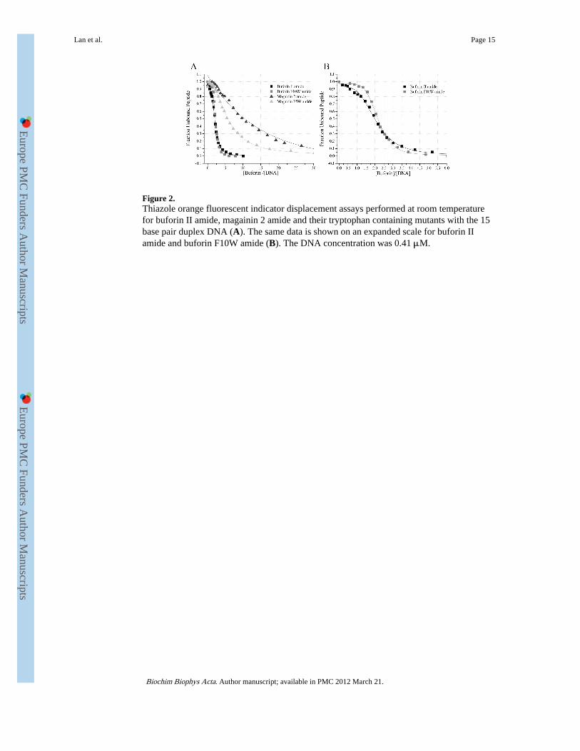
Figure 2.Thiazole orange fluorescent indicator displacement assays performed at room temperaturefor buforin II amide, magainin 2 amide and their tryptophan containing mutants with the 15base pair duplex DNA (A). The same data is shown on an expanded scale for buforin IIamide and buforin F10W amide (B). The DNA concentration was 0.41 μM.
Lan et al. Page 15
Biochim Biophys Acta. Author manuscript; available in PMC 2012 March 21.
Europe PM
C Funders A
uthor Manuscripts
Europe PM
C Funders A
uthor Manuscripts
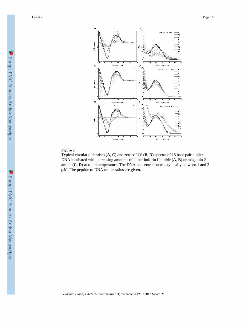
Figure 3.Typical circular dichroism (A, C) and zeroed UV (B, D) spectra of 15 base pair duplexDNA incubated with increasing amounts of either buforin II amide (A, B) or magainin 2amide (C, D) at room temperature. The DNA concentration was typically between 1 and 2μM. The peptide to DNA molar ratios are given.
Lan et al. Page 16
Biochim Biophys Acta. Author manuscript; available in PMC 2012 March 21.
Europe PM
C Funders A
uthor Manuscripts
Europe PM
C Funders A
uthor Manuscripts

Figure 4.The change in normalized CD as a function of peptide to duplex DNA molar ratio revealsthe differential effect of five peptides on molecular CD events (A). The change innormalized CD at 272 nm (as shown in Fig. 4A) is plotted as a function of the relativedissociation constant (EC50) for each peptide revealing the relationship between the twoparameters (B).
Lan et al. Page 17
Biochim Biophys Acta. Author manuscript; available in PMC 2012 March 21.
Europe PM
C Funders A
uthor Manuscripts
Europe PM
C Funders A
uthor Manuscripts
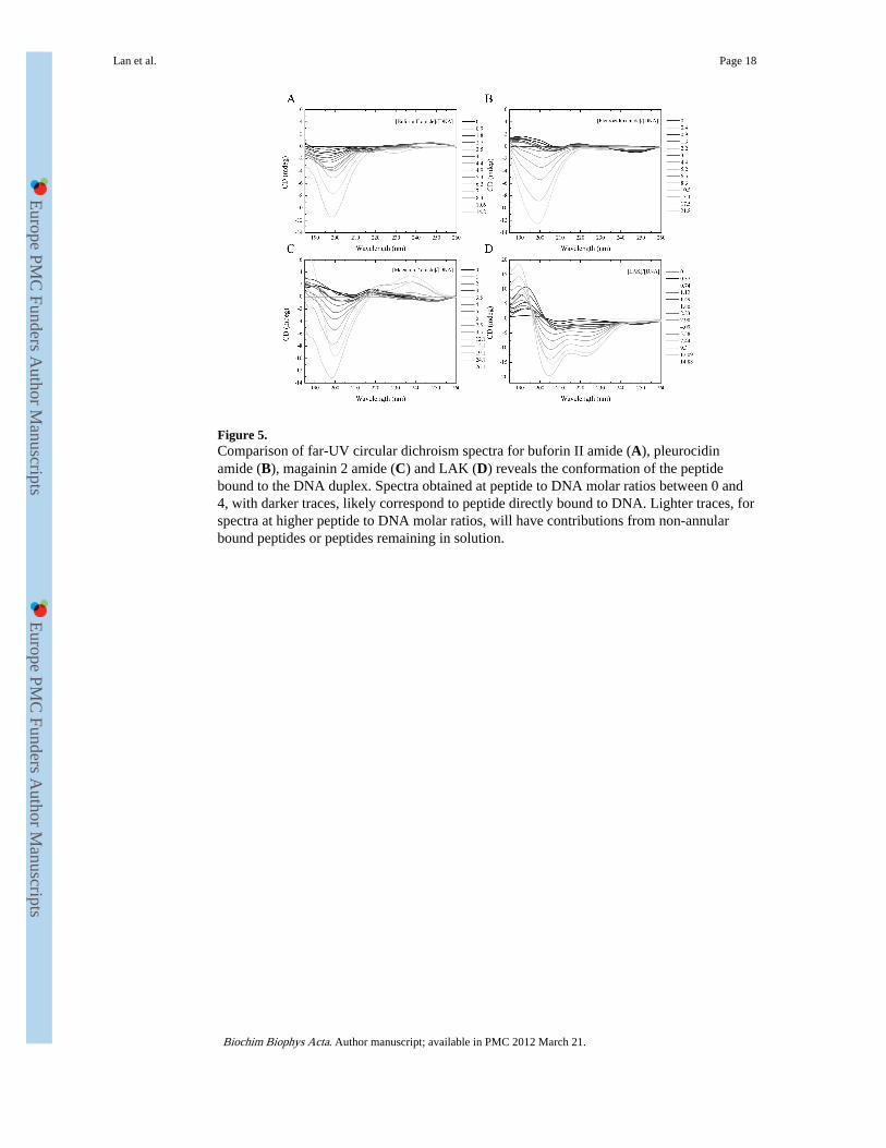
Figure 5.Comparison of far-UV circular dichroism spectra for buforin II amide (A), pleurocidinamide (B), magainin 2 amide (C) and LAK (D) reveals the conformation of the peptidebound to the DNA duplex. Spectra obtained at peptide to DNA molar ratios between 0 and4, with darker traces, likely correspond to peptide directly bound to DNA. Lighter traces, forspectra at higher peptide to DNA molar ratios, will have contributions from non-annularbound peptides or peptides remaining in solution.
Lan et al. Page 18
Biochim Biophys Acta. Author manuscript; available in PMC 2012 March 21.
Europe PM
C Funders A
uthor Manuscripts
Europe PM
C Funders A
uthor Manuscripts
Europe PM
C Funders A
uthor Manuscripts
Europe PM
C Funders A
uthor Manuscripts
Lan et al. Page 19
Tabl
e 1
Pep
tide
Sequ
ence
Cha
rge
(H)
(μH
)*
Buf
orin
II
amid
eT
RSS
RA
GL
QFP
VG
RV
HR
LL
RK
-NH
2+
7−
0.37
0.3
Buf
orin
F10
W a
mid
eT
RSS
RA
GL
QW
PVG
RV
HR
LL
RK
-NH
2+
7−
0.38
0.3
Buf
orin
II
TR
SSR
AG
LQ
FPV
GR
VH
RL
LR
K+
6−
0.37
0.3
Mag
aini
n 2
amid
eG
IGK
FLH
SAK
KFG
KA
FVG
EIM
NS-
NH
2+
4−
0.03
0.28
Mag
aini
n F5
W a
mid
eG
IGK
WL
HSA
KK
FGK
AFV
GE
IMN
S-N
H2
+4
−0.
040.
27
Pleu
roci
din
amid
eG
WG
SFFK
KA
AH
VG
KH
VG
KA
AL
TH
YL
-NH
2+
5−
0.02
0.22
LA
KK
KL
AK
AL
KL
LA
LL
WL
KL
AK
AL
KK
A-N
H2
+9
−0.
090.
34
Com
pari
son
of p
hysi
cal a
nd b
iolo
gica
l fea
ture
s of
pep
tides
use
d in
this
stu
dy. H
ydro
phob
icity
(H
) an
d m
ean
hydr
opho
bic
mom
ent (μ
H)
are
show
n ac
cord
ing
to th
e E
isen
berg
sca
le [
43]
and
wer
e ca
lcul
ated
usin
g th
e H
ydro
MC
alc
Java
app
let m
ade
avai
labl
e by
Ale
x T
ossi
(ht
tp://
ww
w.b
bcm
.uni
v.tr
iest
e.it/
~tos
si/H
ydro
Cal
c/H
ydro
MC
alc.
htm
l).
* Mea
n hy
drop
hobi
c m
omen
t ass
umin
g fo
rmat
ion
of id
eal α
-hel
ix.
Biochim Biophys Acta. Author manuscript; available in PMC 2012 March 21.
Europe PM
C Funders A
uthor Manuscripts
Europe PM
C Funders A
uthor Manuscripts
Lan et al. Page 20
Tabl
e 2
Pep
tide
α-H
elix
Con
tent
5 m
M T
ris
50%
TF
E50
mM
SD
SD
MP
C/D
MP
GP
OP
C/P
OP
GP
OP
E/P
OP
G
Buf
orin
II
amid
e0.
040.
280.
130.
260.
330.
36
Buf
orin
F10
W a
mid
e0.
060.
350.
100.
200.
280.
25
Buf
orin
II
amid
e0.
050.
200.
230.
160.
190.
19
Mag
aini
n 2
amid
e0.
050.
500.
760.
910.
900.
90
Mag
aini
n F5
W a
mid
e0.
060.
480.
810.
930.
890.
91
Pleu
roci
din
amid
e0.
050.
470.
830.
890.
840.
47
Com
pari
son
of α
-hel
ix c
onte
nt d
eriv
ed f
rom
CD
Pro
anal
ysis
of
the
spec
tra
in F
igur
e 2.
Spe
ctra
wer
e an
alyz
ed b
etw
een
260
and
185
nm in
sol
utio
n, 5
0% T
FE a
nd 5
0 m
M S
DS
and
betw
een
260
and
195
nmfo
r lip
id s
uspe
nsio
ns.
Biochim Biophys Acta. Author manuscript; available in PMC 2012 March 21.

Europe PM
C Funders A
uthor Manuscripts
Europe PM
C Funders A
uthor Manuscripts
Lan et al. Page 21
Tabl
e 3
Pep
tide
EC
50Δ
CD
@ 2
72 n
m
MIC
50
E. c
oli
(NC
TC
900
1)E
. col
i(T
OP
10)
P. a
erug
inos
a(P
AO
1)
Buf
orin
II
amid
e2.
51 ±
0.6
4−
0.19
8 ±
0.0
07>
26.
30>
26.
30>
26.
30
Buf
orin
F10
W a
mid
e2.
52 ±
0.4
9−
0.22
0 ±
0.0
16>
25.
89>
25.
89>
25.
89
Buf
orin
II
3.78
± 0
.29
−0.
133
± 0
.029
> 2
6.28
> 2
6.28
> 2
6.28
Mag
aini
n 2
amid
e9.
45 ±
0.7
1−
0.06
6 ±
0.0
246.
86 ±
1.5
81.
79 ±
0.1
310
.60
± 2
.21
Mag
aini
n F5
W a
mid
e5.
29 ±
0.5
0−
0.11
3 ±
0.0
204.
21 ±
1.2
91.
25 ±
0.2
87.
29 ±
2.7
3
Pleu
roci
din
amid
e4.
32 ±
0.8
0−
0.12
1 ±
0.0
030.
66 ±
0.2
20.
18 ±
0.0
21.
65 ±
0.8
7
Com
pari
son
of th
e D
NA
bin
ding
cha
ract
eris
tics
and
antim
icro
bial
act
iviti
es o
f th
e ca
tioni
c pe
ptid
es. D
isso
ciat
ion
cons
tant
(E
C50
) de
rive
d fr
om th
iazo
le o
rang
e FI
D a
ssay
.
Biochim Biophys Acta. Author manuscript; available in PMC 2012 March 21.
Related Documents