604-612 Nucleic Acids Research, 1994, Vol. 22, No. 4 Structural characterization of a ribonuclease Ill processing signal David C.Schweisguth, Bhadrani S.Chelladurai1, Allen W.Nicholsonl,* and Peter B.Moore Departments of Chemistry and Molecular Biophysics and Biochemistry, Yale University, PO Box 6666, New Haven, CT 06511-8118 and 1Department of Biological Sciences, Wayne State University, Detroit, Ml 48202, USA Received November 10, 1993; Revised and Accepted January 13, 1994 ABSTRACT The structure of a ribonuclease Ill processing signal from bacteriophage T7 was examined by NMR spectroscopy, optical melting, and chemical and enzymatic modification. A 41 nucleotide variant of the T7 Ri .1 processing signal has two Watson - Crick base- paired helices separated by an internal loop, consistent with its predicted secondary structure. The internal loop is neither rigidly structured nor completely exposed to solvent, and seems to be helical. The secondary structure of R1.1 RNA is largely insensitive to the monovalent cation concentration, which suggests that the monovalent cation sensitivity of secondary site cleavage by RNase Ill is not due to a low salt-induced RNA conformational change. However, spectroscopic data show that Mg2+ affects the conformation of the internal loop, suggesting a divalent cation binding site(s) within this region. The Mg2+-dependence of RNase Ill processing of some substrates may reflect not only a requirement for a divalent cation as a catalytic cofactor, but also a requirement for a local RNA conformation which is divalent cation-stabilized. INTRODUCTION Protein recognition of double-stranded RNA is important in many gene regulatory mechanisms [1-3]. Escherichia coli ribonuclease HI (RNase III, E.C.3.1.24)1 [4] is an endonuclease that specifically hydrolyzes dsRNA (reviewed in [5]). It cleaves primary rRNA transcripts, releasing the immediate precursors to the 16S and 23S rRNAs. A number of cellular pre-mRNAs are also substrates for RNase mI. RNA cleavage occurs at specific sites called RNase IH processing signals, which invariably contain approximately two turns of more or less regular dsRNA. Additional sequence and structural elements are present in RNase HI processing signals which determine the way they are cleaved, and often have important functional consequences for the metabolic stabilities and translational activities of the transcripts [5]. The life cycles of a number of coliphages depend on RNase HI. The early region of the bacteriophage T7 genome, for example, encodes five RNase IH processing signals that occur in the intercistronic regions, and several cleavage sites have been found in transcripts from the T7 late region [6]. RNase HI cleavage of the early primary transcript creates mature mono- and dicistronic mRNAs, and also determines their translational activities and stabilities [6-8]. The T7 Rl.1 processing signal (Figure 1), immediately upstream of gene 1.1, is an approximately 60 nt, irregular RNA hairpin, consisting of two dsRNA segments which are separated by an asymmetric internal loop. It contains the sequence information which is necessary and sufficient for accurate in vitro RNase m cleavage [9,10]. The Rl. 1 element is cleaved at a single point within the internal loop, leaving a short hairpin which protects the upstream RNA segment from 3'-5' exonucleolytic degradation [6,11]. The Rl.1 processing signal has been placed in plasmid expression vectors in order to produce transcripts with precisely defined 5' ends, or having prolonged metabolic stabilities [12-14]. Although over 30 different RNase III processing signals have been identified, and several studied biochemically and genetically, little is known about their solution conformations. The T7 Ri. 1 RNA has been studied as a model substrate with which to determine the RNA sequence and structural elements that establish RNase HI processing reactivity [10,15]. Determination of its structure may provide information as to why some substrates are cleaved once, while others undergo coordinate double cleavage, and afford insight into the reason for secondary site cleavage and its dependence on salt concentration. In this report we describe imino proton NMR and optical melting studies of the Rl. 1 processing signal, and the results of enzymatic and chemical probing experiments using reagents specific for structured or unstructured RNA regions. The data indicate that the RI. 1 processing signal is extensively base-paired, and that its internal loop is helical in character but is less stable than a canonical dsRNA element. The conformation of the Rl. 1 processing signal is largely insensitive to the monovalent salt concentration, suggesting that the influence of salt on secondary site cleavage by RNase IH reflects altered interactions between *To whom correspondence should be addressed k. 1994 Oxford University Press

Welcome message from author
This document is posted to help you gain knowledge. Please leave a comment to let me know what you think about it! Share it to your friends and learn new things together.
Transcript
604-612 Nucleic Acids Research, 1994, Vol. 22, No. 4
Structural characterization of a ribonuclease Ill processingsignal
David C.Schweisguth, Bhadrani S.Chelladurai1, Allen W.Nicholsonl,* and Peter B.MooreDepartments of Chemistry and Molecular Biophysics and Biochemistry, Yale University, PO Box 6666,New Haven, CT 06511-8118 and 1Department of Biological Sciences, Wayne State University,Detroit, Ml 48202, USA
Received November 10, 1993; Revised and Accepted January 13, 1994
ABSTRACTThe structure of a ribonuclease Ill processing signalfrom bacteriophage T7 was examined by NMRspectroscopy, optical melting, and chemical andenzymatic modification. A 41 nucleotide variant of theT7 Ri .1 processing signal has two Watson - Crick base-paired helices separated by an internal loop, consistentwith its predicted secondary structure. The internalloop is neither rigidly structured nor completelyexposed to solvent, and seems to be helical. Thesecondary structure of R1.1 RNA is largely insensitiveto the monovalent cation concentration, whichsuggests that the monovalent cation sensitivity ofsecondary site cleavage by RNase Ill is not due to alow salt-induced RNA conformational change.However, spectroscopic data show that Mg2+ affectsthe conformation of the internal loop, suggesting adivalent cation binding site(s) within this region. TheMg2+-dependence of RNase Ill processing of somesubstrates may reflect not only a requirement for adivalent cation as a catalytic cofactor, but also arequirement for a local RNA conformation which isdivalent cation-stabilized.
INTRODUCTIONProtein recognition of double-stranded RNA is important in manygene regulatory mechanisms [1-3]. Escherichia coli ribonucleaseHI (RNase III, E.C.3.1.24)1 [4] is an endonuclease thatspecifically hydrolyzes dsRNA (reviewed in [5]). It cleavesprimary rRNA transcripts, releasing the immediate precursorsto the 16S and 23S rRNAs. A number of cellular pre-mRNAsare also substrates for RNase mI. RNA cleavage occurs at specificsites called RNase IH processing signals, which invariably containapproximately two turns of more or less regular dsRNA.Additional sequence and structural elements are present in RNaseHI processing signals which determine the way they are cleaved,and often have important functional consequences for themetabolic stabilities and translational activities of the transcripts[5].
The life cycles of a number of coliphages depend on RNaseHI. The early region of the bacteriophage T7 genome, forexample, encodes five RNase IH processing signals that occurin the intercistronic regions, and several cleavage sites have beenfound in transcripts from the T7 late region [6]. RNase HIcleavage of the early primary transcript creates mature mono-and dicistronic mRNAs, and also determines their translationalactivities and stabilities [6-8]. The T7 Rl.1 processing signal(Figure 1), immediately upstream of gene 1.1, is anapproximately 60 nt, irregular RNA hairpin, consisting of twodsRNA segments which are separated by an asymmetric internalloop. It contains the sequence information which is necessaryand sufficient for accurate in vitro RNase m cleavage [9,10].The Rl. 1 element is cleaved at a single point within the internalloop, leaving a short hairpin which protects the upstream RNAsegment from 3'-5' exonucleolytic degradation [6,11]. The Rl.1processing signal has been placed in plasmid expression vectorsin order to produce transcripts with precisely defined 5' ends,or having prolonged metabolic stabilities [12-14].Although over 30 different RNase III processing signals have
been identified, and several studied biochemically and genetically,little is known about their solution conformations. The T7 Ri. 1RNA has been studied as a model substrate with which todetermine the RNA sequence and structural elements thatestablish RNase HI processing reactivity [10,15]. Determinationof its structure may provide information as to why some substratesare cleaved once, while others undergo coordinate doublecleavage, and afford insight into the reason for secondary sitecleavage and its dependence on salt concentration. In this reportwe describe imino proton NMR and optical melting studies ofthe Rl. 1 processing signal, and the results of enzymatic andchemical probing experiments using reagents specific forstructured or unstructured RNA regions. The data indicate thatthe RI. 1 processing signal is extensively base-paired, and thatits internal loop is helical in character but is less stable than acanonical dsRNA element. The conformation of the Rl. 1processing signal is largely insensitive to the monovalent saltconcentration, suggesting that the influence of salt on secondarysite cleavage by RNase IH reflects altered interactions between
*To whom correspondence should be addressed
k. 1994 Oxford University Press
Nucleic Acids Research, 1994, Vol. 22, No. 4 605
enzyme and substrate rather than changes in RNA structure. Incontrast, Mg2+ does influence the conformation of the internalloop. Divalent cations may direct the pattern RNasem processingby altering substrate structure, as well as serving as essentialcatalytic cofactors.
MATERIALS AND METHODSMaterialsWater was deionized and distilled. Chemicals were reagent grade.Ribonucleoside triphosphates were purchased from Boehringer-Mannheim. [,y-32P]ATP (3000 Ci/mmol) and [a-32P]UTP (3000Ci/mmol) were purchased from Dupont-NEN. DEPC and DMSwere obtained from Aldrich, while RNases U2, T2, and TI werefrom GIBCO-BRL, and RNase VI from Pharmacia. T7 RNApolymerase and RNase mI were purified as described fromoverexpressing bacterial strains [16,17]. The oligodeoxynucleo-tide transcription templates were synthesized at the Wayne StateMacromolecular Analysis Facility, and the fully deprotectedforms purified by denaturing gel electrophoresis [10]. Thesequence of the Rl.1[LSA6] RNA template, and the 18 ntpromoter oligonucleotide are as follows: 5'-AGGATCATAAA-GGCCACTCTTGCGAATGACCTTGAGTTCCCTATAGTG-AGTCGTATTA-3'; 5'-TAATACGACTCACTATAG-3'.
RNA synthesis and purificationPreparative-scale synthesis of RI. 1[LSA6] RNA was carried outby the procedure of Uhlenbeck and coworkers [18] with themodifications described by Wyatt et al. [19]. Transcriptionreactions (5-10 ml) were prepared in standard buffer [18], whichincluded 4 mM of each ribonucleoside triphosphate, 20 mMMg2+, T7 RNA polymerase (2500 units/ml) and 55 nM oftranscription template annealed to the promoter oligonucleotide.Following incubation for 4 hours at 37°C, the reactions wereterminated by adding EDTA (50 mM final concentration),dialyzed against water, then phenol extracted and ethanolprecipitated. The crude RNA was resuspended in water andelectrophoresed (25 V/cm) in 1.5 mm thick, 15% polyacrylamidegels containing 7 M urea in TBE buffer. The gel region containingthe full-length transcript was located by UV-shadowing. Minoramounts of longer RNA species were also observed, representingtranscripts with one or two additional nucleotides at the 3'terminus [18]. The RNA was extracted from crushed gel bandsusing 0.5 M ammonium acetate, 1 mM EDTA (pH 7). Theaqueous volume was reduced by repeated extraction with1-butanol, and the RNA ethanol-precipitated and resuspended inH20. The yield was determined by UV absorbance (calculatedextinction coefficient: 4.45 x10⁵ M- cm-1). 5'-32P-labeledRi. 1[LSA6] RNA was prepared by dephosphorylation ofunlabeled transcript with calf alkaline phosphatase, followed byreaction (approximately 50 pmol; 30 minutes at 37°C) with T4polynucleotide kinase (20 units) and [y-32P] ATP (0.89 IM;1870 Ci/mmol) in kinase buffer. The 32P-labeled RNA waspurified by gel electrophoresis as described above. Base-specificcleavage analysis of 32P-labeled R1. 1 [LSA6] RNA usingRNases TI and U2 was in full agreement with the predictedsequence.
NMR spectroscopyAll NMR experiments were conducted on a sample containingapproximately 0.6 ,mol of RI. 1[LSA6] RNA. Buffers contained4 mM sodium cacodylate and 0.2 mM EDTA, pH 7.5, with 200
mM KCl and/or 5 mM MgCl2 added as described. The RNAwas dialyzed extensively against each of the buffers in whichit was to be examined and concentrated to approximately 400yd in a Centricon-3 concentration device (Amicon). 20 ,l D20(MSD Isotopes) was added to the sample as a lock reference and1 Al 2.5% dioxane as a chemical shift standard. The chemicalshift of dioxane was taken to be 3.741 ppm at all temperatures.
One-dimensional spectra were water-suppressed using the twin-pulse method [20]. Difference NOE experiments were collectedin interleaved sets of 64 scans, irradiating each frequency ofinterest in turn with a 300 ms low-power pulse [21]. Two-dimensional NOESY spectra were collected in phase-sensitivemode with States phase cycling and a 300 ms mixing time. Waterwas suppressed using a self-refocused 1331 pulse beforeacquisition [22]. 2048 complex points were collected in t andapproximately 300 points were collected in tl. All data wereapodized with 300 to 70°-shifted sine bells before Fouriertransformation.
One-dimensional NMR experiments were done using ahomebuilt 490 MHz spectrometer and NOESYs were collectedon a Bruker AM-500, both in Yale's Chemical InstrumentationCenter. Data were processed on a Silicon Graphics workstationusing Felix (Hare Research).
Optical meltingAliquots of RI. 1 [LSA6] RNA were dialyzed extensively againsteach buffer, diluted to concentrations of 0.1, 0.3, 1, 3 and 10OD, degassed under vacuum, and placed in stoppered quartzcuvettes with path lengths of 1 cm (0.1, 0.3 and 1 OD samples)or 0.1 cm (3 and 10 OD samples). Melting was done in a VarianCary 1 UV-visible double-beam spectrophotometer. Fivesamples, one at each concentration, were melted simultaneously.Cuvettes filled with degassed buffer were used as references. Thetemperature was raised from 5 to 95 OC at 0.5 °C per minuteand the absorbance at 260 nm of each sample recorded every0.5 °C. The temperature was returned to 20 °C at the end ofeach melt and the absorbance checked to ensure that solvent hadnot been lost by evaporation during the melt. Each melt was donein duplicate.
Melting data were normalized to 1 OD at 20 0C and duplicateswere averaged. Observed absorbance was modeled as the sumof a baseline (the absorbance of fully stacked RNA) and twosigmoids (the hypochromicities of two independentiy meltingregions), each varying linearly with temperature, using thefollowing equation:
al+b1T a 2+b2TA =a +b T+ a 1 + 2 20
° AH°1 (1 1A AH 2 (41_'1R T Tm, R T Tm.2
l+e i+eA is absorbance, T is temperature, AH01 and AH02 are the
enthalpies of the two transitions, Tmi and Tm2 are their meltingtemperatures and an and b,, are the zero and first-ordercoefficients of variation of each term with temperature. Thisequation was fit to the averaged data using an implementationof the simplex algorithm [23]. Nearly all fits converged to thedata within a RMS difference of 10-6; those which did not, dueto experimental error, were rejected. Restarting converged fitsdid not significantly change either the values of the parametersor the goodness of fit. The enthalpies and Tm values wereaveraged, and the standard deviations calculated. Entropies were
606 Nucleic Acids Research, 1994, Vol. 22, No. 4
calculated as AH0/Tm and free energies at 37 °C asAH0 -(310.15 K * AS0). Enthalpies, entropies and energies forthe upper and lower stems were summed to obtain the parametersfor the entire molecule.
Enzymatic and chemical structure probingThe protocols followed for probing of 5 '-32P-labeledRI. 1[LSA6] RNA using RNase T2 or RNase VI were based onthose described previously [24]. Chemical probing using DMSor DEPC was carried out essentially as described elsewhere [25].The reaction buffer was 4 mM sodium cacodylate (pH 7.5), 0.2mM EDTA, supplemented with 200 mM KCI and/or 5 mMMgCl2, as indicated. Enzymatic RNA sequencing reactionswere performed using RNase U2 or RNase TI, following theprotocol provided by GIBCO-BRL. Reactions were analyzed byelectrophoresis in 0.4 mm thick, 15% polyacrylamide, 1.5%bisacrylamide sequencing gels containing 7 M urea and TBEbuffer. Autoradiography was carried out using Fuji RX film andintensifying screens.
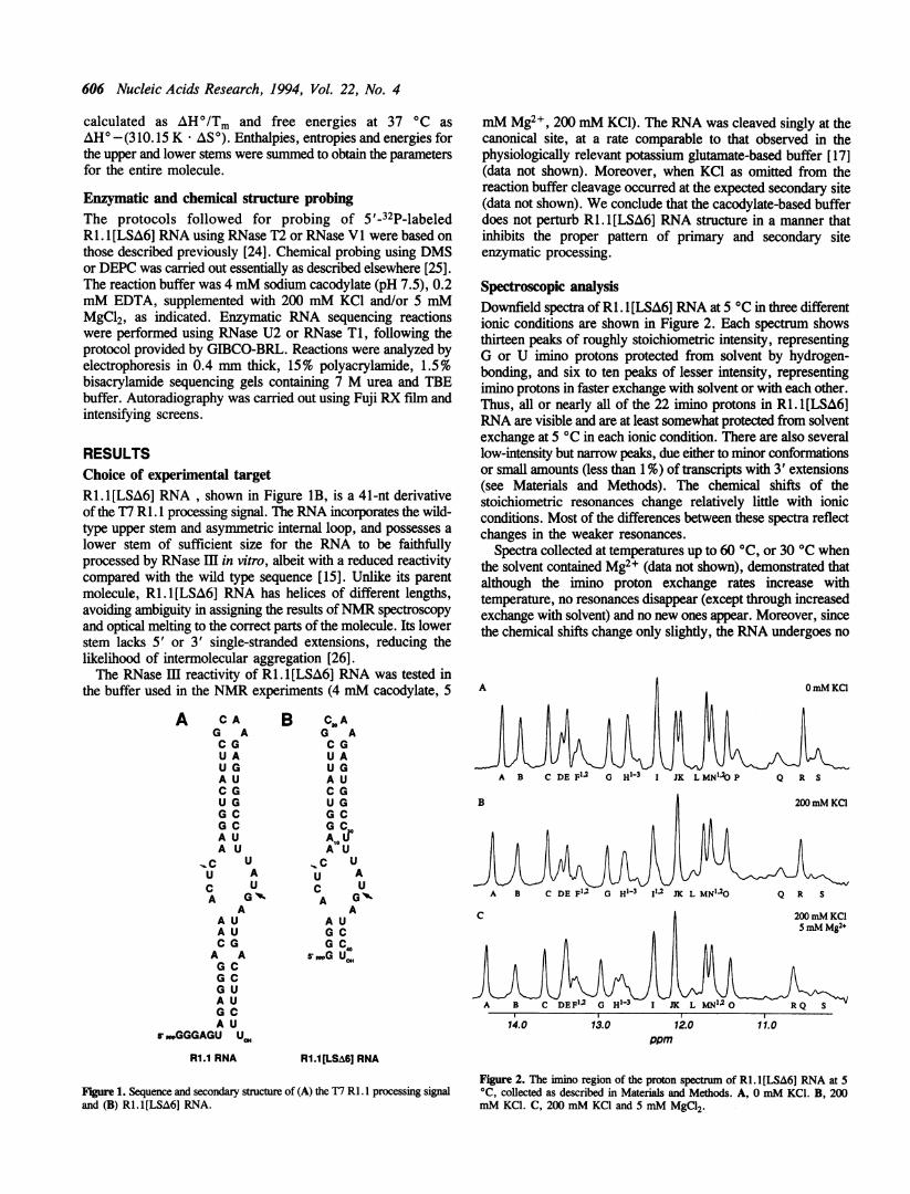
RESULTSChoice of experimental targetRi. 1[LSA6] RNA, shown in Figure IB, is a 41-nt derivativeof the T7 RI. 1 processing signal. The RNA incorporates the wild-type upper stem and asymmetric internal loop, and possesses alower stem of sufficient size for the RNA to be faithfullyprocessed by RNase III in vitro, albeit with a reduced reactivitycompared with the wild type sequence [15]. Unlike its parentmolecule, RI. 1[LSA6] RNA has helices of different lengths,avoiding ambiguity in assigning the results ofNMR spectroscopyand optical melting to the correct parts of the molecule. Its lowerstem lacks 5' or 3' single-stranded extensions, reducing thelikelihood of intermolecular aggregation [26].The RNase IH reactivity of RI. 1 [LSA6] RNA was tested in
the buffer used in the NMR experiments (4 mM cacodylate, 5
A C AG AC GU AU GA UC GU GG CG CA UA U
IC U
U AC UA GV
AA UA UC GA AG CG CG UA UG CA U
s'PwGGGAGU U.,R1.1 RNA
mM Mg2+, 200 mM KCl). The RNA was cleaved singly at thecanonical site, at a rate comparable to that observed in thephysiologically relevant potassium glutamate-based buffer [17](data not shown). Moreover, when KCl as omitted from thereaction buffer cleavage occurred at the expected secondary site(data not shown). We conclude that the cacodylate-based bufferdoes not perturb RI. 1[LSA6] RNA structure in a manner thatinhibits the proper pattern of primary and secondary siteenzymatic processing.
Spectroscopic analysisDownfield spectra of R1. l[LSA6] RNA at 5 0C in three differentionic conditions are shown in Figure 2. Each spectrum showsthirteen peaks of roughly stoichiometric intensity, representingG or U imino protons protected from solvent by hydrogen-bonding, and six to ten peaks of lesser intensity, representingimino protons in faster exchange with solvent or with each other.Thus, all or nearly all of the 22 imino protons in RI. 1[LSA6]RNA are visible and are at least somewhat protected from solventexchange at 5 °C in each ionic condition. There are also severallow-intensity but narrow peaks, due either to minor conformationsor small amounts (less than 1%) of transcripts with 3' extensions(see Materials and Methods). The chemical shifts of thestoichiometric resonances change relatively little with ionicconditions. Most of the differences between these spectra reflectchanges in the weaker resonances.
Spectra collected at temperatures up to 60 °C, or 30 °C whenthe solvent contained Mg2+ (data not shown), demonstrated thatalthough the imino proton exchange rates increase withtemperature, no resonances disappear (except through increasedexchange with solvent) and no new ones appear. Moreover, sincethe chemical shifts change only slightly, the RNA undergoes no
B C3o AG AC GU AU GA UC GU GG CG CA LPA U
_C UU AC UA G
AA UG CG C
5000G Ucm
14.0 13.0 12.0 11.0ppm
Ri.1[LSA6J RNA
Figure 1. Sequence and secondary structure of (A) the T7 Rl. I processing signaland (B) R1.1[LSA6] RNA.
Figure 2. The imino region of the proton spectrum of R1.1[LSA6] RNA at 5°C, collected as described in Materials and Methods. A, 0 mM KCI. B, 200mM KCl. C, 200 mM KCI and 5 mM MgCl2.
Nucleic Acids Research, 1994, Vol. 22, No. 4 607
14.0 13.0 12.0 11.0D1 (ppm)
Figure 3. The imino-imino region of a proton-proton NOESY of RI. l [LSA6]RNA in 200 mM KCI at 5 °C, collected as described in Materials and Methods.
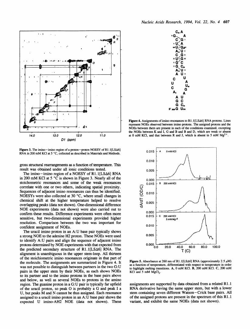
gross structural rearrangements as a function of temperature. Thisresult was obtained under all ionic conditions tested.The imino -imino region of a NOESY of Ri1.I[LSA6] RNA
in 200 mM KCl at 5 °C is shown in Figure 3. Nearly all of thestoichometric resonances and some of the weak resonancescorrelate with one or two others, indicating spatial proximity.Sequences of adjacent imino resonances can thus be identified.NOESYs were also collected at 30 °C, where small changes inchemical shift at the higher temperature helped to resolveoverlapping peaks (data not shown). One-dimensional differenceNOE experiments (data not shown) were also carried out toconfirm these results. Difference experiments were often moresensitive, but two-dimensional experiments provided higherresolution. Comparison between the two was important forconfident assignment of NOEs.The uracil imino proton in an A:U base pair typically shows
a strong NOE to the adenine H2 proton. These NOEs were usedto identify A:U pairs and align the sequence of adjacent iminoprotons determined by NOE experiments with that expected fromthe predicted secondary structure of Ri.1[LSA6] RNA. Thealignment is unambiguous in the upper stem-loop. All thirteenof the stoichiometric imino resonances originate in that part ofthe molecule. The assignments are summarized in Figure 4. Itwas not possible to distinguish between partners in the two G:Upairs in the upper stem by their NOEs, as each shows NOEsto its partner and to the imino protons in the base pairs aboveand below, as well as several NOEs to protons in the aminoregion. The guanine proton in a G:U pair is typically far upfieldof the uracil proton, so peak 0 is probably a G and peak I aU, but peaks M and N cannot be thus assigned. Each resonanceassigned to a uracil imino proton in an A:U base pair shows theexpected U imino-AH2 NOE (data not shown). These
Cm ARG AC/G
-U-G
A IJ
A1XU cG0Us ; 10
AE Al
G :
G C,A OD
C UU A
A,G
AsAA UG CG Co
PppG Uo
Fige 4. Assignments of imino resonances to RI. 1[LSA6] RNA protons. Linesrepresent NOEs observed between imino protons. The assigned protons and theNOEs between them are present in each of the conditions examined, exceptingthe NOEs between K and I, G and B and B and D, which are weak or absentat 0 mM KC1, and that between R and J, which is absent in 5 mM Mg2 .
0.015
0.010
0.005
00
7-V
0.0000.015
0.010
0.005
0.0000.015
0.010
0.005
0.000 L0.0 20.0 40.0 60.0
T (C)80.0 100.0
Figure 5. Absorbance at 260 nm of RI. I [LSA6] RNA (approximately 2.5 /tM)as a function of temperature, differentiated with respect to temperature in orderto highlight melting transitions. A, 0 mM KCI. B, 200 mM KCI. C, 200 mMKCI and 5 mM MgC12.
assignments are supported by data obtained from a related Rl. 1RNA derivative having the same upper stem, but with a lowerstem containing seven different Watson-Crick base pairs. Allof the assigned protons are present in the spectrum of this R 1.1variant, and exhibit the same NOEs (data not shown).
608 Nucleic Acids Research, 1994, Vol. 22, No. 4
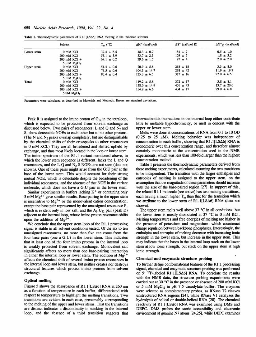
Table 1. Thennodynamic parameters of RI.l[LSA6] RNA melting in the indicated solvents
Solvent Tm (OC) AH° (kcal/mol) AS' (cal/mol K) AG037 (kcal/mol)
Lower stem 0 mM KC1 39.4 6.5 48.3 A 0.7 154 2 0.5 + 1.0200 mM KCI 55.1 4 3.9 33.7 A 2.3 103 A 7 1.8 + 3.2200 mM KCI + 69.1 0.2 29.6 k 1.5 8744 2.0 2.0
5 mM MgCl2Upper stem 0 mM KCI 51.4 + 0.6 70.9 + 5.8 218 ± 18 3.3 + 8.0
200 mM KCI 76.5 + 0.6 104.3 + 14.7 298 + 42 11.9 A 19.7200 mM KCl + 80.4 + 0.4 125.3 + 6.5 317 + 16 27.0 4 6.5
5 mM MgCl2Total 0 mM KCl 119.2 + 5.8 372 + 17 3.8 A 8.1
200 mM KCl 138.0 i 14.9 401 A 43 13.7 + 20.0200 mM KCI + 154.9 + 6.6 404 + 17 29.0 + 6.85mM MgCl2
Parameters were calculated as described in Materials and Methods. Errors are standard deviations.
Peak R is assigned to the imino proton of Glg in the tetraloop,which is expected to be protected from solvent exchange as
discussed below. Two pairs of resonances, L and Q and N2 andS, show detectable NOEs to each other but to no other protons.(The N and N2 peaks overlap completely, but are distinguishableby the chemical shifts of their crosspeaks to other resonances
in 0 mM KCI.) They are all broadened and shifted upfield byexchange, and thus most likely located in the loop or lower stem.The imino spectrum of the RI. I variant mentioned above, inwhich the lower stem sequence is different, lacks the L and Qresonances, and the L-Q and N2-S NOEs are not seen (data notshown). One of these pairs might arise from the G:U pair at thebase of the lower stem. This would account for their strongmutual NOE, which is detectable despite the broadening of theindividual resonances, and the absence of that NOE in the variantmolecule, which does not have a G:U pair in the lower stem.
Similar experiments in buffers lacking K+ or containing only5 mM Mg2+ gave similar results. Base-pairing in the upper stemis insensitive to Mg2+ or the monovalent cation concentration,except the base pair represented by the unassigned resonance P,which is evident only in low salt, and the A9:U32 pair (peak D)adjacent to the internal loop, whose imino proton resonance shiftsupon the addition of Mg2+.We conclude that the upper stem-loop of the RI. processing
signal is stable in all solvent conditions tested. Of the six to tenunassigned resonances, no more than five can come from thefour base pairs (one a G:U) in the lower stem. This indicatesthat at least one of the four imino protons in the internal loopis weakly protected from solvent exchange. Monovalent saltsignificantly affects no more than one base-pairing interactionin either the internal loop or lower stem. The addition of Mg2+affects the chemical shift of several imino proton resonances inthe internal loop and lower stem, but neither creates nor destroysstructural features which protect imino protons from solventexchange.
Optical meltingFigure 5 shows the absorbance of RI. 1 [LSA6] RNA at 260 nmas a function of temperature in each buffer, differentiated withrespect to temperature to highlight the melting transitions. Twotransitions are evident in each case, presumably correspondingto the melting of the upper and lower stems. That the transitionsare distinct indicates a discontinuity in stacking in the internalloop, and the absence of a third transition suggests that
internucleotide interactions in the internal loop either contributelittle to meltable hypochromicity, or melt in concert with theupper or lower stem.
Melts were done at concentrations ofRNA from 0.1 to 10 OD(0.25 to 25 jtM). Melting behavior was independent ofconcentration in each buffer, showing that RI. 1 [LSA6] RNA ismonomeric over this concentration range, and therefore almostcertainly monomeric at the concentration used in the NMRexperiments, which was less than 100-fold larger than the highestconcentration melted.
Table 1 presents the thermodynamic parameters derived fromthese melting experiments, calculated assuming the two transitionsto be independent. The transition with the larger enthalpies andentropies of melting is assigned to the upper stem, on theassumption that the magnitude of these parameters should increasewith the size of the base-paired region [27]. In support of this,the related Ri. 1 molecule (see above) has two melting transitions,each having a much higher Tm than that for the transition whichwe attribute to the lower stem of RI. 1 [LSA6] RNA (data notshown).The upper stem melts well above 37 °C in all conditions, but
the lower stem is mostly dissociated at 37 °C in 0 mM KCI.Melting temperatures and free energies of melting are higher inthe presence of potassium and magnesium, which counteractcharge repulsion between backbone phosphates. Interestingly, theenthalpies and entropies of melting decrease with increasing ionicstrength in the lower stem, but increase in the upper stem. Thismay indicate that the bases in the internal loop stack on the lowerstem at low ionic strength, but stack on the upper stem at highionic strength.
Chemical and enzymatic structure probingTo further define conformational features of the RI. 1 processingsignal, chemical and enzymatic structure probing was performedon 5' 32P-labeled Ri.1[LSA6] RNA. To correlate the resultswith the NMR data, the structure probing experiments werecarried out at 30 °C in the presence or absence of 200 mM KCIor 5 mM MgCl2 in pH 7.5 cacodylate buffer. The enzymeswere selected as complementary probes, as RNase T2 cleavesunstructured RNA regions [24], while RNase VI catalyzes thehydrolysis of helical or double-helical RNA [28]. The chemicalreactivity of Ri. I[LSA6] RNA was examined using DMS andDEPC. DMS probes the steric accessibility and electronicenvironment of guanine N7 atoms [24,25], while DEPC examines
Nucleic Acids Research, 1994, Vol. 22, No. 4 609
BDEPC
oh - + - + tll .. (,h i 1 2xirXC -.
1w
'p
....
noV..iS
-A-4...
*p-AF :,
- -A A.
0 SW
*
A,0_ @ *
bp- * * v. t * = *abp-
1 2 3 4 5 6 7 8 9 10 11 1213 1 2 3 4 5 6 7 8 91011 12
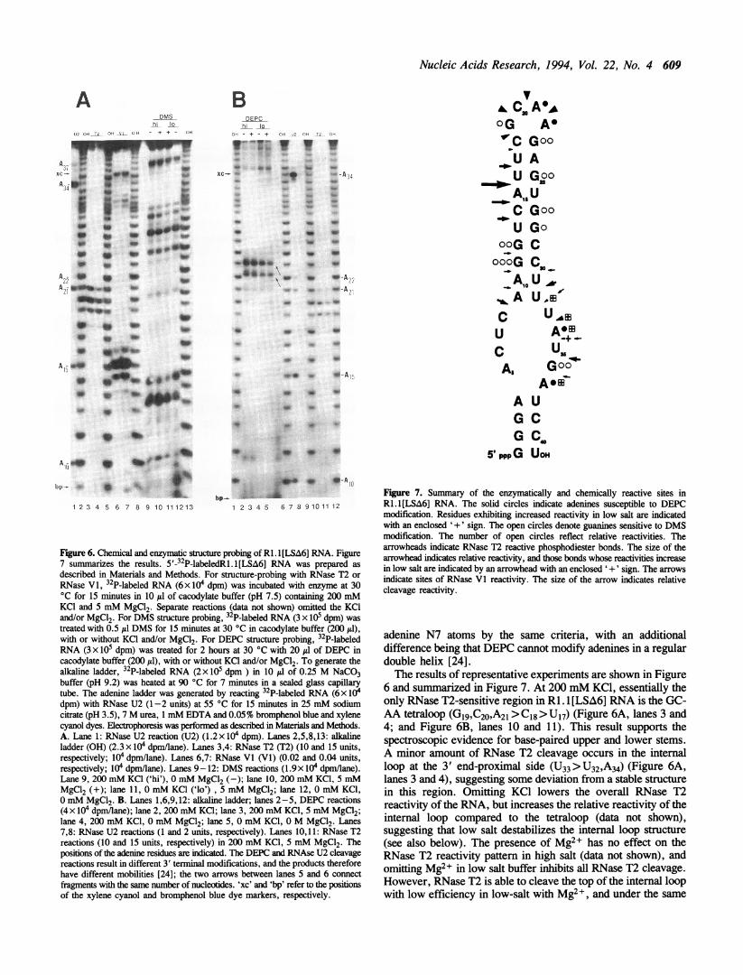
Figure 6. Chemical and enzymatic structure probing of R1.1[LSA6] RNA. Figure7 sumnuarizes the results. 5'-32P-labeledRi.l[LSA6] RNA was prepared asdescribed in Materials and Methods. For structure-probing with RNase T2 or
RNase VI, 32P-labeled RNA (6x 10⁴ dpm) was incubated with enzyme at 30'C for 15 minutes in 10 yd of cacodylate buffer (pH 7.5) containing 200 mMKCI and 5 mM MgCl2. Separate reactions (data not shown) omitted the KCIand/or MgCl2. For DMS structure probing, 32P-labeled RNA (3 x 10⁵ dpm) wastreated with 0.5 Id DMS for 15 minutes at 30 'C in cacodylate buffer (200 1l),with or without KCI and/or MgCl2. For DEPC structure probing, 32P-labeledRNA (3 x 10⁵ dpm) was treated for 2 hours at 30 'C with 20 yd of DEPC incacodylate buffer (200 I1), with or without KCI and/or MgCl2. To generate thealkaline ladder, 32P-labeled RNA (2 x 10⁵ dpm ) in 10 yd of 0.25 M NaCO3buffer (pH 9.2) was heated at 90 'C for 7 minutes in a sealed glass capillarytube. The adenine ladder was generated by reacting 32P-labeled RNA (6 x 10⁴dpm) with RNase U2 (1-2 units) at 55 'C for 15 minutes in 25 mM sodiumcitrate (pH 3.5), 7 M urea, 1 mM EDTA and 0.05% bromphenol blue and xylenecyanol dyes. Electrophoresis was performed as described in Materials and Methods.A. Lane 1: RNase U2 reaction (U2) (1.2x 10⁴ dpm). Lanes 2,5,8,13: alkalineladder (OH) (2.3 x 10⁴ dpm/lane). Lanes 3,4: RNase T2 (T2) (10 and 15 units,respectively; 104 dpm/lane). Lanes 6,7: RNase VI (VI) (0.02 and 0.04 units,respectively; I04 dpm/lane). Lanes 9-12: DMS reactions (1.9x 10⁴ dpm/lane).Lane 9, 200 mM KCI ('hi'), 0 mM MgCl2 (-); lane 10, 200 mM KCI, 5 mMMgCl2 (+); lane 11, 0 mM KCI ('lo') , 5 mM MgCl2; lane 12, 0 mM KCI,0 mM MgCl2. B. Lanes 1,6,9,12: aLkaline ladder; lanes 2-5, DEPC reactions(4 x 10⁴ dpm/lane); lane 2, 200 mM KCI; lane 3, 200 mM KCI, 5 mM MgCl2;lane 4, 200 mM KCI, 0 mM MgCl2; lane 5, 0 mM KCI, 0 M MgCI2. Lanes7,8: RNase U2 reactions (1 and 2 units, respectively). Lanes 10,11: RNase T2reactions (10 and 15 units, respectively) in 200 mM KCI, 5 mM MgCl2. Thepositions of the adenine residues are indicated. The DEPC and RNAse U2 cleavagereactions result in different 3' terminal modifications, and the products thereforehave different mobilities [24]; the two arrows between lanes 5 and 6 connectfragments with the same number of nucleotides. 'xc' and 'bp' refer to the positionsof the xylene cyanol and bromphenol blue dye markers, respectively.
V. Cm A,AOG AG'C GooU AU GooA,,UC GooU Go
ooG CoooG C,O
A U "C UAEU +
C U,,As Goo~
AomA UG CG C4,
5' pppG UOH
Fgure 7. Summary of the enzymatically and chemically reactive sites inRl.l[LSA6] RNA. The solid circles indicate adenines susceptible to DEPCmodification. Residues exhibiting increased reactivity in low salt are indicatedwith an enclosed '+' sign. The open circles denote guanines sensitive to DMSmodification. The number of open circles reflect relative reactivities. Thearrowheads indicate RNase T2 reactive phosphodiester bonds. The size of thearrowhead indicates relative reactivity, and those bonds whose reactivities increasein low salt are indicated by an arrowhead with an enclosed '+' sign. The arrowsindicate sites of RNase Vl reactivity. The size of the arrow indicates relativecleavage reactivity.
adenine N7 atoms by the same criteria, with an additionaldifference being that DEPC cannot modify adenines in a regulardouble helix [24].The results of representative experiments are shown in Figure
6 and summarized in Figure 7. At 200 mM KCl, essentially theonly RNase T2-sensitive region in RI. 1[LSA6] RNA is the GC-AA tetraloop (G19,C20,A21 > C18 > U17) (Figure 6A, lanes 3 and4; and Figure 6B, lanes 10 and 11). This result supports thespectroscopic evidence for base-paired upper and lower stems.A minor amount of RNase T2 cleavage occurs in the internalloop at the 3' end-proximal side (U33>U32,A34) (Figure 6A,lanes 3 and 4), suggesting some deviation from a stable structurein this region. Omitting KCI lowers the overall RNase T2reactivity of the RNA, but increases the relative reactivity of theinternal loop compared to the tetraloop (data not shown),suggesting that low salt destabilizes the internal loop structure(see also below). The presence of Mg2+ has no effect on theRNase T2 reactivity pattern in high salt (data not shown), andomitting Mg2+ in low salt buffer inhibits all RNase T2 cleavage.However, RNase T2 is able to cleave the top of the internal loopwith low efficiency in low-salt with Mg2+, and under the same
A-DMS
hi 1QL,2 OFi T.0i yo0, - + + - owl
w .
A25 S * _
* * lb
. _ ~~b
* 9 b

610 Nucleic Acids Research, 1994, Vol. 22, No. 4
conditions RNase III is able to cleave a second time in the internalloop. If these differences in enzymatic reactivity result solely frommonovalent ion-induced changes in RNA structure, those changesmust be quite subtle and largely undetectable using thesetechniques.As anticipated, RI. 1 [LSA6] RNA is sensitive to RNase VI,
but the cleavage pattern is quite selective. In 200 mM KCI, 5mM MgCl2 the most reactive region is the 5' side of the upperstem, with A15 as the most reactive residue. A lesser amount ofcleavage occurs at the flanking nucleotides U13, C14 and U16(Figure 6A, lanes 6 and 7). The reactivity of these sites isconsistent with the double-helical nature of the upper stem. Somereactivity is also observed at several nucleotides centered at Alo,which is also located in the upper stem. Interestingly, the 3' end-proximal segment of the internal loop is also cleaved by RNaseV1. Specifically, a significant amount of cleavage occurs at U35,which is also the primary RNase III cleavage site (Figure 6A,lanes 6 and 7). Additional cleavages are observed at flankingresidues A34 and G36, as well as C30, U31 and U32, indicatingthat this portion of the internal loop possesses (or has access to)a helical structure. There is no significant RNase VI reactivityof the sequence between A37 and the 3' end (Figure 6A), or thesequence between the 5' end and U8 (data not shown). The lackof RNase VI reactivity of these segments does not rule out thedouble-stranded nature of the lower stem, as nonuniformreactivities of dsRNA towards RNase VI have been observedelsewhere [29,30], and reflect the specifics of RNase VIinteraction with helical RNA. Moreover, both of these segmentsare resistant to RNase T2 (see above), supporting the double-helical nature of this region. Finally, RNase VI cleavage ofRI. 1[LSA6] RNA is greatly inhibited at low salt (data not shown),and omitting Mg2+ abolishes all cleavage, reflecting the divalentcation requirement of RNase VI [28].The DMS reactivity pattern of Ri . 1 [LSA6] RNA at 30°C in
200 mM KCl indicates that all guanine N7 atoms are reactive,with no guanine residue notably more reactive than the others(the methylation sensitivity of GI, G2 and G3 could not bedefinitively assessed, due to the compressed gel electrophoreticmobilities of the short RNA products). Moreover, the DMSreactivity pattern is insensitive to KCl or Mg2+ (Figure 6A,lanes 9-12). Specifically, we observe that the N7 of G36 in theinternal loop is no more reactive than the other guanine N7 atoms,indicating that G36 is not looped out or otherwise solventexposed. Interestingly, Gi9 in the tetraloop is significantly lessreactive than the other guanine residues, and the relative amountof DMS methylation of this residue does not increase at 90 °C(data not shown). The reduced reactivity may reflect the localelectronic and steric environment of G19 within the highlystructured context of the GCAA tetraloop [31,32]. The DEPCpattern indicates that A21 and A22 in the tetraloop are the mostreactive residues (Figure 6B, lanes 2-5). A smaller yetsignificant amount of reaction is observed at A34 and A37(A34> A37), whose DEPC sensitivities are enhanced in low salt.The low salt reactivity enhancement is not seen with the otheradenines in the upper stem-loop. The DEPC sensitivities of A3and A4 could not be definitively assessed, due to the small sizeof the reaction products and the differing mobilities of enzymeand DEPC-cleaved fragments (see also the legend to Figure 6).The overall DEPC reactivity pattern is insensitive to Mg2+ ineither the absence or presence of 200 mM KCI (Figure 6B, lanes2-5). The lack of reactivity of the adenine residues in the upper
[24]. Minor DEPC reactivity was observed with non-adenineresidues in the tetraloop and internal loop. The DEPC sensitivityof non-adenine nucleotides in non-double-helical regions has beennoted elsewhere [24]. This particular aspect of the DEPCreactivity pattern provides further evidence for the lack of acanonical double helical structure in the internal loop.
DISCUSSIONThe secondary structure of Rl.l[LSA6] RNAThe continuous chain of NOE connectivities and susceptibilityto RNase VI cleavage clearly show that the upper stem ofRI. [LSA6] RNA is an A form double helix, as predicted bysequence comparison (see [10] and references therein). The datafor the lower stem are less clear, as the imino resonances aretoo broadened by exchange to give detectable NOEs. However,given (i) the detectability (if not complete assignability) of allbut one to three of the twenty-two imino protons and (ii) theRNase T2 resistance of nucleotides near the 5' and 3' ends, weargue that the RI. [LSA6] RNA possesses a double-helical lowerstem. Also, it is possible that either the L-Q or N2-S NOE arisesfrom the terminal G:U pair in the lower stem.The monovalent salt concentration has a minor effect on the
structure of Ri. 1 [LSA6] RNA, as only one imino resonance (P)undergoes a significant shift. The relative chemical shifts ofresonances J and K also change slightly as a function of saltconcentration, but as the chain ofNOEs in the upper stem persiststhis is likely to represent only a minor structural perturbation.The salt-dependent, opposing trends in enthalpy and entropy ofupper and lower stem melting may indicate a conformationalchange in the internal loop (see also below). If this is the case,it must reflect a relatively small quantitative structural change,rather than a qualitative one. The increase in Tm values and freeenergies of melting with increasing ionic strength may influencethe energetics of interaction of RNase IH with its processingsubstrates. At physiological salt concentrations RNase Im cleavesonly primary sites, while at low salt secondary sites are alsorecognized, and otherwise unreactive RNAs are cleaved[7,10,15]. Given the salt-insensitivity of RI. 1 RNA secondarystructure, our results suggest that secondary site cleavage byRNase mII results from an altered enzyme-substrate interaction,rather than from a salt-induced change in RNA conformation.The R1.1 tetraloop, which is distant from the RNase HI
cleavage site and irrelevant to recognition [15], appears similarin conformation to a tetraloop with the same loop sequence andclosing base pair [31]. The Gi9 imino proton of the R1.1tetraloop exchanges slowly with solvent, as was observed withthe corresponding guanine of the model tetraloop. The GCAAtetraloop guanine imino proton is not predicted to hydrogen-bondto another nucleotide, but it may be protected from solventexchange by hydrogen binding to a water molecule [32]. TheRI. 1 tetraloop is cleaved by RNase T2 at Gl9, C20, A21 >C18> U17, and A21 and A22 are the most DEPC-reactiveresidues. Although the NMR analysis suggests that these adenines(and in particular the N6 protons) are part of a network ofhydrogen bonds, these bonds have small free energies offormation [32], and would not be expected to significantly affectthe chemical or enzymatic modification reactions.
The R1.1 internal loopThe RI. 1 internal loop is asymmetric, and cannot be a canonical
stem is evidence for their existence witiin an A-form double helix double helix. A lack of a rigid helical structure for this local
Nucleic Acids Research, 1994, Vol. 22, No. 4 611
structure is suggested by (i) the RNase T2 sensitivity of the 3'end-proximal portion of the internal loop, (ii) the DEPC-sensitivity of nucleotides within the internal loop, (iii) the absenceof measurable protection of several of the four imino protonsin the internal loop from exchange with solvent and (iv) theseparate melting transitions of the upper and lower stems. Theexchange-broadening of the imino protons in the internal loopand the lower stem is also evidence of flexibility. This flexibilitymay be responsible for the UV light-induced covalent crosslinkingof the two RNase TI-resistant oligonucleotides which encompassthe RI. 1 internal loop [33]. The UV reactivity pattern maysuggest the presence of interstrand base-stacking in the Ri. 1internal loop, which occurs in 5S RNA loop E, and is responsiblefor the UV sensitivity of this structure [34, 35].However, RNase HI efficiently cleaves Watson-Crick base-
paired dsRNA [5], so one might expect the internal loop topossess dsRNA character. Indeed, RNase VI cleavage of theU35-G36 phosphodiester bond (which is also the primary RNaseHI processing site) indicates that this portion of the Ri. 1 internalloop is engaged in (or has access to) a helical conformation. Asimilar coincidence of RNase VI and RNase III cleavage siteshas also been observed in an RNase 11 processing signal of phageX [36]. The unexceptional methylation sensitivity of the N7 atomof G36 suggests that this residue is not solvent exposed. This canbe contrasted with the strong chemical reactivity of the conservedguanine in the 5S rRNA internal loop E [37].
Similar internal loops are present in a number of other T7 andT3 RNase Im processing signals [5,6]. Apparently this motif wasselected to optimize efficient single cleavage of the phagetranscripts in vivo. Other types of asymmetric internal loops inunrelated phage and cellular substrates also enforce singleenzymatic cleavage [5,6]. It remains to be determined howprocessing signals with internal loops of different sequences andstructures interact with RNase HI to yield a similar outcome.
The effect of magnesiumMagnesium is absolutely required for RNase HI processing, soits influence on substrate structure is of particular interest. Thechemical and enzymatic reactivity of Ri. 1 RNA is largelyinsensitive to magnesium, but magnesium does shift theresonances of a number of unassigned imino protons, several ofwhich are within the internal loop, and it also affects the iminoproton of the Ag:U32 pair. It does not so affect the imino protonsin the upper stem or tetraloop, so its effect has some specificity.The spectroscopic changes are unlikely to reflect a majorconformational change in the internal loop, however, asmagnesium only perturbs these resonances, instead of abolishingthem or creating new ones (as seen, for example, in the bindingof argininamide to the bulge loop of TAR RNA [38]).Unfortunately, no further structural information could be gained,as no NOEs between any of these resonances were detected.Although perhaps not relevant to every processing signal,
magnesium may stabilize RNA tertiary structure important forRNAse Im reactivity, in addition to being an essential cofactorin the chemical step. In this regard, we have found that theinteraction of Ri. 1 RNA with a catalytically inactive mutant ofRNase II is significantly stabilized in the presence of magnesium(H.Li and A.W.N., unpublished experiments). Magnesium maytherefore play an important role in the determining the reactivityof other RNase HI substrates containing higher-order structuralfeatures.
Comparison with the thermodynamically predicted secondarystructureThe RNA structure prediction program MFOLD [39] predictsa lowest-energy secondary structure for RI. 1 [LSA6] RNA similarto that presented here, but with U7-Ag base-paired to U35 -A37.This structure is incompatible with our assignment of iminoresonance D to the Ag:U32 base pair. Moreover, the computer-predicted conformation has bases U32 -A34 in a bulge loop,which would coincide with several RNase V1 cleavage sites. Thelowest-energy structure which contains the full-length upper stemhas a similar bulge loop, but the next higher-energy structurepairs C6 with G36, and has no completely unopposed bulgeloops. This internal loop pairing scheme is supported by theunexceptional DMS reactivity of G36. This Ri 1.I[LSA6] RNAstructure differs from the lowest-energy conformation by less than3 kcal/mol, which is insignificant since MFOLD neglects mostnon Watson -Crick interactions other than G:U base pairs, anduses thermodynamic data collected in ionic conditions (1 M NaCl)different from those used here.For the same reason, we can only compare in a qualitative
fashion our thermodynamic parameters with those calculated byMFOLD. The parameters calculated for what we argue is themost likely secondary structure of RI. 1 RNA are very similarto those we observed in either salt condition (data not shown).We would expect the melting temperature to rise at higher ionicstrength, but fall if additional stabilizing non Watson -Crickinteractions were neglected, and perhaps the two effects cancel.
The dsRNA mimicry modelFinally, the NMR and structure probing data presented here, aswell as a previous mutational analysis [10], argue against theproposal of a more complicated internal loop folding motif [40].Specifically, if the 'dsRNA mimicry' tertiary folding werepresent, we would expect an imino proton resonance from theproposed A5:U33 pair, with accompanying NOEs to adjacentbase-pairs in the upper and lower stem, and these were notobserved.
CONCLUSIONSThe T7 RI. 1 RNase RI processing signal consists of two dsRNAstems, separated by an internal loop having a helical but relativelyloosely structured conformation.The conformation of the RI. 1 processing signal is largely
insensitive to the monovalent salt concentration, suggesting thatsecondary site versus primary site enzymatic cleavage of thissubstrate (and others) reflects an alteration in the enzyme-substratecomplex, rather than a change in RNA conformation.Magnesium appears to influence the structure of the internal
loop. In addition to serving as the catalytic cofactor, magnesiummay stabilize the proper substrate conformation for RNase HIprocessing reactivity.
ACKNOWLEDGEMENTSWe thank H.Li for providing purified T7 RNA polymerase andRNase HI, and D.M.Crothers for the use of the UVspectrophotometer. This work was supported by NIH GrantGM41283 (to A.W.N.) and GM41651 (to P.B.M.). D.C.S. isthe recipient of a Howard Hughes Medical Institute predoctoralfellowship.
612 Nucleic Acids Research, 1994, Vol. 22, No. 4
REFERENCES
1. St Johnston, D., Brown, N.H., Gall, J.G. and Jantsch, M. (1992) Proc.Natl. Acad. Sci USA 89, 10979-10983.
2. Haines, D.S., Strauss, K.I. and Gillepsie, D.H. (1991) J. Cell. Biochem.46, 9-20.
3. Libonati, M., Carsana, A. and Furia, A. (1980) Mol. Cell. Biochem. 31,147-164.
4. Robertson, H.D., Webster, R.E. and Zinder, N.D. (1968) J. Bio. Chem.243, 82-91.
5. Court, D. (1993) in Control of messenger RNA Stability (Belasco, J. G.,and Brawerman, G., eds) Academic Press, NY.
6. Dunn, J.J. and Studier, F.W. (1983) J. Mol. Biol. 166, 477-535.7. Dunn, J.J. (1976) J. Biol. Chem. 251, 3807-3814.8. Saito, H. and Richardson, C.C. (1981) Cell 27, 533-542.9. Nicholson, A.W., Niebling, K.R., McOsker, P.L. and Robertson, H.D.
(1988)Nucleic Acids Res. 46, 1577-1591.10. Chelladurai, B.S., Li, H. and Nicholson, A.W. (1991) Nucleic Acids Res.
19, 1759-1766.11. Panayotatos, N. and Truong, K. (1985) Nucleic Acids Res. 13, 2227-2240.
12. Linn, T. and St. Pierre, R. (1990) J. Bacteriol. 172, 1077-1084.13. Studier, F.W., Rosenberg, A.H., Dunn, J.J. and Dubendorff, J.W. (1990)
Methods Enzymol. 185, 60-89.14. Ziemke, P. and McCarthy, J.E.G. (1992) Biochim. Biophys. Acta 1130,
297-306.15. Chelladurai, B.S., Li, H., Zhang, K. and Nicholson, A.W. (1993)
Biochemistry 32, 7549-7558.16. Grodberg, J. and Dunn, J.J. (1988) J. Bacteriol. 170, 1245-1253.17. Li, H., Chelladurai, B.S., Zhang, K. and Nicholson, A.W. (1993) Nucleic
Acids Res. 21, 1919-1925.18. Milligan, J.F., Groebe, D.R., Witherell, G.W. and Uhlenbeck, O.C. (1987)
Nucleic Acids Res. 15, 8783 -8798.19. Wyatt, J.R., Chastain, M. and Puglisi, J. D. (1991) Biotechniques 11,
764-769.20. Kime, M.J. and Moore, P.B. (1983) FEBS Lett. 153, 199-203.21. Kime, M.J. and Moore, P.B. (1983) Biochemistry 22, 2615-2622.22. Takegoshi, K., Tsuda, S. and Hikichi, K. (1990) J. Mag. Res. 89, 399-405.23. 'fit', J. Tirado-Rives, Dept. of Chemistry, Yale University.24. Ehresmann, C., Baudin, F., Mougel, M., Romby, P., Ebel, J-P. and
Ehresmann, B. (1987) Nucleic Acids Res. 15, 9109-9128.25. Peattie, D.A. and Gilbert, W. (1980) Proc. Natl. Acad. Sci. USA 77,
4679-4682.26. Szewczak, A.A., White, S.A., Gewirth, D.T. and Moore, P.B. (1990) Nucleic
Acids Res. 18, 4139-4142.27. Searle, M.S. and Williams, D.H. (1993) Nucleic Acids Res. 21, 2051-2056.28. Lowman, H.B. and Draper, D.E. (1986) J. Biol. Chem. 261, 5396-5403.29. Lockard, R.E. and Kumar, A. (1981) Nucleic Acids Res. 9, 5125-5140.30. Auron, P.E., Weber, L.D. and Rich, A. (1982) Biochemistry 21, 4700-4706.31. Heus, H.A. and Pardi, A. (1991) Science 253, 191-194.32. SantaLucia, J., Kierzek, R. and Turner, D.H. (1992) Science 256, 217-219.33. Robertson, H.D. (1990) Methods Enzymol. 181, 189-202.34. Branch, A.D., Benenfeld, B.J., and Robertson, H.D. (1985) Proc. Natl.
Acad. Sci USA 82, 6590-6594.35. Wimberly, B., Varani, G., and Tinoco, I. (1993) Biochemistry 32,
1078-1087.36. Steege, D.A., Cone, K.C., Queen, C. and Rosenberg, M. (1987) J. Biol.
Chem. 262, 17651-17658.37. Romaniuk, P.J. , deStevenson, I.L., Ehresmann, C., Romby, P., and
Ehresmann, B. (1988) Nucleic Acids Res. 16, 2295-2312.38. Puglisi, J.D., Tan, R., Culman, B.J., Frankel, A.D. and Williamson, J.R.
(1992) Science 257, 76-80.39. Zuker, M. (1989) Science 244, 48-52.40. Robertson, H.D. and Barany, F. (1978) in Proceedings of the 12th FEBS
Congress, Pergamon Press, NY pp. 285-295.
Related Documents