Pergamon NamSku~ Matials. Vol. 8. No. 2. pp. 179-189.1997 Elsevier Science Ltd Copyi@ 0 1997ActaMetallurgica Inc. printed inthe USA. All rights rcsavd 0965477397 $17.00 + .oo PlI s0965-9773(97)00001-9 STRUCTURAL CHARACTERIZATION AND OPTICAL PROPERTIES OF NANOMETER-SIZED SnO2 CAPPED BY STEARIC ACID Xiaochun Wu*, Bingsuo Zou*, Jiren Xu*, Baolong Yut, Guoqing Tang?, Guilan Zhangf, Wenju Chenf *Institute of Physics, Chinese Academy of Sciences, Beijing, 100080, P.R. China TInstitute of Modem Optics, Nankai University, Tianjin, 30007 1, P.R. China (Accepted January 7,1997) Abstract-Structure and optical properties of nanometer-sized SnOz capped by a layer of stearic acid are investigated using TEM, XRD, ESR. UV-visible absorption spectrum, photoluminescence spectra, IR absorption spectra and Z-scan techniques. It is found that such capped Sn02 samples display new optical properties, which will enlarge their applications in nonlinear optics. The relationship between structure and opticalpropertiesof the Sn02 nanoparticles is also discussed. 1. INTRODUCTION From the viewpoints of both theoretical research and potential applications, nanoparticles have recently attracted great attention due to their unique structures and properties (1). On the one hand, with decreasing particle size, the electronic states of nanoparticles become discrete from continuous energy bands in bulk and the lowest exciton energy level blue-shifts. This is the so-called “quantum size effect” (2). Therefore, the study of the transition between their electronic levels has very important theoretical significance for understanding the transformation of matter from the macroscopic to microscopic state. On the other hand, the enhanced nonlinear optical responses of nanoparticles observed experimentally (3), making nanoparticles an attractive nonlinear optical material, has stimulated further research. For the past ten years the study of nanoparticles has made great progress (4). In order to enlarge the materiakalm and find new nonlinear optical materials, SnO;! was chosen as the subject for this work, because bulk St@ is an important semiconductor material and it also has relatively large nonresonant third-order nonlinear optical susceptibility among inorganic oxides (5). In order to study its optical properties, nanometer-sized SnOz. capped with a layer of steak acid was synthesized and characterized through a series of spectroscopic techniques. 179

Welcome message from author
This document is posted to help you gain knowledge. Please leave a comment to let me know what you think about it! Share it to your friends and learn new things together.
Transcript
Pergamon NamSku~ Matials. Vol. 8. No. 2. pp. 179-189.1997
Elsevier Science Ltd Copyi@ 0 1997 Acta Metallurgica Inc.
printed in the USA. All rights rcsavd 0965477397 $17.00 + .oo
PlI s0965-9773(97)00001-9
STRUCTURAL CHARACTERIZATION AND OPTICAL PROPERTIES OF NANOMETER-SIZED SnO2
CAPPED BY STEARIC ACID
Xiaochun Wu*, Bingsuo Zou*, Jiren Xu*, Baolong Yut, Guoqing Tang?, Guilan Zhangf, Wenju Chenf
*Institute of Physics, Chinese Academy of Sciences, Beijing, 100080, P.R. China TInstitute of Modem Optics, Nankai University, Tianjin, 30007 1, P.R. China
(Accepted January 7,1997)
Abstract-Structure and optical properties of nanometer-sized SnOz capped by a layer of stearic acid are investigated using TEM, XRD, ESR. UV-visible absorption spectrum, photoluminescence spectra, IR absorption spectra and Z-scan techniques. It is found that such capped Sn02 samples display new optical properties, which will enlarge their applications in nonlinear optics. The relationship between structure and opticalpropertiesof the Sn02 nanoparticles is also discussed.
1. INTRODUCTION
From the viewpoints of both theoretical research and potential applications, nanoparticles have recently attracted great attention due to their unique structures and properties (1). On the one hand, with decreasing particle size, the electronic states of nanoparticles become discrete from continuous energy bands in bulk and the lowest exciton energy level blue-shifts. This is the so-called “quantum size effect” (2). Therefore, the study of the transition between their electronic levels has very important theoretical significance for understanding the transformation of matter from the macroscopic to microscopic state. On the other hand, the enhanced nonlinear optical responses of nanoparticles observed experimentally (3), making nanoparticles an attractive nonlinear optical material, has stimulated further research.
For the past ten years the study of nanoparticles has made great progress (4). In order to enlarge the materiakalm and find new nonlinear optical materials, SnO;! was chosen as the subject for this work, because bulk St@ is an important semiconductor material and it also has relatively large nonresonant third-order nonlinear optical susceptibility among inorganic oxides (5). In order to study its optical properties, nanometer-sized SnOz. capped with a layer of steak acid was synthesized and characterized through a series of spectroscopic techniques.
179
180 X Wu, B Zou, J Xu. B Yu, G TANG, G ZHANG, W CHEN
2. EXPERIMENTAL
2.1 The Synthesis of Nanometer-sized S& Sample
Nanometer-sized Sn% samples were prepared by the microemulsion method. 10 ml of newly-made SnC4 aqueous solution was added to 100 ml of xylene containing 1 x10” mol of stearic acid. The resulting mixture was stirred to form a microemulsion, then 20 ml of 0.1 M NaOH aqueous solution was added to the microemulsion to produce hydroxide under stirring. When the reaction was completed, the reaction product was extracted into the organic phase, because its surface was capped with a layer of stearic acid, and then the reaction system was allowed to stand still in order to separate the organic phase from the water phase. The water phase was discarded and the organic phase was refluxed 0.5 hr, then let stand at room temperature. The small amount of water on the bottom of the flask was discarded and the organic phase was washed with distilled water several times to further purify the reaction product. After purification, the organic phase was distilled to remove the residual water, then nanometer-sized Sn& organosol was formed. One part of Sn@ organosol (sample 1) was used to carry out TEM, UV-vis absorption, fluorescence and Z-scan measurements. The other part of SnO;! organosol was distilled to dryness at reduced pressures to obtain Sna powder capped with stearic acids, which was divided into two parts. One is sample 2, and the other was heated at 400°C within a muffle to remove stearic acid (sample 3). The two powder samples were used to carry out XRD, FTIR, and ESR measurements.
The reaction schematic diagram is shown as follows:
stearic acid stirring
SnC14 aqueous solution w mixed solution w organic solvent dropping OH-
refluxing
Sn(OH)nm+microemulsion 4 nanometer-sized Sn02 organosol washing
part of Sn02 organosol as sample 1
nanometer-sized Sn02(distilkd todtyness)+ powder ofi capped Sn02 powder as sample 2 (km off SU&C~~ ts) bareSn0pwderas sample 3
2.2 Measurement Instruments and Methods
The size of the nanoparticle organosol was measured through Philips EM-400 transmission electron microscopy (TEM). The average particle diameter was 7 nm. The XRD measurements were performed on Rigaku D/MAX-RA X-ray diffractometer using Cu K, radiation source. Absorption and photoluminescence spectra were recorded on a Shimadzu UV-240 spectropho- tometer and a RF-540 fluorimeter, respectively. IR absorption spectra were measured on Nicolet NX-240 FIIR spectrometer: wavenumber precision is 0.1 cm- ’ . Mid-IR and far-IR measurements adopted KBr pelleting and nujol mull coating, respectively. The transmittances of two were normahzed through computer and merged into one figure to output. ESR measurement was performed on Bruker 2OOD-SRC spectrometer. The magnetic field was calibrated with Mn2+ as a reference sample.
NANOMETER-SIZED SNO* CAPPED BY STEARC ACID 181
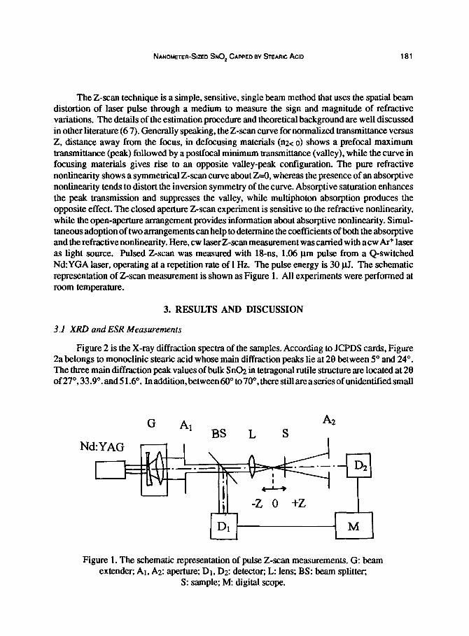
The Z-scan technique is a simple, sensitive, single beam method that uses the spatial beam distortion of laser pulse through a medium to measure the sign and magnitude of refractive variations. The details of the estimation procedure and theoretical background are well discussed in other literature (6 7). Generally speaking, the Z-scan curve for normalized transmittance versus Z, distance away from the focus, in defocusing materials (nZ< 0) shows a prefocal maximum transmittance (peak) followed by a postfocal minimum transmittance (valley), while the curve in focusing materials gives rise to an opposite valley-peak configuration. The pure refractive nonlinearity shows a symmetrical Z-scan curve about z--O, whereas the presence of an absorptive nonlinearity tends to distort the inversion symmetry of the curve. Absorptive saturation enhances the peak transmission and suppresses the valley, while multiphoton absorption produces the opposite effect. The closed aperture Z-scan experiment is sensitive to the refractive nonlinearity, while the open-aperture arrangement provides information about absorptive nonlinearity. Simul- taneous adoption of two arrangements can help to determine the coefficients of both the absorptive and the refractive nonlinearity. Here, cw laser Z-scan measurement was carried with acw Ar+ laser as light source. Pulsed Z-scan was measured with 18-ns, 1.06 pm pulse from a Q-switched Nd:YGA laser, operating at a repetition rate of 1 Hz. The pulse energy is 30 pJ. The schematic representation of Z-scan measurement is shown as Figure 1. All experiments were performed at room temperature.
3. RESULTS AND DISCUSSION
3.1 XRD and ESR Measurements
Figure 2 is the X-ray diffraction spectra of the samples. According to JCPDS cards, Figure 2a belongs to monoclinic stearic acid whose main diffraction peaks lie at 28 between 5” and 24’. The three main diffraction peak values of bulk Sn@ in tetragonal rutile structure are located at 28 of 27”, 33.9”. and 5 1.6”. In addition, between 6OO” to 70”, there still are a series of unidentified small
G Al A2
BS L s
Figure 1. The schematic representation of pulse Z-scan measurements. G: beam extender; Al, AZ: aperture: Dt, D2: detector; L: lens; BS: beam splitter;
S: sample: M: digital scope.
182 X Wu, B Zou, J Xu, B Yu, G TANG, G ZHANG, W CHEN
.2 51.4 64.2
I 26.5 C
10 20 30 40 50 60
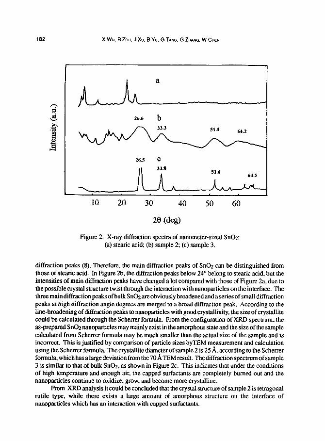
Figure 2. X-ray diffraction spectra of nanometer-sized Sn02: (a) steak acid; (b) sample 2; (c) sample 3.
dilfraction peaks (8). Therefore, the main diffraction peaks of SnO;! can be distinguished from those of stearic acid. In Figure 2b, the diffraction peaks below 24” belong to steak acid, but the intensities of main diffraction peaks have changed a lot compared with those of Figure 2a, due to the possible crystal structure twist through the interaction with nanoparticles on the interface. The three main diffraction peaks of bulk SnO.L are obviously broadened and a series of small diffraction peaks at high diffraction angle degrees are merged to a broad diffraction peak. According to the line-broadening of diffraction peaks to nanoparticles with good crystallinity, the size of crystallite could be calculated through the Scherrer formula. From the configuration of XRD spectrum, the as-prepared SnO;! nanoparticles may mainly exist in the amorphous state and the size of the sample calculated from Scherrer formula may be much smaller than the actual size of the sample and is incorrect. This is justified by comparison of particle sizes byTEM measurement and calculation using the Scherrer formula. The crystallite diameter of sample 2 is 25 A, according to the Scherrer formula, which has a large deviation from the 70 A TEM result. The diffrac tion spectrum of sample 3 is similar to that of bulk Sn@, as shown in Figure 2c. This indicates that under the conditions of high temperature and enough air, the capped surfactants are completely burned out and the nanoparticles continue to oxidize, grow, and become more crystalline.
From XRD analysis it could be concluded that the crystal structure of sample 2 is tetragonal rutile type, while there exists a large amount of amorphous structure on the interface of nanoparticles which has an interaction with capped surfactants.
NANOMETER-SIZED SNO, CAPPED BY STEARIC ACIO 183
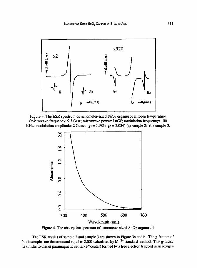
Figure 3. The ESR spectrum of nanometer-sized SnOz organosol at room temperature (microwave frequency: 9.3 GHz; microwave power: 1 mW, modulation frequency: 100
KHz; modulation amplitude: 2 Gauss: gl = 1.981; gz = 2.034) (a) sample 2; (b) sample 3.
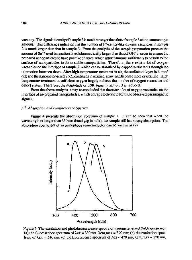
300 400 500 600 700
wavel~~ (nm) Figure 4. The absorption spectrum of nanometer-sized St@ organosol.
The ESR results of sample 2 and sample 3 are shown in Figure 3a and b. The g-factors of both samples are the same and equal to 2.001 calculated by Mn2+ standard method. This g-factor is similar to that of paramagnetic center (F+ center) formed by a free electron trapped in an oxygen
184 X Wu, B Zou, J Xu, B Yu, G TANG, G ZHANG, W CHEN
vacancy. The signal intensity of sample 2 is much stronger than that of sample 3 at the same sample amount. This difference indicates that the number of F+-center-like oxygen vacancies in sample 2 is much larger than that in sample 3. From the analysis of the sample preparation process the amount of Sn4+ used in reaction is stoichiometrically larger than that of OH- in order to ensure the prepared nanoparticles to have positive charges, which attract anionic surfactants to adsorb to the surface of nanoparticles to form stable nanoparticles. Therefore, there exist a lot of oxygen vacancies on the interface of sample 2, which can be stabilized by capped surfactants through the interaction between them. After high temperature treatment in air, the surfactant layer is burned off; and the nanometer-sized Sn@ continues to oxidize, grow, and become more crystalline. High temperature treatment in suffkient oxygen largely reduces the number of oxygen vacancies and defect states. Therefore, the magnitude of ESR signal in sample 3 is reduced.
From the above analysis it may be concluded that there are a lot of oxygen vacancies on the interface of as-prepared nanoparticles, which entrap electrons to form the observed paramagnetic signals.
3.2 Absorption and Luminescence Spectra
Figure 4 presents the absorption spectrum of sample 1. It can be seen that when the wavelength is longer than 350 nm (band gap in bulk), the sample still has strong absorption. The absorption coefficient of an amorphous semiconductor can be written as (9)
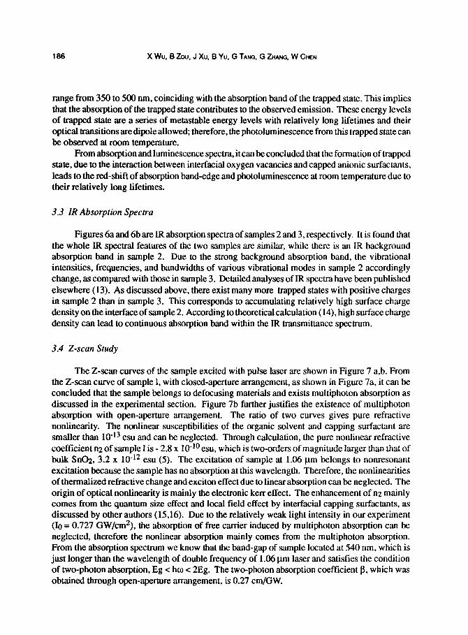
300 400 500 600 700
WW1~t.h (nm)
Figure 5. The excitation and photoluminescence spectra of nanometer-sized Sn@ organosol: (a) the fluorescence spectrum of hex = 330 nm, hem,max = 390 nm; (b) the excitation spec- trum of Aem = 540 nm; (c) the fluorescence spectrum of hex = 470 nm, hem,max = 520 nm.
NANOMETER-SIZED SNO, CAPPED BY STEARIC ACID 185
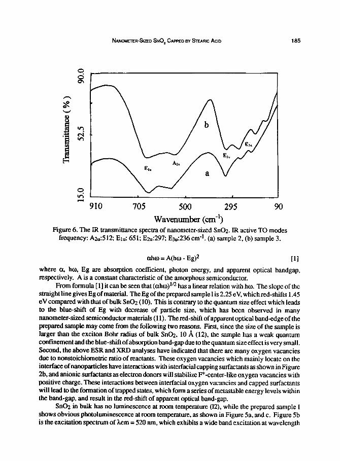
705 500 295
Wavenumber (cm-‘) Figure 6. The IR transmittance spectra of nanometer-sized Sn@. IR active TO modes
frequency: A2,:512; Et”: 651; Ezu:297; E3”:236 cm-l. (a) sample 2, (b) sample 3.
ahw = A(ho - Eg)2 111
where a, ho,, Eg are absorption coefficient, photon energy, and apparent optical bandgap, respectively. A is a constant characteristic of the amorphous semiconductor.
From formula [l] it can be seen that (aho)ln has a linear relation with ho. The slope of the straight line gives Eg of material. The Eg of the prepared sample 1 is 2.25 eV, which red-shifts 1.45 eV compared with that of bulk SnOz (10). This is contrary to the quantum size effect which leads to the blue-shift of Eg with decrease of particle size, which has been observed in many nanometer-sized semiconductor materials (11). The red-shift of apparent optical band-edge of the prepared sample may come from the following two reasons. First, since the size of the sample is larger than the exciton Bohr radius of bulk Sn@, 10 A (12), the sample has a weak quantum confinement and the blue-shift of absorption band-gap due to the quantum size effect is very small. Second, the above ESR and XRD analyses have indicated that there are many oxygen vacancies due to nonstoichiometric ratio of reactants. These oxygen vacancies which mainly locate on the interface of nanoparticles have interactions with interfacial capping surfactants as shown in Figure 2b, and anionic surfactants as electron donors will stabilize F+-center-like oxygen vacancies with positive charge. These interactions between inter-facial oxygen vacancies and capped surfactants will lead to the formation of trapped states, which form a series of metastable energy levels within the band-gap, and result in the red-shift of apparent optical band-gap.
SnOz in bulk has no luminescence at room temperature (12), while the prepared sample 1 shows obvious photoluminescence at room temperature, as shown in Figure 5a, and c. Figure 5b is the excitation spectrum of hem = 520 nm, which exhibits a wide band excitation at wavelength
188 X Wu, B Zou. J Xu, B Yu, G TANG, G ZHANG, W CHEN
range from 350 to 500 nm, coinciding with the absorption band of the trapped state. This implies that the absorption of the trapped state contributes to the observed emission. These energy levels of trapped state are a series of metastable energy levels with relatively long lifetimes and their optical transitions are dipole allowed; therefore, the photoluminescence from this trapped state can be observed at room temperature.
From absorption and luminescence spectra, it can be concluded that the formation of trapped state, due to the interaction between interfacial oxygen vacancies andcapped anionic surfactants, leads to the red-shift of absorption band-edge and photoluminescence at room temperature due to their relatively long lifetimes.
3.3 IR Absorption Spectra
Figures 6a and 6b are IR absorption spectra of samples 2 and 3, respectively. It is found that the whole IR spectral features of the two samples are similar, while there is an IR background absorption band in sample 2. Due to the strong background absorption band, the vibrational intensities, frequencies, and bandwidths of various vibrational modes in sample 2 accordingly change, as compared with those in sample 3. Detailed analyses of IR spectra have been published elsewhere (13). As discussed above, there exist many more trapped states with positive charges in sample 2 than in sample 3. This corresponds to accumulating relatively high surface charge density on the interface of sample 2. According to theoretical calculation (14) high surface charge density can lead to continuous absorption band within the IR transmittance spectrum.
3.4 Z-scan Study
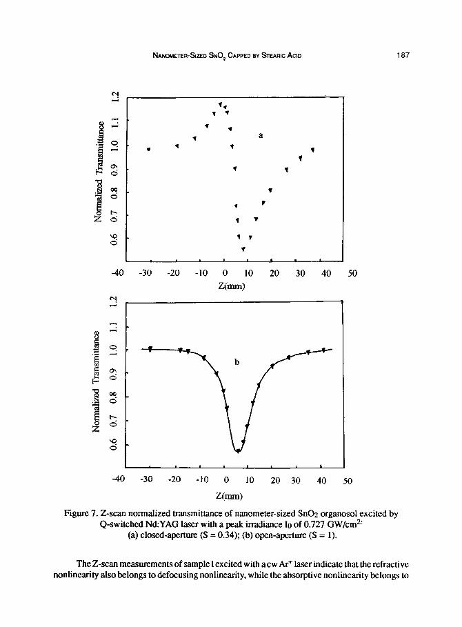
The Z-scan curves of the sample excited with pulse laser are shown in Figure 7 a,b. From the Z-scan curve of sample 1, with closed-aperture arrangement, as shown in Figure 7a, it can be concluded that the sample belongs to defocusing materials and exists multiphoton absorption as discussed in the experimental section. Figure 7b further justifies the existence of multiphoton absorption with open-aperture arrangement. The ratio of two curves gives pure refractive nonlinearity. The nonlinear susceptibilities of the organic solvent and capping surfactant are smaller than lo-l3 esu and can be neglected. Through calculation, the pure nonlinear refractive coefficient n2 of sample 1 is - 2.8 x lO-‘O esu, which is two-orders of magnitude larger than that of bulk Sri@. 3.2 x lo-l2 esu (5). The excitation of sample at 1.06 pm belongs to nonresonant excitation because the sample has no absorption at this wavelength. Therefore, the nonlinearities of thermalized refractive change and exciton effect due to linear absorption can be neglected. The origin of optical nonlinearity is mainly the electronic kerr effect. The enhancement of n2 mainly comes from the quantum size effect and local field effect by interfacial capping surfactants, as discussed by other authors (1516). Due to the relatively weak light intensity in our experiment (IO = 0.727 GW/cm2), the absorption of free carrier induced by multiphoton absorption can be neglected, therefore the nonlinear absorption mainly comes from the multiphoton absorption. From the absorption spectrum we know that the band-gap of sample located at 540 nm, which is just longer than the wavelength of double frequency of 1.06 pm laser and satisfies the condition of two-photon absorption, Eg < ho c 2Eg. The two-photon absorption coefficient p, which was obtained through open-aperture arrangement, is 0.27 cm/GW.
NANOMEIER-SIZED SNO, CAPPED BY STEARK) ACLD 187
.
1 i
1 1
1
l 1
1
Figure 7. Z-scan normalized transmittance of nanometer-sized Sn02 organosol excited by Q-switched Nd:YAG laser with a peak irradiance IO of 0.727 GW/cm*’
(a) closed-aperture (S = 0.34); (b) open-aperture (S = 1).
The Z-scan measurements of sample 1 excited with a cw Ar+ laser indicate that the refractive nonlinearity also belongs to defocusing nonlinearity, while the absorptive nonlinearity belongs to
188 X Wu, 6 Zou, J Xu, B Yu, G TANG, G ZHANG, W CHEN
saturation absorption. The n2 of sample 1 at 5 14.5 nm is -6.6 x 10m5 esu. The saturation absorption light intensity Isat is lower than 10 W/cm2. The excitation of sample with a cw AI-+ laser at 5 14.5 nm belongs to resonant excitation because excitation wavelength is shorter than band-edge wavelength. From the linear absorption spectrum, on the one hand, it is known that the excitation of the prepared sample at h = 5 14.5 nm comes from trapped state on the interface of nanoparticles, which shows strong electron-lattice interaction due to its strong interaction with capped surfactants and disorder of interfacial structure (17,18). The strong electron-lattice interaction indicates that the system has high nonirradiative efficiency, which can lead to large thermalized refractive nonlinearity at strong laser irradiation. Therefore, the large n2 of the sample at 514.5 nm comes from thermalized refractive change. On the other hand, those trapped states exist in the form of metastable states, which have relatively long lifetimes. Their excitation under strong laser irradiation can lead to the nonlinear saturation absorption. The low saturation intensity of the sample is due to the long lifetimes and relatively high absorption coefficient of those trapped states.
4. CONCLUSIONS
From the above experiments and discussion, it can be seen that the prepared nanometer-sized Sn@ organosol exhibits a series of new linear and nonlinear optical properties due to its special structural features. They can be summarized as follows: (i) Through XRD, it can be concluded that the as-prepared nanometer-sized Sn02 sample mainly exists in the form of amorphous state and has interaction with capped surfactants. (ii) The existence of P-center-like oxygen vacancies is justified through ESR spectrum. (iii) The nanometer-sized SnO;! organosol shows red-shift of absorption band-edge due to the formation of metastable energy levels of capped states within the prohibited band. These me&table energy levels can fluoresce at room temperature due to their relatively long lifetimes. (iv) IR spectrum is indicative of a high surface charge density. (v) From the pulse laserz-scan study at the wavelength of l.O6pm, n2 of the sample is -2.8 x lo-‘*esu, larger than that of the bulk. Due to the red-shift of band-gap, the two-photon absorption is also observed and p is 0.27 cm/GW. (vi) From the cw laser Z-scan study at the wavelength of 514.5 nm, the n2 of the sample is -6.6 x 10e5 esu and belongs to thermally induced refractive nonlinearity. Due to the excitation of metastable energy levels of long life-time at this wavelength, low saturation absorption intensity is observed and the saturation light intensity Isat is lower than 10 W/cm2.
These new optical properties can be reasonably explained by its special interfacial structures under nanometer-sized range. These new optical properties also enlarge the applications of SnGz in nonlinear optics, such as optical switching and optical limiting.
ACKNOWLEDGMENT
The authors acknowledge the National Natural Science Foundation of China for financial support.
REFERENCES
1. Henglein, A., Chemical Review, 1989.89.1861. 2. Wang, Y., and Herron, N., Journal of Physical Chemistry, 1987,91,5005. 3. Jain, K.R., and Lii, R.C., Journal of Optical Society ofAmerica, 1983,73,647.
NANOMETER-SIZED SNO, CAPPED BY STEARK) ACID 189
4. 5.
6. 7.
8.
9. 10. 11. 12. 13. 14. 15. 16. 17. 18.
Yoffe, D.A., Advanced Physics, 1993,42, 173. Nie, W.J., Advanced Materials, 1993,5,520.
Mansor, S.B., Said, A.A., VanStryland. W.E., Optical Letters, 1989, 14.955. Mansor,S.B.,Said,A.A.,VanShyland,W.E.,IEEEJournalofQuanhunElectronics, 1990,26,760. Zhang, D.Y., Wang, D. Zh., Wang, G.M., Wang, Zh., and Wu,Y.H.,Acta Physica Sinica, 1991.40, 844. Mill, G., Li. Z.G., and Meisel, D., Journal of Physical Chemistry, 1988,92,822. Arlinghaus, F.J., Journal Physical Chemistry of Solids, 1974.35,931.
Brus, L.E., Applied Physics A, 1991,53,465. DeMurcia, M., Egee, M., Fiilard, J.P., Journal Physical Chemistry of Solids, 1977,39,629. Wu, X.C., Tang, G.Q., Zhang, G.L., et al.,Acta Optica Sinica, 1995.15, 1355. Knipp, P.A., Reineeke, T.L., Physics Review B, 1992,46, 10310. Schmitt-Rink, S., Miller, D.A.B., Chemla, S.D., Physics Review B, 1987,35,8113. Wang, Y., Act. Chem. Research, 1991,24, 133. Galperin. YuM., Katpov, V.G., and Kozub, V.I., Advanced Physics, 1989,38,669. Pnevmatikos,St.,Yanovitakii, O., Friggis,Th, andEconomou, E.N., PhysicsReviewfetters, 1992, 68.2370.
Related Documents