Int. J. Mol. Sci. 2009, 10, 221-231; doi:10.3390/ijms10010221 International Journal of Molecular Sciences ISSN 1422-0067 www.mdpi.com/journal/ijms Article Structural Antitumoral Activity Relationships of Synthetic Chalcones Cesar Echeverria 1 , Juan Francisco Santibañez 1 , Oscar Donoso-Tauda 2 , Carlos A. Escobar 2 and Rodrigo Ramirez-Tagle 2, * 1 Instituto de Nutrición y Tecnología de los Alimentos, Universidad de Chile, El Líbano 5524, Macul, Santiago, Chile. E-Mails: [email protected] (C. E.); [email protected] (J. S.) 2 Universidad Andres Bello, Departamento de Ciencias Químicas, Av. República 275, Santiago, Chile. E-Mails: [email protected] (O. D.); [email protected] (C. E.) * Author to whom correspondence should be addressed; E-Mail: [email protected]; Tel. +56-2-6615880 Received: 30 September 2008; in revised form: 31 December 2008 / Accepted: 4 January 2009 / Published: 9 January 2009 Abstract: Relationships between the structural characteristic of synthetic chalcones and their antitumoral activity were studied. Treatment of HepG2 cells for 24 h with synthetic 2’-hydroxychalcones resulted in apoptosis induction and dose-dependent inhibition of cell proliferation. The calculated reactivity indexes and the adiabatic electron affinities using the DFT method including solvent effects, suggest a structure-activity relationship between the Chalcones structure and the apoptosis in HepG2 cells. The absence of methoxy substituents in the B ring of synthetic 2’-hydroxychalcones, showed the mayor structure-activity pattern along the series. Keywords: Antitumoral activity; DFT; structure-activity relationships. 1. Introduction Dietary flavonoids, commonly present in edible plants, are known to have beneficial effects, such as antioxidative effects, tumor cell growth inhibitory activity, and apoptosis induction in cancer cell OPEN ACCESS

Welcome message from author
This document is posted to help you gain knowledge. Please leave a comment to let me know what you think about it! Share it to your friends and learn new things together.
Transcript
Int. J. Mol. Sci. 2009, 10, 221-231; doi:10.3390/ijms10010221
International Journal of Molecular Sciences
ISSN 1422-0067 www.mdpi.com/journal/ijms
Article Structural Antitumoral Activity Relationships of Synthetic Chalcones Cesar Echeverria 1, Juan Francisco Santibañez 1, Oscar Donoso-Tauda 2, Carlos A. Escobar 2 and Rodrigo Ramirez-Tagle 2,* 1 Instituto de Nutrición y Tecnología de los Alimentos, Universidad de Chile, El Líbano 5524,
Macul, Santiago, Chile. E-Mails: [email protected] (C. E.); [email protected] (J. S.) 2 Universidad Andres Bello, Departamento de Ciencias Químicas, Av. República 275, Santiago,
Chile. E-Mails: [email protected] (O. D.); [email protected] (C. E.) * Author to whom correspondence should be addressed; E-Mail: [email protected];
Tel. +56-2-6615880 Received: 30 September 2008; in revised form: 31 December 2008 / Accepted: 4 January 2009 / Published: 9 January 2009
Abstract: Relationships between the structural characteristic of synthetic chalcones and their antitumoral activity were studied. Treatment of HepG2 cells for 24 h with synthetic 2’-hydroxychalcones resulted in apoptosis induction and dose-dependent inhibition of cell proliferation. The calculated reactivity indexes and the adiabatic electron affinities using the DFT method including solvent effects, suggest a structure-activity relationship between the Chalcones structure and the apoptosis in HepG2 cells. The absence of methoxy substituents in the B ring of synthetic 2’-hydroxychalcones, showed the mayor structure-activity pattern along the series. Keywords: Antitumoral activity; DFT; structure-activity relationships.
1. Introduction
Dietary flavonoids, commonly present in edible plants, are known to have beneficial effects, such as antioxidative effects, tumor cell growth inhibitory activity, and apoptosis induction in cancer cell
OPEN ACCESS
Int. J. Mol. Sci. 2009, 10
222
lines. Therefore dietary flavonoids have attracted attention as chemopreventive agents [1]. Flavonoids are classified into several groups (i.e., flavonols, flavones, catechins, flavanones, chalcones, anthocyanidins, and isoflavonoids). Chalcones are the immediate precursors in the biosynthesis of flavonoids, and their structure differs considerably from the others members of the flavonoid family, since chalcones are open-chain analogs in contrast to the other family’s members.
Chalcones are abundantly present in nature starting from ferns to higher plants [2]. Chemically they are 1,3-diphenyl-2-propen-1-ones and are often cytotoxic in vitro [3] and some of their derivatives are reported to be antimutagenic [4]. A hydroxychalcone, isolated from Pityrogramma calomelanos was found to be a cytotoxic and tumor reducing agent [5]. Among flavonoids, chalcones have been identified as interesting compounds that are associated with several biological activities [6]. The most common chalcones found in foods are phloretin and its glucoside phloridzin (phloretin 2´-0-β-glucopyranoside), and chalconaringenin. Studies on the bioavailability of chalcones from food sources are limited, but synthetic chalcones have been reported to have a wide range of biological properties.
In an effort to develop a potent anti-inflammatory and cancer chemopreventive agents a series of chalcones were synthesized [7]. These compounds were tested for their inhibitory effects on the activation of mast cells, neutrophils, macrophages, and microgial cells. It is conceivable that mast cells, neutrophiles, and macrophages are important players in inflammatory disorders [7]. Activation of microglial cells also plays a crucial role in inflammatory diseases of the Central Nervous System. Thus, inhibition of the activation of these inflammatory cells appears to be an important therapeutic target for the design of new drugs for the inflammatory diseases treatment.
Particularly interesting are the properties of chalcones in the induction of apoptosis [8-9] and their ability to change mitochondrial membrane potential [10]. These authors noted that chalcones with fewer hydroxyl groups on rings A and B were more effective in this regards, as compared to chalcones containing more hydroxyl groups. This difference was attributed to the acidity of the phenolic hydroxyl groups. One of the most widely cited mechanisms by which chalcones exert their cytotoxic activity is that of interference with the mitotic phase of the cell cycle. Edwards et al [11] proposed a hypothetical basis for the anti-mitotic activity of chalcones. Indeed, they found a large number of methoxylated chalcones with antimitotic activity against HeLa cells. In the present work we evaluated the capacity of 2’-hydroxychalcones with different methoxy subtitutions on ring B to inhibit cellular proliferation and induce apoptosis and correlate it with the chemical reactive indexes in HepG2 hepatocellular carcinoma cells.
2. Results and Discussion
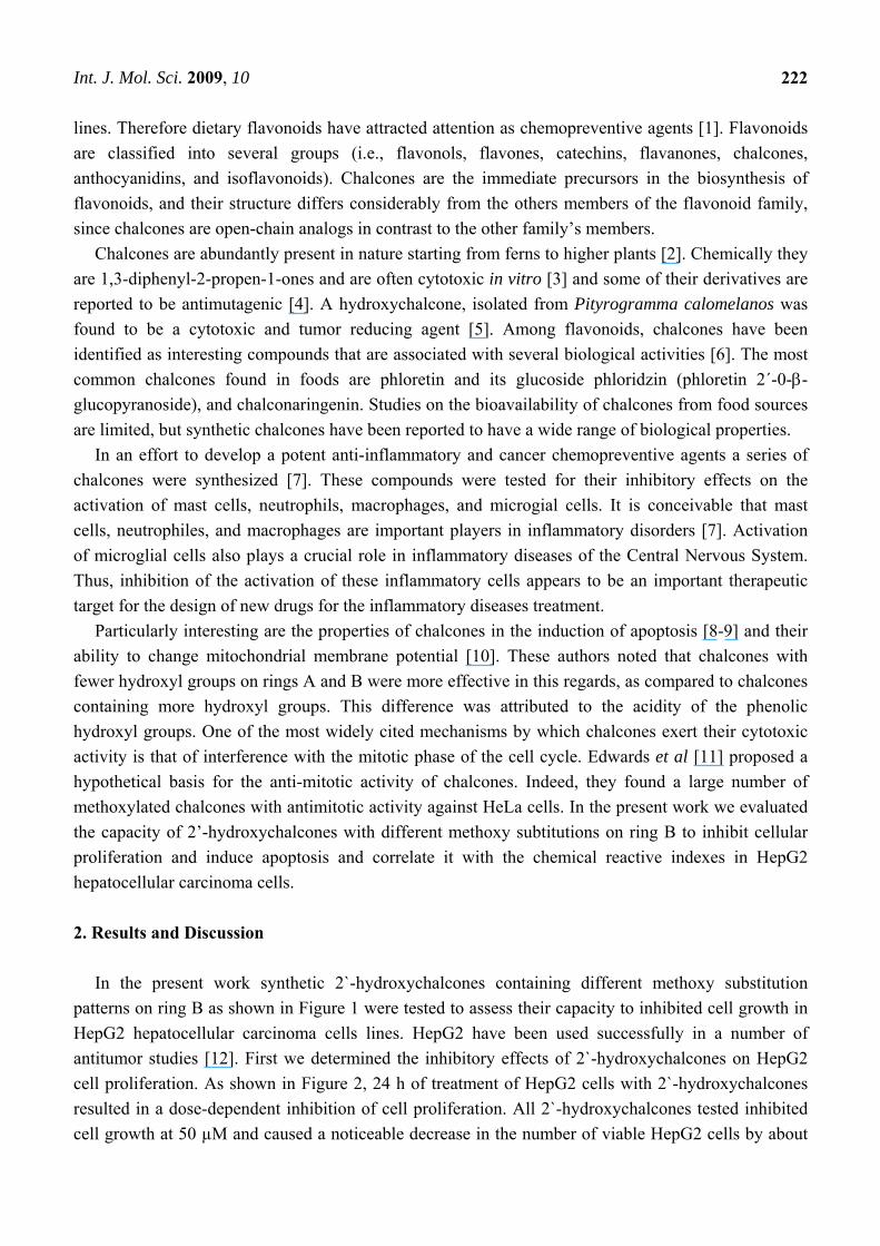
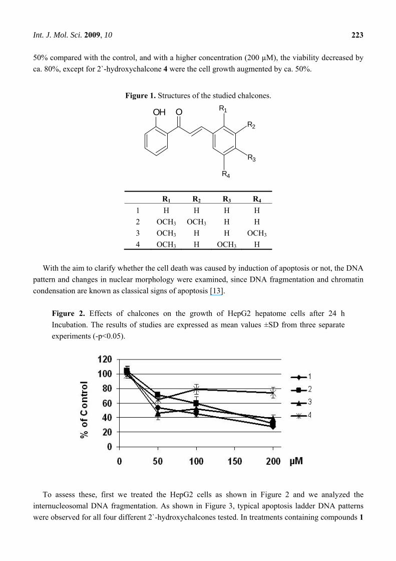
In the present work synthetic 2`-hydroxychalcones containing different methoxy substitution patterns on ring B as shown in Figure 1 were tested to assess their capacity to inhibited cell growth in HepG2 hepatocellular carcinoma cells lines. HepG2 have been used successfully in a number of antitumor studies [12]. First we determined the inhibitory effects of 2`-hydroxychalcones on HepG2 cell proliferation. As shown in Figure 2, 24 h of treatment of HepG2 cells with 2`-hydroxychalcones resulted in a dose-dependent inhibition of cell proliferation. All 2`-hydroxychalcones tested inhibited cell growth at 50 µM and caused a noticeable decrease in the number of viable HepG2 cells by about
Int. J. Mol. Sci. 2009, 10
223
50% compared with the control, and with a higher concentration (200 µM), the viability decreased by ca. 80%, except for 2`-hydroxychalcone 4 were the cell growth augmented by ca. 50%.
Figure 1. Structures of the studied chalcones.
OOH R1
R2
R3
R4
R1 R2 R3 R4 1 H H H H 2 OCH3 OCH3 H H 3 OCH3 H H OCH3 4 OCH3 H OCH3 H
With the aim to clarify whether the cell death was caused by induction of apoptosis or not, the DNA
pattern and changes in nuclear morphology were examined, since DNA fragmentation and chromatin condensation are known as classical signs of apoptosis [13].
Figure 2. Effects of chalcones on the growth of HepG2 hepatome cells after 24 h Incubation. The results of studies are expressed as mean values ±SD from three separate experiments (-p<0.05).
To assess these, first we treated the HepG2 cells as shown in Figure 2 and we analyzed the
internucleosomal DNA fragmentation. As shown in Figure 3, typical apoptosis ladder DNA patterns were observed for all four different 2`-hydroxychalcones tested. In treatments containing compounds 1
Int. J. Mol. Sci. 2009, 10
224
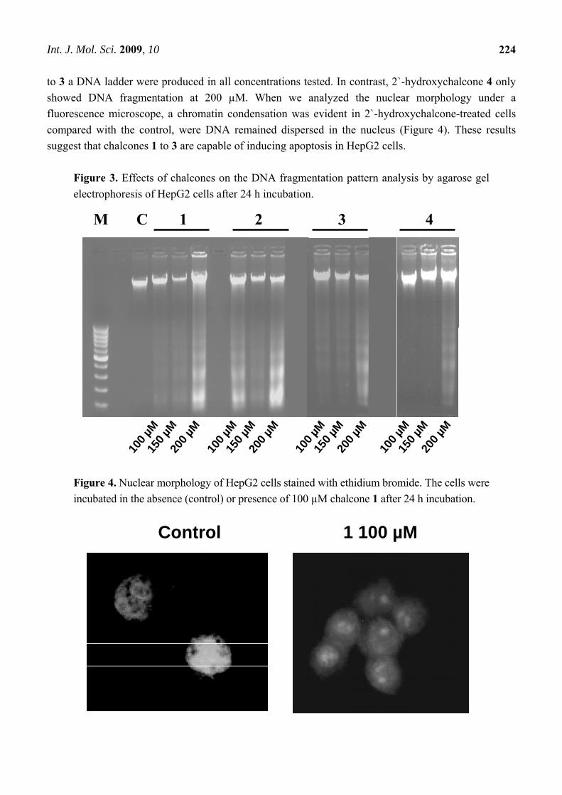
to 3 a DNA ladder were produced in all concentrations tested. In contrast, 2`-hydroxychalcone 4 only showed DNA fragmentation at 200 µM. When we analyzed the nuclear morphology under a fluorescence microscope, a chromatin condensation was evident in 2`-hydroxychalcone-treated cells compared with the control, were DNA remained dispersed in the nucleus (Figure 4). These results suggest that chalcones 1 to 3 are capable of inducing apoptosis in HepG2 cells.
Figure 3. Effects of chalcones on the DNA fragmentation pattern analysis by agarose gel electrophoresis of HepG2 cells after 24 h incubation.
Figure 4. Nuclear morphology of HepG2 cells stained with ethidium bromide. The cells were incubated in the absence (control) or presence of 100 µM chalcone 1 after 24 h incubation.
CE1 CE2 CE3
CE4
100 µ
M15
0 µM
200 µ
M10
0 µM
150 µ
M20
0 µM
M C
100 µ
M15
0 µM
200 µ
M
100 µ
M15
0 µM
200 µ
M
CE4 M C 1 2 3 4
Control 1 100 µM
Int. J. Mol. Sci. 2009, 10
225
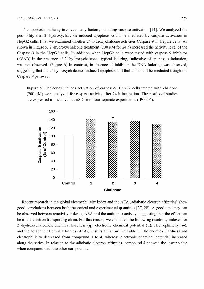
The apoptosis pathway involves many factors, including caspase activation [14]. We analyzed the possibility that 2`-hydroxychalcone-induced apoptosis could be mediated by caspase activation in HepG2 cells. First we examined whether 2`-hydroxychalcone activates Caspase-9 in HepG2 cells. As shown in Figure 5, 2`-hydroxychalcone treatment (200 µM for 24 h) increased the activity level of the Caspase-9 in the HepG2 cells. In addition when HepG2 cells were tested with caspase 9 inhibitor (zVAD) in the presence of 2`-hydroxychalcones typical ladering, indicative of apoptosos induction, was not observed. (Figure 6) In contrast, in absence of inhibitor the DNA ladering was observed, suggesting that the 2`-hydroxychalcones-induced apoptosis and that this could be mediated trough the Caspase 9 pathway.
Figure 5. Chalcones induces activation of caspase-9. HepG2 cells treated with chalcone (200 µM) were analyzed for caspase activity after 24 h incubation. The results of studies are expressed as mean values ±SD from four separate experiments (-P<0.05).
0
20
40
60
80
100
120
140
160
Control 1 2 3 4
Chalcone
Casp
ase
9 ac
tivat
ion
(% o
f Con
trol)
Recent research in the global electrophilicity index and the AEA (adiabatic electron affinities) show
good correlations between both theoretical and experimental quantities [27, 28]. A good tendency can be observed between reactivity indexes, AEA and the antitumor activity, suggesting that the effect can be in the electron transporting chain. For this reason, we estimated the following reactivity indexes for 2`-hydroxychalcones: chemical hardness (η), electronic chemical potential (µ), electrophilicity (ω), and the adiabatic electron affinities (AEA); Results are shown in Table 1. The chemical hardness and electrophilicity decreased from compound 1 to 4, whereas electronic chemical potential increased along the series. In relation to the adiabatic electron affinities, compound 4 showed the lower value when compared with the other compounds.
Int. J. Mol. Sci. 2009, 10
226
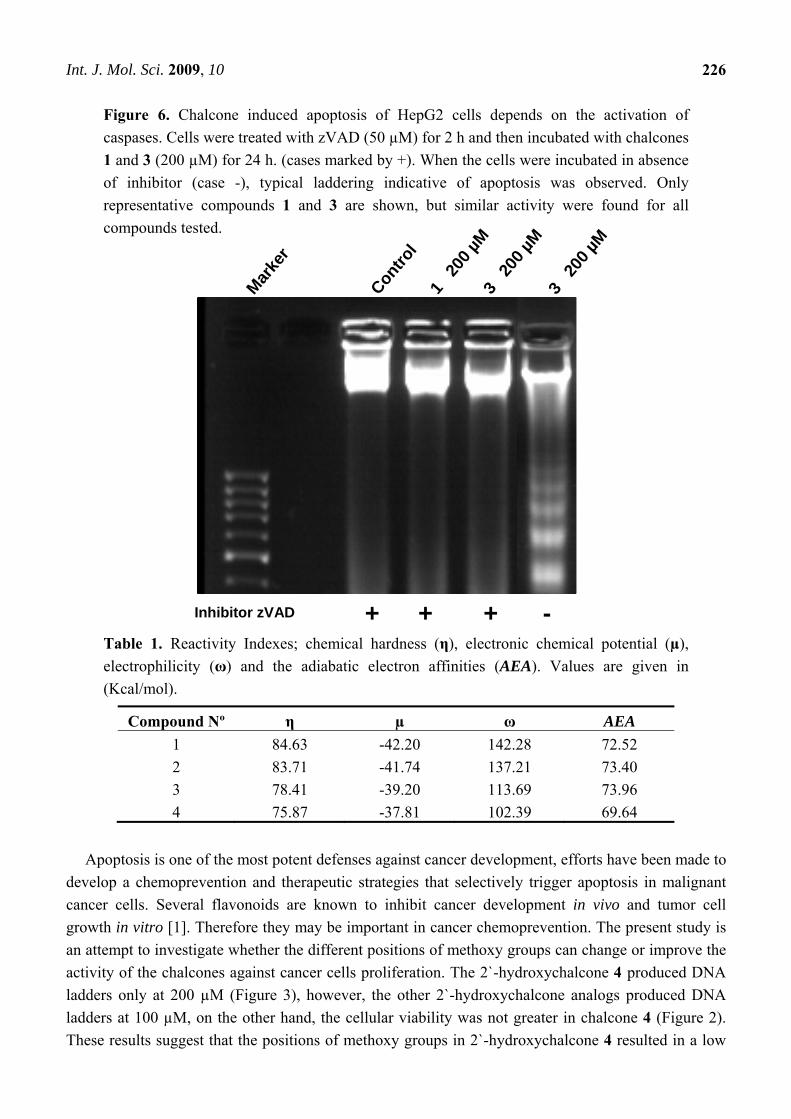
Figure 6. Chalcone induced apoptosis of HepG2 cells depends on the activation of caspases. Cells were treated with zVAD (50 µM) for 2 h and then incubated with chalcones 1 and 3 (200 µM) for 24 h. (cases marked by +). When the cells were incubated in absence of inhibitor (case -), typical laddering indicative of apoptosis was observed. Only representative compounds 1 and 3 are shown, but similar activity were found for all compounds tested.
Table 1. Reactivity Indexes; chemical hardness (η), electronic chemical potential (µ), electrophilicity (ω) and the adiabatic electron affinities (AEA). Values are given in (Kcal/mol).
Compound Nº η µ ω AEA 1 84.63 -42.20 142.28 72.52 2 83.71 -41.74 137.21 73.40 3 78.41 -39.20 113.69 73.96 4 75.87 -37.81 102.39 69.64
Apoptosis is one of the most potent defenses against cancer development, efforts have been made to
develop a chemoprevention and therapeutic strategies that selectively trigger apoptosis in malignant cancer cells. Several flavonoids are known to inhibit cancer development in vivo and tumor cell growth in vitro [1]. Therefore they may be important in cancer chemoprevention. The present study is an attempt to investigate whether the different positions of methoxy groups can change or improve the activity of the chalcones against cancer cells proliferation. The 2`-hydroxychalcone 4 produced DNA ladders only at 200 µM (Figure 3), however, the other 2`-hydroxychalcone analogs produced DNA ladders at 100 µM, on the other hand, the cellular viability was not greater in chalcone 4 (Figure 2). These results suggest that the positions of methoxy groups in 2`-hydroxychalcone 4 resulted in a low
Marke
r
Contro
l1
200 µ
M3
200 µ
M
Inhibitor zVAD + + +
3 20
0 µM
-
Int. J. Mol. Sci. 2009, 10
227
death increase of the HepG2 cells. After treatment with 2`-hydroxychalcones, the number of HepG2 cells decreased after 24 h, suggesting the possibility of an increase in the rate of cell death. Agarose gel electrophoresis verified this suggestion (Figure 3). A DNA ladder was observed after fractionation of the nuclear DNA by agarose gel electrophoresis under UV and also chromatin condensation was observed under a fluorescence microscope (Figure 4). On the basis of these hallmarks of apoptosis, we conclude that chalcones induce apoptosis in HepG2 cells. Caspase-9 was activated by chalcones demonstrating activation in the intrinsic apoptotic pathways [25, 26] (Figure 5). The intrinsic and extrinsic apoptotic pathways converge to caspase-3, which cleaves the inhibitor of the caspase-activated deoxyribonuclease, and the caspase-activated deoxyribonuclease becomes active leading to nuclear apoptosis [25, 27]. The inhibitor of caspase-3 zVAD, eliminated the formation of ladders in all tested compounds (Figure 6). These experiments demonstrated that the caspase pathways are participating in the activation of the apoptotic events that conclude in the formation of DNA ladders, stimulated by chalcones. All our data suggests that chalcones are capable to induce apoptosis in HepG2 cells via a mitochondrial-mediated pathway, which results in the activation of caspase-9 and subsequently nuclear apoptosis.
Recent research in the global electrophilicity index and the AEA show good correlations between both theoretical and experimental quantities [28, 29]. A good correlation can be observed between reactivity indexes, AEA and the antitumor activity, suggesting that the effect can be in the electron transporting chain. This is observed clearly when comparing the activity of chalcone 4 with the rest of chalcones (Table 1). One of the electrophilic sites in the chalcone moiety can be found at the C3 carbon atom of the 2-propen-1-one moiety. The electrophilicity in this site is modulated by the presence of methoxy groups attached to the aromatic ring directly attached to the C3 carbon atom. Since the methoxy groups are electron donor substituents, they can only act stabilizing charges generated at the C3 carbon atom by mesomeric effects. This effect is expected to be similar in compounds 2 and 3. In contrast, this stabilizing effect should be of importance in the case of compound 4, where both methoxy groups are oriented in ortho and para positions, reducing as a consequence, the local electrophilicity at the C3 carbon atom. The absence of such substituents in compound 1, produces the mayor electrophilicity, pattern in the series.
The calculated values of total electrophilicity are in agreement with the previously described local electrophilicity at C3 carbon atom and they are in the same trend as the biological results obtained, suggesting a relationship between the electrophilicity and the apoptotic activity of the tested 2`-hydroxychalcones in HepG2 hepatocellular carcinoma cells and the chalcone 1 would be the most active.
3. Experimental Section 3.1. Chemicals
The structures of the studied compounds 1-4 are given in Figure 1. The dimethoxy substituted 2’-
hydroxychalcones 2-4 were synthesized by the Claisen-Schmidt condensation of 2`-hydroxy-acetophenone and the appropriately substituted dimethoxybenzaldehyde in basic media (KOH) as described previously by Quintana et al. [24] The 2’-hydroxychalcone 1 was purchased from Merck
Int. J. Mol. Sci. 2009, 10
228
and used without further purification. These chalcones were dissolved in dimethylsulfoxide and then added to the culture medium. 3.2. Cell Culture
The HepG2 hepatocellular carcinoma cell line (HB 8065; American Type Culture Collection),
derived from a human hepatoblastoma [30], were maintained in Dulbecco´s modified Eagle´s (DMEM) with 10% heat-inactivated fetal bovine serum (FBS) at 37°C with 5% CO2. 3.3. Cell growth assay
Cells were seeded in a 96 wells plates at density of 10E4/well and treated by 24 h, in the last 2 h a
0.5 mg/mL of MTT at final concentration was added. Cells washed twice with PBS were treated with isopropanol-DMSO (3:2) to dissolve the formazan crystals and quantified the stain by reader a 630 nm. Cell viability is expressed as the optical density ratio of the treatment to control. 3.4. Internucleosomal DNA fragmentation assay
DNA was extracted from cells as described previously by Fernandes et al [16]. Fragmented DNA
samples were separated by electrophoresis on 1.5 agarose gel and visualized with ethidium bromide. 3.5. Nuclear DNA condensation assay
Nuclear morphological changes in cells treated with chalcones were analyzed by ethidium bromide stain [31]. Briefly, Cells (10E5/well) were seeded in glass coverslip, and after indicates treatments cells were fixed with ice cold methanol and stained 10 min with 1 µg/mL of ethidium bromide in PBS, and after mounting in glass slice the cells nucleus condensation was observed under epi-fluorescent microscopy. 3.6. Activity Caspase-9
Cells were seeded in a 96 wells plate at density of 10E4/well, the activity caspase-9 was analyzed
using the Caspase-Glo® 9 Assay (Promega) kit according the manufacturing instructions.
3.7. Section model equations
The global electrophilicity index ω, which measures the stabilization in energy when the system acquires an additional electronic charge ΔN from the environment, is been given by the following simple expression due to Parr et al. [16, 17]:
ω = μ2/ 2η (1) where in terms of the electronic chemical potential μ and the chemical hardness η, ω may be approximated in terms of the one electron energy of the frontier molecular orbital HOMO and LUMO
Int. J. Mol. Sci. 2009, 10
229
ЄН and ЄL, as μ ≈ (ЄН + ЄL)/2 and η ≈ ЄН – ЄL respectively [16,17]. The electrophilicity index encompasses both the tendency of the electrophile to acquire an additional electronic charge driven by μ2 (the squared of the chemical potential) and the resistance of the system to the exchanging electronic charge with the environment described by η.
The electron affinity is defined as the total energy of the neutral Chalcones minus that of the anion radical (E° - E-). The calculation of the adiabatic electron affinity (AEA) is based on the optimized geometry of the neutral species and the optimized geometry of the anion radical species. 3.8. Computational Details
Our calculations were carried out by using the Amsterdam Density Functional (ADF) code [18]. All the molecular structures were fully optimized via the analytical energy gradient method implemented by Verluis and Ziegler employing the local density approximation (LDA) within the Vosko-Wilk-Nusair parameterization for local exchange correlations [20, 21]. We also used the GGA (Generalized Gradient Approximation) BLYP and hybrid B3LYP functional [19]. Solvation effects were modeled by a conductor-like screening model for real solvents (COSMO) [22, 23] using water as solvent. The cluster geometry optimization and the energies were calculated using a standard Slater-type-orbital (STO) basis sets with triple-zeta quality with double polarization functions (TZ2P) for all the atoms. The global electrophilicity (ω) was evaluated using eq 1. The electronic chemical potential μ and the chemical hardness η were evaluated using the frontier molecular orbital model described in the Model Equations Section.
4. Conclusions
Treatment of HepG2 cells for 24 h with synthetic 2’-hydroxy-Chalcones resulted in a dose-dependent inhibition of cell proliferation and apoptosis induction. The calculated reactivity indexes and the adiabatic electron affinities using the DFT method including solvent effects, suggest a structure-activity relationship between the 2’-hydroxy-chalcones structure and the apoptosis in HepG2 cells. Acknowledgements
Authors gratefully acknowledge generous financial support from Fondecyt (grant N° 1080147 and 1050476), UNAB DI05-06/I. R.R.T. thanks to CONICYT AT-23070006 and MECESUP2-FSM0605 for his Fellowships. Dr. Ramiro Arratia-Perez (Universidad Andres Bello) is thanked for granting access to the ADF program. References and Notes 1. Kuo, S. Dietary flavonoids and cancer prevention: Evidence and potentials mechanisms
Oncogenesis 1997, 8, 47-69.
Int. J. Mol. Sci. 2009, 10
230
2. Mark, C.; Nagarathnam, D. Cytotoxicities of some flavonoid analogues. J. Nat. Prod. 1991, 54, 1656-1660.
3. Dharn, D.N. The Chemistry of Chalcones and Related Compounds; Wiley: New York, 1981; p. 213.
4. Torigoo, T.; Arisawa, M.; Iloch, S.; Fujiu, M.; Mayuyama, H.B. Anti-mutagenic chalcones: Antagonizing the mutagenicity of benzo(a)pyrene in Salmonella typhymurium. Biophys. Res. Commun. 1983, 112, 833-842.
5. Sukumaran, K.; Kuttan, R. Screening of 11 ferns of cytotoxic and antitumor potential with special reference to Pityrogramma calomelanos. J. Ethnopharmacol. 1991, 36, 93-96.
6. Calliste, C.; Le Bail, J.; Trouilas, P.; Pouget, C.; Habrious, G.; Chulea, A.; Duroux, J.L. Chalcones: structural requirements for antioxidant, estrigenic and antiproliferative activities. J. Anticancer Res. 2001, 21, 3949-3956.
7. Won, S.; Liu, C.; Tsao, L.; Weng, J.; Ko, H.; Wang, J.; Lin, C. Synthetics chalcones as potential anti-inflammatory and cancer chemopreventive agents. Europ. J. Med. Chem. 2005, 40, 103-112.
8. De Vincenazo, R.; Ferlini, C.; Distefano, M.; Gaggini, C.; Riva, A.; Bombardelli, E.; Morazzoni, P.; Valenti, P.; Belluti, F.; Ranelletti, F.O.; Mancuso, S.; Scambia, G. In vitro evaluation of newly developed chalcone analogues in human cancer cells. Cancer Chemother. Pharmacol. 2000, 46, 305-312.
9. Saydam, G.; Aydin, H.; Sahin, F.; Kucukogin, O.; Erciyas, E.; Terzioglu, E.; Buyukkececi, F.; Omay, S.; Cytotoxic and inhibitory effects os 4,4’-dihydrochalcone RVC-588 on proliferation of human leukemic HL-60 cells. Leuk. Res. 2003, 27, 57-64.
10. Sabzevari, O.; Galati, G.; Moridani, M.; Siraki, A.; Obrien, P. Molecular cytotoxic mechanisms of anticancer hydrochalcones. Chem. Biol. Interact. 2004, 148, 57-67.
11. Edwards, M.; Stemerick, D.; Sunkara, P. Chalcones: A new class of antimitotic agents. J. Med. Chem. 1990, 33, 1948-1954.
12. Tanigawa, S.; Fujii, M.; Hou, D. Stabilization of p53 is involved in Quercetin-induced Cell Cycle Arrest and apoptosis in HepG2 cells. Biosci. Biotechnol. Biochem. 2008, 72, 797-804.
13. Allen, P.; Newland, A. Electrophoresis DNA analysis for the detection of apoptosis. Mol. Biotechnol. 1998, 9, 247-251.
14. Kohler, C.; Orrenius, S. Evaluation of caspase activity in apoptotic cells. J. Immunol. Meth. 2002, 265, 97-110.
15. Fernandes, R.; McGowan, A.; Cotter, T. Mutant Ha-Ras overexpression inhibits drug and U.V. induced apoptosis. Anticancer Res. 1996, 16, 1691-1706.
16. Parr, R.; Pearsons, R. Absolute hardness: Companion parameter to absolute electronegativity. J. Am. Chem. Soc. 1983, 105, 7512-7516.
17. Parr, R.; Szentpaly, L.; Liu, S. Electrophilicity Index. J. Am. Chem. Soc. 1999, 121, 1922-1924. 18. Amsterdam Density Functional (ADF) code, release 2007. Vrije Universiteit: Amsterdam,
The Netherlands. 19. Te Velde, G.; Bickelhaupt, F.M.; Van Gisberger, S.J.A.; Fonseca Guerra, C.; Baerends, E.J.;
Snijders, J.G.; Ziegler, T.J. Chemistry with ADF. Comput. Chem. 2001, 22, 931-967.
Int. J. Mol. Sci. 2009, 10
231
20. Verluis, L.; Ziegler, T. The determination of molecular structures by density functional theory. The evaluation of analytical energy gradients by numerical integration. J. Chem. Phys, 1988, 88, 322-328.
21. Vosko, S.H.; Wilk, L.; Nusair, M.; Accurate spin-dependent electron liquid correlation energies for local spin density calculations: A critical analysis. Can. J. Phys. 1980, 58, 1200-1211.
22. Klamt, A. Conductor-like screening model for real solvents: A new approach to the quantitative calculation of salvation phenomena. J. Chem. Phys. 1995, 99, 2224-2235.
23. Klamt, A.; Jonas V.; Treatment of the outlying charge in continuum salvation models. J. Chem. Phys. 1996, 105, 9972-9981.
24. Quintana, P.; Yáñez, C.; Escobar, C.A.; Sicker, D.; Araya-Maturana, R.; Squella, J.A. Electrochemical approach to the radical anion formation from 2’-hydroxy chalcone derivatives. Electroanalysis 2006, 18, 521-525.
25. Thornberry, N.; Lazebnik, Y. Caspases: Enemies within. Science 1998, 281, 1312-1316. 26. Kobori, M.; Iwashita, K.; Shinmoto, H.; Tsushida, T. Phloretin-induced apoptosis in B16
melanoma 4A5 cells and HL60 human leukemia cells. Biosci. Biotechnol. Biochem. 1999, 63, 719-725.
27. Mancini, M.; Nicholson, D.; Roy, S. The caspase-3 precursor has a cytosolic and mitochondrial distribution: Implication for apoptotic signaling. J. Cell Biol. 1998, 140, 1485-1495.
28. Perez, P.; Toro-Labbe, A.; Aizman, A.; Contreras, R. Comparison between experimental and theoretical scales of electrophilicity in benzhydryl cations. J. Org. Chem. 2002, 67, 4747-4752.
29. Li, X.; Cai, Z.I.; Sevilla, M.D. DFT Calculations of the electron affinities of nucleic acid bases: dealing with negative electron affinities. J. Phys. Chem. A 2002, 106, 1596-1603.
30. Javitt N. HepG2 cells as a resource for metabolic studies: lipoprotein, cholesterol, and bile acids. FASEB J. 1990, 4, 161-168.
31 Sakahira, H.; Enari, M.; Ohsawa, Y.; Uchiyama, Y.; Nagata, S. Apoptotic nuclear morphological change without DNA fragmentation. Curr. Biol. 1999, 20, 543-546.
© 2009 by the authors; licensee Molecular Diversity Preservation International, Basel, Switzerland. This article is an open-access article distributed under the terms and conditions of the Creative Commons Attribution license (http://creativecommons.org/licenses/by/3.0/).
Related Documents