Structural and functional characterization of TRI3 trichothecene 15-O-acetyltransferase from Fusarium sporotrichioides Graeme S. Garvey, 1 Susan P. McCormick, 2 Nancy J. Alexander, 2 and Ivan Rayment 1 * 1 Department of Biochemistry, University of Wisconsin, Madison, Wisconsin 53706 2 Mycotoxin Research Unit, USDA/ARS, National Center for Agricultural Utilization Research, Peoria, Illinois 61604 Received 15 October 2008; Revised 6 January 2009; Accepted 15 January 2009 DOI: 10.1002/pro.80 Published online 16 March 2009 proteinscience.org Abstract: Fusarium head blight is a devastating disease of cereal crops whose worldwide incidence is increasing and at present there is no satisfactory way of combating this pathogen or its associated toxins. There is a wide variety of trichothecene mycotoxins and they all contain a 12,13-epoxytrichothecene skeleton but differ in their substitutions. Indeed, there is considerable variation in the toxin profile across the numerous Fusarium species that has been ascribed to differences in the presence or absence of biosynthetic enzymes and their relative activity. This article addresses the source of differences in acetylation at the C15 position of the trichothecene molecule. Here, we present the in vitro structural and biochemical characterization of TRI3, a 15-O-trichothecene acetyltransferase isolated from F. sporotrichioides and the ‘‘in vivo’’ characterization of Dtri3 mutants of deoxynivalenol (DON) producing F. graminearum strains. A kinetic analysis shows that TRI3 is an efficient enzyme with the native substrate, 15-decalonectrin, but is inactive with either DON or nivalenol. The structure of TRI3 complexed with 15-decalonectrin provides an explanation for this specificity and shows that Tri3 and Tri101 (3-O-trichothecene acetyltransferase) are evolutionarily related. The active site residues are conserved across all sequences for TRI3 orthologs, suggesting that differences in acetylation at C15 are not due to differences in Tri3. The tri3 deletion mutant shows that acetylation at C15 is required for DON biosynthesis even though DON lacks a C15 acetyl group. The enzyme(s) responsible for deacetylation at the 15 position of the trichothecene mycotoxins have not been identified. Keywords: Fusarium head blight; trichothecene mycotoxin; deoxynivalenol; T-2 toxin; Fusarium graminearum; Fusarium sporotrichioides; acetyltransferase; coenzyme A; BAHD superfamily Introduction Fusarium head blight (FHB) is a serious disease of ce- real crops whose worldwide incidence is increasing and is a major factor limiting wheat production in many parts of the world. 1 The disease is caused by sev- eral species of the fungus Fusarium which pose a dual threat: first by reducing the yield and quality of grain and secondly by contaminating the food grains with trichothecene mycotoxins. In 1998–2000, economic losses in the United States alone were estimated to exceed $ 2.7 billion 2 which had devastating effects on Additional Supporting Information may be found in the online version of this article. Abbreviations: 3ADON, 3-acetyl-deoxynivalenol; 15ADON, 15- acetyl-deoxynivalenol; DON, deoxynivalenol; FHB, Fusarium head blight; NIV, nivalenol. Grant sponsor: NIH; Grant number: AR35186; Grant sponsor: U.S. Department of Agriculture; Grant number: 59-0790-6-066. *Correspondence to: Ivan Rayment, Department of Biochemistry, 433 Babcock Dr., Madison, WI 53706. E-mail: ivan_rayment@ biochem.wisc.edu Published by Wiley-Blackwell. V C 2009 The Protein Society PROTEIN SCIENCE 2009 VOL 18:747—761 747

Welcome message from author
This document is posted to help you gain knowledge. Please leave a comment to let me know what you think about it! Share it to your friends and learn new things together.
Transcript
Structural and functionalcharacterization of TRI3 trichothecene15-O-acetyltransferase fromFusarium sporotrichioides
Graeme S. Garvey,1 Susan P. McCormick,2 Nancy J. Alexander,2
and Ivan Rayment1*
1Department of Biochemistry, University of Wisconsin, Madison, Wisconsin 537062Mycotoxin Research Unit, USDA/ARS, National Center for Agricultural Utilization Research, Peoria, Illinois 61604
Received 15 October 2008; Revised 6 January 2009; Accepted 15 January 2009
DOI: 10.1002/pro.80Published online 16 March 2009 proteinscience.org
Abstract: Fusarium head blight is a devastating disease of cereal crops whose worldwide
incidence is increasing and at present there is no satisfactory way of combating this pathogen orits associated toxins. There is a wide variety of trichothecene mycotoxins and they all contain a
12,13-epoxytrichothecene skeleton but differ in their substitutions. Indeed, there is considerable
variation in the toxin profile across the numerous Fusarium species that has been ascribed todifferences in the presence or absence of biosynthetic enzymes and their relative activity. This
article addresses the source of differences in acetylation at the C15 position of the trichothecene
molecule. Here, we present the in vitro structural and biochemical characterization of TRI3,a 15-O-trichothecene acetyltransferase isolated from F. sporotrichioides and the ‘‘in vivo’’
characterization of Dtri3 mutants of deoxynivalenol (DON) producing F. graminearum strains. A
kinetic analysis shows that TRI3 is an efficient enzyme with the native substrate, 15-decalonectrin,but is inactive with either DON or nivalenol. The structure of TRI3 complexed with 15-decalonectrin
provides an explanation for this specificity and shows that Tri3 and Tri101 (3-O-trichothecene
acetyltransferase) are evolutionarily related. The active site residues are conserved across allsequences for TRI3 orthologs, suggesting that differences in acetylation at C15 are not due to
differences in Tri3. The tri3 deletion mutant shows that acetylation at C15 is required for DON
biosynthesis even though DON lacks a C15 acetyl group. The enzyme(s) responsible fordeacetylation at the 15 position of the trichothecene mycotoxins have not been identified.
Keywords: Fusarium head blight; trichothecene mycotoxin; deoxynivalenol; T-2 toxin; Fusariumgraminearum; Fusarium sporotrichioides; acetyltransferase; coenzyme A; BAHD superfamily
Introduction
Fusarium head blight (FHB) is a serious disease of ce-
real crops whose worldwide incidence is increasing
and is a major factor limiting wheat production in
many parts of the world.1 The disease is caused by sev-
eral species of the fungus Fusarium which pose a dual
threat: first by reducing the yield and quality of grain
and secondly by contaminating the food grains with
trichothecene mycotoxins. In 1998–2000, economic
losses in the United States alone were estimated to
exceed $ 2.7 billion2 which had devastating effects on
Additional Supporting Information may be found in the onlineversion of this article.
Abbreviations: 3ADON, 3-acetyl-deoxynivalenol; 15ADON, 15-acetyl-deoxynivalenol; DON, deoxynivalenol; FHB, Fusariumhead blight; NIV, nivalenol.
Grant sponsor: NIH; Grant number: AR35186; Grant sponsor:U.S. Department of Agriculture; Grant number: 59-0790-6-066.
*Correspondence to: Ivan Rayment, Department of Biochemistry,433 Babcock Dr., Madison, WI 53706. E-mail: [email protected]
Published by Wiley-Blackwell. VC 2009 The Protein Society PROTEIN SCIENCE 2009 VOL 18:747—761 747
farm communities.3 At present there is no satisfactory
way of combating this pathogen or the associated toxins.
This problem is accentuated by an incomplete under-
standing of the pathway and enzymes responsible for
the biosynthesis of the trichothecene mycotoxins.
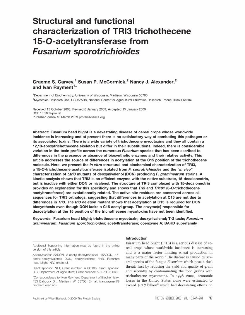
Trichothecene mycotoxins are sesquiterpene epox-
ide secondary metabolites that inhibit protein transla-
tion in eukaryotes and have several acute adverse
effects in animals, including food refusal, diarrhea,
and alimentary hemorrhaging.4 There is a wide variety
of trichothecene mycotoxins, and they all contain a
12,13-epoxytrichothecene skeleton, but differ in their
substitutions (see Fig. 1). Indeed, the substitution pat-
tern on the core ring structure differs markedly
between phylogenetically closely related Fusarium spe-
cies where these alternative substitution patterns can
have drastic effects on cytotoxicity. As much as 5 �103 fold difference in LC50 has been reported between
trichothecene variants.5 Studies have shown that acet-
ylation at the C3 position of trichothecenes can
decrease the phytotoxicity6,7 indicating the importance
of acetylation. This study focuses on the structure and
function of the acetyltransferase that is responsible for
acetylation of the hydroxyl group at C15.
Figure 1. Abbreviated biosynthetic pathway for 15-acetyl deoxynivalenol and the structures of the common trichothecene
myocotoxins. T-2 toxin is an A-type trichothecene with an ester-linked isovaleryl group at C8, whereas DON and nivalenol (NIV)
are both B-type trichothecenes with a ketone moiety at C8 and are further differentiated by their substitution patterns at C4.
[Color figure can be viewed in the online issue, which is available at www.interscience.wiley.com.]
748 PROTEINSCIENCE.ORG Structure and Kinetics of TRI3 Acetyltransferase

The biosynthetic pathway has been largely deter-
mined in the T-2 toxin producing strain, F. sporotri-
chioides through the use of genetic mutants and pre-
cursor feeding studies.8–11 The core trichothecene ring
structure is formed from the cyclization of farnesyl
pyrophosphate by trichodiene synthase encoded by
Tri512 and subsequent multiple oxygenations by TRI4,
yielding the toxic intermediate, isotrichodermol.13 The
fungus then protects itself by acetylating the C3
hydroxyl group through the action of TRI101, which
reduces the toxicity of trichothecene mycotoxins � 100
fold.14 Thereafter, a complex series of coupled reac-
tions lead to a final ensemble of toxins (see Fig. 1).
Modifications to the C4 and C15 positions are per-
formed by the P450 monoxygenase/acetyltransferase
pairs TRI13/TRI7 and TRI11/TRI3, respectively.9,15–18
C8 oxygenation is performed by TRI1,19–21 and this
position is further modified by TRI16 to an isovaleryl
group in T-2 toxin producers.22 The last step in the
biosynthetic pathway involves removal of the protect-
ing acetyl group at C3 by the action of TRI8.23
The toxins are often classified according to their
substitution at C8 (see Fig. 1).24 Trichothecenes that
carry an ester side chain at C8, or no side chain at all
are classified as Type A, whereas a ketone functional
group at this position defines the Type B toxins. T2-
toxin and deoxynivalenol (DON) are examples of A-
Type and B-Type toxins, respectively. Fusarium strains
have, by extension, been classified into chemotypes on
the basis of the toxins that they produce.25 The
description and assignment of a Fusarium to a tricho-
thecene chemotype in the literature has been compli-
cated by the use of different growth substrates, such as
sterile rice grains, sterile wheat grains, and liquid cul-
ture media, and different extraction methods.26–30
Furthermore, single Fusarium isolates often produce
mixtures of acetylated and deacetylated trichothecenes,
making their chemotype classification difficult. Given
the importance of the toxin profile in the management
of FHB, care should be taken when correlating a DNA
sequence or genotype of the fungus with a chemical
phenotype.31
It is generally assumed that the alternative substi-
tution patterns on the core ring are determined by the
presence or absence of functional copies of the biosyn-
thetic genes in a Fusarium strain.18 For example, the
basis for variations at C4 arise from differences in the
coding sequence of Tri13. Nivalenol (NIV) types have
a functional copy, whereas this gene is inactivated in
DON producing strains. A switch from DON to NIV
can be accomplished by heterologous expression of a
functional TRI13 in a DON producing strain. NIV pro-
ducers often produce mixtures of 4-acetyl-NIV and
NIV and the inactivation of Tri7 will shift the chemo-
type to NIV only.18
The genetic and biochemical basis for differences
of acetylation at C15 are less clear than for differences
at C4. The predicted amino acid sequences for Tri3
from strains that produce 3-acetyl-deoxynivalenol
(3ADON) and strains that produce 15-acetyl-deoxyni-
valenol (15ADON) are highly homologous, sharing
greater than 90% sequence identity.27 Furthermore,
there are no obvious mutations which would render
these orthologs nonfunctional. Transcription of Tri3
was reported in a 3ADON producing strain (Fusarium
graminearum F15), however, recombinant protein
expressed in E. coli exhibited poor activity with
DON.32 Several hypotheses for the reduced levels of
C15 acetylation in DON and 3ADON chemotypes can
be envisaged. First, the C15 position is deacetylated
by an as yet unidentified enzyme analogous to the
TRI101/TRI8 acetylation and deacetylation of C3,
second, the Tri3 gene is transcribed but not translated,
or third, the enzyme is produced but is nonfunctional.
The last hypothesis of a transcribed but nonfunctional
TRI3 in chemotypes that produce 3ADON would
be similar to the control of modification at the C4
position.
The apparent dichotomy between the conserved
sequence of TRI3 and its inactivity in similar strains of
Fusarium is in contrast to the promiscuous behavior
of TRI101 which is another acetyltransferase from the
trichothecene biosynthetic pathway. In our previous
work with TRI101, we demonstrated that this self pro-
tection enzyme from F. graminearum was equally
effective at performing the 3-O-trichothecene acetyl-
transferase reaction with T-2, DON, or NIV as sub-
strates. In contrast, the ortholog of TRI101 in F. spor-
otrichioides, which had been used in transgenic
resistance strategies with wheat and barley, had a 70-
fold reduced catalytic efficiency, kcat/Km, with DON
compared to T-2 toxin.33 Differences in activity of
TRI101 between T-2 toxin and DON chemotype pro-
ducers correlate directly with variations in residues
lining the active site. This raises the question of
whether the differences in C15 chemotypes can be
similarly ascribed to differences in the active site of
TRI3. To address these questions a structural and
functional analysis of TRI3 from F sporotrichioides
was initiated.
Here, we present the in vitro structural and bio-
chemical characterization of TRI3, a 15-O-trichothe-
cene acetyltransferase isolated from F. sporotrichioides
and the ‘‘in vivo’’ characterization of Dtri3 mutants of
DON producing F. graminearum strains. The kinetic
results indicate that TRI3 is an efficient enzyme with
the native substrate, 15-decalonectrin, and is inactive
with either DON or NIV. The structural studies pro-
vide an explanation for the specificity and reveal that
Tri3 and Tri101 are likely evolutionarily related. Fur-
thermore, the structural studies show that the residues
that line the active site of Tri3 are strictly conserved,
even in strains that do not appear to have C15 acetyla-
tion. Finally, biochemical characterization of the Tri3
deletion mutants reveal its gene product is essential
for the biosynthesis of B type trichothecene products.
Garvey et al. PROTEIN SCIENCE VOL 18:747—761 749

Results and Discussion
Tertiary and quaternary structure of TRI3
TRI3 was crystallized in space group P212121 with unit
cell dimensions a ¼ 64.28 A, b ¼ 81.56 A, and c ¼95.88 A. The unit cell dimensions remained unchanged
during heavy atom derivative soaks and freezing, thus
allowing the apo-TRI3 structure to be determined by
multiple isomorphous replacement (MIR) phasing
with two heavy atom derivatives. One monomer is
present in the asymmetric unit. Gel filtration analysis
confirms that TRI3 is a monomer in solution (data not
shown). Comparison of the apo structure with the bi-
nary complex of 15-decalonectrin bound to TRI3
reveals no major structural differences in the presence
or absence of substrate (root mean square difference
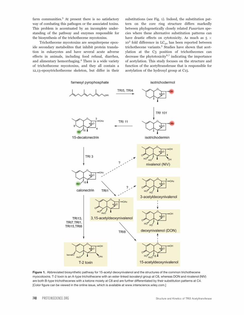
(rmsd) 0.26 A for 463 Ca).The structure of TRI3 is best described as a two
domain protein whose N- and C-terminal domains as-
sociate to form a doughnut-shaped protein [Fig. 2(A)].
The active site lies in the doughnut hole formed at the
interface between the two domains. The N-terminal
domain consists of two mixed b-sheets with three and
five strands each. The five-stranded b-sheet is built
from four N-terminal domain strands (b-2,5,6,7) and
one domain swapped strand (b12). Packed on both
faces of the five stranded b-sheet are nine a-helices,eight are from the N-terminal domain (a-1,2,3,4,5,6,7,8), and the ninth helix is from the C ter-
minal domain swapped loop (a17 þ b12). The three-
stranded b-sheet (b-1,3,4) is on the surface of the pro-
tein distal from the base of the active site. This sheet
does not participate in the domain interface or ligand
binding. The proposed catalytic histidine is located on
the loop between b7 and a6 at the interface of the two
domains.
The C-terminal domain contains a six-stranded
mixed b-sheet (b-8,9,10,11,13,14) located at the inter-
face. Six a-helices (a-10,11,13,14,15,18) are packed
against the exterior face of this b-sheet. Three a-heli-ces (a-9,12,16) form part of the domain interface and
are packed between the C-terminal mixed b-sheet (b-9,10) and the N-terminal domain. A topology drawing
is provided in Supporting Information Figure 1.
Figure 2. Structural representations of TRI3. (A) Stereoview of TRI3 complexed with 15-decalonectrin (PDB accession
number 3fp0). The N- and C-terminal domains are colored magenta and red, respectively, and the domain swapped b-strand12 is colored yellow. Bound ligand 15-decalonectrin is colored dark gray. (B) Stereo overlay of TRI3 (colored white) and
vinorine synthase, PDB ID 2bgh, (colored light blue). The loop between residues Asp362 and Gly366 of vinorine synthase are
colored black and the corresponding loop residues, Glu449-Ser461, of TRI3 are colored yellow.
750 PROTEINSCIENCE.ORG Structure and Kinetics of TRI3 Acetyltransferase
Comparison with BAHD family membersA search for structurally homologous proteins with the
SSM server34 shows that the fold observed in TRI3
belongs to the BAHD superfamily. The closest struc-
tural relatives are: phthiocerol dimycocerosyl transfer-
ase from Mycobacterium tuberculosis (PapA5),35,36
vinorine synthase (VS),37 and TRI101 from F. grami-
nearum33 which exhibit Q values of 0.29, 0.20, and
0.19, respectively. All of these proteins show very lim-
ited sequence similarity to TRI3. For example, VS
shares only 10% sequence identity even though 306
structurally equivalent a-carbons superimpose with an
rmsd of 3.1 A. If only the secondary structure elements
are considered, these structures align remarkably well
with an rmsd of 2.6 A for 145 stereochemically equiva-
lent a-carbons. A superposition of TRI3 with VS is
shown in Figure 2(B).
All enzymes of the BAHD family catalyze acyl
transfer reactions and have two conserved sequence
motifs: a catalytically important HX3D and a structur-
ally important DFGWG motif.38 The HX3D motif is
conserved in TRI3, where residue His186 superim-
poses closely with other BAHD family member cata-
lytic histidine residues. In both the apo and 15-decalo-
nectrin-bound TRI3 structures, the Asp190 residue of
the HX3D motif is directed away from the substrate,
forming a salt bridge with Arg342 and participating in
helix capping. This structural role for the aspartate
residue of the HX3D motif is consistent with previous
structures of other BAHD enzymes.37,39
The DFGWG motif, which is believed to have a
structural role in the BAHD family members,33,37 is
not present in TRI3. In all other BAHD members this
motif forms a turn between the domain swapped a17helix and b12 strand. The recent structures for two tri-
chothecene 3-O-acetyltransferases, FsTRI101 and
FgTRI101, suggested that the fourth position of this
DFGWG motif is variable and may only require a large
hydrophobic residue for anchoring the turn. In TRI3,
the residues which take the place of the DFGWG motif
form a much larger loop extending from residues
Glu449-Ser461. The structure of TRI3 demonstrates
that this structural motif is not strictly required for
membership within the BAHD family of enzymes.
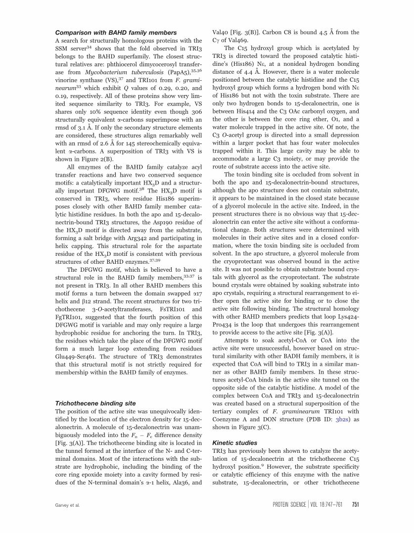
Trichothecene binding site
The position of the active site was unequivocally iden-
tified by the location of the electron density for 15-dec-
alonectrin. A molecule of 15-decalonectrin was unam-
biguously modeled into the Fo � Fc difference density
[Fig. 3(A)]. The trichothecene binding site is located in
the tunnel formed at the interface of the N- and C-ter-
minal domains. Most of the interactions with the sub-
strate are hydrophobic, including the binding of the
core ring epoxide moiety into a cavity formed by resi-
dues of the N-terminal domain’s a-1 helix, Ala36, and
Val40 [Fig. 3(B)]. Carbon C8 is bound 4.5 A from the
Cc of Val469.
The C15 hydroxyl group which is acetylated by
TRI3 is directed toward the proposed catalytic histi-
dine’s (His186) Ne, at a nonideal hydrogen bonding
distance of 4.4 A. However, there is a water molecule
positioned between the catalytic histidine and the C15
hydroxyl group which forms a hydrogen bond with Neof His186 but not with the toxin substrate. There are
only two hydrogen bonds to 15-decalonectrin, one is
between His414 and the C3 OAc carbonyl oxygen, and
the other is between the core ring ether, O1, and a
water molecule trapped in the active site. Of note, the
C3 O-acetyl group is directed into a small depression
within a larger pocket that has four water molecules
trapped within it. This large cavity may be able to
accommodate a large C3 moiety, or may provide the
route of substrate access into the active site.
The toxin binding site is occluded from solvent in
both the apo and 15-decalonectrin-bound structures,
although the apo structure does not contain substrate,
it appears to be maintained in the closed state because
of a glycerol molecule in the active site. Indeed, in the
present structures there is no obvious way that 15-dec-
alonectrin can enter the active site without a conforma-
tional change. Both structures were determined with
molecules in their active sites and in a closed confor-
mation, where the toxin binding site is occluded from
solvent. In the apo structure, a glycerol molecule from
the cryoprotectant was observed bound in the active
site. It was not possible to obtain substrate bound crys-
tals with glycerol as the cryoprotectant. The substrate
bound crystals were obtained by soaking substrate into
apo crystals, requiring a structural rearrangement to ei-
ther open the active site for binding or to close the
active site following binding. The structural homology
with other BAHD members predicts that loop Lys424-
Pro434 is the loop that undergoes this rearrangement
to provide access to the active site [Fig. 3(A)].
Attempts to soak acetyl-CoA or CoA into the
active site were unsuccessful, however based on struc-
tural similarity with other BADH family members, it is
expected that CoA will bind to TRI3 in a similar man-
ner as other BAHD family members. In these struc-
tures acetyl-CoA binds in the active site tunnel on the
opposite side of the catalytic histidine. A model of the
complex between CoA and TRI3 and 15-decalonectrin
was created based on a structural superposition of the
tertiary complex of F. graminearum TRI101 with
Coenzyme A and DON structure (PDB ID: 3b2s) as
shown in Figure 3(C).
Kinetic studiesTRI3 has previously been shown to catalyze the acety-
lation of 15-decalonectrin at the trichothecene C15
hydroxyl position.9 However, the substrate specificity
or catalytic efficiency of this enzyme with the native
substrate, 15-decalonectrin, or other trichothecene
Garvey et al. PROTEIN SCIENCE VOL 18:747—761 751

intermediates has not been assessed. This kinetic anal-
ysis reveals that TRI3 is an efficient enzyme with cata-
lytic parameters similar to TRI101 in the same path-
way.33 Acetylation of C15 by this TRI3 ortholog with
the final mycotoxins DON or NIV was not detectable
in this assay. The kcat, Km, and kcat/Km kinetic con-
stants determined for FsTRI3 with 15-decalonectrin
are presented in Table I.
Figure 3. Details of the interaction of trichothecene intermediate 15-decalonectrin with TRI3. (A) Stereoview of the electron
density corresponding to 15-decalonectrin (dark gray) bound to TRI3 (white). The map, contoured at 3r was calculated from
coefficients of the form Fo � Fc where the ligand was omitted from the phase calculation and refinement. Residues that
interact with the epoxide moiety are depicted as sticks. Loop residues Lys424-Pro434, proposed to allow active site access
are colored red. (B) Schematic representation of the interactions made by TRI3 with 15-decalonectrin. Polar and apolar
interactions and distances in A are indicated as red and gray dashes, respectively. Distance between the catalytic His186 and
C15 Oxygen is not ideal for hydrogen bonding. (C) Coenzyme A binding site model. The proposed CoA binding site of TRI3
based on a structural superposition of the structure of FgTRI101 complexed with CoA and DON onto the structure of the
TRI3�15-decalonectin complex is shown. TRI3 is colored white, FgTRI101 is colored light blue, CoA from the FgTRI101
structure is shown in stick representation, and 15-decalonectrin is depicted with spheres.
752 PROTEINSCIENCE.ORG Structure and Kinetics of TRI3 Acetyltransferase
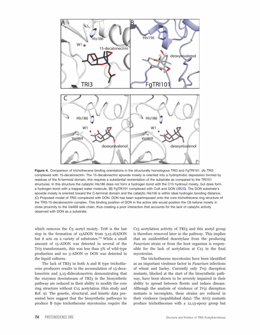
Trichothecene binding comparisons betweenTRI3 and TRI101
TRI3 and TRI101 both are BAHD family members
whose secondary structures and catalytic motifs align
closely. Additionally, both enzymes catalyze the trans-
fer of an acetyl group from acetyl coenzyme A to a tri-
chothecene mycotoxin. The structure of FgTRI101 can
be superimposed with TRI3 with an rmsd of 2.6 A for
145 a-carbons of secondary structure. That both
enzymes are in the same pathway and have strong
structural and functional homology suggests an evolu-
tionary relationship where the catalytic mechanism
has been retained following a gene duplication event.
Maintaining the catalytic mechanism freed the redun-
dant gene to evolve new substrate specificity.
It is not possible to determine which gene, Tri3 or
Tri101, was the primordial gene and which evolved the
new specificity for binding the different trichothecenes
in very different orientations. The hydroxyl moiety at
different positions of the core ring structure must be
positioned proximally to the conserved catalytic histi-
dine. In TRI3 the C15 hydroxyl group is not positioned
within hydrogen bonding distance to the catalytic his-
tidine. A water molecule observed in the high resolu-
tion 15-decalonectrin-bound structure is positioned
such that it could be activated by His186 for proton
abstraction of the C15 hydroxyl [Fig. 3(A)]. This struc-
ture suggests that the TRI3 mechanism may be dis-
tinct from other BAHD family members, using an acti-
vated water for the proton abstraction.
An additional requirement for substrate specificity
in this pathway is to keep the epoxide moiety in a non-
reactive environment. In TRI3 and TRI101 the reactive
epoxide moiety is bound into a hydrophobic pocket.
However, the pocket is formed by residues from the
N- and C-terminal domains in TRI3 and TRI101, respec-
tively [Fig. 4(A,B)]. In TRI101 the epoxide is bound into
a hydrophobic cavity formed by the residues of the
C terminal domain’s central bsheet (b9 þ b11). The 15-
decalonectrin bound TRI3 structure reveals the epoxide
inserted into a shallow pocket formed by residues of the
N-terminal domain’s a1 helix. Given the reactive nature
of epoxides, it is surprising that during divergent evolu-
tion the substrate was able to rotate 180� in the active
site and a new epoxide binding pocket was formed.
Role of TRI3 in determining chemotype
A hypothesis that Tri3 is transcribed but nonfunctional
in 3ADON chemotypes was suggested by the poor C15-
O-acetyltransferase activity of recombinantly expressed
TRI3 from a 3ADON producer with the final mycotoxin,
DON.32 The crystal structure of TRI3 with 15-decalo-
nectrin bound provides a structural basis for interpret-
ing these earlier results. The structure of TRI3 indicates
that binding of trichothecene mycotoxins differentiated
to A or B-type by the activity of TRI1 would form an
unfavorable interaction with the C8 moiety. Modeling
of DON into the 15-decalonectrin position shows that
the C8 isovaleryl or ketone moieties of A or B type tri-
chothecenes, respectively, would be bound into a hydro-
phobic environment with close proximity to Val469
[Fig. 4(C)]. This unfavorable interaction is proposed to
be the basis for the lack of C15 acetylation activity that
we and others have observed with DON and NIV.
While Tri3 genomic polymorphisms correlate with
the B type trichothecene mixtures, these variations do
not lie in the substrate binding pockets. A mapping of
the sequence conservation of a ClustalW alignment
from available TRI3 amino acid sequences onto the
TRI3 structure is presented in Figure 5. This figure
reveals that the trichothecene and predicted acetyl-
coenzyme A binding site residues are highly conserved
while the variable residues are located on surface
exposed loops. The strict conservation of active site
architecture suggests that both and A and B type tri-
chothecene producing strains would be expected to
possess a functional TRI3, in contrast to the earlier
hypothesis based upon a lack of activity with a final
mycotoxin. To examine whether TRI3 activity is essen-
tial in the biosynthesis of B-type trichothecenes, Tri3
was deleted in a B-type trichothecene producer and
the toxin profiles analyzed.
Effects of Dtri3 in F. graminearumThe structural analysis of TRI3 suggested that this
enzyme would be functional in DON producing Fusar-
ium strains, unfortunately attempts to obtain recombi-
nant TRI3 from F. graminearum PH-1 were unsuc-
cessful (the protein was expressed in an unfolded
form). To confirm the role of the Tri3 gene product,
‘‘in vivo’’ deletion mutants of F. graminearum strain
GZ3639 were generated and their trichothecene myco-
toxin profiles assayed (see Fig. 6). In 2-stage liquid
cultures, the wild-type strain produces primarily 15-
ADON and a small amount of 3,15-diADON. Tri3 gene
deletion mutants produced 15-decalonectrin and 3,15-
didecalonectrin. The presence of both 15-decalonectrin
and 3,15-didecalonectrin reflects the activity of TRI8
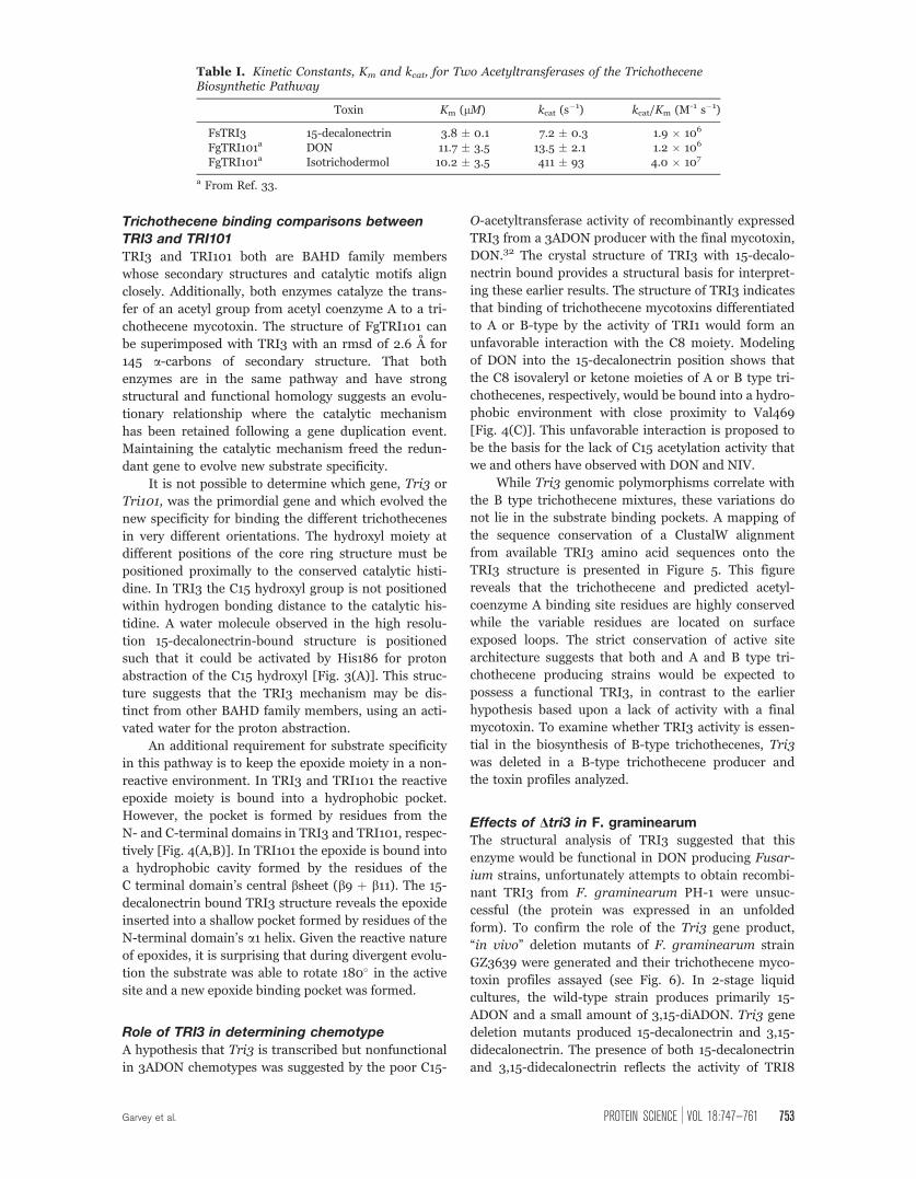
Table I. Kinetic Constants, Km and kcat, for Two Acetyltransferases of the TrichotheceneBiosynthetic Pathway
Toxin Km (lM) kcat (s�1) kcat/Km (M-1 s�1)
FsTRI3 15-decalonectrin 3.8 � 0.1 7.2 � 0.3 1.9 � 106
FgTRI101a DON 11.7 � 3.5 13.5 � 2.1 1.2 � 106
FgTRI101a Isotrichodermol 10.2 � 3.5 411 � 93 4.0 � 107
a From Ref. 33.
Garvey et al. PROTEIN SCIENCE VOL 18:747—761 753
which removes the C3 acetyl moiety. Tri8 is the last
step in the formation of 15ADON from 3,15-diADON
but it acts on a variety of substrates.23 While a small
amount of 15-ADON was detected in several of the
Tri3 transformants, this was less than 5% of wild-type
production and no 3-ADON or DON was detected in
the liquid cultures.
The lack of TRI3 in both A and B type trichothe-
cene producers results in the accumulation of 15-deca-
lonectrin and 3,15-didecalonectrin demonstrating that
the enzymes downstream of TRI3 in the biosynthetic
pathway are reduced in their ability to modify the core
ring structure without C15 acetylation (this study and
Ref. 9). The genetic, structural, and kinetic data pre-
sented here suggest that the biosynthetic pathways to
produce B type trichothecene mycotoxins require the
C15 acetylation activity of TRI3 and this acetyl group
is therefore removed later in the pathway. This implies
that an unidentified deacetylase from the producing
Fusarium strain or from the host organism is respon-
sible for the lack of acetylation at C15 in the final
mycotoxins.
The trichothecene mycotoxins have been identified
as an important virulence factor in Fusarium infections
of wheat and barley. Currently only Tri5 disruption
mutants, blocked at the start of the biosynthetic path-
way, have been shown to be severely impaired in their
ability to spread between florets and induce disease.
Although the analysis of virulence of Tri3 disruption
mutants is incomplete, these strains are reduced in
their virulence (unpublished data). The Dtri3 mutants
produce trichothecenes with a 12,13-epoxy group but
Figure 4. Comparison of trichothecene binding orientations in the structurally homologous TRI3 and FgTRI101. (A) TRI3
complexed with 15-decalonectrin. The 15-decalonectrin epoxide moiety is oriented into a hydrophobic depression formed by
residues of the N-terminal domain, this requires a substantial reorientation of the substrate as compared to the TRI101
structures. In this structure the catalytic His186 does not form a hydrogen bond with the C15 hydroxyl moiety, but does form
a hydrogen bond with a trapped water molecule. (B) FgTRI101 complexed with CoA and DON (3B2S). The DON substrate’s
epoxide moiety is oriented toward the C-terminal domain and the catalytic His156 is within ideal hydrogen bonding distance.
(C) Proposed model of TRI3 complexed with DON. DON has been superimposed onto the core trichothecene ring structure of
the TRI3�15-decalonectrin complex. This binding position of DON in the active site would position the C8 ketone moiety in
close proximity to the Val469 side chain, thus creating a poor interaction that accounts for the lack of catalytic activity
observed with DON as a substrate.
754 PROTEINSCIENCE.ORG Structure and Kinetics of TRI3 Acetyltransferase

the specific compounds are not very phytotoxic.6,7 The
reduced virulence of Dtri3 strains and essential role in
biosynthesis of trichothecene mycotoxins establishes
TRI3 as an additional target for combating FHB.
Materials and Methods
Plasmid and strain construction
All cloning was done in E. coli strain Top10. Primers
were designed to amplify the F. sporotrichioides TRI3
Figure 5. Sequence conservation mapped onto a stereo representation of TRI3. TRI3 (white ribbon) with bound 15-
decalonectrin (colored black) and CoA (colored gray) modeled in as described above. Residues with a sequence conservation
score derived from the T-coffee ClustalW alignment of TRI3 homologs from different chemotype Fusarium strains are shown
as spheres colored purple ¼ 1–4 (low), teal ¼ 5 (mid), light blue ¼ 6–8 (high).40
Figure 6. GC-MS metabolic profiles of (A) Fusarium graminearum wild-type strain GZ3639 and (B) FgTri3#228, a transformant
with a disrupted Tri3. Y-axis indicates relative abundance. Retention times are (a) 3,15-diacetyldeoxynivalenol (14.9 min), (b)
15-acetyldeoxynivalenol (14.6 min), (c) 15-decalonectrin (13.1 min), and (d) 3,15-didecalonectrin (11.8 min).
Garvey et al. PROTEIN SCIENCE VOL 18:747—761 755

gene (accession # AAK33072) using TRI3_forward
primer 50 GCTAGCATGAGCGCTT CATCCTCCT CC-
GCTTTACC30 and TRI3_reverse primer 50 gCTgAgCC-TAAAGTCGGAAGGCAAGCA
TGAACTCGACTATACTCTGC30 from genomic DNA of
F. sporotrichioides strain NRRL 3299 (a gift from Dr.
Jaechuk Yu, UW Madison). Product bands were gel
extracted using QIAquickVR gel extraction kit (Qiagen),
ligated into Zero Blunt Topo (Invitrogen) and then
sequenced using M13F and M13R(�27) primers. Seq-
uencing reactions were prepared using nonradioactive
BigDyeVR protocols (ABI PRISM) and resolved at the
UW-Madison Biotechnology Center. A single mutation
from the deposited sequence of AAK33072 was observed,
A291D. Introns were removed by overlap PCR exten-
sion.41 The exons were amplified with the following
primer pairs: TRI3_forward þ Intron_1R 50GGTT-GAAATTTCATACCCCGGCTTTTATAATTGGCTACAAG-
AGC30, Intron_1F 50GC TCTTGTAGCCAATTATAAAA-
GCCGGGGTATGAAATTTCAACC30 þ Intron_2R 50CCCTTCGAACGGAATTGGTTTTGCGTGCAGATAGCTCGC-
C30, Intron_2F 50GGCGAGCTAT CTGCACGCAAAACC-
AATTCCGTTCGAAGGG30 þ Intron_3R 50GCCATCACT-GATGAA CAATGGGTTTGCTTCCCCTTCG30, Intron_3F50CGAAGGGGAAGCAAACCCA TTGTTCATCAGTGAT-
GGC30 þ Intron_4R 50CCAGCTGTCCAGTCGGATTGC-CAGATATGGCAAAGACTGG30, Intron_4F 50CCAG TC-
TTTGCCATATCTGGCAATCCGACTGGACAGCTGG30 þTRI3_Reverse. The amplified exons were gel purified
as described above and then joined by fusion PCR
using TRI3_forward and TRI3_reverse primers. The
validity of the gene was verified by sequencing.
Plasmid TRI3/pKLD37
The ‘‘intron-less’’ TRI3 allele was then excised from
the Topo vector with restriction enzymes NheI and
BlpI (New England Biolabs), and ligated with T4 DNA
ligase (Fermentas) into a modified pET31b (Novagen)
vector cut with NheI and BlpI. The pET31b vector was
previously modified to contain an N terminal His6 tag
followed by a TEV protease cut site two amino acids
upstream of the gene start. The N-terminal amino acid
sequence of this construct is MSYYHHHHHHDY-
DIPTSELYFQGASM1S2.. . . where the location of the
TEV protease cleavage site is underlined.42 The valid-
ity of the final expression construct was verified by
sequencing.
Protein expression and purification
Native TRI3 protein was overproduced utilizing plas-
mid TRI3/pKLD37 transformed into E. coli strain
HMS174(kDE3). A starter culture from a single colony
was grown overnight at 37�C in M9 minimal medium
supplemented with ampicillin (0.2 lg/mL). The fol-
lowing day, 10 mL of the starter culture was used to
inoculate 650 mL M9 minimal media þ ampicillin in a
2-L shaker flask. Cultures were grown at 37�C until
they reached an optical density (OD600) of � 1.2. Cul-
tures were transferred to a 16�C incubator and allowed
to equilibrate for 30 min. After equilibration, CoA
(Sigma) was added to 15 lM, TRI3 expression was
induced by the addition of isopropyl-b-D-thiogalacto-pyranoside (IPTG) to a final concentration of 1.0 mM.
After a 20-h incubation period with IPTG under aero-
bic conditions, cells were harvested by centrifugation
at 5000g, flash frozen in liquid nitrogen, and stored at
�80�C until used.
TRI3 protein was purified from 30 g of cells
thawed and resuspended in 210 mL of lysis buffer,
which contained 0.5 mg/mL Lysozyme (Sigma),
50 mM NaH2PO4/K2HPO4, 300 mM NaCl, 20 mM
Imidazole, 5 mM 2-mercaptoethanol, 1 tablet/50 mL
of Complete Inhibitor (Roche), at pH 8.0. This mix-
ture was incubated with stirring at 4�C for 1 h and
then subjected to three rounds of sonication, each
round of sonication continued until the temperature
reached 10�C and then was left on ice until the tem-
perature returned to 4�C. Cellular debris was removed
by centrifugation at 40,000g for 30 min. The superna-
tant was loaded onto a 10-mL column of nickel-nitrilo-
triacetic acid-agarose (Qiagen) previously equilibrated
with lysis buffer. The column was washed with lysis
buffer until the A280 of the outflow reached back-
ground level. TRI3 protein was eluted with a linear
gradient of 20–300 mM imidazole in lysis buffer.
Fractions containing TRI3 protein were identified with
SDS-PAGE43 and Coomassie Blue stain 3 and were
pooled and dialyzed against 10 mM N-2-Hydroxyethyl-
piperazine-N0-2-ethanesulfonic acid (HEPES), 250 mM
NaCl, 0.5 mM tris(2-carboxyethyl)phosphine hydro-
chloride (TCEP), 2 mM ethylenediaminetetraacetic
acid, pH 7.5 buffer 4�C. The N-terminal hexahistidine
(H6) tag was removed by treatment with recombinant
tobacco etch virus (rTEV) protease.44 The remaining
uncleaved TRI3 protein enzyme and rTEV protease
were removed from the mixture with nickel-nitrilotri-
acetic acid-agarose affinity chromatography. Tagless
TRI3 protein was concentrated to � 20 mg/mL in a
centriprep YM30 (Millipore) concentrator. Concen-
trated protein was dialyzed against a buffer containing
20 mM HEPES, 20 mM NaCl, 0.5 mM TCEP (pH 7.5
at 4�C).
Crystallization and structural
determination of TRI3A search for crystallization conditions was conducted
at 4�C via the hanging drop method of vapor diffusion
utilizing an ‘‘in-house’’ designed sparse matrix screen
composed of 144 conditions. The crystals for 13 mg/
mL enzyme were observed from hanging drop experi-
ments with precipitant solutions of 2M ammonium
sulfate buffered with HEPES buffer (100 mM, pH 7.5
at 25�C). Single crystals of dimensions 0.1 � 0.1 �0.01 lm, were transferred stepwise to 1M ammonium
sulfate, 0.1M HEPES, pH 7.5 at 25�C solution until the
crystal edges began to visibly degrade. Each crystal
756 PROTEINSCIENCE.ORG Structure and Kinetics of TRI3 Acetyltransferase
was then transferred to a hanging drop equilibrated
for 48 h prepared by mixing equal volumes of 13 mg/
mL TRI3 and precipitant solution of 2M ammonium
sulfate, 0.1M HEPES, pH 7.5 at 25�C. Wild type apo
crystals of dimensions 0.6 � 0.25 � 0.05 lm were
transferred stepwise into a cryoprotectant solution of
20% glycerol, 2M ammonium sulfate, 50 mM NaCl,
0.1M HEPES, pH 7.5 at 25�C for 24 h, then flash fro-
zen directly into liquid nitrogen.
A mercury heavy atom derivative was prepared
by soaking an apo crystal in 25 mM CH3HgCl2, 2M
ammonium sulfate, 50 mM NaCl, 0.1M HEPES, pH
7.5 at 25�C for 75 min. The crystal was then trans-
ferred stepwise every minute into a 20% Glycerol,
1.6M ammonium sulfate, 50 mM NaCl, 0.1M HEPES,
pH 7.5 at 25�C, then flash frozen directly into liquid
nitrogen.
A platinum heavy atom derivative was prepared
by soaking an apo crystal in 20 mM K2PtCl4, 2M am-
monium sulfate, 50 mM NaCl, 0.1M HEPES, pH 7.5 at
25�C for 75 min. The crystal was then transferred step-
wise every minute into a 20% Glycerol, 1.6M ammo-
nium sulfate, 50 mM NaCl, 0.1M HEPES, pH 7.5 at
25�C, then flash frozen directly into liquid nitrogen.
X-ray data were collected from crystals of native
and heavy atom derivatives on a CCD detector at SBC
Beamline 19-BM (Advanced Photon Source, Argonne
National Laboratory, Argonne, IL) The X-ray data
were processed and scaled with HKL2000.45 X-ray
data collection statistics are presented in Table II.
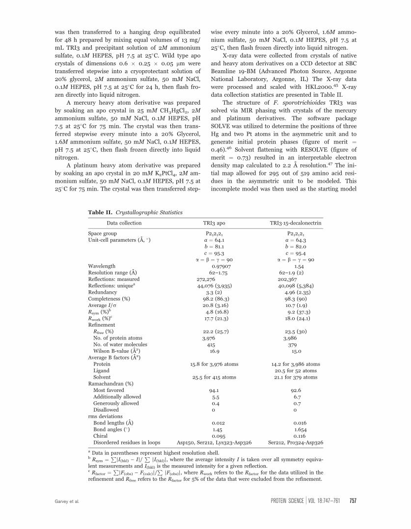
The structure of F. sporotrichioides TRI3 was
solved via MIR phasing with crystals of the mercury
and platinum derivatives. The software package
SOLVE was utilized to determine the positions of three
Hg and two Pt atoms in the asymmetric unit and to
generate initial protein phases (figure of merit ¼0.46).46 Solvent flattening with RESOLVE (figure of
merit ¼ 0.73) resulted in an interpretable electron
density map calculated to 2.2 A resolution.47 The ini-
tial map allowed for 295 out of 519 amino acid resi-
dues in the asymmetric unit to be modeled. This
incomplete model was then used as the starting model
Table II. Crystallographic Statistics
Data collection TRI3 apo TRI3�15-decalonectrinSpace group P212121 P212121Unit-cell parameters (A, �) a ¼ 64.1 a ¼ 64.3
b ¼ 81.1 b ¼ 82.0c ¼ 95.3 c ¼ 95.4
a ¼ b ¼ c ¼ 90 a ¼ b ¼ c ¼ 90Wavelength 0.97907 1.54Resolution range (A) 62–1.75 62–1.9 (2)Reflections: measured 272,276 202,367Reflections: uniquea 44,076 (3,935) 40,098 (5,384)Redundancy 3.3 (2) 4.96 (2.35)Completeness (%) 98.2 (86.3) 98.3 (90)Average I/r 20.8 (3.16) 10.7 (1.9)Rsym (%)b 4.8 (16.8) 9.2 (37.3)Rwork (%)c 17.7 (21.3) 18.0 (24.1)RefinementRfree (%) 22.2 (25.7) 23.5 (30)No. of protein atoms 3,976 3,986No. of water molecules 415 379Wilson B-value (A2) 16.9 15.0
Average B factors (A2)Protein 15.8 for 3,976 atoms 14.2 for 3,986 atomsLigand 20.5 for 52 atomsSolvent 25.5 for 415 atoms 21.1 for 379 atoms
Ramachandran (%)Most favored 94.1 92.6Additionally allowed 5.5 6.7Generously allowed 0.4 0.7Disallowed 0 0
rms deviationsBond lengths (A) 0.012 0.016Bond angles (�) 1.45 1.654Chiral 0.095 0.116Disordered residues in loops Asp150, Ser212, Lys323-Asp326 Ser212, Pro324-Asp326
a Data in parentheses represent highest resolution shell.b Rsym ¼ P
|I(hkl) – I|/P
|I(hkl)|, where the average intensity I is taken over all symmetry equiva-lent measurements and I(hkl) is the measured intensity for a given reflection.c Rfactor ¼
P|F(obs) – F(calc)|/
P|F(obs)|, where Rwork refers to the Rfactor for the data utilized in the
refinement and Rfree refers to the Rfactor for 5% of the data that were excluded from the refinement.
Garvey et al. PROTEIN SCIENCE VOL 18:747—761 757

for ARP/wARP48 which traced 491 residues into the
electron density map for the native apo data set.
Alternate cycles of manual model building and
least squares refinement with the programs COOT49
and Refmac50 reduced the R-factor to 17.5% for all
X-ray data from 50–1.75 A resolution. Refinement sta-
tistics are presented in Table II. In this model there
are three breaks in the polypeptide chain between
Asn149 and Asn151, Arg211 and Asp213, Leu322 and
Leu327, the N terminus is disordered to residue Leu9.
The Ramachandran plot as calculated by PROCHECK51
has no residues in the disallowed regions, 94.1% in the
most favored, 5.5% in the additionally allowed, and
0.4% in the generously allowed region.
Structural determination of the
TRI3�15-decalonectrin complexCrystals of the complex of TRI3 with 15-decalonectrin
mycotoxin were prepared by soaking apo crystals in
500 lM 15-decalonectirn, 2M ammonium sulfate, 25%
sucrose, 0.1M HEPES, pH 7.5 at 25�C for 3 h. The
crystal was then flash frozen directly into liquid nitro-
gen. X-ray data were collected with a Bruker AXS Plat-
inum 135 CCD detector controlled with the PROTEUM
software suite (Bruker AXS, Madison, WI). The X-ray
source was CuKa radiation from a Rigaku RU200 X-
ray generator equipped with Montel optics, operated
at 50 kV and 90 mA. The X-ray data were processed
with SAINT version 7.06 A (Bruker AXS) and inter-
nally scaled with SADABS version 2005/1 (Bruker
AXS). X-ray data collection statistics are presented in
Table II.
The structure of TRI3 complexed to 15-decalonec-
trin was solved by molecular replacement with the
program MOLREP52 starting from the apo native TRI3
model. Alternate cycles of manual model building and
least squares refinement with the programs COOT49
and Refmac50 reduced the Rfactor to 18% for all X-ray
data from 50–1.9 A resolution. Refinement statistics
are presented in Table II. In this model there are three
breaks in the polypeptide chain between Arg211 and
Asp213, Lys323 and Leu327, the N terminus is dis-
ordered to residue Ser5. The Ramachandran plot as
calculated by PROCHECK51 has no residues in the dis-
allowed regions, 92.6% in the most favored, 6.7% in
the additionally allowed, and 0.7% in the generously
allowed region.
Acetyltransferase enzymatic assay
The trichothecene 15-O-acetyltransferase reaction
catalyzed by TRI3 was monitored by following the
production of CoA in a 5,50-dithiobis-(2-nitroben-zoic acid) (DTNB) coupled continuous assay (k ¼14,150 cm�1 M�1).53 Reaction mixtures were prepared
at 25�C by combining 50 lL of 1.5 mM acetyl CoA
(Sigma), 0.6 mM DTNB (Sigma), 0.1M potassium
phosphate buffer, pH 8, with 50 lL of trichothecene
toxin, 4.5% DMSO, 0.1M potassium phosphate buffer,
pH 8.0. The reaction was initiated by the addition of
50 lL of 332 ng/mL enzyme, 200 lg/mL BSA, 0.1M
potassium phosphate buffer, pH 8. After mixing,
100 lL of the reaction mixture was transferred to a
1 cm pathlength cuvette and the change in absorbance
at 412 nm was followed. The rates of reaction were
determined at various concentrations of 15-decalonec-
trin, NIV, and DON and then fit by nonlinear regres-
sion to the Michaelis-Menten equation.
Construction of DTri3 deletion strainFgTri3 disruption was made using a gene replacement
strategy by amplifying genomic GZ3639 (available as
FGSC 8630 from the Fungal Genetics Stock Center,
Department of Microbiology, University of Kansas
Medical Center, Kansas City) DNA using primers spe-
cific to sequences prior to the 50 translation start site
(50-CTATGGCGCCGCATCACC-30 paired with 50-CGC-GTTAACGCTCAGAAGATACTTGGC-30) and primers
specific to sequences beyond the 30 translation stop
site (50-CGCGTTAACGGCAACACTTTGGGTGG-30 pairedwith 50-GCTGAGATGTACCTCGCC-30). The amplified
fragments had a HpaI site incorporated (underlined
sequence). Each segment of DNA was cloned sepa-
rately into pCR2.1 (Invitrogen, Carlsbad, CA) and then
cut with HpaI. The plasmid containing the 30 fragment
was further digested with NotI and this fragment was
inserted into the HpaI-cut 50-segment vector. The
resulting vector was then cut with HpaI and the hygB
(hygromycin) selectable marker was inserted. This
final vector contained � 975 bp of sequence upstream
of the FgTri3 start site, followed by 2.5 bp of hygB,
followed by 1070 bp of sequence downstream of the
translational stop site of FgTri3.
Fungal transformations of GZ3639 protoplasts were
carried out as described previously.54 All transformants
were single-spored to ensure purity, tested by PCR to
confirm disruption of FgTri3, and tested by Southern
analysis55 to further confirm gene disruption.
Trichothecene toxin analysisTrichothecenes were produced in liquid culture with a
two-stage media protocol, (modified from Ref. 56).
Strains were maintained in glycerol stocks. Cultures
were grown initially on V-8 juice agar plates under
alternating 12 h, 25�C-12 h 22�C dark cycle. Mycelia
were washed from V8 plates with 3.5 mL water and
used to inoculate 50 mL of 1st stage media (3 g
NH4Cl, 2 g MgSO4-7H20, 0.2 g FeSO4-7H2O, 2 g
KH2PO4, 2 g peptone, 2 g yeast extract, 2 g malt
extract, 20 g glucose in 1 L distilled water) in 250 mL
flasks. The cultures were grown at 28�C on a rotary
shaker at 200 rpm in the dark for 3 days. The culture
was then transferred to a 250 mL beaker and dis-
persed with a Toastmaster stick blender. The macer-
ated culture was transferred to a 50-mL conical tube
and centrifuged 5 min at 1600 rpm. Half of the super-
natant was removed and the remaining fungal mass
758 PROTEINSCIENCE.ORG Structure and Kinetics of TRI3 Acetyltransferase
and medium was mixed well. Second stage cultures
were initiated by adding 1.5 mL of the concentrated
culture to 20 mL of 2nd stage medium (1 g
(NH4)2HPO4, 3 g KH2PO4, 0.2 g MgSO4-7H2O, 5 g
NaCl, 50 g sucrose, 10 g glycerol in 1 L distilled water)
in a 50 mL flask. After 4 days, 5 mL aliquots were
removed from each culture and extracted with 2 mL
ethyl acetate. The extract was analyzed by GC-MS.
GC-MS measurements were made with a Hewlett-
Packard 5890 Gas Chromatograph fitted with a 30 m
fused silica capillary column (HP-5MS; 0.25 lm; J&W
Scientific, Palo Alto, CA). The column was held at
120�C at injection; then heated to 210�C at 15�C/min
and held for 1 min; then heated to 260�C at 5�C/min
and held for 3 min. Trichothecenes were identified by
comparison of retention times and mass spectral frag-
mentation patterns with authentic standards.
Conclusions
The ensemble of toxins synthesized by a particular
Fusarium species is an important biological character-
istic that is generally assumed to be controlled by
the genetic make up of the fungus responsible for the
toxin biosynthesis. The study here reveals that the
DNA polymorphisms of TRI3 which are predictive of
C15 chemotype in trichothecene producers do not map
to residues lining the enzyme’s active site, which sug-
gests that, if expressed, all TRI3 orthologs should be
capable of acetylating 15-calonectrin. Furthermore,
deletion mutants of Tri3 in F. graminearum are
blocked in their trichothecene biosynthesis and accu-
mulate the pathway intermediates 15-decalonectrin
and 3,15-didecalonectrin. This implies that acetylation
at the 15 position is an obligate step in mycotoxin bio-
synthesis. Since the majority of the final mycotoxins
generated by F. graminearum grown on cereal sub-
strates are not acetylated at the 15 position there must
be another activity responsible for the removal of the
15-acetyl moiety. The enzyme(s) responsible for
removal of the acetyl group from the 15 position of the
trichothecene mycotoxins have yet to be identified.
The structure of TRI3 with bound native substrate,
15-decalonectrin, reveals a high structural homology
with another acetyltransferase in the same biosynthetic
pathway, TRI101. The bound structures of TRI3 and
TRI101 reveal an evolutionary relationship between
enzymes within the trichothecene mycotoxin biosyn-
thetic pathway where the catalytic mechanism has been
maintained and substrate specificity has evolved. This
has led to a restructuring of the active sites to bind the
core trichothecene ring in two different orientations,
binding the reactive epoxide moiety into the N-terminal
domain in TRI3 and into the C-terminal domain in
TRI101. In TRI3 this new binding orientation places the
C8 ring position in close proximity to a hydrophobic
patch of the active site, thereby preventing substrate
promiscuity with the later products of the biosynthetic
pathway that are modified at C8. The biochemical,
structural, and kinetic data presented in this study iden-
tify TRI3 as a potential target in combating FHB.
CoordinatesThe atomic coordinates and structure factors for the
apo TRI3 and TRI3�15-decalonectrin complex have
been deposited in the Protein Data Bank, Research
Collaboratory for Structural Bioinformatics, Rutgers
University, New Brunswick, NJ (http://www.rcsb.org/)
with accession numbers 3FOT and 3FP0, respectively.
Acknowledgments
The authors thank Kirsten Dennison for creating the
modified pET vector utilized in construction of the over
expression plasmid for TRI3 and Dr. Martin St. Maurice
for input to the kinetic assay design. This is a cooperative
project with the U.S. Wheat and Barley Scab Initiative.
SPM and NJA are supported by the U.S. Department of
Agriculture NP 108 Food Safety. Any opinions, findings,
conclusions, or recommendations expressed in this publi-
cation are those of the authors and do not necessarily
reflect the view of the U.S. Department of Agriculture. Use
of the Structural Biology BM19 beamline Argonne
National Laboratory Advanced Photon Source was sup-
ported by the U.S. Department of Energy, Office of Energy
Research, under ContractNo.W-31-109-ENG-38.
References
1. Stack RW (1999) Return of an old problem: Fusariumhead blight of small grains. Available at: http://www.apsnetorg/online/feature/FHB/Top.html (accessed onFebruary 2, 2008).
2. Nganje WE, Bangsund DA, Leistritz FL, Wilson WW,Tiapo NM (2004) Regional economic impacts of Fusar-ium head blight in wheat and barley. Rev Agric Econ 26:332–347.
3. Windels CE (2000) Economic and social impacts of Fusar-ium head blight: changing farms and rural communities inthe Northern Great Plains. Phytopathology 90:17–21.
4. Bennett JW, Klich M (2003) Mycotoxins. Clin MicrobiolRev 16:497–516.
5. Anderson DW, Black RM, Lee CG, Pottage C, RickardRL, Sandford MS, Webber TD, Williams NE (1989)Structure-activity studies of trichothecenes: cytotoxicityof analogues and reaction products derived from T-2toxin and neosolaniol. J Med Chem 32:555–562.
6. Alexander NJ, McCormick SP, Ziegenhorn SL (1999)Phytotoxicity of selected trichothecenes using Chlamydo-monas reinhardtii as a model system. Natural Toxins 7:265–269.
7. Desjardins AE, McCormick SP, Appell M (2007) Struc-ture-activity relationships of trichothecene toxins in anArabidopsis thaliana leaf assay. J Agric Food Chem 55:6487–6492.
8. Zamir LO, Devor KA, Sauriol F (1991) Biosynthesis of thetrichothecene 3-acetyldeoxynivalenol. Identification ofthe oxygenation steps after isotrichodermin. J Biol Chem266:14992–15000.
9. McCormick SP, Hohn TM, Desjardins AE (1996) Isola-tion and characterization of Tri3, a gene encoding 15-O-acetyltransferase from Fusarium sporotrichioides. ApplEnviron Microbiol 62:353–359.
Garvey et al. PROTEIN SCIENCE VOL 18:747—761 759
10. Kimura M, Tokai T, Takahashi-Ando N, Ohsato S,Fujimura M (2007) Molecular and genetic studies offusarium trichothecene biosynthesis: pathways, genes,and evolution. Biosci Biotechnol Biochem 71:2105–2123.
11. Tokai T, Koshino H, Takahashi-Ando N, Sato M, FujimuraM, Kimura M (2007) Fusarium Tri4 encodes a key multi-functional cytochrome P450 monooxygenase for four con-secutive oxygenation steps in trichothecene biosynthesis.Biochem Biophys Res Commun 353:412–417.
12. Hohn TM, Beremand PD (1989) Isolation and nucleotidesequence of a sesquiterpene cyclase gene from the tricho-thecene-producing fungus Fusarium sporotrichioides.Gene 79:131–138.
13. McCormick SP, Alexander NJ, Proctor RH (2006) Fusar-ium Tri4 encodes a multifunctional oxygenase requiredfor trichothecene biosynthesis. Can J Microbiol 52:636–642.
14. Kimura M, Kaneko I, Komiyama M, Takatsuki A, KoshinoH, Yoneyama K, Yamaguchi I (1998) Trichothecene 3-O-acetyltransferase protects both the producing organismand transformed yeast from related mycotoxins. Cloningand characterization of Tri101. J Biol Chem 273:1654–1661.
15. Alexander NJ, Hohn TM, McCormick SP (1998) TheTRI11 gene of Fusarium sporotrichioides encodes acytochrome P-450 monooxygenase required for C-15hydroxylation in trichothecene biosynthesis. Appl EnvironMicrobiol 64:221–225.
16. Brown DW, McCormick SP, Alexander NJ, Proctor RH,Desjardins AE (2001) A genetic and biochemicalapproach to study trichothecene diversity in Fusariumsporotrichioides and Fusarium graminearum. FungalGenet Biol 32:121–133.
17. Brown DW, McCormick SP, Alexander NJ, Proctor RH,Desjardins AE (2002) Inactivation of a cytochrome P-450is a determinant of trichothecene diversity in Fusariumspecies. Fungal Genet Biol 36:224–233.
18. Lee T, Han YK, Kim KH, Yun SH, Lee YW (2002) Tri13and Tri7 determine deoxynivalenol- and nivalenol-pro-ducing chemotypes of Gibberella zeae. Appl EnvironMicrobiol 68:2148–2154.
19. Brown DW, Proctor RH, Dyer RB, Plattner RD (2003)Characterization of a fusarium 2-gene cluster involved intrichothecene C-8 modification. J Agric Food Chem 51:7936–7944.
20. Meek IB, Peplow AW, Ake C, Jr,Phillips TD, BeremandMN (2003) Tri1 encodes the cytochrome P450 monooxy-genase for C-8 hydroxylation during trichothecene bio-synthesis in Fusarium sporotrichioides and residesupstream of another new Tri gene. Appl Environ Micro-biol 69:1607–1613.
21. McCormick SP, Harris LJ, Alexander NJ, Ouellet T,Saparno A, Allard S, Desjardins AE (2004) Tri1 in Fusar-ium graminearum encodes a P450 oxygenase. Appl Envi-ron Microbiol 70:2044–2051.
22. Peplow AW, Meek IB, Wiles MC, Phillips TD, BeremandMN (2003) Tri16 is required for esterification of positionC-8 during trichothecene mycotoxin production byFusarium sporotrichioides. Appl Environ Microbiol 69:5935–5940.
23. McCormick SP, Alexander NJ (2002) Fusarium Tri8encodes a trichothecene C-3 esterase. Appl EnvironMicrobiol 68:2959–2964.
24. Ueno Y, Sawano M, Ishii K (1975) Production of tricho-thecene mycotoxins by Fusarium species in shake culture.Appl Microbiol 30:4–9.
25. Miller JD, Greenhalgh R, Wang Y, Lu M (1991) Tricho-thecene chemotypes of three Fusarium species. Mycologia83:121–130.
26. O’Donnell K, Kistler HC, Tacke BK, Casper HH (2000)Gene genealogies reveal global phylogeographic structureand reproductive isolation among lineages of Fusariumgraminearum, the fungus causing wheat scab. Proc NatlAcad Sci USA 97:7905–7910.
27. Ward TJ, Bielawski JP, Kistler HC, Sullivan E, O’Donnell K(2002) Ancestral polymorphism and adaptive evolution inthe trichothecene mycotoxin gene cluster of phytopatho-genic Fusarium. Proc Natl Acad Sci USA 99:9278–9283.
28. Goswami RS, Kistler HC (2005) Pathogenicity and inplanta mycotoxin accumulation among members of theFusarium graminearum species complex on wheat andrice. Phytopathology 95:1397–1404.
29. Starkey DE, Ward TJ, Aoki T, Gale LR, Kistler HC, GeiserDM, Suga H, Toth B, Varga J, O’Donnell K (2007) Globalmolecular surveillance reveals novel Fusarium head blightspecies and trichothecene toxin diversity. Fungal GenetBiol 44:1191–1204.
30. Ward TJ, Clear RM, Rooney AP, O’Donnell K, Gaba D,Patrick S, Starkey DE, Gilbert J, Geiser DM, Nowicki TW(2008) An adaptive evolutionary shift in Fusarium headblight pathogen populations is driving the rapid spreadof more toxigenic Fusarium graminearum in NorthAmerica. Fungal Genet Biol 45:473–484.
31. Desjardins AE (2008) Natural product chemistry meetsgenetics: when is a genotype a chemotype? J Agric FoodChem 56:7587–7592.
32. Kimura M, Tokai T, O’Donnell K, Ward TJ, Fujimura M,Hamamoto H, Shibata T, Yamaguchi I (2003) The tricho-thecene biosynthesis gene cluster of Fusarium graminea-rum F15 contains a limited number of essential pathwaygenes and expressed non-essential genes. FEBS Lett 539:105–110.
33. Garvey GS, McCormick SP, Rayment I (2008) Structuraland functional characterization of the TRI101 trichothe-cene 3-O-acetyltransferase from Fusarium sporotri-chioides and Fusarium graminearum: kinetic insights tocombating Fusarium head blight. J Biol Chem 283:1660–1669.
34. Krissinel E, Henrick K (2004) Secondary-structurematching (SSM), a new tool for fast protein structurealignment in three dimensions. Acta Crystallogr Sect D60(12, Part 1):2256–2268.
35. Buglino J, Onwueme KC, Ferreras JA, Quadri LE, LimaCD (2004) Crystal structure of PapA5, a phthioceroldimycocerosyl transferase from Mycobacterium tubercu-losis. J Biol Chem 279:30634–30642.
36. Onwueme KC, Ferreras JA, Buglino J, Lima CD, QuadriLE (2004) Mycobacterial polyketide-associated proteinsare acyltransferases: proof of principle with Mycobacte-rium tuberculosis PapA5. Proc Natl Acad Sci USA 101:4608–4613.
37. Ma X, Koepke J, Panjikar S, Fritzsch G, Stockigt J(2005) Crystal structure of vinorine synthase, the firstrepresentative of the BAHD superfamily. J Biol Chem280:13576–13583.
38. D’Auria JC (2006) Acyltransferases in plants: a goodtime to be BAHD. Curr Opin Plant Biol 9:331–340.
39. Bayer A, Ma X, Stockigt J (2004) Acetyltransfer in natu-ral product biosynthesis—functional cloning and molecu-lar analysis of vinorine synthase. Bioorg Med Chem 12:2787–2795.
40. Poirot O, O’Toole E, Notredame C (2003) Tcoffee@igs: aweb server for computing, evaluating and combiningmultiple sequence alignments. Nucleic Acids Res 31:3503–3506.
41. Ho SN, Hunt HD, Horton RM, Pullen JK, Pease LR(1989) Site-directed mutagenesis by overlap extensionusing the polymerase chain reaction. Gene 77:51–59.
760 PROTEINSCIENCE.ORG Structure and Kinetics of TRI3 Acetyltransferase
42. Rocco CJ, Dennison KL, Klenchin VA, Rayment I, Esca-lante-Semerena JC (2008) Construction and use of newcloning vectors for the rapid isolation of recombinantproteins from Escherichia coli. Plasmid 59:231–237.
43. Laemmli UK (1970) Cleavage of structural proteins dur-ing the assembly of the head of bacteriophage T4. Nature227:680–685.
44. Shih YP, Wu HC, Hu SM, Wang TF, Wang AH (2005)Self-cleavage of fusion protein in vivo using TEV proteaseto yield native protein. Protein Sci 14:936–941.
45. Otwinowski Z, Minor W (1997) Processing of X-ray dif-fraction data collected in oscillation mode. MethodsEnzymol 276:307–326.
46. Terwilliger TC, Berendzen J (1999) Automated MAD andMIRstructure solution. Acta Crystallogr D 55 (Part 4):849–861.
47. Terwilliger TC (2000) Maximum-likelihood density modi-fication. Acta Crystallogr D Biol Crystallogr 56 (Part 8):965–972.
48. Perrakis A, Harkiolaki M, Wilson KS, Lamzin VS (2001)ARP/wARP and molecular replacement. Acta CrystallogrD Biol Crystallogr 57 (Part 10):1445–1450.
49. Emsley P, Cowtan K (2004) Coot: model-building toolsfor molecular graphics. Acta Crystallogr D Biol Crystal-logr 60(12, Part 1):2126–2132a.
50. Murshudov GN, Vagin AA, Dodson EJ (1997) Refinementof macromolecular structures by the maximum-likelihoodmethod. Acta Crystallogr D Biol Crystallogr 53 (Part 3):240–255.
51. Laskowski RA, MacArthur MW, Moss DS, Thornton JM(1993) PROCHECK: a program to check the stereochemi-cal quality of protein structures. J Appl Crystallogr 26:283–291.
52. Vagin A, Teplyakov A (1997) MOLREP: an automatedprogram for molecular replacement. J Appl Crystallogr30:1022–1025.
53. Ellman GL (1959) Tissue sulphydryl groups. Arch Bio-chem Biophys 82:70.
54. Proctor RH, Hohn TM, McCormick SP (1995) Reducedvirulence of Gibberella zeae caused by disruption of a tri-chothecene toxin biosynthetic gene. Mol Plant MicrobeInteract 8:593–601.
55. Sambrook J, Fritsch EF, Maniatis T (1989) Molecularcloning: a laboratory manual. New York: Cold SpringHarbor Laboratory, Cold Spring Harbor.
56. Miller JD, Blackwell BA (1986) Biosynthesis of 3-Acetyl-deoxynivalenol and other metabolites by Fusarium-cul-morum Hlx 1503 in a stirred jar fermenter. Can J BotRev Can Bot 64:1–5.
Garvey et al. PROTEIN SCIENCE VOL 18:747—761 761
Related Documents