International Journal of Molecular Sciences Review Structural and Functional Changes and Possible Molecular Mechanisms in Aged Skin Hyunji Lee , Yongjun Hong and Miri Kim * Citation: Lee, H.; Hong, Y.; Kim, M. Structural and Functional Changes and Possible Molecular Mechanisms in Aged Skin. Int. J. Mol. Sci. 2021, 22, 12489. https://doi.org/10.3390/ ijms222212489 Academic Editors: Lukas Lacina and Michal Kolᡠr Received: 20 October 2021 Accepted: 18 November 2021 Published: 19 November 2021 Publisher’s Note: MDPI stays neutral with regard to jurisdictional claims in published maps and institutional affil- iations. Copyright: © 2021 by the authors. Licensee MDPI, Basel, Switzerland. This article is an open access article distributed under the terms and conditions of the Creative Commons Attribution (CC BY) license (https:// creativecommons.org/licenses/by/ 4.0/). Department of Dermatology, Yeouido St. Mary’s Hospital, College of Medicine, The Catholic University of Korea, #10, 63-ro, Yeongdeungpo-gu, Seoul 07345, Korea; [email protected] (H.L.); [email protected] (Y.H.) * Correspondence: [email protected]; Tel.: +82-3779-1056 Abstract: Skin aging is a complex process influenced by intrinsic and extrinsic factors. Together, these factors affect the structure and function of the epidermis and dermis. Histologically, aging skin typically shows epidermal atrophy due to decreased cell numbers. The dermis of aged skin shows decreased numbers of mast cells and fibroblasts. Fibroblast senescence contributes to skin aging by secreting a senescence-associated secretory phenotype, which decreases proliferation by impairing the release of essential growth factors and enhancing degradation of the extracellular matrix through activation of matrix metalloproteinases (MMPs). Several molecular mechanisms affect skin aging including telomere shortening, oxidative stress and MMP, cytokines, autophagic control, microRNAs, and the microbiome. Accumulating evidence on the molecular mechanisms of skin aging has provided clinicians with a wide range of therapeutic targets for treating aging skin. Keywords: skin aging; intrinsic aging; photoaging; molecular mechanisms 1. Introduction Skin is the outermost organ of the human body, with an extensive surface area of 1.5–2 m 2 [1]. Skin aging, resulting in cumulative changes in skin structure, function, and appearance, is a complex process affected by both intrinsic and extrinsic factors. Skin aging is not only a physiologic phenomenon but also a health risk, resulting in increased skin fragility, delayed and impaired wound healing, and increased incidence of infection and skin cancers. Intrinsic or chronological skin aging can be seen in areas unexposed to sunlight, revealing the influence of genetic factors. Photoaging, also referred to as extrinsic aging, mainly results from ultraviolet (UV) irradiation, and mainly occurs on the face and forearms due to frequent exposure to sunlight, and other factors such as air pollution and cigarette smoke [2,3]. Generally, alterations in skin structure, function, and appearance are more pronounced in photoaged than chronologically aged skin. However, these two types of aging are dif- ficult to separate [4] and are superimposed in sun-exposed skin, as they have common clinical features caused by dermal matrix alterations that contribute to wrinkle formation, laxity, and fragility [5]. The dermal matrix contains extracellular matrix (ECM) proteins such as collagen, elastin, and proteoglycans that confer skin strength and resilience. Skin ag- ing associated with dermal matrix alterations and atrophy can be caused by the senescence of dermal cells such as fibroblasts, and decreased synthesis and accelerated breakdown of dermal collagen fibers [3]. In the last 30 years, substantial progress has been made in understanding the molecular mechanisms responsible for the aging of human skin. This review focuses on the molecular mechanisms of skin aging and summarizes its clinical and histological features. 2. Histology of Chronological Aging and Photoaging Histological changes in chronologically aged skin are characterized by decreased recovery capacity, altered permeability of the stratum corneum, epidermal atrophy particu- Int. J. Mol. Sci. 2021, 22, 12489. https://doi.org/10.3390/ijms222212489 https://www.mdpi.com/journal/ijms

Welcome message from author
This document is posted to help you gain knowledge. Please leave a comment to let me know what you think about it! Share it to your friends and learn new things together.
Transcript
International Journal of
Molecular Sciences
Review
Structural and Functional Changes and Possible MolecularMechanisms in Aged Skin
Hyunji Lee , Yongjun Hong and Miri Kim *
�����������������
Citation: Lee, H.; Hong, Y.; Kim, M.
Structural and Functional Changes
and Possible Molecular Mechanisms
in Aged Skin. Int. J. Mol. Sci. 2021, 22,
12489. https://doi.org/10.3390/
ijms222212489
Academic Editors: Lukas Lacina and
Michal Kolár
Received: 20 October 2021
Accepted: 18 November 2021
Published: 19 November 2021
Publisher’s Note: MDPI stays neutral
with regard to jurisdictional claims in
published maps and institutional affil-
iations.
Copyright: © 2021 by the authors.
Licensee MDPI, Basel, Switzerland.
This article is an open access article
distributed under the terms and
conditions of the Creative Commons
Attribution (CC BY) license (https://
creativecommons.org/licenses/by/
4.0/).
Department of Dermatology, Yeouido St. Mary’s Hospital, College of Medicine, The Catholic University of Korea,#10, 63-ro, Yeongdeungpo-gu, Seoul 07345, Korea; [email protected] (H.L.); [email protected] (Y.H.)* Correspondence: [email protected]; Tel.: +82-3779-1056
Abstract: Skin aging is a complex process influenced by intrinsic and extrinsic factors. Together,these factors affect the structure and function of the epidermis and dermis. Histologically, agingskin typically shows epidermal atrophy due to decreased cell numbers. The dermis of aged skinshows decreased numbers of mast cells and fibroblasts. Fibroblast senescence contributes to skinaging by secreting a senescence-associated secretory phenotype, which decreases proliferation byimpairing the release of essential growth factors and enhancing degradation of the extracellularmatrix through activation of matrix metalloproteinases (MMPs). Several molecular mechanismsaffect skin aging including telomere shortening, oxidative stress and MMP, cytokines, autophagiccontrol, microRNAs, and the microbiome. Accumulating evidence on the molecular mechanisms ofskin aging has provided clinicians with a wide range of therapeutic targets for treating aging skin.
Keywords: skin aging; intrinsic aging; photoaging; molecular mechanisms
1. Introduction
Skin is the outermost organ of the human body, with an extensive surface area of1.5–2 m2 [1]. Skin aging, resulting in cumulative changes in skin structure, function, andappearance, is a complex process affected by both intrinsic and extrinsic factors. Skinaging is not only a physiologic phenomenon but also a health risk, resulting in increasedskin fragility, delayed and impaired wound healing, and increased incidence of infectionand skin cancers. Intrinsic or chronological skin aging can be seen in areas unexposed tosunlight, revealing the influence of genetic factors. Photoaging, also referred to as extrinsicaging, mainly results from ultraviolet (UV) irradiation, and mainly occurs on the face andforearms due to frequent exposure to sunlight, and other factors such as air pollution andcigarette smoke [2,3].
Generally, alterations in skin structure, function, and appearance are more pronouncedin photoaged than chronologically aged skin. However, these two types of aging are dif-ficult to separate [4] and are superimposed in sun-exposed skin, as they have commonclinical features caused by dermal matrix alterations that contribute to wrinkle formation,laxity, and fragility [5]. The dermal matrix contains extracellular matrix (ECM) proteinssuch as collagen, elastin, and proteoglycans that confer skin strength and resilience. Skin ag-ing associated with dermal matrix alterations and atrophy can be caused by the senescenceof dermal cells such as fibroblasts, and decreased synthesis and accelerated breakdownof dermal collagen fibers [3]. In the last 30 years, substantial progress has been made inunderstanding the molecular mechanisms responsible for the aging of human skin. Thisreview focuses on the molecular mechanisms of skin aging and summarizes its clinical andhistological features.
2. Histology of Chronological Aging and Photoaging
Histological changes in chronologically aged skin are characterized by decreasedrecovery capacity, altered permeability of the stratum corneum, epidermal atrophy particu-
Int. J. Mol. Sci. 2021, 22, 12489. https://doi.org/10.3390/ijms222212489 https://www.mdpi.com/journal/ijms
Int. J. Mol. Sci. 2021, 22, 12489 2 of 17
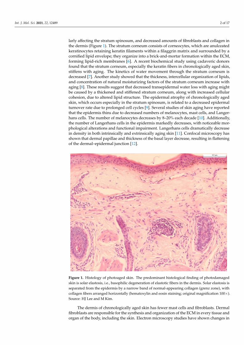
larly affecting the stratum spinosum, and decreased amounts of fibroblasts and collagen inthe dermis (Figure 1). The stratum corneum consists of corneocytes, which are anulceatedkeratinocytes retaining keratin filaments within a filaggrin matrix and surrounded by acornified lipid envelope; they organize into a brick-and-mortar formation within the ECM,forming lipid-rich membranes [6]. A recent biochemical study using cadaveric donorsfound that the stratum corneum, especially the keratin fibers in chronologically aged skin,stiffens with aging. The kinetics of water movement through the stratum corneum isdecreased [7]. Another study showed that the thickness, intercellular organization of lipids,and concentration of natural moisturizing factors of the stratum corneum increase withaging [8]. These results suggest that decreased transepidermal water loss with aging mightbe caused by a thickened and stiffened stratum corneum, along with increased cellularcohesion, due to altered lipid structure. The epidermal atrophy of chronologically agedskin, which occurs especially in the stratum spinosum, is related to a decreased epidermalturnover rate due to prolonged cell cycles [9]. Several studies of skin aging have reportedthat the epidermis thins due to decreased numbers of melanocytes, mast cells, and Langer-hans cells. The number of melanocytes decreases by 8–20% each decade [10]. Additionally,the number of Langerhans cells in the epidermis markedly decreases, with noticeable mor-phological alterations and functional impairment. Langerhans cells dramatically decreasein density in both intrinsically and extrinsically aging skin [11]. Confocal microscopy hasshown that dermal papillae and thickness of the basal layer decrease, resulting in flatteningof the dermal–epidermal junction [12].
Int. J. Mol. Sci. 2021, 22, x FOR PEER REVIEW 2 of 16
2. Histology of Chronological Aging and Photoaging Histological changes in chronologically aged skin are characterized by decreased re-
covery capacity, altered permeability of the stratum corneum, epidermal atrophy partic-ularly affecting the stratum spinosum, and decreased amounts of fibroblasts and collagen in the dermis (Figure 1). The stratum corneum consists of corneocytes, which are anul-ceated keratinocytes retaining keratin filaments within a filaggrin matrix and surrounded by a cornified lipid envelope; they organize into a brick-and-mortar formation within the ECM, forming lipid-rich membranes [6]. A recent biochemical study using cadaveric do-nors found that the stratum corneum, especially the keratin fibers in chronologically aged skin, stiffens with aging. The kinetics of water movement through the stratum corneum is decreased [7]. Another study showed that the thickness, intercellular organization of li-pids, and concentration of natural moisturizing factors of the stratum corneum increase with aging [8]. These results suggest that decreased transepidermal water loss with aging might be caused by a thickened and stiffened stratum corneum, along with increased cel-lular cohesion, due to altered lipid structure. The epidermal atrophy of chronologically aged skin, which occurs especially in the stratum spinosum, is related to a decreased epi-dermal turnover rate due to prolonged cell cycles [9]. Several studies of skin aging have reported that the epidermis thins due to decreased numbers of melanocytes, mast cells, and Langerhans cells. The number of melanocytes decreases by 8–20% each decade [10]. Additionally, the number of Langerhans cells in the epidermis markedly decreases, with noticeable morphological alterations and functional impairment. Langerhans cells dra-matically decrease in density in both intrinsically and extrinsically aging skin [11]. Confo-cal microscopy has shown that dermal papillae and thickness of the basal layer decrease, resulting in flattening of the dermal–epidermal junction [12].
Figure 1. Histology of photoaged skin. The predominant histological finding of photodamaged skin is solar elastosis, i.e., basophilic degeneration of elastotic fibers in the dermis. Solar elastosis is sep-arated from the epidermis by a narrow band of normal-appearing collagen (grenz zone), with col-lagen fibers arranged horizontally (hematoxylin and eosin staining; original magnification 100×). Source: HJ Lee and M Kim.
The dermis of chronologically aged skin has fewer mast cells and fibroblasts. Dermal fibroblasts are responsible for the synthesis and organization of the ECM in every tissue
Figure 1. Histology of photoaged skin. The predominant histological finding of photodamagedskin is solar elastosis, i.e., basophilic degeneration of elastotic fibers in the dermis. Solar elastosis isseparated from the epidermis by a narrow band of normal-appearing collagen (grenz zone), withcollagen fibers arranged horizontally (hematoxylin and eosin staining; original magnification 100×).Source: HJ Lee and M Kim.
The dermis of chronologically aged skin has fewer mast cells and fibroblasts. Dermalfibroblasts are responsible for the synthesis and organization of the ECM in every tissue andorgan of the body, including the skin. Electron microscopy studies have shown changes in
Int. J. Mol. Sci. 2021, 22, 12489 3 of 17
the structure and morphology of fibroblasts from the dermis of aged skin [9]. An emerginghypothesis is that these changes, referred to as “fibroblast senescence”, are the main causeof irreversible skin aging due to their inherent ability to be almost free from apoptosisand to not be eliminated by the adaptive immune system [13]. Fibroblast senescencecontributes to skin aging by secreting a senescence-associated secretory phenotype (SASP),which decreases proliferation by impairing the release of essential growth factors andenhances degradation of the ECM by activating matrix metalloproteinases (MMPs) [14].Recently, Basisty et al. [15] presented the SASP Atlas, which is a proteomic database ofsoluble proteins and exosomal cargo SASP factors from multiple senescence inducers andcell types. The authors further suggested several possible biomarkers of the aging process,such as growth/differentiation factor 15, stanniocalcin 1, and serine protease inhibitors.
Collagen and elastin, two of the main constituents of the ECM, change throughoutthe life cycle. In the dermis of chronologically aged skin, density, thickness, and thedegree of organization of dermal collagen, which are the main contributors to overall tissuestiffness and resilience, are all decreased [11]. Zouboulis et al. [16] showed that collagensynthesis sharply decreases after menopause in chronologically aged skin. Varani et al. [17]showed that, in chronologically aged skin, dermal collagen production decreases withaging, and the amount of collagen in people aged ≥80 years is 75% lower than in youngadults. A negative feedback loop in the collagen synthesis process in aged skin hasbeen hypothesized to inhibit collagen synthesis through high-molecular weight collagenfragments [15]. Additionally, collagen fragmentation by MMPs increases oxidative levelswithin damaged cells, which contributes to skin aging and fibroblast damage [16]. In thedermis, collagen reduction lowers the mechanical tension on fibroblasts and efficiency ofcollagen synthesis compared with younger skin [17]. Collagen degradation leads to lossof skin stiffness and resiliency, which manifest clinically as wrinkling and sagging [18].The superficial dermis and dermal papillae of the skin also contain microvessel structures.Although UV radiation induces angiogenesis, cutaneous blood vessels decrease in number,size, and architectural complexity in photoaged and chronologically aged skin [19].
Elastic fibers, which are composed of filaments called microfibrils and an amorphouscomponent, show structural changes during the aging process. In the papillary dermis,fibrillin-rich microfibrils are selectively disintegrated during chronological aging. Fur-thermore, fibulin-5 (FLBN5) binds to tropoelastin, a precursor of elastin, or FLBN1, as ascaffold protein to organize elastic fibers [20,21]. FLBN5 is associated with elastic fiberremodeling in the dermis during chronological aging [22,23]. In the skin of young people,FLBN5 is localized within elastic fibers and distributed throughout the dermis; however, inphotoprotected aged skin, this FLBN5 expression is absent. In summary, with aging, theECM structure is decreased and disrupted, which is correlated with functional changes inaged skin such as loss of elasticity and wrinkles [24].
Photoaged skin is characterized by several histologic findings distinct from those ofchronologic aging skin. In photoaged skin, the thickness and composition of the epidermalrete become heterogeneous [25]. The thickness of the epidermal rete of photoaged skinis greater than that of chronologic aged skin, whereas epidermal atrophy is observed inseverely photoaged skin [26]. Additionally, atypical melanocytes and keratinocytes can beincreased by UV radiation [27]. Melanogenesis is upregulated for neutralization of reactiveoxygen species (ROS) induced by UV radiation exposure and can act as a mechanismprotecting against photodamage [28]. In this context, a cadaveric study found that darkerskin is more photoprotected than fair skin because of the increased melanin [29].
The most notable histological feature of photoaging is the abundance of patholog-ically altered elastic fibers, commonly referred to “solar elastosis” [30]. Altered elasticfibers, which are commonly amorphous, thickened, curled, and fragmented, are essentiallynonfunctional. Elastotic material consists of elastin, fibrillin, and glycosaminoglycans,particularly hyaluronic acid and versican (a large chondroitin sulfate proteoglycan). Thepathogenesis of solar elastosis could be the result of both degradation and de novo synthesisof elastic fibers due to UV exposure, although the mechanism is not understood fully [31].
Int. J. Mol. Sci. 2021, 22, 12489 4 of 17
The expression level of FLBN5, an important constituent of elastic fiber associated withtropoelastin and microfibrils, is markedly decreased with UV irradiation. Paradoxically,in solar elastosis, FLBN5 is increased with other elastic fiber components [32]. The maincause of elastic fiber structure destruction in photoaging is activation of MMPs, which alsocharacterizes the degradation of collagen fiber [33]. The enzymes responsible for the break-down of elastic fibers are MMPs 2, 3, 9, 12, and 13 [34]. Among them, MMP12, also knownas human macrophage metalloelastase, plays the most important role in elastic fiber degra-dation [35]. Upon UV radiation, expression of the MMP12 gene is elevated and MMP12protein activity is increased, resulting in solar elastosis [36]. Moreover, Imokawa et al. [37]reported that repetitive UV radiation induces elevated skin fibroblast-derived elastaseactivity and destruction of elastic fiber structure, leading to subsequent loss of elasticity,appearing as wrinkling and sagging skin. In photoaged skin, not only the density of elasticfibers, but also the synthesis of new elastic fibers, are reduced. Cenizo et al. [38] showedthat the messenger RNA (mRNA) expression of lysyl oxidase and lysyl oxidase-like, ratherthan the expression of elastin mRNAs, is responsible for elastin crosslinking. These mRNAlevels are decreased with aging, resulting in elastogenesis inefficiency [38]. Another studyreported that, in photodamaged skin, alternative splicing of the elastin gene occurs, leadingto inadequate synthesis of the proteins required for correct assembly of elastic fibers [39,40].Additionally, the increased hyperplastic/activated fibroblast and MMP levels induced byUV irradiation can lead to the synthesis of glycosaminoglycans and proteoglycans, anothermajor constituent of the ECM [41]. Moreover, inflammatory cells such as eosinophils,lymphocytes, mast cells, and mononuclear cells are increased in photoaging skin [42].Finally, the amount of ECM decreases and the breakdown of collagen fibers increases [43].Various studies related to histological findings of chronological aging and photoaging aredescribed in Table 1.
Table 1. Histology of chronological aging and photoaging with supporting studies.
Aging Process Histological Findings References
chronological aging
Epidermis
Stratum corneum [6–8]
Epidermal atrophy [9,10]
Langerhans cells [11]
Dermis
Fibroblast senescence [13,14,44]
Collagen structure [15–18,43,44]
Elastic fibers [22,23]
Photoaging
Epidermal thickness [25,26]
Cell atypism [27]
Solar elastosis [32,34,36,38,40]
3. Molecular Mechanisms of Skin Aging3.1. Telomere Shortening
Telomeres, consisting of repetitive TTAGGG sequences, are located at the ends oflinear chromosomes in eukaryotes. Together with other proteins, telomeres constitutethe shelterin complex. At the end of the chromosome, telomeres play a role in inhibitingdegradation or fusion with the surrounding chromosome through the recognition ofdouble-stranded breaks [45]. Telomere shortening indicates dysfunction of this protectivemechanism, resulting in cellular senescence and aging via activation of the DNA damageresponse (DDR) [46]. Telomerase is responsible for adding the telomeric sequence TTAGGGto the 3′ end of telomeres to maintain telomere length [47]. Telomeres play a significant rolein chronological skin aging. Dysfunctional telomeres activate the DDR pathway, whichin turn activates downstream effectors such as the cyclin-dependent kinase inhibitors p16
Int. J. Mol. Sci. 2021, 22, 12489 5 of 17
and p21 for cell-cycle arrest. Gradual shortening of telomeres can explain the cellularsenescence caused by intrinsic aging because it results in cell division [48].
On the other hand, UV exposure induces telomere shortening by producing ROS inthe skin [5,49]. Oikawa et al. [50] demonstrated that exposure to UVA induces site-specificDNA damage in the telomere sequence, leading to telomere dysfunction. By contrast,Sugimoto et al. [51] reported that telomere length is shortened with time, and there is nodifference between sun-exposed and sun-protected areas in this respect [52]. The molecularevents and functions of telomere shortening, particularly as they pertain to skin aging, arenot fully understood. However, because the advances in research in this field are all recent,further important advances are expected shortly.
3.2. Oxidative Stress and MMPs
In the past 30 years, a considerable number of studies have been conducted on thecorrelation between skin aging and oxidative stress and/or MMPs. ROS are accumulatedby free radicals, which are indispensable for mitochondrial aerobic metabolism and are con-sidered a main factor in chronological aging. Mitochondrial dysfunction is a major effectorin both chronological aging and photoaging and may link the two phenomena [50,51]). Ac-cording to previous studies, about 1.5–5% of oxygen is converted to ROS in sun-protectedareas of the skin [53]. Similarly, in photoaging, UV exposure can cause accumulation ofROS and production of nitric oxide, resulting in skin inflammation and wrinkle forma-tion [54,55]. Oxidative stress, which is defined as an oxidant–antioxidant imbalance, isinduced by ROS accumulation [56]. When oxidative stress accumulates in cells, membranephospholipids can be oxidized, which leads to disruption of the transmembrane signalingpathway [57].
In fibroblasts of the dermal skin, ROS accumulation results in DNA damage [58]. Ad-ditionally, excessive ROS activates the two main regulatory signaling pathways for SASPby senescent cells, the mitogen-activated protein kinase (MAPK) and nuclear factor kappaB (NF-κB) signaling pathways, which induce activator protein-1 (AP-1) for the expressionof c-Fos, c-Jun, and NF-κB [14,59,60]. Activated AP-1 and NF-κB result in increased tumornecrosis factor-α (TNFα) and MMP expression. MMPs are a superfamily of zinc-containingmetalloproteinases that have the capacity to degrade the ECM molecules that comprise theskin dermal connective tissue [61]. In particular, induction of AP-1 is elevated in MMP1(collagenase), MMP3 (stromelysin-1), and MMP9 (92 kDa gelatinase), resulting in the degra-dation of ECM components in human skin in vivo [59]. The combined actions of MMP1,MMP3, and MMP9 decompose type-I and type-III dermal collagen into fragmented, disor-ganized fibrils. These decomposed products downregulate collagen synthesis, suggestinga negative feedback loop in collagen synthesis via collagen breakdown [60]. Furthermore,AP-1 activated by the MAPK pathway induces Smad7, which blocks Smad2/3 via thetransforming growth factor beta (TGF-β) receptor, thereby regulating TGF-β signaling andinhibiting collagen production by dermal fibroblasts and reducing collagen density [62,63].
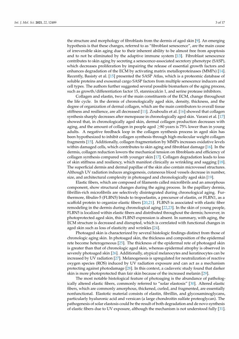
Increased ROS also results in the oxidation of macromolecules, such as cellular lipids,proteins, and DNA, which cause cellular dysfunction with aging. Oxidative protein damageis the main biomarker of aging, and is frequently observed in photodamaged skin [64].Cellular accumulation of lipofuscin, a large protein–lipid aggregate consisting of 30–70%proteins and 20–50% lipids, gradually increases with age, often forming “age spots”.Lipofuscin acts as another redox-active site for free radicals and proteasome inhibitors,eventually initiating a cycle leading to protein aggregation [57]. Figure 2 summarized themajor signaling pathways involved in the aging process.

Int. J. Mol. Sci. 2021, 22, 12489 6 of 17Int. J. Mol. Sci. 2021, 22, x FOR PEER REVIEW 6 of 16
Figure 2. Schematic illustration of the major signaling pathways involved in the aging process; these pathways decrease the density, thickness, and organization of collagen. Both chronological aging and photoaging induce ROS, which leads to the upregulation of MMPs. ROS, reactive oxygen spe-cies; MAPK, mitogen-activated protein kinase; NF-κB, nuclear factor kappa B ; AP-1, activated pro-tein 1; MMP, matrix metalloprotease; TNF-α, tumor necrosis factor-α; TGF-β, transforming growth factor-β.
3.3. Cytokines in Aging Skin Cytokines are another important element in the skin aging process [65]. Upon UV
radiation, several inflammatory signaling pathways are activated via different surface re-ceptors such as epidermal growth factor receptors, the TGF receptor, Toll-like receptors, the interleukin 1 (IL-1) receptor, and the TNF receptor [66]. Major cytokines secreted from keratinocytes are interleukins (IL-1, IL-3, IL-6, IL-8, IL-33), colony-stimulating factors (CSFs) (granulocyte macrophage [GM]-CSF, M-CSF, G-CSF), TGF-α, TGF-β, TNF-α, and platelet-derived growth factor [67]. UV radiation can activate signaling directly via ROS production, or indirectly through DNA or mitochondrial damage, which causes inflam-mation. It is characterized by increased levels of circulating inflammatory cytokines and a shift towards cellular senescence, and these changes are known to cause many age-re-lated diseases, including dementia, arthritis, and type 2 diabetes [68].
TNF-α, a main effector cytokine in proinflammatory processes of the skin, inhibits collagen synthesis and induces MMP9 elevation, as mentioned in a previous section. When exposed to persistent TNF-α, the production of MMP-9 is disturbed, and the epi-dermis can be damaged irreversibly [69]. Increased TNF-α level is associated with multi-ple pathways including NF-kB, AP-1, hypoxia-inducible factor 1-alpha (HIF-1a), and nu-clear factor erythroid 2-related factor 2 (Nrf-2) [70]. Increased TNF-α level is associated with multiple pathways, including NF-kB, AP-1, hypoxia-inducible factor 1-alpha (HIF-1a), and nuclear factor erythroid 2-related factor 2 (Nrf-2), which are associated with MMP up-regulation [70,71].
The level of IL-1 increases with age and promotes skin inflammation, which induces age-related processes [72]. Upon UV radiation, the IL-1 receptor antagonist (IL-1ra), a competitive inhibitor of IL-1, is stimulated and plays a regulatory role in the IL-1-related proinflammatory response. IL-1ra production in the skin decreases with age, while IL-1α levels were higher in aged skin [72]. In chronological aging and photoaging processes, IL-1 and IL-6 induce the activation of key transcription factors associated with inflammatory and immune responses, such as NF-κB, AP-1, c-Jun N-terminal kinase (JNK), and MAPKs
Figure 2. Schematic illustration of the major signaling pathways involved in the aging process; thesepathways decrease the density, thickness, and organization of collagen. Both chronological aging andphotoaging induce ROS, which leads to the upregulation of MMPs. ROS, reactive oxygen species;MAPK, mitogen-activated protein kinase; NF-κB, nuclear factor kappa B ; AP-1, activated protein 1;MMP, matrix metalloprotease; TNF-α, tumor necrosis factor-α; TGF-β, transforming growth factor-β.
3.3. Cytokines in Aging Skin
Cytokines are another important element in the skin aging process [65]. Upon UVradiation, several inflammatory signaling pathways are activated via different surfacereceptors such as epidermal growth factor receptors, the TGF receptor, Toll-like receptors,the interleukin 1 (IL-1) receptor, and the TNF receptor [66]. Major cytokines secreted fromkeratinocytes are interleukins (IL-1, IL-3, IL-6, IL-8, IL-33), colony-stimulating factors (CSFs)(granulocyte macrophage [GM]-CSF, M-CSF, G-CSF), TGF-α, TGF-β, TNF-α, and platelet-derived growth factor [67]. UV radiation can activate signaling directly via ROS production,or indirectly through DNA or mitochondrial damage, which causes inflammation. It ischaracterized by increased levels of circulating inflammatory cytokines and a shift towardscellular senescence, and these changes are known to cause many age-related diseases,including dementia, arthritis, and type 2 diabetes [68].
TNF-α, a main effector cytokine in proinflammatory processes of the skin, inhibitscollagen synthesis and induces MMP9 elevation, as mentioned in a previous section. Whenexposed to persistent TNF-α, the production of MMP-9 is disturbed, and the epidermiscan be damaged irreversibly [69]. Increased TNF-α level is associated with multiplepathways including NF-kB, AP-1, hypoxia-inducible factor 1-alpha (HIF-1a), and nuclearfactor erythroid 2-related factor 2 (Nrf-2) [70]. Increased TNF-α level is associated withmultiple pathways, including NF-kB, AP-1, hypoxia-inducible factor 1-alpha (HIF-1a),and nuclear factor erythroid 2-related factor 2 (Nrf-2), which are associated with MMPup-regulation [70,71].
The level of IL-1 increases with age and promotes skin inflammation, which inducesage-related processes [72]. Upon UV radiation, the IL-1 receptor antagonist (IL-1ra), acompetitive inhibitor of IL-1, is stimulated and plays a regulatory role in the IL-1-relatedproinflammatory response. IL-1ra production in the skin decreases with age, while IL-1αlevels were higher in aged skin [72]. In chronological aging and photoaging processes,IL-1 and IL-6 induce the activation of key transcription factors associated with inflamma-tory and immune responses, such as NF-κB, AP-1, c-Jun N-terminal kinase (JNK), andMAPKs [73–75]. Additionally, IL-18 is an IL-1 superfamily cytokine that acts as an an-
Int. J. Mol. Sci. 2021, 22, 12489 7 of 17
giogenic mediator in inflammatory processes. In aged skin, heterodimerization of IL-18receptor activates the NF-κB signaling pathway; IL-18 is associated with the pathogenesisof several age-related diseases, acting as a strong proinflammatory mediator by inducinginterferon-γ [76]. Another proinflammatory cytokine, IL-6, is elevated in elderly people andis related to skin aging [77,78]. Kim et al. [79] showed that, in female skin after menopause,IL-6 expression is increased and associated with wrinkle formation. The IL-6 level isincreased by exposure to UV radiation, indicating its association with photoaging [79,80].
Cysteine-rich protein 61 (CCN1), an ECM-associated matricellular protein, can beinduced in dermal fibroblasts through AP-1-dependent activation [81]. Increased CCN1expression is observed in the dermal fibroblasts of aged human skin [82]. CCN1 stimulatesthe production of MMP1, thereby contributing to significant type-I collagen degradation inthe dermis [82]. Furthermore, CCN1 downregulates TGF-β type-II receptor, inhibiting theTGF-β signaling essential for maintaining ECM homeostasis [83,84].
3.4. Autophagic Control
Autophagy (ATG) is an evolutionarily conserved mechanism of cell self-digestionfor removing unwanted or damaged proteins, lipids, and other cellular constituents bydelivering these materials to the lysosome for degradation. ATG consists of five mainstages: initiation, nucleation, elongation, fusion, and cargo degradation. The detailedmolecular mechanisms of ATG are extensively reviewed in Klionsky et al. [85]. The keyprotein in the initiation phase, mammalian target of rapamycin (mTOR) C1, acts in concertwith other serine-threonine protein kinases. mTORC1 is inhibited by cellular and envi-ronmental stresses incompatible with continued growth, such as glucose and amino aciddeprivation, DNA damage, and hypoxia [86]. These signals activate the Unc-51-like kinase1 complex, which induces phosphorylation of the components of the class III phosphoinosi-tide 3-kinase complex I, thus enabling nucleation of the phagophore. The elongation ofautophagosomes requires the ubiquitin-like conjugation system to orchestrate the activityof ATG-related proteins, microtubule-associated protein light chain 3 (LC3) and/or GABAtype A receptor-associated protein (GABARAP) [87]. The ATG12–ATG5–ATG16L1 complexenhances the final connection of phosphatidylethanolamine (PE) molecules, resulting inthe formation of membrane-bound LC3-II and/or GABARAP-PE [88]. Cellular membranescontribute to elongation of the phagophore by providing membrane material, which givesrise to double-layered vesicles called autophagosomes. UV radiation resistance-associatedgene (UVRAG) competes with ATG14L for binding to Beclin 1. When bound to Beclin 1,UVRAG stimulates RAB7 GTPase activity and autophagosome fusion with lysosomes [89].Autophagosome-lysosome fusion is managed by syntaxin 17 on autophagosomes, vesicle-associated membrane protein 8 on lysosomes, and accessory proteins such as ATG14 andhomotypic fusion, and the protein sorting homotypic fusion and HOPS (homotypic fusionand vacuole protein sorting) tethering complex [90].
The formation stage involves various proteins that form autophagosomes. In thedegradation stage, autophagosomes and lysosomes fuse to form autolysosomes [91]. ATGacts on damaged mitochondria marked with ubiquitin Ser65 [92] and readily aggregatedcytoplasmic proteins [93]. ATG relieves intracellular oxidative stress by degrading oxi-dized proteins and lipids, while also contributing to many physiological and pathologicalactivities of organisms such as tumor development and regulation, cell survival and death,and aging [94,95].
Autophagic processes significantly extend the replicative lifespan of individual cellsand inhibit stress-induced cellular senescence in vivo. Furthermore, inhibition of ATGmight result in premature senescence [96]. The senescent cell count and SASP secretionlevel are elevated after treatment with tobacco smoke extract and ATG inhibition in primarybronchial epithelial cells. However, cigarette smoke extract-induced cellular senescenceis significantly inhibited by rapamycin, an mTOR inhibitor, to activate the autophagicprocess [97].
Int. J. Mol. Sci. 2021, 22, 12489 8 of 17
Despite the above research, the association between ATG and skin aging remainsambiguous. A significant increase in autophagic vacuoles has been observed by electronmicroscopy in senescent fibroblasts [98] and senescent keratinocytes [99]. A study byYoung et al. [100] showed that fibroblast senescence might depend on prior ATG, andpharmacological or genetic methods for reducing ATG can slow the senescence process.However, some studies drew different conclusions on how ATG contributes to the agingprocess or showed that aging and ATG are independent processes [56,101].
Upon knockdown of the ATG-associated proteins ATG7 and ATG12, or lysosomal-associated membrane proteins in human primary fibroblasts, premature aging has beenobserved in a ROS- and p53-dependent manner, suggesting that inhibition of ATG isassociated with senescence [102]. DNA damage and aging are abnormally increased whenATG-deficient keratinocytes are exposed to oxidative stress [100]. Thus, although ATGmight be required for UV-mediated senescence, this assumption requires validation underother UV-exposure conditions, and in other cell types [103].
3.5. Apoptosis in Skin Aging
Apoptosis promotes tissue integrity by removing injured cells without evoking in-flammation. However, apoptosis seems to be a double-edged sword; during low-levelchronic stress, as seen in aging, for example, increased resistance to apoptosis can lead tothe survival of functionally deficient, post-mitotic cells with poor housekeeping functions.Aging cells are markedly resistant to apoptosis, and several studies have indicated that hostdefense mechanisms can enhance anti-apoptotic signaling, which subsequently induces asenescent, pro-inflammatory phenotype during the aging process [104]. At the molecularlevel, age-related resistance to apoptosis mainly results from a functional deficiency in thep53 network.
In physiological conditions, DNA damage activates checkpoint pathways and DNArepair mechanisms. Checkpoint proteins such as p53 and ataxia-telangiectasia mutated(ATM) are activated upon UV exposure and provoke cell cycle arrest to allow proper DNArepair [105]. A gene downstream of ATM, p53, is a transcription factor that primarilyfunctions as a gatekeeper for DNA mutations [106]. During aging, the efficiency of thestress recognition system in the ER declines [107], which can prevent the initiation ofapoptosis. The functional activity of p53 has been shown to decline during aging in amurine model [108,109]. It has also been demonstrated that premature aging induced byforced activation of p53 does not represent a physiological aging process. The functionalinefficiency of p53 could explain the reduction of mitochondrial respiration and increase inglycolytic metabolism seen during aging [110].
3.6. Role of microRNAs in Skin Aging
MicroRNAs (miRNAs) are small non-coding RNAs that post-transcriptionally regulatemRNA translation and are involved in most biological and pathological processes, includ-ing aging. The expression of specific miRNAs can serve as a biomarker of aging, includingchronological aging and photoaging, as well as a marker of age-related diseases [111]. Therole of miRNAs in skin aging is related to the regulation of molecules involved in theinsulin-like growth factor 1 and mTOR signaling pathways [112].
Analyses of age-associated cutaneous miRNAs in keratinocytes by Rivetti et al. [113]showed increased expression of miR-130, miR-138, and miR-181a/b during replicativesenescence; these processes target the p63 (a member of the p53 superfamily involved inepidermal development and tumor regulation) and sirtuin 1 (SIRT1) (a NAD-dependenthistone deacetylase involved in cellular differentiation, metabolism, immune response,and apoptosis) mRNAs. Upregulation of miR-137 and miR-668 is associated with β-galactosidase activity, along elevated levels of p16INK4A and p53 senescence markers of theARF/p53 and p16INK4A/RB pathways, respectively [114]. In addition, miR-191 is capableof blocking the G1–S phase transition. This blockade is manifested as cell cycle arrest and aquiescent state that contributes to the development of senescence processes [115].
Int. J. Mol. Sci. 2021, 22, 12489 9 of 17
The downregulation of miRNAs is associated with the reduced expression of trans-membrane receptors and components of the ECM in senescent skin fibroblasts. Senescentdermal fibroblasts show increased levels of miRNAs, such as let-7d-5p, let-7e-5p, miR-23a-3p, miR-34a-5p, miR-125a-3p/5p, miR-152, miR-181a-5p, and miR-221/222-3p, whichaffects all phases of the cellular life cycle [116]. MiR-152 can significantly decrease dermalfibroblast adhesion through the downregulation of integrin alpha 5, which participates incell-surface-mediated signaling [115]. The expression of collagen XVI, a minor componentof the cutaneous ECM, in senescent fibroblasts is downregulated directly by upregulatedmiR-181a [113]. Additionally, the miR-29 and miR-30 miRNA families are upregulatedduring fibroblast senescence, which directly regulates the expression of the B-Myb tran-scription factor present in all cell cycle phases [117]. Additionally, the miR-17-92 clusterand miR-106 are downregulated in aged dermal fibroblasts, resulting in activation of thep53, ERB, and MAPK signaling pathways [118]. In Langerhans cells, overexpression of miR-449 and miR-9 is related to the downregulation of key molecules in the TGF-β signalingpathway, resulting in decreased function of Langerhans cells [119].
Upon UV irradiation, miRNA expression is changed; miR-27a, miR-145, miR-383,and miR-1246 levels are increased, and mi-155, miR-663b, miR-3648, and miR-6879 aredecreased. Mi-27a facilitates the removal of cyclobutane pyrimidine dimers and reducescell apoptosis [120]. The overexpression of miR-1246 directly upregulates UVB-inducedapoptosis through the suppression of rhotekin 2 expression [121]. MiR-34a, miR-134,and miR-383 target the p53 complex via G1 phase arrest and the cell-cycle regulatorcyclin, resulting in senescence. Upregulated miR-134 is associated with activation of theNF-κB signaling pathway and SIRT1 inhibition [122]. UVA exposure downregulates miR-155, leading to the upregulation of c-Jun, which affects collagen gene activity in humanfibroblasts [123].
3.7. Skin Microbiome
The human microbiome is the genome of all microorganisms that reside on or withinhuman tissues, including the skin. The skin hosts a diverse ecosystem, housing up to onemillion bacteria per square centimeter of surface area, in addition to viruses and fungi [124].Imbalances of the skin microbiota, an essential element of the skin barrier system, cancause various pathologic conditions, including aging [125,126]. The composition of the skinmicrobiome varies according to preservation measures, physiological condition, antibacte-rial treatment, and demographic characteristics [127]. Several studies have used bacterial16S rRNA gene sequencing to show that the skin microbiome is altered and diversifiedwith aging [128–130]. These studies have consistently demonstrated higher alpha diversityin the skin of older than younger people, indicating that the former group may be moresusceptible to pathogenic invasion due to altered diversity of the skin microbiota. Inaddition, during puberty, the density of lipophilic bacteria on the skin increases in linewith increasing sebum levels and is lower in the skin of the elderly [130]. Kim et al. [131]studied age-related changes in the skin microbiota of Korean women on the forehead andhands and found that the overall microbial distribution on the forehead varied among agegroups, but not on the hands. The authors showed that Firmicutes was more abundantin the younger age group, whereas Bacteroidetes and Proteobacteria increased linearly withaging. The overall microbiome was characterized by beta diversity analysis, and the twogroups were significantly different. Zichao et al. [130] recently reported age-related micro-biota profiles for both intrinsic skin aging and photoaging. The microbial composition ofthe elderly group was significantly different from that of the younger group, consistentwith previous studies [126]. The authors also conducted a correlation analysis, in whicheach group showed high enrichment of nine microbial communities (i.e., Cyanobacteria,Staphylococcus, Cutibacterium, Lactobacillus, Corynebacterium, Streptococcus, Neisseria, Candida,and Malassezia) and 18 pathways (e.g., MAPK signaling pathway, glutathione metabolism,photosynthesis, and pantothenate and coenzyme A biosynthesis) that likely contribute

Int. J. Mol. Sci. 2021, 22, 12489 10 of 17
to skin aging, suggesting that the skin microbiome plays a key role in skin aging [126].Various studies related to molecular mechanisms of skin aging are described in Table 2.
Table 2. Molecular mechanisms of skin aging. ROS, reactive oxygen species; UV, ultraviolet ray, CCN1; Cellular Communi-cation Network Factor 1.
Molecular Mechanisms Details References
Telomere shorteningIn intrinsic aging [48]
In chronological aging [49,52]
Oxidative stress and matrix metalloproteinasesROS and photoaging [53,55,56]
Signal cascade activated by ROS [58,59,61,63,132,133]
Cytokines
Cytokines released by UV radiation [65–67]
Tumor necrosis factor α [69]
Interleukins [72,73,77–80,134]
CCN1 [82,83]
Autophagic control Association with aging process [92–98]
Apoptosis Association with aging process [104,107–109]
MicroRNAsChronological aging [113–119,135]
photoaging [120–123]
Skin microbiome Association with aging process [128–130]
4. Other Environmental Stressors Associated with Skin Aging4.1. Tobacco Smoke
In addition to UV radiation exposure, tobacco smoke can lead to extrinsic skin aging.Greg et al. [136] recently reported that smoking is associated with more facial lines anddecreased face volume, indicative of premature skin aging. Long-term changes in theskin can lead to “smoker’s face”, which is characterized by a grayish appearance andperiorbital and perioral facial lines caused by post-inflammatory hyperpigmentation andthe breakdown of collagen and elastin fibers in the dermis [131]. One study showed thatMMP1 mRNA was significantly increased in the dermis of smokers compared with non-smokers, leading to collagen and elastin fiber breakdown [136]. To better understand thepathogenic role of tobacco in skin aging, Morita et al. [137] performed an in vivo studyusing a mouse model. They applied a water-soluble tobacco smoke extract to the backskin of mice, which resulted in the loss of normal collagen structure and a significantincrease of collagen breakdown in the papillary and upper dermis, mimicking aged skin.Several other studies have suggested a potential pathologic mechanism by which tobaccosmoke accelerates skin aging. Tobacco smoke extract decreases procollagen type-I and -III,and increases MMP1 and MMP3, which results in degradation of the ECM structure andabnormal regulation of ECM deposition in human cultured skin fibroblasts [137]. Anotherstudy found that tobacco smoke extract is associated with decreased responsiveness tothe TGF-β signaling pathway and reduced expression of the TGF-β receptor in the su-pernatants of cultured skin fibroblasts, leading to decreased synthesis of ECM moleculesand structure [138] Additionally, tobacco smoke is a major source of polycyclic aromatichydrocarbon exposure in humans. Tobacco smoke extract increases the MMP-1 level byactivating the aryl hydrocarbon receptor (AhR) signaling pathway in human keratinocytesand fibroblasts [139].
4.2. Environmental Pollutants
Exposure to indoor or outdoor chemical, physical, or biological air pollution is oneof the major environmental health issues worldwide [140]. It is also associated with anincreased risk of extrinsic skin aging, particularly in the form of pigmented spots and
Int. J. Mol. Sci. 2021, 22, 12489 11 of 17
wrinkles in white people of European ancestry [141]. Polycyclic aromatic hydrocarbons,which are also present in tobacco extract, are a major effector, triggering the AhR sig-naling pathway. Due to the high lipophilicity of polycyclic aromatic hydrocarbons, theycan penetrate the skin barrier. Activation of AhR pathways lead to increased MMP1 ex-pression in keratinocytes and melanogenesis, via increased tyrosinase enzyme activity inmelanocytes [142]. Regarding particulate matter, soot, and nitrogen dioxide, which haveemerged as serious pollutants, are well known factors in extrinsic skin aging. Topicalexposure of human skin to environmentally relevant concentrations of an internationallyestablished reference standard diesel exhaust mixture increased dark pigmentation ofthe skin. This tanning effect is due to increased melanogenesis caused by the oxidativestress associated with activation of the p53 signaling pathway [143]. Recently, Li et al. [59]showed that indoor air pollution associated with cooking with solid fuels was significantlyassociated with 5–8% deeper facial wrinkles and folds, and an increased risk of develop-ing fine wrinkles on the dorsum of the hands in Chinese women. It is likely that indoorcombustion of solid fuels activates the same molecular pathways in skin cells as outdoorpollution, and thus causes wrinkles [144].
5. Conclusions
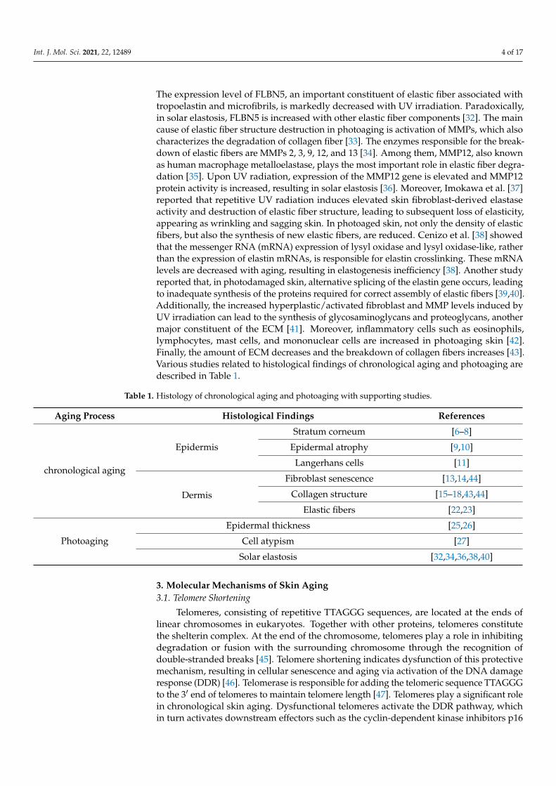
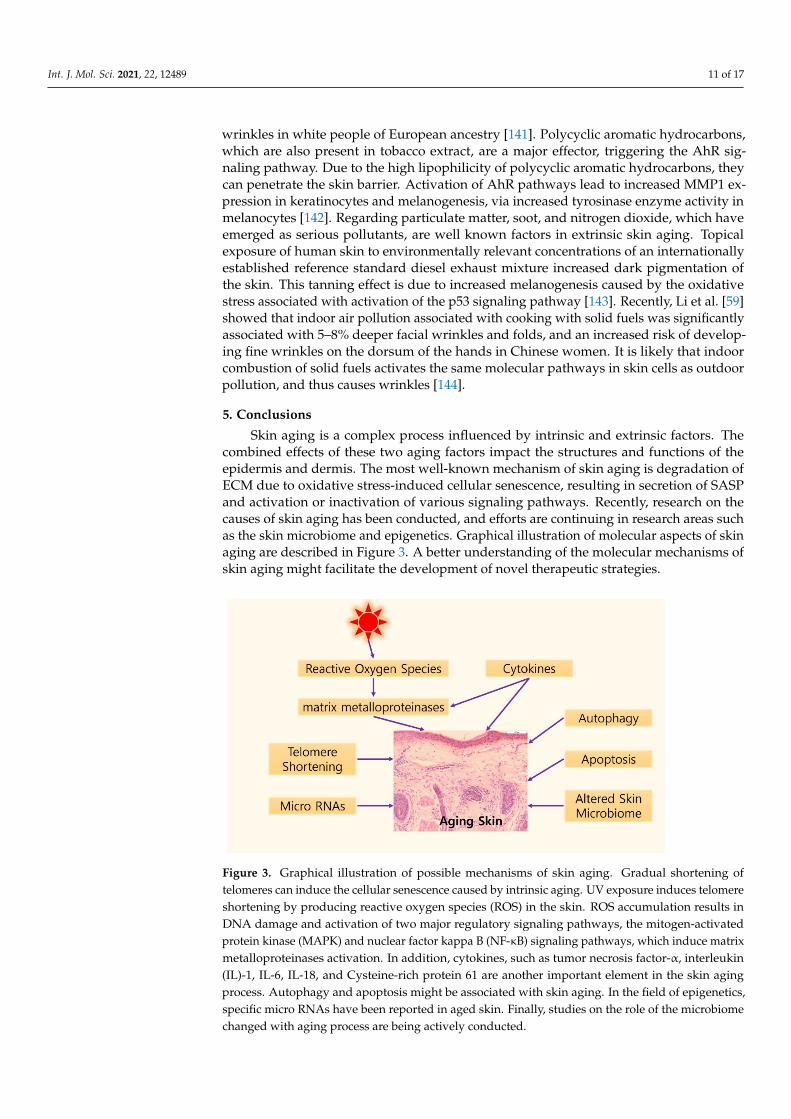
Skin aging is a complex process influenced by intrinsic and extrinsic factors. Thecombined effects of these two aging factors impact the structures and functions of theepidermis and dermis. The most well-known mechanism of skin aging is degradation ofECM due to oxidative stress-induced cellular senescence, resulting in secretion of SASPand activation or inactivation of various signaling pathways. Recently, research on thecauses of skin aging has been conducted, and efforts are continuing in research areas suchas the skin microbiome and epigenetics. Graphical illustration of molecular aspects of skinaging are described in Figure 3. A better understanding of the molecular mechanisms ofskin aging might facilitate the development of novel therapeutic strategies.
Int. J. Mol. Sci. 2021, 22, x FOR PEER REVIEW 11 of 16
which are also present in tobacco extract, are a major effector, triggering the AhR signaling pathway. Due to the high lipophilicity of polycyclic aromatic hydrocarbons, they can pen-etrate the skin barrier. Activation of AhR pathways lead to increased MMP1 expression in keratinocytes and melanogenesis, via increased tyrosinase enzyme activity in melano-cytes [142]. Regarding particulate matter, soot, and nitrogen dioxide, which have emerged as serious pollutants, are well known factors in extrinsic skin aging. Topical exposure of human skin to environmentally relevant concentrations of an internationally established reference standard diesel exhaust mixture increased dark pigmentation of the skin. This tanning effect is due to increased melanogenesis caused by the oxidative stress associated with activation of the p53 signaling pathway [143]. Recently, Li et al. [59] showed that indoor air pollution associated with cooking with solid fuels was significantly associated with 5–8% deeper facial wrinkles and folds, and an increased risk of developing fine wrin-kles on the dorsum of the hands in Chinese women. It is likely that indoor combustion of solid fuels activates the same molecular pathways in skin cells as outdoor pollution, and thus causes wrinkles [144].
5. Conclusions Skin aging is a complex process influenced by intrinsic and extrinsic factors. The com-
bined effects of these two aging factors impact the structures and functions of the epider-mis and dermis. The most well-known mechanism of skin aging is degradation of ECM due to oxidative stress-induced cellular senescence, resulting in secretion of SASP and activation or inactivation of various signaling pathways. Recently, research on the causes of skin aging has been conducted, and efforts are continuing in research areas such as the skin microbiome and epigenetics. Graphical illustration of molecular aspects of skin aging are described in Figure 3. A better understanding of the molecular mechanisms of skin aging might facilitate the development of novel therapeutic strategies.
Figure 3. Graphical illustration of possible mechanisms of skin aging. Gradual shortening of telo-meres can induce the cellular senescence caused by intrinsic aging. UV exposure induces telomere shortening by producing reactive oxygen species (ROS) in the skin. ROS accumulation results in DNA damage and activation of two major regulatory signaling pathways, the mitogen-activated protein kinase (MAPK) and nuclear factor kappa B (NF-κB) signaling pathways, which induce ma-trix metalloproteinases activation. In addition, cytokines, such as tumor necrosis factor-α, interleu-kin (IL)-1, IL-6, IL-18, and Cysteine-rich protein 61 are another important element in the skin aging process. Autophagy and apoptosis might be associated with skin aging. In the field of epigenetics, specific micro RNAs have been reported in aged skin. Finally, studies on the role of the microbiome changed with aging process are being actively conducted.
Figure 3. Graphical illustration of possible mechanisms of skin aging. Gradual shortening oftelomeres can induce the cellular senescence caused by intrinsic aging. UV exposure induces telomereshortening by producing reactive oxygen species (ROS) in the skin. ROS accumulation results inDNA damage and activation of two major regulatory signaling pathways, the mitogen-activatedprotein kinase (MAPK) and nuclear factor kappa B (NF-κB) signaling pathways, which induce matrixmetalloproteinases activation. In addition, cytokines, such as tumor necrosis factor-α, interleukin(IL)-1, IL-6, IL-18, and Cysteine-rich protein 61 are another important element in the skin agingprocess. Autophagy and apoptosis might be associated with skin aging. In the field of epigenetics,specific micro RNAs have been reported in aged skin. Finally, studies on the role of the microbiomechanged with aging process are being actively conducted.
Int. J. Mol. Sci. 2021, 22, 12489 12 of 17
Author Contributions: H.L. analyzed wrote the manuscript. Y.H. contributed to the figure designand arrangement of references. M.K. took the concept of the review and edited the manuscript. Allauthors have read and agreed to the published version of the manuscript.
Funding: This work was supported by the Basic Science Research Program through the National Re-search Foundation of Korea (NRF), funded by the Ministry of Education, Science (2019R1F1A1059460).
Institutional Review Board Statement: Not applicable.
Informed Consent Statement: Not applicable.
Data Availability Statement: The data underlying this article will be shared on reasonable requestfrom the corresponding author.
Conflicts of Interest: The authors declare no conflict of interest.
References1. Chambers, E.S.; Vukmanovic-Stejic, M. Skin barrier immunity and ageing. Immunology 2020, 160, 116–125. [CrossRef]2. Koohgoli, R.; Hudson, L.; Naidoo, K.; Wilkinson, S.; Chavan, B.; Birch-Machin, M.A. Bad air gets under your skin. Exp. Dermatol.
2017, 26, 384–387. [CrossRef]3. Rittié, L.; Fisher, G.J. Natural and sun-induced aging of human skin. Cold Spring Harb. Perspect. Med. 2015, 5, a015370. [CrossRef]
[PubMed]4. Laga, A.C.; Murphy, G.F. The Translational Basis of Human Cutaneous Photoaging: On Models, Methods, and Meaning. Am. J.
Pathol. 2009, 174, 357–360. [CrossRef]5. Fisher, G.J.; Kang, S.; Varani, J.; Bata-Csorgo, Z.; Wan, Y.; Datta, S.; Voorhees, J.J. Mechanisms of photoaging and chronological
skin aging. Arch. Dermatol. 2002, 138, 1462–1470. [CrossRef] [PubMed]6. Stanley, J.R. Synergy of understanding dermatologic disease and epidermal biology. J. Clin. Investig. 2012, 122, 436–439. [CrossRef]
[PubMed]7. Biniek, K.; Kaczvinsky, J.; Matts, P.; Dauskardt, R.H. Understanding age-induced alterations to the biomechanical barrier function
of human stratum corneum. J. Dermatol. Sci. 2015, 80, 94–101. [CrossRef]8. Choe, C.; Schleusener, J.; Lademann, J.; Darvin, M.E. Age related depth profiles of human Stratum Corneum barrier-related
molecular parameters by confocal Raman microscopy in vivo. Mech. Ageing Dev. 2018, 172, 6–12. [CrossRef]9. Lavker, R.M. Structural alterations in exposed and unexposed aged skin. J. Investig. Dermatol. 1979, 73, 59–66. [CrossRef]
[PubMed]10. Gilchrest, B.A.; Blog, F.B.; Szabo, G. Effects of aging and chronic sun exposure on melanocytes in human skin. J. Investig. Dermatol.
1979, 73, 141–143. [CrossRef]11. Russell-Goldman, E.; Murphy, G.F. The Pathobiology of Skin Aging: New Insights into an Old Dilemma. Am. J. Pathol. 2020, 190,
1356–1369. [CrossRef]12. Sauermann, K.; Clemann, S.; Jaspers, S.; Gambichler, T.; Altmeyer, P.; Hoffmann, K.; Ennen, J. Age related changes of human skin
investigated with histometric measurements by confocal laser scanning microscopy in vivo. Skin Res. Technol. 2002, 8, 52–56.[CrossRef]
13. Wlaschek, M.; Maity, P.; Makrantonaki, E.; Scharffetter-Kochanek, K. Connective Tissue and Fibroblast Senescence in Skin Aging.J. Investig. Dermatol. 2021, 141, 985–992. [CrossRef]
14. Ghosh, K.; Capell, B.C. The Senescence-Associated Secretory Phenotype: Critical Effector in Skin Cancer and Aging. J. Investig.Dermatol. 2016, 136, 2133–2139. [CrossRef]
15. Varani, J.; Spearman, D.; Perone, P.; Fligiel, S.E.; Datta, S.C.; Wang, Z.Q.; Shao, Y.; Kang, S.; Fisher, G.J.; Voorhees, J.J. Inhibition oftype I procollagen synthesis by damaged collagen in photoaged skin and by collagenase-degraded collagen in vitro. Am. J. Pathol.2001, 158, 931–942. [CrossRef]
16. Fisher, G.J.; Quan, T.; Purohit, T.; Shao, Y.; Cho, M.K.; He, T.; Varani, J.; Kang, S.; Voorhees, J.J. Collagen fragmentation promotesoxidative stress and elevates matrix metalloproteinase-1 in fibroblasts in aged human skin. Am. J. Pathol. 2009, 174, 101–114.[CrossRef] [PubMed]
17. Varani, J.; Dame, M.K.; Rittie, L.; Fligiel, S.E.; Kang, S.; Fisher, G.J.; Voorhees, J.J. Decreased collagen production in chronologicallyaged skin: Roles of age-dependent alteration in fibroblast function and defective mechanical stimulation. Am. J. Pathol. 2006, 168,1861–1868. [CrossRef]
18. Makrantonaki, E.; Zouboulis, C.C. The skin as a mirror of the aging process in the human organism-state of the art and results ofthe aging research in the German National Genome Research Network 2 (NGFN-2). Exp. Gerontol. 2007, 42, 879–886. [CrossRef]
19. Chung, J.H.; Eun, H.C. Angiogenesis in skin aging and photoaging. J. Dermatol. 2007, 34, 593–600. [CrossRef] [PubMed]20. Yanagisawa, H.; Davis, E.C.; Starcher, B.C.; Ouchi, T.; Yanagisawa, M.; Richardson, J.A.; Olson, E.N. Fibulin-5 is an elastin-binding
protein essential for elastic fibre development in vivo. Nature 2002, 415, 168–171. [CrossRef] [PubMed]
Int. J. Mol. Sci. 2021, 22, 12489 13 of 17
21. Choudhury, R.; McGovern, A.; Ridley, C.; Cain, S.A.; Baldwin, A.; Wang, M.C.; Guo, C.; Mironov, A., Jr.; Drymoussi, Z.;Trump, D.; et al. Differential regulation of elastic fiber formation by fibulin-4 and -5. J. Biol. Chem. 2009, 284, 24553–24567.[CrossRef]
22. Amano, S. Characterization and mechanisms of photoageing-related changes in skin. Damages of basement membrane anddermal structures. Exp. Dermatol. 2016, 25 (Suppl. S3), 14–19. [CrossRef] [PubMed]
23. Langton, A.K.; Sherratt, M.J.; Griffiths, C.E.; Watson, R.E. Differential expression of elastic fibre components in intrinsically agedskin. Biogerontology 2012, 13, 37–48. [CrossRef] [PubMed]
24. Shin, J.W.; Kwon, S.H.; Choi, J.Y.; Na, J.I.; Huh, C.H.; Choi, H.R.; Park, K.C. Molecular Mechanisms of Dermal Aging andAntiaging Approaches. Int. J. Mol. Sci. 2019, 20, 2126. [CrossRef]
25. Kurban, R.S.; Bhawan, J. Histologic changes in skin associated with aging. J. Dermatol. Surg. Oncol. 1990, 16, 908–914. [CrossRef]26. Bhawan, J.; Andersen, W.; Lee, J.; Labadie, R.; Solares, G. Photoaging versus intrinsic aging: A morphologic assessment of facial
skin. J. Cutan. Pathol. 1995, 22, 154–159. [CrossRef] [PubMed]27. Han, A.; Chien, A.L.; Kang, S. Photoaging. Dermatol. Clin. 2014, 32, 291–299. [CrossRef]28. Eller, M.S.; Yaar, M.; Gilchrest, B.A. DNA damage and melanogenesis. Nature 1994, 372, 413–414. [CrossRef]29. Kaidbey, K.H.; Agin, P.P.; Sayre, R.M.; Kligman, A.M. Photoprotection by melanin—A comparison of black and Caucasian skin. J.
Am. Acad. Dermatol. 1979, 1, 249–260. [CrossRef]30. Tsuji, T. Loss of dermal elastic tissue in solar elastosis. Arch. Dermatol. 1980, 116, 474–475. [CrossRef]31. Sellheyer, K. Pathogenesis of solar elastosis: Synthesis or degradation? J. Cutan. Pathol. 2003, 30, 123–127. [CrossRef] [PubMed]32. Naylor, E.C.; Watson, R.E.; Sherratt, M.J. Molecular aspects of skin ageing. Maturitas 2011, 69, 249–256. [CrossRef]33. Fisher, G.J.; Wang, Z.Q.; Datta, S.C.; Varani, J.; Kang, S.; Voorhees, J.J. Pathophysiology of premature skin aging induced by
ultraviolet light. N. Engl. J. Med. 1997, 337, 1419–1428. [CrossRef]34. Chakraborti, S.; Mandal, M.; Das, S.; Mandal, A.; Chakraborti, T. Regulation of matrix metalloproteinases: An overview. Mol. Cell.
Biochem. 2003, 253, 269–285. [CrossRef]35. Ryu, J.; Park, S.J.; Kim, I.H.; Choi, Y.H.; Nam, T.J. Protective effect of porphyra-334 on UVA-induced photoaging in human skin
fibroblasts. Int. J. Mol. Med. 2014, 34, 796–803. [CrossRef] [PubMed]36. Chung, J.H.; Seo, J.Y.; Lee, M.K.; Eun, H.C.; Lee, J.H.; Kang, S.; Fisher, G.J.; Voorhees, J.J. Ultraviolet modulation of human
macrophage metalloelastase in human skin in vivo. J. Investig. Dermatol. 2002, 119, 507–512. [CrossRef] [PubMed]37. Imokawa, G.; Ishida, K. Biological mechanisms underlying the ultraviolet radiation-induced formation of skin wrinkling and
sagging I: Reduced skin elasticity, highly associated with enhanced dermal elastase activity, triggers wrinkling and sagging. Int. J.Mol. Sci. 2015, 16, 7753–7775. [CrossRef]
38. Cenizo, V.; André, V.; Reymermier, C.; Sommer, P.; Damour, O.; Perrier, E. LOXL as a target to increase the elastin content in adultskin: A dill extract induces the LOXL gene expression. Exp. Dermatol. 2006, 15, 574–581. [CrossRef]
39. Weihermann, A.C.; Lorencini, M.; Brohem, C.A.; de Carvalho, C.M. Elastin structure and its involvement in skin photoageing. Int.J. Cosmet. Sci. 2017, 39, 241–247. [CrossRef]
40. Schwartz, E.; Feinberg, E.; Lebwohl, M.; Mariani, T.J.; Boyd, C.D. Ultraviolet radiation increases tropoelastin accumulation by apost-transcriptional mechanism in dermal fibroblasts. J. Investig. Dermatol. 1995, 105, 65–69. [CrossRef] [PubMed]
41. Schwarz, T. Photoimmunosuppression. Photodermatol. Photoimmunol. Photomed. 2002, 18, 141–145. [CrossRef] [PubMed]42. Bosset, S.; Bonnet-Duquennoy, M.; Barré, P.; Chalon, A.; Kurfurst, R.; Bonté, F.; Schnébert, S.; Le Varlet, B.; Nicolas, J.F. Photoageing
shows histological features of chronic skin inflammation without clinical and molecular abnormalities. Br. J. Dermatol. 2003, 149,826–835. [CrossRef] [PubMed]
43. Varani, J.; Warner, R.L.; Gharaee-Kermani, M.; Phan, S.H.; Kang, S.; Chung, J.H.; Wang, Z.Q.; Datta, S.C.; Fisher, G.J.; Voorhees, J.J.Vitamin A antagonizes decreased cell growth and elevated collagen-degrading matrix metalloproteinases and stimulates collagenaccumulation in naturally aged human skin. J. Investig. Dermatol. 2000, 114, 480–486. [CrossRef] [PubMed]
44. Zouboulis Ch, C. Intrinsic skin aging. A critical appraisal of the role of hormones. Der Hautarzt Z. Fur Dermatol. Venerol. UndVerwandte Geb. 2003, 54, 825–832.
45. Griffith, J.D.; Comeau, L.; Rosenfield, S.; Stansel, R.M.; Bianchi, A.; Moss, H.; de Lange, T. Mammalian telomeres end in a largeduplex loop. Cell 1999, 97, 503–514. [CrossRef]
46. D’Adda di Fagagna, F.; Reaper, P.M.; Clay-Farrace, L.; Fiegler, H.; Carr, P.; Von Zglinicki, T.; Saretzki, G.; Carter, N.P.; Jackson, S.P.A DNA damage checkpoint response in telomere-initiated senescence. Nature 2003, 426, 194–198. [CrossRef] [PubMed]
47. Buckingham, E.M.; Klingelhutz, A.J. The role of telomeres in the ageing of human skin. Exp. Dermatol. 2011, 20, 297–302.[CrossRef]
48. Victorelli, S.; Passos, J.F. Telomeres and Cell Senescence—Size Matters Not. EBioMedicine 2017, 21, 14–20. [CrossRef]49. Oikawa, S.; Tada-Oikawa, S.; Kawanishi, S. Site-specific DNA damage at the GGG sequence by UVA involves acceleration of
telomere shortening. Biochemistry 2001, 40, 4763–4768. [CrossRef]50. Birch, J.; Barnes, P.J.; Passos, J.F. Mitochondria, telomeres and cell senescence: Implications for lung ageing and disease. Pharmacol.
Ther. 2018, 183, 34–49. [CrossRef]51. Krutmann, J.; Schroeder, P. Role of mitochondria in photoaging of human skin: The defective powerhouse model. J. Investig.
Dermatol. Symp. Proc. 2009, 14, 44–49. [CrossRef]

Int. J. Mol. Sci. 2021, 22, 12489 14 of 17
52. Sugimoto, M.; Yamashita, R.; Ueda, M. Telomere length of the skin in association with chronological aging and photoaging. J.Dermatol. Sci. 2006, 43, 43–47. [CrossRef] [PubMed]
53. Poljšak, B.; Dahmane, R.G.; Godic, A. Intrinsic skin aging: The role of oxidative stress. Acta Dermatovenerol. Alp. Pannonica Adriat.2012, 21, 33–36.
54. Sajo, M.E.J.; Kim, C.S.; Kim, S.K.; Shim, K.Y.; Kang, T.Y.; Lee, K.J. Antioxidant and Anti-Inflammatory Effects of Shungite againstUltraviolet B Irradiation-Induced Skin Damage in Hairless Mice. Oxidative Med. Cell. Longev. 2017, 2017, 7340143. [CrossRef]
55. Pandel, R.; Poljšak, B.; Godic, A.; Dahmane, R. Skin photoaging and the role of antioxidants in its prevention. ISRN Dermatol.2013, 2013, 930164. [CrossRef] [PubMed]
56. Gu, Y.; Han, J.; Jiang, C.; Zhang, Y. Biomarkers, oxidative stress and autophagy in skin aging. Ageing Res. Rev. 2020, 59, 101036.[CrossRef] [PubMed]
57. Rinnerthaler, M.; Bischof, J.; Streubel, M.K.; Trost, A.; Richter, K. Oxidative stress in aging human skin. Biomolecules 2015, 5,545–589. [CrossRef]
58. Demaria, M.; Desprez, P.Y.; Campisi, J.; Velarde, M.C. Cell Autonomous and Non-Autonomous Effects of Senescent Cells in theSkin. J. Investig. Dermatol. 2015, 135, 1722–1726. [CrossRef]
59. Fisher, G.J.; Talwar, H.S.; Lin, J.; Lin, P.; McPhillips, F.; Wang, Z.; Li, X.; Wan, Y.; Kang, S.; Voorhees, J.J. Retinoic acid inhibitsinduction of c-Jun protein by ultraviolet radiation that occurs subsequent to activation of mitogen-activated protein kinasepathways in human skin in vivo. J. Clin. Investig. 1998, 101, 1432–1440. [CrossRef]
60. Fligiel, S.E.; Varani, J.; Datta, S.C.; Kang, S.; Fisher, G.J.; Voorhees, J.J. Collagen degradation in aged/photodamaged skin in vivoand after exposure to matrix metalloproteinase-1 in vitro. J. Investig. Dermatol. 2003, 120, 842–848. [CrossRef]
61. Nelson, A.R.; Fingleton, B.; Rothenberg, M.L.; Matrisian, L.M. Matrix metalloproteinases: Biologic activity and clinical implica-tions. J. Clin. Oncol. Off. J. Am. Soc. Clin. Oncol. 2000, 18, 1135–1149. [CrossRef] [PubMed]
62. Sternlicht, M.D.; Werb, Z. How matrix metalloproteinases regulate cell behavior. Annu. Rev. Cell Dev. Biol. 2001, 17, 463–516.[CrossRef] [PubMed]
63. Quan, T.; He, T.; Voorhees, J.J.; Fisher, G.J. Ultraviolet irradiation induces Smad7 via induction of transcription factor AP-1 inhuman skin fibroblasts. J. Biol. Chem. 2005, 280, 8079–8085. [CrossRef] [PubMed]
64. Hipkiss, A.R. Accumulation of altered proteins and ageing: Causes and effects. Exp. Gerontol. 2006, 41, 464–473. [CrossRef]65. Borg, M.; Brincat, S.; Camilleri, G.; Schembri-Wismayer, P.; Brincat, M.; Calleja-Agius, J. The role of cytokines in skin aging.
Climacteric J. Int. Menopause Soc. 2013, 16, 514–521. [CrossRef] [PubMed]66. Ansary, T.M.; Hossain, M.R.; Kamiya, K.; Komine, M.; Ohtsuki, M. Inflammatory Molecules Associated with Ultraviolet
Radiation-Mediated Skin Aging. Int. J. Mol. Sci. 2021, 22, 3974. [CrossRef] [PubMed]67. Ansel, J.; Perry, P.; Brown, J.; Damm, D.; Phan, T.; Hart, C.; Luger, T.; Hefeneider, S. Cytokine modulation of keratinocyte cytokines.
J. Investig. Dermatol. 1990, 94, 101s–107s. [CrossRef]68. Pilkington, S.M.; Bulfone-Paus, S.; Griffiths, C.E.M.; Watson, R.E.B. Inflammaging and the Skin. J. Investig. Dermatol. 2021, 141,
1087–1095. [CrossRef]69. Youn, U.J.; Nam, K.W.; Kim, H.S.; Choi, G.; Jeong, W.S.; Lee, M.Y.; Chae, S. 3-Deoxysappanchalcone inhibits tumor necrosis
factor-α-induced matrix metalloproteinase-9 expression in human keratinocytes through activated protein-1 inhibition andnuclear factor-kappa B DNA binding activity. Biol. Pharm. Bull. 2011, 34, 890–893. [CrossRef]
70. Chou, D.H.; Lee, W.; McCulloch, C.A. TNF-alpha inactivation of collagen receptors: Implications for fibroblast function andfibrosis. J. Immunol. 1996, 156, 4354–4362.
71. Crawford, H.C.; Matrisian, L.M. Mechanisms controlling the transcription of matrix metalloproteinase genes in normal andneoplastic cells. Enzym. Protein 1996, 49, 20–37. [CrossRef] [PubMed]
72. Hirao, T.; Aoki, H.; Yoshida, T.; Sato, Y.; Kamoda, H. Elevation of interleukin 1 receptor antagonist in the stratum corneum ofsun-exposed and ultraviolet B-irradiated human skin. J. Investig. Dermatol. 1996, 106, 1102–1107. [CrossRef] [PubMed]
73. Schneider, L.A.; Raizner, K.; Wlaschek, M.; Brenneisen, P.; Gethöffer, K.; Scharffetter-Kochanek, K. UVA-1 exposure in vivo leadsto an IL-6 surge within the skin. Exp. Dermatol. 2017, 26, 830–832. [CrossRef]
74. Wlaschek, M.; Heinen, G.; Poswig, A.; Schwarz, A.; Krieg, T.; Scharffetter-Kochanek, K. UVA-induced autocrine stimulation offibroblast-derived collagenase/MMP-1 by interrelated loops of interleukin-1 and interleukin-6. Photochem. Photobiol. 1994, 59,550–556. [CrossRef]
75. Mantovani, A.; Dinarello, C.A.; Molgora, M.; Garlanda, C. Interleukin-1 and Related Cytokines in the Regulation of Inflammationand Immunity. Immunity 2019, 50, 778–795. [CrossRef] [PubMed]
76. Lee, J.H.; Cho, D.H.; Park, H.J. IL-18 and Cutaneous Inflammatory Diseases. Int. J. Mol. Sci. 2015, 16, 29357–29369. [CrossRef]77. Cohen, H.J.; Pieper, C.F.; Harris, T.; Rao, K.M.; Currie, M.S. The association of plasma IL-6 levels with functional disability in
community-dwelling elderly. J. Gerontol. Ser. A Biol. Sci. Med Sci. 1997, 52, M201–M208. [CrossRef]78. Beyer, I.; Mets, T.; Bautmans, I. Chronic low-grade inflammation and age-related sarcopenia. Curr. Opin. Clin. Nutr. Metab. Care
2012, 15, 12–22. [CrossRef] [PubMed]79. Fagot, D.; Asselineau, D.; Bernerd, F. Direct role of human dermal fibroblasts and indirect participation of epidermal keratinocytes
in MMP-1 production after UV-B irradiation. Arch. Dermatol. Res. 2002, 293, 576–583. [CrossRef]80. Omoigui, S. The Interleukin-6 inflammation pathway from cholesterol to aging-role of statins, bisphosphonates and plant
polyphenols in aging and age-related diseases. Immun. Ageing I A 2007, 4, 1. [CrossRef]
Int. J. Mol. Sci. 2021, 22, 12489 15 of 17
81. Qin, Z.; Robichaud, P.; He, T.; Fisher, G.J.; Voorhees, J.J.; Quan, T. Oxidant exposure induces cysteine-rich protein 61 (CCN1) viac-Jun/AP-1 to reduce collagen expression in human dermal fibroblasts. PLoS ONE 2014, 9, e115402. [CrossRef]
82. Quan, T.; Qin, Z.; Robichaud, P.; Voorhees, J.J.; Fisher, G.J. CCN1 contributes to skin connective tissue aging by inducingage-associated secretory phenotype in human skin dermal fibroblasts. J. Cell Commun. Signal. 2011, 5, 201–207. [CrossRef][PubMed]
83. Quan, T.; He, T.; Shao, Y.; Lin, L.; Kang, S.; Voorhees, J.J.; Fisher, G.J. Elevated cysteine-rich 61 mediates aberrant collagenhomeostasis in chronologically aged and photoaged human skin. Am. J. Pathol. 2006, 169, 482–490. [CrossRef] [PubMed]
84. Cole, M.A.; Quan, T.; Voorhees, J.J.; Fisher, G.J. Extracellular matrix regulation of fibroblast function: Redefining our perspectiveon skin aging. J. Cell Commun. Signal. 2018, 12, 35–43. [CrossRef]
85. Klionsky, D.J.; Abdel-Aziz, A.K.; Abdelfatah, S.; Abdellatif, M.; Abdoli, A.; Abel, S.; Abeliovich, H.; Abildgaard, M.H.; Abudu, Y.P.;Acevedo-Arozena, A.; et al. Guidelines for the use and interpretation of assays for monitoring autophagy (4th edition). Autophagy2021, 17, 1–382.
86. Saxton, R.A.; Sabatini, D.M. mTOR Signaling in Growth, Metabolism, and Disease. Cell 2017, 168, 960–976. [CrossRef] [PubMed]87. Dancourt, J.; Melia, T.J. Lipidation of the autophagy proteins LC3 and GABARAP is a membrane-curvature dependent process.
Autophagy 2014, 10, 1470–1471. [CrossRef] [PubMed]88. Kharaziha, P.; Panaretakis, T. Dynamics of Atg5-Atg12-Atg16L1 Aggregation and Deaggregation. Methods Enzymol. 2017, 587,
247–255.89. Itakura, E.; Kishi, C.; Inoue, K.; Mizushima, N. Beclin 1 forms two distinct phosphatidylinositol 3-kinase complexes with
mammalian Atg14 and UVRAG. Mol. Biol. Cell 2008, 19, 5360–5372. [CrossRef]90. Liang, C.; Lee, J.S.; Inn, K.S.; Gack, M.U.; Li, Q.; Roberts, E.A.; Vergne, I.; Deretic, V.; Feng, P.; Akazawa, C.; et al. Beclin1-binding
UVRAG targets the class C Vps complex to coordinate autophagosome maturation and endocytic trafficking. Nat. Cell Biol. 2008,10, 776–787. [CrossRef]
91. Devkota, S. The autophagy process. Oncotarget 2017, 8, 18623. [CrossRef]92. Yamano, K.; Matsuda, N.; Tanaka, K. The ubiquitin signal and autophagy: An orchestrated dance leading to mitochondrial
degradation. EMBO Rep. 2016, 17, 300–316. [CrossRef]93. Rubinsztein, D.C.; Mariño, G.; Kroemer, G. Autophagy and aging. Cell 2011, 146, 682–695. [CrossRef] [PubMed]94. Dikic, I.; Elazar, Z. Mechanism and medical implications of mammalian autophagy. Nat. Rev. Mol. Cell Biol. 2018, 19, 349–364.
[CrossRef] [PubMed]95. Levine, B.; Kroemer, G. Biological Functions of Autophagy Genes: A Disease Perspective. Cell 2019, 176, 11–42. [CrossRef]
[PubMed]96. Pride, H.; Yu, Z.; Sunchu, B.; Mochnick, J.; Coles, A.; Zhang, Y.; Buffenstein, R.; Hornsby, P.J.; Austad, S.N.; Pérez, V.I. Long-lived
species have improved proteostasis compared to phylogenetically-related shorter-lived species. Biochem. Biophys. Res. Commun.2015, 457, 669–675. [CrossRef]
97. Fujii, S.; Hara, H.; Araya, J.; Takasaka, N.; Kojima, J.; Ito, S.; Minagawa, S.; Yumino, Y.; Ishikawa, T.; Numata, T.; et al. Insufficientautophagy promotes bronchial epithelial cell senescence in chronic obstructive pulmonary disease. Oncoimmunology 2012, 1,630–641. [CrossRef]
98. Gerland, L.M.; Peyrol, S.; Lallemand, C.; Branche, R.; Magaud, J.P.; Ffrench, M. Association of increased autophagic inclusionslabeled for beta-galactosidase with fibroblastic aging. Exp. Gerontol. 2003, 38, 887–895. [CrossRef]
99. Gosselin, K.; Deruy, E.; Martien, S.; Vercamer, C.; Bouali, F.; Dujardin, T.; Slomianny, C.; Houel-Renault, L.; Chelli, F.;De Launoit, Y.; et al. Senescent keratinocytes die by autophagic programmed cell death. Am. J. Pathol. 2009, 174, 423–435.[CrossRef]
100. Song, X.; Narzt, M.S.; Nagelreiter, I.M.; Hohensinner, P.; Terlecki-Zaniewicz, L.; Tschachler, E.; Grillari, J.; Gruber, F. Autophagydeficient keratinocytes display increased DNA damage, senescence and aberrant lipid composition after oxidative stress in vitroand in vivo. Redox Biol. 2017, 11, 219–230. [CrossRef]
101. Gewirtz, D.A. Autophagy and senescence: A partnership in search of definition. Autophagy 2013, 9, 808–812. [CrossRef]102. Kang, H.T.; Lee, K.B.; Kim, S.Y.; Choi, H.R.; Park, S.C. Autophagy impairment induces premature senescence in primary human
fibroblasts. PLoS ONE 2011, 6, e23367. [CrossRef]103. Cavinato, M.; Jansen-Dürr, P. Molecular mechanisms of UVB-induced senescence of dermal fibroblasts and its relevance for
photoaging of the human skin. Exp. Gerontol. 2017, 94, 78–82. [CrossRef]104. Salminen, A.; Ojala, J.; Kaarniranta, K. Apoptosis and aging: Increased resistance to apoptosis enhances the aging process. Cell.
Mol. Life Sci. CMLS 2011, 68, 1021–1031. [CrossRef] [PubMed]105. Craig, A.L.; Holcakova, J.; Finlan, L.E.; Nekulova, M.; Hrstka, R.; Gueven, N.; DiRenzo, J.; Smith, G.; Hupp, T.R.; Vojtesek, B.
DeltaNp63 transcriptionally regulates ATM to control p53 Serine-15 phosphorylation. Mol. Cancer 2010, 9, 195. [CrossRef]106. Lakin, N.D.; Jackson, S.P. Regulation of p53 in response to DNA damage. Oncogene 1999, 18, 7644–7655. [CrossRef] [PubMed]107. Salminen, A.; Kaarniranta, K. ER stress and hormetic regulation of the aging process. Ageing Res. Rev. 2010, 9, 211–217. [CrossRef]
[PubMed]108. Feng, Z.; Hu, W.; Teresky, A.K.; Hernando, E.; Cordon-Cardo, C.; Levine, A.J. Declining p53 function in the aging process: A
possible mechanism for the increased tumor incidence in older populations. Proc. Natl. Acad. Sci. USA 2007, 104, 16633–16638.[CrossRef]
Int. J. Mol. Sci. 2021, 22, 12489 16 of 17
109. Keyes, W.M.; Wu, Y.; Vogel, H.; Guo, X.; Lowe, S.W.; Mills, A.A. p63 deficiency activates a program of cellular senescence andleads to accelerated aging. Genes Dev. 2005, 19, 1986–1999. [CrossRef]
110. Salminen, A.; Kaarniranta, K. Glycolysis links p53 function with NF-kappaB signaling: Impact on cancer and aging process. J.Cell. Physiol. 2010, 224, 1–6.
111. Kinser, H.E.; Pincus, Z. MicroRNAs as modulators of longevity and the aging process. Hum. Genet. 2020, 139, 291–308. [CrossRef][PubMed]
112. Grillari, J.; Hackl, M.; Grillari-Voglauer, R. miR-17-92 cluster: Ups and downs in cancer and aging. Biogerontology 2010, 11, 501–506.[CrossRef]
113. Ratzinger, S.; Grässel, S.; Dowejko, A.; Reichert, T.E.; Bauer, R.J. Induction of type XVI collagen expression facilitates proliferationof oral cancer cells. Matrix Biol. J. Int. Soc. Matrix Biol. 2011, 30, 118–125. [CrossRef]
114. Shin, K.H.; Pucar, A.; Kim, R.H.; Bae, S.D.; Chen, W.; Kang, M.K.; Park, N.H. Identification of senescence-inducing microRNAs innormal human keratinocytes. Int. J. Oncol. 2011, 39, 1205–1211. [CrossRef]
115. Mancini, M.; Lena, A.M.; Saintigny, G.; Mahé, C.; Di Daniele, N.; Melino, G.; Candi, E. MicroRNAs in human skin ageing. AgeingRes. Rev. 2014, 17, 9–15. [CrossRef] [PubMed]
116. Markopoulos, G.S.; Roupakia, E.; Tokamani, M.; Vartholomatos, G.; Tzavaras, T.; Hatziapostolou, M.; Fackelmayer, F.O.;Sandaltzopoulos, R.; Polytarchou, C.; Kolettas, E. Senescence-associated microRNAs target cell cycle regulatory genes in normalhuman lung fibroblasts. Exp. Gerontol. 2017, 96, 110–122. [CrossRef]
117. Martinez, I.; Cazalla, D.; Almstead, L.L.; Steitz, J.A.; DiMaio, D. miR-29 and miR-30 regulate B-Myb expression during cellularsenescence. Proc. Natl. Acad. Sci. USA 2011, 108, 522–527. [CrossRef]
118. Dong, Y.; Chen, H.; Gao, J.; Liu, Y.; Li, J.; Wang, J. Bioactive Ingredients in Chinese Herbal Medicines That Target Non-codingRNAs: Promising New Choices for Disease Treatment. Front. Pharmacol. 2019, 10, 515. [CrossRef] [PubMed]
119. Xu, Y.P.; Qi, R.Q.; Chen, W.; Shi, Y.; Cui, Z.Z.; Gao, X.H.; Chen, H.D.; Zhou, L.; Mi, Q.S. Aging affects epidermal Langerhans celldevelopment and function and alters their miRNA gene expression profile. Aging 2012, 4, 742–754. [CrossRef]
120. Zhang, Y.; Yang, C.; Yang, S.; Guo, Z. MiRNA-27a decreases ultraviolet B irradiation-induced cell damage. J. Cell. Biochem. 2020,121, 1032–1038. [CrossRef]
121. Li, W.; Wu, Y.F.; Xu, R.H.; Lu, H.; Hu, C.; Qian, H. miR-1246 releases RTKN2-dependent resistance to UVB-induced apoptosis inHaCaT cells. Mol. Cell. Biochem. 2014, 394, 299–306. [CrossRef]
122. Srivastava, A.; Karlsson, M.; Marionnet, C.; Bernerd, F.; Gueniche, A.; Rawadi, C.E.L.; Ståhle, M.; Sonkoly, E.; Breton, L.;Pivarcsi, A. Identification of chronological and photoageing-associated microRNAs in human skin. Sci. Rep. 2018, 8, 12990.[CrossRef]
123. Song, J.; Liu, P.; Yang, Z.; Li, L.; Su, H.; Lu, N.; Peng, Z. MiR-155 negatively regulates c-Jun expression at the post-transcriptionallevel in human dermal fibroblasts in vitro: Implications in UVA irradiation-induced photoaging. Cell. Physiol. Biochem. Int. J. Exp.Cell. Physiol. Biochem. Pharmacol. 2012, 29, 331–340. [CrossRef]
124. Chen, Y.E.; Tsao, H. The skin microbiome: Current perspectives and future challenges. J. Am. Acad. Dermatol. 2013, 69, 143–155.[CrossRef]
125. Prescott, S.L.; Larcombe, D.L.; Logan, A.C.; West, C.; Burks, W.; Caraballo, L.; Levin, M.; Etten, E.V.; Horwitz, P.;Kozyrskyj, A.; et al. The skin microbiome: Impact of modern environments on skin ecology, barrier integrity, and systemicimmune programming. World Allergy Organ. J. 2017, 10, 29. [CrossRef] [PubMed]
126. Li, Z.; Bai, X.; Peng, T.; Yi, X.; Luo, L.; Yang, J.; Liu, J.; Wang, Y.; He, T.; Wang, X.; et al. New Insights Into the Skin MicrobialCommunities and Skin Aging. Front. Microbiol. 2020, 11, 565549. [CrossRef]
127. Dréno, B.; Araviiskaia, E.; Berardesca, E.; Gontijo, G.; Sanchez Viera, M.; Xiang, L.F.; Martin, R.; Bieber, T. Microbiome in healthyskin, update for dermatologists. J. Eur. Acad. Dermatol. Venereol. JEADV 2016, 30, 2038–2047. [CrossRef]
128. Shibagaki, N.; Suda, W.; Clavaud, C.; Bastien, P.; Takayasu, L.; Iioka, E.; Kurokawa, R.; Yamashita, N.; Hattori, Y.; Shindo, C.; et al.Aging-related changes in the diversity of women’s skin microbiomes associated with oral bacteria. Sci. Rep. 2017, 7, 10567.[CrossRef] [PubMed]
129. Wilantho, A.; Deekaew, P.; Srisuttiyakorn, C.; Tongsima, S.; Somboonna, N. Diversity of bacterial communities on the facial skinof different age-group Thai males. PeerJ 2017, 5, e4084. [CrossRef]
130. Jugé, R.; Rouaud-Tinguely, P.; Breugnot, J.; Servaes, K.; Grimaldi, C.; Roth, M.P.; Coppin, H.; Closs, B. Shift in skin microbiota ofWestern European women across aging. J. Appl. Microbiol. 2018, 125, 907–916. [CrossRef] [PubMed]
131. Morita, A.; Torii, K.; Maeda, A.; Yamaguchi, Y. Molecular basis of tobacco smoke-induced premature skin aging. J. Investig.Dermatol. Symp. Proc. 2009, 14, 53–55. [CrossRef] [PubMed]
132. Meyer, P.; Maity, P.; Burkovski, A.; Schwab, J.; Müssel, C.; Singh, K.; Ferreira, F.F.; Krug, L.; Maier, H.J.; Wlaschek, M.; et al. Amodel of the onset of the senescence associated secretory phenotype after DNA damage induced senescence. PLoS Comput. Biol.2017, 13, e1005741. [CrossRef]
133. Hall, M.C.; Young, D.A.; Waters, J.G.; Rowan, A.D.; Chantry, A.; Edwards, D.R.; Clark, I.M. The comparative role of activatorprotein 1 and Smad factors in the regulation of Timp-1 and MMP-1 gene expression by transforming growth factor-beta 1. J. Biol.Chem. 2003, 278, 10304–10313. [CrossRef]
134. Kim, O.Y.; Chae, J.S.; Paik, J.K.; Seo, H.S.; Jang, Y.; Cavaillon, J.M.; Lee, J.H. Effects of aging and menopause on serum interleukin-6levels and peripheral blood mononuclear cell cytokine production in healthy nonobese women. Age 2012, 34, 415–425. [CrossRef]
Int. J. Mol. Sci. 2021, 22, 12489 17 of 17
135. Rivetti di Val Cervo, P.; Lena, A.M.; Nicoloso, M.; Rossi, S.; Mancini, M.; Zhou, H.; Saintigny, G.; Dellambra, E.; Odorisio, T.;Mahé, C.; et al. p63-microRNA feedback in keratinocyte senescence. Proc. Natl. Acad. Sci. USA 2012, 109, 1133–1138. [CrossRef]
136. Lahmann, C.; Bergemann, J.; Harrison, G.; Young, A.R. Matrix metalloproteinase-1 and skin ageing in smokers. Lancet 2001, 357,935–936. [CrossRef]
137. Yin, L.; Morita, A.; Tsuji, T. Alterations of extracellular matrix induced by tobacco smoke extract. Arch. Dermatol. Res. 2000, 292,188–194. [CrossRef] [PubMed]
138. Yin, L.; Morita, A.; Tsuji, T. Tobacco smoke extract induces age-related changes due to modulation of TGF-beta. Exp. Dermatol.2003, 12 (Suppl. S2), 51–56. [CrossRef] [PubMed]
139. Ono, Y.; Torii, K.; Fritsche, E.; Shintani, Y.; Nishida, E.; Nakamura, M.; Shirakata, Y.; Haarmann-Stemmann, T.; Abel, J.;Krutmann, J.; et al. Role of the aryl hydrocarbon receptor in tobacco smoke extract-induced matrix metalloproteinase-1 expression.Exp. Dermatol. 2013, 22, 349–353. [CrossRef]
140. Schikowski, T.; Hüls, A. Air Pollution and Skin Aging. Curr. Environ. Health Rep. 2020, 7, 58–64. [CrossRef]141. Vierkötter, A.; Schikowski, T.; Ranft, U.; Sugiri, D.; Matsui, M.; Krämer, U.; Krutmann, J. Airborne particle exposure and extrinsic
skin aging. J. Investig. Dermatol. 2010, 130, 2719–2726. [CrossRef] [PubMed]142. Murphy, K.A.; Villano, C.M.; Dorn, R.; White, L.A. Interaction between the aryl hydrocarbon receptor and retinoic acid pathways
increases matrix metalloproteinase-1 expression in keratinocytes. J. Biol. Chem. 2004, 279, 25284–25293. [CrossRef] [PubMed]143. Grether-Beck, S.; Felsner, I.; Brenden, H.; Marini, A.; Jaenicke, T.; Aue, N.; Welss, T.; Uthe, I.; Krutmann, J. Air pollution-induced
tanning of human skin. Br. J. Dermatol. 2021. [CrossRef]144. Li, M.; Vierkötter, A.; Schikowski, T.; Hüls, A.; Ding, A.; Matsui, M.S.; Deng, B.; Ma, C.; Ren, A.; Zhang, J.; et al. Epidemiological
evidence that indoor air pollution from cooking with solid fuels accelerates skin aging in Chinese women. J. Dermatol. Sci. 2015,79, 148–154. [CrossRef] [PubMed]
Related Documents


