Structural and enzymatic characterization of BacD, an L-amino acid dipeptide ligase from Bacillus subtilis Yasuhito Shomura, 1,2 Emi Hinokuchi, 1 Hajime Ikeda, 3 Akihiro Senoo, 3 Yuichi Takahashi, 4 Jun-ichi Saito, 4 Hirofumi Komori, 1,2 Naoki Shibata, 1,2 Yoshiyuki Yonetani, 3 and Yoshiki Higuchi 1,2 * 1 Department of Life Science, Graduate School of Life Science, University of Hyogo, 3-2-1 Koto, Kamigori-cho, Ako-gun, Hyogo 678-1297, Japan 2 Biometal Science Laboratory, RIKEN SPring-8 Center, 1-1-1 Koto, Sayo-gun, Sayo-cho, Hyogo 679-5148, Japan 3 Bioprocess Development Center, Kyowa Hakko Bio Co., Ltd., 2 Miyukigaoka, Tsukuba-shi, Ibaraki 305-0841, Japan 4 Drug Discovery Research Laboratories, Kyowa Hakko Kirin Co., Ltd., 1188 Shimotogari, Nagaizumi-cho, Suntou-gun, Shizuoka 411-8731, Japan Received 18 January 2012; Revised 26 February 2012; Accepted 29 February 2012 DOI: 10.1002/pro.2058 Published online 9 March 2012 proteinscience.org Abstract: BacD is an ATP-dependent dipeptide ligase responsible for the biosynthesis of L-alanyl- L-anticapsin, a precursor of an antibiotic produced by Bacillus spp. In contrast to the well-studied and phylogenetically related D-alanine: D-alanine ligase (Ddl), BacD synthesizes dipeptides using L-amino acids as substrates and has a low substrate specificity in vitro. The enzyme is of great interest because of its potential application in industrial protein engineering for the environmentally friendly biological production of useful peptide compounds, such as physiologically active peptides, artificial sweeteners and antibiotics, but the determinants of its substrate specificity and its catalytic mechanism have not yet been established due to a lack of structural information. In this study, we report the crystal structure of BacD in complex with ADP and an intermediate analog, phosphorylated phosphinate L-alanyl-L-phenylalanine, refined to 2.5-A ˚ resolution. The complex structure reveals that ADP and two magnesium ions bind in a manner similar to that of Ddl. However, the dipeptide orientation is reversed, and, concomitantly, the entrance to the amino acid binding cavity differs in position. Enzymatic characterization of two mutants, Y265F and S185A, demonstrates that these conserved residues are not catalytic residues at least in the reaction where L-phenylalanine is used as a substrate. On the basis of the biochemical and the structural data, we propose a reaction scheme and a catalytic mechanism for BacD. Keywords: nonribosomal peptide synthetase; BacD; bacilysin; anticapsin; ATP-grasp domain Additional Supporting Information may be found in the online version of this article. Hajime Ikeda’s current address is: Yamaguchi Production Center Hofu, Kyowa Hakko Bio Co., Ltd., 1-1 Kyowa-cho, Hofu-shi, Yamaguchi 747-8522, Japan. Akihiro Senoo’s current address is: Technical Development and Research Division, Kyowa Hakko Bio Co., Ltd, 1-6-1,Ohtemachi, Chiyoda-ku, Tokyo 100-8185, Japan. *Correspondence to: Yoshiki Higuchi, Department of Life Science, Graduate School of Life Science, University of Hyogo, 3-2-1 Koto, Kamigori-cho, Ako-gun, Hyogo 678-1297, Japan. E-mail: [email protected] Published by Wiley-Blackwell. V C 2012 The Protein Society PROTEIN SCIENCE 2012 VOL 21:707—716 707

Welcome message from author
This document is posted to help you gain knowledge. Please leave a comment to let me know what you think about it! Share it to your friends and learn new things together.
Transcript
Structural and enzymatic characterizationof BacD, an L-amino acid dipeptide ligasefrom Bacillus subtilis
Yasuhito Shomura,1,2 Emi Hinokuchi,1 Hajime Ikeda,3 Akihiro Senoo,3
Yuichi Takahashi,4 Jun-ichi Saito,4 Hirofumi Komori,1,2 Naoki Shibata,1,2
Yoshiyuki Yonetani,3 and Yoshiki Higuchi1,2*
1Department of Life Science, Graduate School of Life Science, University of Hyogo, 3-2-1 Koto, Kamigori-cho,Ako-gun, Hyogo 678-1297, Japan2Biometal Science Laboratory, RIKEN SPring-8 Center, 1-1-1 Koto, Sayo-gun, Sayo-cho, Hyogo 679-5148, Japan3Bioprocess Development Center, Kyowa Hakko Bio Co., Ltd., 2 Miyukigaoka, Tsukuba-shi, Ibaraki 305-0841, Japan4Drug Discovery Research Laboratories, Kyowa Hakko Kirin Co., Ltd., 1188 Shimotogari, Nagaizumi-cho, Suntou-gun,Shizuoka 411-8731, Japan
Received 18 January 2012; Revised 26 February 2012; Accepted 29 February 2012DOI: 10.1002/pro.2058Published online 9 March 2012 proteinscience.org
Abstract: BacD is an ATP-dependent dipeptide ligase responsible for the biosynthesis of L-alanyl-
L-anticapsin, a precursor of an antibiotic produced by Bacillus spp. In contrast to the well-studied
and phylogenetically related D-alanine: D-alanine ligase (Ddl), BacD synthesizes dipeptides usingL-amino acids as substrates and has a low substrate specificity in vitro. The enzyme is of great
interest because of its potential application in industrial protein engineering for the
environmentally friendly biological production of useful peptide compounds, such asphysiologically active peptides, artificial sweeteners and antibiotics, but the determinants of its
substrate specificity and its catalytic mechanism have not yet been established due to a lack of
structural information. In this study, we report the crystal structure of BacD in complex with ADPand an intermediate analog, phosphorylated phosphinate L-alanyl-L-phenylalanine, refined to 2.5-A
resolution. The complex structure reveals that ADP and two magnesium ions bind in a manner
similar to that of Ddl. However, the dipeptide orientation is reversed, and, concomitantly, theentrance to the amino acid binding cavity differs in position. Enzymatic characterization of two
mutants, Y265F and S185A, demonstrates that these conserved residues are not catalytic residues
at least in the reaction where L-phenylalanine is used as a substrate. On the basis of thebiochemical and the structural data, we propose a reaction scheme and a catalytic mechanism for
BacD.
Keywords: nonribosomal peptide synthetase; BacD; bacilysin; anticapsin; ATP-grasp domain
Additional Supporting Information may be found in the online version of this article.
Hajime Ikeda’s current address is: Yamaguchi Production Center Hofu, Kyowa Hakko Bio Co., Ltd., 1-1 Kyowa-cho, Hofu-shi,Yamaguchi 747-8522, Japan.Akihiro Senoo’s current address is: Technical Development and Research Division, Kyowa Hakko Bio Co., Ltd, 1-6-1,Ohtemachi,Chiyoda-ku, Tokyo 100-8185, Japan.
*Correspondence to: Yoshiki Higuchi, Department of Life Science, Graduate School of Life Science, University of Hyogo, 3-2-1 Koto,Kamigori-cho, Ako-gun, Hyogo 678-1297, Japan. E-mail: [email protected]
Published by Wiley-Blackwell. VC 2012 The Protein Society PROTEIN SCIENCE 2012 VOL 21:707—716 707
Introduction
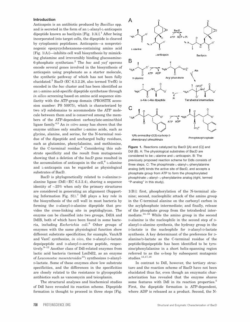
Anticapsin is an antibiotic produced by Bacillus spp.
and is secreted in the form of an L-alanyl-L-anticapsin
dipeptide known as bacilysin [Fig. 1(A)].1 After being
incorporated into target cells, the dipeptide is cleaved
by cytoplasmic peptidases. Anticapsin—a nonprotei-
nogenic epoxycyclohexanone-containing amino acid
[Fig. 1(A)]—inhibits cell wall biosynthesis by mimick-
ing glutamine and irreversibly binding glucosamine-
6-phosphate synthetase.2 The bac and ywf operons
encode several genes involved in the biosynthesis of
anticapsin using prephenate as a starter molecule,
the synthetic pathway of which has not been fully
elucidated.3 BacD (EC 6.3.2.28, also termed YwfE) is
encoded in the bac cluster and has been identified as
an L-amino acid-specific dipeptide synthetase through
in silico screening based on amino acid sequence sim-
ilarity with the ATP-grasp domain (PROSITE acces-
sion number: PS 50975), which is characterized by
two a/b subdomains to accommodate the ATP mole-
cule between them and is conserved among the mem-
bers of the ATP-dependent carboxylate-amine/thiol
ligase family.4,5 An in vitro assay has shown that the
enzyme utilizes only smaller L-amino acids, such as
glycine, alanine, and serine, for the N-terminal resi-
due of the dipeptide and uncharged bulky residues,
such as glutamine, phenylalanine, and methionine,
for the C-terminal residue.5 Considering this sub-
strate specificity and the result from mutagenesis
showing that a deletion of the bacD gene resulted in
the accumulation of anticapsin in the cell,6 L-alanine
and L-anticapsin can be regarded as physiological
substrates of BacD.
BacD is phylogenetically related to D-alanine:D-
alanine ligase (Ddl) (EC 6.3.2.4), sharing a sequence
identity of �25% when only the primary structures
are considered in generating an alignment (Support-
ing Information Fig. S1).7 Ddl plays a key role in
the biosynthesis of the cell wall in most bacteria by
forming the D-alanyl-D-alanine dipeptide that pro-
vides the cross-linking site in peptidoglycan. The
enzyme can be classified into two groups, DdlA and
DdlB, both of which have been found in some bacte-
ria, including Escherichia coli.8 Other groups of
enzymes with the same physiological function show
different substrate specificities; for example, VanA/B
and VanC synthesize, in vivo, the D-alanyl-D-lactate
depsipeptide and D-alanyl-D-serine peptide, respec-
tively.9–12 Another class of Ddl-related enzymes from
lactic acid bacteria (termed LmDdl2, as an enzyme
of Leuconostoc mesenteroides13) synthesizes D-alanyl-
D-lactate. Some of these enzymes show low substrate
specificities, and the differences in the specificities
are closely related to the resistance to glycopeptide
antibiotics such as vancomycin and teicoplanin.
The structural analyses and biochemical studies
of Ddl have revealed its reaction scheme. Dipeptide
formation is thought to proceed in three steps [Fig.
1(B)]: first, phosphorylation of the N-terminal ala-
nine; second, nucleophilic attack of the amino group
in the C-terminal alanine on the carbonyl carbon in
the acylphosphate intermediate; and finally, release
of the phosphate group from the tetrahedral inter-
mediate.14–16 While the amino group in the second
D-alanine is the nucleophile in the second step of D-
alanyl-D-alanine synthesis, the hydroxy group in the
D-lactate is the nucleophile for D-alanyl-D-lactate
synthesis. A key determinant of the preference for D-
alanine/D-lactate as the C-terminal residue of the
peptide/depsipeptide has been identified to be tyro-
sine/phenylalanine in a short helix-spanning region
referred to as the x-loop by subsequent mutagenic
studies.13,17,18
In contrast to Ddl, however, the tertiary struc-
ture and the reaction scheme of BacD have not been
elucidated thus far, even though an enzymatic char-
acterization has revealed that the enzyme shares
some features with Ddl in its reaction properties.5
First, the dipeptide formation is ATP-dependent,
where ADP is released as a product. Second, the N-
Figure 1. Reactions catalyzed by BacD [(A) and (C)] and
Ddl (B). A: The physiological substrates of BacD are
considered to be L-alanine and L-anticapsin. B: The
previously proposed reaction scheme for Ddls consists of
three steps. C: The phosphinate L-alanyl-L-phenylalanine
analog (left) binds the active site of BacD, and accepts a
phosphate group from ATP to form the phosphorylated
phosphinate L-alanyl- L-phenylalanine analog (right, termed
‘‘P-analog’’ in this study).
708 PROTEINSCIENCE.ORG Structural and Enzymatic Characterization of BacD
terminal amino acid affinities for the enzyme are
higher than those of the C-terminal amino acid.
Third, the amide-bond formation activities of the
enzymes show an optimum pH higher than 9.5,17,19
To investigate structural and functional features
of BacD, we have determined the crystal structure
of the enzyme in complex with ADP and an interme-
diate analog, phosphorylated phosphinate L-alanyl-
L-phenylalanine. Our structural description and an
accompanying enzymatic characterization have
implications for substrate specificity and for the
reaction mechanism of the dipeptide synthesis cata-
lyzed by BacD.
Results and Discussion
Enzymatic properties of BacD and inhibitory
effect of a phosphinate analog
The kinetic parameters of BacD have been deter-
mined using L-alanine and L-phenylalanine as sub-
strates (Table I). The pair of these two amino acids
showed the highest activity among the combinations
of less bulky amino acids (glycine, L-alanine, and
L-serine) and uncharged bulky amino acids (L-leucine,
L-isolucine, L-methionine, L-phenylalanine, L-tyrosine,
L-tryptophane, and L-glutamine), which have been
reported to be substrates of BacD.5 Some D-amino
acids (D-alanine, D-serine, D-phenylalanine, and D-glu-
tamine) were also tested but showed no activity,
consistent with the previous study.5
Interpretation of the results for BacD was much
more straightforward than for Ddl, which possesses
two amino acid binding sites for a single substrate
(i.e., D-alanine), but with different substrate affin-
ities. The plots of activity versus substrate concen-
tration for both L-alanine and L-phenylalanine fit
well with Michaelis–Menten kinetics [Supporting
Information Fig. S2(A)], confirming that each amino
acid is recognized by a unique binding site. In the
absence of either amino acid, the enzyme showed no
activity, also supporting the simple model for BacD.
The Km1 (for L-alanine) and Km2 (for L-phenylala-
nine) values are significantly higher than those of
DdlB from E. coli but are comparable to those of
VanA for D-alanine and D-alanine (Table I). VanA is
an Enterococcus faecium homolog of Ddl that utilizes
D-alanine and D-lactate to produce the D-alanyl-D-lac-
tate depsipeptide in vivo, and the values shown in
Table I are for a nonphysiological reaction where
only D-alanine was used as the substrate.9 Although
the affinities of BacD for L-alanine and L-phenylala-
nine are significantly lower than those of DdlB for
D-alanine, the kcat value is comparable to that of
Ddl. The observed high Km values may be related to
the use of a nonphysiological substrate in the assay,
since the most likely physiological substrates of
BacD are L-alanine and L-anticapsin.
In previous structural analyses of Ddls, a syn-
thetic phosphinate D-alanyl-D-alanine analog was
used to investigate the binding mode of the sub-
strates to the enzyme.14,20–22 For the same purpose,
we used a phosphinate L-alanyl-L-phenylalanine ana-
log [Fig. 1(C)], and assayed its inhibitory effect on
the enzyme. The Dixon plots over a range of the
analog compound concentration show a clear sign of
competitive inhibition for L-alanine but not for
L-phenylalanine [Supporting Information Fig.
S2(B,C)]. On the hypothesis that the compound occu-
pies the both amino-acid binding sites, this finding
suggests that BacD has a reaction sequence similar
to that of Ddls, where the amino acid for the N-ter-
minal residue of the dipeptide (L-alanine) first binds
the enzyme to form the acylphosphate intermediate.
In this scheme, L-phenylalanine does not compete
for the binding to the enzyme with the analog
because L-phenylalanine can bind the enzyme only
after the binding of L-alanine and the analog com-
petes for the binding to the enzyme with L-alanine.
The value of Ki was determined to be 15.4 lM based
on a Lineweaver–Burk plot over a range of L-alanine
at the inhibitor concentration of 20 lM [Supporting
Information Fig. S2(D)]. It should be emphasized
that the plot considers only the initial rates of the
reactions, and therefore, the possibility of slow onset
of inhibition [i.e., phosphorylation of the inhibitor as
shown in Fig. 1(C)]23 has not been taken into
account.
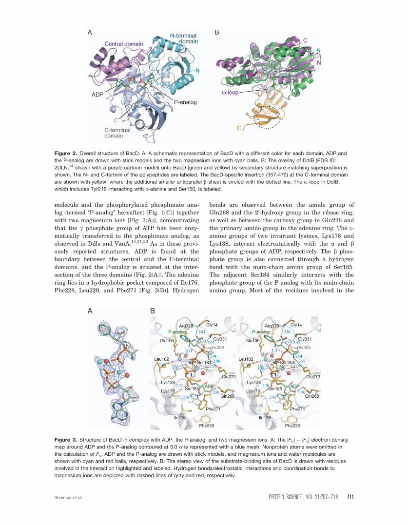
Overall structure of BacD
The crystal structure of BacD was determined by
the multiple-wavelength anomalous dispersion
method with a selenomethionine derivative crystal
(Table II). One BacD molecule has been found in the
asymmetric unit with continuous clear electron den-
sity for the polypeptide from Glu2 to the C-terminus
(Val472). On a tertiary structural level, BacD shares
some structural features with Ddls, as expected
from amino acid sequence identities of �25%. BacD
can be superimposed on DdlB from E. coli (PDB ID:
2DLN)14 with a root-mean-square deviation value of
2.6 A for 249 Ca atoms, based on secondary
Table I. Steady-State Kinetic Constants for AminoAcid Ligases
Enzyme pHKm1
(mM)Km2
(mM)kcat
(min�1)
BacD 7.7 1.7 6 0.11c 20 6 0.57d 630 6 6.5DdlB
(E. coli)a7.8 0.0033 1.2 1018
VanA(E. faecium)b
8.6 3.4 38 295
a For D-alanine:D-alanine ligase activity.8b For D-alanine:D-alanine ligase activity.12
c For L-alanine.d For L-phenylalanine.
Shomura et al. PROTEIN SCIENCE VOL 21:707—716 709
structure matching superposition [Fig. 2(A,B)].24 As
in Ddls and related enzymes,4,25 BacD can be di-
vided into three a/b domains: an N-terminal domain
(Glu2–Gly126), a central domain (Ala127–Leu229),
and a C-terminal domain (Gln230–Gly471). The N-
terminal domain includes a parallel b-sheet, whereas
the central domain contains an antiparallel b-sheet
like all Ddls, whose structures have already been
reported. The most prominent difference in the over-
all structures between BacD and DdlB is the pres-
ence of an extra region composed of �100 residues
in the C-terminal domain of BacD, including an
additional smaller antiparallel b-sheet [Fig. 2(B) and
Supporting Information Figs. S1 and S3]. Although
the BacD-specific region shows no direct interaction
with bound substrates, the region unambiguously
affects the conformation of two amino-acid recogni-
tion loops (Loop 5 and 6 as mentioned below).
BacD elutes as a monomer in size-exclusion
chromatography, and there is no sign of oligomeriza-
tion in the crystal, given that the largest contact
area with the adjacent molecule shares only 6.5% of
the total accessible surface area. This contrasts with
Ddl, whose structures have been exclusively
reported to be homodimeric, even though the dimeri-
zation is unlikely to be of functional/mechanistic
relevance. The tertiary structure of the dimerization
site identified in Ddls is relatively conserved in
BacD. Furthermore, the extra C-terminal region of
BacD shows no interference with the dimerization
site, implying that the difference in oligomerization
state between BacD and Ddls is ascribable to minor
amino acid substitutions. A sequence alignment of
BacD and Ddls indicates that most of the residues
forming hydrophobic interactions in the dimerization
interface of DdlB from E. coli are not conserved in
BacD (Supporting Information Fig. S1). Specifically,
nonhydrophobic properties of Gln130, Glu133, and
Asn134 would determine the oligomerization state of
BacD to be monomer.
Active site structure
Crystallization of BacD has been performed in the
presence of ATP, magnesium chloride, and the phos-
phinate L-alanyl-L-phenylalanine analog. An |Fo| �|Fc| electron density map clearly delineates an ADP
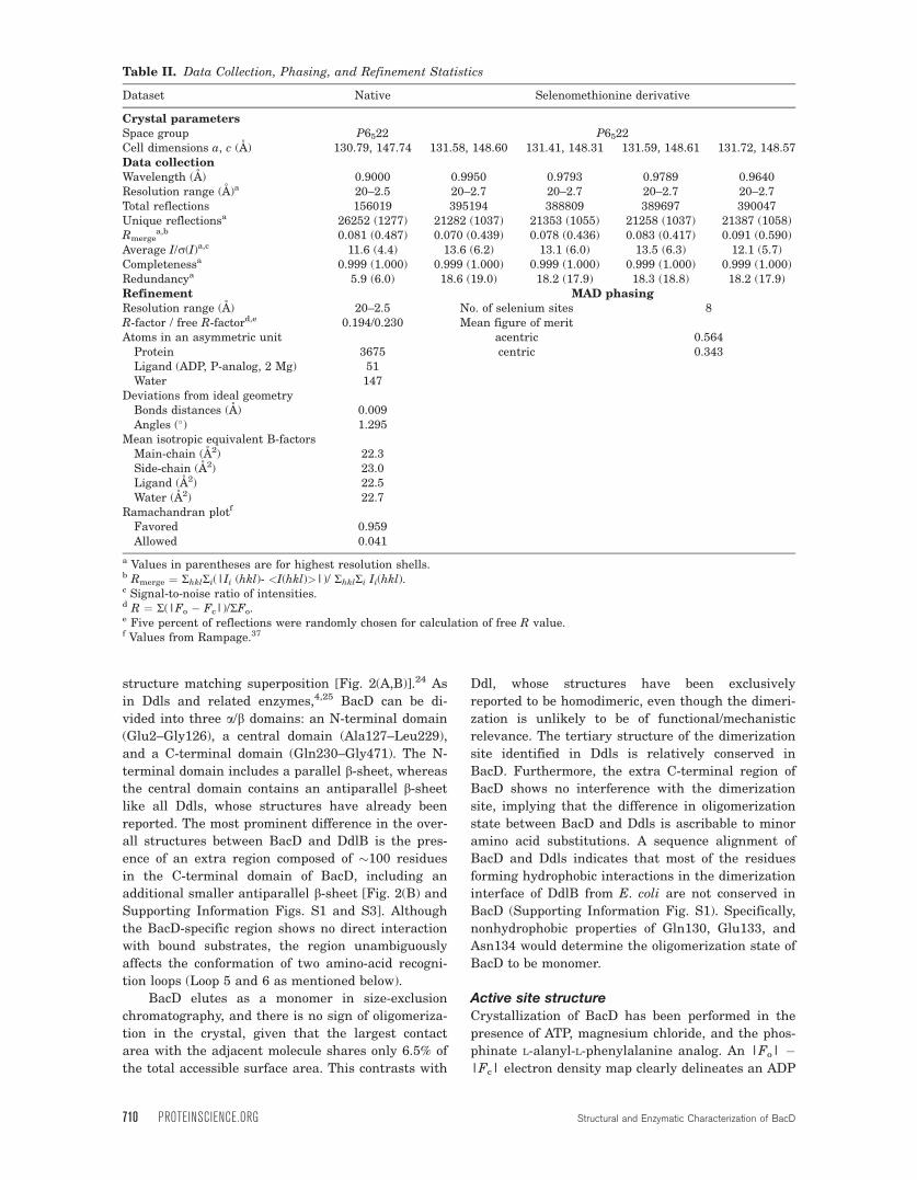
Table II. Data Collection, Phasing, and Refinement Statistics
Dataset Native Selenomethionine derivative
Crystal parametersSpace group P6522 P6522Cell dimensions a, c (A) 130.79, 147.74 131.58, 148.60 131.41, 148.31 131.59, 148.61 131.72, 148.57Data collectionWavelength (A) 0.9000 0.9950 0.9793 0.9789 0.9640Resolution range (A)a 20–2.5 20–2.7 20–2.7 20–2.7 20–2.7Total reflections 156019 395194 388809 389697 390047Unique reflectionsa 26252 (1277) 21282 (1037) 21353 (1055) 21258 (1037) 21387 (1058)Rmerge
a,b 0.081 (0.487) 0.070 (0.439) 0.078 (0.436) 0.083 (0.417) 0.091 (0.590)Average I/r(I)a,c 11.6 (4.4) 13.6 (6.2) 13.1 (6.0) 13.5 (6.3) 12.1 (5.7)Completenessa 0.999 (1.000) 0.999 (1.000) 0.999 (1.000) 0.999 (1.000) 0.999 (1.000)Redundancya 5.9 (6.0) 18.6 (19.0) 18.2 (17.9) 18.3 (18.8) 18.2 (17.9)Refinement MAD phasingResolution range (A) 20–2.5 No. of selenium sites 8R-factor / free R-factord,e 0.194/0.230 Mean figure of meritAtoms in an asymmetric unit acentric 0.564
Protein 3675 centric 0.343Ligand (ADP, P-analog, 2 Mg) 51Water 147
Deviations from ideal geometryBonds distances (A) 0.009Angles (�) 1.295
Mean isotropic equivalent B-factorsMain-chain (A2) 22.3Side-chain (A2) 23.0Ligand (A2) 22.5Water (A2) 22.7
Ramachandran plotf
Favored 0.959Allowed 0.041
a Values in parentheses are for highest resolution shells.b Rmerge ¼ RhklRi(|Ii (hkl)- <I(hkl)>|)/ RhklRi Ii(hkl).c Signal-to-noise ratio of intensities.d R ¼ R(|Fo � Fc|)/RFo.e Five percent of reflections were randomly chosen for calculation of free R value.f Values from Rampage.37
710 PROTEINSCIENCE.ORG Structural and Enzymatic Characterization of BacD
molecule and the phosphorylated phosphinate ana-
log (termed ‘‘P-analog’’ hereafter) [Fig. 1(C)] together
with two magnesium ions [Fig. 3(A)], demonstrating
that the c phosphate group of ATP has been enzy-
matically transferred to the phosphinate analog, as
observed in Ddls and VanA.14,21,22 As in these previ-
ously reported structures, ADP is found at the
boundary between the central and the C-terminal
domains, and the P-analog is situated at the inter-
section of the three domains [Fig. 2(A)]. The adenine
ring lies in a hydrophobic pocket composed of Ile176,
Phe228, Leu229, and Phe271 [Fig. 3(B)]. Hydrogen
bonds are observed between the amide group of
Gln268 and the 20-hydroxy group in the ribose ring,
as well as between the carboxy group in Glu226 and
the primary amino group in the adenine ring. The e-amino groups of two invariant lysines, Lys178 and
Lys138, interact electrostatically with the a and bphosphate groups of ADP, respectively. The b phos-
phate group is also connected through a hydrogen
bond with the main-chain amino group of Ser185.
The adjacent Ser184 similarly interacts with the
phosphate group of the P-analog with its main-chain
amino group. Most of the residues involved in the
Figure 2. Overall structure of BacD. A: A schematic representation of BacD with a different color for each domain. ADP and
the P-analog are drawn with stick models and the two magnesium ions with cyan balls. B: The overlay of DdlB (PDB ID:
2DLN,14 shown with a purple cartoon model) onto BacD (green and yellow) by secondary structure matching superposition is
shown. The N- and C-termini of the polypeptides are labeled. The BacD-specific insertion (357-472) at the C-terminal domain
are shown with yellow, where the additional smaller antiparallel b-sheet is circled with the dotted line. The x-loop in DdlB,
which includes Tyr216 interacting with D-alanine and Ser150, is labeled.
Figure 3. Structure of BacD in complex with ADP, the P-analog, and two magnesium ions. A: The |Fo| � |Fc| electron density
map around ADP and the P-analog contoured at 3.0 r is represented with a blue mesh. Nonprotein atoms were omitted in
the calculation of Fc. ADP and the P-analog are drawn with stick models, and magnesium ions and water molecules are
shown with cyan and red balls, respectively. B: The stereo view of the substrate-binding site of BacD is drawn with residues
involved in the interaction highlighted and labeled. Hydrogen bonds/electrostatic interactions and coordination bonds to
magnesium ions are depicted with dashed lines of gray and red, respectively.
Shomura et al. PROTEIN SCIENCE VOL 21:707—716 711
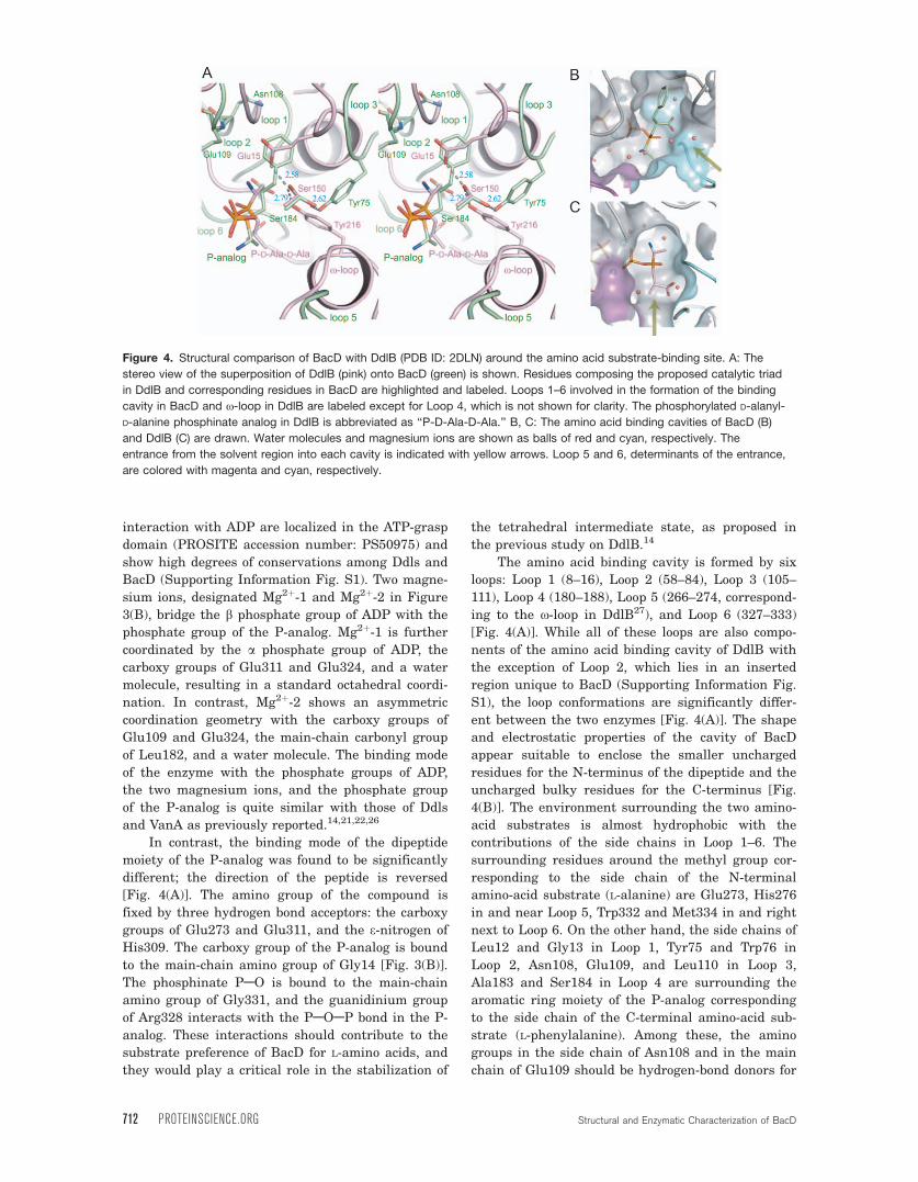
interaction with ADP are localized in the ATP-grasp
domain (PROSITE accession number: PS50975) and
show high degrees of conservations among Ddls and
BacD (Supporting Information Fig. S1). Two magne-
sium ions, designated Mg2þ-1 and Mg2þ-2 in Figure
3(B), bridge the b phosphate group of ADP with the
phosphate group of the P-analog. Mg2þ-1 is further
coordinated by the a phosphate group of ADP, the
carboxy groups of Glu311 and Glu324, and a water
molecule, resulting in a standard octahedral coordi-
nation. In contrast, Mg2þ-2 shows an asymmetric
coordination geometry with the carboxy groups of
Glu109 and Glu324, the main-chain carbonyl group
of Leu182, and a water molecule. The binding mode
of the enzyme with the phosphate groups of ADP,
the two magnesium ions, and the phosphate group
of the P-analog is quite similar with those of Ddls
and VanA as previously reported.14,21,22,26
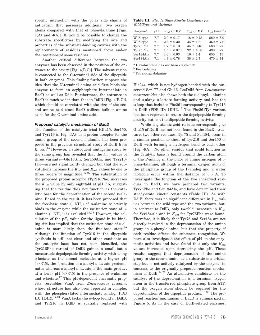
In contrast, the binding mode of the dipeptide
moiety of the P-analog was found to be significantly
different; the direction of the peptide is reversed
[Fig. 4(A)]. The amino group of the compound is
fixed by three hydrogen bond acceptors: the carboxy
groups of Glu273 and Glu311, and the e-nitrogen of
His309. The carboxy group of the P-analog is bound
to the main-chain amino group of Gly14 [Fig. 3(B)].
The phosphinate PAO is bound to the main-chain
amino group of Gly331, and the guanidinium group
of Arg328 interacts with the PAOAP bond in the P-
analog. These interactions should contribute to the
substrate preference of BacD for L-amino acids, and
they would play a critical role in the stabilization of
the tetrahedral intermediate state, as proposed in
the previous study on DdlB.14
The amino acid binding cavity is formed by six
loops: Loop 1 (8–16), Loop 2 (58–84), Loop 3 (105–
111), Loop 4 (180–188), Loop 5 (266–274, correspond-
ing to the x-loop in DdlB27), and Loop 6 (327–333)
[Fig. 4(A)]. While all of these loops are also compo-
nents of the amino acid binding cavity of DdlB with
the exception of Loop 2, which lies in an inserted
region unique to BacD (Supporting Information Fig.
S1), the loop conformations are significantly differ-
ent between the two enzymes [Fig. 4(A)]. The shape
and electrostatic properties of the cavity of BacD
appear suitable to enclose the smaller uncharged
residues for the N-terminus of the dipeptide and the
uncharged bulky residues for the C-terminus [Fig.
4(B)]. The environment surrounding the two amino-
acid substrates is almost hydrophobic with the
contributions of the side chains in Loop 1–6. The
surrounding residues around the methyl group cor-
responding to the side chain of the N-terminal
amino-acid substrate (L-alanine) are Glu273, His276
in and near Loop 5, Trp332 and Met334 in and right
next to Loop 6. On the other hand, the side chains of
Leu12 and Gly13 in Loop 1, Tyr75 and Trp76 in
Loop 2, Asn108, Glu109, and Leu110 in Loop 3,
Ala183 and Ser184 in Loop 4 are surrounding the
aromatic ring moiety of the P-analog corresponding
to the side chain of the C-terminal amino-acid sub-
strate (L-phenylalanine). Among these, the amino
groups in the side chain of Asn108 and in the main
chain of Glu109 should be hydrogen-bond donors for
Figure 4. Structural comparison of BacD with DdlB (PDB ID: 2DLN) around the amino acid substrate-binding site. A: The
stereo view of the superposition of DdlB (pink) onto BacD (green) is shown. Residues composing the proposed catalytic triad
in DdlB and corresponding residues in BacD are highlighted and labeled. Loops 1–6 involved in the formation of the binding
cavity in BacD and x-loop in DdlB are labeled except for Loop 4, which is not shown for clarity. The phosphorylated D-alanyl-
D-alanine phosphinate analog in DdlB is abbreviated as ‘‘P-D-Ala-D-Ala.’’ B, C: The amino acid binding cavities of BacD (B)
and DdlB (C) are drawn. Water molecules and magnesium ions are shown as balls of red and cyan, respectively. The
entrance from the solvent region into each cavity is indicated with yellow arrows. Loop 5 and 6, determinants of the entrance,
are colored with magenta and cyan, respectively.
712 PROTEINSCIENCE.ORG Structural and Enzymatic Characterization of BacD
specific interaction with the polar side chains of
anticapsin that possesses additional two oxygen
atoms compared with that of phenylalanine [Figs.
1(A) and 4(A)]. It would be possible to change the
substrate specificities by modifying the size and
properties of the substrate-binding cavities with the
replacements of residues mentioned above and/or
the insertions of some residues.
Another critical difference between the two
enzymes has been observed in the position of the en-
trance to the cavity [Fig. 4(B,C)]. The solvent region
is connected to the C-terminal side of the dipeptide
in both enzymes. This finding further supports the
idea that the N-terminal amino acid first binds the
enzyme to form an acylphosphate intermediate in
BacD as well as Ddls. Furthermore, the entrance in
BacD is much wider than that in DdlB [Fig. 4(B,C)],
which should be correlated with the size of the sec-
ond amino acid since BacD utilizes bulkier amino
acids for the C-terminal amino acid.
Proposed catalytic mechanism of BacD
The function of the catalytic triad [Glu15, Ser150,
and Tyr216 in Fig. 4(A)] as a proton acceptor for the
amino group of the second D-alanine has been pro-
posed in the previous structural study of DdlB from
E. coli.14 However, a subsequent mutagenic study by
the same group has revealed that the kcat values of
three variants—Glu15Gln, Ser150Ala, and Tyr216-
Phe—are not significantly changed but that the sub-
stitutions increase the Km1 and Km2 values by one to
three orders of magnitude.17,27 The substitution of
the proposed proton acceptor (Tyr216Phe) increases
the Km2 value by only eightfold at pH 7.5, suggest-
ing that the residue does not function as the cata-
lytic base for the deprotonation of the second D-ala-
nine. Based on the result, it has been proposed that
the free-base state (ANH2) of D-alanine selectively
binds to the enzyme while the zwitterion state of D-
alanine (ANH3þ) is excluded.27,28 However, the cal-
culation of the pKa value for the ligand in its bind-
ing site has implied that the zwitterion state of D-al-
anine is more likely than the free-base state.29
Although the function of Tyr216 in the dipeptide
synthesis is still not clear and other candidate as
the catalytic base has not been identified, the
Tyr216Phe variant of DdlB gained a small but a
measurable depsipeptide-forming activity with using
D-lactate as the second molecule; at a higher pH
(>�7.5), the formation of D-alanyl-D-lactate predomi-
nates whereas D-alanyl-D-lactate is the main product
at a lower pH (<�7.5) in the presence of D-alanine
and D-lactate.17 This pH-dependent enzymatic prop-
erty resembles VanA from Enterococcus faecium,
whose structure has also been reported in complex
with the phosphorylated intermediate analog (PDB
ID: 1E4E).17,21 VanA lacks the x-loop found in DdlB,
and Tyr216 in DdlB is spatially replaced with
His244, which is not hydrogen-bonded with the con-
served Ser177 and Glu16. LmDdl2 from Leuconostoc
mesenteroides also shows both the D-alanyl-D-alanine
and D-alanyl-D-lactate forming activity and has the
x-loop that includes Phe261 corresponding to Tyr216
in DdlB (PDB ID: 1EHI).13 The Phe261Tyr variant
has been reported to retain the depsipeptide-forming
activity but lost the dipeptide-forming activity.
While a glutamic acid residue corresponding to
Glu15 of DdlB has not been found in the BacD struc-
ture, two other residues, Tyr75 and Ser184, occur in
a similar position to those of Tyr216 and Ser150 of
DdlB with forming a hydrogen bond to each other
[Fig. 4(A)]. No other residue that could function as
the catalytic base is found around the carbon atom
of the P-analog in the place of amino nitrogen of L-
phenylalanine, although a terminal oxygen atom of
the phosphate group of the P-analog and a water
molecule occur within the distance of 3.5 A. To
investigate the function of the two conserved resi-
dues in BacD, we have prepared two variants,
Tyr75Phe and Ser184Ala, and have determined their
steady-state kinetic constants (Table III). As with
DdlB, there was no significant difference in kcat val-
ues between the wild type and the two variants, but,
in contrast to DdlB, only twofold increases in Km1
for Ser184Ala and in Km2 for Tyr75Phe were found.
Therefore, it is likely that Tyr75 and Ser184 are not
directly involved in the deprotonation of the amino
group in L-phenylalanine, but that the property of
each residue affects the substrate recognition. We
have also investigated the effect of pH on the enzy-
matic activities and have found that only the Km2
values increased upon decreasing the pH. These
results suggest that deprotonation of the amino
group in the second amino acid substrate is a critical
step but is not actively catalyzed by the enzyme, in
contrast to the originally proposed reaction mecha-
nism of DdlB.14,27 An alternative candidate for the
catalyst of the deprotonation is a terminal oxygen
atom in the transferred phosphate group from ATP,
but the oxygen atom should be required for the
deprotonation of the dipeptide product.18,28 The pro-
posed reaction mechanism of BacD is summarized in
Figure 5. As in the case of DdlB-related enzymes,
Table III. Steady-State Kinetic Constants forWild-Type and Variants
Enzymea pH Km1 (mM)b Km2 (mM)c kcat (min�1)
Wild-type 7.7 2.0 6 0.17 18 6 0.78 580 6 8.9Wild-type 7.1 2.6 6 0.32 44 6 1.6 460 6 7.6Tyr75Phe 7.7 1.7 6 0.10 45 6 0.48 580 6 2.8Tyr75Phe 7.1 1.5 6 0.078 92 6 10.0 430 6 27Ser184Ala 7.7 4.6 6 0.63 16 6 1.4 650 6 19Ser184Ala 7.1 4.9 6 0.78 38 6 2.7 470 6 14
a Hexahistidine has not been cleaved off.b For L-alanine.c For L-phenylalanine.
Shomura et al. PROTEIN SCIENCE VOL 21:707—716 713
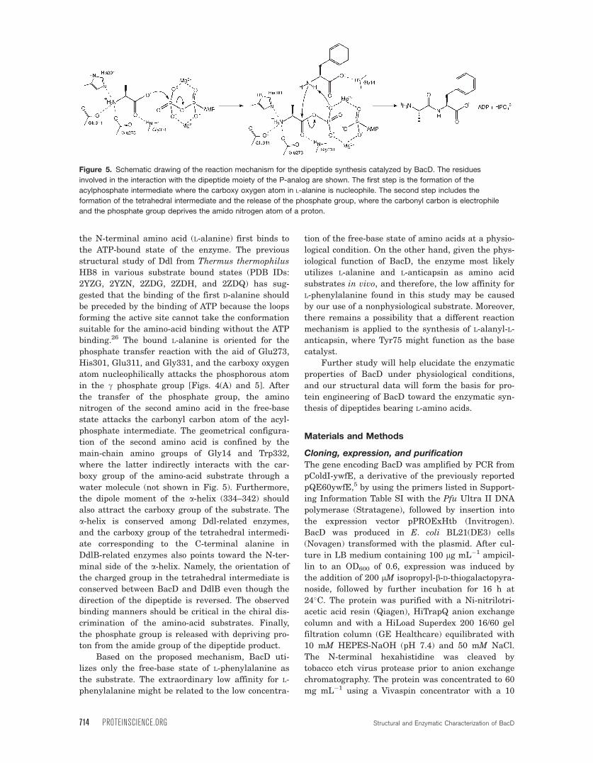
the N-terminal amino acid (L-alanine) first binds to
the ATP-bound state of the enzyme. The previous
structural study of Ddl from Thermus thermophilus
HB8 in various substrate bound states (PDB IDs:
2YZG, 2YZN, 2ZDG, 2ZDH, and 2ZDQ) has sug-
gested that the binding of the first D-alanine should
be preceded by the binding of ATP because the loops
forming the active site cannot take the conformation
suitable for the amino-acid binding without the ATP
binding.26 The bound L-alanine is oriented for the
phosphate transfer reaction with the aid of Glu273,
His301, Glu311, and Gly331, and the carboxy oxygen
atom nucleophilically attacks the phosphorous atom
in the c phosphate group [Figs. 4(A) and 5]. After
the transfer of the phosphate group, the amino
nitrogen of the second amino acid in the free-base
state attacks the carbonyl carbon atom of the acyl-
phosphate intermediate. The geometrical configura-
tion of the second amino acid is confined by the
main-chain amino groups of Gly14 and Trp332,
where the latter indirectly interacts with the car-
boxy group of the amino-acid substrate through a
water molecule (not shown in Fig. 5). Furthermore,
the dipole moment of the a-helix (334–342) should
also attract the carboxy group of the substrate. The
a-helix is conserved among Ddl-related enzymes,
and the carboxy group of the tetrahedral intermedi-
ate corresponding to the C-terminal alanine in
DdlB-related enzymes also points toward the N-ter-
minal side of the a-helix. Namely, the orientation of
the charged group in the tetrahedral intermediate is
conserved between BacD and DdlB even though the
direction of the dipeptide is reversed. The observed
binding manners should be critical in the chiral dis-
crimination of the amino-acid substrates. Finally,
the phosphate group is released with depriving pro-
ton from the amide group of the dipeptide product.
Based on the proposed mechanism, BacD uti-
lizes only the free-base state of L-phenylalanine as
the substrate. The extraordinary low affinity for L-
phenylalanine might be related to the low concentra-
tion of the free-base state of amino acids at a physio-
logical condition. On the other hand, given the phys-
iological function of BacD, the enzyme most likely
utilizes L-alanine and L-anticapsin as amino acid
substrates in vivo, and therefore, the low affinity for
L-phenylalanine found in this study may be caused
by our use of a nonphysiological substrate. Moreover,
there remains a possibility that a different reaction
mechanism is applied to the synthesis of L-alanyl-L-
anticapsin, where Tyr75 might function as the base
catalyst.
Further study will help elucidate the enzymatic
properties of BacD under physiological conditions,
and our structural data will form the basis for pro-
tein engineering of BacD toward the enzymatic syn-
thesis of dipeptides bearing L-amino acids.
Materials and Methods
Cloning, expression, and purification
The gene encoding BacD was amplified by PCR from
pColdI-ywfE, a derivative of the previously reported
pQE60ywfE,5 by using the primers listed in Support-
ing Information Table SI with the Pfu Ultra II DNA
polymerase (Stratagene), followed by insertion into
the expression vector pPROExHtb (Invitrogen).
BacD was produced in E. coli BL21(DE3) cells
(Novagen) transformed with the plasmid. After cul-
ture in LB medium containing 100 lg mL�1 ampicil-
lin to an OD600 of 0.6, expression was induced by
the addition of 200 lM isopropyl-b-D-thiogalactopyra-
noside, followed by further incubation for 16 h at
24�C. The protein was purified with a Ni-nitrilotri-
acetic acid resin (Qiagen), HiTrapQ anion exchange
column and with a HiLoad Superdex 200 16/60 gel
filtration column (GE Healthcare) equilibrated with
10 mM HEPES-NaOH (pH 7.4) and 50 mM NaCl.
The N-terminal hexahistidine was cleaved by
tobacco etch virus protease prior to anion exchange
chromatography. The protein was concentrated to 60
mg mL�1 using a Vivaspin concentrator with a 10
Figure 5. Schematic drawing of the reaction mechanism for the dipeptide synthesis catalyzed by BacD. The residues
involved in the interaction with the dipeptide moiety of the P-analog are shown. The first step is the formation of the
acylphosphate intermediate where the carboxy oxygen atom in L-alanine is nucleophile. The second step includes the
formation of the tetrahedral intermediate and the release of the phosphate group, where the carbonyl carbon is electrophile
and the phosphate group deprives the amido nitrogen atom of a proton.
714 PROTEINSCIENCE.ORG Structural and Enzymatic Characterization of BacD
kDa cutoff (Sartorius) and was stored at �80�C.
Selenomethionine-substituted BacD was produced as
described previously,30 and was purified using the
same procedure as for the native enzyme.
Crystallization and structure determination
of BacD
Crystals of both native and selenomethionine-substi-
tuted BacD were obtained under the same conditions.
Crystallization was performed at 293 K by the sitting-
drop vapor-diffusion method by using a CrystalQuick
96-well plate (Greiner). The drop was prepared by
mixing 1 lL of the protein solution, containing 20 mg
mL�1 BacD, 10 mM HEPES-NaOH (pH 7.4), 50 mM
NaCl, 4 mM ATP, 8 mM MgCl2, and 4 mM phosphi-
nate L-alanyl-L-phenylalanine analog [left compound
in Fig. 1(C)], with 1 lL of reservoir solution contain-
ing 100 mM Bis-Tris propane, 60 mM sodium citrate,
and 18% (w/v) PEG3, 350. Crystals grown to a maxi-
mum size of 0.1 � 0.1 � 0.2 mm3 were soaked in a cry-
oprotectant buffer composed of 100 mM Bis-Tris pro-
pane, 60 mM sodium citrate, 25% (w/v) PEG3,350,
and 20% glycerol, prior to flash cooling with liquid
nitrogen. Single-wavelength native data and multi-
ple-wavelength selenium derivative data were col-
lected at SPring-8 beamline BL44XU, with the crys-
tals maintained at 90 K using a gaseous nitrogen
stream. All data were processed and scaled with
HKL2000.31 Eight selenium sites were found, of
which the positions, occupancies, and B-factors were
refined and the initial phases calculated with autoSH-
ARP.32 After density modification using SOLOMON33
and automatic model building by Buccaneer,34 an ini-
tial model covering �80% of the BacD molecule was
obtained. Subsequent iterative manual model build-
ing/corrections and refinement were performed with
Coot24 and Refmac5,35 respectively. Statistics for data
collection, phasing, and refinement are summarized
in Table II. Secondary structural elements were
assigned with DSSP,36 and main-chain dihedral
angles were checked with RAMPAGE.37 The coordi-
nates and the structure factors have been deposited
in the Protein Data Bank, under the accession code of
3VMM. Graphical representations of the model were
prepared with PyMOL (DeLano Scientific).
Enzymatic assayThe enzymatic activity of the wild type and BacD
point mutants was assayed spectrophotometrically by
using an ATP/NADH coupled system with pyruvate
kinase (PK) and lactate dehydrogenase (LDH).38 The
assay solution contained 10 mM magnesium acetate,
10 mM potassium acetate, 5 mM ATP, 2.5 mM phos-
phoenolpyruvate, 0.8 mM NADH, 34.1 U mL�1 PK,
and 49.5 U mL�1 LDH (Sigma). The pH was meas-
ured to be 7.7 and 7.1 for reaction solutions contain-
ing 50 mM Tris-HCl (pH 8.5) and 100 mM HEPES-
NaOH (pH 7.4), respectively. All assays were per-
formed at 310 K with a 96-well plate reader (BioRad),
by monitoring the decrease in absorbance at 340 nm.
The values of Km1 for L-alanine and of Km2 for L-phe-
nylalanine were determined in the presence of 100
mM L-phenylalanine and of 25 mM L-alanine, respec-
tively, over a range of the concentration of the coun-
terpart amino acid. The value of kcat was determined
from the latter plot because a saturating concentra-
tion of L-phenylalanine is unattainable for some con-
ditions, because of its low solubility. All measure-
ments were performed at least three times.
Site-directed mutagenesisPoint mutations were introduced to prepare two var-
iants (Tyr75Phe and Ser185Ala) by PCR using the Pfu
Ultra II DNA polymerase (Stratagene) with the pri-
mers listed in Table SI. The mutations were verified by
DNA sequencing. In the enzymatic assay for compar-
ing the wild-type and the two variants, proteins puri-
fied by Ni-affinity chromatography and by dialysis
with 10 mM HEPES-NaOH (pH 7.4) and 50 mM NaCl
were used. The purified proteins were tagged with an
N-terminal hexahistidine, whose effect on steady-state
kinetic parameters has been found to be negligible.
Acknowledgments
The authors acknowledge the assistance of the staff at
the SPring-8 beamline BL44XU (Proposal No.:
2011B6623). The MX225-HE (Rayonix) CCD detector
at BL44XU was financially supported by Academia
Sinica and by the National Synchrotron Radiation
Research Center (Taiwan, ROC). The authors are also
grateful to Prof. Jun Hiratake at Kyoto University for
the synthesis of the phosphinate analog.
References1. Foster JW, Woodruff HB (1946) Bacillin, a new antibi-
otic substance from a soil isolate of Bacillus subtilis. JBacteriol 51:363–369.
2. Kenig M, Vandamme E, Abraham EP (1976) The modeof action of bacilysin and anticapsin and biochemicalproperties of bacilysin-resistant mutants. J Gen Micro-biol 94:46–54.
3. Mahlstedt SA, Walsh CT (2010) Investigation of anti-capsin biosynthesis reveals a four-enzyme pathway totetrahydrotyrosine in Bacillus subtilis. Biochemistry49:912–923.
4. Fan C, Moews PC, Shi Y, Walsh CT, Knox JR (1995) Acommon fold for peptide synthetases cleaving ATP toADP: glutathione synthetase and D-alanine:D-alanineligase of Escherichia coli. Proc Natl Acad Sci USA 92:1172–1176.
5. Tabata K, Ikeda H, Hashimoto S (2005) ywfE in Bacil-lus subtilis codes for a novel enzyme, L-amino acidligase. J Bacteriol 187:5195–5202.
6. Steinborn G, Hajirezaei MR, Hofemeister J (2005) bacgenes for recombinant bacilysin and anticapsin produc-tion in Bacillus host strains. Arch Microbiol 183:71–79.
7. Thompson JD, Higgins DG, Gibson TJ (1994) CLUS-TAL W: improving the sensitivity of progressive multi-ple sequence alignment through sequence weighting,
Shomura et al. PROTEIN SCIENCE VOL 21:707—716 715
position-specific gap penalties and weight matrixchoice. Nucleic Acids Res 22:4673–4680.
8. Zawadzke LE, Bugg TD, Walsh CT (1991) Existence oftwo D-alanine:D-alanine ligases in Escherichia coli:cloning and sequencing of the ddlA gene and purifica-tion and characterization of the DdlA and DdlBenzymes. Biochemistry 30:1673–1682.
9. Bugg TD, Wright GD, Dutka-Malen S, Arthur M, Cour-valin P, Walsh CT (1991) Molecular basis for vancomy-cin resistance in Enterococcus faecium BM4147:biosynthesis of a depsipeptide peptidoglycan precursorby vancomycin resistance proteins VanH and VanA.Biochemistry 30:10408–10415.
10. Billot-Klein D, Gutmann L, Sable S, Guittet E, van Hei-jenoort J (1994) Modification of peptidoglycan precursorsis a common feature of the low-level vancomycin-resist-ant VANB-type Enterococcus D366 and of the naturallyglycopeptide-resistant species Lactobacillus casei, Pedio-coccus pentosaceus, Leuconostoc mesenteroides, andEnterococcus gallinarum. J Bacteriol 176:2398–2405.
11. Reynolds PE, Snaith HA, Maguire AJ, Dutka-Malen S,Courvalin P (1994) Analysis of peptidoglycan precur-sors in vancomycin-resistant Enterococcus gallinarumBM4174. Biochem J 301:5–8.
12. Bugg TD, Dutka-Malen S, Arthur M, Courvalin P, WalshCT (1991) Identification of vancomycin resistance pro-tein VanA as a D-alanine:D-alanine ligase of altered sub-strate specificity. Biochemistry 30:2017–2021.
13. Park IS, Walsh CT (1997) D-Alanyl-D-lactate and D-ala-nyl-D-alanine synthesis by D-alanyl-D-alanine ligasefrom vancomycin-resistant Leuconostoc mesenteroides.Effects of a phenylalanine 261 to tyrosine mutation. JBiol Chem 272:9210–9214.
14. Fan C, Moews PC, Walsh CT, Knox JR (1994) Vanco-mycin resistance: structure of D-alanine:D-alanineligase at 2.3 A resolution. Science 266:439–443.
15. Mullins LS, Zawadzke LE, Walsh CT, Raushel FM(1990) Kinetic evidence for the formation of D-alanylphosphate in the mechanism of D-alanyl-D-alanineligase. J Biol Chem 265:8993–8998.
16. Neuhaus FC (1962) The enzymatic synthesis of D-ala-nyl-D-alanine. II. Kinetic studies on D-alanyl-D-alaninesynthetase. J Biol Chem 237:3128–3135.
17. Park IS, Lin CH, Walsh CT (1996) Gain of D-alanyl-D-lactate or D-lactyl-D-alanine synthetase activities inthree active-site mutants of the Escherichia coli D-ala-nyl-D-alanine ligase B. Biochemistry 35:10464–10471.
18. Lessard IA, Healy VL, Park IS, Walsh CT (1999) Deter-minants for differential effects on D-Ala-D-lactate vs D-Ala-D-Ala formation by the VanA ligase from vancomy-cin-resistant enterococci. Biochemistry 38:14006–14022.
19. Wu D, Zhang L, Kong Y, Du J, Chen S, Chen J, DingJ, Jiang H, Shen X (2008) Enzymatic characterizationand crystal structure analysis of the D-alanine-D-ala-nine ligase from Helicobacter pylori. Proteins 72:1148–1160.
20. Fan C, Park IS, Walsh CT, Knox JR (1997) D-alanine:D-alanine ligase: phosphonate and phosphinate inter-mediates with wild type and the Y216F mutant. Bio-chemistry 36:2531–2538.
21. Roper DI, Huyton T, Vagin A, Dodson G (2000) The mo-lecular basis of vancomycin resistance in clinically rele-
vant Enterococci: crystal structure of D-alanyl-D-lactateligase (VanA). Proc Natl Acad Sci USA 97:8921–8925.
22. Kuzin AP, Sun T, Jorczak-Baillass J, Healy VL, WalshCT, Knox JR (2000) Enzymes of vancomycin resistance:the structure of D-alanine-D-lactate ligase of naturallyresistant Leuconostoc mesenteroides. Structure 8:463–470.
23. Duncan K, Walsh CT (1988) ATP-dependent inactiva-tion and slow binding inhibition of Salmonella typhi-murium D-alanine:D-alanine ligase (ADP) by(aminoalkyl)phosphinate and aminophosphonate ana-logues of D-alanine. Biochemistry 27:3709–3714.
24. Emsley P, Cowtan K (2004) Coot: model-building toolsfor molecular graphics. Acta Cryst D60:2126–2132.
25. Artymiuk PJ, Poirrette AR, Rice DW, Willett P (1996)Biotin carboxylase comes into the fold. Nat Struct Biol3:128–132.
26. Kitamura Y, Ebihara A, Agari Y, Shinkai A, Hirotsu K,Kuramitsu S (2009) Structure of D-alanine-D-alanineligase from Thermus thermophilus HB8: cumulativeconformational change and enzyme-ligand interactions.Acta Cryst D65:1098–1106.
27. Shi Y, Walsh CT (1995) Active site mapping of Esche-richia coli D-Ala-D-Ala ligase by structure-based muta-genesis. Biochemistry 34:2768–2776.
28. Ellsworth BA, Tom NJ, Bartlett PA (1996) Synthesisand evaluation of inhibitors of bacterial D-alanine:D-ala-nine ligases. Chem Biol 3:37–44.
29. Carlson HA, Briggs JM, McCammon JA (1999) Calcula-tion of the pKa values for the ligands and side chainsof Escherichia coli D-alanine:D-alanine ligase. J MedChem 42:109–117.
30. Van Duyne GD, Standaert RF, Karplus PA, SchreiberSL, Clardy J (1993) Atomic structures of the humanimmunophilin FKBP-12 complexes with FK506 andrapamycin. J Mol Biol 229:105–124.
31. Otwinowski Z, Minor W (1997) Processing of X-ray dif-fraction data collected in oscillation mode. MethodsEnzymol 276:307–326.
32. Vonrhein C, Blanc E, Roversi P, Bricogne G (2007)Automated structure solution with autoSHARP. Meth-ods Mol Biol 364:215–230.
33. Abrahams JP, Leslie AG (1996) Methods used in thestructure determination of bovine mitochondrial F1ATPase. Acta Cryst D52:30–42.
34. Cowtan K (2006) The Buccaneer software for auto-mated model building. 1. Tracing protein chains. ActaCryst D62:1002–1011.
35. Murshudov GN, Vagin AA, Dodson EJ (1997) Refine-ment of macromolecular structures by the maximum-likelihood method. Acta Cryst D53:240–255.
36. Kabsch W, Sander C (1983) Dictionary of protein sec-ondary structure: pattern recognition of hydrogen-bonded and geometrical features. Biopolymers 22:2577–2637.
37. Lovell SC, Davis IW, Arendall WB, III, de Bakker PI,Word JM, Prisant MG, Richardson JS, Richardson DC(2003) Structure validation by Calpha geometry: phi,psiand Cbeta deviation. Proteins 50:437–450.
38. Neuhaus FC (1962) The enzymatic synthesis of D-ala-nyl-D-alanine. I. Purification and properties of D-alanyl-D-alanine synthetase. J Biol Chem 237:778–786.
716 PROTEINSCIENCE.ORG Structural and Enzymatic Characterization of BacD
Related Documents