Structural and dynamic determinants of protein-peptide recognition Onur Dagliyan a,b , Elizabeth A. Proctor a,c , Kevin M. D’Auria e , Feng Ding b,d,* , and Nikolay V. Dokholyan a,b,c,d,* a Program in Molecular and Cellular Biophysics, University of North Carolina at Chapel Hill, Chapel Hill, North Carolina b Department of Biochemistry and Biophysics, School of Medicine, University of North Carolina at Chapel Hill, Chapel Hill, North Carolina c Curriculum in Bioinformatics and Computational Biology, University of North Carolina at Chapel Hill, Chapel Hill, North Carolina d Center for Systems and Computational Biology, University of North Carolina at Chapel Hill, Chapel Hill, North Carolina e Department of Biomedical Engineering, University of Virginia, Charlottesville, VA 22908, United States of America Summary Protein-peptide interactions play important roles in many cellular processes, including signal transduction, trafficking, and immune recognition. Protein conformational changes upon binding, an ill-defined peptide binding surface, and the large number of peptide degrees of freedom make the prediction of protein-peptide interactions particularly challenging. To address these challenges, we perform rapid molecular dynamics simulations in order to examine the energetic and dynamic aspects of protein-peptide binding. We find that, in most cases, we recapitulate the native binding sites and native-like poses of protein-peptide complexes. Inclusion of electrostatic interactions in simulations significantly improves the prediction accuracy. Our results also highlight the importance of protein conformational flexibility, especially side-chain movement, which allows the peptide to optimize its conformation. Our findings not only demonstrate the importance of sufficient sampling of the protein and peptide conformations, but also reveal the possible effects of electrostatics and conformational flexibility on peptide recognition. Introduction Protein-peptide interactions play a key role in many cellular processes, such as signaling, regulation, and the formation of protein networks. Peptides are the substrates of many physiological macromolecules, including major histocompatibility complex, insulin degrading enzyme, and HIV protease. They also mediate immune recognition and the induction of immune response (Neduva et al., 2005). Protein-peptide interactions have been exploited in various biotechnological and pharmaceutical applications, such as peptide- © 2011 Elsevier Inc. All rights reserved. * Corresponding authors: Feng Ding ([email protected]), Nikolay V. Dokholyan ([email protected]). Publisher's Disclaimer: This is a PDF file of an unedited manuscript that has been accepted for publication. As a service to our customers we are providing this early version of the manuscript. The manuscript will undergo copyediting, typesetting, and review of the resulting proof before it is published in its final citable form. Please note that during the production process errors may be discovered which could affect the content, and all legal disclaimers that apply to the journal pertain. NIH Public Access Author Manuscript Structure. Author manuscript; available in PMC 2012 December 7. Published in final edited form as: Structure. 2011 December 7; 19(12): 1837–1845. doi:10.1016/j.str.2011.09.014. NIH-PA Author Manuscript NIH-PA Author Manuscript NIH-PA Author Manuscript

Welcome message from author
This document is posted to help you gain knowledge. Please leave a comment to let me know what you think about it! Share it to your friends and learn new things together.
Transcript
Structural and dynamic determinants of protein-peptiderecognition
Onur Dagliyana,b, Elizabeth A. Proctora,c, Kevin M. D’Auriae, Feng Dingb,d,*, and Nikolay V.Dokholyana,b,c,d,*
aProgram in Molecular and Cellular Biophysics, University of North Carolina at Chapel Hill,Chapel Hill, North CarolinabDepartment of Biochemistry and Biophysics, School of Medicine, University of North Carolina atChapel Hill, Chapel Hill, North CarolinacCurriculum in Bioinformatics and Computational Biology, University of North Carolina at ChapelHill, Chapel Hill, North CarolinadCenter for Systems and Computational Biology, University of North Carolina at Chapel Hill,Chapel Hill, North CarolinaeDepartment of Biomedical Engineering, University of Virginia, Charlottesville, VA 22908, UnitedStates of America
SummaryProtein-peptide interactions play important roles in many cellular processes, including signaltransduction, trafficking, and immune recognition. Protein conformational changes upon binding,an ill-defined peptide binding surface, and the large number of peptide degrees of freedom makethe prediction of protein-peptide interactions particularly challenging. To address these challenges,we perform rapid molecular dynamics simulations in order to examine the energetic and dynamicaspects of protein-peptide binding. We find that, in most cases, we recapitulate the native bindingsites and native-like poses of protein-peptide complexes. Inclusion of electrostatic interactions insimulations significantly improves the prediction accuracy. Our results also highlight theimportance of protein conformational flexibility, especially side-chain movement, which allowsthe peptide to optimize its conformation. Our findings not only demonstrate the importance ofsufficient sampling of the protein and peptide conformations, but also reveal the possible effects ofelectrostatics and conformational flexibility on peptide recognition.
IntroductionProtein-peptide interactions play a key role in many cellular processes, such as signaling,regulation, and the formation of protein networks. Peptides are the substrates of manyphysiological macromolecules, including major histocompatibility complex, insulindegrading enzyme, and HIV protease. They also mediate immune recognition and theinduction of immune response (Neduva et al., 2005). Protein-peptide interactions have beenexploited in various biotechnological and pharmaceutical applications, such as peptide-
© 2011 Elsevier Inc. All rights reserved.*Corresponding authors: Feng Ding ([email protected]), Nikolay V. Dokholyan ([email protected]).Publisher's Disclaimer: This is a PDF file of an unedited manuscript that has been accepted for publication. As a service to ourcustomers we are providing this early version of the manuscript. The manuscript will undergo copyediting, typesetting, and review ofthe resulting proof before it is published in its final citable form. Please note that during the production process errors may bediscovered which could affect the content, and all legal disclaimers that apply to the journal pertain.
NIH Public AccessAuthor ManuscriptStructure. Author manuscript; available in PMC 2012 December 7.
Published in final edited form as:Structure. 2011 December 7; 19(12): 1837–1845. doi:10.1016/j.str.2011.09.014.
NIH
-PA Author Manuscript
NIH
-PA Author Manuscript
NIH
-PA Author Manuscript
based therapeutics (Vlieghe et al., 2010), biosensors, biomarkers (Hao et al., 2008), andfunctional modulators of proteins (Karanicolas and Kuhlman, 2009). Therefore,understanding the molecular mechanism of protein-peptide recognition and having theability to predict, manipulate, and design novel protein-peptide interactions will have broadapplications in the fields of biology, medicine, and pharmaceutical sciences.
High-resolution structure determination methods such as x-ray crystallography and nuclearmagnetic resonance have offered atomic insight into the formation of the protein-peptidecomplex. Based on available structures, both hydrophobic and hydrophilic interactions,including hydrogen bonds and salt-bridges, are important for stability of the protein-peptidecomplex. Upon peptide binding, many receptor proteins change their conformations, knownas induced fit (Koshland et al., 1958). Furthermore, peptides also experience orderingtransitions upon binding to their receptors (London et al., 2010). However, the molecularmechanism of the recognition and binding events that occur between the bound and unboundstates remains elusive. Computational modeling offers the opportunity to directly observethe binding event and deconstruct the determinants of protein-peptide recognition.
The modeling of protein-peptide complexes is most often approached in two steps: (i)identification of the peptide binding sites on a protein, and (ii) determination of the nativepose of the peptide. A number of methods have been developed to address the first step ofmodeling, based on sequence (Lopez et al., 2007), structure (Brady and Stouten, 2000;Huang and Schroeder, 2006; Liang et al., 1998), or both (Capra et al., 2009). However, moststructure-based methods do not consider binding-induced conformational changes of thereceptor. Only a very limited number of blind docking (docking without any priorinformation about the binding site) studies exist for peptide binding in the literature.Autodock is a docking method commonly used for blind peptide docking; however, thelength of the peptide is limited up to four residues (Hetenyi and van der Spoel, 2002). Inanother blind docking study, coarse-grained modeling and four-body statistical pseudo-potentials are implemented (Aita et al., 2010), however, the binding sites in the selectedcomplexes are also usually the largest or second-largest pockets in the protein (Aita et al.,2010). However, in some cases, the peptide-unbound protein structures do not have a well-defined pocket or the binding site is not one of the largest pockets on the protein (Colemanand Sharp, 2010). In addition, it has been suggested that electrostatic interactions play animportant role in the formation of the “encounter complex,” which is the meta-stable stateprior to optimization of the binding pose in the formation of the final complex (Sheinermanet al., 2000; Suh et al., 2007; Tang et al., 2006). Considering the net charge variation onprotein and peptide surfaces, the electrostatic contribution to peptide recognition can varyfrom case to case; for example, electrostatics is the major determinant in Calmodulin-peptide recognition (Andre et al., 2004), whereas it has been proposed that electrostaticinteractions have no role in PDZ domain-peptide interaction (Harris et al., 2003). Thequestions remain as to what degree electrostatic interactions contribute to peptiderecognition and how the binding site is identified without prior knowledge of peptide-binding-induced conformational changes.
The second step of the protein-peptide recognition problem is often referred to as thedocking problem. Flexible docking methods considering both ligand and receptorconformational flexibilities are believed to increase the accuracy of predicting the nativepose of small molecules and peptides (Anderson et al., 2001; Antes, 2010; Davis and Baker,2009; Ding et al., 2010). However, the conformational space of peptides is significantlylarger than that of small molecules, due to a larger number of rotatable bonds. As a result,most flexible docking methods developed for small molecules are not applicable indetermining protein-peptide binding poses. Moreover, the modeling of proteinconformational flexibility, including side-chain and/or backbone flexibility, is
Dagliyan et al. Page 2
Structure. Author manuscript; available in PMC 2012 December 7.
NIH
-PA Author Manuscript
NIH
-PA Author Manuscript
NIH
-PA Author Manuscript
computationally expensive (Carlson and McCammon, 2000). Hence, a crucial step in theefficient modeling of protein-peptide interactions is to determine the optimal level of proteinconformational flexibility required in order to accurately define the correct binding pose. Inorder to address these issues, we conduct systematic studies of peptide binding to thepeptide-unbound receptor state, at various levels of receptor flexibility.
Molecular dynamics (MD), with its accurate description of atomic interactions, can beemployed to study protein-peptide binding. However, the time scale accessible to traditionalMD simulations limits their broad applications in MD-based peptide binding prediction(Shan et al., 2011). On the other hand, all-atom discrete molecular dynamics (DMD) canaccurately and efficiently fold small, fast-folding proteins (Ding et al., 2008) and sample theconformational dynamics of protein complexes (Karginov et al., 2010; Proctor et al., 2011).We use replica exchange all-atom DMD simulations (Ding et al., 2008) to study protein-peptide binding in a set of ten protein-peptide systems. We perform a set of replicasimulations for each system, where the receptors initially are in the unbound state, withvarying levels of protein side- and main-chain conformational flexibility. In order to studythe effect of long-range electrostatics on peptide binding site recognition, we conduct sets ofsimulations in both the presence and absence of these interactions. Our computationalstudies reveal the important contributions of electrostatics and conformational flexibility inprotein-peptide binding. Our findings suggest that electrostatic interactions may be thedriving force for the formation of an energy landscape favoring the native-like structure,independent of any conformational change of the protein. For nine out of ten complexes, wecapture the native peptide binding site area, and in several cases we also recapitulate thenear-native binding pose.
ResultsWe perform replica exchange DMD simulations of ten experimentally well-characterizedprotein-peptide complexes (see Table S1). No prior knowledge of the binding site locationor peptide binding pose is assumed in simulations; we use the peptide-unbound structure(i.e., the apo-structure) of the receptor, and the peptide is initially positioned randomly withrespect to the receptor (see Figure S1A). In order to evaluate the effect of conformationalflexibility on the accurate modeling of peptide binding, we vary the level of receptorflexibility in simulations: (i) rigid receptor, where both side- and main-chain of the proteinapo-structure are fixed; (ii) flexible side-chain, where the side-chains of the apo-structure areallowed to move; and (iii) flexible receptor, where we allow the side-chains to move freelybut assign a bias potential to the backbone α-carbons, favoring the native apo-structurecontacts. The protein backbone is therefore able to sample conformations near the apo-state.
Recapitulation of experimental bindingWe first test whether our simulation methods are able to recapitulate the experimentally-observed protein-peptide complexes. In our simulations, the peptide randomly diffuses andforms both non-native and native contacts with the protein. We select for analysis only thosecomplex structures in which the peptide and the protein are in contact, which we define asany heavy atom of the peptide being within a distance of 5.5 Å from any heavy atom of thereceptor (see Figure S1B). We then perform hierarchical clustering of the peptide bindingconformations using root-mean-square distances (RMSD) calculated over all heavy atoms ofthe peptides (see Figure S1C). Finally, we select the lowest energy poses from the highlypopulated clusters as the putative peptide-binding poses, and calculate the heavy-atomRMSDs of the peptide conformation with respect to the native pose (Figure S1D).
In the case of the PDZ domain-peptide complex (PDB ID: 1BFE), we observe a significantfraction of native-like populations in the flexible side-chain simulation. As illustrated in a
Dagliyan et al. Page 3
Structure. Author manuscript; available in PMC 2012 December 7.
NIH
-PA Author Manuscript
NIH
-PA Author Manuscript
NIH
-PA Author Manuscript
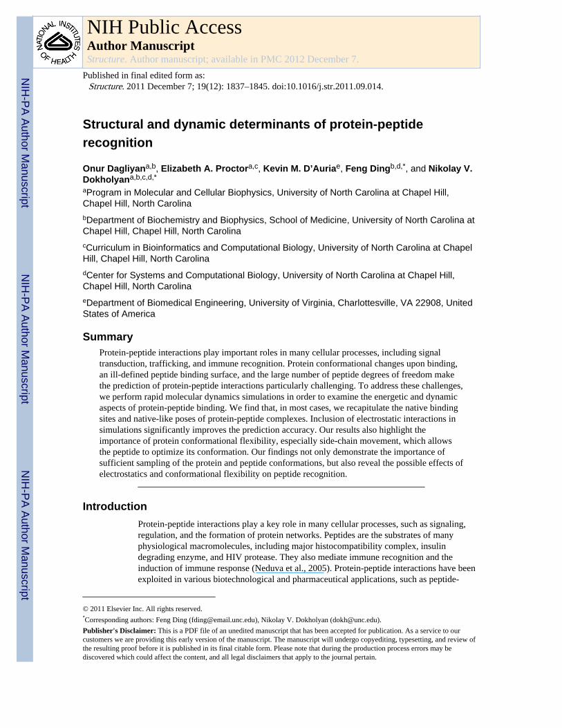
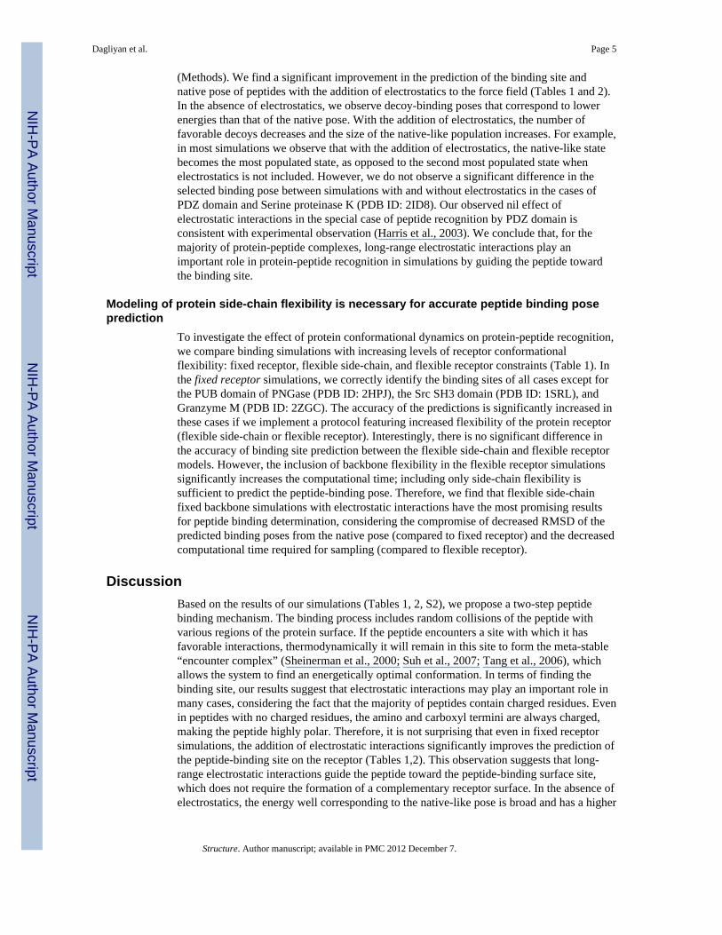
typical trajectory starting from the unbound state (gray dots in Fig. 1A), the peptiderandomly collides with the protein and forms transient complexes (scattered solid dots inFig. 1A). Once the native binding-site is sampled (~30 ns), the peptide forms a metastable“encounter-complex” (Sheinerman et al., 2000; Suh et al., 2007; Tang et al., 2006), whichallows further conformational rearrangement of the system in order to form the native-likebinding complex (~40 ns; RMSD ~ 2–3 Å). In order to identify the binding poses, we collectall bound states from each of the eight replicas (Figure 1B). Without knowledge of thenative binding pose, we select the putative binding ensemble of the peptide in the context ofthe energy landscape (Figure 1C). Here, we use MedusaScore (Yin et al., 2008) in order toevaluate the energy of binding between the peptide and the protein. MedusaScore is basedon inter-atomic interactions, including van der Waals, solvation, hydrogen bonding, andelectrostatic interactions. The PDZ domain-peptide complex features a well-defined funnel-like energy landscape; lower RMSD results in a more favorable binding energy. Notably, theminimum energy peptide pose in the complex has the minimum RMSD from the native pose(Figure 1C). Furthermore, we perform clustering analysis of the bound conformations. Weobserve that peptides are present in the native binding site if their RMSD from the nativepose is lower than 10 Å. Therefore, we use 10 Å as our clustering cutoff (it is 15 Å for 1JBEin which the peptide is 13-mer). The most highly populated clusters correspond to the lowfree energy states. For these highly populated clusters, we select the pose with the lowestMedusaScore as the representative structure, and we compare that structure with the crystalstructure. The representative structure of the most highly populated cluster of the PDZdomain-peptide complex has a RMSD of 2.5 Å from the crystal structure pose (Figure 1D).Thus, we obtain a native-like conformation of the PDZ domain-binding peptide without anyknowledge of the binding site, the conformation of the peptide, or the bound-state structureof the protein.
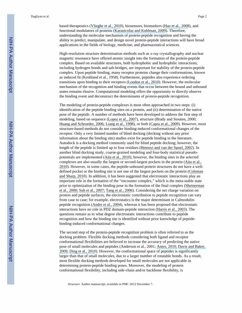
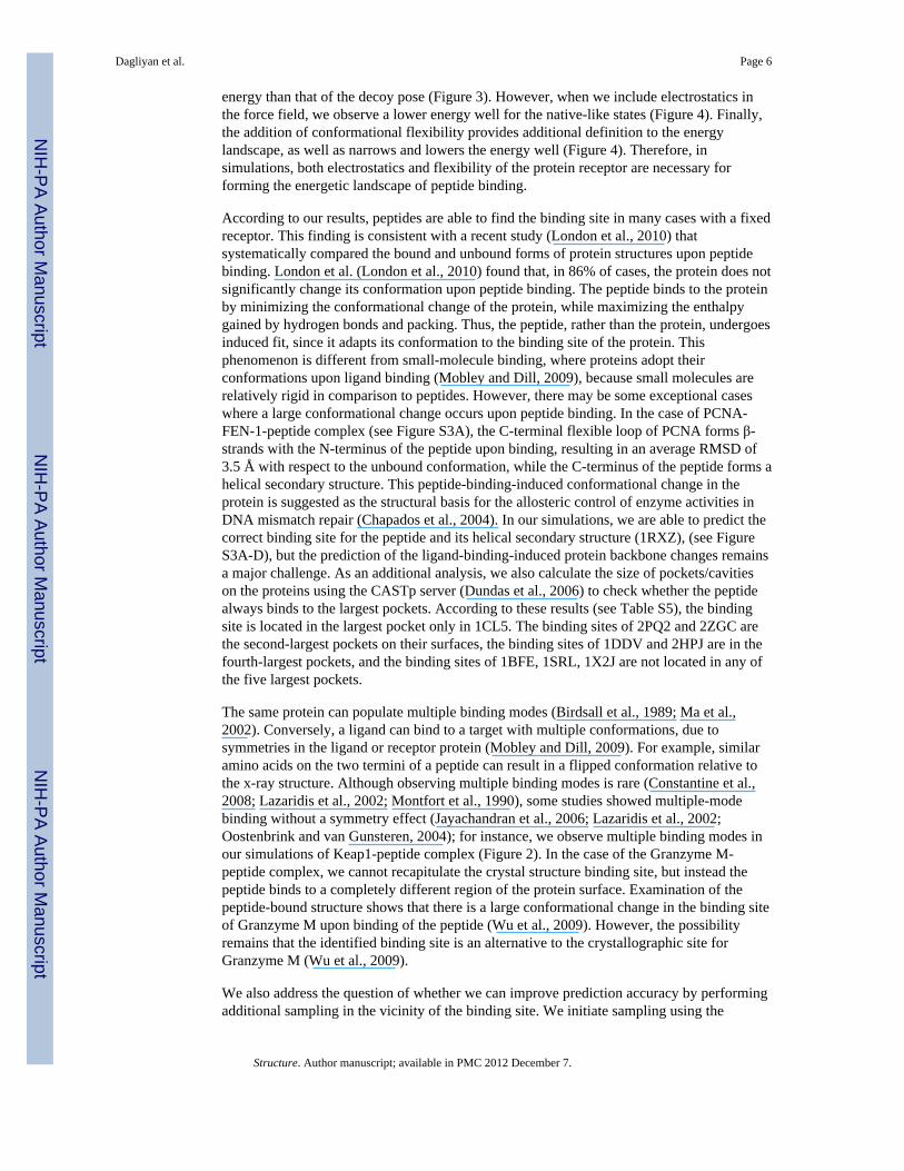
In molecular dynamics simulations, the most-populated cluster corresponds to the lowestfree energy state, which is not always the state with the lowest potential energy. In proteinswith more than one potential binding site, the energy landscapes demonstrate differenttrends from those of proteins with only one binding site (Figures 1, 2). If multiple bindingsites are identified during simulations, clustering analysis is necessary to determine thelowest free energy binding state. In the case of Keap1-peptide complex (PDB ID: 1X2J), theminimum energy pose from the most populated cluster is associated with the native-likeconformation, but it does not correspond to the global minimum energy, whereas the lowestenergy pose from the entire trajectory (from the second most-populated cluster) suggests adifferent binding site (Figure 2). Using our clustering analysis, we are able to obtain aconformation similar to the native pose of the peptide in the Keap1-peptide complex.
We perform similar analysis on six additional protein-peptide complexes. For each complex,we report the clustering results (see Table S2). Except the case 2ZGC, we identify the nativebinding site from within the first two most-populated clusters (Table 1, see Figure S2). Inaddition, we test two more cases of longer peptides that form secondary structure in thebound form (PDB IDs: 1RWZ, 1JBE). For these two cases, we can recapitulate the bindingsites of the protein and the helical structure of the peptides in the bound conformation (seeFigure S3). Our ability to identify the native binding site of the peptide from an arbitraryinitial position highlights the sampling efficiency of DMD simulations and the accuracy ofour all-atom force field.
Electrostatic interactions may be necessary for the identification of the native peptide-binding site
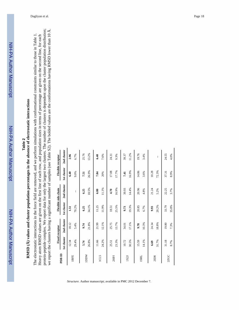
To test the effect of electrostatics on protein-peptide recognition, we also performsimulations without electrostatic interactions (Table 2). Here, we use the Debye-Hückelapproximation to model screened electrostatic interactions between charged residues
Dagliyan et al. Page 4
Structure. Author manuscript; available in PMC 2012 December 7.
NIH
-PA Author Manuscript
NIH
-PA Author Manuscript
NIH
-PA Author Manuscript
(Methods). We find a significant improvement in the prediction of the binding site andnative pose of peptides with the addition of electrostatics to the force field (Tables 1 and 2).In the absence of electrostatics, we observe decoy-binding poses that correspond to lowerenergies than that of the native pose. With the addition of electrostatics, the number offavorable decoys decreases and the size of the native-like population increases. For example,in most simulations we observe that with the addition of electrostatics, the native-like statebecomes the most populated state, as opposed to the second most populated state whenelectrostatics is not included. However, we do not observe a significant difference in theselected binding pose between simulations with and without electrostatics in the cases ofPDZ domain and Serine proteinase K (PDB ID: 2ID8). Our observed nil effect ofelectrostatic interactions in the special case of peptide recognition by PDZ domain isconsistent with experimental observation (Harris et al., 2003). We conclude that, for themajority of protein-peptide complexes, long-range electrostatic interactions play animportant role in protein-peptide recognition in simulations by guiding the peptide towardthe binding site.
Modeling of protein side-chain flexibility is necessary for accurate peptide binding poseprediction
To investigate the effect of protein conformational dynamics on protein-peptide recognition,we compare binding simulations with increasing levels of receptor conformationalflexibility: fixed receptor, flexible side-chain, and flexible receptor constraints (Table 1). Inthe fixed receptor simulations, we correctly identify the binding sites of all cases except forthe PUB domain of PNGase (PDB ID: 2HPJ), the Src SH3 domain (PDB ID: 1SRL), andGranzyme M (PDB ID: 2ZGC). The accuracy of the predictions is significantly increased inthese cases if we implement a protocol featuring increased flexibility of the protein receptor(flexible side-chain or flexible receptor). Interestingly, there is no significant difference inthe accuracy of binding site prediction between the flexible side-chain and flexible receptormodels. However, the inclusion of backbone flexibility in the flexible receptor simulationssignificantly increases the computational time; including only side-chain flexibility issufficient to predict the peptide-binding pose. Therefore, we find that flexible side-chainfixed backbone simulations with electrostatic interactions have the most promising resultsfor peptide binding determination, considering the compromise of decreased RMSD of thepredicted binding poses from the native pose (compared to fixed receptor) and the decreasedcomputational time required for sampling (compared to flexible receptor).
DiscussionBased on the results of our simulations (Tables 1, 2, S2), we propose a two-step peptidebinding mechanism. The binding process includes random collisions of the peptide withvarious regions of the protein surface. If the peptide encounters a site with which it hasfavorable interactions, thermodynamically it will remain in this site to form the meta-stable“encounter complex” (Sheinerman et al., 2000; Suh et al., 2007; Tang et al., 2006), whichallows the system to find an energetically optimal conformation. In terms of finding thebinding site, our results suggest that electrostatic interactions may play an important role inmany cases, considering the fact that the majority of peptides contain charged residues. Evenin peptides with no charged residues, the amino and carboxyl termini are always charged,making the peptide highly polar. Therefore, it is not surprising that even in fixed receptorsimulations, the addition of electrostatic interactions significantly improves the prediction ofthe peptide-binding site on the receptor (Tables 1,2). This observation suggests that long-range electrostatic interactions guide the peptide toward the peptide-binding surface site,which does not require the formation of a complementary receptor surface. In the absence ofelectrostatics, the energy well corresponding to the native-like pose is broad and has a higher
Dagliyan et al. Page 5
Structure. Author manuscript; available in PMC 2012 December 7.
NIH
-PA Author Manuscript
NIH
-PA Author Manuscript
NIH
-PA Author Manuscript
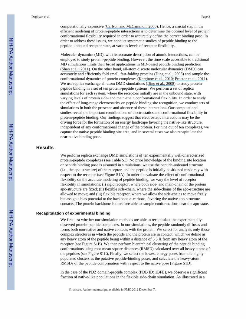
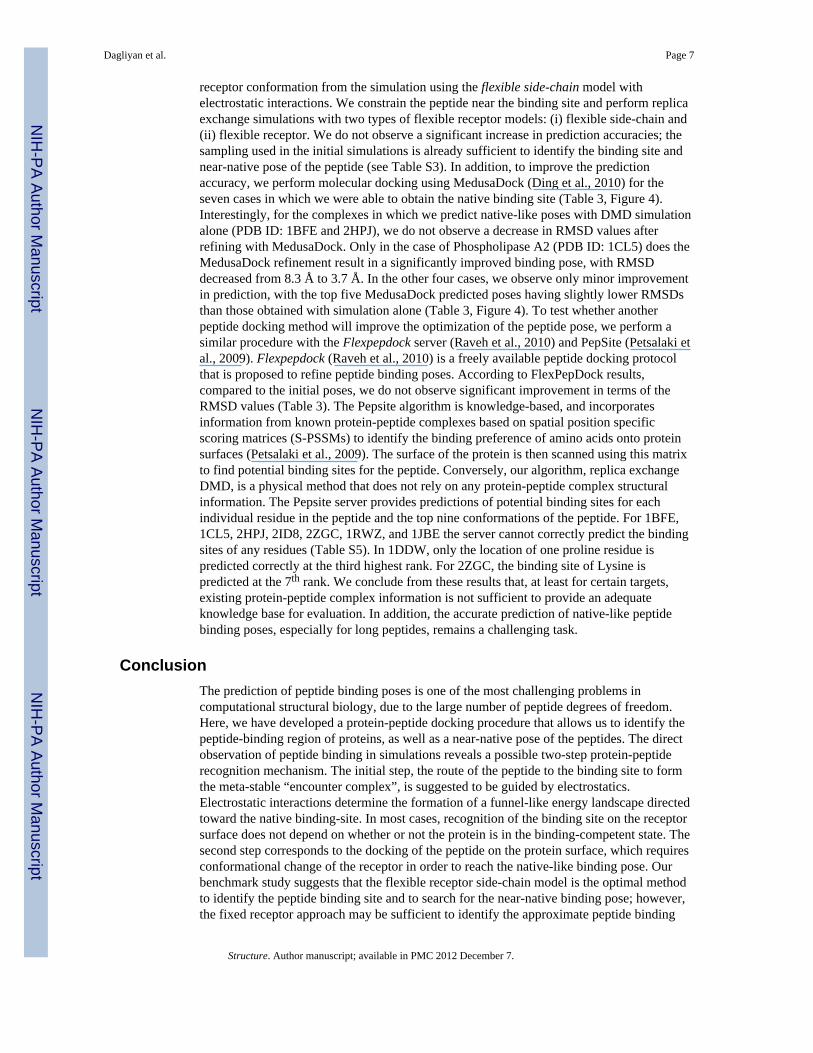
energy than that of the decoy pose (Figure 3). However, when we include electrostatics inthe force field, we observe a lower energy well for the native-like states (Figure 4). Finally,the addition of conformational flexibility provides additional definition to the energylandscape, as well as narrows and lowers the energy well (Figure 4). Therefore, insimulations, both electrostatics and flexibility of the protein receptor are necessary forforming the energetic landscape of peptide binding.
According to our results, peptides are able to find the binding site in many cases with a fixedreceptor. This finding is consistent with a recent study (London et al., 2010) thatsystematically compared the bound and unbound forms of protein structures upon peptidebinding. London et al. (London et al., 2010) found that, in 86% of cases, the protein does notsignificantly change its conformation upon peptide binding. The peptide binds to the proteinby minimizing the conformational change of the protein, while maximizing the enthalpygained by hydrogen bonds and packing. Thus, the peptide, rather than the protein, undergoesinduced fit, since it adapts its conformation to the binding site of the protein. Thisphenomenon is different from small-molecule binding, where proteins adopt theirconformations upon ligand binding (Mobley and Dill, 2009), because small molecules arerelatively rigid in comparison to peptides. However, there may be some exceptional caseswhere a large conformational change occurs upon peptide binding. In the case of PCNA-FEN-1-peptide complex (see Figure S3A), the C-terminal flexible loop of PCNA forms β-strands with the N-terminus of the peptide upon binding, resulting in an average RMSD of3.5 Å with respect to the unbound conformation, while the C-terminus of the peptide forms ahelical secondary structure. This peptide-binding-induced conformational change in theprotein is suggested as the structural basis for the allosteric control of enzyme activities inDNA mismatch repair (Chapados et al., 2004). In our simulations, we are able to predict thecorrect binding site for the peptide and its helical secondary structure (1RXZ), (see FigureS3A-D), but the prediction of the ligand-binding-induced protein backbone changes remainsa major challenge. As an additional analysis, we also calculate the size of pockets/cavitieson the proteins using the CASTp server (Dundas et al., 2006) to check whether the peptidealways binds to the largest pockets. According to these results (see Table S5), the bindingsite is located in the largest pocket only in 1CL5. The binding sites of 2PQ2 and 2ZGC arethe second-largest pockets on their surfaces, the binding sites of 1DDV and 2HPJ are in thefourth-largest pockets, and the binding sites of 1BFE, 1SRL, 1X2J are not located in any ofthe five largest pockets.
The same protein can populate multiple binding modes (Birdsall et al., 1989; Ma et al.,2002). Conversely, a ligand can bind to a target with multiple conformations, due tosymmetries in the ligand or receptor protein (Mobley and Dill, 2009). For example, similaramino acids on the two termini of a peptide can result in a flipped conformation relative tothe x-ray structure. Although observing multiple binding modes is rare (Constantine et al.,2008; Lazaridis et al., 2002; Montfort et al., 1990), some studies showed multiple-modebinding without a symmetry effect (Jayachandran et al., 2006; Lazaridis et al., 2002;Oostenbrink and van Gunsteren, 2004); for instance, we observe multiple binding modes inour simulations of Keap1-peptide complex (Figure 2). In the case of the Granzyme M-peptide complex, we cannot recapitulate the crystal structure binding site, but instead thepeptide binds to a completely different region of the protein surface. Examination of thepeptide-bound structure shows that there is a large conformational change in the binding siteof Granzyme M upon binding of the peptide (Wu et al., 2009). However, the possibilityremains that the identified binding site is an alternative to the crystallographic site forGranzyme M (Wu et al., 2009).
We also address the question of whether we can improve prediction accuracy by performingadditional sampling in the vicinity of the binding site. We initiate sampling using the
Dagliyan et al. Page 6
Structure. Author manuscript; available in PMC 2012 December 7.
NIH
-PA Author Manuscript
NIH
-PA Author Manuscript
NIH
-PA Author Manuscript
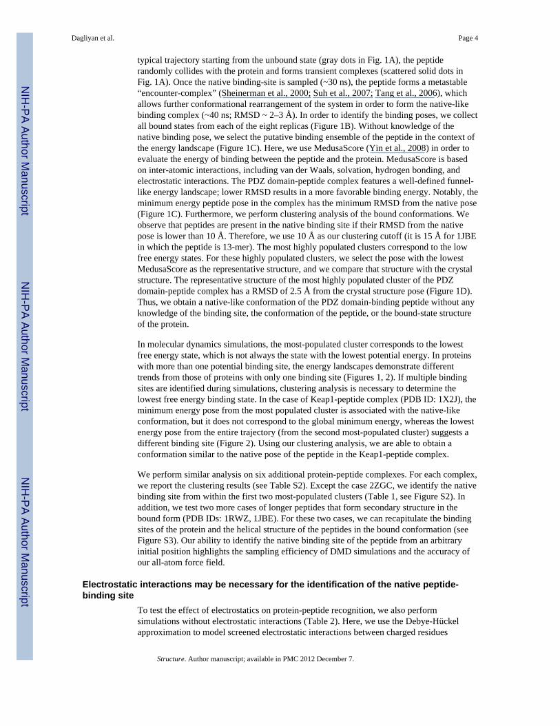
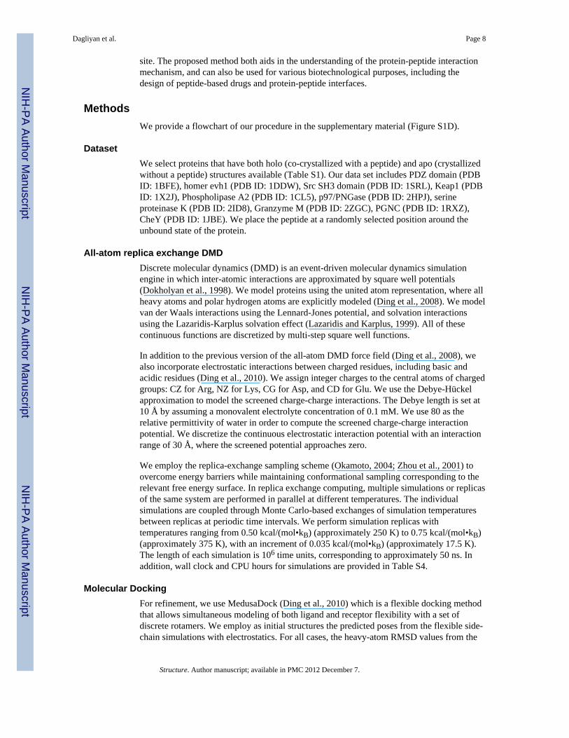
receptor conformation from the simulation using the flexible side-chain model withelectrostatic interactions. We constrain the peptide near the binding site and perform replicaexchange simulations with two types of flexible receptor models: (i) flexible side-chain and(ii) flexible receptor. We do not observe a significant increase in prediction accuracies; thesampling used in the initial simulations is already sufficient to identify the binding site andnear-native pose of the peptide (see Table S3). In addition, to improve the predictionaccuracy, we perform molecular docking using MedusaDock (Ding et al., 2010) for theseven cases in which we were able to obtain the native binding site (Table 3, Figure 4).Interestingly, for the complexes in which we predict native-like poses with DMD simulationalone (PDB ID: 1BFE and 2HPJ), we do not observe a decrease in RMSD values afterrefining with MedusaDock. Only in the case of Phospholipase A2 (PDB ID: 1CL5) does theMedusaDock refinement result in a significantly improved binding pose, with RMSDdecreased from 8.3 Å to 3.7 Å. In the other four cases, we observe only minor improvementin prediction, with the top five MedusaDock predicted poses having slightly lower RMSDsthan those obtained with simulation alone (Table 3, Figure 4). To test whether anotherpeptide docking method will improve the optimization of the peptide pose, we perform asimilar procedure with the Flexpepdock server (Raveh et al., 2010) and PepSite (Petsalaki etal., 2009). Flexpepdock (Raveh et al., 2010) is a freely available peptide docking protocolthat is proposed to refine peptide binding poses. According to FlexPepDock results,compared to the initial poses, we do not observe significant improvement in terms of theRMSD values (Table 3). The Pepsite algorithm is knowledge-based, and incorporatesinformation from known protein-peptide complexes based on spatial position specificscoring matrices (S-PSSMs) to identify the binding preference of amino acids onto proteinsurfaces (Petsalaki et al., 2009). The surface of the protein is then scanned using this matrixto find potential binding sites for the peptide. Conversely, our algorithm, replica exchangeDMD, is a physical method that does not rely on any protein-peptide complex structuralinformation. The Pepsite server provides predictions of potential binding sites for eachindividual residue in the peptide and the top nine conformations of the peptide. For 1BFE,1CL5, 2HPJ, 2ID8, 2ZGC, 1RWZ, and 1JBE the server cannot correctly predict the bindingsites of any residues (Table S5). In 1DDW, only the location of one proline residue ispredicted correctly at the third highest rank. For 2ZGC, the binding site of Lysine ispredicted at the 7th rank. We conclude from these results that, at least for certain targets,existing protein-peptide complex information is not sufficient to provide an adequateknowledge base for evaluation. In addition, the accurate prediction of native-like peptidebinding poses, especially for long peptides, remains a challenging task.
ConclusionThe prediction of peptide binding poses is one of the most challenging problems incomputational structural biology, due to the large number of peptide degrees of freedom.Here, we have developed a protein-peptide docking procedure that allows us to identify thepeptide-binding region of proteins, as well as a near-native pose of the peptides. The directobservation of peptide binding in simulations reveals a possible two-step protein-peptiderecognition mechanism. The initial step, the route of the peptide to the binding site to formthe meta-stable “encounter complex”, is suggested to be guided by electrostatics.Electrostatic interactions determine the formation of a funnel-like energy landscape directedtoward the native binding-site. In most cases, recognition of the binding site on the receptorsurface does not depend on whether or not the protein is in the binding-competent state. Thesecond step corresponds to the docking of the peptide on the protein surface, which requiresconformational change of the receptor in order to reach the native-like binding pose. Ourbenchmark study suggests that the flexible receptor side-chain model is the optimal methodto identify the peptide binding site and to search for the near-native binding pose; however,the fixed receptor approach may be sufficient to identify the approximate peptide binding
Dagliyan et al. Page 7
Structure. Author manuscript; available in PMC 2012 December 7.
NIH
-PA Author Manuscript
NIH
-PA Author Manuscript
NIH
-PA Author Manuscript
site. The proposed method both aids in the understanding of the protein-peptide interactionmechanism, and can also be used for various biotechnological purposes, including thedesign of peptide-based drugs and protein-peptide interfaces.
MethodsWe provide a flowchart of our procedure in the supplementary material (Figure S1D).
DatasetWe select proteins that have both holo (co-crystallized with a peptide) and apo (crystallizedwithout a peptide) structures available (Table S1). Our data set includes PDZ domain (PDBID: 1BFE), homer evh1 (PDB ID: 1DDW), Src SH3 domain (PDB ID: 1SRL), Keap1 (PDBID: 1X2J), Phospholipase A2 (PDB ID: 1CL5), p97/PNGase (PDB ID: 2HPJ), serineproteinase K (PDB ID: 2ID8), Granzyme M (PDB ID: 2ZGC), PGNC (PDB ID: 1RXZ),CheY (PDB ID: 1JBE). We place the peptide at a randomly selected position around theunbound state of the protein.
All-atom replica exchange DMDDiscrete molecular dynamics (DMD) is an event-driven molecular dynamics simulationengine in which inter-atomic interactions are approximated by square well potentials(Dokholyan et al., 1998). We model proteins using the united atom representation, where allheavy atoms and polar hydrogen atoms are explicitly modeled (Ding et al., 2008). We modelvan der Waals interactions using the Lennard-Jones potential, and solvation interactionsusing the Lazaridis-Karplus solvation effect (Lazaridis and Karplus, 1999). All of thesecontinuous functions are discretized by multi-step square well functions.
In addition to the previous version of the all-atom DMD force field (Ding et al., 2008), wealso incorporate electrostatic interactions between charged residues, including basic andacidic residues (Ding et al., 2010). We assign integer charges to the central atoms of chargedgroups: CZ for Arg, NZ for Lys, CG for Asp, and CD for Glu. We use the Debye-Hückelapproximation to model the screened charge-charge interactions. The Debye length is set at10 Å by assuming a monovalent electrolyte concentration of 0.1 mM. We use 80 as therelative permittivity of water in order to compute the screened charge-charge interactionpotential. We discretize the continuous electrostatic interaction potential with an interactionrange of 30 Å, where the screened potential approaches zero.
We employ the replica-exchange sampling scheme (Okamoto, 2004; Zhou et al., 2001) toovercome energy barriers while maintaining conformational sampling corresponding to therelevant free energy surface. In replica exchange computing, multiple simulations or replicasof the same system are performed in parallel at different temperatures. The individualsimulations are coupled through Monte Carlo-based exchanges of simulation temperaturesbetween replicas at periodic time intervals. We perform simulation replicas withtemperatures ranging from 0.50 kcal/(mol•kB) (approximately 250 K) to 0.75 kcal/(mol•kB)(approximately 375 K), with an increment of 0.035 kcal/(mol•kB) (approximately 17.5 K).The length of each simulation is 106 time units, corresponding to approximately 50 ns. Inaddition, wall clock and CPU hours for simulations are provided in Table S4.
Molecular DockingFor refinement, we use MedusaDock (Ding et al., 2010) which is a flexible docking methodthat allows simultaneous modeling of both ligand and receptor flexibility with a set ofdiscrete rotamers. We employ as initial structures the predicted poses from the flexible side-chain simulations with electrostatics. For all cases, the heavy-atom RMSD values from the
Dagliyan et al. Page 8
Structure. Author manuscript; available in PMC 2012 December 7.
NIH
-PA Author Manuscript
NIH
-PA Author Manuscript
NIH
-PA Author Manuscript
experimentally-determine conformation decrease significantly, approaching the native-likepose (Table 3, Figure 3).
Supplementary MaterialRefer to Web version on PubMed Central for supplementary material.
AcknowledgmentsWe thank Rachel L. Redler and Irem Dagliyan for critical reading of the manuscript. This work is supported by theNational Institute of Health Grant R01GM080742 and the ARRA supplement 3R01GM080742-03S1 (to N.V.D.),National Institute of Health Predoctoral Fellowship F31AG039266-01 from the National Institute on Aging (toE.A.P.), and by the UNC Research Council (to F.D.). Calculations were performed on the high-performancecomputing cluster at the University of North Carolina at Chapel Hill.
ReferencesAita T, Nishigaki K, Husimi Y. Toward the fast blind docking of a peptide to a target protein by using
a four-body statistical pseudo-potential. Comput Biol Chem. 2010; 34:53–62. [PubMed: 19939735]Anderson AC, O’Neil RH, Surti TS, Stroud RM. Approaches to solving the rigid receptor problem by
identifying a minimal set of flexible residues during ligand docking. Chem Biol. 2001; 8:445–457.[PubMed: 11358692]
Andre I, Kesvatera T, Jonsson B, Akerfeldt KS, Linse S. The role of electrostatic interactions incalmodulin-peptide complex formation. Biophys J. 2004; 87:1929–1938. [PubMed: 15345569]
Antes I. DynaDock: A new molecular dynamics-based algorithm for protein-peptide docking includingreceptor flexibility. Proteins. 2010; 78:1084–1104. [PubMed: 20017216]
Birdsall B, Feeney J, Tendler SJ, Hammond SJ, Roberts GC. Dihydrofolate reductase: multipleconformations and alternative modes of substrate binding. Biochemistry. 1989; 28:2297–2305.[PubMed: 2524214]
Brady GP Jr, Stouten PF. Fast prediction and visualization of protein binding pockets with PASS. JComput Aided Mol Des. 2000; 14:383–401. [PubMed: 10815774]
Capra JA, Laskowski RA, Thornton JM, Singh M, Funkhouser TA. Predicting protein ligand bindingsites by combining evolutionary sequence conservation and 3D structure. PLoS Comput Biol. 2009;5:e1000585. [PubMed: 19997483]
Carlson HA, McCammon JA. Accommodating protein flexibility in computational drug design. MolPharmacol. 2000; 57:213–218. [PubMed: 10648630]
Chapados BR, Hosfield DJ, Han S, Qiu J, Yelent B, Shen B, Tainer JA. Structural basis for FEN-1substrate specificity and PCNA-mediated activation in DNA replication and repair. Cell. 2004;116:39–50. [PubMed: 14718165]
Coleman RG, Sharp KA. Protein pockets: inventory, shape, and comparison. J Chem Inf Model. 2010;50:589–603. [PubMed: 20205445]
Constantine KL, Mueller L, Metzler WJ, McDonnell PA, Todderud G, Goldfarb V, Fan Y, Newitt JA,Kiefer SE, Gao M, et al. Multiple and single binding modes of fragment-like kinase inhibitorsrevealed by molecular modeling, residue type-selective protonation, and nuclear overhausereffects. J Med Chem. 2008; 51:6225–6229. [PubMed: 18771253]
Davis IW, Baker D. RosettaLigand docking with full ligand and receptor flexibility. J Mol Biol. 2009;385:381–392. [PubMed: 19041878]
Ding F, Tsao D, Nie H, Dokholyan NV. Ab initio folding of proteins with all-atom discrete moleculardynamics. Structure. 2008; 16:1010–1018. [PubMed: 18611374]
Ding F, Yin S, Dokholyan NV. Rapid flexible docking using a stochastic rotamer library of ligands. JChem Inf Model. 2010; 50:1623–1632. [PubMed: 20712341]
Dokholyan NV, Buldyrev SV, Stanley HE, Shakhnovich EI. Discrete molecular dynamics studies ofthe folding of a protein-like model. Fold Des. 1998; 3:577–587. [PubMed: 9889167]
Dagliyan et al. Page 9
Structure. Author manuscript; available in PMC 2012 December 7.
NIH
-PA Author Manuscript
NIH
-PA Author Manuscript
NIH
-PA Author Manuscript
Dundas J, Ouyang Z, Tseng J, Binkowski A, Turpaz Y, Liang J. CASTp: computed atlas of surfacetopography of proteins with structural and topographical mapping of functionally annotatedresidues. Nucleic Acids Res. 2006; 34:W116–118. [PubMed: 16844972]
Hao J, Serohijos AW, Newton G, Tassone G, Wang Z, Sgroi DC, Dokholyan NV, Basilion JP.Identification and rational redesign of peptide ligands to CRIP1, a novel biomarker for cancers.PLoS Comput Biol. 2008; 4:e1000138. [PubMed: 18670594]
Harris BZ, Lau FW, Fujii N, Guy RK, Lim WA. Role of electrostatic interactions in PDZ domainligand recognition. Biochemistry. 2003; 42:2797–2805. [PubMed: 12627945]
Hetenyi C, van der Spoel D. Efficient docking of peptides to proteins without prior knowledge of thebinding site. Protein Sci. 2002; 11:1729–1737. [PubMed: 12070326]
Huang B, Schroeder M. LIGSITEcsc: predicting ligand binding sites using the Connolly surface anddegree of conservation. BMC Struct Biol. 2006; 6:19. [PubMed: 16995956]
Jayachandran G, Shirts MR, Park S, Pande VS. Parallelized-over-parts computation of absolutebinding free energy with docking and molecular dynamics. J Chem Phys. 2006; 125:084901.[PubMed: 16965051]
Karanicolas J, Kuhlman B. Computational design of affinity and specificity at protein-proteininterfaces. Curr Opin Struct Biol. 2009; 19:458–463. [PubMed: 19646858]
Karginov AV, Ding F, Kota P, Dokholyan NV, Hahn KM. Engineered allosteric activation of kinasesin living cells. Nat Biotechnol. 2010; 28:743–747. [PubMed: 20581846]
Koshland DE Jr, Ray WJ Jr, Erwin MJ. Protein structure and enzyme action. Fed Proc. 1958; 17:1145–1150. [PubMed: 13619786]
Lazaridis T, Karplus M. Effective energy function for proteins in solution. Proteins. 1999; 35:133–152.[PubMed: 10223287]
Lazaridis T, Masunov A, Gandolfo F. Contributions to the binding free energy of ligands to avidin andstreptavidin. Proteins. 2002; 47:194–208. [PubMed: 11933066]
Liang J, Edelsbrunner H, Woodward C. Anatomy of protein pockets and cavities: measurement ofbinding site geometry and implications for ligand design. Protein Sci. 1998; 7:1884–1897.[PubMed: 9761470]
London N, Movshovitz-Attias D, Schueler-Furman O. The structural basis of peptide-protein bindingstrategies. Structure. 2010; 18:188–199. [PubMed: 20159464]
Lopez G, Valencia A, Tress ML. firestar--prediction of functionally important residues using structuraltemplates and alignment reliability. Nucleic Acids Res. 2007; 35:W573–577. [PubMed:17584799]
Ma B, Shatsky M, Wolfson HJ, Nussinov R. Multiple diverse ligands binding at a single protein site: amatter of pre-existing populations. Protein Sci. 2002; 11:184–197. [PubMed: 11790828]
Mobley DL, Dill KA. Binding of small-molecule ligands to proteins: “what you see” is not always“what you get”. Structure. 2009; 17:489–498. [PubMed: 19368882]
Montfort WR, Perry KM, Fauman EB, Finer-Moore JS, Maley GF, Hardy L, Maley F, Stroud RM.Structure, multiple site binding, and segmental accommodation in thymidylate synthase on bindingdUMP and an anti-folate. Biochemistry. 1990; 29:6964–6977. [PubMed: 2223754]
Neduva V, Linding R, Su-Angrand I, Stark A, de Masi F, Gibson TJ, Lewis J, Serrano L, Russell RB.Systematic discovery of new recognition peptides mediating protein interaction networks. PLoSBiol. 2005; 3:e405. [PubMed: 16279839]
Okamoto Y. Generalized-ensemble algorithms: enhanced sampling techniques for Monte Carlo andmolecular dynamics simulations. J Mol Graph Model. 2004; 22:425–439. [PubMed: 15099838]
Oostenbrink C, van Gunsteren WF. Free energies of binding of polychlorinated biphenyls to theestrogen receptor from a single simulation. Proteins. 2004; 54:237–246. [PubMed: 14696186]
Petsalaki E, Stark A, Garcia-Urdiales E, Russell RB. Accurate prediction of peptide binding sites onprotein surfaces. PLoS Comput Biol. 2009; 5:e1000335. [PubMed: 19325869]
Proctor EA, Ding F, Dokholyan NV. Structural and Thermodynamic Effects of Post-translationalModifications in Mutant and Wild Type Cu, Zn Superoxide Dismutase. J Mol Biol. 2011;408:555–567. [PubMed: 21396374]
Dagliyan et al. Page 10
Structure. Author manuscript; available in PMC 2012 December 7.
NIH
-PA Author Manuscript
NIH
-PA Author Manuscript
NIH
-PA Author Manuscript
Raveh B, London N, Schueler-Furman O. Sub-angstrom modeling of complexes between flexiblepeptides and globular proteins. Proteins. 2010; 78:2029–2040. [PubMed: 20455260]
Shan Y, Kim ET, Eastwood MP, Dror RO, Seeliger MA, Shaw DE. How Does a Drug Molecule FindIts Target Binding Site? J Am Chem Soc. 2011
Sheinerman FB, Norel R, Honig B. Electrostatic aspects of protein-protein interactions. Curr OpinStruct Biol. 2000; 10:153–159. [PubMed: 10753808]
Suh JY, Tang C, Clore GM. Role of electrostatic interactions in transient encounter complexes inprotein-protein association investigated by paramagnetic relaxation enhancement. J Am ChemSoc. 2007; 129:12954–12955. [PubMed: 17918946]
Tang C, Iwahara J, Clore GM. Visualization of transient encounter complexes in protein-proteinassociation. Nature. 2006; 444:383–386. [PubMed: 17051159]
Vlieghe P, Lisowski V, Martinez J, Khrestchatisky M. Synthetic therapeutic peptides: science andmarket. Drug Discov Today. 2010; 15:40–56. [PubMed: 19879957]
Wu L, Wang L, Hua G, Liu K, Yang X, Zhai Y, Bartlam M, Sun F, Fan Z. Structural basis forproteolytic specificity of the human apoptosis-inducing granzyme M. J Immunol. 2009; 183:421–429. [PubMed: 19542453]
Yin S, Biedermannova L, Vondrasek J, Dokholyan NV. MedusaScore: an accurate force field-basedscoring function for virtual drug screening. J Chem Inf Model. 2008; 48:1656–1662. [PubMed:18672869]
Zhou R, Berne BJ, Germain R. The free energy landscape for beta hairpin folding in explicit water.Proc Natl Acad Sci U S A. 2001; 98:14931–14936. [PubMed: 11752441]
Dagliyan et al. Page 11
Structure. Author manuscript; available in PMC 2012 December 7.
NIH
-PA Author Manuscript
NIH
-PA Author Manuscript
NIH
-PA Author Manuscript

Highlights
• Direct observation of protein-peptide binding in molecular dynamicssimulations
• Electrostatic interactions guide protein-peptide recognition
• Direct observation of induced-fit phenomenon in peptides
• Novel method for protein-peptide docking
Dagliyan et al. Page 12
Structure. Author manuscript; available in PMC 2012 December 7.
NIH
-PA Author Manuscript
NIH
-PA Author Manuscript
NIH
-PA Author Manuscript
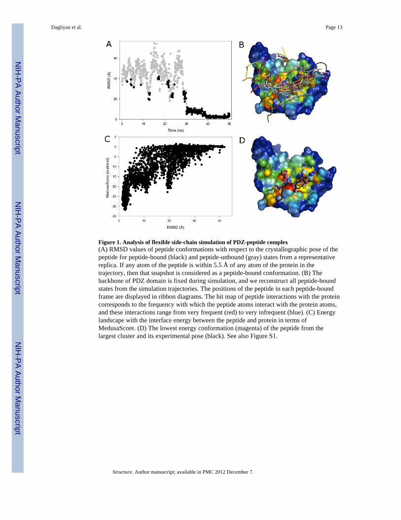
Figure 1. Analysis of flexible side-chain simulation of PDZ-peptide complex(A) RMSD values of peptide conformations with respect to the crystallographic pose of thepeptide for peptide-bound (black) and peptide-unbound (gray) states from a representativereplica. If any atom of the peptide is within 5.5 Å of any atom of the protein in thetrajectory, then that snapshot is considered as a peptide-bound conformation. (B) Thebackbone of PDZ domain is fixed during simulation, and we reconstruct all peptide-boundstates from the simulation trajectories. The positions of the peptide in each peptide-boundframe are displayed in ribbon diagrams. The hit map of peptide interactions with the proteincorresponds to the frequency with which the peptide atoms interact with the protein atoms,and these interactions range from very frequent (red) to very infrequent (blue). (C) Energylandscape with the interface energy between the peptide and protein in terms ofMedusaScore. (D) The lowest energy conformation (magenta) of the peptide from thelargest cluster and its experimental pose (black). See also Figure S1.
Dagliyan et al. Page 13
Structure. Author manuscript; available in PMC 2012 December 7.
NIH
-PA Author Manuscript
NIH
-PA Author Manuscript
NIH
-PA Author Manuscript
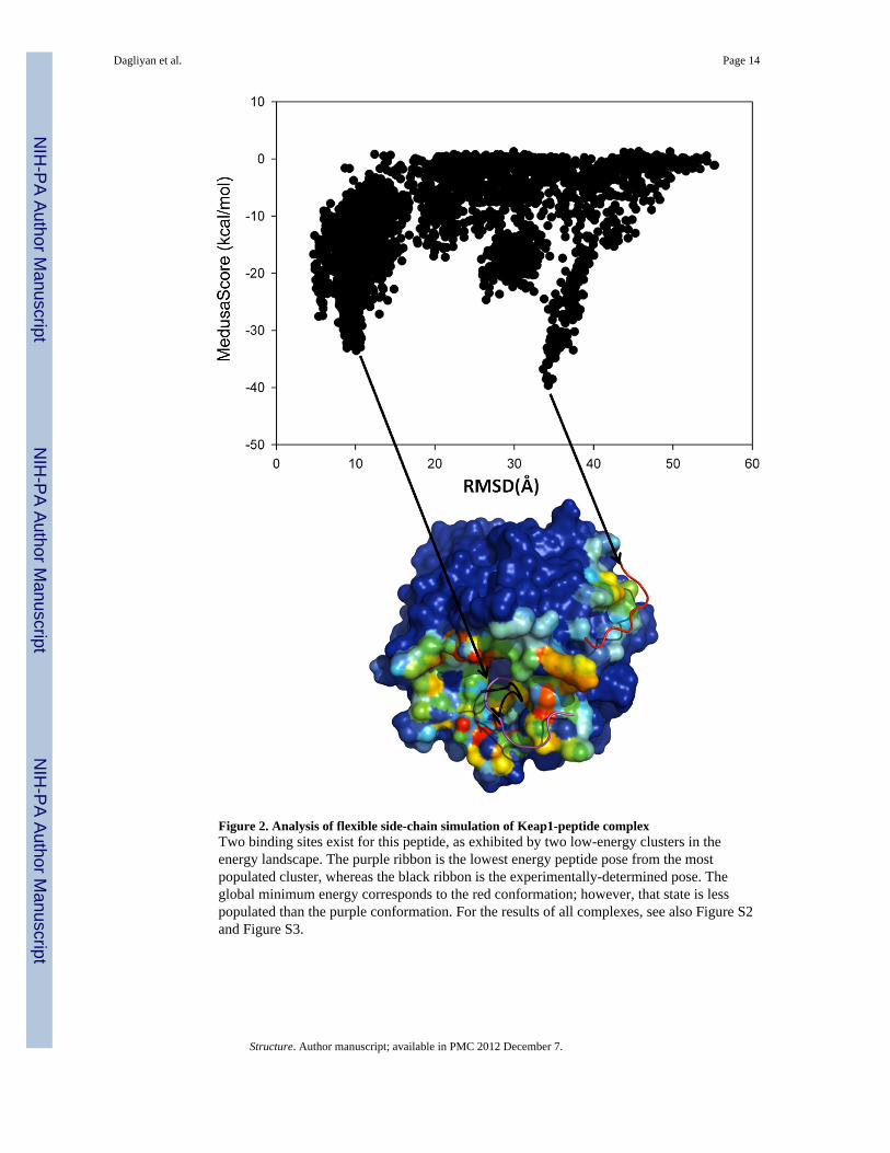
Figure 2. Analysis of flexible side-chain simulation of Keap1-peptide complexTwo binding sites exist for this peptide, as exhibited by two low-energy clusters in theenergy landscape. The purple ribbon is the lowest energy peptide pose from the mostpopulated cluster, whereas the black ribbon is the experimentally-determined pose. Theglobal minimum energy corresponds to the red conformation; however, that state is lesspopulated than the purple conformation. For the results of all complexes, see also Figure S2and Figure S3.
Dagliyan et al. Page 14
Structure. Author manuscript; available in PMC 2012 December 7.
NIH
-PA Author Manuscript
NIH
-PA Author Manuscript
NIH
-PA Author Manuscript
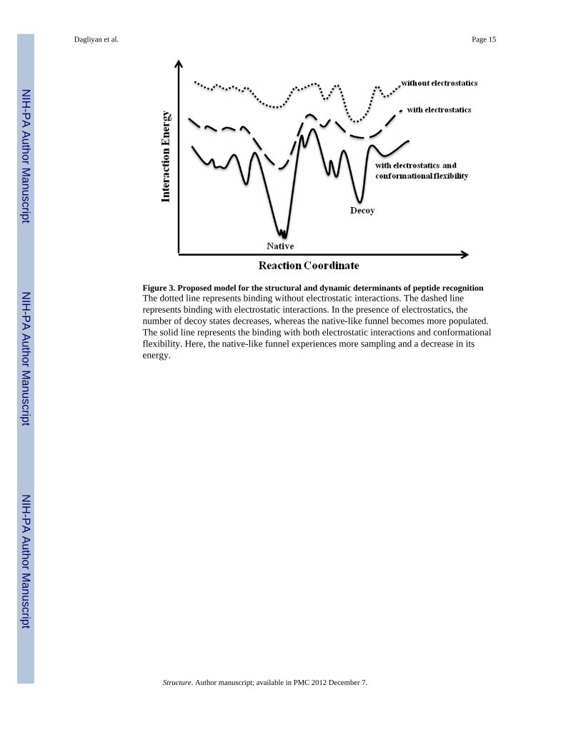
Figure 3. Proposed model for the structural and dynamic determinants of peptide recognitionThe dotted line represents binding without electrostatic interactions. The dashed linerepresents binding with electrostatic interactions. In the presence of electrostatics, thenumber of decoy states decreases, whereas the native-like funnel becomes more populated.The solid line represents the binding with both electrostatic interactions and conformationalflexibility. Here, the native-like funnel experiences more sampling and a decrease in itsenergy.
Dagliyan et al. Page 15
Structure. Author manuscript; available in PMC 2012 December 7.
NIH
-PA Author Manuscript
NIH
-PA Author Manuscript
NIH
-PA Author Manuscript
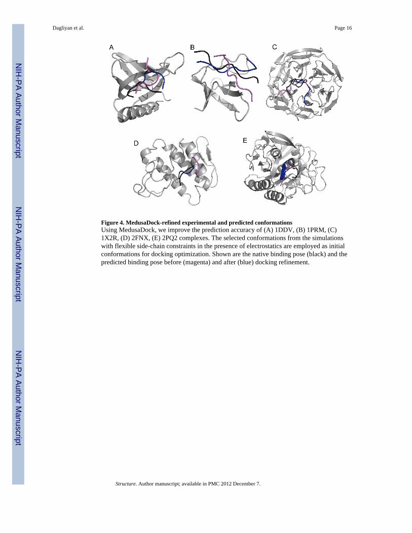
Figure 4. MedusaDock-refined experimental and predicted conformationsUsing MedusaDock, we improve the prediction accuracy of (A) 1DDV, (B) 1PRM, (C)1X2R, (D) 2FNX, (E) 2PQ2 complexes. The selected conformations from the simulationswith flexible side-chain constraints in the presence of electrostatics are employed as initialconformations for docking optimization. Shown are the native binding pose (black) and thepredicted binding pose before (magenta) and after (blue) docking refinement.
Dagliyan et al. Page 16
Structure. Author manuscript; available in PMC 2012 December 7.
NIH
-PA Author Manuscript
NIH
-PA Author Manuscript
NIH
-PA Author Manuscript
NIH
-PA Author Manuscript
NIH
-PA Author Manuscript
NIH
-PA Author Manuscript
Dagliyan et al. Page 17
Tabl
e 1
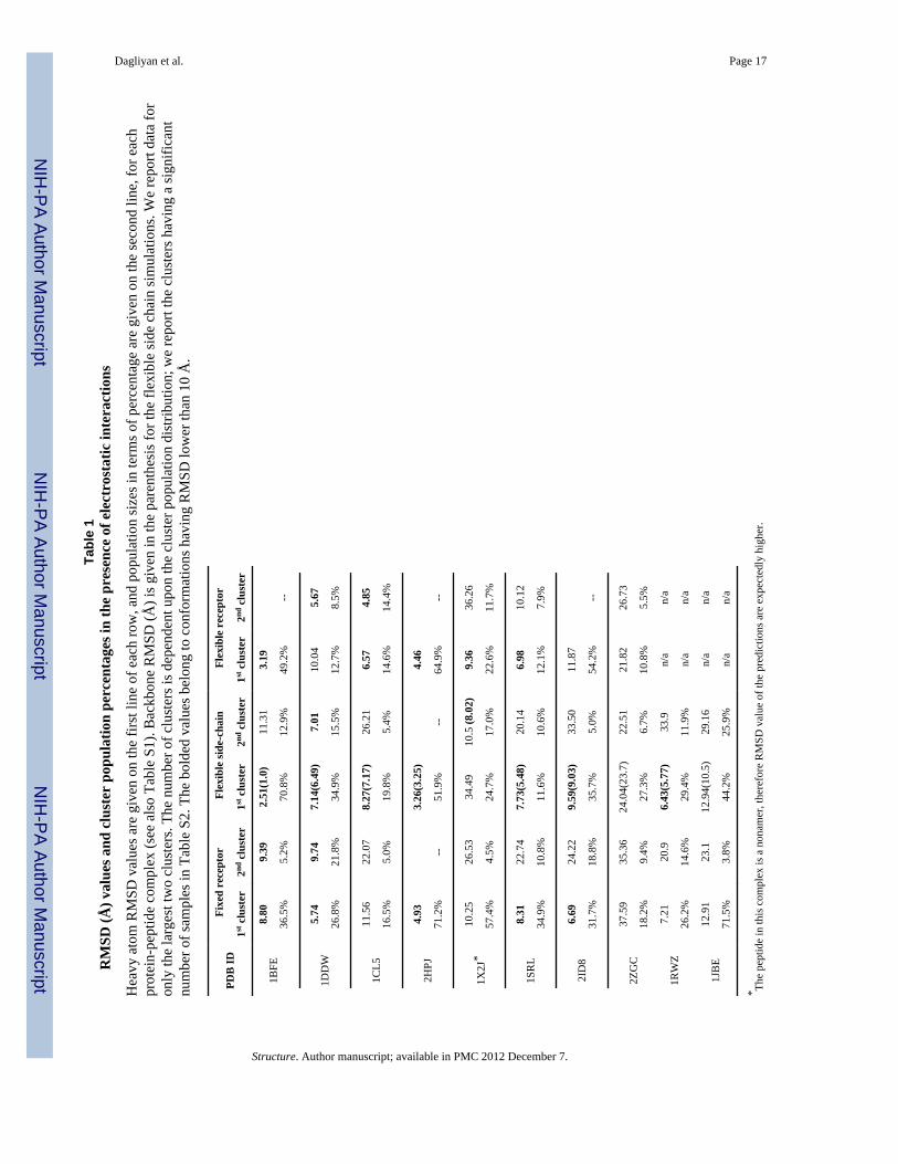
RM
SD (Å
) val
ues a
nd c
lust
er p
opul
atio
n pe
rcen
tage
s in
the
pres
ence
of e
lect
rost
atic
inte
ract
ions
Hea
vy a
tom
RM
SD v
alue
s are
giv
en o
n th
e fir
st li
ne o
f eac
h ro
w, a
nd p
opul
atio
n si
zes i
n te
rms o
f per
cent
age
are
give
n on
the
seco
nd li
ne, f
or e
ach
prot
ein-
pept
ide
com
plex
(see
als
o Ta
ble
S1).
Bac
kbon
e R
MSD
(Å) i
s giv
en in
the
pare
nthe
sis f
or th
e fle
xibl
e si
de c
hain
sim
ulat
ions
. We
repo
rt da
ta fo
ron
ly th
e la
rges
t tw
o cl
uste
rs. T
he n
umbe
r of c
lust
ers i
s dep
ende
nt u
pon
the
clus
ter p
opul
atio
n di
strib
utio
n; w
e re
port
the
clus
ters
hav
ing
a si
gnifi
cant
num
ber o
f sam
ples
in T
able
S2.
The
bol
ded
valu
es b
elon
g to
con
form
atio
ns h
avin
g R
MSD
low
er th
an 1
0 Å
.
PDB
IDFi
xed
rece
ptor
Flex
ible
side
-cha
inFl
exib
le r
ecep
tor
1st c
lust
er2nd
clu
ster
1st c
lust
er2nd
clu
ster
1st c
lust
er2nd
clu
ster
1BFE
8.80
9.39
2.51
(1.0
)11
.31
3.19
36.5
%5.
2%70
.8%
12.9
%49
.2%
--
1DD
W5.
749.
747.
14(6
.49)
7.01
10.0
45.
67
26.8
%21
.8%
34.9
%15
.5%
12.7
%8.
5%
1CL5
11.5
622
.07
8.27
(7.1
7)26
.21
6.57
4.85
16.5
%5.
0%19
.8%
5.4%
14.6
%14
.4%
2HPJ
4.93
3.26
(3.2
5)4.
46
71.2
%--
51.9
%--
64.9
%--
1X2J
*10
.25
26.5
334
.49
10.5
(8.0
2)9.
3636
.26
57.4
%4.
5%24
.7%
17.0
%22
.6%
11.7
%
1SR
L8.
3122
.74
7.73
(5.4
8)20
.14
6.98
10.1
2
34.9
%10
.8%
11.6
%10
.6%
12.1
%7.
9%
2ID
86.
6924
.22
9.59
(9.0
3)33
.50
11.8
7
31.7
%18
.8%
35.7
%5.
0%54
.2%
--
2ZG
C37
.59
35.3
624
.04(
23.7
)22
.51
21.8
226
.73
18.2
%9.
4%27
.3%
6.7%
10.8
%5.
5%
1RW
Z7.
2120
.96.
43(5
.77)
33.9
n/a
n/a
26.2
%14
.6%
29.4
%11
.9%
n/a
n/a
1JB
E12
.91
23.1
12.9
4(10
.5)
29.1
6n/
an/
a
71.5
%3.
8%44
.2%
25.9
%n/
an/
a
* The
pept
ide
in th
is c
ompl
ex is
a n
onam
er, t
here
fore
RM
SD v
alue
of t
he p
redi
ctio
ns a
re e
xpec
tedl
y hi
gher
.
Structure. Author manuscript; available in PMC 2012 December 7.
NIH
-PA Author Manuscript
NIH
-PA Author Manuscript
NIH
-PA Author Manuscript
Dagliyan et al. Page 18
Tabl
e 2
RM
SD (Å
) val
ues a
nd c
lust
er p
opul
atio
n pe
rcen
tage
s in
the
abse
nce
of e
lect
rost
atic
inte
ract
ions
The
elec
trost
atic
inte
ract
ions
in th
e fo
rce
field
are
rem
oved
, and
we
perf
orm
sim
ulat
ions
with
con
form
atio
nal c
onst
rain
ts si
mila
r to
thos
e in
Tab
le 1
.H
eavy
ato
m R
MSD
val
ues a
re g
iven
on
the
first
line
of e
ach
row
, and
pop
ulat
ion
size
s in
term
s of p
erce
ntag
e ar
e gi
ven
on th
e se
cond
line
, for
eac
hpr
otei
n-pe
ptid
e co
mpl
ex. W
e re
port
data
for o
nly
the
larg
est t
wo
clus
ters
. The
num
ber o
f clu
ster
s is d
epen
dent
upo
n th
e cl
uste
r pop
ulat
ion
dist
ribut
ion;
we
repo
rt th
e cl
uste
rs h
avin
g a
sign
ifica
nt n
umbe
r of s
ampl
es (s
ee T
able
S2)
. The
bol
ded
valu
es a
re th
e co
nfor
mat
ions
hav
ing
RM
SD lo
wer
than
10
Å.
PDB
IDFi
xed
rece
ptor
Flex
ible
side
-cha
inFl
exib
le r
ecep
tor
1st c
lust
er2n
d cl
uste
r1s
t clu
ster
2nd
clus
ter
1st c
lust
er2n
d cl
uste
r
1BFE
12.3
418
.12
1.51
6.40
2.96
29.4
%5.
4%70
.2%
--9.
0%6.
7%
1DD
W5.
749.
746.
256.
257.
5125
.76
26.8
%21
.8%
34.1
%18
.5%
26.9
%13
.2%
1CL5
11.6
621
.99
11.2
56.
807.
846.
40
24.2
%12
.1%
11.9
%11
.1%
20%
7.6%
2HPJ
25.5
125
.75
18.5
14.
7017
.08
24.3
1
23.3
%13
.7%
25.5
%16
.9%
17.7
%9.
3%
1X2J
10.7
234
.01
9.73
39.9
37.
4139
.37
30.5
%17
.1%
19.3
%14
.4%
16.9
%11
.2%
1SR
L15
.59
9.78
20.8
520
.96
14.8
619
.76
14.1
%10
.3%
6.7%
4.8%
3.6%
3.4%
2ID
86.
6924
.34
9.93
21.2
410
.28
31.7
%18
.8%
20.2
%5.
2%72
.3%
--
2ZG
C31
.18
19.8
033
.79
22.2
537
.31
24.5
5
8.7%
7.3%
35.8
%3.
7%8.
9%4.
0%
Structure. Author manuscript; available in PMC 2012 December 7.
NIH
-PA Author Manuscript
NIH
-PA Author Manuscript
NIH
-PA Author Manuscript
Dagliyan et al. Page 19
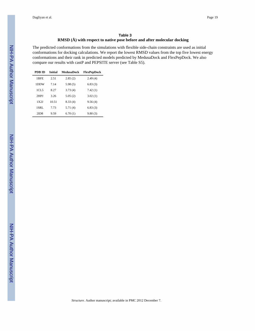
Table 3RMSD (Å) with respect to native pose before and after molecular docking
The predicted conformations from the simulations with flexible side-chain constraints are used as initialconformations for docking calculations. We report the lowest RMSD values from the top five lowest energyconformations and their rank in predicted models predicted by MedusaDock and FlexPepDock. We alsocompare our results with castP and PEPSITE server (see Table S5).
PDB ID Initial MedusaDock FlexPepDock
1BFE 2.51 2.85 (2) 2.49 (4)
1DDW 7.14 5.98 (5) 6.83 (3)
1CL5 8.27 3.73 (4) 7.42 (1)
2HPJ 3.26 5.05 (2) 3.02 (1)
1X2J 10.51 8.33 (4) 9.56 (4)
1SRL 7.73 5.71 (4) 6.83 (3)
2ID8 9.59 6.70 (1) 9.80 (3)
Structure. Author manuscript; available in PMC 2012 December 7.
Related Documents