Carbohydrate Polymers 87 (2012) 1460–1466 Contents lists available at SciVerse ScienceDirect Carbohydrate Polymers j ourna l ho me pag e: www.elsevier.com/locate/carbpol Structural and conformational differences of acylated hyaluronan modified in protic and aprotic solvent system Daniela ˇ Smejkalová a,∗ , Martina Hermannová a , Romana ˇ Suláková a , Alena Pr ˚ uˇ sová b , Jiˇ rí Kuˇ cerík b , Vladimír Velebn ´ y a a Contipro C, Dolní Dobrouˇ c 401, 561 02 Dolní Dobrouˇ c, Czech Republic b Brno University of Technology, Faculty of Chemistry, Purky ˇ nova 118, 612 00 Brno, Czech Republic a r t i c l e i n f o Article history: Received 7 July 2011 Received in revised form 2 September 2011 Accepted 12 September 2011 Available online 29 September 2011 Keywords: Hyaluronan Acylation NMR DSC UV–vis Mass spectrometry a b s t r a c t Acylated hyaluronan (HA) in aqueous (DMSO/H 2 O) and nonaqueous (DMSO) solutions was studied by means of nuclear magnetic resonance, differential scanning calorimetry (DSC), mass spectrometry and UV/vis spectroscopy. It has been demonstrated that structural and conformational properties of the acy- lated hyaluronan derivates are strongly dependent on the nature of reaction solvent. Acylation in DMSO was more selective than that carried out in DMSO/H 2 O, though in both cases in average a maximum of one acyl chain was detected per HA dimer. The hydrophobic functionalization of hyaluronan induced its interaction with hydrophobic dye as a consequence of acyl chain aggregation. The higher the degree of acylation the more hydrophobic dye was interacting with HA. For concentrated samples, aggregation was more evident in case of acylated HA in aqueous solution. This phenomenon was explained by its different conformational arrangement in solution which was further supported by DSC data indicating an existence of hydrophobic cavities. The formation of self-aggregated assemblies indicates potential applications of this type of HA derivate as drug delivery system. © 2011 Elsevier Ltd. All rights reserved. 1. Introduction Carbohydrate fatty acid esters are an important class of biodegradable and non-toxic surfactants with broad applications in food, cosmetics and pharmaceutical industries as detergents, oral care products and medical supplies (Hill & LeHen-Ferrenbach, 2008). They were also reported to be applicable as antibiotics and antitumorals (Deleu & Paquot, 2004). In addition, non-toxic and biodegradable polysaccharide surfactants are considered to be attractive drug delivery systems. Among polysaccharides, a great attention is focused on esterification of hyaluronan (HA) (Kawaguchi, Matsukawa, & Ishigami, 1993; Kong, Chen, & Park, 2011; Taglienti, Valentini, Sequi, & Crescenzi, 2005). HA is a linear polysaccharide consisting of alternating -1,4-linked units of -1,3-linked glucuronic acid and N-acetyl-d- glucosamine (Laurent, 1998; Scott, 1998). HA is a main component of the extracellular matrix in connective, epithelial, and neural tis- sues and is known to play an important role in organ development, cell proliferation and migration. Additionally, HA contributes to the lubrication and maintenance of cartilage, where it is a major com- ponent of synovial fluid and forms a coating around chondrocytes (Collis et al., 1998; Entwistle, Hall, & Turley, 1996; Laurent, 1998). ∗ Corresponding author. Tel.: +420 465519569; fax: +420 465543793. E-mail address: [email protected] (D. ˇ Smejkalová). Except for being biodegradable and non-toxic, HA is biocompatible and renewable, which is important on industrial scale production of HA derivates. The major advantage of modified HA over the native HA is the higher resistance against enzymatic degradation (Abatangelo, Barbucci, Brun, & Lamponi, 1997; Prestwitch, Marecak, Marecek, Vercuysse, & Ziebell, 1998; ˇ Soltés et al., 2006). In addition, besides retaining its inherently superior properties, HA derivates acquire additional physicochemical characteristics that can be tailored according to the desired requirements. For example, HA hav- ing desired amount of hydrophobic functional groups may be achieved varying the degree of substitution. In case of esteri- fication, the degree of substitution and the length of attached carbon chain are directly related to conformational behavior of the substituted molecule in solution and the possibility of forming supramolecular assemblies (Akiyoshi & Sunamoto, 1996). Forma- tion of supramolecular assemblies is than in turn related to the possibility of carbohydrate interaction with non-polar compounds and therefore directly affects its pharmaceutical and industrial applications. Modified HA is therefore also considered to have a great potential as a novel drug carrier in form of conjugates. Despite its excellent biocompatible and biodegradable properties, HA based drug delivery systems have been reported to work as an efficient depot for sustained release of protein drugs without denaturation (Oh et al., 2010; Prestwitch & Vercruysse, 1998). Moreover, the absence of positive charge on HA surface alleviate the problems 0144-8617/$ – see front matter © 2011 Elsevier Ltd. All rights reserved. doi:10.1016/j.carbpol.2011.09.057

Welcome message from author
This document is posted to help you gain knowledge. Please leave a comment to let me know what you think about it! Share it to your friends and learn new things together.
Transcript
Sp
DJa
b
a
ARRAA
KHANDUM
1
bio2aabg(2
�gosclp(
0d
Carbohydrate Polymers 87 (2012) 1460– 1466
Contents lists available at SciVerse ScienceDirect
Carbohydrate Polymers
j ourna l ho me pag e: www.elsev ier .com/ locate /carbpol
tructural and conformational differences of acylated hyaluronan modified inrotic and aprotic solvent system
aniela Smejkalováa,∗, Martina Hermannováa, Romana Sulákováa, Alena Prusováb,irí Kuceríkb, Vladimír Velebnya
Contipro C, Dolní Dobrouc 401, 561 02 Dolní Dobrouc, Czech RepublicBrno University of Technology, Faculty of Chemistry, Purkynova 118, 612 00 Brno, Czech Republic
r t i c l e i n f o
rticle history:eceived 7 July 2011eceived in revised form 2 September 2011ccepted 12 September 2011vailable online 29 September 2011
eywords:
a b s t r a c t
Acylated hyaluronan (HA) in aqueous (DMSO/H2O) and nonaqueous (DMSO) solutions was studied bymeans of nuclear magnetic resonance, differential scanning calorimetry (DSC), mass spectrometry andUV/vis spectroscopy. It has been demonstrated that structural and conformational properties of the acy-lated hyaluronan derivates are strongly dependent on the nature of reaction solvent. Acylation in DMSOwas more selective than that carried out in DMSO/H2O, though in both cases in average a maximum ofone acyl chain was detected per HA dimer. The hydrophobic functionalization of hyaluronan induced
yaluronancylationMRSCV–vis
its interaction with hydrophobic dye as a consequence of acyl chain aggregation. The higher the degreeof acylation the more hydrophobic dye was interacting with HA. For concentrated samples, aggregationwas more evident in case of acylated HA in aqueous solution. This phenomenon was explained by itsdifferent conformational arrangement in solution which was further supported by DSC data indicatingan existence of hydrophobic cavities. The formation of self-aggregated assemblies indicates potential
f HA
ass spectrometry applications of this type o. Introduction
Carbohydrate fatty acid esters are an important class ofiodegradable and non-toxic surfactants with broad applications
n food, cosmetics and pharmaceutical industries as detergents,ral care products and medical supplies (Hill & LeHen-Ferrenbach,008). They were also reported to be applicable as antibioticsnd antitumorals (Deleu & Paquot, 2004). In addition, non-toxicnd biodegradable polysaccharide surfactants are considered toe attractive drug delivery systems. Among polysaccharides, areat attention is focused on esterification of hyaluronan (HA)Kawaguchi, Matsukawa, & Ishigami, 1993; Kong, Chen, & Park,011; Taglienti, Valentini, Sequi, & Crescenzi, 2005).
HA is a linear polysaccharide consisting of alternating-1,4-linked units of �-1,3-linked glucuronic acid and N-acetyl-d-lucosamine (Laurent, 1998; Scott, 1998). HA is a main componentf the extracellular matrix in connective, epithelial, and neural tis-ues and is known to play an important role in organ development,ell proliferation and migration. Additionally, HA contributes to the
ubrication and maintenance of cartilage, where it is a major com-onent of synovial fluid and forms a coating around chondrocytesCollis et al., 1998; Entwistle, Hall, & Turley, 1996; Laurent, 1998).∗ Corresponding author. Tel.: +420 465519569; fax: +420 465543793.E-mail address: [email protected] (D. Smejkalová).
144-8617/$ – see front matter © 2011 Elsevier Ltd. All rights reserved.oi:10.1016/j.carbpol.2011.09.057
derivate as drug delivery system.© 2011 Elsevier Ltd. All rights reserved.
Except for being biodegradable and non-toxic, HA is biocompatibleand renewable, which is important on industrial scale productionof HA derivates.
The major advantage of modified HA over the native HA isthe higher resistance against enzymatic degradation (Abatangelo,Barbucci, Brun, & Lamponi, 1997; Prestwitch, Marecak, Marecek,Vercuysse, & Ziebell, 1998; Soltés et al., 2006). In addition, besidesretaining its inherently superior properties, HA derivates acquireadditional physicochemical characteristics that can be tailoredaccording to the desired requirements. For example, HA hav-ing desired amount of hydrophobic functional groups may beachieved varying the degree of substitution. In case of esteri-fication, the degree of substitution and the length of attachedcarbon chain are directly related to conformational behavior ofthe substituted molecule in solution and the possibility of formingsupramolecular assemblies (Akiyoshi & Sunamoto, 1996). Forma-tion of supramolecular assemblies is than in turn related to thepossibility of carbohydrate interaction with non-polar compoundsand therefore directly affects its pharmaceutical and industrialapplications. Modified HA is therefore also considered to have agreat potential as a novel drug carrier in form of conjugates. Despiteits excellent biocompatible and biodegradable properties, HA based
drug delivery systems have been reported to work as an efficientdepot for sustained release of protein drugs without denaturation(Oh et al., 2010; Prestwitch & Vercruysse, 1998). Moreover, theabsence of positive charge on HA surface alleviate the problems
rate P
wta
cDtobwa
2
2
pd(Cp
2s
d(krrf
ciwoossda8
2
aadttfptwtos
2
H2
D. Smejkalová et al. / Carbohyd
ith severe cytotoxicity and aggregations with serum proteins inhe body found for cationic liposomes and polymers investigateds drug carriers (Oh et al., 2010).
In this study, we followed the structural and conformationalhanges of HA induced after acylation with hexanoic anhydride inMSO and DMSO/H2O solvent. The main attention was focused on
he comparison of reaction selectivity and conformational changesf HA followed after acylation. The structural changes were studiedy NMR, ESI-MS/MS and DSC. Formation of hydrophobic domainsas examined by comparing the ability of acyl derivates to dissolve
hydrophobic dye.
. Experimental
.1. Materials
Hyaluronic acid sodium salt (200 kDa, 155 kDa and 34 kDa) wasrovided by CPN Dolní Dobrouc, Czech Republic. Hexanoic anhy-rides, triethylamine, dimethylsulfoxide, dimethylaminopyridineDMAP), Oil Red O (Solvent Red 27, Sudan Red 5B, C.I. 26125,26H24N4O), and deuterated water were of analytical grade andurchased from Sigma–Aldrich.
.2. Preparation of hyaluronan acid form and hyaluronan sodiumalt
Hyaluronan (Mw = 200 kDa, 15 g) was dissolved in 600 mL ofemineralized water and then Amberlite IR 120 Na exchange resinwet state, 100 g) was added to the mixture. The mixture wasept at room temperature with occasional stirring. Cation exchangeesin was removed by centrifugation at 5000 rpm for 5 min and theesulting solution was lyophilized. About 13 g of hyaluronan acidorm Mw = 50 kDa was obtained.
Since each transformation of hyaluronan into its acid formauses HA degradation, it was necessary to have a comparable start-ng Mw of both HA sodium salt and HA acid form as both materials
ere used as substrates for acylation reaction. For this reason, thebtained hyaluronan acid form was divided into two parts. One partf the material was used for acylation reaction in its acid form. Theecond half of hyaluronan acid form was returned into its initialodium salt state in a following way. Hyaluronan acid form wasiluted in water, neutralized to pH 6.5 and precipitated off withbsolute 2-propanol. The precipitate was washed three times with0% 2-propanol, twice with absolute 2-propanol and dried at 40 ◦C.
.3. Acylation of hyaluronan sodium salt (Ac-HA-Na)
HA sodium salt (5 g) was first dissolved in 50 mL of deminer-lized water and then diluted with 50 mL of DMSO. Hexanoicnhydride (2.5 equiv./HA dimer), triethylamine (2.5 equiv./HAimer) and DMAP (0.05 equiv./HA dimer) were added into the mix-ure and the mixture was stirred at room temperature for 2 h. Athe end of reaction, the mixture was diluted with 100 mL of waterollowed by the addition of 15 mL of saturated NaCl solution. Theroduct Ac-HA-Na (acylated HA-Na+ in its Na+ form) was precipi-ated with another 200 mL of absolute 2-propanol. The precipitateas first washed three times with 80% 2-propanol in water and
hen with absolute 2-propanol. The solid was filtered and dried inven at 40 ◦C. The yield of final product was 5.4 g. The degree ofubstitution (DS) calculated from NMR spectra was 70%.
.4. Acylation of hyaluronan acid form (Ac-HA-H)
Hyaluronan acid form (5 g) was dissolved in 100 mL of DMSO.exanoic anhydride (1.5–3.0 equiv./HA dimer), triethylamine (1.5,.5 and 3.0 equiv./HA dimer) and DMAP (0.05 equiv./HA dimer)
olymers 87 (2012) 1460– 1466 1461
were added into the mixture and the mixture was stirred at roomtemperature for 2 h. At the end of reaction, the reaction wasquenched with 100 mL of water and the pH was adjusted with 0.1 MNaOH to pH 6, followed by the addition of 15 mL of saturated NaClsolution. The product Ac-HA-H (acylated HA-H+ in its Na+ form)was precipitated with 200 mL of absolute 2-propanol. The precip-itate was washed three times with 80% 2-propanol in water andthen absolute 2-propanol. The solid was filtered and dried in ovenat 40 ◦C. The yield of final product was between 4.5 and 4.8 g. Thedegree of substitution (DS) calculated from NMR spectra was 33%,60% and 70% for 1.5, 2.5 and 3.0 equiv. of triethylamine/HA dimer,respectively.
2.5. NMR analyses
HA acyl derivates (10 mg) were solubilized in 750 �L of D2O,transferred into NMR tubes and directly analyzed.
The NMR analyses were performed on Bruker AvanceTM
500 MHz equipped with BBFO plus probe. The 1H and 13C chemi-cal shift were referenced to 3-trimethylsilylpropanoic acid sodiumsalt (TSPA) used as an internal standard. 1H–1H TOCSY spectra wererecorded with 2048 data points, 80 scans per increment and 128increments. TOCSY mixing time was set at 80 ms. 1H–13C HSQCspectra were acquired using gradient pulse sequences and 2048data points, 80 scans per increment, 256 increments, and heteronu-clear scalar coupling C–H set at 145 Hz. DOSY (diffusion orderedspectra) were obtained using a stimulated echo pulse sequencewith bipolar gradients (STEBPGP). Scans (32) were collected using2.5 ms sine-shaped pulses (5 ms bipolar pulse pair) ranging from0.674 to 32.030 G cm−1 in 24 increments with a diffusion time of200–600 ms, and 8192 time domain data points. Apodization wasmade by multiplying the data with a line broadening of 1.0 Hz, spikesuppression factor of 4.0, maximum interactions number set to 100,noise sensitivity factor of 2, and number of components set to 1.
1H NMR spectra were used for the calculation of the degree ofsubstitution (DS) of acylated HA. DS (in %) was determined as rela-tive integral of signal at 2.4 ppm, when the integration of signal at2.0 ppm was normalized to 150. Explanation of resonating signalsis given in the text.
2.6. MS analyses
Powdered hyaluronan (100 mg) was first dissolved in 10 mLof 0.1 M sodium acetate with 0.15 M NaCl (pH 5.3, adjustedwith glacial acetic acid), and then incubated with 2000 IU ofhyaluronidase (Finepharm) at 37 ◦C for 2 days. The enzyme wasremoved by short boiling of the solution at the end of incuba-tion. The sample was filtered through 0.2 �m Nylon syringe filter.Filtered solution (2 mL) was transferred into the Vivaspin 15R con-centrator (2000 MWCO Hydrosart, Sartorius) and centrifuged at9000 rpm for 15 min. After preconcentration of the sample, the con-centrator was filled with 10 mL of deionized water and centrifugedat 9000 rpm for 30 min. 4 wash cycles were used to remove theinitial salt content. The sample was recovered from the bottomof the concentrator, diluted with 0.1% HCOOH:methanol = 1:1 toa final concentration of 1 mg mL−1 and directly injected into massspectrometer.
Mass spectroscopic analyses of digested and desalted derivateswere performed using a Synapt HDMS mass spectrometer (Waters),equipped with an electrospray ionization source operating in neg-ative ion mode. The effluent was introduced into an electrospraysource with a syringe pump at a flow rate of 10 �L min−1. Nitrogen
was used as cone gas (100 L h−1) and desolvation gas (800 L h−1).Capillary voltage was set at 3 kV. Sampling cone was set at 100 V.Extraction cone was set at 5 V. The source block temperature wasset at 100 ◦C, while the desolvation temperature was 250 ◦C. For
1 rate P
eaaife
2
adOs5rsw
2
omat(pmtpe
tR
e3−s
othflassV
mcd
2s
ci(EOn0a
462 D. Smejkalová et al. / Carbohyd
ach sample full MS and MS/MS scans from m/z 50 to 2000 werecquired for 2 min. For MS/MS measurements, argon was used as
collision gas. The collision energy was optimized to fragment theon of interest, typically 55 eV for the ions with higher m/z and 25 eVor the ions with lower m/z. Data were collected at 1 scan s−1 andlaborated using MassLynx software.
.7. UV–vis analyses
Powdered HA (10–200 mg) HA 34 kDa, HA 155 kDa, Ac-HA-Hnd Ac-HA-Na was first soaked with 750 �L of H2O and then leftissolving overnight under constant stirring. Then 200 �L of Oil Red
solution (20 mg mL−1 in hexane) was added to the dissolved HAamples, the mixtures were heated up to 50 ◦C and shaken for 2 h at0 ◦C, and for 2 days at room temperature. The experiments wereepeated in two independent series, each consisting of replicateamples. Absorbances (522 nm) of the water phase were measuredith UV-Vis Carry 100 (Varian).
.8. Thermal analyses
HA samples of approximately 2 mg (weighted with an accuracyf ±0.01 mg) were placed in aluminum sample pans (TA Instru-ents, Tzero® Technology) and the excess of water (milli-Q) was
dded. Surplus of water was allowed to slowly evaporate at roomemperature until the desired water content (Wc = mass of waterg)/mass of dry sample (g); [Wc] = g g−1) was obtained. Several sam-les having Wc between 0.1 and 3 g g−1 were prepared for each HAaterial. The pans were subsequently hermetically sealed and left
o equilibrate at room temperature for 72 h. Similar way of sam-les preparation was used for freezing/thawing as well as for thevaporation experiments.
Differential scanning calorimetry (DSC) was carried out usinghe TA Instruments DSC Q-200 equipped with a cooling accessoryCS-90 and assessed by the TA-Universal Analysis 2000 software.
The following thermal protocol was used for freezing/thawingxperiments: start at 40.0 ◦C; cooling from 40.0 to −70.0 ◦C at.0 ◦C min−1; isothermal at −70.0 ◦C for 1.0 min; heating from70.0 to 40 ◦C at 5.0 ◦C min−1. Flow rate of dynamic nitrogen atmo-
phere was 50 mL min−1.The following thermal protocol was used for the measurement
f evaporation enthalpy: equilibration at 27.0 ◦C; cooling from 27.0o −50.0 ◦C at 10.0 ◦C min−1; isothermal at −50.0 ◦C for 1.0 min;eating from −50.0 to 200.0 ◦C at 5.0 ◦C min−1 and switching theow rate of nitrogen from 50 mL min−1 to 5 mL min−1. Immedi-tely before the measurement, the hermetic lid (necessary for theample preparation) was perforated using a sharp tool and the mea-urement was carried out straightway (Prusová, Smejkalová, Chytil,elebny, & Kucerík, 2010).
To obtain precise water content, thermogravimetry (TA Instru-ents, Q500 IR) was used to determine the equilibrium moisture
ontent as a mass loss in the temperature interval 25–220 ◦C underynamic atmosphere of nitrogen 25 mL min−1.
.9. Size exclusion chromatography coupled to multi-angle lightcattering (SEC-MALS)
SEC was performed using an Agilent 1100 series liquidhromatograph equipped with a degasser (Model G1379A), ansocratic HPLC pump (Model G1310A), an automatic injectorModel G1313A), a column thermostat (Model G1316A), a DAWNOS multi-angle light scattering photometer followed by an
ptilab rEX differential refractometer (both from Wyatt Tech-ology Corporation, USA). The injection volume was 100 �L of.1–1.0% (w/v) solutions. The separation was carried out using PLquagel-OH 40 (300 mm × 7.5 mm; 8 �m) and PL aquagel-OH 20olymers 87 (2012) 1460– 1466
(300 mm × 7.5 mm; 5 �m) columns connected in series. Columnswere thermostated at 40 ◦C. The mobile phase was 0.1 M sodiumphosphate buffer (pH adjusted to 7.5) + 0.05% NaN3 at a flow rate0.8 mL min−1. Data acquisition and molecular weights calculationswere performed using the ASTRA V software (Wyatt TechnologyCorporation, USA). The specific refractive index increment dn/dcwas determined at 690 nm using the Optilab rEX refractome-ter for all samples according to procedure described elsewhere(Podzimek, Hermannová, Bílerová, Bezáková, & Velebny, 2010). Themean value of 9 dn/dc measurements was 0.155 ± 0.003 mL g−1.
Each sample was filtered through Acrodisc Syringe Filter0.45 �m 25 mm diameter with the Supor membrane (Pall). Allreagents for SEC were HPLC grade and the mobile phase was filteredthrough Nylaflo Nylon Membrane Filter 0.2 �m (Pall).
3. Results and discussion
3.1. Acylation of hyaluronan
One of the main problems related to chemical modification ofhyaluronan is its insolubility in organic solvents. For this reason,hyaluronan is mostly transformed prior modification into its acidform which is soluble in polar organic solvents such as DMSO(Oudshoorn, Rissmann, Bouwstra, & Hennink, 2007). However, themajor disadvantage of this procedure is the contemporary degra-dation of HA during cation exchange step. For example, in this worka starting HA material was having Mw = 200 kDa, while after trans-formation into its acid form the Mw was reduced to about 50 kDa.For this reason, we tried to overcome this disadvantage by a directacylation of hyaluronan as sodium salt in DMSO/H2O solution. Theacylation reactions are shown in Fig. 1. Regardless of the startingmaterial and solvent choice, sodium salt of acylated HA was formedin both cases (Fig. 1). However, since the choice of reaction solventmay affect substitution position on HA chain, modified HA prod-ucts received after acylation in DMSO (Ac-HA-H) and DMSO/H2O(Ac-HA-Na) were further analyzed and compared by NMR, LC–MS,UV–vis and thermal analysis.
3.2. NMR analyses
1H NMR spectra of HA, Ac-HA-H and Ac-HA-Na are shown inFig. 2. All of the spectra show typical proton chemical shifts of HAinvolving signal at 2.0 ppm belonging to COCH3 group, skeletal sig-nals at 3.4–3.9 and anomeric resonances at 4.4–4.6 ppm. Remainingsignals detected in modified HA at 0.8, 1.2, 1.5 and 2.4 ppm wereattributed to the CH2 in acyl chain as shown in Fig. 2. Relativeintegration of signals at 2.0 and 2.4 ppm were used for the determi-nation of degree of substitution (DS). A comparable DS = 70% wasdetermined for both acylated products shown in Fig. 2. A downfieldchemical shift of one of the HA skeletal signals is evident in Ac-HA-Na at 3.1 ppm. Less significant is the appearance of a new signal at3.3 ppm in Ac-HA-H. The new signals detected after HA modifica-tion are different for Ac-HA-Na and Ac-HA-H, and thus suggest thatacylation reaction in DMSO yielded structurally different reactionoutcome as compared to DMSO/H2O reaction.
The linkage between hexyl chain and HA was established in bothderivates by DOSY experiment (data not shown). Because of themarked difference between the diffusion coefficients of hexanoicacid and HA, the DOSY map can easily establish the presence of non-attached hexanoic acid to HA, which obviously is much faster thanthe diffusion of the bound acyl chain. In both cases, DOSY exper-
iments showed similar diffusion behavior for all signals between0.8 and 4.6 ppm (except for isopropanol and HDO signal) and thusindicated that all of the proton resonances in this region belongedto one structural complex.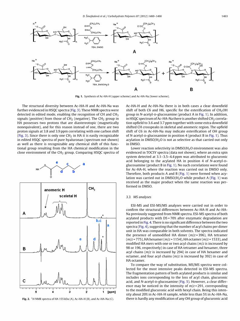
D. Smejkalová et al. / Carbohydrate Polymers 87 (2012) 1460– 1466 1463
schem
fdsHnp(iatc
Fig. 1. Synthesis of Ac-HA-H (upper
The structural diversity between Ac-HA-H and Ac-HA-Na wasurther evidenced in HSQC spectra (Fig. 3). These NMR spectra wereetected in edited mode, enabling the recognition of CH and CH3ignals (positive) from those of CH2 (negative). The CH2 group inA possesses two protons that are diastereotopic (magneticallyonequivalent), and for this reason instead of one, there are tworoton signals at 3.8 and 3.9 ppm correlating with one carbon shiftFig. 3). Since there is only one CH2 in HA it is easily recognizable
n edited HSQC spectra of pure hyaluronan (spectrum not shown)s well as there is recognizable any chemical shift of this func-ional group resulting from the HA chemical modification in thelose environment of the CH2 group. Comparing HSQC spectra ofFig. 2. 1H NMR spectra of HA 155 kDa (A), Ac-HA-H (B), and Ac-HA-Na (C).
e) and Ac-HA-Na (lower scheme).
Ac-HA-H and Ac-HA-Na there is in both cases a clear downfieldshift of both C6 and H6, specific for the esterification of CH2OHgroup in N-acetyl-d-glucosamine (product A in Fig. 1). In addition,in HSQC spectrum of Ac-HA-Na there is another shifted CH2 correla-tion upfield to 3.6 and 3.7 ppm together with some extra downfieldshifted CH crosspeaks in skeletal and anomeric region. The upfieldshift of C6 in Ac-HA-Na may indicate esterification of OH groupof N-acetyl-d-glucosamine in position 4 (product B in Fig. 1). Thusacylation in DMSO/H2O is not as selective as that carried out onlyin DMSO.
Lower reaction selectivity in DMSO/H2O environment was alsoevidenced in TOCSY spectra (data not shown), where an extra spinsystem detected at 3.1–3.5–4.4 ppm was attributed to glucuronicacid belonging to the acylated HA in position 4 of N-acetyl-d-glucosamine (product B in Fig. 1). No such correlations were foundfor Ac-HA-H, where the reaction was carried out in DMSO only.Therefore, both products A and B (Fig. 1) were formed when acy-lation was carried out in DMSO/H2O while product A (Fig. 1) wasreceived as the major product when the same reaction was per-formed in DMSO.
3.3. MS analyses
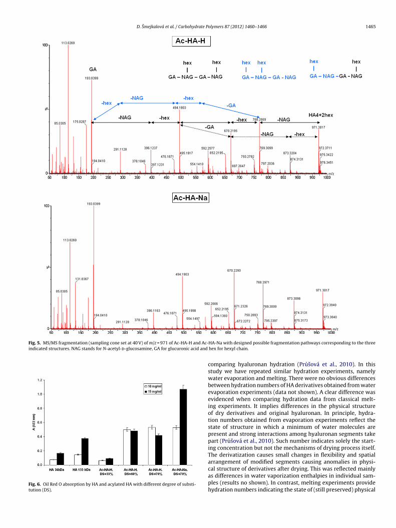
ESI-MS and ESI-MS/MS analyses were carried out in order toconfirm the structural differences between Ac-HA-H and Ac-HA-Na previously suggested from NMR spectra. ESI-MS spectra of bothacylated products with DS = 70% after enzymatic degradation arereported in Fig. 4. There is no significant difference between the twospectra (Fig. 4), suggesting that the number of acyl chains per dimerunit in HA was comparable in both solvents. The spectra indicatedthe presence of unmodified HA dimer (m/z = 396), HA tetramer(m/z = 775), HA hexamer (m/z = 1154), HA octamer (m/z = 1533), andmodified HA mers with one or two acyl chains (m/z is increased by98 or 196, respectively) in case of HA tetramer and hexamer, threeacyl chains (m/z is increased by 294) in case of HA hexamer andoctamer, and four acyl chains (m/z is increased by 392) in case ofHA octamer.
To compare the way of substitution, MS/MS spectra were col-lected for the most intensive peaks detected in ESI-MS spectra.The fragmentation pattern of both acylated products is similar andincludes ions corresponding to the loss of acyl chain, glucuronicacid, and N-acetyl-d-glucosamine (Fig. 5). However, a clear differ-
ence may be noticed in the intensity of m/z = 291, correspondingto the modified glucuronic acid with hexyl chain. Being this inten-sity about 20% in Ac-HA-H sample, while less than 5% in Ac-HA-Na,there is hardly any modification of any OH group of glucuronic acid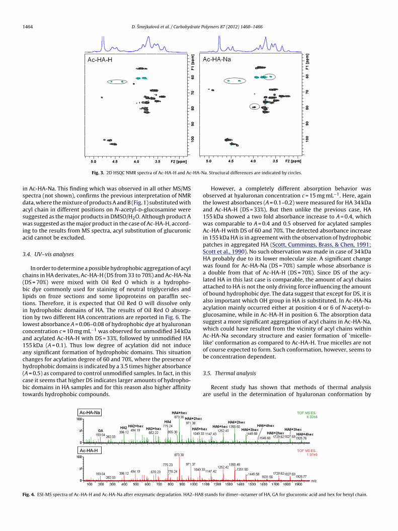
1464 D. Smejkalová et al. / Carbohydrate Polymers 87 (2012) 1460– 1466
-HA-N
isdaswia
3
c(bltitlca1ach(cbt
F
Fig. 3. 2D HSQC NMR spectra of Ac-HA-H and Ac
n Ac-HA-Na. This finding which was observed in all other MS/MSpectra (not shown), confirms the previous interpretation of NMRata, where the mixture of products A and B (Fig. 1) substituted withcyl chain in different positions on N-acetyl-d-glucosamine wereuggested as the major products in DMSO/H2O. Although product Aas suggested as the major product in the case of Ac-HA-H, accord-
ng to the results from MS spectra, acyl substitution of glucuroniccid cannot be excluded.
.4. UV–vis analyses
In order to determine a possible hydrophobic aggregation of acylhains in HA derivates, Ac-HA-H (DS from 33 to 70%) and Ac-HA-NaDS = 70%) were mixed with Oil Red O which is a hydropho-ic dye commonly used for staining of neutral triglycerides and
ipids on froze sections and some lipoproteins on paraffin sec-ions. Therefore, it is expected that Oil Red O will dissolve onlyn hydrophobic domains of HA. The results of Oil Red O absorp-ion by two different HA concentrations are reported in Fig. 6. Theowest absorbance A = 0.06–0.08 of hydrophobic dye at hyaluronanoncentration c = 10 mg mL−1 was observed for unmodified 34 kDand acylated Ac-HA-H with DS = 33%, followed by unmodified HA55 kDa (A = 0.1). Thus low degree of acylation did not induceny significant formation of hydrophobic domains. This situationhanges for acylation degree of 60 and 70%, where the presence ofydrophobic domains is indicated by a 3.5 times higher absorbance
A = 0.5) as compared to control unmodified samples. In fact, in thisase it seems that higher DS indicates larger amounts of hydropho-ic domains in HA samples and for this reason also higher affinityowards hydrophobic compounds.ig. 4. ESI-MS spectra of Ac-HA-H and Ac-HA-Na after enzymatic degradation. HA2–HA8
a. Structural differences are indicated by circles.
However, a completely different absorption behavior wasobserved at hyaluronan concentration c = 15 mg mL−1. Here, againthe lowest absorbances (A = 0.1–0.2) were measured for HA 34 kDaand Ac-HA-H (DS = 33%). But then unlike the previous case, HA155 kDa showed a two fold absorbance increase to A = 0.4, whichwas comparable to A = 0.4 and 0.5 observed for acylated samplesAc-HA-H with DS of 60 and 70%. The detected absorbance increasein 155 kDa HA is in agreement with the observation of hydrophobicpatches in aggregated HA (Scott, Cummings, Brass, & Chen, 1991;Scott et al., 1990). No such observation was made in case of 34 kDaHA probably due to its lower molecular size. A significant changewas found for Ac-HA-Na (DS = 70%) sample whose absorbance isa double from that of Ac-HA-H (DS = 70%). Since DS of the acy-lated HA in this last case is comparable, the amount of acyl chainsattached to HA is not the only driving force influencing the amountof bound hydrophobic dye. The data suggest that except for DS, it isalso important which OH group in HA is substituted. In Ac-HA-Naacylation mainly occurred either at position 4 or 6 of N-acetyl-d-glucosamine, while in Ac-HA-H in position 6. The absorption datasuggest a more significant aggregation of acyl chains in Ac-HA-Na,which could have resulted from the vicinity of acyl chains withinAc-HA-Na secondary structure and easier formation of ‘micelle-like’ conformation as compared to Ac-HA-H. True micelles are notof course expected to form. Such conformation, however, seems tobe concentration dependent.
3.5. Thermal analysis
Recent study has shown that methods of thermal analysisare useful in the determination of hyaluronan conformation by
stands for dimer–octamer of HA, GA for glucuronic acid and hex for hexyl chain.
D. Smejkalová et al. / Carbohydrate Polymers 87 (2012) 1460– 1466 1465
Fig. 5. MS/MS fragmentation (sampling cone set at 40 V) of m/z = 971 of Ac-HA-H and Ac-indicated structures. NAG stands for N-acetyl-d-glucosamine, GA for glucuronic acid and
Fig. 6. Oil Red O absorption by HA and acylated HA with different degree of substi-tution (DS).
HA-Na with designed possible fragmentation pathways corresponding to the threehex for hexyl chain.
comparing hyaluronan hydration (Prusová et al., 2010). In thisstudy we have repeated similar hydration experiments, namelywater evaporation and melting. There were no obvious differencesbetween hydration numbers of HA derivatives obtained from waterevaporation experiments (data not shown). A clear difference wasevidenced when comparing hydration data from classical melt-ing experiments. It implies differences in the physical structureof dry derivatives and original hyaluronan. In principle, hydra-tion numbers obtained from evaporation experiments reflect thestate of structure in which a minimum of water molecules arepresent and strong interactions among hyaluronan segments takepart (Prusová et al., 2010). Such number indicates solely the start-ing concentration but not the mechanisms of drying process itself.The derivatization causes small changes in flexibility and spatialarrangement of modified segments causing anomalies in physi-
cal structure of derivatives after drying. This was reflected mainlyas differences in water vaporization enthalpies in individual sam-ples (results no shown). In contrast, melting experiments providehydration numbers indicating the state of (still preserved) physical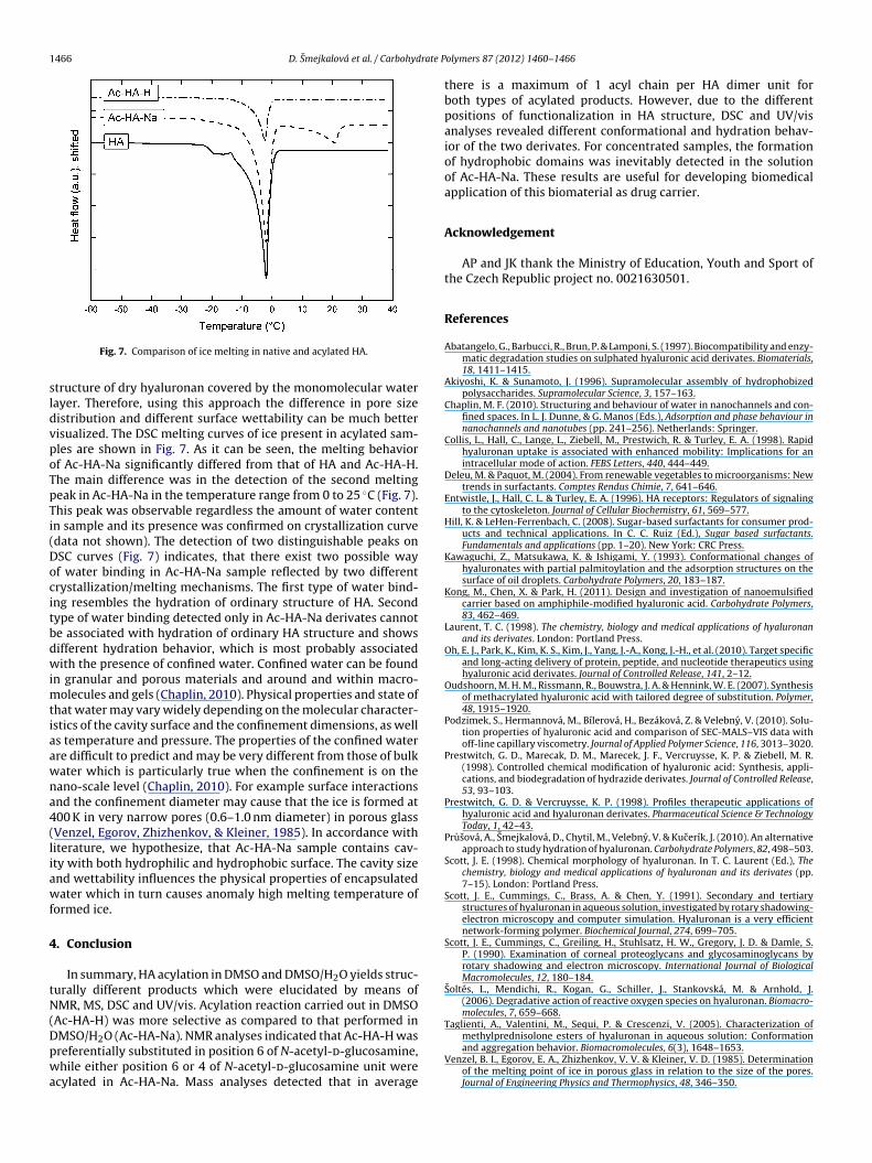
1466 D. Smejkalová et al. / Carbohydrate P
sldvpoTpTi(Docitbdwimtiaawna4(liawf
4
tN(Dpwa
molecules, 7, 659–668.Taglienti, A., Valentini, M., Sequi, P. & Crescenzi, V. (2005). Characterization of
Fig. 7. Comparison of ice melting in native and acylated HA.
tructure of dry hyaluronan covered by the monomolecular waterayer. Therefore, using this approach the difference in pore sizeistribution and different surface wettability can be much betterisualized. The DSC melting curves of ice present in acylated sam-les are shown in Fig. 7. As it can be seen, the melting behaviorf Ac-HA-Na significantly differed from that of HA and Ac-HA-H.he main difference was in the detection of the second meltingeak in Ac-HA-Na in the temperature range from 0 to 25 ◦C (Fig. 7).his peak was observable regardless the amount of water contentn sample and its presence was confirmed on crystallization curvedata not shown). The detection of two distinguishable peaks onSC curves (Fig. 7) indicates, that there exist two possible wayf water binding in Ac-HA-Na sample reflected by two differentrystallization/melting mechanisms. The first type of water bind-ng resembles the hydration of ordinary structure of HA. Secondype of water binding detected only in Ac-HA-Na derivates cannote associated with hydration of ordinary HA structure and showsifferent hydration behavior, which is most probably associatedith the presence of confined water. Confined water can be found
n granular and porous materials and around and within macro-olecules and gels (Chaplin, 2010). Physical properties and state of
hat water may vary widely depending on the molecular character-stics of the cavity surface and the confinement dimensions, as wells temperature and pressure. The properties of the confined waterre difficult to predict and may be very different from those of bulkater which is particularly true when the confinement is on theano-scale level (Chaplin, 2010). For example surface interactionsnd the confinement diameter may cause that the ice is formed at00 K in very narrow pores (0.6–1.0 nm diameter) in porous glassVenzel, Egorov, Zhizhenkov, & Kleiner, 1985). In accordance withiterature, we hypothesize, that Ac-HA-Na sample contains cav-ty with both hydrophilic and hydrophobic surface. The cavity sizend wettability influences the physical properties of encapsulatedater which in turn causes anomaly high melting temperature of
ormed ice.
. Conclusion
In summary, HA acylation in DMSO and DMSO/H2O yields struc-urally different products which were elucidated by means ofMR, MS, DSC and UV/vis. Acylation reaction carried out in DMSO
Ac-HA-H) was more selective as compared to that performed in
MSO/H2O (Ac-HA-Na). NMR analyses indicated that Ac-HA-H wasreferentially substituted in position 6 of N-acetyl-d-glucosamine,hile either position 6 or 4 of N-acetyl-d-glucosamine unit werecylated in Ac-HA-Na. Mass analyses detected that in average
olymers 87 (2012) 1460– 1466
there is a maximum of 1 acyl chain per HA dimer unit forboth types of acylated products. However, due to the differentpositions of functionalization in HA structure, DSC and UV/visanalyses revealed different conformational and hydration behav-ior of the two derivates. For concentrated samples, the formationof hydrophobic domains was inevitably detected in the solutionof Ac-HA-Na. These results are useful for developing biomedicalapplication of this biomaterial as drug carrier.
Acknowledgement
AP and JK thank the Ministry of Education, Youth and Sport ofthe Czech Republic project no. 0021630501.
References
Abatangelo, G., Barbucci, R., Brun, P. & Lamponi, S. (1997). Biocompatibility and enzy-matic degradation studies on sulphated hyaluronic acid derivates. Biomaterials,18, 1411–1415.
Akiyoshi, K. & Sunamoto, J. (1996). Supramolecular assembly of hydrophobizedpolysaccharides. Supramolecular Science, 3, 157–163.
Chaplin, M. F. (2010). Structuring and behaviour of water in nanochannels and con-fined spaces. In L. J. Dunne, & G. Manos (Eds.), Adsorption and phase behaviour innanochannels and nanotubes (pp. 241–256). Netherlands: Springer.
Collis, L., Hall, C., Lange, L., Ziebell, M., Prestwich, R. & Turley, E. A. (1998). Rapidhyaluronan uptake is associated with enhanced mobility: Implications for anintracellular mode of action. FEBS Letters, 440, 444–449.
Deleu, M. & Paquot, M. (2004). From renewable vegetables to microorganisms: Newtrends in surfactants. Comptes Rendus Chimie, 7, 641–646.
Entwistle, J., Hall, C. L. & Turley, E. A. (1996). HA receptors: Regulators of signalingto the cytoskeleton. Journal of Cellular Biochemistry, 61, 569–577.
Hill, K. & LeHen-Ferrenbach, C. (2008). Sugar-based surfactants for consumer prod-ucts and technical applications. In C. C. Ruiz (Ed.), Sugar based surfactants.Fundamentals and applications (pp. 1–20). New York: CRC Press.
Kawaguchi, Z., Matsukawa, K. & Ishigami, Y. (1993). Conformational changes ofhyaluronates with partial palmitoylation and the adsorption structures on thesurface of oil droplets. Carbohydrate Polymers, 20, 183–187.
Kong, M., Chen, X. & Park, H. (2011). Design and investigation of nanoemulsifiedcarrier based on amphiphile-modified hyaluronic acid. Carbohydrate Polymers,83, 462–469.
Laurent, T. C. (1998). The chemistry, biology and medical applications of hyaluronanand its derivates. London: Portland Press.
Oh, E. J., Park, K., Kim, K. S., Kim, J., Yang, J.-A., Kong, J.-H., et al. (2010). Target specificand long-acting delivery of protein, peptide, and nucleotide therapeutics usinghyaluronic acid derivates. Journal of Controlled Release, 141, 2–12.
Oudshoorn, M. H. M., Rissmann, R., Bouwstra, J. A. & Hennink, W. E. (2007). Synthesisof methacrylated hyaluronic acid with tailored degree of substitution. Polymer,48, 1915–1920.
Podzimek, S., Hermannová, M., Bílerová, H., Bezáková, Z. & Velebny, V. (2010). Solu-tion properties of hyaluronic acid and comparison of SEC-MALS–VIS data withoff-line capillary viscometry. Journal of Applied Polymer Science, 116, 3013–3020.
Prestwitch, G. D., Marecak, D. M., Marecek, J. F., Vercruysse, K. P. & Ziebell, M. R.(1998). Controlled chemical modification of hyaluronic acid: Synthesis, appli-cations, and biodegradation of hydrazide derivates. Journal of Controlled Release,53, 93–103.
Prestwitch, G. D. & Vercruysse, K. P. (1998). Profiles therapeutic applications ofhyaluronic acid and hyaluronan derivates. Pharmaceutical Science & TechnologyToday, 1, 42–43.
Prusová, A., Smejkalová, D., Chytil, M., Velebny, V. & Kucerík, J. (2010). An alternativeapproach to study hydration of hyaluronan. Carbohydrate Polymers, 82, 498–503.
Scott, J. E. (1998). Chemical morphology of hyaluronan. In T. C. Laurent (Ed.), Thechemistry, biology and medical applications of hyaluronan and its derivates (pp.7–15). London: Portland Press.
Scott, J. E., Cummings, C., Brass, A. & Chen, Y. (1991). Secondary and tertiarystructures of hyaluronan in aqueous solution, investigated by rotary shadowing-electron microscopy and computer simulation. Hyaluronan is a very efficientnetwork-forming polymer. Biochemical Journal, 274, 699–705.
Scott, J. E., Cummings, C., Greiling, H., Stuhlsatz, H. W., Gregory, J. D. & Damle, S.P. (1990). Examination of corneal proteoglycans and glycosaminoglycans byrotary shadowing and electron microscopy. International Journal of BiologicalMacromolecules, 12, 180–184.
Soltés, L., Mendichi, R., Kogan, G., Schiller, J., Stankovská, M. & Arnhold, J.(2006). Degradative action of reactive oxygen species on hyaluronan. Biomacro-
methylprednisolone esters of hyaluronan in aqueous solution: Conformationand aggregation behavior. Biomacromolecules, 6(3), 1648–1653.
Venzel, B. I., Egorov, E. A., Zhizhenkov, V. V. & Kleiner, V. D. (1985). Determinationof the melting point of ice in porous glass in relation to the size of the pores.Journal of Engineering Physics and Thermophysics, 48, 346–350.
Related Documents


![PHYSICAL DATA OF ORDINARY SOLVENTS...diisopropyl ether [ 108-20-3] 102.18 68.5 -85.5 0.724 34.0 0 Aprotic Polar dimethyl acetoamide[ 127-19-5] 87.12 164.5 -20 0.937 Aprotic Polar DME](https://static.cupdf.com/doc/110x72/60d793b2c488ae0bfb4a57e9/physical-data-of-ordinary-solvents-diisopropyl-ether-108-20-3-10218-685.jpg)


