Strontium and Caries: A Long and Complicated Relationship F Lippert, AT Hara Department of Preventive and Community Dentistry, Oral Health Research Institute, Indiana University School of Dentistry, USA Short title: Critical Review on Strontium and its Role in Caries Keywords: review, caries prevention, strontium Corresponding author: Frank Lippert Department of Preventive and Community Dentistry Oral Health Research Institute, Indiana University School of Dentistry 415 Lansing Street, Indianapolis, IN 46202, USA Tel. +1 317 274 3983, Fax +1 317 274 5425, E-Mail [email protected] _________________________________________________________________________________ This is the author's manuscript of the article published in final edited form as: Lippert, F., & Hara, A. T. (2013). Strontium and caries: a long and complicated relationship. Caries research, 47(1), 34-49. https://doi.org/10.1159/000343008

Welcome message from author
This document is posted to help you gain knowledge. Please leave a comment to let me know what you think about it! Share it to your friends and learn new things together.
Transcript
Strontium and Caries: A Long and Complicated Relationship
F Lippert, AT Hara
Department of Preventive and Community Dentistry, Oral Health Research Institute, Indiana
University School of Dentistry, USA
Short title: Critical Review on Strontium and its Role in Caries
Keywords: review, caries prevention, strontium
Corresponding author:
Frank Lippert
Department of Preventive and Community Dentistry
Oral Health Research Institute, Indiana University School of Dentistry
415 Lansing Street, Indianapolis, IN 46202, USA
Tel. +1 317 274 3983, Fax +1 317 274 5425, E-Mail [email protected]
_________________________________________________________________________________ This is the author's manuscript of the article published in final edited form as:
Lippert, F., & Hara, A. T. (2013). Strontium and caries: a long and complicated relationship. Caries research, 47(1), 34-49. https://doi.org/10.1159/000343008

2
ABSTRACT
Investigations into the role of strontium (Sr) in caries prevention have attracted great interest in
the research community in the past, with its peak in the 1970- and 80ies. To this date, no clear
indication of the relative importance of Sr in caries prevention has been provided. A vast number
of animal caries, epidemiological and mechanistic studies have been conducted. Albeit Although
a great level ofthere is much discrepancyexists in the literature, the majority of studies suggest
that Sr exhibits some cariostatic properties and, predominantly in the presence of fluoride (F). An
optimum Sr concentration of 5 to 10 ppm in drinking water has been proposed as a direct result
of several epidemiological caries studies. Despite these results, no direct link can be established
between Sr and caries prevention as, to date, no relevant, randomized control trials have been
reported. The extrapolation of potential cariostatic properties of Sr from epidemiological studies
is difficult due to the co-presence of several other trace elements in the water of the study areas,
with many of these elements being attributed cariostatic properties in their own right.
Furthermore, the role of caries risk factors was not taken into consideration. There is a clear need
for further research, especially on the mineral phases in the dental hard tissues, plaque and
plaque fluid associated with Sr as these may give rise to a better understanding of this subject
matter. Based on the current data, the, at least by some authors, proposed cariostatic properties
of Sr, or at least those proposed by some authors, cannot be supported.

3
INTRODUCTION
The caries-preventative effects of fluoride (F), and especially its sodium salt, have long been
established and are beyond any reasonable doubt. However, despite F’s proven track record,
caries is still endemic in parts of the world populations globally and its elimination will remain
the main challenge for dental researchers for decades to come. Its main negative side effect,
fluorosis, has led investigators to conduct research on other, preferably non-toxic, (trace)
elements, ideally exhibiting synergy with F, and their relative anti-caries effects – one being
strontium (Sr).
Due to its similarity with to calcium (Ca), Sr has attracted considerable amounts of
interest in the caries and caries-related research community, with its peak in the 1970ies and
1980ies. Despite a vast number of animal caries, epidemiological and mechanistic studies
conducted by various investigators, to this date, no clear indication of the role of Sr in caries
prevention has been provided. Previous reviews on the relationship of between trace elements in
general and caries [e.g. Büttner, 1969; Losee and Ludwig, 1970; Navia, 1972; Curzon and
Crocker, 1978; (anonymous), 1978; Beaton, 1983; Olson, 1987] highlighted the complexity of
the matter, but were also somewhat limited in scope. Therefore, the aim of the present paper was
to critically and comprehensively review the literature concerned with the role of Sr in the caries
process.
CRITICAL REVIEW
Search Strategy and Structure
A search strategy was developed for articles indexed in MEDLINE, Web of Science® and
PubMed databases written in English up to August 30, 2011. Several hand searches were
required to obtain articles which could not be retrieved via the aforementioned databases. The
following key words were used in the searches in combination with ‘strontium’ or ‘sr’: ‘caries’,
‘tooth’, ‘teeth’, ‘enamel’, ‘dentin*’, ‘demin*’, ‘remin*’, ‘apatite’, ‘hydroxyapatite’, ‘calcium
phosphate’, ‘fluoride’.

4
Articles were then divided into the following groups based on the topics investigated
topics, which will also form the order of the present review:
1. Animal studies – Sr effects on enamel and dentin, caries studies
2. Caries eEpidemiological caries studies
3. Sr in oral care products
4. Sr in the oral cavity – presence in teeth, plaque and saliva
5. Sr and calcium phosphates (CaPi)
6. Discussion
7. Conclusions
Articles concerned with multiple topics were considered in all relevant groups.
1. Sr Studies in Animals
Sr Effects on Dentin and Enamel
There is a discrimination against Sr in CaPi mineralization when Ca is present (see also chapter
5.), which was shown by Likins et al. [1959] in weanling rats. Later studies [Likins et al., 1961;
Menczel et al., 1962] were able to support these findings, and a higher Sr discrimination was
found in dentin in relation to enamel [Likins et al., 1961].
Studies in weanling rats [Likins et al., 1959] have shown that when both Ca and Sr were present
during tooth formation, Ca was incorporated into the apatitic lattice preferentially, resulting in a
higher Ca:Sr ratio in the subsequent mineral than might be expected. Later studies [Likins et al.,
1961; Menczel et al., 1962] were able to support these findings, with the effect more pronounced
in dentin than in enamel [Likins et al., 1961].
Sr injections were shown to cause dentin hypomineralisation in rats, resulting in the
formation of a hypomineralized layer of dentin, comparable to that seen after F injection
[Weinmann, 1942; Irving and Weinmann, 1948; Yaeger, 1963]. These effects were almost
entirely confined to dentin as enamel was not affected [Weinmann, 1942]. Yaeger and
Eisenmann [1963] showed that the degree of dentin hypomineralisation was positively correlated
to [Sr] (i.e. Sr concentrations). Although a similar correlation was found for [F], the wider zones
of hypomineralized dentin observed for Sr and the lack of a thinner hypermineralized layer of

5
dentin as observed for F suggested differences in their etiology. A later study [Yaeger et al.,
1964] suggested that Sr (or F) inhibit or reverse the matrix aggregation normally occurring at the
dentin-predentin junction, thus inhibiting mineralization. Grady and Yaeger [1965] reported that,
while in normal dentin collagen fibrils are orientated perpendicular to the dentinal tubules, in
hypomineralized layers caused by Sr (or F), fibrils are arranged at 45° to the fibrils of the normal
dentin. The possibility for that hypomineralized dentin canto recover after Sr injection seizes
ceases was shown by Yager [1966]. Regions of hypomineralized rat dentine induced by Sr (or F)
were reported to show greater ability to mineralize than adjacent normal untreated dentine in
vitro [Eisenmann and Yaeger, 1972]. Johnson et al. [1970] demonstrated that Sr was not lost
preferentially lost from high-Sr dentin and suggested that Sr was not predominantly surface
located at the surface. In addition, many studies reported that Sr injection causes the formation of
more than one hypomineralized layer in dentin [Yaeger and Eisenmann, 1963; Yaeger et al.,
1964; Eisenmann and Yaeger, 1969; Ogawa et al., 1981]; however, while two hypomineralized
layers were reported in labial dentin, only a single layer was found in lateral, medial and lingual
walls [Ogawa et al., 1981; Appleton, 1993]. Ogawa et al. [1981] postulated that Sr may exert its
effect on dentin hypomineralisation not only because of its ability to retard crystal growth, but
also because of a direct effect on odontoblasts and collagen synthesis.
Castillo Mercado and Bibby [1973] studied the effects of Sr injections on molar
morphology and found wider fissures and thicker dentin. A subsequent study [Curzon et al.,
1982] investigating Sr effects when given in drinking water found an increase in horizontal and
vertical dentin thickness at a [Sr] of 50 ppm, but not at 150 ppm which was indistinguishable in
its effect to the water control.
While Sr effects on dentin were studied almost solely as a result of Sr injections with
Sri.e. systemically, its effects on enamel were predominantly studied as a result of dietary Sr
administration. Johnson et al. [1966] and Johnson [1967] demonstrated that isomorphous
substitution of Sr for Ca occurs in the enamel-HAp and postulated that Sr may form Sr6H3(PO4)
∙ 2 H2O, which was shown to be a precursor of SrHAp [Collin, 1966]. Furthermore, Johnson and
Singer [1967] found a gradient of increasing [Sr] from the incisal to apical areas in enamel of
rats raised on a Sr-rich diet.
Similar to its effects on dentin, Sr has been shown to cause disturbances in ameloblast
morphology and amelogenesis [Weinmann, 1943; Neiman and Eisenmann, 1975] in rats when
6
injected. White et al. [1980] demonstrated that the enamel organ limits Sr uptake in both the
secretory and maturation phases of enamel formation. A further study by Suga et al. [1987]
supported these findings and reported that Sr, unlike F, inhibits the early stage of enamel
maturation and that mineralization ceases earlier in the inner layers of Sr-treated rats than in
control groups.
Sr uptake byin surface enamel of rats given water with different [Sr] (0 – 100ppm) was
studied by Spector et al. [1978]. The authors found a dose-response relationship between [Sr]
administered and [Sr] in surface enamel, but only when Sr was given pre- and post-eruptively.
When given pre-eruptively only, a [Sr] of 50 ppm yielded the highest Sr uptake. Comparing the
relative pre- and post-eruptive contributions to [Sr] in surface enamel, the present authors
calculated that at [Sr] ≤ 50 ppm, 70 to 74% of [Sr] in surface enamel of rats was due to Sr given
pre-eruptively, while at [Sr] = 100 ppm, only 29% could be attributed to Sr given pre-eruptively.
A subsequent study [Curzon and Spector, 1980] showed some variations between different Sr
salts in Sr uptake by enamel, especially when administered pre-eruptively. A dose-response
relationship between [Sr] administered through the diet and [Sr] in surface and near-surface
enamel was also observed by Ashrafi et al. [1980].
Animal Caries Studies
Before relevant Sr animal caries will be are discussed, some light must be shed on differences in
the etiology of caries in animals and humans must be shed. Ericsson [1962] and Tatevossian and
Wright [1974] have shown that rat in comparison to human saliva exhibits larger higher pH
values, lower [P] but higher [Ca] and a considerably higher buffering capacity, presumably due
to higher [CO3]. In addition, Haldi et al. [1960] have demonstrated that the pH at the tooth
surface of rats ‘rarely fell as low as 7.0’ after the administration of a cariogenic diet or sugar
alone. In a review, White [1992] concluded that while animal models have tremendous value in
studying the caries process they are by no means perfect profile tools and that results of animal
caries studies on new anti-caries agents and formulations do not necessarily mirror clinical
results (differences in anti-caries effectiveness and efficacy observed in animal caries studies and
RCTs between amine fluoride, MFP and NaF are worth noting in this context). Furthermore, the
duration and frequency of administration, salivary clearance of actives and the effect of diet on

7
retention of actives are not well understood in animals, making comparisons to caries in humans
difficult and highlighting that further research is warranted in this area.
Animal studies on the effects Sr in preventing caries have been equivocal as no effects
[Johnson and Hein, 1953; Olson et al., 1978], an increase [Hunt and Navia, 1972, 1975; Joseph
et al., 1977] or a decrease [Losee and Adkins, 1968; Gedalia et al., 1975; Meyerowitz et al.,
1979; Ashrafi et al., 1980; Curzon and Spector, 1981; Curzon, 1988] in caries incidence were
have been reported. Generalization of overall Sr effects, however, is unjustified as differences
depending on the dose and time of administration (pre- and/or post-eruptively) or the combined
administration of Sr and F need to be analyzed for separately. In addition, scoring of animal
caries was not uniform between these studies, thus further complicating the matter. Accordingly,
tThe present authors decided to conduct further analyzes only on studies using the caries scoring
method according to Keyes [1958]. Individual ‘E’ scores (enamel units) for bucco-lingual, sulcal
and proximal lesions were combined to yield a total caries score in line with the ‘Indiana rat
caries model’, and subsequently, percentage values of caries reduction were calculated in relation
to appropriate controls [Stookey et al., 1995]. The results of the rat caries studies conducted by
Meyerowitz et al. [1979], Ashrafi et al. [1980] and Curzon [1988] are presented in table 5.
Studies are somewhat comparable to some extent due the same diet (MIT 200) being fed,
although rats were inoculated with S. mutans only in the study by Curzon [1988]. Comparing
these data it can be noted that there is a curvilinear relationship between [Sr] administered and
caries reduction, regardless of whetherif Sr was administered pre- and/or post-eruptively.
However, caution must be issued as no statistical analysis is possible to the unavailability of the
raw data. The reason for this curvilinear behavior is not clear and cannot be sufficiently
explained based on what is currently known. However, Driessens [1982, 1986] provided
proposed some hypotheses which , however, will be discussed at a later stage. In this context, it
is wWorth mentioning in this context is that data can be over- or even mis-interpreted if only
caries scores on from only one particular site, or if the wrong comparisons, are considered.
Meyerowitz et al. [1979] concluded that a combination of 50 ppm Sr and 10 ppm F resulted in
the greatest reduction in bucco-lingual rat caries scores. The authors, however, ignored sulcal
and proximal caries scores where Sr + F was less effective in reducing caries than F alone. The
present authors combined all three scores and found a 7.6 % increase in rat caries in comparison
to F alone (table 5), thus the opposite result (this may or may not be of statistical significance).

8
Other animal caries studies will now be considered. Johnson and Hein [1953] found no
cariostatic effect for Sr when administered to hamsters at 50 ppm (as SrCl2) in the drinking
water. Shaw and Griffiths [1961] studied the effects of dietary Sr supplementation on rat caries
(using the sparingly soluble SrCO3). A positive effect, i.e. a on caries reduction, was noted when
administered post-eruptively; however, this was paired with a decrease in weight gain by the
animals (at 2 % SrCO3). When given pre-eruptively, an increase in caries occurrence was noted,
and this could not be offset by giving Sr post-eruptively to the same rats. Losee and Adkins
[1968] studied the effects of a dietary supplementation with the ash of green beans cooked in
water containing different amounts of trace elements. The lowest overall caries scores could be
related to the water sourced from Ohio, rich in Li, Mo, Sr, B and F. As multiple trace elements
were present, no direct relationship for Sr in reducing caries could be established. A later study
[Losee et al., 1976], investigating the effect of the ‘Ohio water’ when given to rats in comparison
to deionized water, concluded that these trace minerals may act synergistically with fluoride in
reducing caries prevalence in rats.
When Sr (as SrCl2) was administered pre-eruptively at concentrations of 1000 or 2000 µg
per 10 g body weight (100x or 200x the maximum [Sr] as compared to the study by Meyerowitz
et al. [1979] and therefore of questionable physiological relevance), an increase in rat caries was
noted [Hunt and Navia, 1972]. However, at 100× or 200× the maximum [Sr] as compared to the
study by Meyerowitz et al. [1979] the physiological relevance is questionable. A later study by
the same authors [Hunt and Navia, 1975] found similar effects at [Sr] = 500 µg in the presence or
absence of [F] = 100 µg per 10 g body weight, thus indicating no additive or synergistic Sr + F
effects. However, both studies reported incomplete rat caries scores which does not allow for
decisive conclusions to be drawn. Gedalia et al. [1975] found a somewhat similar curvilinear
relationship as noted above when studying the effects of Sr in water on caries in hamsters when
given post-eruptively. [Sr] = 25 ppm resulted in overall increase, whereas [Sr] = 75 ppm resulted
in a decrease of caries occurrence. Considerably stronger Sr effects were noted when Sr was
administered pre-eruptively, and both [Sr] resulted in a marked decrease in caries in a dose-
response manner. These results mirror later observations by Meyerowitz et al. [1979] and it was
concluded that Sr exhibits its cariostatic effect mainly because it is laid down in enamel and
dentin before eruption, therefore somewhat excluding topical effects to some extent.

9
Joseph et al. [1977] found a marginal increase in caries in hamsters when Sr was given
pre- and post-eruptively as part of the drinking water at 10 and 25 ppm. F was found to offset the
negative Sr effects. Similar results effects were obtained reported on rats exposed to Sr post-
eruptively by Olson et al. [1978]. Curzon and Spector [1981] studied the effects of different Sr
salts at [Sr] = 50 ppm on rat caries reduction and concluded that SrF2 was most effective, mainly
because of its [F] = 22 ppm. Other salts varied in their effectiveness based on their
bioavailability. In addition, Sr effects were somewhat greater when given post- than pre-
eruptively.
Seppä et al. [1988] reported that a total of two treatments with 500 ppm Sr with or
without NaF varnish application had no effect in reducing on rat caries in comparison to the
appropriate controls. Luoma et al. [1984] studied the effects of chlorhexidine (CHX)-F-Sr ([Sr] =
1000 ppm) and found a reduction in fissure caries and approximal lesions, but an increase in
plaque scores in relation to CHX-F. In a subsequent study, Spets-Happonen et al. [1996] found a
similar curvilinear Sr caries relationship as reported before, as a CHX-F gel supplemented with
50 ppm Sr did show some marginal benefits in reducing rat caries, whereas 250 ppm Sr appeared
to markedly weaken the CHX-F effect.
2. Epidemiological Caries Studies
Several major questions are pertinent when analyzing data from epidemiological studies: a) iIs
the relationship between Sr and caries reduction causative or simply incidental?, and b) iIs the
reduction in caries related to the sole contribution of Sr, or is it due to the additive or synergistic
action of two or more trace elements present at the same time? Furthermore, as both topical and
systemic effects are thought to play roles in the caries-preventative action of Sr (see ‘Animal
Caries Studies’), it must also beis necessary to considered whether subjects were lifelong
residents of, or if they immigrated to, the study area (i.e. the area from which water or soil
samples were taken and analyzed for [Sr]). In case they immigrated, when (i.e. pre- or post-
eruptively in relation to their permanent dentition), and what were environmental [Sr] in their
previous area. And fFinally, what were the oral care habits of the study subjects, their age,
socioeconomic status and sugar intake/dietary habits; i.e. have caries risk factors been

10
considered? To the author’s’ knowledge, not a single study was concerned with all these aspects,
thus making it almost impossible to perform an unbiased review of the available studies, which,
sadly, are therefore only of very limited value. Nonetheless, epidemiological studies will be
discussed in chronological order and based on the extent in the context of the information
provided.
Anderson [1966] studied a relatively small group of 12-year old children (n = 51) from an
area in Gloucestershire (UK) which’ whose soil is rich in SrSO4. [Sr] in water in this area was
estimated at 0.33 ppm and therefore extremely low in comparison to later studies (see below),
whereas [F] was 0.15 ppm. No differences in children’s DMF scores (6.03 vs. 5.63) between the
study area and a control area (n = 537; [Sr] = nil; [F] = 0.13 – 0.15 ppm) were observed, but
considerably lower DMF scores (4.36 and 4.38) were noted in ‘F’ (n = 74; [F] = 0.9 ppm) and
‘Mo areas’ (n = 270; [Mo] = 0.0036 ppm; [F] = 0.09 ppm).
During a sA study on caries-resistant navy recruits [Losee and Adkins, 1969], which was
perhaps was the key study that sparked the interest in Sr in the caries research community in the
1970ies, it was able possible to correlate the high number of caries-resistant recruits from NW
Ohio (USA) with the simultaneous occurrence of elevated [B], [Li], [Mo], [Sr] in NW Ohio
water in combination with F. A median [Sr] of 6100 ppm was found in the water of the study
area and the transfer of F, Li, Mo and Sr from water to green beans during cooking was shown.
A subsequent rat caries study [see above; Losee et al., 1976] was able to support the findings of
this epidemiological study. Losee and Bibby [1970] were able to negatively correlate [Sr], [B]
and [F] in water with DMFT scores (r < – 0.8 for Sr, B and F) based on the data from six cities in
Illinois (USA). A more comprehensive study by Adkins and Losee [1970] compared trace
elements in water between low and high caries statues (in the USA) and found significant
differences between the two for Ba, B, Li, Mo and Sr with higher concentrations observed in
states with lower caries incidence. Strong, positive co-variations were found between these trace
elements, and Sr was found to exhibit the lowest correlation coefficient with caries prevalence (r
= – 0.59), thus indicating the strongest effect. F, however, was not considered.
A study [Curzon et al., 1970] on 251 children aged 12 to 14 from two towns in Ohio
(USA) with different [Sr] (5.37 vs. 0.2 ppm) and [B] (0.35 vs. 0.04 ppm) and similar [F] (1.2 vs.
1 ppm) showed mean DMFS scores of 3.56 vs. 5.54 and mean DMFT scores of 2.25 vs. 3.04,
and it was concluded that differences in [B] and [Sr] in water were attributable to the reduction
11
in caries rather than the 0.2 ppm difference in [F]. It must be noted that only subjects with fully
erupted canines, premolars and second molars were included in the study. A ten-year follow-up
study [Curzon, 1983] found essentially the same caries distribution pattern between these
communities and the changes in DMFS scores were attributed to changes in [Sr] in water.
Ludwig et al. [1970] also studied children aged 12 to 14 (who were lifelong residents) from 19
town in various states (in the USA) with very low [F] in water (0 – 0.30 ppm) but different
DMFT scores (3.8 – 9). [Sr] in water ranged from 12 to 2200 ppm, but only a weak negative
correlation between [Sr] and DMFT was established (r = – 0.29). Again, Sr and B (r = – 0.3)
showed similar effects. However, the present authors calculated that the exclusion of the two
towns with the highest [Sr] of 1300 and 2200 ppm (leaving [Sr] = 280 ppm the next highest)
would have yielded r = 0.03 and therefore no correlation at all. An investigation into differences
in caries activity between two semi-isolated communities in Colombia with [F] < 0.2 ppm in
water by Bowen et al. [1977] did not find a relationship between [Sr] in water (8 – 120 ppm in
low- and 14 ppm in high-caries activity communities) and caries activity. However, water [B]
were substantially different (21 – 85 ppm vs. 1.5 ppm).
Perhaps the most compelling set of data was reported by Curzon et al. [1978] who
conducted caries examinations on 1313 children aged 12 to 14 years, lifelong residents and
immigrants, living in seven communities in Wisconsin (USA). [F] in drinking water was
comparable between communities with 1 to 1.29 ppm; however, [Sr] varied between 0.022 and
33.94 ppm. Figure 1 shows the results of this study in combination with the results of two
comparable studies by Curzon et al. [1970] and Curzon [1985]. As water [F] were almost
identical and as the caries evaluation was performed similarly, all three studies were combined.
Considering lifelong residents only, some sort of an optimum [Sr] in water in relation to caries
prevention can be seenis suggested. It is worth mentioning that as concentrations of other trace
elements in water were also provided, the present authors found a very similar relationship for
[Fe] and a linear relationship for [B]. In relation to these findings about with regard to Fe and B
it must be mentioned that a follow-up study by Curzon [1983] found essentially the same [B] but
considerably lower [Fe] in water in the areas studied. Somewhat surprising were the results of
the immigrants, showing an almost inverse relationship compared to lifelong residents. No
information regarding their previous Sr exposure was presented and, regrettably, the authors did
not discuss these data, making it difficult to provide any comments now. The final study by
12
Curzon [1985] was also able to also reported an inversely correlation betweene caries prevalence
with and [Sr] in water in communities with water [F] of 0.9 to 1.2 ppm. It must should also be
mentioned that the method for determining carious surfaces was somewhat inappropriate as ‘any
fissure or enamel surface in which the explorer stuck and penetrated into dentine was regarded as
carious’ [Curzon et al. 1978].
In contradiction to earlier studies [Ludwig et al., 1970; Bowen et al., 1977], showing no
or only marginal anti-caries benefits for Sr in the presence of low [F], Athanassouli et al. [1983]
was able to correlate lower DMFT scores (5.26 vs. 6.95) in 582 children aged 11 to 14 years with
an area of higher [Sr] in water (2.9 – 7 ppm vs. 0.2 – 1.3 ppm). [F] in water was very low (< 0.06
ppm) in both districts. Furthermore, Vrbic and Stupar [1980] were also able tofound a
negatively correlationte between [Sr] in water with DMFT scores in areas with low [F] (< 0.15
ppm).
In addition to the studies trying to establishinvestigating a possible link between caries
reduction and [Sr] in drinking water, several studies were also concerned withattempted to
establish ing athis similar link for Sr in surface enamel and/or in plaque. Little and Barrett
conducted two studies [1976a,b] investigateding possible relationships between [Sr] and [F] in
surface and near-surface enamel and caries prevalence. Studies were conducted on teeth obtained
from lifelong residents of either east or west coast in the USA. Contradicting results were found
when teeth were grouped according to DMFT scores of < 3 or > 7. In east coast samples from the
east coast, both [Sr] and [F] were higher in surface and near-surface enamel in low caries teeth,
whereas in west coast samples from the west coast, this was only true for [F], with [Sr] being
higher in high caries samples. It must be noted that, in general, [Sr] and [F] were somewhat
higher in west compared to east coast samples, although [Sr] were virtually identical in low
caries teeth on east and west coast. The authors explained the east coast-west coast discrepancy
with the above-proposed optimum [Sr] as an excess in Sr or lack thereof may increase the tooth’s
susceptibility to caries. Two comparable studies by Curzon and Losee [1977b, 1978] were able
to provide similar results, showing a stronger relationship between [Sr] in whole enamel and
lower caries incidence in east than in west coast enamel samples.
Furthermore, Curzon and Losee [1977a] were able to demonstrate that high [Sr] in
enamel were associated with low caries prevalence by studying 147 samples obtained from 59
communities in 19 states in the USA. Other elements, such as F, were not studied. A subsequent
13
study by Spector and Curzon [1979] did involve [Sr] and [F] analyszes. However, the authors
were not able to demonstrate a relationship between [Sr] in surface enamel and DMFT scores. A
weak correlation between [F] and DMFT was found (r = – 0.16) and also between [Sr] and [F] (r
= 0.48). In contradiction, Vrbic and Stupar [1980] were able to demonstrate a negative
correlation between [Sr] in enamel and caries incidence, which was supported by athe later
studies ofby Athanassouli et al. [1983] and Curzon [1985].
Only very few studies were concerned withinvestigated the possible correlation between
Sr in plaque and caries prevalence. Schamschula et al. [1977b] reported a negative correlation
between [Sr] and DMFT (r = – 0.15) in a primitive population in Papua New Guinea (n = 301;
12 – 24 years of age), which, however, was weaker than those correlations for [Ca] and [F].
Similar results (r = – 0.23) were obtained by the same group [Schamschula et al., 1978a] when
studying 72 children aged 9.7 to 13 years. Curzon [1985] was also able to demonstrate this
relationship (r = – 0.83) in addition to the earlier reported negative correlations between [Sr] in
water, and in enamel and DMFS scores, reported earlier.
Only two studies on salivary Sr and caries prevalence could be retrievedfound. Curzon
[1984] was able to demonstrate a weak, negative relationship between [Sr] in saliva andon caries
prevalence (r = – 0.13) in 105 children aged 14 years. In contradiction, during one very recent
study [Shigemi et al., 2008] on 521 children aged 6 to 12 years was able to demonstrate a
positive correlation between [Sr] in saliva and using the author’s terminology ‘caries experience’
(no DMFT/S scores were recorded) was reported. In addition, the authors showed that in groups
with high ‘fluoride experience rates’ (due to F mouth rinsing at school), [Sr] in saliva tended to
be lower.
In the context of epidemiological studies, it must alsoshould be mentioned that Curzon
and Spector [1977] reported on enamel mottling when examining 1313 12 to 14-year old
children in seven towns in Wisconsin (USA). As water [F] were very similar in the study area (1
– 1.29 ppm), only a correlation between [Sr] in water (0.02 – 33.9 ppm) and mottling scores (r =
0.85) could be established, and, interestingly, only in lifelong residents.
Finally, Riyat and Sharma [2010] reported , although only in a relatively small group of
subjects, that [Sr] in blood was higher in a group (n = 15) who had no history of caries in
comparison to a group (n = 15) with previous caries experience, although only in a relatively
small group of subjects. A similar relationship was found for [F] and [Se].

14
3. Sr in Oral Care Products
Considering the interest in Sr within the caries research community, it was not surprising that
awareness of Sr was also raised awareness in the oral care industry.
Zero et al. [1982] conducted a series of investigations, which are perhaps better termed
described as ‘product safety studies’ as a dentifrice containing not only Sr but also EDTA was
evaluated for changes in surface enamel morphology, [Sr] in enamel and enamel solubility. The
test dentifrice was compared to a commercially available control dentifrice. No significant
changes in surface morphology were noted; however, both products rendered the enamel surface
less soluble and [Sr] in enamel increased in the test but not in the control dentifrice.
It appears that several manufacturers of oral care products were pursuing Sr as a novel
anti-caries agent in the late 1980ies. A total of three in situ studies, two enamel fluoride uptake
(EFU) studies [Bowman et al., 1988a,b] and one de-/remineralization caries study [Wefel et al.,
1995] were reported. All studies evaluated NaF formulations containing a so-called
‘polyampholyte delivery system’ (PAA-Sr), which was essentially a combination of a soluble Sr
salt (not specified) and a polyacrylic acid (MW = 4500 Da). Substantially enhanced EFU values
were reported for PAA-Sr, for both the mouth rinse [Bowman et al., 1988a] and dentifrice
[Bowman et al., 1988b] delivery formats, in comparison to controls with the same [F]. The study
by Wefel et al. [1995] was able to demonstrate anti-caries effectiveness of PAA-Sr which was
comparable to a 2800 ppm F (as NaF) control dentifrice. Two studies [Mellberg and Fletcher,
1990; Afflitto et al., 1992] conducted by a direct competitor and comparing PAA-Sr with a
different control dentifrice were also reported. The study by Mellberg and Fletcher [1990] found
comparatively lower EFU in vitro for PAA-Sr. The more comprehensive study by Afflitto et al.
[1992] reported comparatively lower salivary fluoride bioavailability and less cariostatic activity
in the rat caries model for PAA-Sr.
A Polish group conducted two in situ studies on experimental dentifrices containing Sr +
F [Kaczmarek et al., 2005] and Sr-HAp [Surdacka et al., 2007]. Both studies were primarily
concerned with investigating the deposition of Sr into artificial white spot lesions, and both
15
studies were able to demonstrate an increase in [Sr] in enamel after 3 and 6 months of study
duration.
Two further studies are worth mentioning here. Nishino [1981] investigated the effect of
a Zn-acetate/Sr-acetate/tannic acid mouth rinse on caries reduction in 24 children aged 3 to 10.3
years and using the Cariostat test. A reduction in ‘caries activity’ was achieved by this rinse, but
due to the study design no Sr effect can be extrapolated. Klinger and Wiedemann [1986] studied
the effect of a mineralizing solution containing Ca, Sr, Pi and tartrate in comparison to an amine
F solution on the remineralization of approximal lesions in vivo. The mineral rinse did not
induce measureable remineralization, whereas the F solution did.
No reports on potential cariostatic properties of products containing SrCl2 × 6 H2O
(SCH), designed for the relief of dentin hypersensitivity, could be retrieved.
4. Sr in the Oral Cavity
Sr in Teeth
A summary of results from various investigators can be found in supplementary data tables 1
(enamel) and 2 (dentin), with similar Sr concentrations reported for both tissues. Regardless of
the investigator, study site or analytical technique employed, considerable variations in Sr
concentration were found, and especially in enamel.
Not taking into accountWith the exception of the osteoporosis drug Sr ranelate [for
review see Marie et al., 2001], the dentine hypersensitivity treatment agent SCH [for review see
Addy and Dowell, 1983] or Sr-containing glass ionomers cements (Sr-GIC) [Kim et al., 2010];
the diet is the only Sr source for the human body (approx. 2.1-2.4 mg/day) [Schroeder et al.,
1972].
Comparatively higher [Sr] were found in enamel of permanent than in deciduous teeth
[Cutress, 1972a; Nixon and Helsby, 1976; Shashikiran et al., 2007], whereas Zaichick and
Ovchjarenko [1996] found no differences. To the author’s’ knowledge, only two studies
[Steadman et al., 1958; Lundberg et al., 1965] were concerned with theconducted Sr analysis
ofin enamel from unerupted, permanent teeth, thus limitingassuring that any Sr present
incorporation into the dental hard tissues via systemic means would have been incorporated

16
systemically. Both studies reported similar [Sr] in relation to the enamel of erupted teeth,
suggesting that most of the Sr is incorporated before eruption and that little change in [Sr] in
enamel occurs with age. Later studies either proved supported [Little and Steadman, 1966] or
disproved contradicted [Derise and Ritchey, 1974] these findings.
Several studies were concerned withinvestigated the depth distribution of Sr in enamel
with respect to depth, in enamel, and, again, considerable variation was noted. Steadman et al.
[1958] found an almost uniform Sr distribution in enamel which was supported by other studies
[Vrbic and Stupar, 1980; Noren et al., 1983; Frank et al., 1989], whereas Cutress [1972a]
reported either higher surface or higher bulk [Sr] depending on the sample’s origin. The concept
of a Sr gradient in teeth was also supported by Little and Barrett [1976a,b], who reported higher
[Sr] in surface than in bulk enamel.
Strong, positive linear relationships were found a) between a)[Sr] in water and deciduous
as well as permanent (bulk) enamel and b) between [Sr] and [F] in water; but only a weak,
positive relationship were was found between [Sr] and [Ca] in water [Nixon and Helsby, 1976].
However, lLater studies [Spector and Curzon, 1978; Curzon, 1985] conducted in the USA,
however, found only weak, positive correlations between [Sr] in water and [Sr] in surface
enamel. Cutress [1972a] reported positive correlations between Sr and Ca and Sr and F in surface
and near-surface enamel, which was supported for Sr-Ca in general [Noren et al., 1983; Brown et
al., 2004] and for Sr-F in surface enamel [Spector and Curzon, 1979].
Sr in the Oral Fluids –Plaque and Saliva
[Sr] in plaque (supplementary data table 3) and saliva (supplementary data table 4)
exhibited similar variability compared to values reported in teeth, presumably due to
environmental and dietary influences. Schamschula et al. [1977b] found strong, positive
correlations in plaque for Sr-Ca and Sr-P, which was confirmed by further studies [Schamschula
et al., 1978a,b]. However, weaker Sr-F and Sr-Mg correlations and no correlation between [Sr]
and the dry weight of plaque were found. Curzon [1984, 1985] reported considerable differences
depending on the geographical origin of the donor with plaque and saliva [Sr] being positively
correlated with the water [Sr]. Comparatively high Sr plaque values were reported by a more
recent study [Spets-Happonen et al., 1998], presumably due to dietary Sr [Rytömaa et al., 1975].
In saliva, positive correlations were found for Sr-Ca and Sr-Mg, but not for Sr-F [Schamschula et

17
al., 1978b]. By far the highest plaque and the only plaque-fluid [Sr] were reported by Shields et
al. [1984] who found plaque fluid [Sr] of up to 1570 ppm. Study subjects were from an area
known for its high drinking water [Sr] of up to 12.3 ppm, clearly demonstrating environmental
effects of Sr accumulation in the oral cavity.
5. Sr and CaPi
Sr has been shown to be adsorbed by enamel and to a greater extent by dentin and HAp
[Hodge et al., 1946]. It is generally assumed that Sr is incorporated into the crystal lattice of HAp
due to the similarity in ionic radii between Sr (1.12 Å) and Ca (0.99 Å) [Elliott, 1973]. Studies
on HAp [Collin, 1959; Likins et al., 1960; Schoenberg, 1963; Koutsoukos and Nancollas, 1981;
Markovic and Brecevic, 1992], and monetite [Likins et al., 1959] have shown that precipitates
formed in the presence of Ca and Sr had Ca:Sr ratios which were higher than the Ca:Sr ratio in
solution, suggesting a marked discrimination againstpreferential incorporation in favor of
calcium strontium. Collin [1959] also demonstrated that a0 and c0 lattice constants increase
linearly with Sr substitution, which was supported by other studies [Lagergren and Carlström,
1957; Schoenberg, 1963; LeGeros et al., 1977; Okayama et al., 1991; Markovic and Brecevic,
1992]. Pan et al. [2009b] studied HAp nucleation from simulated body fluid in the presence of
various [Sr] and found that only [Sr]s ≥ 0.3 mM (at [Ca] = 2.5mM) induce the formation of Sr-
HAp, whereas no Sr was detected in the formed HAp formed at [Sr] ≤ 0.1 mM, highlighting
some sort of a threshold [Sr], or perhaps more importantly a threshold Sr:Ca ratio. While Sr
incorporation into HAp is limited to a few mol %, Sr is more favorably substituted in OCP than
in HAp, thus stabilizing this HAp precursor phase [Matsunaga and Murata, 2009].
In addition to the incorporation of Sr into the crystal lattice, several studies [Dedhiya et
al., 1973; Dedhiya et al., 1974] reported the formation of surface Sr complexes, approximately
one unit-cell thick, with the formulae of Ca6Sr4(PO4)6(OH)2 in the absence of F, and
Ca6Sr4(PO4)6F2 in the presence of F. These complexes were shown to form in the presence of Sr
(and F) and under conditions resembling cariogenic attacks. A later study by Stranick and Root
[1991] suggested the formation of a surface apatitic phase with a Sr:Ca ratio of 3.4:6.6 and an
increase in surface SrFAp formation with increasing [Sr]. Investigations into the metastable
equilibrium solubility (MES) behavior of carbonated HAp by Heslop et al. [2004, 2005]

18
supported earlier investigations by Dedhiya et al. [1973], but only for solution Sr:Ca ratios > 1.5.
At Sr:Ca ratios < 2/3, however, the stoichiometry yielding MES data superpositioning was found
to be that of HAp.
Bachra and Fischer [1969] reported that Sr can slow down HAp crystal growth, which
was supported by other studies for HAp [Koutsoukos and Nancollas, 1981; Christoffersen et al.
1997; Verberckmoes et al., 2004], for ACP [Root, 1990; Hidaka et al., 1991], for ACP to OCP to
HAp conversions [Markovic and Brecevic, 1992], and for the α-tricalcium phosphate to HAp
conversion [Boanini et al., 2010]. Bigi et al. [1988] suggested that Sr does not ‘greatly affect’ the
conversion of OCP and BR into HAp, but stilland to a lesser extent than Mg. Sr can, however,
also be seen to stabilize HAp precursor phases [Matsunaga and Murata, 2009] and therefore to
increase the number of biological nucleation sites [Drouet et al., 2008]. Pan et al. [2009b] also
postulated that nucleation of SrHAp is easier than HAp, and that this may act as a template for
HAp growth. This would explain the results of Thuy et al. [2009], who demonstrated enhanced
in vitro remineralization of caries lesions in the presence of Sr and F compared to F alone.
Since the incorporation of Sr into the lattice somewhat distorts the crystallinity and leads
to an expansion of the crystallite, the incorporation of elements with smaller ionic radii is
therefore possible [Lappalainen and Knuuttila, 1982]. Li et al. [2007] studied Sr-HAp prepared
with different [Sr] and found considerably higher [CO3] with increasing [Sr] in Sr-HAp. Vice
versa, a greater ability of carbonated in comparison to non-carbonated HAp to ‘fix’ Sr was found
by Drouet et al. [2008]. In addition, Featherstone and Nelson [1980] as well as Nelson et al.
[1982] reported that Sr can at least partially offset the paracrystalline disorder in HAp induced by
carbonate, and Sr and F in combination were shown to improve the crystallinity of carbonated
HAp to a greater extent than by Sr or F alone, suggesting synergistic effects between Sr and F in
low-carbonated HAp, which is very similar to enamel [Featherstone et al., 1983]. Earlier
investigations by Featherstone et al. [1981] postulated that Sr (or Zn) is incorporated into Ca-
deficient areas of enamel, which were related to carbonate inclusion. LeGeros et al. [1988]
showed that the simultaneous presence of F and Sr will negate the otherwise negative impact of
Sr on the formation and stability of HAp, presumably due to simultaneous substitution of Sr for
Ca and of F for OH [LeGeros et al., 1977; Stranick and Root, 1991]. It has also been reported
that greater Sr HAp incorporation is possible in the presence of F [LeGeros et al., 1988] or
monofluorophosphate [Stranick and Root, 1991].
19
Considering the aforementioned effects of Sr incorporation into the crystal lattice, it is
not surprising that SrHAp [Saleeb and DeBruyn, 1972] or partially-substituted Sr-HAp [LeGeros
et al., 1988; LeGeros, 1990; Okayama et al., 1991; Christoffersen et al., 1997; Verberckmoes et
al., 2004; Pan et al., 2009a] have been shown to be more soluble than HAp, and that even a Sr
for Ca substitution at 1 mol% drastically increased HAp dissolution rates [Pan et al., 2009a].
Similar results were obtained for Sr- substituted carbonated HAp and fluoridated HAp by
LeGeros [1990]; however, Featherstone et al. [1983] reported synergistic effects between Sr and
F in reducing the dissolution of low and high carbonated HAp when incorporated into the HAp
crystals.
6. Discussion
When studying the (primarily dental) literature concerned with Sr and caries or caries-related
areas, two facts are apparent – the lack of RCTs and the level of discrepancy in the literature in
general. To understand potential Sr effects on decreasing caries prevalence, several questions
must and will be answered:
How and where does Sr accumulate in the oral cavity?
What are the effects of Sr incorporation into the dental hard tissues?
How does solution Sr affect CaPi dissolution, formation or transformation?
Does Sr exhibit antimicrobial activity?
It has been established by many investigators that Sr is present in enamel, dentin, saliva
and plaque. [Sr] have been found to vary considerably (supplementary data tables 1 to 4), and it
is safe to assume that both the geographical origin of the sample (i.e. the direct result of the [Sr]
in soil and water) and the donor’s diet are accountable for these differences. Sampling
techniques, sample preparation and analyszes can add further error [e.g. Curzon, 1984]. In
enamel, not only overall [Sr] but also its distribution was found to vary considerably [Rytömaa et
al., 1975] and several reports exist on Sr gradients in enamel have been reported by several
researchers, with higher [Sr] found in surface than in bulk enamel [Little and Barrett, 1976a,b].
Again, it is safe to assume that both topical and systemic effects are involved in Sr accumulation
in the dental hard tissues, and especially in enamel. Animal studies have shown that Sr causes at

20
least disturbances in ameloblast morphology and amelogenesis [Weinmann, 1943; Neiman and
Eisenmann, 1975], whereas several reports exist on Sr-mediated dentin hypomineralisation [e.g.
Yaeger and Eisenmann, 1963]. In humans, information is limited to one epidemiological study
[Curzon and Spector, 1977] which established a link between [Sr] in water and enamel mottling,
but only for lifelong residents, thus indicating that Sr can cause disturbances duringaffect
amelogenesis and is actually incorporated into the dental hard tissues during their formation.
Several laboratory studies [e.g. Neumann et al., 1963] have shown that Sr can be incorporated
into the (carbonated) HAp crystal lattice, substituting for Ca. In addition, Sr surface complexes
were proposed to form during HAp dissolution in the presence of Sr, suggesting a different form
of Sr accumulation [Dedhiya et al., 1973, 1974]. At present, however, only one report exists on
the mineral phase associated with Sr in enamel in vivo. Although LeGeros et al. [1977] only
found apatitic phases only in enamel with varying [Sr], the relationship between lattice
parameters did not vary clearly with and [Sr] was not strong, suggesting that Sr was associated
with enamel in another form. Furthermore, as Sr was shown to stabilize HAp precursor phases
[Matsunaga and Murata, 2009] and to slow down HAp conversion [e.g. Markovic and Brecevic,
1992], it cannot be excluded that Sr is present in enamel or dentin, and possibly exclusively,
(also or solely) in a non-apatitic CaPi phase. This is further supported by the Sr discrimination
during HAp formation [e.g. Collin, 1959], which , however, is not, however, the case for other
CaPi phases, such as OCP [Matsunaga and Murata, 2009]. Driessens [1982, 1986] suggested that
Sr is associated with whitlockite (WH) [Ca10(HPO4)(PO4)6] a CaPi phase not normally found in
enamel. As no direct proof can be provided for apatitic or non-apatitic Sr phases in enamel and
dentin, further research is clearly needed in this area.
While tIf the accumulation of Sr in enamel during amelogenesis leaves several questions
unanswered, post-eruptive Sr accumulation in (surface) enamel post-eruptively is equally
poorlyill understood,; especially when the mineral phase with which Sr is associated with Sr is
considered. Several epidemiological studies [e.g. Spector and Curzon, 1978] have undoubtedly
proven the clearly shown a positive correlation between [Sr] in water and surface enamel. Based
on the aforementioned laboratory experiments, It whether or not Sr replaces Ca in the HAp
lattice can only be speculated nowupon, based on the aforementioned laboratory experiments,
that Sr substitutes for Ca in the enamel-HAp lattice, as other forms of accumulation or
adsorption, as shown for F [White et al., 1994], have not (yet) been reported (yet). The fact that

21
Sr can easily substitute for Ca in enamel can be explained as carbonate (one of the major
impurities in enamel) expands the HAp crystal lattice and therefore allows ions with bigger ionic
radii than Ca (such as Sr) to enter and substitute for Ca [Nelson et al., 1982]. At the same time,
Sr incorporation into the lattice allows for better F-OH substitution [Featherstone et al., 1983].
Does this mean there is a synergistic accumulation of Sr and F? Although plausible, tThis theory
cannot currently not be supported, primarily based onbecause previous studies [e.g. Steadman et
al., 1958] which have failed to show associations between Sr and F. The potential consequences
of these lattice substitutions have been studied by many investigators. Although there is some
discrepancy, the majority of the literature supports the proposition that Sr incorporation into
(carbonated) HAp increases its solubility, and that F greatly minimizes but not fully mitigates the
negative effect of Sr [e.g. LeGeros, 1990].
Few reports exist on potential antimicrobial effects of Sr, and based on the current
literature, it can be concluded that Sr does not exhibit antimicrobial properties at the [Sr]s found
in saliva and, more importantly, plaque (hence, the relevant literature was not discussed).
Considering what has been discussed so far, it appears that Sr is more likely to show
caries-potentiating rather than –preventing effects. How can the results of the numerous animal
caries and epidemiological studies, which, according to the authors, show caries-preventative
effects,( even although strongly [Sr] dependent), be explained? As pointed out earlier (see
‘Animal Caries Studies’), animal caries studies are a closegood, but by no means a complete,
surrogate for caries studies in humans. Therefore, these studiestheir findings should be seen
regarded with some caution. Nonetheless, the results of several studies do somewhat mirror the
findings of epidemiological studies, at least to some extent. Sr effects were observed in the
presence or absence of F, and ‘optimum’ [Sr] were seen in most studies. In this context, it must
be mentioned that animal caries studies often give rise to mis- or at least over-interpretation as
caries scores are rarely combined (no DMFS/DMFT etc. equivalent exists), leaving the authors
the no other option than to concentrate only on one particular site of occurrence where observed
caries reductions wereas observed in line with what the authors may have hoped for. The present
authors found at least one study where this biased practice of ‘cherry picking’ led to
misinterpretation of data. Nonetheless, the observed reduction in caries is compelling, and even
more so as an ‘optimum’ [Sr] was proposed.

22
The results of several epidemiological studies are presented in Figure 1. In agreement
with the rat caries studies, a [Sr]- dependent reduction in caries was observed with an optimum
[Sr] of approximately 5 to 10 ppm in water. However, three four facts must be mentioned in this
context: a) [F] in water in all study areas was approximately 1 ppm; b) no correlations with other
trace elements were conducted by the authors; c) the results of immigrants do show a completely
different [Sr] caries prevalence pattern compared to lifelong residents, and d) sadly, none of the
studies were concerned with caries risk factors, such as socioeconomic status and dietary habits.
With regards to c) it is difficult to judge whether these effects were causative or co-incidental as
the study population was relatively small (as low as 22 subjects in one of the towns),; not to
mention that no information wasere provided about the caries historiesy of the subjects, their oral
care or dietary habits. This, however, can also be seen made as a general comment about the
presented epidemiological studies, making it difficult and perhaps impossible to extract any
meaningful conclusions from them. Considering b), this is perhaps the greatest weakness of most
epidemiological studies concerned with Sr. Early studies [e.g. Losee and Adkins, 1968, 1969]
suggested that perhaps not Sr alone is associated with the observed caries reduction, whereas
later studies were solely concerned with Sr. In one more than one study, the present authors
would have been able to ‘make a case’ for B or Fe as similar relationships compared to Sr were
found for these trace elements and caries reduction. In relation to this thought suggestion it is
perhaps time to review the role of trace elements in general (and not just Sr) in relation to caries
prevention – again, as the latest review (published in English) that could be retrieved dates back
to 1987. In more than one previous review [e.g. Curzon and Crocker, 1978], it was concluded
that apart from F and Sr, other trace elements, such as Al, Fe and Se, also exhibit negative
correlations with caries prevalence. In relation to a) it must be noted that two epidemiological
studies [Athanassouli et al., 1983; Vrbic and Stupar, 1980] were reported, correlating [Sr] with a
decrease in caries in areas with negligible [F] in water. The voiced criticism by LeGeros [1990],
that the ‘possible ‘cariostatic’ effect of Sr…may be due principally to the effect of F which was
simultaneously present’, is therefore not entirely justified.
Overall, it must be noted that the available literature on Sr and its role in caries
prevention, despite its many flaws, discrepancies and the lack of RCT’s, is compelling to say the
least. One particular aspect, the more than once noted ‘optimum’ [Sr], noted more than once, is
particularly especially interesting. So far, only Driessens [1982, 1986] has provided a hypothesis

23
for this phenomenon, which will now be discussed. It is worth mentioning that his hypothesis is,
strangely, ignored by the research community, as the present authors were not able to retrieve a
single article even presenting his reasoning. Driessens [1982, 1986] proposed that Sr is
incorporated into whitlockite (WH) rather than an apatitic phase during tooth formation. As Sr
stabilizes WH it renders it therefore less susceptible to acid attack. Mg, one of the major
impurities in enamel-HAp and present at [Mg] between 0.21 and 0.44 %, is also strongly
associated with WH. The introduction of Sr decreases the solubility of Mg-WH, without
increasing the amount of that phase in relation to enamel-HAp. This would only explain the
generally noted reduction in susceptibility to caries in rat caries studies. Considering the
‘optimum’ [Sr], Driessens [1982] hypothesized that in when SR is in excess Sr, the WH phase
would then be extended at the expense of the less soluble enamel-HAp phase, therefore
rendering the enamel more susceptible to caries at elevated [Sr]. Although only a hypothesis, it is
the soleonly explanation proposed for the ‘optimum’ [Sr] observed in many studies so far.
Whereas Driessens suggested that Sr exhibits its cariostatic properties only pre-eruptively, the
here data presented heredata do not support this, although stronger pre- than post-eruptive effects
were noted in rat caries studies (table 5).
For the moment, the thought that a [Sr] in water of 5 to 10 ppm in the presence of 1 ppm
F would present an optimum concentration of both trace elements in caries prevention is
entertained. How could this be implemented and what are the practicalities? Water fluoridation is
considered ‘a relevant and valid choice as a population measure for the prevention of dental
caries’ [Parnell et al., 2009], although this is only practiced in some countries, and then only after
lengthy debates, mainly due to concerns about fluorosis. The opposition to Sr- enrichment of
drinking water is therefore unthinkable as, unlike for F, no clear indication about itsevidence
exists for its possible caries preventative efficacy have been provided; and the present data on Sr
simply do not allow for any ‘final’ conclusions to be drawn.
Sr can also be found in dentifrices designed for the relief of dentin hypersensitivity.
However, no information about the relative anti-caries benefits of these products, which often
contain F at approximately 1000 ppm, could be retrieved. These products typically contain 10 %
(w/w) SCH, which, at a twice daily application of 1.5 g, would result in a total Sr dose of 100 mg
per day. Although this a somewhat flawed comparison is somewhat flawed, thisit is similar to the
proposed ‘optimum’ [Sr] as a consumption of 2 l of water at a [Sr] of 10 ppm would result in a
24
daily Sr dose of 20 mg alone, not taking into account other Sr sources. Information on these
products would therefore be beneficial, as some anti-caries benefits of Sr-containing dentifrices
and mouthwashes, although not commercially available anymore, were have been reported
[Bowman et al., 1988a,b; Wefel et al., 1995]. Furthermore, the addition of Sr to oral care
products would perhaps provide an opportunity to increase oral F retention. Unlike Ca, Sr can be
formulated in the presence of F without greatly reducing F bioavailability, as SrF2 is 77 times
more soluble than CaF2 (KSPSrF2 = 3 × 10⁻⁹; KSPCaF2 = 3 × 10-11) [Cameron et al., 1961]. The
overall benefits of Ca pre-rinses on increasing oral F retention have been shown in many studies
[e.g. Vogel et al., 2006], and Sr + F rinses would be expected to show a similar potential due to
the similarity between Ca and Sr and the earlier reported possibility to accumulate Sr in plaque
through environmental means, thus reducing the number of rinses to one and therefore increasing
compliance (providing commercialization of these products). However, further research is
necessary to prove these hypotheses and the suggestion that an increase in [Sr] in plaque (fluid)
is directly correlated with the ability of plaque to acquire more F.
Finally, the present authors were somewhat surprised by the current lack of interest in Sr
and caries in the dental research community as the related caries research has come to an almost
standstill over the last ten years. Considering the many unanswered questions and the
phenomenon that an ‘optimum’ [Sr] for caries prevention may exist, the lack of interest is
somewhat rather puzzling. In view of the long and complicated relationship between Sr and
caries, is it now time to ‘file for divorce’? To put it simplye, no, not yet.
7. Conclusions
Sr has been shown to exhibited some cariostatic properties in the majority of animal caries and
epidemiological studies reported to date. The results of several epidemiological studies (Figure
1) led investigators to suggest (and almost believe in) an ‘optimum’ [Sr] in water. However, no
definite proof of the role of Sr in caries prevention or about the existence of an ‘optimum’ [Sr]
can be provided based on the current data, mainly due to the lack of RCTs and the insufficient
information provided in the epidemiological studies. A thorough understanding of the role of Sr
in caries prevention is therefore required, especially regarding its association with the dental hard

25
tissues, plaque and plaque fluid and the mineral phases involved. Furthermore, associations
between F, Ca and Sr in the oral cavity need to be investigated.
Acknowledgments
The authors would like to thank the reviewers for their invaluable contributions to the structure
and content of this review and Dr. RJM Lynch for the help provided in the preparation of this
manuscript.
REFERENCES
(anonymous). Strontium, other trace elements and dental caries: Nutr Rev 1978;36:334-337.
Addy M, Dowell P: Dentine hypersensitivity-a review. Clinical and in vitro evaluation of
treatment agents. J Clin Periodontol 1983;10:351-363.
Adkins BL, Losee FL: A study of the covariation of dental caries prevalence and multiple trace
element content of water supplies. N Y State Dent J 1970;36:618-622.
Afflitto J, Schmid R, Esposito A, Toddywala R, Gaffar A: Fluoride availability in human saliva
after dentifrice use: correlation with anticaries effects in rats. J Dent Res 1992;71(Spec
Iss):841-845.
Ahlberg M, Akselsson R: Proton-induced x-ray-emission in trace analysis of human tooth
enamel and dentin. Int J Appl Radiat lsot 1976;27:279-290.
Anderson RJ: Dental caries prevalence in relation to trace elements. Br Dent J 1966;120:271-
275.
Anttila A: Proton-induced X-ray emission analysis of Zn, Sr and Pb in human deciduous tooth
enamel and its relationship to dental caries scores. Arch Oral Biol 1986;31:723-726.
Appleton J: The structure of dentin after the injection of strontium chloride by backscattered
electron imaging in the scanning electron-microscope. Arch Oral Biol 1993;38:1-4.
Arwill T, Myrberg N, Soremark R: The concentration of Cl, Na, Cu, Sr, and Mn in human mixed
saliva. Odontol Revy 1967;18:1-6.
Ashrafi MH, Spector PC, Curzon ME: Pre- and posteruptive effects of low doses of strotium on
dental caries in the rat. Caries Res 1980;14:341-346.

26
Athanassouli TM, Papastathopoulos DS, Apostolopoulos AX: Dental caries and strontium
concentration in drinking water and surface enamel. J Dent Res 1983;62:989-991.
Bachra BN, Fischer HR: The effect of some inhibitors on the nucleation and crystal growth of
apatite. Calcif Tissue Res 1969;3:348-357.
Beaton GH: Strontium and dental caries. Nutr Rev 1983;41:342-344.
Bigi A, Gazzano M, Ripamonti A, Roveri N: Effect of foreign ions on the conversion of brushite
and octacalcium phosphate into hydroxyapatite. J Inorg Biochem 1988;32:251-257.
Boanini E, Panzavolta S, Rubini K, Gandolfi M, Bigi A: Effect of strontium and gelatin on the
reactivity of alpha-tricalcium phosphate. Acta Biomater 2010;6:936-942.
Bowen WH, Velez H, Aguila M, Velasquez H, Sierra LI, Gillespie G: The microbiology and
biochemistry of plaque, saliva, and drinking water from two communities with contrasting
levels of caries in Colombia, S.A. J Dent Res 1977;56(Spec Iss):C32-39.
Bowman WD, Evans MD, Wietfeldt JR, Faller RV, Agricola FO, Schemehorn BR, Stookey GK,
Dunipace AJ, White DJ: In situ fluoride uptake from 0.05% neutral NaF mouthrinses:
effects of a novel enhanced delivery system. Am J Dent 1988a;1:113-117.
Bowman WD, Wietfeldt JR, Faller RV, Agricola FO, Schemehorn BR, Stookey GK, White DJ:
In situ fluoride uptake from NaF dentifrices: dose response and effects of a novel enhanced
delivery system. Am J Dent 1988b;1:105-111.
Brown CJ, Chenery SRN, Smith B, Mason C, Tomkins A, Roberts GJ, Sserunjogi L, Tiberindwa
JV: Environmental influences on the trace element content of teeth - implications for disease
and nutritional status. Arch Oral Biol 2004;49:705-717.
Brudevold F, Reda A, Aasenden R, Bakhos Y: Determination of trace elements in surface
enamel of human teeth by a new biopsy procedure. Arch Oral Biol 1975;20:667-673.
Calonius PE, Visapaeae A: The inorganic constituents of human teeth and bone examined by x-
ray emission spectrography. Arch Oral Biol 1965;10:9-13.
Cameron SA, Heil JM, Holmes OG: Incorporation of strontium into precipitates of calcium
fluoride and calcium phosphate. Can J Med Technol 1961;23:28-29.
Castillo Mercado R, Bibby BG: Trace element effects on enamel pigmentation, incisor growth
and molar morphology in rats. Arch Oral Biol 1973;18:629-635.
27
Christoffersen J, Christoffersen MR, Kolthoff N, Barenholdt O: Effects of strontium ions on
growth and dissolution of hydroxyapatite and on bone mineral detection. Bone 1997;20:47-
54.
Collin RL: Strontium-calcium hydroxyapatite solid solutions: preparation and lattice constant
measurements. J Amer Chem Soc 1959;81:5275-5278.
Collin RL: Precipitate formation in the strontium-phosphate system. Science 1966;151:1386-
1388.
Curzon ME, Adkins BL, Bibby BG, Losee FL: Combined effect of trace elements and fluorine
on caries. J Dent Res 1970;49:526-528.
Curzon ME, Losee FL, Macalister AD: Trace elements in the enamel of teeth from New Zealand
and the USA. N Z Dent J 1975;71:80-83.
Curzon ME, Losee FL: Strontium content of enamel and dental caries. Caries Res 1977a;11:321-
326.
Curzon ME, Losee FL: Dental caries and trace element composition of whole human enamel:
Eastern United States. J Am Dent Assoc 1977b;94:1146-1150.
Curzon ME, Spector PC: Enamel mottling in a high strontium area of the U.S.A. Community
Dent Oral Epidemiol 1977;5:243-247.
Curzon ME, Crocker DC: Relationships of trace elements in human tooth enamel to dental
caries. Arch Oral Biol 1978;23:647-653.
Curzon ME, Losee FL: Dental caries and trace element composition of whole human enamel:
Western United States. J Am Dent Assoc 1978;96:819-822.
Curzon ME, Spector PC, Iker HP: An association between strontium in drinking water supplies
and low caries prevalence in man. Arch Oral Biol 1978;23:317-321.
Curzon ME, Spector PC: Inhibition of dental-caries in the rat by strontium. Caries Res
1979;13:118-119.
Curzon ME, Spector PC: Strontium uptake by rat enamel from various strontium salts. J Dent
Res 1980;59:1988.
Curzon ME, Spector PC: Effect of using different strontium salts on dental caries in the rat.
Caries Res 1981;15:296-301.
Curzon ME, Ashrafi MH, Spector PC: Effects of strontium administration on rat molar
morphology. Arch Oral Biol 1982;27:667-671.

28
Curzon ME: Combined effect of trace elements and fluoride on caries: changes over ten years in
northwest Ohio (U.S.A.). J Dent Res 1983;62:96-99.
Curzon ME, Spector PC: Strontium uptake and enamel dissolution in bovine and human enamel.
Caries Res 1983;17:249-252.
Curzon ME: Strontium concentrations in whole human saliva. Arch Oral Biol 1984;29:211-214.
Curzon ME: The relation between caries prevalence and strontium concentrations in drinking
water, plaque, and surface enamel. J Dent Res 1985;64:1386-1388.
Curzon ME: Effects of a combination of strontium and fluoride on dental-caries in the rat. Nutr
Res 1988;8:321-326.
Cutress TW: The inorganic composition and solubility of dental enamel from several specified
population groups. Arch Oral Biol 1972a;17:93-109.
Cutress TW: Composition, flow-rate and pH of mixed and parotid salivas from trisomic 21 and
other mentally retarded subjects. Arch Oral Biol 1972b;17:1081-1094.
Dedhiya MG, Young F, Higuchi WI: Mechanism for the retardation of the acid dissolution rate
of hydroxapatite by strontium. J Dent Res 1973;52:1097-1109.
Dedhiya MG, Young F, Higuchi WI: Mechanism of hydroxyapatite dissolution. Synergistic
effects of solution fluoride, strontium, and phosphate. J Phys Chem 1974;78:1273-1279.
Derise NL, Ritchey SJ: Mineral composition of normal human enamel and dentin and the
relation of composition to dental caries. II. Microminerals. J Dent Res 1974;53:853-858.
Dreizen S, Levy BM, Niedermeier W, Griggs JH: Comparative concentrations of selected trace
metals in human and marmoset saliva. Arch Oral Biol 1970;15:179-188.
Driessens FC: Strontium and caries; in Myers HM (ed): Monographs in oral science, Vol. 10,
Mineral aspects of dentistry. Philadelphia, Karger, 1982, pp 148-153.
Driessens FC: Enamel caries and strontium; in Driessens FC, Wöltgens JHM (eds): Tooth
development and caries, Volume II. Boca Raton, CRC Press, Inc., 1986, pp 115-129.
Drouet C, Carayon MT, Combes C, Rey C: Surface enrichment of biomimetic apatites with
biologically-active ions Mg2+ and Sr2+: A preamble to the activation of bone repair
materials. Mat Sci Eng C-Biomimetic Supramol Sys 2008;28:1544-1550.
Eisenmann DR, Yaeger JA: In-vitro mineralization of hypomineralized dentine induced by
strontium and fluoride in the rat. Arch Oral Biol 1972;17:987-999.
29
Elliott JC: The problems of the composition and structure of the mineral components of the hard
tissues. Clin Orthop Relat Res 1973;93:313-345.
Ericsson Y: Some differences between human and rodent saliva of probable importance for the
different specie reactions to cariogenic and cariostatic agents. Arch Oral Biol 1962;7(Suppl
1):327-336.
Featherstone JD, Nelson DGA: The effect of fluoride, zinc, strontium, magnesium and iron on
the crystal-structural disorder in synthetic carbonated apatites. Aus J Chem 1980;33:2363-
2368.
Featherstone JD, Nelson DGA, McLean JD: An electron microscope study of modifications in
defective regions of dental enamel and synthetic apatites. Caries Res 1981;15:278-288.
Featherstone JD, Shields CP, Khademazad B, Oldershaw MD: Acid reactivity of carbonated
apatites with strontium and fluoride substitutions. J Dent Res 1983;62:1049-1053.
Frank RM, Sargentini-Maier ML, Turlot JC, Leroy MJ: Zinc and strontium analyses by energy
dispersive X-ray fluorescence in human permanent teeth. Arch Oral Biol 1989;34:593-597.
Frostell G, Larsson SJ, Lodding A, Odelius H, Petersson LG: SIMS study of element
concentration profiles in enamel and dentin. Scand J Dent Res 1977;85:18-21.
Gedalia I, Anaise J, Laufer E: Effect of prenatal, preeruptive, and posteruptive strontium
administration on dental caries in hamster molars. J Dent Res 1975;54:1240.
Grady JE, Yaeger JA: Polarizing Microscopy of Abnormal Dentine Produced by Injections of
Strontium or Fluoride. Arch Oral Biol 1965;10:175-178.
Haldi J, Wynn W, Law ML, Bentley KD: pH on the teeth of albino rats under various conditions
conducive to dental caries. Arch Oral Biol 1960;2:46-56.
Hardwick JL, Martin CJ: A pilot study using mass spectrometry for the estimation of the trace
element content of dental tissues. Helv Odontol Acta 1967;11:62-70.
Helsby CA: Determination of strontium in human tooth enamel by atomic absorption
spectrometry. Anal Chim Acta 1974;69:259-265.
Helsby CA: Determination of strontium in human tooth enamel by flameless atomic-absorption
spectrometry. Talanta 1977;24:46-48.
Heslop DD, Bi Y, Baig AA, Higuchi WI: Metastable equilibrium solubility behavior of
carbonated apatite in the presence of solution strontium. Calcif Tissue Int 2004;74:72-85.
30
Heslop DD, Bi Y, Baig AA, Otsuka M, Higuchi WI: A comparative study of the metastable
equilibrium solubility behavior of high-crystallinity and low-crystallinity carbonated
apatites using pH and solution strontium as independent variables. J Colloid Interface Sci
2005;289:14-25.
Hidaka S, Abe K, Liu SY: A new method for the study of the formation and transformation of
calcium phosphate precipitates: effects of several chemical agents and Chinese folk
medicines. Arch Oral Biol 1991;36:49-54.
Hodge HC, Gavett E, Thomas I: The adsorption of strontium at forty degrees by enamel, dentin,
bone, and hydroxyapatite as shown by the radioactive isotope. J Biol Chem 1946;163:1-6.
Hunt CE, Navia JM: Pre-eruptive effects of Mo, B, Sr and F on dental caries in the rat. Arch Oral
Biol 1975;20:497-501.
Irving JT, Weinmann JP: Experimental studies in calcification. J Dent Res 1948;27:669-680.
Johnson AR, Armstrong WD, Singer L: Strontium incorporation into dental enamel. Science
1966;153:1396-1397.
Johnson AR: X-ray diffraction patterns of rat incisor tooth enamel with a low or high strontium
content. J Dent Res 1967;46:79-81.
Johnson AR, Singer L: An electron microprobe study of rat incisor teeth with low or high
concentrations of strontium. Arch Oral Biol 1967;12:389-99.
Johnson AR, Armstrong WD, Singer L: The solubility of the mineral phase in the rat of
powdered bone and dentine laden with strontium. Arch Oral Biol 1970;15:401-409.
Joseph M, Gedalia I, Fuks A: Effect of strontium and fluoride administration on caries resistance
of hamster molars. J Dent Res 1977;56:924.
Kaczmarek E, Surdacka A, Matthews-Brzozowska T, Miskowiak B: Digital image analysis and
visualization of early caries changes in human teeth. Mater Sci-Poland 2005;23:551-558.
Keyes PH: Dental caries in the molar teeth of rats: II. A method for diagnosing and scoring
several types of lesions simultaneously. J Dent Res 1958;37:1088-1099.
Kim YK, Yiu CK, Kim JR, Gu L, Kim SK, Weller RN, Pashley DH, Tay FR: Failure of a glass
ionomer to remineralize apatite-depleted dentin. J Dent Res 2010;89:230-235.
Klinger HG, Wiedemann W: Enhancement of in-vivo remineralization of approximal initial
caries in man by an organic and inorganic remineralization agent. Arch Oral Biol
1986;31:269-272.

31
Koutsoukos PG, Nancollas GH: Influence of strontium ion on the crystallization of
hydroxyapatite from aqueous solution. J Phys Chem 1981;85:2403-2408.
Lagergren C, Carlstroem D: Crystallographic Studies of Calcium and Strontium Hydroxyapatite.
Acta Chem Scand 1957;11:545-550.
Lappalainen R, Knuuttila M: Atomic absorption spectrometric evidence of relationships between
some cationic elements in human dentine. Arch Oral Biol 1982;27:827-30.
LeGeros RZ, Miravite MA, Quirolgico GB, Curzon ME: The effect of some trace elements on
the lattice parameters of human and synthetic apatites. Calcif Tissue Res
1977;22(Suppl):362-367.
LeGeros RZ, Kijkowska R, Jia W, Legeros JP: Fluoride-cation interactions in the formation and
stability of apatites. J Fluor Chem 1988;41:53-64.
LeGeros RZ: Chemical and crystallographic events in the caries process. J Dent Res
1990;69(Spec Iss):567-74; discussion 634-636.
Li ZY, Lam WM, Yang C, Xu B, Ni GX, Abbah SA, Cheung KMC, Luk KDK, Lu WW:
Chemical composition, crystal size and lattice structural changes after incorporation of
strontium into biomimetic apatite. Biomaterials 2007;28:1452-1460.
Likins RC, Posner AS, Kunde ML, Craven DL: Comparative metabolism of calcium and
strontium in the rat. Arch Biochem Biophys 1959;83:472-481.
Likins RC, McCann HG, Posner AS, Scott DB: Comparative fixation of calcium and strontium
by synthetic hydroxyapatite. J Biol Chem 1960;235:2152-2156.
Likins RC, Posner AS, Paretzkin B, Frost AP: Effect of Crystal Growth on the Comparative
Fixation of Sr89 and Ca45 by Calcified Tissues. J Biol Chem 1961;236:2804-2806.
Little MF, Steadman LT: Chemical and physical properties of altered and sound enamel. IV.
Trace element composition. Arch Oral Biol 1966;11:273-278.
Little MF, Barrett K: Trace element content of surface and subsurface enamel relative to caries
prevalence of the west coast of the United States of America. Arch Oral Biol 1976a;21:651-
657.
Little MF, Barrett K: Strontium and fluoride content of surface and inner enamel versus caries
prevalence in the Atlantic coast of the United States of America. Caries Res 1976b;10:297-
307.

32
Losee FL, Adkins BL: Anti-cariogenic effect of minerals in food and water. Nature
1968;219:630-631.
Losee FL, Adkins BL: A study of the mineral environment of caries-resistant Navy recruits.
Caries Res 1969;3:23-31.
Losee FL, Bibby BG: Caries inhibition by trace elements other than fluorine. N Y State Dent J
1970;36:15-19.
Losee FL, Ludwig TG: Trace elements and caries. J Dent Res 1970;49:1229-1236.
Losee FL, Curzon ME, Little MF: Trace element concentrations in human enamel. Arch Oral
Biol 1974;19:467-470.
Losee FL, Cutress TW, Brown R: Natural elements of the periodic table in human dental enamel.
Caries Res 1974;8:123-134.
Losee FL, Adkins BL, Curzon ME: Effect of water solids on dental caries in the rat. Caries Res
1976;10:332-336.
Ludwig TG, Adkins BL, Losee FL: Relationship of concentrations of eleven elements in public
water supplies to caries prevalence in American schoolchildren. Aust Dent J 1970;15:126-
132.
Lundberg M, Soeremark R, Thilander H (1965). The Concentration of Some Elements in the
Enamel of Unerupted (Impacted) Human Teeth. Odontol Revy 16:8-11.
Luoma H, Seppa L, Koskinen M, Syrjanen S: Effect of chlorhexidine-fluoride applications
without and with Sr and Zn on caries, plaque, and gingiva in rats. J Dent Res 1984;63:1193-
1196.
Marie PJ, Ammann P, Boivin G, Rey C: Mechanisms of action and therapeutic potential of
strontium in bone. Calcif Tissue Int 2001;69:121-129.
Markovic M, Brecevic L: The role of strontium in the precipitation system CaCl2-SrCl2-H3PO4-
NaOH-H2O-0.15m NaCl. J Cryst Growth 1992;118:93-100.
Matsunaga K, Murata H: Strontium substitution in bioactive calcium phosphates: a first-
principles study. J Phys Chem B 2009;113:3584-3589.
Mellberg JR, Fletcher R: Effect of a strontium complex on fluoride uptake by artificial caries
lesions and sound enamel in vitro. Caries Res 1990;24:93-96.

33
Menczel J, Posner AS, Schraer H, Pakis G, Likins RC: Comparative fixation of Sr89 and Ca45
by calcified tissues as related to fluoride induced changes in crystallinity. Proc Soc Exp Biol
Med 1962;110:609-613.
Meyerowitz C, Spector PC, Curzon ME: Pre- or post-eruptive effects of strontium alone or in
combination with fluoride on dental caries in the rat. Caries Res 1979;13:203-210.
Möller B, Carlsson LE: Particle-induced X-ray emission (PIXE) analysis of trace elements in
human coronal dentin. Swed Dent J 1984;8:67-72.
Navia JM: Prevention of dental caries: agents which increase tooth resistance to dental caries. Int
Dent J 1972;22:427-440.
Neiman A, Eisenmann DR: The effect of strontium, cobalt and fluoride on rat incisor enamel
formation. Anat Rec 1975;183:303-321.
Nelson DG, Featherstone JD, Duncan JF, Cutress TW: Paracrystalline disorder of biological and
synthetic carbonate-substituted apatites. J Dent Res 1982;61:1274-1281.
Neuman WF, Bjornerstedt R, Mulryan BJ: Synthetic hydroxy apatite crystals. II. Aging and
strontium incorporation. Arch Biochem Biophys 1963;101:215-224.
Nishino M: Effect of mouth rinses of a zinc acetate/strontium acetate/tannic acid mixture on
caries activity in children. J Pedod 1981;5:136-141.
Nixon GS, Helsby CA: The relationship between strontium in water supplies and human tooth
enamel. Arch Oral Biol 1976;21:691-695.
Noren JG, Lodding A, Odelius H, Linde A: Secondary ion mass spectrometry of human
deciduous enamel. Distribution of Na, K, Mg, Sr, F and Cl. Caries Res 1983;17:496-502.
Ogawa Y, Ishida T, Yagi T: Ultramicroscopy of hypomineralized responses in rat incisor dentine
to injected strontium. Arch Oral Biol 1981;26:229-236.
Okayama S, Akao M, Nakamura S, Shin Y, Higashikata M, Aoki H: The mechanical properties
and solubility of strontium-substituted hydroxyapatite. Biomed Mater Eng 1991;1:11-17.
Olson BL, McDonald JL, Jr., Stookey GK: The effect of strontium and fluoride upon in vitro
plaque and rat caries. J Dent Res 1978;57:903.
Olson BL: Trace elements and dental caries. J Indiana Dent Assoc 1987;66:27-30.
Pan HB, Li ZY, Lam WM, Wong JC, Darvell BW, Luk KDK, Lu WW: Solubility of strontium-
substituted apatite by solid titration. Acta Biomater 2009a;5:1678-1685.

34
Pan HB, Li ZY, Wang T, Lam WM, Wong CT, Darvell BW, Luk KDK, Hu Y, Lu WW:
Nucleation of Strontium-Substituted Apatite. Crys Growth Des 2009b;9:3342-3345.
Parnell C, Whelton H, O'Mullane D: Water fluoridation. Eur Arch Paediatr Dent 2009;10:141-
148.
Pearce EI, Sissons CH: The concomitant deposition of strontium and fluoride in dental plaque. J
Dent Res 1987;66:1518-1522.
Retief DH, Cleaton-Jones PE, Turkstra J, De Wet WJ: The quantitative analysis of sixteen
elements in normal human enamel and dentine by neutron activation analysis and high-
resolution gamma-spectrometry. Arch Oral Biol 1971;16:1257-1267.
Riyat M, Sharma DC: Significance of Trace Element Profile of Blood of Persons with Multiple
Caries versus Sound Teeth. Biol Trace Elem Res 2010;134:174-179.
Root MJ: Inhibition of the amorphous calcium phosphate phase transformation reaction by
polyphosphates and metal ions. Calcif Tissue Int 1990;47:112-116.
Rytömaa I, Kolehmainen L, Tuompo H: Strontium content of deciduous teeth in northern and
southern Finland. Acta Odontol Scand 1975;33:115-118.
Saleeb FZ, De Bruyn PL: Surface properties of alkaline earth apatites. J Electroanal Chem
1972;37:99-118.
Schamschula RG, Agus H, Bunzel M, Adkins BL, Barmes DE: The concentration of selected
major and trace minerals in human dental plaque. Arch Oral Biol 1977a;22:321-325.
Schamschula RG, Adkins BL, Barmes DE, Charlton G, Davey BG: Caries experience and the
mineral content of plaque in a primitive population in New Guinea. J Dent Res
1977b;56(Spec Iss):C62-70.
Schamschula RG, Bunzel M, Agus HM, Adkins BL, Barmes DE, Charlton G: Plaque minerals
and caries experience: associations and interrelationships. J Dent Res 1978a;57:427-432.
Schamschula RG, Adkins BL, Barmes DE, Charlton G, Davey BG: WHO study of dental caries
etiology in Papua New Guinea. Geneva: World Health Organization. 1978b.
Schoenberg HP: Extent of strontium substitution for calcium in hydroxyapatite. Biochim
Biophys Acta 1963;75:96-103.
Schroeder HA, Tipton IH, Nason AP: Trace metals in man: strontium and barium. J Chronic Dis
1972;25:491-517.
35
Seppä L, Nykanen I, Spets-Happonen S, Luoma H: Prevention of fissure caries in rats by dietary
F supplement with and without topical application of F and Sr + F. Caries Res 1988;22:353-
356.
Shashikiran ND, Subba Reddy VV, Hiremath MC: Estimation of trace elements in sound and
carious enamel of primary and permanent teeth by atomic absorption spectrophotometry: an
in vitro study. Indian J Dent Res 2007;18:157-162.
Shaw JH, Griffiths D: Development and post-developmental influences on incidence of
experimental dental caries resulting from dietary supplementation by various elements. Arch
Oral Biol 1961;5:301-322.
Shields CP, Curzon ME, Featherstone JD: Strontium concentrations in plaque fluids and solids.
Caries Res 1984;18:495-498.
Shigemi T, Tanaka T, Hayashida Y, Maki K: Study of salivary strontium and silver
concentrations in primary school children related to dental caries. Biol Trace Elem Res
2008;123:80-90.
Söremark R, Samsahl K: Gamma-ray spectrometric analysis of elements in normal human
enamel. Arch Oral Biol 1961;6:275-283.
Söremark R, Samsahl K: Gamma-ray spectrometric analysis of elements in normal human
dentin. J Dent Res 1962;41:603-606.
Spector PC, Curzon ME: Relationship of strontium in drinking water and surface enamel. J Dent
Res 1978;57:55-58.
Spector PC, Lang CC, Parmeter D: Strontium in surface enamel of rat molars. Caries Res
1978;12:264-267.
Spector PC, Curzon ME: Surface enamel fluoride and strontium in relation to caries prevalence
in man. Caries Res 1979;13:227-230.
Spets-Happonen S, Luoma H, Seppa L: High strontium addition to chlorhexidine-fluoride gel
does not increase its caries-preventive effect in rats. Acta Odontol Scand 1996;54:92-95.
Spets-Happonen S, Seppa L, Korhonen A, Alakuijala P: Accumulation of strontium and fluoride
in approximal dental plaque and changes in plaque microflora after rinsing with
chlorhexidine-fluoride-strontium solution. Oral Dis 1998;4:114-119.
Steadman LT, Brudevold F, Smith FA: Distribution of strontium in teeth from different
geographic areas. J Am Dent Assoc 1958;57:340-344.

36
Stookey GK, Warrick JM, Miller LL, Greene AL: Animal caries models for evaluating fluoride
dentifrices. Adv Dent Res 1995;9:198-207; discussion 208-13.
Stranick MA, Root MJ: Influence of strontium on monofluorophosphate uptake by
hydroxyapatite - XPS characterization of the hydroxyapatite surface. Coll Surf 1991;55:137-
147.
Suga S, Aoki H, Yamashita Y, Tsuno M, Ogawa M: A comparative study of disturbed
mineralization of rat incisor enamel induced by strontium and fluoride administration. Adv
Dent Res 1987;1:339-355.
Surdacka A, Stopa J, Torlinski L: In situ effect of strontium toothpaste on artificially decalcified
human enamel. Biol Trace Elem Res 2007;116:147-153.
Tatevossian A, Wright WG: The collection and analysis of resting rat saliva. Arch Oral Biol
1974;19:825-827.
Thuy TT, Nakagaki H, Kato K, Hung PA, Inukai J, Tsuboi S, Nakagaki H, Hirose MN, Igarashi
S, Robinson C: Effect of strontium in combination with fluoride on enamel remineralization
in vitro. Arch Oral Biol 2008;53:1017-1022.
Verberckmoes SC, Behets GJ, Oste L, Bervoets AR, Lamberts LV, Drakopoulos M, Somogyi A,
Cool P, Dorrine´ W, De Broe ME, D’Haese PC: Effects of strontium on the
physicochemical characteristics of hydroxyapatite. Calcif Tissue Int 2004;75:405-415.
Vogel GL, Shim D, Schumacher GE, Carey CM, Chow LC, Takagi S: Salivary fluoride from
fluoride dentifrices or rinses after use of a calcium pre-rinse or calcium dentifrice. Caries
Res 2006;40:449-454.
Vrbic V, Stupar J: Dental caries and the concentration of aluminium and strontium in enamel.
Caries Res 1980;14:141-147.
Wefel JS, Jensen ME, Triolo PT, Faller RV, Hogan MM, Bowman WD: De/remineralization
from sodium fluoride dentifrices. Am J Dent 1995;8:217-220.
Weinmann JP: The effect of strontium on the incisor of the rat: I. Injections of small doses of
strontium chloride as a means of measuring the rate of incremental dentin apposition. J Dent
Res 1942;21:497-504.
Weinmann JP: Recovery of ameloblasts. J Amer Dent Assoc 1943;30:874-888.
White BA, Deaton TG, Bawden JW: In vivo and in vitro study of 90Sr in developing rat molar
enamel. J Dent Res 1980;59:2091-2099.

37
White DJ: The comparative sensitivity of intraoral, invitro, and animal-models in the profile
evaluation of topical fluorides. J Dent Res 1992;71(SpecIss):884-894.
White DJ, Nelson DG, Faller RV: Mode of action of fluoride: application of new techniques and
test methods to the examination of the mechanism of action of topical fluoride. Adv Dent
Res 1994;8:166-174.
Yaeger JA: Finer structure of the matrix of the response in rat incisor dentin to injected
strontium. J Dent Res 1963;42:1178-1182.
Yaeger JA, Eisenmann DR: Response in rat incisor dentin to injected strontium, fluoride, and
parathyroid extract. J Dent Res 1963;42:1208-1216.
Yaeger JA, Hinrichsen CF, Cohen MJ: Development of the response in rat incisor dentin to
injected strontium and fluoride. Am J Anat 1964;114:255-272.
Zaichick VY, Ovchjarenko NN: In vivo X-ray fluorescent analysis of Ca, Zn, Sr, and Pb in
frontal tooth enamel. J Trace Microprobe Techn 1996;14:143-152.
Zaichick V, Ovchjarenko N, Zaichick S: In vivo energy dispersive X-ray fluorescence for
measuring the content of essential and toxic trace elements in teeth. Appl Radiat Isot
1999;50:283-293.
Zero DT, Curzon ME, Zander HA: Physical and chemical effects of toothpastes on dental
enamel. J Dent Res 1982;61:451-455.

38
Table 1. Concentrations of Sr found in EEnamel (in chronological, then alphabetical order; permanent teeth unless noted otherwise)
Reference Tooth type Age of teeth Analysis depth n
ppm
(SD)
Range
(ppm)
Method of
Analysis
Steadman et al.
[1958]
whole enamel in layers >12
24-583a AES
Söremark and
Samsahl [1961] Premolars 14-16 yrs. whole enamel 15
91
(22)a
GRS
Calonius and Visapää
[1965]
whole enamel 16
10-100 WDXRF
Lundberg et al.
[1965]
unerupted
premolars 14-25 yrs. 10
81
(32)ab
GRS
Little and Steadman
[1966]
<30 yrs.
near-surface and bulk
enamel 328
60-110 AES
>30 yrs.
near-surface and bulk
enamel 332
60-110 AES
Hardwick and Martin
[1967]
2
100-
1000 MS
Retief et al. [1971]
whole enamel 7
111
(10)
INAA
Cutress [1972a] Premolars
surface and near-surface
enamel
97-633 ES

39
deciduous canines
surface and near-surface
enamel
18-165 ES
Derise and Ritchey
[1974]
10-12 yrs. whole enamel 39
33
(27)
AAS
13-16 yrs. whole enamel 47
308
(24)
AAS
17-24 yrs. whole enamel 43
231
(25)
AAS
≥25 yrs. whole enamel 44
283
(25)
AAS
Helsby [1974]
whole enamel 1 116a
AAS
Losee et al. [1974a] first premolar <20 yrs.
coronal 2/3 of buccal
cusp 93 76 (8) 14-450 AAS
Losee et al. [1974b] first premolar <20 yrs.
coronal 2/3 of buccal
cusp 28
79
(11)a 25-272a MS
Brudevold et al.
[1975] Incisors 9-15 yrs. surface enamel 35
67
(20) 26-132 AAS
Curzon et al. [1975] Premolars 11 yrs. whole enamel 36 93 (5) 39-170 MS
Rytömaa et al. [1975] (deciduous)
whole enamel 2327
28-157 AAS
Ahlberg and
Akselsson [1976]
‘external’ and ‘internal’
enamel 1
120-170 PIXE
Little and Barrett premolars 12-14 yrs. surface, near-surface and 87
30-1200b MS

40
[1976a] bulk enamel
Little and Barrett
[1976b] Premolars <20 yrs.
surface, near-surface and
bulk enamel 84
115-600b MS
Nixon and Helsby
[1976]
deciduous whole enamel 114
56-203a AAS
whole enamel 181
70-232a AAS
Curzon and Losee
[1977a] Premolars 11-19 yrs.
coronal 2/3 of buccal
cusp 147
183
(15) 21-1200 MS
Curzon and Losee
[1977b] Premolars <20 yrs. whole enamel 208
60-210b AAS
Frostell et al. [1977] Premolar
whole enamel as depth
profile 1
150-250c SIMS
Helsby [1977]
whole enamel 1 100a
AAS
Curzon and Losee
[1978] Premolars <20 yrs. whole enamel 83
50-170b AAS
Schamschula et al.
[1978a]
12-14 yrs. surface enamel 299
101
(29) 83-116 ES
Spector and Curzon
[1978] Premolars 10-20 yrs. surface enamel 223
50-540b AAS
Spector and Curzon
[1979] premolars 10-70 yrs. surface enamel 439
366
(435)
AAS
Vrbic and Stupar premolars 8-15 yrs. surface and near-surface 100
68-122 AAS

41
[1980] enamel
Athanassouli et al.
[1983] premolars
surface enamel 27
39-152 AAS
deciduous surface enamel 13
38-150 AAS
Curzon [1985] premolars 14 yrs. surface enamel 80
421
(31)
ndd-
1270c AAS
Antilla [1986] incisors, canines
deciduous, 6-
9 yrs. 77
40-110ab PIXE
Frank et al. [1989] premolars, molars 9-64 yrs.
near-surface and bulk
enamel 22
40-270b EDXRF
Zaichick and
Ovchjarenko [1996]
incisors, canines,
premolars
whole enamel 10
352
(101)
EDXRF
incisors, canines deciduous whole enamel 10
354
(95)
EDXRF
Zaichick et al. [1999]
incisors, canines,
premolars 12-60 yrs. surface enamel 35
50-800b EDXRF
Shashikiran et al.
[2007] incisors
deciduous, 7-
12 yrs. whole enamel 10
131
(1) 129-133 AAS
premolars 14-20 yrs. whole enamel 10
209
(1) 209-210 AAS
a corrected for enamel water content of 2.8%

42
b estimated values based on reported mean and standard error/deviation for multiple sampling depths and/or populations c estimated value based on profile data d not detectable
* acronyms used: AES – arc emission spectrometry; GRS – gamma-ray spectroscopy; WDXRF – wavelength dispersive X-ray
fluorescence; MS – mass spectrometry; INAA – instrumental neutron activation analysis; ES – emission spectography; AAS –
atomic absorption spectrometry; PIXE – proton-induced X-ray emission; SIMS – secondary ion mass spectrometry; EDXRF –
energy dispersive X-ray fluorescence (EDXRF)
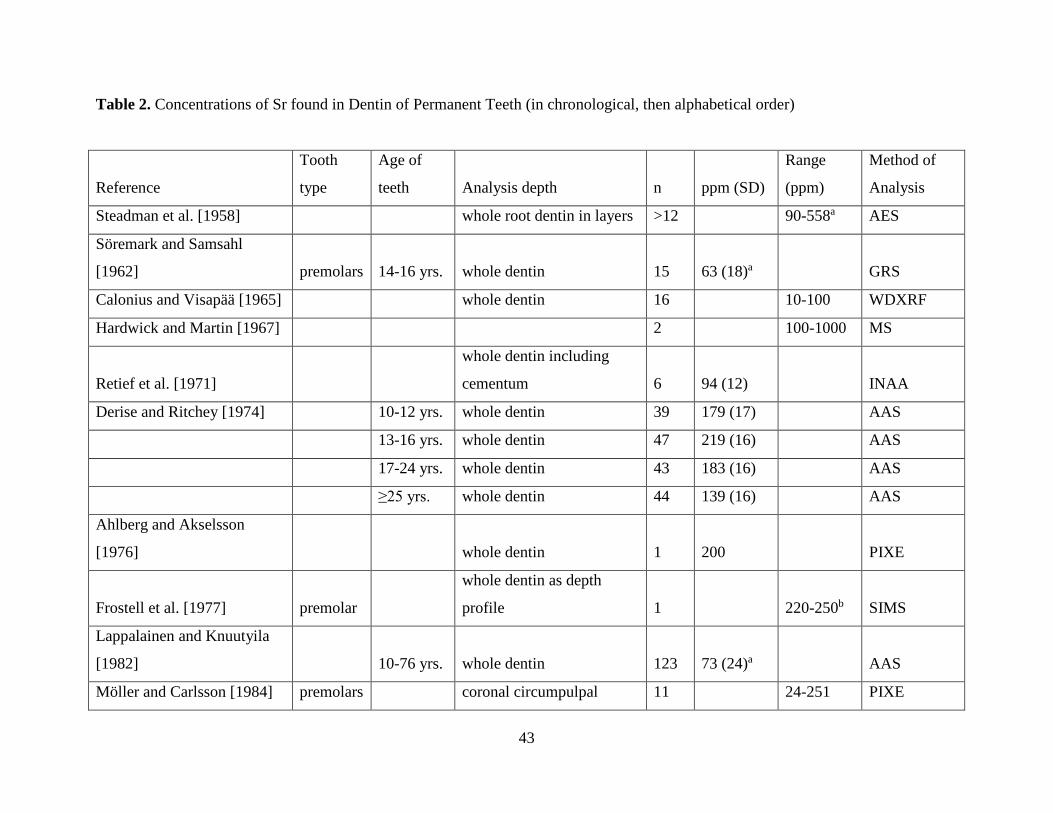
43
Table 2. Concentrations of Sr found in Dentin of Permanent Teeth (in chronological, then alphabetical order)
Reference
Tooth
type
Age of
teeth Analysis depth n ppm (SD)
Range
(ppm)
Method of
Analysis
Steadman et al. [1958]
whole root dentin in layers >12
90-558a AES
Söremark and Samsahl
[1962] premolars 14-16 yrs. whole dentin 15 63 (18)a
GRS
Calonius and Visapää [1965]
whole dentin 16
10-100 WDXRF
Hardwick and Martin [1967]
2
100-1000 MS
Retief et al. [1971]
whole dentin including
cementum 6 94 (12)
INAA
Derise and Ritchey [1974]
10-12 yrs. whole dentin 39 179 (17)
AAS
13-16 yrs. whole dentin 47 219 (16)
AAS
17-24 yrs. whole dentin 43 183 (16)
AAS
≥25 yrs. whole dentin 44 139 (16)
AAS
Ahlberg and Akselsson
[1976]
whole dentin 1 200
PIXE
Frostell et al. [1977] premolar
whole dentin as depth
profile 1
220-250b SIMS
Lappalainen and Knuutyila
[1982]
10-76 yrs. whole dentin 123 73 (24)a
AAS
Möller and Carlsson [1984] premolars
coronal circumpulpal 11
24-251 PIXE
44
dentin
a corrected for dentin water content of 10% b estimated value based on profile data

45
Table 3. Concentrations of Sr found in Plaque (in chronological order)
Reference n Age Geographical area ppm (SD) Comments
Schamschula et al. [1977a] 72
9.7-13.0
yrs. New South Wales, Australia 2.6 (2.2) in dry weight
Schamschula et al.
[1978a,b] 293 12-24 yrs.
several small, neighboring villages in
Papua New Guinea 20.4 (17.4) in dry weight
Shields et al. [1984] 32 14 yrs. Ohio, USA 140 (106) in solids
Shields et al. [1984] 19 14 yrs. Ohio, USA 338 (449) in fluid
Curzon [1985] 80 14 yrs. Ohio, USA 12.3 (14.6) in dry weight
Curzon [1985] 80 14 yrs. Ohio, USA 1.4 (1.6) in wet weight
Pearce and Sissons [1987] 4
Wellington, New Zealand 58.7 (14.9)a in dry weight
Spets-Happonen et al.
[1998] 19 30-55 yrs. Kuopio, Finland 17.8 in dry weight
a measured after the use of a Sr-free mouthrinse

46
Table 4. Concentrations of Sr found in Saliva (in chronological order)
Reference n Age Geographical area ppb (SD)
Range
(ppb) Comments
Arwill et al. [1967] 6
22-24
yrs. Umea, Sweden 11.3 (3.5) 7.7-16 resting
6
22-24
yrs. Umea, Sweden 21.7 (20.8) 8-63 stimulated (paraffin)
Dreizen et al. [1970] 15 adults Texas, USA 5 (6) <5-24
stimulated
(pilocarpine)
11 adults Texas, USA 13 (23) <5-89 stimulated (paraffin)
Cutress [1972b] 31
6-23
yrs. Wellington, New Zealand 20 (20) 0-?
stimulated, mixed
saliva
31
6-23
yrs. Wellington, New Zealand 20 (20) 0-?
stimulated, parotid
saliva
Schamschula et al.
[1978b] 283
12-24
yrs.
several small, neighboring villages in
Papua New Guinea 89 (122)
stimulated
Curzon [1984] 91 14 yrs. Wisonsin, USA 35 (30) 20-2930 resting
Shigemi et al. [2008] 521
6-12
yrs. Kitakyushu, Japan 7.7 (3.6) 1.7-20.6 resting

47
Table 5. Rat Caries Reduction as a Function of Sr Dose Pre- and Post-eruptively in the Presence or Absence of F
Reference Pre-/Post-a n Srb Fb E (total)c % change
vs. waterd
% change
vs.
fluorided
Meyerowitz et al. [1979] Pre- 10 0 0 29.6 - -
10 2.5 0 23.2 -21.6 -
9 5 0 21.5 -27.4 -
10 10 0 25.4 -14.2 -
10 0 1 28.5 -3.7 -
9 2.5 1 26.5 -10.5 -7.0
10 5 1 24.4 -17.6 -14.4
10 10 1 26.7 -9.8 -6.3
Post- 10 0 0 21.4 - -
9 50 0 18.2 -15.0 -
10 100 0 23.4 +9.3 -
10 150 0 21.3 -0.5 -
10 0 10 13.2 -38.3 -
8 50 10 14.2 -33.6 +7.6
10 100 10 15.4 -28.0 +16.7
Ashrafi et al. [1980] Pre- +
Post-
5 0 0 35.4 - -

48
5 0.5 + 5 0 29.3 -17.2 -
5 1.0 + 10 0 24.8 -29.9 -
4 2.5 + 25 0 28.3 -20.2 -
8 5.0 + 50 0 29.9 -15.6 -
6 7.5 + 75 0 21.5 -39.3 -
Curzon [1988] 10 0 0 34.9 - -
10 0 10 + 10 21.7 -37.8 -
10 0.5 + 5 10 + 10 24.1 -30.9 11.1
10 5 + 50 10 + 10 17.0 -51.3 -21.7
10 10 + 100 10 + 10 19.2 -45.0 -11.5
10 15 + 150 10 + 10 17.8 -49.0 -18.0
8 20 + 200 10 + 10 19.1 -45.3 -12.0
10 30 + 300 10 + 10 20.0 -42.7 -7.8
a Sr and/or F given pre- or post-eruptively b concentration in µg/10 g body weight if given pre-eruptively, or in mg/l if given post-eruptively (water) c sum of buccolingual, sulcal and proximal enamel caries scores d negative values are indicative of a caries reduction, positive values indicate increase

49
Figure 1. Summary of three epidemiological studies on effects of Sr in water on caries
occurrence (measured as DMFS scores): ■□ data from Curzon et al. [1978]; ● data from Curzon
et al. [1970]; ♦ data from Curzon [1985].
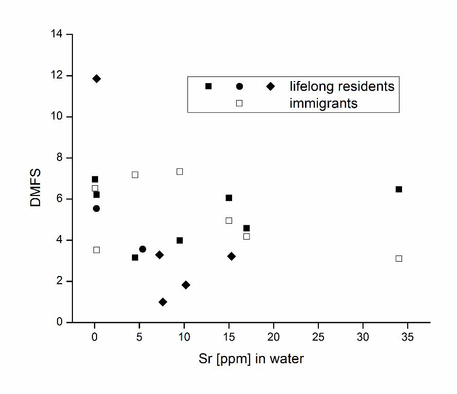
Related Documents