An International Journal of MINERALOGY, CRYSTALLOGRAPHY, GEOCHEMISTRY, ORE DEPOSITS, PETROLOGY, VOLCANOLOGY and applied topics on Environment, Archeometry and Cultural Heritage DOI: 10.2451/2011PM0006 Periodico di Mineralogia (2011), 79, 1 (Special Issue), 75-87 perIodIco di MInerAlogIA established in 1930 Special Issue in memory of Sergio Lucchesi Introduction The morphology of natural crystals strongly depends not only on the crystal structure but also on the environmental growth conditions. Morphology may be also regulated by impurity absorption and dissolution-corrosion and alteration (weathering, erosion, transportation and burial). Characterizing growth and post- growth effects can lead to a better understanding of growth conditions (P, T, X) as morphological features could be related to specific growth conditions (e.g.: striations on prism faces which are vertical in tourmaline and horizontal in quartz). Rounded morphologies, pipe-like channels and irregular voids are fairly common in a number of minerals in different growth environments and are often thought to have a post-growth origin. Yet their origins are under discussion, with different possible mechanisms being hypothesized. The exact mechanism in operation may depend not only on the crystal’s structure but also on the conditions present during and after growth. And Striations and hollow channels in rounded beryl crystals Gioacchino Tempesta * , Eugenio Scandale and Giovanna Agrosì Dipartimento Geomineralogico, Università di Bari, Italy * Corresponding author: [email protected] Abstract Structural defects in natural colourless beryl crystals from Minas Gerais (Brazil) were studied using X-ray Diffraction Topography (XRDT). The samples are characterised by a strongly rounded morphology and by the presence of hollow channels parallel to the c-axis, some of them visible to the naked eye and partially filled with kaolinite. The analysis of structural defects as dislocations, growth bands, solid inclusions and precipitates has been essential for the reconstruction of growth history in this study. The formation of hollow channels was attributed to the corrosion and post-genetic alteration of strongly deformed areas surrounding branches of dislocations parallel to the c-axis. The final rounded morphologies and the striations may have been attained either as the results of a parallel growth of some individuals or as the result of a variation of the growth rates in the final stages of growth. Due to the occurrence of kaolinite, post-genetic corrosion has been speculated. However, this process appears to have contributed solely to the hollow channel formation and not to the rounded morphology evident in these samples. Key words: X-ray topography; beryl; channels; morphology and striations.

Welcome message from author
This document is posted to help you gain knowledge. Please leave a comment to let me know what you think about it! Share it to your friends and learn new things together.
Transcript

An International Journal of
MINERALOGY, CRYSTALLOGRAPHY, GEOCHEMISTRY,
ORE DEPOSITS, PETROLOGY, VOLCANOLOGY
and applied topics on Environment, Archeometry and Cultural Heritage
DOI: 10.2451/2011PM0006Periodico di Mineralogia (2011), 79, 1 (Special Issue), 75-87
perIodIco di MInerAlogIA
established in 1930
Special Issue in memory of Sergio Lucchesi
Introduction
The morphology of natural crystals strongly
depends not only on the crystal structure but also
on the environmental growth conditions.
Morphology may be also regulated by impurity
absorption and dissolution-corrosion and
alteration (weathering, erosion, transportation
and burial). Characterizing growth and post-
growth effects can lead to a better understanding
of growth conditions (P, T, X) as morphological
features could be related to specific growth
conditions (e.g.: striations on prism faces which
are vertical in tourmaline and horizontal in
quartz).
Rounded morphologies, pipe-like channels and
irregular voids are fairly common in a number of
minerals in different growth environments and are
often thought to have a post-growth origin. Yet
their origins are under discussion, with different
possible mechanisms being hypothesized. The
exact mechanism in operation may depend not
only on the crystal’s structure but also on the
conditions present during and after growth. And
Striations and hollow channels in rounded beryl crystalsGioacchino Tempesta*, Eugenio Scandale and Giovanna Agrosì
Dipartimento Geomineralogico, Università di Bari, Italy*Corresponding author: [email protected]
Abstract
Structural defects in natural colourless beryl crystals from Minas Gerais (Brazil) were
studied using X-ray Diffraction Topography (XRDT). The samples are characterised by a
strongly rounded morphology and by the presence of hollow channels parallel to the c-axis,
some of them visible to the naked eye and partially filled with kaolinite. The analysis of
structural defects as dislocations, growth bands, solid inclusions and precipitates has been
essential for the reconstruction of growth history in this study. The formation of hollow
channels was attributed to the corrosion and post-genetic alteration of strongly deformed areas
surrounding branches of dislocations parallel to the c-axis. The final rounded morphologies
and the striations may have been attained either as the results of a parallel growth of some
individuals or as the result of a variation of the growth rates in the final stages of growth. Due
to the occurrence of kaolinite, post-genetic corrosion has been speculated. However, this
process appears to have contributed solely to the hollow channel formation and not to the
rounded morphology evident in these samples.
Key words: X-ray topography; beryl; channels; morphology and striations.
10.2451/2011PM0006_periodico 14/04/11 10.44 Pagina 75

G. Tempesta, E. Scandale and G. Agrosì76 Periodico di Mineralogia (2011), 80, 1 (Special Issue), 75-87
yet, even if rounded morphologies do depend on
the last stages of dissolution-corrosion
(weathering, erosion, transportation and burial),
crystals that grow at high supersaturations
(Argiolas and Baumer, 1978; Sunagawa, 1984)
may also be rounded.
In general, channels are to be expected with
screw dislocations of large Burgers vectors
(Frank, 1951; Baronnet, 1972; Dudley et al.,
1999) or with bunching of hundreds of parallel
dislocations and subsequent selective corrosion
of dislocation-core material (Scandale and
Zarka, 1982). Channels are also related to hollow
elongated cavities due to inclusion absorption
(Kawasaki et al., 2003; Minkoff, 1965). Voids
may result not only from the bunching of
channels (Scandale et al., 1993) but also from
branched crystal growth (Minkoff and Nixon,
1966) and from spherulitic growth (Morse and
Donnay, 1936) or from negative crystals.
Striations and thin parallel grooves on some
crystal faces of minerals, synthetic crystals and
kidney stones, are often attributed to the
convergence or juxtaposition of two or more small
crystal faces (Smolsky et al., 1999; Tolansky,
1945) belonging to the same or different
morphological forms. Lamellar twinning,
hundreds of twins repeated in a single specimen,
is also considered a common cause of striations
(Akizuki et al., 2001) as well as corrosion effects
(Akizuki et al., 2001).
The reconstruction of growth history on
minerals with X-ray Diffraction Topography-
XRDT (Authier and Zarka, 1994; Scandale,
1996) can aid in answering some general
questions. Recently, the growth history
reconstruction of tourmaline crystals, occurring
in pegmatite pockets on Elba Island (Italy),
strongly suggests a relationship between
tourmaline striations and the transition from
pegmatitic to hydrothermal growth stages
(Agrosì et al., 2006). In particular, two main
growth stages - the first one pegmatitic and the
second one hydrothermal - have been identified
and the observed striations may be connected
with a near parallel intergrowth of small blocks,
elongated along the c-axis, which developed in
the final hydrothermal growth stage.
These results prompted us to investigate the
occurrence of striations on beryl crystals in order
to determine if there are general links between the
development of striations, a channel’s origin and
the transition from magmatic to hydrothermal
pegmatite growth. The results shown here indicate
that the origin of both vertical striations and
channels cannot be simply attributed to a single
cause and that XRDT is a basic scientific tool that
can contribute to our understanding of the
mechanisms which are responsible for the
observed growth features.
Methods and Techniques
Geological Background
The samples used in the study originate from
the Coronel Murta district (Minas Gerais), a part
of the Oriental Pegmatitic Province of Brazil. The
pegmatites of this province are of a wide
typological and geochemical variety (Sá, 1977).
These pegmatites of Brasiliano-cycle have been
dated 525 Ma (Siga Jr., 1986) and intrude into
biotite - andalusite kyanite and staurolite bearing
schist (Costa, 1987) that correspond to a
metamorphosed flysch of the Eocambrian Salinas
Formation. They are associated with two-micas
and tourmaline granites. These pegmatites are
lithaniferous and the Li, Rb and Cs contents vary
in each zone of each pegmatite body.
Samples
Natural beryl crystals from Minas Gerais
(Brazil) were examined and found to share
coarse rounded elongated prism morphology
which presents vertical striations, on the prism
faces, hollow channels and irregular voids
parallel to the c-axis, in the inner zones, within
which a white powder identified as kaolinite was
observed.
10.2451/2011PM0006_periodico 14/04/11 10.44 Pagina 76

Striations and hollow channels in rounded beryl... 77Periodico di Mineralogia (2011), 80, 1 (Special Issue), 75-87
The results presented here discuss three natural
colourless beryl crystals labelled HB1, HB2 and
HB3 which are representative of the other
crystals studied. The samples are elongated,
strongly rounded {101-0} and {112
-0} prisms,
terminating in a prominent {0001} pinacoid.
Beryl HB1 has a length of about 3.2 cm and a
basal section of about 1.3 x 0.9 cm; beryl HB2
has a length of about 2.6 cm and a basal section
of 0.8 x 0.9 cm; and beryl HB3 has a length of
about 3.6 cm and a basal section of 0.6 x 0.7 cm.
Beryl HB1 and HB2 were studied using X-ray
Diffraction Topography (XRDT), Powder XRD,
and Optical Microscopy, whereas HB3 was
studied with Optical Microscopy and Powder
XRD only.
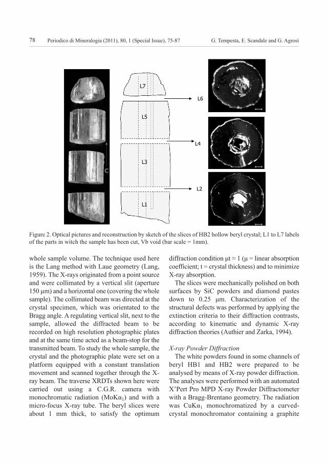
To reconstruct the growth history, 9 basal
slices labelled from bottom to top L1 to L9 were
cut from HB1 (Figure 1) and 7 basal slices
labelled from L1 to L7 were obtained from HB2
(Figure 2). Structural defects were investigated
using XRDT on slice HB1-L8 and on slices
HB2-L2, HB2-L4 and HB2-L6.
X-ray Diffraction Topography (XRDT)
XRDT is an imaging technique based on
Bragg diffraction and can characterize extended
defects in nearly perfect crystals (Lang, 1959).
The size of the slices, up to several cm2 in the
cross-sectional basal area and up to a few
millimetres in thickness, ensures that the
structural defects observed are typical of the bulk
samples. Mapping of structural defects
represents the local variations of diffracted
intensities and/or diffracted beam directions
produced by the strain fields which are
associated with extended defects and/or
distortions in the single-crystal lattice. XRDT
techniques yield spatial distribution and full
characterization of the crystal defects in the
Figure 1. Optical pictures and reconstruction by sketch of the slices of HB1 hollow beryl crystal; L1 to L9 labels
of the parts in witch the sample has been cut, Va1 and Va2 voids (bar scale = 2 mm).
10.2451/2011PM0006_periodico 14/04/11 10.44 Pagina 77
G. Tempesta, E. Scandale and G. Agrosì78 Periodico di Mineralogia (2011), 80, 1 (Special Issue), 75-87
whole sample volume. The technique used here
is the Lang method with Laue geometry (Lang,
1959). The X-rays originated from a point source
and were collimated by a vertical slit (aperture
150 µm) and a horizontal one (covering the whole
sample). The collimated beam was directed at the
crystal specimen, which was orientated to the
Bragg angle. A regulating vertical slit, next to the
sample, allowed the diffracted beam to be
recorded on high resolution photographic plates
and at the same time acted as a beam-stop for the
transmitted beam. To study the whole sample, the
crystal and the photographic plate were set on a
platform equipped with a constant translation
movement and scanned together through the X-
ray beam. The traverse XRDTs shown here were
carried out using a C.G.R. camera with
monochromatic radiation (MoKα1) and with a
micro-focus X-ray tube. The beryl slices were
about 1 mm thick, to satisfy the optimum
diffraction condition μt ≈ 1 (μ = linear absorption
coefficient; t = crystal thickness) and to minimize
X-ray absorption.
The slices were mechanically polished on both
surfaces by SiC powders and diamond pastes
down to 0.25 μm. Characterization of the
structural defects was performed by applying the
extinction criteria to their diffraction contrasts,
according to kinematic and dynamic X-ray
diffraction theories (Authier and Zarka, 1994).
X-ray Powder Diffraction
The white powders found in some channels of
beryl HB1 and HB2 were prepared to be
analysed by means of X-ray powder diffraction.
The analyses were performed with an automated
X’Pert Pro MPD X-ray Powder Diffractometer
with a Bragg-Brentano geometry. The radiation
was CuKα1 monochromatized by a curved-
crystal monochromator containing a graphite
Figure 2. Optical pictures and reconstruction by sketch of the slices of HB2 hollow beryl crystal; L1 to L7 labels
of the parts in witch the sample has been cut, Vb void (bar scale = 1mm).
10.2451/2011PM0006_periodico 14/04/11 10.44 Pagina 78

Striations and hollow channels in rounded beryl... 79Periodico di Mineralogia (2011), 80, 1 (Special Issue), 75-87
crystal. The step scans were 0.02° as 2θ and the
step time was 0.8 seconds while the angular
range was 2θ = 5° - 75°. The spectrum analysis,
performed with a specific software (X’Pert
Graphics and Identify), identified the white
powder as kaolinite Al2Si2O5(OH)4.
results
Optical Observations
Optical Microscopy (OM) showed vertical
striations and parallel grooves on the prism faces
(Figures 1 and 2). Pyramidal etch patterns, with
strongly [0001] elongated pseudo-hexagonal
bases, were observed on the prism faces of both
samples and are similar to those observed on
beryl crystals from Elba Island (Italy) previously
studied with XRDT (Scandale et al., 1990). No
relations were found between the etch patterns
and bulk structural defects.
Pipe-like hollow channels parallel to the c-
axis with diameters ranging from about 1 to
500 microns are present in the inner zones and
generally outcrop on the basal face, exhibiting
various morphologies, from conic-cylindrical
to sharp hexagonal, with edges parallel to the
{101-0} and {112
-0} prism faces (Figure 3). A few
channels contain fluid inclusions and others are
optically discontinuous. Similar optical
observations had been made in other synthetic
and natural crystals, beryl included (Scandale
and Zarka, 1982; Agrosì et al., 2005).
Large irregular voids, often penetrating
Figure 3. a) Optical image of the slice HB1-L8 and enlarged details of channels and voids (bar scale on the left
= 2 mm; bar scale on right 0.2 mm); b) Optical image of the slice HB2-L6and enlarged details of channels and
void (bar scale on the left = 1 mm; bar scale on right 0.5 mm).
10.2451/2011PM0006_periodico 14/04/11 10.44 Pagina 79

G. Tempesta, E. Scandale and G. Agrosì80 Periodico di Mineralogia (2011), 80, 1 (Special Issue), 75-87
through the entire sample, were observed in the
central inner cores. Voids and the larger
channels, visible to the naked eyes, are often
filled with a white polycrystalline material that
was found to be kaolinite. A closer observation
confirmed that the channels and voids, crossing
the sample, are filled with kaolinite,
continuously from top to bottom in some cases
and in others discontinuously in a dashed trail.
The large central irregular voids in both
samples present different shapes in the different
slices (Figures 1 and 2). In particular, it can be
observed that void Va in HB1 changes from top
to bottom, splitting into smaller voids while its
total empty area decreases. On the contrary, in
HB2, void Vb retains the outline while its
internal area increases slightly from top to
bottom. The channels C of HB1 and HB2 are
parallel and their shape matches the
morphological faces. It could be observed that
the different slices change dimensions and
contours and that the channel abundance of HB2
is less than that of HB1.
Large uneven voids V and channels C can be
noted in Figure 3. The larger voids Va1 and Va2
separate blocks T1, T2 and T3 from each other,
whereas the smaller ones are contained within
the blocks. Most channels are localized in block
T1 where they can be divided into two groups. In
fact, those close to the external rim have
elongated hexagonal shapes with sides parallel
to the morphological faces while the others, near
the internal rim, have a mainly rounded elliptic
shape with a long semi-major axis parallel to the
crystal faces.
Observations under crossed polarisers revealed
an anomalous wavy extinction originating from
the channels in HB2 and from differently
oriented crystal domains in HB1 (Figure 4).
X-ray Topographic Observations
XRDT images of all basal slices of the HB1
and HB2 samples were taken with the three 101-0
equivalent reflections. The diffraction contrasts
observed with the equivalent reflections result
quite similar in type and distribution and thus,
Figure 4. Optical image using crossed polarizes of a detail of the studied slice L6 of HB2 showing stress areas
around channels.
10.2451/2011PM0006_periodico 14/04/11 10.44 Pagina 80

Striations and hollow channels in rounded beryl... 81Periodico di Mineralogia (2011), 80, 1 (Special Issue), 75-87
Figure 5. a) XRDT of HB1-L8 obtained combining three topographs, taken with the same diffraction vector
g = 101-0 from regions (T1, T2 and T3) slightly disoriented (bar scale = 1 mm); b) Enlarged detail of HB1-L8
topograph shown in Figure 5 coupled with a sketch of T1 representing dislocation bundles DB1 nucleated from
inclusions adsorbed on boundary B1, and dislocation bundles DB2 ending on the growth stage boundary B2
(bar scale = 0.5 mm).
a
b
10.2451/2011PM0006_periodico 14/04/11 10.44 Pagina 81

G. Tempesta, E. Scandale and G. Agrosì82
for the sake of brevity, only those taken with
diffracting vector g = 101-0 will be shown and
discussed here.
The topographs of HB1 (Figure 5) and those
of HB2 (Figure 6 and 7) showed that the main
structural defects of the studied crystals are
precipitates (P), solid inclusions (I), dislocation
bundles nucleated from solid inclusions (DB),
pipe-like channels (C), voids (V), and growth
bands (G). The topographs demonstrated that
even if the structural defects observed in the
studied crystals are similar, the diffraction
contrasts of each sample characterize and
differentiate each other. For this reason it is
necessary a separate illustration of the diffraction
contrasts of the two samples was required.
HB1 sample
Figure 5 illustrates the composition of three
topographs, T1, T2 and T3, taken with the same
diffracting vector g = 101-0 from slightly
disoriented regions of the slice HB1-L8. The
composition shows that the crystal is an aggregate
of three independently diffracting blocks,
coherently scattering, within which phase
relationships hold. The complementary topographs
were taken with the same diffracting vector g with
a rotation of a few seconds of arc about the
Bragg angle θ101-0.
In the blocks T1 and T3 basal dislocation
bundle DB (Figure 5b), with individual contrasts
generally not resolved, nucleated from solid
inclusions I, were observed in the late growth
stages where the defect density increases. In the
block T1, dislocation bundles DB1 were nucleated
from inclusions adsorbed on boundary B1,
whereas, dislocation bundles DB2 end on the
growth stage boundary B2 (Figure 5b). The DB1
dislocation lines point from B1 towards the
external rim whereas DB2 dislocation lines point
to B2, following two opposite growth directions,
n1 and n2 respectively (Klapper, 1972; Graziani
et al., 1981).
The inhomogeneous diffraction contrasts
observed in the T1 crystal region and bounded by
B1 and B2, arise from local variations of
diffracted intensities which were due to the strain
fields associated to subgrain boundaries, as well
as, from direction variation of the diffracted
beams which were caused by misoriented grains.
Therefore B1 and B2 boundaries are polygonal-
lines made up of a large number of subgrain
boundary sides of small dimensions.
In T1 and T3, precipitates P were found
Periodico di Mineralogia (2011), 80, 1 (Special Issue), 75-87
Figure 6. a) XRDT of HB2 L2 taken with the diffracting vector g = 101-0 (bar scale = 1 mm); b) XRDT of HB2
L4 taken with the diffracting vector g = 101-0; G1 and G2 = growth bands, DB = dislocation bundles (bar scale
= 1 mm).
a b
10.2451/2011PM0006_periodico 14/04/11 10.44 Pagina 82
Striations and hollow channels in rounded beryl... 83
together with their classical diffraction contrasts
which consist of two circular parts connected
together by a contrast-free plane normal to the
operating diffracting vector g (Tanner, 1976).
HB2 sample
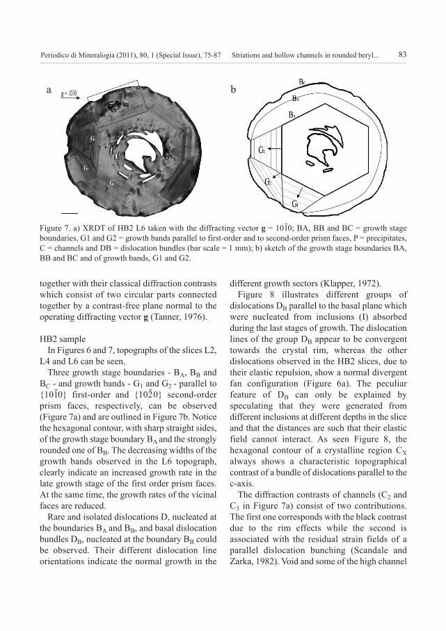
In Figures 6 and 7, topographs of the slices L2,
L4 and L6 can be seen.
Three growth stage boundaries - BA, BB and
BC - and growth bands - G1 and G2 - parallel to
{101-0} first-order and {102
-0} second-order
prism faces, respectively, can be observed
(Figure 7a) and are outlined in Figure 7b. Notice
the hexagonal contour, with sharp straight sides,
of the growth stage boundary BA and the strongly
rounded one of BB. The decreasing widths of the
growth bands observed in the L6 topograph,
clearly indicate an increased growth rate in the
late growth stage of the first order prism faces.
At the same time, the growth rates of the vicinal
faces are reduced.
Rare and isolated dislocations D, nucleated at
the boundaries BA and BB, and basal dislocation
bundles DB, nucleated at the boundary BB could
be observed. Their different dislocation line
orientations indicate the normal growth in the
different growth sectors (Klapper, 1972).
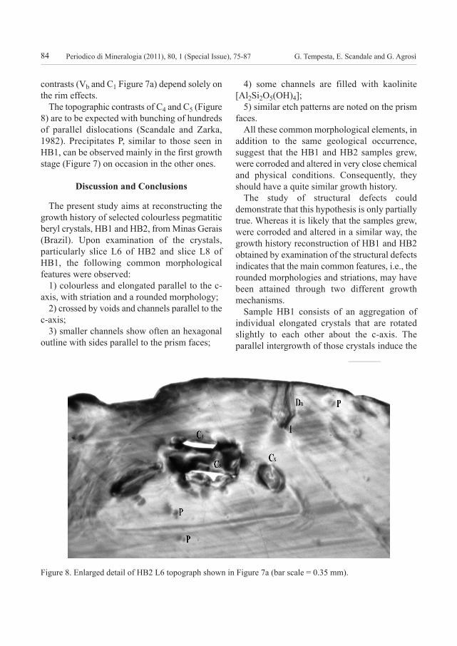
Figure 8 illustrates different groups of
dislocations DB parallel to the basal plane which
were nucleated from inclusions (I) absorbed
during the last stages of growth. The dislocation
lines of the group DB appear to be convergent
towards the crystal rim, whereas the other
dislocations observed in the HB2 slices, due to
their elastic repulsion, show a normal divergent
fan configuration (Figure 6a). The peculiar
feature of DB can only be explained by
speculating that they were generated from
different inclusions at different depths in the slice
and that the distances are such that their elastic
field cannot interact. As seen Figure 8, the
hexagonal contour of a crystalline region CX
always shows a characteristic topographical
contrast of a bundle of dislocations parallel to the
c-axis.
The diffraction contrasts of channels (C2 and
C3 in Figure 7a) consist of two contributions.
The first one corresponds with the black contrast
due to the rim effects while the second is
associated with the residual strain fields of a
parallel dislocation bunching (Scandale and
Zarka, 1982). Void and some of the high channel
Periodico di Mineralogia (2011), 80, 1 (Special Issue), 75-87
Figure 7. a) XRDT of HB2 L6 taken with the diffracting vector g = 101-0; BA, BB and BC = growth stage
boundaries, G1 and G2 = growth bands parallel to first-order and to second-order prism faces, P = precipitates,
C = channels and DB = dislocation bundles (bar scale = 1 mm); b) sketch of the growth stage boundaries BA,
BB and BC and of growth bands, G1 and G2.
a b
10.2451/2011PM0006_periodico 14/04/11 10.44 Pagina 83
G. Tempesta, E. Scandale and G. Agrosì84
contrasts (Vb and C1 Figure 7a) depend solely on
the rim effects.
The topographic contrasts of C4 and C5 (Figure
8) are to be expected with bunching of hundreds
of parallel dislocations (Scandale and Zarka,
1982). Precipitates P, similar to those seen in
HB1, can be observed mainly in the first growth
stage (Figure 7) on occasion in the other ones.
discussion and conclusions
The present study aims at reconstructing the
growth history of selected colourless pegmatitic
beryl crystals, HB1 and HB2, from Minas Gerais
(Brazil). Upon examination of the crystals,
particularly slice L6 of HB2 and slice L8 of
HB1, the following common morphological
features were observed:
1) colourless and elongated parallel to the c-
axis, with striation and a rounded morphology;
2) crossed by voids and channels parallel to the
c-axis;
3) smaller channels show often an hexagonal
outline with sides parallel to the prism faces;
4) some channels are filled with kaolinite
[Al2Si2O5(OH)4];
5) similar etch patterns are noted on the prism
faces.
All these common morphological elements, in
addition to the same geological occurrence,
suggest that the HB1 and HB2 samples grew,
were corroded and altered in very close chemical
and physical conditions. Consequently, they
should have a quite similar growth history.
The study of structural defects could
demonstrate that this hypothesis is only partially
true. Whereas it is likely that the samples grew,
were corroded and altered in a similar way, the
growth history reconstruction of HB1 and HB2
obtained by examination of the structural defects
indicates that the main common features, i.e., the
rounded morphologies and striations, may have
been attained through two different growth
mechanisms.
Sample HB1 consists of an aggregation of
individual elongated crystals that are rotated
slightly to each other about the c-axis. The
parallel intergrowth of those crystals induce the
Periodico di Mineralogia (2011), 80, 1 (Special Issue), 75-87
Figure 8. Enlarged detail of HB2 L6 topograph shown in Figure 7a (bar scale = 0.35 mm).
10.2451/2011PM0006_periodico 14/04/11 10.44 Pagina 84
Striations and hollow channels in rounded beryl... 85
formation of cavities and voids in the inner part
and striations on the morphological faces.
Due to the fact that voids and cavities are
bounded by morphological faces of different
mineral individuals, it can be observed that the
growth occurs in opposite directions. As an
example, Figure 5b illustrates bundles of DB
dislocations perpendicular to the outer rim B1 vand
to the growth stage boundary B2, that point in
opposite directions starting from solid inclusions I.
Post-genetic selective dissolution, driven by the
deformation fields of dislocation bundles
(Scandale and Zarka, 1982) may have contributed
also to the formation of channels and irregular
voids in the inner parts of the individual grains and
not to the rounded morphology of the sample. The
final alteration resulted in the formation of
kaolinite inside the larger channels and irregular
voids.
The observation and the analysis of the defects
of the crystal HB1, shows that the defects are
identical to those revealed in HB2 but that their
distribution could point to a different evolution.
In the case of beryl HB2, the final morphology
is mainly due to an abrupt increase of the growth
rates of the first-order prism faces {101-0} with
respect to second-order prism {102-0} (Figure 7).
Hence, the rounded morphology may not be the
result of a strong corrosion process but, instead,
the result of a dramatic variation of the growth
velocities of the crystal faces as seen in the
diffraction images.
The HB2 topographs provide clear evidence
of well-defined growth marks as observed in all
the topographs. In fact, it is possible to draw
various growth boundaries in the crystal, which
represent the complete evolution, as it follows
from the schematic illustration in Figure 7b.
Upon observation of the growth boundaries,
two main boundaries could be identified: BA and
BB (Figure 7b) which are connected to
variations in growth conditions. The transition
from the sharp hexagonal boundaries BA to the
rounded one BB is related to asymmetrical
variations of the growth velocities of the {101-0}
and {102-0} growth sectors. From this we can
draw the unequivocal conclusion that the early
crystal shape was a hexagonal prism that over
time became strongly rounded off. This most
likely is not due to a secondary corrosion, as a
first macroscopic observation of the striations
might indicate, but because of (P, T, X).
Periodico di Mineralogia (2011), 80, 1 (Special Issue), 75-87
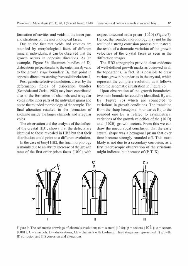
Figure 9. The schematic drawings of channels evolution; m = sectors {101-0}; p = sectors {101
-1}; c = sectors
{0001}; C = channels; D = dislocations; Ck = channels with kaolinite. Three stages are represented: I) growth,
II) corrosion and III) corrosion and alterations.
10.2451/2011PM0006_periodico 14/04/11 10.44 Pagina 85

G. Tempesta, E. Scandale and G. Agrosì86
X-ray topographs have been invaluable in
finding the origin of channels. In fact, the
topographic contrasts CX (Figure 8) were able to
localize the crystal region, where corrosion
preferentially occured, due to the high strain
concentration produced by the bundle of
parallel dislocations (Scandale & Zarka, 1982).
Figure 9 is a schematic illustration describing
in general terms the formation of the channels in
HB1 and HB2. Three stages are represented: I)
growth, II) corrosion, III) corrosion and
alterations. Hence, the deformation fields,
associated to the dislocation bundles normal to
(0001), may have determined the favourable
conditions for a preferential etching that produced
the observed channels. However, the same
mechanism does not appear to have operated on
the dislocation bundles working on the sectors m
{101-0} and p {112
-0}. There is in fact (Sangwal,
1987) an anisotropy in the macroscopic
dissolutions rates; therefore on some faces the
process is so slow to be considered negligible.
Moreover, in agreement with the kinematic
theory, anisotropy was the same when considering
growth, and the nucleation rate and dissolution
rate are proportional among them. In conclusion,
it can be stated that the channels’ origin is due to
a post-genetic corrosion.
It seems therefore that the X-ray topography
method is central in the reconstruction of the
growth history of natural crystals. This
technique can give answers to mineralogical
problems originating from the fact that the
conditions of crystal growth are still not fully
understood. All the common features analysed
indicate that HB1 and HB2 originated from a
geological environment with similar chemical
and physical conditions. However, the analysis
of the structural defects using XRDT confirmed
that two kinds of growth mechanisms occurred:
a parallel growth for the HB1 sample; and a
single crystal growth with a different growth
rate on the faces of the first order and second
order prism for the HB2 sample.
Acknowledgements
The Authors are grateful to Prof. Dino Aquilano and
to the Anonymous Referee for the constructive
contribution in the reviewing of the paper. Moreover,
the authors thank Dr. Carlaina Brown, who revised the
English text. The research was funded by MURST
grants to E.S.
references
Agrosì G., Fregola R.A., Monno A., Scandale E. and
Tempesta G. (2005) - XRDT Study of Structural
Defects of 6H-SiC Crystals. Materials Science
Forum, 483-485, 311-314.
Agrosì G., Bosi F., Lucchesi S., Melchiorre G. and
Scandale E. (2006) - Mn-tourmaline crystals from
island of Elba (Italy): Growth history and growth
marks. American Mineralogist, 91, 944-952.
Akizuki M., Kuribayshi T., Nagase T. and Kitakaze A.
(2001) - Triclinic Liddicoatite and Elbaite in
Growth Sectors of Tormaline from Madagascar.
American Mineralogist, 86, 364-369.
Appleman D.E. and Evans H.T. Jr. (1973) - Indexing
and least-squares refinement of powder diffraction
data. National Tech. Information Service, U.S.
Department of Commerce, Springfield, Virginia,
Doc. PB-216, 188.
Argiolas R. and Baumer A. (1978) - Synthése de
chlorapatite par voie hydrothermale; étude de
l’influence de la sursaturation sur l’évolution des
faciés des cristaux. Canadian Mineralogist, 16,
285-290.
Authier A. and Zarka A. (1994) - X-ray topographic
study of the real structure of minerals. In A.S.
Marfunin Ed., Composition, Structure and
Properties of Mineral Matter, Springer-Verlag,
Berlin, 221-233.
Azaroff L.V., Kaplow R., Kato N., Weiss R.I., Wilson
A.J.C., Young, R.A. (1974) - X-ray diffraction. Mc
Graw Hill, New York.
Baronnet A. (1972) - Growth mechanisms and
polytypism in synthetic hydroxyl-bearing phlogopite.
American Mineralogist, 57, 1271-1293.
Costa A.G. (1987) - Petrologie und geochemische
Untersuchungen des Gneis-Migmatite-Gebiets
von Itinga, Jequitinhonha-Tal, Nordoestliches
Minas Gerais, Brasilien. West Germany. (Dr.
Thesis, TU-Clausthal).
Periodico di Mineralogia (2011), 80, 1 (Special Issue), 75-87
10.2451/2011PM0006_periodico 14/04/11 10.44 Pagina 86
Striations and hollow channels in rounded beryl... 87
Dudley M., Huang H.R. and Huang W. (1999) -
Assessment of orientation and extinction contrast
contributions to direct dislocation image. Journal
of Physics D: Applied Physics, 32, A139-A144.
Frank F.C. (1951) - Capillary equilibria of dislocated
crystals. Acta Crystallographica, 4, 497- 501.
Graziani G., Lucchesi S. and Scandale E. (1990) -
General and Specific Growth Marks in Pegmatite
Beryls. Physics and Chemistry of Minerals, 17, 379-
384.
Graziani G., Scandale E. and Zarka A. (1981) -
Growth of Beryl Single Crystal – History of
development and genetic medium. Journal of
Applied Crystallography, 14, 241-246.
Kawasaki M., Onuma K. and Sunagawa I. (2003) -
Morphological instabilities during growth of rough
interface: AFM observations of cobbles on the
(0001) face of synthetic quartz crystals. Journal of
Crystal Growth, 258, 188-196.
Klapper H. (1972) - Elastische Energie und
Vorzugsrichtungen geradliniger. Versetzungen in
aus der Losung gewachsenen organischen
Kristallen. Physica Status Solidi A, 14, 99-106.
Lang A.R. (1959) - The projection topograph: a new
method in X-ray diffraction microradiography. Acta
Crystallographica, 12, 249-250.
Minkoff I. (1965) - Hole Formation in Crystal Growth
by Surface Adsorption of an Impurity.
Philosophical Magazine, 12, 1083-1086.
Minkoff I. and Nixon W.C. (1966) - Scanning Electron
Microscopy of Graphite Growth in Iron and Nickel
Alloys. Journal of Applied Physics, 37, 4848-4855.
Morse H.W. and Donnay D.H. (1936) - Optics and
structure of three-dimensional spherulites.
American Mineralogist, 21, 391-426.
Sá J.H. de (1977) - Pegmatitos litiniferos da região de
Ilinga-Araçuaí. Ph. D. thesis, USP, São Paulo,
Brasil.
Sangwal K. (1987) - Etching of Crystals: Theory,
Experiment and Application. Defects in Solids,
Amsterdam, Oxford, New York, Tokyo: North
Holland, 15, 497.
Scandale E. and Lucchesi S. (2000) - Growth and
sector zoning in a beryl crystal. European Journal
of Mineralogy, 12, 357-366.
Scandale E. (1996) - Growth and Sector zonings. In
E. Scandale and A. Baronnet, Eds., Crystal Growth
in Earth Sciences, EDISU, Torino, 176-194.
Scandale E., Lucchesi S. and Graziani G. (1993) -
Improvements on the growth history reconstruction
of a beryl crystal by growth marks. Neues Jahrbuch
für Mineralogie Monatshefte, 4, 172-184.
Scandale E., Lucchesi S. and Graziani G. (1990) -
Growth defects and growth marks in pegmatite
beryls. European Journal of Mineralogy, 2, 305-311.
Scandale E. and Zarka A. (1982) - Sur l’origine des
canaux dans les cristaux. Journal of Applied
Crystallography, 15, 417-422.
Scandale E., Scordari F. and Zarka A. (1979a) - Etude
des défauts dans des monocristaux naturels de béryl.
I. Observations des dislocations. Journal of Applied
Crystallography, 12, 70-77.
Scandale E., Scordari F. and Zarka A. (1979b) - Etude
des défauts dans des monocristaux naturels de béryl.
II, Etude de croissance. Journal of Applied
Crystallography, 12, 78-83.
Siga Jr. O. (1986) - A evolução geocronológica da
porão nordeste de Minas Gerais, com base em
interpretações geocronológicas. Inst. Geociências,
Universidade de São Paulo, São Paulo, Brazil, MSc
thesis.
Smolsky I.L., Voloshin A.E., Zaitseva N.P., Rudneva
E.B. and Klapper H. (1999) - X-ray topographic
study of striation formation in layer growth of
crystals from solutions. X-ray Topography and
Crystal Characterization, Philosophical
Transactions of the Royal Society of London, A
357, 2631-2649.
Sunagawa I. (1984) - Growth of crystals in nature.
Materials science of the earth’s interior, I. Sunagawa.
Terra Scientific Publishing, Tokyo, 61-103.
Tanner B.K. (1976) - X-ray Diffraction Topography.
Pergamon Press, Oxford.
Tolansky S. (1945) - The Topography of Crystal
Faces. I. The Topography of a (100) Face of a Left-
Handed Quartz Crystal. Proceedings of the Royal
Society of London, A 184, 41-51.
Submitted, October 2010 - Accepted, January 2011
Periodico di Mineralogia (2011), 80, 1 (Special Issue), 75-87
10.2451/2011PM0006_periodico 14/04/11 10.44 Pagina 87
Related Documents