Stretching of Single Collapsed DNA Molecules Christoph G. Baumann,* Victor A. Bloomfield,* Steven B. Smith, Carlos Bustamante, ‡ Michelle D. Wang, § and Steven M. Block § *Department of Biochemistry, University of Minnesota, St. Paul, MN 55108; Department of Molecular and Cell Biology, University of California, Berkeley, CA 94720; ‡ Department of Physics, University of California, Berkeley, and Physical Biosciences Division, Lawrence Berkeley Laboratory, Berkeley, CA 94720; and § Department of Molecular Biology, Princeton University, Princeton, NJ 08544 ABSTRACT The elastic response of single plasmid and lambda phage DNA molecules was probed using optical tweezers at concentrations of trivalent cations that provoked DNA condensation in bulk. For uncondensed plasmids, the persistence length, P, decreased with increasing spermidine concentration before reaching a limiting value 40 nm. When condensed plasmids were stretched, two types of behavior were observed: a stick-release pattern and a plateau at ;20 pN. These behaviors are attributed to unpacking from a condensed structure, such as coiled DNA. Similarly, condensing concentrations of hexaammine cobalt(III) (CoHex) and spermidine induced extensive changes in the low and high force elasticity of lambda DNA. The high force (5–15 pN) entropic elasticity showed worm-like chain (WLC) behavior, with P two- to fivefold lower than in low monovalent salt. At lower forces, a 14-pN plateau abruptly appeared. This corresponds to an intramolecular attraction of 0.083– 0.33 kT/bp, consistent with osmotic stress measurements in bulk condensed DNA. The intramolecular attractive force with CoHex is larger than with spermidine, consistent with the greater efficiency with which CoHex condenses DNA in bulk. The transition from WLC behavior to condensation occurs at an extension about 85% of the contour length, permitting looping and nucleation of condensation. Approximately half as many base pairs are required to nucleate collapse in a stretched chain when CoHex is the condensing agent. INTRODUCTION DNA undergoes ordered collapse, or condensation, in dilute solution in the presence of multivalent cations such as spermidine 31 , hexaammine cobalt(III), and spermine 41 (Gosule and Schellman, 1976; Widom and Baldwin, 1980; Wilson and Bloomfield, 1979). The toroidal condensed state is similar to the structure of packaged bacteriophage DNA, and some of the physical interactions involved in DNA condensation, such as bending, entropy loss, and modifica- tion of coulombic interactions must also be involved in DNA packaging in eukaryotic nuclei and prokaryotic nucle- oids (see reviews by Bloomfield, 1991, 1996, 1997; Mar- quet and Houssier, 1991). Theory predicts that DNA col- lapse will involve an abrupt phase transition (Grosberg and Khokhlov, 1994), the sharpness of which is augmented by the inflexibility of DNA (Post and Zimm, 1979). Laser light scattering measurements of viral DNA collapse often yield continuous, albeit abrupt, transitions with respect to con- densing agent (Benbasat, 1984; Widom and Baldwin, 1980; Wilson and Bloomfield, 1979). The discrepancy between theory and experiment is thought to arise from competing condensation and aggregation reactions present in solutions not infinitely dilute (Post and Zimm, 1982a,b). Under highly dilute conditions in which overlap of molecular domains is negligible, fluorescence microscopy has been used to follow the collapse of single T4 bacteriophage DNAs induced by multivalent cations (Yoshikawa et al., 1996a,b). These experiments showed that the transition was indeed discontinuous at the single molecule level but con- tinuous for the ensemble average, thus unifying theory and experiment. The impetus for DNA condensation is generally thought to arise from attractive lateral interactions between adjacent helices generated upon binding a critical amount of multi- valent cations. The favorable interactions may reflect the influences of helix hydration (Arscott et al., 1995; Rau and Parsegian, 1992), helix secondary structure (Ma et al., 1995; Reich et al., 1991) and/or electrostatic attraction through ion correlation (Gronbech-Jensen et al., 1997; Marquet and Houssier, 1991; Oosawa, 1968; Rouzina and Bloomfield, 1996). Manning (1980, 1985), however, has put forth an- other idea: that sufficiently neutralized DNA will crumple like an overloaded elastic column. This implies that the impetus for collapse arises from the unbalanced retractile force exerted along the backbone of neutralized DNA, rather than from the lateral interactions between helices. In this paper we use manipulations of single DNA molecules, at extensions that preclude intramolecular contacts, to test these competing hypotheses. In previous work, our two groups, one at Princeton (Wang et al., 1997) and the other a Minnesota-Oregon collaboration (Baumann et al., 1997), used optical tweezers Received for publication 15 October 1999 and in final form 3 January 2000. C. Baumann’s current address: Department of Biology, University of York, Heslington, York Y010 5DD, United Kingdom. M. D. Wang’s current address: Department of Physics, LASSP, 514 Clark Hall, Cornell University, Ithaca, NY 14853-2501. S. M. Block’s current address: Departments of Biological Sciences and Applied Physics, Herrin Labs 029, Stanford University, Stanford, CA 94305-5020. Address reprint requests to Victor A. Bloomfield, Department of Biochem- istry, University of Minnesota, St. Paul, MN 55108. Tel.: 612-625-2268; Fax: 612-625-6775; E-mail: [email protected]. © 2000 by the Biophysical Society 0006-3495/00/04/1965/14 $2.00 1965 Biophysical Journal Volume 78 April 2000 1965–1978

Welcome message from author
This document is posted to help you gain knowledge. Please leave a comment to let me know what you think about it! Share it to your friends and learn new things together.
Transcript
Stretching of Single Collapsed DNA Molecules
Christoph G. Baumann,* Victor A. Bloomfield,* Steven B. Smith,† Carlos Bustamante,†‡ Michelle D. Wang,§
and Steven M. Block§
*Department of Biochemistry, University of Minnesota, St. Paul, MN 55108; †Department of Molecular and Cell Biology, University ofCalifornia, Berkeley, CA 94720; ‡Department of Physics, University of California, Berkeley, and Physical Biosciences Division, LawrenceBerkeley Laboratory, Berkeley, CA 94720; and §Department of Molecular Biology, Princeton University, Princeton, NJ 08544
ABSTRACT The elastic response of single plasmid and lambda phage DNA molecules was probed using optical tweezersat concentrations of trivalent cations that provoked DNA condensation in bulk. For uncondensed plasmids, the persistencelength, P, decreased with increasing spermidine concentration before reaching a limiting value 40 nm. When condensedplasmids were stretched, two types of behavior were observed: a stick-release pattern and a plateau at ;20 pN. Thesebehaviors are attributed to unpacking from a condensed structure, such as coiled DNA. Similarly, condensing concentrationsof hexaammine cobalt(III) (CoHex) and spermidine induced extensive changes in the low and high force elasticity of lambdaDNA. The high force (5–15 pN) entropic elasticity showed worm-like chain (WLC) behavior, with P two- to fivefold lower thanin low monovalent salt. At lower forces, a 14-pN plateau abruptly appeared. This corresponds to an intramolecular attractionof 0.083–0.33 kT/bp, consistent with osmotic stress measurements in bulk condensed DNA. The intramolecular attractiveforce with CoHex is larger than with spermidine, consistent with the greater efficiency with which CoHex condenses DNA inbulk. The transition from WLC behavior to condensation occurs at an extension about 85% of the contour length, permittinglooping and nucleation of condensation. Approximately half as many base pairs are required to nucleate collapse in astretched chain when CoHex is the condensing agent.
INTRODUCTION
DNA undergoes ordered collapse, or condensation, in dilutesolution in the presence of multivalent cations such asspermidine31, hexaammine cobalt(III), and spermine41
(Gosule and Schellman, 1976; Widom and Baldwin, 1980;Wilson and Bloomfield, 1979). The toroidal condensed stateis similar to the structure of packaged bacteriophage DNA,and some of the physical interactions involved in DNAcondensation, such as bending, entropy loss, and modifica-tion of coulombic interactions must also be involved inDNA packaging in eukaryotic nuclei and prokaryotic nucle-oids (see reviews by Bloomfield, 1991, 1996, 1997; Mar-quet and Houssier, 1991). Theory predicts that DNA col-lapse will involve an abrupt phase transition (Grosberg andKhokhlov, 1994), the sharpness of which is augmented bythe inflexibility of DNA (Post and Zimm, 1979). Laser lightscattering measurements of viral DNA collapse often yieldcontinuous, albeit abrupt, transitions with respect to con-densing agent (Benbasat, 1984; Widom and Baldwin, 1980;
Wilson and Bloomfield, 1979). The discrepancy betweentheory and experiment is thought to arise from competingcondensation and aggregation reactions present in solutionsnot infinitely dilute (Post and Zimm, 1982a,b). Underhighly dilute conditions in which overlap of moleculardomains is negligible, fluorescence microscopy has beenused to follow the collapse of single T4 bacteriophageDNAs induced by multivalent cations (Yoshikawa et al.,1996a,b). These experiments showed that the transition wasindeed discontinuous at the single molecule level but con-tinuous for the ensemble average, thus unifying theory andexperiment.
The impetus for DNA condensation is generally thoughtto arise from attractive lateral interactions between adjacenthelices generated upon binding a critical amount of multi-valent cations. The favorable interactions may reflect theinfluences of helix hydration (Arscott et al., 1995; Rau andParsegian, 1992), helix secondary structure (Ma et al., 1995;Reich et al., 1991) and/or electrostatic attraction through ioncorrelation (Gronbech-Jensen et al., 1997; Marquet andHoussier, 1991; Oosawa, 1968; Rouzina and Bloomfield,1996). Manning (1980, 1985), however, has put forth an-other idea: that sufficiently neutralized DNA will crumplelike an overloaded elastic column. This implies that theimpetus for collapse arises from the unbalanced retractileforce exerted along the backbone of neutralized DNA,rather than from the lateral interactions between helices. Inthis paper we use manipulations of single DNA molecules,at extensions that preclude intramolecular contacts, to testthese competing hypotheses.
In previous work, our two groups, one at Princeton(Wang et al., 1997) and the other a Minnesota-Oregoncollaboration (Baumann et al., 1997), used optical tweezers
Received for publication 15 October 1999 and in final form 3 January2000.
C. Baumann’s current address: Department of Biology, University of York,Heslington, York Y010 5DD, United Kingdom.
M. D. Wang’s current address: Department of Physics, LASSP, 514 ClarkHall, Cornell University, Ithaca, NY 14853-2501.
S. M. Block’s current address: Departments of Biological Sciences andApplied Physics, Herrin Labs 029, Stanford University, Stanford, CA94305-5020.
Address reprint requests to Victor A. Bloomfield, Department of Biochem-istry, University of Minnesota, St. Paul, MN 55108. Tel.: 612-625-2268;Fax: 612-625-6775; E-mail: [email protected].
© 2000 by the Biophysical Society
0006-3495/00/04/1965/14 $2.00
1965Biophysical Journal Volume 78 April 2000 1965–1978
to study the effects of cations on the elasticity of singleDNA molecules. We used different apparatus and differentDNA: a plasmid containing 3888 basepairs (bp) in one case,and l bacteriophage DNA containing 48,502 bp in theother. However, we obtained very similar results, notablythat the trivalent cations spermidine and hexaammine co-balt(III), commonly used as DNA condensing agents, mark-edly increase the apparent bending flexibility (entropic elas-ticity) of DNA above the supposed limiting value achievedin high concentrations of monovalent salt. The increasedbending is due to asymmetric shielding of the DNA phos-phate charge by multivalent ligands (Rouzina and Bloom-field, 1998; Stigter, 1998). The force-extension curves arewell fit by a worm-like chain (WLC) model, with a loweredpersistence lengthP resulting from increased bending flex-ibility. In those experiments we observed that at low stretch-ing forces and under condensing ionic conditions, individ-ual molecules often deviated from WLC elastic behavior,yielding instead a reversible force plateau which was attrib-uted to intramolecular DNA condensation.
In this paper we report further investigations on thedependence of single molecule condensation on ionic con-ditions and extent of stretching. We present results from ourtwo groups together in a single paper, despite significantdifferences in experimental details, so that we and ourreaders can critically examine the dependence of the resultson these details. We find good correlation with the condi-tions for DNA condensation in solution. We find that in-tramolecular condensation can occur only when the DNA issufficiently relaxed that intramolecular loops can occur,providing strong support for a lateral interaction rather thanan elastic buckling mechanism. A first-order phase transi-tion explains the discontinuous change in force with extension.
MATERIALS AND METHODS
DNA, buffers, and multivalent cation solutions
The Minnesota-Oregon group usedl DNA. The 59-overhangs ofl DNA(methylatedc1857ind 1 Sam7, New England Biolabs, Beverly, MA) werebiotinylated with the Klenowexo-enzyme (New England Biolabs) usingbio-11-dCTP (Sigma, St. Louis, MO), dATP, dGTP, and dUTP as de-scribed previously (Smith et al., 1996). Single-strand nicks were repairedwith T4 DNA ligase. After biotinylation and nick ligation, DNA stockswere stored in an EDTA-containing buffer.
Monovalent salt buffer solutions were prepared using 100 mM cacody-late, pH 7, buffer stocks (86.2 mM sodium cacodylate, 13.8 mM cacodylicacid) supplemented with either 100 or 500 mM NaCl (total Na1 concen-tration ;186 and 586 mM, respectively). Spermidine trihydrochloride(Sigma) and hexaammine cobalt (III) trichloride (Eastman), hereafter ab-breviated CoHex, were utilized without further purification and prepared as0.1-M stocks in deionized water (r $ 12 MV-cm). Final experimentalsolutions were prepared before use by diluting the above buffer andtrivalent cation stocks with deionized water. Background buffer (B buffer)is a low ionic strength monovalent buffer, diluted to 1 mM NaCl and 1 mMcacodylate, pH 7; it is typically utilized for bulk condensation studies.Complete buffer exchange between experiments was ensured by monitor-ing the conductivity of the fluid chamber eluant.
The Princeton group used a 3888-bp DNA molecule, which was PCR-amplified from the plasmid pRL574 as previously described (Wang et al.,1997; Yin et al., 1995). The experimental buffer was NaHPO4, pH 7,containing 10 mM Na1 to which various concentrations of spermidinewere added.
Optical trap manipulations of singleDNA molecules
In the Oregon apparatus, a biotinylatedl DNA molecule was tetheredbetween two streptavidin-coated polystyrene beads 3.54mm in diameter(Spherotech, Libertyville, IL). One bead was held by a micropipette (the“micropipette bead”) while the other was optically trapped. Two counter-propagating laser beams generated the optical trap within a speciallydesigned fluid chamber, far (.100 mm) from the chamber walls. Thetension of the DNA molecule was altered by moving the micropipetterelative to the trapped bead, and the relative extension determined from thedistance between bead centers. The absolute extension was determined bystretching each DNA molecule to the left and right of the trap; this waspossible because the trapped bead can rotate and the micropipette beadcannot. The force acting on the molecule was inferred from the displace-ment of the laser beams on position-sensitive photodetectors, and cali-brated against the viscous drag on a bead using Stokes’ law (Smith et al.,1996). All single molecule manipulations were conducted within a tem-perature range of 22.2 to 22.8°C.
The molecule was allowed to equilibrate at each extension for 2 s beforeaveraging the signal at the position detectors for 2 s. At the experimentalmolecular extensions employed (x 5 8–16 mm), the slowest internalnormal modes of motion forl DNA relax in,2 s (Quake et al., 1997). Thepause at each extension should thus be sufficient to allow the stretchedchain to reach a pseudo-equilibrium. Each force-extension (F-x) data pointwas separated by;0.44mm, dictated by the gearing of the stepper motoractuating the micropipette manipulator. The discrete movements of themicropipette subjected the tethered DNA molecule to a stretch/release rate,0.22 mm/s.
Thermal drift in a dual-beam force transducer is normally reset at zeroevery few minutes by removing all forces on the trapped particle andnulling the residual signal. In strong condensing solutions, however, sometension must be constantly maintained on the molecule to prevent itspremature condensation. In such conditions, the DNA molecule was main-tained stretched between trap and pipette along they axis, while the forcetransducer was zeroed along itsx axis. Data were then obtained by pullingthe molecule along thex axis, to both the right and left of the trap. A forceplateau was recognized as a constant force acting on the trap bead directlytoward the micropipette bead, which simultaneously satisfied the followingcriteria: it was observed at fractional extensions where the entropic elasticresponse ofl DNA was expected to be negligible; and it was independentof the spatial orientation (right vs. left) of the micropipette bead relative tothe trapped bead.
The elastic response in ionic conditions where DNA condensation isstrongly favored was determined by exchanging the sample chambersolution with either 25mM CoHex or 100mM spermidine in B buffer.These concentrations are about fourfold higher than required to inducemonomolecular condensation ofl DNA in bulk solution (Widom andBaldwin, 1980; Wilson and Bloomfield, 1979). The sample chamber wasflushed for 10 to 15 min with the solution of interest, while the moleculewas maintained at an extension near its contour length. During the processof tethering a molecule between the two beads, extraneous molecules wereoften snagged on the beads. Thus, maintaining the primary molecule nearfull extension prevented unwanted secondary interactions with extraneousmolecules during buffer exchange. After complete buffer exchange thesemolecules collapse and do not interfere with the properly tethered DNAmolecule.
In the Princeton apparatus, an optical trapping interferometer, a DNAmolecule biotinylated at only one end was tethered between a trapped
1966 Baumann et al.
Biophysical Journal 78(4) 1965–1978
avidin-coated polystyrene bead 0.52mm in diameter (Polysciences, War-rington, PA) and a stalledEscherichia coliRNA polymerase complexbound nonspecifically to the microscope coverglass surface (see Yin et al.,1995, for experimental details). Force-extension data were collected aspreviously described (Wang et al., 1997) with individual moleculesstretched/released at a rate,0.1 mm/s without pausing.
These measurements were performed at a tether density of;0.02tethers/mm2. (In the DNA stretching measurements the tether density was,0.003 tethers/mm2.) A tethered bead displayed Brownian motion with amagnitude of;1.5 mm diameter around its polymerase anchor. Thespermidine concentration was raised gradually by flowing the same buffer,with a specific spermidine concentration, through the sample chamber. Theextents of Brownian motion ofN 5 16 tethered beads were examined. Asthe spermidine concentration increased, an increasing number of tetheredbeads displayed drastic reductions in their Brownian motions. A DNAmolecule was considered to be condensed when its extent of Brownianmotion was,0.5mm in diameter. The fraction of condensed DNA (f) wascounted at each spermidine concentration. The error bars were calculatedbased on the binomial distribution:=Nf(1 2 f ).
Analysis of force-extension curves for WLCs
The WLC model describes the behavior of a DNA molecule as interme-diate between a rigid rod and a flexible coil, accounting for both localstiffness and long-range flexibility (Grosberg and Khokhlov, 1994). Theflexibility of the chain is described by the persistence lengthP, the distanceover which two segments of the chain remain directionally correlated. Aninterpolation formula that describes the extension (x) of a WLC withcontour lengthL0 in response to a stretching forceF is (Bustamante et al.,1994; Marko and Siggia, 1995)
FP
kBT5
1
4S1 2x
L0D22
21
41
x
L0(1)
wherekB is the Boltzmann constant andT is the absolute temperature. Thisequation describes the entropic elasticity of a WLC, arising from thereduced entropy of the stretched chain (x/Lo , 0.97), and assumes the DNAis inextensible. At the high force limit of Eq. 1 (x/Lo 5 0.85–0.97),xapproachesL0 asF21/2 (Bustamante et al., 1994; Kovac and Crabb, 1982;Marko and Siggia, 1995); thus plots ofF21/2 versusx yield L0 uponextrapolation to infiniteF, while P is extracted from they-intercept as2(P/kBT)1/2. Deviations from this behavior are observed at still higherforces, due to the stretching of the molecule beyond its theoretical (zeroforce) contour length, a regime termed enthalpic elasticity. This regime canbe accounted for by using a stretch modulus as originally proposed byOdjik (1995) and experimentally measured by Smith et al. (1996).
A modification of Eq. 1 that takes into account stretching of the DNAat or slightly beyond full extension is
FP
kBT5
1
4S1 2x
L01
F
K0D22
21
41
x
L02
F
K0(2)
whereK0 is the stretch modulus. In general, the Minnesota-Oregon groupused Eq. 1 to analyze data onl DNA, whereas the Princeton group usedEq. 2 to analyze data on plasmid-length DNA. There was little differencein the derived values ofL0 or P regardless of which equation was used.
For F-x curves obtained withl DNA under condensing conditions,Pwas extracted from the high force entropic elasticity (5# F # 15 pN) ofindividuall DNA molecules after replotting the data asF21/2 vs.x. At F .5 pN the replotted data were linear and not affected by the retractile force(see below). AtF . 15 pN the replotted data deviated from linearity dueto enthalpic elasticity and were omitted from the WLC analysis.
Laser light scattering
To comparel DNA condensation behavior in single molecule stretchingexperiments with those in solution, total intensity laser light scatteringmeasurements were performed in Minnesota with a Lexel model 95–3argon-ion laser (Lexel Laser, Fremont, CA) operating at a wavelength of488 nm and a power output of 100 to 200 mW. The laser beam was focusedon a cylindrical scattering cell immersed in a thermostatically controlled(25°C) refractive index matching bath. Scattered light was detected with aITT FW-130 photomultiplier tube, and the photocurrent generated sent toa Brookhaven Instruments model BI-9000AT correlator (Brookhaven In-struments, Holtsville, NY) after pulse amplification and discrimination.The photon counts recorded per second were calibrated using benzene as areference scatterer. DNA condensation was monitored at a scattering angleof 90° using al DNA molecular concentration of;3 pM (correspondingto 9.53 1025 mg/ml).
RESULTS
Stretching single DNA molecules undercondensing conditions
The F-x curve of a singlel DNA molecule in the Oregonapparatus under various ionic conditions is plotted in Fig. 1.
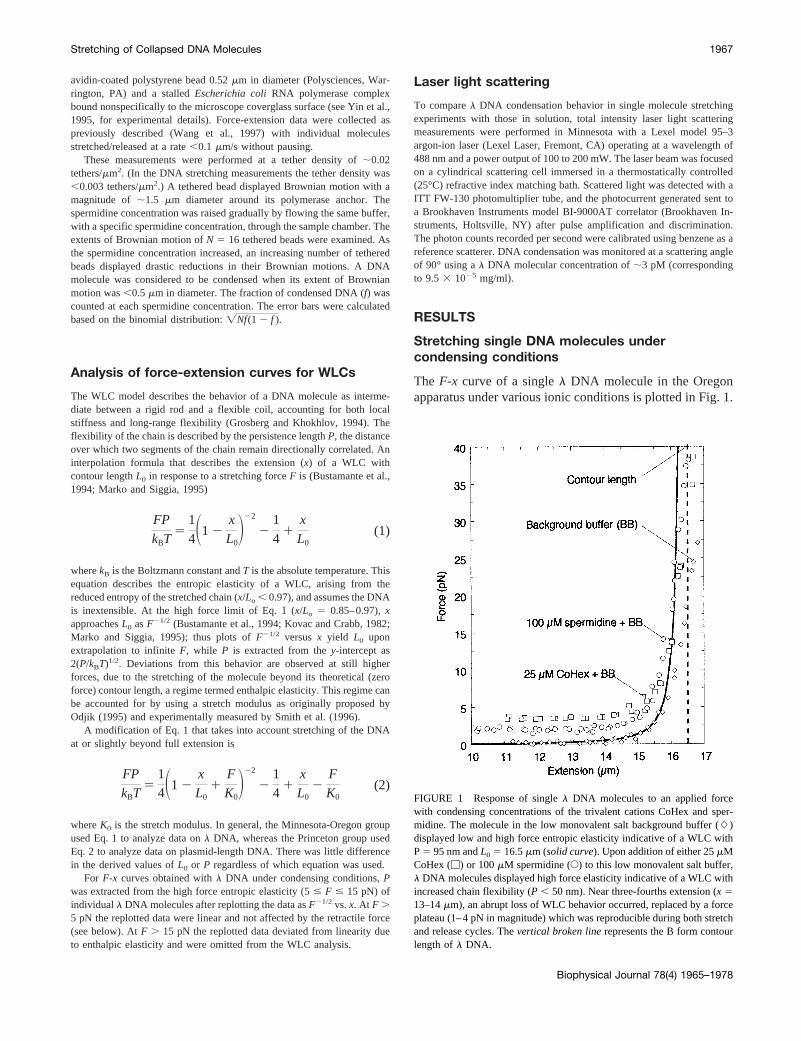
FIGURE 1 Response of singlel DNA molecules to an applied forcewith condensing concentrations of the trivalent cations CoHex and sper-midine. The molecule in the low monovalent salt background buffer (L)displayed low and high force entropic elasticity indicative of a WLC withP5 95 nm andL0 5 16.5mm (solid curve). Upon addition of either 25mMCoHex (M) or 100mM spermidine (E) to this low monovalent salt buffer,l DNA molecules displayed high force elasticity indicative of a WLC withincreased chain flexibility (P , 50 nm). Near three-fourths extension (x 513–14mm), an abrupt loss of WLC behavior occurred, replaced by a forceplateau (1–4 pN in magnitude) which was reproducible during both stretchand release cycles. Thevertical broken linerepresents the B form contourlength ofl DNA.
Stretching of Collapsed DNA Molecules 1967
Biophysical Journal 78(4) 1965–1978
The elastic response in B buffer, containing just monovalentcations and thus not causing condensation, yields from thehigh force limit of Eq. 1 a WLC with a persistence length of95 nm and a contour length of 16.5mm, in accord with ourprevious results (Baumann et al., 1997).
The elastic response was then recorded under ionic con-ditions where DNA condensation would be strongly favoredby addition of trivalent cations (see below). Fig. 1 showsdata for molecules in the presence of 25mM CoHex plus Bbuffer, and 100mM spermidine plus B buffer. At nearly fullextensions, theF-x curves rose more rapidly than in mono-valent buffer alone asx3 L0, indicating increased flexibil-ity, i.e., a reduced persistence length (12–26 nm for CoHexand 25–38 nm for spermidine, compared with 50 nm forhigh concentrations of NaCl). The extension was then de-creased, and near three-fourths maximal extension (x 513–14mm), stretchedl DNA molecules abruptly departedfrom WLC behavior, yielding a retractile force of constantmagnitude (1–4 pN). The force remained constant down tothe shortest molecular extensions probed (x # 8 mm). Datawere not collected below this extension to prevent potentialbead-DNA entanglements under condensing solution con-ditions. This abrupt departure from WLC behavior wasreproducible during both stretch and release cycles.
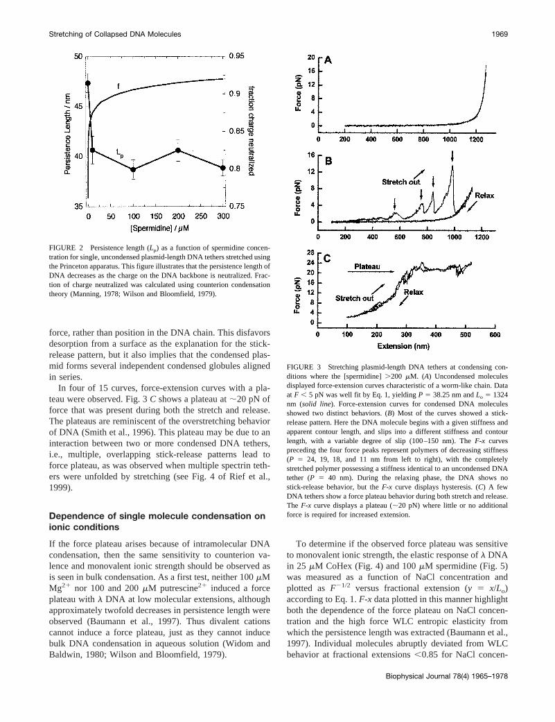
The Princeton group measured the force-extension rela-tion for plasmid-length DNA molecules at relatively highspermidine concentrations (.200 mM), where most of theDNA molecules are condensed and only a small fractionwere not condensed. The behavior of plasmid-length DNAwith the Princeton apparatus was similar to that ofl DNAin the Oregon apparatus for those molecules that did notcondense at a given spermidine concentration.F-x curves ofuncondensed DNA displayed the same characteristics asthose measured in the absence of spermidine. Fig. 3Ashows an example of these curves. Data (solid dots) withF , 5 pN were well fit with Eq. 1:P 5 38.25 nm andLo 51324 nm. For data withF . 5 pN, a slight deviation from
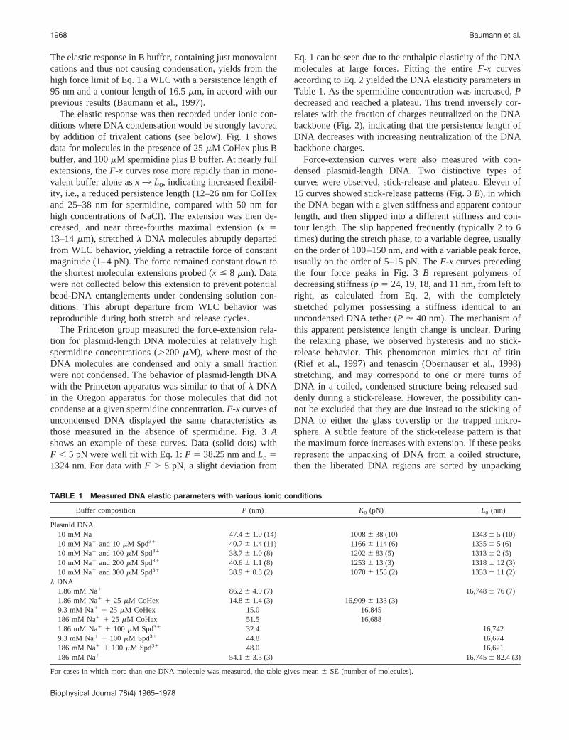
Eq. 1 can be seen due to the enthalpic elasticity of the DNAmolecules at large forces. Fitting the entireF-x curvesaccording to Eq. 2 yielded the DNA elasticity parameters inTable 1. As the spermidine concentration was increased,Pdecreased and reached a plateau. This trend inversely cor-relates with the fraction of charges neutralized on the DNAbackbone (Fig. 2), indicating that the persistence length ofDNA decreases with increasing neutralization of the DNAbackbone charges.
Force-extension curves were also measured with con-densed plasmid-length DNA. Two distinctive types ofcurves were observed, stick-release and plateau. Eleven of15 curves showed stick-release patterns (Fig. 3B), in whichthe DNA began with a given stiffness and apparent contourlength, and then slipped into a different stiffness and con-tour length. The slip happened frequently (typically 2 to 6times) during the stretch phase, to a variable degree, usuallyon the order of 100–150 nm, and with a variable peak force,usually on the order of 5–15 pN. TheF-x curves precedingthe four force peaks in Fig. 3B represent polymers ofdecreasing stiffness (p 5 24, 19, 18, and 11 nm, from left toright, as calculated from Eq. 2, with the completelystretched polymer possessing a stiffness identical to anuncondensed DNA tether (P ' 40 nm). The mechanism ofthis apparent persistence length change is unclear. Duringthe relaxing phase, we observed hysteresis and no stick-release behavior. This phenomenon mimics that of titin(Rief et al., 1997) and tenascin (Oberhauser et al., 1998)stretching, and may correspond to one or more turns ofDNA in a coiled, condensed structure being released sud-denly during a stick-release. However, the possibility can-not be excluded that they are due instead to the sticking ofDNA to either the glass coverslip or the trapped micro-sphere. A subtle feature of the stick-release pattern is thatthe maximum force increases with extension. If these peaksrepresent the unpacking of DNA from a coiled structure,then the liberated DNA regions are sorted by unpacking
TABLE 1 Measured DNA elastic parameters with various ionic conditions
Buffer composition P (nm) K0 (pN) L0 (nm)
Plasmid DNA10 mM Na1 47.46 1.0 (14) 10086 38 (10) 13436 5 (10)10 mM Na1 and 10mM Spd31 40.76 1.4 (11) 11666 114 (6) 13356 5 (6)10 mM Na1 and 100mM Spd31 38.76 1.0 (8) 12026 83 (5) 13136 2 (5)10 mM Na1 and 200mM Spd31 40.66 1.1 (8) 12536 13 (3) 13186 12 (3)10 mM Na1 and 300mM Spd31 38.96 0.8 (2) 10706 158 (2) 13336 11 (2)
l DNA1.86 mM Na1 86.26 4.9 (7) 16,7486 76 (7)1.86 mM Na1 1 25 mM CoHex 14.86 1.4 (3) 16,9096 133 (3)9.3 mM Na1 1 25 mM CoHex 15.0 16,845186 mM Na1 1 25 mM CoHex 51.5 16,6881.86 mM Na1 1 100 mM Spd31 32.4 16,7429.3 mM Na1 1 100 mM Spd31 44.8 16,674186 mM Na1 1 100 mM Spd31 48.0 16,621186 mM Na1 54.16 3.3 (3) 16,7456 82.4 (3)
For cases in which more than one DNA molecule was measured, the table gives mean6 SE (number of molecules).
1968 Baumann et al.
Biophysical Journal 78(4) 1965–1978
force, rather than position in the DNA chain. This disfavorsdesorption from a surface as the explanation for the stick-release pattern, but it also implies that the condensed plas-mid forms several independent condensed globules alignedin series.
In four of 15 curves, force-extension curves with a pla-teau were observed. Fig. 3C shows a plateau at;20 pN offorce that was present during both the stretch and release.The plateaus are reminiscent of the overstretching behaviorof DNA (Smith et al., 1996). This plateau may be due to aninteraction between two or more condensed DNA tethers,i.e., multiple, overlapping stick-release patterns lead toforce plateau, as was observed when multiple spectrin teth-ers were unfolded by stretching (see Fig. 4 of Rief et al.,1999).
Dependence of single molecule condensation onionic conditions
If the force plateau arises because of intramolecular DNAcondensation, then the same sensitivity to counterion va-lence and monovalent ionic strength should be observed asis seen in bulk condensation. As a first test, neither 100mMMg21 nor 100 and 200mM putrescine21 induced a forceplateau withl DNA at low molecular extensions, althoughapproximately twofold decreases in persistence length wereobserved (Baumann et al., 1997). Thus divalent cationscannot induce a force plateau, just as they cannot inducebulk DNA condensation in aqueous solution (Widom andBaldwin, 1980; Wilson and Bloomfield, 1979).
To determine if the observed force plateau was sensitiveto monovalent ionic strength, the elastic response ofl DNAin 25 mM CoHex (Fig. 4) and 100mM spermidine (Fig. 5)was measured as a function of NaCl concentration andplotted asF21/2 versus fractional extension (y 5 x/Lo)according to Eq. 1.F-x data plotted in this manner highlightboth the dependence of the force plateau on NaCl concen-tration and the high force WLC entropic elasticity fromwhich the persistence length was extracted (Baumann et al.,1997). Individual molecules abruptly deviated from WLCbehavior at fractional extensions,0.85 for NaCl concen-
FIGURE 2 Persistence length (Lp) as a function of spermidine concen-tration for single, uncondensed plasmid-length DNA tethers stretched usingthe Princeton apparatus. This figure illustrates that the persistence length ofDNA decreases as the charge on the DNA backbone is neutralized. Frac-tion of charge neutralized was calculated using counterion condensationtheory (Manning, 1978; Wilson and Bloomfield, 1979).
FIGURE 3 Stretching plasmid-length DNA tethers at condensing con-ditions where the [spermidine].200 mM. (A) Uncondensed moleculesdisplayed force-extension curves characteristic of a worm-like chain. Dataat F , 5 pN was well fit by Eq. 1, yieldingP 5 38.25 nm andLo 5 1324nm (solid line). Force-extension curves for condensed DNA moleculesshowed two distinct behaviors. (B) Most of the curves showed a stick-release pattern. Here the DNA molecule begins with a given stiffness andapparent contour length, and slips into a different stiffness and contourlength, with a variable degree of slip (100–150 nm). TheF-x curvespreceding the four force peaks represent polymers of decreasing stiffness(P 5 24, 19, 18, and 11 nm from left to right), with the completelystretched polymer possessing a stiffness identical to an uncondensed DNAtether (P 5 40 nm). During the relaxing phase, the DNA shows nostick-release behavior, but theF-x curve displays hysteresis. (C) A fewDNA tethers show a force plateau behavior during both stretch and release.The F-x curve displays a plateau (;20 pN) where little or no additionalforce is required for increased extension.
Stretching of Collapsed DNA Molecules 1969
Biophysical Journal 78(4) 1965–1978
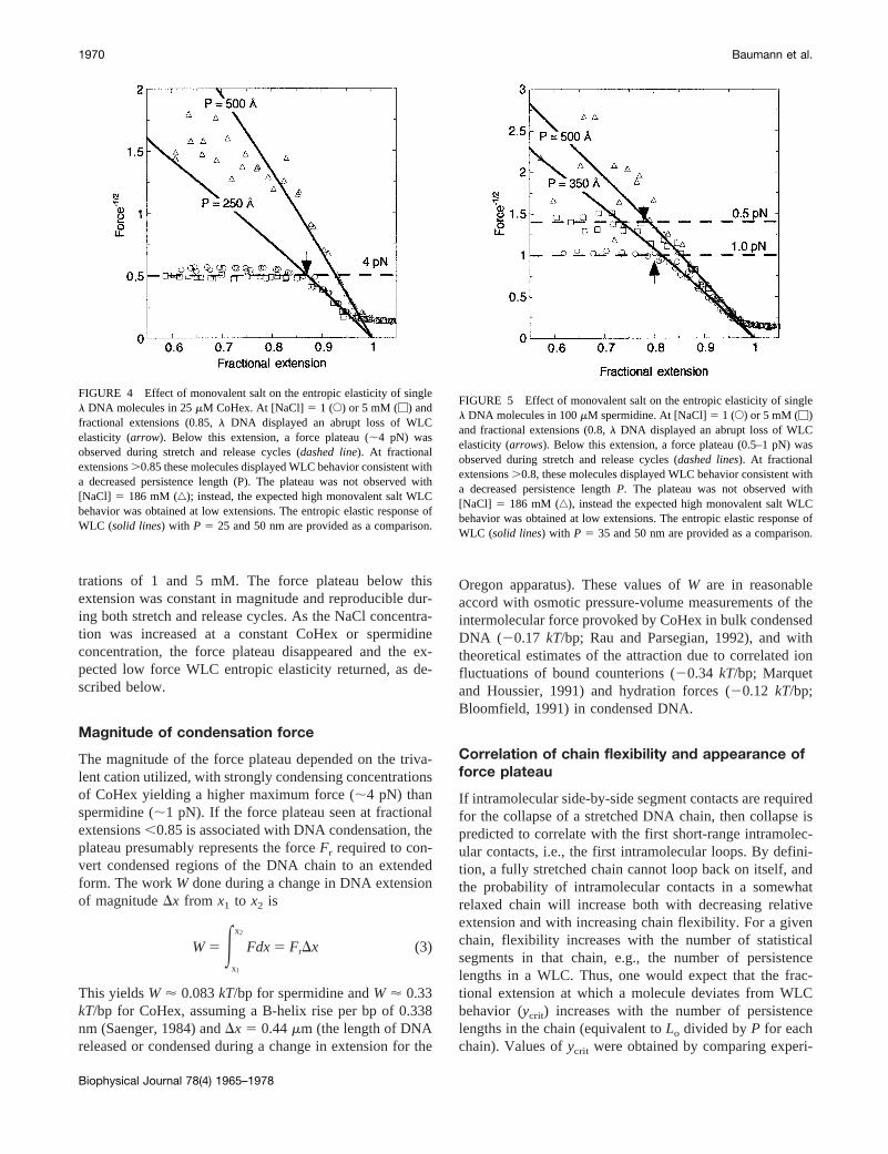
trations of 1 and 5 mM. The force plateau below thisextension was constant in magnitude and reproducible dur-ing both stretch and release cycles. As the NaCl concentra-tion was increased at a constant CoHex or spermidineconcentration, the force plateau disappeared and the ex-pected low force WLC entropic elasticity returned, as de-scribed below.
Magnitude of condensation force
The magnitude of the force plateau depended on the triva-lent cation utilized, with strongly condensing concentrationsof CoHex yielding a higher maximum force (;4 pN) thanspermidine (;1 pN). If the force plateau seen at fractionalextensions,0.85 is associated with DNA condensation, theplateau presumably represents the forceFr required to con-vert condensed regions of the DNA chain to an extendedform. The workW done during a change in DNA extensionof magnitudeDx from x1 to x2 is
W5 Ex1
x2
Fdx5 FrDx (3)
This yieldsW ' 0.083kT/bp for spermidine andW ' 0.33kT/bp for CoHex, assuming a B-helix rise per bp of 0.338nm (Saenger, 1984) andDx 5 0.44mm (the length of DNAreleased or condensed during a change in extension for the
Oregon apparatus). These values ofW are in reasonableaccord with osmotic pressure-volume measurements of theintermolecular force provoked by CoHex in bulk condensedDNA (20.17 kT/bp; Rau and Parsegian, 1992), and withtheoretical estimates of the attraction due to correlated ionfluctuations of bound counterions (20.34 kT/bp; Marquetand Houssier, 1991) and hydration forces (20.12 kT/bp;Bloomfield, 1991) in condensed DNA.
Correlation of chain flexibility and appearance offorce plateau
If intramolecular side-by-side segment contacts are requiredfor the collapse of a stretched DNA chain, then collapse ispredicted to correlate with the first short-range intramolec-ular contacts, i.e., the first intramolecular loops. By defini-tion, a fully stretched chain cannot loop back on itself, andthe probability of intramolecular contacts in a somewhatrelaxed chain will increase both with decreasing relativeextension and with increasing chain flexibility. For a givenchain, flexibility increases with the number of statisticalsegments in that chain, e.g., the number of persistencelengths in a WLC. Thus, one would expect that the frac-tional extension at which a molecule deviates from WLCbehavior (ycrit) increases with the number of persistencelengths in the chain (equivalent toLo divided byP for eachchain). Values ofycrit were obtained by comparing experi-
FIGURE 4 Effect of monovalent salt on the entropic elasticity of singlel DNA molecules in 25mM CoHex. At [NaCl]5 1 (E) or 5 mM (M) andfractional extensions (0.85,l DNA displayed an abrupt loss of WLCelasticity (arrow). Below this extension, a force plateau (;4 pN) wasobserved during stretch and release cycles (dashed line). At fractionalextensions.0.85 these molecules displayed WLC behavior consistent witha decreased persistence length (P). The plateau was not observed with[NaCl] 5 186 mM (‚); instead, the expected high monovalent salt WLCbehavior was obtained at low extensions. The entropic elastic response ofWLC (solid lines) with P 5 25 and 50 nm are provided as a comparison.
FIGURE 5 Effect of monovalent salt on the entropic elasticity of singlel DNA molecules in 100mM spermidine. At [NaCl]5 1 (E) or 5 mM (M)and fractional extensions (0.8,l DNA displayed an abrupt loss of WLCelasticity (arrows). Below this extension, a force plateau (0.5–1 pN) wasobserved during stretch and release cycles (dashed lines). At fractionalextensions.0.8, these molecules displayed WLC behavior consistent witha decreased persistence lengthP. The plateau was not observed with[NaCl] 5 186 mM (‚), instead the expected high monovalent salt WLCbehavior was obtained at low extensions. The entropic elastic response ofWLC (solid lines) with P 5 35 and 50 nm are provided as a comparison.
1970 Baumann et al.
Biophysical Journal 78(4) 1965–1978
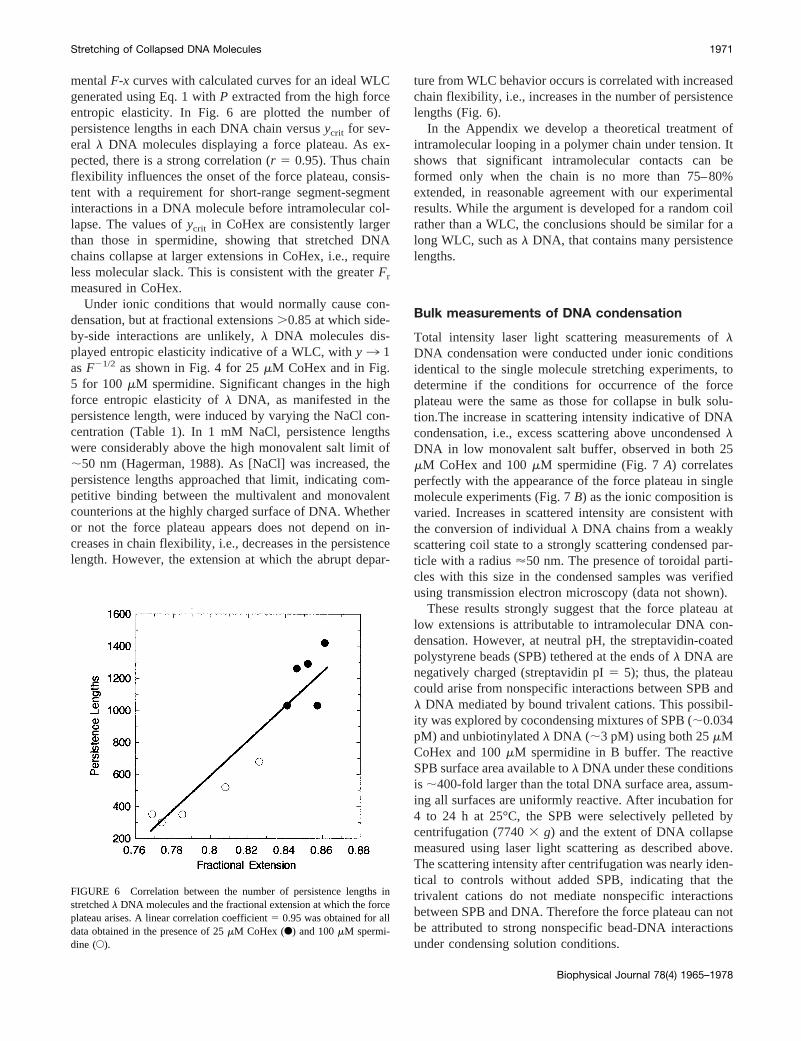
mentalF-x curves with calculated curves for an ideal WLCgenerated using Eq. 1 withP extracted from the high forceentropic elasticity. In Fig. 6 are plotted the number ofpersistence lengths in each DNA chain versusycrit for sev-eral l DNA molecules displaying a force plateau. As ex-pected, there is a strong correlation (r 5 0.95). Thus chainflexibility influences the onset of the force plateau, consis-tent with a requirement for short-range segment-segmentinteractions in a DNA molecule before intramolecular col-lapse. The values ofycrit in CoHex are consistently largerthan those in spermidine, showing that stretched DNAchains collapse at larger extensions in CoHex, i.e., requireless molecular slack. This is consistent with the greaterFr
measured in CoHex.Under ionic conditions that would normally cause con-
densation, but at fractional extensions.0.85 at which side-by-side interactions are unlikely,l DNA molecules dis-played entropic elasticity indicative of a WLC, withy3 1asF21/2 as shown in Fig. 4 for 25mM CoHex and in Fig.5 for 100mM spermidine. Significant changes in the highforce entropic elasticity ofl DNA, as manifested in thepersistence length, were induced by varying the NaCl con-centration (Table 1). In 1 mM NaCl, persistence lengthswere considerably above the high monovalent salt limit of;50 nm (Hagerman, 1988). As [NaCl] was increased, thepersistence lengths approached that limit, indicating com-petitive binding between the multivalent and monovalentcounterions at the highly charged surface of DNA. Whetheror not the force plateau appears does not depend on in-creases in chain flexibility, i.e., decreases in the persistencelength. However, the extension at which the abrupt depar-
ture from WLC behavior occurs is correlated with increasedchain flexibility, i.e., increases in the number of persistencelengths (Fig. 6).
In the Appendix we develop a theoretical treatment ofintramolecular looping in a polymer chain under tension. Itshows that significant intramolecular contacts can beformed only when the chain is no more than 75–80%extended, in reasonable agreement with our experimentalresults. While the argument is developed for a random coilrather than a WLC, the conclusions should be similar for along WLC, such asl DNA, that contains many persistencelengths.
Bulk measurements of DNA condensation
Total intensity laser light scattering measurements oflDNA condensation were conducted under ionic conditionsidentical to the single molecule stretching experiments, todetermine if the conditions for occurrence of the forceplateau were the same as those for collapse in bulk solu-tion.The increase in scattering intensity indicative of DNAcondensation, i.e., excess scattering above uncondensedlDNA in low monovalent salt buffer, observed in both 25mM CoHex and 100mM spermidine (Fig. 7A) correlatesperfectly with the appearance of the force plateau in singlemolecule experiments (Fig. 7B) as the ionic composition isvaried. Increases in scattered intensity are consistent withthe conversion of individuall DNA chains from a weaklyscattering coil state to a strongly scattering condensed par-ticle with a radius'50 nm. The presence of toroidal parti-cles with this size in the condensed samples was verifiedusing transmission electron microscopy (data not shown).
These results strongly suggest that the force plateau atlow extensions is attributable to intramolecular DNA con-densation. However, at neutral pH, the streptavidin-coatedpolystyrene beads (SPB) tethered at the ends ofl DNA arenegatively charged (streptavidin pI5 5); thus, the plateaucould arise from nonspecific interactions between SPB andl DNA mediated by bound trivalent cations. This possibil-ity was explored by cocondensing mixtures of SPB (;0.034pM) and unbiotinylatedl DNA (;3 pM) using both 25mMCoHex and 100mM spermidine in B buffer. The reactiveSPB surface area available tol DNA under these conditionsis ;400-fold larger than the total DNA surface area, assum-ing all surfaces are uniformly reactive. After incubation for4 to 24 h at 25°C, the SPB were selectively pelleted bycentrifugation (77403 g) and the extent of DNA collapsemeasured using laser light scattering as described above.The scattering intensity after centrifugation was nearly iden-tical to controls without added SPB, indicating that thetrivalent cations do not mediate nonspecific interactionsbetween SPB and DNA. Therefore the force plateau can notbe attributed to strong nonspecific bead-DNA interactionsunder condensing solution conditions.
FIGURE 6 Correlation between the number of persistence lengths instretchedl DNA molecules and the fractional extension at which the forceplateau arises. A linear correlation coefficient5 0.95 was obtained for alldata obtained in the presence of 25mM CoHex (F) and 100mM spermi-dine (E).
Stretching of Collapsed DNA Molecules 1971
Biophysical Journal 78(4) 1965–1978
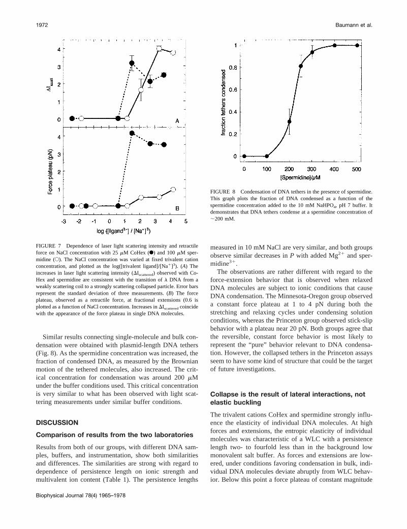
Similar results connecting single-molecule and bulk con-densation were obtained with plasmid-length DNA tethers(Fig. 8). As the spermidine concentration was increased, thefraction of condensed DNA, as measured by the Brownianmotion of the tethered molecules, also increased. The crit-ical concentration for condensation was around 200mMunder the buffer conditions used. This critical concentrationis very similar to what has been observed with light scat-tering measurements under similar buffer conditions.
DISCUSSION
Comparison of results from the two laboratories
Results from both of our groups, with different DNA sam-ples, buffers, and instrumentation, show both similaritiesand differences. The similarities are strong with regard todependence of persistence length on ionic strength andmultivalent ion content (Table 1). The persistence lengths
measured in 10 mM NaCl are very similar, and both groupsobserve similar decreases inP with added Mg21 and sper-midine31.
The observations are rather different with regard to theforce-extension behavior that is observed when relaxedDNA molecules are subject to ionic conditions that causeDNA condensation. The Minnesota-Oregon group observeda constant force plateau at 1 to 4 pN during both thestretching and relaxing cycles under condensing solutionconditions, whereas the Princeton group observed stick-slipbehavior with a plateau near 20 pN. Both groups agree thatthe reversible, constant force behavior is most likely torepresent the “pure” behavior relevant to DNA condensa-tion. However, the collapsed tethers in the Princeton assaysseem to have some kind of structure that could be the targetof future investigations.
Collapse is the result of lateral interactions, notelastic buckling
The trivalent cations CoHex and spermidine strongly influ-ence the elasticity of individual DNA molecules. At highforces and extensions, the entropic elasticity of individualmolecules was characteristic of a WLC with a persistencelength two- to fourfold less than in the background lowmonovalent salt buffer. As forces and extensions are low-ered, under conditions favoring condensation in bulk, indi-vidual DNA molecules deviate abruptly from WLC behav-ior. Below this point a force plateau of constant magnitude
FIGURE 7 Dependence of laser light scattering intensity and retractileforce on NaCl concentration with 25mM CoHex (F) and 100mM sper-midine (E). The NaCl concentration was varied at fixed trivalent cationconcentration, and plotted as the log([trivalent ligand]/[Na1]3). (A) Theincreases in laser light scattering intensity (DIscattered) observed with Co-Hex and spermidine are consistent with the transition ofl DNA from aweakly scattering coil to a strongly scattering collapsed particle. Error barsrepresent the standard deviation of three measurements. (B) The forceplateau, observed as a retractile force, at fractional extensions (0.6 isplotted as a function of NaCl concentration. Increases inDIscatteredcoincidewith the appearance of the force plateau in single DNA molecules.
FIGURE 8 Condensation of DNA tethers in the presence of spermidine.This graph plots the fraction of DNA condensed as a function of thespermidine concentration added to the 10 mM NaHPO4, pH 7 buffer. Itdemonstrates that DNA tethers condense at a spermidine concentration of;200 mM.
1972 Baumann et al.
Biophysical Journal 78(4) 1965–1978
is observed withl DNA, which is reproduced during bothstretch and release cycles.
We postulate that the force plateau induced by thesecations arises from intramolecular DNA condensation. Thefollowing observations buttress this interpretation: (i) theplateau occurs in the presence of the trivalent cations Co-Hex31 and spermidine31, known condensing agents inaqueous solution (Widom and Baldwin, 1980; Wilson andBloomfield, 1979), but not with the divalent cations Mg21
and putrescine21, even though these ions reduce the persis-tence length (which would seem equivalent to elastic buck-ling); (ii) the ionic conditions required to induce the forceplateau are consistent with those needed to induce a criticaldegree of charge neutralization before DNA condensation(Wilson and Bloomfield, 1979); and (iii) light scatteringincreases that accompany DNA condensation in bulk solu-tion coincide with a loss of WLC behavior and the appear-ance of the force plateau in single molecule experiments.
A buckling theory, based on elastic theory for macro-scopic rods, predicts that the intrinsic elastic instability ofneutralized DNA drives collapse (Manning, 1980, 1985). Inthis model, stable lateral contacts are a consequence ofbuckling, rather than the source of nucleation as proposedbelow. If lateral contacts are not required for collapse, thebuckling should manifest itself in stretched DNA moleculesat or near their contour length, where lateral contacts can notoccur. This does not occur in our experiments. Furthermore,the polyelectrolyte force that prevents buckling is predictedto vary roughly as the logarithm of added monovalent salt ata constant trivalent cation concentration. If our force plateauwere due to buckling it should display a similar dependenceon monovalent salt. This dependence is not observed (seeFig. 7 B). Instead, there is an abrupt transition when acritical total charge neutralization of the DNA is reached ascalculated according to counterion condensation theory(Manning, 1978; Wilson and Bloomfield, 1979). The ex-perimental results obtained here argue against buckling asthe source of DNA condensation.
An alternate theory (Halperin and Zhulina, 1991) describ-ing the F-x response of a single polymer chain in a poorsolvent predicts that intramolecular collapse will occur onlyin the presence of appreciable lateral contacts. Solutionconditions favoring DNA condensation can be thought of aspoor solvent conditions for DNA. At a fixed extension, theprobability of short-range lateral contacts or loop formationwill increase with increases in chain flexibility (decreases inthe persistence length of a WLC). We observe that the exactextension where the loss of WLC elasticity occurs is pro-portional to chain flexibility (inversely proportional to thepersistence length). This strongly suggests that the loss ofWLC behavior requires intramolecular contacts.
According to the above theory (Halperin and Zhulina,1991), a stretched polymer chain in a poor solvent willcollapse when the attractive free energy favoring collapseexceeds the free energy of the stretched polymer, leading to
a first-order phase transition. The transition is observed as adiscontinuous change in force with extension, representinga regime where collapsed and uncollapsed regions of thechain coexist. The force plateau observed in the elasticresponse of single DNA molecules under condensing solu-tion conditions exactly follows this predicted behavior.First-order behavior implies that collapse is nucleation-limited (i.e., requires formation of a nucleus of attractivelyinteracting segments), which in turn stipulates lateral con-tacts. This interpretation is in complete agreement withrecent fluorescence microscopy experiments, where thefirst-order intramolecular collapse of single viral DNA mol-ecules was observed in the presence of multivalent cations(Yoshikawa et al., 1996a,b).
The theoretical work of Post and Zimm (1979) dealt withthe contributions of both chain flexibility and favorablesegment-segment interactions to monomolecular DNA col-lapse. Using Flory-Huggins polymer lattice theory, theypredicted that collapse would involve a first-order transitionat a critical segment density (dependent on P) andx, theFlory polymer-solvent interaction parameter (Post andZimm, 1979). For a more flexible chain (shorter persistencelength), the discontinuous change in polymer volume ac-companying collapse occurs at a lower value ofx (lowercation concentration). These predictions are in good agree-ment with the putative first-order transition seen here insinglel DNA molecules, and the four- to sixfold efficiencyof CoHex over isovalent spermidine as a condensing agentin bulk solution (Benbasat, 1984; Thomas and Bloomfield,1983; Widom and Baldwin, 1980).
Nucleation of intramolecular collapse in astretched DNA chain
The solution conditions favoring DNA condensation may bethought of as a poor solvent for DNA, one in which seg-ment-segment interactions are favored over segment-sol-vent interactions. TheF-x response of a single stretchedpolymer chain in a poor solvent has been treated theoreti-cally (Halperin and Zhulina, 1991). Three deformation re-gimes are predicted as a function of extension, due to aninterplay of the entropic elasticity of the uncollapsed chainand the surface energy of the collapsed globule. First (re-gime I), at low extensions (extension less than the radius ofcollapsed globule), the collapsed globule is weakly per-turbed by increases in extension. At intermediate extensions(regime II), collapsed and uncollapsed regions of chaincoexist and the force is predicted to be roughly independentof changes in extension. At large extensions (regime III),the intrinsic entropic elasticity of the stretched chain isobserved. The extension at which the elastic behavior in-terconverts between regimes II and III is that at which therepulsive force acting on the stretched polymer chain bal-ances the attractive force favoring collapse. If stretchingoccurs continuously, thisF-x behavior resembles a first-
Stretching of Collapsed DNA Molecules 1973
Biophysical Journal 78(4) 1965–1978
order transition, such as that between a gas and a liquid,with extension analogous to volume and force to tempera-ture. In our work, regime II would coincide with the exten-sions where the force plateau is observed, and regime IIIwould represent the high force WLC elasticity.
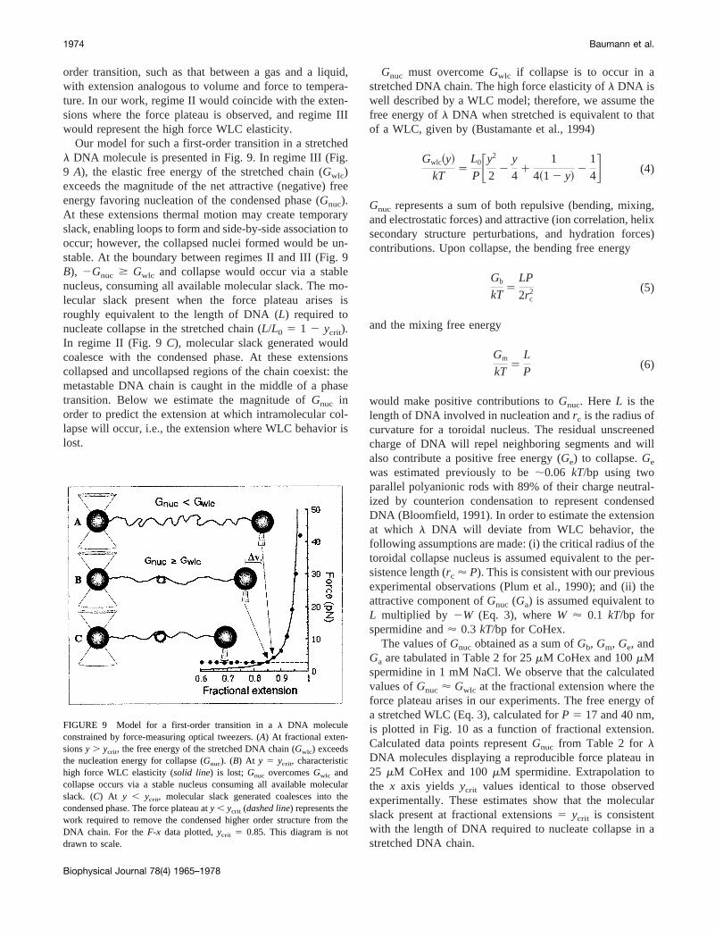
Our model for such a first-order transition in a stretchedl DNA molecule is presented in Fig. 9. In regime III (Fig.9 A), the elastic free energy of the stretched chain (Gwlc)exceeds the magnitude of the net attractive (negative) freeenergy favoring nucleation of the condensed phase (Gnuc).At these extensions thermal motion may create temporaryslack, enabling loops to form and side-by-side association tooccur; however, the collapsed nuclei formed would be un-stable. At the boundary between regimes II and III (Fig. 9B), 2Gnuc $ Gwlc and collapse would occur via a stablenucleus, consuming all available molecular slack. The mo-lecular slack present when the force plateau arises isroughly equivalent to the length of DNA (L) required tonucleate collapse in the stretched chain (L/L0 5 1 2 ycrit).In regime II (Fig. 9C), molecular slack generated wouldcoalesce with the condensed phase. At these extensionscollapsed and uncollapsed regions of the chain coexist: themetastable DNA chain is caught in the middle of a phasetransition. Below we estimate the magnitude ofGnuc inorder to predict the extension at which intramolecular col-lapse will occur, i.e., the extension where WLC behavior islost.
Gnuc must overcomeGwlc if collapse is to occur in astretched DNA chain. The high force elasticity ofl DNA iswell described by a WLC model; therefore, we assume thefree energy ofl DNA when stretched is equivalent to thatof a WLC, given by (Bustamante et al., 1994)
Gwlc~y!
kT5
L0
PFy2
22
y
41
1
4~1 2 y!2
1
4G (4)
Gnuc represents a sum of both repulsive (bending, mixing,and electrostatic forces) and attractive (ion correlation, helixsecondary structure perturbations, and hydration forces)contributions. Upon collapse, the bending free energy
Gb
kT5
LP
2rc2 (5)
and the mixing free energy
Gm
kT5
L
P(6)
would make positive contributions toGnuc. Here L is thelength of DNA involved in nucleation andrc is the radius ofcurvature for a toroidal nucleus. The residual unscreenedcharge of DNA will repel neighboring segments and willalso contribute a positive free energy (Ge) to collapse.Ge
was estimated previously to be;0.06 kT/bp using twoparallel polyanionic rods with 89% of their charge neutral-ized by counterion condensation to represent condensedDNA (Bloomfield, 1991). In order to estimate the extensionat which l DNA will deviate from WLC behavior, thefollowing assumptions are made: (i) the critical radius of thetoroidal collapse nucleus is assumed equivalent to the per-sistence length (rc ' P). This is consistent with our previousexperimental observations (Plum et al., 1990); and (ii) theattractive component ofGnuc (Ga) is assumed equivalent toL multiplied by 2W (Eq. 3), whereW ' 0.1 kT/bp forspermidine and' 0.3 kT/bp for CoHex.
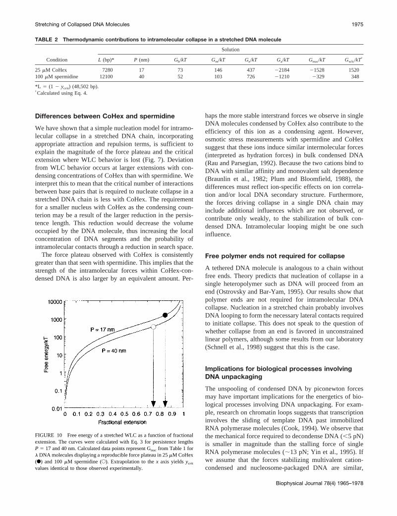
The values ofGnuc obtained as a sum ofGb, Gm, Ge, andGa are tabulated in Table 2 for 25mM CoHex and 100mMspermidine in 1 mM NaCl. We observe that the calculatedvalues ofGnuc ' Gwlc at the fractional extension where theforce plateau arises in our experiments. The free energy ofa stretched WLC (Eq. 3), calculated forP 5 17 and 40 nm,is plotted in Fig. 10 as a function of fractional extension.Calculated data points representGnuc from Table 2 forlDNA molecules displaying a reproducible force plateau in25 mM CoHex and 100mM spermidine. Extrapolation tothe x axis yields ycrit values identical to those observedexperimentally. These estimates show that the molecularslack present at fractional extensions5 ycrit is consistentwith the length of DNA required to nucleate collapse in astretched DNA chain.
FIGURE 9 Model for a first-order transition in al DNA moleculeconstrained by force-measuring optical tweezers. (A) At fractional exten-sionsy . ycrit, the free energy of the stretched DNA chain (Gwlc) exceedsthe nucleation energy for collapse (Gnuc). (B) At y 5 ycrit, characteristichigh force WLC elasticity (solid line) is lost; Gnuc overcomesGwlc andcollapse occurs via a stable nucleus consuming all available molecularslack. (C) At y , ycrit, molecular slack generated coalesces into thecondensed phase. The force plateau aty , ycrit (dashed line) represents thework required to remove the condensed higher order structure from theDNA chain. For theF-x data plotted,ycrit 5 0.85. This diagram is notdrawn to scale.
1974 Baumann et al.
Biophysical Journal 78(4) 1965–1978
Differences between CoHex and spermidine
We have shown that a simple nucleation model for intramo-lecular collapse in a stretched DNA chain, incorporatingappropriate attraction and repulsion terms, is sufficient toexplain the magnitude of the force plateau and the criticalextension where WLC behavior is lost (Fig. 7). Deviationfrom WLC behavior occurs at larger extensions with con-densing concentrations of CoHex than with spermidine. Weinterpret this to mean that the critical number of interactionsbetween base pairs that is required to nucleate collapse in astretched DNA chain is less with CoHex. The requirementfor a smaller nucleus with CoHex as the condensing coun-terion may be a result of the larger reduction in the persis-tence length. This reduction would decrease the volumeoccupied by the DNA molecule, thus increasing the localconcentration of DNA segments and the probability ofintramolecular contacts through a reduction in search space.
The force plateau observed with CoHex is consistentlygreater than that seen with spermidine. This implies that thestrength of the intramolecular forces within CoHex-con-densed DNA is also larger by an equivalent amount. Per-
haps the more stable interstrand forces we observe in singleDNA molecules condensed by CoHex also contribute to theefficiency of this ion as a condensing agent. However,osmotic stress measurements with spermidine and CoHexsuggest that these ions induce similar intermolecular forces(interpreted as hydration forces) in bulk condensed DNA(Rau and Parsegian, 1992). Because the two cations bind toDNA with similar affinity and monovalent salt dependence(Braunlin et al., 1982; Plum and Bloomfield, 1988), thedifferences must reflect ion-specific effects on ion correla-tion and/or local DNA secondary structure. Furthermore,the forces driving collapse in a single DNA chain mayinclude additional influences which are not observed, orcontribute only weakly, to the stabilization of bulk con-densed DNA. Intramolecular looping might be one suchinfluence.
Free polymer ends not required for collapse
A tethered DNA molecule is analogous to a chain withoutfree ends. Theory predicts that nucleation of collapse in asingle heteropolymer such as DNA will proceed from anend (Ostrovsky and Bar-Yam, 1995). Our results show thatpolymer ends are not required for intramolecular DNAcollapse. Nucleation in a stretched chain probably involvesDNA looping to form the necessary lateral contacts requiredto initiate collapse. This does not speak to the question ofwhether collapse from an end is favored in unconstrainedlinear polymers, although some results from our laboratory(Schnell et al., 1998) suggest that this is the case.
Implications for biological processes involvingDNA unpackaging
The unspooling of condensed DNA by piconewton forcesmay have important implications for the energetics of bio-logical processes involving DNA unpackaging. For exam-ple, research on chromatin loops suggests that transcriptioninvolves the sliding of template DNA past immobilizedRNA polymerase molecules (Cook, 1994). We observe thatthe mechanical force required to decondense DNA (,5 pN)is smaller in magnitude than the stalling force of singleRNA polymerase molecules (;13 pN; Yin et al., 1995). Ifwe assume that the forces stabilizing multivalent cation-condensed and nucleosome-packaged DNA are similar,
TABLE 2 Thermodynamic contributions to intramolecular collapse in a stretched DNA molecule
Condition
Solution
L (bp)* P (nm) Gb/kT Gm/kT Ge/kT Ga/kT Gnuc/kT Gwlc/kT†
25 mM CoHex 7280 17 73 146 437 22184 21528 1520100 mM spermidine 12100 40 52 103 726 21210 2329 348
*L 5 (1 2 ycrit) (48,502 bp).†Calculated using Eq. 4.
FIGURE 10 Free energy of a stretched WLC as a function of fractionalextension. The curves were calculated with Eq. 3 for persistence lengthsP 5 17 and 40 nm. Calculated data points represent Gnuc from Table 1 forl DNA molecules displaying a reproducible force plateau in 25mM CoHex(F) and 100mM spermidine (E). Extrapolation to thex axis yieldsycrit
values identical to those observed experimentally.
Stretching of Collapsed DNA Molecules 1975
Biophysical Journal 78(4) 1965–1978
then perhaps immobilized processive enzymes generate suf-ficient mechanical force in vivo to unpackage downstreamnucleosomal DNA, thus enabling processing of the DNAtemplate.
SUMMARY
Our results provide the following new information aboutintramolecular DNA condensation: (i) intramolecular DNAcollapse proceeds via a nucleation-limited mechanism thatdoes not require free polymer chain ends; (ii) Approxi-mately half as many basepairs are required to nucleatecollapse in a stretched DNA chain when CoHex, rather thanspermidine, is the condensing agent; and (iii) The forcesstabilizing DNA collapse are not equivalent for the isova-lent cations CoHex and spermidine. The larger attractiveforce seen with CoHex may explain the efficiency of thision as a condensing agent.
APPENDIX: Probability of loop formation instretched polymer chain
We assume that the region of DNA extensions in which the force isunvarying, but non-zero, represents the region in which short-range inter-actions between segments of the same DNA molecule can occur. Weinvestigate whether this is consistent with theoretical expectation by cal-culating the conditions under which loops in a highly stretched chain canoccur with significant probability.
We begin with the familiar treatment of a stretched, freely jointedpolymer chain (Flory, 1953; James and Guth, 1943) in which the end-to-end extensionx# in the direction of a forcet is
x# 5 nlLS lt
kBTD (A1)
wheren is the number of statistical segments in the chain, each of lengthl, andL( ) is the Langevin function. The force needed to maintain a givenextension is then
t 5kBT
lL* ~x/nl!(A2)
whereL* is the inverse Langevin function. Each segment in the chain issubject to this force.
We now simplify the problem in two respects, neither of which is likelyto affect the qualitative validity of the results. We assume that the stretchedchain is effectively one-dimensional, and we assume that each segment canassume only two orientations, either parallel or antiparallel to the force.The probability of the parallel orientation is
p 5 A expS lt
kBTD 5 A expFL*S x#
nlDG 5 A exp@L* ~y!# (A3)
wherey is the relative extension and the normalization factor is
A21 5 expS lt
kBTD 1 expS2 lt
kBTD (A4)
The probability of the antiparallel orientation is
q 5 1 2 p. (A5)
The binomial distributionP(m, k; p) gives the number of ways in whichkstates occurring with probabilityp can be chosen fromm total states. Inorder for a sequence ofm segments within the stretched chain to form aloop, the number of parallel and antiparallel orientations must be equal tom/2. The expected number of loops in a chain ofn segments whose endsare at relative extensiony, W0(n, y), is thereforeP(m, m/2; p) summed overall values ofm:
W0~n, y! 5 Om52
n m!
@~m/2!!#2 @p~1 2 p!#m/2 (A6)
The probability that there is at least one loop in the chain, assuming aPoisson distribution of the number of loops, is then
W1~n, y! 5 1 2 exp@2W0~n, y!# (A7)
We expect to begin to see short-range interactions between DNA segmentsseparated along the chain whenW1(n, y) is significantly greater than zero.
The inverse of the Langevin function
L* ~x! 5 coth~x! 21
x(A8)
has the following series expansion, which is accurate to 0.25%:
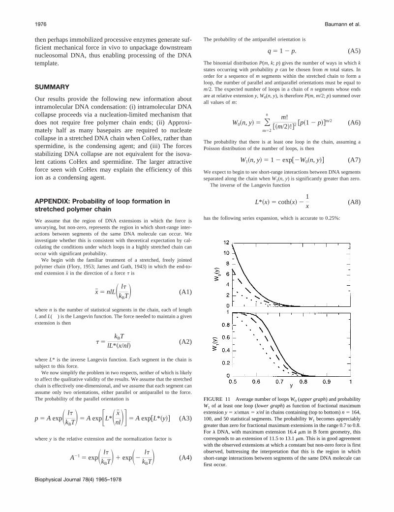
FIGURE 11 Average number of loopsW0 (upper graph) and probabilityW1 of at least one loop (lower graph) as function of fractional maximumextensiony 5 x/xmax5 x/nl in chains containing (top to bottom)n 5 164,100, and 50 statistical segments. The probabilityW1 becomes appreciablygreater than zero for fractional maximum extensions in the range 0.7 to 0.8.For l DNA, with maximum extension 16.4mm in B form geometry, thiscorresponds to an extension of 11.5 to 13.1mm. This is in good agreementwith the observed extensions at which a constant but non-zero force is firstobserved, buttressing the interpretation that this is the region in whichshort-range interactions between segments of the same DNA molecule canfirst occur.
1976 Baumann et al.
Biophysical Journal 78(4) 1965–1978
L* ~y! 5 3y 19
5y3 1
297
175y5 1
1539
875y7 1
126117
67375y9
143733439
21896875y110 # y # 0.537 (A9a)
5 21.0371 5.660y 1 9.376~y 2 c!2
1 17.04~y 2 c!3 1 52.02~y 2 c!4 1 94.86~y 2 c!5
0.537, y # 1 (A9b)
wherec 5 0.5366. We use this expansion in Eqs. A2-A4 to obtainp(y), andsubstitute this value in Eqs. A6 and A7 to obtain the desired probability ofat least one loop. The results for chains containingn 5 50, 100, and 164statistical segments are shown in Fig. 11.
C. G. Baumann thanks I. Rouzina for many helpful discussions and S.Fiddler for her hospitality during visits to the University of Oregon. M. W.and S. B. thank Dr. Eric Siggia for his helpful advice. This work wassupported in part by National Science Foundation grant MBC 9118482 andNational Institutes of Health grant GM 32543 to C. Bustamante, NationalInstitutes of Health Grant GM 28093 to V. Bloomfield, and an NationalInstitutes of Health Traineeship (GM 08277) to C. G. Baumann; and by theCancer Research Fund of the Damon Runyon - Walter Winchell Founda-tion fellowship, DRG-1326 to M. D. Wang.
REFERENCES
Arscott, P. G., C. Ma, J. R. Wenner, and V. A. Bloomfield. 1995. DNAcondensation by cobalt hexaammine(III) in alcohol-water mixtures: di-electric constant and other solvent effects.Biopolymers.36:345–364.
Baumann, C. G., S. B. Smith, V. A. Bloomfield, and C. Bustamante. 1997.Ionic effects on the elasticity of single DNA molecules.Proc. Natl.Acad. Sci. USA.94:6185–6190.
Benbasat, J. A. 1984. Condensation of bacteriophagefW14 DNA ofvarying charge densities by trivalent counterions.Biochemistry.23:3609–3619.
Bloomfield, V. A. 1991. Condensation of DNA by multivalent cations:considerations on mechanism.Biopolymers.31:1471–1481.
Bloomfield, V. A. 1996. DNA condensation.Curr. Opinion Struct. Biol.6:334–341.
Bloomfield, V. A. 1997. DNA condensation by multivalent cations.Biopolymers.44:269–282.
Braunlin, W. H., T. J. Strick, and M. T. Record, Jr. 1982. Equilibriumdialysis studies of polyamine binding to DNA.Biopolymers.21:1301–1314.
Bustamante, C., J. F. Marko, E. D. Siggia, and S. Smith. 1994. Entropicelasticity of lambda-phage DNA.Science.265:1599–1600.
Cook, P. R. 1994. RNA polymerase: structural determinant of the chro-matin loop and the chromosome.BioEssays.16:425–430.
Flory, P. J. 1953. Principles of Polymer Chemistry. Cornell UniversityPress, Ithaca, NY.
Gosule, L. C., and J. A. Schellman. 1976. Compact form of DNA inducedby spermidine.Nature.259:333–335.
Gronbech-Jensen, N., R. J. Mashl, R. F. Bruinsma, and W. M. Gelbart.1997. Counterion-induced attraction between rigid polyelectrolytes.Phys. Rev. Lett.78:2477–2480.
Grosberg, A. Y., and A. R. Khokhlov. 1994. Polyelectrolytes. Trans.Atanov, Y. A. In Statistical Physics of Macromolecules. AmericanInstitute of Physics Press, New York. 3,5,217–220.
Hagerman, P. J. 1988. Flexibility of DNA.Annu. Rev. Biophys. Biophys.Chem.17:265–286.
Halperin, A., and E. B. Zhulina. 1991. Stretching polymer brushes in poorsolvents.Macromolecules.24:5393–5397.
James, H. M., and E. Guth. 1943. Theory of the elastic properties of rubber.J. Chem. Phys.11:455–481.
Kovac, J., and C. C. Crabb. 1982. Modified gaussian model for rubberelasticity. 2. The worm-like chain.Macromolecules.15:537–541.
Ma, C., L. Sun, and V. A. Bloomfield. 1995. Condensation of plasmidsenhanced by Z-DNA conformation of d(CG)n inserts.Biochemistry.34:3521–3528.
Manning, G. S. 1978. The molecular theory of polyelectrolyte solutionswith applications to the electrostatic properties of polynucleotides.Q. Rev. Biophys.11:179–246.
Manning, G. S. 1980. Thermodynamic stability theory for DNA doughnutshapes induced by charge neutralization.Biopolymers.19:37–59.
Manning, G. S. 1985. Packaged DNA: an elastic model.Cell. Biophys.7:57–89.
Marko, J. F., and E. D. Siggia. 1995. Stretching DNA.Macromolecules.28:8759–8770.
Marquet, R., and C. Houssier. 1991. Thermodynamics of cation-inducedDNA condensation.J. Biomol. Struct. Dynam.9:159–167.
Oberhauser, A. F., P. E. Marszalek, H. P. Erickson, and J. M. Fernandez.1998. The molecular elasticity of the extracellular matrix protein tena-scin.Nature.393:181–185.
Odijk, T. 1995. Stiff chains and filaments under tension.Macromolecules.28:7016–7018.
Oosawa, F. 1968. Interaction between parallel rodlike macroions.Biopoly-mers.6:1633–1647.
Ostrovsky, B., and Y. Bar-Yam. 1995. Motion of polymer ends in ho-mopolymer and heteropolymer collapse.Biophys J.68:1694–1698.
Plum, G. E., P. G. Arscott, and V. A. Bloomfield. 1990. Condensation ofDNA by trivalent cations. 2. Effect of cation structure.Biopolymers.30:631–643.
Plum, G. E., and V. A. Bloomfield. 1988. Equilibrium dialysis study ofbinding of hexammine cobalt(III) to DNA.Biopolymers.27:1045–1051.
Post, C. B., and B. H. Zimm. 1979. Internal condensation of a single DNAmolecule.Biopolymers.18:1487–1501.
Post, C. B., and B. H. Zimm. 1982a. Light scattering study of DNAcondensation: Competition between collapse and aggregation.Biopoly-mers.21:2139–2160.
Post, C. B., and B. H. Zimm. 1982b. Theory of DNA condensation:collapse vs. aggregation.Biopolymers.21:2123–2137.
Quake, S. R., H. Babcock, and S. Chu. 1997. The dynamics of partiallyextended single molecules of DNA.Nature.388:151–154.
Rau, D. C., and V. A. Parsegian. 1992. Direct measurement of the inter-molecular forces between counterion-condensed DNA double helices:evidence for long range attractive hydration forces.Biophys. J.61:246–259.
Reich, Z., R. Ghirlando, and A. Minsky. 1991. Secondary conformationalpolymorphism of nucleic acids as a possible functional link betweencellular parameters and DNA packaging processes.Biochemistry.30:7828–7836.
Rief, M., M. Gautel, F. Oesterhelt, J. M. Fernandez, and H. E. Gaub. 1997.Reversible unfolding of individual titin immunoglobulin domains byAFM. Science.276:1109–1112.
Rief, M., J. Pascual, M. Saraste, and H. E. Gaub. 1999. Single moleculeforce spectroscopy of spectrin repeats: low unfolding forces in helixbundles.J. Mol. Biol. 286:553–561.
Rouzina, I., and V. A. Bloomfield. 1996. Macroion attraction due toelectrostatic correlation between screening counterions. 1. Mobile sur-face-adsorbed ions and diffuse ion cloud.J. Phys. Chem.100:9977–9989.
Rouzina, I., and V. A. Bloomfield. 1998. DNA bending by small, mobilemultivalent cations.Biophys. J.74:3152–3164.
Saenger, W. 1984. Principles of Nucleic Acid Structure. Springer-Verlag,New York.
Stretching of Collapsed DNA Molecules 1977
Biophysical Journal 78(4) 1965–1978
Schnell, J. R., J. Berman, and V. A. Bloomfield. 1998. Insertion oftelomere repeat sequence decreases plasmid DNA condensation bycobalt (III) hexaammine.Biophys. J.74:1484–1491.
Smith, S. B., Y. Cui, and C. Bustamante. 1996. Overstretching B-DNA: theelastic response of individual double-stranded and single-stranded DNAmolecules.Science.271:795–799.
Stigter, D. 1998. An electrostatic model for the dielectric effects, theadsorption of multivalent ions, and the bending of B-DNA.Biopolymers.46:503–516.
Thomas, T. J., and V. A. Bloomfield. 1983. Collapse of DNA caused bytrivalent cations: pH and ionic specificity effects.Biopolymers.22:1097–1106.
Wang, M. D., H. Yin, R. Landick, J. Gelles, and S. M. Block. 1997.Stretching DNA with optical tweezers.Biophys. J.72:1335–1346.
Widom, J., and R. L. Baldwin. 1980. Cation-induced toroidal condensationof DNA: studies with Co31(NH3)6. J. Mol. Biol. 144:431–453.
Wilson, R. W., and V. A. Bloomfield. 1979. Counterion-induced conden-sation of deoxyribonucleic acid. A light-scattering study.Biochemistry.18:2192–2196.
Yin, H., M. D. Wang, K. Svoboda, R. Landick, S. M. Block, and J. Gelles.1995. Transcription against an applied force.Science.270:1653–1657.
Yoshikawa, K., S. Kidoaki, M. Takahashi, V. V. Vasilevskaya, and A. R.Khokhlov. 1996a. Marked discreteness on the coil-globule transition ofsingle duplex DNA.Berichte der Bunsen Gesellschaft fur PhysikalischeChemie.100: 876–880.
Yoshikawa, K., M. Takahashi, V. V. Vasilevskaya, and A. R. Khokhlov.1996b. Large discrete transition in a single DNA molecule appearscontinuous in the ensemble.Phys. Rev. Lett.76:3029–3031.
1978 Baumann et al.
Biophysical Journal 78(4) 1965–1978
Related Documents