Stretch-Induced Injury Alters Mitochondrial Membrane Potential and Cellular ATP in Cultured Astrocytes and Neurons Syed M. Ahmed, Beverly A. Rzigalinski, Karen A. Willoughby, Heather A. Sitterding, and Earl F. Ellis Department of Pharmacology and Toxicology, School of Medicine, Virginia Commonwealth University, Richmond, Virginia, U.S.A. Abstract: Energy deficit after traumatic brain injury (TBI) may alter ionic homeostasis, neurotransmission, biosyn- thesis, and cellular transport. Using an in vitro model for TBI, we tested the hypothesis that stretch-induced injury alters mitochondrial membrane potential (Dc m ) and ATP in astrocytes and neurons. Astrocytes, pure neuronal cul- tures, and mixed neuronal plus glial cultures grown on Silastic membranes were subjected to mild, moderate, and severe stretch. After injury, Dc m was measured using rhodamine-123, and ATP was quantified with a luciferin– luciferase assay. In astrocytes, Dc m dropped signifi- cantly, and ATP content declined 43–52% 15 min after mild or moderate stretch but recovered by 24 h. In pure neurons, Dc m declined at 15 min only in the severely stretched group. At 48 h postinjury, Dc m remained de- creased in severely stretched neurons and dropped in moderately stretched neurons. Intracellular ATP content did not change in any group of injured pure neurons. We also found that astrocytes and neurons release ATP ex- tracellularly following injury. In contrast to pure neurons, Dc m in neurons of mixed neuronal plus glial cultures declined 15 min after mild, moderate, or severe stretch and recovered by 24 – 48 h. ATP content in mixed cul- tures declined 22–28% after mild to severe stretch with recovery by 24 h. Our findings demonstrate that injury causes mitochondrial dysfunction in astrocytes and sug- gest that astrocyte injury alters mitochondrial function in local neurons. Key Words: Mitochondria—ATP—Ener- gy—Astrocytes—Neurons—In vitro studies. J. Neurochem. 74, 1951–1960 (2000). The CNS has one of the highest energy demands of any organ system in the body and is highly vulnerable to a decline in energy supply (Chinnery and Turnbull, 1997). In the brain, ATP is used for various functions, including maintenance of ionic gradients, protein and lipid synthesis, intracellular transport, and neurotrans- mitter release and metabolism (Erecin ´ska and Silver, 1989). Therefore, trauma-induced energy deficit result- ing from alterations in cellular ATP production could have wide-ranging effects on neuronal and glial function in the brain. Evidence supporting a role for energy deficit after traumatic brain injury (TBI) has not been entirely clear. Studies conducted on rats and cats using 31 P-NMR found no significant change in tissue ATP levels after fluid- percussion injury (Vink et al., 1987, 1994; Andersen et al., 1988; Unterberg et al., 1988). However, declines were noted in the ATP/ADP ratio and in the ratio of phosphocreatine to inorganic phosphate. Recent studies examining whole brain homogenates noted a decline in ATP levels beginning 2 h after a mild weight-drop injury that was maximal by 6 h and recovered within 120 h (Vagnozzi et al., 1999). Xiong et al. (1997) demonstrated a significant decline in respiratory rates of mitochondria isolated from injured rat brain, and injury-induced alter- ations in mitochondrial morphology have been noted in vivo (Pettus et al., 1994) and in vitro (Ellis et al., 1995). Studies have also found elevations in CSF lactate levels after in vivo TBI (DeWitt et al., 1988), consistent with increased glycolysis. Decreased mitochondrial respira- tion, increased glycolysis, and alterations in mitochon- drial morphology suggest trauma-induced mitochondrial dysfunction. As mitochondria produce the majority of ATP within the cell, injury-induced mitochondrial dam- age may lead to an energy deficit. Evidence obtained from our laboratory using an in vitro model of TBI suggests a decline in certain bio- chemical processes that require ATP. Stretch injury of neurons in mixed neuronal– glial cultures evoked a “stretch-induced delayed depolarization,” which may be Received September 8, 1999; revised manuscript received January 4, 2000; accepted January 5, 2000. Address correspondence and reprint requests to Dr. B. A. Rzigalinski at Department of Pharmacology and Toxicology, School of Medicine, Virginia Commonwealth University, P.O. Box 980613, Richmond, VA 23298-0613, U.S.A. E-mail: [email protected] Abbreviations used: AM, acetoxymethyl ester; [Ca 21 ] i , intracellular free calcium concentration; DMEM, Dulbecco’s modified Eagle’s me- dium; DPBS, Dulbecco’s phosphate-buffered saline; Dc m , mitochon- drial membrane potential; FCCP, carbonylcyanide-p-carboxytrifluoro- methoxyphenylhydrazone; Rh 123 , rhodamine-123; RLU, relative light units; SERCA, sarcoplasmic– endoplasmic reticulum Ca 21 -ATPase; TBI, traumatic brain injury. 1951 Journal of Neurochemistry Lippincott Williams & Wilkins, Inc., Philadelphia © 2000 International Society for Neurochemistry

Welcome message from author
This document is posted to help you gain knowledge. Please leave a comment to let me know what you think about it! Share it to your friends and learn new things together.
Transcript
Stretch-Induced Injury Alters Mitochondrial Membrane Potentialand Cellular ATP in Cultured Astrocytes and Neurons
Syed M. Ahmed, Beverly A. Rzigalinski, Karen A. Willoughby,Heather A. Sitterding, and Earl F. Ellis
Department of Pharmacology and Toxicology, School of Medicine,Virginia Commonwealth University, Richmond, Virginia, U.S.A.
Abstract: Energy deficit after traumatic brain injury (TBI)may alter ionic homeostasis, neurotransmission, biosyn-thesis, and cellular transport. Using an in vitro model forTBI, we tested the hypothesis that stretch-induced injuryalters mitochondrial membrane potential (Dcm) and ATPin astrocytes and neurons. Astrocytes, pure neuronal cul-tures, and mixed neuronal plus glial cultures grown onSilastic membranes were subjected to mild, moderate,and severe stretch. After injury, Dcm was measured usingrhodamine-123, and ATP was quantified with a luciferin–luciferase assay. In astrocytes, Dcm dropped signifi-cantly, and ATP content declined 43–52% 15 min aftermild or moderate stretch but recovered by 24 h. In pureneurons, Dcm declined at 15 min only in the severelystretched group. At 48 h postinjury, Dcm remained de-creased in severely stretched neurons and dropped inmoderately stretched neurons. Intracellular ATP contentdid not change in any group of injured pure neurons. Wealso found that astrocytes and neurons release ATP ex-tracellularly following injury. In contrast to pure neurons,Dcm in neurons of mixed neuronal plus glial culturesdeclined 15 min after mild, moderate, or severe stretchand recovered by 24–48 h. ATP content in mixed cul-tures declined 22–28% after mild to severe stretch withrecovery by 24 h. Our findings demonstrate that injurycauses mitochondrial dysfunction in astrocytes and sug-gest that astrocyte injury alters mitochondrial function inlocal neurons. Key Words: Mitochondria—ATP—Ener-gy—Astrocytes—Neurons—In vitro studies.J. Neurochem. 74, 1951–1960 (2000).
The CNS has one of the highest energy demands ofany organ system in the body and is highly vulnerable toa decline in energy supply (Chinnery and Turnbull,1997). In the brain, ATP is used for various functions,including maintenance of ionic gradients, protein andlipid synthesis, intracellular transport, and neurotrans-mitter release and metabolism (Erecin´ska and Silver,1989). Therefore, trauma-induced energy deficit result-ing from alterations in cellular ATP production couldhave wide-ranging effects on neuronal and glial functionin the brain.
Evidence supporting a role for energy deficit aftertraumatic brain injury (TBI) has not been entirely clear.Studies conducted on rats and cats using31P-NMR foundno significant change in tissue ATP levels after fluid-percussion injury (Vink et al., 1987, 1994; Andersenet al., 1988; Unterberg et al., 1988). However, declineswere noted in the ATP/ADP ratio and in the ratio ofphosphocreatine to inorganic phosphate. Recent studiesexamining whole brain homogenates noted a decline inATP levels beginning 2 h after a mild weight-drop injurythat was maximal by 6 h and recovered within 120 h(Vagnozzi et al., 1999). Xiong et al. (1997) demonstrateda significant decline in respiratory rates of mitochondriaisolated from injured rat brain, and injury-induced alter-ations in mitochondrial morphology have been noted invivo (Pettus et al., 1994) and in vitro (Ellis et al., 1995).Studies have also found elevations in CSF lactate levelsafter in vivo TBI (DeWitt et al., 1988), consistent withincreased glycolysis. Decreased mitochondrial respira-tion, increased glycolysis, and alterations in mitochon-drial morphology suggest trauma-induced mitochondrialdysfunction. As mitochondria produce the majority ofATP within the cell, injury-induced mitochondrial dam-age may lead to an energy deficit.
Evidence obtained from our laboratory using an invitro model of TBI suggests a decline in certain bio-chemical processes that require ATP. Stretch injury ofneurons in mixed neuronal–glial cultures evoked a“stretch-induced delayed depolarization,” which may be
Received September 8, 1999; revised manuscript received January 4,2000; accepted January 5, 2000.
Address correspondence and reprint requests to Dr. B. A. Rzigalinskiat Department of Pharmacology and Toxicology, School of Medicine,Virginia Commonwealth University, P.O. Box 980613, Richmond, VA23298-0613, U.S.A. E-mail: [email protected]
Abbreviations used:AM, acetoxymethyl ester; [Ca21]i, intracellularfree calcium concentration; DMEM, Dulbecco’s modified Eagle’s me-dium; DPBS, Dulbecco’s phosphate-buffered saline;Dcm, mitochon-drial membrane potential; FCCP, carbonylcyanide-p-carboxytrifluoro-methoxyphenylhydrazone; Rh123, rhodamine-123; RLU, relative lightunits; SERCA, sarcoplasmic–endoplasmic reticulum Ca21-ATPase;TBI, traumatic brain injury.
1951
Journal of NeurochemistryLippincott Williams & Wilkins, Inc., Philadelphia© 2000 International Society for Neurochemistry
linked to decreased Na1,K1-ATPase activity (Tavalinet al., 1995, 1997). Stretch-induced delayed depolariza-tion is reversed by adding ATP intracellularly, suggest-ing that it may be caused by an energy deficit (Tavalinet al., 1997). We have also observed a persistent deple-tion of intracellular calcium stores in injured astrocytes,which may be due to a decline in the sarcoplasmic–endoplasmic reticulum Ca21-ATPase (SERCA) activitythat replenishes calcium stores (Rzigalinski et al.,1998a). Taken together, these findings suggest a role forATP deficit after TBI.
In the present report, we use a well-characterized invitro model of traumatic injury (Ellis et al., 1995) toassess changes in astrocyte and neuronal energy statusafter injury. In previous studies, we have demonstratedthat in vitro stretch injury produces many of the post-traumatic responses observed in vivo, including intracel-lular lesions to mitochondria, Golgi, and cytoskeletalelements in astrocytes and neurons (Dietrich et al., 1994;Ellis et al., 1995; McKinney et al., 1996), increased totalcell calcium in astrocytes (Fineman et al., 1993; Rziga-linski et al., 1997), transient elevation in astrocyte intra-cellular free calcium concentration ([Ca21]i) (Rzigalinskiet al., 1998a), phospholipase activation (Wei et al., 1982;Lamb et al., 1997), free radical formation (McKinneyet al., 1996; Lamb et al., 1997), and the above-mentionedstretch-induced delayed neuronal depolarization, whichmay be related to the transient neuronal dysfunctionobserved in vivo (Hamm et al., 1993, 1994; Tavalinet al., 1995). Consistent with reports that Mg21 reducesthe severity of NMDA- and TBI-induced neuronal injuryin animals (McIntosh, 1992), we have used our model toidentify a novel trauma-induced reduction in the Mg21
block of the NMDA receptor current and increasedNMDA-stimulated [Ca21]i elevation in injured neurons(Zhang et al., 1996). Together, these findings support therelevance and applicability of our in vitro model to thestudy of in vivo TBI.
We now report that mitochondrial membrane potential(Dcm) drops significantly in astrocytes 15 min after mildor moderate stretch and returns to normal by 24 h.Cellular ATP levels in astrocytes exhibit a correspondingdecline and recovery, suggesting that mitochondrial dys-function underlies a trauma-induced ATP deficit. In neu-rons of pure neuronal cultures devoid of astrocytes adecline inDcm was noted only after severe stretch, andno significant change in ATP levels was detected. Inneurons of mixed neuronal plus glial cultures, we founda significant decline inDcm after mild, moderate, andsevere stretch, suggesting that neurons cultured in thepresence of astrocytes may be susceptible to energydeficit. ATP levels declined significantly after injury inmixed neuronal plus glial cultures as a whole, althoughnot to the same extent as in pure astrocyte culture. Wealso report that stretch injury causes release of ATP frominjured astrocytes and neurons, which raises the possi-bility of purinergic receptor stimulation in response totraumatic injury.
MATERIALS AND METHODS
MaterialsPregnant Sprague–Dawley rats and 1–2-day-old rat pups
were obtained from Zivic-Miller (Allison Park, PA, U.S.A.).Minimal essential medium and Dulbecco’s modified essentialmedium (DMEM) were obtained from Mediatech (Herndon,VA, U.S.A.). Neurobasal medium, B27 supplement, Dulbec-co’s phosphate-buffered saline (DPBS), and horse serum werepurchased from GIBCO (Grand Island, NY, U.S.A.). Fetalbovine serum was obtained from Summit Biotechnologies (FortCollins, CO, U.S.A.). Flex Plates were from Flexcell Interna-tional (Hillsborough, NC, U.S.A.). Oligomycin was purchasedfrom Calbiochem (San Diego, CA, U.S.A.). Bovine trypsin,porcine trypsin, poly-D-lysine, carbonylcyanide-p-carboxytri-fluoromethoxyphenylhydrazone (FCCP), EDTA, Tris-base, fat-ty-acid free bovine serum albumin, and glutamate were pur-chased from Sigma (St. Louis, MO, U.S.A.). Rhodamine-123(Rh123) and fura-2 acetoxymethyl ester (AM) were obtainedfrom Molecular Probes (Eugene, OR, U.S.A.). ATP assay kitswere purchased from Roche Molecular Biochemicals (India-napolis, IN, U.S.A.). The commercially available model 94ACell Injury Controller was manufactured by the BiomedicalEngineering Facility at Virginia Commonwealth University(Richmond, VA, U.S.A.).
Cell cultureAstrocytes.Astrocyte cultures were prepared from 1–2-day-
old rat pups as previously described (Amruthesh et al., 1993).In brief, cortices were isolated, cleaned of white matter andmeninges, minced, and trypsin-digested for 10 min. The disso-ciated tissue was diluted into DMEM supplemented with 10%fetal calf serum and 2 mM L-glutamine and seeded into 75-cm2
flasks at an initial density of 1–23 106 cells per flask. Flaskswere cultured to confluency (10–14 days) in a 5% CO2 incu-bator at 37°C. The medium was changed every 2–3 days.During medium changes, flasks were forcefully shaken to re-move microglia, which were decanted with the medium. Onreaching confluency, flasks were trypsinized (0.25% trypsinand 0.02% EDTA in saline) for 1 min. The trypsin solution wasaspirated, and the cell monolayer (still adherent to the flask)was covered with 10 ml of Eagle’s minimum essential mediumand incubated at 37°C for 10 min, thus allowing the cells to liftfrom the flask bottom. Lifted cells were washed and plated ontocollagen-coated 25-mm-diameter Flex Plates, 23 105 cells perwell. The Flex Plate well has a 2-mm-thick Silastic bottom,with six wells per plate. Cells were then cultured to confluencyfor 2 weeks. Astrocyte cultures were characterized for purity asdescribed previously (Amruthesh et al., 1993) and found to be.98% pure. Astrocyte cultures were used in experiments at atotal of 4 weeks after removal from the rat.
Mixed neuronal–glial cultures.Mixed cultures of neuronalplus glial cells were prepared from 1–2-day-old neonatalSprague–Dawley rats as previously described (McKinney et al.,1996). Cortices were dissected, and cells were suspended inculture medium as described for astrocytes. One-milliliter ali-quots of the cell suspension containing 13 106 cells wereseeded into each well of the collagen-coated Flex Plates. Cellswere cultured in a 5% CO2 incubator at 37°C. After 3 days, halfof the medium was removed from each well, and a neuronalgrowth medium containing Eagle’s minimum essential me-dium, 20 mmol/L glucose, 100 U/ml penicillin, 100mg/mlstreptomycin, and 5% horse serum was added. Cells were usedfor experiments within 10–15 days after removal from the rat.Glial cells typically adhered to the membrane substrate, and the
J. Neurochem., Vol. 74, No. 5, 2000
1952 S. M. AHMED ET AL.
neuronal cells adhered to the glial cells. Neuronal processes andinterconnections between the various neuronal cell clusterswere readily apparent.
Pure embryonic neuronal cultures.Neuronal cultures wereprepared from rat embryos (embryonic day 17). Pregnant ratswere anesthetized with methoxyflurane, a midline incision wasmade in the abdomen, and the uterus was removed. The em-bryos were isolated, and cerebral hemispheres were dissectedand placed in sterile dissecting saline (33 mM glucose, 44 mMsucrose, 137 mM NaCl, 5.3 mM KCl, 0.17 mM Na2HPO4 z7H2O, 0.22 mM KH2PO4, 10 mM HEPES, 0.0012 g/L phenolred, 100 U/ml penicillin, and 100mg/ml streptomycin, pH 7.3,325 mOsm). The hemispheres were cleaned of olfactory bulbs,hippocampal formations, basal ganglia, and meninges, trans-ferred to 5 ml of dissecting saline containing 0.125% porcine-derived trypsin, and minced into small pieces with two no. 10scalpels. The pieces remained in the trypsin–saline solution for10 min. Next, the tissue was placed in a culture tube containing5 ml of minimal essential medium supplemented with 10 mMglucose, 100 U/ml penicillin, 100mg/ml streptomycin, and 5%horse serum. The tissue was washed gently by pipetting up anddown with a 10-ml plastic pipette. The supernatant was dis-carded, and the tissue was washed two additional times in thesame manner. The tissue fragments were then dissociated bytrituration using a 9-in. sterile glass Pasteur pipette, until nolarge pieces were apparent. A second trituration was done witha flame-narrowed glass pipette having a smaller bore. Theresulting suspension was filtered with a nylon cell strainer (poresize, 70mm) and diluted with Neurobasal medium containingB-27 supplement, 500mM L-glutamine, and 25mM glutamate.Cell counts were determined with a hemocytometer, and 1-mlaliquots of the cell suspension containing 1.73 106 cells wereseeded into each well of a poly-D-lysine-coated, amino-treatedFlex Plate. The cells were incubated at 37°C in an incubatorwith a mixture of 95% air–5% CO2. After 3 days, half themedium was removed, and a neuronal growth medium consist-ing of Neurobasal medium, B-27 supplement, and 500mML-glutamine was added. Neuronal cultures were fed by replac-ing half the growth medium every third day. Cultures were usedfor experiments 10–14 days after removal from the rat. Neu-rons in these pure cultures did not form aggregates but adheredto the poly-D-lysine substrate. Neuronal processes and inter-connections between the neurons were readily apparent.
Cell injuryCells grown in Flex Plates were injured using a model 94A
Cell Injury Controller as previously described (Ellis et al.,1995). In brief, the device delivers a 50-ms pressure pulse ofcompressed gas, which transiently deforms the Silastic mem-brane of the Flex Plate and adherent cells to varying degreescontrolled by the pulse pressure. We have arbitrarily defined amembrane deformation of 5.7 mm as mild stretch, 6.5 mm asmoderate stretch, and 7.5 mm as severe stretch. These degreesof membrane deformation correspond to a biaxial strain orstretch of 31, 38, and 54%, respectively. Models of injury usinggel-filled human skulls have indicated that this range of stretchis relevant to the kind that occurs in humans after rotationalacceleration–deceleration injury (Schreiber et al., 1995). It isimportant to note that our model, as currently used, does notmimic hypoxic or ischemic conditions. For measurements car-ried out at 15 min postinjury, cells were injured in DPBSsupplemented with 1 mM glucose and 1% fatty acid-free bo-vine serum albumin. For measurements at 24 and 48 h postin-jury, cells were injured in serum-free growth medium without
phenol red. Serum was added back to cultures immediatelyafter injury.
Quantification of ATPATP was quantified in pure astrocyte, pure neuronal, and
mixed neuronal plus glial cultures 15 min and 24 h after injuryusing a commercially available luciferin–luciferase assay kit.At the indicated time points, cells were washed with 1 ml ofsupplemented DPBS. Next, the cells were placed in 300ml ofTris-HCl buffer (pH 7.78), and 300ml of lysis reagent wasadded. The plate was swirled gently, and lysis was allowed tocontinue for 5 min at room temperature. The lysate was mixedwith a micropipette, transferred to a microcentrifuge tube, andvortex-mixed. The lysate (10ml) was diluted into 1 ml ofice-cold Tris-HCl buffer and kept on ice. The samples werewarmed to room temperature just before ATP assay. A 100-mlaliquot of the diluted lysate was added to a 5-ml polystyrenetube containing 300ml of Tris-HCl buffer and placed in aLumat LB9501 luminometer (Berthold Analysis Instruments,Nashua, NH, U.S.A.). The luminometer injects a 100-ml aliquotof luciferin–luciferase dissolved in dilution buffer providedwith the assay kit and records a chemiluminescence value inrelative light units (RLU). For calculation of ATP levels, sam-ple RLU readings were compared with RLU values obtainedfrom standard solutions of ATP. ATP released from cellswithin 15 min after injury was also measured by assaying theATP content of a 100-ml aliquot of the DPBS/glucose bufferbathing the cells before lysis.
Measurement ofDcmRh123is a positively charged dye that accumulates within the
negative mitochondrial matrix in proportion to theDcm (Ehren-berg et al., 1988; Bunting, 1992; Budd and Nicholls, 1996;Peuchen et al., 1996). Accumulation of dye inside the matrixleads to a concentration-dependent quenching of fluorescence.The mitochondrial uncoupler FCCP eliminatesDcm (Bunting,1992; Budd and Nicholls, 1996; Peuchen et al., 1996). Whenadministered to cells loaded with Rh123, FCCP causesDcm tocollapse, and Rh123 is released into the cytoplasm, causing anincrease in fluorescence (Ehrenberg et al., 1988; Bunting, 1992;Budd and Nicholls, 1996; Peuchen et al., 1996). Because theamount of dye accumulated in the mitochondria is dependenton Dcm, the increase in fluorescence due to FCCP-inducedrelease of dye from the mitochondrial matrix is proportional toDcm (Peuchen et al., 1996). We used the FCCP-induced changein Rh123 fluorescence as an indicator of the magnitude ofDcm.
Dye loading.Astrocytes were washed twice with 1 ml ofsupplemented DPBS and placed in 1 ml of supplementedDPBS. Rh123 (5 mM) was added, and the cells were incubatedfor 20 min at 37°C. After loading, astrocytes were washedtwice and placed in 1.5 ml of supplemented DPBS for mea-surement ofDcm. Pure neuronal cultures were loaded by thesame technique except that the neurons were washed gentlyonly once before and after loading.
For neurons of mixed neuronal plus glial cultures, cells weredouble-labeled with Rh123and the calcium indicator dye fura-2.Fura-2 was used to differentiate neurons from other cell typesin mixed neuronal plus glial cultures by virtue of the neuron’scharacteristic spike in [Ca21]i in response to KCl stimulation,assessed after completion ofDcm measurement as described inMeasurement of fluorescence. For fura-2 AM loading, cellswere washed once and placed in supplemented DPBS. Fura-2AM (5 mM) was added, and the cells were incubated in the darkat 37°C for 1 h. After 40 min of incubation with fura-2 AM,
J. Neurochem., Vol. 74, No. 5, 2000
1953MITOCHONDRIA IN ASTROCYTE AND NEURONAL INJURY
Rh123 (5 mM) was added to each well. The cells were returnedto the incubator with both dyes present for an additional 20min. The cultures were washed once and placed in 1.5 ml ofsupplemented DPBS for measurement.
In experiments measuringDcm at 15 min postinjury, cellswere loaded with dye before injury. For measurement ofDcm
at 24 and 48 h, dye was loaded after injury just before recordingfluorescence from cells.
Measurement of fluorescence.Rh123 fluorescence was mea-sured using a Ratiomaster (Photon Technologies International,South Brunswick, NJ, U.S.A.) microspectrophotometry system.Excitation light was provided by a xenon arc lamp coupled toa RAM scanner set at 4886 1 nm for Rh123. Excitation lightwas delivered to the cells via fiber optic cable through theepifluorescence port of a Zeiss Standard 16 microscope coupledto a Zeiss Achroplan 203 water immersion lens. Emission wasmeasured at 535 nm using a microspectrophotometer.
Control or injured Rh123-loaded cells were placed on themicroscope stage. A field of three to 10 astrocytes or a singleneuron was randomly selected using slit-width adjustments onthe microphotometer. A baseline fluorescence was recorded for1–2 min. At 15 min postinjury or sham injury, FCCP (5mM)was added to the well. We determined experimentally that 5mM FCCP produces a maximal increase in Rh123 fluorescence.The FCCP-induced change in fluorescence was recorded andexpressed as a percentage of basal fluorescence before additionof FCCP.
For experiments in whichDcm was measured 24 or 48 hafter injury, cells were injured as described above and incu-bated in growth medium without phenol red for the indicatedintervals. Uninjured controls were incubated under the sameconditions. After the indicated incubation time, cells wereloaded with Rh123, and the FCCP-induced change in Rh123
fluorescence was measured as described above.Use of fura-2 to distinguish neurons in mixed neuronal plus
glial cultures.In experiments conducted with mixed neuronalplus glial cultures, cells were loaded with fura-2 AM andRh123 at 37°C as described above. Under these conditions,astrocytes do not retain fura-2 as strongly as neurons andmicroglia. In our hands, astrocytes require room temperatureloading to retain fura-2 without dye compartmentalization(Milani et al., 1990; Rzigalinski et al., 1998a). To distin-guish neurons from the remaining cell types, we used thecharacteristic [Ca21] i spike displayed by neurons on stimu-lation with KCl. On KCl addition, neurons display a sharpincrease in [Ca21] i to micromolar levels, due to rapid depo-larization. Astrocytes and microglia, which do not have anabundance of voltage-gated calcium channels, do not show arapid and robust elevation of [Ca21] i. After completion ofmeasurements with Rh123, excitation wavelength was alter-nated between 350 and 380 nm using the RAM scanner, andfura-2 emission was monitored at 510 nm as describedpreviously (Rzigalinski et al., 1998a). KCl (50 mM ) wasadded to depolarize the neurons. Only Rh123 measurementsfrom cells that exhibited a rapid rise of [Ca21] i to micro-molar levels after KCl depolarization were included in ex-periments examining neurons of mixed neuronal plus glialculture.
Statistical analysisData are mean6 SEM values. Statistical significance was
established by ANOVA followed by Fisher’s Protected LeastSignificant Difference test.
RESULTS
In astrocytes, Dcm is decreased 15 min after stretchinjury but recovers within 24 h
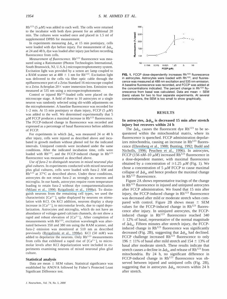
The Dcm causes the fluorescent dye Rh123 to be se-questered within the mitochondrial matrix, where itsfluorescence is quenched. FCCP administration depolar-izes mitochondria, causing an increase in Rh123 fluores-cence (Ehrenberg et al., 1988; Bunting, 1992; Budd andNicholls, 1996; Peuchen et al., 1996). In astrocytes,FCCP (156 nM–10mM) increased Rh123 fluorescence ina dose-dependent manner, with maximal fluorescenceobtained by a concentration of$1.25 mM (Fig. 1). Wechose a concentration of 5mM FCCP to cause maximalcollapse ofDcm and hence produce the maximal changein Rh123 fluorescence.
Figure 2A shows representative tracings of the changein Rh123 fluorescence in injured and uninjured astrocytesafter FCCP administration. We found that 15 min afterinjury, the FCCP-induced change in Rh123 fluorescencewas decreased after mild or moderate stretch when com-pared with control. Figure 2B shows mean6 SEMvalues for the FCCP-induced change in Rh123 fluores-cence after injury. In uninjured astrocytes, the FCCP-induced change in Rh123 fluorescence reached 3406 12% of basal, representative of the normal magnitudeof Dcm. Fifteen minutes after stretch injury, the FCCP-induced change in Rh123 fluorescence was significantlydecreased (Fig. 2B), suggesting thatDcm had declined.FCCP challenge increased Rh123 fluorescence to only1966 11% of basal after mild stretch and 1546 13% ofbasal after moderate stretch. These results indicate thatstretch causes a decline inDcm and release of Rh123frommitochondria. By 24 h, no significant difference inFCCP-induced change in Rh123 fluorescence was ob-served between injured and uninjured cells (Fig. 2B),suggesting that in astrocytesDcm recovers within 24 hafter stretch.
FIG. 1. FCCP dose-dependently increases Rh123 fluorescencein astrocytes. Astrocytes were loaded with Rh123, and fluores-cence was measured at 488 nm excitation and 535 nm emission.A baseline fluorescence was recorded, and FCCP was added atthe concentrations indicated. The percent change in Rh123 flu-orescence from basal was calculated. Data are mean 6 SEM(bars) values for two to four separate experiments. At severalconcentrations, the SEM is too small to show graphically.
J. Neurochem., Vol. 74, No. 5, 2000
1954 S. M. AHMED ET AL.

In injured neurons of pure embryonic neuronalcultures, Dcm declines only after severe stretch
Having noted apparent mitochondrial deficits in astro-cytes after injury, we next examined whether neuronalmitochondria sustain similar functional damage withstretch. In uninjured cultures of pure embryonic neurons,dischargingDcm with FCCP caused Rh123 fluorescenceto rise to 3066 15% of basal. The FCCP-inducedchange in Rh123 fluorescence was not significantly dif-ferent from control at 15 min, 24 h, or 48 h after mildstretch (Fig. 3), suggesting thatDcm was not affected bymild neuronal stretch. Moderate stretch had no immedi-ate effect on FCCP-induced change in Rh123 fluores-cence. However, 48 h after moderate stretch, the FCCP-induced change in Rh123 fluorescence was significantlylower in injured cells versus uninjured controls, suggest-ing that delayed mitochondrial damage may occur aftermoderate stretch. Fifteen minutes after severe stretch,Rh123fluorescence rose to only 2086 20% of basal afteraddition of FCCP, which represents a response 32% lessthan that of uninjured controls. This suggests that imme-diate mitochondrial damage occurs with severe stretch.Unlike injured astrocyte cultures, in whichDcm recov-ered within 24 h, the FCCP-induced change in Rh123
fluorescence in severely injured pure neuronal culturesremained significantly lower at 24 and 48 h after severestretch.
Neurons in mixed neuronal–glial cultures exhibit adecline in Dcm that recovers with time
Glial cells are responsible for maintaining the extra-cellular environment around neurons. They also secretefactors that affect neuronal adhesion, differentiation, andreceptor characteristics. Therefore, we determinedwhether mitochondria in neurons cultured with glial cellsreacted differently to injury as compared with neurons inpure cultures devoid of astrocytes. FCCP administrationincreased Rh123 fluorescence to 3876 19% of basal inuninjured neurons of mixed neuronal plus glial cultures(Fig. 4). Fifteen minutes after mild, moderate, or severestretch, there was a significant decline in neuronalDcm,with FCCP-induced Rh123 fluorescence reaching 2046 20, 1816 9, and 1816 11% of basal, respectively.Thus, our findings suggest that neuronal mitochondria in
FIG. 2. Astrocyte Dcm is decreased 15 min after injury butrecovers within 24 h. Rh123-loaded astrocytes were stimulatedwith 5 mM FCCP at 15 min and 24 h postinjury, and the changein fluorescence (indicative of the magnitude of Dcm) was mea-sured. A: The percent change from basal fluorescence wasdetermined. Controls consisted of uninjured cells stimulatedwith 5 mM FCCP. Tracings shown are representative of eight to10 separate experiments. B: Summary of mean 6 SEM (bars)values for eight to 10 separate experiments. Data at 24 h postin-jury were obtained from cells loaded with Rh123 30 min beforerecording. *p , 0.01 versus control.
FIG. 3. In neurons of pure embryonic neuronal cultures, Dcm isdecreased 15 min after severe stretch and remains depressedthrough 48 h. Rh123-loaded neurons were stimulated with 5 mMFCCP at 15 min, 24 h, and 48 h postinjury, and the change influorescence was measured. The percent change from basalfluorescence was determined. Controls consisted of uninjuredcells stimulated with 5 mM FCCP. Data at 24 and 48 h postinjurywere obtained from cells loaded with Rh123 30 min before re-cording. Data are mean 6 SEM (bars) values for six to eightdifferent experiments. *p , 0.05, **p , 0.01 versus control.
FIG. 4. In neurons of mixed neuronal plus glial cultures, Dcm isdecreased 15 min after injury but recovers with time. Rh123-loaded mixed cultures of neurons plus glia were stimulated with5 mM FCCP at 15 min, 24 h, and 48 h postinjury, and the changein neuronal fluorescence was measured. The percent changefrom pre-FCCP basal fluorescence was determined. Controlsconsisted of uninjured cells stimulated with 5 mM FCCP. Dataare mean 6 SEM (bars) values for six or seven different exper-iments. *p , 0.05, **p , 0.01 versus control.
J. Neurochem., Vol. 74, No. 5, 2000
1955MITOCHONDRIA IN ASTROCYTE AND NEURONAL INJURY
mixed neuronal plus glial cultures are more sensitive toloss ofDcm than pure embryonic neurons cultured in theabsence of astrocytes. By 48 h postinjury, no differencesin Dcm were found between injured and uninjured cells,suggesting that, unlike severely injured pure neurons,mitochondrial function recovers within 48 h in neuronsof mixed culture.
The pure neuronal cultures were prepared from em-bryonic rat brain, whereas the mixed cultures were pre-pared from neonatal rats. As such, the question may ariseas to whether the different ages of the neuron sourcesmight account for differences inDcm noted betweenneurons of pure neuronal versus mixed neuronal plusglial cultures. We therefore plated embryonic neuronsonto astrocyte cultures and found in two of two cellpreparations that, similar to neurons in neonatal mixedcultures, embryonic neurons plated onto astrocytes dis-played a significant loss ofDcm after mild stretch (datanot shown). Thus, the presence of astrocytes appears toaugment loss ofDcm in neurons after stretch injury.
Cellular ATP levels in astrocytes decline afterstretch injury
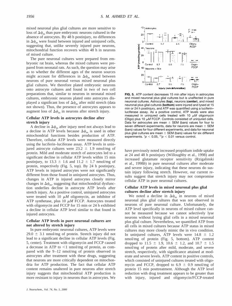
A decline inDcm after injury need not always lead toa decline in ATP levels becauseDcm is used in othermitochondrial functions besides production of ATP.Therefore, cellular ATP levels were measured directlyusing the luciferin–luciferase assay. ATP levels in unin-jured astrocyte cultures were 23.26 1.9 nmol/mg ofprotein. Mild and moderate stretch of astrocytes led to asignificant decline in cellular ATP levels within 15 minpostinjury, to 13.36 1.6 and 11.26 1.7 nmol/mg ofprotein, respectively (Fig. 5, top). By 24 h postinjury,ATP levels in injured astrocytes were not significantlydifferent from those found in uninjured astrocytes. Thus,changes in ATP in injured astrocytes closely mirrorchanges inDcm, suggesting that mitochondrial dysfunc-tion underlies decline in astrocyte ATP levels afterstretch injury. As a positive control, uninjured astrocyteswere treated with 10mM oligomycin, an inhibitor ofATP synthetase, plus 10mM FCCP. Astrocytes treatedwith oligomycin and FCCP for 15 min or 24 h exhibiteda decline in cellular ATP level similar to that found ininjured astrocytes.
Cellular ATP levels in pure neuronal cultures arenot altered by stretch injury
In pure embryonic neuronal cultures, ATP levels were29.0 6 3.1 nmol/mg of protein. Stretch injury did notlead to a significant decline in cellular ATP levels (Fig.5, center). Treatment with oligomycin and FCCP causeda decrease in ATP to,1 nmol/mg of protein, as com-pared with the 9–12 nmol/mg of protein observed inastrocytes after treatment with these drugs, suggestingthat neurons are more critically dependent on mitochon-dria for ATP production. The fact that cellular ATPcontent remains unaltered in pure neurons after stretchinjury suggests that mitochondrial ATP production ismore resistant to injury in neurons than in astrocytes. We
have previously noted increased propidium iodide uptakeat 24 and 48 h postinjury (Willoughby et al., 1998) andincreased glutamate receptor sensitivity (Rzigalinskiet al., 1998b) in pure neuronal cultures after moderateand severe injury, indicating that these neurons do sus-tain injury following stretch. However, our current re-sults suggest that stretch injury may not compromisecellular ATP in pure neuronal cultures.
Cellular ATP levels in mixed neuronal plus glialcultures decline after stretch injury
We noted a decline inDcm in neurons of mixedneuronal plus glial cultures that was not observed inneurons of pure neuronal culture. Unfortunately, theATP level specifically in neurons of mixed culture can-not be measured because we cannot selectively lyseneurons without lysing glial cells in a mixed neuronalplus glial culture. Nevertheless, we quantified ATP fromall cells in mixed cultures because ATP status in mixedcultures may more closely mimic the in vivo condition.In uninjured cultures, ATP levels were 14.86 1.2nmol/mg of protein (Fig. 5, bottom). ATP contentdropped to 11.56 1.9, 10.66 1.2, and 10.76 1.5nmol/mg of protein after mild, moderate, and severestretch, respectively, with significance attained at mod-erate and severe levels. ATP content in positive controls,which consisted of uninjured cultures treated with oligo-mycin and FCCP, dropped to 6.96 0.9 nmol/mg ofprotein 15 min posttreatment. Although the ATP levelreduction with drug treatment appears to be greater thanwith injury, injured and oligomycin/FCCP-treated
FIG. 5. ATP content decreases 15 min after injury in astrocytesand mixed neuronal plus glial cultures but is unaffected in pureneuronal cultures. Astrocytes (top), neurons (center), and mixedneuronal plus glial cultures (bottom) were injured and lysed at 15min or 24 h postinjury, and ATP was quantified using a luciferin–luciferase assay. As a positive control, ATP levels were alsomeasured in uninjured cells treated with 10 mM oligomycin(Oligo) plus 10 mM FCCP. Controls consisted of uninjured cells.Data for astrocytes are mean 6 SEM (bars) values for four toseven different experiments, data for neurons are mean 6 SEM(bars) values for four different experiments, and data for neuronalplus glial cultures are mean 6 SEM (bars) values for six differentexperiments. *p , 0.05, **p , 0.01 versus control.
J. Neurochem., Vol. 74, No. 5, 2000
1956 S. M. AHMED ET AL.
groups were not significantly different. In general, itappears that total ATP in mixed cultures of neurons plusglia is less susceptible to injury than pure astrocytecultures yet more susceptible to injury than pure neuro-nal cultures, possibly a reflection of the presence of bothcell types.
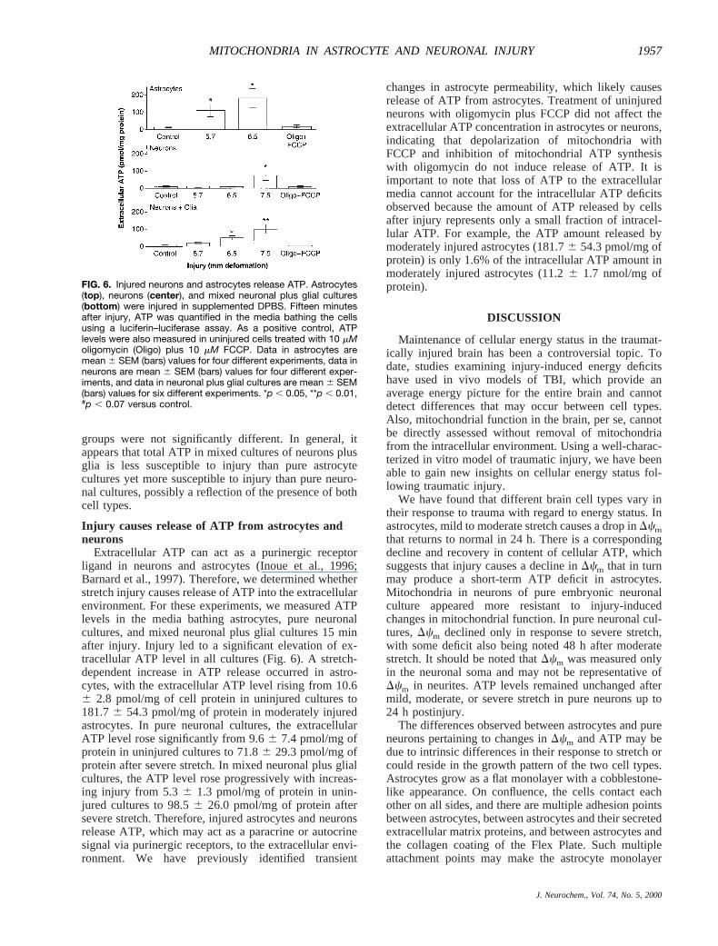
Injury causes release of ATP from astrocytes andneurons
Extracellular ATP can act as a purinergic receptorligand in neurons and astrocytes (Inoue et al., 1996;Barnard et al., 1997). Therefore, we determined whetherstretch injury causes release of ATP into the extracellularenvironment. For these experiments, we measured ATPlevels in the media bathing astrocytes, pure neuronalcultures, and mixed neuronal plus glial cultures 15 minafter injury. Injury led to a significant elevation of ex-tracellular ATP level in all cultures (Fig. 6). A stretch-dependent increase in ATP release occurred in astro-cytes, with the extracellular ATP level rising from 10.66 2.8 pmol/mg of cell protein in uninjured cultures to181.76 54.3 pmol/mg of protein in moderately injuredastrocytes. In pure neuronal cultures, the extracellularATP level rose significantly from 9.66 7.4 pmol/mg ofprotein in uninjured cultures to 71.86 29.3 pmol/mg ofprotein after severe stretch. In mixed neuronal plus glialcultures, the ATP level rose progressively with increas-ing injury from 5.36 1.3 pmol/mg of protein in unin-jured cultures to 98.56 26.0 pmol/mg of protein aftersevere stretch. Therefore, injured astrocytes and neuronsrelease ATP, which may act as a paracrine or autocrinesignal via purinergic receptors, to the extracellular envi-ronment. We have previously identified transient
changes in astrocyte permeability, which likely causesrelease of ATP from astrocytes. Treatment of uninjuredneurons with oligomycin plus FCCP did not affect theextracellular ATP concentration in astrocytes or neurons,indicating that depolarization of mitochondria withFCCP and inhibition of mitochondrial ATP synthesiswith oligomycin do not induce release of ATP. It isimportant to note that loss of ATP to the extracellularmedia cannot account for the intracellular ATP deficitsobserved because the amount of ATP released by cellsafter injury represents only a small fraction of intracel-lular ATP. For example, the ATP amount released bymoderately injured astrocytes (181.76 54.3 pmol/mg ofprotein) is only 1.6% of the intracellular ATP amount inmoderately injured astrocytes (11.26 1.7 nmol/mg ofprotein).
DISCUSSION
Maintenance of cellular energy status in the traumat-ically injured brain has been a controversial topic. Todate, studies examining injury-induced energy deficitshave used in vivo models of TBI, which provide anaverage energy picture for the entire brain and cannotdetect differences that may occur between cell types.Also, mitochondrial function in the brain, per se, cannotbe directly assessed without removal of mitochondriafrom the intracellular environment. Using a well-charac-terized in vitro model of traumatic injury, we have beenable to gain new insights on cellular energy status fol-lowing traumatic injury.
We have found that different brain cell types vary intheir response to trauma with regard to energy status. Inastrocytes, mild to moderate stretch causes a drop inDcmthat returns to normal in 24 h. There is a correspondingdecline and recovery in content of cellular ATP, whichsuggests that injury causes a decline inDcm that in turnmay produce a short-term ATP deficit in astrocytes.Mitochondria in neurons of pure embryonic neuronalculture appeared more resistant to injury-inducedchanges in mitochondrial function. In pure neuronal cul-tures,Dcm declined only in response to severe stretch,with some deficit also being noted 48 h after moderatestretch. It should be noted thatDcm was measured onlyin the neuronal soma and may not be representative ofDcm in neurites. ATP levels remained unchanged aftermild, moderate, or severe stretch in pure neurons up to24 h postinjury.
The differences observed between astrocytes and pureneurons pertaining to changes inDcm and ATP may bedue to intrinsic differences in their response to stretch orcould reside in the growth pattern of the two cell types.Astrocytes grow as a flat monolayer with a cobblestone-like appearance. On confluence, the cells contact eachother on all sides, and there are multiple adhesion pointsbetween astrocytes, between astrocytes and their secretedextracellular matrix proteins, and between astrocytes andthe collagen coating of the Flex Plate. Such multipleattachment points may make the astrocyte monolayer
FIG. 6. Injured neurons and astrocytes release ATP. Astrocytes(top), neurons (center), and mixed neuronal plus glial cultures(bottom) were injured in supplemented DPBS. Fifteen minutesafter injury, ATP was quantified in the media bathing the cellsusing a luciferin–luciferase assay. As a positive control, ATPlevels were also measured in uninjured cells treated with 10 mMoligomycin (Oligo) plus 10 mM FCCP. Data in astrocytes aremean 6 SEM (bars) values for four different experiments, data inneurons are mean 6 SEM (bars) values for four different exper-iments, and data in neuronal plus glial cultures are mean 6 SEM(bars) values for six different experiments. *p , 0.05, **p , 0.01,#p , 0.07 versus control.
J. Neurochem., Vol. 74, No. 5, 2000
1957MITOCHONDRIA IN ASTROCYTE AND NEURONAL INJURY

more rigidly attached to the Silastic membrane and hencemore susceptible to stretch injury induced by deforma-tion of the Silastic membrane. Neurons in pure cultureare grown on poly-D-lysine-coated Flex Plates. Becauseno astrocytes are present they attach less strongly to thesubstratum via electrostatic attraction between nega-tively charged lipids and polysaccharides in the plasmamembrane and positively charged poly-D-lysine mole-cules (Yavin and Yavin, 1974). It is our experience thatpure neuronal cultures form a somewhat loosely attachedneuronal network that can easily be washed off theSilastic substrate, especially when compared with astro-cyte cultures. Thus, the pure neurons may not experiencethe same degree of strain as the astrocytes because theycontact the Silastic substrate at fewer points and they aremore loosely attached.
In contrast to pure neurons, neurons in mixed culturewith astrocytes exhibited a decline inDcm in response tomild, moderate, and severe stretch, suggesting that thepresence of astrocytes somehow alters the neuronal re-sponse to injury. The presence of astrocytes may affectthe neuronal response in one of several ways. The firstdeals once again with the growth pattern. Pure neuronsadhere loosely to the poly-D-lysine-coated Silastic mem-brane. However, in mixed preparations containing astro-cytes, the neurons have multiple points at which attach-ments to the astrocyte monolayer may be made, includ-ing cell adhesion molecules present on astrocytes andcomponents of the extracellular matrix secreted by as-trocytes. Thus, the neurons in mixed cultures may have asomewhat firmer attachment to the substrate, via theastrocyte monolayer, and may experience a somewhatgreater amount of stretch than the more loosely attachedpure neuronal cultures. Alternatively, substances re-leased from astrocytes, such as glutamate and ATP, maystimulate receptor-mediated [Ca21]i elevation and exces-sive mitochondrial calcium uptake (Peng et al., 1998;Boitier et al., 1999), thereby causing a drop inDcm.Finally, the presence of astrocytes may promote neuronaldifferentiation and alterations in receptor characteristics,making the neurons of mixed cultures more susceptibleto injury than pure neurons in culture.
In previous studies, we have identified injury-inducedalterations in biochemical processes linked to ATP use.The mitochondrial dysfunction identified in the presentexperiments may be the cause of these deficits. We havepreviously described a depletion of thapsigargin-sensi-tive calcium stores in injured astrocytes, which lasts forat least 3 h after mild to moderate stretch and recovers by24 h (Rzigalinski et al., 1998a). Replenishment of intra-cellular calcium stores depends on hydrolysis of ATP bySERCA. We currently report that ATP levels drop 43–52% in astrocytes after injury, raising the possibility thatthapsigargin-sensitive calcium store depletion resultsfrom inhibition of SERCA due to ATP deficit.
In our current experiments, we have found that neu-rons in pure neuronal cultures do not show any injury-induced changes in cellular ATP levels. It is interestingthat neurons in pure cultures also exhibit no alteration of
thapsigargin-sensitive stores after injury (Weber et al.,1998), suggesting that SERCA function is maintainedwith maintenance of cellular ATP. In contrast to neuronsof pure culture, neurons in mixed neuronal plus glialcultures show alterations in ion homeostasis suggestiveof ATP deficit. Similar to astrocytes, neurons in mixedneuronal plus glial cultures have depleted thapsigargin-sensitive intracellular calcium stores 15 min postinjury(Weber et al., 1998), again suggesting inhibition ofSERCA function. We have also observed a 10 mV de-polarization of neurons in mixed culture 1 h after stretchinjury, which we have termed stretch-induced delayeddepolarization (Tavalin et al., 1995). Inhibition of theNa1,K1-ATPase appears to underlie this depolarizationbecause it can be reversed by intracellular dialysis withATP (Tavalin et al., 1997). We hypothesize that an ATPdeficit occurs in neurons of mixed neuronal plus glialculture, which inhibits the Na1,K1-ATPase andSERCA. Consistent with this hypothesis, we currentlyreport that neurons of mixed neuronal plus glial culturesshow a decline inDcm even after mild stretch. We alsosee a significant decline in ATP content of mixed cul-tures after stretch injury. We cannot say with certaintywhether neurons in these mixed cultures sustain an ATPdeficit. In fact, ATP levels in mixed neuronal plus glialcultures decline less (22–28%) after injury than levels inpure astrocyte cultures (43–52%).
Injury-induced changes inDcm may result from vari-ous mechanisms. One possibility is mitochondrial cal-cium accumulation. We have previously demonstratedthat injury increases glutamate release from neurons andastrocytes (Rzigalinski et al., 1998b; S. Liang et al.,unpublished data) and enhances glutamate receptor sen-sitivity, as indicated by increased glutamate and NMDA-stimulated elevation in [Ca21]i (Zhang et al., 1996; Rzi-galinski et al., 1998b). Also, it is known that stimulationof neurons with glutamate and NMDA causes mitochon-drial calcium accumulation (White and Reynolds, 1997;Peng et al., 1998) and loss ofDcm (Budd and Nicholls,1996; Schinder et al., 1996; White and Reynolds, 1996).Glutamate released after injury may induce calcium en-try through NMDA receptor channels with subsequentmitochondrial calcium influx and decline inDcm. Wepreviously reported that [Ca21]i in astrocytes is elevatedimmediately after injury but returns to near basal levelsby 15 min postinjury (Rzigalinski et al., 1998a). Incontrast, total cell calcium content, measured using45Ca21, remains elevated in excess of 3 h after injury(Rzigalinski et al., 1997). This poses the question as towhere the excess calcium resides past 15 min postinjury.One possibility is that the excess calcium may be takenup by mitochondria, with a resulting decline ofDcm.
Free radical-mediated mitochondrial damage repre-sents another possible mechanism leading to loss inDcm.Free radical production has been widely implicated in thepathology of TBI (Muir et al., 1995; Hall et al., 1996;Hoffman and Ellis, 1996; Shohami et al., 1997), and wehave demonstrated increases in changes in phospholipidmetabolism indicative of free radical damage through
J. Neurochem., Vol. 74, No. 5, 2000
1958 S. M. AHMED ET AL.
lipid peroxidation after injury (Lamb et al., 1997). Freeradicals may increase mitochondrial permeability vialipid and protein oxidation (Schild et al., 1997) or, alter-natively, via action on the permeability transition pore(Vercesi et al., 1997). Free radicals may also inducemitochondrial dysfunction via attack of components onthe electron transport chain (Bolan˜os et al., 1997).
We also report that stretch injury leads to release ofATP from neurons and astrocytes. ATP may mediateinjury-related changes in the brain through action onATP receptors such as P2X and P2Y receptors (Inoueet al., 1996; Barnard et al., 1997). P2X receptors areligand-gated channels (Barnard et al., 1997). Stimulationof P2X receptors on neurons by ATP has been postulatedto modulate synaptic efficiency across glutaminergic,dopaminergic, and nociceptive synapses (Inoue et al.,1996). P2Y receptors are coupled to phospholipase C viaG proteins found on neuronal and nonneuronal cells inthe brain (Barnard et al., 1997). In astrocytes, stimulationof P2Y receptors leads to mobilization of calcium frominositol trisphosphate-sensitive calcium stores (Kastritsiset al., 1992). Recently, ATP released from astrocytes hasbeen shown to mediate calcium wave activity in glialcells (Guthrie et al., 1999). Increases in extracellularATP levels resulting from injury may play a role inalteration of calcium homeostasis observed after injuryin astrocytes and neurons through action on P2X and P2Yreceptors.
In summary, we report that in vitro traumatic injurycauses changes in mitochondrial function in neurons andastrocytes. We report for the first time that in vitrotraumatic insult to cells from the CNS altersDcm. Inastrocytes, injury causes a reversible decline inDcm thatappears to underlie a transient decline in cellular ATPcontent. Neurons in pure neuronal culture are more re-sistant to loss of mitochondrial function, withDcm al-tered only at higher injury levels and ATP levels remain-ing unchanged with injury. Neuronal susceptibility toloss ofDcm is influenced by the presence of glial cells.In the early phase, the presence of astrocytes increasesneuronal mitochondrial susceptibility to loss ofDcm. Atlater time points, glial cells may actually help promoterecovery of neuronal mitochondrial function. We alsofind increased release of ATP from astrocytes and neu-rons after injury that may mediate some of the pathologyof TBI through purinergic receptors. Our results showthat traumatic injury of astrocytes and neurons may leadto deficits in mitochondrial function that affect cellularenergy status and ionic homeostasis in a manner depen-dent on cell type and intercellular events.
Acknowledgment: This work was supported by grants NS-27214 and NS-07288 from the National Institutes of Health anda Center of Excellence grant-in-aid from the Commonwealth ofVirginia. We wish to thank Dr. Joyce Lloyd in the VirginiaCommonwealth University Department of Human Genetics forassistance and use of the luminometer and Sallie Holt for hertechnical assistance.
REFERENCES
Amruthesh S. C., Boerschel M. F., McKinney J. S., Willoughby K. A.,and Ellis E. F. (1993) Metabolism of arachidonic acid to epoxyei-cosatrienoic acids, hydroxyeicosatetraenoic acids, and prostaglan-dins in cultured rat hippocampal astrocytes.J. Neurochem.61,150–159.
Andersen B. J., Unterberg A. W., Clarke G. D., and Marmarou A.(1988) Effect of posttraumatic hypoventilation on cerebral energymetabolism.J. Neurosurg.68, 601–607.
Barnard E. A., Simon J., and Webb T. E. (1997) Nucleotide receptorsin the nervous system. An abundant component using diversetransduction mechanisms.Mol. Neurobiol.15, 103–130.
Boitier E., Rea R., and Duchen M. R. (1999) Mitochondria exert anegative feedback on the propagation of intracellular Ca21 wavesin rat cortical astrocytes.J. Cell Biol. 145,795–808.
Bolanos J. P., Almeida A., Fernandez E., Medina J. M., Land J. M.,Clark J. B., and Heales S. J. R. (1997) Potential mechanisms fornitric oxide-mediated impairment of brain mitochondrial energymetabolism.Biochem. Soc. Trans.25, 944–949.
Budd S. L. and Nicholls D. G. (1996) Mitochondria, calcium regula-tion, and acute glutamate excitoxicity in cultured cerebellar gran-ule cells.J. Neurochem.67, 2282–2291.
Bunting J. R. (1992) Influx and efflux kinetics of cationic dye bindingto respiring mitochondria.Biophys. Chem.42, 163–175.
Chinnery P. F. and Turnbull D. M. (1997) Mitochondrial medicine.Q. J. Med.90, 657–667.
DeWitt D. S., Hayes R. L., Lyeth B. G., Yuan X. Q., and Prough D. S.(1988) Effects of traumatic brain injury on cerebral blood flow andmetabolism: autoradiographic studies.Anesthesiol. Rev.15, 31–32.
Dietrich W. D., Alonso O., and Halley M. (1994) Early microvascularand neuronal consequences of traumatic brain injury: a light andelectron microscopic study in rats.J. Neurotrauma11, 289–301.
Ehrenberg B., Montana V., Wei M.-D., Wuskell J. P., and Loew L. M.(1988) Membrane potential can be determined in individual cellsfrom the Nernstian distribution of cationic dyes.Biophys. J.53,785–794.
Ellis E. F., McKinney J. S., Willoughby K. A., Liang S., and Pov-lishock J. T. (1995) A new model for rapid stretch-induced injuryof cells in culture: characterization of the model using astrocytes.J. Neurotrauma12, 325–339.
Erecinska M. and Silver I. A. (1989) ATP and brain function.J. Cereb.Blood Flow Metab.9, 2–19.
Fineman I., Hovda D. A., Smith M., Yoshino A., and Becker D. P.(1993) Concussive brain injury is associated with a prolongedaccumulation of calcium: a45Ca21 autoradiographic study.BrainRes.624,94–102.
Guthrie P. B., Knappenberger J., Segal M., Bennett M. V. L., CharlesA. C., and Kater S. B. (1999) ATP released from astrocytesmediates glial calcium waves.J. Neurosci.19, 520–528.
Hall E. D., Smith S. L., and Oostveen J. A. (1996) Inhibition of lipidperoxidation attenuates axotomy-induced apoptotic degenerationof facial motor neurons in neonatal rats.J. Neurosci. Res.44,293–299.
Hamm R. J., O’Dell D. M., Pike B. R., and Lyeth B. G. (1993)Cognitive impairment following traumatic brain injury: the effectof pre- and post-injury administration of scopolamine and MK-801.Brain Res. Cogn. Brain Res.1, 223–226.
Hamm R. J., Lyeth B. G., Jenkins L. W., O’Dell D. M., and Pike B. R.(1994) Selective cognitive impairment following traumatic braininjury in rats.Behav. Brain Res.59, 169–173.
Hoffman S. W. and Ellis E. F. (1996) Isoprostanes, free radical gen-erated prostaglandins with vasoconstrictive actions.J. Neuro-trauma13, 619.
Inoue K., Koizumi S., and Ueno S. (1996) Implication of ATP recep-tors in brain functions.Prog. Neurobiol.50, 483–492.
Kastritsis C. H., Salm A. K., and McCarthy K. (1992) Stimulation ofthe P2Y purinergic receptor on type-1 astroglia results in inositolphosphate formation and calcium mobilization.J. Neurochem.58,1277–1284.
J. Neurochem., Vol. 74, No. 5, 2000
1959MITOCHONDRIA IN ASTROCYTE AND NEURONAL INJURY

Lamb R. G., Harper C. C., McKinney J. S., Rzigalinski B. A., and EllisE. F. (1997) Alterations in phosphatidylcholine metabolism ofstretch injured cultured rat astrocytes.J. Neurochem.68, 1904–1910.
McIntosh T. K. (1992) Pharmacologic strategies in the treatment ofexperimental brain injury.J. Neurotrauma9 (Suppl. 1), S201–S209.
McKinney J. S., Willoughby K. A., Liang S., and Ellis E. F. (1996)Stretch-induced injury of cultured neuronal, glial, and endothelialcells.Stroke27, 934–940.
Milani D., Malgaroli A., Guidolin D., Fasolato C., Skaper S. D.,Meldolesi J., and Pozzan T. (1990) Ca21 channels and intracel-lular Ca21 stores in neuronal and neuroendocrine cells.Cell Cal-cium 11, 191–199.
Muir J. K., Tynan M., Caldwell R., and Ellis E. F. (1995) Superoxidedismutase improves cortical blood flow in rats.J. Neurotrauma12, 179–188.
Peng T. I., Jou M.-J., Sheu S.-S., and Greenamyre J. T. (1998) Visu-alization of NMDA receptor-induced mitochondrial calcium ac-cumulation in striatal neurons.Exp. Neurol.149,1–12.
Pettus E. H., Christman C. W., Giebel M. L., and Povlishock J. T.(1994) Traumatically induced altered membrane permeability: itsrelationship to traumatically induced reactive axonal change.J. Neurotrauma11, 507–522.
Peuchen S., Duchen M. R., and Clark J. B. (1996) Energy metabolismin adult astrocytes in vitro.Neuroscience71, 855–870.
Rzigalinski B. A., Liang S., McKinney J. S., Willoughby K. A., andEllis E. F. (1997) Effect of Ca21 on in vitro astrocyte injury.J. Neurochem.68, 289–296.
Rzigalinski B. A., Weber J. T., Willoughby K. A., and Ellis E. F.(1998a) Intracellular free calcium dynamics in stretch-injuredastrocytes.J. Neurochem.70, 2377–2385.
Rzigalinski B. A., Weber J. T., Willoughby K. A., Liang S., WoodwardJ. J., and Ellis E. F. (1998b) Glutamate release and enhancedreceptor sensitivity in stretch-injured neurons.J. Neurotrauma15,894.
Schild L., Reinheckel T., Wiswedel I., and Augustin W. (1997) Short-term impairment of energy production in isolated rat liver mito-chondria by hypoxia/reoxygenation: involvement of oxidativeprotein modification.Biochem. J.328,205–210.
Schinder A. F., Olson E. C., Spitzer N. C., and Montal M. (1996)Mitochondrial dysfunction is a primary event in glutamate neuro-toxicity. J. Neurosci.16, 6125–6133.
Schreiber D., Gennarelli T. A., and Meaney D. F. (1995)Proceedingsof the 1995 International Research Conference on Biomechanicsof Impact,pp. 233–244. International Research Council on Bio-kinetics of Impact, Brunen, Switzerland.
Shohami E. S., Beit-Yannai E., Horowitz M., and Kohen R. (1997)Oxidative stress in closed-head injury: brain antioxidant capacityas an indicator of functional outcome.J. Cereb. Blood FlowMetab.17, 1007–1019.
Tavalin S. J., Ellis E. F., and Satin L. S. (1995) Mechanical perturba-tion of cultured cortical neurons reveals a stretch-induced delayeddepolarization.J. Neurophysiol.74, 2767–2773.
Tavalin S. J., Ellis E. F., and Satin L. S. (1997) Inhibition of theelectrogenic Na pump underlies delayed depolarization of corticalneurons after mechanical injury or glutamate.J. Neurophysiol.77,632–638.
Unterberg A. W., Andersen B. J., Clark G. D., and Marmarou A. (1988)Cerebral energy metabolism following fluid-percussion brain in-jury in cats.J. Neurosurg.68, 594–600.
Vagnozzi R., Marmarou A., Tavazzi B., Signoretti S., Di Pierro D., DelBolgia F., Amorini A. M., Fazzina G., Sherkat S., and LazzarinoG. (1999) Changes of cerebral energy metabolism and lipid per-oxidation in rats leading to mitochondrial dysfunction after diffusebrain injury.J. Neurotrauma16, 903–913.
Vercesi A. E., Kowaltowski A. J., Grijalba M. T., Meinicke A. R., andCastilho R. F. (1997) The role of reactive oxygen species inmitochondrial permeability transition.Biosci. Rep.17, 43–52.
Vink R., McIntosh T. K., Weiner M. W., and Faden A. I. (1987) Effectsof traumatic brain injury on cerebral high-energy phosphates andpH: a31P magnetic resonance spectroscopy study.J. Cereb. BloodFlow Metab.7, 563–571.
Vink R., Golding E. M., and Headrick J. P. (1994) Bioenergeticanalysis of oxidative metabolism following traumatic brain injuryin rats.J. Neurotrauma11, 265–274.
Weber J. T., Rzigalinski B. A., Floyd C. L., Willoughby K. A., andEllis E. F. (1998) Alterations in intracellular calcium stores afterstretch-induced injury.J. Neurotrauma15, 901.
Wei E. P., Lamb R. G., and Kontos H. A. (1982) Increased phospho-lipase C activity after experimental brain injury.J. Neurosurg.56,695–698.
White R. J. and Reynolds I. J. (1996) Mitochondrial depolarization inglutamate-stimulated neurons: an early signal specific to excito-toxin exposure.J. Neurosci.16, 5688–5697.
White R. J. and Reynolds I. J. (1997) Mitochondria accumulate Ca21
following intense glutamate stimulation of cultured rat forebrainneurones.J. Physiol. (Lond.)498,31–47.
Willoughby K. A., Rzigalinski B. A., and Ellis E. F. (1998) Stretch-induced injury of cultured cells causes delayed alterations inneurons and immediate transient alterations in astrocytes.J. Neu-rotrauma15, 902.
Xiong Y., Gu Q., Peterson P. L., Muizelaar J. P., and Lee C. P. (1997)Mitochondrial dysfunction and calcium perturbation induced bytraumatic brain injury.J. Neurotrauma14, 23–34.
Yavin E. and Yavin Z. (1974) Attachment and culture of dissociatedcells from rat embryo cerebral hemispheres on polylysine-coatedsurface.J. Cell Biol. 62, 540–546.
Zhang L., Rzigalinski B. A., Ellis E. F., and Satin L. S. (1996)Reduction of voltage-dependent Mg21 blockade of NMDA cur-rent in mechanically injured neurons.Science274,1921–1923.
J. Neurochem., Vol. 74, No. 5, 2000
1960 S. M. AHMED ET AL.
Related Documents