Streptococcus mitis Induces Conversion of Helicobacter pylori to Coccoid Cells during Co-Culture In Vitro Yalda Khosravi 1 , Yakhya Dieye 2 , Mun Fai Loke 1 , Khean Lee Goh 3 , Jamuna Vadivelu 1 * 1 Department of Medical Microbiology, Faculty of Medicine, University of Malaya, Kuala Lumpur, Malaysia, 2 Vice-chancellor’s Office, University of Malaya, Kuala Lumpur, Malaysia, 3 Department of Medicine, Faculty of Medicine, University of Malaya, Kuala Lumpur, Malaysia Abstract Helicobacter pylori (H. pylori) is a major gastric pathogen that has been associated with humans for more than 60,000 years. H. pylori causes different gastric diseases including dyspepsia, ulcers and gastric cancers. Disease development depends on several factors including the infecting H. pylori strain, environmental and host factors. Another factor that might influence H. pylori colonization and diseases is the gastric microbiota that was overlooked for long because of the belief that human stomach was a hostile environment that cannot support microbial life. Once established, H. pylori mainly resides in the gastric mucosa and interacts with the resident bacteria. How these interactions impact on H. pylori-caused diseases has been poorly studied in human. In this study, we analyzed the interactions between H. pylori and two bacteria, Streptocccus mitis and Lactobacillus fermentum that are present in the stomach of both healthy and gastric disease human patients. We have found that S. mitis produced and released one or more diffusible factors that induce growth inhibition and coccoid conversion of H. pylori cells. In contrast, both H. pylori and L. fermentum secreted factors that promote survival of S. mitis during the stationary phase of growth. Using a metabolomics approach, we identified compounds that might be responsible for the conversion of H. pylori from spiral to coccoid cells. This study provide evidences that gastric bacteria influences H. pylori physiology and therefore possibly the diseases this bacterium causes. Citation: Khosravi Y, Dieye Y, Loke MF, Goh KL, Vadivelu J (2014) Streptococcus mitis Induces Conversion of Helicobacter pylori to Coccoid Cells during Co-Culture In Vitro. PLoS ONE 9(11): e112214. doi:10.1371/journal.pone.0112214 Editor: Holger Bru ¨ ggemann, Aarhus University, Denmark Received June 5, 2014; Accepted October 10, 2014; Published November 11, 2014 Copyright: ß 2014 Khosravi et al. This is an open-access article distributed under the terms of the Creative Commons Attribution License, which permits unrestricted use, distribution, and reproduction in any medium, provided the original author and source are credited. Data Availability: The authors confirm that all data underlying the findings are fully available without restriction. All relevant data are within the paper and its Supporting Information files. Raw data from the LCMS analysis can be obtained by contacting the corresponding author. Funding: This work was supported by University of Malaya-Ministry of Higher Education, UM-MOHE, High Impact Research (HIR) grant UM.C/625/1/HIR/MOHE/ CHAN-02; account no. A000002-50001, ‘‘Molecular Genetics’’. The funders had no role in study design, data collection and analysis, decision to publish, or preparation of the manuscript. Competing Interests: The authors confirm that Jamunarani Vadivelu, author of the manuscript, is a PLOS ONE Editorial Board member. However, this does not alter the authors’ adherence to PLOS ONE Editorial policies and criteria. * Email: [email protected] Introduction Helicobacter pylori (H. pylori) is a major gastric pathogen that has been associated with humans for more than 60,000 years [1]. Most of H. pylori-infected individuals develop an asymptomatic gastritis and can harbor this bacterium for their lifetime. In a subset of hosts, H. pylori causes different gastric diseases including dyspepsia, ulcers and gastric cancers. Disease development depends on several factors including the infecting H. pylori strain, environmental and host factors [2,3]. Another factor that is emerging as playing an important role in H. pylori-caused diseases is the host gastric microbiota. The role of gastric microbiota was previously overlooked because of the belief for long that human stomach was a hostile environment that cannot support microbial life. Studies subsequent to H. pylori discovery in 1981 showed that in fact human stomach constitutes a very diverse and complex ecosystem with a bacterial density comparable to that of the duodenum [4]. Gut microbiota plays important roles in several host functions including energy harvest and storage from the diet [5], development and regulation of the gut-associated mucosal immune system [6], regulation of the central nervous system [7], detoxification of xenobiotics and carcinogens, and protection against colonization by pathogens [8]. Although the gastric microbiota has been less studied than the microbiota in other gut sites, it is obvious that its composition and diversity are crucial for gut homeostasis. Once established, H. pylori mainly resides in the gastric mucosa, a site that has a specific microbiota closely associated with the host [9]. H. pylori continuously interacts with the resident gastric bacteria, which affect not only H. pylori colonization but also the immune response to the infection [10]. Although not formerly demonstrated, it is likely that these interactions influence H. pylori colonization and disease development. This question has been poorly addressed in humans. However, studies in animal models of H. pylori infection have provided important insights. Studies in gerbil identified gastric bacteria that inhibited H. pylori coloniza- tion while others were enriched in H. pylori-infected animals [11– 13]. Additionally, long-term infection by H. pylori significantly modified the composition of the gastric microbiota of gerbils [14]. A similar observation was made in mice where H. pylori infection altered the composition and diversity of the gastric flora [15]. In a mouse model of gastric cancer, H. pylori-induced carcinogenesis was delayed in the absence of the microbiota suggesting a role for gastric bacteria in the development of the disease [16,17]. Studies that analyzed the gastric microbiota of humans infected with H. pylori present the limitation of including a small number of PLOS ONE | www.plosone.org 1 November 2014 | Volume 9 | Issue 11 | e112214

Welcome message from author
This document is posted to help you gain knowledge. Please leave a comment to let me know what you think about it! Share it to your friends and learn new things together.
Transcript
Streptococcus mitis Induces Conversion of Helicobacterpylori to Coccoid Cells during Co-Culture In VitroYalda Khosravi1, Yakhya Dieye2, Mun Fai Loke1, Khean Lee Goh3, Jamuna Vadivelu1*
1 Department of Medical Microbiology, Faculty of Medicine, University of Malaya, Kuala Lumpur, Malaysia, 2 Vice-chancellor’s Office, University of Malaya, Kuala Lumpur,
Malaysia, 3 Department of Medicine, Faculty of Medicine, University of Malaya, Kuala Lumpur, Malaysia
Abstract
Helicobacter pylori (H. pylori) is a major gastric pathogen that has been associated with humans for more than 60,000 years.H. pylori causes different gastric diseases including dyspepsia, ulcers and gastric cancers. Disease development depends onseveral factors including the infecting H. pylori strain, environmental and host factors. Another factor that might influence H.pylori colonization and diseases is the gastric microbiota that was overlooked for long because of the belief that humanstomach was a hostile environment that cannot support microbial life. Once established, H. pylori mainly resides in thegastric mucosa and interacts with the resident bacteria. How these interactions impact on H. pylori-caused diseases hasbeen poorly studied in human. In this study, we analyzed the interactions between H. pylori and two bacteria, Streptocccusmitis and Lactobacillus fermentum that are present in the stomach of both healthy and gastric disease human patients. Wehave found that S. mitis produced and released one or more diffusible factors that induce growth inhibition and coccoidconversion of H. pylori cells. In contrast, both H. pylori and L. fermentum secreted factors that promote survival of S. mitisduring the stationary phase of growth. Using a metabolomics approach, we identified compounds that might beresponsible for the conversion of H. pylori from spiral to coccoid cells. This study provide evidences that gastric bacteriainfluences H. pylori physiology and therefore possibly the diseases this bacterium causes.
Citation: Khosravi Y, Dieye Y, Loke MF, Goh KL, Vadivelu J (2014) Streptococcus mitis Induces Conversion of Helicobacter pylori to Coccoid Cells during Co-CultureIn Vitro. PLoS ONE 9(11): e112214. doi:10.1371/journal.pone.0112214
Editor: Holger Bruggemann, Aarhus University, Denmark
Received June 5, 2014; Accepted October 10, 2014; Published November 11, 2014
Copyright: � 2014 Khosravi et al. This is an open-access article distributed under the terms of the Creative Commons Attribution License, which permitsunrestricted use, distribution, and reproduction in any medium, provided the original author and source are credited.
Data Availability: The authors confirm that all data underlying the findings are fully available without restriction. All relevant data are within the paper and itsSupporting Information files. Raw data from the LCMS analysis can be obtained by contacting the corresponding author.
Funding: This work was supported by University of Malaya-Ministry of Higher Education, UM-MOHE, High Impact Research (HIR) grant UM.C/625/1/HIR/MOHE/CHAN-02; account no. A000002-50001, ‘‘Molecular Genetics’’. The funders had no role in study design, data collection and analysis, decision to publish, orpreparation of the manuscript.
Competing Interests: The authors confirm that Jamunarani Vadivelu, author of the manuscript, is a PLOS ONE Editorial Board member. However, this does notalter the authors’ adherence to PLOS ONE Editorial policies and criteria.
* Email: [email protected]
Introduction
Helicobacter pylori (H. pylori) is a major gastric pathogen that
has been associated with humans for more than 60,000 years [1].
Most of H. pylori-infected individuals develop an asymptomatic
gastritis and can harbor this bacterium for their lifetime. In a
subset of hosts, H. pylori causes different gastric diseases including
dyspepsia, ulcers and gastric cancers. Disease development
depends on several factors including the infecting H. pylori strain,
environmental and host factors [2,3]. Another factor that is
emerging as playing an important role in H. pylori-caused diseases
is the host gastric microbiota. The role of gastric microbiota was
previously overlooked because of the belief for long that human
stomach was a hostile environment that cannot support microbial
life. Studies subsequent to H. pylori discovery in 1981 showed that
in fact human stomach constitutes a very diverse and complex
ecosystem with a bacterial density comparable to that of the
duodenum [4]. Gut microbiota plays important roles in several
host functions including energy harvest and storage from the diet
[5], development and regulation of the gut-associated mucosal
immune system [6], regulation of the central nervous system [7],
detoxification of xenobiotics and carcinogens, and protection
against colonization by pathogens [8]. Although the gastric
microbiota has been less studied than the microbiota in other
gut sites, it is obvious that its composition and diversity are crucial
for gut homeostasis.
Once established, H. pylori mainly resides in the gastric mucosa,
a site that has a specific microbiota closely associated with the host
[9]. H. pylori continuously interacts with the resident gastric
bacteria, which affect not only H. pylori colonization but also the
immune response to the infection [10]. Although not formerly
demonstrated, it is likely that these interactions influence H. pyloricolonization and disease development. This question has been
poorly addressed in humans. However, studies in animal models of
H. pylori infection have provided important insights. Studies in
gerbil identified gastric bacteria that inhibited H. pylori coloniza-
tion while others were enriched in H. pylori-infected animals [11–
13]. Additionally, long-term infection by H. pylori significantly
modified the composition of the gastric microbiota of gerbils [14].
A similar observation was made in mice where H. pylori infection
altered the composition and diversity of the gastric flora [15]. In a
mouse model of gastric cancer, H. pylori-induced carcinogenesis
was delayed in the absence of the microbiota suggesting a role for
gastric bacteria in the development of the disease [16,17]. Studies
that analyzed the gastric microbiota of humans infected with H.pylori present the limitation of including a small number of
PLOS ONE | www.plosone.org 1 November 2014 | Volume 9 | Issue 11 | e112214
patients and have yielded conflicted results [18]. However it can
be hypothesized that the modification induced by H. pyloricolonization including elevation of gastric pH, destruction of
epithelial cells and production of metabolites favor the develop-
ment of certain bacterial species and inhibit others.
In this study, we wanted to investigate the interactions between
H. pylori and two bacteria, Streptoccus mitis and Lactobacillusfermentum, which have been isolated from gastric biopsies of both
healthy and gastric disease human patients including H. pylori-infected individuals [4,9,19,20]. S. mitis is a member of the human
oral microbiota [21]. Considered for long as a commensal, S. mitisis presently viewed at least as an opportunistic pathogen as
evidenced by several studies that have demonstrated its involve-
ment in oral and systemic diseases [22]. Interestingly, S. mitis was
found to be significantly enriched in the stomach of atrophic
gastritis and gastric cancer patients [19]. L. fermentum belongs to
the group of GRAS (Generally Regarded As Safe) lactic acid
bacteria. It is a member of the human gastrointestinal microbiota
and strains of L. fermentum have shown probiotic properties
providing protection against respiratory infections [23–25] or
being used in functional food [26]. Probiotics have recently
attracted interest for the treatment of H. pylori infection, several
lactic acid bacteria showing anti-H. pylori properties and can
possibly provide an alternative to address the increase of antibiotic
resistance [27]. We have found that S. mitis produced and released
factors that induce coccoid conversion of H. pylori cells during co-
culture in vitro. In contrast, both H. pylori and L. fermentumreleased products that improved S. mitis survival during the
stationary phase of growth. These interactions possibly impact on
the diseases caused by H. pylori and could explain the increase of
S. mitis cells in the stomach of certain gastric disease patients.
Materials and Methods
Bacterial strains, growth conditions and co-culture assayH. pylori strain NCTC 11637, S. mitis strain ATCC 6249 and
L. fermentum strain ATCC 8289 were obtained from the
American Type Culture Collection (ATCC, USA). H. pyloristrain UM032 is a clinical isolate from the University of Malaya
Medical Centre, Kuala Lumpur, Malaysia that was previously
described [28]. All the bacteria were grown on chocolate-agar
plate or in Brain Heart Infusion (BHI) broth supplemented with
0.4% yeast extract and 1% b-cyclodextrin, and incubated at 37uCin a humidified incubator with 10% CO2. This microaerophilic
condition is needed for growth of H. pylori in vitro but is not a
requirement for S. mitis and L. fermentum. However, for
consistency, we grew all the organisms in the same conditions
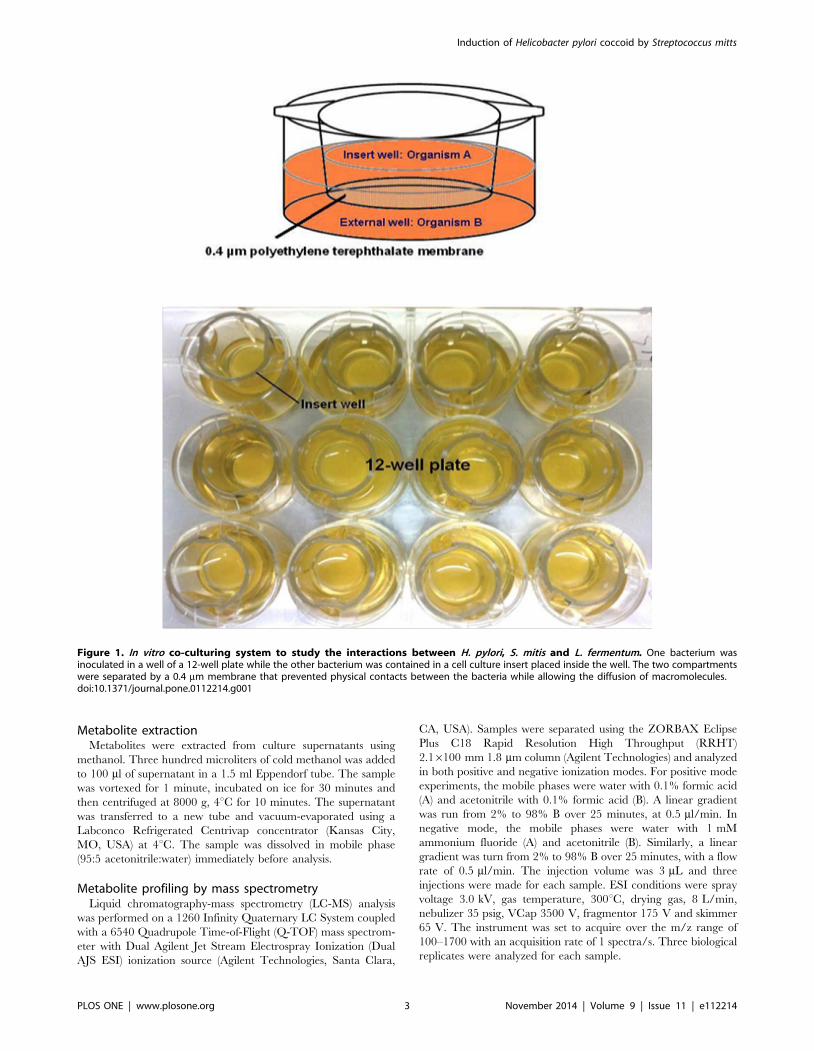
throughout the study. For bacterial co-culture, we used cell culture
inserts (BD Biosciences, San Jose, CA, USA) that can be placed
inside the wells of a12-well plate (BD Biosciences, San Jose, CA,
USA) (Fig. 1). One bacterial culture was inoculated in the 12-well
plate while the other microorganism was placed inside the insert.
The two co-culture compartments were separated by a polyeth-
ylene terephathalate (PTE) membrane with 0.4 mm pores that
prevented physical contact between the two bacteria while
allowing free diffusion of macromolecules. To verify the absence
of bacterial crossing of the membrane between the 2 compart-
ments, BHI cultures of H. pylori, S. mitis and L. fermentum were
inoculated in inserts that were placed on wells of a 12-well plate
containing fresh BHI broth and incubated for 5 days in an
incubator as described above. Presence of bacterial cells in the
wells was verified daily by plating 100 ml of the broth onto
chocolate-agar plates that were incubated at 37uC in an incubator
with 10% CO2. No bacterial growth was detected in these tests for
the 5 consecutive days (not shown). For co-culture assay with H.pylori, cells from a 2–3 day old chocolate-agar plate were used to
make a suspension of OD600 ,0.02 (106–107 cfu/ml). Two ml of
suspension of strain NCTC 11637 or of strain UM032 were
distributed in each well of a 12-well plate. A cell culture insert
containing 0.5 ml of a suspension of S. mitis or of L. fermentum at
OD600 ,0.008 (105–106 cfu/ml) prepared from an overnight
culture in BHI broth was then placed in each well. We found these
proportions between the slow growing H. pylori and the faster
growing S. mitis and L. fermentum to give the most reproducible
results. For co-culture of S. mitis and L. fermentum, each
compartment received a bacterial suspension at OD600 ,0.008.
The co-cultured bacteria were incubated at 37uC in a humidified
incubator with 10% CO2 for 7 consecutive days. At each day,
dilutions from each of the co-culture compartments were plated
onto chocolate-agar plates that were incubated at 37uC as
described above. The bacterial count was determined after 1
day for S. mitis and L. fermentum, and after 3 days for H. pylori.Additionally, H. pylori cells were recovered during both mono and
co-culture, Gram-stained and examined at the microscope to
inspect the morphology of the bacteria. Each co-culture experi-
ment was repeated at least three times.
Effect of supernatant on bacterial cultureSupernatant of H. pylori cultures were obtained by inoculating
BHI broth with cells from a 2–3 day old chocolate-agar plate and
incubated at 37uC as described above. After 1, 2 and 4 days of
growth, the culture was centrifuged at 5,000 g for 5 min. and the
supernatant recovered and filtered with a 0.22 mm syringe filter
(Terumo Europe N.V., Leuven, Belgium). The filtered superna-
tant was used immediately. S. mitis supernatant was prepared
similarly except that the BHI broth was inoculated (1/100
dilution) from an overnight liquid culture. To test the effect of
the supernatants, 2 ml of H. pylori and of S. mitis suspension in
BHI broth at OD600 ,0.02 and ,0.008 respectively, prepared as
described above were distributed in each well of a 12-well plate.
Five hundred microliters of filtered supernatant of 1-, 2- and 4-day
old cultures of the other bacterium were added to the wells and the
plates incubated at 37uC as described above. At each day,
dilutions of the cultures were plated onto chocolate-agar plate and
incubated at 37uC and the bacterial count determined as described
above. Additionally, H. pylori cells were recovered, Gram-stained
and examined at the microscope to monitor the presence of
coccoids.
Determination of bacterial viabilityBacterial viability was assessed by determining the metabolic
activities of bacterial cells. For this purpose, the levels of cellular
ATP were measured using the luciferase-based BacTiter-Glo assay
kit (Promega, Madison, WI, USA) according to the manufacturer’s
recommendations. Briefly, 100 ml of bacterial cells from mono-
cultures and from each compartment of co-cultures were
separately mixed with 100 ml of BacTiter-Glo Reagent, incubated
for five minutes at room temperature and the luminescence
measured using a microplate reader (Varioskan Flash, Thermo
Scientific, USA). The results were expressed as relative light units.
To determine the baseline ATP content of the bacteria (level of
ATP in dead cells), cells from a bacterial culture at OD600 ,1
were pelleted, then re-suspended in an equal volume of 10%
formalin. The ATP levels in these formalin-killed bacteria were
measured immediately after mixing and at different time points up
to 4 hours.
Induction of Helicobacter pylori coccoid by Streptococcus mitts
PLOS ONE | www.plosone.org 2 November 2014 | Volume 9 | Issue 11 | e112214
Metabolite extractionMetabolites were extracted from culture supernatants using
methanol. Three hundred microliters of cold methanol was added
to 100 ml of supernatant in a 1.5 ml Eppendorf tube. The sample
was vortexed for 1 minute, incubated on ice for 30 minutes and
then centrifuged at 8000 g, 4uC for 10 minutes. The supernatant
was transferred to a new tube and vacuum-evaporated using a
Labconco Refrigerated Centrivap concentrator (Kansas City,
MO, USA) at 4uC. The sample was dissolved in mobile phase
(95:5 acetonitrile:water) immediately before analysis.
Metabolite profiling by mass spectrometryLiquid chromatography-mass spectrometry (LC-MS) analysis
was performed on a 1260 Infinity Quaternary LC System coupled
with a 6540 Quadrupole Time-of-Flight (Q-TOF) mass spectrom-
eter with Dual Agilent Jet Stream Electrospray Ionization (Dual
AJS ESI) ionization source (Agilent Technologies, Santa Clara,
CA, USA). Samples were separated using the ZORBAX Eclipse
Plus C18 Rapid Resolution High Throughput (RRHT)
2.16100 mm 1.8 mm column (Agilent Technologies) and analyzed
in both positive and negative ionization modes. For positive mode
experiments, the mobile phases were water with 0.1% formic acid
(A) and acetonitrile with 0.1% formic acid (B). A linear gradient
was run from 2% to 98% B over 25 minutes, at 0.5 ml/min. In
negative mode, the mobile phases were water with 1 mM
ammonium fluoride (A) and acetonitrile (B). Similarly, a linear
gradient was turn from 2% to 98% B over 25 minutes, with a flow
rate of 0.5 ml/min. The injection volume was 3 mL and three
injections were made for each sample. ESI conditions were spray
voltage 3.0 kV, gas temperature, 300uC, drying gas, 8 L/min,
nebulizer 35 psig, VCap 3500 V, fragmentor 175 V and skimmer
65 V. The instrument was set to acquire over the m/z range of
100–1700 with an acquisition rate of 1 spectra/s. Three biological
replicates were analyzed for each sample.
Figure 1. In vitro co-culturing system to study the interactions between H. pylori, S. mitis and L. fermentum. One bacterium wasinoculated in a well of a 12-well plate while the other bacterium was contained in a cell culture insert placed inside the well. The two compartmentswere separated by a 0.4 mm membrane that prevented physical contacts between the bacteria while allowing the diffusion of macromolecules.doi:10.1371/journal.pone.0112214.g001
Induction of Helicobacter pylori coccoid by Streptococcus mitts
PLOS ONE | www.plosone.org 3 November 2014 | Volume 9 | Issue 11 | e112214
Data processing and statistical analysisSpectrum was extracted using the MassHunter Qualitative
Analysis software (version B.06.00) (Agilent Technologies). Statis-
tical analysis was performed by Mass Profiler Professional (MPP)
software (version 3.12.61) (Agilent Technologies). For normaliza-
tion, entities were baselined to the median of all samples. This
entity list was used for statistical analysis by applying unpaired T-
test (one-way ANOVA, asymptotic p-value,0.05) and Benjamini-
Hochberg FDR of 1.0% as multiple testing corrections. Entities
were compared between samples by fold change in relative
intensity.
Results
To study the interactions between H. pylori, S. mitis and L.fermentum during growth in vitro, we established a co-culture
method in which 2 bacterial species were grown in 2 compart-
ments separated by a membrane that allowed exchange of
diffusible molecules produced and released by the bacteria while
preventing them from making a physical contact. In these assays,
we wanted the cell densities in the 2 compartments to be as close as
possible. For this we had to overcome 2 constraints: (i) H. pylorislowly grows in vitro while S. mitis and L. fermentum are fast
growing organisms and (ii) in our growth conditions, inocula of
high density were needed for H. pylori cultures to expand. We
performed preliminary tests and from the results we chose starting
inocula of H. pylori at 106–107 cfu/ml and of S. mitis and L.fermentum at 105–106 cfu/ml and monitored the cultures from 1
to 7 days after inoculation.
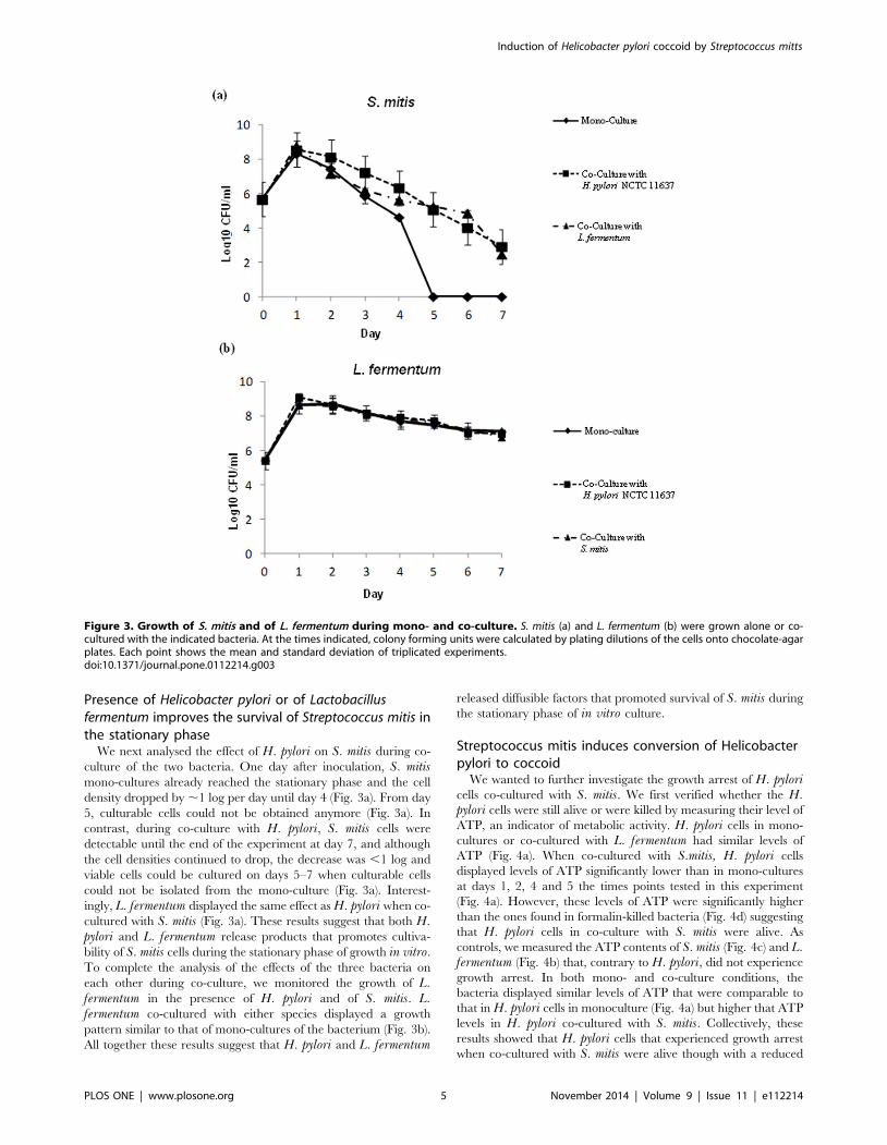
Streptococcus mitis induces growth arrest ofHelicobacter pylori cultures
H. pylori mono-cultures increased in cell density by ,2 logs
between day 1 and day 4 following inoculation and then stabilized
up to day 7 (Fig. 2a). Interestingly, when co-cultured with S. mitis,cell density of H. pylori cultures dramatically dropped from day 1
and viable cells could not be detected after two days of co-culture
(Fig. 2a). This phenomenon was not strain specific since both H.pylori UM032 [28], a clinical isolate and H. pylori NCTC, a
laboratory strain displayed the same behaviour (Fig. 2b). In
contrast to S. mitis, co-culture with L. fermentum did not affect the
growth of H. pylori cells that was comparable to that of a mono-
culture (Fig. 2a). These results suggest that S. mitis specifically
inhibits growth of H. pylori cells in co-culture.
Figure 2. Effect of S. mitis and of L. fermentum on H. pylori growth during co-culture. H. pylori reference (NCTC 11637, (a)) and clinical(UM032, (b)) strains were grown alone or co-cultured with S. mitis or with L. fermentum. At the times indicated, colony forming unit of H. pylori werecalculated by plating dilutions of the cells onto chocolate-agar plates. Each point shows the means and standard deviation of triplicated experiments.doi:10.1371/journal.pone.0112214.g002
Induction of Helicobacter pylori coccoid by Streptococcus mitts
PLOS ONE | www.plosone.org 4 November 2014 | Volume 9 | Issue 11 | e112214
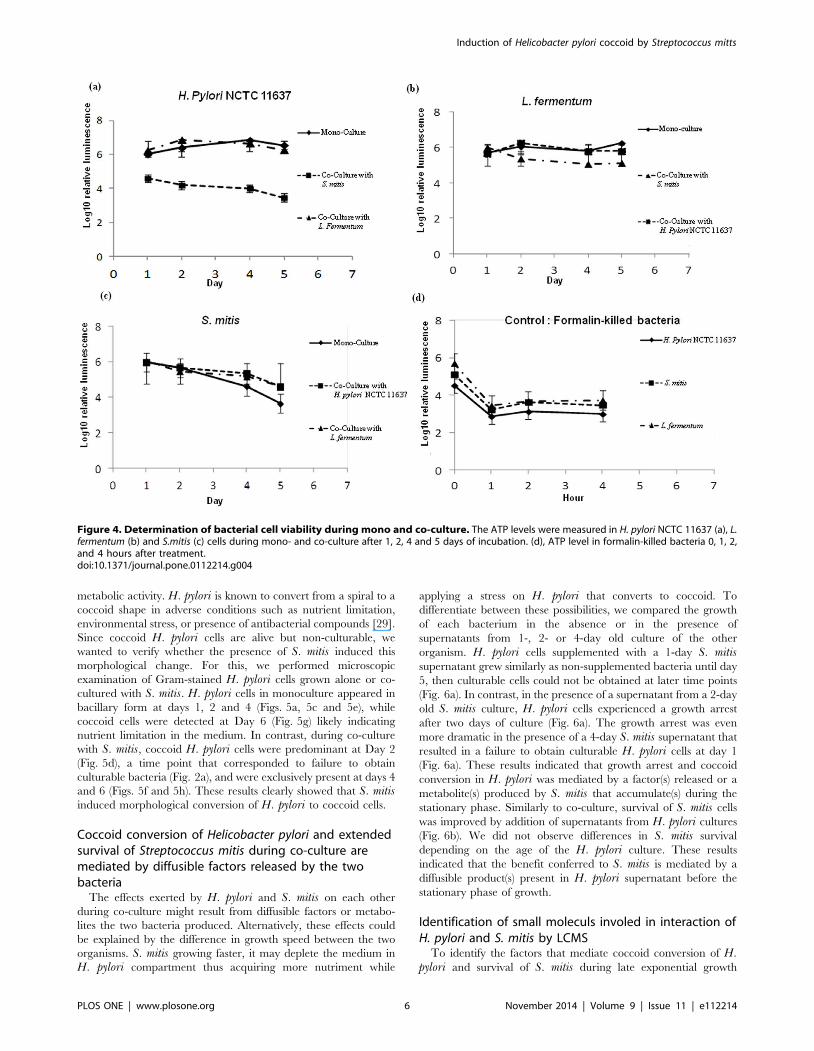
Presence of Helicobacter pylori or of Lactobacillusfermentum improves the survival of Streptococcus mitis inthe stationary phase
We next analysed the effect of H. pylori on S. mitis during co-
culture of the two bacteria. One day after inoculation, S. mitismono-cultures already reached the stationary phase and the cell
density dropped by ,1 log per day until day 4 (Fig. 3a). From day
5, culturable cells could not be obtained anymore (Fig. 3a). In
contrast, during co-culture with H. pylori, S. mitis cells were
detectable until the end of the experiment at day 7, and although
the cell densities continued to drop, the decrease was ,1 log and
viable cells could be cultured on days 5–7 when culturable cells
could not be isolated from the mono-culture (Fig. 3a). Interest-
ingly, L. fermentum displayed the same effect as H. pylori when co-
cultured with S. mitis (Fig. 3a). These results suggest that both H.pylori and L. fermentum release products that promotes cultiva-
bility of S. mitis cells during the stationary phase of growth in vitro.
To complete the analysis of the effects of the three bacteria on
each other during co-culture, we monitored the growth of L.fermentum in the presence of H. pylori and of S. mitis. L.fermentum co-cultured with either species displayed a growth
pattern similar to that of mono-cultures of the bacterium (Fig. 3b).
All together these results suggest that H. pylori and L. fermentum
released diffusible factors that promoted survival of S. mitis during
the stationary phase of in vitro culture.
Streptococcus mitis induces conversion of Helicobacterpylori to coccoid
We wanted to further investigate the growth arrest of H. pyloricells co-cultured with S. mitis. We first verified whether the H.pylori cells were still alive or were killed by measuring their level of
ATP, an indicator of metabolic activity. H. pylori cells in mono-
cultures or co-cultured with L. fermentum had similar levels of
ATP (Fig. 4a). When co-cultured with S.mitis, H. pylori cells
displayed levels of ATP significantly lower than in mono-cultures
at days 1, 2, 4 and 5 the times points tested in this experiment
(Fig. 4a). However, these levels of ATP were significantly higher
than the ones found in formalin-killed bacteria (Fig. 4d) suggesting
that H. pylori cells in co-culture with S. mitis were alive. As
controls, we measured the ATP contents of S. mitis (Fig. 4c) and L.fermentum (Fig. 4b) that, contrary to H. pylori, did not experience
growth arrest. In both mono- and co-culture conditions, the
bacteria displayed similar levels of ATP that were comparable to
that in H. pylori cells in monoculture (Fig. 4a) but higher that ATP
levels in H. pylori co-cultured with S. mitis. Collectively, these
results showed that H. pylori cells that experienced growth arrest
when co-cultured with S. mitis were alive though with a reduced
Figure 3. Growth of S. mitis and of L. fermentum during mono- and co-culture. S. mitis (a) and L. fermentum (b) were grown alone or co-cultured with the indicated bacteria. At the times indicated, colony forming units were calculated by plating dilutions of the cells onto chocolate-agarplates. Each point shows the mean and standard deviation of triplicated experiments.doi:10.1371/journal.pone.0112214.g003
Induction of Helicobacter pylori coccoid by Streptococcus mitts
PLOS ONE | www.plosone.org 5 November 2014 | Volume 9 | Issue 11 | e112214
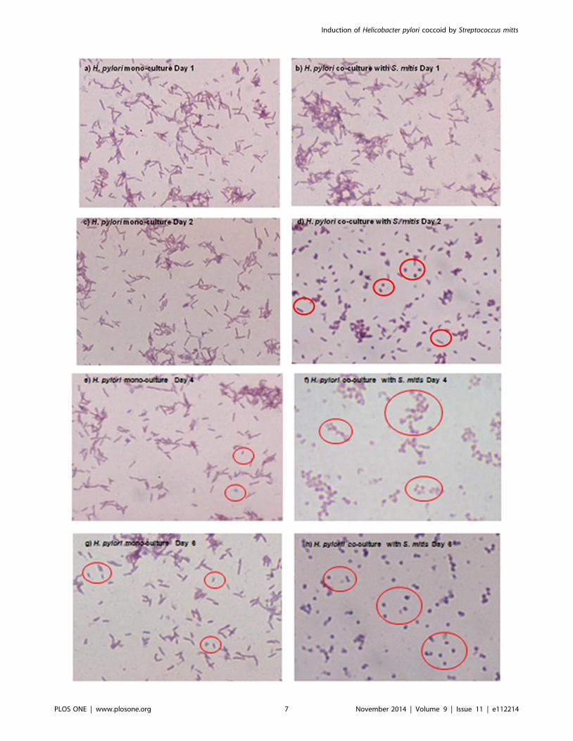
metabolic activity. H. pylori is known to convert from a spiral to a
coccoid shape in adverse conditions such as nutrient limitation,
environmental stress, or presence of antibacterial compounds [29].
Since coccoid H. pylori cells are alive but non-culturable, we
wanted to verify whether the presence of S. mitis induced this
morphological change. For this, we performed microscopic
examination of Gram-stained H. pylori cells grown alone or co-
cultured with S. mitis. H. pylori cells in monoculture appeared in
bacillary form at days 1, 2 and 4 (Figs. 5a, 5c and 5e), while
coccoid cells were detected at Day 6 (Fig. 5g) likely indicating
nutrient limitation in the medium. In contrast, during co-culture
with S. mitis, coccoid H. pylori cells were predominant at Day 2
(Fig. 5d), a time point that corresponded to failure to obtain
culturable bacteria (Fig. 2a), and were exclusively present at days 4
and 6 (Figs. 5f and 5h). These results clearly showed that S. mitisinduced morphological conversion of H. pylori to coccoid cells.
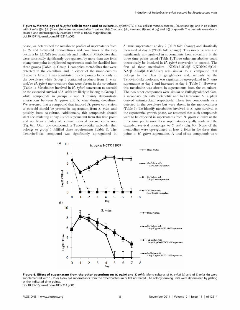
Coccoid conversion of Helicobacter pylori and extendedsurvival of Streptococcus mitis during co-culture aremediated by diffusible factors released by the twobacteria
The effects exerted by H. pylori and S. mitis on each other
during co-culture might result from diffusible factors or metabo-
lites the two bacteria produced. Alternatively, these effects could
be explained by the difference in growth speed between the two
organisms. S. mitis growing faster, it may deplete the medium in
H. pylori compartment thus acquiring more nutriment while
applying a stress on H. pylori that converts to coccoid. To
differentiate between these possibilities, we compared the growth
of each bacterium in the absence or in the presence of
supernatants from 1-, 2- or 4-day old culture of the other
organism. H. pylori cells supplemented with a 1-day S. mitissupernatant grew similarly as non-supplemented bacteria until day
5, then culturable cells could not be obtained at later time points
(Fig. 6a). In contrast, in the presence of a supernatant from a 2-day
old S. mitis culture, H. pylori cells experienced a growth arrest
after two days of culture (Fig. 6a). The growth arrest was even
more dramatic in the presence of a 4-day S. mitis supernatant that
resulted in a failure to obtain culturable H. pylori cells at day 1
(Fig. 6a). These results indicated that growth arrest and coccoid
conversion in H. pylori was mediated by a factor(s) released or a
metabolite(s) produced by S. mitis that accumulate(s) during the
stationary phase. Similarly to co-culture, survival of S. mitis cells
was improved by addition of supernatants from H. pylori cultures
(Fig. 6b). We did not observe differences in S. mitis survival
depending on the age of the H. pylori culture. These results
indicated that the benefit conferred to S. mitis is mediated by a
diffusible product(s) present in H. pylori supernatant before the
stationary phase of growth.
Identification of small moleculs involed in interaction ofH. pylori and S. mitis by LCMS
To identify the factors that mediate coccoid conversion of H.pylori and survival of S. mitis during late exponential growth
Figure 4. Determination of bacterial cell viability during mono and co-culture. The ATP levels were measured in H. pylori NCTC 11637 (a), L.fermentum (b) and S.mitis (c) cells during mono- and co-culture after 1, 2, 4 and 5 days of incubation. (d), ATP level in formalin-killed bacteria 0, 1, 2,and 4 hours after treatment.doi:10.1371/journal.pone.0112214.g004
Induction of Helicobacter pylori coccoid by Streptococcus mitts
PLOS ONE | www.plosone.org 6 November 2014 | Volume 9 | Issue 11 | e112214
Induction of Helicobacter pylori coccoid by Streptococcus mitts
PLOS ONE | www.plosone.org 7 November 2014 | Volume 9 | Issue 11 | e112214
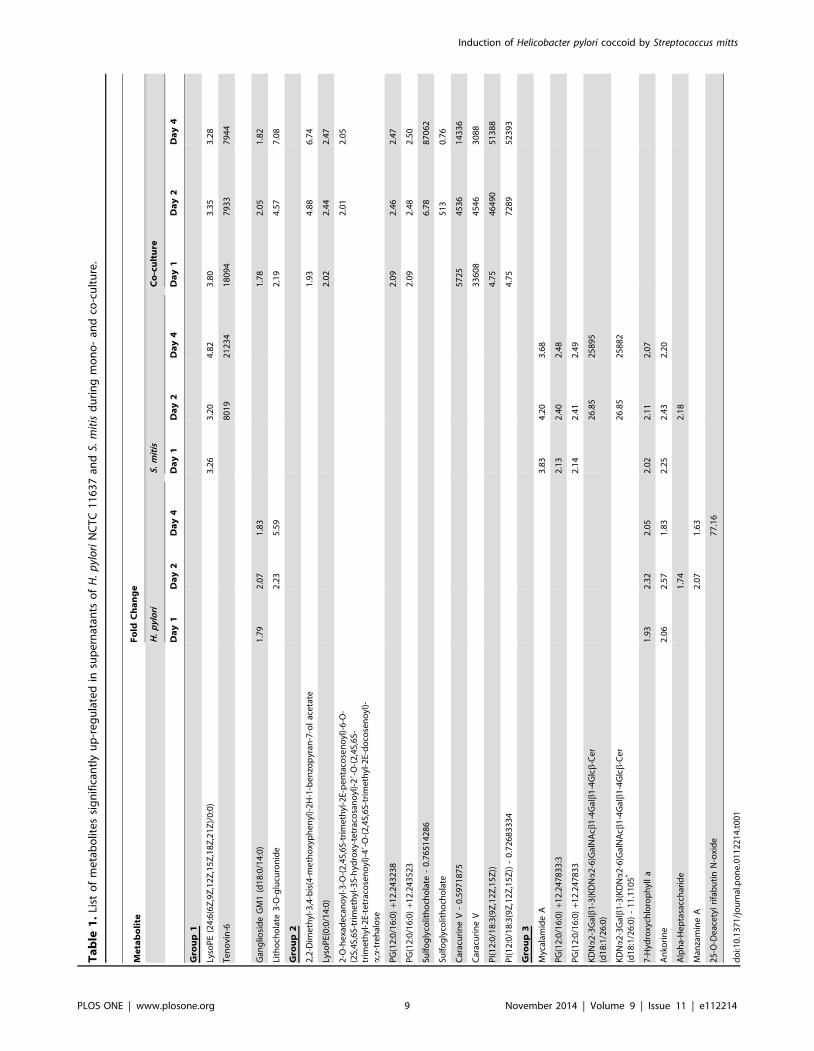
phase, we determined the metabolite profiles of supernatants from
1-, 2- and 4-day old monocultures and co-cultures of the two
bacteria by LC/MS (see materials and methods). Metabolites that
were statistically significantly up-regulated by more than two folds
at any time point in triplicated experiments could be classified into
three groups (Table 1). Group 1 comprises metabolites that were
detected in the co-culture and in either of the mono-cultures
(Table 1). Group 2 was constituted by compounds found only in
the co-culture while Group 3 contained products from S. mitisand/or H. pylori mono-culture that were absent in the co-culture
(Table 1). Metabolites involved in H. pylori conversion to coccoid
or the extended survival of S. mitis are likely to belong to Group 1
while compounds in groups 2 and 3 mainly demonstrate
interactions between H. pylori and S. mitis during co-culture.
We reasoned that a compound that induced H. pylori conversion
to coccoid should be present in supernatant from S. mitis and
possibly from co-culture. Additionally, this compounds should
start accumulating at day 2 since supernatant from this time point
and not from a 1-day old culture induced coccoid conversion
(Fig. 6a). Only one compound, a Tenovin-6-like molecule, that
belongs to group 1 fulfilled these requirements (Table 1). The
Tenovin-6-like compound was significantly up-regulated in
S. mitis supernatant at day 2 (8019 fold change) and drastically
increased at day 4 (21234 fold change). This molecule was also
significantly up-regulated in supernatants from co-culture at the
three time points tested (Table 1).Three other metabolites could
theoretically be involved in H. pylori conversion to coccoid. The
first of these metabolites (KDNa2-3Galb1-3(KDNa2-6)Gal-
NAcb1-4Galb1-4Glcb-Cer) was similar to a compound that
belongs to the class of gangliosides and, similarly to the
Tenovin-6-like molecule, was significantly up-regulated in S. mitissupernatant at day 2 and increased at day 4 (Table 1). However,
this metabolite was absent in supernatants from the co-culture.
The two other compounds were similar to Sulfoglycolithocholate,
a secondary bile salts metabolite and to Caracurine V, a plant
derived antimicrobial, respectively. These two compounds were
detected in the co-culture but were absent in the mono-cultures
(Table 1). To identify metabolites involved in S. mitis survival at
the exponential growth phase, we reasoned that such compounds
were to be expected in supernatants from H. pylori cultures at the
three time points since these supernatants equally conferred the
extended survival phenotype to S. mitis (Fig. 6b). None of the
metabolites were up-regulated at least 2 folds in the three time
points in H. pylori supernatant. A total of six compounds were
Figure 5. Morphology of H. pylori cells in mono and co-culture. H. pylori NCTC 11637 cells in monoculture ((a), (c), (e) and (g)) and in co-culturewith S. mitis ((b), (d), (f) and (h)) were recovered after 1 ((a) and (b)), 2 ((c) and (d)), 4 (e) and (f)) and 6 ((g) and (h)) of growth. The bacteria were Gram-stained and microscopically examined with a 1000X magnification.doi:10.1371/journal.pone.0112214.g005
Figure 6. Effect of supernatant from the other bacterium on H. pylori and S. mitis. Mono-cultures of H. pylori (a) and of S. mitis (b) weresupplemented with 1-, 2- or 4-day old supernatants from the other bacterium or left untreated. The colony forming units were determined by platingat the indicated time points.doi:10.1371/journal.pone.0112214.g006
Induction of Helicobacter pylori coccoid by Streptococcus mitts
PLOS ONE | www.plosone.org 8 November 2014 | Volume 9 | Issue 11 | e112214
Ta
ble
1.
List
of
me
tab
olit
es
sig
nif
ican
tly
up
-re
gu
late
din
sup
ern
atan
tso
fH
.p
ylo
riN
CT
C1
16
37
and
S.m
itis
du
rin
gm
on
o-
and
co-c
ult
ure
.
Me
tab
oli
teF
old
Ch
an
ge
H.
pyl
ori
S.
mit
isC
o-c
ult
ure
Da
y1
Da
y2
Da
y4
Da
y1
Da
y2
Da
y4
Da
y1
Da
y2
Da
y4
Gro
up
1
Lyso
PE
(24
:6(6
Z,9
Z,1
2Z
,15
Z,1
8Z
,21
Z)/
0:0
)3
.26
3.2
04
.82
3.8
03
.35
3.2
8
Te
no
vin
-68
01
92
12
34
18
09
47
93
37
94
4
Gan
glio
sid
eG
M1
(d1
8:0
/14
:0)
1.7
92
.07
1.8
31
.78
2.0
51
.82
Lith
och
ola
te3
-O-g
lucu
ron
ide
2.2
35
.59
2.1
94
.57
7.0
8
Gro
up
2
2,2
-Dim
eth
yl-3
,4-b
is(4
-me
tho
xyp
he
nyl
)-2
H-1
-be
nzo
pyr
an-7
-ol
ace
tate
1.9
34
.88
6.7
4
Lyso
PE(
0:0
/14
:0)
2.0
22
.44
2.4
7
2-O
-he
xad
eca
no
yl-3
-O-(
2,4
S,6
S-tr
ime
thyl
-2E-
pe
nta
cose
no
yl)-
6-O
-(2
S,4
S,6
S-tr
ime
thyl
-3S-
hyd
roxy
-te
trac
osa
no
yl)-
29-
O-(
2,4
S,6
S-tr
ime
thyl
-2E-
tetr
aco
sen
oyl
)-4
9-O
-(2
,4S,
6S-
trim
eth
yl-2
E-d
oco
sen
oyl
)-a
,a-t
reh
alo
se
2.0
12
.05
PG
(12
:0/1
6:0
)+1
2.2
43
23
82
.09
2.4
62
.47
PG
(12
:0/1
6:0
)+1
2.2
43
52
32
.09
2.4
82
.50
Sulf
og
lyco
lith
och
ola
te-
0.7
65
14
28
66
.78
87
06
2
Sulf
og
lyco
lith
och
ola
te5
13
0.7
6
Car
acu
rin
eV
-0
.59
71
87
55
72
54
53
61
43
36
Car
acu
rin
eV
33
60
84
54
63
08
8
PI(
12
:0/1
8:3
(9Z
,12
Z,1
5Z
))4
.75
46
49
05
13
88
PI(
12
:0/1
8:3
(9Z
,12
Z,1
5Z
))-
0.7
26
83
33
44
.75
72
89
52
39
3
Gro
up
3
Myc
alam
ide
A3
.83
4.2
03
.68
PG
(12
:0/1
6:0
)+1
2.2
47
83
3:3
2.1
32
.40
2.4
8
PG
(12
:0/1
6:0
)+1
2.2
47
83
32
.14
2.4
12
.49
KD
Na
2-3
Galb
1-3
(KD
Na
2-6
)Gal
NA
cb1
-4G
alb
1-4
Glcb
-Ce
r(d
18
:1/2
6:0
)2
6.8
52
58
95
KD
Na
2-3
Galb
1-3
(KD
Na
2-6
)Gal
NA
cb1
-4G
alb
1-4
Glcb
-Ce
r(d
18
:1/2
6:0
)-
11
.11
05
*2
6.8
52
58
82
7-H
ydro
xych
loro
ph
yll
a1
.93
2.3
22
.05
2.0
22
.11
2.0
7
An
kori
ne
2.0
62
.57
1.8
32
.25
2.4
32
.20
Alp
ha-
He
pta
sacc
har
ide
1.7
42
.18
Man
zam
ine
A2
.07
1.6
3
25
-O-D
eac
ety
lri
fab
uti
nN
-oxi
de
77
.16
do
i:10
.13
71
/jo
urn
al.p
on
e.0
11
22
14
.t0
01
Induction of Helicobacter pylori coccoid by Streptococcus mitts
PLOS ONE | www.plosone.org 9 November 2014 | Volume 9 | Issue 11 | e112214
moderately up-regulated in H. pylori supernatants (1.8 to 5.6 fold
change). An additional metabolite similar to 25-O-Deacetyl
rifabutin N-oxide displayed a 77 fold change but only at day 4
(Table 1). Although these compounds could theoretically play a
role in S. mitis survival we believe their involvement in this
phenomenon to be unlikely.
Discussion
In this study, we analyzed the interactions, during growth invitro, between H. pylori and two bacteria, S. mitis and L.fermentum that have been isolated from the stomach of both
healthy and H. pylori-infected gastric disease patients [4,9,19,20].
Using a co-culture method, we found that S. mitis produced and
released one or more diffusible factors that directly or indirectly
induce coccoid conversion of H. pylori cells. In contrast, both H.pylori and L. fermentum secreted factors that promote survival of
S. mitis during the stationary phase of growth. We did not find any
effect of H. pylori or S. mitis on the growth of L. fermentum during
co-culture. To identify the factors responsible for coccoid
conversion of H. pylori and for survival of S. mitis in the
stationary phase, we performed metabolomics analysis of super-
natants from mono- and co-cultures of H. pylori and S. mitis. We
detected a few compounds that could possibly be involved in H.pylori’s morphological conversion while we did not find molecules
that match the phenotype conferred to S. mitis by co-culture or H.pylori supernatant supplementation. It should be noted that both
H. pylori conversion to coccoid and S. mitis survival in the
stationary phase could be mediated by proteins secreted by these
bacteria. Such factors could not be detected by the LC/MS
approach we used in this study.
One of the compounds we detected was similar to Tenovin-6 an
anticancer molecule that was first identified in a screen for p53
activators (Lain et al 2008). p53 is a tumor suppressor encoded by
a gene that is the most mutated gene in cancer. Tenovin-6 is
currently subjected to intensive studies because of promises this
molecule held in cancer treatment [30–32]. The Tenovin-6-like
molecule identified in our metabolic profiling was the compound
that best matched the induction of coccoid conversion of H. pylori.It was not significantly increased in supernatant from a 1-day old
S. mitis mono-culture but was highly induced from day 2 and
drastically increased at day 4 (Table 1). Coccoid conversion of H.pylori during co-culture with S. mitis was detected from day 2.
Furthermore, 2-day old but not 1-day old S. mitis supernatants
induced H. pylori coccoid conversion and this phenotype was
more pronounced with supernatant from a 4 day old culture
(Fig. 6a). Whether the effect of the Tenovin-6-like molecule has
any significance during colonization of the stomach by H. pyloriand S. mitis is unknown. However, the anticancer properties of
Tenovin-6 and the possible role of coccoid H. pylori in
tumorisation deserve much attention. Chan et al. analyzed
gastrectomy specimens from cancer and peptic ulcer patients
and found that coccoid H. pylori cells were enriched in
adenocarcinoma compared to peptic ulcer samples [33]. Consis-
tent with this finding, another study that compared the effects of
spiral and coccoid H. pylori cells on gastric epithelial cells reported
that coccoid H. pylori exerted a stronger effect on proliferation
and a weaker effect on apoptosis than did spiral form [34]. These
observations suggest an involvement of coccoid H. pylori in
carcinogenesis. It is tempting to hypothesize from these observa-
tions that the Tenovin-6-like molecule produced by S. mitisantagonizes coccoid cells during colonization of the stomach by
the two bacteria. Interestingly, during co-culture, the Tenovin-6-
like molecule started accumulating at day 1 while it was detected at
day 2 in S. mitis mono-culture (Table 1). This observation suggests
that spiral H. pylori but not coccoid cells stimulates the production
of the compound by S. mitis. Further investigation are needed to
elucidate the effect of Tenovin-6-like molecule on H. pylori both invitro and in vivo.
The findings in this study reflect the numerous interactions that
take place between the members of the gastric microbiota. These
interactions contribute to shaping the composition of the gastric
microbiota and indirectly influence the pathogenesis of bacteria
like H. pylori. H. pylori is known to undergo a morphological
change from spiral to coccoid form in adverse conditions [35–39].
Coccoid cells are more resistant to different stresses and represent
a survival form of the bacterium [40,41]. However, how spiral H.pylori cells convert to coccoid in vivo and the role of coccoid cells
in H. pylori pathogenesis are still unclear. Our findings point to a
possible mechanism in which members of the gastric microbiota
secrete factors that induce coccoid conversion of spiral H. pyloricells. These bacteria by this means indirectly influence H. pyloripathogenesis and disease outcome in infected individuals.
Acknowledgments
The authors wish to thank the staff of the Endoscopy Unit at the University
of Malaya Medical Centre (UMMC) for all the assistance provided in the
process of sample collection for this study.
Author Contributions
Conceived and designed the experiments: YK YD MFL KLG JV.
Performed the experiments: YK. Analyzed the data: YK MFL YD. Wrote
the paper: YD.
References
1. Moodley Y, Linz B, Bond RP, Nieuwoudt M, Soodyall H, et al. (2012) Age of
the association between Helicobacter pylori and man. PLoS Pathog 8:
e1002693.
2. Khalifa MM, Sharaf RR, Aziz RK (2010) Helicobacter pylori: a poor man’s gut
pathogen? Gut Pathog 2: 2.
3. Ricci V, Romano M, Boquet P (2011) Molecular cross-talk between
Helicobacter pylori and human gastric mucosa. World J Gastroenterol 17:
1383–1399.
4. Sheh A, Fox JG (2013) The role of the gastrointestinal microbiome in
Helicobacter pylori pathogenesis. Gut Microbes 4: 505–531.
5. Musso G, Gambino R, Cassader M (2011) Interactions between gut microbiota
and host metabolism predisposing to obesity and diabetes. Annu Rev Med 62:
361–380.
6. Chung H, Pamp SJ, Hill JA, Surana NK, Edelman SM, et al. (2012) Gut
immune maturation depends on colonization with a host-specific microbiota.
Cell 149: 1578–1593.
7. Cryan JF, Dinan TG (2012) Mind-altering microorganisms: the impact of the
gut microbiota on brain and behaviour. Nat Rev Neurosci 13: 701–712.
8. Sekirov I, Russell SL, Antunes LC, Finlay BB (2010) Gut microbiota in health
and disease. Physiol Rev 90: 859–904.
9. Li XX, Wong GLH, To KF, Wong VWS, Lai LH, et al. (2009) Bacterial
Microbiota Profiling in Gastritis without Helicobacter pylori Infection or Non-
Steroidal Anti-Inflammatory Drug Use. Plos One 4.
10. Rolig AS, Cech C, Ahler E, Carter JE, Ottemann KM (2013) The degree of
Helicobacter pylori-triggered inflammation is manipulated by preinfection host
microbiota. Infect Immun 81: 1382–1389.
11. Osaki T, Matsuki T, Asahara T, Zaman C, Hanawa T, et al. (2012)
Comparative analysis of gastric bacterial microbiota in Mongolian gerbils after
long-term infection with Helicobacter pylori. Microb Pathog 53: 12–18.
12. Zaman C, Osaki T, Hanawa T, Yonezawa H, Kurata S, et al. (2014) Analysis of
the microbial ecology between Helicobacter pylori and the gastric microbiota of
Mongolian gerbils. J Med Microbiol 63: 129–137.
13. Sun Y-Q, Monstein H-J, Nilsson LE, Petersson F, Borch K (2003) Profiling and
Identification of Eubacteria in the Stomach of Mongolian Gerbils With and
Without Helicobacter pylori Infection. Helicobacter 8: 149–157.
Induction of Helicobacter pylori coccoid by Streptococcus mitts
PLOS ONE | www.plosone.org 10 November 2014 | Volume 9 | Issue 11 | e112214
14. Yin YN, Wang CL, Liu XW, Cui Y, Xie N, et al. (2011) Gastric and duodenum
microflora analysis after long-term Helicobacter pylori infection in MongolianGerbils. Helicobacter 16: 389–397.
15. Aebischer T, Fischer A, Walduck A, Schlotelburg C, Lindig M, et al. (2006)
Vaccination preventsHelicobacter pylori-induced alterations of the gastric florain mice. FEMS Immunology & Medical Microbiology 46: 221–229.
16. Lofgren JL, Whary MT, Ge Z, Muthupalani S, Taylor NS, et al. (2011) Lack ofcommensal flora in Helicobacter pylori-infected INS-GAS mice reduces gastritis
and delays intraepithelial neoplasia. Gastroenterology 140: 210–220.
17. Lee CW, Rickman B, Rogers AB, Muthupalani S, Takaishi S, et al. (2009)Combination of sulindac and antimicrobial eradication of Helicobacter pylori
prevents progression of gastric cancer in hypergastrinemic INS-GAS mice.Cancer Res 69: 8166–8174.
18. Wang ZK, Yang YS (2013) Upper gastrointestinal microbiota and digestivediseases. World J Gastroenterol 19: 1541–1550.
19. Engstrand L, Lindberg M (2013) Helicobacter pylori and the gastric microbiota.
Best Practice & Research in Clinical Gastroenterology 27: 39–45.20. Hu Y, He LH, Xiao D, Liu GD, Gu YX, et al. (2012) Bacterial flora concurrent
with Helicobacter pylori in the stomach of patients with upper gastrointestinaldiseases. World J Gastroenterol 18: 1257–1261.
21. Denapaite D, Bruckner R, Nuhn M, Reichmann P, Henrich B, et al. (2010) The
genome of Streptococcus mitis B6–what is a commensal? PLoS One 5: e9426.22. Mitchell J (2011) Streptococcus mitis: walking the line between commensalism
and pathogenesis. Mol Oral Microbiol 26: 89–98.23. Maldonado J, Canabate F, Sempere L, Vela F, Sanchez AR, et al. (2012)
Human milk probiotic Lactobacillus fermentum CECT5716 reduces theincidence of gastrointestinal and upper respiratory tract infections in infants.
J Pediatr Gastroenterol Nutr 54: 55–61.
24. Cox AJ, Pyne DB, Saunders PU, Fricker PA (2010) Oral administration of theprobiotic Lactobacillus fermentum VRI-003 and mucosal immunity in
endurance athletes. Br J Sports Med 44: 222–226.25. Mikelsaar M, Zilmer M (2009) Lactobacillus fermentum ME-3 - an
antimicrobial and antioxidative probiotic. Microb Ecol Health Dis 21: 1–27.
26. Ruiz-Moyano S, Martin A, Benito MJ, Hernandez A, Casquete R, et al. (2011)Application of Lactobacillus fermentum HL57 and Pediococcus acidilactici
SP979 as potential probiotics in the manufacture of traditional Iberian dry-fermented sausages. Food Microbiol 28: 839–847.
27. Pacifico L, Osborn JF, Bonci E, Romaggioli S, Baldini R, et al. (2014) Probioticsfor the treatment of Helicobacter pylori infection in children. World J Gas-
troenterol 20: 673–683.
28. Khosravi Y, Rehvathy V, Wee WY, Wang S, Baybayan P, et al. (2013)
Comparing the genomes of Helicobacter pylori clinical strain UM032 and Mice-
adapted derivatives. Gut Pathog 5: 25.
29. Kusters JG, van Vliet AH, Kuipers EJ (2006) Pathogenesis of Helicobacter pylori
infection. Clin Microbiol Rev 19: 449–490.
30. van Leeuwen IM, Rao B, Sachweh MC, Lain S (2012) An evaluation of small-
molecule p53 activators as chemoprotectants ameliorating adverse effects of
anticancer drugs in normal cells. Cell Cycle 11: 1851–1861.
31. McCarthy AR, Pirrie L, Hollick JJ, Ronseaux S, Campbell J, et al. (2012)
Synthesis and biological characterisation of sirtuin inhibitors based on the
tenovins. Bioorg Med Chem 20: 1779–1793.
32. Ueno T, Endo S, Saito R, Hirose M, Hirai S, et al. (2014) The sirtuin inhibitor
tenovin-6 upregulates death receptor 5 and enhances cytotoxic effects of 5-
fluorouracil and oxaliplatin in colon cancer cells. Oncol Res 21: 155–164.
33. Chan WY, Hui PK, Leung KM, Chow J, Kwok F, et al. (1994) Coccoid forms of
Helicobacter pylori in the human stomach. Am J Clin Pathol 102: 503–507.
34. Li N, Han L, Chen J, Lin X, Chen H, et al. (2013) Proliferative and apoptotic
effects of gastric epithelial cells induced by coccoid Helicobacter pylori. J Basic
Microbiol 53: 147–155.
35. Catrenich CE, Makin KM (1991) Characterization of the morphologic
conversion of Helicobacter pylori from bacillary to coccoid forms. Scand J Gas-
troenterol Suppl 181: 58–64.
36. Donelli G, Matarrese P, Fiorentini C, Dainelli B, Taraborelli T, et al. (1998) The
effect of oxygen on the growth and cell morphology of Helicobacter pylori.
FEMS Microbiol Lett 168: 9–15.
37. Cellini L, Allocati N, Di Campli E, Dainelli B (1994) Helicobacter pylori: a fickle
germ. Microbiol Immunol 38: 25–30.
38. Nilius M, Strohle A, Bode G, Malfertheiner P (1993) Coccoid like forms (CLF) of
Helicobacter pylori. Enzyme activity and antigenicity. Zentralbl Bakteriol 280:
259–272.
39. Nilsson HO, Blom J, Al-Soud WA, Ljungh A, Andersen LP, et al. (2002) Effect
of Cold Starvation, Acid Stress, and Nutrients on Metabolic Activity of
Helicobacter pylori. Applied and Environmental Microbiology 68: 11–19.
40. Figura N, Moretti E, Vaglio L, Langone F, Vernillo R, et al. (2012) Factors
modulating the outcome of treatment for the eradication of Helicobacter pylori
infection. New Microbiol 35: 335–340.
41. Saito N, Konishi K, Kato M, Takeda H, Asaka M, et al. (2008) Coccoid
formation as a mechanism of species-preservation in Helicobacter pylori: an
ultrastructural study. Hokkaido Igaku Zasshi 83: 291–295.
Induction of Helicobacter pylori coccoid by Streptococcus mitts
PLOS ONE | www.plosone.org 11 November 2014 | Volume 9 | Issue 11 | e112214
Related Documents