UCSF UC San Francisco Previously Published Works Title Strain-guided mineralization in the bone-PDL-cementum complex of a rat periodontium. Permalink https://escholarship.org/uc/item/3xk421vb Authors Grandfield, Kathryn Herber, Ralf Peter Chen, Ling et al. Publication Date 2015-12-01 DOI 10.1016/j.bonr.2015.04.002 Peer reviewed eScholarship.org Powered by the California Digital Library University of California

Welcome message from author
This document is posted to help you gain knowledge. Please leave a comment to let me know what you think about it! Share it to your friends and learn new things together.
Transcript

UCSFUC San Francisco Previously Published Works
TitleStrain-guided mineralization in the bone-PDL-cementum complex of a rat periodontium.
Permalinkhttps://escholarship.org/uc/item/3xk421vb
AuthorsGrandfield, KathrynHerber, Ralf PeterChen, Linget al.
Publication Date2015-12-01
DOI10.1016/j.bonr.2015.04.002 Peer reviewed
eScholarship.org Powered by the California Digital LibraryUniversity of California
Bone Reports 3 (2015) 20–31
Contents lists available at ScienceDirect
Bone Reports
j ourna l homepage: www.e lsev ie r .com/ locate /bonr
Strain-guided mineralization in the bone–PDL–cementum complex of arat periodontium
Kathryn Grandfield a,1, Ralf-Peter Herber b,1, Ling Chen a, Sabra Djomehri a, Caleb Tam a, Ji-Hyun Lee a,Evan Brown a, Wood R. Woolwine III a, Don Curtis a, Mark Ryder c, Jim Schuck d, Samuel Webb e,William Landis f, Sunita P. Ho a,⁎a Division of Biomaterials and Bioengineering, Department of Preventive and Restorative Dental Sciences, University of California San Francisco, San Francisco, CA 94143, United Statesb Division of Orthodontics, Department of Orofacial Sciences, University of California San Francisco, San Francisco, CA 94143, United Statesc Division of Periodontics, Department of Orofacial Sciences, University of California San Francisco, San Francisco, CA 94143, United Statesd Materials Science Division, The Molecular Foundry, Lawrence Berkeley National Laboratory, Berkeley, CA 94720, United Statese Stanford Synchrotron Radiation Lightsource, SLAC National Accelerator Laboratory, Menlo Park, CA 94025, United Statesf Department of Polymer Science, The University of Akron, OH 44325, United States
⁎ Corresponding author at: Division of Biomaterials andPreventive and Restorative Dental Sciences, 707 ParCalifornia, San Francisco, CA 94143, United States.
E-mail address: [email protected] (S.P. Ho).1 Both authors contributed equally.
http://dx.doi.org/10.1016/j.bonr.2015.04.0022352-1872/© 2015 The Authors. Published by Elsevier Inc
a b s t r a c t
a r t i c l e i n f oArticle history:
Received 3 January 2015Received in revised form 25 March 2015Accepted 13 April 2015Available online 18 April 2015Keywords:MineralizationAdaptationsBone–periodontal ligament–tooth complexMechanical strainInterfacesAttachment sites
Objective: The objective of this studywas to investigate the effect ofmechanical strain bymapping physicochem-ical properties at periodontal ligament (PDL)–bone and PDL–cementum attachment sites and within the tissuesper se.Design: Accentuated mechanical strain was induced by applying a unidirectional force of 0.06 N for 14 days onmolars in a rat model. The associated changes in functional space between the tooth and bone, mineral formingand resorbing events at the PDL–bone and PDL–cementum attachment sites were identified by using micro-X-ray computed tomography (micro-XCT), atomic force microscopy (AFM), dynamic histomorphometry, Ramanmicrospectroscopy, and AFM-based nanoindentation technique. Results from these analytical techniques werecorrelated with histochemical strains specific to low and high molecular weight GAGs, including biglycan, andosteoclast distribution through tartrate resistant acid phosphatase (TRAP) staining.Results: Unique chemical and mechanical qualities including heterogeneous bony fingers with hygroscopicSharpey's fibers contributing to a higher organic (amide III — 1240 cm−1) to inorganic (phosphate —
960 cm−1) ratio, with lower average elastic modulus of 8 GPa versus 12 GPa in unadapted regions were identi-fied. Furthermore, an increased presence of elemental Zn in cement lines and mineralizing fronts of PDL–bonewas observed. Adapted regions containing bony fingers exhibited woven bone-like architecture and these re-gions rich in biglycan (BGN) and bone sialoprotein (BSP) also contained high-molecular weight polysaccharidespredominantly at the site of polarized bone growth.Conclusions: Froma fundamental science perspective the shift in local properties due to strain amplification at thesoft–hard tissue attachment sites is governed by semiautonomous cellular events at the PDL–bone and PDL–ce-mentum sites. Over time, these strain-mediated events can alter the physicochemical properties of tissues per se,and consequently the overall biomechanics of the bone–PDL–tooth complex. From a clinical perspective, theshifts inmagnitude and duration of forces on the periodontal ligament can prompt a shift in physiologic mineralapposition in cementum and alveolar bone albeit of an adapted quality owing to the rapidmechanical translationof the tooth.© 2015 The Authors. Published by Elsevier Inc. This is an open access article under the CC BY-NC-ND license(http://creativecommons.org/licenses/by-nc-nd/4.0/).
Bioengineering, Department ofnassus Avenue, University of
. This is an open access article under
1. Introduction
The periodontal ligament (PDL) is vascularized and innervatedsofter structural component that assumes the role of regulating func-tional loads in the tooth–PDL–bone fibrous joint. While this joint isoptimally engineered by nature to transfer loads and maintain tissuesfor function, it is also exposed to the inevitable parafunction, that is, ex-acerbated forces in the form of bruxism, jaw clenching, nail biting,
the CC BY-NC-ND license (http://creativecommons.org/licenses/by-nc-nd/4.0/).

21K. Grandfield et al. / Bone Reports 3 (2015) 20–31
altered loads resulting from hardness variation in diet intake, and/orthrough clinical interventions such as the application of orthodonticappliances. The common denominator and a notable factor for theaforementioned conditions include eccentrically placed mechanicalloads (Jang et al., 2014; Lin et al., 2013). Biomechanically, an optimalPDL-space is thought to permit the redistribution and transfer of loadsfrom the tooth to alveolar bone specifically when loads are placedalong the axis of the bone–tooth complex. This configuration constitutesa concentric loading system,whichmost often in nature prompts an op-timum function when loaded within physiological limits. It is thoughtthat the resulting optimum PDL-space enables force transfer acrossthe mineralized tissues of this complex organ. Hence the PDL-spacealong with the PDL has also been suggested to act as a vital constructalong which alveolar bone remodels/models (Beertsen et al., 1997),with the potential for cementum formation and resorption to alsooccur. Therefore, an improved knowledge of the “mechano-responsive”nature of the PDL, specifically at the PDL–bone and PDL–cementumattachment sites is relevant to understanding the strain-induced re-sponses to our clinical interventions. Mechanical forces exert strains atthe attachment sites and can “turn on” cell-surface receptors and conse-quently a cascade of biochemical signals, which in turn cause mineralformation and resorption related events in bone and/or cementum.
There exist many clinical interventions in skeletal (distraction oste-ogenesis), and oral and craniofacial orthopedics (for example, cranialgrafts, orthodontics) that involve the use of mechanical forces to“mold” and/or regenerate bone and subsequently its adjacent tissues.However, very little is known about the influence of mechanicalstimulus on the biomineralization of tissues per se within thebone–PDL–tooth complex or the functional interfaces between theligament–bone and ligament–cementum. The current doctrine re-garding the role of PDL in response to applied loads has been summa-rized as: 1) the PDL distributes applied loads to the alveolar bone,2) the direction, frequency, duration and magnitude of loading deter-mine both the extent, rate of bone remodeling and quality of modeledbone, 3) the absence of PDL severely limits the extent of bone remodel-ing (Beertsen et al., 1997). In this study, we propose that themechanoresponsive activity at the PDL–bone and PDL–cementum at-tachment sites responds to eccentric loading by mineral formationand/or resorption at these sites by altering the functional space be-tween the tooth and bone.
The importance of functional adaptations of the bone, in particular atinterfaces, is not limited to orthodontics, but extends into the orthope-dics. Within orthopedics, it has been suggested that functional adapta-tion of the bone results from cellular responses to strain densitywithin a softer matrix (Carter et al., 1987). In this study, we identifythe effects of perceived unidirectional forces on mineralization withinthe bone–PDL–tooth joint, and specifically at the soft–hard tissue inter-faces of the PDL–bone and PDL–cementum sites and correlate it to thecellular and tissue adaptations in a rat model. The specific sitesthat were mechanically stimulated included the PDL–bone and thePDL–cementum functional attachment sites. Due to the nature of theligament–bone interfaces in the musculoskeletal system, includingthe ligament–cementum interfaces in the bone–PDL–tooth complex,i.e. disparate tissues interfacing over a distance of 10 μm, thesesites are mechanically strained (Lu and Thomopoulos, 2013; Qianet al., 2009) and the rate of adaptation at the interfaces is highercompared to other modeling sites. Furthermore, the attachment sitespresent themselves as excellent model systems where mechano-responsiveness, i.e. the response of mechanical strain amplification onmineral formation and resorption can be investigated and better under-stood. However, based on literature, mechano-responsiveness is as-sumed to occur at two different sites, the soft–hard tissue attachmentsite per se, and 5 μm inwards defined as an interface where the soft tis-sue transitions into a harder tissue.
Fundamentally, from a structural engineering perspective, eccentricloads are thought to accentuate strains, specifically at regions where
dissimilar materials are attached. This concept was examined in thisstudy, where strains at the functional interfaces and attachment sitesof the PDL–bone and PDL–cementum were amplified by placing an ec-centric load on the periodontiumof a rat for fourteen days. In particular,cellularmorphological phenotype and tissue adaptationswere correlat-ed to matrix structure, mechanical properties, and biochemical and ele-mental compositions facilitated through various techniques, includingimmunohistochemistry, microcomputed X-ray tomography (micro-XCT), microprobe X-ray fluorescence (micro-XRF), micro Raman spec-troscopy and nanoindentation. The importance of this study lies in com-plementary higher-resolution imaging techniques combined withconventional histological and immunohistochemical measuresallowing an understanding of the effect of mechanical strains at the in-terfaces of the bone–PDL–tooth complex. Thus, the objective of thisstudy is to investigate gross morphological changes within tissues ofthe bone–PDL–cementum complex, changes in architecture and physi-cochemical characteristics of the formed structures due to amplifiedstrain at the PDL–bone attachment site and interface.
2. Materials & methods
Please see appendix for details on the animal model used in thisstudy (Fig. S1). In brief, 60 cN of forcewas applied tomolars and incisorsof 4 month old male Sprague–Dawley rats (Charles River LaboratoriesInternational Inc.,Wilmington,MA, USA). Following 14 days ofmechan-ical stimulus, the specimens (N = 16, 8 experimental and 8 in controlgroup) were harvested and prepared for further characterization. Thestudywas conducted according to the regulations of the animal protocolAN080608-02, approved by the Institutional Animal Care and UseCommittee (IACUC), University of California, San Francisco (UCSF).
2.1. Micro X-ray computed tomography (Micro-XCT™)
Entire dissected and fixed maxillae from experimental animals(N = 8) and control animals (N = 8) were placed in 70% ethanol, andwere imaged at 2× magnification at 90 kVp, a power of 6 W and acamera binning of 2 using a Micro XCT-200 (micro-XCT, Xradia Inc.,Pleasanton, CA, USA). For imaging of bone forming areas, magnifica-tion was increased to 10× and 20×. Exposure times were adjusted toyield 25% of the original X-ray intensity passing through the speci-men and arriving at the detector. Tomographies were reconstructed(XMReconstructor, Version 7.0.2817, Xradia Inc., Pleasanton, CA,USA) and post-processed using the Xradia 3D viewer and Avizo®Fire 7.1 for 3D segmentation (Visage Imaging Inc., Version 5.2.2,San Diego, CA, USA).
The tomograms were also used to calculate volumes including bonevolume (BV) and canal volume (Ca.V), and tissue volume (TV) based onprevious works (Carter et al., 2013; Cooper et al., 2006; Parfitt et al.,1987). To note, “tissue volume” is referred to as “total volume” in thiswork, and is a sum of the BV and CV.
2.2. Histology and histochemistry
The appliance was removed and the specimens were cut into blocksconsisting of the molars, PDL, and alveolar bone, only. After scanningwith micro-XCT, the hemimaxillae were decalcified with ethylenedi-aminetetraacetic acid (EDTA) solution for 3weeks. The decalcified spec-imens were dehydrated with 80%, 95% and 100% Flex alcohol (Richard-Allan Scientific, Kalamazoo, MI) before embedding in paraffin (TissuePrep-II, Fisher Scientific, Fair Lawn, NJ). The paraffin blocks were sagit-tally sectioned on a rotary microtome (Reichert-Jung Biocut, Vienna,Austria) using a disposable steel blade (TBF Inc., Shur/Sharp, Fisher Sci-entific, Fair Lawn, NJ). The paraffin serial sections were mounted onSuperfrost Plus microscope slides (Fisher Scientific, Fair Lawn, NJ). Sec-tions were deparaffinized with xylene and rehydrated through a de-scending ethanol series before further use. The stained tissues were

22 K. Grandfield et al. / Bone Reports 3 (2015) 20–31
characterized using a light microscope (BX 51, Olympus America Inc.,San Diego, CA) and analyzed using Image Pro Plus v6.0 software(Media Cybernetics Inc., Silver Spring, MD) unless mentioned other-wise. For details on staining protocols, please refer to supplementalinformation.
2.2.1. Tartrate resistant acid phosphatase (TRAP) histochemistryDeparaffinized serial sections were used for tartrate resistant acid
phosphatase (TRAP) staining. In brief, the method included treatingthe rehydrated specimens with 0.2 M acetate buffer, a solution of0.2 M sodium acetate and 50 mM L(+) tartaric acid (Sigma-Aldrich,St. Louis, MO) (Erlebacher and Derynck, 1996). After incubation atroom temperature for 20 min, naphthol AS-MX phosphate and fastred TR salt were added, followed by incubation at 37 °C for 1 h withclose monitoring under the microscope after the first half hour forbright red staining of osteoclastic activity. The stained sections werewashed in deionized water, counterstained with hematoxylin andmountedwith Immu-mount (ThermoScientific, Fremont, CA) for subse-quent examination under a light microscope, as stated above. A surveyof the distribution of TRAP stained osteoclasts in the periodontium ofthe 2nd molar was made using sections from 5 animals from experi-mental and control groups respectively.
2.2.2. Hematoxylin & eosin and picrosirius red stainingSections were stained with hematoxylin (Fisher Scientific,
Kalamazoo, MI) and eosin (Fisher Scientific, Kalamazoo, MI) (Carson1990). Deparaffinized sections were stained with Sirius red F3B (C.I.35,782) and picric acid (American MasterTech Scientific Co., Lodi, CA).Polarized light was used to enhance the birefringence of collagen toillustrate changes in collagen fiber orientation and birefringence inten-sity throughout the complex.
2.2.3. Alcian blue histochemistryDeparaffinized serial sections were rehydrated through descending
alcohols and placed in 1% alcian blue (American MasterTech ScientificCo., Lodi, CA) in 3% acetic acid solution at pH 2.5 for 30min. After rinsingin running water, the sections were counterstained with nuclear-fastred solution (American MasterTech Scientific Co., Lodi, CA) for 5 min,followed by dehydration through ascending alcohols and xylene beforemounting the sectionswith Permount (Fisher Scientific, Pittsburgh, PA).
2.2.4. Immunohistochemistry for biglycan (BGN) and bone sialoprotein(BSP) localization
Immunohistochemistry (IHC) of BGN was performed using anavidin-biotinylated peroxidase enzyme complex-based kit (VectastainElite: Vector Labs, Burlingame, CA) with 3,3′-diaminobenzidine (DAB)substrate (Sigma-Aldrich, St. Louis, MO) to produce a brown reactionproduct indicating antigen location. Deparaffinized and rehydratedsections were incubated in 80% methanol and 0.6% H2O2 to quench en-dogenous peroxidase. To retrieve antigen, trypsin digestion was per-formed for 10 min at room temperature in 0.1% trypsin, 0.1% CaCl2,20 mM Tris–HCl pH 7.8, followed by rinsing with deionized water 3times for 5 min. Glycosaminoglycan (GAG) removal was next per-formed enzymatically in 35 mM Tris–HCl pH 7.4, 35 mM sodium ace-tate, and 15 mU/mL chondroitinase ABC (Seikagaku Biobusiness,Tokyo, Japan) for 1 h at 37 °C in a humidified chamber. Following anti-gen retrieval, sections were incubated with blocking buffer (1% normaldonkey serum, 0.03% Triton X-100 in 1× phosphate-buffered saline(PBS)) for 60min to prevent non-specific binding. Theprimary antibody(rabbit polyclonal anti-BGN antibody, LF-159, generously provided byDr. Larry Fisher, NIH/NIDCR, Bethesda, MD) was applied on the sectionsat a dilution of 1:200 in blocking buffer and incubated overnight at 4 °C.The next day, the sections were washed in 0.1% Tween 20 in 1× PBS(PBST) and incubated for 30min at room temperature with the second-ary antibody (biotinylated anti-rabbit IgG antibody, PK-6101, VectorLabs, Burlingame, CA). Antigen–antibody complexes were visualized
with DAB tetrachloride solution (Sigma, St. Louis, MO), washed in dis-tilled water and counterstained with Gill hematoxylin (Fisher Scientific,Kalamazoo, MI), and rinsed in running water. Finally, the sections weredehydrated in ascending series of ethanol, cleared with xylene andmounted with Permount (Fisher Scientific, Pittsburgh, PA). Negativecontrols were obtained by the substitution of the primary antibodieswith non-immunized rabbit IgG (Santa Cruz Biotechnology Inc.,sc-2027, Santa Cruz, CA) and blank controls by omission of the primaryantibodies. Condyles harvested from the same animals were used aspositive controls. The sections were evaluated by a single examiner,blinded to the treatment assignment.
Bone sialoprotein (BSP) was assessed by immunohistochemistry(IHC) on a Leica Bond Max (Leica Microsystems, Inc., Buffalo Grove, IL)using the Bond Refine Detection System. Paraffin sections previouslymounted on slides (Superfrost™ Plus Gold Slides, Thermo FisherScientific Inc., Waltham, MA) were deparaffinized, rehydrated, and sur-face decalcified for 10 min in Immunocal (Fisher Scientific, Pittsburgh,PA). All sections were blocked for endogenous peroxidases by immer-sion in 0.3% hydrogen peroxide in 30% methanol for 30 min at 25 °C.Epitope retrieval was achieved with citrate buffer (ER1, Bond RefineDetection System) for 60 min at 37 °C. Blocking of all sections wasperformed with the addition of Background Buster (Innovex Biosci-ences, Richmond, CA) for 30 min at 25 °C just prior to the addition ofprimary antibody. Monoclonal anti-BSP (mouse clone WVID1(9C5),Developmental Studies Hybridoma Bank, Iowa City, IA) was bound for2 h at 25 °C at a 1:100 dilution (70 μg/mL). Mayer's hematoxylin(Bond Refine Detection System) was utilized as counterstain followingthe polymer-linked 3, 3′-diaminobenzidine (DAB, Bond Refine Detec-tion System) secondary reaction. After counterstaining, all sectionswere dehydrated and sealed using DPX mountant (Sigma-Aldrich,St. Louis, MO). All experimental staining procedures were accompaniedby control slides replacing primary antibody binding with buffer ormouse IgG isotype (Dako North America, Inc., Carpinteria, CA) to assessthe level of non-specific staining.
2.2.5. Fluorescence microscopyExperimental rats (N = 3) and control rats (N = 3) were given in-
traperitoneal injections with alternating alizarin red and tetracyclinehydrochloride (both Sigma-Aldrich, St. Louis, MO, USA) on days 0, 3, 6,9 and 12 of the procedure. According to themethod used, 25mg fluo-rochrome per kg body mass was diluted in 2% NaHCO3 to a concen-tration of 0.01 mg/μl concentration of 0.01 mg/kg body mass (Luanet al., 2007). After harvest, maxillae were dissected, fixed in 4% para-formaldehyde overnight, and embedded in polymethylmethacrylate.Sagittal sections 10 μm thick were sliced on a rotary microtome(Leica RM2255, Leica microsystems, Wetzlar, Germany) using atungsten carbide blade. Sections were viewed under Nikon 6D Ti-Econventional wide-field microscope (UCSF Nikon Imaging Facility,San Francisco, CA) equipped with an mCherry epifluorescence filter(ex=560/55 nm, CWL/BW, em=630/60 nm, CWL/BW) used to visual-ize Alizarin red fluorescent bands (abs. = 530–560 nm, em. = 580 nm),and Sapphire epifluorescence (filter: ex = 400/50 nm, CWL/BW, em =515/50 nm, CWL/BW) used to visualize tetracycline HCL green fluores-cent bands (abs.= 390–425 nm, em. = 525–560 nm).
2.3. Microprobe X-ray fluorescence (μ-XRF)
Ultrasectioned block specimens (0.64 mm thick) of an adaptedbone-finger region, tooth and PDL were analyzed using μ-XRF mi-croscopy at the Stanford Synchrotron Radiation Lightsource (SSRL)on beamline 2–3. Data acquisition was performed using constant pa-rameters with an incident energy beam of 10 keV, dwell time of 25–50 ms per pixel, and a step size of 1 μm. The elemental concentrationswere determined by calibration against a set of NIST traceable, thinfilm elemental X-ray fluorescence standards provided by Micromatter(Vancouver, Canada). Corrections to the area-based concentrations
23K. Grandfield et al. / Bone Reports 3 (2015) 20–31
(μg/cm2) were performed based on the absorption and attenuation ofboth the incident (excitatory) X-ray photons (10 keV) and the variouselemental fluorescence photons in the matrix.
2.4. Atomic force microscopy (AFM) imaging, AFM-based nanoindentation,and Raman spectroscopy
The topography of ultrasectioned surfaced-blocks was qualitativelyanalyzed using an atomic force microscope (Nanoscope III, Multimode;Bruker Corporation, Santa Barbara, CA) in contact mode under both dryand wet conditions. The ultrasectioned surface was scanned using aSi3N4 tip attached to a “V shaped” cantilever (Bruker Corporation) at ascanning frequency of 1.4 Hz. The nominal radius of curvature of thetip was less than 50 nm. Scanning under wet conditions was performedwith the specimen and probe immersed in deionizedwater as previous-ly described (Ho et al., 2004). Nanoscope III version 5.12r3 software(Bruker Corporation) was used for data processing.
2.4.1. AFM-based nanoindentationNanoindentation was performed on the ultrasectioned block
specimens using an AFM attached to a load displacement transducer(Triboscope, Hysitron Incorporated, Minneapolis, MN). A sharp dia-mond Berkovich indenter with a conventional radius of curvatureless than 100 nm (Triboscope, Hysitron Incorporated, Minneapolis,MN) was fitted to the transducer. Site-specific measurements acrossthe adapted bone interface were made under wet conditions using adisplacement control mode and a penetration depth of 500 nm, witha load, hold, and unload for 3 s each. Fused silica was used to calibratethe transducer under dry and wet conditions.
2.4.2. micro Raman spectroscopyThe chemical makeup of alveolar bone was probed with an ARAMIS
Raman microscope (HORIBA Scientific, Santa Clara, CA), operated at768 nm wavelength, with a 1200 grating, and at 50× magnification.Peak heights corresponding to phosphate (960 cm−1, 1003 cm−1,1030 cm−1) and amide III (1242 cm−1) were compared to determinethe mineral:matrix ratio.
3. Results
3.1. Mineralized zones, osteoclastic activity, and ratio of canal volume tototal volume
Transverse micro-XCT images (Fig. 1) were used to determine thegross macroscale movements of the teeth relative to the bone afterforce application. The virtual sections demonstrated movement of theteeth in mesial direction, accompanied by an enlarged PDL-space onthe distal side of roots (Fig. 1c), and decreased PDL-space on the mesialside. Resorption pits were observed in cementum opposite to bony fin-ger extensions (Fig. 1d) (Fig. S1, Movies S1a, S1b, and S1c).
Active root resorption represented by the distribution of TRAPstained osteoclasts is shown in Fig. 1. Control specimens showed osteo-clastic activity predominantly on the distal aspect of distal roots(Fig. 1e), while the experimental specimens exhibited TRAP activityon the mesial aspect of the mesial root (Fig. 1f). Resorption activitywas highest on the bone–PDL interface but also undermining resorptionwas found occasionally in both experimental and control within the al-veoli of the alveolar bone. No difference of resorption activitywas foundbetween the bones located in the apical or cervical half of the socket.
Alveolar socket and root morphology were consistent with signs ofbone and cementum resorption. Micro-XCT images showed concaveresorption pits, with diameters of approximately 50–100 μm, on thedistal side of the root in control animals and on themesial side of the ex-perimental group. Similarly, resorption craters signifying root resorp-tion were found on the mesial and distal sides of the roots in theexperimental, while the control only exhibited natural root resorption
on the distal side of the roots (Herber et al., 2012). There was no signif-icant difference in themorphology of resorption pits in control or exper-imental groups, but the average size and total amount of resorptionwere higher on the mesial side of the mesial 2nd molar root comparedto the experimental distal side and control. Additionally, based on trans-verse and longitudinal sections, resorption and growth occurred over anarea/volume (white arrows, Fig. 1, Supplemental Movies S1a and S1b).
Average bone volume to total volume (BV/TV) and average canalvolume to total volume (Ca.V/TV) in percentages are shown in Fig. 1gfor control and experimental groups. The average Ca.V/TV for controland experimental groups was 25% and 40%, respectively, and that forBV/TVwas 75% and 60%. SupplementalMovie S1c illustrates representa-tive cubic subvolumes within 3D segmented, and Movies S1d and S1eillustrate bone and canal networks in control and experimental groups.
3.2. Cell morphology, tissue architecture, and biomolecular expressions atthe PDL–bone and PDL–cementum attachment sites
Serial sections were stained and analyzed (Fig. 2). The hematoxylinand eosin stain was informative, in that varying cell morphology specif-ically at the PDL–bone attachment site, including increased capillaries,was observed within the PDL (Fig. S2). At the PDL–bone, there existeda zone of cells with an altered morphology and closer to the commonlyknown cuboidal shape of osteoblasts (Fig. 2). Additionally, the alternat-ing but irregular layers of bone encroaching the PDL-space (Δ, Δ1, Δ2,Fig. 2c and d) stained pale pink were observed when compared to theexisting bone (stars, Fig. 2c and d), and uniform layering in the controls(Fig. 2a and b).
PSR staining of control specimens displayed high birefringence indi-catingwell organized collagen fibers and PDL inserts at both the cemen-tum and bone interfaces (Fig. 2e and f), while the experimental groupdisplayed birefringence only at the cementum–PDL interface, suggest-ing less organization at the bone–PDL sitewhere strain-guidedmineral-ization occurred (Fig. 2g and h). Finger-like extensions that appeargreen, indicating a lack of organized collagen, are noted protrudingfrom the bone into the PDL-space. Hatched patterns suggest newlyformed adapted alveolar bone is woven. Supplemental Movies S2aand S2b highlight the birefringence of control and experimental com-plexes through 360° rotation.
Alcian bluewith nuclear fast red counterstaining (AB-NFR)was con-sistently positive in the bone, secondary cementum, and mantle dentinadjacent to secondary cementum (Fig. 2i, j, k and l), although the paleblue color was more intense in the control group (Fig. 2i and j). Onthe other hand, PDL, dental pulp and endosteal spaces exhibited pinkcell nuclei and cytoplasm. Most interestingly, an intense blue stainingwas observed along the tip of finger-like bone protrusion at the bone–PDL interface in the experimental group (black arrows in Fig. 2kand l), which is absent in the control group. Large cuboidal cells(arrow heads in panel l) lining bone fingers are distinguished fromsmall squamous cells in PDL and they are thought to be osteoblastic.
BGN was distributed uniformly throughout the PDL of the controlgroup (Fig. 2m and n). BGN was also localized in the predentin layer,the bone lacunae and the cementum lacunae, irrespective of experi-mental condition. A strong positive reaction was observed along boththebone–PDL interface and cementum–PDL interface in the experimen-tal group (black arrows in Fig. 2p). As seen inH&E (arrowheads in paneld) and AB-NFR (arrow heads in panel l), osteoblastic cells were alongthe bone-fingers, more specifically near the newly secreted BGNwithinwhat appears to bemineralizingmatrix (note corresponding alcian bluestain in a similar region) (arrowheads in panel p).
The distribution of BSP was mainly associated with mineralizedregions of the tissue such as cementum and alveolar bone as well asnewly formed bony protrusions (Fig. 3). BSP was absent in the PDL.The light brown color in the new bone (NB, Fig. 3b2) indicated alower content of BSP, suggesting an early stage of BSP depositionamong other matrix proteins (Fig. 3e). A strong immunostaining was

24 K. Grandfield et al. / Bone Reports 3 (2015) 20–31
observed in the cementum and alveolar bone, as indicated by the darkerbrown, suggesting that these mineralized regions could be mature interms of matrix protein deposition (Fig. 3). Birefringence using polar-ized light indicated well-aligned collagen fibers at specific regionssuch as the PDL–cementum and PDL–bone interfaces (Fig. 3a3 andb3). The birefringence in the new bone region is sparse, suggestingthat the collagen fibers do not have the same extent of organization(Fig. 3b3). This is consistent with the results obtained from PSR staining(Fig. 2g and h).
3.3. Mineralization through dynamic histomorphometry, microprobe X-rayfluorescence, and Raman microspectroscopy
Experimental specimens indicated an irregular mineralization pat-tern with finger-like protrusions on the distal side, and accompanyingbanding indicating enhanced mineralization of the finger-like regions(Fig. 4a and b). Fig. 4a highlights the increased rate of biomineralizationin strained regions (boxed region) compared to less-strained regions(stars, Fig. 4a).
μ-XRF imaging within the adapted complex (Fig. 4d) illustratedhighly variable mineral patterns with the presence of gradual andsharp mineral gradients (line profiles in Fig. 4d1 and d2). The distribu-tion of Ca and Zn illustrated in μ-XRF maps within the bone, and at thebone–PDL–cementum interface demonstrated considerable heteroge-neity as compared to μCT data, in which themineral contentwas highernear the bone–PDL interface and near endosteal spaces. In μ-XRF imag-ing, similar patterns in elemental composition were observed with hotspots of Ca concentration appearing in mature bone (dark orange re-gions in Fig. 4d and dark red regions in Fig. 4d1 indicated by an unfilleddiamond) and regions of reduced Ca concentration outlining both end-osteal and vascular spaces of the adapted bone (yellow regions in Fig. 4dand yellow to green regions in Fig. 4d1). Elemental Zn was predomi-nantly observed at the bone–PDL interface, outlining the newly devel-oping bone fingers (yellow and green regions in Fig. 4d and d2), aswell as in cement lines (yellow regions in Fig. 4d, asterisks in Fig. 4d2)and surrounding endosteal spaces. These local increases or decreasesin calcium and zinc are illustrated by gradient profiles along lines inFig. 4d1 and d2. Regions of new bone growth are highlighted in theZn/Ca map in Fig. 4d3 and cement lines are further highlighted in theZn/Camap in Fig. 4d4 indicating that these areaswithin bone are higherin Zn and lower in Ca contents. It should be noted, that along the localcompression side of the bone (the bone–PDL interface opposite thebone fingers), no bone finger protrusions were found in elementalmaps (also none found in micro-XCT images), but resorption pits inbone were observed.
Raman spectroscopy line scans across regions of existing and bonyfinger-like protrusions, reported in Fig. 5a, showed an abrupt increasein the organic:inorganic composition of the bone from old into newerprotruded bone. This finding exposed the nature of newly formedadapted bone as less mineralized compared to the original compositionof mature bone.
3.4. Structural andmechanical properties as indicated by AFM Imaging andAFM-based nanoindentation
AFM imaging of the finger-like bone protrusions and their interfaceto existing boneunder dry (Fig. 5a) andwet (Fig. 5b) conditions indicat-ed higher hygroscopicity, representative of organic PDL-inserts, alongthe growth direction of finger-like protrusions (overlay Fig. 5b).
Fig. 1. Transversemicro-XCT images of control (a) and experimental (b) specimens indicated thby an increased distal PDL-space, and decreasedmesial PDL-space.With higher resolution, (c) anspace. TRAP staining for osteoclastic activity demonstrated a shift in resorption from predominatributing to the overall movement of teeth in the mesial direction. (g) The average bone volumwith an average total volume (TV) of 0.11mm3. BV: alveolar bone volume; Ca.V: canal volume (new bone, T: tooth, C: cementum, PDL: periodontal ligament.
Mechanical properties of the adapted bone protrusions were investigat-ed with nanoindentation across the existing bone and protrusion inter-face. A gradual decrease in elastic modulus occurred over the interface,accompanied by a reduction in elastic modulus in the bulk of the finger-like bone protrusions.
4. Discussion
The objective of this study was to investigate the effect of amplifiedmechanical strain at the PDL–bone and PDL–cementum attachmentsites and the resulting changes in physicochemical response to strainby mapping site-specific histochemical, elemental, and mechanicalproperties at the interfaces andwithin the tissues per se. Hence, the an-imal model presented herewith is only a version to prompt an activeand abrupt stretch through strain amplification at the PDL–bone andPDL–cementum attachment sites. Results presented in this study vali-date the model as reported by others (Krishnan and Davidovitch,2006;Meikle, 2006), in that; accentuated strains prompt rapidmineral-ization along the strained fibrils. However, it should be noted that theplacement of the appliance can result in more chewing on the controlside compared to the experimental side of the jaw. The result of this po-tential effect on the experimental side should also be taken into account.
Novel aspects include complementary maps of changes in biomole-cule localization and physicochemical properties. It is these physico-chemical changes that could have prompted the observed results,which are, mineral formation albeit shifting the bone–PDL–tooth fi-brous joint into malfunction. While applied force in this study has ledto gross tooth movements and perhaps shifts in rates of biomineraliza-tion, it is important to consider that bone modeling and drifts thatprompted the form are an attempt tomaintain optimum biomechanicalfunction. As mentioned by Boyde, Frost and others, the waffle-like pat-tern of a woven bone and drifts in mineralization as seen using micro-XCT (Supplemental figures and all relevant figures), picrosirius red,and fluorochrome in this study are indicative of rapid mineralizationof the strained organic matrix (Currey, 2002; Frost, 1994), and containsincreased vasculature as seen in Figs. 1 and 2 as observed before (Frost,1994). Interestingly, the appearance of the mineralized matrix was inthe form of bony fingers indicating that certain fibers within the peri-odontal ligament will continue to be predominantly strained. This ob-servation is counterintuitive, in that mineralization should occur alongthe overall structure of the strained PDL, a continuous “biphasic gasket”between the tooth and the alveolar socket. While we suspect that thespaces between the bony fingers are original endosteal spaces, it is pos-sible that the rapid rate of intramembranous mineralization centersabut and can leave mineralizing free spaces (Boyde, 1980), thus en-abling the buildup of the observed waffle-like structure of the newbone. However, what is minimally known is the “quality” of resultingwoven bone, an essential characteristic that plays an integral role inmaintaining organ function. Despite the need to locally regenerate tis-sue in response to a given impulse, it is important to note that thefinal outcome should be that the adapted complex that is, cementum,ligament and bone should accommodate functional loads throughoutthe life span of the organism. This implies that the tissue should acquirefunctional characteristics as it is regenerating in response to physicalinput and that knowledge about the resulting biochemical cues are nec-essary to prompt or resorb mineral with the ultimate goal of optimizedbiomechanics for organ function. Hence, the commonly and historicallyinterrogated qualifiers such as mineral density, elemental composition,mechanical strength and homogeneity, are useful indicators to evaluate
e gross general movement of teeth in themesial direction after applied force, accompaniedd (d) newbone formation in the form of bone-likefingers is noted extending into the PDL-ntlymesial sites in (e) control specimens to distal sites in (f) the experimental group, con-e fraction (BV/TV) and canal volume fraction (Ca.V/TV) were calculated from subvolumesblood vessels, endosteal spaces); TV: total volume, TV=BV+Ca.V. AB: alveolar bone, NB:

25K. Grandfield et al. / Bone Reports 3 (2015) 20–31

26 K. Grandfield et al. / Bone Reports 3 (2015) 20–31
“functional quality” of regenerated tissues and the shift in physicochem-ical properties relative to the original lamellar bone. At this juncture, wepostulate that the degree of variation in physicochemical propertiesbetween the adapted and original bone can potentiate elastic disconti-nuities, a stress concentration zone in alveolar bone during function.In previous works, such elastic discontinuities in the periodontiumwere proposed as potential precursors for of pathology and can disruptnormal joint function, i.e. shifting a joint toward malfunction (Ho et al.,2013; Hurng et al., 2011).
It is difficult to conceive that rapid mineralization can also promptconcomitantmaturation of organicmatrix and amorphous to crystallinephase transformation ofmineral. Furthermore, thematuration of organ-ic and inorganic is dependent on the effect of magnitude and rate ofstimulus on an animal's age and gender. In studies on rat cortices,newer bone showed a 79% lower mineral to matrix ratio, and a higherCO3:PO4 ratio than the older bone, which was also accompanied by alower hardness (Donnelly et al., 2010). Lower mineral indicated by X-ray attenuation and inorganic to organic ratio was reflected in ourstudy, indicating that it is likely the newer formation in response to me-chanical stimuli. In the absence of phase transformation information fromamorphous to crystalline, the lower mineral content, yet heterogeneousdue to lower and higher attenuation differences (Fig. 1, Movies S1 andS2), is likely responsible for the lower elastic modulus of these regions(Fig. 5). Rate at which a strong impulse is given often forms wovenbone (Frost, 1994). The intense signal over a shorter time, the cascadeof biochemical events, and the orchestration of biomolecules that is nec-essary to resemble the natural architecture are all prime candidates to in-duce “functional quality” to regenerating bone. The lower level ofmineralization and resulting lower elastic modulus could be the lack oforganization of the strain guided formations (PSR images Fig. 2g, h,Movies S2a and S2b) and higher polysaccharide composition at the min-eralization front (Fig. 2k, l).
In understanding the biomineralization processes that underlieheterogeneities in the bone, it is first necessary to provide a perspectiveon how and why heterogeneity occurs within tissue types and inter-faces. Hard tissues other than enamel having similar mechanisms ofmineral formation (Nanci, 1999;Weiner and Traub, 1986), utilize colla-gen (the organic matrix) and an arsenal of noncollagenous matrix pro-teins (NCPs) to form intra- and extra-fibrillar mineral (Nanci, 1999).These formations, established duringdevelopment and growth, contrib-ute to structural integrity within each hard tissue per se and to structur-al integrity at the interfaces between the tissues. The net change indynamic events of interfaces is relative and can affect an organ and itssubsequent function. For example, both the dentin enamel junctionand cementum dentin junction lie within the mineralized continuumof a tooth, but the PDL–bone and PDL–cementum functional interfacesare relatively more dynamic because of their role in transmitting andabsorbing mechanical loads simply due to the anatomical location,and the nature of tooth movement in the alveolar socket. Functionalloads in and of themselves affect the rate and occurrence of mineraliza-tion, but when compounded with extraneous forces, the enhanced ef-fects at PDL–bone and PDL–cementum attachment sites often result inareas of accelerated tissue adaptation, the physicochemical propertiesof which are labeled in this study. Thus, the resulting mechanical andchemical heterogeneity could be a result of shift in magnitude and therate at which the incremental load was applied. While this could betrue and that bone remodels at a higher rate compared to cementum,what is not obvious from the images is that the adapted regions of theboneneed not directly correspond to the changes at the PDL–cementumattachment site within the same geometric 2D plane. This effect is alsoobserved when therapeutic loads are placed on the human teeth(Reitan and Rygh, 1994). In order to highlight the effect of bone growth,and a corresponding affected cementum, several movies are includedwithin this manuscript (Supplemental movies). It is to be noted that aone-to-one correlation between affected bone and cementum cannotbe made within the same 2D plane. We speculate that this could be
due to either of the following two reasons, or a combination of thetwo: 1. Function could affect the 3D strain profile within the complexon to which an appliance was placed. 2. The placement of the appliancenot only prompted lateral translation, but could have torqued the toothabout the interradicular bone. Regardless, future work related to organ-level biomechanical studies can provide insights about the overall toothmovement in the socket following adaptation of the complex.
With this bioengineered model, it is possible to investigate vectorialbiomineralization due to the innate mechano-responsive nature of thePDL. It is postulated that the rapid shift in strain, and the abundance ofnoncollagenous proteins at the strained attachment site could have re-duced the thermodynamic barrier for nucleation (nucleation barrier)causing a significant change in the rate of mineralization (Mann,2001). The amplified strains felt by cells at the attachment sites due toeccentric placement of mechanical loads, prompted a shift in turnoverrates of the extracellular matrix. The resulting shifts in turnover ratesof the matrices interspersed with cell at the attachment sites can resultin heterogeneous nucleation along the dominant vector of the appliedforce. Moreover, the rapid rate of mineralization was also suggestedby the nodules noted in fluorochrome images (Fig. 4), further suggest-ing that the applied strain initiated their irregular formation, and con-trary to the regular mineral apposition in controls (not shown).
Concepts related to biomineralization and functional adaptations inthe skeletal bone are not new. In fact, some of the founding principleson the bone formation, such as Wolff's law, are based on the responseof bone arrangement to mechanical stimuli (Wolff, 1892). Among thecompeting theories in orthopedic based studies, Liskova et al. demon-strated the inability of cells to sense the difference between tensionand compression, or rather that irrespective of the type of appliedforce bone formation resulted (Liskova andHert, 1971). Sandy et al. pos-tulated that cells cannot differentiate between compression and tensionforces, and that the bone formation under orthodontic force is governedby cytokines produced locally (Sandy et al., 1993). It has been longknown that the growth of the alveolar bone can also be a result of thebending of the alveolar wall in response to the pull produced bySharpey's fibers (Melsen, 1999). Indeed, polarized light microscopycomplemented with AFM imaging clearly demonstrated that the orga-nization of Sharpey's fibers or the PDL-inserts (Figs. 2 and 5), wereclosely related to the formation of adapted bone under tension bysuch fibers. The higher density of cellular clusters at the interfacebetween original and newly (Figs. 2 and 5) formed bone suggestsstrained PDL in initiating mineralization. The higher molecular weightproteoglycans identified with AB-NFR and low molecular weight pro-teoglycans mapped by BGN were observed. Biglycan in general is oneof the players in the cascade of events leading to osteoinductivity(Parisuthiman et al., 2005) and its upregulated expression surroundingthe bone-finger protrusions suggests the induction of higher osteoblas-tic activity. Moreover, the noted change in the morphology of the cellsspecifically at the attachment sites corroborates with the notion thatstrain amplification prompts semiautonomous regions to promote tis-sue adaptation in response to demands placed by extraneous loads.H&E stained images (Fig. 2c, d) also highlight increased large cuboidalosteoblastic cells lining bone-fingers. These mechanisms can prompt astructure similar to that observed in the woven bone, with increasedvascularity, but altered mineral density. Therefore, from a clinical per-spective one should consider the possibility that protracted loading,whether due to functional demands or clinical intervention, may shiftthe natural balance of zones that reside closer to PDL–cementum andPDL–bone outer walls. These zones that contain multicellular networkscan form semiautonomous regions over time and cause uncontrolled orerratic mineralization, potentially at the expense of whole organ func-tion. BSP is a putative non-collagenous proteins (NCPs) closely associat-ed with normal and pathologic mineralization (Fisher et al., n.d.). Thefunctions of BSP are rather versatile presumably because of the structur-al features on BSP's backbone containing acidic groups binding calciumions and arginine–glycine–aspartate (RGD) sequence helping the

j k l
m n o p
Fig. 2. Tension-dominating regions in control groups and experimental groups showing hematoxylin and eosin (H&E) staining (a–d), picrosirius red (PSR) staining visualized under po-larized light microscope (e–h), alcian blue counterstained with nuclear fast red (AB-NFR) (i–l), and immunohistochemistry for biglycan (BGN) (m–p). These are all tension-dominatingregions, that is, mesial side of maxillary secondmolar in control groups and distal side of maxillary second molar in experimental groups. Bone–PDL interface in control group is smooth,well-demarcated and highly-birefringent (a, b), whereas experimental group shows finger-like bone protrusions (white arrows in panels of c, d), which is correspondent to the greenish-orange colored bone adjacent to the bone–PDL interface (g, h). In the control group, high birefringence of collagen is shownboth at bone–PDL and cementum–PDL interfaces indicating theexistence ofwell-organized Sharpey's fibers (e, f). In the experimental group, however, collagenfibers appear to bemore stretched but they showpoor quality of organization at bone–PDLinterface and the alveolar bone adjacent to the interface (indicated by greenish-orange color, enclosed in dotted lines in panels of g, h). AB-NFR staining reveals a layer of acidic polysac-charides-rich zone on the front line of finger-like bone protrusions in experimental group (k, l), which is absent in control group (i, j). BGN is strongly expressed along both bone–PDL andcementum–PDL interfaces in experimental group (black arrows in panels of o, p), but in control group (m, n), BGN is distributed evenly in periodontal ligament and also positive inpredentin (asterisk in panel of m). Enclosed boxes in panels a, c, e, g, I, k, m and o (original magnification, ×100) represent regions shown at higher magnification in panels b, d, f, h, j,l, n and p (original magnification, ×200), respectively. Alveolar bone (AB), periodontal ligament (PDL), cementum (C), dentin (D) new bone (NB), and blood vessel (V).
27K. Grandfield et al. / Bone Reports 3 (2015) 20–31
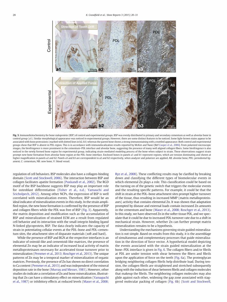
Fig. 3. Immunohistochemistry for bone sialoprotein (BSP) of control and experimental groups. BSP was evenly distributed in primary and secondary cementum aswell as alveolar bone incontrol group (a1). Similar morphological appearance was noticed in experimental groups. However, there are some distinct features to be noticed. Some light-brown stains appear to beassociatedwith boneprotrusions (markedwith dotted lines inb2, b3)whereas theparent bone shows a strong immunostainingwith amottled appearance. Both control and experimentalgroups show that BSP is absent in PDL region. This is in accordance with immunolocalization results reported by McKee and Nanci [Ref Cooper et al., 2006]. From polarized microscopicimages, the birefringence is more prominent in the cementum–PDL interface and alveolar bone, suggesting the presence of many well aligned collagen fibers. Some birefringence is alsonoticed in the newly formed bone region for experimental group, indicating strain-mediated modeling process of the bone when subject to strain. These observations suggest strainprompt new bone formation from alveolar bone region at the PDL–bone interface. Enclosed boxes in panels a1 and b1 represent regions, which are tension dominating and shown athigher magnification in panels a2 and b2. Panels a3 and b3 are correspondent to a2 and b2 respectively, when analyzer and polarizer are applied. AB: alveolar bone, PDL: periodontal lig-ament, C: cementum, NB: new bone, V: blood vessel.
28 K. Grandfield et al. / Bone Reports 3 (2015) 20–31
regulation of cell behaviors. BSPmolecules also have a collagen-bindingdomain (Scott and Stockwell, 2006). The interaction between BSP andcollagen facilitates apatite formation (Puxkandl et al., 2002). The RGDmotif of the BSP backbone suggests BSP may play an important rolefor osteoblast differentiation (Fisher et al., n.d.; Yamauchi andSricholpech, 2012). Among other NCPs, the expression of BSP is wellcorrelated with mineralization events. Therefore, BSP would be anideal indicator ofmineralization events in this study. In the strain ampli-fied region, the new bone formation is confirmed by the presence of BSPand collagen fibers while the PDL was free of BSP (Fig. 3). Apparently,the matrix deposition and modification such as the accumulation ofBSP and mineralization of strained ECM are a result from regulatedcell behavior and its interaction with the small integrin binding ligandN-linked glycoprotein, BSP. The data clearly indicates the capacity ofstrain in potentiating cellular events at the PDL–bone and PDL–cemen-tum sites, the attachment sites of disparate materials (soft and hard).
While the presence of BSP and BGN at the respective interfaces is anindicator of osteoid-like and cementoid-like matrices, the presence ofelemental Zn may be an indicator of increased local activity of matrixmetalloproteinases necessary for accelerated tissue turnover prior tomineralization (Pemmer et al., 2013; Roschger et al., 2013). The uniquepatterns of Zn may be a temporal marker of mineralization of organicmatrices. Previously, the presence of Zn has shown no direct correlationto Ca content (Pemmer et al., 2013) andwas independent of themineraldeposition rate in the bone (Murray andMesser, 1981). However, otherstudies do indicate a correlation of Zn and bonemineralization, illustrat-ing that Zn can have a stimulatory effect on mineralization (Yamaguchiet al., 1987) or inhibitory effects at reduced levels (Maser et al., 2008;
Ryz et al., 2008). These conflicting results may be clarified by breakingdown and classifying the different types of biomolecular events inwhich elemental Zn plays a role. This classification could be based onthe turning on of the genetic switch that triggers the molecular eventsand the resulting specific patterns. For example, it could be that theshift in strain at the PDL–bone attachment sites prompt higher turnoverof the tissue, thus resulting in increased MMP (matrix metalloprotein-ases) activity that contains elemental Zn. It was shown that adaptationprompted by disease and external loads contain increased Zn amountsin the cementum and bone (Maser et al., 2008; Roschger et al., 2013).In this study, we have observed Zn in the softer tissue PDL, andwe spec-ulate that it could be due to increased PDL turnover rate due to a shift inmechanical strain. However, whether Zn can further prompt matrixmineralization remains to be a hypothesis.
Understanding themechanisms governing strain guidedmineraliza-tion is not simple. Based on results from this study, it is the assemblageof simultaneous and complementary processes that guide mineraliza-tion in the direction of force vector. A hypothetical model depictingthe events associated with the strain guided mineralization at thebone–PDL interface is given in Fig. 6. The collagen fibers and/or fibrilsat PDL are under tension with shear between the fibers and fibrilsupon the application of force on the teeth (Fig. 6a). The proteoglycanbridging neighboring collagen fibrils help distribute load. During ten-sion, the collagen fibrils are straightened and stretched subsequentlyalongwith the induction of shear betweenfibrils and collagenmoleculesthat makeup the fibrils. The neighboring collagen molecules may alsoglide against each other, widening the gap zone associated with stag-gered molecular packing of collagen (Fig. 6b) (Scott and Stockwell,

Fig. 4. Fluorochrome images of experimental (a, b) groups reveal irregular patterns ofmineralization. Control specimens displayed regular banding patternswhich indicated a progressingbiomineralization front. Experimental specimens indicate much less organizedmineralization and nodules or finger-like protrusions of biomineralization in regions experiencing tension,for example the boxed region, compared to unstrained regions marked by *. Upon closer inspection of D, the strained region shows increased mineralization consistently surroundingblood vessel or endosteal spaces as marked by arrows. The vessels appear to be elongated along the direction of strain, and such a mechanical cue may be contributing to the enhancedmineral formation in their vicinity. Gradients inmicroprobeXRF elementalmapping are illustrated and are correlatedwith structural features as indicated by lightmicroscopy technique. Abicolor elementalmap for calciumand zinc signals shown in (d) and a corresponding lightmicroscope image in (c). Gradients in (d) show calciumand zinc in existing bone (orange to darkorange), zinc only in areas of newbone growthwithin PDL (green), and new bone-finger regions high in zinc yet low in calcium (yellow)which correspond to cement lines (also observedin controls) and active sites of remodeling surrounding bone finger protrusions. AB: alveolar bone, NB: new bone, T: tooth, PDL: periodontal ligament.
29K. Grandfield et al. / Bone Reports 3 (2015) 20–31
2006). Inside the collagen fibrils, the telopeptides of collagen moleculesare typically cross-linked with other neighboring collagen molecules(Puxkandl et al., 2002). These covalent crosslinks between the collagenmolecules can hinder further slippage of molecules and also increasethe overall stiffness of collagen fibrils. Meanwhile, cells attached to thematrix through small integrin binding ligand N-linked glycoproteinsuch as BSP are also regulated by the strain in collagen of the PDL. As
suggested in Fig. 6c and d, the force acting on the cytoskeletal compo-nents of cells triggers a series of downstream processes including geneexpression, protein synthesis, ECM deposition and mineralization aswell as osteoclast recruitment (Yamauchi and Sricholpech, 2012). Asvery little is known, a number of theorieswere extrapolated from similartissues such as ligaments and tendons, and in vitro studieswhich provid-ed analogous insights to the observed in vivo events in this study.

Fig. 5.The interface betweenexisting and newbone (identifiedasfinger-like protrusions) as imagedwith AFMunder dry (A) and hydrated (B) conditions. Notice thehighly populated cell-density along the interface or genesis site of adapted bone formation, indicative of a strongly differentiating zone. Collagen fiber inserts display increased hygroscopicity under hydrated con-ditions with their orientation consistent with the finger-like growth direction. Raman spectroscopy, overlain in A, indicated the higher organic:inorganic ratio in newly formed protrusionsversus existing bone. Nanoindentation values in B support the finding of higher organic components in the newer bone by the lower reduced elastic modulus versus existing bone.
30 K. Grandfield et al. / Bone Reports 3 (2015) 20–31
5. Conclusion
The present study enabled investigation of biomineralization effectsin the bone–PDL–tooth complex such as tooth movement, resultingdeformation of the PDL in three-dimensions, as well as correlation tophysicochemical properties of tissues. The results provide evidence for
Fig. 6. A model describing potential events at the PDL–bone functional attachment site of the cstress cause straightening and elongation of the collagen fibers PDL. (b) At microscale, collagepacked collagen molecules. Tension of collagen fibers exerts stretching of collagen fibrils (thiinterfibrillar transmission of load. Sliding of collagen molecules (ribbons of blue triple helix) pthe intermolecular crosslinking bonds prevent the further slippage. The proteoglycans betweethemechanical behavior of collagen fibers and benefit the structural integrity during the tensiothe strain resulted from external force on teeth to cytoskeletal components of cells in PDL. Suchthe ECM such as regulated gene expression, protein synthesis, matrix deposition andmineralizformation and remodeling takes place at the PDL–bone interface, suggesting an active cell behava result of such strained guided adaption at PDL–bone interface.
strain-guided or vectorial biomineralization in the complex, character-ized by physiologically reversed resorption patterns and protrudingbone formations with altered physicochemical properties into thePDL-space. These results suggest that the rates of adaptation are in-creased at the richly differentiating semiautonomous zones of the PDLentheses, and therefore susceptible to controlled modulation or
omplex is highlighted in this figure. (a) At macroscale, mechanical stimuli such as tensilen fibers are made from collagen fibril bundles while collagen fibrils consist of staggeredn rods with blue and white bands). The proteoglycans bridging collagen fibrils allow therovides additional displacements and widens the gap zone of a collagen fibril. However,n collagen fibrils and the covalent crosslinks between collagen molecules all contribute ton. (c) Apparently, cells embedded in the ECM of PDL are subject to tension. ECM transmitsmechanical stimuli trigger a cascade of intracellular and extracellular processes to modifyation. Meanwhile, the matrix signaling facilitates recruitment of osteoclasts. (d) New boneior such as differentiation and ECMproduction. Regulatedmineralization of ECMoccurs as

31K. Grandfield et al. / Bone Reports 3 (2015) 20–31
modulation toward pathology under exacerbated conditions. An alteredbiomechanics of the entire bone–PDL–tooth complex resulting from or-thodontic forces highlight the clinical relevance of this work.
Supplementary data to this article can be found online at http://dx.doi.org/10.1016/j.bonr.2015.04.002.
Acknowledgments
The authors acknowledge funding support from NIH/NIDCRR00DE018212 (SPH), NIH/NIDCR-R01DE022032 (SPH), NIH/NIDCRT32 DE07306, NIH/NCRR S10RR026645, (SPH) and Departments ofPreventive and Restorative Dental Sciences and Orofacial Sciences,UCSF; Faculty of Engineering, McMaster University (Hamilton,Canada) (KG). The authors thank Linda Prentice of the HistologyCore Facility, and The UCSF Biomaterials and Bioengineering Micro-CT Facility, Department of PRDS, UCSF for their services with the tis-sue preparation and imaging. Additional thanks to Andrew Jang,DDS, for his assistance in generating the movies related to micro-XCT data. Assistance from national facilities through user based pro-gram was provided by Stanford Synchrotron Radiation Lightsource(SSRL), SLAC National Accelerator Laboratory, Stanford University,CA and TheMolecular Foundry, Lawrence Berkeley National Laboratory,Berkeley, CA.Work at the SSRLwas supported by theU.S. Department ofEnergy under contract number DE-AC02-76SF00515. Work at the Mo-lecular Foundry was supported by the Office of Science, Office of BasicEnergy Sciences, of the U.S. Department of Energy under Contract No.DE-AC02-05CH11231. The WVID1(9C5) monoclonal antibody againstBSP developed by M. Solursh and A. Franzen was obtained from theDevelopmental Studies Hybridoma Bank developed under the auspicesof the NICHD and maintained by the University of Iowa, Department ofBiology, Iowa City, IA.
References
Beertsen, W., McCulloch, C.A., Sodek, J., 1997. The periodontal ligament: a unique, multi-functional connective tissue. Periodontology 2000 (13), 20–40.
Boyde, A., 1980. Electron microscopy of the mineralizing front. Metab. Bone Dis. Relat.Res. 2, 69–78.
Carter, D.R., Fyhrie, D.P., Whalen, R.T., 1987. Trabecular bone density and loading history:regulation of connective tissue biology by mechanical energy. J. Biomech. 20,785–794. http://dx.doi.org/10.1016/0021-9290(87)90058-3.
Carter, Y., Thomas, C.D.L., Clement, J.G., Cooper, D.M.L., 2013. Femoral osteocyte lacunardensity, volume and morphology in women across the lifespan. J. Struct. Biol. 183,519–526. http://dx.doi.org/10.1016/j.jsb.2013.07.004.
Cooper, G.M., Singhal, V.K., Barbano, T., Wigginton, W., Rabold, T., Losken, H.W., Siegel,M.I., Mooney, M.P., 2006. Intracranial volume changes in craniosynostotic rabbits:effects of age and surgical correction. Plast. Reconstr. Surg. 117, 1886–1890. http://dx.doi.org/10.1097/01.prs.0000218845.70591.7e.
Currey, J.D., 2002. The structure of bone tissue. Bones: Structure and Mechanics.Donnelly, E., Boskey, A.L., Baker, S.P., van der Meulen, M.C.H., 2010. Effects of tissue age on
bone tissue material composition and nanomechanical properties in the rat cortex.J. Biomed. Mater. Res. 92, 1048–1056. http://dx.doi.org/10.1002/jbm.a.32442.
Erlebacher, A., Derynck, R., 1996. Increased expression of TGF-B2 in osteoblasts results inosteoporosis-like phenotype. J. Cell Biol. 132, 195–210.
Fisher, L.W., Torchia, D.A., Fohr, B., Young, M.F., Fedarko, N.S., n.d. Flexible Structures ofSIBLING Proteins, Bone Sialoprotein, and Osteopontin. Biochem. Biophys. Res.Commun. 280, 460–465.
Frost, H.M., 1994. Wolff's Law and bone's structural adaptations to mechanical usage: anoverview for clinicians. Angle Orthod. 64, 175–188. http://dx.doi.org/10.1043/0003-3219(1994)064b0175:WLABSAN2.0.CO;2.
Herber, R.-P., Fong, J., Lucas, S.A., Ho, S.P., 2012. Imaging an Adapted DentoalveolarComplex. Anatomy Research International, pp. 1–13 http://dx.doi.org/10.1155/2012/782571.
Ho, S.P., Goodis, H., Balooch, M., Nonomura, G., Marshall, S.J., Marshall, G., 2004. The effectof sample preparation technique on determination of structure and nanomechanicalproperties of human cementum hard tissue. Biomaterials 25, 4847–4857. http://dx.doi.org/10.1016/j.biomaterials.2003.11.047.
Ho, S.P., Kurylo, M.P., Grandfield, K., Hurng, J., Herber, R.-P., Ryder, M.I., Altoe, V., Aloni, S.,Feng, J., WEbb, S., Marshall, G.W., Curtis, D., Piennatta, P., Hayter, J.A., 2013. The plasticnature of the human bone–periodontal ligament–tooth fibrous joint. Bone 57 (2),455–467. http://dx.doi.org/10.1016/j.bone.2013.09.007.
Hurng, J.M., Kurylo, M.P., Marshall, G.W., Webb, S.M., Ryder, M.I., Ho, S.P., 2011. Disconti-nuities in the human bone–PDL–cementum complex. Biomaterials 32, 7106–7117.http://dx.doi.org/10.1016/j.biomaterials.2011.06.021.
Jang, A.T., Lin, J.D., Seo, Y., Etchin, S., Merkle, A., Fahey, K., Ho, S.P., 2014. In situ compres-sive loading and correlative noninvasive imaging of the bone–periodontal ligament–tooth fibrous joint. J. Vis. Exp. http://dx.doi.org/10.3791/51147.
Krishnan, V., Davidovitch, Z., 2006. Cellular, molecular, and tissue-level reactions to ortho-dontic force. Am. J. Orthod. Dentofac. Orthop. 129, 469.e1–469.e32. http://dx.doi.org/10.1016/j.ajodo.2005.10.007.
Lin, J.D., Özcoban, H., Greene, J.P., Jang, A.T., Djomehri, S.I., Fahey, K.P., Hunter, L.L.,Schneider, G.A., Ho, S.P., 2013. Biomechanics of a bone–periodontal ligament–tooth fibrous joint. J. Biomech. 46, 443–449. http://dx.doi.org/10.1016/j.jbiomech.2012.11.010.
Liskova, M., Hert, J., 1971. Reaction of bone to mechanical stimuli. 2. Periosteal and end-osteal reaction of tibial diaphysis in rabbit to intermittent loading. Folia Morphol.(Warsz) 19, 301–317.
Lu, H.H., Thomopoulos, S., 2013. Functional attachment of soft tissues to bone: develop-ment, healing, and tissue engineering. Annu. Rev. Biomed. Eng. 15, 201–226. http://dx.doi.org/10.1146/annurev-bioeng-071910-124656.
Luan, X., Ito, Y., Holliday, S., Walker, C., Daniel, J., Galang, T.M., Fukui, T., Yamane, A.,Begole, E., Evans, C., Diekwisch, T.G.H., 2007. Extracellular matrix-mediated tissue re-modeling following axial movement of teeth. J. Histochem. Cytochem. 55, 127–140.http://dx.doi.org/10.1369/jhc.6A7018.2006.
Mann, S., 2001. Biomineralization: Principles and Concepts in Bioinorganic MaterialsChemistry. Oxford University Press.
Maser, R.E., Stabley, J.N., Lenhard, M.J., Owusu-Griffin, P., Provost-Craig, M.A., Farquhar,W.B., 2008. Zinc intake and biochemical markers of bone turnover in type 1 diabetes.Diabetes Care 31, 2279–2280. http://dx.doi.org/10.2337/dc08.
Meikle, M.C., 2006. The tissue, cellular, and molecular regulation of orthodontic toothmovement: 100 years after Carl Sandstedt. Eur. J. Orthod. 28, 221–240. http://dx.doi.org/10.1093/ejo/cjl001.
Melsen, B., 1999. Biological reaction of alveolar bone to orthodontic tooth movement.Angle Orthod. 69, 151–158. http://dx.doi.org/10.1043/0003-3219(1999)069b0151:BROABTN2.3.CO;2.
Murray, E.J., Messer, H.H., 1981. Turnover of bone zinc during normal and acceleratedbone loss in rats. J. Nutr. 111, 1641.
Nanci, A., 1999. Content and distribution of noncollagenous matrix proteins in bone andcementum: relationship to speed of formation and collagen packing density.J. Struct. Biol. 126, 256–269. http://dx.doi.org/10.1006/jsbi.1999.4137.
Parfitt, A.M., Drezner, M.K., Glorieux, F.H., Kanis, J.A., Malluche, H., Meunier, P.J., Ott, S.M.,Recker, R.R., 1987. Bone histomorphometry: standardization of nomenclature, sym-bols, and units. Report of the ASBMR Histomorphometry Nomenclature Committee.J. Bone Miner. Res. 2, 595–610. http://dx.doi.org/10.1002/jbmr.5650020617.
Parisuthiman, D., Mochida, Y., Duarte,W.R., Yamauchi, M., 2005. Biglycan modulates oste-oblast differentiation and matrix mineralization. J. Bone Miner. Res. 20, 1878–1886.http://dx.doi.org/10.1359/JBMR.050612.
Pemmer, B., Roschger, A., Wastl, A., Hofstaetter, J.G., Wobrauschek, P., Simon, R., Thaler,H.W., Roschger, P., Klaushofer, K., Streli, C., 2013. Spatial distribution of the trace ele-ments zinc, strontium and lead in human bone tissue. Bone 57, 184–193. http://dx.doi.org/10.1016/j.bone.2013.07.038.
Puxkandl, R., Zizak, I., Paris, O., Keckes, J., Tesch, W., Bernstorff, S., Purslow, P., Fratzl, P.,2002. Viscoelastic properties of collagen: synchrotron radiation investigations andstructural model. Philos. Trans. R. Soc. B Biol. Sci. 357, 191–197. http://dx.doi.org/10.1098/rstb.2001.1033.
Qian, L., Todo, M., Morita, Y., Matsushita, Y., Koyano, K., 2009. Deformation analysis of theperiodontium considering the viscoelasticity of the periodontal ligament. Dent.Mater. 25, 1285–1292. http://dx.doi.org/10.1016/j.dental.2009.03.014.
Reitan, K., Rygh, P., 1994. Biomechanical principles and reactions. In: Graber, T.M.,Vanarsdall, R.L. (Eds.), Orthodontics: Current Principles and Techniques. MosbyYear Book, Inc., St. Louis, Missouri, pp. 159–161.
Roschger, A., Hofstaetter, J.G., Pemmer, B., Zoeger, N., Wobrauschek, P., Falkenberg, G.,Simon, R., Berzlanovich, A., Thaler, H.W., Roschger, P., Klaushofer, K., Streli, C.,2013. Differential accumulation of lead and zinc in double-tidemarks of articularcartilage. Osteoarthr. Cartil. 21 (11), 1707–1715. http://dx.doi.org/10.1016/j.joca.2013.06.029.
Ryz, N.R., Meddings, J.B., Taylor, C.G., 2008. Long-chain inulin increases dendritic cells inthe Peyer's patches and increases ex vivo cytokine secretion in the spleen and mes-enteric lymph nodes of growing female rats, independent of zinc status. BJN 101,1653. http://dx.doi.org/10.1017/S000711450812342X.
Sandy, J.R., Farndale, R.W., Meikle, M.C., 1993. Recent advances in understandingmechan-ically induced bone remodeling and their relevance to orthodontic theory and prac-tice. Am. J. Orthod. Dentofac. Orthop. 103, 212–222.
Scott, J.E., Stockwell, R.A., 2006. Cartilage elasticity resides in shape module decoran andaggrecan sumps of damping fluid: implications in osteoarthrosis. J. Physiol. Lond.574, 643–650. http://dx.doi.org/10.1113/jphysiol.2006.108100.
Weiner, S., Traub, W., 1986. Organization of hydroxyapatite crystals within collagen fi-brils. FEBS Lett. 206, 262–266.
Wolff, J., 1892. Das Gesetz der Transformation der knochen. A. Hirschwald, Berlin (Thismonograph was translated to English and was published by Springer-Verlag in1986).
Yamaguchi, M., Oishi, H., Suketa, Y., 1987. Stimulatory effect of zinc on bone formation intissue culture. Biochem. Pharmacol. 36, 4007–4012. http://dx.doi.org/10.1016/0006-2952(87)90471-0.
Yamauchi, M., Sricholpech, M., 2012. Lysine post-translational modifications of collagen.Essays Biochem. 52, 113–133. http://dx.doi.org/10.1042/bse0520113.
Related Documents

![Cementum in Disease[Nalini]](https://static.cupdf.com/doc/110x72/55cf9d52550346d033ad2077/cementum-in-diseasenalini.jpg)