ORIGINAL RESEARCH ARTICLE published: 21 May 2013 doi: 10.3389/fnhum.2013.00201 Still feeling it: the time course of emotional recovery from an attentional perspective Jayne Morriss 1 *, Alexander N. W. Taylor 1 , Etienne B. Roesch 2 and Carien M. van Reekum 1 * 1 Centre for Integrative Neuroscience and Neurodynamics, School of Psychology and Clinical Language Sciences, University of Reading, Reading, UK 2 Centre for Integrative Neuroscience and Neurodynamics, School of Systems Engineering, University of Reading, Reading, UK Edited by: Alexander J. Shackman, University of Maryland, USA Reviewed by: Raffael Kalisch, University Medical Center Hamburg-Eppendorf, Germany Greg Hajcak, Stony Brook University, USA *Correspondence: Jayne Morriss, Centre for Integrative Neuroscience and Neurodynamics, School of Psychology and Clinical Language Sciences, University of Reading, Earley Gate, Whiteknights Campus, Reading RG6 6AH, UK. e-mail: j.e.morriss@pgr. reading.ac.uk; Carien M. van Reekum, Centre for Integrative Neuroscience and Neurodynamics, School of Psychology and Clinical Language Sciences, University of Reading, Earley Gate, Whiteknights Campus, PO Box 238, Reading RG6 6AL, UK. e-mail: [email protected] Emotional reactivity and the time taken to recover, particularly from negative, stressful, events, are inextricably linked, and both are crucial for maintaining well-being. It is unclear, however, to what extent emotional reactivity during stimulus onset predicts the time course of recovery after stimulus offset. To address this question, 25 participants viewed arousing (negative and positive) and neutral pictures from the International Affective Picture System (IAPS) followed by task-relevant face targets, which were to be gender categorized. Faces were presented early (400–1500 ms) or late (2400–3500 ms) after picture offset to capture the time course of recovery from emotional stimuli. Measures of reaction time (RT), as well as face-locked N170 and P3 components were taken as indicators of the impact of lingering emotion on attentional facilitation or interference. Electrophysiological effects revealed negative and positive images to facilitate face-target processing on the P3 component, regardless of temporal interval. At the individual level, increased reactivity to: (1) negative pictures, quantified as the IAPS picture-locked Late Positive Potential (LPP), predicted larger attentional interference on the face-locked P3 component to faces presented in the late time window after picture offset. (2) Positive pictures, denoted by the LPP, predicted larger facilitation on the face-locked P3 component to faces presented in the earlier time window after picture offset. These results suggest that subsequent processing is still impacted up to 3500 ms after the offset of negative pictures and 1500ms after the offset of positive pictures for individuals reacting more strongly to these pictures, respectively. Such findings emphasize the importance of individual differences in reactivity when predicting the temporality of emotional recovery. The current experimental model provides a novel basis for future research aiming to identify profiles of adaptive and maladaptive recovery. Keywords: emotion, attention, reactivity, recovery, face, late positive potential, P3, N170 INTRODUCTION Emotional events that bear relevance to an organisms’ well-being, demand center stage in selective attention, and initiate a cascade of typical behavioral and psychophysiological response tenden- cies (Frijda, 1986; Davidson, 1998; Lang and Bradley, 2010). These responses can be considered to originate from an adap- tive emotional auto-regulation process (Kappas, 2011); that is, modification of the intensity in emotional responding, or termi- nation of emotional responding, is instigated without motivation, in order to avoid negative stimuli, approach positive stimuli, or return to a steady emotional state. Whilst the majority of research in the field examines affective responding upon the onset of an emotional stimulus, few studies have investigated the extent to which emotional responding continues after stimulus offset (Jackson et al., 2003; Hajcak and Olvet, 2008). Examining the time course of auto-regulation after the offset of an emotional event may provide crucial information to understand adaptive recov- ery time, which is described in the affective chronometry model, as the speed of return to a baseline state in a response system after an emotion eliciting stimulus (Davidson, 1998). A landmark study by Jackson et al. (2003) tested this notion by collecting electroencephalography (EEG) frontal asymmetry data to serve as a metric of individual differences in affective style, which was then used to predict outcomes on physiological markers of recovery such as eye-blink startle in an emotional task. The task in this study consisted of presenting International Affective Picture System (IAPS) pictures for 6 s, with audio probes pre- sented either 2.5 or 4.5 s during the picture or 1 s after picture offset. Jackson and colleagues found individuals with right electri- cal frontal-asymmetry to have larger eye-blink startle magnitude to an audio probe presented 1 s after a negative picture, compared to a neutral picture. Furthermore, with a similar paradigm Larson et al. (2007) found healthy participants exhibiting depressive and anxiety symptoms to have different profiles of emotional recov- ery. For example, those with depressive symptoms had shown a blunted startle response to audio probes presented 1.5 s after positive pictures, compared to controls. In addition, individu- als high in anxious apprehension showed potentiated startle to audio probes presented 1.5 s after unpleasant and pleasant pic- tures, relative to controls. Importantly, unraveling how adaptive Frontiers in Human Neuroscience www.frontiersin.org May 2013 | Volume 7 | Article 201 | 1 HUMAN NEUROSCIENCE

Welcome message from author
This document is posted to help you gain knowledge. Please leave a comment to let me know what you think about it! Share it to your friends and learn new things together.
Transcript

ORIGINAL RESEARCH ARTICLEpublished: 21 May 2013
doi: 10.3389/fnhum.2013.00201
Still feeling it: the time course of emotional recovery froman attentional perspectiveJayne Morriss1*, Alexander N. W. Taylor 1, Etienne B. Roesch2 and Carien M. van Reekum1*
1 Centre for Integrative Neuroscience and Neurodynamics, School of Psychology and Clinical Language Sciences, University of Reading, Reading, UK2 Centre for Integrative Neuroscience and Neurodynamics, School of Systems Engineering, University of Reading, Reading, UK
Edited by:
Alexander J. Shackman, Universityof Maryland, USA
Reviewed by:
Raffael Kalisch, University MedicalCenter Hamburg-Eppendorf,GermanyGreg Hajcak, Stony BrookUniversity, USA
*Correspondence:
Jayne Morriss, Centre for IntegrativeNeuroscience and Neurodynamics,School of Psychology and ClinicalLanguage Sciences, University ofReading, Earley Gate, WhiteknightsCampus, Reading RG6 6AH, UK.e-mail: [email protected];
Carien M. van Reekum, Centre forIntegrative Neuroscience andNeurodynamics, School ofPsychology and Clinical LanguageSciences, University of Reading,Earley Gate, Whiteknights Campus,PO Box 238, Reading RG6 6AL, UK.e-mail: [email protected]
Emotional reactivity and the time taken to recover, particularly from negative, stressful,events, are inextricably linked, and both are crucial for maintaining well-being. It is unclear,however, to what extent emotional reactivity during stimulus onset predicts the timecourse of recovery after stimulus offset. To address this question, 25 participants viewedarousing (negative and positive) and neutral pictures from the International AffectivePicture System (IAPS) followed by task-relevant face targets, which were to be gendercategorized. Faces were presented early (400–1500 ms) or late (2400–3500 ms) afterpicture offset to capture the time course of recovery from emotional stimuli. Measuresof reaction time (RT), as well as face-locked N170 and P3 components were taken asindicators of the impact of lingering emotion on attentional facilitation or interference.Electrophysiological effects revealed negative and positive images to facilitate face-targetprocessing on the P3 component, regardless of temporal interval. At the individual level,increased reactivity to: (1) negative pictures, quantified as the IAPS picture-locked LatePositive Potential (LPP), predicted larger attentional interference on the face-locked P3component to faces presented in the late time window after picture offset. (2) Positivepictures, denoted by the LPP, predicted larger facilitation on the face-locked P3 componentto faces presented in the earlier time window after picture offset. These results suggestthat subsequent processing is still impacted up to 3500 ms after the offset of negativepictures and 1500 ms after the offset of positive pictures for individuals reacting morestrongly to these pictures, respectively. Such findings emphasize the importance ofindividual differences in reactivity when predicting the temporality of emotional recovery.The current experimental model provides a novel basis for future research aiming toidentify profiles of adaptive and maladaptive recovery.
Keywords: emotion, attention, reactivity, recovery, face, late positive potential, P3, N170
INTRODUCTIONEmotional events that bear relevance to an organisms’ well-being,demand center stage in selective attention, and initiate a cascadeof typical behavioral and psychophysiological response tenden-cies (Frijda, 1986; Davidson, 1998; Lang and Bradley, 2010).These responses can be considered to originate from an adap-tive emotional auto-regulation process (Kappas, 2011); that is,modification of the intensity in emotional responding, or termi-nation of emotional responding, is instigated without motivation,in order to avoid negative stimuli, approach positive stimuli, orreturn to a steady emotional state. Whilst the majority of researchin the field examines affective responding upon the onset ofan emotional stimulus, few studies have investigated the extentto which emotional responding continues after stimulus offset(Jackson et al., 2003; Hajcak and Olvet, 2008). Examining the timecourse of auto-regulation after the offset of an emotional eventmay provide crucial information to understand adaptive recov-ery time, which is described in the affective chronometry model,as the speed of return to a baseline state in a response systemafter an emotion eliciting stimulus (Davidson, 1998). A landmark
study by Jackson et al. (2003) tested this notion by collectingelectroencephalography (EEG) frontal asymmetry data to serveas a metric of individual differences in affective style, whichwas then used to predict outcomes on physiological markersof recovery such as eye-blink startle in an emotional task. Thetask in this study consisted of presenting International AffectivePicture System (IAPS) pictures for 6 s, with audio probes pre-sented either 2.5 or 4.5 s during the picture or 1 s after pictureoffset. Jackson and colleagues found individuals with right electri-cal frontal-asymmetry to have larger eye-blink startle magnitudeto an audio probe presented 1 s after a negative picture, comparedto a neutral picture. Furthermore, with a similar paradigm Larsonet al. (2007) found healthy participants exhibiting depressive andanxiety symptoms to have different profiles of emotional recov-ery. For example, those with depressive symptoms had showna blunted startle response to audio probes presented 1.5 s afterpositive pictures, compared to controls. In addition, individu-als high in anxious apprehension showed potentiated startle toaudio probes presented 1.5 s after unpleasant and pleasant pic-tures, relative to controls. Importantly, unraveling how adaptive
Frontiers in Human Neuroscience www.frontiersin.org May 2013 | Volume 7 | Article 201 | 1
HUMAN NEUROSCIENCE
Morriss et al. Emotional recovery and individual differences
emotional recovery functions in the healthy population couldserve as a useful comparison when recovery is compromised inclinical populations. A wealth of literature indeed demonstratespatients with depression and anxiety to ruminate and worry overpast emotional events (for review Nolen-Hoeksema et al., 2008),which may be linked to the dysfunction of recovery mechanisms.For example, recovery from negative events in depressed patientsmay be stifled because of the sustainment of negative affect (Siegleet al., 2002), as well as the difficulty in maintaining positive affect(Heller et al., 2009).
One way to gage emotional recovery is through attentionalparadigms. Attention and emotion have been shown to bestrongly interconnected, with affective stimuli taking precedenceover competing stimuli, regardless of task relevance (for reviewsee Yiend, 2010). This effect has been well documented by meansof an event-related potential (ERP) component known as theLate Positive Potential (LPP), which is located over centro-parietalsites at approximately 300 ms after stimulus onset, and is thoughtto reflect the process of sustained attention (Olofsson et al., 2008;Hajcak et al., 2010; Lang and Bradley, 2010). In emotional con-texts, the LPP component is typically enhanced for arousingnegative and positive pictures, relative to neutral, both duringpassive viewing (Cuthbert et al., 1999; Schupp et al., 2000), andconcurrent task performance (Hajcak et al., 2007). This enlarge-ment of the LPP for arousing stimuli, relative to neutral stimuli,has been postulated to signify the global inhibition of competingstimuli in the environment, permitting motivationally relevantstimuli to be selectively processed (Schupp et al., 2004; Brownet al., 2012). To test this notion, Schupp et al. (2004) presentedemotional images in tandem with acoustic startle probes. Theirresults indicated negative and positive images to reliably inhibitthe processing of secondary acoustic startle probes, denoted bylarger LPPs to the arousing images and smaller P3 components tothe probes. In addition, sustained attention as measured throughthe magnitude of the LPP has also been shown to extend beyondthe offset of an emotional stimulus and to disrupt the process-ing of subsequent stimuli. For instance, the LPP has been shownto continue for up to 800 ms after pleasant pictures and for upto 1000 ms after unpleasant pictures (Hajcak and Olvet, 2008).Furthermore, Weinberg and Hajcak (2011) found pictures thatelicited larger LPPs within individuals to predict slower reactiontimes (RTs) and reduced P300 amplitudes over parietal areas tosubsequent categorization of shape targets. Given the temporalityof attentional-emotional processes, represented through the LPP,these findings suggest that the LPP may serve as: (1) an importantindicator of individual differences in the intensity of emotionalreactivity, (2) a useful predictor of recovery outcomes e.g., apredictor of attentional interference on subsequent task-relevantstimuli.
Quantifying emotional recovery via the level of attentionalmodulation on task-relevant targets that appear after emotionalstimuli may be useful in determining the lingering effect ofemotion after offset. For instance, attentional interference or facil-itation upon a following target can be considered a marker ofcontinued processing of task-irrelevant emotional stimuli, withthe former disrupting attention to following targets, whilst thelatter widens attention to following targets. Furthermore, a recent
body of behavioral research using rapid serial visual presenta-tion tasks has provided ample evidence that viewing emotionalstimuli can both interfere and facilitate the attentional process-ing of following targets, depending on the temporal proximitybetween stimuli (Bocanegra and Zeelenberg, 2009; Ciesielskiet al., 2010). Bocanegra and Zeelenberg (2009) found emotionalwords impaired accuracy on subsequent neutral word targetswhen distances in time were as small as 50 ms and 500 ms, whilstlonger time intervals of 1000 ms improved accuracy. Similarly,Ciesielski et al. (2010) observed that emotional picture distracters,particularly those exhibiting erotic and disgusting content, onlyreduced the participants’ accuracy on a subsequent task duringsmaller distracter-target lags, e.g., 200 ms, 400 ms, and 600 ms.Longer lags, i.e., 800 ms, however, produced facilitation effectsin accuracy. In addition, studies using target detection tasks havefound comparable behavioral results. For example, Weinberg andHajcak (2011) found interference, denoted as slower RTs on shapetargets presented directly after emotional images (e.g., 0 ms).
Given the extent to which differential effects of attentionalfacilitation and interference appear dependent upon temporalaspects, as evidenced above, it can be postulated that severaldistinct mechanisms are at work during the recovery of an emo-tional stimulus. Indeed, support for this argument can be foundfrom recent ERP studies, which demonstrate emotional picturesto modulate specific target ERP waveform components over time,thus indicating emotional stimuli to impact upon various stagesof subsequent target processing (Ihssen et al., 2007; Weinbergand Hajcak, 2011; Brown et al., 2012). For example, Ihssen et al.(2007) found arousing images to disrupt processing of lexicaltargets as shown by slower RT and reduced amplitude on twoERP components: (1) the early attention-specific N1, observedover occipital sites, and time locked to 184–284 ms, and (2)the later LPP, observed over parieto-central regions and timelocked to 412–712 ms. These effects occurred over three differ-ent temporal intervals between the emotional image and target,i.e., 80 ms, 200 ms, and 440 ms. Likewise, Brown et al. (2012)found that briefly presented negative images (e.g., 200 ms) dis-rupt processing on the early N1 to flashed probes, but not theN1 to Gabor patches, over short intervals of 570 ms betweennegative images and targets. Furthermore, Weinberg and Hajcak(2011) revealed emotional images to slow RTs and to attenu-ate subsequent P300 amplitude to shape targets which directlyfollowed the images. The disparity between valence specificityin these studies may be due to differences in task type, specificstate induced by the emotional images (positive vs. negative), aswell as timing of the target stimuli presented. Despite this, it isimportant to note that these ERP studies are coherent in show-ing distraction from emotional stimuli on early ERP componentslocked to subsequent targets. In addition, the electrophysiologicalfindings from these studies overlap with the behavioral researchpresented above, whereby shorter temporal proximities betweenan emotional prime and target result in interference effects. Yet,it remains unclear whether longer time intervals yield similarpatterns of attentional interference or perhaps facilitation forelectrophysiological and behavioral metrics.
In the study reported here, we used behavioral and ERPmethodology in conjunction with an attentional paradigm to
Frontiers in Human Neuroscience www.frontiersin.org May 2013 | Volume 7 | Article 201 | 2

Morriss et al. Emotional recovery and individual differences
investigate: (1) the extent of recovery from arousing negative andpositive stimuli, relative to neutral stimuli; (2) the impact of indi-vidual differences in emotional reactivity upon recovery speed.The experimental task consisted of presenting emotional imagesfor 3 s, followed by a probe stimulus of 500 ms consisting of a neu-tral face-target controlled with FACSGen and validated in a pre-vious study (Roesch et al., 2011). Participants were instructed toidentify the gender of the face and respond accordingly. In addi-tion, we manipulated the inter-stimulus interval (ISI) betweenthe picture and face-target in the form of a fixation cross pre-sented for a random period of time in two conditions, varyingbetween 400–1500 ms and 2400–3500 ms, respectively. We usedIAPS images (Lang et al., 2005) as emotional stimuli becausethey have been shown to induce emotion (Lang and Bradley,2010), reliably modulate the LPP component (Olofsson et al.,2008; Hajcak et al., 2010) and impact subsequent task process-ing (Ihssen et al., 2007; Weinberg and Hajcak, 2011; Brown et al.,2012). Our subset of IAPS pictures consisted of negative and pos-itive emotional pictures that were matched in arousal, as well asneutral pictures, to assess the influence of valence and arousalupon recovery outcomes. The LPP component to the emotionalimages was recorded, to serve as a metric of individual differencesin emotional reactivity and a predictor of individual differencesin emotional recovery, quantified as the level of interference onsubsequent face-targets. Face stimuli were used as probes fora number of reasons. Firstly, we aimed to expand the line ofbehavioral research that had previously used categorization tasksto assess the extent of attentional capture by emotional stimuliafter offset more generally (Ihssen et al., 2007) and within indi-viduals (Weinberg and Hajcak, 2011). Secondly, we wanted tocapture ERPs that have been shown to be modulated by attention,such as: (1) the face-specific N170, which is a negative poten-tial occurring around 150–200 ms over occipito-temporal sites,and is thought to reflect early perceptual and holistic encoding(Bentin et al., 1996). (2) The P3 component, a positive deflec-tion found over parieto-occipital areas around 300–400 ms, whichhas been associated with target detection (Schupp et al., 2004;Weinberg and Hajcak, 2011). Isolating those stages of processingthat may show effects of preceding emotion-laden stimuli may beimportant for understanding mechanisms relevant to emotionalreactivity and regulation. An advantage of using the FACSGenstimuli, compared to other face stimuli, is that the expressions arecomputer generated based upon parametrically controlled facialaction units, which means that the expressions portrayed on ourstimuli set are as intrinsically neutral as possible and are exactlythe same across the set. Lastly, temporal intervals were includedin experiment to examine how valence and arousal would impactthe temporality of emotional recovery speed. We opted for shorterand longer temporal intervals because of the paucity of ERPresearch examining the impact of preceding emotion-laden stim-uli on attention over a timescale of several seconds within groupsand individuals.
Our main hypotheses were fourfold. Firstly, we expected nega-tive and positive images to elicit more sustained attention thanneutral images, indexed by larger LPP amplitudes for negativeand positive images, relative to neutral (Lang and Bradley, 2010).Secondly, we expected arousing pictures to interfere with the
subsequent processing of face-targets, as shown by slower RTsand smaller N170/P3 amplitudes on following face stimuli, com-pared to neutral pictures (Ihssen et al., 2007; Weinberg andHajcak, 2011). Thirdly, modulation of RT and N170/P3 ampli-tude would be contingent upon the temporal interval between thearousing picture and target. We proposed that attentional inter-ference between an arousing image and target will occur overshorter temporal intervals due to increased competition betweenthe image and target, thus suggesting a slower recovery speedto emotional images, relative to neutral images (Bocanegra andZeelenberg, 2009; Ciesielski et al., 2010). We expected this to beshown by slower RTs and smaller N170/P3 amplitudes to face-targets (Weinberg and Hajcak, 2011). Based on the behavioralfindings of Bocanegra and Zeelenberg (2009) and Ciesielski et al.(2010), we predict that attentional facilitation will ensue whenthe temporal interval between an arousing picture and target islonger, as the competition between the image and target will bereduced but with a lingering effect of emotional image on atten-tional focus. We anticipated this to be evidenced by faster RTs andlarger N170/P3 amplitudes to face-targets. Lastly, we examinedhow individual differences in emotional reactivity could predictspeed of emotional recovery, by correlating IAPS-locked LPP val-ues to RTs and N170/P3 amplitudes on subsequent early and lateface-targets. We expected higher LPP values for arousing imagesto predict a more sustained impact on the face targets, reflectedin larger differences between RTs and ERP amplitudes on follow-ing face-targets after arousing vs. neutral images. Given previouswork showing interference by the LPP upon the visual P3 of sub-sequent targets specifically (see Weinberg and Hajcak, 2011), wepredicted this relationship to be stronger for the P3 componentthan the N170.
MATERIALS AND METHODSPARTICIPANTSTwenty-five right-handed students from the University ofReading Psychology Department were recruited for this study(mean age = 20.2 years, 18 females and 7 males). All participantshad normal or corrected to normal vision. Students providedwritten informed consent and received partial course credit fortheir participation. The procedure was approved by the Universityof Reading Ethics Committee.
STIMULIWe selected 216 pictures from the IAPS (Lang et al., 2005), depict-ing seventy-two events from each valence category (see Table 1);negative, positive, and neutral. Mean (SD) normative ratings ofvalence across the negative pictures was 2.61 (1.57); for positive7.41 (1.57); and for neutral, 5.00 (1.25). Mean valence ratings foreach picture category were significantly different to the other pic-ture categories, p < 0.001. For arousal, mean (SD) negative, andpositive picture ratings were matched, negative 5.66 (2.22); posi-tive 5.61 (2.28); neutral 3.20 (1.93). Negative and positive arousalratings did not significantly differ, p = 0.6. Both negative andpositive arousal ratings significantly differed from neutral, p <
0.001. The mean (SD) complexity and luminance of the imagesselected were matched across categories by using the scores ofcomplexity and luminance (see also van Reekum et al., 2007):
Frontiers in Human Neuroscience www.frontiersin.org May 2013 | Volume 7 | Article 201 | 3
Morriss et al. Emotional recovery and individual differences
Table 1 | Reference numbers to images taken from the International
Affective Picture System (IAPS; Lang et al., 2005).
Negative Neutral Positive
1052 9040 2038 2594 1463 7200
1111 9050 2102 2595 1710 7230
1220 9140 2104 2749 1722 7260
1274 9180 2191 2830 1811 7270
1301 9181 2210 2840 2058 7330
2095 9250 2214 4605 2071 7350
2141 9253 2215 5130 2150 7400
2683 9300 2235 5410 2160 7430
2688 9301 2271 5534 2208 7470
2691 9320 2272 5740 2209 7502
2710 9340 2280 5875 2216 7508
2751 9373 2305 7020 2224 8030
2981 9400 2357 7030 2340 8034
3015 9419 2381 7034 2345 8080
3051 9420 2383 7036 2346 8090
3061 9421 2385 7038 2352.1 8170
3160 9423 2393 7040 4599 8180
3181 9424 2396 7050 4603 8185
3215 9425 2397 7053 4610 8186
3220 9428 2440 7055 4623 8200
3230 9429 2441 7059 4626 8210
3350 9430 2446 7110 4640 8300
5971 9433 2480 7150 4641 8350
5973 9470 2485 7160 5260 8370
6213 9495 2491 7161 5270 8380
6242 9520 2493 7180 5450 8400
6243 9560 2506 7185 5470 8420
6244 9570 2512 7234 5480 8461
6540 9584 2513 7491 5600 8470
6570.1 9592 2514 7493 5621 8490
6571 9620 2515 7595 5623 8496
6821 9621 2516 7705 5629 8499
6830 9630 2518 7950 5700 8503
7359 9901 2570 8311 5830 8510
7380 9911 2579 9070 5833 8531
8485 9925 2593 9210 5910 8540
Complexity, negative 124606.40 (40955.07); positive 121928.66(31956.27); neutral 126801.65 (45579.57), and luminance, neg-ative 0.37 (0.13); positive 0.38 (0.13); neutral 0.37 (0.16). Meancomplexity and luminance did not significantly differ acrosscategories, p > 0.4.
Pictures of thirty-six synthetic 3-dimensional neutral facestimuli (18 male and 18 female) were selected (see Table 2)from the stimuli used in the validation procedures for theFACSGen software (Roesch et al., 2011). As part of this valida-tion procedure, 44 students from the University of Geneva wereinstructed to rate synthetic faces created with FaceGen Modeller(Singular Inversion Inc., 2012) on three continuous dimensionsspanning 0–100: gender (anchored male-female), believabil-ity (anchored synthetic-believable), and intrinsic emotionality(anchored positive-neutral-negative). We selected thirty-six faces
Table 2 | Reference numbers to faces taken from the Facial Action
Coding System Generator (FACSGen: Roesch et al., 2011).
Male Female
95 7
96 23
102 30
109 45
111 49
119 56
136 59
139 60
141 70
143 72
145 75
146 77
147 86
157 87
163 88
164 92
166 176
171 178
(18 male; 18 female) for being the most unambiguous gender-wise, the most believable, and the most emotionally neutralfaces as possible. Mean (SD) normative ratings of gender forthe seventy-two faces were; female 78.61 (5.895) and male 4.655(3.46), where ratings of male and female gender significantly dif-fered, p < 0.001. In addition, male and female faces were matchedfor mean (SD) ratings of neutral emotional expression and cred-ibility of the face: Neutral expression, female 47.41 (6.46); male48.16 (5.77), and credibility, female 52.21 (11.33); male 55.65(9.6). Mean neutral expression or credibility ratings of male andfemale categories did not significantly differ, p > 0.2.
TASK DESIGNAll of the tasks were administered using E-Prime 2.0 (PsychologySoftware Tools Ltd, Pittsburgh, PA). Within each task, the exper-imental trials were randomized and the response button press onthe mouse for the gender task was counterbalanced across partici-pants. Tasks were presented on a Viewsonic 22 inch monitor witha 60 Hertz refresh rate. Screen resolution was set at 1024 × 768pixels. For both the emotional recovery task and IAPS ratingtask, participants sat at approximately 60 cm from the screen. Theresulting visual angles were: 19◦ × 15◦ for FACSGen faces and35.6◦ × 22.5◦ for IAPS images.
Emotional recovery taskParticipants were required to passively view emotional picturesand identify the gender of following neutral face-targets, bypressing the appropriate button on the mouse. Participants wereinstructed to focus on a fixation cross displayed between the pic-ture and the face-target, to minimize noise from eye and musclemovement on the EEG. The face target was presented at a randomtime between either 400–1500 ms or 2400–3500 ms, in order toassess the temporality of emotional recovery, demonstrated as the
Frontiers in Human Neuroscience www.frontiersin.org May 2013 | Volume 7 | Article 201 | 4

Morriss et al. Emotional recovery and individual differences
degree of attentional spill-over from the previous emotional pic-ture upon a subsequent face-target. The emotional recovery taskconsisted of 216 trials: 3 Valence (negative, neutral, positive) × 2Temporal Interval (early: 400–1500 ms, late: 2400–3500 ms) × 36Neutral Faces. Each IAPS picture was presented once; each neu-tral face was repeated six times and paired with a specific valence,which was counterbalanced across time and gender. A trial thusconsisted of a 1000 ms fixation cross, 3000 ms IAPS picture pre-sentation, 400–1500 ms or 2400–3500 ms fixation cross, 500 msneutral face-target, and a 1500–3000 ms response window (seeFigure 1).
IAPS rating taskUpon completion of the main experiment, all IAPS pictures werepresented again to the participants, in a random order for 2 s.Participants were instructed to provide ratings on two dimen-sions, valence and arousal. The next trial started after participantscompleted both ratings using the keyboard. Each rating was pro-vided on a nine point Likert scale, where participants were giveninstructions to rate valence, i.e., “how positive or negative youfelt in response to the picture” and arousal, i.e., “the extent towhich you felt calm or excited in response to the pictures.” For thevalence ratings, a response of 9 represented “very pleasant” and 1“very unpleasant,” while for arousal 1 represented “very calm” and9 represented “very excited” (cf. Lang et al., 2005).
PROCEDUREThe current study was assessed and conducted in line with theethics procedures in place at the University of Reading. Uponarrival, participants were informed of the experimental procedureand asked to complete a consent form. Participants were seatedin a RF-shielded, sound-attenuated room for the EEG setup andexperimental testing. Firstly, the participant’s head was measured,in order to identify the correct size EEG cap. Secondly, once thecap was attached, the skin was cleaned with alcohol and a con-ductive gel was used to obtain sufficient impedance between the
electrodes and scalp. When the EEG was stable and the partici-pant was ready, the emotional recovery task was started on thecomputer. After completing the emotional recovery task, the EEGcap was removed. Next, participants performed the picture ratingtask. Lastly, participants were thanked for their participation anddebriefed about the details of the study.
DATA COLLECTION AND REDUCTIONFor EEG recording we used an MR compatible, 32-channel, pow-erpack operated amplifier (Brain Products, GmBH, Germany).Thirty-two Ag/AgCl pellet pin electrodes were positioned onan elastic cap according to the standard 10/20 system. Therewere three midline electrodes (Fz/Cz/Pz) and 14 electrodes overeach hemisphere (FP1/FP2, F3/F4, F7/F8, FC1/FC2, FC5/FC6,FT9/FT10, C3/C4, CP1/CP2, CP5/CP6, T7/T8, TP9/TP10, P3/P4,P7/P8, and O1/O2). Electrode FCz served as the referencepoint and electrode AFz the ground. One electrode (IO) wasplaced on the outer canthi of the right eye for horizon-tal eye movement recordings. Electrode impedance was keptbelow 5 k�. EEG signals were recorded using Brain VisionRecorder Version 2.01 (Brain Products). Off-line EEG analy-sis was performed with Brain Vision Analyzer Version 2.01(Brain Products). Firstly, the raw data were inspected for elec-tromyographic (EMG) artifacts and other noise that could distortthe EEG signal. These artifacts were highlighted and removedfrom the data. Secondly, data were filtered with a low cut-off of 0.1 Hz (Hajcak and Olvet, 2008) and a high cutoff of40 Hz. Thirdly, to identify and remove eye movement arti-facts, an ocular Independent Component Analysis was performedon the data. We used the IO electrode to identify horizontaleye movements and FP1 to identify vertical eye movements.Lastly, a semi-automated visual inspection for remaining phys-iological artifacts was made on each channel within each trial.Trials were rejected if there was: (1) a voltage step of morethan 50 µV between sample points, (2) a voltage difference of300 µV within a trial, and (3) a maximum voltage difference
FIGURE 1 | A sample trial from the emotional recovery task: a fixation
cross was presented at the center of the screen to direct participants’
attention. Next, an IAPS image was presented, followed by a variable
temporal interval. Lastly, a face-target was briefly presented. Participantswere instructed to watch each picture and identify the gender of thefollowing face-target as quickly and as accurately as possible.
Frontiers in Human Neuroscience www.frontiersin.org May 2013 | Volume 7 | Article 201 | 5
Morriss et al. Emotional recovery and individual differences
of less than 0.50 µV within 100 ms intervals (Hajcak and Olvet,2008).
EEG data from the emotional pictures and face-targets wereonly segmented if the emotional picture and face-targets were fol-lowed by a correct response (95% of trials). After completion ofthese artifact rejection steps, 74% of emotional picture trials and70% of face trials were included in the segmentation and aver-aging process. The percentage of picture trials across participantsfor each condition were: Negative Early = 75%; Negative Late =71%; Neutral Early = 75%; Neutral Late = 74%; Positive Early =75%; Positive Late = 71%. The percentage of face trials across par-ticipants for each condition were: Negative Early = 70%; NegativeLate = 72%; Neutral Early = 71%; Neutral Late = 66%; PositiveEarly = 72%; Positive Late = 71%.
For emotional picture stimuli, segments were extracted from−200 ms before the image and 3000 ms after image onset. Theremaining trials were −200 to 0 ms baseline corrected and aver-aged. The average voltage ∗ ms from 300–1200 ms, 1200–2100 ms,and 2100–3000 ms post image onset was calculated for three elec-trodes associated with the LPP: Fz/Cz/Pz. The average voltage ∗ms values were then collapsed individually for Fz/Cz/Pz, for eachsubject and experimental condition.
Epochs around the face stimuli were extracted from −100 msbefore the face-target and 600 ms after face-target onset. Theremaining trials were then −100 to 0 ms baseline corrected andaveraged. A peak detection method was used to locate the negativepolarity of the N170 in TP9/TP10, and to identify the posi-tive polarity of the P3 in P7/P8/Pz/O1/O2. The time parametersfor finding the peaks were 140–220 ms for the N170 and 300–390 ms for the P3. Average amplitude values were then collapsedacross each subject and condition: independently for TP9/TP10electrodes for the N170 and across P7/P8/Pz/O1/O2 electrodesfor the P3.
RTs in the emotional recovery task were scored for correctresponses and only those RTs above 300 ms were retained (95%of trials). Accuracy scores from the emotional recovery taskwere expressed as the proportion of correct trials to total trialsincluded.
The data of one participant was excluded from analyses due toloss of signal over the temporal electrode sites, thus leaving a totalof 24 participants for statistical analyses.
RESULTSMAIN EFFECTS ANALYSISTo test the extent to which IAPS stimuli modulate subsequentprocessing of neutral face information over time, we conducteda 3 Valence (negative, neutral, positive) × 2 Temporal Interval(early: 400–1500 ms, late: 2400–3500 ms) repeated measures anal-ysis of variance (ANOVA) for RT, accuracy, and P3 ampli-tude. In addition, a 3 Valence (negative, neutral, positive) × 2Temporal Interval (early: 400–1500 ms, late: 2400–3500 ms) × 2Hemisphere (right, left) repeated measures ANOVA was con-ducted on N170 amplitude. The additional factor of hemispherewas included to assess the laterality of the N170, where N170amplitudes are found to be typically larger over the right hemi-sphere sites, compared to left (Bentin et al., 1996). To exam-ine LPP amplitude, we utilized a 3 Valence (negative, neutral,
positive) × 2 Temporal Interval (early: 400–1500 ms, late: 2400–3500 ms) × 3 LPP Window (early: 300–1200 ms, middle: 1200–2100 ms, late: 2100–3000 ms) × 3 Lead (Fz, Cz, Pz) repeatedmeasures ANOVA. Furthermore, to assess whether our partici-pants’ ratings of IAPS reflected the normative ratings, we used a3 Picture (negative, neutral, positive) repeated ANOVA for IAPSratings. Significant predicted effects in the omnibus tests were fol-lowed up with pairwise comparisons. All analyses were conductedusing SPSS 17.0 (IBM Ltd).
IAPS ratingsThe IAPS ratings produced a significant main effect of Valence,F(2, 46) = 156.664, p < 0.001. Participants reported negative pic-tures (M = 2.52, SD = 0.88) to be the most unpleasant, positivepictures (M = 6.50, SD = 0.81) as the most pleasant and neutralratings (M = 4.83, SD = 0.75) as neither unpleasant nor pleas-ant, p < 0.001. The ANOVA revealed arousal ratings to have amain effect of Valence, F(2, 46) = 2.545, p < 0.001. Both nega-tive (M = 5.31, SD = 1.58) and positive (M = 4.66, SD = 1.75)arousal ratings significantly differed from neutral (M = 3.29,SD = 1.50), p < 0.001. Although the arousal ratings were higherfor negative than positive pictures in our sample, negative arousalratings were not significantly different from arousal ratings ofpositive pictures, p = 0.087. The spread of arousal ratings ofthe positive pictures was higher in our sample relative to thenormative ratings (see Figure 2).
IAPS-elicited LPPAs expected, a significant main effect of Valence was found,F(2, 46) = 7.492, p = 0.002. Findings were partially in line withpredictions, as LPP amplitudes were larger for negative pic-tures, relative to neutral pictures, at trend level, p = 0.056 (seeFigure 3). Reflecting the (non-significant) effect observed in theIAPS ratings, negative pictures evoked larger LPPs than positivepictures, p < 0.001. Moreover, the LPP amplitude to positive pic-tures and neutral pictures did not significantly differ, p = 0.163(see Figure 3 and Table 3).
To test that the LPP waveforms’ temporality and topogra-phy was comparable to previous research (e.g., Olofsson et al.,2008; Hajcak et al., 2010; Lang and Bradley, 2010) we split theLPP into three windows and assessed the LPP at each lead. Asexpected, the analysis yielded significant interactions betweenLead × Valence × LPP Window, F(8, 184) = 2.059, p = 0.042,Lead × Valence, F(4, 92) = 12.235, p < 0.001, and Lead × LPPWindow, F(4, 92) = 124.989, p < 0.001. The results were in accor-dance with previous studies that have examined the LPP (seeFigure 3 and Table 4), as negative images had the largest centro-parietal activation and the smallest frontal activation during theearly portion of the LPP, compared to neutral and positive images.In addition, within the middle portion of the LPP, negative imageswere found to elicit the strongest activity in central areas, rel-ative to positive and neutral images. Furthermore, in the lateportion of the LPP, negative images elicited more activity incentro-parietal regions than neutral and positive images. The LPPfor neutral images were larger than positive images during middleand late windows, but only over parietal regions. The ANOVA alsorevealed a main effect of Lead, F(2,46) = 4.897, p < 0.012, and
Frontiers in Human Neuroscience www.frontiersin.org May 2013 | Volume 7 | Article 201 | 6

Morriss et al. Emotional recovery and individual differences
FIGURE 2 | Mean IAPS valence and arousal ratings from the current
study sample and the original sample for the 216 pictures selected. Onthe valence scale, lower numbers denote negative valence, whilst highernumbers reflect positive valence. Similarly, in the arousal scale, high arousalis reflected by higher figures and lower arousal by lower figures. Asexpected, the current sample rated negative images as high arousing andunpleasant, and neutral images as low in arousal and neither pleasant or
unpleasant. Positive images, however, have more variation in arousal ratings,particularly when comparing them to the original sample. The currentexperimental sample ratings may be typical for a young British sample, giventhat the images selected were controlled for arousal and valence based uponthe original ratings that came with IAPS set (Lang et al., 2005). Neg,Negative; Neu, Neutral; Pos, Positive; S, current study sample ratings;O, original IAPS sample ratings.
Window, F(2,46) = 115.590, p < 0.001, where the LPP was maxi-mal: (1) at the Pz electrode, followed by Cz, and Fz electrodes. (2)In the early window, followed by the middle and late windows.No significant interaction between Valence × LPP Window wasfound, F(4, 92) = 0.387, p = 0.817.
As a control, we tested for random differences in LPP val-ues for the pictures preceding early vs. late face-targets. Therewas no Valence × Temporal Interval interaction, F(2, 46) = 0.192,p = 0.826, for the LPP. Thus, while negative pictures elicited ahigher LPP than positive and neutral pictures, no significant dif-ference between LPP values from valence × early vs. late faceconditions was found (see Table 3). No other interaction effectswith Time were found, largest F = 2.18, n.s.
RTWhile the average RT to faces following negative pictures werefaster than those following positive and neutral pictures in thelate interval, we found no significant main effect of Valence forRT, F(2, 46) = 0.974, p = 0.385, nor Valence × Temporal Intervalfor RT, F(2, 46) = 0.331, p = 0.720. As anticipated, however, RTwas faster for later face-targets, compared to earlier face-targets, asreflected in a main effect of Temporal Interval, F(1, 23) = 43.250,p < 0.001 (see Table 3).
AccuracyAccuracy scores in emotional recovery task were relatively highacross conditions, with on average 95% correct responses (seeTable 3). Picture valence did not impact target-face Accuracyscores, F(2, 46) = 0.247, p = 0.782, nor was there an effect of
Valence × Temporal Interval, F(2, 46) = 1.369, p = 0.265 or maineffect of Temporal Interval, F(1, 23) = 0.919, p = 0.348.
The face-locked N170 componentThe N170 amplitudes revealed no main effect of Valence,F(2, 46) = 1.444, p = 0.247, or Valence × Temporal Intervalinteraction, F(2, 46) = 0.002, p = 0.998. Reflecting the behavioraleffect observed for RT, the results yielded a significant maineffect of Temporal Interval on N170 amplitude, F(1, 23) = 24.684,p < 0.001 (see Table 3), whereby N170 amplitudes were poten-tiated for the late face-targets, relative to the early face-targets,p < 0.001. While larger N170 amplitudes were exhibited onthe right TP10 electrode (M = −10.46 µV, SD = 5.43 µV), rel-ative to the left TP9 electrode (M = −9.88, SD = 5.10 µV), thisdifference was not significant, F(1, 23) = 0.638, p = 0.433. Noother interaction effects with Hemisphere were found, largestF = 1.21, n.s.
The face-locked P3 componentAs predicted, P3 amplitudes revealed a main effect of Valence,F(2, 46) = 4.024, p = 0.025, where P3 amplitudes were accentu-ated for faces that had followed negative, p = 0.012, and positivepictures, p = 0.010, relative to neutral (see Table 3 and Figure 4).However, there was no significant difference in P3 amplitudebetween faces that had followed negative vs. positive pictures, p =0.890. We did not find support for the prediction that the impactof picture valence on the face-locked P3 would dissipate overtime, however; Valence × Temporal Interval, F(2, 46) = 0.224, p =0.800. Similar to N170, the ANOVA revealed a significant main
Frontiers in Human Neuroscience www.frontiersin.org May 2013 | Volume 7 | Article 201 | 7
Morriss et al. Emotional recovery and individual differences
FIGURE 3 | Figure demonstrating: (A) Grand-averaged ERP waveforms
of subjects during emotional picture presentation for each valence.
The Pz sensor was selected for representing the LPP component overparietal regions, where the LPP was maximal. (B) Voltage difference mapsacross the LPP for negative images, relative to neutral images. (C) Voltagedifference maps across the LPP for positive images, relative to neutralimages. Neg, Negative; Neu, Neutral; Pos, Positive; µV, microvolts;ms, milliseconds.
effect of Temporal Interval on P3 amplitude, F(1, 23) = 17.384,p < 0.001, where P3 amplitudes were larger for early face-targets,relative to late face-targets, p < 0.001. The same analyses, ran onthe area metric data instead of the amplitudes at peak, yieldedcomparable results: Main effect of Valence, F(2, 46) = 4.749, p =0.013; and individual differences in the negative late—neutral latecondition, r = −0.498, p = 0.013.
INDIVIDUAL DIFFERENCES ANALYSISTo investigate what impact the reactivity to emotional pictureshad upon an individuals’ recovery speed, we correlated differencescores for the LPP and P3 components in negative and positiveconditions, relative to neutral, for both temporal interval condi-tions. We considered correlations to be significant if the p-valuewas lower than 0.0125 (i.e., p < 0.05, Bonferroni corrected forthe four conditions of interest: negative early—neutral early, pos-itive early—neutral early, negative late—neutral late, and positivelate—neutral late). Temporal specificity was assessed by conduct-ing a test of significant difference between the two correlationcoefficients, if a significant correlation was found for one tem-poral interval condition but not the other (e.g., for negativeearly—neutral early but not for negative late—neutral late).
We focused our individual difference analyses on the LPPand P3 because both measures yielded a main effect of Valencein the ANOVA. Nevertheless, we conducted the same correla-tions between the LPP and RT/N170 as a control, despite findingno main effect of Valence in these measures. For the individ-ual difference analysis, we collapsed the LPP across windows(300–3000 ms) and centro-parietal (Cz, Pz) electrodes, in orderto increase predictive power, given that only subtle changes in Czand Pz electrodes were found when the LPP was split into win-dows, and that the Fz electrode showed little valence modulation.
Relationship between reactivity and recoveryLPP amplitude significantly correlated with P3 amplitude in thenegative late—neutral late comparison, r = −0.611, p = 0.002,thus demonstrating individuals who had larger LPP amplitudesto negative pictures to show reduced P3 facilitation, even inter-ference (i.e., a negative difference score) P3 amplitude on laterface-targets presented after these negative images, relative toneutral images (see Figure 5). Additionally, temporal intervalspecificity was found for the negative—neutral LPP-P3 rela-tionship, whereby the two correlation coefficients of negativeearly—neutral early (r = −0.017, p = 0.937) and negative late—neutral late significantly differed, p = 0.0069. Furthermore, wealso found LPP amplitude to significantly correlate with P3amplitude in the positive early—neutral early comparison, r =0.503, p = 0.012. Individuals with larger LPP amplitudes to pos-itive pictures showed an enhanced P3, denoting facilitation (i.e.,a positive difference score) on earlier face-targets presented afterthese positive images, relative to neutral images (see Figure 6).This effect was temporally specific for the positive-neutral LPP-P3 relationship, as we found the two correlation coefficients ofpositive early—neutral early and positive late—neutral late (r =−0.216, p = 0.310) to significantly differ, p = 0.0037. However,the LPP did not predict any other dependent measures of recov-ery, such as RT or N170 (correlations did not survive Bonferonnicorrection).
DISCUSSIONIn the current study, we found negative pictures to consistentlyevoke larger LPPs, relative to positive and neutral pictures. Bothnegative and positive images facilitated target detection of follow-ing face-targets, as indexed by increased face-locked P3 ampli-tudes. This relationship was found for both temporal interval
Frontiers in Human Neuroscience www.frontiersin.org May 2013 | Volume 7 | Article 201 | 8

Morriss et al. Emotional recovery and individual differences
Table 3 | Summary of means (SD) for each dependent measure as a function of picture valence, separately for early and late temporal interval
face-targets.
Measure Early Late
Negative Neutral Positive Negative Neutral Positive
BEHAVIORAL
RT (ms) 666.06 (139.98) 668.84 (138.55) 667.24 (150.35) 631.73 (128.06) 647.42 (131.51) 641.09 (120.62)
ACC 0.94 (0.04) 0.96 (0.03) 0.95 (0.04) 0.96 (0.04) 0.95 (0.05) 0.95 (0.05)
FACE-LOCKED ERPs
N170 (µV) −8.36 (5.21) −9.35 (5.24) −8.74 (5.45) −11.10 (5.81) −12.03 (5.97) −11.44 (5.70)
P3 (µV) 19.26 (5.55) 17.46 (5.64) 18.99 (6.49) 15.99 (6.98) 14.89 (7.31) 16.07 (8.00)
IAPS-LOCKED ERPs
LPP (µV * ms) 2843.84 (4114.14) 1611.45 (3200.67) 535.69 (2636.49) 3259.33 (3327.15) 1808.66 (3186.92) 1248.49 (3779.14)
IAPS, International Affective Picture System; ERPs, Event-related potentials; N170 and P3 amplitude measured in microvolts; LPP, late positive potential amplitude
measured in microvolts * milliseconds; RT, reaction time measured in milliseconds; ACC, proportion accuracy score.
Table 4 | Summary of means (SD) for the LPP as a function of picture valence, window, and lead.
IAPS-locked
LPP at each
lead (µV * ms)
Early Middle Late
Negative Neutral Positive Negative Neutral Positive Negative Neutral Positive
Fz −1855.85a
(3346.85)−770.30b
(2883.90)−904.33bc
(3129.23)−791.99a
(2825.47)−56.04ab
(2436.742)834.71b
(2082.33)−297.19a
(3232.13)231.76ab
(2595.35)1067.98b
(2442.37)
Cz 4089.91a
(2036.926)3032.69b
(2013.362)2874.85bc
(1808.24)1694.53s
(2307.81)519.18b
(2071.53)110.99b
(1918.96)1396.79a
(2632.48)−159.69b
(2259.08)−527.82b
(2305.09)
Pz 12829.49a
(6865.04)10142.60b
(5783.34)8563.41bc
(6241.78)1285.88a
(6958.60)−474.70a
(4963.65)−2729.79b
(5546.25)−42.03a
(6284.98)−2205.13ab
(4582.51)−3937.41b
(5262.74)
IAPS, International Affective Picture System; LPP, late positive potential amplitude measured in microvolts * milliseconds. Means that do not share subscripts within
rows from the same window condition are significantly different at the p < 0.05 based on Fisher’s LSD post-hoc paired comparisons.
delays between emotional image offset and face-target onset.However, individual differences in emotional reactivity to neg-ative images and positive images, as demonstrated by the LPP,predicted the extent of interference and facilitation on subsequentface-targets after image offset: (1) The stronger the LPP to a neg-ative image the smaller the P3 amplitude to a subsequent lateface-target, thus suggesting a slower emotional recovery in thoseindividuals who responded more strongly to the negative infor-mation. (2) The stronger the LPP to a positive image, the largerthe P3 amplitude to a subsequent early face-target, therefore indi-cating a (short-lived) widening of attention in those individualswho responded more strongly to the positive information. Noother metrics of emotional recovery (e.g., RT and the N170 com-ponent) were shown to be modulated by preceding emotionalpictures at the group or individual difference level.
Our findings suggest preceding negative and positive stim-uli to reliably modulate attentional processes, as indexed in ourstudy by subsequent face-locked P3 amplitude, similarly to otherERP components (Ihssen et al., 2007; Weinberg and Hajcak,2011; Brown et al., 2012) and other metrics of attention suchas defense startle reflex (Jackson et al., 2003; Larson et al.,2007). Such findings are in line with past behavioral experimentswhich suggest emotional stimuli to increase attentional vigi-lance toward following task-relevant stimuli, when the temporal
intervals between stimuli are longer (Bocanegra and Zeelenberg,2009; Ciesielski et al., 2010). The main effect of valence was in partnot in the predicted direction however, as we expected interfer-ence in the early time window, denoted as smaller P3 amplitudesafter a negative and positive picture, similar to that reported byIhssen et al. (2007). In addition, we did not find a valence bytemporal interval interaction on P3 amplitude. These findingsmay reflect the current study design, as we incorporated longertemporal intervals between emotional pictures and face-targetscompared to other studies, which have used either immediatepresentation (Weinberg and Hajcak, 2011) or shorter temporalintervals (Ihssen et al., 2007; Bocanegra and Zeelenberg, 2009;Ciesielski et al., 2010).
Importantly, we found the P3 component to exhibit facilita-tion, or even interference effects based on valence and temporalinterval in the individual difference data, as: (1) larger LPPs tonegative relative to neutral pictures significantly predicted smallerP3 amplitudes to faces that were presented later in time, and (2)larger LPPs to positive relative to neutral pictures significantlypredicted larger P3 amplitudes to faces that were presented ear-lier in time. Such effects suggest heightened reactivity to negativeand positive stimuli to disrupt or facilitate the processing of fol-lowing face-targets, dependent on the temporal interval betweenemotional stimuli and subsequent face-targets. These P3 results,
Frontiers in Human Neuroscience www.frontiersin.org May 2013 | Volume 7 | Article 201 | 9
Morriss et al. Emotional recovery and individual differences
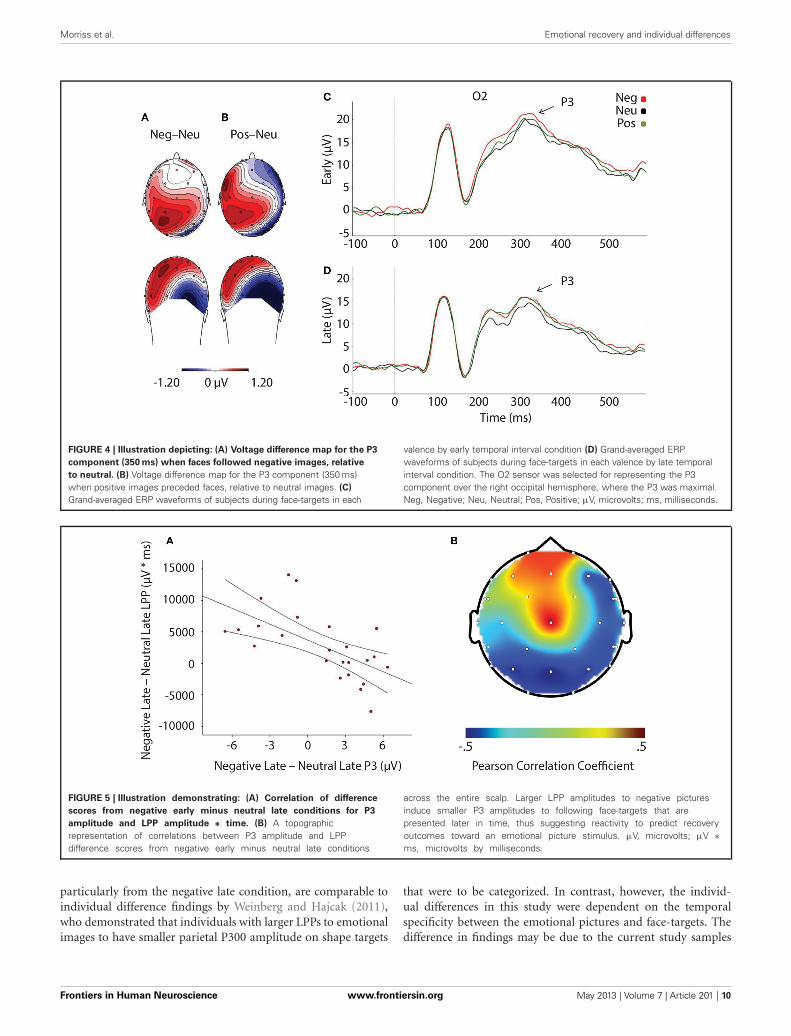
FIGURE 4 | Illustration depicting: (A) Voltage difference map for the P3
component (350 ms) when faces followed negative images, relative
to neutral. (B) Voltage difference map for the P3 component (350 ms)when positive images preceded faces, relative to neutral images. (C)
Grand-averaged ERP waveforms of subjects during face-targets in each
valence by early temporal interval condition (D) Grand-averaged ERPwaveforms of subjects during face-targets in each valence by late temporalinterval condition. The O2 sensor was selected for representing the P3component over the right occipital hemisphere, where the P3 was maximal.Neg, Negative; Neu, Neutral; Pos, Positive; µV, microvolts; ms, milliseconds.
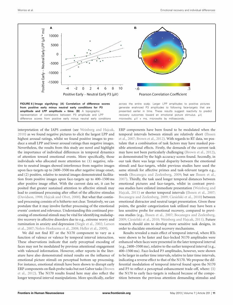
FIGURE 5 | Illustration demonstrating: (A) Correlation of difference
scores from negative early minus neutral late conditions for P3
amplitude and LPP amplitude ∗ time. (B) A topographicrepresentation of correlations between P3 amplitude and LPPdifference scores from negative early minus neutral late conditions
across the entire scalp. Larger LPP amplitudes to negative picturesinduce smaller P3 amplitudes to following face-targets that arepresented later in time, thus suggesting reactivity to predict recoveryoutcomes toward an emotional picture stimulus. µV, microvolts; µV ∗ms, microvolts by milliseconds.
particularly from the negative late condition, are comparable toindividual difference findings by Weinberg and Hajcak (2011),who demonstrated that individuals with larger LPPs to emotionalimages to have smaller parietal P300 amplitude on shape targets
that were to be categorized. In contrast, however, the individ-ual differences in this study were dependent on the temporalspecificity between the emotional pictures and face-targets. Thedifference in findings may be due to the current study samples
Frontiers in Human Neuroscience www.frontiersin.org May 2013 | Volume 7 | Article 201 | 10
Morriss et al. Emotional recovery and individual differences
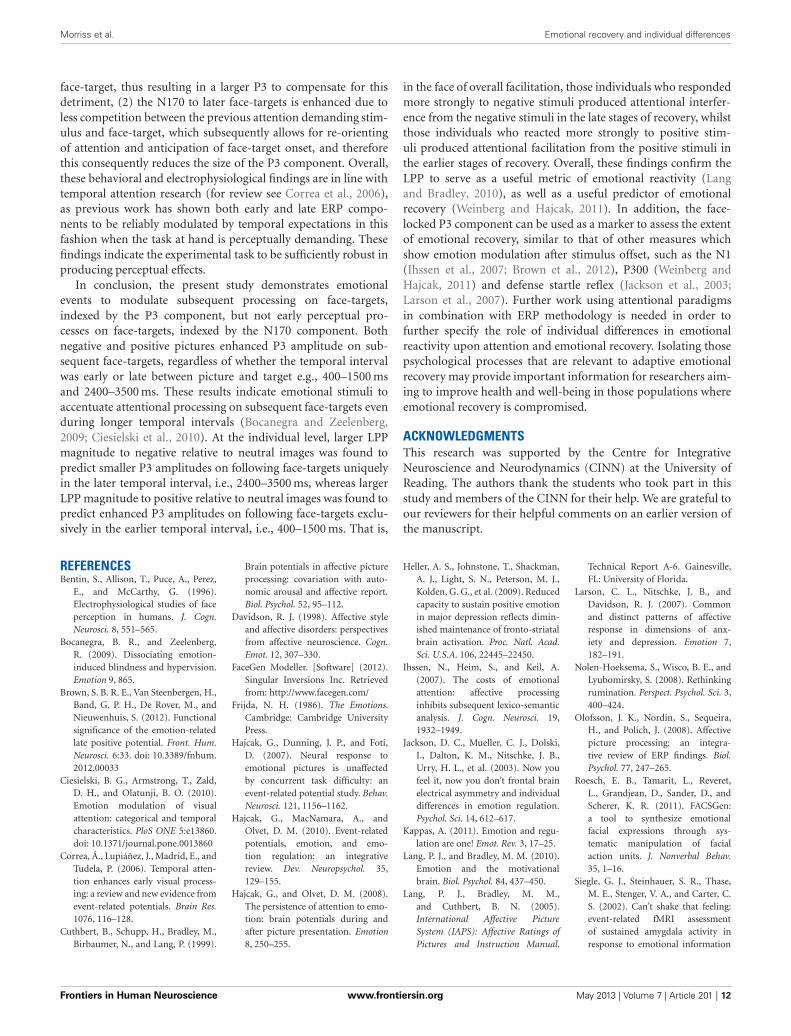
FIGURE 6 | Image signifying: (A) Correlation of difference scores
from positive early minus neutral early conditions for P3
amplitude and LPP amplitude ∗ time. (B) A topographicrepresentation of correlations between P3 amplitude and LPPdifference scores from positive early minus neutral early conditions
across the entire scalp. Larger LPP amplitudes to positive picturesgenerate enahnced P3 amplitudes to following face-targets that arepresented earlier in time. These results suggest reactivity to predictrecovery outcomes toward an emotional picture stimulus. µV,microvolts; µV ∗ ms, microvolts by milliseconds.
interpretation of the IAPS content (see Weinberg and Hajcak,2010) as we found negative pictures to elicit the largest LPP andhighest arousal ratings, whilst we found positive images to pro-duce a small LPP and lower arousal ratings than negative images.Nevertheless, the results from this study are novel and highlightthe importance of individual differences in temporal dynamicsof attention toward emotional events. More specifically, thoseindividuals who allocated more attention to: (1) negative, rela-tive to neutral images showed interference from negative imagesupon face-targets up to 2400–3500 ms after negative image onset,and (2) positive, relative to neutral images demonstrated facilita-tion from positive images upon face-targets up to 400–1500 msafter positive image offset. With the current data set, it can beposited that greater sustained attention to affective stimuli maylead to continued processing after offset of the affective stimulus(Davidson, 1998; Hajcak and Olvet, 2008). But what that contin-ued processing consists of is hitherto not clear. Tentatively, we canpostulate that it may involve further processing of the emotionalevents’ content and relevance. Understanding this continued pro-cessing of emotional stimuli may be vital for identifying maladap-tive recovery in affective disorders due to e.g., extreme worry andrumination in anxiety and depression (Siegle et al., 2002; Larsonet al., 2007; Nolen-Hoeksema et al., 2008; Heller et al., 2009).
We did not find RT or the N170 component to vary as afunction of valence or valence by temporal interval interaction.These observations indicate that early perceptual encoding offaces may not be modulated by previous attentional engagementwith valenced information. However, other reports in the liter-ature have also demonstrated mixed results on the influence ofemotional picture stimuli on perceptual bottom up processing.For instance, emotional stimuli have been shown to disrupt earlyERP components on flash probe tasks but not Gabor tasks (Brownet al., 2012). The N170 results found here may also reflect thelonger temporal interval manipulations. More specifically, earlier
ERP components have been found to be modulated when thetemporal intervals between stimuli are relatively short (Ihssenet al., 2007; Brown et al., 2012). With regards to RT data, we pos-tulate that a combination of task factors may have masked pos-sible attentional effects. Firstly, the demands of the current taskmay have not been particularly challenging (Brown et al., 2012),as demonstrated by the high accuracy scores found. Secondly, inour task there was large visual disparity between the emotionalstimuli and face-targets, whilst previous studies have used thesame stimuli for affective primes and task-relevant targets e.g.,words (Bocanegra and Zeelenberg, 2009; but see Ihssen et al.,2007). Thirdly, the task used longer temporal distances betweenemotional pictures and face-targets, whilst in contrast previ-ous studies have enlisted immediate presentation (Weinberg andHajcak, 2011) or shorter temporal intervals (Ihssen et al., 2007;Bocanegra and Zeelenberg, 2009; Ciesielski et al., 2010) betweenemotional distractor and neutral target presentation. Given thesepoints, the gender categorization task utilized may have been aless sensitive probe for emotional recovery, compared to previ-ous studies (e.g., Ihssen et al., 2007; Bocanegra and Zeelenberg,2009; Ciesielski et al., 2010; Weinberg and Hajcak, 2011). Futureresearch should aim to develop more sensitive task designs, inorder to elucidate emotional recovery mechanisms.
Results revealed a main effect of temporal interval, where RTswere shown to be faster and face-locked N170 amplitudes wereenhanced when faces were presented in the later temporal interval(e.g., 2400–3500 ms), relative to the earlier temporal interval (e.g.,400–1500 ms). Face-locked P3 amplitudes, however, were shownto be larger in earlier time intervals, relative to later time intervals,indicating a reverse effect to that of the N170. We propose the dif-ferential modulation of temporal interval found upon the N170and P3 to reflect a perceptual enhancement trade-off, where: (1)the N170 to early face-targets is reduced because of the compe-tition between the previous attention demanding stimulus and
Frontiers in Human Neuroscience www.frontiersin.org May 2013 | Volume 7 | Article 201 | 11
Morriss et al. Emotional recovery and individual differences
face-target, thus resulting in a larger P3 to compensate for thisdetriment, (2) the N170 to later face-targets is enhanced due toless competition between the previous attention demanding stim-ulus and face-target, which subsequently allows for re-orientingof attention and anticipation of face-target onset, and thereforethis consequently reduces the size of the P3 component. Overall,these behavioral and electrophysiological findings are in line withtemporal attention research (for review see Correa et al., 2006),as previous work has shown both early and late ERP compo-nents to be reliably modulated by temporal expectations in thisfashion when the task at hand is perceptually demanding. Thesefindings indicate the experimental task to be sufficiently robust inproducing perceptual effects.
In conclusion, the present study demonstrates emotionalevents to modulate subsequent processing on face-targets,indexed by the P3 component, but not early perceptual pro-cesses on face-targets, indexed by the N170 component. Bothnegative and positive pictures enhanced P3 amplitude on sub-sequent face-targets, regardless of whether the temporal intervalwas early or late between picture and target e.g., 400–1500 msand 2400–3500 ms. These results indicate emotional stimuli toaccentuate attentional processing on subsequent face-targets evenduring longer temporal intervals (Bocanegra and Zeelenberg,2009; Ciesielski et al., 2010). At the individual level, larger LPPmagnitude to negative relative to neutral images was found topredict smaller P3 amplitudes on following face-targets uniquelyin the later temporal interval, i.e., 2400–3500 ms, whereas largerLPP magnitude to positive relative to neutral images was found topredict enhanced P3 amplitudes on following face-targets exclu-sively in the earlier temporal interval, i.e., 400–1500 ms. That is,
in the face of overall facilitation, those individuals who respondedmore strongly to negative stimuli produced attentional interfer-ence from the negative stimuli in the late stages of recovery, whilstthose individuals who reacted more strongly to positive stim-uli produced attentional facilitation from the positive stimuli inthe earlier stages of recovery. Overall, these findings confirm theLPP to serve as a useful metric of emotional reactivity (Langand Bradley, 2010), as well as a useful predictor of emotionalrecovery (Weinberg and Hajcak, 2011). In addition, the face-locked P3 component can be used as a marker to assess the extentof emotional recovery, similar to that of other measures whichshow emotion modulation after stimulus offset, such as the N1(Ihssen et al., 2007; Brown et al., 2012), P300 (Weinberg andHajcak, 2011) and defense startle reflex (Jackson et al., 2003;Larson et al., 2007). Further work using attentional paradigmsin combination with ERP methodology is needed in order tofurther specify the role of individual differences in emotionalreactivity upon attention and emotional recovery. Isolating thosepsychological processes that are relevant to adaptive emotionalrecovery may provide important information for researchers aim-ing to improve health and well-being in those populations whereemotional recovery is compromised.
ACKNOWLEDGMENTSThis research was supported by the Centre for IntegrativeNeuroscience and Neurodynamics (CINN) at the University ofReading. The authors thank the students who took part in thisstudy and members of the CINN for their help. We are grateful toour reviewers for their helpful comments on an earlier version ofthe manuscript.
REFERENCESBentin, S., Allison, T., Puce, A., Perez,
E., and McCarthy, G. (1996).Electrophysiological studies of faceperception in humans. J. Cogn.Neurosci. 8, 551–565.
Bocanegra, B. R., and Zeelenberg,R. (2009). Dissociating emotion-induced blindness and hypervision.Emotion 9, 865.
Brown, S. B. R. E., Van Steenbergen, H.,Band, G. P. H., De Rover, M., andNieuwenhuis, S. (2012). Functionalsignificance of the emotion-relatedlate positive potential. Front. Hum.Neurosci. 6:33. doi: 10.3389/fnhum.2012.00033
Ciesielski, B. G., Armstrong, T., Zald,D. H., and Olatunji, B. O. (2010).Emotion modulation of visualattention: categorical and temporalcharacteristics. PloS ONE 5:e13860.doi: 10.1371/journal.pone.0013860
Correa, Á., Lupiáñez, J., Madrid, E., andTudela, P. (2006). Temporal atten-tion enhances early visual process-ing: a review and new evidence fromevent-related potentials. Brain Res.1076, 116–128.
Cuthbert, B., Schupp, H., Bradley, M.,Birbaumer, N., and Lang, P. (1999).
Brain potentials in affective pictureprocessing: covariation with auto-nomic arousal and affective report.Biol. Psychol. 52, 95–112.
Davidson, R. J. (1998). Affective styleand affective disorders: perspectivesfrom affective neuroscience. Cogn.Emot. 12, 307–330.
FaceGen Modeller. [Software] (2012).Singular Inversions Inc. Retrievedfrom: http://www.facegen.com/
Frijda, N. H. (1986). The Emotions.Cambridge: Cambridge UniversityPress.
Hajcak, G., Dunning, J. P., and Foti,D. (2007). Neural response toemotional pictures is unaffectedby concurrent task difficulty: anevent-related potential study. Behav.Neurosci. 121, 1156–1162.
Hajcak, G., MacNamara, A., andOlvet, D. M. (2010). Event-relatedpotentials, emotion, and emo-tion regulation: an integrativereview. Dev. Neuropsychol. 35,129–155.
Hajcak, G., and Olvet, D. M. (2008).The persistence of attention to emo-tion: brain potentials during andafter picture presentation. Emotion8, 250–255.
Heller, A. S., Johnstone, T., Shackman,A. J., Light, S. N., Peterson, M. J.,Kolden, G. G., et al. (2009). Reducedcapacity to sustain positive emotionin major depression reflects dimin-ished maintenance of fronto-striatalbrain activation. Proc. Natl. Acad.Sci. U.S.A. 106, 22445–22450.
Ihssen, N., Heim, S., and Keil, A.(2007). The costs of emotionalattention: affective processinginhibits subsequent lexico-semanticanalysis. J. Cogn. Neurosci. 19,1932–1949.
Jackson, D. C., Mueller, C. J., Dolski,I., Dalton, K. M., Nitschke, J. B.,Urry, H. L., et al. (2003). Now youfeel it, now you don’t frontal brainelectrical asymmetry and individualdifferences in emotion regulation.Psychol. Sci. 14, 612–617.
Kappas, A. (2011). Emotion and regu-lation are one! Emot. Rev. 3, 17–25.
Lang, P. J., and Bradley, M. M. (2010).Emotion and the motivationalbrain. Biol. Psychol. 84, 437–450.
Lang, P. J., Bradley, M. M.,and Cuthbert, B. N. (2005).International Affective PictureSystem (IAPS): Affective Ratings ofPictures and Instruction Manual.
Technical Report A-6. Gainesville,FL: University of Florida.
Larson, C. L., Nitschke, J. B., andDavidson, R. J. (2007). Commonand distinct patterns of affectiveresponse in dimensions of anx-iety and depression. Emotion 7,182–191.
Nolen-Hoeksema, S., Wisco, B. E., andLyubomirsky, S. (2008). Rethinkingrumination. Perspect. Psychol. Sci. 3,400–424.
Olofsson, J. K., Nordin, S., Sequeira,H., and Polich, J. (2008). Affectivepicture processing: an integra-tive review of ERP findings. Biol.Psychol. 77, 247–265.
Roesch, E. B., Tamarit, L., Reveret,L., Grandjean, D., Sander, D., andScherer, K. R. (2011). FACSGen:a tool to synthesize emotionalfacial expressions through sys-tematic manipulation of facialaction units. J. Nonverbal Behav.35, 1–16.
Siegle, G. J., Steinhauer, S. R., Thase,M. E., Stenger, V. A., and Carter, C.S. (2002). Can’t shake that feeling:event-related fMRI assessmentof sustained amygdala activity inresponse to emotional information
Frontiers in Human Neuroscience www.frontiersin.org May 2013 | Volume 7 | Article 201 | 12

Morriss et al. Emotional recovery and individual differences
in depressed individuals. Biol.Psychiatry 51, 693–707.
Schupp, H., Cuthbert, B., Bradley,M., Hillman, C., Hamm, A., andLang, P. (2004). Brain processesin emotional perception: moti-vated attention. Cogn. Emot. 18,593–611.
Schupp, H. T., Cuthbert, B. N., Bradley,M. M., Cacioppo, J. T., Ito, T.,and Lang, P. J. (2000). Affectivepicture processing: the late posi-tive potential is modulated by moti-vational relevance. Psychophysiology37, 257–261.
van Reekum, C. M., Urry, H. L.,Johnstone, T., Thurow, M. E., Frye,
C. J., Jackson, C. A., et al. (2007).Individual differences in amygdalaand ventromedial prefrontal cor-tex activity are associated withevaluation speed and psychologicalwell-being. J. Cogn. Neurosci. 19,237–248.
Weinberg, A., and Hajcak, G. (2010).Beyond good and evil: implicationsof examining electrocortical activityelicited by specific picture content.Emotion 10, 767–782.
Weinberg, A., and Hajcak, G. (2011).The late positive potential predictssubsequent interference with targetprocessing. J. Cogn. Neurosci. 23,2994–3007.
Yiend, J. (2010). The effects of emo-tion on attention: a review ofattentional processing of emo-tional information. Cogn. Emot.24, 3–47.
Conflict of Interest Statement: Theauthors declare that the researchwas conducted in the absence of anycommercial or financial relationshipsthat could be construed as a potentialconflict of interest.
Received: 10 January 2013; accepted: 29April 2013; published online: 21 May2013.
Citation: Morriss J, Taylor ANW, RoeschEB and van Reekum CM (2013) Stillfeeling it: the time course of emotionalrecovery from an attentional perspec-tive. Front. Hum. Neurosci. 7:201. doi:10.3389/fnhum.2013.00201Copyright © 2013 Morriss, Taylor,Roesch and van Reekum. This is anopen-access article distributed underthe terms of the Creative CommonsAttribution License, which permitsuse, distribution and reproduc-tion in other forums, provided theoriginal authors and source are cred-ited and subject to any copyrightnotices concerning any third-partygraphics etc.
Frontiers in Human Neuroscience www.frontiersin.org May 2013 | Volume 7 | Article 201 | 13
Related Documents