1 Stickler syndrome caused by COL2A1 mutations: genotype-phenotype correlation 1 in a series of 100 patients 2 3 Kristien P Hoornaert 1 , Inge Vereecke 1 , Chantal Dewinter 1 , Thomas Rosenberg 2 , Frits A 4 Beemer 3 , Jules G Leroy 1 , Laila Bendix 4 , Erik Björck 5 , Maryse Bonduelle 6 , Odile 5 Boute 7 , Valerie Cormier-Daire 8 , Christine De Die-Smulders 9 , Anne Dieux-Coeslier 7 , 6 Hélène Dollfus 10 , Mariet Elting 11 , Andrew Green 12, 13 , Veronica I Guerci 14 , Raoul CM 7 Hennekam 15, 16 , Yvonne Hilhorts-Hofstee 17 , Muriel Holder 7 , Carel Hoyng 18 , Kristi J 8 Jones 19, 20 , Dragana Josifova 21 , Ilkka Kaitila 22 , Suzanne Kjaergaard 23 , Yolande H 9 Kroes 3 , Kristina Lagerstedt 5 , Melissa Lees 15 , Martine LeMerrer 8 , Cinzia Magnani 24 , 10 Carlo Marcelis 25 , Loreto Martorell 26 , Michèle Mathieu 27 , Meriel McEntagart 28 , Angela 11 Mendicino 29 , Jenny Morton 30 , Gabrielli Orazio 31 , Véronique Paquis 32 , Orit Reish 33 , 12 Kalle O. J. Simola 34 , Sarah F Smithson 35 , Karen I Temple 36 , Elisabeth Van Aken 37 , 13 Yolande Van Bever 38 , Jenneke van den Ende 39 , Johanna M Van Hagen 40 , Leopoldo 14 Zelante 41 , Riina Zordania 42 , Anne De Paepe 1 , Bart P Leroy 1, 37 , Marc De Buyzere 43 , 15 Paul J Coucke 1 , Geert R Mortier 1 16 17 1 Center for Medical Genetics, Ghent University Hospital, Gent, Belgium 18 2 National Eye Clinic, Gordon Norrie Centre for Genetic Eye Diseases, Hellerup, 19 Denmark 20 3 Center for Medical Genetics, Utrecht, Netherlands 21 4 Department of Clinical Genetics, University of Southern Denmark, Vejle Hospital, 22 Denmark 23 5 Department of Clinical Genetics, Karolinska University Hospital, Stockholm, Sweden 24 peer-00511182, version 1 - 24 Aug 2010 Author manuscript, published in "European Journal of Human Genetics n/a, n/a (2010) n/a-n/a" DOI : 10.1038/ejhg.2010.23

Welcome message from author
This document is posted to help you gain knowledge. Please leave a comment to let me know what you think about it! Share it to your friends and learn new things together.
Transcript
1
Stickler syndrome caused by COL2A1 mutations: genotype-phenotype correlation 1
in a series of 100 patients 2
3
Kristien P Hoornaert1, Inge Vereecke1, Chantal Dewinter1, Thomas Rosenberg2, Frits A 4
Beemer3, Jules G Leroy1, Laila Bendix4, Erik Björck5, Maryse Bonduelle6, Odile 5
Boute7, Valerie Cormier-Daire8, Christine De Die-Smulders9, Anne Dieux-Coeslier7, 6
Hélène Dollfus10, Mariet Elting11, Andrew Green12, 13, Veronica I Guerci14, Raoul CM 7
Hennekam15, 16, Yvonne Hilhorts-Hofstee17, Muriel Holder7, Carel Hoyng18, Kristi J 8
Jones19, 20, Dragana Josifova21, Ilkka Kaitila22, Suzanne Kjaergaard23, Yolande H 9
Kroes3, Kristina Lagerstedt5, Melissa Lees15, Martine LeMerrer8, Cinzia Magnani24, 10
Carlo Marcelis25, Loreto Martorell26, Michèle Mathieu27, Meriel McEntagart28, Angela 11
Mendicino29, Jenny Morton30, Gabrielli Orazio31, Véronique Paquis32, Orit Reish33, 12
Kalle O. J. Simola34, Sarah F Smithson35, Karen I Temple36, Elisabeth Van Aken37, 13
Yolande Van Bever38, Jenneke van den Ende39, Johanna M Van Hagen40, Leopoldo 14
Zelante41, Riina Zordania42, Anne De Paepe1, Bart P Leroy1, 37, Marc De Buyzere43, 15
Paul J Coucke1, Geert R Mortier1 16
17
1 Center for Medical Genetics, Ghent University Hospital, Gent, Belgium 18
2 National Eye Clinic, Gordon Norrie Centre for Genetic Eye Diseases, Hellerup, 19
Denmark 20
3 Center for Medical Genetics, Utrecht, Netherlands 21
4 Department of Clinical Genetics, University of Southern Denmark, Vejle Hospital, 22
Denmark 23
5 Department of Clinical Genetics, Karolinska University Hospital, Stockholm, Sweden 24
peer
-005
1118
2, v
ersi
on 1
- 24
Aug
201
0Author manuscript, published in "European Journal of Human Genetics n/a, n/a (2010) n/a-n/a"
DOI : 10.1038/ejhg.2010.23

2
6 Center for Medical Genetics, UZ Brussel, Belgium 25
7 Center for Clinical Genetics, Hopital Jeanne de Flandre, Lille, France 26
8 Department of Medical Genetics, Université Paris Descartes, Necker Enfants Malades 27
Hospital, Paris, France 28
9 Department of Clinical Genetics, University Hospital Maastricht, Maastricht, The 29
Netherlands 30
10 Centre de reference pour les Affections Rares et Génétique Ophthalmologique 31
(CARGO), Hôpitaux Universitaires de Strasbourg, France 32
11 Department of Clinical Genetics, VU University Medical Center, Amsterdam, The 33
Netherlands 34
12 National Centre for Medical Genetics Our Lady’s Hospital, Dublin, Ireland 35
13 University College Dublin School of Medicine and Medical Science 36
14 Metabolic Disease Unit, IRCCS Burlo Garofolo, Trieste, Italy 37
15 Clinical and Molecular Genetics Unit, Institute of Child Health, University College 38
London, UK 39
16 Department of Pediatrics, Academic Medical Center, University of Amsterdam, 40
Amsterdam, The Netherlands 41
17 Department of Clinical Genetics, Leiden University Medical Center, Leiden, the 42
Netherlands 43
18 Department of Ophthalmology, Radboud University Nijmegen Medical Centre, 44
Nijmegen, The Netherlands 45
19 Western Sydney Genetics Program, The Children’s Hospital at Westmead, Sydney, 46
Australia 47
20 Discipline of Paediatrics and Child Health, University of Sydney, Sydney, Australia 48
peer
-005
1118
2, v
ersi
on 1
- 24
Aug
201
0

3
21 Department of Clinical Genetics, Guy’s Hospital, London, UK 49
22 Department of Clinical Genetics, Helsinki University Central Hospital, Hus, Finland 50
23 Department of Clinical Genetics, Rigshospitalet, Copenhagen, Denmark 51
24 Department of Paediatrics, University Hospital of Parma, Italy 52
25 Department of Human Genetics, Radboud University Medical Centre Nijmegen, the 53
Netherlands 54
26 Department of Genetics, Hospital Sant Joan de Déu, Barcelona, Spain 55
27 Clinical Genetics, CHU Nord, Amiens, France 56
28 Clinical Genetics, St George's University of London, London, UK 57
29 U.O.S. di Genetica, DTMI, ASL RME, Roma, Italy 58
30 Clinical Genetics Unit, Birmingham Women’s Hospital, Birmingham, UK 59
31 Department of Clinical Genetics, Ospedale G. Salesi, Ancona, Italy 60
32 Department of Clinical Genetics, Hopital de L’Arghet, Nice, France 61
33 Genetics Institute, Assaf Harofeh Medical Center, Zerifin, Israel 62
34 Department of Pediatrics, Tampere University Hospital, Tampere, Finland 63
35 Department for Clinical Genetics, St. Michael’s Hospital, Bristol, UK 64
36 Academic Unit of Genetic Medicine, Division of Human Genetics, University of 65
Southampton, Southampton, UK 66
37 Department of Ophthalmology, Ghent University Hospital, Gent, Belgium 67
38 Department of Clinical Genetics, Erasmus MC, Rotterdam, the Netherlands 68
39 Center for Medical Genetics, University Hospital, Antwerp, Belgium 69
40 Department of Clinical Genetics, University Medical Center, Amsterdam, the 70
Netherlands 71
peer
-005
1118
2, v
ersi
on 1
- 24
Aug
201
0

4
41 Medical Genetics Service, IRCCS Casa Sollievo della Sofferenza, San Giovanni 72
Rotondo, Italy 73
42 Clinical Genetics, Tallinn Children’s Hospital, Tallinn, Estonia 74
43 Heart Center, Ghent University Hospital, Gent, Belgium 75
76
Correspondence: Geert Mortier, MD, PhD 77
Center for Medical Genetics 78
Ghent University Hospital 79
De Pintelaan 185, B-9000 Ghent, Belgium 80
Tel 0032 9 332 36 03 81
Fax 0032 9 332 49 70 82
email: [email protected] 83
84
85
Keywords: COL2A1, Stickler syndrome, genotype-phenotype correlation, 86
type II collagenopathies, splice site mutation 87
88 peer
-005
1118
2, v
ersi
on 1
- 24
Aug
201
0

5
ABSTRACT 89
Stickler syndrome is an autosomal dominant connective tissue disorder caused by 90
mutations in different collagen genes. The aim of our study was to define more 91
precisely the phenotype and genotype of Stickler syndrome type 1 by investigating a 92
large series of patients with a heterozygous mutation in COL2A1. In 188 probands with 93
the clinical diagnosis of Stickler syndrome, the COL2A1 gene was analysed by either a 94
mutation scanning technique or bidirectional fluorescent DNA sequencing. The effect of 95
splice site alterations was investigated by analysing mRNA. MLPA analysis was used 96
for the detection of intragenic deletions. We identified 77 different COL2A1 mutations 97
in 100 affected individuals. Analysis of the splice site mutations revealed unusual RNA 98
isoforms, most of which contained a premature stop codon. Vitreous anomalies and 99
retinal detachments were found more frequently in patients with a COL2A1 mutation 100
compared to the mutation negative group (p<0.01). 20 of the 23 sporadic patients with a 101
COL2A1 mutation had either a cleft palate or retinal detachment with vitreous 102
anomalies. The presence of vitreous anomalies, retinal tears or detachments, cleft palate 103
and a positive family history were shown to be good indicators for a COL2A1 defect. In 104
conclusion, we confirm that Stickler syndrome type 1 is predominantly caused by loss-105
of-function mutations in the COL2A1 gene since more than 90% of the mutations were 106
predicted to result in nonsense mediated decay. Based on binary regression analysis we 107
developed a scoring system that may be useful when evaluating patients with Stickler 108
syndrome. 109
110
111
112
peer
-005
1118
2, v
ersi
on 1
- 24
Aug
201
0

6
INTRODUCTION 113
Stickler syndrome (MIM# 108300) is a connective tissue disorder first described 114
by Stickler et al in 1965. It is characterized by ocular, orofacial, auditory and skeletal 115
manifestations with considerable intra- and interfamilial variability.[1, 2] The incidence 116
is estimated to range between 1 in 7,500 to 1 in 9,000 newborns 117
(http://ghr.nlm.nih.gov/condition%3Dsticklersyndrome US Dept of Health). The most 118
characteristic ocular features are congenital myopia, vitreous alterations, cataract, 119
glaucoma and a high risk of spontaneous retinal detachments. The orofacial changes 120
include cleft palate, midfacial hypoplasia, low nasal bridge and micrognathia. Joint pain 121
is common in childhood and osteoarthrosis may be apparent from the third or fourth 122
decade. Radiographs may show signs of a spondyloepiphyseal dysplasia. Mild 123
sensorineural hearing loss, mainly for the high tones, can be present in Stickler 124
syndrome type 1 (COL2A1 gene), more severe sensorineural hearing loss is usually 125
found in the other types of Stickler syndrome.[3, 4] 126
At present, at least 3 types of autosomal dominant Stickler syndrome have been 127
discerned. A correlation between these different types and their accompanying vitreous 128
anomalies has been suggested.[5] ‘Membranous’ or type 1 vitreous has been associated 129
with Stickler syndrome type 1 caused by heterozygous mutations in the COL2A1 gene 130
(MIM# 108300).[6] Type 2 or ‘beaded’ vitreous is mainly found in patients with 131
Stickler syndrome type 2 which is due to a heterozygous mutation in the COL11A1 gene 132
(MIM# 604841).[3, 7] Stickler syndrome type 3 or ‘non-ocular Stickler syndrome’ 133
refers to the phenotype of patients with a mutation in the COL11A2 gene that is not 134
expressed in the eye (MIM# 184840).[8] In addition to the different types of autosomal 135
peer
-005
1118
2, v
ersi
on 1
- 24
Aug
201
0

7
dominant Stickler syndrome, recently also a recessive form of Stickler syndrome, 136
caused by a mutation in the COL9A1 gene, has been described (MIM# 120210).[9] 137
Stickler syndrome type 1 is the most common form. The majority of COL2A1 138
mutations identified in patients with Stickler syndrome type 1 are predicted to result in 139
nonsense mediated decay (NMD). On the other hand, missense mutations (usually 140
glycine substitutions) in COL2A1 usually result in short stature disorders such as 141
achondrogenesis type II/hypochondrogenesis, spondyloepiphyseal dysplasia (SEDC), 142
Kniest dysplasia, spondyloperipheral dysplasia (SPD) and Torrance dysplasia (MIM# 143
200610, 183900, 156550, 271700, 151210).[5, 10, 11] 144
The aim of this study was to define more precisely the phenotype and genotype 145
of Stickler syndrome type 1 by investigating a large series of Stickler syndrome patients 146
with a heterozygous mutation in the COL2A1 gene. 147
148
peer
-005
1118
2, v
ersi
on 1
- 24
Aug
201
0

8
MATERIALS & METHODS 149
Evaluation of phenotype 150
Over the past 10 years, blood or DNA samples from 278 individuals were 151
referred for mutation analysis of the COL2A1 gene in order to confirm or exclude the 152
clinical diagnosis of Stickler syndrome. 153
Information on clinical and radiographic features of each patient was requested 154
by using a specific questionnaire (Supplementary Table 1). Ninety patients were 155
excluded from the study because insufficient clinical data were available (in 11 of those 156
patients a COL2A1 mutation was identified). Each patient in the group of 188 remaining 157
subjects had two or more of the following features reminiscent of Stickler syndrome: 158
myopia, spontaneous retinal detachment, cleft palate, sensorineural hearing loss and 159
arthropathy. Informed consent was obtained from each enrolled patient. 160
Analysis of genomic DNA 161
Genomic DNA was extracted from blood samples by standard procedures, 162
followed by touchdown PCR amplification of the COL2A1 gene using forward and 163
reverse primers located in the flanking introns. The PCR products were analysed by gel 164
electrophoresis and visualized by ethidium bromide staining on 2% agarose gels. 165
Mutation screening was performed by SSCP and CSGE (period 1997-2002) or 166
by DHPLC analysis (period 2003-2006) using the WAVE DNA fragment analysis 167
system (Transgenomic, Cheshire, UK).[12, 13, 14] All fragments showing an aberrant 168
pattern were directly sequenced on the ABI PRISM 3730 automated sequencer (Applied 169
Biosystems, Foster City, CA) using the BigDye terminator cycle sequencing chemistry. 170
From 2007 on, direct sequencing of all 54 exons was performed. These obtained 171
sequences were compared to the wild-type sequence as submitted to GenBank 172
peer
-005
1118
2, v
ersi
on 1
- 24
Aug
201
0
9
Accession number NM_001844. The nucleotides were numbered starting from the first 173
base of the start codon (ATG) of the cDNA reference sequence. Amino acid residues 174
were numbered from the first methionine (start codon for translation) of the procollagen 175
α1(II)-chain (GenBank Accession number L10347). 176
RNA studies 177
In patients with a splice site mutation, an EBV cell line or skin biopsy was 178
requested for analysis of mRNA splicing. In order to stabilize mutant COL2A1 mRNA, 179
cycloheximide (Sigma, www.sigmaaldrich.com) was added to the cultures, followed by 180
mRNA isolation and cDNA preparation. Nested PCR was used to obtain sufficient PCR 181
fragments for direct sequencing. 182
MLPA analysis 183
Multiplex ligation-dependent amplification (MLPA) was set up, following the 184
directions provided by the manufacturer (MRC Holland, Amsterdam, The Netherlands) 185
(www.MPLA.com).[15] The probe set for COL2A1 (SALSA MLPA kit P214) covering 186
exons 1, 4, 6, 8, 10, 16, 17, 19, 20, 24, 27, 29, 31, 35, 39, 43, 46, 49, 51 and 54 was 187
used. 188
Binary logistic regression analysis/statistics 189
The formula for the proposed scoring system was developed using binary (mutation 190
positive or not) logistic regression analysis.[16, 17, 18] The parameters tested in the 191
model comprised: vitreous abnormalities, retinal abnormalities, flat face, micrognathia, 192
retinal tear and/or detachment, cataract, low nasal bridge, cleft palate, positive family 193
history, myopia, conductive hearing loss, premature arthropathy, hypermobility, 194
epiphyseal dysplasia on X-rays and sensorineural hearing loss. The weight (score) for 195
each characteristic in the scoring system was proportional to its regression coefficient in 196
peer
-005
1118
2, v
ersi
on 1
- 24
Aug
201
0

10
the model. To simplify the scoring system the scores were rounded to positive integers 197
and the scores of the characteristics with lowest significant regression coefficients were 198
conventionally given a score value of one and the intercept of the linear predictor was 199
neglected. Otherwise, no recalibration, shrinkage factor or model revision or extension 200
seemed to be needed to study the whole study population. Calibration of the scoring 201
system was further evaluated with the Hosmer-Lemeshow test. The clinical applicability 202
of the obtained score was evaluated for several thresholds using conventional receiver 203
operating characteristics (positive and negative predictive value, sensitivity and 204
specificity). All statistical analyses were performed with SPSS 15.0 for Windows (SPSS 205
Inc. Chicago, IL). 206
207
peer
-005
1118
2, v
ersi
on 1
- 24
Aug
201
0

11
RESULTS 208
In 100/188 individuals referred with a potential diagnosis of Stickler syndrome, 209
a heterozygous COL2A1 mutation was identified. This panel of 77 different mutations 210
included 1 deletion of the entire gene [19], 13 nonsense mutations, 21 deletions, 1 211
insertion, 9 duplications, 2 combinations of an insertion and a deletion, 22 splice site 212
alterations, 1 synonymous mutation, 2 missense mutations resulting in an arginine-to-213
cysteine substitution [20] and 5 missense mutations substituting a glycine residue in the 214
triple helical domain of the protein. The mutations were distributed over the entire gene 215
and no hot spot regions were apparent (Table 1). Thirteen mutations were observed in 216
more than one proband: c.625C>T, p.Arg209X and c.1833+1G>A, p.GlyfsX619 were 217
found in four patients each; c.3106C>T, p.Arg1036X occurred five times (Table 1). 218
Two mutations were located in the alternatively spliced exon 2. The first one, a 219
duplication of 23 nucleotides (c.211_233dup; p.Glu79ThrfsX2) causes a frameshift that 220
leads to a premature stop codon within the exon itself. The patient with this mutation 221
only had ocular features (retinal detachment) of Stickler syndrome as expected since 222
exon 2 is retained in the eye but spliced out in the cartilage.[21, 22, 23] The second 223
patient had the deletion c.264_276del; p.Cys89SerfsX24 that causes a frameshift with a 224
premature stop codon in exon 3. However, this patient had both ocular and extra-ocular 225
manifestations of Stickler syndrome including vitreal abnormalities, a retinal 226
detachment, flat face, sensorineural hearing loss, arthropathy and epiphyseal changes on 227
radiographs. 228
In the skin fibroblasts or the EBV cell line available from 13 patients with 12 229
different splice site alterations, cDNA analysis showed that each splice site alteration 230
resulted in a premature stop codon (data not shown). For the three splice site mutations, 231
peer
-005
1118
2, v
ersi
on 1
- 24
Aug
201
0

12
multiple isoforms of mRNA were detected. In each case at least one isoform harboured 232
a premature stop codon (Supplementary Figure 1: isoforms A3, B2 and C2). In the 233
additional isoforms A1, B1 and C1 only skipping of the adjacent exon was observed. In 234
the C1 isoform skipping of even three consecutive exons (51-53) had occurred. These 235
exons constitute the carboxypropeptide of the procollagen α1(II)-chain, which is 236
necessary for chain association and initiation of the triple helix formation.[24] 237
Consequently, the resulting truncated protein will most likely be lost and not 238
incorporated into the collagen trimer. In the isoform A2, exon 7 was deleted but intron 239
5 retained, the latter containing an in-frame stop codon. Both patients harbouring the 240
c.430-1G>C and c.4074+1G>T splice site mutation suffered from myopia, vitreoretinal 241
abnormalities and spontaneous retinal detachments. They also showed a flat face. The 242
individual with the c.3003+5G>A splice site mutation was born with a Pierre-Robin 243
anomaly and had myopia, a retinal detachment and cataract. He also suffered from 244
conductive hearing loss. His affected father had a history of spontaneous bilateral retinal 245
detachments in childhood. 246
One patient was heterozygous for a synonymous mutation (c.2862C>T; 247
p.Gly954Gly) in exon 42. Since this mutation was cosegregating with Stickler 248
syndrome in the affected family, the pathogenic effect was further explored at the 249
mRNA level. cDNA analysis showed that this mutation generated a cryptic splice site 250
35 nucleotides upstream of the normal donor splice site in intron 42, resulting in a 251
frameshift with a premature stop codon (Supplementary Figure 2). 252
The nonsense (p.Trp1293X) and frameshift mutations (p.Cys1289ProfsX3, 253
p.Ile1300ThrfsX15, p.Asn1303ThrfsX9) residing in the carboxypropeptide were 254
predicted to result in NMD since they occur before the last 50 nucleotides of the last 255
peer
-005
1118
2, v
ersi
on 1
- 24
Aug
201
0

13
exon-exon junction (Table 1).[25] In the patient with the splice site alteration in intron 256
53 (c.4317+2T>C), the splice site prediction program 257
(http://www.fruitfly.org/seq_tools/splice.html) computed an insertion of a part of the 258
intron 53 containing an in-frame stop codon. 259
Since only 100 mutations were identified in a series of 188 patients, we decided 260
to expand the molecular analysis with MLPA to explore the possibility of missed 261
intragenic deletions. For this analysis we selected 20 patients in whom we strongly 262
suspected the diagnosis of Stickler syndrome because of the presence of severe myopia, 263
retinal detachment and/or cleft palate. However, no additional mutations were identified 264
in these affected individuals. 265
In a next step we evaluated the clinical and radiographic features in our series of 266
188 patients and looked for differences between the mutation positive (n=100) and 267
mutation negative (n=88) group. The results are summarized in Figure 1. A positive 268
family history, orofacial anomalies (cleft palate, low nasal bridge, flat face, 269
micrognathia) and vitreoretinal changes were more frequently (p value ≤ 0.05) present 270
in the mutation positive group. On the other hand, sensorineural hearing loss was 271
observed more frequently in the mutation negative group (p<0.005). 20/23 of the 272
sporadic patients with a COL2A1 mutation had either a cleft palate or retinal 273
detachment(s) with vitreous anomalies and myopia. 274
To determine the discriminating power of these features, we performed a binary 275
logistic regression analysis. The following characteristics were most distinguishing 276
between both groups : a) vitreous abnormalities, b) retinal abnormalities, c) history of 277
retinal tear and/or detachment, d) low nasal bridge, e) cleft palate, f) micrognathia and 278
g) positive family history (Figure 2). Based on the regression coefficient of each 279
peer
-005
1118
2, v
ersi
on 1
- 24
Aug
201
0

14
distinguishing characteristic, a specific scoring system was proposed. The highest score 280
(score 5) was attributed to retinal abnormalities and positive family history, a score of 4 281
was assigned to cleft palate and vitreous abnormalities, a retinal tear and/or detachment 282
represented a score of 3, whereas low nasal bridge and micrognathia received the lowest 283
score (score 1) (Table 2). When applying this scoring system to each patient, we 284
observed a higher median score for patients with a COL2A1 mutation compared to those 285
without a mutation (11.5 versus 6). The calculated score ranges from 0 to 21 with a 286
theoretical maximum of 23. The distribution of the score for mutation positive and 287
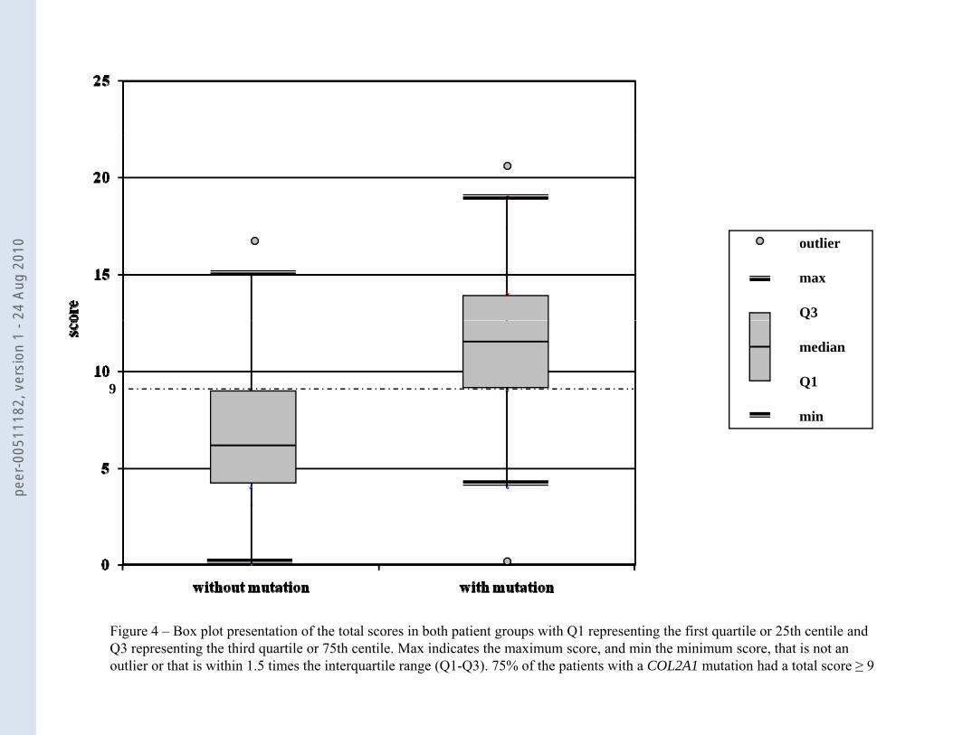
mutation negative cases is shown in Figure 3. 75% of the patients with a COL2A1 288
mutation had a total score ≥ 9 (Figure 4). The presence of vitreoretinal anomalies and a 289
retinal detachment yields a total score of 12, illustrating the importance of a thorough 290
ophthalmological evaluation in patients with Stickler syndrome. 291
292
peer
-005
1118
2, v
ersi
on 1
- 24
Aug
201
0

15
DISCUSSION 293
The past decade we have identified a large series of COL2A1 mutations in a 294
group of patients referred with the diagnosis of Stickler syndrome. The availability of 295
these data prompted us to retrospectively analyse both genotype and phenotype of these 296
patients. With this study we aimed to define more precisely the phenotype of Stickler 297
syndrome type 1 and were interested in identifying discriminating features between 298
patients with and those without a COL2A1 mutation. In addition, we wanted to 299
investigate in what respect Stickler syndrome type 1 mutations were different from 300
other COL2A1 mutations causing the type II collagenopathies with short stature. More 301
precisely, we wanted to learn if all Stickler syndrome mutations were predicted to have 302
a loss-of-function effect on the procollagen α1(II)-chain. 303
Sufficient clinical and radiographic data were available on 188 probands and in 304
100 of these individuals a heterozygous COL2A1 mutation was identified. The 77 305
different mutations were distributed over the entire gene and no regions of mutation 306
clustering were found. Thirteen mutations were observed in more than one proband, 307
with 10 involving a CpG dinucleotide. One patient was heterozygous for a deletion of 308
the entire gene and details have been published earlier.[19] The 34 smaller and 309
intragenic deletions, insertions, duplications and insertion-deletions were all out of 310
frame and therefore predicted to result in NMD. A similar effect was demonstrated for 311
the synonymous mutation (p.Gly954Gly) which created a cryptic splice site 312
(Supplementary Figure 2). This mutation is the second example of an apparently silent 313
COL2A1 mutation that alters RNA splicing, illustrating the importance of studying the 314
effect of so-called synonymous mutations at the mRNA level.[26] Analysis of cDNA 315
also allowed us to study the effect of 12 different splice site alterations. In addition, it 316
peer
-005
1118
2, v
ersi
on 1
- 24
Aug
201
0

16
gave us more insights into the complexity of mRNA splicing of the COL2A1 gene. Each 317
splice site mutation was shown to create at least one isoform with a frameshift and 318
premature stop codon as a consequence (Supplementary Figure 1). In addition, some 319
unexpected splice site outcomes were observed with skipping of one or more 320
consecutive exons and even retention of introns more remote from the mutation. As 321
shown before for collagen types I and V, introns are not consecutively removed in a 5’ 322
to 3’ direction which may explain some unusual RNA isoforms observed in our 323
patients.[27, 28] 324
In addition to the above mentioned hypomorphic mutations, also 7 different 325
missense mutations were identified in this series of patients. Five mutations 326
(p.Gly216Asp; p.Gly219Arg; p.Gly222Val; p.Gly492Asp; p.Gly1131Ala) were 327
predicted to result in a glycine substitution. Glycine substitutions in the triple helical 328
domain usually have a dramatic effect by hampering proper triple helix formation of the 329
collagen trimer. They usually result in a type II collagen disorder with either lethal 330
outcome (achondrogenesis type 2/hypochondrogenesis) or severe short stature (SEDC, 331
Kniest dysplasia). Upon review of the literature and our own data, glycine substitutions 332
causing these short stature phenotypes never seem to occur amino-terminal to the 333
glycine residue at position 303.[29] Glycine substitutions upstream of this residue seem 334
to have a less deleterious effect on collagen trimer formation and function which may 335
explain the Stickler syndrome phenotype in our patients with the p.Gly216Asp, 336
p.Gly219Arg or p.Gly222Val substitution. For the more carboxy-terminally located 337
missense mutations, there is a less clear correlation between the location of the glycine 338
substitution and the phenotypic outcome. The nature of the substituting amino acid may 339
also play a role as is exemplified by the p.Gly492Val mutation that causes 340
peer
-005
1118
2, v
ersi
on 1
- 24
Aug
201
0

17
spondyloepiphyseal dysplasia [30] and the Gly492Asp mutation that results in Stickler 341
syndrome (our series). 342
Not only missense mutations substituting a glycine residue were identified but 343
also two different missense mutations changing an arginine for a cysteine residue 344
(Arg565Cys; Arg904Cys) were found in a group of 5 patients. These substitutions 345
involve an arginine residue in the X position of the Gly-X-Y triplet.[20, 31] As we 346
reported before, substituting an arginine in the X position seems to cause Stickler 347
syndrome, whereas substituting an arginine in the Y position rather causes a type II 348
collagenopathy without ocular involvement.[20] Cysteine residues are normally not 349
present in the triple helical domain of the procollagen α1(II)-chain.[24] The insertion of 350
such a residue may generate aberrant disulphide bonds between mutant procollagen 351
chains and as such hamper proper chain alignation and trimer formation. In these 352
circumstances, the mutation may have a loss-of-function effect on the protein. 353
The second major goal of this study was to delineate the phenotype of Stickler 354
syndrome type 1 and to try and identify distinguishing characteristics between patients 355
with and without a COL2A1 mutation. In the group of 100 patients with a mutation, 356
89% had myopia and 55% suffered from at least one episode of spontaneous retinal 357
detachment. Vitreous abnormalities were identified in 42% of the affected individuals, 358
but it proved difficult for most referring ophthalmologists to classify these anomalies 359
into either a type 1 or type 2 vitreous anomaly. Sixty per cent of the mutation positive 360
patients presented with a cleft palate at birth. Binary logistic regression analysis 361
revealed that the ocular and orofacial features were the most distinguishing clinical 362
characteristics between both groups. An affected first degree relative, the presence of 363
vitreoretinal anomalies and cleft palate were good indicators for Stickler syndrome type 364
peer
-005
1118
2, v
ersi
on 1
- 24
Aug
201
0
18
1. Their presence in a patient with Stickler syndrome increases the likelihood of finding 365
a COL2A1 mutation upon molecular analysis. On the other hand, severe sensorineural 366
hearing loss was more frequently observed in the mutation negative group (Figure 1). 367
The latter confirms the findings of previous studies indicating that hearing loss is more 368
prevalent and pronounced in type 2 Stickler syndrome.[3] Some features (e.g. myopia) 369
were not included in the scoring system because they were frequently reported in both 370
groups and thus only had a weak discriminating power. Interestingly, there was no 371
statistical difference in the occurrence of early-onset osteoarthrosis and 372
spondyloepiphyseal anomalies between the group with and without a COL2A1 373
mutation. When applying the proposed score system, a higher total score was found in 374
the group of patients with a COL2A1 mutation (Figures 3 and 4), which is in contrast to 375
previous studies in which no differences were observed.[32] Nevertheless, a 376
considerable overlap between both groups was present. This overlap is most likely due 377
to an age-of-onset effect in the mutation positive group and genetic heterogeneity in the 378
mutation negative group. In the latter group, individuals with a COL11A1 mutation may 379
be present (especially those with severe hearing loss) as well as patients with an 380
undetected COL2A1 mutation (false negative patients). Indeed, samples referred at the 381
beginning of the study were analysed with less sensitive mutation screening techniques 382
such as SSCP and CSGE. In addition, deletions involving one particular amplicon will 383
be missed by sequencing analysis. However, MLPA analysis in a selected group of 384
patients failed to unravel new mutations. Also, regions outside the coding sequences 385
such as the promoter were not analysed in this study. Lower scores in the mutation 386
positive group may be due to the young age of the affected inviduals not yet showing all 387
peer
-005
1118
2, v
ersi
on 1
- 24
Aug
201
0

19
features (such as retinal detachments) of Stickler syndrome type 1. Of the 16 cases with 388
a score of ≤ 8, fourteen patients were less than 14 years of age (Table 1). 389
In conclusion, this study conducted in a large series of patients, confirms that 390
Stickler syndrome type 1 is predominantly caused by loss-of-function mutations in the 391
COL2A1 gene. Only 10% of the gene alterations were missense mutations residing in 392
the triple helical domain, some of which may still exert a hypomorphic effect (e.g. the 393
arginine-to-cysteine substitutions). Vitreoretinal abnormalities including the occurrence 394
of a retinal tear or detachment were statistically more frequent in Stickler syndrome 395
patients with a COL2A1 mutation compared to those without a mutation. Together with 396
cleft palate and a positive family history these characteristics were shown to be good 397
indicators for a type II collagen defect (in contrast to severe sensorineural hearing loss). 398
Finally, binary regression analysis allowed us to develop a scoring system that 399
highlighted the importance of a thorough vitreoretinal assessment when evaluating 400
individuals suspected with Stickler syndrome type 1. 401
peer
-005
1118
2, v
ersi
on 1
- 24
Aug
201
0

20
ACKNOWLEDGEMENTS
We are grateful to the patients and their families for their cooperation. We would like to thank
the following clinicians for the referral of samples: M. Ausems, M. Baumgartner, K. Becker,
S. Bertok, F. Betis, A.M. Bisgaard, K. Bouman, H. Brunner, O. Calabrese, K. Chandler, S. De
Almeida, T. De Ravel, K. Devriendt, M. Drolenga, I. Feenstra, J.P. Fryns, H. Fryssira, F.
Goodman, B.C.J. Hamel, J. M. Hertz, T. Homfray, J. Hurst, S. Janssens, D. Johnson, J.
Kamphoven, W.S. Kerstjens-Frederikse, K. Keymolen, I. Liebaers, M. Maas, F. Malfait, H.
Malmgren, S. Mancini, S. Mansour, I. Mathijssen, T. McDevitt, E.J. Meijers, F. Meire, A.
Mendicino, N. Mignone, A. Muellner-Eidenbock, R. Newbury-Ecob, A. Nordgren, C.
Postma, E.M. Ruiter, P. Schmidt, C. Schrander-Stumpel, F. Stanzial, A. Superti-Furga, K.
Ten Berg, P. Terhal, S. Tinschert, A. Tzschach, D. van den Boogaard, I. Van Der Burgt, P.
Van Kerrebroeck, L. Van Maldergem, N. Van Regemorter, J. Vigneron, A.M.C. Vos, M.
Wright, A. Zankl.
This work was made possible by grants G.0331.03 from the Research Foundation-Flanders
(FWO) and GOA-grant 12051203 from the Ghent University. Geert Mortier is senior clinical
investigator at the Research Foundation – Flanders (FWO).
The Corresponding Author has the right to grant on behalf of all authors and does grant on
behalf of all authors, an exclusive licence (or non-exclusive for government employees) on a
worldwide basis to the BMJ Publishing Group Ltd and its Licensees to permit this article (if
accepted) to be published in Journal of Medical Genetics and any other BMJPGL products to
exploit all subsidiary rights, as set out in our licence
peer
-005
1118
2, v
ersi
on 1
- 24
Aug
201
0

21
Supplementary information is available at the European Journal of Human Genetics website
(http://www.nature.com/ejhg/index.html)
peer
-005
1118
2, v
ersi
on 1
- 24
Aug
201
0

22
REFERENCES
1 Stickler GB, Belau PG, Farrell FJ, et al. Hereditary Progressive Arthro-
Ophthalmopathy. Mayo Clin Proc 1965;40:433-55.
2 Stickler GB, Hughes W, Houchin P. Clinical features of hereditary progressive arthro-
ophthalmopathy (Stickler syndrome): a survey. Genet Med 2001;3(3):192-6.
3 Majava M, Hoornaert KP, Bartholdi D, et al. A report on 10 new patients with
heterozygous mutations in the COL11A1 gene and a review of genotype-phenotype
correlations in type XI collagenopathies. Am J Med Genet A 2007;143(3):258-64.
4 Snead MP, Yates JR. Clinical and Molecular genetics of Stickler syndrome. Journal of
medical genetics 1999;36(5):353-9.
5 Richards AJ, Laidlaw M, Whittaker J, et al. High efficiency of mutation detection in
type 1 stickler syndrome using a two-stage approach: vitreoretinal assessment coupled
with exon sequencing for screening COL2A1. Hum Mutat 2006;27(7):696-704.
6 Ahmad NN, Ala-Kokko L, Knowlton RG, et al. Stop codon in the procollagen II gene
(COL2A1) in a family with the Stickler syndrome (arthro-ophthalmopathy). Proc Natl
Acad Sci U S A 1991;88(15):6624-7.
7 Richards AJ, Yates JR, Williams R, et al. A family with Stickler syndrome type 2 has
a mutation in the COL11A1 gene resulting in the substitution of glycine 97 by valine
in alpha 1 (XI) collagen. Human molecular genetics 1996;5(9):1339-43.
8 Vikkula M, Mariman EC, Lui VC, et al. Autosomal dominant and recessive
osteochondrodysplasias associated with the COL11A2 locus. Cell 1995;80(3):431-7.
9 Van Camp G, Snoeckx RL, Hilgert N, et al. A new autosomal recessive form of
Stickler syndrome is caused by a mutation in the COL9A1 gene. Am J Hum Genet
2006;79(3):449-57.
peer
-005
1118
2, v
ersi
on 1
- 24
Aug
201
0

23
10 Spranger J, Winterpacht A, Zabel B. The type II collagenopathies: a spectrum of
chondrodysplasias. Eur J Pediatr 1994;153(2):56-65.
11 Zankl A, Neumann L, Ignatius J, et al. Dominant negative mutations in the C-
propeptide of COL2A1 cause platyspondylic lethal skeletal dysplasia, torrance type,
and define a novel subfamily within the type 2 collagenopathies. American journal of
medical genetics 2005;133(1):61-7.
12 Ganguly A, Rock MJ, Prockop DJ. Conformation-sensitive gel electrophoresis for
rapid detection of single-base differences in double-stranded PCR products and DNA
fragments: evidence for solvent-induced bends in DNA heteroduplexes. Proceedings
of the National Academy of Sciences of the United States of America
1993;90(21):10325-9.
13 Orita M, Iwahana H, Kanazawa H, et al. Detection of polymorphisms of human DNA
by gel electrophoresis as single-strand conformation polymorphisms. Proceedings of
the National Academy of Sciences of the United States of America 1989;86(8):2766-
70.
14 Xiao W, Oefner PJ. Denaturing high-performance liquid chromatography: A review.
Hum Mutat 2001;17(6):439-74.
15 Schouten JP, McElgunn CJ, Waaijer R, et al. Relative quantification of 40 nucleic
acid sequences by multiplex ligation-dependent probe amplification. Nucleic acids
research 2002;30(12):e57.
16 Harrell FE, Jr., Lee KL, Mark DB. Multivariable prognostic models: issues in
developing models, evaluating assumptions and adequacy, and measuring and
reducing errors. Stat Med 1996;15(4):361-87.
17 Laupacis A, Sekar N, Stiell IG. Clinical prediction rules. A review and suggested
modifications of methodological standards. JAMA 1997;277(6):488-94.
peer
-005
1118
2, v
ersi
on 1
- 24
Aug
201
0

24
18 Spiegelhalter DJ. Probabilistic prediction in patient management and clinical trials.
Stat Med 1986;5(5):421-33.
19 Van Der Hout AH, Verlind E, Beemer FA, et al. Occurrence of deletion of a COL2A1
allele as the mutation in Stickler syndrome shows that a collagen type II dosage effect
underlies this syndrome. Hum Mutat 2002;20(3):236.
20 Hoornaert KP, Dewinter C, Vereecke I, et al. The phenotypic spectrum in patients
with arginine to cysteine mutations in the COL2A1 gene. J Med Genet
2006;43(5):406-13.
21 Donoso LA, Edwards AO, Frost AT, et al. Clinical variability of Stickler syndrome:
role of exon 2 of the collagen COL2A1 gene. Surv Ophthalmol 2003;48(2):191-203.
22 McAlinden A, Majava M, Bishop PN, et al. Missense and nonsense mutations in the
alternatively-spliced exon 2 of COL2A1 cause the ocular variant of Stickler syndrome.
Hum Mutat 2008;29(1):83-90.
23 Richards AJ, Martin S, Yates JR, et al. COL2A1 exon 2 mutations: relevance to the
Stickler and Wagner syndromes. Br J Ophthalmol 2000;84(4):364-71.
24 Kielty C, Grant M. The collagen family: Structure, Assembly, and Organization in the
Extracellular Matrix. In: Royce PM, Steinmann B, eds. Connective Tissue and its
Heritable Disorders: Wiley Liss Inc 2002:159-222.
25 Schell T, Kulozik AE, Hentze MW. Integration of splicing, transport and translation to
achieve mRNA quality control by the nonsense-mediated decay pathway. Genome
Biol 2002;3(3):REVIEWS1006.
26 Richards AJ, Laidlaw M, Meredith SP, et al. Missense and silent mutations in
COL2A1 result in Stickler syndrome but via different molecular mechanisms. Human
mutation 2007;28(6):639.
peer
-005
1118
2, v
ersi
on 1
- 24
Aug
201
0

25
27 Takahara K, Schwarze U, Imamura Y, et al. Order of intron removal influences
multiple splice outcomes, including a two-exon skip, in a COL5A1 acceptor-site
mutation that results in abnormal pro-alpha1(V) N-propeptides and Ehlers-Danlos
syndrome type I. American journal of human genetics 2002;71(3):451-65.
28 Schwarze U, Starman BJ, Byers PH. Redefinition of exon 7 in the COL1A1 gene of
type I collagen by an intron 8 splice-donor-site mutation in a form of osteogenesis
imperfecta: influence of intron splice order on outcome of splice-site mutation. Am J
Hum Genet 1999;65(2):336-44.
29 Wilkin DJ, Bogaert R, Lachman RS, et al. A single amino acid substitution (G103D)
in the type II collagen triple helix produces Kniest dysplasia. Hum Mol Genet
1994;3(11):1999-2003.
30 Tiller GE, Polumbo PA, Weis MA, et al. Dominant mutations in the type II collagen
gene, COL2A1, produce spondyloepimetaphyseal dysplasia, Strudwick type. Nat
Genet 1995;11(1):87-9.
31 Richards AJ, Baguley DM, Yates JR, et al. Variation in the vitreous phenotype of
Stickler syndrome can be caused by different amino acid substitutions in the X
position of the type II collagen Gly-X-Y triple helix. American journal of human
genetics 2000;67(5):1083-94.
32 Zechi-Ceide RM, Jesus Oliveira NA, Guion-Almeida ML, et al. Clinical evaluation
and COL2A1 gene analysis in 21 Brazilian families with Stickler syndrome:
Identification of novel mutations, further genotype/phenotype correlation, and its
implications for the diagnosis. Eur J Med Genet 2008.
peer
-005
1118
2, v
ersi
on 1
- 24
Aug
201
0

Table 1: Summary of 77 different COL2A1 mutations identified in a series of 100 affected individuals
Patient ID Age Score Exon/Intron cDNA Protein Mutation type Mutation effect
1 46 15 del COL2A1 del COL2A1 large deletion deletion[19]
2 54 13 02 c.211_233dup p.Glu79ThrfsX2 duplication frameshift
3 58 17 02 c.264_276del p.Cys89SerfsX24 deletion frameshift
4 4 6 IVS 04 c.342+1G>A
p.Asp114_Ile115insIleSerAlaAsnTyr-
SerHisProValLeuGlnLeuLeuX14 RNA processing insertion with premature stop codon
5 42 17 IVS 06 c.430-1G>C
p.Gly144ValfsX54;
p.Gln125_Gly126insArgGluGlyGlu-
AsnLeuPheLeuArgProPheLeuAlaAla-
GlnValThrAspLeuX20;
p.Lys143_Asn178delExon7** RNA processing
frameshift;
insertion with premature stop codon;
exon deletion6 6 6 07 c.492delT p.Gly165ValfsX34 deletion frameshift
7 3 6 09 c.625C>T p.Arg209X nonsense premature stop codon
8 6 11 09 c.625C>T p.Arg209X nonsense premature stop codon
9 19 13 09 c.625C>T p.Arg209X nonsense premature stop codon
10 12 14 09 c.625C>T p.Arg209X nonsense premature stop codon
11 22 15 09 c.647G>A p.Gly216Asp missense glycine substitution
12 8 8 IVS 09 c.654+1G>A ND RNA processing ND
13 34 10 10 c.655G>C p.Gly219Arg missense glycine substitution
14 27 14 10 c.665G>T p.Gly222Val missense glycine substitution
15 24 11 11 c.744delT p.Gly249GlufsX59 deletion frameshift
16 45 18 12 c.793delG p.Glu265fsX43 deletion frameshift
17 37 14 IVS 13 c.870+5 G> A ND RNA processing ND
18 30 9 IVS 14 c.925-1G>A
p.Lys308_Gly309insGluPheAlaGly-
GlyGlnGluTrpGlyProArgHisX13 RNA processing insertion with premature stop codon
19 67 12 17 c.1030C>T p.Arg344X nonsense premature stop codon
20 9 9 17 c.1030C>T p.Arg344X nonsense premature stop codon
21 62 21 IVS 18 c.1123-1G>A p.Gly375ValfsX253 RNA processing frameshift
22 6 7 19 c.1172delC p.Pro391LeufsX238 deletion frameshift
23 11 11 IVS 19 c.1221+1G>A ND RNA processing ND
24 43 12 21 c.1311_1313delinsCA p.Gly438ThrfsX191 deletion/insertion frameshift
25 33 12 23 c.1428_1429insTGGC p.Gly477TrpfsX12 insertion frameshift
26 13 8 23 c.1475G>A p.Gly492Asp missense glycine substitution
27 40 13 25 c.1597C>T p.Arg533X nonsense premature stop codon
28 10 10 25 c.1597C>T p.Arg533X nonsense premature stop codon
29 12 15 IVS 25 c.1680+2delGTinsAA ND RNA processing ND
30 24 10 26 c.1693C>T p.Arg565Cys missense arginine-to-cysteine substitution[20]
31 20 8 26 c.1693C>T p.Arg565Cys missense arginine-to-cysteine substitution[20]
32 9 7 26 c.1693C>T p.Arg565Cys missense arginine-to-cysteine substitution[20]
33 11 19 27 c.1777C>T p.Gln593X nonsense premature stop codon
34 14 14 27 c.1828delG p.Ala610ProfsX19 deletion frameshift
35 11 10 IVS 27 c.1833+1G>A ND RNA processing ND
peer
-005
1118
2, v
ersi
on 1
- 24
Aug
201
0

Patient ID Age Score Exon/Intron cDNA Protein Mutation type Mutation effect
36 36 12 IVS 27 c.1833+1G>A p.Gly609GlyfsX1 RNA processing frameshift
37 40 14 IVS 27 c.1833+1G>A ND RNA processing ND
38 17 17 IVS 27 c.1833+1 G>A p.Gly609GlyfsX1 RNA processing frameshift
39 14 10 IVS 28 c.1888-2A>G p.Gly630MetfsX53 RNA processing frameshift
40 13 6 29 c.1931delC p.Pro644LeufsX144 deletion frameshift
41 40 19 30 c.1957C>T p.Arg653X nonsense premature stop codon
42 41 12 30 c.1957C>T p.Arg653X nonsense premature stop codon
43 4 6 IVS 32 c.2094+1G>A ND RNA processing ND
44 35 9 IVS 32 c.2095-1G>A ND RNA processing ND
45 40 8 33 c.2101C>T p.Arg701X nonsense premature stop codon
46 31 10 33 c.2101C>T p.Arg701X nonsense premature stop codon
47 8 11 IVS 33 c.2193+2T>C ND RNA processing ND
48 43 14 34 c.2257_2264delGGCGAGAG p.Glu754SerfsX13 deletion frameshift
49 5 5 34 c.2263_2264delAG p.Arg755GlyfsX14 deletion frameshift
50 9 11 35 c.2353C>T p.Arg785X nonsense premature stop codon
51 14 10 35 c.2353C>T p.Arg785X nonsense premature stop codon
52 37 7 35 c.2353C>T p.Arg785X nonsense premature stop codon
53 33 8 IVS 35 c.2355+5G>A ND RNA processing ND
54 38 13 IVS 35 c2355+5G>A
p.Arg785_Gly786insValAsnGluCys-
GlyLeuLeuAspCysTrpAlaPheGlySerX15 RNA processing insertion with premature stop codon
55 11 11 36 c.2381dupC p.Gly795TrpfsX6 duplication frameshift
56 5 10 36 c.2382delT p.Gly795Alafs86 deletion frameshift
57 41 12 36 c.2382delT p.Gly795Alafs86 deletion frameshift
58 14 9 38 c.2467G>T p.Glu823X nonsense premature stop codon
59 44 9 38 c.2493dupA p.Pro832ThrfsX11 duplication frameshift
60 66 13 IVS 38 c.2517+2T>G ND RNA processing ND
61 24 15 IVS 38 c.2518-1 G>A p.Gly840ValfsX41 RNA processing frameshift
62 41 17 39
c.2588-2604delCTGG
TCCTCAGGGCCCC p.Pro863LeufsX16 deletion frameshift
63 39 17 40 c.2659C>T p.Arg887X nonsense premature stop codon
64 12 12 40 c.2673dupC p.Ala895SerfsX49 duplication frameshift
65 33 9 40 c.2673delC p.Pro893ArgfsX135 deletion frameshift
66 9 14 41 c.2710C>T p.Arg904Cys missense arginine-to-cysteine substitution[20]
67 18 (8 at exam) 0 41 c.2710C>T p.Arg904Cys missense arginine-to-cysteine substitution[20]
68 70 9 41 c.2715dupT p.Gly906TrpfsX38 duplication frameshift
69 40 17 41 c.2719dupC p.Gly909ArgfsX35 duplication frameshift
70 44 9 42 c.2813delC p.Pro938LeufsX90 deletion frameshift
71 58 9 42 c.2839C>T p.Gln947X nonsense premature stop codon
72 12 10 42 c.2862C>T p.Gly954Gly* synonymous frameshift
73 11 8 IVS 43 c.3003+1G>A ND RNA processing ND
74 20 13 IVS 43 c.3003+5G>A
p.Gly966_Ser1001del;
p.Gly990GlyfsX1** RNA processing
deletion;
frameshift
peer
-005
1118
2, v
ersi
on 1
- 24
Aug
201
0

Patient ID Age Score Exon/Intron cDNA Protein Mutation type Mutation effect
75 32 16 44
c.3081_3087delGACGGT
insCCTGG p.Thr1028LeufsX100 deletion/insertion frameshift
76 18 17 44 c.3106C>T p.Arg1036X nonsense premature stop codon
77 39 14 44 c.3106C>T p.Arg1036X nonsense premature stop codon
78 10 11 44 c.3106C>T p.Arg1036X nonsense premature stop codon
79 47 13 44 c.3106C>T p.Arg1036X nonsense premature stop codon
80 45 10 44 c.3106C>T p.Arg1036X nonsense premature stop codon
81 49 13 IVS 44 c.3111+1G>T p.Glu1033LysfsX4 RNA processing frameshift
82 8 11 IVS 44 c.3112-1G>A p.Gly1038GlufsX92 RNA processing frameshift
83 29 12 45 c.3137delC p.Pro1046LeufsX84 deletion frameshift
84 8 12 45 c.3137dupC p.Gly1047TrpfsX11 duplication frameshift
85 17 13 45 c.3138delT p.Gly1047AlafsX83 deletion frameshift
86 20 12 46 c.3228delT p.Gly1077AlafsX53 deletion frameshift
87 35 18 46 c.3258_3261delAGAC p.Asp1087GlufsX42 deletion frameshift
88 42 9 47 c.3325delC p.Gln1109ArgfsX21 deletion frameshift
89 18 11 48 c.3392G>C p.Gly1131Ala missense glycine substitution
90 8 11 50 c.3574C>T p.Arg1192X nonsense premature stop codon
91 47 16 50 c.3574C>T p.Arg1192X nonsense premature stop codon
92 40 18 50 c.3574C>T p.Arg1192X nonsense premature stop codon
93 42 11 51 c.3623delC p.Pro1208LeufsX19 deletion frameshift94 33 12 51 c.3641dupC p.Gly1215TrpfsX38 duplication frameshift
95 41 10 51 c.3864-3865delCT p.Cys1289ProfsX3 deletion frameshift
96 11 10 51 c.3878G>A p.Trp1293X nonsense premature stop codon
97 55 12 52 c.3891_3898dupCTACTGGA p.Ile1300ThrfsX15 duplication frameshift
98 53 12 52 c.3903delC p.Asn1303ThrfsX9 deletion frameshift
99 52 17 IVS 52 c.4074+1 G>T
p.Gln1238_Leu1411del;
p.Trp1348CysfsX17** RNA processing
deletion;
frameshift
100 8 10 IVS 53 c.4317+2T>C ND RNA processing ND
Items in italic are recurrent mutations
* Synonymous mutation: see Supplementary Figure 2
Score as calculated by the proposed scoring system in Table 2
indication N-propeptide (p.26-181) - triple helical domain (p.201-1214) - C-propeptide (p.1242-1487)
IVS: intervening sequence
ND: not determined
cDNA mutations are numbered
starting from the first base of the
Amino acid mutations were
numbered from the first
Exons are numbered 1-54
** Splice site mutations with multiple isoforms: see Supplementary Figure 1
peer
-005
1118
2, v
ersi
on 1
- 24
Aug
201
0

Figure 1 –Frequency of clinical and radiographic characteristics in patients with a COL2A1 mutation (white bars) and patients without a COL2A1 mutation (black bars). From left to right: the first 9 characteristics have a p-value ≤ 0.05, the following 5 characteristics are not statistically significant, the remaining characteristic (sensorineural hearing loss) shows reverse significance with p-value < 0.005
peer
-005
1118
2, v
ersi
on 1
- 24
Aug
201
0
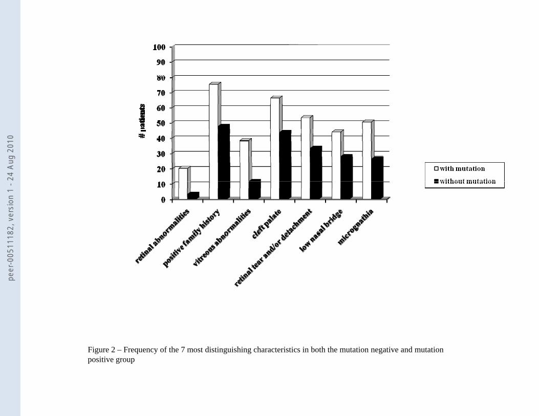
Figure 2 – Frequency of the 7 most distinguishing characteristics in both the mutation negative and mutationpositive group
peer
-005
1118
2, v
ersi
on 1
- 24
Aug
201
0

Table 2: Proposed scoring system
Characteristics Score
- retinal abnormalities 5- positive family history 5p y y- vitreous abnormalities 4- cleft palate 4- retinal tear and/or detachment 3- low nasal bridge 1
micrognathia 1- micrognathia 1
Total score 23
peer
-005
1118
2, v
ersi
on 1
- 24
Aug
201
0

nts
# pa
tien
score
Figure 3 – Overlap in total score between mutation positive and mutation negative group of patients.
peer
-005
1118
2, v
ersi
on 1
- 24
Aug
201
0
outlier
max
Q3
median
Q1
i
9
min
Figure 4 – Box plot presentation of the total scores in both patient groups with Q1 representing the first quartile or 25th centile and Q3 representing the third quartile or 75th centile. Max indicates the maximum score, and min the minimum score, that is not an outlier or that is within 1.5 times the interquartile range (Q1-Q3). 75% of the patients with a COL2A1 mutation had a total score ≥ 9
peer
-005
1118
2, v
ersi
on 1
- 24
Aug
201
0
Related Documents