8 Steroid Hormones in Drosophila: How Ecdysone Coordinates Developmental Signalling with Cell Growth and Division Leonie Quinn 1 , Jane Lin 2 , Nicola Cranna 1 , Jue Er Amanda Lee 1 , Naomi Mitchell 1 and Ross Hannan 2,3,4 1 Department of Anatomy and Cell Biology, University of Melbourne, 2 Peter MacCallum Cancer Centre, St Andrews Place, East Melbourne, 3 Department of Biochemistry and Molecular Biology, University of Melbourne, 4 Department of Biochemistry and Cell Biology, Monash University, Australia 1. Introduction 1.1 The ecdysone pathway directs Drosophila development Ecdysone is the major steroid hormone in all holometabolous insects responsible for driving the metamorphosis of larval tissues into adult structures. During metamorphosis, ecdysone is essential for upregulating the genes required to control apoptosis and differentiation, essential processes for removal of larval structures which have become obsolete and for tissue remodelling. In addition, ecdysone directs cell growth and division in many tissues throughout the larval to pupal transition. This chapter will discuss the many diverse mechanisms reported for connecting the ecdysone pulse to the developmentally regulated cell growth and cycle progression required for tissue growth and for insects to reach their target body size. Like all other holometabolous insects, the size of Drosophila adult flies is set by the size of the larvae prior to metamorphosis, at the time of pupariation when feeding has ceased and growth can no longer occur. The major developmental hormone in Drosophila, the steroid hormone 20-hydroxyecdysone (20E), commonly known as ecdysone, is required for all the developmental transitions needed for metamorphosis (Figure 1-3; (Thummel 1995, 1996, 2001)). Ecdysone is produced in and released by the prothoracic gland (PG), a component of the ring gland, which also contains the corpora allata (CA) and corpora cardiaca (CC) (Figure 1; (Zitnan et al. 2007; McBrayer et al. 2007)). Ecdysone release is controlled by a complex combination of upstream factors, including peptide hormones and neuropeptide signals (see section 2.2). For example, Prothoracicotropic hormone (PTTH) from the central nervous system (CNS) is required to regulate the synthesis and release of ecdysone from the PG (McBrayer et al. 2007). Ecdysone pulses from the PG are required for all aspects of morphogenesis, starting with the formation of the body plan during late embryogenesis, hatching and development of the first larval instar, and for cuticle moulting at the end of the first and second instars. A large

Welcome message from author
This document is posted to help you gain knowledge. Please leave a comment to let me know what you think about it! Share it to your friends and learn new things together.
Transcript
8
Steroid Hormones in Drosophila: How Ecdysone Coordinates Developmental
Signalling with Cell Growth and Division
Leonie Quinn1, Jane Lin2, Nicola Cranna1, Jue Er Amanda Lee1, Naomi Mitchell1 and Ross Hannan2,3,4
1Department of Anatomy and Cell Biology, University of Melbourne, 2Peter MacCallum Cancer Centre, St Andrews Place, East Melbourne,
3Department of Biochemistry and Molecular Biology, University of Melbourne, 4Department of Biochemistry and Cell Biology, Monash University,
Australia
1. Introduction
1.1 The ecdysone pathway directs Drosophila development Ecdysone is the major steroid hormone in all holometabolous insects responsible for driving the metamorphosis of larval tissues into adult structures. During metamorphosis, ecdysone is essential for upregulating the genes required to control apoptosis and differentiation, essential processes for removal of larval structures which have become obsolete and for tissue remodelling. In addition, ecdysone directs cell growth and division in many tissues throughout the larval to pupal transition. This chapter will discuss the many diverse mechanisms reported for connecting the ecdysone pulse to the developmentally regulated cell growth and cycle progression required for tissue growth and for insects to reach their target body size. Like all other holometabolous insects, the size of Drosophila adult flies is set by the size of the larvae prior to metamorphosis, at the time of pupariation when feeding has ceased and growth can no longer occur. The major developmental hormone in Drosophila, the steroid hormone 20-hydroxyecdysone (20E), commonly known as ecdysone, is required for all the developmental transitions needed for metamorphosis (Figure 1-3; (Thummel 1995, 1996, 2001)). Ecdysone is produced in and released by the prothoracic gland (PG), a component of the ring gland, which also contains the corpora allata (CA) and corpora cardiaca (CC) (Figure 1; (Zitnan et al. 2007; McBrayer et al. 2007)). Ecdysone release is controlled by a complex combination of upstream factors, including peptide hormones and neuropeptide signals (see section 2.2). For example, Prothoracicotropic hormone (PTTH) from the central nervous system (CNS) is required to regulate the synthesis and release of ecdysone from the PG (McBrayer et al. 2007). Ecdysone pulses from the PG are required for all aspects of morphogenesis, starting with the formation of the body plan during late embryogenesis, hatching and development of the first larval instar, and for cuticle moulting at the end of the first and second instars. A large

Steroids – Basic Science
142
Fig. 1. The ring gland. An important organ in Drosophila is the ring gland, which is situated in between the two brain lobes in the larvae. (A) Diagram of the components of the ring gland: the prothoracic gland (PG), the corpora allata (CA) and corpora cardiaca (CC). Ecdysone is produced by the PG, whilst the CA is thought to synthesise the Juvenile hormone. (B) A confocal image of a 3rd instar prothoracic gland overexpressing GFP.
titre of ecdysone is released at the end of the third instar, in the wandering larvae in preparation for pupation, which marks the beginning of adult tissue metamorphosis (Figure 3; (Thummel 1995, 1996, 2001)). Insect metamorphosis is characterized by vast changes in tissue morphology, where larval tissues are replaced by adult structures. In Drosophila, the pulse of ecdysone at the end of the third larval instar initiates metamorphosis (Riddiford 1993). During metamorphosis, an extensive range of larval structures respond to the ecdysone pulse, which triggers the complex array of cellular responses required to achieve conversion from the larval tissue to give the adult (Bender et al. 1997). This begins with the secretion of glue proteins for the larvae to attach itself onto a surface for puparium development. Subsequently ecdysone drives the larval body to shorten, and promotes the subsequent cuticle darkening and hardening required to form the pupal case. During the late larval to early pupal stages the ecdysone pulse drives removal of obsolete larval tissues, such as the larval gut and salivary glands (Thummel 2001). The major morphological changes for metamorphosis involve development of adult structures from the imaginal discs, which are epithelial structures formed from invaginations of the ectoderm during embryogenesis (Gates and Thummel 2000). The imaginal discs include 2 pairs for the eye/antenna, wing, and halteres, 3 pairs for the legs and a single disc for the gonads, which evert, elongate, and differentiate during metamorphosis
Steroid Hormones in Drosophila: How Ecdysone Coordinates Developmental Signalling with Cell Growth and Division
143
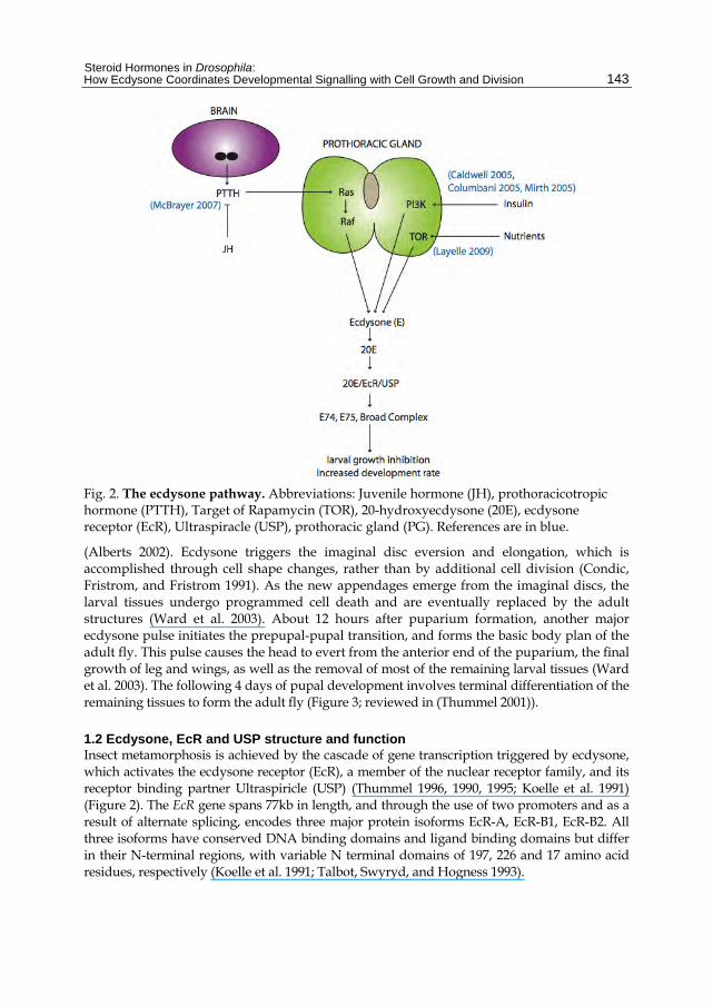
Fig. 2. The ecdysone pathway. Abbreviations: Juvenile hormone (JH), prothoracicotropic hormone (PTTH), Target of Rapamycin (TOR), 20-hydroxyecdysone (20E), ecdysone receptor (EcR), Ultraspiracle (USP), prothoracic gland (PG). References are in blue.
(Alberts 2002). Ecdysone triggers the imaginal disc eversion and elongation, which is accomplished through cell shape changes, rather than by additional cell division (Condic, Fristrom, and Fristrom 1991). As the new appendages emerge from the imaginal discs, the larval tissues undergo programmed cell death and are eventually replaced by the adult structures (Ward et al. 2003). About 12 hours after puparium formation, another major ecdysone pulse initiates the prepupal-pupal transition, and forms the basic body plan of the adult fly. This pulse causes the head to evert from the anterior end of the puparium, the final growth of leg and wings, as well as the removal of most of the remaining larval tissues (Ward et al. 2003). The following 4 days of pupal development involves terminal differentiation of the remaining tissues to form the adult fly (Figure 3; reviewed in (Thummel 2001)).
1.2 Ecdysone, EcR and USP structure and function Insect metamorphosis is achieved by the cascade of gene transcription triggered by ecdysone, which activates the ecdysone receptor (EcR), a member of the nuclear receptor family, and its receptor binding partner Ultraspiricle (USP) (Thummel 1996, 1990, 1995; Koelle et al. 1991) (Figure 2). The EcR gene spans 77kb in length, and through the use of two promoters and as a result of alternate splicing, encodes three major protein isoforms EcR-A, EcR-B1, EcR-B2. All three isoforms have conserved DNA binding domains and ligand binding domains but differ in their N-terminal regions, with variable N terminal domains of 197, 226 and 17 amino acid residues, respectively (Koelle et al. 1991; Talbot, Swyryd, and Hogness 1993).

Steroids – Basic Science
144
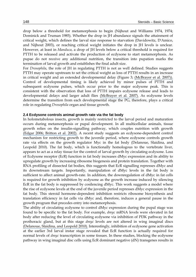
Fig. 3. Manipulation of growth pathways in the prothoracic gland alters body size. Light micrographs of female adult flies raised at 25°C bearing the genotypes: (A) control (AmnC651>+) (B) overexpressing activated Ras (AmnC651>RasV12) in the PG (C) overexpressing dominant negative PI3K (AmnC651>Dp110DN) in the PG. Red arrows indicate length of larval growth period. (D) Graph of ecdysone titres during Drosophila development (modified from (Thummel, 2001)).
Although EcR can bind ecdysone alone, optimal binding to the ecdysone response elements (EcRE) and activation of transcriptional targets requires the addition of USP (Grad et al. 2001; Grebe, Fauth, and Spindler-Barth 2004). USP exhibits a strong structural and functional similarity to the orthologous vertebrate retinoid X receptor (RXR) (Yao et al. 1992; Oro, McKeown, and Evans 1990). Like RXR, which forms heterodimers with non-steroid receptors for thyroid hormone, retinoic acid and vitamin D, and thereby activates them for DNA-binding (Mangelsdorf and Evans 1995), USP interacts with each of the EcR isoforms to form DNA-binding heterodimers (Yao et al. 1992; Bender et al. 1997). In this respect

Steroid Hormones in Drosophila: How Ecdysone Coordinates Developmental Signalling with Cell Growth and Division
145
Drosophila EcRs are, therefore, analogous to the vertebrate family of RXR heterodimeric receptors rather than the vertebrate family of steroid hormone receptors, which bind DNA as homodimers (Beato and Klug 2000). Therefore, similar to vertebrate nuclear receptors, the EcR/USP heterodimer functions as a ligand-dependent transcription factor. In the presence of the ecdysone ligand, the appropriate EcR nuclear receptor isoform dimerizes with USP, and the complex is stabilised by the active form of ecdysone, 20-hydroxyecdysone (20E) to allow efficient binding to the ecdysone response element (EcRE) (Hall and Thummel 1998; Yao et al. 1993) and transcriptional activation of ecdysone-responsive genes (D'Avino and Thummel 1998; Kozlova and Thummel 2002; Thummel 2002; Thummel, Burtis, and Hogness 1990; Urness and Thummel 1995). Genes that are directly activated by the 20E/EcR/USP complex include three “early” ecdysone pathway genes; E74, E75 and the Broad-Complex (BR-C), which all encode transcription factors. The E74, E75 and BR-C transcription factors control the late genes in order to elicit the biological changes associated with each ecdysone pulse. This hierarchy of gene activation is required for modulating expression of the many cell death, cell cycle and differentiation genes required for metamorphosis (Thummel 1996, 2001). E74 encodes two proteins with an identical ETS DNA binding domain, designated E74A and E74B (Burtis et al. 1990; Thummel, Burtis, and Hogness 1990). E75 encodes three members of the nuclear receptor superfamily (designated E75A, E75B, and E75C), which are often referred to as orphan nuclear receptors due to their unidentified ligand (Segraves and Hogness 1990). The BR-C is a multigene locus, which encodes several zinc finger proteins (DiBello et al. 1991). To increase the output of the ecdysone pulse, EcR provides an autoregulatory loop to activate its own transcription and further increase receptor levels in response to the ecdysone ligand (Koelle et al. 1991).
1.3 Ecdysone signalling coordinates proliferation, death and differentiation Metamorphosis of Drosophila requires co-ordination of proliferation (cell growth and division), differentiation and death in order to form an adult fly of the appropriate size and with correctly differentiated structures. An essential process driven by the ecdysone pulse is the removal of larval tissues no longer required in the adult (Baehrecke 2000). The process of steroid hormone driven apoptosis is an important part of tissue remodelling, whereby selective death removes unwanted cells towards generating the mature structure (Rusconi, Hays, and Cagan 2000; Thummel 2001). For example, the histolysis of the larval salivary gland and midgut at the end of metamorphosis is stage-specific, ecdysone triggered, programmed cell death, which results in the removal of the component of these larval structures no longer required in the adult fly. In line with an apoptotic mechanism, previous studies have shown that cell death activators are upregulated in the third instar larval tissues, including the salivary glands and midgut in response to ecdysone (reviewed in (Jiang, Baehrecke, and Thummel 1997; Baehrecke 2000; Yin and Thummel 2005)). The ecdysone pulse is also essential for differentiation and patterning of the larval imaginal tissues required for development of adult structures (Hall and Thummel 1998; D'Avino and Thummel 2000, 1998; Zheng et al. 2003). As cell division and patterning are tightly linked in Drosophila imaginal tissues, the process of metamorphosis controlled by ecdysone involves coordination of the developmental signals that regulate proliferation and differentiation. Although much work has focused on the downstream targets linking the ecdysone pathway to programmed cell death and cell differentiation (Baehrecke 2000; Jiang, Baehrecke, and

Steroids – Basic Science
146
Thummel 1997; Yin and Thummel 2005), the relationship between ecdysone and cell cycle is a relatively unexplored field. Here we review the evidence that the ecdysone pulse is critical for controlling cell growth and division in Drosophila.
1.4 Linking the Ecdysone pulse to cell cycle In Drosophila, cell growth and cell cycle progression are regulated by a number of key genes, which have been shown to control the cell cycle in an analogous manner in all multicellular organisms. These include the Drosophila orthologue of the mammalian c-myc transcription factor and oncogene, dMyc, which drives growth and progression through G1 to S-phase (Johnston et al. 1999), the essential G1 to S-phase Cyclin complex, Cyclin E (CycE) and its Cyclin-dependent-kinase (Cdk) partner Cdk2, which triggers S-phase by promoting DNA replication (Knoblich et al. 1994; Neufeld et al. 1998; Richardson et al. 1995), and the Drosophila orthologue of the Cdc25 phosphatase, String (Stg), which is required for G2/M progression and promotes mitotic entry by activating the Cdk1/Cyclin B complex (Edgar and Datar 1996). CycE and Stg are the rate limiting factors for S-phase and mitosis, respectively, and both are activated by the Drosophila orthologue of human E2F1 protein, dE2F1 (Neufeld et al. 1998). dE2F1 responds to the relevant Cdk-Cyclin complex (CycE/Cdk2 for S-phase and CycB/Cdk1 for mitosis) to coordinate cell cycle progression from G1 to S-phase and G2 into mitosis (Reis and Edgar 2004). During metamorphosis, following removal of the obsolete larval structures, proliferation of the remaining tissue occurs in an ecdysone-dependent manner to produce adult structures. For example, during pupal development the larval midgut is removed by apoptosis and is replaced through proliferation of the remaining tissue to form the adult midgut (Jiang, Baehrecke, and Thummel 1997). Microarray analysis has revealed that the ecdysone signal is associated with the activation of key cell cycle genes, including Cyclin B, Cdc2 and Cyclin D, during the initiation of midgut metamorphosis (Li and White 2003). Analysis of EcR null mutants also revealed that EcR function was necessary for the cell cycle and growth genes to be activated in the larval midgut, suggesting that the ecdysone pathway is required for cell division control. The body of this chapter will discuss how the ecdysone pulse achieves changes to cell growth and cell cycle progression. First we will describe how ecdysone levels dictate body size cell extrinsically by controlling developmental timing. Then we will discuss how ecdysone works with its receptors, in a tissue autonomous manner to control transcription of cell cycle genes, which most likely occurs indirectly by modifying the activity of developmental signalling pathways.
2. Cell extrinsic effects of ecdysone on larval growth and body size
The ecdysone pulse can act indirectly to affect larval growth as a consequence of the link between the ecdysone titre and developmental timing. Here we will discuss how cell extrinsic effects of the ecdysone pathway control Drosophila larval growth and final body size non-autonomously, at least in part, through interactions between the ecdysone and insulin pathways (King-Jones and Thummel 2005; Shingleton 2005; Mirth and Riddiford 2007; Nijhout 2008).
2.1 The prothoracic gland directs body size The prothoracic gland (PG) is tightly associated with the developmental timing of all holometabolous insects, including Drosophila, as it produces the ecdysone pulse that dictates
Steroid Hormones in Drosophila: How Ecdysone Coordinates Developmental Signalling with Cell Growth and Division
147
the timing of the larval-pupal transition and metamorphosis. As the adult fly size is determined by the size of the larvae at the pupal molt, the timing of ecdysone release plays a vital role in the growth of the fly (reviewed in (King-Jones and Thummel 2005)). Studies in 2005 demonstrated the importance of the size of the PG and its effect on ecdysone production and, therefore, determination of the final adult fly size (Caldwell, Walkiewicz, and Stern 2005; Colombani et al. 2005; Mirth, Truman, and Riddiford 2005). Specifically, these groups reported a role for insulin signalling in the PG, and also characterised a size-assessing feature of the PG (Figure 3). As a size-assessment tissue, inhibiting the growth of the PG causes an underestimation of body size and results in pupation at a larger size, whereas promoting this tissue’s growth results in smaller flies (Mirth, Truman, and Riddiford 2005). Consistent with this, overexpression of activated PI3K or Ras (RasV12), both key components of growth control pathways in flies and mammals, specifically in the PG resulted in a larger PG but reduced the pupal and adult size (Caldwell, Walkiewicz, and Stern 2005; Colombani et al. 2005; Mirth, Truman, and Riddiford 2005), which we have recapitulated as shown in Figure 3 (compare 3B with 3A). Conversely, overexpression of a dominant negative isoform of PI3K (Dp110DN) reduced the PG size but resulted in larger pupae and adults, due to an extended larval growth period (Figure 3, compare C with A). Furthermore, through measurements of the ecdysone target E74B or through an enzyme immunoassay for ecdysteroid titres, it was shown that the extended larval growth period was due to reduced ecdysone levels, which was most likely a result of a smaller PG (Caldwell, Walkiewicz, and Stern 2005; Colombani et al. 2005; Mirth, Truman, and Riddiford 2005).
2.2 PTTH regulates of ecdysone levels In insects, the production and release of ecdysone is responsive to the prothoracicotropic hormone (PTTH), a small, secreted peptide. PTTH is thought to induce the transcription of ecdysone biosynthetic genes that encode enzymes driving the series of dehydrogenation and hydroxylation reactions required to synthesise the active metabolite 20E from the cholesterol precursor (Marchal et al. 2010). In Drosophila PTTH is produced by a pair of bilateral neurosecretory cells in the brain, which innervate the prothoracic gland (PG) ((Figure 2; (McBrayer et al. 2007)). PTTH is expressed throughout 3rd instar in an 8 hour cyclic pattern, with upregulation noticed around 12 hours before pupariation (McBrayer et al. 2007). Ablation of the neurons that produce PTTH results in a 5-day developmental delay in the onset of pupariation, larger 3rd instar larvae and pupae, and adults with larger wings due to increased cell number. In line with the predicted role for PTTH in modulating ecdysone synthesis and release, larvae lacking PTTH producing neurons have reduced ecdysone titres. This suggests PTTH normally modulates ecdysone levels to coordinate larval growth with the onset of metamorphosis. However, as the ecdysone levels still eventually peak in larvae with ablated neurons, PTTH may not be the sole factor required for increasing ecdysone titres (McBrayer et al. 2007). Thus PTTH might be required in addition to the insulin-dependent growth pathways discussed above, to coordinate larval growth with ecdysone-induced moulting and metamorphosis (Figure 2-3).
2.3 Juvenile hormone controls PTTH release and ecdysone production The signals required for metamorphosis have been extensively studied in the tobacco hornworm Manduca sexta. In this insect, the pulse of ecdysone in the last larval instar is inhibited by another hormone, the Juvenile Hormone (JH). In the case of JH, levels need to
Steroids – Basic Science
148
drop below a threshold for metamorphosis to begin (Nijhout and Williams 1974, 1974; Dominick and Truman 1985). Whether the drop in JH abundance signals the attainment of critical weight, which defines the larval size response to starvation (Davidowitz, D'Amico, and Nijhout 2003), or reaching critical weight initiates the drop in JH levels is unclear. However, at least in Manduca, a drop of JH levels below a critical threshold is required for PTTH to be released and activate the production of ecdysone to start metamorphosis. As pupae do not receive any additional nutrition, the transition into pupation marks the termination of larval growth and establishes the final adult size. For Drosophila, the role of JH in regulating PTTH is not as well defined. Studies suggests PTTH may operate upstream to set the critical weight as loss of PTTH results in an increase in critical weight and an extended developmental delay (Figure 3; (McBrayer et al. 2007)). Control of developmental timing is likely achieved by minor pulses of PTTH and subsequent ecdysone pulses, which occur prior to the major ecdysone peak. This is consistent with the observation that loss of PTTH impairs ecdysone release and leads to developmental delays and larger adult flies (McBrayer et al. 2007). As ecdysone levels determine the transition from each developmental stage the PG, therefore, plays a critical role in regulating Drosophila organ and tissue growth.
2.4 Ecdysone controls animal growth rate via the fat body In holometabolous insects, growth is mainly restricted to the larval period and maturation occurs during metamorphosis or pupal development. In all multicellular animals, tissue growth relies on the insulin-signalling pathway, which couples nutrition with growth (Edgar 2006; Britton et al. 2002). A recent study suggests an ecdysone-dependent control mechanism for restricting growth to the juvenile period, where ecdysone controls growth rate via effects on the growth regulator Myc in the fat body (Delanoue, Slaidina, and Leopold 2010). The fat body, which is functionally homologous to the vertebrate liver, appears to act as a relay tissue for the control of larval growth by circulating ecdysone. Loss of Ecdysone receptor (EcR) function in fat body increases dMyc expression and its ability to upregulate growth by increasing ribosome biogenesis and protein translation. Together with RNA profiling of dissected fat bodies, this suggests that EcR signalling represses dMyc and its downstream targets. Importantly, manipulation of dMyc levels in the fat body is sufficient to affect animal growth-rate. In addition, the downregulation of dMyc in fat cells is required for growth inhibition by ecdysone as the growth increase induced by silencing EcR in the fat body is suppressed by cosilencing dMyc. This work suggests a model where the rise of ecdysone levels at the end of the juvenile period represses dMyc expression in the fat body. This steroid hormone-dependent inhibition restricts ribosome biosynthesis and translation efficiency in fat cells via dMyc and, therefore, induces a general pause in the growth program that precedes entry into metamorphosis. The ability of circulating ecdysone to control dMyc expression during the pupal stage was found to be specific to the fat body. For example, dmyc mRNA levels were elevated in fat body after reducing the level of circulating ecdysone via inhibition of PI3K pathway in the prothoracic gland, but at this stage dmyc levels are not altered in wing imaginal discs (Delanoue, Slaidina, and Leopold 2010). Interestingly, inhibition of ecdysone gene activation at the earlier 3rd larval instar stage revealed that EcR function is actually required for normal levels of dmyc transcription in some tissues. In these studies, blocking the ecdysone pathway in wing imaginal disc cells using EcR dominant negative (dN) transgenes results in
Steroid Hormones in Drosophila: How Ecdysone Coordinates Developmental Signalling with Cell Growth and Division
149
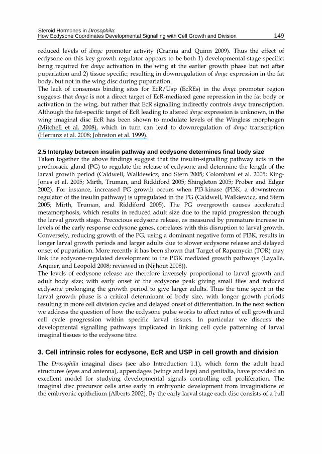
reduced levels of dmyc promoter activity (Cranna and Quinn 2009). Thus the effect of ecdysone on this key growth regulator appears to be both 1) developmental-stage specific; being required for dmyc activation in the wing at the earlier growth phase but not after pupariation and 2) tissue specific; resulting in downregulation of dmyc expression in the fat body, but not in the wing disc during pupariation. The lack of consensus binding sites for EcR/Usp (EcREs) in the dmyc promoter region suggests that dmyc is not a direct target of EcR-mediated gene repression in the fat body or activation in the wing, but rather that EcR signalling indirectly controls dmyc transcription. Although the fat-specific target of EcR leading to altered dmyc expression is unknown, in the wing imaginal disc EcR has been shown to modulate levels of the Wingless morphogen (Mitchell et al. 2008), which in turn can lead to downregulation of dmyc transcription (Herranz et al. 2008; Johnston et al. 1999).
2.5 Interplay between insulin pathway and ecdysone determines final body size Taken together the above findings suggest that the insulin-signalling pathway acts in the prothoracic gland (PG) to regulate the release of ecdysone and determine the length of the larval growth period (Caldwell, Walkiewicz, and Stern 2005; Colombani et al. 2005; King-Jones et al. 2005; Mirth, Truman, and Riddiford 2005; Shingleton 2005; Prober and Edgar 2002). For instance, increased PG growth occurs when PI3-kinase (PI3K, a downstream regulator of the insulin pathway) is upregulated in the PG (Caldwell, Walkiewicz, and Stern 2005; Mirth, Truman, and Riddiford 2005). The PG overgrowth causes accelerated metamorphosis, which results in reduced adult size due to the rapid progression through the larval growth stage. Precocious ecdysone release, as measured by premature increase in levels of the early response ecdysone genes, correlates with this disruption to larval growth. Conversely, reducing growth of the PG, using a dominant negative form of PI3K, results in longer larval growth periods and larger adults due to slower ecdysone release and delayed onset of pupariation. More recently it has been shown that Target of Rapamycin (TOR) may link the ecdysone-regulated development to the PI3K mediated growth pathways (Layalle, Arquier, and Leopold 2008; reviewed in (Nijhout 2008)). The levels of ecdysone release are therefore inversely proportional to larval growth and adult body size; with early onset of the ecdysone peak giving small flies and reduced ecdysone prolonging the growth period to give larger adults. Thus the time spent in the larval growth phase is a critical determinant of body size, with longer growth periods resulting in more cell division cycles and delayed onset of differentiation. In the next section we address the question of how the ecdysone pulse works to affect rates of cell growth and cell cycle progression within specific larval tissues. In particular we discuss the developmental signalling pathways implicated in linking cell cycle patterning of larval imaginal tissues to the ecdysone titre.
3. Cell intrinsic roles for ecdysone, EcR and USP in cell growth and division
The Drosophila imaginal discs (see also Introduction 1.1), which form the adult head structures (eyes and antenna), appendages (wings and legs) and genitalia, have provided an excellent model for studying developmental signals controlling cell proliferation. The imaginal disc precursor cells arise early in embryonic development from invaginations of the embryonic epithelium (Alberts 2002). By the early larval stage each disc consists of a ball
Steroids – Basic Science
150
of around 10-50 undifferentiated stem cells, which undergo extensive growth and proliferation to comprise up to 100,000 cells by the end of the third larval instar. The imaginal discs start differentiation at the end of third instar and complete the process by the end of pupariation, when all adult structures such as the wings, legs and eyes have developed (Fristrom and Fristrom 1993). The third instar larval stage is a critical stage of Drosophila development, containing the major growth and proliferation of all tissues required to form the adult fly (Church and Robertson 1966). Indeed the size of the adult fly is determined at the time when the pupal case is formed, as after this the animal cannot feed again until eclosion. Here we will discuss the developmental signals (including Wingless, Dpp, Hedgehog, Notch) controlling growth of the eye and wing imaginal discs, and how ecdysone impacts on these signalling pathways to control cell division.
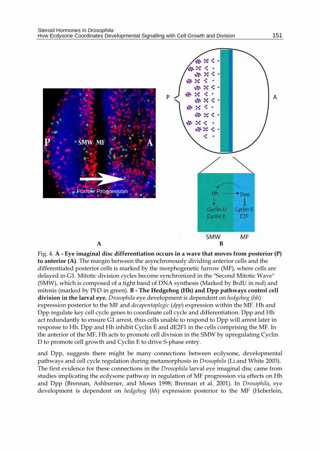
3.1 Ecdysone mediates morphogenetic Furrow progression in the eye imaginal disc The Drosophila eye is composed of an ordered array of photoreceptor clusters or ommatidia, which develop from an epithelial monolayer known as the eye imaginal disc, via an organised pattern of proliferation and differentiation (Figure 4; (Ready, Hanson, and Benzer 1976; Wolff and Ready 1991)). Differentiation of the ommatidia occurs in a wave that moves from the posterior toward the anterior (Thomas and Zipursky 1994). The margin between the asynchronously dividing anterior cells and the differentiated posterior cells is marked by the morphogenetic furrow (MF) (Ready, Hanson, and Benzer 1976). Mitotic division cycles become synchronized in the MF where cells are delayed in G1 and a subset of photoreceptor cells are specified. The remaining retinal cells synchronously re-enter the cell cycle in the "Second Mitotic Wave" (SMW), which is composed of a tight band of DNA synthesis and mitosis (Figure 4). These final cell divisions provide the cells required for differentiation of the ommatidial structures that form the adult eye (Ready, Hanson, and Benzer 1976; Wolff and Ready 1991). Studies in the eye primordium of the tobacco hornworm moth, Manduca sexta, suggest that progression of the MF, including proliferation and differentiation of ommatidial clusters, requires ecdysone. Eye primordia proliferation responds to a critical concentration of ecdysone and below this threshold cells arrest in the G2 phase of the cell cycle (Champlin and Truman 1998). Premature exposure to high levels of ecdysone will also result in MF arrest and precocious maturation of ommatida (Champlin and Truman 1998). These cell cycle responses to ecdysone are consistent with the moderate ecdysone pulse during the larval stage first stimulating eye proliferation and the high levels of ecdysone released after pupariation driving cell cycle exit and eye maturation. The ecdysone pathway has also been implicated in regulation of MF progression in the Drosophila larval eye imaginal disc. The ecdysoneless mutation (ecd-ts) is a hypomorphic temperature-sensitive allele, which reduces ecdysone secretion from the ring gland (Henrich et al. 1987). Homozygous ecd-ts flies show eye defects when shifted to the restrictive temperature during the third instar larval stage (Brennan, Ashburner, and Moses 1998). Consistent with the MF moving much more slowly than normal in the ecd-ts mutant, delayed eye differentiation was shown using the neuronal marker Elav. Microarray analysis has linked the ecdysone pulse during metamorphosis to transcriptional changes in mitogenic signalling molecules, which are essential for coordinating cell cycle and patterning of imaginal tissues. The observation that ecdysone signalling was essential for the activation of factors involved in regulatory signalling pathways such as Wg, Notch
Steroid Hormones in Drosophila: How Ecdysone Coordinates Developmental Signalling with Cell Growth and Division
151
A B
Fig. 4. A - Eye imaginal disc differentiation occurs in a wave that moves from posterior (P) to anterior (A). The margin between the asynchronously dividing anterior cells and the differentiated posterior cells is marked by the morphogenetic furrow (MF), where cells are delayed in G1. Mitotic division cycles become synchronized in the "Second Mitotic Wave" (SMW), which is composed of a tight band of DNA synthesis (Marked by BrdU in red) and mitosis (marked by PH3 in green). B - The Hedgehog (Hh) and Dpp pathways control cell division in the larval eye. Drosophila eye development is dependent on hedgehog (hh) expression posterior to the MF and decapentaplegic (dpp) expression within the MF. Hh and Dpp regulate key cell cycle genes to coordinate cell cycle and differentiation. Dpp and Hh act redundantly to ensure G1 arrest, thus cells unable to respond to Dpp will arrest later in response to Hh. Dpp and Hh inhibit Cyclin E and dE2F1 in the cells comprising the MF. In the anterior of the MF, Hh acts to promote cell division in the SMW by upregulating Cyclin D to promote cell growth and Cyclin E to drive S-phase entry.
and Dpp, suggests there might be many connections between ecdysone, developmental pathways and cell cycle regulation during metamorphosis in Drosophila (Li and White 2003). The first evidence for these connections in the Drosophila larval eye imaginal disc came from studies implicating the ecdysone pathway in regulation of MF progression via effects on Hh and Dpp (Brennan, Ashburner, and Moses 1998; Brennan et al. 2001). In Drosophila, eye development is dependent on hedgehog (hh) expression posterior to the MF (Heberlein,
Steroids – Basic Science
152
Wolff, and Rubin 1993; Heberlein et al. 1995) and decapentaplegic (dpp) expression within the MF (Figure 4; (Blackman et al. 1991)). Drosophila Dpp is a member of the mammalian transforming growth factor-beta (TGF-beta) family of secreted proteins. TGF-beta can behave as a tumour-suppressor or oncogene depending on the tissue microenvironment, thus pathway inhibition or activation can result in cancer progression (Serra and Moses 1996; Derynck, Akhurst, and Balmain 2001; Wakefield and Roberts 2002; Bachman and Park 2005; Elliott and Blobe 2005; Jakowlew 2006; Massague 2008). Aberrant Hh signalling has also been associated with human cancer, with much literature linking activation of the pathway with increased tumour progression (Toftgard 2000; Vestergaard, Bak, and Larsen 2005; Evangelista, Tian, and de Sauvage 2006; Epstein 2008; Varjosalo and Taipale 2008). In the eye disc, Dpp and Hedgehog (Hh) act redundantly to ensure G1 arrest within the MF (Penton, Selleck, and Hoffmann 1997; Horsfield et al. 1998; Firth and Baker 2005) where both pathways can inhibit Cyclin E and dE2F1 (Escudero and Freeman 2007). In addition to this cell cycle inhibitory role of Hh in the MF, Hh promotes cell division in the SMW by upregulating Cyclin D to promote cell growth and Cyclin E to drive S-phase entry (Figure 4; (Duman-Scheel et al. 2002)). In line with ecdysone regulating eye development via Hh, the phenotype observed in ecdysoneless (ecd-ts) mutant is similar to phenotypes resulting from hh loss of function (Heberlein et al. 1995). In addition, the decreased levels of Hh protein posterior to the MF in ecd-ts larval eye discs are consistent with hh being a downstream target of the ecdysone signal (Brennan, Ashburner, and Moses 1998). The delayed MF progression may be, therefore, a consequence of the requirement for Hh in activation of the S-phase genes cyclin D and cyclin E and, therefore, cell cycle re-entry in the SMW (Duman-Scheel et al. 2002). Indeed, the failure of MF movement in ecd-ts mutants is likely a result of impaired cell cycle progression as S-phase numbers were dramatically decreased in the SMW (Brennan, Ashburner, and Moses 1998). Consistent with reduced cell division within the SMW, levels of the mitotic cyclin, Cyclin B, were also reduced posterior to the MF (Brennan, Ashburner, and Moses 1998). The USP receptor has also been implicated in regulation of cell cycle progression and differentiation in the developing eye imaginal disc. Loss-of-function usp clones spanning the morphogenetic furrow show an anterior shift in expression of the MF-specific marker Dpp, consistent with premature progression of the MF and a role for USP in repressing morphogenetic furrow movement (Zelhof et al. 1997). In addition, loss of USP results in ectopic activation of many genes involved in cell fate specification in the eye, including the differentiation markers Spalt and Atonal (Zelhof et al. 1997). Although expression of these differentiation markers occurs prematurely, specification of cells contributing to the ommatidia occurs normally. The cell cycle analysis of usp mutant clones suggested that although the MF was advanced, cell cycle progression was disrupted in the SMW. First staining for Cyclin A, as a marker for cells in either S or G2 phase, revealed fewer Cyclin A-positive cells in usp- clones posterior to the morphogenetic furrow (Ghbeish et al. 2001). Similarly, although the Cyclin B band was not shifted in usp- clones posterior to the MF, the numbers of cells expressing Cyclin B were reduced (Ghbeish and McKeown 2002). The reduction in cell cycle markers posterior of the MF suggests that USP is required for cell cycle progression in the SMW. In support of cell cycle induction in the SMW depending on the presence of USP protein, usp overexpression using the GMR-promoter, which is only expressed posterior of the furrow, can rescue the loss of Cyclin B in the usp mutant clone. As progression through the SMW and differentiation are tightly coupled, the reduced cell

Steroid Hormones in Drosophila: How Ecdysone Coordinates Developmental Signalling with Cell Growth and Division
153
cycles in usp-/- clones may be the underlying cause of the premature differentiation observed (Zelhof et al. 1997). Together these data show that reduction in either ecdysone or USP results in reduced cell cycles. Paradoxically, however, usp mutations increase the rate of MF movement (Zelhof et al. 1997; Ghbeish et al. 2001; Ghbeish and McKeown 2002) while loss of ecdysone stops the MF (Brennan, Ashburner, and Moses 1998; Brennan et al. 2001). One explanation for these observations is that in the absence of ligand, the EcR/USP heterodimer normally acts as a repressor at certain EcREs. For these target genes ecdysone would be required to relieve the transcriptional repression caused by unliganded binding of the EcR/USP complex. This idea emerged from the finding that the Broad-complex (BR-C), which encodes the family of zinc-finger transcription factors upregulated early in response to high ecdysone titres (Karim, Guild, and Thummel 1993), becomes ectopically expressed in loss-of-function wing imaginal disc cells for either usp (Schubiger and Truman 2000) or EcR (Schubiger et al. 2005). Although concrete evidence is lacking, the idea is that the early (pre-ecdysone pulse) repressive effect of the EcR/USP heterodimer at the BR-C promoter will be lost in either EcR or usp mutants. The apparently contradictory effects of USP and ecdysone in the eye might actually be a consequence of the differential effects of the pathway on BR-C transcription. The Z1 isoform of the BR-C (BrC-Z1) is normally expressed posterior to the MF but not anterior to the MF (Emery, Bedian, and Guild 1994; Bayer, Holley, and Fristrom 1996) and reduced induction of BrC-Z1 occurs in ecd-ts eye discs (Brennan, Ashburner, and Moses 1998). Loss of USP function has the opposite effect, leading to high level BrC-Z1 protein expression both anterior and posterior to the MF, which might occur as a consequence of de-repression of BR-C transcription (Brennan et al. 2001). This high level of BrC-Z1 protein in usp mutant clones may explain the MF advancement phenotypes, as ectopic BrC-Z1 protein has been shown to induce premature differentiation of photoreceptor cells (Zelhof et al. 1997; Ghbeish et al. 2001; Ghbeish and McKeown 2002). Yet even though BrC-Z1 expression is downregulated in ecd-ts mutants (Brennan, Ashburner, and Moses 1998), BrC-Z1 loss of function eye imaginal discs are phenotypically different (Ghbeish et al. 2001), suggesting that other downstream targets of ecdysone pathway transcription mediate the reported effects on eye development. Like ecd-ts, impaired BrC-Z1 function results in decreased levels of Hh, defective MF progression and photoreceptor recruitment. However, unlike the findings for ecd-ts, reduced levels of Cyclin B were not detected in BrC-Z1 loss of function clones (Ghbeish et al. 2001). Rather loss of BrC-Z1 function results in defects in ommatidial assembly, suggesting a role for BR-C in post-MF differentiation rather than cell cycle regulation in the SMW (Brennan et al. 2001). This suggests that some ecdysone regulation in the eye is mediated by BrC-Z1, but that an alternate target(s) of the ecdysone pathway regulates the cell cycle activity required for SMW cell cycles and MF progression. The ecd-ts and USP studies suggest a role for the ecdysone pathway and the USP receptor in furrow progression, however, analysis of EcR mutant clones led to the conclusion that EcR was not required for furrow progression (Brennan et al. 2001). This was surprising given the EcR isoforms are the major mediators of the ecdysone signal, combined with the Manduca Sexta (Champlin and Truman 1998, 1998) and Drosophila studies (Brennan, Ashburner, and Moses 1998) that have demonstrated a requirement for ecdysone in MF progression. This led the authors of this study to propose a novel hormone transduction pathway involving an uncharacterized receptor to explain USP functioning independent of EcR in the eye, which

Steroids – Basic Science
154
could occur via heterodimerisation of USP with one of the 16 orphan nuclear receptors identified in Drosophila (Sullivan and Thummel 2003). For example USP has been found to heterodimerize with the orphan nuclear receptor, DHR38, to regulate cuticle formation (Kozlova et al. 1998; Sutherland et al. 1995). The USP/DHR38 complex responds to a different class of ecdysteroids in larval fat body and epidermis in an EcR independent manner, which does not involve direct binding of the ecdysone ligand to either DHR38 or USP (Baker et al. 2003). However, as DHR38 expression does not appear to be induced by ecdysteroids in the larval eye (Baker et al. 2003), it is unlikely that DHR38 partners USP during eye development. We believe it is premature to rule out a function for EcR in MF progression as the absence of a furrow progression phenotype reported (Brennan et al. 2001) may be a consequence of perdurance of EcR protein after clone induction. As studies using dominant negative EcR transgenes have shown that EcR is required for normal signalling and cell cycle progression in the wing (discussed in section 3.2; (Mitchell et al. 2008; Cranna and Quinn 2009)), similar methods should be used to inhibit EcR activity before any definitive conclusions about whether EcR is required for eye proliferation can be made. Together the evidence suggests that larval ecdysone signalling is essential for cell cycle progression in the eye imaginal disc. The effect of ecdysone on cell division may, in part, be mediated by increasing Hedgehog (Hh) signalling (Brennan, Ashburner, and Moses 1998) posterior to the MF to drive S-phase gene activity and cell cycle progression in the second mitotic wave (SMW) (Duman-Scheel et al. 2002). In addition to this cell cycle promoting role of ecdysone via Hh activity in the SMW, the shift in the Dpp band of expression in usp- clones suggests the ecdysone pathway might also act on Dpp to coordinate G1 arrest in the furrow with division in the SMW (Escudero and Freeman 2007; Firth and Baker 2005). Further work is required to understand how ecdysone might coordinate these developmental signals with the G1 arrest/MF formation and stimulation of the SMW required for eye development (Figure 4).
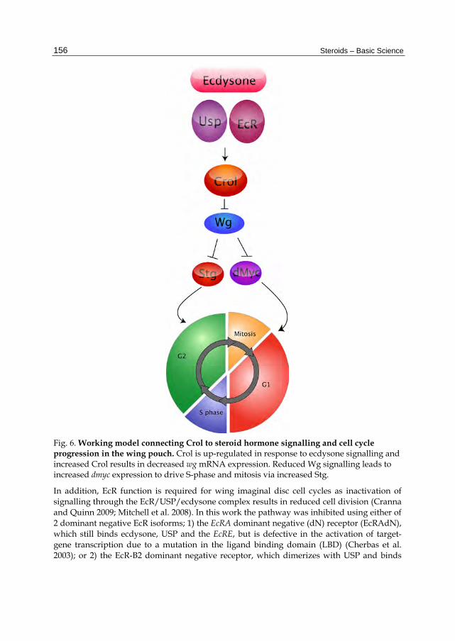
3.2 The ecdysone pathway regulates cell cycle progression in the larval wing disc Like the eye disc, the larval wing disc is also comprised of an epithelial sheet, which can be divided into distinct domains based on cell fate in the adult wing; the notum, hinge and pouch (Figure 5). The wing pouch, which ultimately forms the adult wing blade, has been a focus for studying signals impacting upon cell cycle, as wing morphogenesis involves patterned cell cycles that are tightly linked with developmental signalling (Johnson, Grenier, and Scott 1995; Johnston and Edgar 1998; Johnston et al. 1999; Johnston and Sanders 2003; Baker 2007). Early studies demonstrated that Crol, which is a zinc finger transcription factor, is activated in late larval imaginal discs by the steroid hormone ecdysone (D'Avino and Thummel 1998). Pupal lethal, hypomorphic crol mutants (crol4418) have defects in ecdysone-induced gene expression (D'Avino and Thummel 1998). Crol is both necessary and sufficient for cell cycle progression in the wing imaginal disc as crol mutant clones in the wing pouch fail to proliferate, whilst overexpression of crol results in ectopic proliferation (Mitchell et al. 2008). Crol is also required to downregulate the Wingless (Wg) pathway, which normally acts to drive cell cycle exit and differentiation (Figure 5). Therefore, by inhibiting the Wg pathway, crol drives wing disc cell division and potentially provides a link between the ecdysone pathway and the developmental signals that regulate cell cycle patterning (discussed in more detail section 3.3; Figure 6).

Steroid Hormones in Drosophila: How Ecdysone Coordinates Developmental Signalling with Cell Growth and Division
155
Fig. 5. (A) - Drosophila wing imaginal disc patterning. (A) The orange domain forms the notum, the blue region gives the hinge and the purple region (the pouch) forms the wing blade. The green line marks the anterior-posterior (A/P) boundary while the red line defines the dorsal-ventral (D/V) boundary. (B) Within D/V boundary of the pouch, Notch (N) expression activates Wingless (Wg) in the central domain. In the anterior compartment, Wg induces G2 arrest via string (stg) through Achaete (ac) and Scute (sc). In the posterior compartment, Wg induces G1 arrest via repression of dE2F. (B) - Wg protein, dmyc expression and cell cycle patterning in the Drosophila wing pouch. (A) Wg protein (red) is strongly expressed along the dorsal-ventral boundary of the wing pouch. (B) β-gal antibody staining (pink) of dmyc-lacZ discs shows a pattern consistent with dmyc transcription throughout the cycling cells of the pouch and downregulation of dmyc within the G1 arrested cells of the zone of non-proliferating cells (ZNC). (C) The ZNC can be seen by the reduced BrdU staining (red) for S-phase.
Steroids – Basic Science
156
Fig. 6. Working model connecting Crol to steroid hormone signalling and cell cycle progression in the wing pouch. Crol is up-regulated in response to ecdysone signalling and increased Crol results in decreased wg mRNA expression. Reduced Wg signalling leads to increased dmyc expression to drive S-phase and mitosis via increased Stg.
In addition, EcR function is required for wing imaginal disc cell cycles as inactivation of signalling through the EcR/USP/ecdysone complex results in reduced cell division (Cranna and Quinn 2009; Mitchell et al. 2008). In this work the pathway was inhibited using either of 2 dominant negative EcR isoforms; 1) the EcRA dominant negative (dN) receptor (EcRAdN), which still binds ecdysone, USP and the EcRE, but is defective in the activation of target-gene transcription due to a mutation in the ligand binding domain (LBD) (Cherbas et al. 2003); or 2) the EcR-B2 dominant negative receptor, which dimerizes with USP and binds

Steroid Hormones in Drosophila: How Ecdysone Coordinates Developmental Signalling with Cell Growth and Division
157
the EcRE, but cannot bind ecdysone, thus preventing optimal activation of ecdysone responsive genes (Cherbas et al. 2003; Hu, Cherbas, and Cherbas 2003). Blocking the EcR signal via overexpression of either EcRAdN or EcRB2dN in third instar wing imaginal disc flip-out clones (Pignoni and Zipursky 1997) results in a significant decrease in S-phase progression and mitosis, as measured by BrdU incorporation (Figure 7) and staining for anti-phosphohistone-H3, respectively (Cranna and Quinn 2009; Mitchell et al. 2008). Consistent with ecdysone signalling through EcR/USP normally being required for dmyc transcription, reduced dmyc promoter activity was observed in EcRAdN clones generated in the dmyc-lacZ enhancer trap background (Cranna and Quinn 2009). Thus ecdysone signalling through EcR/USP might normally control cell cycle progression in the wing imaginal disc by upregulating dMyc to drive growth by increasing ribosome biogenesis and protein translation (Johnston et al. 1999) and S phase via increased activity of the G1 cyclins (Duman-Scheel, Johnston, and Du 2004). Together this suggests EcR signalling might modulate cell growth and division of the wing imaginal disc by modulating dmyc levels (Cranna and Quinn 2009). In support of the reduced cell division in loss-of-function EcR cells being mediated by Crol, EcRAdN clones generated in the heterozygous crol mutant background show a further, significant reduction in cell cycle progression, when compared with either EcRdN cells alone or crol heterozygotes (Figure 7). This suggests that the reduction in cell cycle resulting from loss of EcR is sensitive to the level of Crol and that the ecdysone pathway normally regulates cell cycle in a Crol-dependent manner.
3.3 EcR is required for Wingless repression A key signalling molecule in the morphogenesis of the wing is the Wingless (Wg) protein, a member of the Wnt family of secreted morphogens. Wg is secreted in a band across the dorsal-ventral (D/V) boundary in the wing pouch (Figure 5; (Williams, Paddock, and Carroll 1993)) and is essential for cell cycle arrest in a region of the wing disc called the "Zone of Non-Proliferating Cells", or ZNC, at the end of larval development. The Wg pathway acts to downregulate key cell cycle genes (eg. dmyc, cycE, dE2F1 and stg) to link the Wg patterning signal to the cell cycle delay preceding the onset of differentiation at the wing margin (Johnston and Edgar 1998; Johnston et al. 1999; Johnston and Sanders 2003; Duman-Scheel, Johnston, and Du 2004). Indeed, the cell cycle arrest in the ZNC mediated by Wg is required for these cells to differentiate and develop into the adult wing blade (Figure 5; (Johnston and Edgar 1998; Johnston et al. 1999)). In the wing pouch EcR signalling is required for repression of wg transcription (Mitchell et al. 2008; Cranna and Quinn 2009), which together with the data above showing EcR is required for cell division, suggests the ecdysone signal might normally control cell cycle via Wg (Figure 6). Consistent with EcR normally being required to repress wg transcription, expansion of the wg expression domain occurs in UAS-EcRAdN (Mitchell et al. 2008) and UAS-EcRBdN (Cranna and Quinn 2009) "flip-out" clones generated in a wg-lacZ enhancer trap background (Kassis et al. 1992). These results suggest repression of wg transcription in the wing pouch is dependent on the ecdysone pathway. Given that increased Wg protein causes reduction of cell cycle regulators such as dmyc and stg, leading to decreased cells in S-phase and mitosis in the pouch (Figure 5; (Johnston and Edgar 1998; Johnston et al. 1999)), this finding is consistent with the reduced cell cycles observed in EcR loss-of-function clones.

Steroids – Basic Science
158
Fig. 7. S phase progression in UAS-EcRAdN clones is sensitive to the dose of crol. (A,B) Representative images of the wing pouch with control clones in heterozygous crol mutant (crolk05205) background; (C,D) UAS-EcRdN-A clones and (E,F) UAS-EcRdN-A clones in heterozygous crol mutant background, (A, C, E) S-phase is shown using BrdU (red), (B, D, F) GFP (green) marks clonal tissue. Scale bars indicate 50μm. (G) Quantification of S-phases for each of the genotypes; heterozygous crol mutant, UAS-EcRAdN alone and EcRAdN in the heterozygous crol mutant background. A significant reduction in the number of S-phase cells was found for the UAS-EcRdN-A alone compared to the control (p=0.0055) and for the UAS-EcRdN-A in the crol mutant background compared to UAS-EcRdN-A alone (p=0.0011). (H) Mean number of BrdU (S-phase) cells + SEM in control (clones in tissue heterozygous for the crol mutant); UAS-EcRdN-A alone and UAS-EcRdN-A in the crol mutant background. n=sample size.
Together this data suggests that EcR activity and the ecdysone-responsive transcription factor Crol are required for cell cycle progression in the wing imaginal disc (Mitchell et al. 2008). First Crol affects the Wg pathway by downregulating wg transcription and driving cells through the Wg-mediated cell cycle arrest (Mitchell et al. 2008). In support of ecdysone acting upstream of Crol to regulate the Wg pathway, blocking EcR activity in the wing results in increased wg transcription and reduced cell cycle progression, which is further impaired by halving the dose of crol (Figure 7). As Wg is one of the key developmental signals required for inhibition of cell cycle progression in the wing pouch (Duman-Scheel, Johnston, and Du 2004; Johnston and Edgar 1998; Johnston et al. 1999; Johnston and Sanders 2003; Milan 1998), this would be consistent with EcR regulating cell cycle by acting to increase levels of crol transcription, which will in turn decrease levels of Wg signalling. Thus we would predict that ecdysone/EcR/USP would normally act to upregulate Crol and drive cell cycle progression in the wing pouch via inhibition of Wg (Figure 6).
Steroid Hormones in Drosophila: How Ecdysone Coordinates Developmental Signalling with Cell Growth and Division
159
Cross-talk between the Wg pathway and other signalling pathways is required to coordinate proliferation and patterning of the wing imaginal disc. Dpp is expressed in a band of cells in the anterior compartment along the anterior-posterior boundary (Lecuit et al. 1996) and is required for cell cycle progression and tissue growth (Martin-Castellanos and Edgar 2002). Proliferation is dependent on careful regulation of the relative levels of the Dpp and Wg signalling pathways (Edgar and Lehner 1996). The Hedgehog (Hh) (Strigini and Cohen 2000) and Notch (N) (de Celis, Garcia-Bellido, and Bray 1996) pathways are key upstream regulators of Wg in the wing disc. Notch activity also plays a role in cell cycle arrest during wing development (Herranz et al. 2008; Johnston and Edgar 1998). Notch is activated in cells along the dorso-ventral (D/V) boundary (ZNC) of the wing disc, where it is required for Wg expression (de Celis, Garcia-Bellido, and Bray 1996). The activation of Wg target genes achaete (ac) and scute (sc) specifically within the anterior compartment of the cells flanking the D/V boundary results in downregulation of the mitotic inducer, Cdc25c/Stg, to arrest these cells in G2 (Johnston and Edgar 1998). The expression of Notch within the D/V boundary prevents the G2 arrest, allowing Wg to mediate G1 arrest within the anterior cells comprising the D/V boundary and all cells comprising the posterior compartment ZNC (Figure 5); (Johnston and Edgar 1998; Johnston et al. 1999). More recent reports have demonstrated that Notch also acts downstream of Wg to control G1 to S phase progression in the ZNC (Herranz et al. 2008). Together these studies suggest that a Wg and N "double-repression mechanism" controls cell cycle exit in the ZNC through controlling levels of dmyc expression, which drives growth and regulates the S phase transcription factor, E2F1 (Johnston and Edgar 1998; Johnston et al. 1999; Herranz et al. 2008). Thus, interplay between these signalling pathways is essential for cell cycle patterning and differentiation of the wing pouch, which is required to form the adult wing. The Hh pathway is critical for regulating wg transcription during wing development (Murone, Rosenthal, and de Sauvage 1999), but as ectopic levels of the Hh pathway activator, Ci, were not detected in crol mutant clones, Crol is unlikely to affect wg transcription indirectly via the Hh pathway (Mitchell et al. 2008). Notch is required for Wg expression (de Celis, Garcia-Bellido, and Bray 1996) and plays a critical role in cell cycle arrest during wing development (Herranz et al. 2008; Johnston and Edgar 1998). The Notch target, En(spl)m7 was not however decreased in crol over-expressing cells, suggesting Notch signalling is not downregulated by Crol (Mitchell et al. 2008). The effects of Crol on cell cycle in the wing via downregulation of wg transcription are therefore unlikely to be due to indirect effects on either the Notch or Hh pathways. Future studies are therefore aimed to determine whether Crol mediates ecdysone signalling via repression of Wg by directly binding the wg promoter to down-regulate wg transcription.
3.4 Ecdysone couples growth and division in larval histoblasts Another Drosophila tissue where ecdysone has been connected with control of growth and/or cell division is the developing histoblast, which gives rise to the abdominal epithelium (Ninov, Chiarelli, and Martin-Blanco 2007). In the canonical cell division cycles of the eye and wing imaginal discs DNA synthesis is coupled with cell division; cells grow in G1, initiate DNA replication and enter S phase, which is separated from mitosis by the G2 phase. In these cells G1 progression is stimulated by growth factors, which trigger cell growth and activate the G1-S cell cycle machinery (see Introduction 1.4), including the cyclin/Cdk complexes and E2F activity. The progression from G2 to mitosis is coupled to S
Steroids – Basic Science
160
phase and is controlled by mitotic cyclin/Cdk complexes, which are activated by removal of the inhibitory phosphates from Cdk1 by the Cdc25 phosphatases (eg. String in Drosophila) (Edgar and O'Farrell 1990). For cells to maintain their size, cell cycle progression must be accompanied by cell growth. However, during morphogenesis of the Drosophila abdominal epidermis from histoblasts, growth and division are uncoupled. The progenitor abdominal histoblasts are quiescent during the larval stages, but undergo rapid proliferation after pupation and eventually form the adult abdominal epidermis. Neither cell size nor division rate is constant for the developmentally regulated divisions that histoblast cells undergo during the larval and pupal stages. The onset of histoblast proliferation occurs 1–2 h after pupal formation (Ninov, Chiarelli, and Martin-Blanco 2007), which follows the ecdysone maximum at 0 h APF (Thummel 2001) and recent work has revealed that ecdysone is important for coupling growth and proliferation in abdominal histoblasts (Ninov, Manjon, and Martin-Blanco 2009). In contrast to the wing and eye epithelium, during larval stages histoblasts grow in a G2 arrested state prior to entering a proliferative stage during pupal metamorphosis (Hayashi 1996; Lawrence, Casal, and Struhl 1999, 1999). During larval stages the arrested histoblasts accumulate cellular mass in a process dependent on the insulin receptor/PI3K pathway and the transition to a proliferative state is initiated by ecdysone-dependent string/Cdc25 phosphatase transcription (Ninov, Manjon, and Martin-Blanco 2009). The latter can occur because the larval histoblasts have preaccumulated stores of the G1 cyclin, Cyclin E, which is sufficient to trigger S phase after mitosis. These cells show a progressive reduction of cell size as a consequence of the lack of a growth phase. After depletion of the stored Cyclin E, histoblasts proliferate more slowly and G1 is restored and cell proliferation again depends on growth factor signalling, requiring epidermal growth factor receptor (EGFR) signalling during the G2/M transition and the insulin receptor/PI3K-pathway for growth. Initiation of histoblast division by ecdysone/EcR occurs via transcriptional control of the cell cycle regulator String (Ninov, Manjon, and Martin-Blanco 2009). Previous work has shown that string overexpression triggers cell-cycle progression in embryonic and imaginal cells previously arrested in G2 (Edgar and O'Farrell 1990; Edgar, Lehman, and O'Farrell 1994; Milan, Campuzano, and Garcia-Bellido 1996), but not in G1-arrested cells (Kylsten and Saint 1997). Accordingly, the overexpression of String, but not Cyclin A, Cyclin B, or Cdk1, in histoblasts triggered their premature hyperproliferation in larval stages (Ninov, Manjon, and Martin-Blanco 2009). Although ecdysone is necessary to trigger histoblast proliferation (Ninov, Chiarelli, and Martin-Blanco 2007), upregulation of string transcription in larval stages bypasses the requirement for ecdysone pathway activity. As the block to histoblast proliferation following EcR knockdown with RNAi (Ninov, Chiarelli, and Martin-Blanco 2007) can be overcome by overexpression of string, which can still promote ectopic histoblast proliferation in the EcR loss of function cells (Ninov, Manjon, and Martin-Blanco 2009). As an indirect measure of string transcription a string-enhancer trap element was used, which revealed that EcR knockdown reduces string promoter activity. The authors also demonstrated reduced string mRNA levels by in situ hybridization. Further experiments are, however, required to determine whether these changes in string transcription are due to direct effects of EcR or mediated by another transcriptional regulator. Together this work revealed that the ecdysone pulse at the larval–pupal transition is required for the string transcription triggering histoblast proliferation at the onset of abdomen metamorphosis.
Steroid Hormones in Drosophila: How Ecdysone Coordinates Developmental Signalling with Cell Growth and Division
161
4. Summary and conclusions
At the level of the whole animal, ecdysone controls larval growth and final body size through interactions with the insulin pathway (King-Jones and Thummel 2005; Shingleton 2005; Mirth and Riddiford 2007; Nijhout 2008). The insulin-signalling pathway acts in the prothoracic gland (PG) to regulate the release of ecdysone, therefore influencing the rate and duration of larval growth. For instance, PG overgrowth causes accelerated metamorphosis, which results in reduced adult size due to the rapid progression through the larval growth stage. Conversely, reducing growth of the PG results in longer larval growth periods and larger adults due to slower ecdysone release and delayed onset of pupariation. Correct timing of the critical peak in ecdysone is therefore essential for controlling larval growth and adult body size. In the imaginal tissues and larval histoblasts ecdysone most likely regulates cell cycle genes indirectly by modulating upstream developmental signalling pathways. The effect of ecdysone on promoting SMW division in the eye may, in part, be mediated by Hedgehog (Hh) signalling (Brennan, Ashburner, and Moses 1998), and might coordinate this division with the G1 arrest in the furrow via the Dpp signal (Escudero and Freeman 2007; Firth and Baker 2005). In the wing imaginal disc, cell cycle progression requires EcR activity, which is associated with changes to the levels of wingless transcription. These changes in Wg may be mediated by the ecdysone-responsive transcription factor Crol (Mitchell et al. 2008) since EcR regulates cell cycle progression in a Crol dependent manner (Figure 7). Thus, by regulating the Wg pathway, which is known to control cell cycle in the wing (Johnston and Edgar 1998; Johnston and Sanders 2003; Herranz et al. 2008), the Crol transcription factor may provide a link between the ecdysone pulse and developmental cell cycle regulation in the wing (Figure 6; (Mitchell et al. 2008)). At the larval–pupal transition ecdysone activates string transcription in the histoblasts, triggering exit from G2 phase and histoblast proliferation. It will be of interest to determine whether these changes in string transcription are due to direct effects of EcR or, like the cell cycle changes occurring in imaginal tissues, are mediated by changes to developmental signalling. Together the studies discussed here highlight the diverse mechanisms by which the ecdysone signal can impact on cell division in a range of tissues at different developmental time points. Further work is required to elucidate the molecular mechanisms underlying the ability of ecdysone to modify levels of the complex array of signals required for development.
5. References
Alberts, Bruce. 2002. Molecular biology of the cell. 4th ed. New York: Garland Science. Bachman, K. E., and B. H. Park. 2005. Duel nature of TGF-beta signalling: tumor suppressor
vs. tumor promoter. Curr Opin Oncol 17 (1):49-54. Baehrecke, E. H. 2000. Steroid regulation of programmed cell death during Drosophila
development. Cell Death Differ 7 (11):1057-62. Baker, K. D., L. M. Shewchuk, T. Kozlova, M. Makishima, A. Hassell, B. Wisely, J. A.
Caravella, M. H. Lambert, J. L. Reinking, H. Krause, C. S. Thummel, T. M. Willson, and D. J. Mangelsdorf. 2003. The Drosophila orphan nuclear receptor DHR38 mediates an atypical ecdysteroid signalling pathway. Cell 113 (6):731-42.
Steroids – Basic Science
162
Baker, N. E. 2007. Patterning signals and proliferation in Drosophila imaginal discs. Curr Opin Genet Dev.
Bayer, C. A., B. Holley, and J. W. Fristrom. 1996. A switch in broad-complex zinc-finger isoform expression is regulated posttranscriptionally during the metamorphosis of Drosophila imaginal discs. Dev Biol 177 (1):1-14.
Beato, M., and J. Klug. 2000. Steroid hormone receptors: an update. Hum Reprod Update 6 (3):225-36.
Bender, M., F. B. Imam, W. S. Talbot, B. Ganetzky, and D. S. Hogness. 1997. Drosophila ecdysone receptor mutations reveal functional differences among receptor isoforms. Cell 91 (6):777-88.
Blackman, R. K., M. Sanicola, L. A. Raftery, T. Gillevet, and W. M. Gelbart. 1991. An extensive 3' cis-regulatory region directs the imaginal disk expression of decapentaplegic, a member of the TGF-beta family in Drosophila. Development 111 (3):657-66.
Brennan, C. A., M. Ashburner, and K. Moses. 1998. Ecdysone pathway is required for furrow progression in the developing Drosophila eye. Development 125 (14):2653-64.
Brennan, C. A., T. R. Li, M. Bender, F. Hsiung, and K. Moses. 2001. Broad-complex, but not ecdysone receptor, is required for progression of the morphogenetic furrow in the Drosophila eye. Development 128 (1):1-11.
Britton, J. S., W. K. Lockwood, L. Li, S. M. Cohen, and B. A. Edgar. 2002. Drosophila's insulin/PI3-kinase pathway coordinates cellular metabolism with nutritional conditions. Dev Cell 2 (2):239-49.
Burtis, K. C., C. S. Thummel, C. W. Jones, F. D. Karim, and D. S. Hogness. 1990. The Drosophila 74EF early puff contains E74, a complex ecdysone-inducible gene that encodes two ets-related proteins. Cell 61 (1):85-99.
Caldwell, P. E., M. Walkiewicz, and M. Stern. 2005. Ras activity in the Drosophila prothoracic gland regulates body size and developmental rate via ecdysone release. Curr Biol 15 (20):1785-95.
Champlin, D. T., and J. W. Truman. 1998. Ecdysteroid control of cell proliferation during optic lobe neurogenesis in the moth Manduca sexta. Development 125 (2):269-77.
———. 1998. Ecdysteroids govern two phases of eye development during metamorphosis of the moth, Manduca sexta. Development 125 (11):2009-18.
Cherbas, L., X. Hu, I. Zhimulev, E. Belyaeva, and P. Cherbas. 2003. EcR isoforms in Drosophila: testing tissue-specific requirements by targeted blockade and rescue. Development 130 (2):271-84.
Church, R. B., and F. W. Robertson. 1966. Biochemical analysis of genetic differences in the growth of Drosophila. Genet Res 7 (3):383-407.
Colombani, J., L. Bianchini, S. Layalle, E. Pondeville, C. Dauphin-Villemant, C. Antoniewski, C. Carre, S. Noselli, and P. Leopold. 2005. Antagonistic actions of ecdysone and insulins determine final size in Drosophila. Science 310 (5748):667-70.
Condic, M. L., D. Fristrom, and J. W. Fristrom. 1991. Apical cell shape changes during Drosophila imaginal leg disc elongation: a novel morphogenetic mechanism. Development 111 (1):23-33.
Cranna, N., and L. Quinn. 2009. Impact of steroid hormone signals on Drosophila cell cycle during development. Cell Div 4:3.
Steroid Hormones in Drosophila: How Ecdysone Coordinates Developmental Signalling with Cell Growth and Division
163
D'Avino, P. P., and C. S. Thummel. 1998. crooked legs encodes a family of zinc finger proteins required for leg morphogenesis and ecdysone-regulated gene expression during Drosophila metamorphosis. Development 125 (9):1733-45.
———. 2000. The ecdysone regulatory pathway controls wing morphogenesis and integrin expression during Drosophila metamorphosis. Dev Biol 220 (2):211-24.
Davidowitz, G., L. J. D'Amico, and H. F. Nijhout. 2003. Critical weight in the development of insect body size. Evol Dev 5 (2):188-97.
de Celis, J. F., A. Garcia-Bellido, and S. J. Bray. 1996. Activation and function of Notch at the dorsal-ventral boundary of the wing imaginal disc. Development 122 (1):359-69.
Delanoue, R., M. Slaidina, and P. Leopold. 2010. The steroid hormone ecdysone controls systemic growth by repressing dMyc function in Drosophila fat cells. Dev Cell 18 (6):1012-21.
Derynck, R., R. J. Akhurst, and A. Balmain. 2001. TGF-beta signalling in tumor suppression and cancer progression. Nat Genet 29 (2):117-29.
DiBello, P. R., D. A. Withers, C. A. Bayer, J. W. Fristrom, and G. M. Guild. 1991. The Drosophila Broad-Complex encodes a family of related proteins containing zinc fingers. Genetics 129 (2):385-97.
Dominick, O. S., and J. W. Truman. 1985. The physiology of wandering behaviour in Manduca sexta. II. The endocrine control of wandering behaviour. J Exp Biol 117:45-68.
Duman-Scheel, M., L. A. Johnston, and W. Du. 2004. Repression of dMyc expression by Wingless promotes Rbf-induced G1 arrest in the presumptive Drosophila wing margin. Proc Natl Acad Sci U S A 101 (11):3857-62.
Duman-Scheel, M., L. Weng, S. Xin, and W. Du. 2002. Hedgehog regulates cell growth and proliferation by inducing Cyclin D and Cyclin E. Nature 417 (6886):299-304.
Edgar, B. A. 2006. How flies get their size: genetics meets physiology. Nat Rev Genet 7 (12):907-16.
Edgar, B. A., and S. A. Datar. 1996. Zygotic degradation of two maternal Cdc25 mRNAs terminates Drosophila's early cell cycle program. Genes Dev 10 (15):1966-77.
Edgar, B. A., D. A. Lehman, and P. H. O'Farrell. 1994. Transcriptional regulation of string (cdc25): a link between developmental programming and the cell cycle. Development 120 (11):3131-43.
Edgar, B. A., and C. F. Lehner. 1996. Developmental control of cell cycle regulators: a fly's perspective. Science 274 (5293):1646-52.
Edgar, B. A., and P. H. O'Farrell. 1990. The three postblastoderm cell cycles of Drosophila embryogenesis are regulated in G2 by string. Cell 62 (3):469-80.
Elliott, R. L., and G. C. Blobe. 2005. Role of transforming growth factor Beta in human cancer. J Clin Oncol 23 (9):2078-93.
Emery, I. F., V. Bedian, and G. M. Guild. 1994. Differential expression of Broad-Complex transcription factors may forecast tissue-specific developmental fates during Drosophila metamorphosis. Development 120 (11):3275-87.
Epstein, E. H. 2008. Basal cell carcinomas: attack of the hedgehog. Nat Rev Cancer 8 (10):743-54. Escudero, L. M., and M. Freeman. 2007. Mechanism of G1 arrest in the Drosophila eye
imaginal disc. BMC Dev Biol 7:13. Evangelista, M., H. Tian, and F. J. de Sauvage. 2006. The hedgehog signalling pathway in
cancer. Clin Cancer Res 12 (20 Pt 1):5924-8. Firth, L. C., and N. E. Baker. 2005. Extracellular signals responsible for spatially regulated
proliferation in the differentiating Drosophila eye. Dev Cell 8 (4):541-51.

Steroids – Basic Science
164
Fristrom, J. W., and D. Fristrom. 1993. The Metamorphic Development of the Adult Epidermis. Edited by M. Bates. Vol. 2, The Development of Drosophila melanogaster: Cold Spring Harbour Laboratory Press.
Gates, J., and C. S. Thummel. 2000. An enhancer trap screen for ecdysone-inducible genes required for Drosophila adult leg morphogenesis. Genetics 156 (4):1765-76.
Ghbeish, N., and M. McKeown. 2002. Analyzing the repressive function of ultraspiracle, the Drosophila RXR, in Drosophila eye development. Mech Dev 111 (1-2):89-98.
Ghbeish, N., C. C. Tsai, M. Schubiger, J. Y. Zhou, R. M. Evans, and M. McKeown. 2001. The dual role of ultraspiracle, the Drosophila retinoid X receptor, in the ecdysone response. Proc Natl Acad Sci U S A 98 (7):3867-72.
Grad, I., A. Niedziela-Majka, M. Kochman, and A. Ozyhar. 2001. Analysis of Usp DNA binding domain targeting reveals critical determinants of the ecdysone receptor complex interaction with the response element. Eur J Biochem 268 (13):3751-8.
Grebe, M., T. Fauth, and M. Spindler-Barth. 2004. Dynamic of ligand binding to Drosophila melanogaster ecdysteroid receptor. Insect Biochem Mol Biol 34 (9):981-9.
Hall, B. L., and C. S. Thummel. 1998. The RXR homolog ultraspiracle is an essential component of the Drosophila ecdysone receptor. Development 125 (23):4709-17.
Hayashi, S. 1996. A Cdc2 dependent checkpoint maintains diploidy in Drosophila. Development 122 (4):1051-8.
Heberlein, U., C. M. Singh, A. Y. Luk, and T. J. Donohoe. 1995. Growth and differentiation in the Drosophila eye coordinated by hedgehog. Nature 373 (6516):709-11.
Heberlein, U., T. Wolff, and G. M. Rubin. 1993. The TGF beta homolog dpp and the segment polarity gene hedgehog are required for propagation of a morphogenetic wave in the Drosophila retina. Cell 75 (5):913-26.
Henrich, V. C., R. L. Tucker, G. Maroni, and L. I. Gilbert. 1987. The ecdysoneless (ecd1ts) mutation disrupts ecdysteroid synthesis autonomously in the ring gland of Drosophila melanogaster. Dev Biol 120 (1):50-5.
Herranz, H., L. Perez, F. A. Martin, and M. Milan. 2008. A Wingless and Notch double-repression mechanism regulates G1-S transition in the Drosophila wing. Embo J 27 (11):1633-45.
Horsfield, J., A. Penton, J. Secombe, F. M. Hoffman, and H. Richardson. 1998. decapentaplegic is required for arrest in G1 phase during Drosophila eye development. Development 125 (24):5069-78.
Hu, X., L. Cherbas, and P. Cherbas. 2003. Transcription activation by the ecdysone receptor (EcR/USP): identification of activation functions. Mol Endocrinol 17 (4):716-31.
Jakowlew, S. B. 2006. Transforming growth factor-beta in cancer and metastasis. Cancer Metastasis Rev 25 (3):435-57.
Jiang, C., E. H. Baehrecke, and C. S. Thummel. 1997. Steroid regulated programmed cell death during Drosophila metamorphosis. Development 124 (22):4673-83.
Johnson, R. L., J. K. Grenier, and M. P. Scott. 1995. patched overexpression alters wing disc size and pattern: transcriptional and post-transcriptional effects on hedgehog targets. Development 121 (12):4161-70.
Johnston, L. A., and B. A. Edgar. 1998. Wingless and Notch regulate cell-cycle arrest in the developing Drosophila wing. Nature 394 (6688):82-4.
Johnston, L. A., D. A. Prober, B. A. Edgar, R. N. Eisenman, and P. Gallant. 1999. Drosophila myc regulates cellular growth during development. Cell 98 (6):779-90.
Steroid Hormones in Drosophila: How Ecdysone Coordinates Developmental Signalling with Cell Growth and Division
165
Johnston, L. A., and A. L. Sanders. 2003. Wingless promotes cell survival but constrains growth during Drosophila wing development. Nat Cell Biol 5 (9):827-33.
Karim, F. D., G. M. Guild, and C. S. Thummel. 1993. The Drosophila Broad-Complex plays a key role in controlling ecdysone-regulated gene expression at the onset of metamorphosis. Development 118 (3):977-88.
Kassis, J. A., E. Noll, E. P. VanSickle, W. F. Odenwald, and N. Perrimon. 1992. Altering the insertional specificity of a Drosophila transposable element. Proc Natl Acad Sci U S A 89 (5):1919-23.
King-Jones, K., J. P. Charles, G. Lam, and C. S. Thummel. 2005. The ecdysone-induced DHR4 orphan nuclear receptor coordinates growth and maturation in Drosophila. Cell 121 (5):773-84.
King-Jones, K., and C. S. Thummel. 2005. Developmental biology. Less steroids make bigger flies. Science 310 (5748):630-1.
Knoblich, J. A., K. Sauer, L. Jones, H. Richardson, R. Saint, and C. F. Lehner. 1994. Cyclin E controls S phase progression and its down-regulation during Drosophila embryogenesis is required for the arrest of cell proliferation. Cell 77 (1):107-20.
Koelle, M. R., W. S. Talbot, W. A. Segraves, M. T. Bender, P. Cherbas, and D. S. Hogness. 1991. The Drosophila EcR gene encodes an ecdysone receptor, a new member of the steroid receptor superfamily. Cell 67 (1):59-77.
Kozlova, T., G. V. Pokholkova, G. Tzertzinis, J. D. Sutherland, I. F. Zhimulev, and F. C. Kafatos. 1998. Drosophila hormone receptor 38 functions in metamorphosis: a role in adult cuticle formation. Genetics 149 (3):1465-75.
Kozlova, T., and C. S. Thummel. 2002. Spatial patterns of ecdysteroid receptor activation during the onset of Drosophila metamorphosis. Development 129 (7):1739-50.
Kylsten, P., and R. Saint. 1997. Imaginal tissues of Drosophila melanogaster exhibit different modes of cell proliferation control. Dev Biol 192 (2):509-22.
Lawrence, P. A., J. Casal, and G. Struhl. 1999. hedgehog and engrailed: pattern formation and polarity in the Drosophila abdomen. Development 126 (11):2431-9.
———. 1999. The hedgehog morphogen and gradients of cell affinity in the abdomen of Drosophila. Development 126 (11):2441-9.
Layalle, S., N. Arquier, and P. Leopold. 2008. The TOR pathway couples nutrition and developmental timing in Drosophila. Dev Cell 15 (4):568-77.
Lecuit, T., W. J. Brook, M. Ng, M. Calleja, H. Sun, and S. M. Cohen. 1996. Two distinct mechanisms for long-range patterning by Decapentaplegic in the Drosophila wing. Nature 381 (6581):387-93.
Li, T. R., and K. P. White. 2003. Tissue-specific gene expression and ecdysone-regulated genomic networks in Drosophila. Dev Cell 5 (1):59-72.
Madhavan, M. M., and K. Madhavan. 1980. Morphogenesis of the epidermis of adult abdomen of Drosophila. J Embryol Exp Morphol 60:1-31.
———. 1984. Do larval epidermal cells possess the blueprint for adult pattern in Drosophila? J Embryol Exp Morphol 82:1-8.
Mangelsdorf, D. J., and R. M. Evans. 1995. The RXR heterodimers and orphan receptors. Cell 83 (6):841-50.
Marchal, E., H. P. Vandersmissen, L. Badisco, S. Van de Velde, H. Verlinden, M. Iga, P. Van Wielendaele, R. Huybrechts, G. Simonet, G. Smagghe, and J. Vanden Broeck. 2010. Control of ecdysteroidogenesis in prothoracic glands of insects: a review. Peptides 31 (3):506-19.
Steroids – Basic Science
166
Martin-Castellanos, C., and B. A. Edgar. 2002. A characterization of the effects of Dpp signalling on cell growth and proliferation in the Drosophila wing. Development 129 (4):1003-13.
Massague, J. 2008. TGFbeta in Cancer. Cell 134 (2):215-30. McBrayer, Z., H. Ono, M. Shimell, J. P. Parvy, R. B. Beckstead, J. T. Warren, C. S. Thummel,
C. Dauphin-Villemant, L. I. Gilbert, and M. B. O'Connor. 2007. Prothoracicotropic hormone regulates developmental timing and body size in Drosophila. Dev Cell 13 (6):857-71.
Milan, M. 1998. Cell cycle control in the Drosophila wing. Bioessays 20 (12):969-71. Milan, M., S. Campuzano, and A. Garcia-Bellido. 1996. Cell cycling and patterned cell
proliferation in the wing primordium of Drosophila. Proc Natl Acad Sci U S A 93 (2):640-5.
Mirth, C. K., and L. M. Riddiford. 2007. Size assessment and growth control: how adult size is determined in insects. Bioessays 29 (4):344-55.
Mirth, C., J. W. Truman, and L. M. Riddiford. 2005. The role of the prothoracic gland in determining critical weight for metamorphosis in Drosophila melanogaster. Curr Biol 15 (20):1796-807.
Mitchell, N., N. Cranna, H. Richardson, and L. Quinn. 2008. The Ecdysone-inducible zinc-finger transcription factor Crol regulates Wg transcription and cell cycle progression in Drosophila. Development 135 (16):2707-16.
Murone, M., A. Rosenthal, and F. J. de Sauvage. 1999. Hedgehog signal transduction: from flies to vertebrates. Exp Cell Res 253 (1):25-33.
Neufeld, T. P., A. F. de la Cruz, L. A. Johnston, and B. A. Edgar. 1998. Coordination of growth and cell division in the Drosophila wing. Cell 93 (7):1183-93.
Nijhout, H. F. 2008. Size matters (but so does time), and it's OK to be different. Dev Cell 15 (4):491-2.
Nijhout, H. F., and C. M. Williams. 1974. Control of moulting and metamorphosis in the tobacco hornworm, Manduca sexta (L.): cessation of juvenile hormone secretion as a trigger for pupation. J Exp Biol 61 (2):493-501.
———. 1974. Control of moulting and metamorphosis in the tobacco hornworm, Manduca sexta (L.): growth of the last-instar larva and the decision to pupate. J Exp Biol 61 (2):481-91.
Ninov, N., D. A. Chiarelli, and E. Martin-Blanco. 2007. Extrinsic and intrinsic mechanisms directing epithelial cell sheet replacement during Drosophila metamorphosis. Development 134 (2):367-79.
Ninov, N., C. Manjon, and E. Martin-Blanco. 2009. Dynamic control of cell cycle and growth coupling by ecdysone, EGFR, and PI3K signalling in Drosophila histoblasts. PLoS Biol 7 (4):e1000079.
Oro, A. E., M. McKeown, and R. M. Evans. 1990. Relationship between the product of the Drosophila ultraspiracle locus and the vertebrate retinoid X receptor. Nature 347 (6290):298-301.
Penton, A., S. B. Selleck, and F. M. Hoffmann. 1997. Regulation of cell cycle synchronization by decapentaplegic during Drosophila eye development. Science 275 (5297):203-6.
Peter, A., P. Schottler, M. Werner, N. Beinert, G. Dowe, P. Burkert, F. Mourkioti, L. Dentzer, Y. He, P. Deak, P. V. Benos, M. K. Gatt, L. Murphy, D. Harris, B. Barrell, C. Ferraz, S. Vidal, C. Brun, J. Demaille, E. Cadieu, S. Dreano, S. Gloux, V. Lelaure, S. Mottier, F. Galibert, D. Borkova, B. Minana, F. C. Kafatos, S. Bolshakov, I. Siden-Kiamos, G. Papagiannakis, L. Spanos, C. Louis, E. Madueno, B. de Pablos, J. Modolell, A.
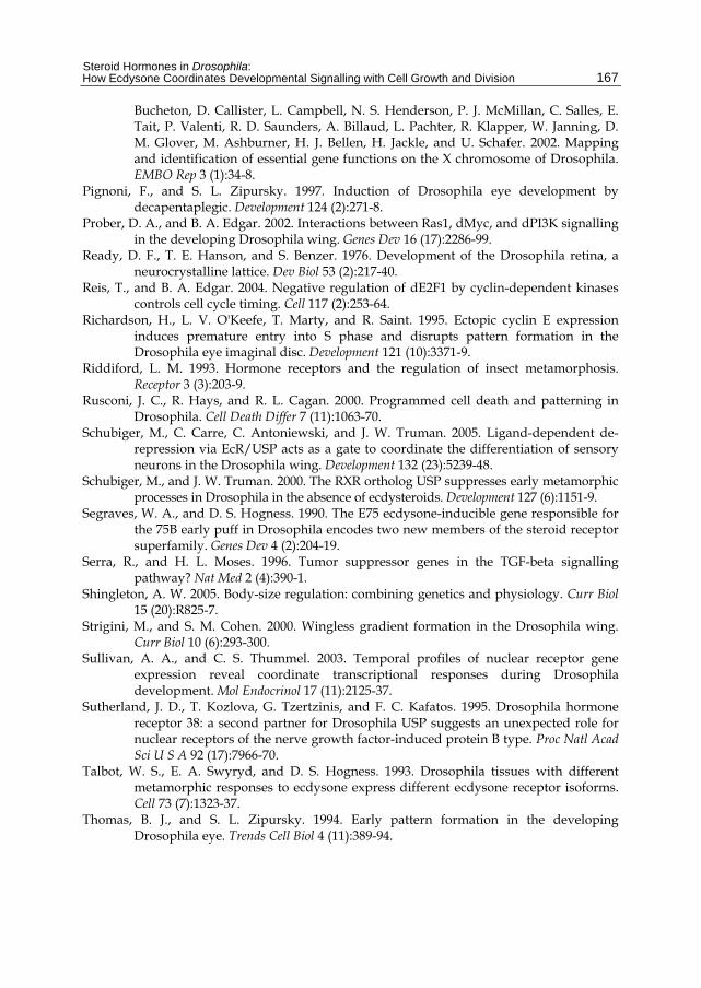
Steroid Hormones in Drosophila: How Ecdysone Coordinates Developmental Signalling with Cell Growth and Division
167
Bucheton, D. Callister, L. Campbell, N. S. Henderson, P. J. McMillan, C. Salles, E. Tait, P. Valenti, R. D. Saunders, A. Billaud, L. Pachter, R. Klapper, W. Janning, D. M. Glover, M. Ashburner, H. J. Bellen, H. Jackle, and U. Schafer. 2002. Mapping and identification of essential gene functions on the X chromosome of Drosophila. EMBO Rep 3 (1):34-8.
Pignoni, F., and S. L. Zipursky. 1997. Induction of Drosophila eye development by decapentaplegic. Development 124 (2):271-8.
Prober, D. A., and B. A. Edgar. 2002. Interactions between Ras1, dMyc, and dPI3K signalling in the developing Drosophila wing. Genes Dev 16 (17):2286-99.
Ready, D. F., T. E. Hanson, and S. Benzer. 1976. Development of the Drosophila retina, a neurocrystalline lattice. Dev Biol 53 (2):217-40.
Reis, T., and B. A. Edgar. 2004. Negative regulation of dE2F1 by cyclin-dependent kinases controls cell cycle timing. Cell 117 (2):253-64.
Richardson, H., L. V. O'Keefe, T. Marty, and R. Saint. 1995. Ectopic cyclin E expression induces premature entry into S phase and disrupts pattern formation in the Drosophila eye imaginal disc. Development 121 (10):3371-9.
Riddiford, L. M. 1993. Hormone receptors and the regulation of insect metamorphosis. Receptor 3 (3):203-9.
Rusconi, J. C., R. Hays, and R. L. Cagan. 2000. Programmed cell death and patterning in Drosophila. Cell Death Differ 7 (11):1063-70.
Schubiger, M., C. Carre, C. Antoniewski, and J. W. Truman. 2005. Ligand-dependent de-repression via EcR/USP acts as a gate to coordinate the differentiation of sensory neurons in the Drosophila wing. Development 132 (23):5239-48.
Schubiger, M., and J. W. Truman. 2000. The RXR ortholog USP suppresses early metamorphic processes in Drosophila in the absence of ecdysteroids. Development 127 (6):1151-9.
Segraves, W. A., and D. S. Hogness. 1990. The E75 ecdysone-inducible gene responsible for the 75B early puff in Drosophila encodes two new members of the steroid receptor superfamily. Genes Dev 4 (2):204-19.
Serra, R., and H. L. Moses. 1996. Tumor suppressor genes in the TGF-beta signalling pathway? Nat Med 2 (4):390-1.
Shingleton, A. W. 2005. Body-size regulation: combining genetics and physiology. Curr Biol 15 (20):R825-7.
Strigini, M., and S. M. Cohen. 2000. Wingless gradient formation in the Drosophila wing. Curr Biol 10 (6):293-300.
Sullivan, A. A., and C. S. Thummel. 2003. Temporal profiles of nuclear receptor gene expression reveal coordinate transcriptional responses during Drosophila development. Mol Endocrinol 17 (11):2125-37.
Sutherland, J. D., T. Kozlova, G. Tzertzinis, and F. C. Kafatos. 1995. Drosophila hormone receptor 38: a second partner for Drosophila USP suggests an unexpected role for nuclear receptors of the nerve growth factor-induced protein B type. Proc Natl Acad Sci U S A 92 (17):7966-70.
Talbot, W. S., E. A. Swyryd, and D. S. Hogness. 1993. Drosophila tissues with different metamorphic responses to ecdysone express different ecdysone receptor isoforms. Cell 73 (7):1323-37.
Thomas, B. J., and S. L. Zipursky. 1994. Early pattern formation in the developing Drosophila eye. Trends Cell Biol 4 (11):389-94.
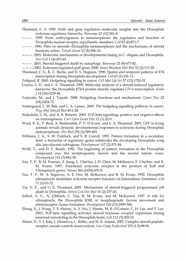
Steroids – Basic Science
168
Thummel, C. S. 1990. Puffs and gene regulation--molecular insights into the Drosophila ecdysone regulatory hierarchy. Bioessays 12 (12):561-8.
———. 1995. From embryogenesis to metamorphosis: the regulation and function of Drosophila nuclear receptor superfamily members. Cell 83 (6):871-7.
———. 1996. Files on steroids--Drosophila metamorphosis and the mechanisms of steroid hormone action. Trends Genet 12 (8):306-10.
———. 2001. Molecular mechanisms of developmental timing in C. elegans and Drosophila. Dev Cell 1 (4):453-65.
———. 2001. Steroid-triggered death by autophagy. Bioessays 23 (8):677-82. ———. 2002. Ecdysone-regulated puff genes 2000. Insect Biochem Mol Biol 32 (2):113-20. Thummel, C. S., K. C. Burtis, and D. S. Hogness. 1990. Spatial and temporal patterns of E74
transcription during Drosophila development. Cell 61 (1):101-11. Toftgard, R. 2000. Hedgehog signalling in cancer. Cell Mol Life Sci 57 (12):1720-31. Urness, L. D., and C. S. Thummel. 1995. Molecular analysis of a steroid-induced regulatory
hierarchy: the Drosophila E74A protein directly regulates L71-6 transcription. Embo J 14 (24):6239-46.
Varjosalo, M., and J. Taipale. 2008. Hedgehog: functions and mechanisms. Genes Dev 22 (18):2454-72.
Vestergaard, J., M. Bak, and L. A. Larsen. 2005. The hedgehog signalling pathway in cancer. Prog Mol Subcell Biol 40:1-28.
Wakefield, L. M., and A. B. Roberts. 2002. TGF-beta signalling: positive and negative effects on tumorigenesis. Curr Opin Genet Dev 12 (1):22-9.
Ward, R. E., P. Reid, A. Bashirullah, P. P. D'Avino, and C. S. Thummel. 2003. GFP in living animals reveals dynamic developmental responses to ecdysone during Drosophila metamorphosis. Dev Biol 256 (2):389-402.
Williams, J. A., S. W. Paddock, and S. B. Carroll. 1993. Pattern formation in a secondary field: a hierarchy of regulatory genes subdivides the developing Drosophila wing disc into discrete subregions. Development 117 (2):571-84.
Wolff, T., and D. F. Ready. 1991. The beginning of pattern formation in the Drosophila compound eye: the morphogenetic furrow and the second mitotic wave. Development 113 (3):841-50.
Yao, T. P., B. M. Forman, Z. Jiang, L. Cherbas, J. D. Chen, M. McKeown, P. Cherbas, and R. M. Evans. 1993. Functional ecdysone receptor is the product of EcR and Ultraspiracle genes. Nature 366 (6454):476-9.
Yao, T. P., W. A. Segraves, A. E. Oro, M. McKeown, and R. M. Evans. 1992. Drosophila ultraspiracle modulates ecdysone receptor function via heterodimer formation. Cell 71 (1):63-72.
Yin, V. P., and C. S. Thummel. 2005. Mechanisms of steroid-triggered programmed cell death in Drosophila. Semin Cell Dev Biol 16 (2):237-43.
Zelhof, A. C., N. Ghbeish, C. Tsai, R. M. Evans, and M. McKeown. 1997. A role for ultraspiracle, the Drosophila RXR, in morphogenetic furrow movement and photoreceptor cluster formation. Development 124 (13):2499-506.
Zheng, X., J. Wang, T. E. Haerry, A. Y. Wu, J. Martin, M. B. O'Connor, C. H. Lee, and T. Lee. 2003. TGF-beta signalling activates steroid hormone receptor expression during neuronal remodeling in the Drosophila brain. Cell 112 (3):303-15.
Zitnan, D., Y. J. Kim, I. Zitnanova, L. Roller, and M. E. Adams. 2007. Complex steroid-peptide-receptor cascade controls insect ecdysis. Gen Comp Endocrinol 153 (1-3):88-96.
Related Documents

![Cooperative Control of Ecdysone Biosynthesis in Drosophila by Transcription … · 2018. 1. 25. · UAS-spo-HA and UAS-nvd-Bm[WT]-HA were previously described (Namiki et al. 2005;](https://static.cupdf.com/doc/110x72/60a64a29bea7ba789815550e/cooperative-control-of-ecdysone-biosynthesis-in-drosophila-by-transcription-2018.jpg)


![RESEARCH ARTICLE Open Access Controlled expression of ......ecdysone controlled systems [14]. An improved version of the ecdysone-inducible approach is a two-hybrid ver-sion, also](https://static.cupdf.com/doc/110x72/60cdfe486cb19b23041f3d6e/research-article-open-access-controlled-expression-of-ecdysone-controlled.jpg)


