ORIGINAL ARTICLE Stereotactic Body Radiation Therapy (SBRT) for lung metastases PAUL OKUNIEFF 1 , ANNCATRINE L. PETERSEN 1 , ABRAHAM PHILIP 1 , MICHAEL T. MILANO 1 , ALAN W. KATZ 1 , LASZLO BOROS 2 & MICHAEL C. SCHELL 1 1 Department of Radiation Oncology, University of Rochester Medical Center, Rochester, NY, USA and, 2 Division of Medical Oncology, University of Rochester Medical Center, Rochester, NY, USA Abstract The curative treatment of oligometastases with radiotherapy remains an area of active investigation. We hypothesise that treating oligometastases with SBRT can prolong life and potentially cure patients, while in patients with multiple lung metastases SBRT can improve quality of life. Fifty patients with lung metastases were treated on this study. Individuals with five or fewer total lesions were treated with curative intent. Individuals with /five metastases were treated palliatively. Most patients (62%) received 5 Gy/fraction for a total of 50 Gy. The number of targets treated per patient ranged from one to five (mean 2.6). Tumor sizes ranged from 0.3 7.7 cm in maximal diameter (median 2.1 cm). Mean follow-up was 18.7 months. Local control of treated lesions was obtained in 42 of 49 evaluable patients (83%). Of the 125 total lesions treated, eight progressed after treatment (94% crude local control). The median overall survival time from time of treatment completion of the curatively treated patients was 23.4 months. The progression-free survival of the same group of patients was 25% and 16% at 12 and 24 months, respectively. Grade 1 toxicity occurred in 35% of all the patients, 6.1% had grade 2 toxicity, and 2% had grade 3 toxicity. Excellent local tumor control rates with low toxicity are seen with SBRT. Median survival time and progression-free survival both appear better than that achieved with standard care alone. Long-term progression-free survival can be seen in a subset of patients when all tumors are targeted For most cancers, the primary cancer itself can be controlled locally [1]. The usual approach to treat- ment of the primary tumor features combined modality therapy, emphasizing a local therapy, typically surgery or radiation. Deaths most com- monly result from tumor that has spread. Therapy for metastastic tumors, unlike that for primary tumors, typically features only systemic chemother- apy or palliative radiation. The latter is given at ‘‘palliative’’ doses that are not expected to be sterilizing, but which reduce symptoms and delay tumor growth. Metastatic disease to the lung is one of the most common life threatening complications of cancer. Metastases to the lung are very common and can be seen with most cancer types. The lethal outcome is hypoxia with ultimate asphyxiation. The impact of lung metastases is substantial, with most patients surviving only about a year and rare survivors at three years [2]. Thus treatments that might reduce the severity of, or that delay the onset of, hypoxia without themselves altering pulmonary function are of great interest. Indeed there is evidence of long-term survi- val for surgical metastectomy for sarcoma and breast cancer, though the approach is not fully utilized [2]. Chemotherapy remains the standard of care and can prolong survival time, but long-term survivors with metastatic disease are extremely rare. Progres- sion-free survivors after chemotherapy are not gen- erally expected; however effective chemotherapy, which produces a complete or near complete re- sponse, might successfully down-stage micrometa- static disease, leaving patients oligometastastic. Oligometastasis refers to a situation wherein metas- tases might not be disseminated, but rather present only in a few sites. In this case, it is possible that chemotherapy down-staging can leave a patient curable by local therapy alone. Surgery might be considered for easily resectable lesions. Other mini- mally invasive or non-invasive therapies might also be considered for control of macroscopic residual disease. Correspondence: Paul Okunieff, Department of Radiation Oncology, University of Rochester Medical Center, 601 Elmwood Avenue, Rochester, NY 14642- 8647, USA. Tel: /1-585-275-5575. Fax: /1-585-275-1531. E-mail: [email protected] Acta Oncologica, 2006; 45: 808 817 (Received 16 June 2006; accepted 11 July 2006) ISSN 0284-186X print/ISSN 1651-226X online # 2006 Taylor & Francis DOI: 10.1080/02841860600908954 Acta Oncol Downloaded from informahealthcare.com by 186.88.19.149 on 05/20/14 For personal use only.

Welcome message from author
This document is posted to help you gain knowledge. Please leave a comment to let me know what you think about it! Share it to your friends and learn new things together.
Transcript
ORIGINAL ARTICLE
Stereotactic Body Radiation Therapy (SBRT) for lung metastases
PAUL OKUNIEFF1, ANNCATRINE L. PETERSEN1, ABRAHAM PHILIP1, MICHAEL T.
MILANO1, ALAN W. KATZ1, LASZLO BOROS2 & MICHAEL C. SCHELL1
1Department of Radiation Oncology, University of Rochester Medical Center, Rochester, NY, USA and, 2Division of Medical
Oncology, University of Rochester Medical Center, Rochester, NY, USA
AbstractThe curative treatment of oligometastases with radiotherapy remains an area of active investigation. We hypothesise thattreating oligometastases with SBRT can prolong life and potentially cure patients, while in patients with multiple lungmetastases SBRT can improve quality of life. Fifty patients with lung metastases were treated on this study. Individuals withfive or fewer total lesions were treated with curative intent. Individuals with�/five metastases were treated palliatively. Mostpatients (62%) received 5 Gy/fraction for a total of 50 Gy. The number of targets treated per patient ranged from one to five(mean 2.6). Tumor sizes ranged from 0.3�7.7 cm in maximal diameter (median 2.1 cm). Mean follow-up was 18.7 months.Local control of treated lesions was obtained in 42 of 49 evaluable patients (83%). Of the 125 total lesions treated, eightprogressed after treatment (94% crude local control). The median overall survival time from time of treatment completionof the curatively treated patients was 23.4 months. The progression-free survival of the same group of patients was 25% and16% at 12 and 24 months, respectively. Grade 1 toxicity occurred in 35% of all the patients, 6.1% had grade 2 toxicity, and2% had grade 3 toxicity. Excellent local tumor control rates with low toxicity are seen with SBRT. Median survival time andprogression-free survival both appear better than that achieved with standard care alone. Long-term progression-freesurvival can be seen in a subset of patients when all tumors are targeted
For most cancers, the primary cancer itself can be
controlled locally [1]. The usual approach to treat-
ment of the primary tumor features combined
modality therapy, emphasizing a local therapy,
typically surgery or radiation. Deaths most com-
monly result from tumor that has spread. Therapy
for metastastic tumors, unlike that for primary
tumors, typically features only systemic chemother-
apy or palliative radiation. The latter is given at
‘‘palliative’’ doses that are not expected to be
sterilizing, but which reduce symptoms and delay
tumor growth.
Metastatic disease to the lung is one of the most
common life threatening complications of cancer.
Metastases to the lung are very common and can be
seen with most cancer types. The lethal outcome is
hypoxia with ultimate asphyxiation. The impact of
lung metastases is substantial, with most patients
surviving only about a year and rare survivors at three
years [2]. Thus treatments that might reduce the
severity of, or that delay the onset of, hypoxia without
themselves altering pulmonary function are of great
interest. Indeed there is evidence of long-term survi-
val for surgical metastectomy for sarcoma and breast
cancer, though the approach is not fully utilized [2].
Chemotherapy remains the standard of care and
can prolong survival time, but long-term survivors
with metastatic disease are extremely rare. Progres-
sion-free survivors after chemotherapy are not gen-
erally expected; however effective chemotherapy,
which produces a complete or near complete re-
sponse, might successfully down-stage micrometa-
static disease, leaving patients oligometastastic.
Oligometastasis refers to a situation wherein metas-
tases might not be disseminated, but rather present
only in a few sites. In this case, it is possible that
chemotherapy down-staging can leave a patient
curable by local therapy alone. Surgery might be
considered for easily resectable lesions. Other mini-
mally invasive or non-invasive therapies might also
be considered for control of macroscopic residual
disease.
Correspondence: Paul Okunieff, Department of Radiation Oncology, University of Rochester Medical Center, 601 Elmwood Avenue, Rochester, NY 14642-
8647, USA. Tel: �/1-585-275-5575. Fax: �/1-585-275-1531. E-mail: [email protected]
Acta Oncologica, 2006; 45: 808�817
(Received 16 June 2006; accepted 11 July 2006)
ISSN 0284-186X print/ISSN 1651-226X online # 2006 Taylor & Francis
DOI: 10.1080/02841860600908954
Act
a O
ncol
Dow
nloa
ded
from
info
rmah
ealth
care
.com
by
186.
88.1
9.14
9 on
05/
20/1
4Fo
r pe
rson
al u
se o
nly.
Stereotactic Body Radiation Therapy (SBRT) is a
method for delivering focused radiation fields target-
ing almost exclusively the tumor nidus while exclud-
ing tissues not grossly involved with tumor. We
hypothesized that aggressive management of pul-
monary metastases, particularly in the presence of
effective chemotherapy, would reduce pulmonary
complications of cancer and prolong survival.
In this study we investigated the feasibility and
potential utility of SBRT (using Novalis† Shaped
Beam SurgeryTM) combined with respiratory gating
in patients with lung metastases. High total dose
radiation therapy with hypofractionation was used
with the aim of achieving permanent local control.
Materials and methods
Patient characteristics
Between February 2001 and December 2005, 50
patients were treated on this phase II study of hypo-
frationated SBRT for lung metastases. The study
was approved by the University of Rochester Med-
ical Center institutional review board. Patients were
stratified into two categories. Those with metastatic
disease confined to the thorax and/or with total
metastases limited to five total lesions (termed
curative), and those with more extensive disease
wherein lung metastasis were considered the most
life limiting component of their disease (termed
palliative). Sequential and concurrent chemotherapy
was encouraged. While patients were allowed to
enroll with or without a chemotherapy response,
most patients who enrolled had failed adjuvant
chemotherapy (if applicable) and at least one course
of systemic chemotherapy. Concurrent anthrocy-
clines were excluded. Patients with poor baseline
pulmonary function or extensive emphysema were
not excluded, but there were more stringent dose
volume histography (DVH) constraints imposed.
Patients with local control and minimal side effects
after a previous course of SBRT were allowed to
undergo repeat cycles of SBRT for new lesions. In this
analysis only the index lesions are reported according
to protocol, though treatment of additional lesions
may have been needed to obtain disease free status.
Stereotactic Body Radiation Therapy technique
Patient immobilization for initial simulation and
treatment was accomplished using the ExacTrac†
patient positing platform (BrainLAB† Inc.). This
platform consists of a vacuum cushion for initial
positioning, and infrared reflecting body fiducial
markers monitored by two ceiling mounted infrared
cameras. Respiratory gating was accomplished using
relaxed expiratory breath hold techniques. The
therapist monitors, real time, the infrared system
and turns off the beam if any axial dimension
exceeds 1 mm. The beam is not turned on unless
all axial dimensions are under 0.5 mm. Using this
technique we have shown the standard deviation of
motion between treatment is under 1.8 mm anterior-
posterior and lateral, and under 2.3 mm superior-
inferior [3]. Quality assurance is further confirmed
by a minimum of four computerized tomographic
(CT) scans per patient.
Treatment planning was performed using a 3D
treatment planning system. CT supplemented by
magnetic resonance imaging (MRI) and positron
emission tomography (PET) data was used to ensure
the accurate definition of gross tumor volume
(GTV). The use of arcs and non-coplanar beams
was encouraged. A DVH was calculated of any
irradiated vital organs including the lung, liver,
spinal cord, stomach, cardiac ventricles, esophagus,
and kidneys. The allowed maximum DVH for the
liver, lung, and spinal cord was specified. There was
no specific size limit placed on tumor diameter.
Instead, the DVH was used to control the size and
number of tumors that could be safely treated. The
combined left and right lung DVH was used
to calculate the percentage of total lung volume
(excluding the GTV) receiving]/10 Gy (V10) and
]/20 Gy (V20).
The target volume was the GTV with no expan-
sion for the clinical target volume (CTV). The GTV
was defined by CT, MRI, or image fusion of both.
The target volume was determined by a radiation
oncologist certified for submission of patients onto
the protocol. The volume includes imaging abnorm-
alities believed to encompass the metastasis(es) but
not microscopic extension. While it is known that
tumors can infiltrate about the imaged abnormality,
infiltration was not included in the target volume.
The 80% isodose volume (defined as the planning
target volume (PTV)) was designed to include the
GTV with 7 mm of lateral and anterior-posterior
margin, and 10 mm of superior inferior margin.
There was no effort made to assure homogeneous
dose to the isocenter or throughout the CTV, and
the prescription dose was the isocenter dose (100%
isodose point).
The target dose acceptability was determined
based on the DVH of normal (uninvolved) lung
and surrounding organs. Normal lung volume was
defined as the portion of the lungs not radiographi-
cally involved by the GTV. Briefly, patients were
required to have 1000 ml of tumor-free lung. For
patients with chronic obstructive pulmonary disease
or existing chronic lung disease, 70% of the lung or
800 ml (whichever was larger), was kept under
1.7 Gy per fraction for a total dose of less than
SBRT for lung metastases 809
Act
a O
ncol
Dow
nloa
ded
from
info
rmah
ealth
care
.com
by
186.
88.1
9.14
9 on
05/
20/1
4Fo
r pe
rson
al u
se o
nly.
17 Gy. For patients with otherwise healthy lungs,
60% of the lung was kept under 2.0 Gy per fraction
for a total dose of less than 20 Gy.
Follow-up
Patients were evaluated by the treating physician
weekly during treatment including physical exam
and appropriate blood work. Additional follow-up
visits were planned at one month following comple-
tion of treatment and every three months after that
for 24 months. Imaging at each visit included at
minimum 3D evaluation of the chest (such as CT9/
PET) and other studies as clinically indicated (such
as bone scan). Most surviving patients maintained a
rigorous three-month visit schedule after official
protocol completion, and all patients have continued
close follow-up schedules.
Evaluation of response
The primary objective was to determine if radio-
therapy for lung metastases combined with standard
therapy is feasible and shows promise in a phase II
study. The primary endpoint was local control.
Secondary endpoints included regional control,
and distant metastasis frequency. Response Evalua-
tion Criteria in Solid Tumors (RECIST) was also
evaluated [4]. Local control differs from a complete
response in that tumor shrinkage is not required, but
there must never be any re-growth. A tumor is not
locally controlled if there is ever any progression of
that individual treated lesion. A regional failure is
defined as a failure within the thorax but outside the
80% isodose volume, and a distant failure is one
outside the thorax. The study was powered to
demonstrate a local control rate of]/50%. Pulmon-
ary and other toxicity was scored using CTCAE v
3.0 [5]. A study objective was to keep grade 3
toxicity as low as possible, and at maximum 5%.
Examination of response was made serially over
time using the follow-up 3D imaging of the chest.
The treated lesions were assessed by the diagnostic
radiologists and three radiation oncologists on each
scan. The time of disease progression included all
local, regional, or distant failures. Actuarial overall
survival, progression-free survival, and local control
rate were defined in the usual way using the Kaplan-
Meier method. There were no non-cancer related
deaths.
Results
Patient characteristics and treatment planning
Between February 2001 and December 2005, 50
patients (49 evaluable, one patient did not return for
follow-up) with 125 lung lesions were treated. Thirty
were treated curatively and 19 palliatively. Patient
ages ranged from 37 to 86 years (median 60 years),
and the number of index targets treated per patient
ranged from 1�5 (mean 2.6). Tumor sizes ranged
from 0.3�7.7 cm in maximal diameter (median
2.1 cm) on CT. The GTV volume ranged from
0.1�125 ml (median 4.7 ml). Mean follow-up was
18.7 months (range 3.7�60.9). Most patients had
undergone multiple therapies for their metastatic
disease and the median time from first diagnosis
of metastatic disease to referral and radiation was
14.8 months. For more details see Table I.
The total dose delivered to the tumor isocenter
was typically 50 to 55 Gy, usually at 5 Gy per dose,
but varied from 2.5 and 6.5 Gy per dose. The
fractionation was determined by restrictions im-
posed by the normal tissue DVH as discussed in
the Materials and methods section. The preferred
fractionation and dose to the tumor isocenter was 50
Gy at 5 Gy/fraction (31 of 49 patients).
Thirty-seven of the 49 patients had received
previous chemotherapy and most had also received
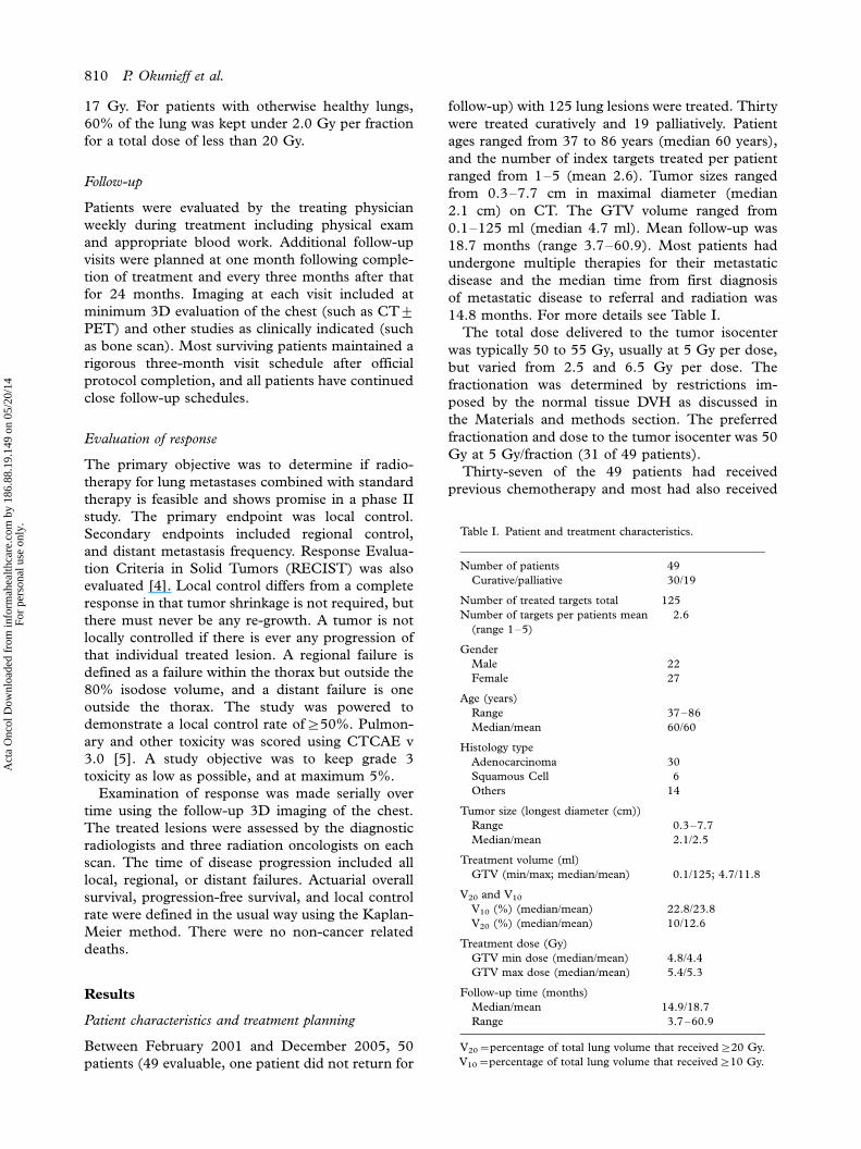
Table I. Patient and treatment characteristics.
Number of patients 49
Curative/palliative 30/19
Number of treated targets total 125
Number of targets per patients mean
(range 1�5)
2.6
Gender
Male 22
Female 27
Age (years)
Range 37�86
Median/mean 60/60
Histology type
Adenocarcinoma 30
Squamous Cell 6
Others 14
Tumor size (longest diameter (cm))
Range 0.3�7.7
Median/mean 2.1/2.5
Treatment volume (ml)
GTV (min/max; median/mean) 0.1/125; 4.7/11.8
V20 and V10
V10 (%) (median/mean) 22.8/23.8
V20 (%) (median/mean) 10/12.6
Treatment dose (Gy)
GTV min dose (median/mean) 4.8/4.4
GTV max dose (median/mean) 5.4/5.3
Follow-up time (months)
Median/mean 14.9/18.7
Range 3.7�60.9
V20�/percentage of total lung volume that received]/20 Gy.
V10�/percentage of total lung volume that received]/10 Gy.
810 P. Okunieff et al.
Act
a O
ncol
Dow
nloa
ded
from
info
rmah
ealth
care
.com
by
186.
88.1
9.14
9 on
05/
20/1
4Fo
r pe
rson
al u
se o
nly.
chemotherapy or other cytotoxic therapy following
focal irradiation. Though concurrent chemotherapy
was permitted, the treating oncologists commonly
held a cycle during the irradiation.
Response
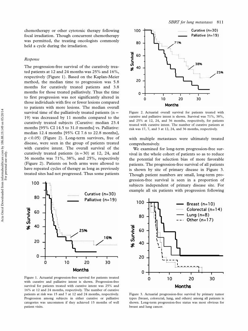
The progression-free survival of the curatively trea-
ted patients at 12 and 24 months was 25% and 16%,
respectively (Figure 1). Based on the Kaplan-Meier
method, the median time to progression was 5.8
months for curatively treated patients and 3.8
months for those treated palliatively. Thus the time
to first progression was not significantly altered in
those individuals with five or fewer lesions compared
to patients with more lesions. The median overall
survival time of the palliatively treated patients (n�/
19) was decreased by 11 months compared to the
curatively treated subjects (Curative: median 23.4
months [95% CI 14.5 to 31.0 months] vs. Palliative:
median 12.4 months [95% CI 7.6 to 22.8 months],
pB/0.05) (Figure 2). Long-term survivors, free of
disease, were seen in the group of patients treated
with curative intent. The overall survival of the
curatively treated patients (n�/30) at 12, 24, and
36 months was 71%, 38%, and 25%, respectively
(Figure 2). Patients on both arms were allowed to
have repeated cycles of therapy as long as previously
treated sites had not progressed. Thus some patients
with multiple metastases were ultimately treated
comprehensively.
We examined for long-term progression-free sur-
vival in the whole cohort of patients so as to reduce
the potential for selection bias of more favorable
patients. The progression-free survival of all patients
is shown by site of primary disease in Figure 3.
Though patient numbers are small, long-term pro-
gression-free survival is seen in a proportion of
subjects independent of primary disease site. For
example all six patients with progression following
Figure 1. Actuarial progression-free survival for patients treated
with curative and palliative intent is shown. Progression-free
survival for patients treated with curative intent was 25% and
16% at 12 and 24 months, respectively. The number of curative
patients at risk was 15 and 7 at 12 and 24 months, respectively.
Progression among subjects in either curative or palliative
categories was uncommon if they achieved 15 months of well
patient visits.
Figure 2. Actuarial overall survival for patients treated with
curative and palliative intent is shown. Survival was 71%, 38%,
and 25% at 12, 24, and 36 months, respectively, for patients
treated with curative intent. The number of curative patients at
risk was 17, 7, and 3 at 12, 24, and 36 months, respectively.
Figure 3. Actuarial progression-free survival by primary tumor
types (breast, colorectal, lung, and others) among all patients is
shown. Long-term progression-free status was most obvious for
breast and lung cancer.
SBRT for lung metastases 811
Act
a O
ncol
Dow
nloa
ded
from
info
rmah
ealth
care
.com
by
186.
88.1
9.14
9 on
05/
20/1
4Fo
r pe
rson
al u
se o
nly.
treatment for primary lung cancer have died, while
the two living patients (23 and 33 months after
treatment) remain free of progression.
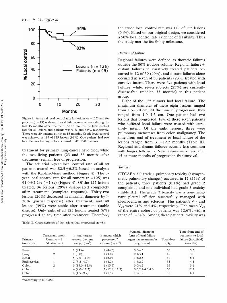
The actuarial 3-year local control rate of all 49
patients treated was 82.59/6.2% based on analysis
with the Kaplan-Meier method (Figure 4). The 3-
year local control rate for all tumors (n�/125) was
91.09/3.2% (9/1 se) (Figure 4). Of the 125 lesions
treated, 36 lesions (29%) disappeared completely
after treatment (complete response). Thirty-two
lesions (26%) decreased in maximal diameter by]/
30% (partial response) after treatment, and 49
lesions (39%) were stable after treatment (stable
disease). Only eight of all 125 lesions treated (6%)
progressed at any time after treatment. Therefore,
the crude local control rate was 117 of 125 lesions
(94%). Based on our original design, we considered
a 50% local control rate evidence of feasibility. Thus
the study met the feasibility milestone.
Pattern of failure
Regional failures were defined as thoracic failures
outside the 80% isodose volume. Regional failure9/
distant failures in curatively treated patients oc-
curred in 12 of 30 (40%), and distant failures alone
occurred in seven of 30 patients (23%) treated with
curative intent. There were five patients with local
failures, while, seven subjects (23%) are currently
disease-free (median 33 months) in this patient
group.
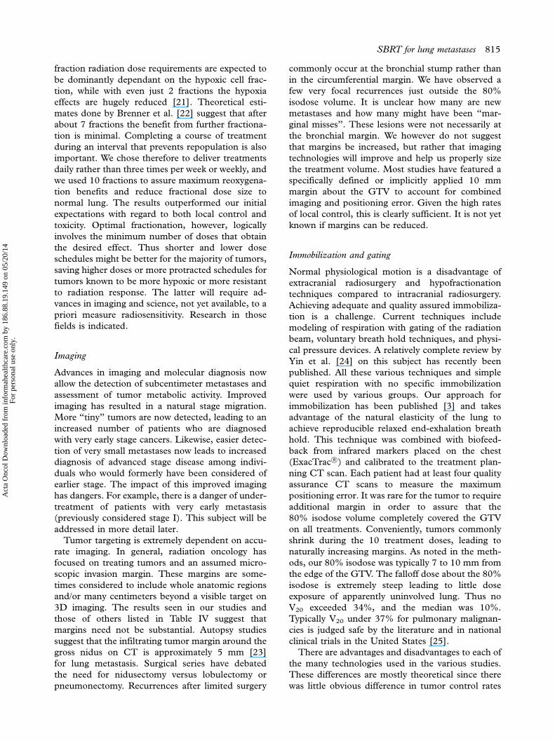
Eight of the 125 tumors had local failure. The
maximum diameter of these eight lesions ranged
from 1.5�3.0 cm. At the time of progression, they
ranged from 1.9�4.5 cm. One patient had two
lesions that progressed. Five of these seven patients
who suffered local failure were treated with cura-
tively intent. Of the eight lesions, three were
pulmonary metastases from colon malignancy. The
time from end of treatment to local failure of the
lesions ranged from 3.1�12.2 months (Table II).
Regional and distant failures became less common
with longer follow-up. New failures were rare after
15 or more months of progression-free survival.
Toxicity
CTCAE v 3.0 grade 1 pulmonary toxicity (asympto-
matic pulmonary changes) occurred in 17 (35%) of
the patients, three patients (6.1%) had grade 2
complaints, and one individual had grade 3 toxicity
(Table III). The grade 3 toxicity was a non-malig-
nant pleural effusion successfully managed with
pleurocentesis and sclerosis. This patient’s V10 and
V20 were 21% and 4%, respectively. The mean V20
of the entire cohort of patients was 12.6%, with a
range of 1�34%. Among these patients, toxicity was
Figure 4. Actuarial local control rate for lesions (n�/125) and for
patients (n�/49) is shown. Local failures were all seen during the
first 15 months after treatment. At 15 months the local control
rate for all lesions and patients was 91% and 83%, respectively.
There were 20 patients at risk at 15 months. Crude local control
was achieved in 117 of 125 lesions (94%). One patient had two
local failures leading to local control in 42 of 49 patients.
Table II. Characteristics of the lesions that progressed (n�/8).
Primary
tumor site
Treatment intent
Curative�/1
Palliative �/ 2
# total targets
treated (volume
range) (cm3)
# targets which
progressedA
(volume) (cm3)
Maximal diameter
(cm) of local failure
targets (at treatment/at
progression)
Total dose
(Gy)
Time from end of
treatment to local
failure (in-infield)
(months)
Breast 1 1 (44.6) 1 (44.6) 3.0/4.5 50 5.3
Lung 2 1 (3.8) 1 (3.8) 2.1/3.1 45 3.8
Renal 1 5 (2.0�11.8) 1 (2.0) 1.5/2.5 40 8.5
Endocervical 1 2 (3.2�4.2) 1 (4.2) 1.6/2.2 55 4.4
Colon 2 3 (15.3�82.8) 1 (15.3) 3.0/4.2 35 3.1
Colon 1 4 (4.0�17.3) 2 (12.8, 17.3) 3.0,2.2/4.0,4.0 50 12.2
Colon 1 4 (1.5�9.7) 1 (1.5) 1.5/1.9 50 6.1
AAccording to RECIST.
812 P. Okunieff et al.
Act
a O
ncol
Dow
nloa
ded
from
info
rmah
ealth
care
.com
by
186.
88.1
9.14
9 on
05/
20/1
4Fo
r pe
rson
al u
se o
nly.
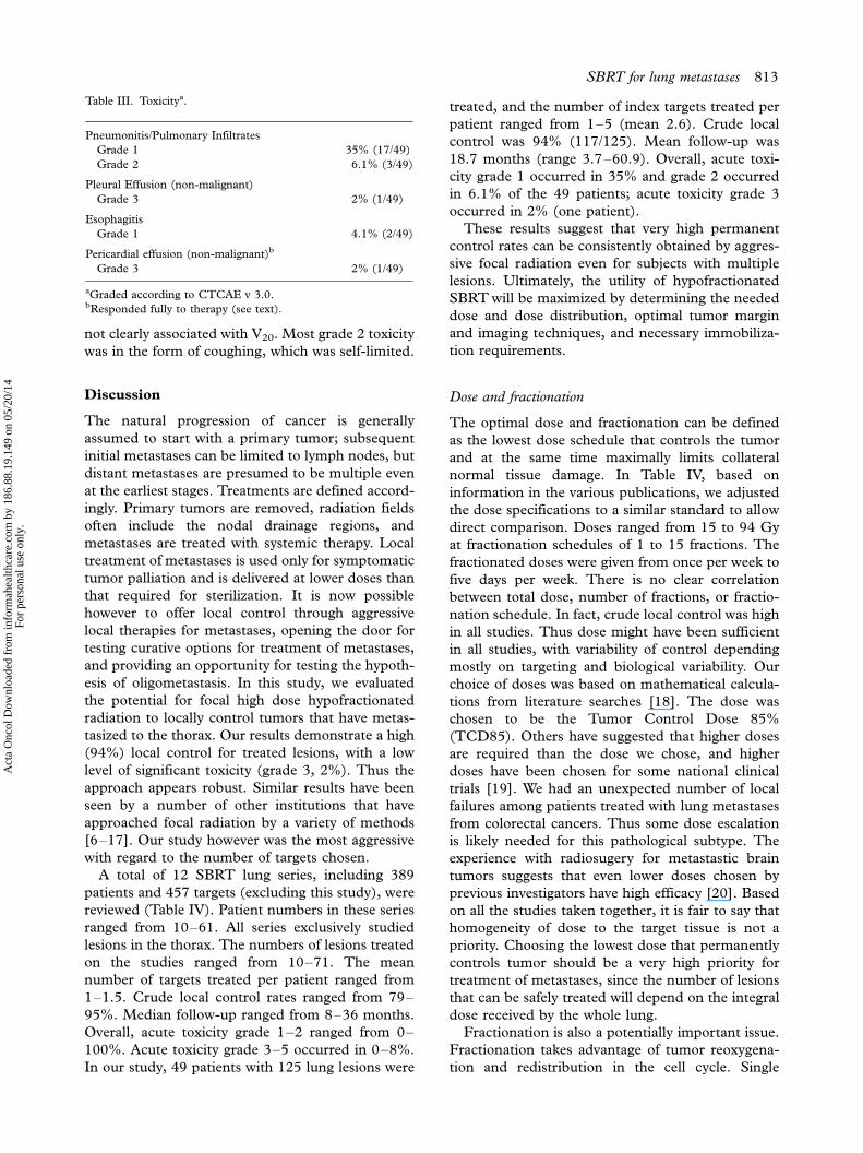
not clearly associated with V20. Most grade 2 toxicity
was in the form of coughing, which was self-limited.
Discussion
The natural progression of cancer is generally
assumed to start with a primary tumor; subsequent
initial metastases can be limited to lymph nodes, but
distant metastases are presumed to be multiple even
at the earliest stages. Treatments are defined accord-
ingly. Primary tumors are removed, radiation fields
often include the nodal drainage regions, and
metastases are treated with systemic therapy. Local
treatment of metastases is used only for symptomatic
tumor palliation and is delivered at lower doses than
that required for sterilization. It is now possible
however to offer local control through aggressive
local therapies for metastases, opening the door for
testing curative options for treatment of metastases,
and providing an opportunity for testing the hypoth-
esis of oligometastasis. In this study, we evaluated
the potential for focal high dose hypofractionated
radiation to locally control tumors that have metas-
tasized to the thorax. Our results demonstrate a high
(94%) local control for treated lesions, with a low
level of significant toxicity (grade 3, 2%). Thus the
approach appears robust. Similar results have been
seen by a number of other institutions that have
approached focal radiation by a variety of methods
[6�17]. Our study however was the most aggressive
with regard to the number of targets chosen.
A total of 12 SBRT lung series, including 389
patients and 457 targets (excluding this study), were
reviewed (Table IV). Patient numbers in these series
ranged from 10�61. All series exclusively studied
lesions in the thorax. The numbers of lesions treated
on the studies ranged from 10�71. The mean
number of targets treated per patient ranged from
1�1.5. Crude local control rates ranged from 79�95%. Median follow-up ranged from 8�36 months.
Overall, acute toxicity grade 1�2 ranged from 0�100%. Acute toxicity grade 3�5 occurred in 0�8%.
In our study, 49 patients with 125 lung lesions were
treated, and the number of index targets treated per
patient ranged from 1�5 (mean 2.6). Crude local
control was 94% (117/125). Mean follow-up was
18.7 months (range 3.7�60.9). Overall, acute toxi-
city grade 1 occurred in 35% and grade 2 occurred
in 6.1% of the 49 patients; acute toxicity grade 3
occurred in 2% (one patient).
These results suggest that very high permanent
control rates can be consistently obtained by aggres-
sive focal radiation even for subjects with multiple
lesions. Ultimately, the utility of hypofractionated
SBRT will be maximized by determining the needed
dose and dose distribution, optimal tumor margin
and imaging techniques, and necessary immobiliza-
tion requirements.
Dose and fractionation
The optimal dose and fractionation can be defined
as the lowest dose schedule that controls the tumor
and at the same time maximally limits collateral
normal tissue damage. In Table IV, based on
information in the various publications, we adjusted
the dose specifications to a similar standard to allow
direct comparison. Doses ranged from 15 to 94 Gy
at fractionation schedules of 1 to 15 fractions. The
fractionated doses were given from once per week to
five days per week. There is no clear correlation
between total dose, number of fractions, or fractio-
nation schedule. In fact, crude local control was high
in all studies. Thus dose might have been sufficient
in all studies, with variability of control depending
mostly on targeting and biological variability. Our
choice of doses was based on mathematical calcula-
tions from literature searches [18]. The dose was
chosen to be the Tumor Control Dose 85%
(TCD85). Others have suggested that higher doses
are required than the dose we chose, and higher
doses have been chosen for some national clinical
trials [19]. We had an unexpected number of local
failures among patients treated with lung metastases
from colorectal cancers. Thus some dose escalation
is likely needed for this pathological subtype. The
experience with radiosugery for metastastic brain
tumors suggests that even lower doses chosen by
previous investigators have high efficacy [20]. Based
on all the studies taken together, it is fair to say that
homogeneity of dose to the target tissue is not a
priority. Choosing the lowest dose that permanently
controls tumor should be a very high priority for
treatment of metastases, since the number of lesions
that can be safely treated will depend on the integral
dose received by the whole lung.
Fractionation is also a potentially important issue.
Fractionation takes advantage of tumor reoxygena-
tion and redistribution in the cell cycle. Single
Table III. Toxicitya.
Pneumonitis/Pulmonary Infiltrates
Grade 1 35% (17/49)
Grade 2 6.1% (3/49)
Pleural Effusion (non-malignant)
Grade 3 2% (1/49)
Esophagitis
Grade 1 4.1% (2/49)
Pericardial effusion (non-malignant)b
Grade 3 2% (1/49)
aGraded according to CTCAE v 3.0.bResponded fully to therapy (see text).
SBRT for lung metastases 813
Act
a O
ncol
Dow
nloa
ded
from
info
rmah
ealth
care
.com
by
186.
88.1
9.14
9 on
05/
20/1
4Fo
r pe
rson
al u
se o
nly.
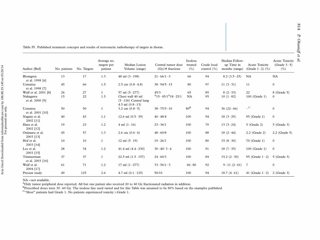
Table IV. Published treatment concepts and results of stereotactic radiotherapy of targets in thorax.
Author [Ref] No. patients No. Targets
Average no.
targets per
patient
Median Lesion
Volume (range)
Central tumor dose
(Gy)/# fractions
Isodose
treated
(%)
Crude local
control (%)
Median Follow-
up Time in
months (range)
Acute Toxicity
(Grade 1�2) (%)
Acute Toxicity
(Grade 3�5)
(%)
Blomgren
et al. 1998 [6]
13 17 1.3 48 ml (3�198) 21�66/1�3 66 94 8.2 (3.5�25) NA NA
Uematsu
et al. 1998 [7]
45 66 1.5 2.5 cm (0.8�4.8) 38�94/5�15 80 97 11 (3�31) 11 0
Wulf et al. 2001 [8] 26 27 1 57 ml (5�277) 45/3 65 85 8 (2�33) 22 8 (Grade 5)
Nakagawa
et al. 2000 [9]
15 22 1.5 Chest wall 40 ml
(5�126) Central lung
4.5 ml (0.8�13)
A15�45/1A18�25/1 NA 95 10 (1�82) 100 (Grade 1) 0
Uematsu
et al. 2001 [10]
50 50 1 3.2 cm (0.8�5) 38�75/5�10 80B 94 36 (22�66) �C 0
Nagata et al.
2002 [11]
40 43 1.1 12.6 ml (0.5�39) 40�48/4 100 94 18 (3�29) 95 (Grade 1) 0
Hara et al.
2002 [12]
19 23 1.2 4 ml (1�16) 23�36/1 100 79 13 (3�24) 5 (Grade 2) 5 (Grade 3)
Onimaru et al.
2003 [13]
45 57 1.3 2.6 cm (0.6�6) 48�60/8 100 88 18 (2�44) 2.2 (Grade 2) 2.2 (Grade 5)
Hof et al.
2003 [14]
10 10 1 12 ml (5�19) 19�26/1 100 80 15 (8�30) 70 (Grade 1) 0
Lee et al.
2003 [15]
28 34 1.2 41.4 ml (4.4�230) 30�40/ 3�4 100 91 18 (7�35) 100 (Grade 1) 0
Timmerman
et al. 2003 [16]
37 37 1 22.5 ml (1.5�157) 24�60/3 100 84 15.2 (2�30) 95 (Grade 1�2) 5 (Grade 3)
Wulf et al.
2004 [17]
61 71 1.2 17 ml (1�277) 33�56/1�3 66�80 92 9�11 (2�61) 7 0
Present study 49 125 2.6 4.7 ml (0.1�125) 50/10 100 94 18.7 (4�61) 41 (Grade 1�2) 2 (Grade 3)
NA�/not available.AOnly tumor peripheral dose reported. All but one patient also received 20 to 40 Gy fractionated radiation in addition.BPrescribed doses were 30�60 Gy. The isodose line used varied and for this Table was assumed to be 80% based on the examples published.C‘‘Most’’ patients had Grade 1. No patients experienced toxicity�/Grade 1.
814
P.O
kunieff
etal.
Act
a O
ncol
Dow
nloa
ded
from
info
rmah
ealth
care
.com
by
186.
88.1
9.14
9 on
05/
20/1
4Fo
r pe
rson
al u
se o
nly.
fraction radiation dose requirements are expected to
be dominantly dependant on the hypoxic cell frac-
tion, while with even just 2 fractions the hypoxia
effects are hugely reduced [21]. Theoretical esti-
mates done by Brenner et al. [22] suggest that after
about 7 fractions the benefit from further fractiona-
tion is minimal. Completing a course of treatment
during an interval that prevents repopulation is also
important. We chose therefore to deliver treatments
daily rather than three times per week or weekly, and
we used 10 fractions to assure maximum reoxygena-
tion benefits and reduce fractional dose size to
normal lung. The results outperformed our initial
expectations with regard to both local control and
toxicity. Optimal fractionation, however, logically
involves the minimum number of doses that obtain
the desired effect. Thus shorter and lower dose
schedules might be better for the majority of tumors,
saving higher doses or more protracted schedules for
tumors known to be more hypoxic or more resistant
to radiation response. The latter will require ad-
vances in imaging and science, not yet available, to a
priori measure radiosensitivity. Research in those
fields is indicated.
Imaging
Advances in imaging and molecular diagnosis now
allow the detection of subcentimeter metastases and
assessment of tumor metabolic activity. Improved
imaging has resulted in a natural stage migration.
More ‘‘tiny’’ tumors are now detected, leading to an
increased number of patients who are diagnosed
with very early stage cancers. Likewise, easier detec-
tion of very small metastases now leads to increased
diagnosis of advanced stage disease among indivi-
duals who would formerly have been considered of
earlier stage. The impact of this improved imaging
has dangers. For example, there is a danger of under-
treatment of patients with very early metastasis
(previously considered stage I). This subject will be
addressed in more detail later.
Tumor targeting is extremely dependent on accu-
rate imaging. In general, radiation oncology has
focused on treating tumors and an assumed micro-
scopic invasion margin. These margins are some-
times considered to include whole anatomic regions
and/or many centimeters beyond a visible target on
3D imaging. The results seen in our studies and
those of others listed in Table IV suggest that
margins need not be substantial. Autopsy studies
suggest that the infiltrating tumor margin around the
gross nidus on CT is approximately 5 mm [23]
for lung metastasis. Surgical series have debated
the need for nidusectomy versus lobulectomy or
pneumonectomy. Recurrences after limited surgery
commonly occur at the bronchial stump rather than
in the circumferential margin. We have observed a
few very focal recurrences just outside the 80%
isodose volume. It is unclear how many are new
metastases and how many might have been ‘‘mar-
ginal misses’’. These lesions were not necessarily at
the bronchial margin. We however do not suggest
that margins be increased, but rather that imaging
technologies will improve and help us properly size
the treatment volume. Most studies have featured a
specifically defined or implicitly applied 10 mm
margin about the GTV to account for combined
imaging and positioning error. Given the high rates
of local control, this is clearly sufficient. It is not yet
known if margins can be reduced.
Immobilization and gating
Normal physiological motion is a disadvantage of
extracranial radiosurgery and hypofractionation
techniques compared to intracranial radiosurgery.
Achieving adequate and quality assured immobiliza-
tion is a challenge. Current techniques include
modeling of respiration with gating of the radiation
beam, voluntary breath hold techniques, and physi-
cal pressure devices. A relatively complete review by
Yin et al. [24] on this subject has recently been
published. All these various techniques and simple
quiet respiration with no specific immobilization
were used by various groups. Our approach for
immobilization has been published [3] and takes
advantage of the natural elasticity of the lung to
achieve reproducible relaxed end-exhalation breath
hold. This technique was combined with biofeed-
back from infrared markers placed on the chest
(ExacTrac†) and calibrated to the treatment plan-
ning CT scan. Each patient had at least four quality
assurance CT scans to measure the maximum
positioning error. It was rare for the tumor to require
additional margin in order to assure that the
80% isodose volume completely covered the GTV
on all treatments. Conveniently, tumors commonly
shrink during the 10 treatment doses, leading to
naturally increasing margins. As noted in the meth-
ods, our 80% isodose was typically 7 to 10 mm from
the edge of the GTV. The falloff dose about the 80%
isodose is extremely steep leading to little dose
exposure of apparently uninvolved lung. Thus no
V20 exceeded 34%, and the median was 10%.
Typically V20 under 37% for pulmonary malignan-
cies is judged safe by the literature and in national
clinical trials in the United States [25].
There are advantages and disadvantages to each of
the many technologies used in the various studies.
These differences are mostly theoretical since there
was little obvious difference in tumor control rates
SBRT for lung metastases 815
Act
a O
ncol
Dow
nloa
ded
from
info
rmah
ealth
care
.com
by
186.
88.1
9.14
9 on
05/
20/1
4Fo
r pe
rson
al u
se o
nly.
among different studies. Toxicity between studies
was not consistently evaluated using the CTCAE v
3.0 methods. Nevertheless, here too the various
methods used over the years by various institutions
are not yet demonstrably different. It is likely that
differences between the methods will not be seen
until more institutions attempt to treat a large
number of targets per patient. Indeed margins and
dose distributions need to be minimized: this will
require pulmonary control techniques that are qual-
ity assured. It is not yet certain how that assurance
should be ascertained, and this is a problem that
must be dealt with as clinical trials are designed in
the future. It is likely that some of the technologies
will prove better than others.
Oligometastases and role of chemotherapy in
downstaging of patients to an oligometastatic state
Improved imaging now allows us to detect tumor
metastases at a size previously impossible. Patients
that formerly were considered early stage therefore
are now commonly upstaged, leading to changes in
their primary therapy. This change is generally
considered an advantage, but may in some cases
result in under-treatment. Specifically, in patients
upstaged from I to IV, primary therapy plus adjuvant
chemotherapy might have controlled some formerly
unseen metastases. Absence of the primary therapy
or less aggressive palliative chemotherapy might have
prevented the desired outcome.
Focal radiation is an obvious opportunity for
consolidation of patients with minimal bulk metas-
tases. Effective chemotherapy might down-stage
metastatic disease to oligometastases. Evidence for
this phenomenon is seen among women in this study
with breast cancer who had pulmonary disease.
Appropriate chemotherapy was offered to all, and
most had experienced good chemotherapy responses
initially. Many continued on some cytotoxic therapy
for a period of time after SBRT. Recurrences during
the months before SBRT were largely in the sites of
initial metastatic disease, which was then targeted by
our protocol. Women with breast cancer treated with
curative intent enjoyed a 36% 40-month progres-
sion-free survival. While the number of subjects is
small and data is preliminary, an advance of this
magnitude in the care of women with metastastic
breast cancer is monumental. The notion that some
women with breast cancer and pulmonary metas-
tases have long-term disease-free survival is not new.
Surgical metastectomy has very similar results [2] to
those shown in our study. While long-term survivors
are common among women with just bone disease,
our patients all had thoracic tumors. Interestingly,
progression, when it occurs, appears to occur early.
The median progression-free survival rates were 5.8
and 3.8 months for individuals treated comprehen-
sively or palliatively, respectively. These numbers are
very similar to those expected from chemotherapy.
Interestingly, late recurrences become scarce after
approximately 15 months of well patient visits. We
do not believe that the patients enrolled on this study
had incidentally favorable disease; indeed most had
already failured chemotherapy and many had been
suffering with metastatic disease for over a year
before enrollment on the study. Thus we interpret
the presence of disease free patients with long follow-
up as evidence for oligometastasis. Proposed na-
tional studies are in development to test this ques-
tion.
Tolerance and complications
In this study we commonly treated patients with
multiple lesions and commonly included midline
structures and the hilum. Patients were allowed to go
on to additional courses of focal radiation but
toxicity was continuously scored. While care was
given to reduce dose to the esophagus to less than
3 Gy per dose, very little adjustment was made for
lesions adherent to the left ventricle, hilum, chest
wall, or vascular structures. Many patients (37/49)
had received previous chemotherapy and many
received chemotherapy following focal irradiation.
Despite this however, toxicity was very mild. Low
levels of toxicity have been seen in most studies
reported in the literature. Indeed it must be stated
that focal toxicity is very high, leading to a ‘‘radiation
lobectomy’’ which is radiographically evident but
clinically well tolerated. Focal radiation therefore is
well tolerated and can be easily inserted into the care
plan for individuals requiring chemotherapy for
metastases. The short course of this radiation is
consistent with currently standard radiation sche-
dules for metastatic disease.
Future plans
Excellent local tumor control rates with low toxicity
are seen with SBRT. Median survival time and
progression-free survival both appear better than
that achieved with standard care alone. Long-term
progression-free survival can be seen in a subset of
patients, supporting the oligometastasis hypothesis.
While long-term survivors might be due to patient
selection, the analyses include individuals with large
numbers of lesions and unfavorable disease pri-
maries. We are currently moving forward with
national feasibility/phase II studies. Randomized
studies to evaluate the role of SBRT may not require
816 P. Okunieff et al.
Act
a O
ncol
Dow
nloa
ded
from
info
rmah
ealth
care
.com
by
186.
88.1
9.14
9 on
05/
20/1
4Fo
r pe
rson
al u
se o
nly.
excessive numbers of patients if the apparent high
survival gains are seen in the phase II studies.
References
[1] Suit H, Skates S, Taghian A, Okunieff P, Efird JT. Clinical
implications of heterogeneity of tumor response to radiation
therapy. Radiother Oncol 1992;/25:/251�60.
[2] DeVita VT, Hellman S, Rosenberg SA. Cancer: Principles
and Practice of Oncology, 5th ed. Philadelphia: Lippincott;
1997. p. 2523�606.
[3] O’Dell WG, Schell MC, Reynolds D, Okunieff P. Dose
broadening due to target position variability during fractio-
nated breath-held radiation therapy. Am Assoc Phys Med
2002;/29:/1430�7.
[4] Therasse P, Arbuck SG, Eisenhauer EA, Wanders J, Kaplan
RS, Rubinstein L, et al. New guidelines to evaluate the
response to treatment in solid tumors. J Natl Cancer Inst
2000;/92:/205�16.
[5] Common Terminology Criteria for Adverse Events version
3.0 (CTCAE v 3.0), Cancer Therapy Evaluation Program
2003. Available at: http://ctep.cancer.gov/reporting/ctc.html.
[6] Blomgren H, Lax I, Goranson H, Kræpelien T, Nilsson B, et
al. Radiosurgery for tumors in the body. Clinical experience
using a new method. J Radiosurg 1998;/1:/63�74.
[7] Uematsu M, Shioda A, Tahara K, Fukui T, Fuyumi Y,
Tsumatori G, et al. Focal, high dose, and fractionated
modified stereotactic radiation therapy for lung carcinoma
patients. Cancer 1998;/82:/1062�70.
[8] Wulf J, Hadinger U, Oppitz U, Thiele W, Ness-Dourdoumas
R, Flentje M. Stereotactic radiotherapy of targets in lung and
liver. Strahlenther Onkol 2001;/177:/645�55.
[9] Nakagawa K, Aoki Y, Tago M, Terahara A, Ohtomo K.
Megavoltage CT-assisted stereotactic radiosurgery for thor-
acic tumors: Original research in the treatment of thoracic
neoplasms. Int J Radiat Oncol Biol Phys 2000;/48:/449�57.
[10] Uematsu M, Shioda A, Suda A, Fukui T, Ozeki Y, Hama Y,
et al. Computed tomography-guided frameless stereotactic
radiotherapy for stage I non-small-cell lung cancer: A 5-year
experience. Int J Radiat Oncol Biol Phys 2001;/51:/666�70.
[11] Nagata Y, Negoro Y, Aoki T, Mizowaki T, Takayama K,
Kokubo M, et al. Clinical outcomes of 3D conformal
hypofractionated single high-dose radiotherapy for one or
two lung tumors using a stereotactic body frame. Int J Radiat
Oncol Biol Phys 2002;/52:/1041�6.
[12] Hara R, Itami J, Kondo T, Aruga T, Abe Y, Ito M, et al.
Stereotactic single high dose irradiation of lung tumors
under respiratory gating. Radiother Oncol 2002;/63:/159�63.
[13] Onimaru R, Shirato H, Shimizu S, Kitamura K, Xu B,
Fukumoto S, et al. Tolerance of organs at risk in small-
volume, hypofractionated, image-guided radiotherapy for
primary and metastatic lung cancers. Int J Radiat Oncol
Biol Phys 2003;/56:/126�35.
[14] Hof H, Herfarth KK, Munter M, Hoess A, Motsch J,
Wannenmacher M, et al. Stereotactic single-dose radio-
therapy of stage I non-small-cell lung cancer (NSCLC).
Int J Radiat Oncol Biol Phys 2003;/56:/335�41.
[15] Lee S, Choi EK, Park HJ, Ahn SD, Kim JH, Kim KJ, et al.
Stereotactic body frame based fractionated radiosurgery on
consecutive days for primary or metastatic tumors in the
lung. Lung Cancer 2003;/40:/309�15.
[16] Timmerman RD, Papiez L, McGarry R, Likes L, DesRo-
siers C, Frost S, et al. Extracranial stereotactic radioablation:
Result of a phase I study in medically inoperable stage I non-
small cell lung cancer. Chest 2003;/124:/1946�55.
[17] Wulf J, Haedinger U, Oppitz U, Thiele W, Mueller G,
Flentje M. Stereotactic radiotherapy for primary lung cancer
and pulmonary metastases: A noninvasive treatment ap-
proach in medically inoperable patients. Int J Radiat Oncol
Biol Phys 2004;/60:/186�96.
[18] Okunieff P, Morgan D, Suit H. Radiation dose-response of
human tumors. Int J Radiat Oncol Biol Phys 1995;/32:/
1227�37.
[19] Timmerman RD, Choy H, Galvin JM, Michalski J, Fowler J,
Johnstone D, et al. A phase II trial of Stereotactic Body
Radiation Therapy (SBRT) in the treatment of patients with
medically inoperable stage I/II non-small cell lung cancer,
RTOG 0236,2004. RTOG protocol available online (PDF)
www.rtog.org/members/protocols/0236/0236
[20] Varlotto JM, Flickinger JC, Niranjan A, Bhatnager A,
Kondziolka D, Lundsford L. The impact of whole-brain
radiation therapy on the long-term control and morbidity of
patients surviving more than one year after gamma knife
radiosurgery for brain metastases. Int J Radiat Oncol Biol
Phys 2005;/62:/1125�32.
[21] Suit HD, Howes AE, Hunter N. Dependence of response of
a C3H mammary carcinoma to fractionated irradiation on
fractionation number and intertreatment interval. Radiat
Res 1977;/72:/440�54.
[22] Hall EJ, Brenner DJ. The radiobiology of radiosurgery:
Rationale for different treatment regimes for AVMs and
malignancies. Int J Radiat Oncol Biol Phys 1993;/25:/381�5.
[23] Li WL, Yu JM, Liu GH, Zhong WX, Li WW, Zhang BJ. A
comparative study on radiology and pathology target volume
in non-small lung cancer. Zhonghua Zhong Liu Za Zhi
2003;/25:/566�8.
[24] Yin F-F, Das S, Kirkpatrick J, Oldham M, Wang Z, Zhou S.
Physics and imaging for targeting of oligometastases. Semin
Radiat Oncol 2006;/16:/85�101.
[25] Bradley J, Graham K, Winter J, Purdy J, Komaki R, Roa W,
et al. Acute and late toxicity results of RTOG 9311: A dose
escalation study using 3D conformal radiation therapy in
patients with inoperable non-small cell lung cancer. Proc Am
Soc Thera Rad Oncol (ASTRO). Int J Radiat Oncol Biol
Phys 2003;/57:/137.
SBRT for lung metastases 817
Act
a O
ncol
Dow
nloa
ded
from
info
rmah
ealth
care
.com
by
186.
88.1
9.14
9 on
05/
20/1
4Fo
r pe
rson
al u
se o
nly.
Related Documents