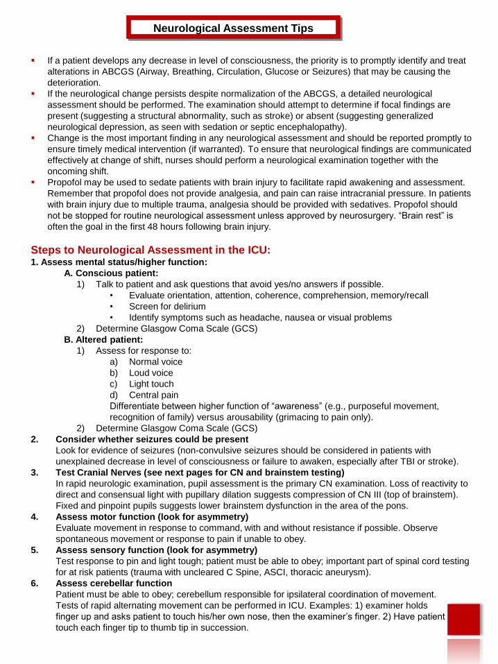
If a patient develops any decrease in level of consciousness, the priority is to promptly identify and treat alterations in ABCGS (Airway, Breathing, Circulation, Glucose or Seizures) that may be causing the deterioration. If the neurological change persists despite normalization of the ABCGS, a detailed neurological assessment should be performed. The examination should attempt to determine if focal findings are present (suggesting a structural abnormality, such as stroke) or absent (suggesting generalized neurological depression, as seen with sedation or septic encephalopathy). Change is the most important finding in any neurological assessment and should be reported promptly to ensure timely medical intervention (if warranted). To ensure that neurological findings are communicated effectively at change of shift, nurses should perform a neurological examination together with the oncoming shift. Propofol may be used to sedate patients with brain injury to facilitate rapid awakening and assessment. Remember that propofol does not provide analgesia, and pain can raise intracranial pressure. In patients with brain injury due to multiple trauma, analgesia should be provided with sedatives. Propofol should not be stopped for routine neurological assessment unless approved by neurosurgery. “Brain rest” is often the goal in the first 48 hours following brain injury. Steps to Neurological Assessment in the ICU: 1. Assess mental status/higher function: A. Conscious patient: 1) Talk to patient and ask questions that avoid yes/no answers if possible. • Evaluate orientation, attention, coherence, comprehension, memory/recall • Screen for delirium • Identify symptoms such as headache, nausea or visual problems 2) Determine Glasgow Coma Scale (GCS) B. Altered patient: 1) Assess for response to: a) Normal voice b) Loud voice c) Light touch d) Central pain Differentiate between higher function of “awareness” (e.g., purposeful movement, recognition of family) versus arousability (grimacing to pain only). 2) Determine Glasgow Coma Scale (GCS) 2. Consider whether seizures could be present Look for evidence of seizures (non-convulsive seizures should be considered in patients with unexplained decrease in level of consciousness or failure to awaken, especially after TBI or stroke). 3. Test Cranial Nerves (see next pages for CN and brainstem testing) In rapid neurologic examination, pupil assessment is the primary CN examination. Loss of reactivity to direct and consensual light with pupillary dilation suggests compression of CN III (top of brainstem). Fixed and pinpoint pupils suggests lower brainstem dysfunction in the area of the pons. 4. Assess motor function (look for asymmetry) Evaluate movement in response to command, with and without resistance if possible. Observe spontaneous movement or response to pain if unable to obey. 5. Assess sensory function (look for asymmetry) Test response to pin and light tough; patient must be able to obey; important part of spinal cord testing for at risk patients (trauma with uncleared C Spine, ASCI, thoracic aneurysm). 6. Assess cerebellar function Patient must be able to obey; cerebellum responsible for ipsilateral coordination of movement. Tests of rapid alternating movement can be performed in ICU. Examples: 1) examiner holds finger up and asks patient to touch his/her own nose, then the examiner’s finger. 2) Have patient touch each finger tip to thumb tip in succession. Neurological Assessment Tips

Welcome message from author
This document is posted to help you gain knowledge. Please leave a comment to let me know what you think about it! Share it to your friends and learn new things together.
Transcript
If a patient develops any decrease in level of consciousness, the priority is to promptly identify and treat
alterations in ABCGS (Airway, Breathing, Circulation, Glucose or Seizures) that may be causing the
deterioration.
If the neurological change persists despite normalization of the ABCGS, a detailed neurological
assessment should be performed. The examination should attempt to determine if focal findings are
present (suggesting a structural abnormality, such as stroke) or absent (suggesting generalized
neurological depression, as seen with sedation or septic encephalopathy).
Change is the most important finding in any neurological assessment and should be reported promptly to
ensure timely medical intervention (if warranted). To ensure that neurological findings are communicated
effectively at change of shift, nurses should perform a neurological examination together with the
oncoming shift.
Propofol may be used to sedate patients with brain injury to facilitate rapid awakening and assessment.
Remember that propofol does not provide analgesia, and pain can raise intracranial pressure. In patients
with brain injury due to multiple trauma, analgesia should be provided with sedatives. Propofol should
not be stopped for routine neurological assessment unless approved by neurosurgery. “Brain rest” is
often the goal in the first 48 hours following brain injury.
Steps to Neurological Assessment in the ICU:1. Assess mental status/higher function:
A. Conscious patient:
1) Talk to patient and ask questions that avoid yes/no answers if possible.
• Evaluate orientation, attention, coherence, comprehension, memory/recall
• Screen for delirium
• Identify symptoms such as headache, nausea or visual problems
2) Determine Glasgow Coma Scale (GCS)
B. Altered patient:
1) Assess for response to:
a) Normal voice
b) Loud voice
c) Light touch
d) Central pain
Differentiate between higher function of “awareness” (e.g., purposeful movement,
recognition of family) versus arousability (grimacing to pain only).
2) Determine Glasgow Coma Scale (GCS)
2. Consider whether seizures could be present
Look for evidence of seizures (non-convulsive seizures should be considered in patients with
unexplained decrease in level of consciousness or failure to awaken, especially after TBI or stroke).
3. Test Cranial Nerves (see next pages for CN and brainstem testing)
In rapid neurologic examination, pupil assessment is the primary CN examination. Loss of reactivity to
direct and consensual light with pupillary dilation suggests compression of CN III (top of brainstem).
Fixed and pinpoint pupils suggests lower brainstem dysfunction in the area of the pons.
4. Assess motor function (look for asymmetry)
Evaluate movement in response to command, with and without resistance if possible. Observe
spontaneous movement or response to pain if unable to obey.
5. Assess sensory function (look for asymmetry)
Test response to pin and light tough; patient must be able to obey; important part of spinal cord testing
for at risk patients (trauma with uncleared C Spine, ASCI, thoracic aneurysm).
6. Assess cerebellar function
Patient must be able to obey; cerebellum responsible for ipsilateral coordination of movement.
Tests of rapid alternating movement can be performed in ICU. Examples: 1) examiner holds
finger up and asks patient to touch his/her own nose, then the examiner’s finger. 2) Have patient
touch each finger tip to thumb tip in succession.
Neurological Assessment Tips
CN ICN II
CN III
CN IV
CN VI
CN V
CN VIICN VIII
CN IXCN X
CN XII
CN XI
V1
V2
V3
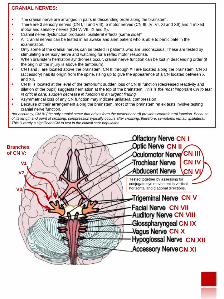
CRANIAL NERVES:
The cranial nerve are arranged in pairs in descending order along the brainstem.
There are 3 sensory nerves (CN I, II and VIII), 5 motor nerves (CN III, IV, VI, XI and XII) and 4 mixed
motor and sensory nerves (CN V, VII, IX and X).
Cranial nerve dysfunction produces ipsilateral effects (same side)*
All cranial nerves can be tested in an awake and alert patient who is able to participate in the
examination.
Only some of the cranial nerves can be tested in patients who are unconscious. These are tested by
stimulating a sensory nerve and watching for a reflex motor response.
When brainstem herniation syndromes occur, cranial nerve function can be lost in descending order (if
the origin of the injury is above the tentorium).
CN I and II are located above the brainstem; CN III through XII are located along the brainstem. CN XI
(accessory) has its origin from the spine, rising up to give the appearance of a CN located between X
and XII.
CN III is located at the level of the tentorium; sudden loss of CN III function (decreased reactivity and
dilation of the pupil) suggests herniation at the top of the brainstem. This is the most important CN to test
in critical care; sudden decrease in function is an urgent finding.
Asymmetrical loss of any CN function may indicate unilateral compression
Because of their arrangement along the brainstem, most of the brainstem reflex tests involve testing
cranial nerve function.*for accuracy, CN IV (the only cranial nerve that arises form the posterior cord) provides contralateral function. Because
of its length and point of crossing, compression typically occurs after crossing, therefore, symptoms remain ipsilateral.
This is rarely a significant CN to test in the critical care population.
Branches
of CN V:
Tested together by assessing for
conjugate eye movement in vertical,
horizontal and diagonal directions.
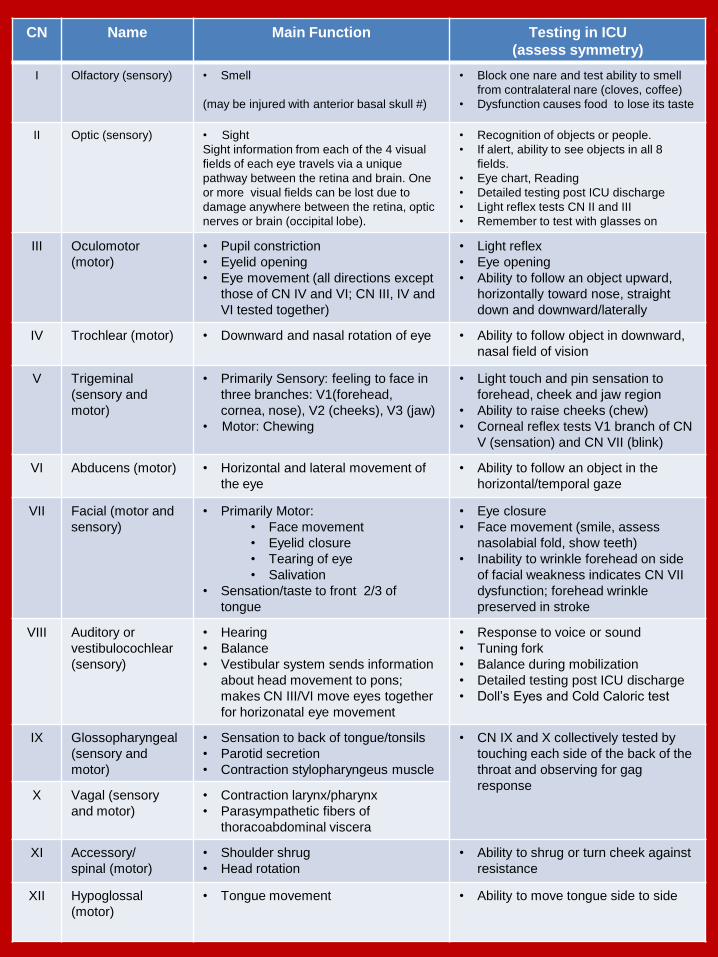
CN Name Main Function Testing in ICU
(assess symmetry)
I Olfactory (sensory) • Smell
(may be injured with anterior basal skull #)
• Block one nare and test ability to smell
from contralateral nare (cloves, coffee)
• Dysfunction causes food to lose its taste
II Optic (sensory) • Sight
Sight information from each of the 4 visual
fields of each eye travels via a unique
pathway between the retina and brain. One
or more visual fields can be lost due to
damage anywhere between the retina, optic
nerves or brain (occipital lobe).
• Recognition of objects or people.
• If alert, ability to see objects in all 8
fields.
• Eye chart, Reading
• Detailed testing post ICU discharge
• Light reflex tests CN II and III
• Remember to test with glasses on
III Oculomotor
(motor)
• Pupil constriction
• Eyelid opening
• Eye movement (all directions except
those of CN IV and VI; CN III, IV and
VI tested together)
• Light reflex
• Eye opening
• Ability to follow an object upward,
horizontally toward nose, straight
down and downward/laterally
IV Trochlear (motor) • Downward and nasal rotation of eye • Ability to follow object in downward,
nasal field of vision
V Trigeminal
(sensory and
motor)
• Primarily Sensory: feeling to face in
three branches: V1(forehead,
cornea, nose), V2 (cheeks), V3 (jaw)
• Motor: Chewing
• Light touch and pin sensation to
forehead, cheek and jaw region
• Ability to raise cheeks (chew)
• Corneal reflex tests V1 branch of CN
V (sensation) and CN VII (blink)
VI Abducens (motor) • Horizontal and lateral movement of
the eye
• Ability to follow an object in the
horizontal/temporal gaze
VII Facial (motor and
sensory)
• Primarily Motor:
• Face movement
• Eyelid closure
• Tearing of eye
• Salivation
• Sensation/taste to front 2/3 of
tongue
• Eye closure
• Face movement (smile, assess
nasolabial fold, show teeth)
• Inability to wrinkle forehead on side
of facial weakness indicates CN VII
dysfunction; forehead wrinkle
preserved in stroke
VIII Auditory or
vestibulocochlear
(sensory)
• Hearing
• Balance
• Vestibular system sends information
about head movement to pons;
makes CN III/VI move eyes together
for horizonatal eye movement
• Response to voice or sound
• Tuning fork
• Balance during mobilization
• Detailed testing post ICU discharge
• Doll’s Eyes and Cold Caloric test
IX Glossopharyngeal
(sensory and
motor)
• Sensation to back of tongue/tonsils
• Parotid secretion
• Contraction stylopharyngeus muscle
• CN IX and X collectively tested by
touching each side of the back of the
throat and observing for gag
responseX Vagal (sensory
and motor)
• Contraction larynx/pharynx
• Parasympathetic fibers of
thoracoabdominal viscera
XI Accessory/
spinal (motor)
• Shoulder shrug
• Head rotation
• Ability to shrug or turn cheek against
resistance
XII Hypoglossal
(motor)
• Tongue movement • Ability to move tongue side to side
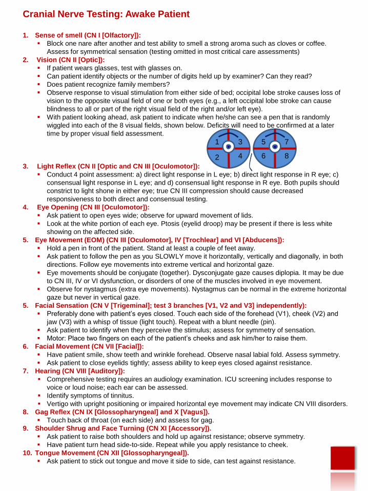
Cranial Nerve Testing: Awake Patient
1. Sense of smell (CN I [Olfactory]):
Block one nare after another and test ability to smell a strong aroma such as cloves or coffee.
Assess for symmetrical sensation (testing omitted in most critical care assessments)
2. Vision (CN II [Optic]):
If patient wears glasses, test with glasses on.
Can patient identify objects or the number of digits held up by examiner? Can they read?
Does patient recognize family members?
Observe response to visual stimulation from either side of bed; occipital lobe stroke causes loss of
vision to the opposite visual field of one or both eyes (e.g., a left occipital lobe stroke can cause
blindness to all or part of the right visual field of the right and/or left eye).
With patient looking ahead, ask patient to indicate when he/she can see a pen that is randomly
wiggled into each of the 8 visual fields, shown below. Deficits will need to be confirmed at a later
time by proper visual field assessment.
3. Light Reflex (CN II [Optic and CN III [Oculomotor]):
Conduct 4 point assessment: a) direct light response in L eye; b) direct light response in R eye; c)
consensual light response in L eye; and d) consensual light response in R eye. Both pupils should
constrict to light shone in either eye; true CN III compression should cause decreased
responsiveness to both direct and consensual testing.
4. Eye Opening (CN III [Oculomotor]):
Ask patient to open eyes wide; observe for upward movement of lids.
Look at the white portion of each eye. Ptosis (eyelid droop) may be present if there is less white
showing on the affected side.
5. Eye Movement (EOM) (CN III [Oculomotor], IV [Trochlear] and VI [Abducens]):
Hold a pen in front of the patient. Stand at least a couple of feet away.
Ask patient to follow the pen as you SLOWLY move it horizontally, vertically and diagonally, in both
directions. Follow eye movements into extreme vertical and horizontal gaze.
Eye movements should be conjugate (together). Dysconjugate gaze causes diplopia. It may be due
to CN III, IV or VI dysfunction, or disorders of one of the muscles involved in eye movement.
Observe for nystagmus (extra eye movements). Nystagmus can be normal in the extreme horizontal
gaze but never in vertical gaze.
5. Facial Sensation (CN V [Trigeminal]; test 3 branches [V1, V2 and V3] independently):
Preferably done with patient’s eyes closed. Touch each side of the forehead (V1), cheek (V2) and
jaw (V3) with a whisp of tissue (light touch). Repeat with a blunt needle (pin).
Ask patient to identify when they perceive the stimulus; assess for symmetry of sensation.
Motor: Place two fingers on each of the patient’s cheeks and ask him/her to raise them.
6. Facial Movement (CN VII [Facial]):
Have patient smile, show teeth and wrinkle forehead. Observe nasal labial fold. Assess symmetry.
Ask patient to close eyelids tightly; assess ability to keep eyes closed against resistance.
7. Hearing (CN VIII [Auditory]):
Comprehensive testing requires an audiology examination. ICU screening includes response to
voice or loud noise; each ear can be assessed.
Identify symptoms of tinnitus.
Vertigo with upright positioning or impaired horizontal eye movement may indicate CN VIII disorders.
8. Gag Reflex (CN IX [Glossopharyngeal] and X [Vagus]).
Touch back of throat (on each side) and assess for gag.
9. Shoulder Shrug and Face Turning (CN XI [Accessory]).
Ask patient to raise both shoulders and hold up against resistance; observe symmetry.
Have patient turn head side-to-side. Repeat while you apply resistance to cheek.
10. Tongue Movement (CN XII [Glossopharyngeal]).
Ask patient to stick out tongue and move it side to side, can test against resistance.
.1
2
3
4
5 7
6 8
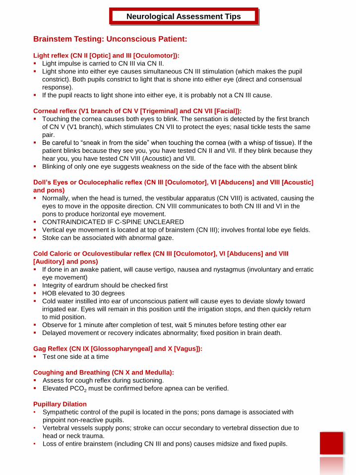
Brainstem Testing: Unconscious Patient:
Light reflex (CN II [Optic] and III [Oculomotor]):
Light impulse is carried to CN III via CN II.
Light shone into either eye causes simultaneous CN III stimulation (which makes the pupil
constrict). Both pupils constrict to light that is shone into either eye (direct and consensual
response).
If the pupil reacts to light shone into either eye, it is probably not a CN III cause.
Corneal reflex (V1 branch of CN V [Trigeminal] and CN VII [Facial]):
Touching the cornea causes both eyes to blink. The sensation is detected by the first branch
of CN V (V1 branch), which stimulates CN VII to protect the eyes; nasal tickle tests the same
pair.
Be careful to “sneak in from the side” when touching the cornea (with a whisp of tissue). If the
patient blinks because they see you, you have tested CN II and VII. If they blink because they
hear you, you have tested CN VIII (Acoustic) and VII.
Blinking of only one eye suggests weakness on the side of the face with the absent blink
Doll’s Eyes or Oculocephalic reflex (CN III [Oculomotor], VI [Abducens] and VIII [Acoustic]
and pons)
Normally, when the head is turned, the vestibular apparatus (CN VIII) is activated, causing the
eyes to move in the opposite direction. CN VIII communicates to both CN III and VI in the
pons to produce horizontal eye movement.
CONTRAINDICATED IF C-SPINE UNCLEARED
Vertical eye movement is located at top of brainstem (CN III); involves frontal lobe eye fields.
Stoke can be associated with abnormal gaze.
Cold Caloric or Oculovestibular reflex (CN III [Oculomotor], VI [Abducens] and VIII
[Auditory] and pons)
If done in an awake patient, will cause vertigo, nausea and nystagmus (involuntary and erratic
eye movement)
Integrity of eardrum should be checked first
HOB elevated to 30 degrees
Cold water instilled into ear of unconscious patient will cause eyes to deviate slowly toward
irrigated ear. Eyes will remain in this position until the irrigation stops, and then quickly return
to mid position.
Observe for 1 minute after completion of test, wait 5 minutes before testing other ear
Delayed movement or recovery indicates abnormality; fixed position in brain death.
Gag Reflex (CN IX [Glossopharyngeal] and X [Vagus]):
Test one side at a time
Coughing and Breathing (CN X and Medulla):
Assess for cough reflex during suctioning.
Elevated PCO2 must be confirmed before apnea can be verified.
Pupillary Dilation
• Sympathetic control of the pupil is located in the pons; pons damage is associated with
pinpoint non-reactive pupils.
• Vertebral vessels supply pons; stroke can occur secondary to vertebral dissection due to
head or neck trauma.
• Loss of entire brainstem (including CN III and pons) causes midsize and fixed pupils.
Neurological Assessment Tips
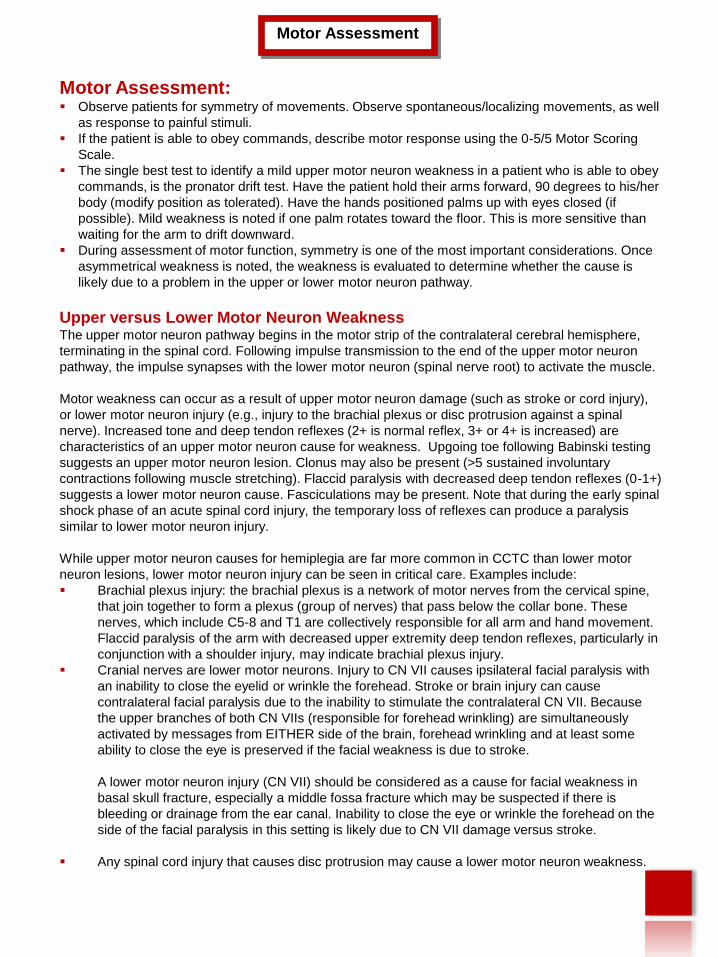
Motor Assessment: Observe patients for symmetry of movements. Observe spontaneous/localizing movements, as well
as response to painful stimuli.
If the patient is able to obey commands, describe motor response using the 0-5/5 Motor Scoring
Scale.
The single best test to identify a mild upper motor neuron weakness in a patient who is able to obey
commands, is the pronator drift test. Have the patient hold their arms forward, 90 degrees to his/her
body (modify position as tolerated). Have the hands positioned palms up with eyes closed (if
possible). Mild weakness is noted if one palm rotates toward the floor. This is more sensitive than
waiting for the arm to drift downward.
During assessment of motor function, symmetry is one of the most important considerations. Once
asymmetrical weakness is noted, the weakness is evaluated to determine whether the cause is
likely due to a problem in the upper or lower motor neuron pathway.
Upper versus Lower Motor Neuron WeaknessThe upper motor neuron pathway begins in the motor strip of the contralateral cerebral hemisphere,
terminating in the spinal cord. Following impulse transmission to the end of the upper motor neuron
pathway, the impulse synapses with the lower motor neuron (spinal nerve root) to activate the muscle.
Motor weakness can occur as a result of upper motor neuron damage (such as stroke or cord injury),
or lower motor neuron injury (e.g., injury to the brachial plexus or disc protrusion against a spinal
nerve). Increased tone and deep tendon reflexes (2+ is normal reflex, 3+ or 4+ is increased) are
characteristics of an upper motor neuron cause for weakness. Upgoing toe following Babinski testing
suggests an upper motor neuron lesion. Clonus may also be present (>5 sustained involuntary
contractions following muscle stretching). Flaccid paralysis with decreased deep tendon reflexes (0-1+)
suggests a lower motor neuron cause. Fasciculations may be present. Note that during the early spinal
shock phase of an acute spinal cord injury, the temporary loss of reflexes can produce a paralysis
similar to lower motor neuron injury.
While upper motor neuron causes for hemiplegia are far more common in CCTC than lower motor
neuron lesions, lower motor neuron injury can be seen in critical care. Examples include:
Brachial plexus injury: the brachial plexus is a network of motor nerves from the cervical spine,
that join together to form a plexus (group of nerves) that pass below the collar bone. These
nerves, which include C5-8 and T1 are collectively responsible for all arm and hand movement.
Flaccid paralysis of the arm with decreased upper extremity deep tendon reflexes, particularly in
conjunction with a shoulder injury, may indicate brachial plexus injury.
Cranial nerves are lower motor neurons. Injury to CN VII causes ipsilateral facial paralysis with
an inability to close the eyelid or wrinkle the forehead. Stroke or brain injury can cause
contralateral facial paralysis due to the inability to stimulate the contralateral CN VII. Because
the upper branches of both CN VIIs (responsible for forehead wrinkling) are simultaneously
activated by messages from EITHER side of the brain, forehead wrinkling and at least some
ability to close the eye is preserved if the facial weakness is due to stroke.
A lower motor neuron injury (CN VII) should be considered as a cause for facial weakness in
basal skull fracture, especially a middle fossa fracture which may be suspected if there is
bleeding or drainage from the ear canal. Inability to close the eye or wrinkle the forehead on the
side of the facial paralysis in this setting is likely due to CN VII damage versus stroke.
Any spinal cord injury that causes disc protrusion may cause a lower motor neuron weakness.
Motor Assessment
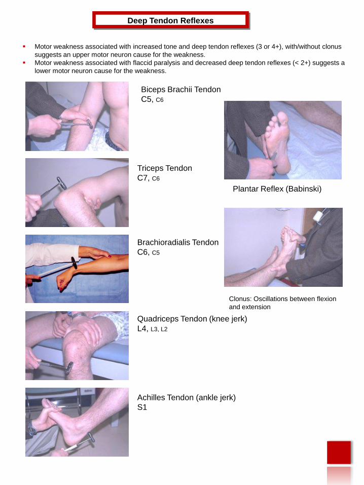
Motor weakness associated with increased tone and deep tendon reflexes (3 or 4+), with/without clonus
suggests an upper motor neuron cause for the weakness.
Motor weakness associated with flaccid paralysis and decreased deep tendon reflexes (< 2+) suggests a
lower motor neuron cause for the weakness.
Deep Tendon Reflexes
Biceps Brachii Tendon
C5, C6
Triceps Tendon
C7, C6
Brachioradialis Tendon
C6, C5
Achilles Tendon (ankle jerk)
S1
Quadriceps Tendon (knee jerk)
L4, L3, L2
Plantar Reflex (Babinski)
Clonus: Oscillations between flexion
and extension
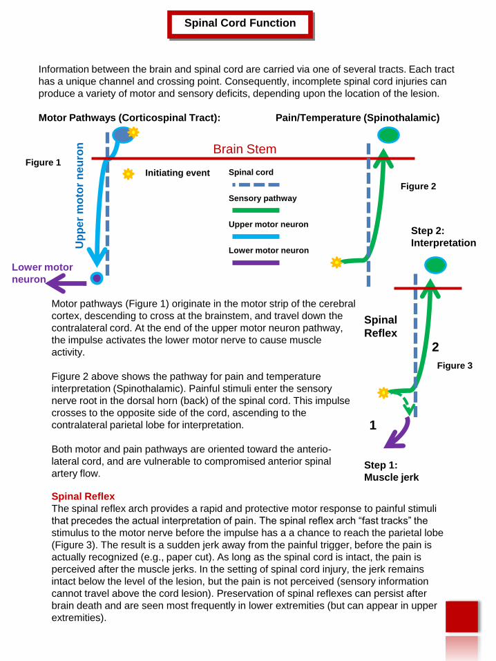
Information between the brain and spinal cord are carried via one of several tracts. Each tract
has a unique channel and crossing point. Consequently, incomplete spinal cord injuries can
produce a variety of motor and sensory deficits, depending upon the location of the lesion.
Motor Pathways (Corticospinal Tract): Pain/Temperature (Spinothalamic)
Spinal Cord Function
Up
pe
r m
oto
r n
eu
ron
Lower motor
neuron
Brain Stem
Spinal cord
Sensory pathway
Upper motor neuron
Motor pathways (Figure 1) originate in the motor strip of the cerebral
cortex, descending to cross at the brainstem, and travel down the
contralateral cord. At the end of the upper motor neuron pathway,
the impulse activates the lower motor nerve to cause muscle
activity.
Figure 2 above shows the pathway for pain and temperature
interpretation (Spinothalamic). Painful stimuli enter the sensory
nerve root in the dorsal horn (back) of the spinal cord. This impulse
crosses to the opposite side of the cord, ascending to the
contralateral parietal lobe for interpretation.
Both motor and pain pathways are oriented toward the anterio-
lateral cord, and are vulnerable to compromised anterior spinal
artery flow.
Lower motor neuron
2
1
Step 1:
Muscle jerk
Step 2:
Interpretation
Figure 1
Figure 2
Figure 3
Spinal Reflex
The spinal reflex arch provides a rapid and protective motor response to painful stimuli
that precedes the actual interpretation of pain. The spinal reflex arch “fast tracks” the
stimulus to the motor nerve before the impulse has a a chance to reach the parietal lobe
(Figure 3). The result is a sudden jerk away from the painful trigger, before the pain is
actually recognized (e.g., paper cut). As long as the spinal cord is intact, the pain is
perceived after the muscle jerks. In the setting of spinal cord injury, the jerk remains
intact below the level of the lesion, but the pain is not perceived (sensory information
cannot travel above the cord lesion). Preservation of spinal reflexes can persist after
brain death and are seen most frequently in lower extremities (but can appear in upper
extremities).
Spinal
Reflex
Initiating event
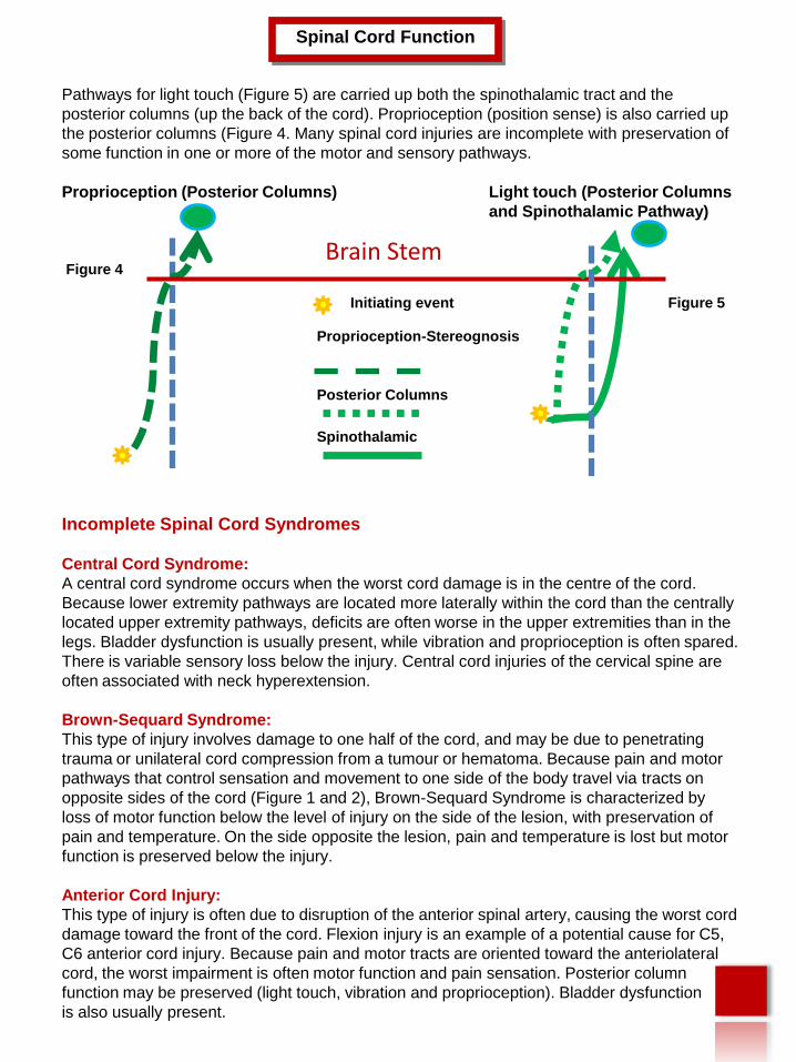
Pathways for light touch (Figure 5) are carried up both the spinothalamic tract and the
posterior columns (up the back of the cord). Proprioception (position sense) is also carried up
the posterior columns (Figure 4. Many spinal cord injuries are incomplete with preservation of
some function in one or more of the motor and sensory pathways.
Proprioception (Posterior Columns) Light touch (Posterior Columns
and Spinothalamic Pathway)
Incomplete Spinal Cord Syndromes
Central Cord Syndrome:
A central cord syndrome occurs when the worst cord damage is in the centre of the cord.
Because lower extremity pathways are located more laterally within the cord than the centrally
located upper extremity pathways, deficits are often worse in the upper extremities than in the
legs. Bladder dysfunction is usually present, while vibration and proprioception is often spared.
There is variable sensory loss below the injury. Central cord injuries of the cervical spine are
often associated with neck hyperextension.
Brown-Sequard Syndrome:
This type of injury involves damage to one half of the cord, and may be due to penetrating
trauma or unilateral cord compression from a tumour or hematoma. Because pain and motor
pathways that control sensation and movement to one side of the body travel via tracts on
opposite sides of the cord (Figure 1 and 2), Brown-Sequard Syndrome is characterized by
loss of motor function below the level of injury on the side of the lesion, with preservation of
pain and temperature. On the side opposite the lesion, pain and temperature is lost but motor
function is preserved below the injury.
Anterior Cord Injury:
This type of injury is often due to disruption of the anterior spinal artery, causing the worst cord
damage toward the front of the cord. Flexion injury is an example of a potential cause for C5,
C6 anterior cord injury. Because pain and motor tracts are oriented toward the anteriolateral
cord, the worst impairment is often motor function and pain sensation. Posterior column
function may be preserved (light touch, vibration and proprioception). Bladder dysfunction
is also usually present.
Spinal Cord Function
Brain Stem
Proprioception-Stereognosis
Posterior Columns
Spinothalamic
Figure 5
Figure 4
Initiating event
Spinal ShockFollowing acute spinal cord injury, all reflexes below the level of injury are typically lost for a
period of hours to days. During this period known as spinal cord shock, the patient typically
has flaccid paralysis with a loss of deep tendon reflexes, with absent bladder and bowel tone.
Anal sphincter reflex is one of the first reflexes to return when the spinal shock phase begins
to resolve. Reflex contraction of the anal sphincter following sensory stimulation produced by
a gentle tug on the Foley catheter, suggests that the spinal shock phase is resolving.
The end of the spinal shock period is significant for the following reasons. One hopes that any
paralysis or sensory deficit that develops immediately following an acute injury will be at least
partially due to swelling and spinal cord shock. When the shock period ends, continued
absence of sensation during a rectal exam and/or inability to voluntarily “squeeze” the anal
sphincter is a bad sign.
During spinal shock, the loss of bladder and anal sphincter reflex is associated with
incontinence. Because sphincter relaxation to facilitate voiding or defecation is a voluntary
function, the end of the spinal shock phase is usually associated with urinary and fecal
retention. Early and aggressive bowel routine is important to facilitate future ADLs.
Conversion to intermittent catheterization should begin as soon as hourly urine output
measurement is no longer needed (e.g., hemodynamic stability is restored). Over distension
of the bladder should be avoided (500 ml per catheterization optimal); over distension can
lead to overflow incontinence (with incomplete emptying) and ureteral reflux. The goal for
intermittent catheterization is to achieve this output with a daily intake of ~2,000 ml.
Dehydration should be avoided, as this will increase the risk for urinary tract infection and
renal injury. An aggressive bowel routine that ensures at minimum of q 2 day bowel
evacuation should be instituted even before the spinal shock phase ends. Diarrhea may be
present in the early phases of ASCI, however, the goal following resolution of spinal shock
should be a soft stool (not diarrhea) facilitate by stool softeners, 2 day Dulcolax and anal
stimulation (not diarrhea).
Neurogenic ShockNeurogenic shock usually mirrors the spinal shock phase (loss of spinal reflexes). It is
characterized by vasodilation, hypotension and bradycardia, due to disruption of autonomic
fibres below the level of the injury. Neurogenic shock usually improves or resolves with time,
however, it may remain an ongoing problem for individuals with complete and high cervical
cord injuries. Turning, head of bed elevation and suctioning can precipitate bradycardia and
hypotension. Cardiac arrest can also occur. Gradual and careful position changes and the
use of TED stockings/abdominal binders to prevent positional hypotension may help.
Preoxygenation with 100% oxygen and abrupt termination of suctioning with return to
mechanical ventilation will usually resolve bradycardias induced by suctioning. Atropine
should be available at the bedside. Temporary pacemakers are occasionally required, less
frequently, patients may need permanent cardiac pacing.
Other causes for shock (e.g., sepsis, myocardial infarction, hypovolemia) may be masked by
the loss of sympathetic response.
Spinal Cord Injury
Autonomic DysreflexiaFollowing resolution of the spinal shock phase with return of spinal cord reflexes, patients with
spinal cord injury are at risk for the development of autonomic dysreflexia. The higher the
cord injury, the greater the potential for autonomic dysreflexia, with virtually all tetraplegics
(quadriplegics) and most individuals with injuries at or above T6 experiencing this problem.
Thus, patients with chronic spinal cord injury or those with acute spinal cord injury and
prolonged CCTC admission should be monitored for signs of autonomic dysreflexia. This will
be a life-long complication for these patients.
Autonomic dysreflexia is a life-threatening event that is triggered by a strong noxious
stimulus. Sensory input causes a release of catecholamines, producing vasoconstriction and
hypertension. The rise in blood pressure stimulates carotid and aortic receptors, causing
inhibitory messages to be sent down the cord. Because inhibitory messages can only
descend as far as the level of the injury, vasoconstriction (and hypertension) continues below
the cord injury. Vasodilation above the lesion causes facial flushing, profuse sweating,
bounding headache, nasal congestion and on occasion, Horner’s syndrome. The higher the
injury, the greater the hypertension. Vagal stimulation (CN X) sends inhibitory messages that
cause bradycardia. Signs and symptoms of autonomic dysreflexia include:
Hypertension (may only be 20-30 mmHg above baseline)
Vasoconstriction below lesion
Vasodilation with flushing above lesion and bounding headache
Profuse sweating above lesion
Bradycardia
Goose bumps above and sometimes below lesion
Visual disturbance; spots may be visible by patient in visual fields
Horner’s Syndrome: constriction of the pupil, mild eyelid droopiness, possible loss of
sweating on one side of face.
The most common triggers for autonomic dysreflexia are a full bowel or bladder. Any painful
situation, including procedures or physical therapy in critical care, can cause this syndrome.
Pregnancy, especially labour and delivery in a patient with spinal cord injury can trigger
autonomic dysreflexia.
The treatment priority is to remove the cause of the autonomic dyreflexia (e.g., bladder
catheterization, fecal disimpaction). Sitting the patient up can cause orthostatic lowering of the
blood pressure. If antihypertensives are needed, use rapid onset and short duration of action
drugs. Nitrates can be used, but are contraindicated if patients are taking sildenafil or other
medications for erectile dysfunction. Calcium channel blockers such as nifedipine can be
useful; labetolol should be used with caution as it may worsen bradycardia.
Brenda Morgan
Clinical Nurse Specialist, CCTC
May 26, 2011
Spinal Cord Injury
Related Documents