Regulation of mesenteric resistance artery diameter by pharmacological modulators of K Ca channels by Stephanie E. Lunn A thesis submitted in partial fulfillment of the requirements for the degree of Doctor of Philosophy Department of Pharmacology University of Alberta © Stephanie E. Lunn, 2018

Welcome message from author
This document is posted to help you gain knowledge. Please leave a comment to let me know what you think about it! Share it to your friends and learn new things together.
Transcript
Regulation of mesenteric resistance artery diameter by
pharmacological modulators of KCa channels
by
Stephanie E. Lunn
A thesis submitted in partial fulfillment of the requirements for the degree of
Doctor of Philosophy
Department of Pharmacology
University of Alberta
© Stephanie E. Lunn, 2018
ii
Abstract
Background: The diameter of resistance arteries, and thus, tissue perfusion and blood pressure, is
tightly regulated through the integrated activity of endothelial and smooth muscle cells, and
sympathetic nerves. The endothelium regulates the contractility of smooth muscle cells by
releasing diffusible factors such as nitric oxide (NO) and via gap junction-mediated electrical
coupling; opening of endothelial Ca2+-activated K+ (KCa) channels causes hyperpolarization which
spreads to underlying smooth muscle cells to reduce opening of voltage-dependent Ca2+ channels,
decrease Ca2+ influx and so limit contraction. The bioavailability and therefore, biological activity,
of NO is determined by its interaction with the free radical superoxide anion (O2-), elevated levels
of which are associated with risk factors for cardiovascular disease.
Traditionally, NO and endothelium-dependent smooth muscle hyperpolarization have been
regarded as two separate mechanisms for regulation of arterial diameter. However, several lines
of recent evidence support the proposal that NO bioavailability and KCa channel activity may be
linked: 1. Exposure of endothelial cells to shear stress results in activation of both small
conductance KCa channels and increased NO production. 2. Agonist-evoked NO production and
NO-mediated relaxations can be inhibited by blockers of endothelial KCa channels. 3. Activators
of endothelial KCa channels can evoke NO-mediated relaxation. 4. Stimulation of smooth muscle
cells by 1-adrenoceptor agonists engages both endothelial intermediate conductance KCa channels
and NO production via a process termed myoendothelial feedback. 5. O2- production by voltage-
sensitive NADPH oxidase is reduced by membrane hyperpolarization which may lead to increased
bioavailability of NO.
iii
Thus, my over-arching goal is to further explore the relationship between endothelial KCa
channels and NO in regulating resistance artery diameter by testing three hypotheses:
1. Activation of small conductance KCa channels can enhance NO-mediated inhibition of
sympathetic vasoconstriction evoked by increases in shear stress.
2. Intermediate conductance KCa channel-mediated myoendothelial feedback plays a role in
NO-dependent modulation of sympathetic vasoconstriction.
3. Pharmacological activators of endothelial KCa channels can reduce vascular O2-
production and enhance NO-mediated modulation of vasoconstriction
To test these hypotheses, I have addressed two major aims:
1. To investigate the role of endothelial KCa channels in NO-mediated modulation of
nerve-evoked vasoconstriction in the perfused mesenteric bed.
2. To investigate whether pharmacological activators of endothelial KCa channels can
modulate vascular O2- production and vasoconstriction stimulated by the 1-
adrenoceptor agonist phenylephrine.
Methods: To address these aims I have used a combination of functional and biochemical
techniques to investigate the effects of modulators of endothelial KCa channels on diameter and
O2- production in rat mesenteric resistance arteries.
Results/Discussion: My data show that although myoendothelial feedback limits contractile
responses to phenylephrine in isolated arteries, this pathway does not appear to contribute to
endothelial modulation of sympathetic vasoconstriction at the level of the intact bed. Instead, shear
stress-induced activation of small conductance KCa channels and release of NO provides the
dominant mechanism for engagement of the endothelium to inhibit sympathetic vasoconstriction.
Furthermore, activators of endothelial KCa channels can significantly limit nerve-evoked
iv
vasoconstriction. CyPPA (N-cyclohexyl-N-[2-(3,5-dimethyl-pyrazol-1-yl)-6-methyl-4-
pyrimidinamine), an activator of small conductance KCa channels, enhances NO-dependent, shear
stress-mediated inhibition of sympathetic vasoconstriction whereas SKA-31 (naphtho[1,2-
d]thiazol-2-ylamine), an activator of intermediate conductance KCa channels, can directly inhibit
release of noradrenaline from perivascular sympathetic nerves. Both CyPPA and SKA-31 can
significantly reduce acute increases in O2- production stimulated by phenylephrine in isolated
arteries but this effect is not associated with enhancement of NO-mediated endothelial modulation
of vasoconstriction.
Conclusion: To conclude, I have demonstrated that small and intermediate conductance KCa
channels play different functional roles in modulation of nerve-evoked vasoconstriction;
endothelial small conductance KCa channels mediate shear stress-induced, NO-dependent
inhibition of vasoconstriction whereas the activity of neuronal IKCa channels can directly inhibit
release of noradrenaline from sympathetic nerves. These functional roles reflect the differing
locations of the channels within endothelial cells and the artery wall. Pharmacological activators
of KCa channels can limit vascular O2- production supporting the proposal that the endothelial cell
membrane potential may play a key role in vascular health and that targeting these channels could
provide a novel approach to reducing O2- levels in disease states.

v
Dedication
To my parents, Phil and Christina Lunn, for their
unconditional love and support
And to Paul Czarnietzki, my rock,
who stayed calm through it all

vi
Acknowledgements
To my supervisors, Dr. Frances Plane and Dr. Paul Kerr, for their endless support and
encouragement over the last five years. This PhD would not have been possible without you both
and it is bittersweet to graduate and leave you.
To Ran Wei (soon to be Dr. Wei!), thanks for being by my side for graduate studies, it was
the best thing that could have ever happened to me. It would have been a million times less fun
and more stressful without you. I will miss seeing your face every day, but we will always have
hotpot.
To my extended family, thank you for the love from afar in the form of emails, phone calls,
visits and care packages.
To Ed and Heidi Czarnietzki, thank you for the home cooked meals, support and love
(especially during flu season!).
To Sabina Baghirova and Xenia Cravetchi, thank you for the endless chats, wine and
laughter.
To my supervisory committee, Dr. Darren DeLorey, Dr. Richard Schulz and Dr. Stephane
Bourque, thank you for accepting positions on my committee and helping along the way. In
particular, thanks to Dr. Darren DeLorey for the loan of the pressure myograph attached to an
IonOptix system.
To my external examiners, Dr. Andrew Braun and Dr. Yves Sauvé, thank you for accepting
my invitation to be examiners and participating in the last step of my graduate studies.
To Lynette Edler from the HistoCore, Alberta Diabetes Institute, University of Alberta,
thank you for cryosectioning my samples.
To the Dr. Xuejun Sun and Mrs. Geraldine (Gerry) Barron from the Cell Imaging Facility,
Department of Oncology, Cross Cancer Institute, thank you for teaching me everything I needed
to know about histological analysis.
To Ken Strynadka, UPLC Analytical Core, Cardiovascular Research Centre, thank you for
developing and performing protocols for my specific samples.
To Dr. Shaun Sandow, thank you for performing the immunohistochemistry experiments.
To Dr. Andrew Holt, Dr. Elena Posse de Chaves, Dr. Nadia Jahroudi, and Dr. Simonetta
Sipione and their respective labs, thank you for your advice and use of your equipment.
And finally, to the Department of Pharmacology, thank you for being my home for my
undergraduate and graduate studies. It has been quite the adventure!
vii
Table of Contents
Abstract ...................................................................................................................................... ii
Dedication ...................................................................................................................................v
Acknowledgements .................................................................................................................. vi
Table of Contents .................................................................................................................... vii
List of Tables ..............................................................................................................................x
List of Figures ........................................................................................................................... xi
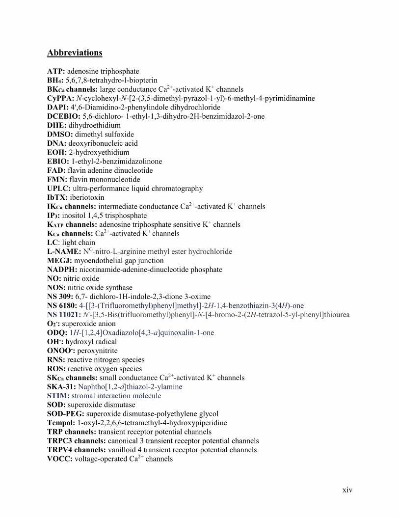
Abbreviations ........................................................................................................................ xiv
Ethics Approval ........................................................................................................................xv
Animal Care and Use ...............................................................................................................xv
Chapter 1: Introduction Introduction ..................................................................................................................................1
1.1 Contraction of vascular smooth muscle cells.......................................................................2
1.1.1 Sources of Ca2+ for smooth muscle contraction .......................................................2
1.1.2 Modulation of vascular smooth muscle contraction by K+ channels .......................7
1.2 Modulation of resistance artery diameter by perivascular sympathetic nerves .................10
1.3 Modulation of resistance artery diameter by the endothelium...........................................11
1.3.1 Endothelial Ca2+ signaling .....................................................................................12
1.3.2 Nitric oxide (NO) ...................................................................................................14
1.3.3 Endothelium-dependent hyperpolarization ............................................................18
1.3.4 Endothelial Ca2+-activated K+ (KCa) channels .......................................................19
1.4 Endothelial dysfunction .....................................................................................................25
1.4.1 Vascular O2- production .........................................................................................26
1.5 Hypothesis and aims ..........................................................................................................35
Chapter 2: Activation of SKCa channels enhances shear stress-mediated
inhibition of sympathetic vasoconstriction in the perfused mesenteric bed 2.1 Introduction ........................................................................................................................36
2.2 Methods and materials .......................................................................................................38
2.2.1 Perfused mesenteric vascular bed ..........................................................................38
2.2.1.1 Responses to stimulation of perivascular nerves ...............................................39
2.2.2 Wire myography ....................................................................................................40
2.2.2.1 Concentration-response curves ..........................................................................40
2.2.3 Analysis of noradrenaline levels in perfusate from the mesenteric vascular bed ..41
2.2.3.1 Measurement of noradrenaline outflow from the perfused mesenteric bed by
UPLC .................................................................................................................41
2.2.4 Statistics .................................................................................................................42
2.3 Results ................................................................................................................................42
2.3.1 Characterization of nerve-evoked vasoconstriction in the rat perfused mesenteric
bed ..........................................................................................................................42
2.3.2 Modulation of nerve-evoked vasoconstriction by increases in shear stress in the rat
perfused mesenteric bed .........................................................................................47
2.3.3 Effect of CyPPA on phenylephrine-induced tone in isolated mesenteric arteries
mounted in a wire myograph ................................................................................ 54
viii
2.3.4 CyPPA enhances shear stress-induced modulation of nerve-evoked
vasoconstriction in the rat perfused mesenteric bed ..............................................58
2.4 Discussion ..........................................................................................................................66
Chapter 3: Activation of IKCa channels directly inhibits sympathetic
vasoconstriction in the perfused mesenteric bed 3.1 Introduction ........................................................................................................................79
3.2 Methods and materials .......................................................................................................81
3.2.1 Perfused mesenteric vascular bed ..........................................................................81
3.2.1.1 Responses to stimulation of perivascular nerves ...............................................82
3.2.2 Wire myography ....................................................................................................82
3.2.2.1 Concentration-response curves ..........................................................................83
3.2.3 Confocal immunohistochemistry ...........................................................................84
3.2.4 Analysis of noradrenaline levels in perfusate from the mesenteric vascular bed ..85
3.2.4.1 Measurement of noradrenaline outflow from the perfused mesenteric bed by
UPLC .................................................................................................................85
3.2.5 Statistics .................................................................................................................86
3.3 Results ................................................................................................................................86
3.3.1 Role of IKCa channels in endothelial modulation of sympathetic vasoconstriction in
the perfused mesenteric bed ...................................................................................86
3.3.2 Effects of SKA-31 on phenylephrine-induced tone in isolated mesenteric
arteries ....................................................................................................................88
3.3.3 Effect of SKA-31 on sympathetic vasoconstriction in the rat perfused mesenteric
bed ..........................................................................................................................91
3.4 Discussion ..........................................................................................................................99
Chapter 4: Effects of activators of SKCa and IKCa channels on agonist-induced
O2- production and vasoconstriction in isolated mesenteric arteries
4.1 Introduction ......................................................................................................................106
4.2 Methods and materials .....................................................................................................109
4.2.1 Simultaneous assessment of O2- production and changes in arterial diameter in
intact arteries ........................................................................................................109
4.2.1.1 Pressure myography .........................................................................................109
4.2.1.2 Use of DHE to assess O2- production in intact mesenteric arteries .................110
4.2.2 Perfused mesenteric vascular bed ........................................................................111
4.2.2.1 Responses to stimulation of perivascular nerves .............................................111
4.2.3 Statistics ...............................................................................................................112
4.3 Results ..............................................................................................................................112
4.3.1 Characterization of phenylephrine-induced O2- production and vasoconstriction in
mesenteric resistance arteries ...............................................................................112
4.3.2 Role of NO in phenylephrine-induced O2- production and vasoconstriction in
mesenteric resistance arteries ...............................................................................119
4.3.3 Effect of inhibitors of SKCa and IKCa channels on phenylephrine-induced O2-
production and vasoconstriction in mesenteric resistance arteries ......................125
4.3.4 Effect of SKCa and IKCa channel activators on phenylephrine-induced changes in
diameter and O2- production in mesenteric resistance arteries ............................128
ix
4.3.5 Effect of modulators of smooth muscle BKCa channels on phenylephrine-induced
changes in O2- production and vasoconstriction in mesenteric resistance
arteries ..................................................................................................................134
4.4 Discussion ........................................................................................................................137
Chapter 5: Activators of SKCa and IKCa channels limit O2- production in isolated
arteries 5.1 Introduction ......................................................................................................................146
5.2 Methods and materials .....................................................................................................146
5.2.1 Histological analysis of mesenteric arteries stained with DHE ...........................146
5.2.1.1 Tissue Preparation ............................................................................................146
5.2.1.2 Imaging ............................................................................................................147
5.2.2 Quantification of DHE-derived oxidation products from aortic samples by
UPLC ...................................................................................................................148
5.2.3 Statistics ...............................................................................................................149
5.3 Results ..............................................................................................................................150
5.3.1 Histological analysis of mesenteric arteries stained with DHE ...........................150
5.3.2 Quantification of DHE-derived oxidation products from aortic samples by
UPLC ...................................................................................................................152
5.4 Discussion ........................................................................................................................153
Chapter 6: General discussion and future directions 6.1 General discussion ...........................................................................................................156
6.2 Future directions ..............................................................................................................162
References ................................................................................................................................164
Appendix: Drugs and Chemicals .....................................................................................201

x
List of Tables
Page
Chapter 1: Introduction
Table 1.1: Cellular location of BKCa, SKCa and IKCa channels 9
Appendix: Drug and Chemicals
Table 1: The mechanism of action, solvent, stock concentration, experimental
concentration and supplier of the drugs and chemicals used
201
xi
List of Figures
Page
Chapter 1: Introduction
Figure 1.1: Schematic of arterial structure 1
Figure 1.2: Mechanism of smooth muscle contraction 3
Figure 1.3: Schematic of L-type VOCC Ca2+ channel with α- and accessory -,- and
2- subunits
5
Figure 1.4: Schematic of structure of BKCa channels 8
Figure 1.5: NO-mediated relaxation of vascular smooth muscle 18
Figure 1.6: Schematic of a SKCa/IKCa channel subunit 20
Figure 1.7: Schematic showing the cellular locations of SKCa and IKCa channels within
endothelial cells
23
Figure 1.8: Myoendothelial feedback 24
Figure 1.9: Schematic showing the deleterious consequences of decreased NO and
increased O2- levels on the vasculature
26
Figure 1.10: Schematic of an NADPH oxidase complex 28
Figure 1.11: The glutathione pathway 33
Chapter 2: Activation of SKCa channels enhances shear stress-mediated
inhibition of sympathetic vasoconstriction in the perfused mesenteric
bed
Figure 2.1: Vasoconstriction elicited by stimulation of perivascular nerves is frequency-
dependent, time-independent and mediated by the release of noradrenaline from
perivascular nerves
44
Figure 2.2: Nerve-evoked vasoconstriction is partially dependent on L-type VOCCs 46
Figure 2.3: Nerve-evoked vasoconstriction is modulated by the release of NO from the
endothelium
48
Figure 2.4: Inhibition of SKCa channels potentiates nerve-evoked vasoconstriction in the
endothelium-intact perfused mesenteric bed
50
Figure 2.5: Inhibition of SKCa channels and NOS potentiates nerve-evoked
vasoconstriction in a non-additive manner in the endothelium-intact perfused mesenteric
bed
51
Figure 2.6: Block of NO signaling is able to enhance the nifedipine-insensitive
component of nerve-evoked vasoconstriction
53
Figure 2.7: CyPPA-mediates endothelium-dependent relaxation through SKCa channel
activation and NO
55
Figure 2.8: CyPPA limits phenylephrine-induced tone in an endothelium-dependent
manner
57
Figure 2.9: CyPPA enhances shear stress-mediated inhibition of nerve-evoked
vasoconstriction in the endothelium-intact perfused mesenteric bed
59
Figure 2.10: CyPPA enhances shear stress-mediated inhibition of nerve-evoked
vasoconstriction in an endothelium-dependent manner
60
Figure 2.11: CyPPA enhances shear stress mediated inhibition of nerve-evoked
vasoconstriction through activation of SKCa channels
62
xii
Figure 2.12: The effect of CyPPA on nerve-evoked vasoconstriction is dependent on
NO
64
Figure 2.13: The ability of CyPPA to inhibit nerve-evoked vasoconstriction is partially
dependent on L- type VOCC.
65
Chapter 3: Activation of IKCa channels directly inhibits sympathetic
vasoconstriction in the perfused mesenteric bed
Figure 3.1: IKCa channels do not play a role in nerve-evoked vasoconstriction 87
Figure 3.2: SKA-31-mediates endothelium-dependent relaxation through IKCa channel
activation
89
Figure 3.3: SKA-31 limits phenylephrine-induced increases in tone through IKCa
channel activation in a NO-independent manner
90
Figure 3.4: SKA-31 limits nerve-evoked vasoconstriction through IKCa channel
activation
92
Figure 3.5: Inhibition of nerve-evoked vasoconstriction by SKA-31 is not mediated by
NO
93
Figure 3.6: SKA-31 inhibits the voltage-independent component of nerve-evoked
vasoconstriction
95
Figure 3.7: SKA-31 limits nerve-evoked vasoconstriction in an endothelium-
independent manner
96
Figure 3.8: IKCa channels are localized on the rat mesenteric artery sympathetic
perivascular plexus
97
Figure 3.9: SKA-31 reduces noradrenaline release from perivascular nerves of rat
mesenteric beds.
98
Chapter 4: Activators of SKCa and IKCa channels limit agonist-induced
O2- production and vasoconstriction in isolated mesenteric arteries:
functional studies
Figure 4.1: Characterization of phenylephrine-induced changes in diameter and O2-
production in rat mesenteric arteries
114
Figure 4.2: Phenylephrine-induced O2- production and vasoconstriction is time-
independent and unaffected by DMSO in rat mesenteric arteries
115
Figure 4.3: Antagonism of α1-adrenoceptors abolishes phenylephrine-induced O2-
production and vasoconstriction
117
Figure 4.4: NADPH oxidase inhibition and/or a O2- scavenging significantly reduces
phenylephrine-induced O2- production and vasoconstriction
118
Figure 4.5: Tempol and SOD reduce phenylephrine-induced O2- production but not
vasoconstriction
119
Figure 4.6: Inhibition of NOS does not affect phenylephrine-induced O2- production but
significantly enhances phenylephrine-induced vasoconstriction
120
Figure 4.7: NADPH significantly reduces both phenylephrine-induced O2- production
and vasoconstriction
122
Figure 4.8: Enhancement of nerve-evoked vasoconstriction caused by L-NAME is
attenuated by NADPH
123
xiii
Figure 4.9: Vascular O2- levels are regulated by glutathione 125
Figure 4.10: Inhibition of IKCa but not SKCa channels significantly reduces
phenylephrine-induced O2- production and enhances phenylephrine-induced
vasoconstriction
127
Figure 4.11: IKCa channel inhibition significantly enhances basal O2- production 128
Figure 4.12: CyPPA inhibits phenylephrine-induced O2- production but not
vasoconstriction
129
Figure 4.13: CyPPA inhibition of phenylephrine-induced O2- production is not
prevented by NOS inhibition
130
Figure 4.14: SKA-31 significantly reduces phenylephrine-induced O2- production and
vasoconstriction in endothelium-intact mesenteric arteries
132
Figure 4.15: SKA-31 significantly reduces phenylephrine-induced O2- production in
endothelium-denuded mesenteric arteries
133
Figure 4.16: L-NAME inhibits the effect of SKA-31 on phenylephrine-induced
vasoconstriction but not O2- production
133
Figure 4.17: BKCa channel inhibition significantly enhances phenylephrine-induced
vasoconstriction
135
Figure 4.18: BKCa channel activation significantly reduces phenylephrine-induced O2-
production
136
Chapter 5: Activators of SKCa and IKCa channels limit agonist-induced
O2- production in isolated mesenteric arteries: biochemical studies
Figure 5.1: Representative images of DAPI and DHE stained sections of rat mesenteric
artery
151
Figure 5.2: CyPPA, SKA-31 and SOD each significantly reduce phenylephrine-induced
O2- levels in rat mesenteric arteries as measured by DHE fluorescence
152
Figure 5.3: CyPPA, SKA-31 and SOD each significantly reduced EOH but not ethidium
in rat mesenteric arteries
153
xiv
Abbreviations
ATP: adenosine triphosphate
BH4: 5,6,7,8-tetrahydro-l-biopterin
BKCa channels: large conductance Ca2+-activated K+ channels
CyPPA: N-cyclohexyl-N-[2-(3,5-dimethyl-pyrazol-1-yl)-6-methyl-4-pyrimidinamine
DAPI: 4′,6-Diamidino-2-phenylindole dihydrochloride
DCEBIO: 5,6-dichloro- 1-ethyl-1,3-dihydro-2H-benzimidazol-2-one
DHE: dihydroethidium
DMSO: dimethyl sulfoxide
DNA: deoxyribonucleic acid
EOH: 2-hydroxyethidium
EBIO: 1-ethyl-2-benzimidazolinone
FAD: flavin adenine dinucleotide
FMN: flavin mononucleotide
UPLC: ultra-performance liquid chromatography
IbTX: iberiotoxin
IKCa channels: intermediate conductance Ca2+-activated K+ channels
IP3: inositol 1,4,5 trisphosphate
KATP channels: adenosine triphosphate sensitive K+ channels
KCa channels: Ca2+-activated K+ channels
LC: light chain
L-NAME: NG-nitro-L-arginine methyl ester hydrochloride
MEGJ: myoendothelial gap junction
NADPH: nicotinamide-adenine-dinucleotide phosphate
NO: nitric oxide
NOS: nitric oxide synthase
NS 309: 6,7- dichloro-1H-indole-2,3-dione 3-oxime
NS 6180: 4-[[3-(Trifluoromethyl)phenyl]methyl]-2H-1,4-benzothiazin-3(4H)-one
NS 11021: N'-[3,5-Bis(trifluoromethyl)phenyl]-N-[4-bromo-2-(2H-tetrazol-5-yl-phenyl]thiourea
O2-: superoxide anion
ODQ: 1H-[1,2,4]Oxadiazolo[4,3-a]quinoxalin-1-one
OH-: hydroxyl radical
ONOO-: peroxynitrite
RNS: reactive nitrogen species
ROS: reactive oxygen species
SKCa channels: small conductance Ca2+-activated K+ channels
SKA-31: Naphtho[1,2-d]thiazol-2-ylamine
STIM: stromal interaction molecule
SOD: superoxide dismutase
SOD-PEG: superoxide dismutase-polyethylene glycol
Tempol: 1-oxyl-2,2,6,6-tetramethyl-4-hydroxypiperidine
TRP channels: transient receptor potential channels
TRPC3 channels: canonical 3 transient receptor potential channels
TRPV4 channels: vanilloid 4 transient receptor potential channels
VOCC: voltage-operated Ca2+ channels

xv
Ethics Approval
All animal care and experimental procedures were approved by the Animal Care and Use
Committee (ACUC HS1; AUP 312) of the Faculty of Medicine and Dentistry at the University
of Alberta, and performed in accordance with Canadian Council on Animal Care guidelines, and
the principles and regulations as described by Grundy1.
Animal Care and Use
Male Sprague-Dawley rats (250-300g; from Science Animal Support Services, University
of Alberta) were housed in an enriched environment maintained on a 12:12 h light–dark cycle at
∼23°C with fresh tap water and standard chow available ad libitum. Rats were euthanized by
inhalation of isoflurane followed by decapitation. The mesenteric bed and aorta were removed and
placed in cold Krebs buffer containing (mM): NaCl 119.0, NaHCO3 25.0, KCl 4.7, MgSO4 1.2,
KH2PO4 1.18, glucose 11, and CaCl2 2.5.
1
Chapter 1: Introduction
According to the World Health Organization, cardiovascular diseases, such as
atherosclerosis, hypertension and diabetes, are the number one cause of death worldwide2. In 2015,
approximately 17.7 million people died from cardiovascular diseases, around 31% of the total
deaths globally2. There are currently a wide range of treatments for patients suffering from
cardiovascular diseases (e.g. drugs, diet and/or lifestyle modifications). But, due to the high
mortality and morbidity rates, new therapies are essential in order to improve our ability to treat
cardiovascular disease in the future, with the development of new therapeutic approaches requiring
a better understanding of blood vessel function and identification of potential targets for new drugs.
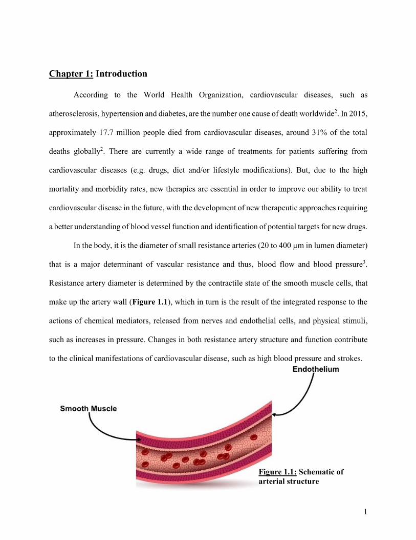
In the body, it is the diameter of small resistance arteries (20 to 400 µm in lumen diameter)
that is a major determinant of vascular resistance and thus, blood flow and blood pressure3.
Resistance artery diameter is determined by the contractile state of the smooth muscle cells, that
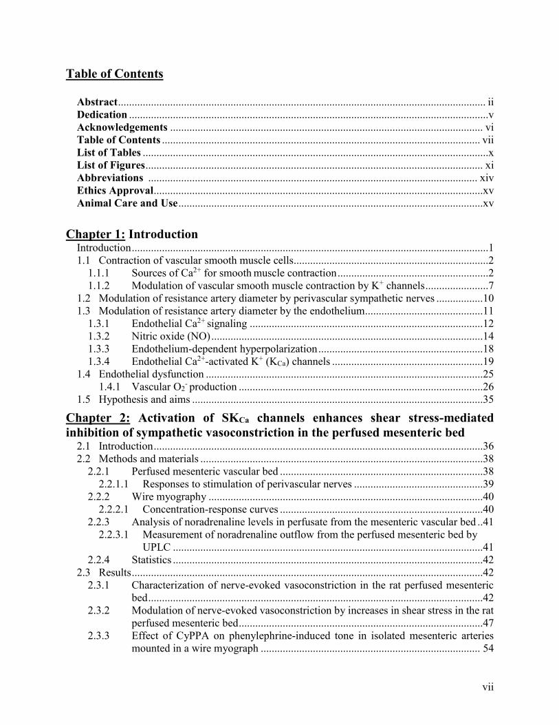
make up the artery wall (Figure 1.1), which in turn is the result of the integrated response to the
actions of chemical mediators, released from nerves and endothelial cells, and physical stimuli,
such as increases in pressure. Changes in both resistance artery structure and function contribute
to the clinical manifestations of cardiovascular disease, such as high blood pressure and strokes.
Figure 1.1: Schematic of
arterial structure
2
1.1: Contraction of vascular smooth muscle cells
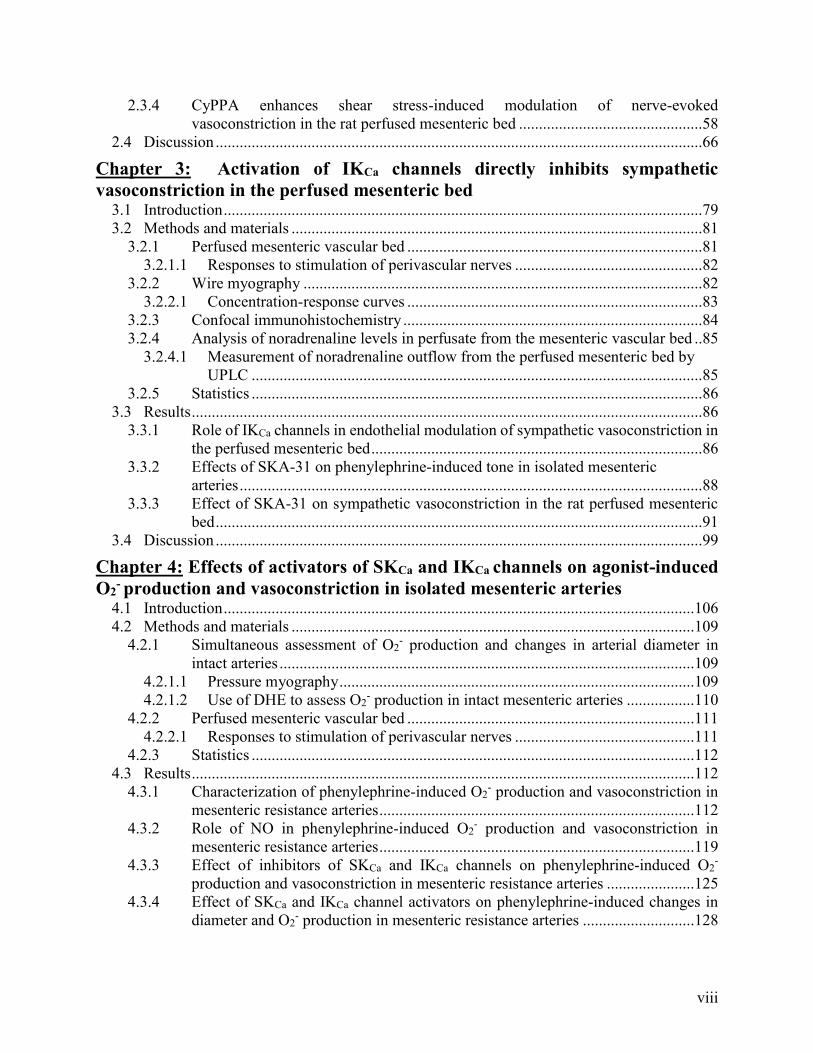
As in all muscle cells, vascular smooth muscle contraction requires adenosine triphosphate
(ATP) and an increase in intracellular Ca2+ concentration4,5, via release from intracellular stores
and/or entry through Ca2+ channels in the cell membrane. Ca2+ binds to calmodulin to form a Ca2+-
calmodulin complex which activates myosin light chain kinase6–8. Myosin light chain kinase is
bound via its N-terminus to actin filaments and activation allows it to phosphorylate nearby myosin
molecules9. Myosin filaments are composed of hexameric myosin molecules, each made up of two
heavy chains and two pairs of light chains (LC17 and LC20)10–12. Activated myosin light chain
kinase phosphorylates LC20 to induce a conformational change that allows interaction between
actin and myosin, and subsequently, an increase in the actin-activated MgATPase activity of
myosin13. Energy generated through hydrolysis of ATP then drives cross-bridge cycling and the
contraction of the muscle cell (reviewed by Saddouk et al. 201712). Myosin light chain kinase is
inactivated by dissociation of Ca2+ from calmodulin and block of the active site of myosin light
chain kinase by an auto-inhibitory domain. Dephosphorylation of LC20 is then mediated by myosin
light chain phosphatase, resulting in disruption of myosin and actin binding and thus, muscle
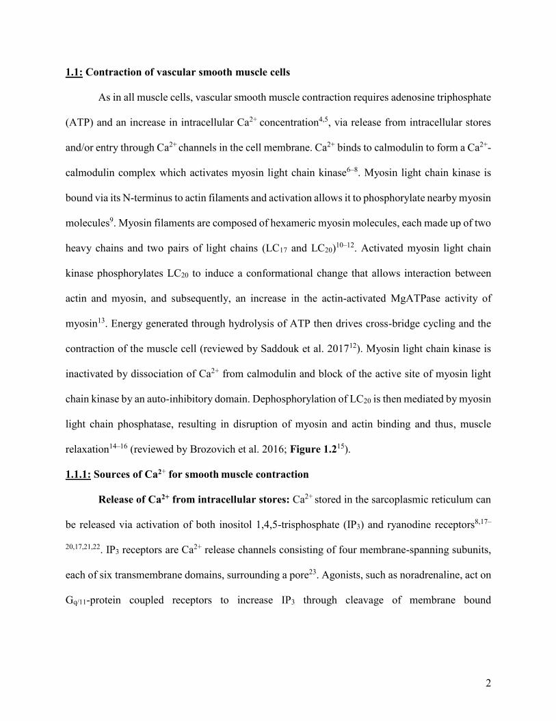
relaxation14–16 (reviewed by Brozovich et al. 2016; Figure 1.215).
1.1.1: Sources of Ca2+ for smooth muscle contraction
Release of Ca2+ from intracellular stores: Ca2+ stored in the sarcoplasmic reticulum can
be released via activation of both inositol 1,4,5-trisphosphate (IP3) and ryanodine receptors8,17–
20,17,21,22. IP3 receptors are Ca2+ release channels consisting of four membrane-spanning subunits,
each of six transmembrane domains, surrounding a pore23. Agonists, such as noradrenaline, act on
Gq/11-protein coupled receptors to increase IP3 through cleavage of membrane bound
3
phosphatidylinositol 4,5-bisphosphate by phospholipase C (reviewed by Berridge et al. 200824)
and so elicit IP3-mediated Ca2+ release.
Figure 1.2: Mechanism of smooth muscle contraction. Schematic of the mechanisms underlying
smooth muscle contraction elicited by a stimulus that increases Ca2+ levels within smooth muscle
cells. An increase in Ca2+ leads to Ca2+ binding to calmodulin which activates myosin light chain
kinase (MLCK). MLCK phosphorylates LC20 to increase the activity of the myosin ATPase which
drives the cycling of actin-myosin cross-bridges to create muscle tension15.
Like IP3 receptors, ryanodine receptors are channels that mediate Ca2+ release from the
sarcoplasmic reticulum but whereas IP3-mediated Ca2+ release is associated with smooth muscle
contraction, discrete increases in Ca2+ caused by release from ryanodine receptors, termed Ca2+
sparks, play an important role in modulating smooth muscle cell contraction due to the proximity
of ryanodine receptors to plasma membrane ion channels (reviewed by Amberg and Navedo,
201325). Ca2+ sparks activate large conductance Ca2+-activated K+ (BKCa; see below) channels to
promote hyperpolarization and oppose vasoconstriction26–32. Modulation of sparks may contribute

4
to the actions of some vasodilator and vasoconstrictor agents, as protein kinase G and protein
kinase C can act on ryanodine receptors to stimulate and inhibit Ca2+ sparks activity,
respectively31,33. Activation of BKCa channels by Ca2+ sparks can be recorded in isolated cerebral
arterial smooth muscle cells as spontaneous transient outward currents, the frequency and
amplitude of which are linked to depolarization and driven by Ca2+sparks27,34.
Ca2+ influx pathways: Store-operated Ca2+ entry. Stromal interaction molecule (STIM)
proteins are single-transmembrane domain proteins located in the sarcoplasmic reticulum that
sense alterations in luminal Ca2+ via their N-terminal domains35. Depletion of Ca2+ stores leads to
dissociation of Ca2+ from these domains, allowing STIM proteins to interact with Orai channels
which mediate Ca2+ influx36. In addition to Orai channels, transient receptor potential (TRP; see
below) channels can also be activated by STIM after store depletion37 but their contribution to
store-operated Ca2+ entry appears to be variable38.
Voltage-dependent Ca2+entry. Many stimuli elicit depolarization of the membrane
potential of vascular smooth muscle cells and so increase the open probability of voltage-operated
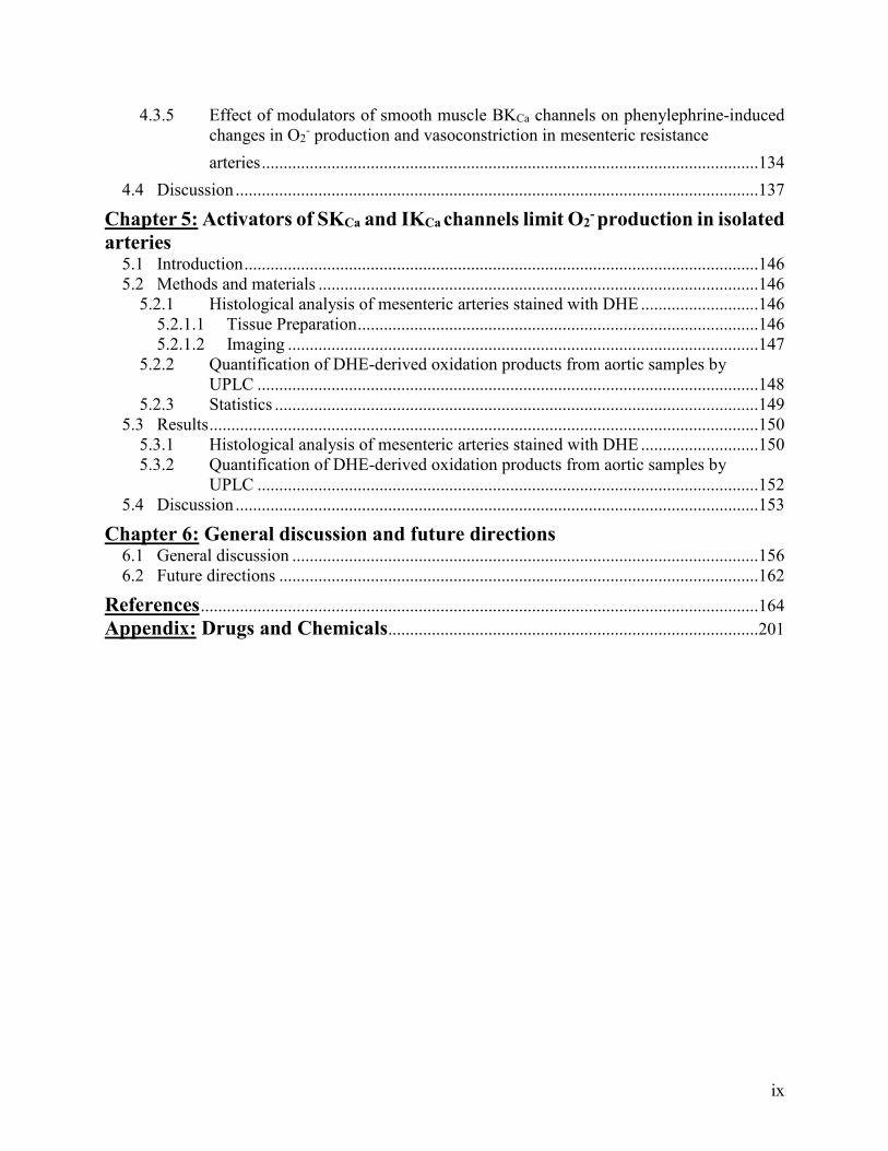
Ca2+ channels (VOCCs). VOCCs are encoded by pore-forming 1 subunits, (Cav1.x, 2.x and 3.x);
with the CaV1.2 channel (L-type) predominantly responsible for mediating vascular smooth muscle
contraction (reviewed by Catterall 201139 and Zamponi et al. 201539). Each channel is comprised
of four α-subunits, each with six transmembrane domains (S1-S6), with S4 conferring voltage
sensitivity40. The α-subunits co-localize with β-, α2δ-, and γ- subunits41–43, which modulate their
voltage sensitivity, conductance and level of expression41,44–46 (Figure 1.347).
L-type VOCCs are slow to activate and inactivate48–50, and have a conductance of around
25 pS48. Membrane depolarization to potentials positive of -30 mV lead to increased opening of
L-type VOCCs and global influx of Ca2+ into smooth muscle cells to cause contraction49. Evidence

5
for the functional role of L-type VOCCs in regulation of arterial diameter has come from the
observations that dihydropyridine antagonists (e.g. nifedipine) that selectively inhibit α1c activity
abolish myogenic reactivity in isolated rat cerebral arteries, whereas dihydropyridine agonists that
stimulate the activity of L-type VOCCs enhance the myogenic response in rabbit ear arteries51,52.
Figure 1.3: Schematic of L-type VOCC Ca2+ channel with α- and accessory -,- and 2-
subunits. Each α-subunit is comprised of six transmembrane domains (S1-S6) with S4 conferring
voltage sensitivity and co-localize with a -,- and/or 2- subunit, which modulate voltage
sensitivity, conductance and level of expression47.
The activity of L-type VOCCs can be regulated by protein kinases C, A and G. Protein
kinase C-mediated phosphorylation of L-type VOCCs has been shown to enhance channel activity
in ventricular myocytes from a range of species53,54 whereas phosphorylation by protein kinase G
inhibits the channel in cultured rat mesenteric arterial55 and aortic56,57 smooth muscle cells, chick
ventricular myocytes58 and guinea pig papillary muscle cells59. The effect of protein kinase A-
mediated phosphorylation is not as clearly defined with both stimulatory and inhibitory effects
reported. For example, in chick embryonic ventricular myocytes58, guinea pig papillary muscle
cells59 and rat mesenteric arterial smooth muscle cells55, protein kinase A enhanced VOCC activity
but was found to have an inhibitory action in cultured rat aortic smooth muscle cells56.
6
The majority of research on VOCCs in vascular smooth muscle cells has focused on L-
type channels but recent evidence indicates that T-type VOCCs (Cav3.x)60,61 can also contribute
to smooth muscle contraction. T-type VOCCs have a conductance of about 8 pS48 and are activated
at more hyperpolarized potentials (positive to −45 mV49) as compared to L-type VOCC currents
(positive to ~-30 mV62). They are also quick to activate and inactivate48–50. Transcript and protein
for Cav3.1 and Cav3.2 channels have been found in vascular smooth muscle cells from rat cerebral
resistance arteries63,64, and Cav3.3 channels has also been identified in human cerebral artery
cells65. Electrophysiological recordings have identified Cav3.1 and Cav3.2 channels as being
responsible for the nifedipine-insensitive component of Ca2+ current in rat cerebral vascular
smooth muscle cells64, as T-type VOCCs have been shown to be insensitive to dihydropyridines,
such as nifedipine and nitrendipine49,50, which block L-type VOCCs49,50. The functional role of T-
type VOCCs in regulation of arterial diameter has largely been explored using the blocker,
mibefradil63.
Ca2+-sensitization: An increase in Ca2+ is obligatory for the initiation of force generation
within vascular smooth muscle cells. However, decreases in myosin light chain phosphatase
activity following protein kinase phosphorylation can enhance contractile force without further
changes in Ca2+ levels via a process termed Ca2+ sensitization66,67. Sensitization evoked by agonists
acting at G-protein coupled receptors is thought to be due to activation of Rho-associated
kinase68,69. Agonist-induced activation of the small GTPase RhoA, via the G12/13 family of
heterotrimeric G-proteins and a guanine nucleotide-exchange factor, leads to activation of Rho-
associated kinase which inhibits myosin light chain phosphatase activity by phosphorylation of
myosin phosphatase target subunit-168. Protein kinases, such as Rho-associated kinase and protein
kinase C11,14,70,71, can also phosphorylate C-kinase potentiated protein phosphate-1 inhibitor72, an

7
endogenous inhibitor of myosin light chain phosphatase, which when phosphorylated, binds the
catalytic site of myosin light chain phosphatase (reviewed by El-Yazbi et al. 201673).
1.1.2: Modulation of vascular smooth muscle contraction by K+ channels
K+ currents are the major ionic conductance in the plasma membrane of vascular smooth
muscle cells and thus, set and regulate membrane potential74–76. In physiological conditions (3–5
mM K+ outside of the cell and 140 mM K+ inside), the driving force for K+ is outward and so
opening of a K+ conducting channel leads to membrane hyperpolarization77. As membrane
resistance is high, opening of a few K+ channels can have a large impact on smooth muscle
membrane potential and thus, the open probability of VOCCs and contractility. Vascular smooth
muscle cells express a wide range of different types of K+ channels: BKCa channels, voltage-gated
K+ channels, ATP-sensitive K+ (KATP) channels, inward-rectifier K+ channels, and members of the
two-pore K+ channel family (recently reviewed by Jackson, 201778)27,29,79–92. However, for the
purposes of this thesis, I will focus on the structure and function of BKCa channels and their role
in regulating smooth muscle contractility and therefore, arterial diameter in resistance arteries.
BKCa channels (encoded by KCNMA1) are composed of homotetramers of pore-forming
α-subunits together with regulatory - and − subunits91–95 (Figure 1.478). The α-subunit is
composed of 7 transmembrane domains with the pore region between S5 and S696. Two regulator
of K+ conductance domains (1 and 2) located in the C-terminus contain Ca2+ binding sites while
positively charged residues in S2-4 serve as voltage sensors77,96–98. These channels have a large
single channel conductance (150-270 pS)97,99,100 and exhibit voltage-dependent gating for which
binding of Ca2+ increases the apparent sensitivity to voltage92–94. Under physiological conditions,
BKCa channels require both depolarization of the membrane potential and a rise in intracellular
Ca2+ to occur simultaneously in order to open81,92,97,101.
8
There are four -subunits (KCNMβ1–4) with 1 the dominant form in vascular smooth
muscle cells82,91,102. The -subunits alter channel gating, sensitivity to Ca2+ and voltage, while the
γ-subunit is associated with increasing channel sensitivity to voltage91,95,103–105.
BKCa channels are widely expressed in a variety of tissues (Table 1.1). They are highly
expressed in all vascular smooth muscle cells27,29,79–92, but are not present in freshly isolated
endothelial cells76,106–108. BKCa channels are of particular importance in the central nervous system
where they regulate the excitability of neurons97,109,110 and have been localized to the inner
membrane of mitochondria in cardiac myocytes where they’ve been proposed as potential targets
for preventing cardiac ischemia-reperfusion injury89,110–112.
Figure 1.4: Schematic of structure of BKCa channels. BKCa channels are composed of pore-
forming α-, and modulatory ß1- and γ- subunits. The α-subunit is made up of 7 transmembrane
domains, S2-4 being the voltage sensor and the pore region located between S5 and S6, and 4
cytoplasmic domains within the C-terminus tail. Two regulator of K+ conductance domains (RCK1
and RCK2) located in the cytoplasmic C-terminus contain the channel’s Ca2+ binding sites. The
auxiliary subunits ß1- and γ- subunits consist of two transmembrane domains and one
transmembrane domain, respectively78.

9
Table 1.1: Cellular location of BKCa, SKCa and IKCa channels.
As mentioned earlier, activation of BKCa channels by ryanodine receptor-dependent Ca2+
sparks regulates diameter in cerebral resistance arteries (reviewed by Jackson 201778). In other
vessels, such as hamster cremaster arterioles, Ca2+ entry through L-type VOCCs may contribute
to BKCa channel activation163. BKCa channels can be targeted by vasodilators, either directly or
through modification of Ca2+ spark activity. For example, nitrosylation via nitric oxide (NO)79,164
Channel Type Location
BKCa ▪ Vascular smooth muscle27,29,79–92
▪ Urinary smooth muscle102
▪ Adrenal chromaffin cells113
▪ Inner mitochondrial membrane of cardiac
myocytes89,110–112
▪ Neurons97,109,110
▪ Inner mitochondrial membrane of
neurons114
▪ B-lymphocytes115
▪ Platelets115
SKCa ▪ Vascular endothelium26,80,84,85,116–133
▪ Cardiac myocytes134–136
▪ Inner mitochondrial membrane of cardiac
myocytes137,138
▪ Neurons139–143
▪ Inner mitochondrial membrane of
neurons144,145
▪ Platelets115,146,147
▪ B-lymphocytes115
IKCa ▪ Vascular endothelium80,84,100,116,118–122,124–
126,128,130,131,148–151
▪ Neurons152–156
▪ T-lymphocytes157
▪ Erythrocytes158
▪ Platelets 115,146,147,159
▪ Pancreas100,160
▪ Intestinal epithelia161,162
▪ Surface epithelia (skin, oral and vaginal
mucosas, oesophageal lining)160
▪ Ducts of fluid-secreting glands (salivary
glands, lacrimal glands)160

10
or phosphorylation via protein kinase G165–168, enhances the open probability of BKCa channels by
shifting voltage-sensitivity of the channels to more hyperpolarized membrane potentials. In
contrast, protein kinase C-mediated phosphorylation of BKCa channels enhances vasoconstriction
by shifting BKCa channel voltage-sensitivity towards more depolarized membrane potentials thus,
limiting their ability to inhibit VOCC-mediated Ca2+ entry169–172. The importance of smooth
muscle BKCa channels in limiting resistance artery vasoconstriction is shown by the observation
that in pressurized rat mesenteric and cerebral arteries, BKCa channel inhibition leads to enhanced
vasoconstriction and membrane depolarization21,26, and mice deficient in BKCa channels were
found to have significantly enhanced arterial blood pressure173.
I will now discuss how resistance artery diameter is modulated by two of the most
physiological important influences, sympathetic nerve activity, and chemical and electrical signals
from endothelial cells.
1.2: Modulation of resistance artery diameter by perivascular sympathetic nerves
The sympathetic nervous system plays a major role in controlling total peripheral vascular
resistance and is a key regulator of resistance artery diameter174. In contrast to structurally well-
defined neuromuscular junctions in skeletal muscle, perivascular nerve fibres do not penetrate into
the smooth muscle layers174. Perivascular nerves appear as a network of axon bundles, with
swollen areas, called varicosities, that release neurotransmitters in a manner similar to paracrine
secretion175. Sensory and nitrergic (that release NO) perivascular nerves have been identified176,177
but for the purposes of this thesis, I will focus on sympathetic innervation as this accounts for the
majority of nerves in resistance arteries (reviewed by Westcott and Segal174).
Stimulation of perivascular sympathetic nerves evokes release of noradrenaline and co-
transmitters, ATP and neuropeptide Y175–188. Noradrenaline acts primarily on post-synaptic 1-
11
adrenoceptors to cause vasoconstriction via a number of mechanisms, including IP3-mediated
release of Ca2+ from stores, membrane depolarization to increase Ca2+ influx through VOCCs and
Ca2+-sensitization189,190. ATP activates post-synaptic P2X receptors to cause an influx of Na+ and
Ca2+ ions that excites the smooth muscle and creates an excitatory junction potential176–179,181,184–
188,191, although the relative contribution of ATP to sympathetic vasoconstriction varies between
arteries and species187,188,192. Neuropeptide Y binds to post-synaptic Y1 or Y2 receptors193, but its
role appears to be to potentiate noradrenaline-evoked responses rather than to evoke direct
vasoconstriction182,194.
1.3: Modulation of resistance artery diameter by the endothelium
In 1980, Furchgott and Zawadzki made the seminal discovery that removal of the
endothelial layer of rabbit aortic rings impaired vasorelaxation to acetylcholine and so provided
the first example of endothelium-dependent vasodilation195. This finding was fundamental to our
understanding of blood vessel function and opened up a wide field of research, which has led to
our current view that the endothelium is a complex endocrine organ that plays a vital role in the
regulation of blood pressure and flow, hemostasis, inflammation, vascular growth and remodeling
in the cardiovascular system196.
We now know that the endothelium releases a wide range of diffusible factors (e.g. NO
and cyclooxygenase products, such as prostacyclin) that can alter the contractility of the
surrounding smooth muscle cells, and that stimulation of the endothelium by agonists acting at G-
protein coupled receptors117,197, or physiological stimuli, such as increases in shear stress198–201,
also result in activation of endothelial Ca2+-activated K+ (KCa) channels80,84,85,100,116–131,148–
151,196,197,202–208. Opening of these channels causes hyperpolarization of the endothelial cell
12
membrane potential which spreads to the underlying smooth muscle cells via myoendothelial gap
junctions (MEGJs) to reduce opening of VOCCs, decreasing Ca2+ influx and causing relaxation.
For the purposes of this thesis, I will briefly discuss endothelial Ca2+ signaling, and then
focus on two of the main pathways for endothelial modulation of smooth muscle contractility, NO
and endothelial KCa channels, and their role in regulating smooth muscle contractility in resistance
arteries.
1.3.1: Endothelial Ca2+ signaling
Endothelium-dependent mechanisms for regulation of smooth muscle contractility share a
common feature in that they are dependent on a rise in Ca2+ levels within endothelial cells202,209–
214. This increase in Ca2+ can be elicited through release from endoplasmic reticulum stores and/or
via Ca2+ influx through TRP channels, with the contribution of these two mechanisms showing
stimulus-dependent variation. It is notable that there are no VOCCs in native endothelial cells215.
Agonists acting on endothelial Gq/11-protein coupled receptors stimulate IP3-mediated
release of Ca2+ stores which, as described above for smooth muscle cells, leads to store-operated
Ca2+ entry through Orai1 and/or TRP channels216–218. The rise in endothelial Ca2+ activates Ca2+-
dependent enzymes, such as nitric oxide synthase (NOS), as well as KCa channels to elicit
endothelial hyperpolarization219. The identity of the TRP channel mediating receptor-linked Ca2+
entry most likely varies between stimuli, arteries and species. TRP vanilloid 4 (TRPV4) channels
have been implicated in acetylcholine-evoked Ca2+ entry in mouse mesenteric220 and carotid221
arteries, whereas TRP canonical 3 (TRPC3) and 4 (TRPC4) have been associated with the same
responses in aorta from knockout mouse models222,223.
In vivo increases in the shear stress across the endothelial cell surface is a major stimulus
for activation of endothelium-dependent vasodilator pathways198–201. The mechanism underlying
13
shear stress-induced increases in endothelial Ca2+ have not been greatly studied but recent reports
indicate a role for mechanosensitive TRP channels, and in particular TRPV4 channels, in shear
stress induced increases in Ca2+, NO production and activation of endothelial KCa channels132,224.
Limited evidence has also been provided that shear stress-induced increases in endothelial Ca2+
are mediated by the release of acetylcholine from endothelial cells. Briefly, Wilson et al. have
suggested that acetylcholine produced by endothelial cells is released into the vascular lumen in
response to increased shear stress225. Acetylcholine then activates endothelial muscarinic
receptors, leading to the generation of IP3 which increases endothelial Ca2+ to stimulate NO
production and activate KCa channels225.
Endothelial TRP channels. As mentioned above, TRP channels have emerged as the most
likely mediators of endothelial Ca2+ influx226,227. The TRP channel super family consists of six
subfamilies: TRPV, TRPC, TRPM (melastatin), TRPML (mucolipin), TRPP (polycystin) and
TRPA (ankyrin)226,228. These channels are all tetramers (either homo- or hetero- mers) with six
transmembrane domains and a pore generated by a pore forming loop between S5 and S6226,228.
All TRP channels are permeable to Ca2+, with the exception of TRPM4 and TRPM5, which are
Ca2+ activated, but not Ca2+ permeable229–231. TRP channels are not gated by voltage, but can
respond to a wide range of different stimuli: TRPV1-V4 and TRPM3 channels are activated by
high temperatures whereas TRPM8, TRPA1, and TRPC5 channels are activated by low
temperatures232,233, TRPC channels are activated either directly by diaglycerol (TRPC2, TRPC3,
TRPC6, and TRPC7 channels), or indirectly through a diacylglycerol-dependent mechanism
(TRPC1, TRPC4, and TRPC5 channels234–236) and TRPM4, TRPM5, TRPM2, and TRPA1
channels are activated by rises in Ca2+ 237–240.
14
Though many of these subfamilies are located on both the endothelium and smooth muscle
of the vasculature, two TRP channels in particular, TRPC3 and TRPV4 channels, have been
identified as potential mediators of Ca2+ influx underlying endothelium-dependent responses to
shear stress and/or agonists.
TRPV4 channels are expressed on the endothelium and smooth muscle of many arteries,
can be activated by shear stress241,242 and IP3243, and have been linked to both NO production and
opening of endothelial KCa channels220,241–248. Mesenteric arteries from mice lacking TRPV4
channels have reduced endothelium-dependent relaxation to acetylcholine in comparison to their
wildtype counterparts220 and in rat carotid and gracilis arteries, shear stress-evoked vasodilation is
inhibited in the presence of a TRPV4 channel inhibitor241. However, there is also evidence that
TRPV4 channels are not involved in endothelium-dependent vasodilation. For example, Pankey
et al.246 found GSK-21939874, a TRPV4 channel inhibitor, did not alter acetylcholine-evoked
reductions in pulmonary and systemic arterial pressures. And in mice, global knockout of TRPV4
channels does not alter systolic or diastolic blood pressure243, heart rate243 or carotid artery dilation
to acetylcholine 247.
TRPC3 channels have also been localized to the vascular endothelium197,249,250 but not to
the smooth muscle cells, and are activated by diacylglycerol, a product of the cleavage of
phosphatidylinositol 4,5-biphosphates by phospholipase C stimulated by Gq/11-protein coupled
receptor activation249,251–255. Also, it has been reported, by our lab and others, that TRPC3 channels
are involved in endothelium-dependent hyperpolarization via the activation of KCa channels256,257.
1.3.2: Nitric oxide (NO)
NO is produced by NOS, which converts L-arginine to citrulline and NO213,258–262. Oxygen
and reduced nicotinamide-adenine-dinucleotide phosphate (NADPH) are co-substrates and flavin

15
adenine dinucleotide (FAD), flavin mononucleotide (FMN), and 5,6,7,8-tetrahydro-l-biopterin
(BH4) are cofactors for this reaction213,258–265. The biological half-life of NO and therefore, its
activity, is determined by its interaction with superoxide anion (O2-) which reacts with NO to form
the highly reactive intermediate peroxynitrite (ONOO-): O2- + NO → ONOO-.
In cardiovascular disease states, enhanced oxidative stress leads to increased inactivation
of NOS by O2- and to uncoupling of NOS; uncoupled NOS produces O2
- rather than NO266–270.
Potential mechanisms underlying this change include: oxidation of BH4 and depletion of L-
arginine268,271.
There are three subtypes of NOS: neuronal, endothelial and inducible213,260,272,273.
Endothelial NOS and neuronal NOS are constitutively active enzymes that produce NO in response
to rises in intracellular Ca2+ 210–213. Their activity is also regulated via phosphorylation by a number
of different protein kinases266,274. The activity of inducible NOS is regulated by its expression
level, which is up-regulated in response to cytokines and/or oxidative stress272,275. For the purposes
of this thesis, I will focus on endothelial NOS located in the vascular endothelium.
The NOS enzyme has two domains, an N-terminal oxygenase domain which binds BH4,
oxygen, L-arginine and heme and a C-terminal reductase domain that binds NADPH, FAD and
FMN263–265. The two domains are linked via a calmodulin-recognition site which is essential for
the linkage between the reductase and oxygenase domains and allows dimerization211,213,263–265.
NO synthesis occurs when electrons are transferred from NADPH via the flavins, FAD and FMN,
in the C-terminal reductase domain, to the heme in the N-terminal oxygenase domain276. At the
heme site, the electrons are used to reduce and activate oxygen and to oxidize L-arginine to L-
citrulline and NO277,278. Binding of BH4 at the dimer interface is required for the stabilization of
16
the NOS dimer and ‘coupled’ NOS activity. As mentioned above, in the absence of BH4, the NOS
domains become uncoupled, leading to the production of O2- rather than NO266–270.
The binding of Ca2+-calmodulin is essential for NO production as it enhances the rate of
electron transfer from NADPH to flavins in the C-terminus region211,279,280 but several other
proteins also interact with NOS to regulate its activity. For example, the molecular chaperone, heat
shock protein 90, acts as an allosteric modulator to increase NOS production of NO281. It has also
been suggested to inhibit uncoupling of NOS to prevent O2- production282. This would suggest that
there is an intrinsic cellular mechanism regulating the uncoupling of NOS and thus, regulating the
balance of NO and O2- production282. The caveolae coat protein, caveolin-1, is a tonic inhibitor of
NOS activity, with recruitment of Ca2+-calmodulin and heat shock protein 90 to NOS displacing
caveolin-1 from the enzyme to activate it283,284. Furthermore, phosphorylation of serine1177 of
NOS in response to prolonged increases in shear stress can stimulate NO production in a Ca2+-
independent manner, with phosphatidylinositol-3 kinase/AKT being implicated as the possible
mediators of this phosphorylation266,274,285.
Once released from endothelial cells, NO causes relaxation of smooth muscle cells via
activation of soluble guanylyl cyclase286 to increase production of cyclic guanosine
monophosphate, which subsequently activates protein kinase G287,288. Protein kinase G interacts
with a number of different protein targets to limit vasoconstriction and cause vasodilation. For
example, protein kinase G can phosphorylate phospholipase C to inhibit IP3 production and thus,
decrease IP3-mediated Ca2+ release required for smooth muscle vasoconstriction166,251,289, and
protein kinase G-mediated phosphorylation increases activity of BKCa channels165–168. NO itself
can also directly nitrosylate BKCa channels to increase their open probability79,290,291,164. The
subsequent smooth muscle hyperpolarization due to the opening of BKCa channels292 decreases the

17
activity of VOCCs and reduces Ca2+ entry and vasoconstriction26. Additionally, protein kinase G-
mediated phosphorylation of L-type VOCCs can inhibit their activity55–59, to further reduce Ca2+
entry. See Figure 1.5293 for a schematic of NO-mediated effects on vascular smooth muscle
contractility.
The role of NO in regulating arterial diameter, blood flow and pressure was facilitated by
the discovery that structural analogues of L-arginine, such as L-NG-nitro arginine (L-NOARG)
and NG-nitro-L-arginine methyl ester hydrochloride (L-NAME) act as selective, competitive
inhibitors of NOS294,295. For example, administration of L-NAME causes hypertension in rats and
L-NOARG inhibits endothelium-dependent relaxation in isolated rat aorta296,297. The
demonstration that deletion of NOS leads to hypertension in mice was also an advance in
demonstrating the physiological importance of this molecule298.
18
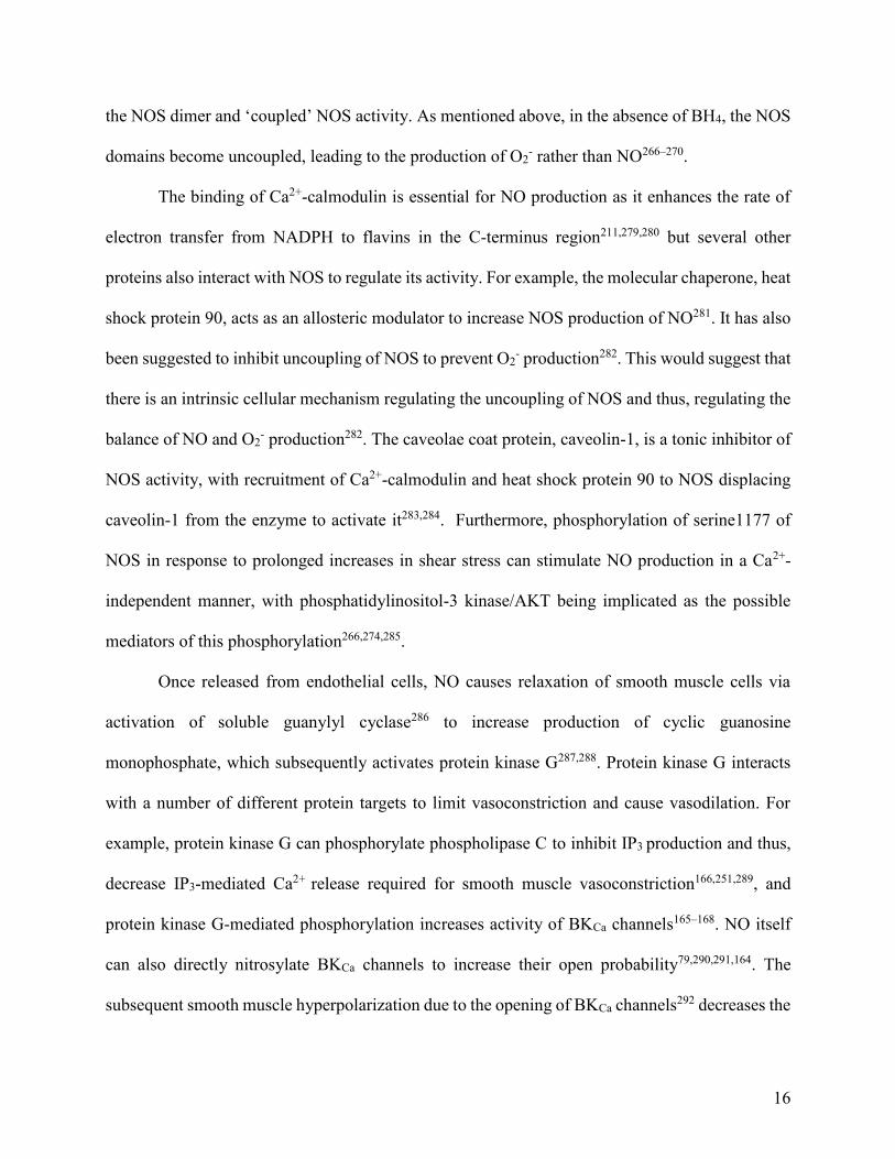
Figure 1.5: NO-mediated relaxation of vascular smooth muscle. NO is produced in endothelial
cells by endothelial NOS and diffuses to the smooth muscle to activate guanylyl cyclase to produce
cyclic guanosine monophosphate which stimulates protein kinase G to phosphorylate a number of
targets to induce vasodilation. For example, protein kinase G phosphorylates VOCCs to decrease
opening probability, and phosphorylates BKCa channels to increase opening probability293.
1.3.3: Endothelium-dependent hyperpolarization
Opening of endothelial small (SKCa) and intermediate (IKCa) conductance Ca2+-activated
K+ channels causes hyperpolarization of the endothelial cell membrane potential which spreads to
underlying smooth muscle cells via MEGJs to reduce opening of VOCCs, leading to a decrease in
Ca2+ influx and thus, relaxation80,117,125,202,299. The defining characteristic of hyperpolarization-
mediated vasodilation is that it persists in the presence of inhibitors of NOS and cyclooxygenase
and, under these conditions, is abolished by blockers of endothelial SKCa and IKCa channels,
apamin and 1-[(2-Chlorophenyl)diphenylmethyl]-1H-pyrazole (TRAM-34) or charybdotoxin,
19
respectively (reviewed by Ledoux et al. 2006103). The physiological importance of endothelial KCa
channels in vivo is highlighted by increased vascular reactivity and raised arterial blood pressure
recorded in mice lacking one or both of the channels127,300.
Endothelium-dependent hyperpolarization plays a more prominent role in endothelium-
dependent dilation of resistance arteries than in large vessels and so it is an important determinant
of local tissue perfusion301–303. Initially, it was thought that endothelium-dependent
hyperpolarization of vascular smooth muscle was mediated by a diffusible factor, but current
consensus it is due to direct electrical coupling of endothelial and smooth muscle cells via
MEGJs303.
1.3.4: Endothelial Ca2+-activated K+ (KCa) channels
There are three types of KCa channels: BKCa, SKCa and IKCa channels. Whereas BKCa
channels are solely located on smooth muscle27,29,79–92, SKCa and IKCa channels are not found on
smooth muscle cells but are located on the endothelium80,84,85,100,116–131,148–151,197,202 (See Table
1.1).
Endothelial SKCa and IKCa channels are voltage-independent K+ channels that are activated
by increases in intracellular Ca2+ 117,202. There are three SKCa channel subtypes (SK1, 2 and 3),
which are encoded by the genes KCNN1-3117,120,139, and one IKCa channel subtype (SK4), encoded
by KCNN4120,202,304. In the vascular endothelium, SK3, rather than SK1 and 2, has been shown to
be the primary subtype present117, and thus, when SKCa channel is referenced in this work it is
denoting the SK3 subtype117.
SKCa and IKCa channels are tetramers consisting of α-subunits that have six transmembrane
domains (S1-S6), with the pore domain encompassed by S5-S6 and a calmodulin binding domain
on the C-terminus directly after S6140. Calmodulin is constitutively bound to the C-terminus of

20
SKCa and IKCa channels and confers their Ca2+ sensitivity (values for half-maximal activation
ranging from 95 nM to 0.3 μM100,140), as the channels themselves do not possess any Ca2+ binding
sites140. Thus, SKCa and IKCa channels will remain closed until Ca2+ binds to each of the bound
calmodulin as Ca2+-calmodulin complexes stabilize the channel’s open state103. See Figure 1.6 for
a schematic of SKCa/IKCa channel subunit structure103.
Figure 1.6. Schematic of a SKCa/IKCa channel subunit. Each subunit consists of six
transmembrane domains with a pore region (P) between S5 and S6. Calmodulin interacts with the
intracellular C-terminus103.
SKCa and IKCa channel activity is modulated by associated proteins. Constitutively bound
casein kinase 2 and protein phosphatase 2A have been shown to alter SKCa channel Ca2+ sensitivity
through phosphorylation or dephosphorylation of the bound calmodulin103,305,306. Casein kinase 2-
mediated phosphorylation occurs at threonine80 on the constitutively bound calmodulin and
decreases the Ca2+ sensitivity (from sub-micromolar ranges to micromolar ranges306), reducing the
opening probability of the channel305. Dephosphorylation mediated by protein phosphatase 2A
removes the inhibitory phosphorylation caused by casein kinase 2305,306.

21
As their name suggests, opening of SKCa channels allows for a small K+ current (10-20
pS100,202,307) to move out of the endothelial cells, causing hyperpolarization of the endothelial
membrane potential. Alternatively, IKCa channels have a conductance of about 30-80 pS100,202. KCa
channel-mediated endothelial hyperpolarization has been recorded in intact porcine coronary
arteries117, internal carotid arteries of guinea pigs80, rat mesenteric122,123,150 and hepatic arteries299,
rat aortas119, and freshly isolated endothelial cells from porcine coronary arteries117,122,202, rat
mesenteric arteries150 and canine mesenteric arteries124, and human umbilical vein endothelial
cells131. This hyperpolarization spreads through the MEGJs to induce hyperpolarization of the
smooth muscle membrane potential, limiting constriction by decreasing the open probability of
VOCCs49,214,299.
Compared to other ion channels, SKCa and IKCa channels have a well-developed
pharmacology which has aided investigation of their physiological functions308. For example
apamin, isolated from bee venom, is a selective inhibitor of rat SK2 and SK3 (IC50 70 pM and 2.6
μM, respectively) but does not block rat SK1 channels309. Apamin is an allosteric modulator,
binding to the outside of SKCa channels and causing a conformational change in the channel’s pore
that blocks the movement of K+309,310. TRAM-34 and 4-[[3-(Trifluoromethyl)phenyl]methyl]-2H-
1,4-benzothiazin-3(4H)-one (NS 6180) are both selective inhibitors of IKCa channels that bind to
threonine250 and valine275 in the inner pore. Up to a concentration of 1 µM NS 6180 and 5 µM
TRAM-34, these two chemicals are highly selective IKCa channel blockers that show no effect on
T-lymphocyte Ca2+ entry or voltage-gated K+, sodium and TRP channels121,311,312.
There are also positive modulators selective for both SKCa and IKCa channels, such as 1-
ethyl-2-benzimidazolinone (EBIO) and 6,7-dichloro-1H-indole-2,3-dione 3-oxime (NS 309),
which have been shown to bind to the interface of where the α-subunits of these channels bind to
22
calmodulin, increasing their Ca2+ sensitivity308. Newer compounds that are selective for either
channel have also been developed. For example, N-cyclohexyl-N-[2-(3,5-dimethyl-pyrazol-1-yl)-
6-methyl-4-pyrimidinamine (CyPPA), was based on NS 309, and as such, is a positive modulator
selective for SK2 and SK3 channels (EC50 value for human SK2 and SK3 channels is 14 μM and
5.6 μM, respectively) but has no effect on human IKCa channels313. It has been shown to enhance
the Ca2+ sensitivity of human SK3 channels by improving the Ca2+ sensitivity of the channel from
429 nM to 59 nM313. As it is structurally similar to NS 309, it is likely that it also binds to the
interface of the α-subunit and calmodulin binding site on SKCa channels to elicit its effects308,313.
Naphtho[1,2-d]thiazol-2-ylamine (SKA-31) is another positive modulator but it is 7- to 10-
fold more selective for IKCa (EC50 value of 260 nM) over SKCa channels (SK3 EC50 value of 2.9
μM)121. SKA-31 does not interact significantly with other channels when used at concentrations
under 25 μM121. SKA-31 has been used in the literature as a positive modulator of both SKCa and
IKCa channels121,124,148,314,315 and as a selective positive modulator for IKCa channels alone204,316.
However, Sankaranarayanan et al. found that the enhancement of acetylcholine-mediated dilation
of mice carotid arteries and reduction of mean arterial pressure in vivo caused by SKA-31 was
entirely through its actions on IKCa channels121.
While SKCa and IKCa channels share significant similarities in their structures and
regulation, their discrete cellular locations within endothelial cells may correspond to differences
in how they regulate vascular tone in response to various stimuli125,317. SKCa are located at
endothelial junctions on the luminal surface and co-localize in caveolae with TRPV4 channels318
where they are able to respond to local Ca2+ increases evoked by increases in shear stress-induced
activation of TRPV4 channels319. In contrast, IKCa channels are located on the abluminal side of

23
endothelial cells at MEGJs, the sites of contact between endothelial and smooth muscle cells
(Figure 1.7).
Figure 1.7: Schematic showing the cellular locations of SKCa and IKCa channels within
endothelial cells. SKCa channels have been localized to the endothelial luminal membrane while
IKCa channels have been localized to the MEGJs. Their differing locations are proposed to confer
them different functional roles in terms of regulating vascular tone. SKCa, small conductance Ca2+-
activated K+; IKCa, intermediate conductance Ca2+-activated K+; ER, endoplasmic reticulum;
NSCC, non-selective calcium channel (most likely a TRPV4 channel); IP3, inositol triphosphate.
Recent work from our lab and others, has demonstrated that the localization of IKCa
channels at MEGJs enables them to mediate myoendothelial feedback, a mechanism by which
contractile activation of smooth muscle cells is limited by the endothelium (Figure 1.8197). Briefly,
IP3, produced in smooth muscle cells by the activation of α1-adrenoceptors8,17–20,251–255,320, diffuses
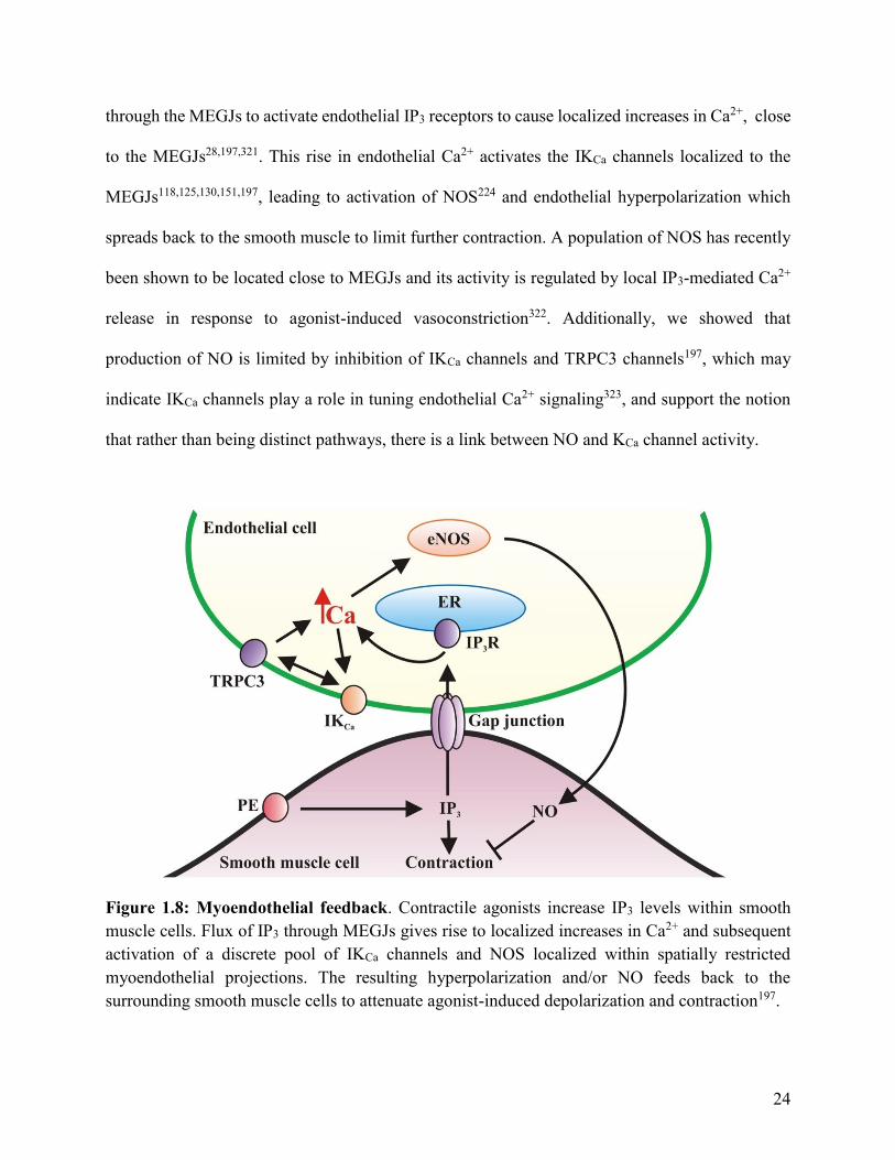
24
through the MEGJs to activate endothelial IP3 receptors to cause localized increases in Ca2+, close
to the MEGJs28,197,321. This rise in endothelial Ca2+ activates the IKCa channels localized to the
MEGJs118,125,130,151,197, leading to activation of NOS224 and endothelial hyperpolarization which
spreads back to the smooth muscle to limit further contraction. A population of NOS has recently
been shown to be located close to MEGJs and its activity is regulated by local IP3-mediated Ca2+
release in response to agonist-induced vasoconstriction322. Additionally, we showed that
production of NO is limited by inhibition of IKCa channels and TRPC3 channels197, which may
indicate IKCa channels play a role in tuning endothelial Ca2+ signaling323, and support the notion
that rather than being distinct pathways, there is a link between NO and KCa channel activity.
Figure 1.8: Myoendothelial feedback. Contractile agonists increase IP3 levels within smooth
muscle cells. Flux of IP3 through MEGJs gives rise to localized increases in Ca2+ and subsequent
activation of a discrete pool of IKCa channels and NOS localized within spatially restricted
myoendothelial projections. The resulting hyperpolarization and/or NO feeds back to the
surrounding smooth muscle cells to attenuate agonist-induced depolarization and contraction197.
25
It has been suggested that endothelial KCa channel-mediated hyperpolarization of the
membrane potential maintains the driving force for Ca2+ influx to endothelial cells necessary for
activation of NOS. But, the ability of hyperpolarization to regulate Ca2+ entry by increasing the
electrical driving force is controversial, particularly as there is a large concentration gradient of
~20,000-fold from outside to inside of endothelial cells324,325. However, recent studies of isolated
endothelial tubes have demonstrated that Ca2+ influx in the presence of acetylcholine is enhanced
by KCa channel-mediated membrane potential hyperpolarization and reduced by membrane
potential depolarization326. In human umbilical vein endothelial cells, inhibition of SKCa and IKCa
channels blocked Ca2+ influx and NO production in response to G protein-coupled receptor
activation131, and both SKCa and IKCa channels have been shown to influence endothelial Ca2+
dynamics in intact mouse mesenteric arteries323. Furthermore, in rat cremaster arterioles, NS 309
and 5,6-dichloro- 1-ethyl-1,3-dihydro-2H-benzimidazol-2-one (DCEBIO), activators of
SKCa/IKCa channels, enhanced ATP-induced hyperpolarization, cytosolic Ca2+ concentration and
NO synthesis128,131. Thus, these findings indicate that opening of endothelial SKCa and IKCa
channels may enhance NO bioavailability80,84,85,100,116–131,148–151,197,202–205.
1.4: Endothelial dysfunction
Endothelium-derived NO elicits relaxation of surrounding smooth muscle cells to cause
vasodilation, regulates local cell growth and protects blood vessels from the deleterious
consequences of platelet aggregation and activation of inflammatory responses327. Endothelial
dysfunction is associated with risk factors for cardiovascular diseases, such as diabetes,
hypertension and atherosclerosis, is characterized by increased production of O2- and decreased
NO bioavailability268,328–336 leading to enhanced vasoconstriction, clot formation and inflammation
within the vasculature (Figure 1.9116).

26
Figure 1.9: Schematic showing the deleterious consequences of decreased NO and increased
O2- levels on the vasculature. Enhanced O2
- production leads to increased ONOO- and reduced
bioavailability of NO leading to increased vasoconstriction, platelet activity, thrombosis,
atherosclerosis and plaque rupture and diminished angiogenesis. eNOS, endothelial nitric oxide;
NO, nitric oxide; O2-, superoxide anion; ONOO-, peroxynitrite; BH4, tetrahydrobiopterin; cGMP,
cyclic guanine monophosphate116.
Attempts to reduce vascular O2- levels through the use of dietary anti-oxidants, such as
vitamins B, C and E, have been unsuccessful in clinical trials337–342 and so there is the need to
identify new targets for therapeutic approaches to reduce O2- levels and enhance NO bioavailability
in pathological settings.
1.4.1: Vascular O2- production
Reactive oxygen or nitrogen species (ROS or RNS, respectively) are highly reactive
compounds involved in a variety of different cellular processes under both physiological and
27
pathophysiological conditions328,329,343. Whether ROS/RNS cause cellular damage or not, is
usually determined by their cellular concentrations, with low levels of ROS and RNS necessary
for normal cellular processes and high concentrations leading to dysfunction329,343–346. For
example, vasoconstriction of rat mesenteric resistance arteries evoked by α1-adrenoceptor agonists
is dependent on production of O2- by NADPH oxidase and vascular smooth muscle
mitochondria347–349 whereas deleterious high levels of O2- production leads to endothelial
dysfunction associated with increased risk of cardiovascular disease268,328–336. Some examples of
ROS and RNS are O2-, hydroxyl radical (OH-), NO, ONOO- and hydrogen peroxide (H2O2) but for
the purposes of this thesis, I will be focusing on O2-.
O2- is produced through the addition of an electron to molecular oxygen350. In the
vasculature, this is primarily mediated by NADPH oxidase enzymes351–356, and to a lesser extent
by xanthine oxidase, the mitochondrial electron transport chain, cyclooxygenase enzymes and
uncoupled endothelial NOS268,329,343,344. In hypoxic conditions, xanthine dehydrogenase can
undergo oxidation or Ca2+-induced proteolysis to form xanthine oxidase which produces O2-357,358.
The electron transport chain on the mitochondrial membrane consistently produces low levels of
O2- from Complex I, II and III, but in neurons and cardiac myocytes, this is significantly enhanced
in conditions of ischemia-reperfusion89,111,144,359–364. Additionally, although the primary function
of endothelial NOS is production of NO, under conditions of oxidative stress, oxidation of the
endothelial NOS co-factor BH4 leads to uncoupling of endothelial NOS266–270. Uncoupled
endothelial NOS produces O2- instead of NO, which directly increases O2
- levels and decreases the
amount of NO available to scavenge O2- 267,268,365,366.
NADPH oxidase enzymes are comprised of catalytic NOX proteins which form a complex,
with p22phox, p47phox, p67phox, p40phox and a small G protein called Rac1, the latter thought to be

28
involved in regulating NADPH oxidase activity through its association and dissociation with the
complex355,367–372(Figure 1.10373). The NOX protein and p22phox form the flavocytochrome b558
reductase to which NADPH, the electron donor, binds on the cytosolic side of the
membrane371,374,375. Oxygen is then reduced across the endothelial membrane to produce O2-
intracellularly375. Thus, NADPH oxidase catalyzes the production of O2- and NADP+ by
transferring an electron from NADPH to oxygen368,372. Vascular NADPH oxidase produce about
90-99%371,374 less O2- than their phagocytic counterparts and do so constantly.
Figure 1.10: Schematic of an NADPH oxidase complex. NADPH oxidase enzymes are
comprised of a NOX protein plus p22phox, p47phox, p67phox, p40phox and a small G protein, Rac1.
The NOX protein and p22phox form the flavocytochrome b558 reductase to which NADPH, the
electron donor, binds on the cytosolic side of the membrane. NADPH, nicotinamide adenine
dinucleotide phosphate; FAD, flavin adenine dinucleotide376.
The NOX catalytic subunits are transmembrane proteins with six α-helices, which include
five conserved histidine residues capable of binding two hemes375,377. There are seven homologues
of the NOX catalytic subunit of NADPH oxidase enzymes: NOX1, NOX2, NOX3, NOX4, NOX5,
DUOX1 and DUOX2377,378. The NOX2 subtype was the first to be identified due to its crucial role
29
in phagocytic O2- production in macrophages379,380. NOX1, NOX2, NOX4 and NOX5 NADPH
oxidase subtypes have all been shown to be expressed in the vasculature368,370,381. NOX2 and
NOX4, and possibly NOX1 (depending on the arterial subtype), have been localized to endothelial
cell membranes356,368,369,381,382, whereas NOX1 and NOX4 can be found on smooth muscle cells332–
336,346,383. NOX4 in particular, appears to be the dominant NADPH oxidase type present in arterial
endothelial cells, with its expression significantly higher than the other NOX homologues368,381.
Furthermore, increased activity of the NOX1 subtype on smooth muscle cells has been linked to
the development of cardiovascular diseases332–336,383.
NADPH oxidase enzymes found in the vasculature are constitutively active371,374 and their
activity can be enhanced under certain conditions. For example, oxidized low-level lipoproteins
have been shown to increase both the expression and activity of endothelial NADPH oxidase374.
Additionally, elevated NADPH levels have been shown to stimulate NADPH oxidase351–353,384,385.
And finally, and of particular importance for this thesis, it has been shown that cell membrane
potential regulates the activity of NADPH oxidase in the vasculature126,367,386–388. Depolarization
of the endothelial membrane potential stimulates endothelial NADPH oxidase enzymes to produce
more O2- in both intact arteries and cultured endothelial cells126,367,386,387, while hyperpolarization
of the membrane potential of cultured endothelial cells was found to reduce O2- production388.
Pharmacological activation of human endothelial umbilical vein KATP channels decreased NADPH
oxidase activity by hyperpolarizing the endothelial membrane potential367. Inhibition of SKCa and
IKCa channels has been shown to increase NADPH oxidase production of O2- in rat perfused
mesenteric beds and lead to increased phosphorylation of endothelial NOS and a reduction in NO
production126. The membrane potential sensitivity of NADPH oxidase has been suggested to be
30
conferred by the binding of Rac1 to the complex, which has been shown to occur as a result of its
phosphorylation induced by endothelial membrane depolarization367.
O2-, as a radical, has the ability to oxidize proteins, such as lipoproteins374 and BH4, the
endothelial NOS co-factor13,171,185,335. O2- has also been shown to evoke phosphorylation of
endothelial NOS at threonine495, leading to a reduction in NO production126.
Under physiological conditions, O2- is quickly reduced to H2O2 by the superoxide
dismutase (SOD) enzymes. Thus, many of the cellular effects of O2- occur indirectly through H2O2.
O2- also interacts with NO to produce ONOO-, at a rate three times faster than O2
- undergoes
dismutation via SOD389,390. Therefore, increases in O2- levels significantly impact the amount of
free NO available to induce vasodilation and other protective effects by regulating both NO
production and bioavailability.
There are three subtypes of SOD found in the vasculature: SOD1 (cytosolic copper/zinc
SOD), SOD2 (manganese SOD) and SOD3 (extracellular copper/zinc SOD)364,391–394. The
copper/zinc SOD types have been shown to be the predominant subtype in the vascular
endothelium and their loss or inhibition results in a significant enhancement of O2- production and
a reduction in NO bioavailability leading to vascular dysfunction390,394. The product of O2-
dismutation by SOD enzymes is H2O2, which itself is an active regulator of vascular tone that can
cause both vasodilation395,396 and vasoconstriction397–400, depending on the vascular bed,
concentration of H2O2 and the overall health status of the vasculature 397.
The production of ONOO- from the combination of NO and O2- is necessary for normal
cellular function. Under physiological conditions, this interaction between NO and O2- regulates
both the amount of NO and O2- in the vasculature344 and has been suggested as a way in which NO
regulates its own production. ONOO- also has important roles in vasodilation when present at

31
normal levels344,401,402. ONOO- has been shown to enhance sarcoendoplasmic reticulum Ca2+
ATPase activity, via S-glutathiolation401, thus, increasing the rate of smooth muscle intracellular
Ca2+ removal to facilitate vasodilation. Additionally, ONOO- has been shown to stimulate guanylyl
cyclase, in a similar manner to NO, to induce vasodilation403. Finally, upon interaction with
glutathione, ONOO- is returned back to NO, which becomes free to induce vasodilation403, making
ONOO- an endogenous NO donor.
However, in pathological conditions, such as atherosclerosis328,329, O2- levels are
significantly enhanced and high levels of ONOO- result in deleterious effects. Arguably the most
important result of ONOO- production is that the conjugation of NO with O2- leads to a decrease
in the amount of NO available to induce vasodilation as well as to regulate platelet adhesion and
aggregation, angiogenesis and vascular smooth muscle cell proliferation298,328,404–406. Additionally,
ONOO- can oxidize BH4, leading to the uncoupling of endothelial NOS and the production of O2-
instead of NO, further reducing NO levels269,270. Thus, increased production of ONOO- initiates a
positive feedback loop for O2- production; more O2
- produced from uncoupled endothelial NOS
further enhances ONOO- levels and subsequently, additional endothelial NOS complexes become
uncoupled and produce even more O2-407. ONOO- has also been shown to detrimentally oxidize
proteins and lipids, such as low-density lipoproteins344.
Glutathione is the most common low-weight molecular peptide in cells408,409 and is
essential for the regulation of ROS levels within cells; it scavenges free radicals, such as O2-, and
reduces RNS, such as ONOO-407, through its thiol moiety408,410 . This thiol moiety also enables
glutathione to reversibly modify the cysteine residues of proteins, such as BKCa channels through
S-glutathiolation, leading to alterations in protein function79,400.
Glutathione is comprised of three amino acids, glutamine, cysteine and glycine, and is
32
produced in two steps; ϒ-glutanylcysteine synthetase catalyzes the first and rate-limiting step, to
generate ϒ-glutanylcysteine from glutamate and cysteine, and then glutathione synthetase is
responsible for the addition of the glycine molecule to produce ϒ-glutanylcysteinylglycine or
glutathione 407,408.
When glutathione undergoes oxidation after interacting with ROS or RNS, glutathione
disulfide is formed from two oxidized glutathione molecules408,411. Glutathione disulfide is also
capable of S-glutathiolation protein modifications of sulfhydryl groups125 and is converted into
two glutathione molecules by glutathione reductase, which requires NADPH as a co-factor and
electron donor408,411. The generation of glutathione from glutathione disulfide thus, results in the
production of NADP+ from NADPH (Figure 1.11).
Glutathione plays a crucial role in regulating the reduction-oxidation potential of cells,
including the vascular endothelial and smooth muscle cells412, through its scavenging of O2-408,410.
The ratio of glutathione to glutathione disulfide can be an indicator of the reduction-oxidation
status of cells, as under physiological conditions, glutathione levels are static and significantly
higher than glutathione disulfide (in canine pancreatic microsomes this ratio has been shown to
range from 30:1 to 100:1)409. However, under pathophysiological conditions characterized by
oxidative stress, NADPH levels become depleted as glutathione reductase activity is drastically
enhanced and glutathione disulfide accumulates. Glutathione disulfide accumulation leads to
abnormal, and possibly detrimental, S-gutathionylation of proteins and/or excretion from the
cell407,413, further reducing glutathione levels.
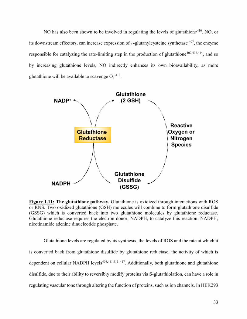
33
NO has also been shown to be involved in regulating the levels of glutathione410. NO, or
its downstream effectors, can increase expression of ϒ-glutanylcysteine synthetase 407, the enzyme
responsible for catalyzing the rate-limiting step in the production of glutathione407,408,414, and so
by increasing glutathione levels, NO indirectly enhances its own bioavailability, as more
glutathione will be available to scavenge O2-410.
Figure 1.11: The glutathione pathway. Glutathione is oxidized through interactions with ROS
or RNS. Two oxidized glutathione (GSH) molecules will combine to form glutathione disulfide
(GSSG) which is converted back into two glutathione molecules by glutathione reductase.
Glutathione reductase requires the electron donor, NADPH, to catalyze this reaction. NADPH,
nicotinamide adenine dinucleotide phosphate.
Glutathione levels are regulated by its synthesis, the levels of ROS and the rate at which it
is converted back from glutathione disulfide by glutathione reductase, the activity of which is
dependent on cellular NADPH levels408,411,415–417. Additionally, both glutathione and glutathione
disulfide, due to their ability to reversibly modify proteins via S-glutathiolation, can have a role in
regulating vascular tone through altering the function of proteins, such as ion channels. In HEK293
NADP+
NADPH
Glutathione (2 GSH)
GlutathioneDisulfide
(GSSG)
Reactive Oxygen or
Nitrogen Species
GlutathioneReductase

34
cells, glutathione disulfide can inhibit KATP channels400 and in guinea-pig smooth muscle cells,
glutathione increases the open probability of BKCa channels by shifting their voltage sensitivity to
more hyperpolarized membrane potentials79.
To conclude, the vascular endothelium plays a crucial role in regulating resistance artery
diameter and thus, blood flow and pressure. This is accomplished through release of NO and
endothelial KCa channel-mediated hyperpolarization which spreads to smooth muscle cells via
MEGJs. Although long thought of as distinct mechanisms for vasodilation, recent evidence
suggests that there may be a link between these two pathways; for example, IKCa channel-mediated
myoendothelial feedback leads to release of NO197, and block of KCa channels can inhibit NO-
mediated vasorelaxation203,418.
Endothelial dysfunction is associated with increased cardiovascular risk328,329 and
characterized by an increase in O2- production and a decrease in NO bioavailability268,328–336.
Therefore, the development of new drugs which possess indirect antioxidant properties mediated
by the stimulation of NO production and simultaneous inhibition of O2- production is an attractive
proposition for cardiovascular disease prevention and therapy. I propose that drugs which activate
SKCa and IKCa channels may fall into this category.
These channels mediate vasodilation through spread of membrane hyperpolarization from
endothelial to smooth muscle cells80,84,85,100,116–131,148–151,197,202, and have recently been suggested
to also be involved with enhancing NO bioavailability116,126,128,131,203–205. Furthermore, it has been
shown that NADPH oxidase enzymatic activity in the vasculature is regulated by membrane
potential; with depolarization stimulating NADPH oxidase production of O2- and
hyperpolarization inhibiting NADPH oxidase production of O2- 126,367,386–388. Therefore, increasing

35
the opening of endothelial KCa channels to elicit hyperpolarization could lead to vasodilation,
decreased O2- production and increased NO bioavailability.
1.5: Hypothesis and aims
My over-arching goal is to further explore the relationship between endothelial KCa
channels and NO in regulating resistance artery diameter by testing three hypotheses:
1. Activation of SKCa channels can enhance NO-mediated inhibition of sympathetic
vasoconstriction evoked by increases in shear stress.
2. IKCa channel-mediated myoendothelial feedback plays a role in NO-dependent modulation
of sympathetic vasoconstriction.
3. Pharmacological activators of endothelial KCa channels can reduce vascular O2-
production and enhance NO-mediated modulation of vasoconstriction
To test these hypotheses, I have addressed two major aims:
1. To investigate the role of endothelial KCa channels in NO-mediated modulation of
nerve-evoked vasoconstriction in the perfused mesenteric bed.
2. To investigate whether pharmacological activators of endothelial KCa channels can
modulate vascular O2- production and vasoconstriction stimulated by the α1-
adrenoceptor agonist phenylephrine.
36
Chapter 2: Activation of SKCa channels enhances shear stress-mediated
inhibition of sympathetic vasoconstriction in the perfused mesenteric bed
2.1: Introduction
As described in Chapter 1, endothelial and vascular smooth muscle cells work together to
regulate resistance artery diameter through a variety of mechanisms, such as release of NO,
cyclooxygenase-derived mediators and changes in membrane potential. In vivo, endothelial
sensing of increases in shear stress, the frictional force exerted by flow of blood across the cell
surface, plays an important role in regulating tissue perfusion by limiting vasoconstriction198–201.
Measurement of acute responses to increases in shear stress is the most widely used method to test
endothelial function in clinical studies419 and attenuation of shear stress-induced dilation is
associated with the early stages of cardiovascular diseases419,420. However, despite its obvious
physiological importance, the mechanisms underlying shear stress-induced increases in arterial
diameter and thus, blood flow are still a topic of debate.
Studies of cultured endothelial cells and isolated arteries have shown that acute increases
in shear stress cause a rise in the intracellular Ca2+ concentration in endothelial cells leading to
both release of NO and membrane hyperpolarization mediated by opening of SKCa
channels206,318,421–424. Data from in vivo and clinical studies have demonstrated an important role
for NO in acute responses to increases in shear stress425,426 and also support the contribution of
endothelial hyperpolarization427–431. However, whether release of NO and activation of SKCa
channels are distinct pathways for shear stress-induced modulation of arterial diameter, or two
facets of the same mechanism is still unclear.
As described in Chapter 1, SKCa channels are located on the luminal membrane of
endothelial cells in rat mesenteric arteries118,122,125,130 , porcine coronary arteries122, mouse and
bovine coronary endothelial cells132,133 and human microvascular endothelial cells132,133, an ideal
37
location for activation by localized increases in Ca2+ elicited by enhanced shear stress118,122,130.
The subsequent hyperpolarization of the endothelial membrane potential then spreads to the
surrounding smooth muscle cells via MEGJs to limit vasoconstriction80,84,85,100,116–131,148–151,197,202.
The identity of the endothelial mechanosensor activated by increases in shear stress has yet to be
defined but recent evidence supports a role for the mechanosensitive TRPV4 channels in mediating
shear stress-induced Ca2+ entry to endothelial cells in isolated arteries132,133,220,241–248,432,433. TRPV4
channels co-localize with both SKCa channels and endothelial NOS to caveolae on the luminal
surface of human microvascular and bovine coronary endothelial cells132,133. Additionally, acute
exposure of bovine coronary endothelial cells to shear stress resulted in the activation of both SKCa
and TRPV4 channels and increased NO production supporting a potential link between SKCa
channel activity and NO133. In rat carotid arteries and gracilis muscle arterioles, shear stress-
mediated vasodilation was blocked by TRPV4 channel inhibition241. Thus, these findings support
a functional relationship between SKCa channels, TRPV4 channels and NO.
A link between endothelial KCa channel activity and NO production is also supported by
previous work from our lab and others. In cultured endothelial cells, NO production is regulated
by KCa channel-mediated changes in membrane potential131 and in rat basilar arteries and cultured
endothelial cells, agonist-evoked endothelium-dependent relaxations mediated by NO are
inhibited by KCa channel blockers203,418. However, the possibility of a link between NO and SKCa
channel activity in endothelial responses to shear stress has not been investigated.
In vivo, sympathetic nerve activity is the primary regulator of resistance artery diameter,
and therefore, peripheral vascular resistance434,435, with the endothelium playing a key role in
limiting the vasoconstriction caused by neurotransmitters released from perivascular sympathetic
nerves. However, the majority of studies examining the contribution of NO and SKCa channels to

38
endothelium-dependent modulation of arterial diameter have focused on their role in
vasorelaxation, i.e. the ability of endothelial stimuli to reverse agonist-induced tone rather than
modulation of vasoconstriction. Thus, given the importance of both the endothelial response to
shear stress and sympathetic nerve activity in controlling arterial diameter, blood flow and blood
pressure, the goal of the experiments described in this chapter was to explore the functional link
between NO and SKCa channel activity in shear stress-induced modulation of sympathetic
vasoconstriction and to test the hypothesis that activation of SKCa channels will enhance NO-
mediated inhibition of sympathetic vasoconstriction evoked by increases in shear stress.
To test this hypothesis I have used the rat mesenteric bed perfused at a constant luminal
flow so vasoconstriction leads to increases in shear stress; decreases in arterial diameter augment
shear stress because of its inverse relationship to the third power of the internal vessel diameter436.
Under these experimental conditions, stimulation of sympathetic perivascular nerves leads to
vasoconstriction (recorded as increases in perfusion pressure) that is limited by stimulation of the
endothelium by acute increases in shear stress. Pharmacological tools were applied to investigate
the contribution of NO and endothelial KCa channels to shear stress-induced modulation of
sympathetic vasoconstriction and CyPPA, a small molecule activator of SKCa channels that
enhances the channels sensitivity to Ca2+ 313,437, was used to examine if increased activation of
SKCa channels could enhance the NO-mediated component of the response to shear stress.
2.2: Methods and materials
See Appendix: Drugs and chemicals for a list of the drugs and chemicals used.
2.2.1: Perfused mesenteric vascular bed
The mesenteric bed was perfused via the superior mesenteric artery as previously
described438. Briefly, the mesenteric vascular bed was separated from the intestine and the superior

39
mesenteric artery cleaned of connective tissue, cannulated with a blunted hypodermic needle (20
G), secured with 5-0 surgical silk (Ethicon) and flushed with Krebs buffer to remove blood. In
some experiments the endothelium was removed by flushing the bed with 0.5% Triton X-100 in
water for 30 seconds followed by rapid washout with Krebs. The vascular bed was placed on a
wire mesh in a warm chamber and perfused with oxygenated Krebs buffer at a constant flow rate
of 5 mlmin-1 (37°C, bubbled with 95% O2/5% CO2). Changes in perfusion pressure were monitored
via an in-line pressure transducer (AD instruments, Colorado) and recorded via a PowerLab data
acquisition system using Chart 5.0 software (AD Instruments, Colorado). In experiments
conducted with endothelium-denuded preparations, endothelial function was assessed as the
response to acetylcholine (1 µM) following vasoconstriction with methoxamine (1 µM); tissues in
which acetylcholine failed to evoke a response were deemed to be endothelium-denuded.
2.2.1.1: Responses to stimulation of perivascular nerves. Electrodes were attached to the
cannulating needle and to the wire mesh to allow electrical field stimulation using a Grass SD9
stimulator (Grass Technologies, USA). Following an equilibration period of 30 minutes, a single
stimulation (30 Hz, 90 V, pulse width 1 millisecond, 30 seconds) was applied to assess the viability
of the preparation. After a further 10 minutes, a frequency-response curve was constructed by
stimulating the preparation at 1-40 Hz (90 V, pulse width 1 millisecond, 30 seconds) at 10 minute
intervals188. The effects of agents on nerve-evoked vasoconstriction were assessed by perfusing
the drugs through the lumen of the preparation for 20 minutes prior to constructing a second
frequency-response curve. In some experiments a third frequency-response curve was constructed
following washout.
Nerve-evoked responses recorded in the perfused mesenteric vascular bed are shown as
normalized values. Changes in perfusion pressure were normalized to the maximum control
40
response (%) as is convention in these types of experiments. For all frequency response curves,
electrical stimulation caused frequency-dependent increases in perfusion pressure (p<0.05).
2.2.2: Wire myography
Third order mesenteric arteries were cleaned of adhering tissue and cut into segments (~2
mm in length). Arterial segments were mounted between two gold-plated tungsten wires (20 µm
diameter) in a Mulvany-Halpern myograph (model 400A, J.P. Trading, Denmark) as previously
described438. Changes in isometric tension were recorded via a PowerLab using Chart 5.0 or 8.0
software (AD Instruments, Colorado, USA). Tissues were maintained in Krebs’ buffer gassed with
95% O2/5% CO2 at 37°C (pH 7.4) and set to a pre-determined optimal resting tension of 5 mN for
mesenteric arteries (this was previously determined from active length-tension curves). In some
experiments, the endothelium was removed by flushing of the mesenteric bed with 0.5% Triton X-
100 (20 μl of Triton X-100 in 40 ml water) for 30 seconds followed by rapid washout with Krebs.
After an equilibration period of 30 minutes, endothelial function was assessed as % relaxation to
acetylcholine (3 μM) following pre-stimulation with phenylephrine (3 μM; 75% of maximal tone).
Arteries in which acetylcholine induced >90% reversal of agonist-induced tone were designated
as endothelium-intact and tissues in which the response to acetylcholine was <10% were deemed
to be endothelium-denuded. Arteries in which the % reversal of agonist-induced tone elicited by
acetylcholine fell between these values were discarded.
2.2.2.1: Concentration-response curves. Cumulative concentration-response curves to CyPPA
(0.001-30 μM) were constructed in arteries in which tone was raised with phenylephrine (3 μM).
For all CyPPA concentration-response curves, CyPPA caused relaxation in a concentration-
dependent manner (p<0.05). In endothelium-intact arterial segments, concentration-response
curves to CyPPA were constructed in the absence and presence of apamin (50 nM) or L-NAME

41
(100 μM). Cumulative concentration-response curves to CyPPA (0.001-30 μM) were also
constructed in endothelium-denuded mesenteric arteries. In all experiments, the level of
phenylephrine -induced tone was matched in the absence and presence of inhibitors and relaxations
to CyPPA were expressed as % relaxation as is the convention in this type of experiment.
Cumulative concentration-response curves to phenylephrine (0.001-100 μM) were
constructed in the absence and presence of CyPPA (5 μM) without and with apamin (50 nM) or
L-NAME (100 μM) in endothelium -intact and -denuded isolated mesenteric arteries. Results were
expressed as % maximal response as is convention for this type of experiment. Phenylephrine
increased tone in a concentration-dependent manner (p<0.05).
2.2.3: Analysis of noradrenaline levels in perfusate from the mesenteric vascular bed
The mesenteric vascular bed was placed on a wire mesh and placed in a plastic dish on a
hot plate (Model HP-A1915B-13, Thermolyne) and maintained at 37°C. The flow rate was 2
mlmin-1 and the mesenteric bed was stimulated at 30 Hz for 60 seconds. Perfusate was collected
for 60 seconds prior to the stimulation and during the stimulation. Samples were immediately
frozen in liquid nitrogen and stored at -80oC prior to analysis by Ultra-performance liquid
chromatography (UPLC).
2.2.3.1: Measurement of noradrenaline outflow from the perfused mesenteric bed by UPLC.
Noradrenaline levels in perfusate samples were analyzed using a Waters Acquity UPLC System
(H Class) consisting of a binary solvent manager, sample manager, column manager and
fluorescence detector. Pre-column derivatization of samples with benzylamine and 1,2-
diphenylethyleendiamine was conducted439. Separation of noradrenaline was achieved by gradient
elution using a mixture of acetonitrile and 15 mM acetate buffer (pH 4.5) containing 1 mM
octanesulfonic acid (sodium salt) on a Waters Acquity UPLC BEH Shield reversed phase column
42
(C18, 2.1 mm ID 100 mm, 1.7 m). The column temperature was 60oC, flow rate 0.7 ml/min and
the run time was 8 minutes. Excitation and emission wavelength were set at 345 and 480 nm,
respectively. All data was acquired and analyzed by means of Waters Empower 3 software.
Noradrenaline and acetonitrile were purchased from Sigma-Aldrich. All chemicals and solvents
were of analytical grade. All solutions were prepared in ultrapure milliQ water (Millipore MilliQm
Germany) and filtered over a 0.22 m filter (Millipore, Bedford, USA). A standard curve for
noradrenaline was obtained each day prior to collection and injection of samples. Analysis was
done with the operator blinded to sample identity. The lowest detectable level of noradrenaline
was 4 fmol/50 µl sample. The concentration of noradrenaline in the perfusate samples are shown
as normalized values due to variation in control values for noradrenaline overflow between
different preparations.
2.2.4: Statistics
All data are expressed as mean ± SEM, n rats used. For repeated measures, two-way
ANOVA followed by either a Tukey’s multiple comparison post-hoc test (used when there were
more than two experimental groups) or Šídák method post-hoc test (used when there was two
experimental groups) was performed. A paired t-test was used in Figure 2.10b. p<0.05 was
considered statistically significant in all cases.
2.3: Results
2.3.1: Characterization of nerve-evoked vasoconstriction in the rat perfused mesenteric
vascular bed
As shown in the representative trace in Figure 2.1a, stimulation of perivascular nerves
evoked frequency-dependent increases in perfusion pressure in the endothelium-intact perfused
mesenteric vascular bed. The mean maximal increase in perfusion pressure at a frequency of 30
Hz in endothelium-intact mesenteric beds was 97.8 ± 16.2 mmHg (n=4). Three repeated

43
frequency-response curves could be constructed at 30 minute intervals without a significant change
in amplitude of responses and sensitivity (p>0.05; Figure 2.1b).
Tetrodotoxin (0.5 μM), a voltage-gated sodium channel inhibitor, abolished responses to
electrical stimulation, confirming that the frequency-dependent changes in perfusion pressure are
due to the release of neurotransmitters from nerves and not caused by direct electrical stimulation
of muscle cells (n=4). In the presence of prazosin (0.1 μM), an α1-adrenoreceptor antagonist,
nerve-evoked vasoconstriction was significantly inhibited (p<0.05, Figure 2.1c). Thus, under my
experimental conditions, nerve-evoked responses in the mesenteric bed can be largely accounted
for by noradrenaline acting on α1-adrenoceptors.
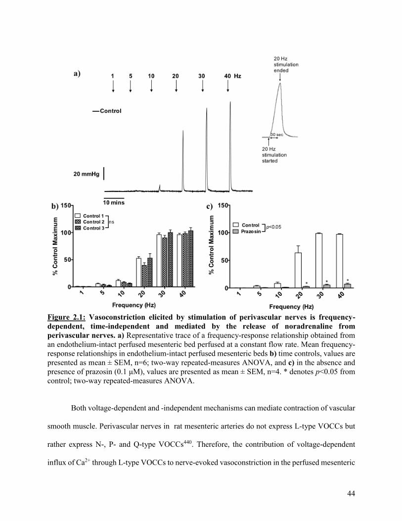
44
Figure 2.1: Vasoconstriction elicited by stimulation of perivascular nerves is frequency-
dependent, time-independent and mediated by the release of noradrenaline from
perivascular nerves. a) Representative trace of a frequency-response relationship obtained from
an endothelium-intact perfused mesenteric bed perfused at a constant flow rate. Mean frequency-
response relationships in endothelium-intact perfused mesenteric beds b) time controls, values are
presented as mean ± SEM, n=6; two-way repeated-measures ANOVA, and c) in the absence and
presence of prazosin (0.1 μM), values are presented as mean ± SEM, n=4. * denotes p<0.05 from
control; two-way repeated-measures ANOVA.
Both voltage-dependent and -independent mechanisms can mediate contraction of vascular
smooth muscle. Perivascular nerves in rat mesenteric arteries do not express L-type VOCCs but
rather express N-, P- and Q-type VOCCs440. Therefore, the contribution of voltage-dependent
influx of Ca2+ through L-type VOCCs to nerve-evoked vasoconstriction in the perfused mesenteric
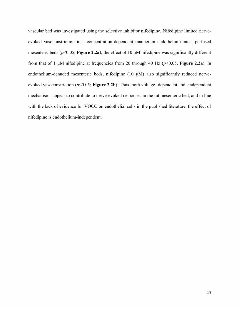
45
vascular bed was investigated using the selective inhibitor nifedipine. Nifedipine limited nerve-
evoked vasoconstriction in a concentration-dependent manner in endothelium-intact perfused
mesenteric beds (p<0.05, Figure 2.2a); the effect of 10 μM nifedipine was significantly different
from that of 1 μM nifedipine at frequencies from 20 through 40 Hz (p<0.05, Figure 2.2a). In
endothelium-denuded mesenteric beds, nifedipine (10 μM) also significantly reduced nerve-
evoked vasoconstriction (p<0.05; Figure 2.2b). Thus, both voltage -dependent and -independent
mechanisms appear to contribute to nerve-evoked responses in the rat mesenteric bed, and in line
with the lack of evidence for VOCC on endothelial cells in the published literature, the effect of
nifedipine is endothelium-independent.

46
Figure 2.2: Nerve-evoked vasoconstriction is partially dependent on L-type VOCCs. a) Mean
frequency-response relationships obtained from endothelium-intact perfused mesenteric beds in
the absence and presence of nifedipine (1 and 10 μM). Values are presented as mean ± SEM, n=4-
10. * denotes p<0.05 from control and # denotes p<0.05 from nifedipine (1 μM); two-way
repeated-measures ANOVA. b) Mean frequency-response relationships obtained from
endothelium-denuded perfused mesenteric beds in the absence and presence of nifedipine (10 μM).
Values are presented as mean ± SEM, n=4. * denotes p<0.05 from control; two-way repeated-
measures ANOVA.
47
2.3.2: Modulation of nerve-evoked vasoconstriction by increases in shear stress in the rat
perfused mesenteric bed
Both NO and endothelial SKCa channels have been suggested to mediate the effects of
shear stress on arterial diameter. Thus, I investigated the contribution of these effectors to shear-
stress-induced, endothelium-dependent modulation of sympathetic vasoconstriction in the
perfused rat mesenteric bed.
The role of NO in modulating nerve-evoked vasoconstriction was investigated by perfusing
preparations with L-NAME, a selective inhibitor of NOS. L-NAME (100 μM) significantly
enhanced nerve-evoked vasoconstriction at stimulation frequencies from 20 to 40 Hz (p<0.05,
Figure 2.3a). Additionally, 1H-[1,2,4]oxadiazolo[4,3-a]quinoxalin-1-one (ODQ; 10 μM)), an
inhibitor of guanylyl cyclase, the downstream target of NO, significantly enhanced nerve-evoked
vasoconstriction (p<0.05) at stimulation frequencies from 15 to 40 Hz (p<0.05, Figure 2.3c).
Removal of the endothelium did not alter the frequency-response relationship (Figure
2.3d) or the magnitude of responses evoked by stimulation of perivascular nerves; the mean
response ± SEM to a stimulation of 30 Hz in endothelium-intact and denuded arteries was 97.8 ±
16.2 and 99.6 ± 8.7 mmHg (n=4; p>0.05), respectively. L-NAME was without effect in
endothelium-denuded mesenteric beds (p>0.05, Figure 2.3d), indicating its actions in potentiating
nerve-evoked vasoconstriction were most likely due to inhibition of endothelial NOS rather than
neuronal NOS.
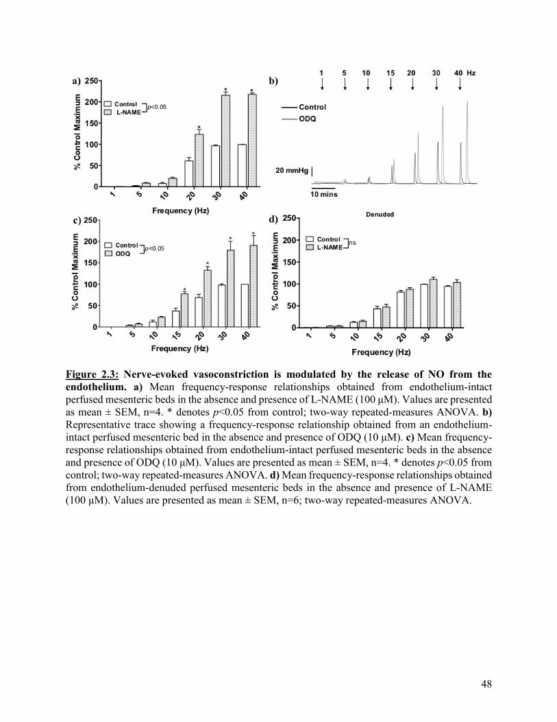
48
Figure 2.3: Nerve-evoked vasoconstriction is modulated by the release of NO from the
endothelium. a) Mean frequency-response relationships obtained from endothelium-intact
perfused mesenteric beds in the absence and presence of L-NAME (100 μM). Values are presented
as mean ± SEM, n=4. * denotes p<0.05 from control; two-way repeated-measures ANOVA. b)
Representative trace showing a frequency-response relationship obtained from an endothelium-
intact perfused mesenteric bed in the absence and presence of ODQ (10 μM). c) Mean frequency-
response relationships obtained from endothelium-intact perfused mesenteric beds in the absence
and presence of ODQ (10 μM). Values are presented as mean ± SEM, n=4. * denotes p<0.05 from
control; two-way repeated-measures ANOVA. d) Mean frequency-response relationships obtained
from endothelium-denuded perfused mesenteric beds in the absence and presence of L-NAME
(100 μM). Values are presented as mean ± SEM, n=6; two-way repeated-measures ANOVA.
49
The functional role of SKCa channels in endothelium-dependent modulation of nerve-
evoked vasoconstriction in the perfused mesenteric vascular bed was investigated using apamin, a
selective inhibitor of SKCa channels. In endothelium-intact mesenteric beds, apamin (50 nM)
significantly enhanced nerve-evoked vasoconstriction (p<0.05) at frequencies from 10 to 40 Hz
(p<0.05, Figure 2.4b), but was without effect in endothelium-denuded tissues (p>0.05, Figure
2.4c). In contrast, inhibition of endothelial IKCa channels with NS 6180 (1 μM) was without effect
on nerve-evoked responses (p>0.05; Figure 2.4d). Furthermore, the effect of apamin was not
additive with that of L-NAME (100 μM), suggesting a link between SKCa channels and
endothelium-derived NO (Figure 2.5). Thus, while SKCa channels play a significant role in
limiting nerve-evoked vasoconstriction in the mesenteric bed, IKCa channels do not. This
difference in their functional importance is likely due to their differing locations within the
endothelium, as SKCa channels are located on the endothelial luminal membrane118,122,125,130,132,133
while IKCa channels are located within the MEGJs118,125,130,151,197.
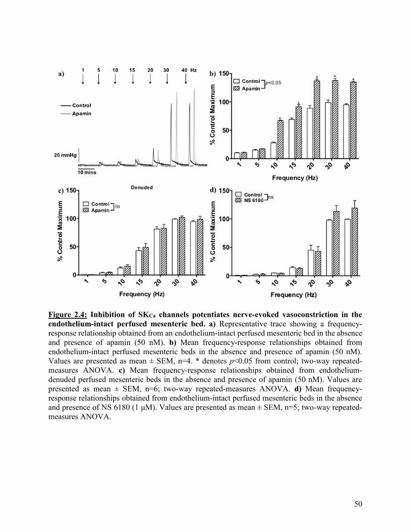
50
Figure 2.4: Inhibition of SKCa channels potentiates nerve-evoked vasoconstriction in the
endothelium-intact perfused mesenteric bed. a) Representative trace showing a frequency-
response relationship obtained from an endothelium-intact perfused mesenteric bed in the absence
and presence of apamin (50 nM). b) Mean frequency-response relationships obtained from
endothelium-intact perfused mesenteric beds in the absence and presence of apamin (50 nM).
Values are presented as mean ± SEM, n=4. * denotes p<0.05 from control; two-way repeated-
measures ANOVA. c) Mean frequency-response relationships obtained from endothelium-
denuded perfused mesenteric beds in the absence and presence of apamin (50 nM). Values are
presented as mean ± SEM, n=6; two-way repeated-measures ANOVA. d) Mean frequency-
response relationships obtained from endothelium-intact perfused mesenteric beds in the absence
and presence of NS 6180 (1 μM). Values are presented as mean ± SEM, n=5; two-way repeated-
measures ANOVA.

51
Figure 2.5: Inhibition of SKCa channels and NOS potentiates nerve-evoked vasoconstriction
in a non-additive manner in the endothelium-intact perfused mesenteric bed. Mean
frequency-response relationships obtained from endothelium-intact perfused mesenteric beds in
the absence and presence of apamin (50 nM), L-NAME (100 μM) and apamin (50 nM) with L-
NAME (100 μM). Values are presented as mean ± SEM, n=5. * denotes p<0.05 from control and
# denotes p<0.05 from apamin (50 nM) and ^ denotes p<0.05 from L-NAME (100 μM); two-way
repeated-measures ANOVA.
52
As shown above (Figure 2.2), both voltage -dependent and -independent mechanisms
underlie nerve-evoked vasoconstriction in the perfused mesenteric bed. Opening of endothelial
KCa channels leads to hyperpolarization of the membrane potential which spreads to surrounding
smooth muscle cells to reduce influx of Ca2+ through VOCCs and so is more effective in reversing
depolarization-mediated smooth muscle contraction than contraction elicited via voltage-
independent mechanisms (i.e. release of Ca2+ from intracellular stores and Ca2+ sensitization). In
contrast, NO can inhibit smooth muscle contractility through both voltage -dependent (e.g.
activation of BKCa channels to hyperpolarize the membrane potential to reduce Ca2+ influx though
VOCCs55–59,79,290,291,164–168) and -independent signaling pathways (e.g. phospholamban
phosphorylation and phospholipase C166,251,289,441,442). Thus, in the next set of experiments L-
NAME was applied in combination with nifedipine. The rationale for these experiments was that
in the presence of nifedipine, the remaining nerve-evoked vasoconstriction occurs independently
of L-type VOCC activity thus, hyperpolarization would not be expected to be an effective
inhibitory mechanism. As shown above, L-NAME (100 M) significantly potentiated nerve-
evoked vasoconstriction in endothelium-intact mesenteric beds (p<0.05, Figure 2.3a) and was also
able to do so in the presence of nifedipine (10 μM; p<0.05; Figure 2.6). However, the size of
responses in the presence of L-NAME and nifedipine was significantly less than with L-NAME
alone (p<0.05) indicating that endothelium-derived NO limit both the voltage -dependent and -
independent components of nerve-evoked vasoconstriction.

53
Figure 2.6: Block of NO signaling is able to enhance the nifedipine-insensitive component of
nerve-evoked vasoconstriction. Mean frequency-response relationships obtained from
endothelium-intact perfused mesenteric beds in the absence and presence of nifedipine (10 μM)
without and with L-NAME (100 μM). Values are presented as mean ± SEM, n=4. * denotes p<0.05
from control and # denotes p<0.05 from nifedipine (10 μM); two-way repeated-measures
ANOVA.

54
2.3.3: Effect of CyPPA on phenylephrine-induced tone in isolated mesenteric arteries
mounted in a wire myograph
The effects of the SKCa channel activator CyPPA on vascular tone in rat mesenteric arteries
have not previously been reported. Therefore, before examining its ability to enhance shear stress-
mediated inhibition of nerve-evoked vasoconstriction in the perfused bed, I characterized the
effects of CyPPA on agonist-evoked increases in tone in arteries mounted under isometric
conditions in a wire myograph. In these experiments, phenylephrine was used to induce tone as it
is a selective α1-adrenoceptor agonist whereas noradrenaline can act at multiple adrenoceptors.
Concentration-relaxation curves to CyPPA (0.001-30 μM) were constructed in
endothelium -intact and -denuded arterial segments in which tone was raised with phenylephrine
(3 μM). In endothelium-intact arterial segments, CyPPA-evoked relaxations were significantly
reduced by the presence of apamin (50 nM) or L-NAME (100 μM; p<0.05, Figure 2.7). In
endothelium-denuded arteries, responses to CyPPA were significantly reduced compared to
endothelium-intact tissues such that relaxation was only observed at the highest concentration of
30 M (p<0.05, Figure 2.7). Thus, CyPPA-evoked relaxation of phenylephrine-induced tone is
endothelium-dependent and reliant on both SKCa channel activation and NO.
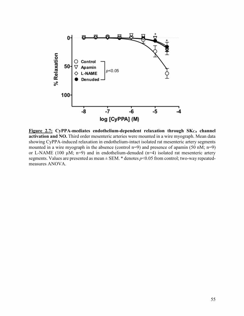
55
Figure 2.7: CyPPA-mediates endothelium-dependent relaxation through SKCa channel
activation and NO. Third order mesenteric arteries were mounted in a wire myograph. Mean data
showing CyPPA-induced relaxation in endothelium-intact isolated rat mesenteric artery segments
mounted in a wire myograph in the absence (control n=9) and presence of apamin (50 nM; n=9)
or L-NAME (100 μM; n=9) and in endothelium-denuded (n=4) isolated rat mesenteric artery
segments. Values are presented as mean ± SEM. * denotes p<0.05 from control; two-way repeated-
measures ANOVA.
56
The effect of CyPPA (5 μM) on phenylephrine-evoked increases in tone was then examined
in arteries mounted in a wire myograph. This concentration of CyPPA was selected as it evoked
relaxation which was endothelium-dependent and sensitive to apamin, the inhibitor of SKCa
channels. Concentration-response curves to phenylephrine (phenylephrine denoted as PE in
Figure 2.8a and b; 0.001- 100 μM) were constructed in the absence and presence of CyPPA. In
endothelium-intact arteries, CyPPA did not affect resting tone but significantly reduced increases
in tone elicited by phenylephrine at concentration of 1-10 μM (p<0.05; Figure 2.8a) causing a
rightward shift in the concentration-response curve. This effect was blocked by apamin (50 nM)
or L-NAME (100 μM) (Figure 2.8a). In contrast, in endothelium-denuded arteries, CyPPA did
not significantly affect phenylephrine-induced increases in tone (p>0.05; Figure 2.8b).
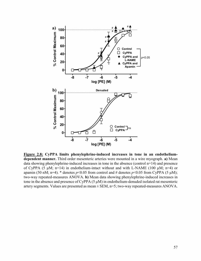
57
Figure 2.8: CyPPA limits phenylephrine-induced increases in tone in an endothelium-
dependent manner. Third order mesenteric arteries were mounted in a wire myograph. a) Mean
data showing phenylephrine-induced increases in tone in the absence (control n=14) and presence
of CyPPA (5 μM; n=14) in endothelium-intact without and with L-NAME (100 μM; n=4) or
apamin (50 nM; n=4). * denotes p<0.05 from control and # denotes p<0.05 from CyPPA (5 μM);
two-way repeated-measures ANOVA. b) Mean data showing phenylephrine-induced increases in
tone in the absence and presence of CyPPA (5 μM) in endothelium-denuded isolated rat mesenteric
artery segments. Values are presented as mean ± SEM, n=5; two-way repeated-measures ANOVA.

58
2.3.4: CyPPA enhances shear stress-induced modulation of nerve-evoked vasoconstriction in
the rat perfused mesenteric bed
Having demonstrated that SKCa channels play a key role in shear stress-induced modulation
of sympathetic vasoconstriction in the perfused mesenteric bed (Figure 2.4), I then examined
whether CyPPA can enhance the effects of shear stress on sympathetic vasoconstriction in the
perfused mesenteric bed. CyPPA (5 and 10 μM) had no effect on basal perfusion pressure but
significantly limited nerve-evoked vasoconstriction in endothelium-intact mesenteric beds in a
concentration-dependent manner (p<0.05, Figure 2.9). For the remaining experiments I used
CyPPA at a concentration of 5 M as at this concentration, CyPPA had little direct relaxant effect
(Figure 2.7).
In contrast, in endothelium-denuded mesenteric beds, CyPPA (5 μM) had no significant
effect on nerve-evoked vasoconstriction (p>0.05, Figure 2.10a). This is in line with functional and
histochemical studies demonstrating that SKCa channels are expressed on endothelial but not
smooth muscle cells in mesenteric arteries125,197. However, whether SKCa channels are located on
nerves has not been investigated. Rat mesenteric arteries are densely innervated176,185, with
sufficient amounts of noradrenaline released as can be measured as overflow in the perfusate438
and so the ability of CyPPA to inhibit the release of noradrenaline from perivascular nerves was
investigated. Perfusate was collected before and during a 30 Hz stimulation in the absence and
presence of CyPPA. Noradrenaline overflow was undetectable in the 60 seconds prior to the 30
Hz stimulation but increased significantly during nerve stimulation, a response which was not
altered by CyPPA (p>0.05, Figure 2.10b), indicating that reductions in nerve-evoked increases in
perfusion pressure caused by this agent are not due to an action on perivascular nerves.

59
Figure 2.9: CyPPA enhances shear stress-mediated inhibition of nerve-evoked
vasoconstriction in the endothelium-intact perfused mesenteric bed. Mean frequency-response
relationships obtained from endothelium-intact perfused mesenteric beds in the absence and
presence of CyPPA (5 and 10 μM). Values are presented as mean ± SEM, n=4. * denotes p<0.05
from control, # denotes p<0.05 from 5 μM CyPPA; two-way repeated-measures ANOVA.
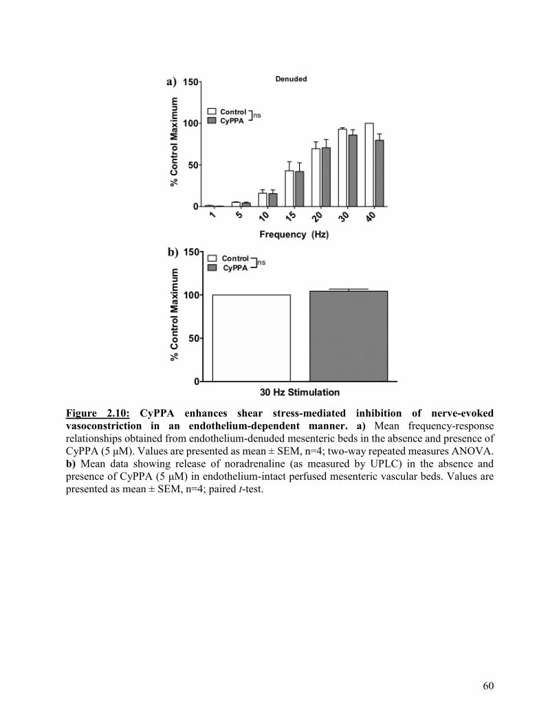
60
Figure 2.10: CyPPA enhances shear stress-mediated inhibition of nerve-evoked
vasoconstriction in an endothelium-dependent manner. a) Mean frequency-response
relationships obtained from endothelium-denuded mesenteric beds in the absence and presence of
CyPPA (5 μM). Values are presented as mean ± SEM, n=4; two-way repeated measures ANOVA.
b) Mean data showing release of noradrenaline (as measured by UPLC) in the absence and
presence of CyPPA (5 μM) in endothelium-intact perfused mesenteric vascular beds. Values are
presented as mean ± SEM, n=4; paired t-test.

61
To confirm the actions of CyPPA were indeed due to activation of SKCa channels, I used
apamin, the selective SKCa channel inhibitor. Apamin (50 nM) significantly enhanced nerve-
evoked vasoconstriction when applied alone (as shown in Figure 2.4) and blocked the effect of
CyPPA (p<0.05, Figure 2.11); responses in the presence of apamin alone in comparison to apamin
with CyPPA were not significantly different (p>0.05, Figure 2.11). Therefore, the actions of
CyPPA can be attributed to activation of endothelial SKCa channels.
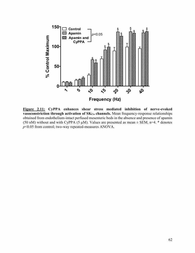
62
Figure 2.11: CyPPA enhances shear stress mediated inhibition of nerve-evoked
vasoconstriction through activation of SKCa channels. Mean frequency-response relationships
obtained from endothelium-intact perfused mesenteric beds in the absence and presence of apamin
(50 nM) without and with CyPPA (5 μM). Values are presented as mean ± SEM, n=4. * denotes
p<0.05 from control; two-way repeated-measures ANOVA.
63
The contribution of NO to the actions of CyPPA was investigated using the NOS inhibitor,
L-NAME, and the soluble guanylyl cyclase inhibitor, ODQ. L-NAME (100 μM) significantly
enhanced nerve-evoked vasoconstriction when applied alone (as shown above Figure 2.3) and
inhibited the effects of CyPPA (p<0.05, Figure 2.12a); responses in the presence of L-NAME
alone in comparison to L-NAME plus CyPPA were not significantly different (p>0.05, Figure
2.12a). Similarly, ODQ (10 μM) significantly enhanced nerve-evoked vasoconstrictions at
frequencies from 15 to 40 Hz (p<0.05, Figure 2.12b) and inhibited the effects of CyPPA;
responses in the presence of ODQ alone in comparison to ODQ plus CyPPA were not significantly
different (p>0.05, Figure 2.12b). Thus, it appears that the ability of CyPPA to inhibit nerve-evoked
vasoconstriction in the perfused mesenteric bed is due to activation of SKCa channels and is largely
dependent on NO.
As described above, smooth muscle hyperpolarization is more effective at reversing
increases in tone caused by depolarization, whereas NO is effective against voltage -dependent
and -independent smooth muscle contraction55–59,79,290,291,164–168,251,443. In my experiments the
actions of CyPPA are dependent on both SKCa channel activity and NO, thus the importance of
CyPPA-mediated smooth muscle hyperpolarization to inhibit nerve-evoked vasoconstriction was
investigated by conducting experiments in the presence of nifedipine. Nifedipine (10 μM) alone
significantly reduced nerve-evoked vasoconstriction (as shown above Figure 2.2) and in the
presence of nifedipine, CyPPA was able to significantly reduce the nifedipine-independent
vasoconstriction (p<0.05, Figure 2.13) indicating that it can inhibit voltage-independent smooth
muscle contraction, an unexpected finding if CyPPA were acting via hyperpolarization alone.
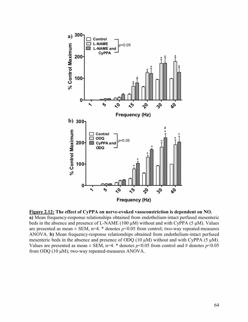
64
Figure 2.12: The effect of CyPPA on nerve-evoked vasoconstriction is dependent on NO.
a) Mean frequency-response relationships obtained from endothelium-intact perfused mesenteric
beds in the absence and presence of L-NAME (100 μM) without and with CyPPA (5 μM). Values
are presented as mean ± SEM, n=4. * denotes p<0.05 from control; two-way repeated-measures
ANOVA. b) Mean frequency-response relationships obtained from endothelium-intact perfused
mesenteric beds in the absence and presence of ODQ (10 μM) without and with CyPPA (5 μM).
Values are presented as mean ± SEM, n=4. * denotes p<0.05 from control and # denotes p<0.05
from ODQ (10 μM); two-way repeated-measures ANOVA.
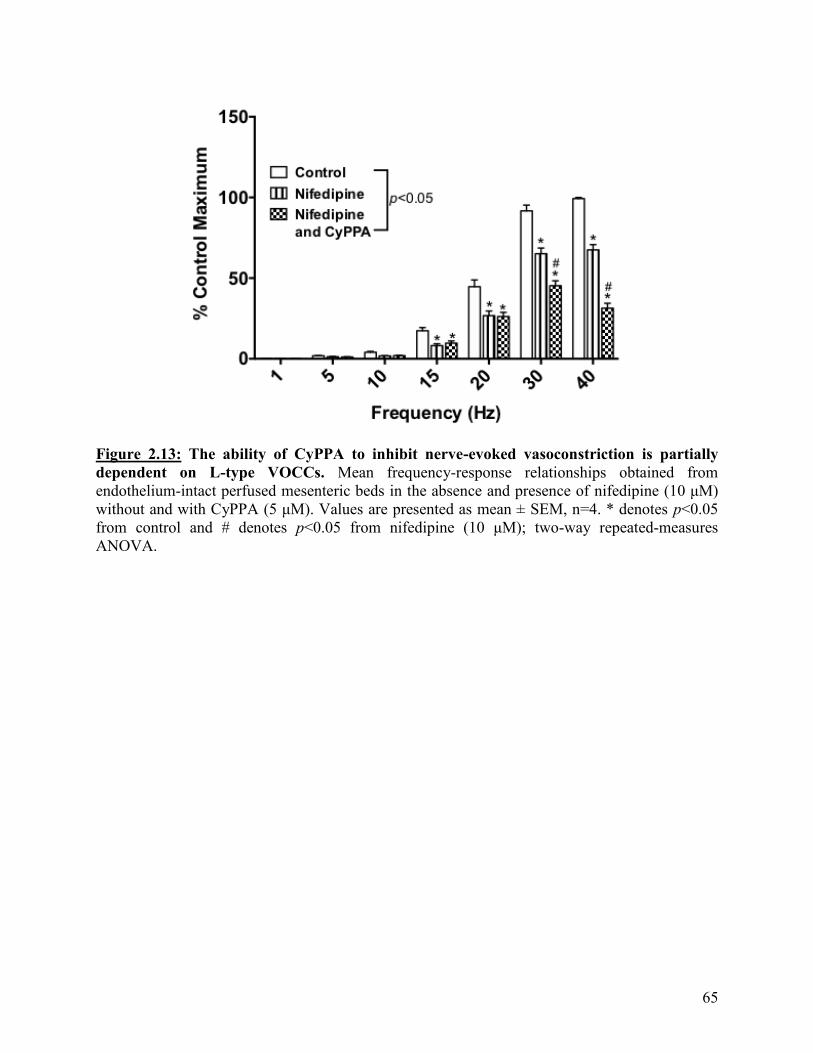
65
Figure 2.13: The ability of CyPPA to inhibit nerve-evoked vasoconstriction is partially
dependent on L-type VOCCs. Mean frequency-response relationships obtained from
endothelium-intact perfused mesenteric beds in the absence and presence of nifedipine (10 μM)
without and with CyPPA (5 μM). Values are presented as mean ± SEM, n=4. * denotes p<0.05
from control and # denotes p<0.05 from nifedipine (10 μM); two-way repeated-measures
ANOVA.
66
2.4: Discussion
Increases in shear stress stimulate an influx of Ca2+ into endothelial cells which leads to
the production of NO and hyperpolarization of the endothelial cell membrane potential via opening
of SKCa channels206,318,421–424. My data show that in the perfused mesenteric bed, shear stress-
induced inhibition of sympathetic vasoconstriction is mediated by both NO and SKCa channels.
Additionally, I found that CyPPA, a small molecule activator of SKCa channels that increases the
channels sensitivity to Ca2+ 313,437, can enhance the response to shear stress in this preparation. This
enhancement of shear stress-induced vasodilation by CyPPA occurred at a concentration that
caused minimal direct vasorelaxation and via a mechanism dependent on endothelium-derived
NO. These findings indicate that opening of SKCa channels in response to acute increases in shear
stress may enhance NO bioavailability and support the proposal that activators of these channels
may have therapeutic potential in enhancing shear stress-induced NO bioavailability in
cardiovascular disease states420.
In the experiments described in this chapter, the rat mesenteric bed was perfused at a
constant luminal flow and the perivascular nerves were stimulated, resulting in vasoconstriction
that was recorded as increases in perfusion pressure. Stimulation of the sympathetic nerves leads
to the release of noradrenaline and the co-transmitter ATP, which act on arterial α1-adrenoceptors
and P2X receptors, respectively176–179,181,184–188. In my experiments, nerve-evoked increases in
perfusion pressure were abolished by the α1-adrenoceptor antagonist prazosin indicating that these
responses are mediated by noradrenaline acting on 1-adrenoceptors with little contribution from
ATP. This is in line with previous studies in which the relative importance of ATP as a functional
sympathetic neurotransmitter in the rat and porcine mesenteric beds was revealed only when the
level of preexisting vascular tone or pressure was increased187,188. Furthermore, the removal of the
67
endothelium had no effect on nerve-evoked vasoconstriction, indicating that both vasoconstrictors
and vasodilators released from the endothelium are modulating these responses.
Activation of smooth muscle 1-adrenoceptors by noradrenaline results in phospholipase
C-mediated increases in diacylglycerol and IP3249,251–255 and membrane depolarization, which
enhances Ca2+-influx through L-type VOCCs21,51,444. The relative contribution of these voltage-
dependent (VOCC-mediated Ca2+ influx) and -independent (mediated by IP3 and diacylglycerol)
mechanisms to 1-adrenoceptor-mediated contraction has been shown to vary depending on
experimental conditions. In isolated hamster skeletal muscle arteries, bath application of the 1-
adrenoceptor agonist phenylephrine (0.1 M), evoked sustained constrictions largely dependent
on Ca2+ influx through L-type VOCCs, whereas focal application of a high dose (1 mM) of
phenylephrine in the same preparation generated a local constriction that was independent of
changes in smooth muscle membrane potential and entirely dependent on IP3445. Additionally, the
ability of phenylephrine to cause vasoconstriction in the hind limb vascular bed in vivo was
reduced by 50% in mice lacking nifedipine-sensitive CaV1.2 as compared to wild-type mice444 and
nifedipine reduced phenylephrine-mediated vasoconstriction by 35-45% in rat pulmonary arteries
mounted in an organ bath8. However, these previous studies utilized exogenous 1-adrenoceptor
agonists and the contribution of voltage-dependent and -independent mechanisms to
noradrenaline-mediated sympathetic vasoconstriction in resistance arteries has not been widely
studied.
In my experiments, inhibition of L-type VOCCs with nifedipine reduced sympathetic
vasoconstriction at all frequencies. However, >50% of the response remained, indicating that
voltage-independent mechanisms of smooth muscle contraction are a major contributor to nerve-
evoked vasoconstriction in the rat mesenteric bed. In perfused preparations of the rabbit isolated
68
ileocolic, saphenous and ear arteries, constriction induced by sympathetic nerve stimulation
(frequencies above 5 Hz) was also found to occur via primarily voltage-independent
pathways446,447 In addition to nifedipine-sensitive L-type VOCCs, nifedipine-insensitive T-type
VOCCs have also been identified on vascular smooth muscle cells48–50,61 but, T-type channels have
a small conductance (~8 pS48) and show low levels of expression in rat mesenteric artery smooth
muscle cells60.
The physiological significance of nerve-evoked vasoconstriction being mediated by
voltage-independent mechanisms is not clear. Depolarization-mediated smooth muscle contraction
is limited by opening of smooth muscle voltage-gated K+ channels and BKCa channels, both of
which hyperpolarize the membrane potential26–29,76,448,449 to limit contraction, whereas voltage-
independent contraction does not engage similar “braking” mechanisms. Therefore, the reliance
of nerve-evoked vasoconstriction on voltage-independent mechanisms of constriction may allow
for a greater range of response than would be possible if smooth muscle depolarization was the
only active mechanism for smooth muscle contraction.
The role of both NO and endothelial KCa channels in mediating endothelium-dependent
vasodilation to agonists, such as acetylcholine, has been well described in many arteries from
different species120,149,195,222,450–452. However, while the primary stimulus for in vivo endothelial
modulation of arterial diameter is increases in shear stress the underlying mechanisms of shear
stress-induced vasodilation have received less attention. In cultured endothelial cells and isolated
arterial segments, acute increases in shear stress lead to both the release of NO and the opening of
SKCa channels206,318,421–424,453 but there appears to be variation between vessels, species and sex in
terms of the relative contribution of NO and hyperpolarization to the response. For example,
matching of rat skeletal muscle blood flow to contractile activity depends on the release of NO
69
from the endothelium of feed arteries in response to elevated shear stress caused by dilation of
downstream arterioles454. In contrast, in hamster cremaster, and rat and mouse mesenteric arteries,
dilation to acute increases in shear stress are mediated by both NO and endothelial
hyperpolarization224,455 and shear stress-induced dilation is impaired in carotid arteries from mice
lacking SK3 channels204. In rat gracilis muscle arterioles, flow-induced dilation is predominantly
NO-dependent and greater in arterioles from female rats in comparison to male rats456, likely due
to estrogen-mediated NO production285,456.
The extent to which shear stress-induced vasodilation is dependent on NO in humans is
controversial425. Early clinical investigations ascribed a major role for NO in flow-mediated
dilation457–459 but more recent studies have demonstrated that NO does not fully account for shear
stress-induced dilation of human brachial and radial arteries460,461 and that in patients with
hypertension or coronary artery disease, NO bioavailability is diminished462,463 and shear stress-
mediated vasodilation may rely more on endothelial hyperpolarization429,430. This reliance on
endothelial-dependent hyperpolarization as the dominant flow-induced dilation mechanism in the
absence of NO has also been shown in rats and sex differences were also discovered during the
course of these experiments456,464,465. In gracilis muscle arterioles from female and male rats
lacking eNOS, endothelial-dependent hyperpolarization was the main contributor of flow-induced
dilation in arteries from females whereas in arteries from male animals, flow-induced dilation was
mediated by increased release of prostaglandins456,464,465. In humans, it has also been shown that
estrogen enhances endothelial NOS expression466 and flow-mediated NO-dependent vasodilation
is significantly impaired in post-menopausal women in comparison to pre-menopausal
women467,468.
70
Nevertheless, while the contribution of NO and SKCa channel activation to shear stress-
induced vasodilation may be impacted by a variety of factors, none of these previous studies have
examined the possibility that there is a link between the NO and SKCa channels. In my experiments,
the NOS inhibitor, L-NAME, and the soluble guanylyl cyclase inhibitor, ODQ, each had no effect
on basal perfusion pressure but significantly enhanced nerve-evoked vasoconstriction in the
perfused mesenteric bed, indicating that NO does play an important role in the acute response to
shear stress but does not regulate basal perfusion pressure in this preparation. This lack of effect
may be due to the low experimental basal perfusion pressure (on average 5-15 mmHg) in
comparison to in vivo conditions, where basal mesenteric conductance has been shown to be
reduced by L-NAME469. Nitrergic nerves also innervate rat mesenteric arteries176,177,180,470 and L-
NAME does not distinguish between NOS isoforms471, but the lack of effect of L-NAME on nerve-
evoked responses in endothelium-denuded preparations indicates that the endothelium is the only
source of NO modulating nerve-evoked vasoconstriction470.
The selective SKCa channel inhibitor, apamin, significantly enhanced nerve-evoked
vasoconstriction in an endothelium-dependent manner, indicating that these channels play an
important role in the response to shear stress in the rat mesenteric bed. SKCa
channels118,122,125,130,132,133, together with TRPV4 channels132,133,432,433, are localized to the luminal
membrane of endothelial cells in rat mesenteric arteries125, an ideal location for activation by the
localized increases in Ca2+ stimulated by increases in shear stress118,122,130. SKCa channel-mediated
hyperpolarization of the endothelial membrane potential can spread to the surrounding smooth
muscle cells via MEGJs to limit vasoconstriction. SKCa channels have also been suggested to play
a role in the regulation of sympathetic neurotransmission in canine pulmonary arteries472.
However, as the effect of apamin and CyPPA on nerve-evoked vasoconstriction was endothelium-

71
dependent, and activation of SKCa channels by CyPPA did not affect noradrenaline outflow, this is
unlikely to be the case in the rat mesenteric bed.
Inhibition of SKCa channels with apamin did not enhance phenylephrine-induced tone in
endothelium-intact arteries mounted under isometric conditions in the wire myograph (i.e. in the
absence of flow). This finding supports previous work from our lab that has shown that IKCa, but
not SKCa, channels mediate endothelial modulation of vascular tone to contractile agonists in
isolated arteries under static conditions via myoendothelial feedback197. As described in Chapter
1, flux of IP3 from smooth muscle cells to endothelial cells via MEGJs elicits localized increases
in Ca2+ 28,197,321,445 leading to the activation of IKCa channels, located at the MEGJs118,125,130,151,197,
and the production of NO. As SKCa channels are present at the endothelial luminal membrane, and
not at MEGJs118,122,125,130,132,133, they do not play a role in myoendothelial feedback. In contrast,
inhibition of IKCa channels had no effect on sympathetic vasoconstriction in the perfused bed,
indicating that IKCa channel-mediated myoendothelial feedback does not play a role in modulating
responses to nerve-derived noradrenaline in the intact bed. This is the first demonstration that shear
stress activates endothelial SKCa channels, and not IKCa channel-dependent myoendothelial
feedback, to modulate sympathetic vasoconstriction in the intact rat mesenteric bed.
The effect of apamin on nerve-evoked vasoconstriction was not additive with that of the
NOS inhibitor, L-NAME, supporting the idea that there is a degree of overlap in these two
pathways. Additional support for this proposal is provided by the data showing CyPPA limited
nerve-evoked vasoconstriction in both an apamin- and L-NAME- sensitive manner. Importantly,
at a concentration of 5 M, CyPPA caused little direct vasorelaxation in vessels mounted static
conditions yet significantly inhibited sympathetic vasoconstriction in the perfused mesenteric bed
suggesting that activators of SKCa channels may be able to enhance the availability of shear stress-

72
induced NO without causing significant direct vasodilation. Thus, small molecule activators of
SKCa channels may provide a means to maintain the coupling between physiological stimuli and
changes in blood flow which is essential for appropriate regulation of tissue perfusion.
Only a few published studies have demonstrated that CyPPA elicits vascular relaxation.
For example, in isolated rat uterine arteries, 30 M CyPPA evoked a dilation that was apamin
sensitive, though the NO- and endothelium- dependence of this effect was not investigated473.
Additionally, in porcine isolated retinal arteries, CyPPA (1–100 µM) caused concentration-
dependent relaxations of induced tone, with complete reversal of tone observed at 100 M437. As
in my experiments in rat mesenteric arteries, the responses in porcine retinal arteries were blocked
by apamin, endothelial removal or inhibition of NO signaling, but only at concentrations up to
10 M437. This indicates that responses to lower concentrations (<10 M) of CyPPA are due to
SKCa channel activation and release of NO, with higher concentrations of CyPPA potentially
having endothelium-independent actions.
Although only a few previous studies utilized CyPPA, additional support for a link between
NO and endothelial SKCa channel activity in modulating arterial diameter has come from studies
showing other activators of KCa channels modulating NO release131 and KCa channel blockers
attenuating relaxation mediated by endothelium-derived NO116,205. NS309 and 5,6-dichloro- 1-
ethyl-1,3-dihydro-2H-benzimidazol-2-one (EBIO), both activators of SKCa and IKCa channels,
enhance ATP-induced hyperpolarization, increased cytosolic Ca2+ concentration and stimulated
NO synthesis in cultured endothelial cells128. NS309 has also been shown to induce NO-dependent
dilation that was blocked by apamin and TRAM-34 in mesenteric arteries88. Furthermore,
inhibition of both KCa channels has been shown to block acetylcholine -mediated NO production
in rat mesenteric arteries126,203. Also, in rat basilar and superior mesenteric arteries, where agonist-
73
induced endothelium-dependent vasorelaxation is primarily mediated through the release of NO,
inhibition of SKCa and IKCa channels reduced smooth muscle hyperpolarization, NO release and
prevented relaxation116,418. And finally, NO-dependent responses to shear stress and to low
concentrations of acetylcholine are diminished in carotid arteries from mice lacking SK3
channels204.
The mechanism linking activation of SKCa channels and NO production elicited by
increases in shear stress is unclear. Since endothelial cells do not express VOCCs, the major route
for Ca2+ entry is through non-selective cation channels, now identified as TRP channels. As
mentioned above, recent evidence supports a structural link between TRPV4 and SKCa channels
in endothelial cells318, and activation of SKCa channels in response to both agonists and increases
in shear stress may be facilitated by TRPV4 channel-mediated Ca2+ influx132,133,432,433.
Furthermore, endothelial NOS has also been localized to the endothelial luminal membrane within
caveolae474–476. Thus, the effects of CyPPA on NO bioavailability could be related to the
localization of SKCa channels to the luminal surface of endothelial cells in signaling microdomains
with caveolin-1, endothelial NOS and mechanosensitive TRPV4 channels132,220,241–248,474–476. In
cultured endothelial cells, increases in shear stress led to the activation of SKCa channel currents
by Ca2+ influx through TRPV4 channels132. Additionally, in mouse small pulmonary and
mesenteric arteries and rat carotid arteries, TRPV4 channel activity has been linked to NO
production and in rat pulmonary arteries, vasodilation to the TRPV4 channel agonist, GSK
1016790A, was shown to be mediated by NO, SKCa and IKCa channels241,432,477. Therefore, one
possibility to explain the NO-dependence of CyPPA-mediated effects in the perfused mesenteric
bed could be that CyPPA evokes SKCa channel-mediated hyperpolarization which enhances
TRPV4 channel-mediated Ca2+ influx and subsequently, increases NO production.
74
Early studies using cultured endothelial cells indicated that agonist-evoked activation of
KCa channels was necessary to increase the driving force for agonist-stimulated Ca2+ influx478–480.
However, in rat mesenteric and porcine retinal arteries, NS309 caused relaxation and NO release
without an increase in endothelial bulk Ca2+ concentration437,481 and in rat cerebral arteries, dilation
to 1-EBIO also occurred without a significant change in endothelial Ca2+ levels482. Furthermore,
in rat mesenteric arteries, agonist-mediated increases in endothelial Ca2+ were not blocked by
inhibition of SKCa and IKCa channels203,451,483. These observations, combined with the large driving
force for Ca2+ entry under physiological conditions131, cast doubt on the importance of endothelial
membrane potential as a modulator of Ca2+ levels. However, the debate has recently been reopened
by reports that inhibition of SKCa and IKCa channels reduces Ca2+ influx and subsequently, NO
production, in response to G protein-coupled receptor activation in cultured endothelial cells131,
and that Ca2+ influx into endothelial cell tubes is enhanced by KCa channel-mediated membrane
potential hyperpolarization and reduced by depolarization326. Additionally, as it is now apparent
that endothelial cells display significant compartmentalization of ion channels, receptors and Ca2+
stores into specific microdomains484, an alternative scenario is that SKCa channel-mediated
membrane hyperpolarization facilitates local, spatially restricted increases in Ca2+ within
specialized microdomains to selectively stimulate eNOS131,326,485 activity in response to enhanced
shear stress.
In my experiments, L-NAME abolished the actions of CyPPA in both isolated mesenteric
arteries mounted under isometric conditions and in the perfused mesenteric bed, supporting the
proposal that CyPPA’s actions are NO-dependent. NO can regulate the permeability of MEGJs to
facilitate the spread of hyperpolarization from endothelial to smooth muscle cells486. Evidence
from experiments using co-cultured endothelial and smooth muscle cells show that NO can directly
75
enhance the permeability of MEGJs, through S-nitrosylation of connexin43 at cysteine 271, which
may be important for maintaining the open state and permeability of MEGJs486,487. Thus, it is
possible that inhibition of NO production could impede the spread of CyPPA-stimulated
hyperpolarization from endothelial to smooth muscle cells. However, the observation that apamin
was able to further enhance vasoconstriction in the presence of L-NAME suggests that this is likely
not the case.
Another possibility is that rather than altering NO production, SKCa channel-mediated
hyperpolarization could enhance NO bioavailability by modulating production of O2-. NO interacts
with O2- to form ONOO- 389,488 and production of O2
- by NADPH oxidase has been shown to be
regulated by cell membrane potential126,367,386–388. Depolarization of the membrane potential of
endothelial cells stimulates production of O2- by NADPH oxidase in both intact arteries and
cultured endothelial cells126,367,386–388, whereas membrane hyperpolarization reduces NADPH
oxidase-mediated production of O2- 388. Additionally, in cultured endothelial cells, activation of
KATP channels to elicit membrane hyperpolarization decreased NADPH oxidase activity388. Also,
inhibition of SKCa and IKCa channels increased production of O2- leading to reduced NO
bioavailability in the rat perfused mesenteric vascular bed126. The membrane potential sensitivity
of NADPH oxidase has been suggested to be conferred by the binding of Rac1 to the NADPH
oxidase complex, which has been shown to occur as a result of its phosphorylation induced by
endothelial membrane depolarization367. CyPPA evokes membrane potential hyperpolarization via
activation of SKCa channels and so could enhance NO bioavailability by reducing production of
O2- by NADPH oxidase. This possibility will be investigated in Chapter 4.
As described above, nerve-evoked vasoconstriction in the mesenteric bed is mediated by
voltage -dependent and -independent contractile mechanisms. To limit voltage-dependent
76
contraction, NO, or its downstream effectors, can activate BKCa channels, causing membrane
potential hyperpolarization to reduce the open probability of VOCCs55–59,79,290,291,164–168 and can
also elicit protein kinase G-mediated phosphorylation of VOCCs to inhibit their activity directly55–
59. NO can also inhibit voltage-independent contractile pathways by enhancing sarcoendoplasmic
reticulum Ca2+ ATPase pump activity to increase the rate of Ca2+ uptake into the sarcoplasmic
reticulum via post-translational modifications of the sarcoendoplasmic reticulum Ca2+ ATPase
protein389,401,489 or by phosphorylating phospholamban to prevent its inhibitory effect on
sarcoendoplasmic reticulum Ca2+ ATPase251,441,442. Furthermore, protein kinase G-mediated
phosphorylation of phospholipase C can also lead to a decrease in IP3 production and subsequently,
IP3-mediated Ca2+ release166,251,289. Evidence for these effects of NO has largely come from studies
of isolated vascular smooth muscle cells so their relative contribution to NO-mediated inhibition
of nerve-evoked vasoconstriction in intact arteries has not previously been investigated. In my
experiments, the ability of L-NAME to significantly enhance nerve-evoked vasoconstriction in the
absence and presence of nifedipine indicates that inhibition of sympathetic vasoconstriction by
endothelium-derived NO may be mediated by effects on both the voltage -dependent and -
independent components of smooth muscle contraction. In contrast to NO, smooth muscle
membrane hyperpolarization is predominantly effective against contractions due to Ca2+ entry
through VOCCs490. Thus, the observation that CyPPA was able to limit nerve-evoked
vasoconstriction at higher frequencies in the presence of nifedipine, supports the proposal that a
voltage-independent mechanism makes a major contribution to the actions of CyPPA.
A limitation of using the mesenteric bed in this study is that it does not allow for
quantification of changes in shear stress. Previous studies have used isolated vessels mounted on
cannulae to examine the impact of changes in flow on arterial diameter, which has the advantage
77
of allowing quantification of changes in shear stress436. However, interpretation of data obtained
from this approach can be confounded by the previously described interaction between myogenic
reactivity and shear stress491. Also, it must be acknowledged that the basal perfusion pressure of
5-15 mmHg is lower than experienced under physiological conditions and that although evoking
vasoconstriction under conditions of constant flow and increasing flow through vessels both
augment shear stress, they may do so via different mechanism. Such methodological differences,
together with species, vessel and sex variation, could contribute to the wide range of signaling
molecules and ion channels which have been implicated in the endothelial response to increases
in shear stress. As all of these factors can impact vascular responses and thus, future studies are
required to determine whether these findings are more broadly applicable.
To summarize, in the present study I have shown that shear stress-mediated inhibition of
sympathetic vasoconstriction in the rat perfused mesenteric bed is mediated by both NO and SKCa
channels. CyPPA, a small molecule activator of SKCa channels313,437, can enhance the response to
shear stress in this preparation via a mechanism which is dependent on endothelium-derived NO,
suggesting a link between these two effector pathways so that opening of SKCa channels may
enhance NO bioavailability. To date, these two pathways have predominantly been regarded as
separate entities, working in parallel to limit vasoconstriction but my data supports the notion that
this is not the case and that they may interact to modulate arterial diameter. The localization of
SKCa channels to the luminal endothelial membrane118,122,125,130,132,133 provides an ideal location for
their activation by increases in shear stress; increased shear stress stimulates Ca2+ influx into
endothelial cells (likely via TRPV4 channels132,133,432,433) leading to a rise in Ca2+ locally near the
luminal endothelial membrane causing SKCa channels to open. By increasing the sensitivity of

78
SKCa channels to Ca2+, CyPPA can selectively enhance the response of the endothelium to shear
stress.
Thus, I have found that instead of myoendothelial feedback, it is shear stress-induced
activation of SKCa channels and the release of NO that provides an endothelial-dependent
vasodilatory response to sympathetic vasoconstriction to ensure appropriate distribution of blood
flow at the level of the intact vascular bed. Together, these findings highlight the role of the
endothelium in integrating responses from direct mechanical stimuli and nerves to regulate
vasoconstriction, and also emphasizes the importance of context in defining the mechanisms
underlying regulation of arterial diameter.
Finally, as loss of shear stress-induced dilation is associated with the development of
cardiovascular diseases420, my data supports the proposal that small molecule activators of SKCa
channels may have therapeutic potential in terms of being able to enhance the bioavailability of
shear stress-induced NO without causing significant direct vasodilation and so be able to maintain
the coupling between physiological stimuli and changes in blood flow essential for appropriate
regulation of tissue perfusion.
79
Chapter 3: Activation of IKCa channels directly inhibits sympathetic
vasoconstriction in the perfused mesenteric bed
3.1: Introduction
In Chapter 2, I demonstrated that increases in shear stress activate endothelial SKCa
channels to limit sympathetic vasoconstriction in the perfused mesenteric bed. The localization of
SKCa channels to the luminal surface of vascular endothelial cells118,122,125,130,132,133 places them in
an ideal position for activation by localized increases in Ca2+ stimulated by enhanced shear
stress118,122,130. In contrast, endothelial IKCa channels are found on the abluminal side of endothelial
cells at MEGJs118,125,130,151,197, sites of contact between endothelial cells and the surrounding
smooth muscle cells. As demonstrated by our lab and others197,445, this localization allows IKCa
channels to play a pivotal role in myoendothelial feedback, the negative feedback pathway by
which agonist-evoked contraction of smooth muscle cells in resistance arteries is limited by
reciprocal activation of the endothelium. This work led to the current model for myoendothelial
feedback in which movement of IP3 from smooth muscle to endothelial cells via MEGJs, generates
localized IP3dependent Ca2+ transients that activate IKCa channels within myoendothelial
projections130,197. The resulting release of NO and hyperpolarization of the endothelial membrane
potential then feeds back to the smooth muscle cells to limit further reductions in vessel
diameter197.
This model arose from experiments utilizing the application of 1-adrenoceptor agonists
to isolated vessels in order to elicit both smooth muscle depolarization to increase Ca2+ influx
through L-type VOCCs, and generation of IP3 by phospholipase C in a large number, if not all,
smooth muscle cells197,492. However, sympathetic nerve activity, a major stimulus for
vasoconstriction in vivo, results in the release of quanta of noradrenaline to act on clusters of 1-
adrenoceptors within spatially restricted post-synaptic regions on a limited number of smooth

80
muscle cells434,435. Furthermore, as demonstrated in Chapter 2, in the intact vasculature, increases
in shear stress appear to play a major role in stimulating endothelial pathways to modulate
vasoconstriction. Thus, whether myoendothelial feedback contributes to endothelial modulation
of sympathetic vasoconstriction in the presence of flow and shear stress has not been investigated.
Furthermore, the contribution of NO and spread of hyperpolarization from endothelial to
smooth muscle cells to myoendothelial feedback appears to vary. In hamster skeletal muscle feed
arteries, spread of IKCa channel-mediated hyperpolarization to limit smooth muscle depolarization
fully accounts for endothelium-dependent modulation of constriction to the 1-adrenoceptor
agonist, phenylephrine492. But in rat mesenteric and basilar arteries, IP3/IKCa mediated
myoendothelial feedback is linked to both hyperpolarization and release of NO197. Smooth muscle
cell hyperpolarization is primarily effective against depolarization-induced contraction whereas
NO can inhibit vasoconstriction through a range of mechanisms, such as decreasing the Ca2+
sensitivity of contractile proteins, inhibiting IP3-induced Ca2+ release, and activating K+ channels
11,165–168,251,289. Thus, as shown previously for acetylcholine -evoked relaxations443, the relative
importance of hyperpolarization and NO to endothelial modulation of vasoconstriction may reflect
variations in the contribution of electrical and non-electrical pathways to vasoconstriction.
The dependence of the effects of shear stress on endothelial SKCa channels, which are not
involved in the myoendothelial feedback pathway (Chapter 2 and previous studies197,492,493), and
the reliance of myoendothelial feedback on IKCa channels, provides the opportunity to use selective
inhibitors of these channels to dissect out the contribution of the two pathways to functional
vascular responses. Thus, given the importance of both the endothelial response to shear stress and
sympathetic nerve activity in control of arterial diameter, blood flow and blood pressure, the goal
of the experiments described in this chapter was to explore the role of the IKCa channel-mediated
81
myoendothelial feedback pathway in limiting sympathetic vasoconstriction in the presence of flow
and to test the hypothesis that IKCa channel-mediated myoendothelial feedback plays a role in NO-
dependent modulation of sympathetic vasoconstriction.
In contracting skeletal muscle, sympathetic vasoconstriction is attenuated in comparison to
resting muscle in order to ensure adequate blood flow, despite the elevated sympathetic drive494.
This process, termed functional sympatholysis495, occurs at the level of the vascular smooth
muscle496 and recent work suggests a role for myoendothelial feedback in blunting sympathetic
vasoconstriction in human skeletal muscle497. Thus, SKA-31, a putative IKCa channel
activator121,124, was also used to determine whether activation of IKCa channels can enhance
myoendothelial feedback to limit vasoconstriction in the perfused bed.
As in Chapter 2, I have primarily used the rat mesenteric bed perfused at a constant
luminal flow such that vasoconstriction leads to increases in shear stress436. Pharmacological tools
were applied to investigate the contribution of IKCa channels to shear stress-induced modulation
of sympathetic vasoconstriction.
3.2: Methods and materials
See Appendix: Drugs and chemicals for a list of the drugs and chemicals used.
3.2.1: Perfused mesenteric vascular bed
The mesenteric bed was perfused via the superior mesenteric artery as previously
described438. Briefly, the mesenteric vascular bed was separated from the intestine and the superior
mesenteric artery cleaned of connective tissue, cannulated with a blunted hypodermic needle (20
G), secured with 5-0 surgical silk (Ethicon) and flushed with Krebs buffer to remove blood. In
some experiments, the endothelium was removed by flushing the bed with 0.5% Triton X-100 in
water for 30 seconds followed by rapid washout with Krebs. The vascular bed was placed on a

82
wire mesh in a warm chamber and perfused with oxygenated Krebs buffer at a constant flow rate
of 5 mlmin-1 (37°C, bubbled with 95% O2/5% CO2). Changes in perfusion pressure were monitored
via an in-line pressure transducer (AD instruments, Colorado) and recorded via a PowerLab data
acquisition system using Chart 5.0 software (AD Instruments, Colorado). In experiments
conducted with endothelium-denuded preparations, endothelial function was assessed as the
response to acetylcholine (1 µM) following vasoconstriction with methoxamine (1 µM); tissues in
which acetylcholine failed to reverse constriction were deemed to be denuded.
3.2.1.1: Responses to stimulation of perivascular nerves. Electrodes were attached to the
cannulating needle and to the wire mesh to allow electrical field stimulation using a Grass SD9
stimulator (Grass Technologies, USA). Following an equilibration period of 30 minutes, a single
stimulation (30 Hz, 90 V, pulse width 1 millisecond, 30 seconds) was applied to assess the viability
of the preparation. After a further 10 minutes, a frequency-response curve was constructed by
stimulating the preparation at 1-40 Hz (90 V, pulse width 1 millisecond, 30 seconds) at 10 minute
intervals188. The effects of agents on nerve-evoked vasoconstriction were assessed by perfusing
the drugs through the lumen of the preparation for 20 minutes prior to constructing a second
frequency-response curve. In some experiments a third frequency-response curve was constructed
following washout of the drugs or the perfusion of different drug combinations.
Nerve-evoked responses recorded in the perfused mesenteric vascular bed are shown as
normalized values. Changes in perfusion pressure were normalized to the maximum control
response (%) as is convention in these types of experiments. For all frequency response curves,
electrical stimulation caused frequency-dependent increases in perfusion pressure (p<0.05).
3.2.2: Wire myography
Third order mesenteric arteries were cleaned of adhering tissue and cut into segments (~2
83
mm in length). Arterial segments were mounted between two gold-plated tungsten wires (20 µm
diameter) in a Mulvany-Halpern myograph (model 400A, J.P. Trading, Denmark) as previously
described438. Changes in isometric tension were recorded via a PowerLab using Chart 5.0 or 8.0
software (AD Instruments, Colorado, USA). Tissues were maintained in Krebs’ buffer gassed with
95% O2/5% CO2 at 37°C (pH 7.4) and set to a pre-determined optimal resting tension of 5 mN for
mesenteric arteries (this was previously determined from active length-tension curves). In some
experiments, the endothelium was removed by flushing of the mesenteric bed with 0.5% Triton X-
100 in water for 30 seconds followed by rapid washout with Krebs. After an equilibration period
of 30 minutes, endothelial function was assessed as % relaxation to acetylcholine (3 μM) following
pre-stimulation with phenylephrine (3 μM; 75% of maximal tone). Arteries in which acetylcholine
induced >90% reversal of agonist-induced tone were designated as endothelium-intact and tissues
in which the response to acetylcholine was <10% were deemed to be endothelium-denuded.
Arteries in which the % reversal of agonist-induced tone elicited by acetylcholine fell between
these values were discarded.
3.2.2.1: Concentration-response curves. Cumulative concentration-response curves to SKA-31
(0.001-30 μM) were constructed in arteries in which tone was raised with phenylephrine (3 μM).
For all SKA-31 concentration-response curves, SKA-31 caused relaxation in a concentration-
dependent manner (p<0.05). In endothelium-intact arterial segments, SKA-31 concentration-
response curves were constructed in the absence and presence of TRAM-34 (1 μM) or L-NAME
(100 μM). Cumulative concentration-response curves to SKA-31 (0.001-30 μM) were also carried
out in endothelium-denuded mesenteric arteries. In all experiments, the level of phenylephrine-
induced tone was matched in the absence and presence of inhibitors and relaxations to SKA-31
were expressed as % relaxation as is the convention in this type of experiment.
84
Cumulative concentration-response curves to phenylephrine (0.001-100 μM) were
constructed in the absence and presence of SKA-31 (10 μM) without and with TRAM-34 (1 μM)
or L-NAME (100 μM) in endothelium-intact isolated mesenteric arteries. Results were expressed
as % maximal response as is convention for this type of experiment. Phenylephrine increased tone
in a concentration-dependent manner (p<0.05).
3.2.3: Confocal immunohistochemistry
IKCa (IK1) channel distribution was determined using conventional confocal
immunohistochemistry, performed by Dr. Shaun Sandow, University of the Sunshine Coast.
Animals were perfused via the left ventricle with a clearance solution (0.1% bovine serum
albumin, 10 U/ml of heparin and 0.1% NaNO3 in saline), and subsequently fixed with 2%
paraformaldehyde in 0.1 mM of phosphate buffered saline. To optimize the area visible in the
narrow internal elastic lamina hole focal region, as potential myoendothelial microdomain
signaling sites, vessel segments were cut along one lateral plane and pinned out as a flat sheet.
Tissues were then incubated in phosphate buffered saline with 1% bovine serum albumin, as
blocking buffer, and 0.2% Triton X-100, for 2 hours at room temperature, rinsed in phosphate
buffered saline (3 × 5 minutes) and incubated with IK1 (M20 bleed; GlaxoSmithKline) and
tyrosine hydroxylase (Immunostar, product number 22941) primary antibodies in blocking buffer
for 18 hours at 4°C. Tissues were then rinsed in phosphate buffered saline (3 × 5 minutes) and
incubated in secondary antibody (AlexaFluor 633 goat anti rabbit; Invitrogen, A21070; lot
1,120,101) diluted in 0.01% Triton X-100, for 2 hours, and rinsed in phosphate buffered saline (3
× 5 minutes), mounted uppermost in anti-fade glycerol and examined using matched settings on a
confocal microscope (Nikon Eclipse Ti; Nikon, Australia). The specificity of the IK1 antibodies
used here was previously characterized using tissue from mice lacking IKCa channels, transfected
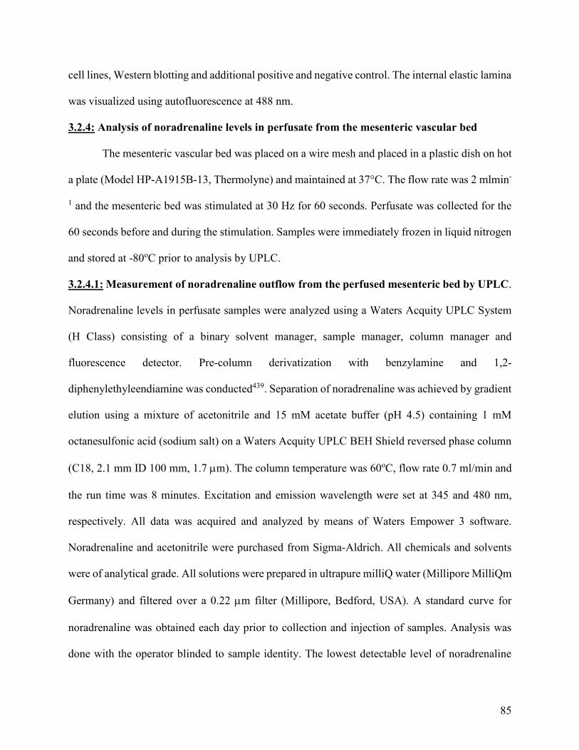
85
cell lines, Western blotting and additional positive and negative control. The internal elastic lamina
was visualized using autofluorescence at 488 nm.
3.2.4: Analysis of noradrenaline levels in perfusate from the mesenteric vascular bed
The mesenteric vascular bed was placed on a wire mesh and placed in a plastic dish on hot
a plate (Model HP-A1915B-13, Thermolyne) and maintained at 37°C. The flow rate was 2 mlmin-
1 and the mesenteric bed was stimulated at 30 Hz for 60 seconds. Perfusate was collected for the
60 seconds before and during the stimulation. Samples were immediately frozen in liquid nitrogen
and stored at -80oC prior to analysis by UPLC.
3.2.4.1: Measurement of noradrenaline outflow from the perfused mesenteric bed by UPLC.
Noradrenaline levels in perfusate samples were analyzed using a Waters Acquity UPLC System
(H Class) consisting of a binary solvent manager, sample manager, column manager and
fluorescence detector. Pre-column derivatization with benzylamine and 1,2-
diphenylethyleendiamine was conducted439. Separation of noradrenaline was achieved by gradient
elution using a mixture of acetonitrile and 15 mM acetate buffer (pH 4.5) containing 1 mM
octanesulfonic acid (sodium salt) on a Waters Acquity UPLC BEH Shield reversed phase column
(C18, 2.1 mm ID 100 mm, 1.7 m). The column temperature was 60oC, flow rate 0.7 ml/min and
the run time was 8 minutes. Excitation and emission wavelength were set at 345 and 480 nm,
respectively. All data was acquired and analyzed by means of Waters Empower 3 software.
Noradrenaline and acetonitrile were purchased from Sigma-Aldrich. All chemicals and solvents
were of analytical grade. All solutions were prepared in ultrapure milliQ water (Millipore MilliQm
Germany) and filtered over a 0.22 m filter (Millipore, Bedford, USA). A standard curve for
noradrenaline was obtained each day prior to collection and injection of samples. Analysis was
done with the operator blinded to sample identity. The lowest detectable level of noradrenaline

86
was 4 fmol/50 µl sample. The concentration of noradrenaline in the perfusate samples are shown
as normalized values due to variation in control values for noradrenaline overflow between
different preparations.
3.2.5: Statistics
All data are expressed as mean ± SEM, n rats used. For repeated measures, two-way
ANOVA followed by either a Tukey’s multiple comparison post-hoc test (used when there were
more than two experimental groups) or a Šídák method post-hoc test (used when there was two
experimental groups) was performed. Paired t-tests were used in Figure 3.9. p<0.05 was
considered statistically significant in all cases.
3.3: Results
3.3.1: Role of IKCa channels in endothelial modulation of sympathetic vasoconstriction in the
perfused mesenteric bed
In endothelium-intact mesenteric beds, infusion of noradrenaline (15 M) caused
vasoconstriction that was significantly enhanced by TRAM-34 (1 M); in the presence of TRAM-
34 the response to noradrenaline was 134.2 ± 10.5 % of control (n=5, p<0.05), indicating that
myoendothelial feedback can occur in this preparation. In endothelium-denuded arteries, TRAM-
34 was without effect on noradrenaline-evoked constriction (n=4, p>0.05).
The functional role of IKCa channels in endothelial-dependent modulation of nerve-evoked
vasoconstriction in the perfused mesenteric bed was investigated using the selective IKCa channel
inhibitors, TRAM-34 and NS 6180 (1 M). Neither NS 6180 (p>0.05, Figure 3.1a and b) nor
TRAM-34 (n=5) significantly affected nerve-evoked vasoconstriction. Also, TRAM-34 in
combination with the SKCa channel inhibitor, apamin (50 nM), did not enhance nerve-evoked
vasoconstriction more than apamin alone; nerve-evoked responses in the presence of apamin or
apamin plus TRAM-34 were not significantly different (p>0.05, Figure 3.1c). Thus, IKCa channel-
87
mediated myoendothelial feedback does not appear to play a significant role in endothelial
modulation of nerve-evoked vasoconstriction in the intact mesenteric bed.
Figure 3.1: IKCa channels do not play a role in nerve-evoked vasoconstriction.
a) Representative trace showing a frequency-response relationship obtained from an endothelium-
intact perfused mesenteric bed in the absence and presence of NS 6180 (1 μM). b) Mean frequency-
response relationships obtained from endothelium-intact perfused mesenteric beds in the absence
and presence of NS 6180 (1 μM). Values are presented as mean ± SEM, n=5; two-way repeated-
measures ANOVA. c) Mean frequency-response relationships obtained from endothelium-intact
perfused mesenteric beds in the absence and presence of apamin (50 nM) without and with TRAM-
34 (1 μM). Values are presented as mean ± SEM, n=5. * denotes p<0.05 from control; two-way
repeated-measures ANOVA.
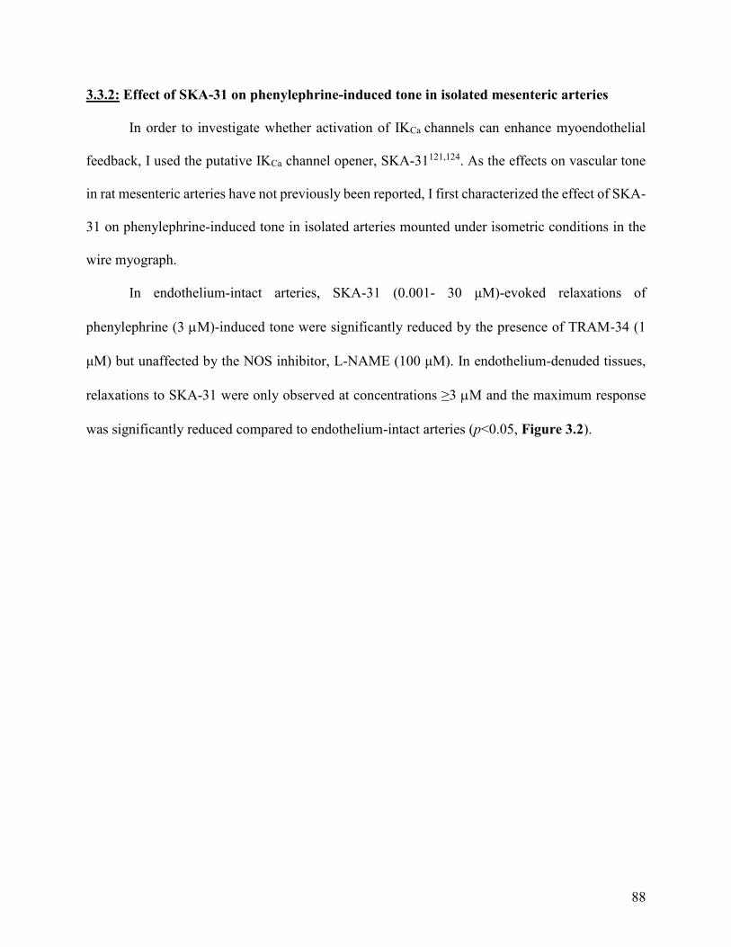
88
3.3.2: Effect of SKA-31 on phenylephrine-induced tone in isolated mesenteric arteries
In order to investigate whether activation of IKCa channels can enhance myoendothelial
feedback, I used the putative IKCa channel opener, SKA-31121,124. As the effects on vascular tone
in rat mesenteric arteries have not previously been reported, I first characterized the effect of SKA-
31 on phenylephrine-induced tone in isolated arteries mounted under isometric conditions in the
wire myograph.
In endothelium-intact arteries, SKA-31 (0.001- 30 μM)-evoked relaxations of
phenylephrine (3 M)-induced tone were significantly reduced by the presence of TRAM-34 (1
μM) but unaffected by the NOS inhibitor, L-NAME (100 μM). In endothelium-denuded tissues,
relaxations to SKA-31 were only observed at concentrations ≥3 M and the maximum response
was significantly reduced compared to endothelium-intact arteries (p<0.05, Figure 3.2).
89
Figure 3.2: SKA-31-mediates endothelium-dependent relaxation through IKCa channel
activation. Third order mesenteric arteries were mounted in a wire myograph. Mean data showing
SKA-31-induced relaxation in endothelium-intact isolated rat mesenteric artery segments mounted
in a wire myograph in the absence (control n=13) and presence of TRAM-34 (1 μM; n=10) or L-
NAME (100 μM; n=13) and in endothelium-denuded (n=5) isolated rat mesenteric artery
segments. Values are presented as mean ± SEM. * denotes p<0.05 from control, # denotes p<0.05
from TRAM-34 (1 μM) and ^ denotes p<0.05 from denuded; two-way repeated-measures
ANOVA.
The effect of SKA-31 (10 μM) on phenylephrine-evoked increases in tone was also
examined as responses to phenylephrine are limited by IKCa channel-mediated myoendothelial
feedback. This concentration of SKA-31 was selected as it evoked relaxations which were
predominantly endothelium-dependent and sensitive to TRAM-34. Concentration-response curves
to phenylephrine (phenylephrine denoted as PE in Figure 3.3; 0.001- 100 μM) were constructed
in the absence and presence of SKA-31. In endothelium-intact arteries, SKA-31 did not affect
resting tone but significantly reduced increases in tone elicited by 1 and 3 μM phenylephrine
(p<0.05; Figure 3.3) causing a rightward shift in the concentration-response curve to

90
phenylephrine. This effect was blocked by TRAM-34 (1 μM) but not L-NAME (100 μM) (Figure
3.3).
Figure 3.3: SKA-31 limits phenylephrine-induced increases in tone through IKCa channel
activation in a NO-independent manner. Third order mesenteric arteries were mounted in a wire
myograph. Mean data showing phenylephrine-induced increases in tone in the absence (control
n=13) and presence of SKA-31 (10 μM; n=13) without and with L-NAME (100 μM; n=4) or
TRAM-34 (1 μM; n=4) in endothelium-intact isolated rat mesenteric artery segments. Values are
presented as mean ± SEM. * denotes p<0.05 from control, # denotes p<0.05 from SKA-31 (10
μM) and ^ denotes p<0.05 from SKA-31(10 μM) with L-NAME (100 μM); two-way repeated-
measures ANOVA.
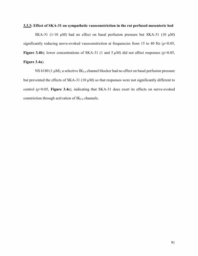
91
3.3.3: Effect of SKA-31 on sympathetic vasoconstriction in the rat perfused mesenteric bed
SKA-31 (1-10 μM) had no effect on basal perfusion pressure but SKA-31 (10 μM)
significantly reducing nerve-evoked vasoconstriction at frequencies from 15 to 40 Hz (p<0.05,
Figure 3.4b); lower concentrations of SKA-31 (1 and 5 M) did not affect responses (p>0.05,
Figure 3.4a).
NS 6180 (1 μM), a selective IKCa channel blocker had no effect on basal perfusion pressure
but prevented the effects of SKA-31 (10 M) so that responses were not significantly different to
control (p>0.05, Figure 3.4c), indicating that SKA-31 does exert its effects on nerve-evoked
constriction through activation of IKCa channels.

92
Figure 3.4: SKA-31 limits nerve-evoked vasoconstriction through IKCa channel activation.
a) Mean frequency-response relationships obtained from endothelium-intact perfused mesenteric
beds in the absence and presence of SKA-31 (1 and 5 μM). Values are presented as mean ± SEM,
n=4; two-way repeated-measures ANOVA. b) Mean frequency-response relationships obtained
from endothelium-intact perfused mesenteric beds in the absence and presence of SKA-31 (10
μM). Values are presented as mean ± SEM, n=4. * denotes p<0.05 from control; two-way repeated-
measures ANOVA. c) Mean frequency-response relationships obtained from endothelium-intact
perfused mesenteric beds in the absence and presence of NS 6180 (1 μM) without and with SKA-
31 (10 μM). Values are presented as mean ± SEM, n=5; two-way repeated-measures ANOVA.
93
To investigate if inhibition of nerve-evoked vasoconstriction by SKA-31 was mediated by
NO, SKA-31 (10 M) was applied in the presence of L-NAME (100 M). As shown in Chapter
2 (Figure 2.3), L-NAME significantly enhanced nerve-evoked vasoconstriction in endothelium-
intact mesenteric beds at frequencies from 20 to 40 Hz (p<0.05, Figure 3.5). In the presence of L-
NAME, SKA-31 was still able to inhibit nerve-evoked responses at frequencies of 20 to 40 Hz
(p<0.05, Figure 3.5).
Figure 3.5: Inhibition of nerve-evoked vasoconstriction by SKA-31 is not mediated by NO.
Mean frequency-response relationships obtained from endothelium-intact perfused mesenteric
beds in the absence and presence of L-NAME (100 μM) without and with SKA-31 (10 μM). Values
are presented as mean ± SEM, n=4. * denotes p<0.05 from control, # denotes p<0.05 from L-
NAME (100 μM); two-way repeated-measures ANOVA.
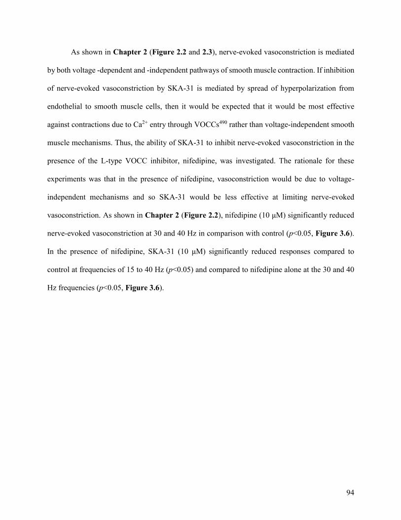
94
As shown in Chapter 2 (Figure 2.2 and 2.3), nerve-evoked vasoconstriction is mediated
by both voltage -dependent and -independent pathways of smooth muscle contraction. If inhibition
of nerve-evoked vasoconstriction by SKA-31 is mediated by spread of hyperpolarization from
endothelial to smooth muscle cells, then it would be expected that it would be most effective
against contractions due to Ca2+ entry through VOCCs490 rather than voltage-independent smooth
muscle mechanisms. Thus, the ability of SKA-31 to inhibit nerve-evoked vasoconstriction in the
presence of the L-type VOCC inhibitor, nifedipine, was investigated. The rationale for these
experiments was that in the presence of nifedipine, vasoconstriction would be due to voltage-
independent mechanisms and so SKA-31 would be less effective at limiting nerve-evoked
vasoconstriction. As shown in Chapter 2 (Figure 2.2), nifedipine (10 μM) significantly reduced
nerve-evoked vasoconstriction at 30 and 40 Hz in comparison with control (p<0.05, Figure 3.6).
In the presence of nifedipine, SKA-31 (10 μM) significantly reduced responses compared to
control at frequencies of 15 to 40 Hz (p<0.05) and compared to nifedipine alone at the 30 and 40
Hz frequencies (p<0.05, Figure 3.6).
95
Figure 3.6: SKA-31 inhibits the voltage-independent component of nerve-evoked
vasoconstriction. Mean frequency-response relationships obtained from endothelium-intact
perfused mesenteric beds in the absence and presence of nifedipine (10 μM) without and with
SKA-31 (10 μM). Values are presented as mean ± SEM, n=4. * denotes p<0.05 from control and
# denotes p<0.05 from nifedipine (10 μM); two-way repeated-measures ANOVA.
The ability of SKA-31 to inhibit nerve-evoked vasoconstriction in the presence of
nifedipine indicates that a mechanism other than hyperpolarization may underlie its effects on
smooth muscle contractility. Thus, I investigated whether SKA-31 could influence
vasoconstriction in endothelium-denuded preparations. SKA-31 (10 M) significantly reduced
nerve-evoked vasoconstriction (p<0.05, Figure 3.7) in endothelium-denuded mesenteric beds.
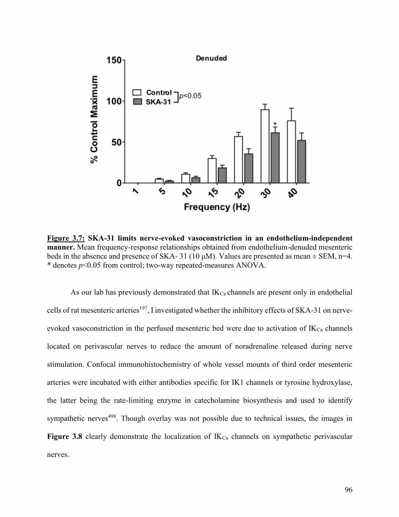
96
Figure 3.7: SKA-31 limits nerve-evoked vasoconstriction in an endothelium-independent
manner. Mean frequency-response relationships obtained from endothelium-denuded mesenteric
beds in the absence and presence of SKA- 31 (10 μM). Values are presented as mean ± SEM, n=4.
* denotes p<0.05 from control; two-way repeated-measures ANOVA.
As our lab has previously demonstrated that IKCa channels are present only in endothelial
cells of rat mesenteric arteries197, I investigated whether the inhibitory effects of SKA-31 on nerve-
evoked vasoconstriction in the perfused mesenteric bed were due to activation of IKCa channels
located on perivascular nerves to reduce the amount of noradrenaline released during nerve
stimulation. Confocal immunohistochemistry of whole vessel mounts of third order mesenteric
arteries were incubated with either antibodies specific for IK1 channels or tyrosine hydroxylase,
the latter being the rate-limiting enzyme in catecholamine biosynthesis and used to identify
sympathetic nerves498. Though overlay was not possible due to technical issues, the images in
Figure 3.8 clearly demonstrate the localization of IKCa channels on sympathetic perivascular
nerves.
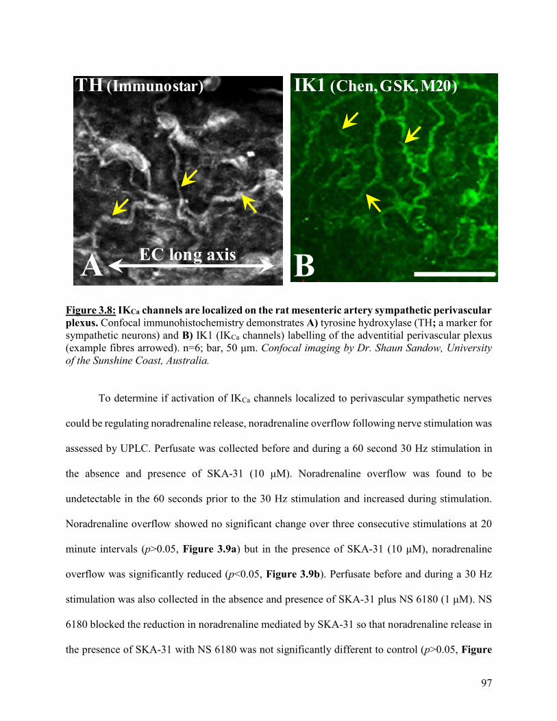
97
Figure 3.8: IKCa channels are localized on the rat mesenteric artery sympathetic perivascular
plexus. Confocal immunohistochemistry demonstrates A) tyrosine hydroxylase (TH; a marker for
sympathetic neurons) and B) IK1 (IKCa channels) labelling of the adventitial perivascular plexus
(example fibres arrowed). n=6; bar, 50 μm. Confocal imaging by Dr. Shaun Sandow, University
of the Sunshine Coast, Australia.
To determine if activation of IKCa channels localized to perivascular sympathetic nerves
could be regulating noradrenaline release, noradrenaline overflow following nerve stimulation was
assessed by UPLC. Perfusate was collected before and during a 60 second 30 Hz stimulation in
the absence and presence of SKA-31 (10 μM). Noradrenaline overflow was found to be
undetectable in the 60 seconds prior to the 30 Hz stimulation and increased during stimulation.
Noradrenaline overflow showed no significant change over three consecutive stimulations at 20
minute intervals (p>0.05, Figure 3.9a) but in the presence of SKA-31 (10 μM), noradrenaline
overflow was significantly reduced (p<0.05, Figure 3.9b). Perfusate before and during a 30 Hz
stimulation was also collected in the absence and presence of SKA-31 plus NS 6180 (1 μM). NS
6180 blocked the reduction in noradrenaline mediated by SKA-31 so that noradrenaline release in
the presence of SKA-31 with NS 6180 was not significantly different to control (p>0.05, Figure
IK1 (Chen, GSK, M20)
A
TH (Immunostar)
BEC long axis
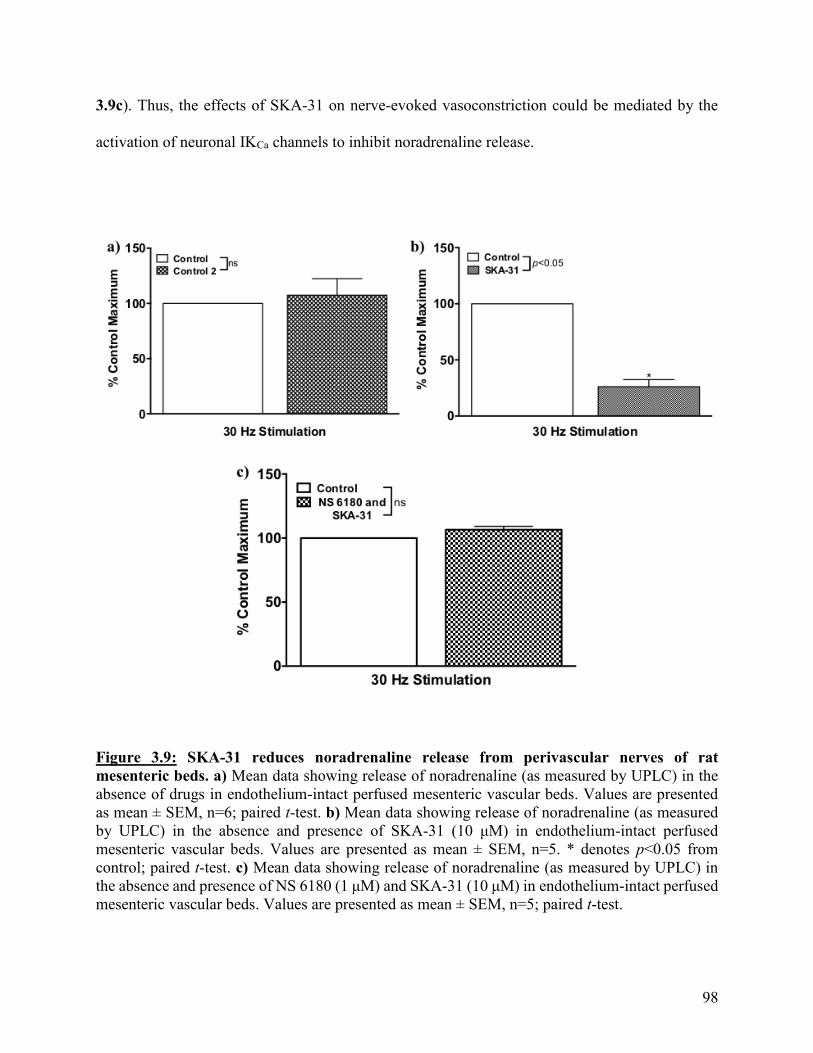
98
3.9c). Thus, the effects of SKA-31 on nerve-evoked vasoconstriction could be mediated by the
activation of neuronal IKCa channels to inhibit noradrenaline release.
Figure 3.9: SKA-31 reduces noradrenaline release from perivascular nerves of rat
mesenteric beds. a) Mean data showing release of noradrenaline (as measured by UPLC) in the
absence of drugs in endothelium-intact perfused mesenteric vascular beds. Values are presented
as mean ± SEM, n=6; paired t-test. b) Mean data showing release of noradrenaline (as measured
by UPLC) in the absence and presence of SKA-31 (10 μM) in endothelium-intact perfused
mesenteric vascular beds. Values are presented as mean ± SEM, n=5. * denotes p<0.05 from
control; paired t-test. c) Mean data showing release of noradrenaline (as measured by UPLC) in
the absence and presence of NS 6180 (1 μM) and SKA-31 (10 μM) in endothelium-intact perfused
mesenteric vascular beds. Values are presented as mean ± SEM, n=5; paired t-test.
99
3.4: Discussion
Endothelial IKCa channels are located on the abluminal side of endothelial cells at
MEGJs118,125,130,151,197, the sites of contact between endothelial and surrounding smooth muscle
cells, where they play a pivotal role in limiting agonist-mediated vasoconstriction via
myoendothelial feedback130,197. Here in Chapter 3, I have shown that IKCa channels are not
involved in shear stress-induced endothelial-dependent modulation of sympathetic
vasoconstriction in the perfused mesenteric bed. SKA-31, an activator of IKCa channels, can inhibit
nerve-evoked vasoconstriction in this preparation but this effect is independent of the endothelium
and appears to be due to the activation of neuronal IKCa channels to reduce noradrenaline release.
This is the first description of a role for IKCa channels in the release of noradrenaline from
perivascular sympathetic nerves in resistance arteries and suggest that targeting these channels
could provide a novel approach to reducing vasoconstriction in conditions associated with
increased sympathetic drive, such as hypertension174,499,500.
As described earlier, the reliance of myoendothelial feedback on IKCa channels and the
availability of selective inhibitors for these channels, provides the opportunity to dissect the
contribution of this pathway in functional vascular responses118,125,130,151,197,493. The inhibition of
IKCa channels by TRAM-34, causes endothelium-dependent enhancement of both phenylephrine-
evoked increases in tone in isolated mesenteric arteries and vasoconstriction to infusion of
noradrenaline in the perfused mesenteric bed. Thus, IKCa channel-mediated myoendothelial
feedback is functional in these arteries. The lack of effect of TRAM-34 and a second selective IKCa
channel inhibitor, NS 6180, on nerve-evoked vasoconstriction indicates that these channels, and
by extension the myoendothelial feedback pathway, are not involved in modulating sympathetic
vasoconstriction in this preparation. Thus, as described in Chapter 2, it appears that shear stress-
100
induced activation of endothelial SKCa channels is the dominant mechanism for engagement of the
endothelium to limit sympathetic vasoconstriction.
The reason for the differential functional roles of the two types of endothelial KCa channels
may be explained by their discrete locations within endothelial cells. As described in Chapter 2,
SKCa channels are located on the luminal membrane of endothelial cells118,122,125,130,132,133 whereas
IKCa channels are located at MEGJs118,125,130,151,197. Increases in shear stress lead to Ca2+ influx
through TRPV4 channels, which are co-localized with SKCa channels and caveolin-1 in discrete
signalling microdomains on the luminal endothelial cell membrane132,220,241–248,474–476. The location
of IKCa channels at MEGJs118,125,130,151,197 may mean that they are not exposed to shear stress-
mediated localized increases in Ca2+ at the luminal membrane and so are unable to participate in
the subsequent endothelial modulation of nerve-evoked vasoconstriction. This conclusion is
supported by previous work showing that responses to increases in shear stress are unaltered in
isolated carotid arteries from IK1-/- mice whereas arteries from animals lacking both SKCa and IKCa
channels had diminished shear stress-induced responses204.
SKA-31 enhances the sensitivity of IKCa channels to Ca2+ 121,124,437 to induce TRAM-34-
sensitive hyperpolarization in isolated endothelial cells from canine mesenteric and mouse carotid
arteries124,316, and to activate IKCa channel currents in isolated murine endothelial cells121,316. Given
this mechanism of action, I proposed that although IKCa channels may not be involved in
modulating nerve-evoked vasoconstriction, SKA-31 may be able enhance IKCa channel activity in
order to limit nerve-evoked vasoconstriction. As the effects of SKA-31 have not been investigated
in rat mesenteric arteries, I first examined its actions in isolated mesenteric arteries and showed
that it evoked relaxations which are dependent on the endothelium and IKCa channels, but
independent of NO. These findings are in line with previous studies showing that SKA-31 dilates
101
rat cerebral314, skeletal muscle arterioles316 and mouse mesenteric arteries501, effects which were
dependent on IKCa channels but not affected by block of NOS. Furthermore, bolus doses of SKA-
31 caused NO-independent dilation of rat coronary arteries in the intact heart502, and L-NAME did
not affect vasodilation to NS309, a structurally-related SKCa/IKCa channel activator, in rat
cremaster and small mesenteric arteries128,205.
SKA-31 activates IKCa channels, with an EC50 value of 0.26 μM, it can also activate SKCa
channels, with an EC50 value of 2.9 μM, showing a 10-fold lower potency for SKCa channels316. In
canine mesenteric artery endothelial cells, SKA-31-evoked KCa currents and membrane
hyperpolarization that were sensitive to both TRAM-34 and the SKCa channel inhibitor, UCL
1684124. SKCa channels have also been shown to contribute to the effects of SKA-31 on arterial
diameter as block of SKCa channels by apamin or UCL1684, inhibited the ability of SKA-31 to
dilate myogenically active rat cremaster, middle cerebral arteries314, and pressurized mouse
mesenteric arteries501. In the present study, SKA-31-evoked responses were abolished by TRAM-
34 and NS 1680 and were shown to be endothelium-independent, the role of SKCa channels was
not investigated.
Bolus doses of SKA-31 caused immediate and transient reductions in blood pressure in
anesthetized pigs503 and conscious mice and dogs121,124,316. This effect was also seen in mice lacking
SKCa channels204 and mouse models of hypertension121, but was lost in mice lacking IKCa
channels121. These effects of SKA-31 are hypothesized to be due to direct actions on blood vessels
and in vitro studies have largely focused on its ability to cause dilation in isolated arteries.
However, other than the demonstration that SKA-31 does dilate the coronary vasculature in
isolated hearts315,502, there is limited information on its effects in intact vascular beds thus, the use
of the rat perfused mesenteric vascular bed in this chapter allowed for the novel investigation of

102
SKA-31’s effects at the level of the intact mesenteric bed. This is an important gap in our
knowledge as in anesthetized pigs, vascular conductance in coronary and carotid arteries was
increased in response to SKA-31 but renal conductance was unaffected503, suggesting that its
effects may not be uniform across the entire vasculature.
In the perfused mesenteric bed, SKA-31 (10 M) reduced sympathetic vasoconstriction in
a TRAM-34- and NS 6180- sensitive manner but its effects were NO- and endothelium-
independent. Also, inhibition of vasoconstriction was observed in the presence of nifedipine, to
block the contribution of L-type VOCCs to vasoconstriction. This was an unexpected result for
two reasons. First, we and others have shown that IKCa channels are localized on endothelial but
not smooth muscle cells of rat mesenteric arteries118,125,130,151,197. IKCa channels have been shown
to be present on proliferating vascular smooth muscle cells173,504–509, with their expression
upregulated by growth factors505,510 but only two reports describe IKCa channels in vascular smooth
muscle cells of intact arteries, one in human chorionic plate arteries511 and the other in rat middle
cerebral arteries512, and neither demonstrated a functional role for these channels in mediating
vasodilation. Furthermore, although NS 6180 and TRAM-34 had no effect on nerve-evoked
vasoconstriction, TRAM-34 did enhance noradrenaline-evoked vasoconstriction in the perfused
bed in an endothelium-dependent manner, indicating that the myoendothelial feedback pathway is
functional in this vascular bed. These findings are in contrast to the human skeletal muscle
vasculature where myoendothelial feedback plays a significant role in blunting sympathetic
vasoconstriction497.
Second, my data on the effects of SKA-31 on isolated arteries described above, and in the
published literature116,121,124,148,204,314,316,502,503, support the notion that SKA-31 causes vascular
relaxation of isolated arteries via endothelium-dependent hyperpolarization of the surrounding

103
smooth muscle to inhibit L-type VOCC-mediated Ca2+ influx. The only report to potentially
contradict this notion demonstrates that intraperitoneal injection of SKA-31 reduced mean arterial
blood pressure in mice lacking connexin40316. This connexin is an essential component of MEGJs
in a number of vessels513,514 thus, the ability of SKA-31 to affect the vasculature in mice lacking
this protein suggests that the response is independent of myoendothelial coupling (i.e. not
dependent on the transfer of an electrical signal from endothelial to smooth muscle cells), although
the mediator of this response was not determined316.
A possible explanation for the endothelial-independent effects of SKA-31 on nerve-evoked
vasoconstriction in the perfused bed versus its predominantly endothelium-dependent effects in
isolated arteries, is that SKA-31 activates IKCa channels on sympathetic nerves to hyperpolarize
neuronal membrane potential and reduce noradrenaline release. IKCa channels are localized on
specific neurons in the rat, mouse, guinea-pig and human enteric nervous system152–156 where they
mediate the slow after-hyperpolarization following an action potential but to date, there are no
reports of these channels on perivascular nerves. Our lab has previously demonstrated that
noradrenaline released by stimulation of perivascular nerves can be measured in the perfusate of
the perfused rat mesenteric bed and that those levels can be modulated by pharmacological agents
which act as sympathomimetics438. Using this approach, I demonstrated that SKA-31 did inhibit
noradrenaline overflow and that this effect was prevented by NS 6180. Furthermore, confocal
immunohistochemistry using antibodies specific for IK1 channels and tyrosine hydroxylase, the
rate-limiting enzyme in catecholamine biosynthesis used to identify sympathetic nerves498,
demonstrated that IKCa channels are localized to perivascular nerves in mesenteric arteries. While
overlay was not possible, there is no parasympathetic innervation of rat mesenteric arteries and
the sensory plexus are sparse and distinct from sympathetic nerves176,179.
104
A neuronal site of action for SKA-31 or other SKCa/IKCa channel openers has not previously
been considered. The lack of effect of TRAM-34 on sympathetic vasoconstriction suggests that
IKCa channels do not play a role in regulating noradrenaline release under normal conditions.
However, stimulation of perivascular nerves leads to depolarization of the neuronal membrane
potential which causes Ca2+-influx through neuronal VOCCs (N-, P- and Q-type440) and thus,
SKA-31 may act by sensitizing neuronal IKCa channels to the increased levels of Ca2+. L-type
VOCCs are co-localized with BKCa channels in smooth muscle cells152, and both with BKCa and
SKCa channels in central nervous system neurons515 but the potential for such interactions between
IKCa channels and VOCCs in perivascular sympathetic nerves has yet to be investigated.
Interestingly, TRPC3 channels, which have been identified as a mediator of Ca2+ influx for
activation of SKCa and IKCa channels in endothelial cells256,257,516,517, have also been localized to
perivascular nerves in mesenteric arteries256, Their role has not been determined but they could
serve as an important Ca2+ source for these neuronal IKCa channels.
To conclude, in isolated mesenteric arteries, endothelial IKCa channels are crucial for
engagement of the endothelium through myoendothelial feedback to limit agonist-evoked
vasoconstriction but these channels do not appear to play a functional role in modulating nerve-
evoked vasoconstriction in the perfused mesenteric bed. Nevertheless, activation of IKCa channels
by SKA-31 does limit vasoconstriction most likely via inhibition of noradrenaline release from
sympathetic nerves. Targeting IKCa channels as a novel strategy for treatment and/or prevention
of cardiovascular diseases has been proposed and SKA-31, a IKCa channel activator, has been
suggested as an agent which may improve endothelial function121,148,316. For example, SKA-31
was able to enhance acetylcholine-mediated dilations in pressurized carotid arteries from mice
lacking eNOS, indicating that increased IKCa channel activity may be able to overcome impaired
105
endothelium-dependent vasodilation due to the absence of NO148. However, although SKA-31 can
cause endothelium-dependent relaxation of rat isolated mesenteric arteries, in the perfused
mesenteric bed, its primary action appears to be inhibition of noradrenaline release from
sympathetic nerves. Thus, while IKCa channels may be a valid target to limit vasoconstriction,
increased activation of these channels may involve both endothelium-dependent and -independent
actions. The data presented in this chapter provides the first description of a role for IKCa channels
in modulating release of noradrenaline from perivascular sympathetic nerves and indicates that
targeting of these channels could provide a new approach to reducing vasoconstriction in
conditions associated with increased sympathetic drive, such as hypertension174,499,500.
106
Chapter 4: Effects of activators of SKCa and IKCa channels on agonist-induced
O2- production and vasoconstriction in isolated mesenteric arteries
4.1: Introduction
As described in Chapter 1, endothelium-derived NO plays a vital role in regulating normal
vascular function and endothelial damage associated with risk factors for cardiovascular diseases,
such as diabetes, hypertension and atherosclerosis, is characterized by increased production of O2-
and decreased NO bioavailability268,328–336. Attempts to reduce O2- levels through the use of dietary
anti-oxidants, such as vitamins B, C and E,` have failed in clinical trials337–342, most likely due to
their inability to reach a high enough concentration in the vasculature and/or within endothelial
cells. Therefore, there is the need to identify new targets for therapeutic approaches to reduce O2-
levels and enhance NO bioavailability in cardiovascular disease settings.
Numerous changes have been proposed to contribute to endothelial dysfunction but a
common mechanism appears to be enhanced O2- production leading to decreased NO
bioavailability and NOS expression518, and increased synthesis of endothelium-derived contractile
factors such as thromboxanes518–521. Elevated O2- levels rapidly diminish NO bioavailability and
generate excessive amounts of ONOO-, an oxidant that accelerates cardiovascular disease
progression by causing structural damage to vascular cells, inhibiting prostacyclin synthesis,
disrupting NO signaling and increasing O2- production through oxidation of the endothelial NOS
co-factor, BH4; in the absence of BH4, endothelial NOS becomes uncoupled and reduces molecular
oxygen to generate O2- rather than NO266–270,521.
Treatment of hypercholesteraemic and diabetic animals with statins or angiotensin receptor
blockers522,523 reduces the activity and expression of NADPH oxidase and “recouples” endothelial
NOS by preventing BH4 oxidation524,525. Statins and angiotensin receptor blockers have also been
shown to improve endothelial function and to reduce the incidence of cardiovascular events in

107
patients with cardiovascular diseases526,527. Thus, the development of new drugs which possess
indirect antioxidant properties, mediated by the enhancement of NO production and simultaneous
inhibition of O2- generation (e.g. from NADPH oxidase), is an attractive proposition for
cardiovascular disease prevention and therapy. I propose that drugs which activate SKCa and IKCa
channels may fall into this category.
NO bioavailability is determined by the balance between production of NO by endothelial
NOS and its interaction with O2-. In numerous disease models, reduced NO bioavailability is
associated with upregulation of expression and activity of NADPH oxidase332–336,383,528, a voltage-
sensitive enzyme which generates O2- by transferring electrons from cytosolic NADPH to
extracellular oxygen126,367,368,371,372,374,386–388,529. In isolated endothelial cells, membrane
depolarization activates NADPH oxidase, either directly or via Akt387,529. In this setting, drugs
which open KATP channels attenuate both the membrane depolarization and O2- production,
indicating that endothelial cell membrane potential can regulate O2- production387,529.
As discussed previously, opening of SKCa and IKCa channels mediates vasodilation through
hyperpolarization of the endothelial membrane potential which spreads to surrounding smooth
muscle cells via MEGJs80,84,85,100,116–131,148–151,197,202. Activators of these channels have been shown
to elicit NO-mediated relaxation and to enhance NO production116,126,128,131,203–205. Also, in
Chapter 2, I demonstrated that the SKCa channel opener CyPPA, elicits endothelium-mediated
relaxation in isolated rat mesenteric arteries and enhances shear stress-induced inhibition of nerve-
evoked vasoconstriction in the perfused mesenteric bed, both through a NO-dependent mechanism.
Our lab has previously shown that endothelial depolarization inhibits agonist-evoked, NO-
mediated relaxation of rat basilar arteries, an effect that was overcome by the KATP channel opener,
pinacidil418. Furthermore, we and others, have demonstrated that the membrane potential of

108
endothelial cells in tail530 and mesenteric arteries531–533 from cardiovascular disease rat models are
depolarized compared to controls. We have also shown NO-mediated relaxations are attenuated in
these depolarized vessels and that these effects can be reversed by the SKCa/IKCa channel opener,
1-EBIO534. Thus, it is possible that the opening of endothelial KCa channels to elicit
hyperpolarization could lead to a decrease in O2- production by voltage-sensitive NADPH oxidase
and thus, an increase in NO bioavailability.
The goal of this, and the following chapter, was to further explore the relationship between
endothelial KCa channels, O2- production and diameter in intact arteries, and to test the hypothesis
that pharmacological activators of endothelial KCa channels can reduce vascular O2- production
and enhance NO-mediated modulation of vasoconstriction.
To test this hypothesis, I utilized isolated endothelium-intact mesenteric resistance arteries
mounted in a pressure myograph coupled to a fluorescence detection system (IonOptix) for
simultaneous measurement of changes in arterial diameter and O2- production using
dihydroethidium (DHE), a dye that interacts with O2- to form 2-hydroxyethidium (EOH) which
fluoresces when bound to deoxyribonucleic acid (DNA)349,535–540. The effects of CyPPA and SKA-
31 on phenylephrine-evoked O2- production and vasoconstriction were assessed to investigate if
increased activation of endothelial KCa channels can modulate vascular O2- levels and endothelial
modulation of arterial diameter.
I chose to use this approach as vasoconstriction of mesenteric arteries elicited by 1-
adrenoceptor agonists has been associated with increases in O2-347–349 and, as stated earlier, a
primary role of the endothelium in vivo is to modulate vasoconstriction. Also, constriction of rat
mesenteric arteries to phenylephrine is modulated by endothelium-derived NO130,197. Thus,
109
changes in O2- levels may lead to changes in the bioavailability of NO and subsequently, alterations
in phenylephrine-evoked vasoconstrictor responses.
4.2: Methods and materials
See Appendix: Drugs and chemicals for a list of the drugs and chemicals used.
4.2.1: Simultaneous assessment of O2- production and changes in arterial diameter in intact
arteries
Real-time assessment of changes in O2- production and diameter were carried out in
mesenteric resistance arteries loaded with DHE and mounted in a pressure myograph (Danish Myo
Technology (DMT) 110P model, Aarhus, Denmark) placed on an inverted microscope (AE30-31,
Motic Instruments Inc. Canada) fitted with an Eiscopic-Fluorescence Attachment (EF-INV-11).
4.2.1.1: Pressure myography. Leak-free segments of third or fourth order mesenteric arteries (2-
3 mm in length) were cleaned of adhering connective tissue and mounted between two glass
cannulae in an arteriograph chamber (DMT 110P model, Aarhus, Denmark) under conditions of
no luminal flow. The glass cannulae (borosilicate glass with OD of 1.2 mm and ID of 0.69 mm)
were fabricated using a Flaming/Brown micropipette puller (Model P87; Sutter Instruments,
Novato, USA) and flame polished. In some experiments, the endothelium was removed by gently
rubbing the lumen of individual arteries with a hair.
The arteriograph was placed on the stage of an inverted microscope (AE30-31, Motic
Instruments Inc. Canada). Vessels were maintained in Krebs buffer at 37oC (pH 7.4) continuously
bubbled with 95% O2, 5% CO2. Intravascular pressure was maintained via a pressure controller
(DMT 110P model, Aarhus, Denmark) and vessel images were captured using a CCD video
camera (CFA300 Cell Framing Adapter, Ionoptix MyoCam). Vessels were loaded with DHE (see
below) and upon excitation at 535 nm, fluorescence of EOH bound to DNA at 610 nm was captured
110
via an Eiscopic-Fluorescence Attachment (EF-INV-11) and an IonOptix Fluorescence System
Interface, and recorded using IonWizard 6.2 (IonOptix, Massachusetts, USA) software.
To avoid the development of myogenic tone, arterial segments were held at an intravascular
pressure of 60 mmHg (mean resting diameter at this pressure was 208.45 ± 6.3m, n=5) for 20
minutes before being tested for viability through the addition of phenylephrine (3 μM) to cause
constriction, and acetylcholine (3 μM) to induce endothelium-dependent vasodilation; vessels in
which acetylcholine elicited >90% reversal of phenylephrine-induced vasoconstriction were
considered to have an intact endothelium and those that elicited <10% relaxation were deemed to
be endothelium denuded. Vessels which did not meet these criteria were discarded.
4.2.1.2: Use of DHE to assess O2- production in intact mesenteric arteries. Solutions of DHE
(10 mM) were freshly made in dimethyl sulfoxide (DMSO)349,535,536 prior to each experiment in a
darkened room and experiments were carried out in the same darkened room. After viability had
been determined through addition of phenylephrine and acetylcholine, DHE (10 μM) was added
to the tissue bath and arteries were incubated for 30 minutes349,535,536. Vessels were then washed
and allowed to recover for 15 minutes before cumulative concentration-response curves to
phenylephrine (0.01-10 μM) were constructed347–349. After washing, pharmacological reagents
were added to the tissue bath for 15 minutes, after which time a second concentration-response
curve to phenylephrine was constructed. At the end of each concentration-response curve to
phenylephrine, acetylcholine (3 μM) or sodium nitroprusside (1 μM) was added to demonstrate
that the vessel remained functional. For some experiments, pharmacological agents were added to
the tissue bath in the absence of phenylephrine, and changes in florescence intensity were
measured over a 15-30 minute time period. The viability of these vessels was then assessed by
addition of phenylephrine (10 μM) followed by acetylcholine (3 μM).

111
For concentration-response curves to phenylephrine, vessel diameter and DHE
fluorescence intensity were recorded simultaneously and expressed as a % of their respective
control maximum. For experiments where basal O2- production was measured over a period of 15-
30 minutes, raw values of fluorescence intensity were plotted as these experiments were performed
in individual arteries. For all experiments, phenylephrine increased O2- production and
vasoconstriction in a concentration-dependent manner (p<0.05).
4.2.2: Perfused mesenteric vascular bed
The mesenteric bed was perfused via the superior mesenteric artery as previously
described438. Briefly, the mesenteric vascular bed was separated from the intestine and the superior
mesenteric artery cleaned of connective tissue, cannulated with a blunted hypodermic needle (20
G), secured with 5-0 surgical silk (Ethicon) and flushed with Krebs buffer to remove blood. The
vascular bed was placed on a wire mesh in a warm chamber and perfused with oxygenated Krebs
buffer at a constant flow rate of 5 mlmin-1 (37°C, bubbled with 95% O2/5% CO2). A short length
of plastic tubing was placed around the needle to ensure that it did not come into contact with the
wire mesh. Changes in perfusion pressure were monitored via an in-line pressure transducer (AD
instruments, Colorado) and recorded via a PowerLab data acquisition system using Chart 5.0
software (AD Instruments, Colorado).
4.2.2.1: Responses to stimulation of perivascular nerves. Electrodes were attached to the
cannulating needle and to the wire mesh to allow electrical field stimulation using a Grass SD9
stimulator (Grass Technologies, USA). Following an equilibration period of 30 minutes, a single
stimulation (30 Hz, 90 V, pulse width 1 millisecond, 30 seconds) was applied to assess the viability
of the preparation. After a further 10 minutes, a frequency-response curve was constructed by
stimulating the preparation at 20-40 Hz (90 V, pulse width 1 millisecond, 30 seconds) at 10 minute
112
intervals188. The effects of agents on nerve-evoked vasoconstriction were assessed by perfusing
the drugs through the lumen of the preparation for 20 minutes prior to constructing a second
frequency-response curve. A third and fourth frequency-response curve were constructed
following perfusion of different drug combinations.
Nerve-evoked responses recorded in the perfused mesenteric vascular bed are shown as
normalized values. Changes in perfusion pressure were normalized to the maximum control
response (%) as is convention in these types of experiments. For all frequency response curves,
electrical stimulation caused frequency-dependent increases in perfusion pressure (p<0.05).
4.2.3: Statistics
All data are expressed as mean ± SEM, n rats used. For repeated measures, two-way
ANOVA followed by either a Tukey’s multiple comparison post-hoc test (used when there were
more than two experimental groups) or Šídák method post-hoc test (used when there was two
experimental groups) was performed. An ordinary one-way ANOVA followed by a Tukey’s
multiple comparisons post-hoc test was performed for Figure 4.9b and an unpaired t-test was used
for Figure 4.11. p<0.05 was considered statistically significant in all cases.
4.3: Results
4.3.1: Characterization of phenylephrine-induced O2- production and vasoconstriction in
mesenteric resistance arteries
Application of the 1-adrenoceptor agonist phenylephrine (0.01-10 μM) to mesenteric
arteries resulted in concentration-dependent increases in O2- production and vasoconstriction
(p<0.05); phenylephrine is denoted as PE in the figures of this chapter. The fluorescence intensity
returned to baseline after arterial relaxation through acetylcholine or SNP, indicating that DHE’s
interaction with O2- is reversible. Representative traces showing changes in O2
- and diameter in
response to increasing concentrations of phenylephrine are shown in Figure 4.1a, and mean
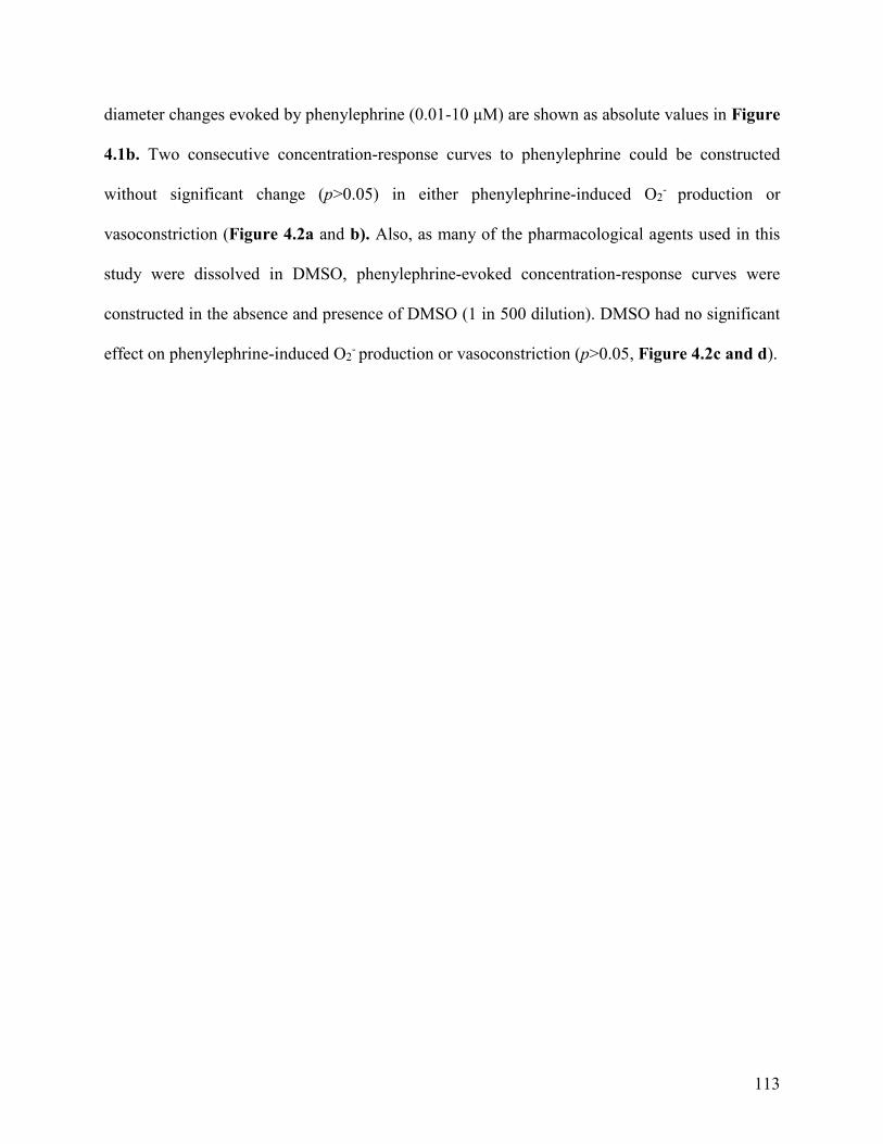
113
diameter changes evoked by phenylephrine (0.01-10 μM) are shown as absolute values in Figure
4.1b. Two consecutive concentration-response curves to phenylephrine could be constructed
without significant change (p>0.05) in either phenylephrine-induced O2- production or
vasoconstriction (Figure 4.2a and b). Also, as many of the pharmacological agents used in this
study were dissolved in DMSO, phenylephrine-evoked concentration-response curves were
constructed in the absence and presence of DMSO (1 in 500 dilution). DMSO had no significant
effect on phenylephrine-induced O2- production or vasoconstriction (p>0.05, Figure 4.2c and d).
114
Figure 4.1: Characterization of phenylephrine-induced changes in diameter and O2-
production in rat mesenteric arteries. a) Representative trace showing simultaneous recording
of phenylephrine-induced O2- production and vasoconstriction in an endothelium-intact rat
mesenteric resistance artery mounted in the pressure myograph. b) Mean changes in diameter in
endothelium-intact rat mesenteric artery segments evoked by phenylephrine; n=6.

115
Figure 4.2: Phenylephrine-induced O2- production and vasoconstriction is time-independent
and unaffected by DMSO in rat mesenteric arteries. Mean data showing phenylephrine-
induced O2- production a) during two consecutive concentration-response curves or c) in the
absence and presence of DMSO (1 in 500 dilution)) and phenylephrine-induced vasoconstriction
b) during two consecutive concentration-response curves or d) in the absence and presence of
DMSO (1 in 500 dilution) in endothelium-intact rat mesenteric artery segments. Values are
presented as mean ± SEM, a, b) n=4 and c, d) n=5; two-way repeated-measures ANOVA.
116
Phenylephrine is a α1-adrenoceptor agonist and so the α1-adrenoceptor antagonist prazosin
was used to demonstrate that the observed effects of phenylephrine on arterial diameter and O2-
production were mediated by this receptor. In endothelium-intact arteries, prazosin (1 μM)
abolished both phenylephrine-induced O2- production (p<0.05; Figure 4.3a) and vasoconstriction
(p<0.05; Figure 4.3b).
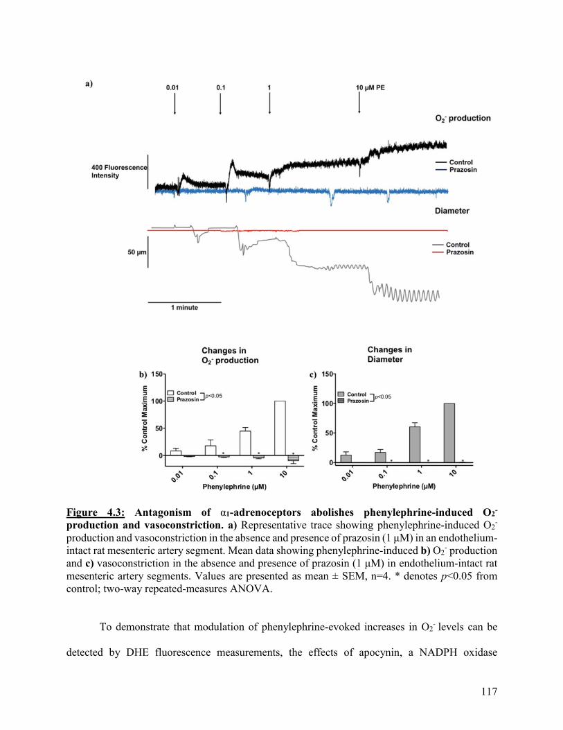
117
Figure 4.3: Antagonism of α1-adrenoceptors abolishes phenylephrine-induced O2-
production and vasoconstriction. a) Representative trace showing phenylephrine-induced O2-
production and vasoconstriction in the absence and presence of prazosin (1 μM) in an endothelium-
intact rat mesenteric artery segment. Mean data showing phenylephrine-induced b) O2- production
and c) vasoconstriction in the absence and presence of prazosin (1 μM) in endothelium-intact rat
mesenteric artery segments. Values are presented as mean ± SEM, n=4. * denotes p<0.05 from
control; two-way repeated-measures ANOVA.
To demonstrate that modulation of phenylephrine-evoked increases in O2- levels can be
detected by DHE fluorescence measurements, the effects of apocynin, a NADPH oxidase
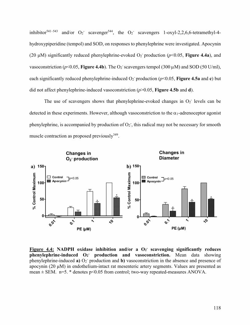
118
inhibitor541–543 and/or O2- scavenger544, the O2
- scavengers 1-oxyl-2,2,6,6-tetramethyl-4-
hydroxypiperidine (tempol) and SOD, on responses to phenylephrine were investigated. Apocynin
(20 μM) significantly reduced phenylephrine-evoked O2- production (p<0.05, Figure 4.4a), and
vasoconstriction (p<0.05, Figure 4.4b). The O2- scavengers tempol (300 M) and SOD (50 U/ml),
each significantly reduced phenylephrine-induced O2- production (p<0.05, Figure 4.5a and c) but
did not affect phenylephrine-induced vasoconstriction (p>0.05, Figure 4.5b and d).
The use of scavengers shows that phenylephrine-evoked changes in O2- levels can be
detected in these experiments. However, although vasoconstriction to the 1-adrenoceptor agonist
phenylephrine, is accompanied by production of O2-, this radical may not be necessary for smooth
muscle contraction as proposed previously349.
Figure 4.4: NADPH oxidase inhibition and/or a O2- scavenging significantly reduces
phenylephrine-induced O2- production and vasoconstriction. Mean data showing
phenylephrine-induced a) O2- production and b) vasoconstriction in the absence and presence of
apocynin (20 μM) in endothelium-intact rat mesenteric artery segments. Values are presented as
mean ± SEM. n=5. * denotes p<0.05 from control; two-way repeated-measures ANOVA.
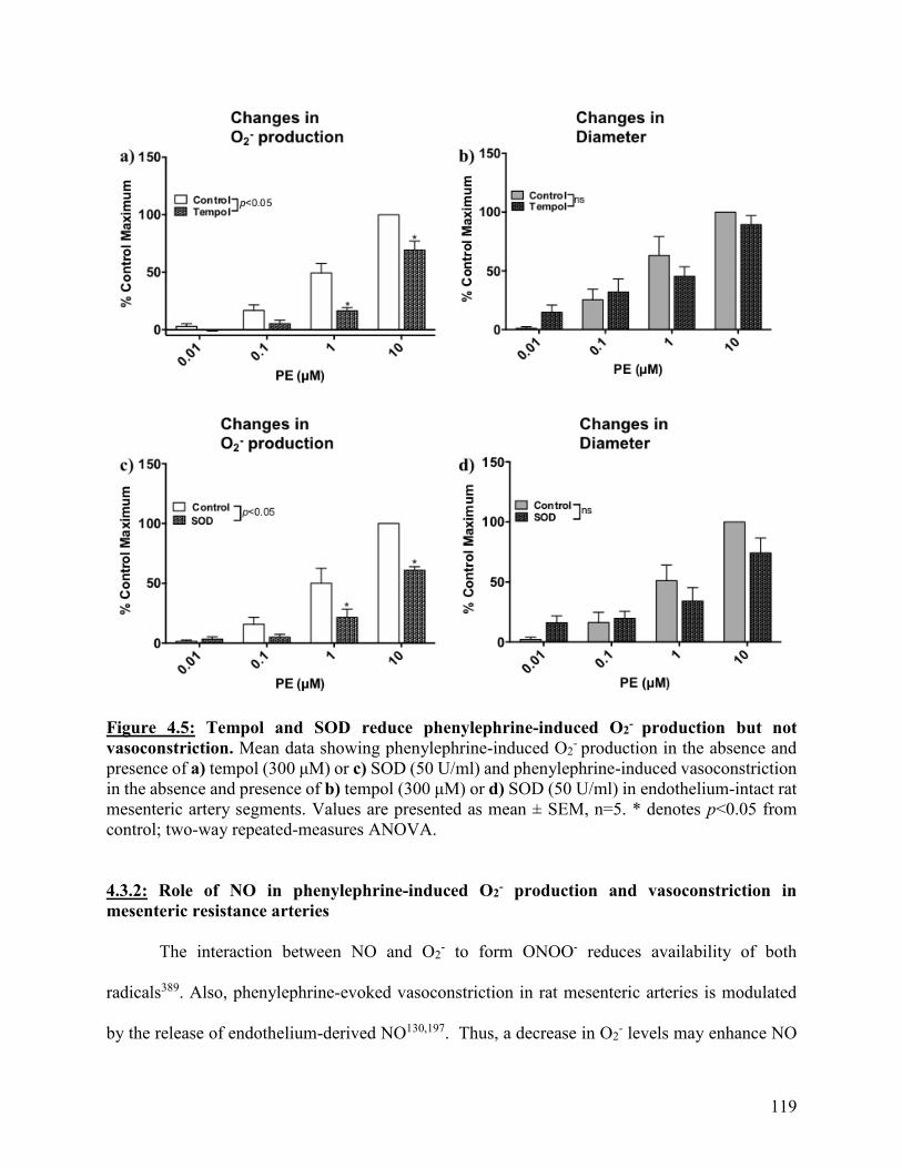
119
Figure 4.5: Tempol and SOD reduce phenylephrine-induced O2- production but not
vasoconstriction. Mean data showing phenylephrine-induced O2- production in the absence and
presence of a) tempol (300 μM) or c) SOD (50 U/ml) and phenylephrine-induced vasoconstriction
in the absence and presence of b) tempol (300 μM) or d) SOD (50 U/ml) in endothelium-intact rat
mesenteric artery segments. Values are presented as mean ± SEM, n=5. * denotes p<0.05 from
control; two-way repeated-measures ANOVA.
4.3.2: Role of NO in phenylephrine-induced O2- production and vasoconstriction in
mesenteric resistance arteries
The interaction between NO and O2- to form ONOO- reduces availability of both
radicals389. Also, phenylephrine-evoked vasoconstriction in rat mesenteric arteries is modulated
by the release of endothelium-derived NO130,197. Thus, a decrease in O2- levels may enhance NO
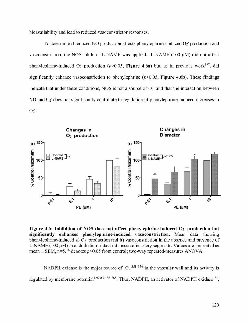
120
bioavailability and lead to reduced vasoconstrictor responses.
To determine if reduced NO production affects phenylephrine-induced O2- production and
vasoconstriction, the NOS inhibitor L-NAME was applied. L-NAME (100 μM) did not affect
phenylephrine-induced O2- production (p>0.05, Figure 4.6a) but, as in previous work197, did
significantly enhance vasoconstriction to phenylephrine (p<0.05, Figure 4.6b). These findings
indicate that under these conditions, NOS is not a source of O2- and that the interaction between
NO and O2- does not significantly contribute to regulation of phenylephrine-induced increases in
O2-.
Figure 4.6: Inhibition of NOS does not affect phenylephrine-induced O2- production but
significantly enhances phenylephrine-induced vasoconstriction. Mean data showing
phenylephrine-induced a) O2- production and b) vasoconstriction in the absence and presence of
L-NAME (100 μM) in endothelium-intact rat mesenteric artery segments. Values are presented as
mean ± SEM, n=5. * denotes p<0.05 from control; two-way repeated-measures ANOVA.
NADPH oxidase is the major source of O2-351–356 in the vascular wall and its activity is
regulated by membrane potential126,367,386–388. Thus, NADPH, an activator of NADPH oxidase384,
121
was applied to determine if phenylephrine-evoked increases in O2- production could be enhanced.
Unexpectedly, NADPH (100 μM) significantly reduced both phenylephrine-induced O2-
production (p<0.05; Figure 4.7a) and vasoconstriction (p<0.05, Figure 4.7b). A possible reason
for this finding is that NADPH is also a co-factor for NOS545, and NADPH has been shown to
stimulate NO production384. Thus, to determine if the effects of NADPH on phenylephrine-
stimulated O2- production and vasoconstriction may be due to increased production of NO, the
effect of NADPH was also examined in the presence of the NOS inhibitor, L-NAME.
As with NADPH alone, in the presence of NADPH and L-NAME (100 μM),
phenylephrine-induced O2- production was significantly reduced (p<0.05, Figure 4.7c). However,
whereas L-NAME alone enhanced phenylephrine-induced vasoconstriction and NADPH reduced
it, in the presence of L-NAME and NADPH together, vasoconstriction was not different to controls
(p>0.05, Figure 4.7d). These data indicate that NO does not appear to be involved in the NADPH-
evoked reductions in O2- but may underlie the reduction in phenylephrine-induced vasoconstriction
caused by this agent.
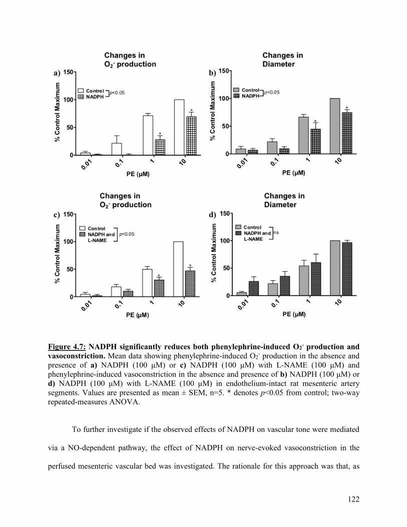
122
Figure 4.7: NADPH significantly reduces both phenylephrine-induced O2- production and
vasoconstriction. Mean data showing phenylephrine-induced O2- production in the absence and
presence of a) NADPH (100 μM) or c) NADPH (100 μM) with L-NAME (100 μM) and
phenylephrine-induced vasoconstriction in the absence and presence of b) NADPH (100 μM) or
d) NADPH (100 μM) with L-NAME (100 μM) in endothelium-intact rat mesenteric artery
segments. Values are presented as mean ± SEM, n=5. * denotes p<0.05 from control; two-way
repeated-measures ANOVA.
To further investigate if the observed effects of NADPH on vascular tone were mediated
via a NO-dependent pathway, the effect of NADPH on nerve-evoked vasoconstriction in the
perfused mesenteric vascular bed was investigated. The rationale for this approach was that, as

123
shown in Chapter 2, shear stress-induced inhibition of sympathetic vasoconstriction in the rat
perfused mesenteric bed is mediated through the release of endothelium-derived NO. As shown in
Chapter 2, L-NAME (100 μM) significantly enhanced nerve-evoked vasoconstriction at
stimulation frequencies of 20 to 40 Hz (p<0.05, Figure 4.8). NADPH (100 M) did not
significantly affect nerve-evoked vasoconstriction (p>0.05, Figure 4.8) but in the presence of both
L-NAME and NADPH, nerve-evoked responses were significantly reduced compared to L-NAME
alone (p<0.05, Figure 4.8). This observation supports the notion that the actions of NADPH on
arterial diameter may be mediated through increased bioavailability of NO.
Figure 4.8: Enhancement of nerve-evoked vasoconstriction caused by L-NAME is attenuated
by NADPH. Mean frequency-response relationships obtained from endothelium-intact perfused
mesenteric beds in the absence and presence of NADPH (100 μM) without and with L-NAME
(100 μM) and L-NAME (100 μM) alone. Values are presented as mean ± SEM, n=5. * denotes
p<0.05 from control, # denotes p<0.05 from NADPH (100 μM) and ^ denotes p<0.05 from
NADPH (100 μM) with L-NAME (100 μM); two-way repeated-measures ANOVA.

124
NADPH is also a rate limiting co-factor for glutathione reductase408,411,415–417, the enzyme
responsible for catalyzing the generation of glutathione from glutathione disulfide408,411.
Glutathione is essential for regulation of ROS levels within cells; it scavenges free radicals, such
as O2-, and through its thiol moiety reduces RNS, such as ONOO-407,408,410. Thus, NADPH may
increase the activity of glutathione reductase to enhance glutathione levels and subsequently,
reduce O2- . To examine this hypothesis carmustine, a glutathione reductase inhibitor, was used.
Carmustine (50 μM) significantly enhanced basal O2- production over the course of its 30-
minute incubation (p<0.05, Figure 4.9) in comparison to control, indicating that the glutathione
pathway may actively regulate O2- levels in endothelium-intact rat mesenteric arteries. In the
absence of carmustine, basal O2- production decreased, denoted by negative fluorescence intensity
values. Thus, for these experiments, fluorescence intensity was measured before application of
carmustine and after 30 minutes, and the difference was plotted to determine change in basal O2-
production.
NADPH (100 μM) caused a significant reduction of basal O2- production (p<0.05, Figure
4.9). In the presence of carmustine and NADPH together, basal O2- was significantly greater than
in the presence of NADPH alone (p<0.05, Figure 4.9) and so it is possible that the observed effects
of NADPH on phenylephrine-induced O2- production may be due to activation of glutathione
reductase.
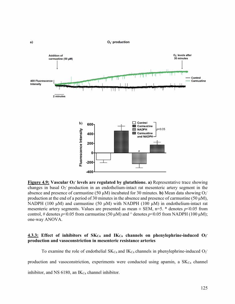
125
Figure 4.9: Vascular O2- levels are regulated by glutathione. a) Representative trace showing
changes in basal O2- production in an endothelium-intact rat mesenteric artery segment in the
absence and presence of carmustine (50 μM) incubated for 30 minutes. b) Mean data showing O2-
production at the end of a period of 30 minutes in the absence and presence of carmustine (50 μM),
NADPH (100 μM) and carmustine (50 μM) with NADPH (100 μM) in endothelium-intact rat
mesenteric artery segments. Values are presented as mean ± SEM, n=5. * denotes p<0.05 from
control, # denotes p<0.05 from carmustine (50 μM) and ̂ denotes p<0.05 from NADPH (100 μM);
one-way ANOVA.
4.3.3: Effect of inhibitors of SKCa and IKCa channels on phenylephrine-induced O2-
production and vasoconstriction in mesenteric resistance arteries
To examine the role of endothelial SKCa and IKCa channels in phenylephrine-induced O2-
production and vasoconstriction, experiments were conducted using apamin, a SKCa channel
inhibitor, and NS 6180, an IKCa channel inhibitor.

126
Apamin (500 nM) did not affect either phenylephrine-induced O2- production or
vasoconstriction (p>0.05, Figure 4.10a and b). In contrast, NS 6180 (1 μM) significantly
decreased phenylephrine-induced O2- production (p<0.05, Figure 4.10c) and significantly
increased phenylephrine-induced vasoconstriction (p<0.05, Figure 4.10d). These effects of
apamin and NS 6180 on phenylephrine-evoked vasoconstriction are in line with our previous work
showing that IKCa but not SKCa channels mediate myoendothelial feedback to limit phenylephrine-
evoked vasoconstriction in isolated mesenteric arteries197. The reason for the apparent inhibition
of phenylephrine-evoked O2- production in the presence of NS6180 is unclear. However, during
the 15 minute pre-incubation period, NS 6180 significantly enhanced basal O2- production in
comparison to control (p<0.05, Figure 4.11) in line with its ability to evoke endothelial
depolarization116,131.

127
Figure 4.10: Inhibition of IKCa but not SKCa channels significantly reduces phenylephrine-
induced O2- production and enhances phenylephrine-induced vasoconstriction. Mean data
showing phenylephrine-induced O2- production in the absence and presence of a) apamin (500 nM)
or c) NS 6180 (1 μM) and phenylephrine-induced vasoconstriction in the absence and presence of
b) apamin (500 nM) or d) NS 6180 (1 μM) in endothelium-intact rat mesenteric artery segments.
Values are presented as mean ± SEM, n=5. * denotes p<0.05 from control; two-way repeated-
measures ANOVA.
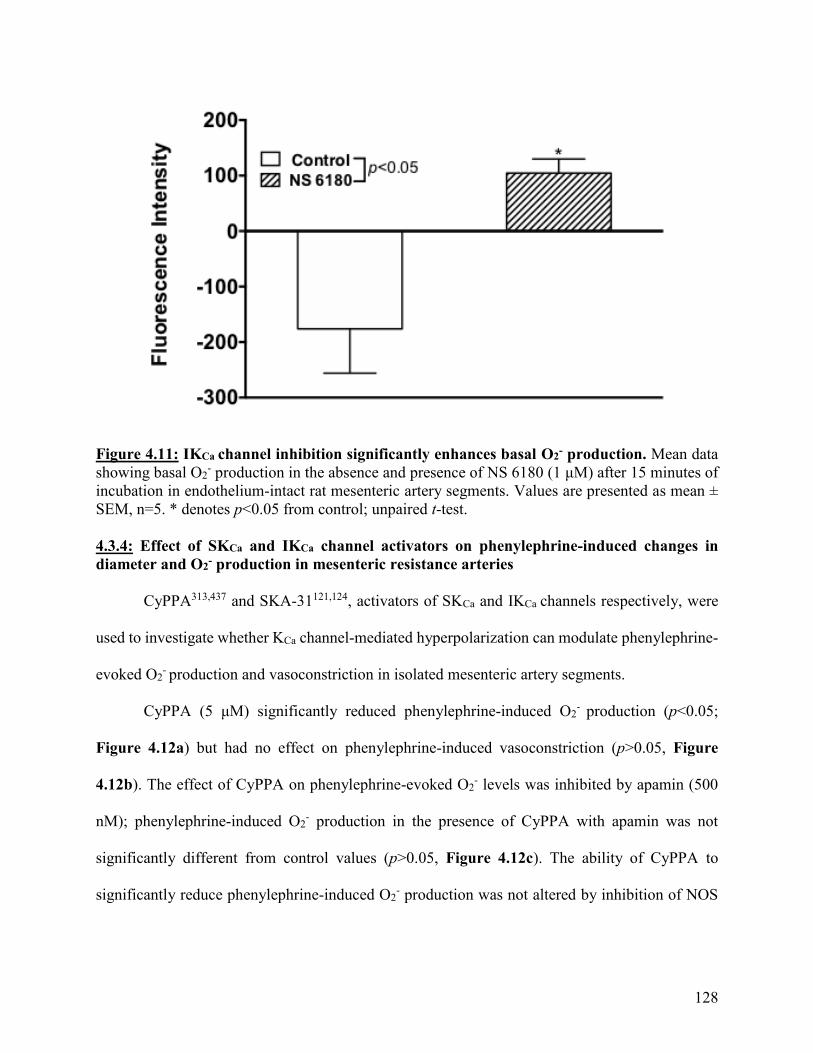
128
Figure 4.11: IKCa channel inhibition significantly enhances basal O2- production. Mean data
showing basal O2- production in the absence and presence of NS 6180 (1 μM) after 15 minutes of
incubation in endothelium-intact rat mesenteric artery segments. Values are presented as mean ±
SEM, n=5. * denotes p<0.05 from control; unpaired t-test.
4.3.4: Effect of SKCa and IKCa channel activators on phenylephrine-induced changes in
diameter and O2- production in mesenteric resistance arteries
CyPPA313,437 and SKA-31121,124, activators of SKCa and IKCa channels respectively, were
used to investigate whether KCa channel-mediated hyperpolarization can modulate phenylephrine-
evoked O2- production and vasoconstriction in isolated mesenteric artery segments.
CyPPA (5 μM) significantly reduced phenylephrine-induced O2- production (p<0.05;
Figure 4.12a) but had no effect on phenylephrine-induced vasoconstriction (p>0.05, Figure
4.12b). The effect of CyPPA on phenylephrine-evoked O2- levels was inhibited by apamin (500
nM); phenylephrine-induced O2- production in the presence of CyPPA with apamin was not
significantly different from control values (p>0.05, Figure 4.12c). The ability of CyPPA to
significantly reduce phenylephrine-induced O2- production was not altered by inhibition of NOS
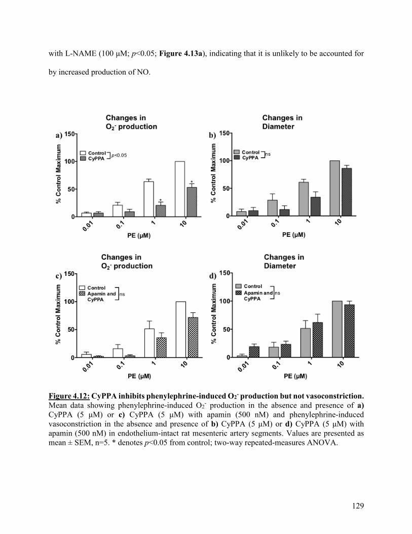
129
with L-NAME (100 μM; p<0.05; Figure 4.13a), indicating that it is unlikely to be accounted for
by increased production of NO.
Figure 4.12: CyPPA inhibits phenylephrine-induced O2- production but not vasoconstriction.
Mean data showing phenylephrine-induced O2- production in the absence and presence of a)
CyPPA (5 μM) or c) CyPPA (5 μM) with apamin (500 nM) and phenylephrine-induced
vasoconstriction in the absence and presence of b) CyPPA (5 μM) or d) CyPPA (5 μM) with
apamin (500 nM) in endothelium-intact rat mesenteric artery segments. Values are presented as
mean ± SEM, n=5. * denotes p<0.05 from control; two-way repeated-measures ANOVA.

130
Figure 4.13: CyPPA inhibition of phenylephrine-induced O2- production is not prevented by
NOS inhibition. Mean data showing phenylephrine-induced a) O2- production and b)
vasoconstriction in the absence and presence of CyPPA (5 μM) with L-NAME (100 μM) in
endothelium-intact rat mesenteric artery segments. Values are presented as mean ± SEM, n=6. *
denotes p<0.05 from control; two-way repeated-measures ANOVA.
SKA-31 (1 M) significantly reduced both phenylephrine-induced O2- production (p<0.05)
and vasoconstriction (p<0.05; Figure 4.14a and b). The IKCa channel inhibitor NS 6180 (1 M),
prevented the effect of SKA-31 (1 M) on phenylephrine-evoked vasoconstriction but did not
block its effects on phenylephrine-induced O2- production (p<0.05, Figure 4.14c and d) suggesting
this effect may be independent of IKCa channels. To investigate whether the effect was independent
of the endothelium, the experiments were repeated in denuded mesenteric arteries.
In endothelium-denuded arteries, SKA-31 (1 M) significantly reduced phenylephrine-
induced O2- levels (p<0.05, Figure 4.15a) but had no effect on phenylephrine-induced
vasoconstriction (p>0.05, Figure 4.15b). As our lab has previously demonstrated that IKCa
channels are present only in endothelial cells of rat mesenteric arteries197, the decrease in
phenylephrine-induced O2- production in endothelium-denuded arteries caused by SKA-31 is
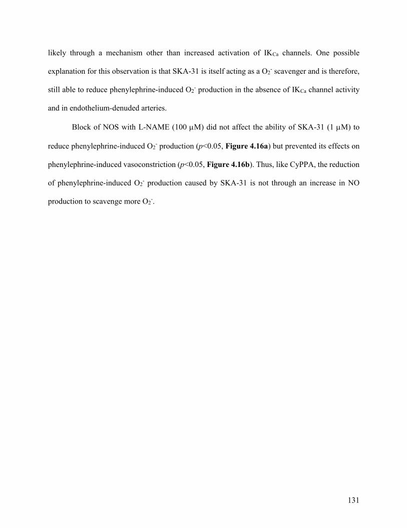
131
likely through a mechanism other than increased activation of IKCa channels. One possible
explanation for this observation is that SKA-31 is itself acting as a O2- scavenger and is therefore,
still able to reduce phenylephrine-induced O2- production in the absence of IKCa channel activity
and in endothelium-denuded arteries.
Block of NOS with L-NAME (100 M) did not affect the ability of SKA-31 (1 M) to
reduce phenylephrine-induced O2- production (p<0.05, Figure 4.16a) but prevented its effects on
phenylephrine-induced vasoconstriction (p<0.05, Figure 4.16b). Thus, like CyPPA, the reduction
of phenylephrine-induced O2- production caused by SKA-31 is not through an increase in NO
production to scavenge more O2-.
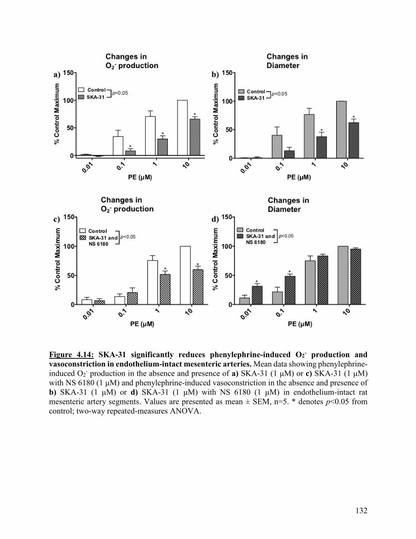
132
Figure 4.14: SKA-31 significantly reduces phenylephrine-induced O2- production and
vasoconstriction in endothelium-intact mesenteric arteries. Mean data showing phenylephrine-
induced O2- production in the absence and presence of a) SKA-31 (1 μM) or c) SKA-31 (1 μM)
with NS 6180 (1 μM) and phenylephrine-induced vasoconstriction in the absence and presence of
b) SKA-31 (1 μM) or d) SKA-31 (1 μM) with NS 6180 (1 μM) in endothelium-intact rat
mesenteric artery segments. Values are presented as mean ± SEM, n=5. * denotes p<0.05 from
control; two-way repeated-measures ANOVA.
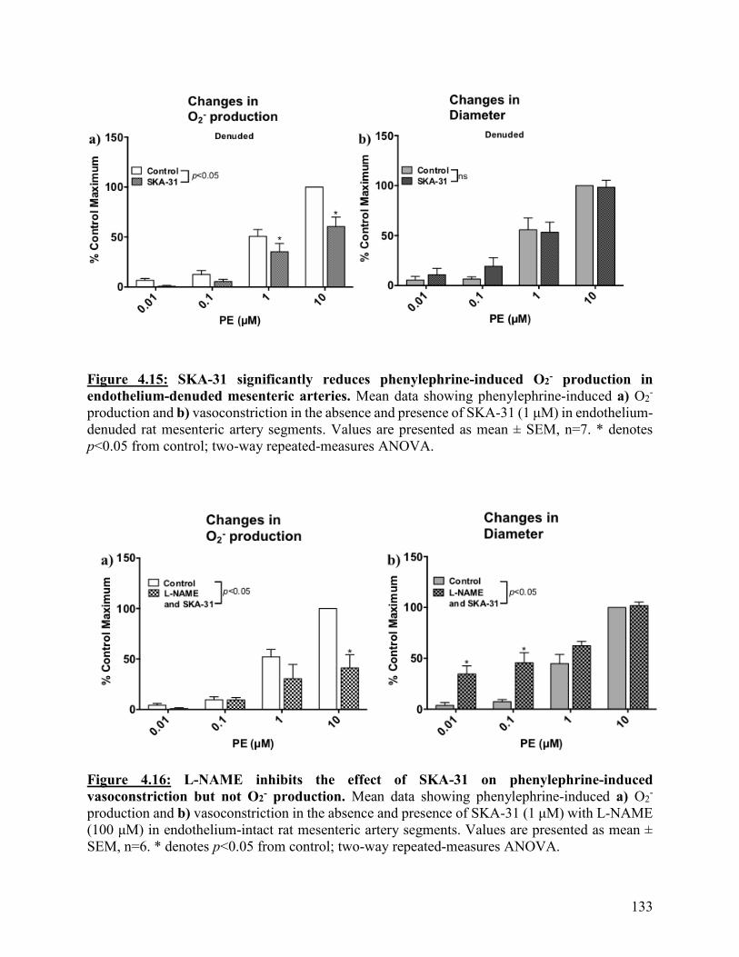
133
Figure 4.15: SKA-31 significantly reduces phenylephrine-induced O2- production in
endothelium-denuded mesenteric arteries. Mean data showing phenylephrine-induced a) O2-
production and b) vasoconstriction in the absence and presence of SKA-31 (1 μM) in endothelium-
denuded rat mesenteric artery segments. Values are presented as mean ± SEM, n=7. * denotes
p<0.05 from control; two-way repeated-measures ANOVA.
Figure 4.16: L-NAME inhibits the effect of SKA-31 on phenylephrine-induced
vasoconstriction but not O2- production. Mean data showing phenylephrine-induced a) O2
-
production and b) vasoconstriction in the absence and presence of SKA-31 (1 μM) with L-NAME
(100 μM) in endothelium-intact rat mesenteric artery segments. Values are presented as mean ±
SEM, n=6. * denotes p<0.05 from control; two-way repeated-measures ANOVA.

134
4.3.5: Effect of modulators of smooth muscle BKCa channels on phenylephrine-induced
changes in O2- production and vasoconstriction in mesenteric resistance arteries
To investigate if the observed effects of activators of endothelial SKCa and IKCa channel on
O2- production are unique to those channels, phenylephrine-induced increases in O2
- production
and vasoconstriction were examined in the presence of iberiotoxin (IbTX) and N'-[3,5-
Bis(trifluoromethyl)phenyl]-N-[4-bromo-2-(2H-tetrazol-5-yl-phenyl]thiourea (NS 11021), a
selective inhibitor and activator of smooth muscle BKCa channels, respectively.
Depolarization of the smooth muscle membrane potential to phenylephrine197,492 is limited
by opening of BKCa channels, an effect which can be blocked by IbTX94,97,99. In line with this,
IbTX (100 nM) significantly enhanced phenylephrine-induced vasoconstriction (p<0.05, Figure
4.17b) but had no effect on phenylephrine-induced O2- production (p>0.05, Figure 4.17a).
Conversely, the BKCa channel opener NS 11021 (100 nM), significantly reduced phenylephrine-
induced O2- production (p<0.05; Figure 4.18a) but had no effect on phenylephrine-induced
vasoconstriction (p>0.05, Figure 4.18b). The effect of NS 11021 on phenylephrine-evoked O2-
was prevented by IbTX (p>0.05, Figure 4.18c), and unlike in the presence of IbTX alone,
phenylephrine-induced vasoconstriction was not different to controls in the presence of both agents
(p>0.05, Figure 4.18d), indicating that NS11021 was able to prevent the actions of IbTX. These
findings support the proposal that membrane hyperpolarization may regulate O2- production in
mesenteric vessels and this is not an effect limited to opening of endothelial SKCa and IKCa
channels.
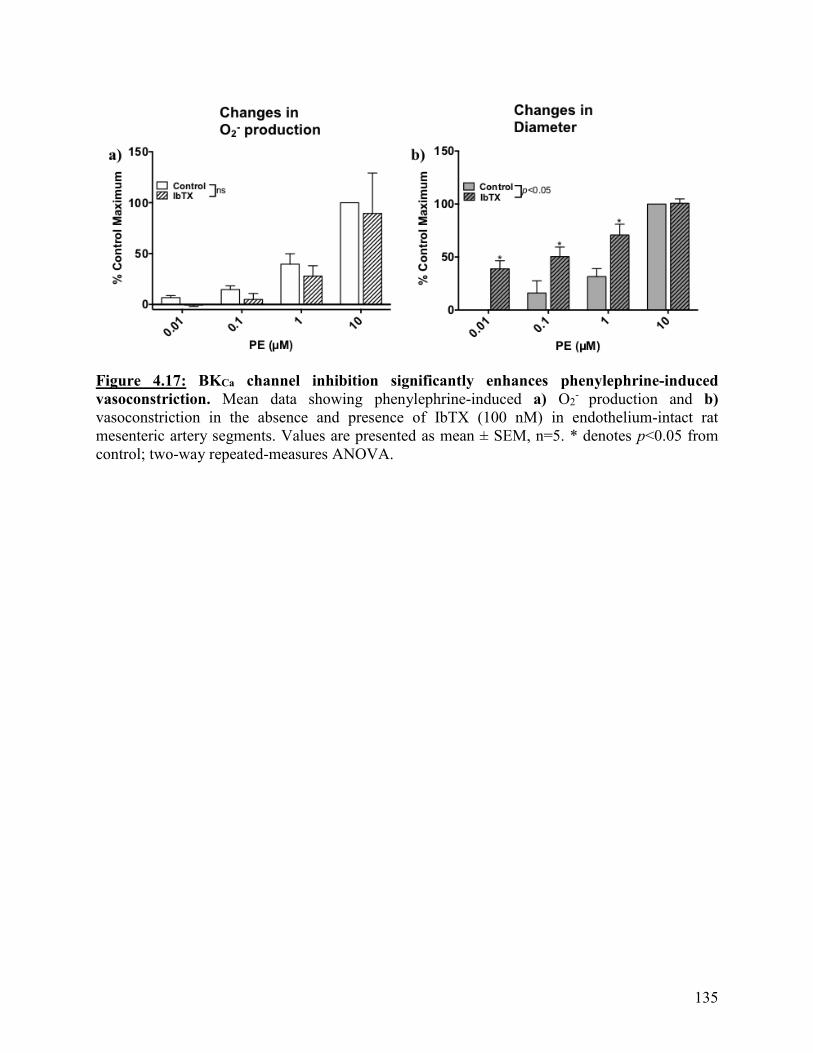
135
Figure 4.17: BKCa channel inhibition significantly enhances phenylephrine-induced
vasoconstriction. Mean data showing phenylephrine-induced a) O2- production and b)
vasoconstriction in the absence and presence of IbTX (100 nM) in endothelium-intact rat
mesenteric artery segments. Values are presented as mean ± SEM, n=5. * denotes p<0.05 from
control; two-way repeated-measures ANOVA.
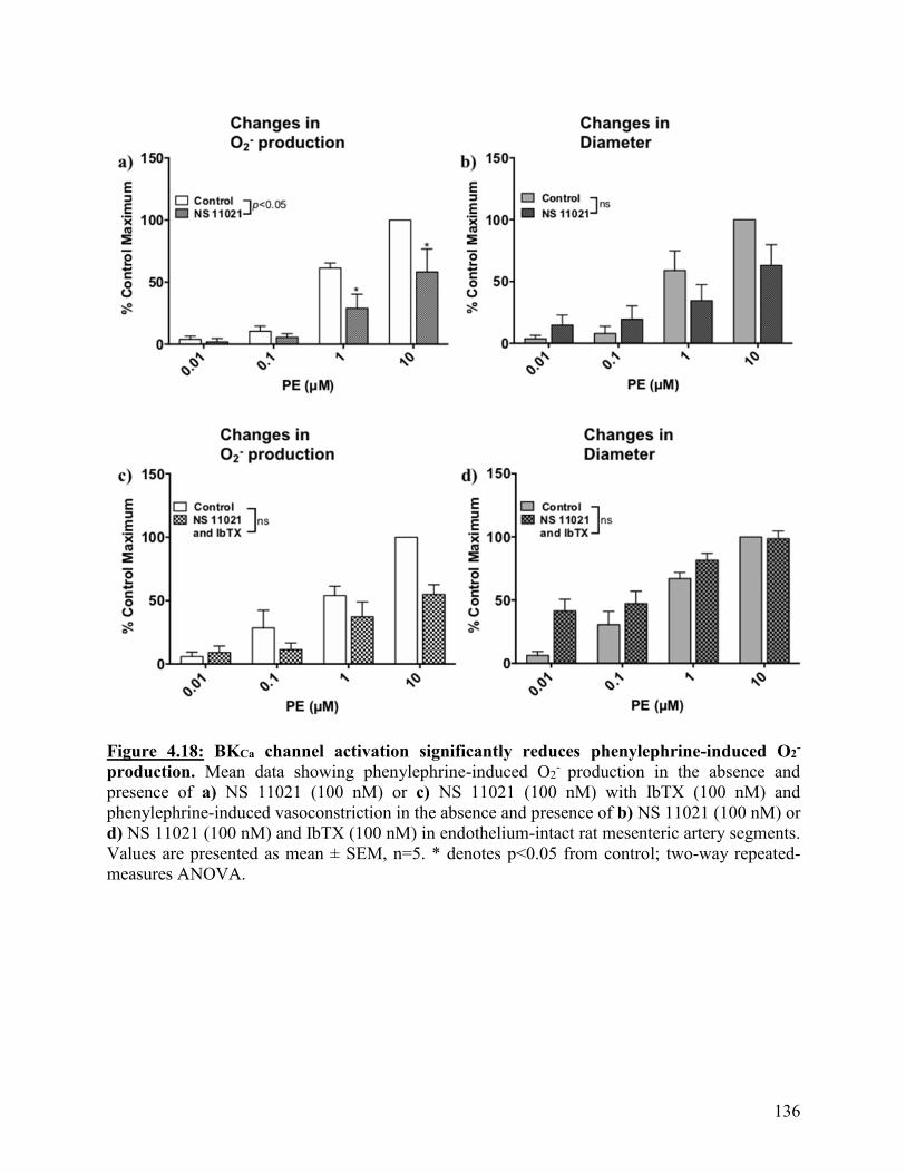
136
Figure 4.18: BKCa channel activation significantly reduces phenylephrine-induced O2-
production. Mean data showing phenylephrine-induced O2- production in the absence and
presence of a) NS 11021 (100 nM) or c) NS 11021 (100 nM) with IbTX (100 nM) and
phenylephrine-induced vasoconstriction in the absence and presence of b) NS 11021 (100 nM) or
d) NS 11021 (100 nM) and IbTX (100 nM) in endothelium-intact rat mesenteric artery segments.
Values are presented as mean ± SEM, n=5. * denotes p<0.05 from control; two-way repeated-
measures ANOVA.
137
4.4: Discussion
NO bioavailability within the vascular wall is determined by the balance between
production by endothelial NOS and its interaction with O2-. Enhanced production of O2
- leading to
decreased bioavailability of endothelium-derived NO518 appears to be a common mechanism
underlying endothelial dysfunction associated with a wide range of risk factors for cardiovascular
diseases, such as diabetes and atherosclerosis268,328–336,546. The data from the experiments described
in this chapter demonstrate that small molecule activators of endothelial SKCa and IKCa channels
can attenuate phenylephrine-induced O2- production in intact arteries. These data support my
hypothesis that pharmacological activators of endothelial KCa channels can reduce vascular O2-
production. It also supports the concept of endothelial cell membrane potential being a key
determinant of vascular function and that activators of endothelial KCa channels may provide a
novel therapeutic approach to reduce O2- in pathological states associated with endothelial
dysfunction.
It is established that α1-adrenoceptor-mediated vasoconstriction involves O2- production by
both NADPH oxidase347,348 and mitochondria349 in rat mesenteric arteries349, coronary myocytes347
and tail arteries348. However, these data were obtained from experimental protocols in which O2-
production was measured in flaccid tissues or fixed/frozen arteries, and so changes in O2- levels
were not recorded in real time and arterial diameter was not assessed. Using arteries mounted in a
pressure myograph in combination with a microscope fitted with a fluorescence attachment, I have
demonstrated, for the first time, simultaneous phenylephrine-induced O2- production and
vasoconstriction in isolated mesenteric arteries.
Using this approach, I have shown that phenylephrine evokes both O2- production and
vasoconstriction in a concentration-dependent manner, that is reproducible over the time course of

138
the experiments and fully accounted for by activation of α1-adrenoceptors. Tempol and SOD,
scavengers of O2-, each significantly reduced phenylephrine-evoked increases in O2
- but did not
affect the accompanying vasoconstriction. These results are in contrast to a previous study in which
α1-adrenergic vasoconstriction in rat mesenteric arteries was proposed to be dependent on O2-
generation and increased activity of matrix metalloproteinase enzymes349. However, in those
experiments, scavenging of O2- production did not alter the peak phenylephrine-induced
vasoconstriction though it did reduce the duration349.
My experiments were conducted in endothelium-intact tissues as it was anticipated that
scavenging of O2- by tempol or SOD would depress phenylephrine-induced vasoconstriction due
to increased bioavailability of NO. The lack of effect of SOD and tempol on phenylephrine-evoked
vasoconstriction could reflect the fact that although these agents did reduce O2- production, this
may not be sufficient to significantly impact bioavailability of endothelium-derived NO and thus,
arterial diameter. However, our lab has shown that endothelial modulation of phenylephrine-
evoked vasoconstriction in rat isolated mesenteric arteries is due to both IKCa channel-mediated
endothelium-dependent hyperpolarization and NO130,197. Thus, the lack of effect of a reduction in
O2- levels on phenylephrine-induced vasoconstriction in these vessels may reflect the predominant
role of activation of IKCa channels in endothelial modulation of smooth muscle contraction.
Alternatively, phenylephrine-induced O2- production enhances contractility via Ca2+
sensitization66,67, through increasing RhoA activation and subsequently, phosphorylation of
myosin phosphatase target subunit-1 and C-kinase potentiated protein phosphate-1 inhibitor; with
these changes being observed after incubation with vasoconstrictors for 5 to 15 minutes348,547,548.
As the phenylephrine dose response curve experiments in this thesis were 3 to 5 minutes in length,
it is possible that the reduction in O2- production did not correlate to a diminished vasoconstriction
139
due to minimal involvement of Ca2+ sensitization as a result of the shorter experimental time frame.
Thus, experiments where phenylephrine doses are incubated for longer periods of time will need
to be carried out in order to examine the possibility that the duration of the phenylephrine-induced
vasoconstriction is reduced in the presence of O2- scavengers.
In contrast to SOD and tempol, apocynin did reduce both phenylephrine-evoked increases
in O2- and vasoconstriction. The reason for this is unclear but in addition to acting as a scavenger
of O2-544, apocynin is also an inhibitor of NADPH oxidase541–543 and can scavenge H2O2
544, which
has been shown to act as a vasoconstrictor397–400 in isolated rat gracilis muscle arterioles397 and
mesenteric arteries399. Thus, scavenging of H2O2 by apocynin could contribute to the observed
attenuation of phenylephrine-induced vasoconstriction observed with apocynin. Together, these
data showed that there may be a complicated relationship between O2- levels and changes in arterial
diameter such that significant reductions in O2- levels do not directly correlate to alterations in
vasoconstrictor responses.
The interaction between NO and O2- to form ONOO- reduces availability of both
radicals389. Also, phenylephrine-evoked vasoconstriction in rat mesenteric arteries is modulated
by the endothelium, in part by the release of endothelium-derived NO130,197. Thus, the NOS
inhibitor L-NAME was used to determine if reduced NO production can affect phenylephrine-
induced O2- production and vasoconstriction in mesenteric arteries. It was expected that inhibition
of NO synthesis would enhance O2- levels. But, although L-NAME significantly potentiated
phenylephrine-induced vasoconstriction, it had no effect on O2- production. Thus, under my
experimental conditions, NO does play a role in endothelial modulation of smooth muscle
contractility but the interaction between NO and O2- may not contribute to regulation of O2
- levels.

140
Further support for a separation between the effects of NO on smooth muscle contractility
and on O2- levels is provided by experiments with NADPH. NADPH oxidase enzymes are one of
the main sources of O2- production in the vasculature and I initially utilized NADPH in
experiments with the idea that as an activator of NADPH oxidase351–353,384,385, it would enhance
phenylephrine-evoked increases in O2- levels in intact arteries. However, NADPH significantly
reduced both phenylephrine-induced O2- production and vasoconstriction. NADPH is also a co-
factor for NOS activity384,545 and in the presence of L-NAME, the ability of NADPH to
significantly reduce phenylephrine-induced O2- production was maintained but its effect on
phenylephrine-induced vasoconstriction was lost. Similarly, in the perfused mesenteric bed,
NADPH significantly reduced the enhancement of nerve-evoked vasoconstriction caused by block
of NOS. Thus, it appears that although NADPH can enhance NO-mediated inhibition of smooth
muscle contractility, this is independent of its ability to reduce O2- production.
As described in Chapter 1, NADPH is also a co-factor for glutathione reductase408,411,415–
417, an enzyme responsible for the reduction of glutathione disulfide to glutathione 408,411.
Glutathione is an antioxidant408,411 and the major antioxidant mechanism within endothelial
cells412. Glutathione is oxidized through interactions with ROS, such as ONOO-, OH-, O2- and
H2O2, and two oxidized glutathione molecules form glutathione disulfide 408,411, which is converted
back into two glutathione molecules through the actions of glutathione reductase. The balance
between glutathione and glutathione disulfide is a determinant of the redox status of a cell79.
Thus, to investigate the role of glutathione reductase in the effects of NADPH on
phenylephrine-evoked O2- production and vasoconstriction, I used the glutathione reductase
inhibitor, carmustine408,411,549,550. Interestingly, when applied alone, carmustine significantly
enhanced basal O2- production, indicating that the glutathione pathway plays an active role in
141
regulating O2- in isolated rat mesenteric arteries. This the first description of such a role in intact
arteries but is supported by a previous study in cultured bovine pulmonary artery endothelial
cells551. Although NADPH had no effect on basal O2- production, the combination of NADPH and
carmustine did significantly increase O2- levels, indicating NADPH may be mediating its effects
on phenylephrine-induced O2- production by increasing the activity of glutathione reductase. This
action would raise glutathione levels to increase the capability for scavenging of O2-.
As described earlier, NADPH oxidase, a major source of O2- in the vascular wall, is a
voltage-sensitive enzyme which generates O2- by transferring electrons from cytosolic NADPH to
extracellular O2126,367,368,371,372,374,386–388,529. As previous work has shown that the activity of
NADPH oxidase can be enhanced by membrane depolarization, it was anticipated that inhibition
of endothelial IKCa channels would enhance both phenylephrine-induced O2- production and
vasoconstriction in isolated arteries. IKCa channels mediate myoendothelial feedback, the
mechanism by which endothelial cells limit agonist-evoked smooth muscle contraction, and our
lab has shown that block of these channels causes both endothelial membrane potential
depolarization and enhancement of phenylephrine-evoked smooth muscle membrane potential
depolarization and constriction130,197. In this study, inhibition of IKCa channels by NS 6180 did
indeed significantly enhance phenylephrine-induced vasoconstriction but unexpectedly, it reduced
phenylephrine-induced O2- production. The reason for this is unclear but during the incubation
period, prior to addition of phenylephrine, NS 6180 did cause a significant increase in production
of O2-, supporting the notion that depolarization of the endothelial cell membrane potential by
inhibition of IKCa channels increases basal O2- production in isolated arteries. In contrast, apamin,
a selective inhibitor of SKCa channels, had no effect on either phenylephrine-induced O2-
production or vasoconstriction. This is in agreement with previous reports from our lab and others
142
that SKCa channels do not play a functional role in regulating endothelial membrane potential or
diameter in these vessels; block of SKCa channels by apamin does not cause endothelial
depolarization483 and does not enhance vasoconstriction130.
To test the hypothesis that small molecule openers of SKCa and IKCa channels can limit
phenylephrine-induced O2- production, I utilized the SKCa channel activator CyPPA313,437, and IKCa
channel activator SKA-31121,124. Both CyPPA and SKA-31 significantly reduced phenylephrine-
induced O2- production, but whereas SKA-31 significantly limited phenylephrine-induced
vasoconstriction, CyPPA had no effect, again demonstrating a dissociation between changes in O2-
levels and arterial diameter. The ability of CyPPA and SKA-31 to reduce O2- levels supports my
hypothesis, as well as previous reports, that hyperpolarization of the endothelial membrane
potential can lead to a decrease in vascular O2- production126,367,386–388.
Evidence that NADPH oxidase activity is regulated by membrane potential via the co-
factor Rac1367 has come from studies of rat aortic endothelial cells maintained under ischemic
conditions387 and human umbilical endothelial cells under normal conditions367. In these cells,
membrane hyperpolarization reduced O2- production388 and depolarization stimulated O2
-
production386. Additionally, in the rat perfused mesenteric bed, inhibition of SKCa and/or IKCa
channels, led to enhanced O2- production via NADPH oxidase and increased perfusion pressure
due to a reduction in NO levels126. However, my data provides the first evidence that activation of
both SKCa and IKCa channels decreases O2- production in isolated resistance arteries. Block of NOS
with L-NAME did not affect the ability of CyPPA or SKA-31 to limit phenylephrine-induced O2-
production but abolished the effect of SKA-31 on phenylephrine-induced vasoconstriction, again,
indicating a disconnect between O2- levels and the effects of NO on smooth muscle contractility.
143
The ability of CyPPA to reduce phenylephrine-evoked O2- production was prevented by
the SKCa channel inhibitor apamin, supporting the hypothesis that it is modulated by endothelial
membrane potential. However, the reduction in O2- production observed with SKA-31 was not
prevented by NS6180, an inhibitor of IKCa channels, despite the fact that the effect of SKA-31 on
vasoconstriction was inhibited by this agent. This finding may indicate that whereas the ability of
CyPPA to limit O2- production is linked to SKCa channel activity, SKA-31 may be able to reduce
O2- levels independently of IKCa channels. My observations that SKA-31 was able to also reduce
phenylephrine-evoked increases in O2- levels but not arterial diameter in endothelium-denuded
arteries supports this proposal, since these channels are not located on vascular smooth muscle
cells197. The possibly that SKA-31 can directly scavenge O2- requires further investigation.
In Chapters 2 and 3, I showed that both SKA-31 and CyPPA can inhibit phenylephrine-
evoked increase in tone in mesenteric arteries mounted under isometric conditions. However, in
arteries mounted under constant pressure, SKA-31 significantly inhibits phenylephrine-evoked
vasoconstriction but CyPPA does not. The reason for this difference is unclear. Previous studies
have described differences in agonist-evoked responses between arteries mounted in the two
systems but not in terms of endothelial modulation552,553. As described earlier, SKCa and IKCa
channels do show differential localization within endothelial cells118,122,125,130,151,197. CyPPA313,437
and SKA-31121,124 act to increase the sensitivity of the channels to Ca2+ and so differences in the
ability of these agents to modulate phenylephrine-evoked vasoconstriction may reflect differences
in endothelial Ca2+ signalling under the two experimental conditions. In support of this proposal,
intraluminal pressure has been shown to influence the frequency of endothelial Ca2+ events linked
to activation of IKCa but not SKCa channels in rat cremaster arterioles554.
144
SKCa and IKCa channels are localized on endothelial cells and opening of these channels
leads to membrane hyperpolarization which can spread to surrounding smooth muscle cells.
Therefore, to investigate whether changes in smooth muscle membrane potential can regulate O2-
production in intact arteries, I utilized a selective inhibitor and activator of BKCa channels, which
are located on the vascular smooth muscle but not endothelial cells76,106–108. Inhibition of BKCa
channels with IbTX significantly enhanced phenylephrine-induced vasoconstriction but did not
affect phenylephrine-induced O2- production. This effect on vasoconstriction was anticipated, as
BKCa channel activity plays a crucial role in limiting vasoconstriction27,29,76,79–92,106,107. The lack of
effect of IbTX on O2- production was unexpected. Phenylephrine-evoked vasoconstriction of rat
mesenteric arteries is due, at least in part, to depolarization of the smooth muscle cell membrane
potential leading to entry of Ca2+ through L-type VOCCs197,492 and this depolarization may be
responsible for the accompanying increase in O2- production as opening of BKCa channels with NS
11021 did reduce phenylephrine-induced O2- production. Although the role of smooth muscle
depolarization in vasoconstriction to 1-adrenoceptor agonists is well established118,123,555,
previous studies have not considered its role in acute phenylephrine-evoked production of O2-348,349
and further work is required to investigate this possibility.
The application of DHE for real-time measurements of O2- production in intact arteries is
novel but does have some limitations. DHE can be oxidized by other ROS/RNS or by cytochrome
c to produce ethidium, which emits at a similar wavelength as EOH bound to DNA (DHE oxidized
by O2-)349,535–540. EOH has an optimal excitation/emission spectrum of 490/590 nm540 while
ethidium has an optimal excitation/emission spectrum of 360/590 nm556. The filters used in this
recording system excite at 535 nm and capture emission at 610 nm, giving minimal excitation of
ethidium and so it is unlikely that production of ethidium is being measured under these

145
experimental conditions. In comparison to other fluorescent probes for O2- detection, such as
lucigenin, DHE does not produce O2- directly538,539. Additionally, although DHE fluorescence
cannot be used alone to quantify O2-, it is very useful for measuring changes in O2
-
production538,557, as it is used for in this chapter. In light of these limitations, two further techniques
(see Chapter 5) have been used to provide support for the use of DHE: histological analysis of
fixed arteries and UPLC of DHE-derived oxidation products (the latter regarded as “the most
unequivocal and quantitative detection of intracellular O2-”538).
Furthermore, it is possible that the reduction in fluorescence intensity observed in the
absence of altered constriction could be the result of dye quenching by the pharmacological
reagents thus, further experiments will need to be performed in order to investigate this possibility.
And finally, while there was a disconnect between changes in O2- and changes in vasoconstriction
(such as, L-NAME significantly enhancing phenylephrine-induced vasoconstriction but not O2-
production), it is possible that movement of the vessel as a result of constriction could be
contributing to the changes in fluorescence intensity. Therefore, further experiments will be
performed to examine this possibility.
In conclusion, the data included in this chapter support my hypothesis that activators of
KCa channels can significantly reduce phenylephrine-induced O2- production, this was not linked
to changes in endothelial modulation of vasoconstriction in mesenteric arteries. These data also
support the proposal that endothelial KCa channels may be a potential novel target for the
prevention and treatment of cardiovascular diseases associated with increased production of O2-
268,328–336.
146
Chapter 5: Activators of SKCa and IKCa channels limit O2- production in isolated
arteries
5.1: Introduction
In Chapter 4, I used DHE for real time measurements of changes in O2- levels in intact
mesenteric arteries. This is in contrast to previous studies in which vascular O2- measurements
have been determined in fixed/frozen tissues347–349,558–560.
Recent studies have shown that the reaction product of DHE and O2- is EOH and not
ethidium, as had been previously thought535. Ethidium is formed from the reaction of DHE with
ROS other than O2-, RNS and cytochrome c, and EOH does not arise from the action of other
intracellular oxidants535. Thus, quantitation of EOH can be considered as being tantamount to
detecting the presence of O2- itself539.
In this chapter, I conducted histological analysis of fixed arteries stained with DHE, to
provide support for the functional data collected in the previous chapter. I also carried out UPLC
analysis of the DHE oxidation products ethidium and EOH to demonstrate that DHE fluorescence
provides a measure of O2- levels in the vascular wall rather than other ROS/ RNS.
5.2: Methods and materials
See Appendix: Drugs and chemicals for a list of the drugs and chemicals used.
5.2.1: Histological analysis of mesenteric arteries stained with DHE
5.2.1.1: Tissue preparation. Vessels were prepared for histological analysis by following the
methods outlined by Hao et al349. Briefly, sections of second or third order mesenteric artery (4-5
mm in length) were cleaned of adhering connective tissue and individually placed in 1 ml of Krebs
buffer in a 1.5 ml tube (Eppendorf, Germany). Stock solutions of DHE (10 mM) were made fresh
in a darkened room349,535,536 and all protocol steps described were also carried out in a darkened
room. Each vessel was incubated with DHE (10 M) for 30 minutes at 37oC during which time
147
one of the following agents was added 10 minutes prior to the end of the DHE incubation period:
SOD-PEG (25 U/ml), CyPPA (5 μM) or SKA-31 (1 μM). In each experiment, one tube was
incubated with DHE alone to act as a control. At the end of the incubation period with DHE,
phenylephrine (10 μM) was added to all tubes and incubated at 37oC for 45 minutes. Tissues were
then washed in cold Krebs buffer and individually placed upright in small plastic containers full
of optimal cutting temperature compound (Scientific Gardena), flash frozen in liquid nitrogen and
placed in a -80°C freezer until cryo-sectioned onto microscope slides (by Ms Lynette Edler,
HistoCore, Alberta Diabetes Institute, University of Alberta). The slides were stored in a -20°C
freezer until stained with 1 in 1000 dilution of 4′,6-Diamidino-2-phenylindole dihydrochloride
(DAPI) in 90% glycerol and cover slips (22x60 mm, thickness #1, Fisher Scientific, USA) placed
on the slides. Slides were then stored flat in containers at 4°C.
5.2.1.2: Imaging. Histological analysis was carried out using an upright digital imaging
microscope (Zeiss Imager Z1, power supply Zeiss 231) attached to a fluorescent lamp (X-cite 120
LED boost, Excelitas Technologies) and camera (Cooke Sensi Cam). For visualization of DAPI,
samples were excited at wavelengths between 353-377 nm and emission was captured at 395 nm
and above. For visualization of EOH bound to DNA349,535,536 (the DHE oxidation product formed
when DHE reacts with O2-), excitation was at 450-490 nm and emission was captured at 500-550
nm.
Metamorph (Universal Imaging, Downington, PA) was used for analysis of images; the
DAPI fluorescent images were over-laid with the EOH fluorescent images and the EOH
fluorescence within each nucleus was calculated, totalled and averaged per image. The average
fluorescent intensity per image was then compared between treatment groups. Expressing EOH
fluorescent intensity per nucleus normalized the responses to allow comparison between different
148
vessels and minimized interference from the internal elastic lamina. Adobe Photoshop (Adobe,
San Jose, CA, USA) was used to add colour to the example images.
5.2.2: Quantification of DHE-derived oxidation products from aortic samples by UPLC
Due to the small size of mesenteric arteries, UPLC was used to quantify DHE-derived
oxidation products349,535,536,540,561 in aortic tissue by following the UPLC protocol for quantitation
of EOH described by Lebed et.al.562 Aortic tissues were prepared for analysis by UPLC by
following the protocol described by Fernandes et.al.561 The aorta was cleaned of connective tissue
and fat and placed in iced cold phosphate buffered saline. The thoracic aorta was cut into 8-12
pieces and 2-3 pieces placed into each pre-weighed tube and weighed to determine tissue weight.
Tissues were washed twice with ice cold phosphate buffered saline before 0.5 ml of phosphate
buffered saline /diethylenetriaminepentaacetic acid was added to each tube. 2.5 μl of DHE, from
a freshly made 10 mM DHE stock solution, was added to each tube in a darkened room. SOD-
PEG (25 U/ml), CyPPA (5 μM) or SKA-31 (1 μM) was added 15 minutes prior to the addition of
DHE and tubes were then incubated for 30 minutes at 37oC. After incubation, and still in the dark,
vessels were washed twice with ice cold phosphate buffered saline before being flash frozen in a
mortar full of liquid nitrogen and ground with a pestle. The ground vessel powder was transferred
from the mortar into new tubes with 0.5 ml of acetonitrile and wrapped in aluminum foil.
The tubes containing ground vessel and acetonitrile were sonicated for 3 x 10 seconds in a
darkened room and then centrifuged at 4°C 18,000 x g for 15 minutes. The supernatant was
removed, placed into separate tubes and wrapped in aluminum foil. The supernatant was
evaporated off using a stream of N2 until a pink pellet formed. All tubes were then wrapped in
aluminum foil and stored in a -80°C freezer until the pink pellets were prepared for UPLC analysis
(by Mr. Ken Strynadka, UPLC Analytical Core, Cardiovascular Research Centre).

149
The pink pellets were re-suspended in 100 μl phosphate buffered
saline/diethylenetriaminepentaacetic acid and 5 μl of each sample was injected into a Waters
Acquity UPLC System (H Class) consisting of a binary solvent manager, sample manager, column
manager and fluorescence detector. The column used was a Zorbax SB-Phenyl column (250 mm,
4.6 mm, 5 m) equilibrated with a mobile phase of 35% acetonitrile and 65% water, both
containing a 0.1% (v/v) of trifluoroacetic acid. Ethidium and EOH were separated using a gradient
from 35% to 55% in 5 min at a flow rate of 2 mlmin-1. The fluorescence excitation was set at 470
nm and the emission was measured at 595 nm. The limit of quantitation for ethidium and EOH
using this method was 0.4 M, close to previously reported values562.
All data were acquired and analyzed by means of Waters Empower 3 software. 2-EOH was
purchased from Noxygen Science Transfer and Diagnostic GmBbH (Germany). All chemicals and
solvents were of analytical grade. All solutions were prepared in ultrapure milliQ water (Millipore
MilliQ, Germany) and filtered over a 0.22 m filter (Millipore, Bedford, USA). Analysis was done
with the operator blinded to sample identity. Data was expressed as the ratio of area under the
curve per mg of initial sample weight (AUC/mg) as in previous studies562.
5.2.3: Statistics
All data are expressed as mean ± SEM, n rats used and ordinary one-way ANOVA followed
by a Tukey’s multiple comparison post-hoc test was performed. p<0.05 was considered
statistically significant in all cases.
150
5.3: Results
5.3.1: Histological analysis of mesenteric arteries stained with DHE
Representative images of DAPI and DHE stained sections of rat mesenteric arteries treated
with phenylephrine (10 μM) plus CyPPA (5 μM), SKA-31 (1 μM) or SOD (25 U/ml) are shown
in Figure 5.1.
CyPPA (5 μM), SKA-31 (1 μM) and SOD-PEG (25 U/ml) each significantly decreased
DHE fluorescence intensity in comparison to control tissues incubated with phenylephrine (10
μM) (p<0.05, Figure 5.2).
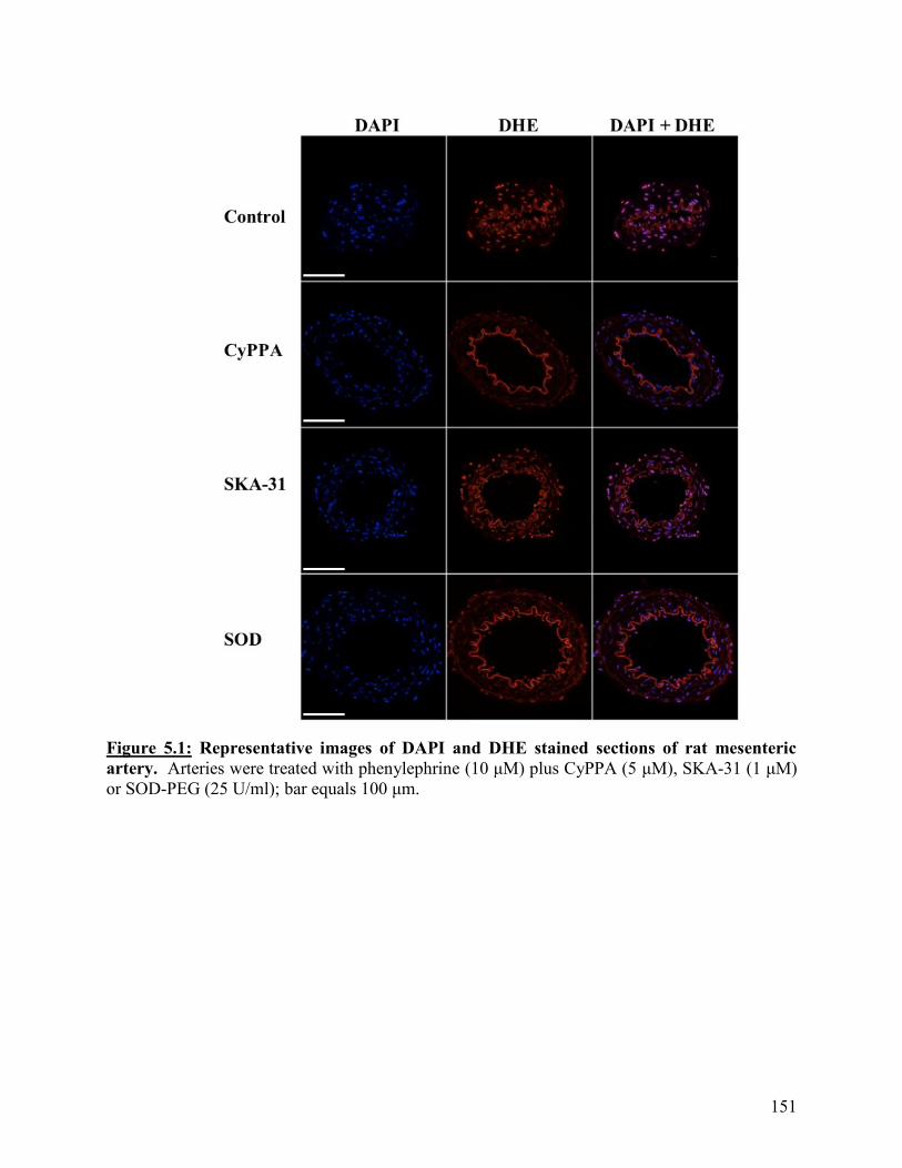
151
Figure 5.1: Representative images of DAPI and DHE stained sections of rat mesenteric
artery. Arteries were treated with phenylephrine (10 μM) plus CyPPA (5 μM), SKA-31 (1 μM)
or SOD-PEG (25 U/ml); bar equals 100 μm.

152
Figure 5.2: CyPPA, SKA-31 and SOD each significantly reduce phenylephrine-induced O2-
levels in rat mesenteric arteries as measured by DHE fluorescence. Mean data showing the
phenylephrine (10 μM) -induced O2- levels in the absence and presence of CyPPA (5 μM), SKA-
31 (1 μM) or SOD (25 U/ml). Values are presented as a mean ± SEM, n=5. * denotes p<0.05 from
control; one-way ANOVA.
5.3.2: Quantification of DHE-derived oxidation products from aortic samples by UPLC
CyPPA (5 μM), SKA-31 (1 μM) and SOD (25 U/ml) each significantly reduced EOH levels
in isolated aortic samples as compared to controls (p<0.05, Figure 5.3a) but did not significantly
affect levels of ethidium (p>0.05, Figure 5.3b)
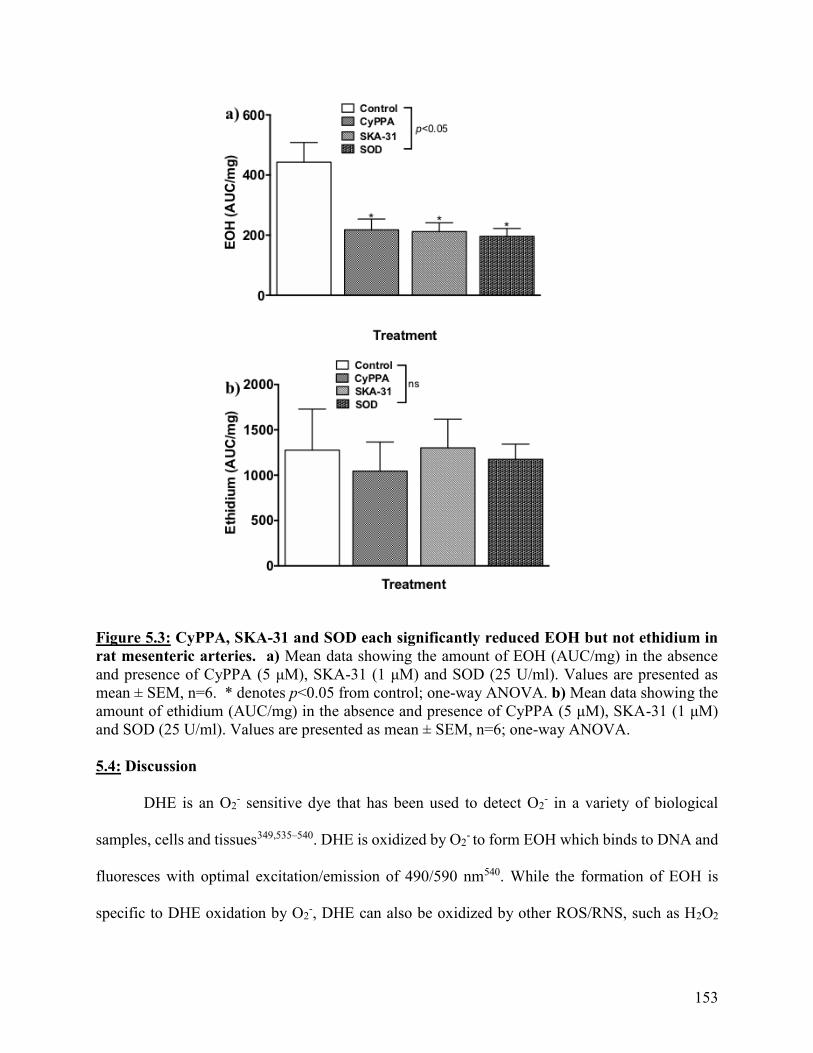
153
Figure 5.3: CyPPA, SKA-31 and SOD each significantly reduced EOH but not ethidium in
rat mesenteric arteries. a) Mean data showing the amount of EOH (AUC/mg) in the absence
and presence of CyPPA (5 μM), SKA-31 (1 μM) and SOD (25 U/ml). Values are presented as
mean ± SEM, n=6. * denotes p<0.05 from control; one-way ANOVA. b) Mean data showing the
amount of ethidium (AUC/mg) in the absence and presence of CyPPA (5 μM), SKA-31 (1 μM)
and SOD (25 U/ml). Values are presented as mean ± SEM, n=6; one-way ANOVA.
5.4: Discussion
DHE is an O2- sensitive dye that has been used to detect O2
- in a variety of biological
samples, cells and tissues349,535–540. DHE is oxidized by O2- to form EOH which binds to DNA and
fluoresces with optimal excitation/emission of 490/590 nm540. While the formation of EOH is
specific to DHE oxidation by O2-, DHE can also be oxidized by other ROS/RNS, such as H2O2
154
and ONOO-, and cytochrome c, to form ethidium349,535–540. Ethidium has an optimal
excitation/emission spectrum of 360/590 nm556 thus, there is overlap between the
excitation/emission spectrums for both DHE-derived oxidation products. However, through the
use of specific filters to selectively excite/emit for EOH, DHE can be used as a means to selectively
detect O2- in tissues538,540,562. I performed histological analysis on fixed mesenteric arteries stained
with DHE to provide support for the functional data collected in the previous chapter. I also carried
out UPLC analysis of the DHE oxidation products ethidium and EOH to demonstrate that DHE
fluorescence provides a measure of O2- levels in the vascular wall rather than other ROS/ RNS.
Histological analysis of fixed mesenteric arteries stained with DHE in the absence and
presence of the phenylephrine plus the SKCa channel activator CyPPA313,437, the IKCa channel
activator SKA-31121,124 and SOD, demonstrated that these agents significantly reduced DHE
fluorescence. The microscope used for this analysis permitted excitation/emission in the narrow
ranges of 450-490/500-550 nm, optimal for detection of EOH540 over ethidium (optimal excitation
of 360 nm556). Thus, it is likely that the bulk of the fluorescent intensity captured was due to EOH.
By using UPLC analysis of DHE-derived oxidation products, I have demonstrated that
CyPPA, SKA-31 and SOD each significantly reduced the amount of EOH levels in aortic samples
without altering levels of ethidium. Ethidium is formed by oxidation of DHE by other ROS/RNS,
such as ONOO- and H2O2, and cytochrome c349,535–540. Thus, these findings give confidence that
with the use of DHE in functional studies, I am measuring changes in EOH, and not ethidium,
thus, reflecting changes in the production of O2-.
The transient lifetime of O2- means that there is little likelihood that any molecular probe
will react with the total amount of O2- generated. This unavoidable underestimation implies that
measurement of O2- levels through the use of DHE and quantitation of EOH can be viewed only

155
as, at best, a semi-quantitative method for assessing O2- concentrations. Also, as in Chapter 4, the
source of O2- was not investigated in these experiments. My hypothesis is that membrane potential
hyperpolarization may reduce O2- production and the data supports this proposal. However, KCa
channels have been identified on the inner mitochondrial membrane in cardiac myocytes137,138
and neurons139–143,152–156 and the possibility that these channels are present in vascular smooth
muscle and endothelial cell mitochondria, and thus, play a role in O2- generation in the vascular
wall, cannot be ruled out.
In conclusion, activators of either SKCa or IKCa channels in rat mesenteric arteries or aorta
significantly reduced the levels of fluorescence intensity and EOH, but not ethidium, as determined
through the use of histological analysis and UPLC analysis of DHE-derived oxidation products,
respectively, in a similar manner to the O2- scavenger, SOD. These findings indicate that the use
of DHE fluorescence is an appropriate method to assess changes in O2- levels in arteries and
support the methodology used in the experiments described in Chapter 4.
156
Chapter 6: General discussion and future directions
6.1: General discussion
Maintenance of adequate blood supply to tissues and organs requires co-ordination of the
activity of nerves, endothelial and smooth muscle cells. Chemical mediators and changes in shear
stress act on the endothelium to release diffusible relaxing and contracting factors and evoke
electrical coupling with underlying smooth muscle cells. Conversely, stimulation of smooth
muscle cells by neurotransmitters and increases in intravascular pressure leads to flux of second
messengers to endothelial cells to elicit feedback to limit smooth muscle contraction. Endothelial
dysfunction is associated with the risk factors for, and development of cardiovascular diseases,
and is characterized by an increase in O2- production116,268,328–336. O2
- interacts with NO to produce
ONOO- at a rate three times faster than O2- undergoes dismutation by SOD389,390. Thus, increased
O2- is associated with both reduced availability of NO and increased formation of ONOO- leading
to enhanced vasoconstriction, augmented platelet adhesion and aggregation, vascular smooth
muscle cell proliferation and diminished angiogenesis298,328,406.
Endothelium-dependent modulation of smooth muscle contractility is initiated by a rise in
Ca2+ levels within endothelial cells leading to activation of NOS and opening of KCa
channels80,84,85,100,116–131,148–151,197,202–205. NOS converts L-arginine to NO213,258–262 which relaxes
smooth muscle cells via stimulation of soluble guanylyl cyclase to increase cyclic guanosine
monophosphate286 and activate protein kinase G-mediated phosphorylation of numerous target
proteins167,168,287,288. Opening of endothelial KCa channels causes hyperpolarization of the
endothelial cell membrane potential which spreads to surrounding smooth muscle cells to limit
contraction by reducing the open probability VOCCs80,84,85,100,116–131,148–151,197,202.
157
NO and spread of hyperpolarization have long being regarded as parallel pathways for
endothelium-dependent vasodilation and the gold-standard test for responses mediated by
hyperpolarization has been the demonstration that blockers of SKCa and IKCa channels inhibit
endothelium-dependent relaxation in the presence of a NOS inhibitor, such as L-
NAME80,84,85,100,116–131,148–151,197,202–205. However, two completely independent pathways for
relaxation is counter to effective signal integration required for effective fine tuning of changes in
arterial diameter in response to multiple stimuli, such as release of neurotransmitter from
sympathetic nerves and increases in shear stress due to vasoconstriction. Several lines of evidence
support a link between NO bioavailability and KCa channel activity. For example, in rat basilar and
superior mesenteric arteries, agonist-evoked NO production and NO-mediated relaxations can be
inhibited by blockers of endothelial KCa channels203,418 and small molecule activators of
endothelial KCa channels can evoke NO-mediated relaxation in rat mesenteric and porcine retinal
arteries437,481. Also, studies in cultured endothelial cells have shown that O2- production by voltage-
sensitive NADPH oxidase is reduced by membrane hyperpolarization387,529 which may lead to
increased bioavailability of NO.
Our lab and others have demonstrated that in small arteries, endothelial KCa channels show
a differential distribution within endothelial cells with SKCa channels located on the luminal
endothelial membrane and IKCa channels localized to MEGJs on the abluminal
side118,122,125,130,132,133,151,197. This distribution supports the proposal that SKCa and IKCa channels
may play different roles in endothelium-dependent modulation of arterial diameter. This proposal
is borne out by reports that in cultured endothelial cells, increases in shear stress are linked to
activation of SKCa channels206,318,421–424, and our demonstration that in rat isolated mesenteric and
basilar arteries, stimulation of smooth muscle cells by 1-adrenoceptor agonists leads to flux of
158
IP3 from smooth muscle to endothelial cells to elicit localized increases in Ca2+ that activates IKCa
channels located at MEGJs and production of NO to limit vasoconstriction, a mechanism termed
myoendothelial feedback197.
In this thesis, I have further explored the relationship between endothelial SKCa and IKCa
channels and NO in regulating resistance artery diameter. In vivo, sympathetic nerve activity is the
primary regulator of resistance artery diameter, and therefore, contributes to peripheral vascular
resistance434,435, with the endothelium playing a key role in limiting the vasoconstriction caused
by neurotransmitters released from perivascular sympathetic nerves. Vasoconstriction increases
shear stress, the frictional force exerted by flow of blood across the surface of endothelial cells,
and sensing of these increases in shear stress plays an important role in regulating tissue
perfusion198–201. However, despite its obvious physiological importance, the mechanisms
underlying shear stress-induced increases in arterial diameter and thus, blood flow, are still a topic
of debate. Data from in vitro, in vivo and clinical studies have demonstrated an important role for
NO in acute responses to increases in shear stress and also support the contribution of endothelial
hyperpolarization mediated by opening of SKCa channels132,133,206,220,241–248,318,421–433. However,
whether release of NO and activation of SKCa channels are distinct pathways or two facets of the
same mechanism is still unclear.
Therefore, in Chapter 2, I used the rat mesenteric bed perfused at a constant luminal flow
so that vasoconstriction leads to increases in shear stress, to explore the functional link between
NO and SKCa channel activity modulation of sympathetic vasoconstriction. I found that shear
stress-induced inhibition of nerve-evoked vasoconstriction is mediated by both NO and SKCa
channels. I also showed that CyPPA, a small molecule activator of SKCa channels that increases
the channels sensitivity to Ca2+ 313,437, can enhance the response to shear stress in this preparation
159
via a mechanism dependent on endothelium-derived NO. These findings support the notion that
activators of SKCa channels may have therapeutic potential in enhancing shear stress-induced NO
bioavailability in pathological states where there is a loss of shear stress-induced dilation420.
Furthermore, as this enhancement occurred at a concentration of CyPPA that caused minimal
direct vasorelaxation, activators of SKCa channels may provide a means to maintain coupling
between physiological stimuli and changes in blood flow and avoid reflex increases in heart rate
and blood pressure caused by direct vasodilators.
These experiments also revealed that voltage-independent mechanisms of smooth muscle
contraction are a major contributor to nerve-evoked vasoconstriction in the rat mesenteric bed.
Depolarization-mediated smooth muscle contraction is limited by opening of smooth muscle
voltage-gated K+ channels and BKCa channels, both of which hyperpolarize the membrane
potential26–29,76,448,449 to limit contraction. In contrast, contraction of smooth muscle by voltage-
independent processes does not engage a similar “braking” mechanism. Therefore, the reliance of
nerve-evoked vasoconstriction on voltage-independent processes may allow for a greater range of
response than would be possible if depolarization was the only active mechanism.
As described above, in contrast to SKCa channels, IKCa channels are localized to MEGJs
on the abluminal side of endothelial cells118,122,125,130,132,133,151,197, a position that allows them to
mediate myoendothelial feedback, the mechanisms by which agonists acting on smooth muscle
cells can activate inhibitory endothelial pathways to limit contraction. This model arose from
experiments utilizing the application of 1-adrenoceptor agonists but its contribution to endothelial
modulation of sympathetic nerve activity, a major stimulus for vasoconstriction in vivo, has not
been investigated. The reliance of myoendothelial feedback on IKCa channels, provides the
opportunity to use selective inhibitors of these channels to dissect out the contribution of the two

160
pathways to functional vascular responses and so in Chapter 3, I explored the role of the IKCa
channel-mediated myoendothelial feedback pathway in limiting sympathetic vasoconstriction in
the perfused mesenteric bed.
Using this approach, I found that in contrast to SKCa channels, endothelial IKCa channels
do not appear to play a functional role in modulating nerve-evoked vasoconstriction in the perfused
mesenteric bed. However, using SKA-31, an activator of IKCa channels, I identified a role for
neuronal IKCa channels in limiting vasoconstriction via inhibition of noradrenaline release. IKCa
channels have previously been localized on specific neurons in rat, mouse, guinea-pig and human
enteric nervous systems152–156 where they mediate the slow after-hyperpolarization following an
action potential but this is the first report of IKCa channels on perivascular sympathetic nerves. The
lack of effect of IKCa channel blockers on sympathetic vasoconstriction suggests that IKCa channels
may not play a role in regulating noradrenaline release under normal conditions but targeting of
IKCa channels could provide a new approach to reducing vasoconstriction in conditions associated
with increased sympathetic drive, such as hypertension174,499,500.
As described in Chapter 1, endothelial dysfunction associated with risk factors for
cardiovascular diseases is characterized by increased production of O2- and decreased NO
bioavailability268,328–336. Attempts to reduce O2- levels through use of dietary anti-oxidants such as
vitamins B, C and E have been unsuccessful337–342 and there is the need to identify new approaches
to reduce O2- generation. In disease models, reduced NO bioavailability has been associated with
upregulation of expression and activity of NADPH oxidase332–336,383,528, a voltage-sensitive
enzyme which generates O2- by transferring electrons from cytosolic NADPH to extracellular
O2126,367,368,371,372,374,386–388,529. In isolated endothelial cells, drugs which open KATP channels
attenuate both membrane depolarization and O2- production, indicating that endothelial cell
161
membrane potential can regulate O2- production387,529. Our lab has previously shown that
endothelial depolarization inhibits agonist-evoked, NO-mediated relaxation of rat basilar arteries,
an effect that was overcome by the KATP channel opener, pinacidil418, and we, and others, have
demonstrated depolarization of the endothelial membrane potential in tail530 and mesenteric
arteries531–533 from rat models of endothelial dysfunction. Therefore, in Chapter 4, I explored the
relationship between endothelial KCa channels, O2- production and diameter in intact mesenteric
arteries mounted in a pressure myograph coupled to a fluorescence detection system (IonOptix)
for simultaneous measurement of changes in arterial diameter and O2- production using DHE.
With this new approach, I found that small molecule activators of endothelial SKCa and
IKCa channels, as well as scavengers of O2-, can attenuate phenylephrine-induced O2
- production
in intact arteries but this is not associated with potentiation of NO-mediated inhibition of
vasoconstriction. I chose to use this bioassay approach to assess NO bioavailability as although it
is not quantitative, it has the advantage over commercially available NO-sensitive electrodes or
fluorescent dyes in that it is functionally and clinically relevant because it shows whether or not
changes in NO bioavailability are sufficient to affect NO-mediated modulation of vasoconstriction.
The observed disconnect between changes in levels of O2- and changes in arterial diameter in my
experiments may indicate that under the acute conditions of my experiments, the changes in O2-
are not sufficient to significantly impact availability of endothelium-derived NO to mediate
relaxation. However, it could also be a reflection of the predominant role of hyperpolarization in
endothelial modulation of smooth muscle contraction in small mesenteric arteries, masking the
impact of enhanced NO bioavailability on vasoconstriction. Thus, for future studies, the use of
larger arteries in which NO plays a more dominant role should be considered, together with
investigation of the effects of KCa channel activators on the longer-term consequences of increases
162
in O2- levels, such as damage to proteins caused by ONOO-.
Simultaneous recording of arterial diameter and changes O2- levels using DHE in isolated
arteries is a new approach. Thus, in Chapter 5, I conducted histological analysis of fixed arteries
stained with DHE and UPLC analysis of the DHE oxidation products, ethidium and EOH, to
demonstrate that DHE fluorescence provides a measure of O2- levels in the vascular wall rather
than other ROS/RNS. Using these approaches, I demonstrated that activators of KCa channels
significantly reduced the levels of fluorescence intensity, and of EOH, but not ethidium, in a
similar manner to the O2- scavenger, SOD. These findings confirm that the use of DHE
fluorescence is an appropriate method to assess changes in O2- levels in arteries and support the
methodology used in the experiments described in Chapter 4.
In conclusion, traditionally, release of NO and opening of KCa channels have been regarded
as distinct endothelium-dependent pathways for modulation of resistance artery diameter. This
thesis presents several lines of evidence to support the proposal that the integrated activity of KCa
channels, and NO provide a stimulus- and context -dependent mechanism to fine tune arterial
diameter and maintain appropriate levels of tissue perfusion. Together, these data support my
hypothesis that pharmacological activators of endothelial KCa channels can reduce vascular O2-
production and may provide a novel therapeutic approach to reducing O2- in pathological states
associated with endothelial dysfunction
6.2: Future directions
The data presented in this thesis opens up a number of potential avenues for future research
such as:
Exploration of the source of O2- regulated by openers of KCa channels. NADPH oxidase
is a voltage-sensitive enzyme and so its activity can be modulated by cell membrane potential. The
163
possibility that other sources of O2-, such as xanthine oxidase, may be modulated by changes in
membrane potential remains to be explored.
SKCa channels identified on the inner mitochondrial membrane in both neurons and cardiac
myocytes have been identified as a potential target for new approaches to the treatment of
conditions associated with increased production of O2-, such as cerebral and cardiac ischemia, as
well as endoplasmic reticulum stress and oxidative cell death137–143,152–156,563,564. Cell surface
expression of SKCa channels has been identified in endothelial cells, but whether they are present
on mitochondria on endothelial and/or smooth muscle cells has yet to be investigated.
Investigation of the ability of activators of SKCa and IKCa to reduce O2- availability in
models of endothelial dysfunction and transgenic animals. Our lab has shown that acute
exposure of isolated arteries from diabetic rats to KCa channel activators can enhance NO-mediated
endothelium-dependent relaxation. Application of the methodology to simultaneously measure
arterial diameter and changes in O2- levels to isolated arteries from animal models of disease, and
potentially patients, will allow for investigation of the ability of KCa channel activators to reduce
O2- production and reduce other deleterious consequences of O2
- generation (such as increased
formation of ONOO- producing detrimental effects on many proteins and lipids) in the setting of
disease.
The use of arteries from mice lacking endothelial SKCa and IKCa channels would allow for
investigation of the role of these channels in regulation of O2- levels and be useful in confirming
the selectivity of KCa channel activators.
164
References: 1. Grundy D. Principles and standards for reporting animal experiments. Exp Physiol.
2015;100(7):755-758.
2. WHO|Cardiovascular diseases (CVDs). WHO.
2017:http://www.who.int/cardiovascular_diseases/en/.
3. Schiffrin EL. The endothelium of resistance arteries: physiology and role in hypertension.
Prostaglandins, Leukot Essent Fat Acids. 1996;54(1):17-25.
4. Somlyo AP, Himpens B. Cell calcium and its regulation in smooth muscle. FASEB J.
1989;3(11):2266-2276.
5. Filo RS, Bohr DF, Ruegg JC. Glycerinated skeletal and smooth muscle: calcium and
magnesium dependence. Science. 1965;147(3665):1581-1583.
6. Allen BG, Walsh MP. The biochemical basis of the regulation of smooth-muscle
contraction. Trends Biochem Sci. 1994;19(9):362-368.
7. Ghosh D, Syed AU, Prada MP, Nystoriak MA, Santana LF, Nieves-Cintrón M, Navedo
MF. Calcium Channels in Vascular Smooth Muscle. Adv Pharmacol. 2017;78:49-87.
8. McDaniel SS, Platoshyn O, Wang J, Yu Y, Sweeney M, Krick S, Rubin LJ, Yuan JX.
Capacitative Ca(2+) entry in agonist-induced pulmonary vasoconstriction. Am J Physiol
Lung Cell Mol Physiol. 2001;280(5):L870-L870.
9. Kamm KE, Stull JT. Dedicated myosin light chain kinases with diverse cellular functions.
J Biol Chem. 2001;276(7):4527-4530.
10. Hasegawa Y, Morita F. Role of 17-kDa essential light chain isoforms of aorta smooth
muscle myosin. J Biochem. 1992;111(6):804-809.
11. Kim HR, Appel S, Vetterkind S, Gangopadhyay SS, Morgan KG. Smooth muscle
signalling pathways in health and disease. J Cell Mol Med. 2008;12(6a):2165-2180.
12. Saddouk FZ, Ginnan R, Singer HA. Ca2+/calmodulin-dependent protein kinase II in
vascular smooth muscle. Adv Pharmacol. 2017;78:171-202.
13. Walsh MP. The Ayerst Award Lecture 1990. Calcium-dependent mechanisms of
regulation of smooth muscle contraction. Biochem Cell Biol. 1991;69(12):771-800.
14. Kuo IY, Ehrlich BE. Signaling in Muscle Contraction. Cold Spring Harb Perspect Biol.
2015;7(2):a006023.
15. Brozovich F V., Nicholson CJ, Degen C V., Gao YZ, Aggarwal M, Morgan KG.
Mechanisms of vascular smooth muscle contraction and the basis for pharmacologic
treatment of smooth muscle disorders. Pharmacol Rev. 2016;68(2):476-532.
16. Kim J-H, Lee L-K, Lee W-D, Lee J-U, Kim -Young, Kim -Hyun, Choi B-R, Yang S-M,
Jeon H-J, Lee T-H, Kwak T-Y, Kim B, Kim J. A Review of Signal Transduction in
Mechanisms of Smooth Muscle Contraction and Its Relevance for Specialized Physical
Therapy. J Phys Ther Sci. 2013;25:129-141.
17. Tribe RM, Borin ML, Blaustein MP. Functionally and spatially distinct Ca2+ stores are
revealed in cultured vascular smooth muscle cells (sarcoplasmic
reticulum/thaplgarn/caffeine/fura-2/chlortetracycline). Physiology. 1994;91:5908-5912.
18. Bian J, Ghosh TK, Wang J-C, Gill DL. Identification of intracellular calcium pools
selective modification by thapsigargin. 1991;266(14):8801-8806.
19. Chueh SH, Gill DL. Inositol 1,4,5-trisphosphate and guanine nucleotides activate calcium
release from endoplasmic reticulum via distinct mechanisms. J Biol Chem.
1986;261(30):13883-13886.
20. Baró I, Eisner DA. The effects of thapsigargin on [Ca2+]i in isolated rat mesenteric artery
165
vascular smooth muscle cells. Pflogers Arch Eur J Physiol. 1992;420(1):115-117.
21. Knot HJ, Standen NB, Nelson MT. Ryanodine receptors regulate arterial diameter and
wall [Ca2+] in cerebral arteries of rat via Ca2+-dependent K+ channels. J Physiol.
1998;508(( Pt 1)):211-221.
22. Kamishima T, McCarron JG. Regulation of the cytosolic Ca2+ concentration by Ca2+
stores in single smooth muscle cells from rat cerebral arteries. J Physiol. 1997;501 ( Pt
3):497-508.
23. Marks AR. Calcium channels expressed in vascular smooth muscle. Circulation.
1992;86(6 Suppl):III61-III67.
24. Berridge MJ. Smooth muscle cell calcium activation mechanisms. J Physiol.
2008;586(21):5047-5061.
25. Amberg GC, Navedo MF. Calcium Dynamics in Vascular Smooth Muscle.
Microcirculation. 2013;20(4):281-289.
26. Nelson MT, Cheng H, Rubart M, Santana LF, Bonev AD, Knot HJ, Lederer WJ.
Relaxation of arterial smooth muscle by calcium sparks. Science. 1995;270(5236):633-
637.
27. Pérez GJ, Bonev AD, Nelson MT. Micromolar Ca(2+) from sparks activates Ca(2+)-
sensitive K(+) channels in rat cerebral artery smooth muscle. Am J Physiol Cell Physiol.
2001;281(6):C1769-C1775.
28. Lamont C, Wier WG. Different roles of ryanodine receptors and inositol (1,4,5)-
trisphosphate receptors in adrenergically stimulated contractions of small arteries. Am J
Physiol Heart Circ Physiol. 2004;287(2):H617-H625.
29. Krishnamoorthy G, Sonkusare SK, Heppner TJ, Nelson MT. Opposing roles of smooth
muscle BK channels and ryanodine receptors in the regulation of nerve-evoked
constriction of mesenteric resistance arteries. Am J Physiol Heart Circ Physiol.
2014;306(7):H981-H988.
30. Ji G, Feldman ME, Greene KS, Sorrentino V, Xin H-B, Kotlikoff MI. RYR2 proteins
contribute to the formation of Ca(2+) sparks in smooth muscle. J Gen Physiol.
2004;123(4):377-386.
31. Jaggar JH, Porter VA, Lederer WJ, Nelson MT. Calcium sparks in smooth muscle. Am J
Physiol Cell Physiol. 2000;278(2):C235-C256.
32. Jaggar JH. Intravascular pressure regulates local and global Ca(2+) signaling in cerebral
artery smooth muscle cells. Am J Physiol Cell Physiol. 2001;281(2):C439-C448.
33. Bonev AD, Jaggar JH, Rubart M, Nelson MT. Activators of protein kinase C decrease
Ca2+ spark frequency in smooth muscle cells from cerebral arteries. Am J Physiol.
1997;273(6 Pt 1):C2090-C2095.
34. Jaggar JH, Wellman GC, Heppner TJ, Porter VA, Perez GJ, Gollasch M, Kleppisch T,
Rubart M, Stevenson AS, Lederer WJ, Knot HJ, Bonev AD, Nelson MT. Ca2+ channels,
ryanodine receptors and Ca(2+)-activated K+ channels: a functional unit for regulating
arterial tone. Acta Physiol Scand. 1998;164(4):577-587.
35. Liou J, Kim ML, Heo W Do, Jones JT, Myers JW, Ferrell JE, Meyer T. STIM is a Ca2+
sensor essential for Ca2+-store-depletion-triggered Ca2+ influx. Curr Biol.
2005;15(13):1235-1241.
36. Trebak M. STIM/Orai signalling complexes in vascular smooth muscle. J Physiol.
2012;590(17):4201-4208.
37. Zeng W, Yuan JP, Kim MS, Choi YJ, Huang GN, Worley PF, Muallem S. STIM1 Gates
166
TRPC Channels, but Not Orai1, by Electrostatic Interaction. Mol Cell. 2008;32(3):439-
448.
38. Beech DJ. Integration of transient receptor potential canonical channels with lipids. Acta
Physiol. 2012;204(2):227-237.
39. Catterall WA. Voltage-Gated Calcium Channels. Cold Spring Harb Perspect Biol.
2011;3(8):a003947.
40. Tanabe T, Takeshima H, Mikami A, Flockerzi V, Takahashi H, Kangawa K, Kojima M,
Matsuo H, Hirose T, Numa S. Primary structure of the receptor for calcium channel
blockers from skeletal muscle. Nature. 1987;328(6128):313-318.
41. Gurnett CA, Waard M De, Campbell KP. Dual Function of the Voltage-Dependent Ca2+
Channel α2δ Subunit in Current Stimulation and Subunit Interaction. Neuron.
1996;16(2):431-440.
42. Jay SD, Ellis SB, McCue AF, Williams ME, Vedvick TS, Harpold MM, Campbell KP.
Primary structure of the gamma subunit of the DHP-sensitive calcium channel from
skeletal muscle. Science. 1990;248(4954):490-492.
43. Ruth P, Röhrkasten A, Biel M, Bosse E, Regulla S, Meyer HE FVH. Primary structure of
the beta subunit of the DHP-sensitive calcium channel from skeletal muscle. Science (80-
). 1989;245(4922):1115-1118.
44. Liu H, De Waard M, Scott VE, Gurnett CA, Lennon VA, Campbell KP. Identification of
three subunits of the high affinity omega-conotoxin MVIIC-sensitive Ca2+ channel. J Biol
Chem. 1996;271(23):13804-13810.
45. Letts VA, Frankel WN, Felix R, Biddlecome GH, Arikkath J, Mahaffey CL, Valenzuela
A, Bartlett FS, Mori Y, Campbell KP. The mouse stargazer gene encodes a neuronal
Ca2+-channel |[gamma]|subunit. Nat Genet. 1998;19(4):340-347.
46. Lacerda AE, Kim HS, Ruth P, Perez-Reyes E, Flockerzi V, Hofmann F, Birnbaumer L,
Brown AM. Normalization of current kinetics by interaction between the α1and β subunits
of the skeletal muscle dihydropyridine-sensitive Ca2+ channel. Nature.
1991;352(6335):527-530.
47. Catterall WA, Perez-Reyes E, Snutch TP, Striessnig J. International Union of
Pharmacology. XLVIII. Nomenclature and structure-function relationships of voltage-
gated calcium channels. Pharmacol Rev. 2005;57(4):411-425.
48. Benham CD, Hess P, Tsien RW. Two types of calcium channels in single smooth muscle
cells from rabbit ear artery studied with whole-cell and single-channel recordings. Circ
Res. 1987;61(4 Pt 2):I10-I16.
49. Loirand G, Pacaud P, Mironneau C, Mironneau J. Evidence for two distinct calcium
channels in rat vascular smooth muscle cells in short-term primary culture. Pfliigers Arch.
1986;407:566-568.
50. Bean BP, Sturek M, Puga A, Hermsmeyer K. Calcium channels in muscle cells isolated
from rat mesenteric arteries: modulation by dihydropyridine drugs. Circ Res.
1986;59(2):229-235.
51. Knot HJ, Nelson MT. Regulation of arterial diameter and wall [Ca2+] in cerebral arteries
of rat by membrane potential and intravascular pressure. J Physiol. 1998;508(Pt 1):199-
209.
52. Hwa JJ, Bevan JA. Stretch-dependent (myogenic) tone in rabbit ear resistance arteries. Am
J Physiol Circ Physiol. 1986;250(1):H87-H95.
53. Lee SHW · CO. Role of PKC in the effects of α1-adrenergic stimulation on Ca2+

167
transients, contraction and Ca2+ current in guinea-pig ventricular myocytes. Pflügers Arch
– Eur J Physiol. 1999;437:335–344.
54. Tseng GN, Boyden PA. Different effects of intracellular Ca and protein kinase C on
cardiac T and L Ca currents. Am J Physiol. 1991;261(2 Pt 2):H364-H379.
55. Taguchi K, Ueda M, Kubo T. Effects of cAMP and cGMP on L-Type Calcium Channel
Currents in Rat Mesenteric Artery Cells. J Pharmacol. 1997;74:179-186.
56. Ousterhout JM, Sperelakis N. Cyclic nucleotides depress action potentials in cultured
aortic smooth muscle cells. Eur J Pharmacol. 1987;144:7-14.
57. Delpy E, Coste H, Gouville AC. Effects of cyclic GMP elevation on isoprenaline-induced
increase in cyclic AMP and relaxation in rat aortic smooth muscle: role of
phosphodiesterase 3. Br J Pharmacol. 1996;119(3):471-478.
58. Bkaily G, Sperelakis N. Injection of guanosine 5’-cyclic monophosphate into heart cells
blocks calcium slow channels. Am J Physiol. 1985;248(5 Pt 2):H745-H749.
59. Wahler GM, Sperelakis N. Intracellular injection of cyclic GMP depresses cardiac slow
action potentials. J Cyclic Nucleotide Protein Phosphor Res. 1985;10(1):83-95.
60. Brueggemann LI, Martin BL, Barakat J, Byron KL, Cribbs LL. Low voltage-activated
calcium channels in vascular smooth muscle: T-type channels and AVP-stimulated
calcium spiking. Am J Physiol Heart Circ Physiol. 2005;288(2):H923-H935.
61. Harraz OF, Abd El-Rahman RR, Bigdely-Shamloo K, Wilson SM, Brett SE, Romero M,
Gonzales AL, Earley S, Vigmond EJ, Nygren A, Menon BK, Mufti RE, Watson T,
Starreveld Y, Furstenhaupt T, Muellerleile PR, Kurjiaka DT, Kyle BD, Braun AP, Welsh
DG. CaV3.2 Channels and the Induction of Negative Feedback in Cerebral Arteries. Circ
Res. 2014;115(7):650-661.
62. Tsien RW, Lipscombe D, Madison D V, Bley KR, Fox AP. Multiple types of neuronal
calcium channels and their selective modulation. Trends Neurosci. 1988;11(10):431-438.
63. Kuo IY, Ellis A, Seymour VAL, Sandow SL, Hill CE. Dihydropyridine-insensitive
calcium currents contribute to function of small cerebral arteries. J Cereb Blood Flow
Metab. 2010;30(6):1226-1239.
64. Abd El-Rahman RR, Harraz OF, Brett SE, Anfinogenova Y, Mufti RE, Goldman D,
Welsh DG. Identification of L- and T-type Ca2+ channels in rat cerebral arteries: role in
myogenic tone development. Am J Physiol Heart Circ Physiol. 2013;304(1):H58-H71.
65. Harraz OF, Visser F, Brett SE, Goldman D, Zechariah A, Hashad AM, Menon BK,
Watson T, Starreveld Y, Welsh DG. CaV1.2/CaV3.x channels mediate divergent
vasomotor responses in human cerebral arteries. J Gen Physiol. 2015;145(5):405-418.
66. Goulopoulou S, Webb RC. Symphony of vascular contraction: how smooth muscle cells
lose harmony to signal increased vascular resistance in hypertension. Hypertension.
2014;63(3):e33-e39.
67. Somlyo AP, Somlyo A V. Ca2+ sensitivity of smooth muscle and nonmuscle myosin II:
modulated by G proteins, kinases, and myosin phosphatase. Physiol Rev.
2003;83(4):1325-1358.
68. Wilson DP, Susnjar M, Kiss E, Sutherland C, Walsh MP. Thromboxane A2 -induced
contraction of rat caudal arterial smooth muscle involves activation of Ca 2+ entry and Ca
2+ sensitization: Rho-associated kinase-mediated phosphorylation of MYPT1 at Thr-855,
but not Thr-697. Biochem J. 2005;389(3):763-774.
69. Swärd K, Mita M, Wilson DP, Deng JT, Susnjar M, Walsh MP. The role of RhoA and
Rho-associated kinase in vascular smooth muscle contraction. Curr Hypertens Rep.

168
2003;5(1):66-72.
70. Feng J, Ito M, Ichikawa K, Isaka N, Nishikawa M, Hartshorne DJ, Nakano T. Inhibitory
phosphorylation site for Rho-associated kinase on smooth muscle myosin phosphatase. J
Biol Chem. 1999;274(52):37385-37390.
71. Nihon Seikagakkai. M, Ohmori T, Suzuki M, Furuya K, Morita F. A Novel Protein
Phosphatase-1 Inhibitory Protein Potentiated by Protein Kinase C. Isolation from Porcine
Aorta Media and Characterization. J Biochem. 1995;118(6):1104-1107.
72. Walsh MP, Susnjar M, Deng J, Sutherland C, Kiss E, Wilson DP. Phosphorylation of the
Protein Phosphatase Type 1 Inhibitor Protein CPI-17 by Protein Kinase C. Protein
Phosphatase Protoc. 2007;365:209-223.
73. El-Yazbi AF, Abd-Elrahman KS. ROK and Arteriolar Myogenic Tone Generation:
Molecular Evidence in Health and Disease. Front Pharmacol. 2017;8:87.
74. Nelson MT. Regulation of arterial tone by potassium channels. Jpn J Pharmacol.
1992;58(Suppl 2):238P-242P.
75. Nelson MT. Ca2+-activated potassium channels and ATP-sensitive potassium channels as
modulators of vascular tone. Trends Cardiovasc Med. 1993;3(2):54-60.
76. Nelson MT, Quayle JM. Physiological roles and properties of potassium channels in
arterial smooth muscle. Am Physiol Soc. 1995;268(4 Pt 1):C799-C822.
77. Nelson MT, Patlak JB, Worley JF, Standen NB. Calcium channels, potassium channels,
and voltage dependence of arterial smooth muscle tone. Am J Physiol Physiol.
1990;259(1):C3-C18.
78. Jackson WF. Potassium Channels in Regulation of Vascular Smooth Muscle Contraction
and Growth. In: Advances in Pharmacology (San Diego, Calif.). Vol 78. ; 2017:89-144.
79. Lang RJ, Harvey JR, McPhee GJ, Klemm MF. Nitric oxide and thiol reagent modulation
of Ca2+-activated K+ (BKCa) channels in myocytes of the guinea-pig taenia caeci. J
Physiol. 2000;525(Pt 2):363-376.
80. Quignard JF, Félétou M, Edwards G, Duhault J, Weston AH, Vanhoutte PM. Role of
endothelial cell hyperpolarization in EDHF-mediated responses in the guinea-pig carotid
artery. Br J Pharmacol. 2000;129(6):1103-1112.
81. Catacuzzeno L, Pisconti DA, Harper AA, Petris A, Franciolini F. Characterization of the
large-conductance Ca-activated K channel in myocytes of rat saphenous artery. Pflugers
Arch. 2000;441(2-3):208-218.
82. Aldrich RW, Brenner R, Peréz GJ, Bonev AD, Eckman DM, Kosek JC, Wiler SW,
Patterson AJ, Nelson MT. Vasoregulation by the beta1 subunit of the calcium-activated
potassium channel. Nature. 2000;407(6806):870-876.
83. Suh EY, Yin MZ, Lin H, Zhang YH, Yoo HY, Kim SJ. Maxi‐K channel (BK C a ) activity
veils the myogenic tone of mesenteric artery in rats. Physiol Rep. 2017;5(14):e13330.
84. Köhler R, Ruth P. Endothelial dysfunction and blood pressure alterations in K+-channel
transgenic mice. Pflügers Arch - Eur J Physiol. 2010;459(6):969-976.
85. Adeagbo AS. 1-Ethyl-2-benzimidazolinone stimulates endothelial K(Ca) channels and
nitric oxide formation in rat mesenteric vessels. Eur J Pharmacol. 1999;379(2-3):151-159.
86. Kun A, Matchkov V V, Stankevicius E, Nardi A, Hughes AD, Kirkeby HJ, Demnitz J,
Simonsen U. NS11021, a novel opener of large-conductance Ca(2+)-activated K(+)
channels, enhances erectile responses in rats. Br J Pharmacol. 2009;158(6):1465-1476.
87. Ohya S, Kuwata Y, Sakamoto K, Muraki K, Imaizumi Y. Cardioprotective effects of
estradiol include the activation of large-conductance Ca(2+)-activated K(+) channels in
169
cardiac mitochondria. Am J Physiol Heart Circ Physiol. 2005;289(4):H1635-H1642.
88. Borchert GH, Hlaváčková M, Kolář F. Pharmacological activation of mitochondrial
BK(Ca) channels protects isolated cardiomyocytes against simulated reperfusion-induced
injury. Exp Biol Med (Maywood). 2013;238(2):233-241.
89. Soltysinska E, Bentzen BH, Barthmes M, Hattel H, Thrush AB, Harper M-E, Qvortrup K,
Larsen FJ, Schiffer TA, Losa-Reyna J, Straubinger J, Kniess A, Thomsen MB,
Brüggemann A, Fenske S, Biel M, Ruth P, Wahl-Schott C, Boushel RC, Olesen S-P,
Lukowski R. KCNMA1 encoded cardiac BK channels afford protection against ischemia-
reperfusion injury. PLoS One. 2014;9(7):e103402.
90. Bentzen BH, Osadchii O, Jespersen T, Hansen RS, Olesen S-P, Grunnet M. Activation of
big conductance Ca(2+)-activated K (+) channels (BK) protects the heart against
ischemia-reperfusion injury. Pflugers Arch. 2009;457(5):979-988.
91. Tanaka Y, Meera P, Song M, Knaus HG, Toro L. Molecular constituents of maxi KCa
channels in human coronary smooth muscle: predominant alpha + beta subunit complexes.
J Physiol. 1997;502 ( Pt 3):545-557.
92. Barrett JN, Magleby KL, Pallotta BS. Properties of single calcium-activated potassium
channels in cultured rat muscle. J Physiol. 1982;331:211-230.
93. Horrigan FT, Cui J, Aldrich RW. Allosteric Voltage Gating of Potassium Channels I:
Mslo ionic currents in the absence of Ca(2+). J Gen Physiol. 1999;114(2):277-304.
94. Pallotta BS, Magleby KL, Barrett JN. Single channel recordings of Ca2+-activated K+
currents in rat muscle cell culture. Nature. 1981;293(5832):471-474.
95. Knaussp H-G, Folandefl K, Garcia-Calvosii M, Garcia ML, Kaczorowskis GJ, Smith M,
Swansonn R. Primary Sequence and Immunological Characterization of beta-subunit of
High Conductance Ca2+-activated K+ Channel from Smooth Muscle. J Biol Chem.
1994;269(25):17274-17278.
96. Balderas E, Zhang J, Stefani E, Toro L. Mitochondrial BKCa channel. Front Physiol.
2015;6:104.
97. Schreiber M, Salkoff L. A novel calcium-sensing domain in the BK channel. Biophys J.
1997;73(3):1355-1363.
98. Hoshi T, Pantazis A, Olcese R. Transduction of voltage and Ca2+ signals by Slo1 BK
channels. Physiology. 2013;28(3):172-189.
99. Marty A. Ca-dependent K channels with large unitary conductance in chromaffin cell
membranes. Nature. 1981;291(5815):497-500.
100. Ishii TM, Silvia C, Hirschberg B, Bond CT, Adelman JP, Maylie J. A human intermediate
conductance calcium-activated potassium channel. Proc Natl Acad Sci U S A.
1997;94(21):11651-11656.
101. Benham CD, Bolton TB, Lang RJ, Takewaki T. Calcium-activated potassium channels in
single smooth muscle cells of rabbit jejunum and guinea-pig mesenteric artery. J Physiol.
1986;371(1):45-67.
102. Petkov G V, Bonev AD, Heppner TJ, Brenner R, Aldrich RW, Nelson MT. Beta1-subunit
of the Ca2+-activated K+ channel regulates contractile activity of mouse urinary bladder
smooth muscle. J Physiol. 2001;537(Pt 2):443-452.
103. Ledoux J, Werner ME, Brayden JE, Nelson MT, Matthias WE, Brayden JE, Nelson MT.
Calcium-Activated Potassium Channels and the Regulation of Vascular Tone. Physiology.
2006;21(1):69-78.
104. McManus OB, Helms LM, Pallanck L, Ganetzky B, Swanson R, Leonard RJ. Functional

170
role of the beta subunit of high conductance calcium-activated potassium channels.
Neuron. 1995;14(3):645-650.
105. Meera P, Wallner M, Jiang Z, Toro L. A calcium switch for the functional coupling
between alpha (hslo) and beta subunits (KV,Ca beta) of maxi K channels. FEBS Lett.
1996;382(1-2):84-88.
106. Yang Y, Sohma Y, Nourian Z, Ella SR, Li M, Stupica A, Korthuis RJ, Davis MJ, Braun
AP, Hill MA. Mechanisms underlying regional differences in the ca 2+ sensitivity of BK
Ca current in arteriolar smooth muscle. J Physiol. 2013;591(5):1277-1293.
107. Yang Y, Murphy T V, Ella SR, Grayson TH, Haddock R, Hwang YT, Braun AP, Peichun
G, Korthuis RJ, Davis MJ, Hill MA. Heterogeneity in function of small artery smooth
muscle BKCa: involvement of the beta1-subunit. J Physiol. 2009;587(Pt 12):3025-3044.
108. Sandow SL, Grayson TH. Limits of isolation and culture: intact vascular endothelium and
BKCa. Am J Physiol Heart Circ Physiol. 2009;297(1):H1-H7.
109. Castillo K, Contreras GF, Pupo A, Torres YP, Neely A, González C, Latorre R. Molecular
mechanism underlying β1 regulation in voltage- and calcium-activated potassium (BK)
channels. Proc Natl Acad Sci U S A. 2015;112(15):4809-4814.
110. Singh H, Lu R, Bopassa JC, Meredith AL, Stefani E, Toro L. MitoBK(Ca) is encoded by
the Kcnma1 gene, and a splicing sequence defines its mitochondrial location. Proc Natl
Acad Sci U S A. 2013;110(26):10836-10841.
111. Stowe DF, Aldakkak M, Camara AKS, Riess ML, Heinen A, Varadarajan SG, Jiang M-T.
Cardiac mitochondrial preconditioning by Big Ca2+-sensitive K+ channel opening
requires superoxide radical generation. Am J Physiol Heart Circ Physiol.
2006;290(1):H434-H440.
112. Aldakkak M, Stowe DF, Cheng Q, Kwok W-M, Camara AKS. Mitochondrial matrix K+
flux independent of large-conductance Ca2+-activated K+ channel opening. Am J Physiol
Cell Physiol. 2010;298(3):C530-C541.
113. Xia X-M, Ding JP, Lingle CJ. Molecular Basis for the Inactivation of Ca 2+ -and Voltage-
Dependent BK Channels in Adrenal Chromaffin Cells and Rat Insulinoma Tumor Cells. J
Neurosci. 1999;19(13):5255-5264.
114. Yan X-H, Guo X-Y, Jiao F-Y, Liu X, Liu Y. Activation of large-conductance Ca(2+)-
activated K(+) channels inhibits glutamate-induced oxidative stress through attenuating
ER stress and mitochondrial dysfunction. Neurochem Int. 2015;90:28-35.
115. Kerbiriou-Nabias D, Arachiche A, Dachary-Prigent J. Phosphatidylserine exposure and
calcium-activated potassium efflux in platelets. Br J Haematol. 2011;155(2):268-270.
116. Kerr PM, Tam R, Narang D, Potts K, Mcmillan D, Mcmillan K, Plane F. Endothelial
calcium-activated potassium channels as therapeutic targets to enhance availability of
nitric oxide. Can J Physiol Pharmacol. 2012;90(6):739-752.
117. Burnham MP, Bychkov R, Félétou M, Richards GR, Vanhoutte PM, Weston AH,
Edwards G. Characterization of an apamin-sensitive small-conductance Ca(2+)-activated
K(+) channel in porcine coronary artery endothelium: relevance to EDHF. Br J
Pharmacol. 2002;135(5):1133-1143.
118. Dora KA, Gallagher NT, Mcneish A, Garland CJ. Modulation of Endothelial Cell K Ca
3.1 Channels During Endothelium-Derived Hyperpolarizing Factor Signaling in
Mesenteric Resistance Arteries. Circ Res. 2008;102(10):1247-1255.
119. Marchenko SM, Sage S 0. Calcium-activated potassium channels in the endothelium of
intact rat aorta. J Physiol. 1996;492(1):53-60.

171
120. Chen MX, Gorman SA, Benson B, Singh K, Hieble JP, Michel MC, Tate SN, Trezise DJ.
Small and intermediate conductance Ca 2+ -activated K + channels confer distinctive
patterns of distribution in human tissues and differential cellular localisation in the colon
and corpus cavernosum. Naunyn Schmiedebergs Arch Pharmacol. 2004;369(6):602-615.
121. Sankaranarayanan A, Raman G, Busch C, Schultz T, Zimin PI, Hoyer J, Köhler R, Wulff
H. Naphtho[1,2-d]thiazol-2-ylamine (SKA-31), a new activator of KCa2 and KCa3.1
potassium channels, potentiates the endothelium-derived hyperpolarizing factor response
and lowers blood pressure. Mol Pharmacol. 2009;75(2):281-295.
122. Absi M, Burnham MP, Weston AH, Harno E, Rogers M, Edwards G. Effects of methyl β-
cyclodextrin on EDHF responses in pig and rat arteries; association between SKCa
channels and caveolin-rich domains. Br J Pharmacol. 2009;151(3):332-340.
123. Crane GJ, Garland CJ. Thromboxane receptor stimulation associated with loss of SK Ca
activity and reduced EDHF responses in the rat isolated mesenteric artery. Br J
Pharmacol. 2004;142(1):43-50.
124. Damkjaer M, Nielsen G, Bodendiek S, Staehr M, Gramsbergen J-B, de Wit C, Jensen BL,
Simonsen U, Bie P, Wulff H, Köhler R. Pharmacological activation of KCa3.1/KCa2.3
channels produces endothelial hyperpolarization and lowers blood pressure in conscious
dogs. Br J Pharmacol. 2012;165(1):223-234.
125. Sandow SL, Neylon CB, Chen MX, Garland CJ. Spatial separation of endothelial small-
and intermediate-conductance calcium-activated potassium channels (KCa) and
connexins: possible relationship to vasodilator function? J Anat. 2006;209(5):689-698.
126. Gaete PS, Lillo MA, Ardiles NM, Pérez FR, Figueroa XF. Ca2+-activated K+ channels of
small and intermediate conductance control eNOS activation through NAD(P)H oxidase.
Free Radic Biol Med. 2012;52(5):860-870.
127. Taylor MS, Bonev AD, Gross TP, Eckman DM, Brayden JE, Bond CT, Adelman JP,
Nelson MT. Altered Expression of Small-Conductance Ca2+-Activated K+ (SK3)
Channels Modulates Arterial Tone and Blood Pressure. Circ Res. 2003;93(2):124-131.
128. Sheng J, Ella S, Davis MJ, Hill MA, Braun AP. Openers of SKCa and IKCa channels
enhance agonist-evoked endothelial nitric oxide synthesis and arteriolar vasodilation.
FASEB J. 2009;23(4):1138-1145.
129. Doughty JM, Plane F, Langton PD. Charybdotoxin and apamin block EDHF in rat
mesenteric artery if selectively applied to the endothelium. Am J Physiol. 1999;276(3 Pt
2):H1107-H1112.
130. Wei R, Lunn SE, Tam R, Gust SL, Classen B, Kerr PM, Plane F. Vasoconstrictor stimulus
determines the functional contribution of myoendothelial feedback to mesenteric arterial
tone. J Physiol. 2018;596(7):1181-1197.
131. Sheng J-Z, Braun AP. Small-and intermediate-conductance Ca 2+ -activated K+ channels
directly control agonist-evoked nitric oxide synthesis in human vascular endothelial cells.
Am J Physiol Cell Physiol. 2007;293(1):458-467.
132. Goedicke-Fritz S, Kaistha A, Kacik M, Markert S, Hofmeister A, Busch C, Bänfer S,
Jacob R, Grgic I, Hoyer J. Evidence for functional and dynamic microcompartmentation
of Cav-1/TRPV4/K(Ca) in caveolae of endothelial cells. Eur J Cell Biol. 2015;94(7-
9):391-400.
133. Lu T, Wang X-L, Chai Q, Sun X, Sieck GC, Katusic ZS, Lee H-C. Role of the endothelial
caveolae microdomain in shear stress-mediated coronary vasorelaxation. J Biol Chem.
2017;292(46):19013-19023.

172
134. Li N, Timofeyev V, Tuteja D, Xu D, Lu L, Zhang Q, Zhang Z, Singapuri A, Albert TR,
Rajagopal A V, Bond CT, Periasamy M, Adelman J, Chiamvimonvat N. Ablation of a
Ca2+-activated K+ channel (SK2 channel) results in action potential prolongation in atrial
myocytes and atrial fibrillation. J Physiol. 2009;587(Pt 5):1087-1100.
135. Mizukami K, Yokoshiki H, Mitsuyama H, Watanabe M, Tenma T, Takada S, Tsutsui H.
Small-conductance Ca2+-activated K+ current is upregulated via the phosphorylation of
CaMKII in cardiac hypertrophy from spontaneously hypertensive rats. Am J Physiol Heart
Circ Physiol. 2015;309(6):H1066-H1074.
136. Tuteja D, Xu D, Timofeyev V, Lu L, Sharma D, Zhang Z, Xu Y, Nie L, Vázquez AE,
Young JN, Glatter KA, Chiamvimonvat N. Differential expression of small-conductance
Ca2+-activated K+ channels SK1, SK2, and SK3 in mouse atrial and ventricular
myocytes. Am J Physiol Heart Circ Physiol. 2005;289(6):H2714-H2723.
137. Yun Kim T, Terentyeva R, Roder KHF, Li W, Liu M, Greener I, Hamilton S, Polina I,
Murphy KR, Clements RT, Dudley SC, Koren G, Choi B-R, Terentyev D. SK Channel
Enhancers Attenuate Ca 2+ -Dependent Arrhythmia in Hypertrophic Hearts by Regulating
Mito-ROS-Dependent Oxidation and Activity of RyR. Cardiovasc Res. 2017;113(3):343-
353.
138. Stowe DF, Gadicherla AK, Zhou Y, Aldakkak M, Cheng Q, Kwok W-M, Jiang MT,
Heisner JS, Yang M, Camara AKS. Protection against cardiac injury by small Ca(2+)-
sensitive K(+) channels identified in guinea pig cardiac inner mitochondrial membrane.
Biochim Biophys Acta. 2013;1828(2):427-442.
139. Köhler M, Hirschberg B, Bond CT, Kinzie JM, Marrion N V, Maylie J, Adelman JP.
Small-conductance, calcium-activated potassium channels from mammalian brain.
Science. 1996;273(5282):1709-1714.
140. Xia XM, Fakler B, Rivard A, Wayman G, Johnson-Pais T, Keen JE, Ishii T, Hirschberg B,
Bond CT, Lutsenko S, Maylie J, Adelman JP. Mechanism of calcium gating in small-
conductance calcium-activated potassium channels. Nature. 1998;395(6701):503-507.
141. Herrik KF, Redrobe JP, Holst D, Hougaard C, Sandager-Nielsen K, Nielsen AN, Ji H,
Holst NM, Rasmussen HB, Nielsen EØ, Strøbæk D, Shepard PD, Christophersen P.
CyPPA, a Positive SK3/SK2 Modulator, Reduces Activity of Dopaminergic Neurons,
Inhibits Dopamine Release, and Counteracts Hyperdopaminergic Behaviors Induced by
Methylphenidate1. Front Pharmacol. 2012;3:11.
142. Bourque CW, Brown DA. Apamin and d-tubocurarine block the after-hyperpolarization of
rat supraoptic neurosecretory neurons. Neurosci Lett. 1987;82(2):185-190.
143. Zhang L, Krnjević K. Apamin depresses selectively the after-hyperpolarization of cat
spinal motoneurons. Neurosci Lett. 1987;74(1):58-62.
144. Richter M, Nickel C, Apel L, Kaas A, Dodel R, Culmsee C, Dolga AM. SK channel
activation modulates mitochondrial respiration and attenuates neuronal HT-22 cell
damage induced by H2O2. Neurochem Int. 2015;81:63-75.
145. Dolga AM, Netter MF, Perocchi F, Doti N, Meissner L, Tobaben S, Grohm J, Zischka H,
Plesnila N, Decher N, Culmsee C. Mitochondrial small conductance SK2 channels prevent
glutamate-induced oxytosis and mitochondrial dysfunction. J Biol Chem.
2013;288(15):10792-10804.
146. Schmidt E-M, Münzer P, Borst O, Kraemer BF, Schmid E, Urban B, Lindemann S, Ruth
P, Gawaz M, Lang F. Ion channels in the regulation of platelet migration. Biochem
Biophys Res Commun. 2011;415(1):54-60.
173
147. Krötz F, Hellwig N, Bürkle MA, Lehrer S, Riexinger T, Mannell H, Sohn H-Y, Klauss V,
Pohl U. A sulfaphenazole-sensitive EDHF opposes platelet-endothelium interactions in
vitro and in the hamster microcirculation in vivo. Cardiovasc Res. 2010;85(3):542-550.
148. Hasenau A-L, Nielsen G, Morisseau C, Hammock BD, Wulff H, Köhler R. Improvement
of endothelium-dependent vasodilations by SKA-31 and SKA-20, activators of small- and
intermediate-conductance Ca2+ -activated K+ -channels. Acta physiol. 2011;203(1):117-
126.
149. Chen G, Cheung DW. Effect of K+-channel blockers on ACh-induced hyperpolarization
and relaxation in mesenteric arteries. Am Physiol Soc. 1997;272(41):H2306-H2312.
150. Walker SD, Dora KA, Ings NT, Crane GJ, Garland CJ. Activation of endothelial cell
IK(Ca) with 1-ethyl-2-benzimidazolinone evokes smooth muscle hyperpolarization in rat
isolated mesenteric artery. Br J Pharmacol. 2001;134(7):1548-1554.
151. Ledoux J, Taylor MS, Bonev AD, Hannah RM, Solodushko V, Shui B, Tallini Y,
Kotlikoff MI, Nelson MT. Functional architecture of inositol 1,4,5-trisphosphate signaling
in restricted spaces of myoendothelial projections. Proc Natl Acad Sci.
2008;105(28):9627-9632.
152. Arnold SJ, Facer P, Yiangou Y, Chen MX, Plumpton C, Tate SN, Bountra C, Chan CLH,
Williams NS, Anand P. Decreased potassium channel IK1 and its regulator neurotrophin-3
(NT-3) in inflamed human bowel. Neuroreport. 2003;14(2):191-195.
153. Vogalis F, Harvey JR, Neylon CB, Furness JB. Regulation of K+ channels underlying the
slow afterhyperpolarization in enteric afterhyperpolarization-generating myenteric
neurons: role of calcium and phosphorylation. Clin Exp Pharmacol Physiol.
2002;29(10):935-943.
154. Vogalis F, Harvey JR, Furness JB. TEA- and apamin-resistant K(Ca) channels in guinea-
pig myenteric neurons: slow AHP channels. J Physiol. 2002;538(Pt 2):421-433.
155. Neylon CB, Nurgali K, Hunne B, Robbins HL, Moore S, Chen MX, Furness JB.
Intermediate-conductance calcium-activated potassium channels in enteric neurones of the
mouse: pharmacological, molecular and immunochemical evidence for their role in
mediating the slow afterhyperpolarization. J Neurochem. 2004;90(6):1414-1422.
156. Furness JB, Robbins HL, Selmer I-S, Hunne B, Chen MX, Hicks GA, Moore S, Neylon
CB. Expression of intermediate conductance potassium channel immunoreactivity in
neurons and epithelial cells of the rat gastrointestinal tract. Cell Tissue Res.
2003;314(2):179-189.
157. Jensen BS, Odum N, Jorgensen NK, Christophersen P, Olesen SP. Inhibition of T cell
proliferation by selective block of Ca(2+)-activated K(+) channels. Proc Natl Acad Sci U
S A. 1999;96(19):10917-10921.
158. Hoffman JF, Joiner W, Nehrke K, Potapova O, Foye K, Wickrema A. The hSK4
(KCNN4) isoform is the Ca2+-activated K+ channel (Gardos channel) in human red blood
cells. Proc Natl Acad Sci. 2003;100(12):7366-7371.
159. de Silva HA, Carver JG, Aronson JK. Pharmacological evidence of calcium-activated and
voltage-gated potassium channels in human platelets. Clin Sci (Lond). 1997;93(3):249-
255.
160. Thompson-Vest N, Shimizu Y, Hunne B, Furness JB. The distribution of intermediate-
conductance, calcium-activated, potassium (IK) channels in epithelial cells. J Anat.
2006;208(2):219-229.
161. Wang J, Morishima S, Okada Y. IK channels are involved in the regulatory volume

174
decrease in human epithelial cells. Am J Physiol Physiol. 2003;284(1):C77-C84.
162. Joiner WJ, Basavappa S, Vidyasagar S, Nehrke K, Krishnan S, Binder HJ, Boulpaep EL,
Rajendran VM. Active K + secretion through multiple K Ca -type channels and regulation
by IK Ca channels in rat proximal colon. Am J Physiol Liver Physiol. 2003;285(1):G185-
G196.
163. Westcott EB, Jackson WF. Heterogeneous function of ryanodine receptors, but not IP3
receptors, in hamster cremaster muscle feed arteries and arterioles. Am J Physiol Circ
Physiol. 2011;300(5):H1616-H1630.
164. Ko EA, Han J, Jung D, Park WS, Ko EA. Hirosi Kuriyama Award 2007 Memorial Review
Physiological roles of K + channels in vascular smooth muscle cells. J Smooth Muscle
Res. 2008;44(2):65-81.
165. Fukao M, Mason HS, Britton FC, Kenyon JL, Horowitz B, Keef KD. Cyclic GMP-
dependent protein kinase activates cloned BKCa channels expressed in mammalian cells
by direct phosphorylation at serine 1072. J Biol Chem. 1999;274(16):10927-10935.
166. Schlossmann J, Feil R, Hofmann F. Annals of Medicine Signaling through NO and
cGMP‐dependent protein kinases Signaling through NO and cGMP-dependent protein
kinases. Ann Med. 2003;35:21-27.
167. Kyle BD, Hurst S, Swayze RD, Sheng J, Braun AP. Specific phosphorylation sites
underlie the stimulation of a large conductance, Ca 2+ -activated K + channel by cGMP-
dependent protein kinase. FASEB J. 2013;27(5):2027-2038.
168. Kyle BD, Mishra RC, Braun AP. The augmentation of BK channel activity by nitric oxide
signaling in rat cerebral arteries involves co-localized regulatory elements. J Cereb Blood
Flow Metab. 2017;37(12):3759-3773.
169. Schubert R, Noack T, Serebryakov VN. Protein kinase C reduces the KCa current of rat
tail artery smooth muscle cells. Am J Physiol. 1999;276(3):C648-C658.
170. Minami K, Fukuzawa K, Nakaya Y, Zeng XR, Inoue I. Mechanism of activation of the
Ca(2+)-activated K+ channel by cyclic AMP in cultured porcine coronary artery smooth
muscle cells. Life Sci. 1993;53(14):1129-1135.
171. Taguchi K, Kaneko K, Kubo T. Protein kinase C modulates Ca2+-activated K+ channels
in cultured rat mesenteric artery smooth muscle cells. Biol Pharm Bull. 2000;23(12):1450-
1454.
172. Barman SA, Zhu S, White RE. Protein kinase C inhibits BKCa channel activity in
pulmonary arterial smooth muscle. Am J Physiol - Lung Cell Mol Physiol.
2003;286(1):L149-L155.
173. Köhler R, Wulff H, Eichler I, Kneifel M, Neumann D, Knorr A, Grgic I, Kämpfe D, Si H,
Wibawa J, Real R, Borner K, Brakemeier S, Orzechowski H-D, Reusch H-P, Paul M,
Chandy KG, Hoyer J. Blockade of the intermediate-conductance calcium-activated
potassium channel as a new therapeutic strategy for restenosis. Circulation.
2003;108(9):1119-1125.
174. Westcott EB, Segal SS. Perivascular innervation: a multiplicity of roles in vasomotor
control and myoendothelial signaling. Microcirculation. 2013;20(3):217-238.
175. Burnstock G, Iwayama T. Fine-structural Identification of Autonomic Nerves and their
Relation to Smooth Muscle. Prog Brain Res. 1971;34:389-404.
176. Yokomizo A, Takatori S, Hashikawa-Hobara N, Goda M, Kawasaki H. Characterization
of Perivascular Nerve Distribution in Rat Mesenteric Small Arteries. Biol Pharm Bull.
2015;38(11):1757-1764.
175
177. Tangsucharit P, Takatori S, Sun P, Zamami Y, Goda M, Pakdeechote P, Takayama F,
Kawasaki H. Do cholinergic nerves innervating rat mesenteric arteries regulate vascular
tone? Am J Physiol Integr Comp Physiol. 2012;303(11):R1147-R1156.
178. Ralevic V. P2X receptors in the cardiovascular system. WIREs Membr Transp Signal.
2012;1:663-674.
179. Hatanaka Y, Hobara N, Honghua J, Akiyama S, Nawa H, Kobayashi Y, Takayama F,
Gomita Y, Kawasaki H. Neuronal nitric-oxide synthase inhibition facilitates adrenergic
neurotransmission in rat mesenteric resistance arteries. J Pharmacol Exp Ther.
2006;316(2):490-497.
180. Förstermann U, Closs EI, Pollock JS, Nakane M, Schwarz P, Gath I, Kleinert H. Nitric
oxide synthase isozymes. Characterization, purification, molecular cloning, and functions.
Hypertension. 1994;23(6 Pt 2):1121-1131.
181. Falck B, Torp A. New Evidence for the Localization of Noradrenalin in the Adrenergic
Nerve Terminals. Pharmacology. 1962;6(3):169-172.
182. Lundeberg JM, Terenius L, Hókfelt T, Martling CR, Tatemoto K, Mutt V, Polak J, Bloom
S, Goldestein M. Neuropeptide Y (NPY)-like immunoreactivity in peripheral
noradrenergic neurons and effects of NPY on sympathetic function. Acta Physiol Scand.
1982;116(4):477-480.
183. Hiromu Kawasaki, Koichiro Takasaki AS& KG. Calcitonin gene-related peptide acts as a
novel vasodilator neurotransmitter in mesenteric resistance vessels of the rat. Nature.
1988;335:164-167.
184. J A Angus, A Broughton and MJM. Role of alpha-adrenoceptors in constrictor responses
of rat, guinea-pig and rabbit small arteries to neural activation. J Physiol. 1988:403: 495–
510.
185. Nilsson H, Goldstein J M, Nilsson O. Adrenergic innervation and neurogenic response in
large and small arteries and veins from the rat. Acta Physiol Scand. 1986;126(1):121-133.
186. Dunn WR, Brock JA, Hardy TA. Electrochemical and electrophysiological
characterization of neurotransmitter release from sympathetic nerves supplying rat
mesenteric arteries. Br J Pharmacol. 1999;128(1):174-180.
187. Shatarat A, Dunn WR, Ralevic V. Raised tone reveals ATP as a sympathetic
neurotransmitter in the porcine mesenteric arterial bed. Purinergic Signal.
2014;10(4):639-649.
188. Pakdeechote P, Rummery NM, Ralevic V, Dunn WR. Raised tone reveals purinergic-
mediated responses to sympathetic nerve stimulation in the rat perfused mesenteric
vascular bed. Eur J Pharmacol. 2007;563(1-3):180-186.
189. Mauban JR, Lamont C, Balke CW, Wier WG. Adrenergic stimulation of rat resistance
arteries affects Ca(2+) sparks, Ca(2+) waves, and Ca(2+) oscillations. Am J Physiol Heart
Circ Physiol. 2001;280(5):H2399-H2405.
190. Villalba N, Stankevicius E, Garcia-Sacristán A, Simonsen U, Prieto D. Contribution of
both Ca2+ entry and Ca2+ sensitization to the α1-adrenergic vasoconstriction of rat penile
small arteries. Am J Physiol Circ Physiol. 2007;292(2):H1157-H1169.
191. Evans RJ, Lewis C, Virginio C, Lundstrom K, Buell G, Surprenant A, North RA. Ionic
permeability of, and divalent cation effects on, two ATP-gated cation channels (P2X
receptors) expressed in mammalian cells. J Physiol. 1996;497(( Pt 2)):413-422.
192. Evans RJ, Cunnane TC. Relative contributions of ATP and noradrenaline to the nerve
evoked contraction of the rabbit jejunal artery. Dependence on stimulation parameters.
176
Naunyn Schmiedebergs Arch Pharmacol. 1992;345(4):424-430.
193. Chu DQ, Cox HM, Costa SKP, Herzog H, Brain SD. The ability of neuropeptide Y to
mediate responses in the murine cutaneous microvasculature: an analysis of the
contribution of Y1 and Y2 receptors. Br J Pharmacol. 2003;140(2):422-430.
194. Edvinsson L, Ekblad E, Håkanson R, Wahlestedt C. Neuropeptide Y potentiates the effect
of various vasoconstrictor agents on rabbit blood vessels. Br J Pharmacol.
1984;83(2):519-525.
195. Furchgott RF, Zawadzki J V. The obligatory role of endothelial cells in the relaxation of
arterial smooth muscle by acetylcholine. Nature. 1980;288(5789):373-376.
196. Sessa WC. Regulation of endothelial derived nitric oxide in health and disease. Mem Inst
Oswaldo Cruz. 2005;100(Suppl 1):15-18.
197. Kerr PM, Wei R, Tam R, Sandow SL, Murphy TV, Ondrusova K, Lunn SE, Tran CHT,
Welsh DG, Plane F. Activation of endothelial IKCa channels underlies NO-dependent
myoendothelial feedback. Vascul Pharmacol. 2015;74:130-138.
198. Erkens R, Suvorava T, Kramer CM, Diederich LD, Kelm M, Cortese-Krott MM.
Modulation of Local and Systemic Heterocellular Communication by Mechanical Forces:
A Role of Endothelial Nitric Oxide Synthase. Antioxid Redox Signal. 2017;26(16):917-
935.
199. Balligand J-L, Feron O, Dessy C. eNOS activation by physical forces: from short-term
regulation of contraction to chronic remodeling of cardiovascular tissues. Physiol Rev.
2009;89(2):481-534.
200. Shen J, Luscinskas FW, Connolly A, Dewey CF, Gimbrone MA. Fluid shear stress
modulates cytosolic free calcium in vascular endothelial cells. Am J Physiol. 1992;262(2
Pt 1):C384-C390.
201. Dewey CF, Bussolari SR, Gimbrone MA, Davies PF. The dynamic response of vascular
endothelial cells to fluid shear stress. J Biomech Eng. 1981;103(3):177-185.
202. Bychkov R, Burnham MP, Richards GR, Edwards G, Weston AH, Félétou M, Vanhoutte
PM. Characterization of a charybdotoxin-sensitive intermediate conductance Ca 2+ -
activated K + channel in porcine coronary endothelium: relevance to EDHF. Br J
Pharmacol. 2002;137(8):1346-1354.
203. Stankevičius E, Lopez-Valverde V, Rivera L, Hughes AD, Mulvany MJ, Simonsen U.
Combination of Ca 2+ -activated K + channel blockers inhibits acetylcholine-evoked nitric
oxide release in rat superior mesenteric artery. Br J Pharmacol. 2006;149(5):560-572.
204. Brahler S, Kaistha A, Schmidt VJ, Wolfle SE, Busch C, Kaistha BP, Kacik M, Hasenau
A-L, Grgic I, Si H, Bond CT, Adelman JP, Wulff H, de Wit C, Hoyer J, Kohler R. Genetic
Deficit of SK3 and IK1 Channels Disrupts the Endothelium-Derived Hyperpolarizing
Factor Vasodilator Pathway and Causes Hypertension. Circulation. 2009;119(17):2323-
2332.
205. Stankevičius E, Dalsgaard T, Kroigaard C, Beck L, Boedtkjer E, Misfeldt MW, Nielsen G,
Schjorring O, Hughes A, Simonsen U. Opening of Small and Intermediate Calcium-
Activated Potassium Channels Induces Relaxation Mainly Mediated by Nitric-Oxide
Release in Large Arteries and Endothelium-Derived Hyperpolarizing Factor in Small
Arteries from Rat. J Pharmacol Exp Ther. 2011;339(3):842-850.
206. Sun D, Huang A, Koller A, Kaley G. Endothelial KCa Channels Mediate Flow-Dependent
Dilation of Arterioles of Skeletal Muscle and Mesentery. Microvasc Res. 2001;61(2):179-
186.
177
207. Mayer B, Hemmens B. Biosynthesis and action of nitric oxide in mammalian cells. Trends
Biochem Sci. 1997;22(12):477-481.
208. Plane F, Zwozdesky M, Wei R, Godin D, Kerr PM. Activation of Endothelial Calcium-
Activated Potassium Channels Limits Nerve-Mediated Vasoconstriction in Resistance
Arteries. Can J Cardiol. 2013;29(10):S228-S229.
209. Burnham MP, Bychkov R, Félétou M, Richards GR, Vanhoutte PM, Weston AH,
Edwards G. Characterization of an apamin-sensitive small-conductance Ca 2+ -activated
K + channel in porcine coronary artery endothelium: relevance to EDHF. Br J Pharmacol.
2002;135(5):1133-1143.
210. Schmidt HH, Pollock JS, Nakane M, Gorsky LD, Förstermann U, Murad F. Purification of
a soluble isoform of guanylyl cyclase-activating-factor synthase. Proc Natl Acad Sci U S
A. 1991;88(2):365-369.
211. Bredt DS, Snyder SH. Isolation of nitric oxide synthetase, a calmodulin-requiring enzyme.
Proc Natl Acad Sci U S A. 1990;87(2):682-685.
212. Mitchell JA, Förstermann U, Warner TD, Pollock JS, Schmidt HH, Heller M, Murad F.
Endothelial cells have a particulate enzyme system responsible for EDRF formation:
measurement by vascular relaxation. Biochem Biophys Res Commun. 1991;176(3):1417-
1423.
213. Förstermann U, Pollock JS, Schmidt HH, Heller M, Murad F. Calmodulin-dependent
endothelium-derived relaxing factor/nitric oxide synthase activity is present in the
particulate and cytosolic fractions of bovine aortic endothelial cells. Proc Natl Acad Sci U
S A. 1991;88(5):1788-1792.
214. Bolz SS, de Wit C, Pohl U. Endothelium-derived hyperpolarizing factor but not NO
reduces smooth muscle Ca2+ during acetylcholine-induced dilation of microvessels. Br J
Pharmacol. 1999;128(1):124-134.
215. Uchida H, Tanaka Y, Ishii K, Nakayama K. L-type Ca2+ channels are not involved in
coronary endothelial Ca2+ influx mechanism responsible for endothelium-dependent
relaxation. Res Commun Mol Pathol Pharmacol. 1999;104(2):127-144.
216. Hirano K, Hirano M, Hanada A. Involvement of STIM1 in the proteinase-activated
receptor 1-mediated Ca2+ influx in vascular endothelial cells. J Cell Biochem.
2009;108(2):499-507.
217. Abdullaev IF, Bisaillon JM, Potier M, Gonzalez JC, Motiani RK, Trebak M. Stim1 and
Orai1 mediate CRAC currents and store-operated calcium entry important for endothelial
cell proliferation. Circ Res. 2008;103(11):1289-1299.
218. Himmel HM, Whorton AR, Strauss HC. Intracellular calcium, currents, and stimulus-
response coupling in endothelial cells. Hypertension. 1993;21(1):112-127.
219. Chen G, Suzuki H, Weston AH. Acetylcholine releases endothelium-derived
hyperpolarizing factor and EDRF from rat blood vessels. Br J Pharmacol.
1988;95(4):1165-1174.
220. Zhang DX, Mendoza SA, Bubolz AH, Mizuno A, Ge Z-D, Li R, Warltier DC, Suzuki M,
Gutterman DD. Transient receptor potential vanilloid type 4-deficient mice exhibit
impaired endothelium-dependent relaxation induced by acetylcholine in vitro and in vivo.
Hypertension. 2009;53(3):532-538.
221. Earley S, Pauyo T, Drapp R, Tavares MJ, Liedtke W, Brayden JE. TRPV4-dependent
dilation of peripheral resistance arteries influences arterial pressure. AJP Hear Circ
Physiol. 2009;297(3):H1096-H1102.
178
222. Kochukov MY, Balasubramanian A, Noel RC, Marrelli SP. Role of TRPC1 and TRPC3
Channels in Contraction and Relaxation of Mouse Thoracic Aorta. J Vasc Res.
2013;50:11-20.
223. Freichel M, Suh SH, Pfeifer A, Schweig U, Trost C, Weissgerber P, Biel M, Philipp S,
Freise D, Droogmans G, Hofmann F, Flockerzi V, Nilius B. Lack of an endothelial store-
operated Ca2+ current impairs agonist-dependent vasorelaxation in TRP4-/- mice. Nat
Cell Biol. 2001;3(2):121-127.
224. Mendoza SA, Fang J, Gutterman DD, Wilcox DA, Bubolz AH, Li R, Suzuki M, Zhang
DX. TRPV4-mediated endothelial Ca2+ influx and vasodilation in response to shear
stress. Am J Physiol Heart Circ Physiol. 2010;298(2):H466-H476.
225. Wilson C, Lee MD, McCarron JG. Acetylcholine released by endothelial cells facilitates
flow-mediated dilatation. J Physiol. 2016;594(24):7267-7307.
226. Zhang DX, Gutterman DD. Transient receptor potential channel activation and
endothelium-dependent dilation in the systemic circulation. J Cardiovasc Pharmacol.
2011;57(2):133-139.
227. Hill-Eubanks DC, Gonzales AL, Sonkusare SK, Nelson MT. Vascular TRP channels:
performing under pressure and going with the flow. Physiology. 2014;29(5):343-360.
228. Pedersen SF, Owsianik G, Nilius B. TRP channels: An overview. Cell Calcium.
2005;38(3-4):233-252.
229. Alonso-Carbajo L, Kecskes M, Jacobs G, Pironet A, Syam N, Talavera K, Vennekens R.
Muscling in on TRP channels in vascular smooth muscle cells and cardiomyocytes. Cell
Calcium. 2017;66:48-61.
230. Nilius B, Szallasi A. Transient receptor potential channels as drug targets: from the
science of basic research to the art of medicine. Pharmacol Rev. 2014;66(3):676-814.
231. Yue Z, Xie J, Yu AS, Stock J, Du J, Yue L. Role of TRP channels in the cardiovascular
system. Am J Physiol Circ Physiol. 2015;308(3):H157-H182.
232. Zimmermann K, Lennerz JK, Hein A, Link AS, Kaczmarek JS, Delling M, Uysal S,
Pfeifer JD, Riccio A, Clapham DE. Transient receptor potential cation channel, subfamily
C, member 5 (TRPC5) is a cold-transducer in the peripheral nervous system. Proc Natl
Acad Sci U S A. 2011;108(44):18114-18119.
233. Nilius B. TRP channels in disease. Biochim Biophys Acta. 2007;1772(8):805-812.
234. Trebak M, St. J. Bird G, McKay RR, Birnbaumer L, Putney JW. Signaling Mechanism for
Receptor-activated Canonical Transient Receptor Potential 3 (TRPC3) Channels. J Biol
Chem. 2003;278(18):16244-16252.
235. Venkatachalam K, Zheng F, Gill DL. Regulation of Canonical Transient Receptor
Potential (TRPC) Channel Function by Diacylglycerol and Protein Kinase C. J Biol Chem.
2003;278(31):29031-29040.
236. Hofmann T, Obukhov AG, Schaefer M, Harteneck C, Gudermann T, Schultz G. Direct
activation of human TRPC6 and TRPC3 channels by diacylglycerol. Nature.
1999;397(6716):259-263.
237. Zurborg S, Yurgionas B, Jira JA, Caspani O, Heppenstall PA. Direct activation of the ion
channel TRPA1 by Ca2+. Nat Neurosci. 2007;10(3):277-279.
238. Du J, Xie J, Yue L. Intracellular calcium activates TRPM2 and its alternative spliced
isoforms. Proc Natl Acad Sci U S A. 2009;106(17):7239-7244.
239. Prawitt D, Monteilh-Zoller MK, Brixel L, Spangenberg C, Zabel B, Fleig A, Penner R.
TRPM5 is a transient Ca2+-activated cation channel responding to rapid changes in
179
[Ca2+]i. Proc Natl Acad Sci U S A. 2003;100(25):15166-15171.
240. Launay P, Fleig A, Perraud AL, Scharenberg AM, Penner R, Kinet JP. TRPM4 is a Ca2+-
activated nonselective cation channel mediating cell membrane depolarization. Cell.
2002;109(3):397-407.
241. Köhler R, Heyken W-T, Heinau P, Schubert R, Si H, Kacik M, Busch C, Grgic I, Maier T,
Hoyer J. Evidence for a functional role of endothelial transient receptor potential V4 in
shear stress-induced vasodilatation. Arterioscler Thromb Vasc Biol. 2006;26(7):1495-
1502.
242. Gao X, Wu L, O’Neil RG. Temperature-modulated diversity of TRPV4 channel gating:
activation by physical stresses and phorbol ester derivatives through protein kinase C-
dependent and -independent pathways. J Biol Chem. 2003;278(29):27129-27137.
243. Hong K, Cope EL, DeLalio LJ, Marziano C, Isakson BE, Sonkusare SK. TRPV4
(Transient Receptor Potential Vanilloid 4) Channel-Dependent Negative Feedback
Mechanism Regulates Gq Protein-Coupled Receptor-Induced Vasoconstriction.
Arterioscler Thromb Vasc Biol. 2018;38(3):542-554.
244. Sonkusare SK, Bonev AD, Ledoux J, Liedtke W, Kotlikoff MI, Heppner TJ, Hill-Eubanks
DC, Nelson MT. Elementary Ca2+ signals through endothelial TRPV4 channels regulate
vascular function. Science. 2012;336(6081):597-601.
245. Pankey EA, Zsombok A, Lasker GF, Kadowitz PJ. Analysis of responses to the TRPV4
agonist GSK1016790A in the pulmonary vascular bed of the intact-chest rat. Am J Physiol
Heart Circ Physiol. 2014;306(1):H33-H40.
246. Pankey EA, Kassan M, Choi S-K, Matrougui K, Nossaman BD, Hyman AL, Kadowitz PJ.
Vasodilator responses to acetylcholine are not mediated by the activation of soluble
guanylate cyclase or TRPV4 channels in the rat. Am J Physiol Hear Circ Physiol.
2014;306(11):H1495-H1506.
247. Hartmannsgruber V, Heyken W-T, Kacik M, Kaistha A, Grgic I, Harteneck C, Liedtke W,
Hoyer J, Köhler R. Arterial response to shear stress critically depends on endothelial
TRPV4 expression. PLoS One. 2007;2(9):e827.
248. Sukumaran S V., Singh TU, Parida S, Narasimha Reddy CEE, Thangamalai R,
Kandasamy K, Singh V, Mishra SK. TRPV4 channel activation leads to endothelium-
dependent relaxation mediated by nitric oxide and endothelium-derived hyperpolarizing
factor in rat pulmonary artery. Pharmacol Res. 2013;78:18-27.
249. Kwan H-Y, Huang Y, Yao X. Protein kinase C can inhibit TRPC3 channels indirectly via
stimulating protein kinase G. J Cell Physiol. 2006;207(2):315-321.
250. Liu C-L, Huang Y, Ngai C-Y, Leung Y-K, Yao X-Q. TRPC3 is involved in flow- and
bradykinin-induced vasodilation in rat small mesenteric arteries. Acta Pharmacol Sin.
2006;27(8):981-990.
251. Xia C, Bao Z, Yue C, Sanborn BM, Liu M. Phosphorylation and regulation of G-protein-
activated phospholipase C-beta 3 by cGMP-dependent protein kinases. J Biol Chem.
2001;276(23):19770-19777.
252. Lee SB, Shin SH, Hepler JR, Gilman AG, Rhee SG. Activation of phospholipase C-beta 2
mutants by G protein alpha q and beta gamma subunits. J Biol Chem.
1993;268(34):25952-25957.
253. Camps M, Carozzi A, Schnabel P, Scheer A, Parker PJ, Gierschik P. Isozyme-selective
stimulation of phospholipase C-beta 2 by G protein beta gamma-subunits. Nature.
1992;360(6405):684-686.
180
254. Katz A, Wu D, Simon MI. Subunits beta gamma of heterotrimeric G protein activate beta
2 isoform of phospholipase C. Nature. 1992;360(6405):686-689.
255. Gaut ZN, Huggins CG. Effect of epinephrine on the metabolism of the inositol
phosphatides in rat heart in vivo. Nature. 1966;212(5062):612-613.
256. Senadheera S, Kim Y, Grayson TH, Toemoe S, Kochukov MY, Abramowitz J, Housley
GD, Bertrand RL, Chadha PS, Bertrand PP, Murphy T V, Tare M, Birnbaumer L, Marrelli
SP, Sandow SL. Transient receptor potential canonical type 3 channels facilitate
endothelium-derived hyperpolarization-mediated resistance artery vasodilator activity.
Cardiovasc Res. 2012;95(4):439-447.
257. Kochukov MY, Balasubramanian A, Abramowitz J, Birnbaumer L, Marrelli SP.
Activation of endothelial transient receptor potential C3 channel is required for small
conductance calcium-activated potassium channel activation and sustained endothelial
hyperpolarization and vasodilation of cerebral artery. J Am Heart Assoc.
2014;3(4):e000913.
258. Pollock JS, Förstermann U, Mitchell JA, Warner TD, Schmidt HH, Nakane M, Murad F.
Purification and characterization of particulate endothelium-derived relaxing factor
synthase from cultured and native bovine aortic endothelial cells. Proc Natl Acad Sci U S
A. 1991;88(23):10480-10484.
259. Bredt DS, Hwang PM, Glatt CE, Lowenstein C, Reed RR, Snyder SH. Cloned and
expressed nitric oxide synthase structurally resembles cytochrome P-450 reductase.
Nature. 1991;351(6329):714-718.
260. Knowles RG, Palacios M, Palmer RMJ, Moncada S. Formation of nitric oxide from L-
arginine in the central nervous system: A transduction mechanism for stimulation of the
soluble guanylate cyclase. Med Sci. 1989;86:5159-5162.
261. Mccall TB, Boughton-Smith NK, Palmer RMJ, Whittle BJR, Moncada S. Synthesis of
nitric oxide from L-arginine by neutrophils Release and interaction with superoxide anion.
Biochem J. 1989;261:293-296.
262. Palmer RMJ, Ashton DS, Moncada S. Vascular endothelial cells synthesize nitric oxide
from L-arginine. Nature. 1988;333(6174):664-666.
263. Ghosh DK, Stuehr DJ. Macrophage NO synthase: characterization of isolated oxygenase
and reductase domains reveals a head-to-head subunit interaction. Biochemistry.
1995;34(3):801-807.
264. Richards MK, Marletta MA. Characterization of neuronal nitric oxide synthase and a
C415H mutant, purified from a baculovirus overexpression system. Biochemistry.
1994;33(49):14723-14732.
265. McMillan K, Masters BS. Prokaryotic expression of the heme- and flavin-binding
domains of rat neuronal nitric oxide synthase as distinct polypeptides: identification of the
heme-binding proximal thiolate ligand as cysteine-415. Biochemistry. 1995;34(11):3686-
3693.
266. Fisslthaler B, Dimmeler S, Hermann C, Busse R, Fleming I. Phosphorylation and
activation of the endothelial nitric oxide synthase by fluid shear stress. Acta Physiol
Scand. 2000;168(1):81-88.
267. Laursen JB, Somers M, Kurz S, McCann L, Warnholtz A, Freeman BA, Tarpey M, Fukai
T, Harrison DG. Endothelial regulation of vasomotion in apoE-deficient mice:
implications for interactions between peroxynitrite and tetrahydrobiopterin. Circulation.
2001;103(9):1282-1288.
181
268. Landmesser U, Dikalov S, Price SR, McCann L, Fukai T, Holland SM, Mitch WE,
Harrison DG. Oxidation of tetrahydrobiopterin leads to uncoupling of endothelial cell
nitric oxide synthase in hypertension. 2003;111(8):1201-1209.
269. Kuzkaya N, Weissmann N, Harrison DG, Dikalov S. Interactions of Peroxynitrite,
Tetrahydrobiopterin, Ascorbic Acid, and Thiols. J Biol Chem. 2003;278(25):22546-
22554.
270. Kohnen SL, Mouithys-Mickalad AA, Deby-Dupont GP, Deby CM, Lamy ML, Noels AF.
Oxidation of tetrahydrobiopterin by peroxynitrite or oxoferryl species occurs by a radical
pathway. Free Radic Res. 2001;35(6):709-721.
271. Vásquez-Vivar J, Kalyanaraman B, Martásek P, Hogg N, Masters BS, Karoui H, Tordo P,
Pritchard KA. Superoxide generation by endothelial nitric oxide synthase: the influence of
cofactors. Proc Natl Acad Sci U S A. 1998;95(16):9220-9225.
272. Hauschildt S, Lückhoff A, Mülsch A, Kohler J, Bessler W, Busse R. Induction and
activity of NO synthase in bone-marrow-derived macrophages are independent of Ca2+.
Biochem J. 1990;270(2):351-356.
273. Garthwaite J, Charles SL, Chess-Williams R. Endothelium-derived relaxing factor release
on activation of NMDA receptors suggests role as intercellular messenger in the brain.
Nature. 1988;336(6197):385-388.
274. Corson MA, James NL, Latta SE, Nerem RM, Berk BC, Harrison DG. Phosphorylation of
endothelial nitric oxide synthase in response to fluid shear stress. Circ Res.
1996;79(5):984-991.
275. Hauschildt S, Bassenge E, Bessler W, Busse R, Mülsch A. L-arginine-dependent nitric
oxide formation and nitrite release in bone marrow-derived macrophages stimulated with
bacterial lipopeptide and lipopolysaccharide. Immunology. 1990;70(3):332-337.
276. Carnicer R, Crabtree MJ, Sivakumaran V, Casadei B, Kass DA. Nitric oxide synthases in
heart failure. Antioxid Redox Signal. 2013;18(9):1078-1099.
277. Vanhoutte PM, Zhao Y, Xu A, Leung SWS. Thirty Years of Saying NO: Sources, Fate,
Actions, and Misfortunes of the Endothelium-Derived Vasodilator Mediator. Circ Res.
2016;119(2):375-396.
278. Vanhoutte PM, Shimokawa H, Feletou M, Tang EHC. Endothelial dysfunction and
vascular disease - a 30th anniversary update. Acta Physiol. 2017;219(1):22-96.
279. Abu-Soud HM, Yoho LL, Stuehr DJ. Calmodulin controls neuronal nitric-oxide synthase
by a dual mechanism. Activation of intra- and interdomain electron transfer. J Biol Chem.
1994;269(51):32047-32050.
280. Gachhui R, Presta A, Bentley DF, Abu-Soud HM, McArthur R, Brudvig G, Ghosh DK,
Stuehr DJ. Characterization of the reductase domain of rat neuronal nitric oxide synthase
generated in the methylotrophic yeast Pichia pastoris. Calmodulin response is complete
within the reductase domain itself. J Biol Chem. 1996;271(34):20594-20602.
281. Alderton WK, Cooper CE, Knowles RG. Nitric oxide synthases : structure, function and
inhibition. Biochem J. 2001;357:593-615.
282. Ou J, Fontana JT, Ou Z, Jones DW, Ackerman AW, Oldham KT, Yu J, Sessa WC,
Pritchard KA. Heat shock protein 90 and tyrosine kinase regulate eNOS NO· generation
but not NO· bioactivity. Am J Physiol - Hear Circ Physiol. 2004;286(2):H561-H569.
283. Drab M, Verkade P, Elger M, Kasper M, Lohn M, Lauterbach B, Menne J, Lindschau C,
Mende F, Luft FC, Schedl A, Haller H, Kurzchalia T V. Loss of caveolae, vascular
dysfunction, and pulmonary defects in caveolin-1 gene-disrupted mice. Science.

182
2001;293(5539):2449-2452.
284. García-Cardeña G, Fan R, Shah V, Sorrentino R, Cirino G, Papapetropoulos A, Sessa WC.
Dynamic activation of endothelial nitric oxide synthase by Hsp90. Nature.
1998;392(6678):821-824.
285. Sheng J-Z, Arshad F, Braun JE, Braun AP. Estrogen and the Ca 2+ -mobilizing agonist
ATP evoke acute NO synthesis via distinct pathways in an individual human vascular
endothelium-derived cell. Am J Physiol Physiol. 2008;294(6):C1531-C1541.
286. Ignarro LJ, Buga GM, Wood KS, Byrns RE, Chaudhuri G. Endothelium-derived relaxing
factor produced and released from artery and vein is nitric oxide. Proc Natl Acad Sci U S
A. 1987;84(24):9265-9269.
287. Lincoln T, Dills W, Corbin J. Purification and subunit composition of guanosine 3’:5’-
monophosphate-dependent protein kinase from bovine lung. J Biol Chem.
1977;(252):4269-4275.
288. Wolfe L CJ, Francis S. Characterization of a novel isozyme of cGMP-dependent protein
kinase from bovine aorta. J Biol Chem. (264):7734-7741.
289. Ruth P, Wang GX, Boekhoff I, May B, Pfeifer A, Penner R, Korth M, Breer H, Hofmann
F. Transfected cGMP-dependent protein kinase suppresses calcium transients by
inhibition of inositol 1,4,5-trisphosphate production. Proc Natl Acad Sci U S A.
1993;90(7):2623-2627.
290. Williams DL, Katz GM, Roy-Contancin L, Reuben JP. Guanosine 5’-monophosphate
modulates gating of high-conductance Ca2+-activated K+ channels in vascular smooth
muscle cells. Proc Natl Acad Sci U S A. 1988;85(23):9360-9364.
291. Robertson BE, Schubert R, Hescheler J, Nelson MT. cGMP-dependent protein kinase
activates Ca-activated K channels in cerebral artery smooth muscle cells. Am J Physiol.
1993;265(1 Pt 1):C299-C303.
292. Brayden J, Nelson M. Regulation of arterial tone by activation of calcium-dependent
potassium channels. Science (80- ). 1992;256(5056):532-535.
293. Gur S, Kadowitz PJ, Serefoglu EC, Hellstrom WJG. PDE5 inhibitor treatment options for
urologic and non-urologic indications: 2012 update. Curr Pharm Des. 2012;18(34):5590-
5606.
294. Pfeiffer S, Leopold E, Schmidt K, Brunner F, Mayer B. Inhibition of nitric oxide synthesis
by N(G)-nitro-L-arginine methyl ester (L-NAME): Requirement for bioactivation to the
free acid, N(G)-nitro-L-arginine. Br J Pharmacol. 1996;118(6):1433-1440.
295. Moore PK, Al-Swayeh OA, Chong NW, Evans RA, Gibson A. L-NG-nitro arginine (L-
NOARG), a novel, L-arginine-reversible inhibitor of endothelium-dependent
vasodilatation in vitro. Br J Pharmacol. 1990;99(2):408-412.
296. Gardiner SM, Compton AM, Bennett T, Palmer RM, Moncada S. Regional
haemodynamic changes during oral ingestion of NG-monomethyl-L-arginine or NG-nitro-
L-arginine methyl ester in conscious Brattleboro rats. Br J Pharmacol. 1990;101(1):10-12.
297. Gardiner SM, Kemp PA, Bennett T, Palmer RM, Moncada S. Nitric oxide synthase
inhibitors cause sustained, but reversible, hypertension and hindquarters vasoconstriction
in Brattleboro rats. Eur J Pharmacol. 1992;213(3):449-451.
298. Huang PL, Huang Z, Mashimo H, Bloch KD, Moskowitz MA, Bevan JA, Fishman MC.
Hypertension in mice lacking the gene for endothelial nitric oxide synthase. Nature.
1995;377(6546):239-242.
299. Weston AH, Edwards G, Dora KA, Gardener MJ, Garland CJ. K+ is an endothelium-
183
derived hyperpolarizing factor in rat arteries. Nature. 1998;396(6708):269-272.
300. Si H, Heyken W-T, Wölfle SE, Tysiac M, Schubert R, Grgic I, Vilianovich L, Giebing G,
Maier T, Gross V, Bader M, de Wit C, Hoyer J, Köhler R. Impaired endothelium-derived
hyperpolarizing factor-mediated dilations and increased blood pressure in mice deficient
of the intermediate-conductance Ca2+-activated K+ channel. Circ Res. 2006;99(5):537-
544.
301. McGuire JJ, Ding H, Triggle CR. Endothelium-derived relaxing factors: a focus on
endothelium-derived hyperpolarizing factor(s). Can J Physiol Pharmacol.
2001;79(6):443-470.
302. Bény J. Electrical coupling between smooth muscle cells and endothelial cells in pig
coronary arteries. Pflugers Arch. 1997;433(3):364-367.
303. Busse R, Edwards G, Félétou M, Fleming I, Vanhoutte PM, Weston AH. EDHF: bringing
the concepts together. Trends Pharmacol Sci. 2002;23(8):374-380.
304. Joiner WJ, Wang LY, Tang MD, Kaczmarek LK. hSK4, a member of a novel subfamily
of calcium-activated potassium channels. Proc Natl Acad Sci U S A. 1997;94(20):11013-
11018.
305. Maingret F, Coste B, Hao J, Giamarchi A, Allen D, Crest M, Litchfield DW, Adelman JP,
Delmas P. Neurotransmitter Modulation of Small-Conductance Ca2+-Activated K+
Channels by Regulation of Ca2+ Gating. Neuron. 2008;59(3):439-449.
306. Bildl W, Strassmaier T, Thurm H, Andersen J, Eble S, Oliver D, Knipper M, Mann M,
Schulte U, Adelman JP, Fakler B. Protein Kinase CK2 Is Coassembled with Small
Conductance Ca. Neuron. 2004;43:847-858.
307. Sakai T. Acetylcholine induces Ca-dependent K currents in rabbit endothelial cells. Jpn J
Pharmacol. 1990;53(2):235-246.
308. Nam Y-W, Orfali R, Liu T, Yu K, Cui M, Wulff H, Zhang M. Structural insights into the
potency of SK channel positive modulators. Sci Rep. 2017;7(1):17178.
309. Weatherall KL, Seutin V, Liegeois J-F, Marrion N V. Crucial role of a shared extracellular
loop in apamin sensitivity and maintenance of pore shape of small-conductance calcium-
activated potassium (SK) channels. Proc Natl Acad Sci. 2011;108(45):18494-18499.
310. Lamy C, Goodchild SJ, Weatherall KL, Jane DE, Liégeois J-F, Seutin V, Marrion N V.
Allosteric Block of KCa2 Channels by Apamin. J Biol Chem. 2010;285(35):27067-27077.
311. Strøbaek D, Brown D, Jenkins D, Chen Y-J, Coleman N, Ando Y, Chiu P, Jørgensen S,
Demnitz J, Wulff H, Christophersen P. NS6180, a new KCa 3.1 channel inhibitor prevents
T-cell activation and inflammation in a rat model of inflammatory bowel disease. Br J
Pharmacol. 2013;168(2):432-444.
312. Nguyen HM, Singh V, Pressly B, Jenkins DP, Wulff H, Yarov-Yarovoy V. Structural
Insights into the Atomistic Mechanisms of Action of Small Molecule Inhibitors Targeting
the KCa3.1 Channel Pore. Mol Pharmacol. 2017;91(4):392-402.
313. Hougaard C, Eriksen BL, Jørgensen S, Johansen TH, Dyhring T, Madsen LS, Strøbaek D,
Christophersen P. Selective positive modulation of the SK3 and SK2 subtypes of small
conductance Ca2+-activated K+ channels. Br J Pharmacol. 2007;151(5):655-665.
314. Mishra RC, Wulff H, Hill MA, Braun AP. Inhibition of Myogenic Tone in Rat Cremaster
and Cerebral Arteries by SKA-31, an Activator of Endothelial KCa2.3 and KCa3.1
Channels. J Cardiovasc Pharmacol. 2015;66(1):118-127.
315. Mishra RC, Wulff H, Cole WC, Braun AP. A pharmacologic activator of endothelial KCa
channels enhances coronary flow in the hearts of type 2 diabetic rats. J Mol Cell Cardiol.
184
2014;72:364-373.
316. Radtke J, Schmidt K, Wulff H, Köhler R, de Wit C. Activation of KCa3.1 by SKA-31
induces arteriolar dilatation and lowers blood pressure in normo- and hypertensive
connexin40-deficient mice. Br J Pharmacol. 2013;170(2):293-303.
317. Kerr PM, Tam R, Narang D, Potts K, McMillan D, McMillan K, Plane F. Endothelial
calcium-activated potassium channels as therapeutic targets to enhance availability of
nitric oxide. Can J Physiol Pharmacol. 2012;90(6):739-752.
318. Saliez J, Bouzin C, Rath G, Ghisdal P, Desjardins F, Rezzani R, Rodella LF, Vriens J,
Nilius B, Feron O, Balligand J-L, Dessy C. Role of caveolar compartmentation in
endothelium-derived hyperpolarizing factor-mediated relaxation: Ca2+ signals and gap
junction function are regulated by caveolin in endothelial cells. Circulation.
2008;117(8):1065-1074.
319. Brähler S, Kaistha A, Schmidt VJ, Wölfle SE, Busch C, Kaistha BP, Kacik M, Hasenau
A-L, Grgic I, Si H, Bond CT, Adelman JP, Wulff H, de Wit C, Hoyer J, Köhler R. Genetic
deficit of SK3 and IK1 channels disrupts the endothelium-derived hyperpolarizing factor
vasodilator pathway and causes hypertension. Circulation. 2009;119(17):2323-2332.
320. Bolton TB, Lang RJ, Takewaki T. Mechanisms of action of noradrenaline and carbachol
on smooth muscle of guinea-pig anterior mesenteric artery. J Physiol. 1984;351:549-572.
321. Isakson BE. Localized expression of an Ins(1,4,5)P3 receptor at the myoendothelial
junction selectively regulates heterocellular Ca2+ communication. J Cell Sci.
2008;121(21):3664-3673.
322. Biwer LA, Taddeo EP, Kenwood BM, Hoehn KL, Straub AC, Isakson BE. Two
functionally distinct pools of eNOS in endothelium are facilitated by myoendothelial
junction lipid composition. Biochim Biophys Acta. 2016;1861(7):671-679.
323. Qian X, Francis M, Köhler R, Solodushko V, Lin M, Taylor MS. Positive feedback
regulation of agonist-stimulated endothelial Ca2+ dynamics by KCa3.1 channels in mouse
mesenteric arteries. Arterioscler Thromb Vasc Biol. 2014;34(1):127-135.
324. Clapham DE. Calcium signaling. Cell. 2007;131(6):1047-1058.
325. Dora KA, Garland CJ. Linking Hyperpolarization to Endothelial Cell Calcium Events in
Arterioles. Microcirculation. 2013;20(3):248-256.
326. Behringer EJ, Segal SS. Membrane potential governs calcium influx into microvascular
endothelium: integral role for muscarinic receptor activation. J Physiol J Physiol J
Physiol. 2015;593(20):4531-4548.
327. Vanhoutte PM. Endothelial control of vasomotor function: from health to coronary
disease. Circ J. 2003;67(7):572-575.
328. Rudic RD, Shesely EG, Maeda N, Smithies O, Segal SS, Sessa WC. Direct evidence for
the importance of endothelium-derived nitric oxide in vascular remodeling. J Clin Invest.
1998;101(4):731-736.
329. Berry C, Hamilton CA, Brosnan MJ, Magill FG, Berg GA, McMurray JJ V., Dominiczak
AF. Investigation Into the Sources of Superoxide in Human Blood Vessels. Circulation.
2000;101(18):2206-2212.
330. Douglas G, Bendall JK, Crabtree MJ, Tatham AL, Carter EE, Hale AB, Channon KM.
Endothelial-specific Nox2 overexpression increases vascular superoxide and macrophage
recruitment in ApoE−/− mice. Cardiovasc Res. 2012;94(1):20-29.
331. Barry-Lane PA, Patterson C, van der Merwe M, Hu Z, Holland SM, Yeh ET, Runge MS.
p47phox is required for atherosclerotic lesion progression in ApoE(-/-) mice. J Clin Invest.
185
2001;108(10):1513-1522.
332. Pagano PJ, Chanock SJ, Siwik DA, Colucci WS, Clark JK, Aranha AB, Tostes RC,
Reudelhuber T, Touyz RM. Angiotensin II induces p67phox mRNA expression and
NADPH oxidase superoxide generation in rabbit aortic adventitial fibroblasts.
Hypertension. 1998;32(2):331-337.
333. Matsuno K, Yamada H, Iwata K, Jin D, Katsuyama M, Matsuki M, Takai S, Yamanishi K,
Miyazaki M, Matsubara H, Yabe-Nishimura C. Nox1 is involved in angiotensin II-
mediated hypertension: a study in Nox1-deficient mice. Circulation. 2005;112(17):2677-
2685.
334. Gavazzi G, Banfi B, Deffert C, Fiette L, Schappi M, Herrmann F, Krause K-H. Decreased
blood pressure in NOX1-deficient mice. FEBS Lett. 2006;580(2):497-504.
335. Laursen JB, Rajagopalan S, Galis Z, Tarpey M, Freeman BA, Harrison DG, Martin AS,
Lyle A, Weber DS, Weiss D, Taylor WR, Schmidt HHHW, Owens GK, Lambeth JD,
Griendling KK. Role of superoxide in angiotensin II-induced but not catecholamine-
induced hypertension. Circulation. 1997;95(3):588-593.
336. Landmesser U, Cai H, Dikalov S, McCann L, Hwang J, Jo H, Holland SM, Harrison DG.
Role of p47(phox) in vascular oxidative stress and hypertension caused by angiotensin II.
Hypertension. 2002;40(4):511-515.
337. Albert CM, Cook NR, Gaziano JM, Zaharris E, MacFadyen J, Danielson E, Buring JE,
Manson JE. Effect of Folic Acid and B Vitamins on Risk of Cardiovascular Events and
Total Mortality Among Women at High Risk for Cardiovascular Disease. JAMA.
2008;299(17):2027-2036.
338. Cook NR, Albert CM, Gaziano JM, Zaharris E, MacFadyen J, Danielson E, Buring JE,
Manson JE. A Randomized Factorial Trial of Vitamins C and E and Beta Carotene in the
Secondary Prevention of Cardiovascular Events in Women. Arch Intern Med.
2007;167(15):1610-1618.
339. Song Y, Cook NR, Albert CM, Van Denburgh M, Manson JE. Effects of vitamins C and E
and -carotene on the risk of type 2 diabetes in women at high risk of cardiovascular
disease: a randomized controlled trial. Am J Clin Nutr. 2009;90(2):429-437.
340. Sesso HD, Buring JE, Christen WG, Kurth T, Belanger C, MacFadyen J, Bubes V,
Manson JE, Glynn RJ, Gaziano JM. Vitamins E and C in the prevention of cardiovascular
disease in men: the Physicians’ Health Study II randomized controlled trial. JAMA.
2008;300(18):2123-2133.
341. Lonn E, Bosch J, Yusuf S, Sheridan P, Pogue J, Arnold JMO, Ross C, Arnold A, Sleight
P, Probstfield J, Dagenais GR, HOPE and HOPE-TOO Trial Investigators. Effects of
Long-term Vitamin E Supplementation on Cardiovascular Events and Cancer. JAMA.
2005;293(11):1338-1347.
342. Lee I-M, Cook NR, Gaziano JM, Gordon D, Ridker PM, Manson JE, Hennekens CH,
Buring JE. Vitamin E in the primary prevention of cardiovascular disease and cancer: the
Women’s Health Study: a randomized controlled trial. JAMA. 2005;294(1):56-65.
343. Di Meo S, Reed TT, Venditti P, Victor VM. Role of ROS and RNS Sources in
Physiological and Pathological Conditions. Oxid Med Cell Longev. 2016;2016(1245049).
344. Guzik TJ, West NEJ, Pillai R, Taggart DP, Channon KM. Nitric Oxide Modulates
Superoxide Release and Peroxynitrite Formation in Human Blood Vessels. Hypertension.
2002;39(6).
345. Konior A, Schramm A, Czesnikiewicz-Guzik M, Guzik TJ. NADPH Oxidases in Vascular
186
Pathology. Antioxid Redox Signal. 2014;20(17):2794-2814.
346. Lassègue B, Sorescu D, Szöcs K, Yin Q, Akers M, Zhang Y, Grant SL, Lambeth JD,
Griendling KK. Novel gp91(phox) homologues in vascular smooth muscle cells : nox1
mediates angiotensin II-induced superoxide formation and redox-sensitive signaling
pathways. Circ Res. 2001;88(9):888-894.
347. Yamaguchi O, Kaneshiro T, Saitoh S-I, Ishibashi T, Maruyama Y, Takeishi Y. Regulation
of coronary vascular tone via redox modulation in the alpha1 -adrenergic-angiotensin-
endothelin axis of the myocardium. Am J Physiol Hear Circ Physiol. 2009;296:H226-
H232.
348. Tsai M-H, Jiang M. Reactive oxygen species are involved in regulating α1-adrenoceptor-
activated vascular smooth muscle contraction. J Biomed Sci. 2010;17(1):67.
349. Hao L, Nishimura T, Wo H, Fernandez-Patron C. Vascular responses to alpha1-adrenergic
receptors in small rat mesenteric arteries depend on mitochondrial reactive oxygen
species. Arterioscler Thromb Vasc Biol. 2006;26(4):819-825.
350. Sies H. Oxidative stress: oxidants and antioxidants. Exp Physiol. 1997;82(2):291-295.
351. Görlach A, Brandes RP, Nguyen K, Amidi M, Dehghani F, Busse R. A gp91phox
Containing NADPH Oxidase Selectively Expressed in Endothelial Cells Is a Major Source
of Oxygen Radical Generation in the Arterial Wall. Circ Res. 2000;87(1):26-32.
352. Barbacanne MA, Souchard JP, Darblade B, Iliou JP, Nepveu F, Pipy B, Bayard F, Arnal
JF. Detection of superoxide anion released extracellularly by endothelial cells using
cytochrome c reduction, ESR, fluorescence and lucigenin-enhanced chemiluminescence
techniques. Free Radic Biol Med. 2000;29(5):388-396.
353. Souza HP, Laurindo FRM, Ziegelstein RC, Berlowitz CO, Zweier JL. Vascular NAD(P)H
oxidase is distinct from the phagocytic enzyme and modulates vascular reactivity control.
Am J Physiol - Hear Circ Physiol. 2001;280(2):H658-H667.
354. Sorescu D, Somers MJ, Lassègue B, Grant S, Harrison DG, Griendling KK. Electron spin
resonance characterization of the NAD(P)H oxidase in vascular smooth muscle cells. Free
Radic Biol Med. 2001;30(6):603-612.
355. Jones SA, O’Donnell VB, Wood JD, Broughton JP, Hughes EJ, Jones OT. Expression of
phagocyte NADPH oxidase components in human endothelial cells. Am J Physiol.
1996;271(4 Pt 2):H1626-H1634.
356. Meyer JW, Holland JA, Ziegler LM, Chang MM, Beebe G, Schmitt ME. Identification of
a functional leukocyte-type NADPH oxidase in human endothelial cells :a potential
atherogenic source of reactive oxygen species. Endothelium. 1999;7(1):11-22.
357. McCord JM, McCord JM. Oxygen-derived free radicals in postischemic tissue injury. N
Engl J Med. 1985;312(3):159-163.
358. McCord JM, Roy RS, Schaffer SW. Free radicals and myocardial ischemia. The role of
xanthine oxidase. Adv Myocardiol. 1985;5:183-189.
359. Weisiger R, Fridovich I. Superoxide Dismutase: Organelle specificity. J Biol Chem.
1973;248(10):3582-3592.
360. Forman HJ, Kennedy JA. Role of superoxide radical in mitochondrial dehydrogenase
reactions. Biochem Biophys Res Commun. 1974;60(3):1044-1050.
361. Loschen G, Azzi A, Richter C, Flohé L. Superoxide radicals as precursors of
mitochondrial hydrogen peroxide. FEBS Lett. 1974;42(1):68-72.
362. Jensen PK. Antimycin-insensitive oxidation of succinate and reduced nicotinamide-
adenine dinucleotide in electron-transport particles. I. pH dependency and hydrogen

187
peroxide formation. Biochim Biophys Acta. 1966;122(2):157-166.
363. Muller FL, Liu Y, Van Remmen H. Complex III releases superoxide to both sides of the
inner mitochondrial membrane. J Biol Chem. 2004;279(47):49064-49073.
364. Cadenas E, Boveris A, Ragan CI, Stoppani AO. Production of superoxide radicals and
hydrogen peroxide by NADH-ubiquinone reductase and ubiquinol-cytochrome c reductase
from beef-heart mitochondria. Arch Biochem Biophys. 1977;180(2):248-257.
365. Fukai T, Ushio-Fukai M. Superoxide dismutases: role in redox signaling, vascular
function, and diseases. Antioxid Redox Signal. 2011;15(6):1583-1606.
366. Schröder K, Zhang M, Benkhoff S, Mieth A, Pliquett R, Kosowski J, Kruse C, Luedike P,
Michaelis UR, Weissmann N, Dimmeler S, Shah AM, Brandes RP. Nox4 is a protective
reactive oxygen species generating vascular NADPH oxidase. Circ Res.
2012;110(9):1217-1225.
367. Sohn HY, Keller M, Gloe T, Morawietz H, Rueckschloss U, Pohl U. The small G-protein
Rac mediates depolarization-induced superoxide formation in human endothelial cells. J
Biol Chem. 2000;275(25):18745-18750.
368. Ago T, Kitazono T, Kuroda J, Kumai Y, Kamouchi M, Ooboshi H, Wakisaka M,
Kawahara T, Rokutan K, Ibayashi S, Iida M. NAD(P)H Oxidases in Rat Basilar Arterial
Endothelial Cells. Stroke. 2005;36(5):1040-1046.
369. Haurani MJ, Cifuentes ME, Shepard AD, Pagano PJ. Nox4 Oxidase Overexpression
Specifically Decreases Endogenous Nox4 mRNA and Inhibits Angiotensin II–Induced
Adventitial Myofibroblast Migration. Hypertension. 2008;52(1):143-149.
370. Lassègue B, Clempus RE. Vascular NAD(P)H oxidases: specific features, expression, and
regulation. Am J Physiol - Regul Integr Comp Physiol. 2003;285(2):R277-R297.
371. Höhler B, Holzapfel B, Kummer W. NADPH oxidase subunits and superoxide production
in porcine pulmonary artery endothelial cells. Histochem Cell Biol. 2000;114(1):29-37.
372. Usatyuk P V, Romer LH, He D, Parinandi NL, Kleinberg ME, Zhan S, Jacobson JR,
Dudek SM, Pendyala S, Garcia JGN, Natarajan V. Regulation of hyperoxia-induced
NADPH oxidase activation in human lung endothelial cells by the actin cytoskeleton and
cortactin. J Biol Chem. 2007;282(32):23284-23295.
373. Félétou M, Vanhoutte PM. Endothelial dysfunction: a multifaceted disorder. Am J Physiol
Hear Circ Physiol. 2006;291:985-1002.
374. Rueckschloss U, Galle J, Holtz J, Zerkowski H-R, Morawietz H. Induction of NAD(P)H
Oxidase by Oxidized Low-Density Lipoprotein in Human Endothelial Cells. Circulation.
2001;104(15):1767-1772.
375. Lambeth JD. NOX enzymes and the biology of reactive oxygen. Nat Rev Immunol.
2004;4(3):181-189.
376. Dusting GJ, Selemidis S, Jiang F. Mechanisms for suppressing NADPH oxidase in the
vascular wall. Mem Inst Oswaldo Cruz. 2005;100(Suppl 1):97-103.
377. Bedard K, Krause K-H. The NOX Family of ROS-Generating NADPH Oxidases:
Physiology and Pathophysiology. Physiol Rev. 2007;87(1):245-313.
378. Lassègue B, Griendling KK, Lassegue B, Griendling KK. NADPH Oxidases: Functions
and Pathologies in the Vasculature. Arterioscler Thromb Vasc Biol. 2010;30(4):653-661.
379. Royer-Pokora B, Kunkel LM, Monaco AP, Goff SC, Newburger PE, Baehner RL, Cole
FS, Curnutte JT, Orkin SH. Cloning the gene for an inherited human disorder—chronic
granulomatous disease—on the basis of its chromosomal location. Nature.
1986;322(6074):32-38.
188
380. Rossi F, Zatti M. Biochemical aspects of phagocytosis in polymorphonuclear leucocytes.
NADH and NADPH oxidation by the granules of resting and phagocytizing cells.
Experientia. 1964;20(1):21-23.
381. Ago T, Kitazono T, Ooboshi H, Iyama T, Han YH, Takada J, Wakisaka M, Ibayashi S,
Utsumi H, Iida M. Nox4 as the Major Catalytic Component of an Endothelial NAD(P)H
Oxidase. Circulation. 2004;109(2):227-233.
382. Buul JD Van, Fernandez-Borja M, Anthony EC, Hordijk PL. Expression and Localization
of NOX2 and NOX4 in Primary Human Endothelial Cells. Antioxid Redox Signal.
2005;7(3-4):308-317.
383. Hilenski LL, Clempus RE, Quinn MT, Lambeth JD, Griendling KK. Distinct Subcellular
Localizations of Nox1 and Nox4 in Vascular Smooth Muscle Cells. Arterioscler Thromb
Vasc Biol. 2004;24(4):677-683.
384. Silverton SF, Mesaros S, Markham GD, Malinski T. Osteoclast radical interactions:
NADPH causes pulsatile release of NO and stimulates superoxide production.
Endocrinology. 1995;136(11):5244-5247.
385. Sullivan MN, Gonzales AL, Pires PW, Bruhl A, Leo MD, Li W, Oulidi A, Boop FA, Feng
Y, Jaggar JH, Welsh DG, Earley S. Localized TRPA1 channel Ca2+ signals stimulated by
reactive oxygen species promote cerebral artery dilation. Sci Signal. 2015;8(358):ra2.
386. Al-Mehdi AB, Zhao G, Dodia C, Tozawa K, Costa K, Muzykantov V, Ross C, Blecha F,
Dinauer M, Fisher AB. Endothelial NADPH Oxidase as the Source of Oxidants in Lungs
Exposed to Ischemia or High K+. Circ Res. 1998;83(7):730-737.
387. Matsuzaki I, Chatterjee S, Debolt K, Manevich Y, Zhang Q, Fisher AB. Membrane
depolarization and NADPH oxidase activation in aortic endothelium during ischemia
reflect altered mechanotransduction. Am J Physiol Heart Circ Physiol. 2005;288(1):H336-
H343.
388. McCabe RD, Bakarich MA, Srivastava K, Young DB. Potassium inhibits free radical
formation. Hypertension. 1994;24(1):77-82.
389. Huie RE, Padmaja S. The reaction of no with superoxide. Free Radic Res Commun.
1993;18(4):195-199.
390. Didion SP, Ryan MJ, Didion LA, Fegan PE, Sigmund CD, Faraci FM. Increased
Superoxide and Vascular Dysfunction in CuZnSOD-Deficient Mice. Circ Res.
2002;91(10):938-944.
391. Mehta JL, Li D. Epinephrine upregulates superoxide dismutase in human coronary artery
endothelial cells. Free Radic Biol Med. 2001;30(2):148-153.
392. Faraci FM, Didion SP. Vascular Protection. Arterioscler Thromb Vasc Biol. 2004;24(8).
393. Crapo JD, Oury T, Rabouille C, Slot JW, Chang LY. Copper,zinc superoxide dismutase is
primarily a cytosolic protein in human cells. Proc Natl Acad Sci U S A.
1992;89(21):10405-10409.
394. Wambi-Kiéssé CO, Katusic ZS. Inhibition of copper/zinc superoxide dismutase impairs
NO ⋅-mediated endothelium-dependent relaxations. Am J Physiol - Hear Circ Physiol.
1999;276(3 Pt 2):H1043-H1048.
395. Matoba T, Shimokawa H, Morikawa K, Kubota H, Kunihiro I, Urakami-Harasawa L,
Mukai Y, Hirakawa Y, Akaike T, Takeshita A. Electron spin resonance detection of
hydrogen peroxide as an endothelium-derived hyperpolarizing factor in porcine coronary
microvessels. Arterioscler Thromb Vasc Biol. 2003;23(7):1224-1230.
396. Matoba T, Shimokawa H, Nakashima M, Hirakawa Y, Mukai Y, Hirano K, Kanaide H,
189
Takeshita A. Hydrogen peroxide is an endothelium-derived hyperpolarizing factor in
mice. J Clin Invest. 2000;106(12):1521-1530.
397. Viktoria Csato, Attila Peto, Akos Koller, Istvan Eds, Attila Toth ZP. Hydrogen Peroxide
Elicits Constriction of Skeletal Muscle Arterioles by Activating the Arachidonic Acid
Pathway. Xu S-Z, ed. PLoS One. 2014;9(8):e103858.
398. Ardanaz N, Beierwaltes WH, Pagano PJ. Distinct hydrogen peroxide-induced constriction
in multiple mouse arteries: potential influence of vascular polarization. Pharmacol Rep.
60(1):61-67.
399. Gao Y-J, Hirota S, Zhang D-W, Janssen LJ, Lee RMKW. Mechanisms of hydrogen-
peroxide-induced biphasic response in rat mesenteric artery. Br J Pharmacol.
2003;138(6):1085-1092.
400. Yang Y, Shi W, Cui N, Wu Z, Jiang C. Oxidative stress inhibits vascular K(ATP)
channels by S-glutathionylation. J Biol Chem. 2010;285(49):38641-38648.
401. Adachi T, Weisbrod RM, Pimentel DR, Ying J, Sharov VS, Schneich C, Cohen RA. S-
Glutathiolation by peroxynitrite activates SERCA during arterial relaxation by nitric
oxide. Nat Med. 2004;10(11):1200-1207.
402. W-u M, Pritchard KA, Kaminski PM, Fayngersh RP, Hintze TH, Wolin MS. Involvement
of nitric oxide and nitrosothiols in relaxation of pulmonary arteries to peroxynitrite. Am
Physiol Soc. 1994;266(5 Pt 2):H2108-13.
403. Francis SH, Busch JL, Corbin JD. cGMP-Dependent Protein Kinases and cGMP
Phosphodiesterases in Nitric Oxide and cGMP Action. Pharmacol Rev. 2010;62(3):525-
563.
404. J. E. Fisha and P.A. Marsden a B. Endothelial nitric oxide synthase: insight into cell-
specific gene regulation in the vascular endothelium. Cell Mol Life Sci. 2006;63:144-162.
405. Lee PC, Salyapongse AN, Bragdon GA, Shears LL, Watkins SC, Edington HD, Billiar
TR. Impaired wound healing and angiogenesis in eNOS-deficient mice. Am J Physiol.
1999;277(4 Pt 2):H1600-H1608.
406. Freedman JE, Sauter R, Battinelli EM, Ault K, Knowles C, Huang PL, Loscalzo J.
Deficient platelet-derived nitric oxide and enhanced hemostasis in mice lacking the
NOSIII gene. Circ Res. 1999;84(12):1416-1421.
407. Moellering D, McAndrew J, Patel RP, Cornwell T, Lincoln T, Cao X, Messina JL,
Forman HJ, Jo H, Darley-Usmar VM. Nitric oxide-dependent induction of glutathione
synthesis through increased expression of gamma-glutamylcysteine synthetase. Arch
Biochem Biophys. 1998;358(1):74-82.
408. Luperchio S, Tamir S, Tannenbaum SR. NO-induced oxidative stress and glutathione
metabolism in rodent and human cells. Free Radic Biol Med. 1996;21(4):513-519.
409. Hwang C, Sinskey AJ, Lodish HF. Oxidized redox state of glutathione in the endoplasmic
reticulum. Science (80- ). 1992;257(5076):1496-1502.
410. Winterbourn CC, Metodiewa D. Reactivity of biologically important thiol compounds
with superoxide and hydrogen peroxide. Free Radic Biol Med. 1999;27(3-4):322-328.
411. Tribble DL, Jones DP. Oxygen dependence of oxidative stress. Rate of NADPH supply
for maintaining the GSH pool during hypoxia. Biochem Pharmacol. 1990;39(4):729-736.
412. Meister A. Selective modification of glutathione metabolism. Science.
1983;220(4596):472-477.
413. Ishikawa T, Esterbauer H, Sies H. Role of cardiac glutathione transferase and of the
glutathione S-conjugate export system in biotransformation of 4-hydroxynonenal in the
190
heart. J Biol Chem. 1986;261(4):1576-1581.
414. Meister A. Glutathione, Ascorbate, and Cellular Protection. Cancer Res. 1994;54(7
Supplement):969s-1975s.
415. Ziegler DM. Role of the reversible oxidation-reduction of enzyme thiols-bisulfides in
metabolic regulation. Ann Rev Biochem. 1985;54:305-329.
416. Schaedle M, Bassham2 JA. Chloroplast Glutathione Reductase1. Plant Physiol.
1977;59:1011-1012.
417. Woodin T, Segel I. Isolation and characterization of glutathione reductase from
Penicillium chrysogenum. Biochim Biophys Acta. 1968;167(1):64-77.
418. Allen T, Iftinca M, Cole WC, Plane F. Smooth muscle membrane potential modulates
endothelium-dependent relaxation of rat basilar artery via myo-endothelial gap junctions.
J Physiol. 2002;545(Pt 3):975-986.
419. Ghiadoni L, Salvetti M, Muiesan ML, Taddei S. Evaluation of endothelial function by
flow mediated dilation: methodological issues and clinical importance. High Blood Press
Cardiovasc Prev. 2015;22(1):17-22.
420. Yeboah J, Crouse JR, Hsu F-C, Burke GL, Herrington DM. Brachial flow-mediated
dilation predicts incident cardiovascular events in older adults: the Cardiovascular Health
Study. Circulation. 2007;115(18):2390-2397.
421. Olesen SP, Clapham DE, Davies PF. Haemodynamic shear stress activates a K+ current in
vascular endothelial cells. Nature. 1988;331(6152):168-170.
422. Macedo MP, Lautt WW. Shear-induced modulation by nitric oxide of sympathetic nerves
in the superior mesenteric artery. Can J Physiol Pharmacol. 1996;74(6):692-700.
423. Macedo MP, Lautt WW. Shear-induced modulation of vasoconstriction in the hepatic
artery and portal vein by nitric oxide. Am J Physiol. 1998;274(2 Pt 1):G253-G260.
424. Takamura Y, Shimokawa H, Zhao H, Igarashi H, Egashira K, Takeshita A. Important role
of endothelium-derived hyperpolarizing factor in shear stress--induced endothelium-
dependent relaxations in the rat mesenteric artery. J Cardiovasc Pharmacol.
1999;34(3):381-387.
425. Green DJ, Dawson EA, Groenewoud HMM, Jones H, Thijssen DHJ. Is Flow-Mediated
Dilation Nitric Oxide Mediated?: A Meta-Analysis. Hypertension. 2014;63(2):376-382.
426. Markos F, Ruane O’Hora T, Noble MIM. What is the mechanism of flow-mediated
arterial dilatation. Clin Exp Pharmacol Physiol. 2013;40(8):489-494.
427. Stoner L, Erickson ML, Young JM, Fryer S, Sabatier MJ, Faulkner J, Lambrick DM,
McCully KK. There’s more to flow-mediated dilation than nitric oxide. J Atheroscler
Thromb. 2012;19(7):589-600.
428. Heiss C, Sievers RE, Amabile N, Momma TY, Chen Q, Natarajan S, Yeghiazarians Y,
Springer ML. In vivo measurement of flow-mediated vasodilation in living rats using
high-resolution ultrasound. Am J Physiol Heart Circ Physiol. 2008;294(2):H1086-H1093.
429. Bellien J, Thuillez C, Joannides R. Role of endothelium-derived hyperpolarizing factor in
the regulation of radial artery basal diameter and endothelium-dependent dilatation in
vivo. Clin Exp Pharmacol Physiol. 2008;35(4):494-497.
430. Bellien J, Iacob M, Gutierrez L, Isabelle M, Lahary A, Thuillez C, Joannides R. Crucial
Role of NO and Endothelium-Derived Hyperpolarizing Factor in Human Sustained
Conduit Artery Flow-Mediated Dilatation. Hypertension. 2006;48(6):1088-1094.
431. Gündüz F, Koçer G, Ulker S, Meiselman HJ, Başkurt OK, Sentürk UK. Exercise training
enhances flow-mediated dilation in spontaneously hypertensive rats. Physiol Res.
191
2011;60(4):589-597.
432. Marziano C, Hong K, Cope EL, Kotlikoff MI, Isakson BE, Sonkusare SK. Nitric Oxide-
Dependent Feedback Loop Regulates Transient Receptor Potential Vanilloid 4 (TRPV4)
Channel Cooperativity and Endothelial Function in Small Pulmonary Arteries. J Am Heart
Assoc. 2017;6(12):e007157.
433. He D, Pan Q, Chen Z, Sun C, Zhang P, Mao A, Zhu Y, Li H, Lu C, Xie M, Zhou Y, Shen
D, Tang C, Yang Z, Jin J, Yao X, Nilius B, Ma X. Treatment of hypertension by
increasing impaired endothelial TRPV4-KCa2.3 interaction. EMBO Mol Med.
2017;9(11):1491-1503.
434. Matheson PJ, Wilson MA, Garrison RN. Regulation of Intestinal Blood Flow. J Surg Res.
2000;93(1):182-196.
435. Fenger-Gron J, Mulvany MJ, Christensen KL. Mesenteric blood pressure profile of
conscious, freely moving rats. J Physiol. 1995;488(Pt 3):753-760.
436. Kamiya A, Togawa T. Adaptive regulation of wall shear stress to flow change in the
canine carotid artery. Am J Physiol Circ Physiol. 1980;239(1):H14-H21.
437. Dalsgaard T, Kroigaard C, Misfeldt M, Bek T, Simonsen U. Openers of small
conductance calcium-activated potassium channels selectively enhance NO-mediated
bradykinin vasodilatation in porcine retinal arterioles. Br J Pharmacol. 2010;160(6):1496-
1508.
438. Narang D, Kerr PM, Lunn SE, Beaudry R, Sigurdson J, Lalies MD, Hudson AL, Light PE,
Holt A, Plane F. Modulation of resistance artery tone by the trace amine β-
phenylethylamine: Dual indirect sympathomimetic and α1-adrenoceptor blocking actions.
J Pharmacol Exp Ther. 2014;351(1):164-171.
439. Yoshitake T, Kehr J, Yoshitake S, Fujino K, Nohta H, Yamaguchi M. Determination of
serotonin, noradrenaline, dopamine and their metabolites in rat brain extracts and
microdialysis samples by column liquid chromatography with fluorescence detection
following derivatization with benzylamine and 1,2-diphenylethylenediamine. J
Chromatogr B Analyt Technol Biomed Life Sci. 2004;807(2):177-183.
440. Wright CE, Angus JA. Effects of N-, P- and Q-type neuronal calcium channel antagonists
on mammalian peripheral neurotransmission. Br J Pharmacol. 1996;119(1):49-56.
441. Mundiña-Weilenmann C, Vittone L, Rinaldi G, Said M, de Cingolani GC, Mattiazzi A.
Endoplasmic reticulum contribution to the relaxant effect of cGMP- and cAMP-elevating
agents in feline aorta. Am J Physiol Heart Circ Physiol. 2000;278(6):H1856-H1865.
442. Trepakova ES, Cohen RA, Bolotina VM. Nitric oxide inhibits capacitative cation influx in
human platelets by promoting sarcoplasmic/endoplasmic reticulum Ca2+-ATPase-
dependent refilling of Ca2+ stores. Circ Res. 1999;84(2):201-209.
443. Plane F, Garland CJ. Influence of contractile agonists on the mechanism of endothelium-
dependent relaxation in rat isolated mesenteric artery. Br J Pharmacol. 1996;119(2):191-
193.
444. Moosmang S1, Schulla V, Welling A, Feil R, Feil S, Wegener JW, Hofmann F KN.
Dominant role of smooth muscle L-type calcium channel Cav1.2 for blood pressure
regulation. EMBO J. 2003;22(22):6027-6034.
445. Tran CHT, Vigmond EJ, Plane F, Welsh DG. Mechanistic basis of differential conduction
in skeletal muscle arteries. J Physiol. 2009;587(6):1301-1318.
446. Skärby T V, Högestätt ED. Differential effects of calcium antagonists and Bay K 8644 on
contractile responses to exogenous noradrenaline and adrenergic nerve stimulation in the
192
rabbit ear artery. Br J Pharmacol. 1990;101(4):961-967.
447. Bulloch JM, MacDonald A, McGrath JC. Different sensitivities of rabbit isolated blood
vessels exhibiting co-transmission to the slow calcium channel blocker, nifedipine. Br J
Pharmacol. 1991;103(3):1685-1690.
448. Gelband CH, Hume JR. Ionic currents in single smooth muscle cells of the canine renal
artery. Circ Res. 1992;71(4):745-758.
449. Bonnet P, Rusch NJ, Harder DR. Characterization of an outward K+ current in freshly
dispersed cerebral arterial muscle cells. Pflugers Arch. 1991;418(3):292-296.
450. Hristovska A-M, Rasmussen LE, Hansen PBL, Nielsen SS, Nüsing RM, Narumiya S,
Vanhoutte P, Skøtt O, Jensen BL. Prostaglandin E2 Induces Vascular Relaxation by E-
Prostanoid 4 Receptor-Mediated Activation of Endothelial Nitric Oxide Synthase.
Hypertension. 2007;50(3):525-530.
451. Ghisdal P, Morel N. Cellular target of voltage and calcium-dependent K(+) channel
blockers involved in EDHF-mediated responses in rat superior mesenteric artery. Br J
Pharmacol. 2001;134(5):1021-1028.
452. Furchgott RF, Carvalho MH, Khan MT, Matsunaga K. Evidence for endothelium-
dependent vasodilation of resistance vessels by acetylcholine. Blood Vessels.
1987;24(3):145-149.
453. Qiu W-P, Hu Q, Paolocci N, Ziegelstein RC, Kass DA. Differential effects of pulsatile
versus steady flow on coronary endothelial membrane potential. Am J Physiol Heart Circ
Physiol. 2003;285(1):H341-H346.
454. Sinkler SY, Segal SS. Rapid versus slow ascending vasodilatation: intercellular
conduction versus flow-mediated signalling with tetanic versus rhythmic muscle
contractions. J Physiol. 2017;595(23):7149-7165.
455. Watanabe S, Yashiro Y, Mizuno R, Ohhashi T. Involvement of NO and EDHF in flow-
induced vasodilation in isolated hamster cremasteric arterioles. J Vasc Res.
2005;42(2):137-147.
456. Huang A, Sun D, Koller A, Kaley G. Gender difference in flow-induced dilation and
regulation of shear stress: role of estrogen and nitric oxide. Am J Physiol. 1998;275(5 Pt
2):R1571-R1577.
457. Mullen MJ, Kharbanda RK, Cross J, Donald AE, Taylor M, Vallance P, Deanfield JE,
MacAllister RJ. Heterogenous nature of flow-mediated dilatation in human conduit
arteries in vivo: relevance to endothelial dysfunction in hypercholesterolemia. Circ Res.
2001;88(2):145-151.
458. Joannides R, Haefeli WE, Linder L, Richard V, Bakkali EH, Thuillez C, Lüscher TF.
Nitric oxide is responsible for flow-dependent dilatation of human peripheral conduit
arteries in vivo. Circulation. 1995;91(5):1314-1319.
459. Celermajer DS, Sorensen KE, Gooch VM, Spiegelhalter DJ, Miller OI, Sullivan ID, Lloyd
JK, Deanfield JE. Non-invasive detection of endothelial dysfunction in children and adults
at risk of atherosclerosis. Lancet (London, England). 1992;340(8828):1111-1115.
460. Bellien J, Iacob M, Eltchaninoff H, Bourkaib R, Thuillez C, Joannides R. AT1 receptor
blockade prevents the decrease in conduit artery flow-mediated dilatation during NOS
inhibition in humans. Clin Sci (Lond). 2007;112(7):393-401.
461. Pyke K, Green DJ, Weisbrod C, Best M, Dembo L, O’Driscoll G, Tschakovsky M. Nitric
oxide is not obligatory for radial artery flow-mediated dilation following release of 5 or 10
min distal occlusion. Am J Physiol Circ Physiol. 2010;298(1):H119-H126.

193
462. Taddei S, Versari D, Cipriano A, Ghiadoni L, Galetta F, Franzoni F, Magagna A, Virdis
A, Salvetti A. Identification of a cytochrome P450 2C9-derived endothelium-derived
hyperpolarizing factor in essential hypertensive patients. J Am Coll Cardiol.
2006;48(3):508-515.
463. Miura H, Bosnjak JJ, Ning G, Saito T, Miura M, Gutterman DD. Role for hydrogen
peroxide in flow-induced dilation of human coronary arterioles. Circ Res. 2003;92(2):e31-
e40.
464. Huang A, Wu Y, Sun D, Koller A, Kaley G. Effect of estrogen on flow-induced dilation in
NO deficiency: role of prostaglandins and EDHF. J Appl Physiol. 2001;91(6):2561-2566.
465. Sun D, Huang A, Smith CJ, Stackpole CJ, Connetta JA, Shesely EG, Koller A, Kaley G.
Enhanced release of prostaglandins contributes to flow-induced arteriolar dilation in
eNOS knockout mice. Circ Res. 1999;85(3):288-293.
466. Kleinert H, Wallerath T, Euchenhofer C, Ihrig-Biedert I, Li H, Förstermann U. Estrogens
increase transcription of the human endothelial NO synthase gene: analysis of the
transcription factors involved. Hypertension. 1998;31(2):582-588.
467. Chambliss KL, Shaul PW. Estrogen Modulation of Endothelial Nitric Oxide Synthase.
Endocr Rev. 2002;23(5):665-686.
468. Sorensen KE, Dorup I, Hermann AP, Mosekilde L. Combined hormone replacement
therapy does not protect women against the age-related decline in endothelium-dependent
vasomotor function. Circulation. 1998;97(13):1234-1238.
469. Parkington HC, Chow JAM, Evans RG, Coleman HA, Tare M. Role for endothelium-
derived hyperpolarizing factor in vascular tone in rat mesenteric and hindlimb circulations
in vivo. J Physiol. 2002;542(Pt 3):929-937.
470. Ralevic V. Endothelial nitric oxide modulates perivascular sensory neurotransmission in
the rat isolated mesenteric arterial bed. Br J Pharmacol. 2002;137(1):19-28.
471. Nakane M, Klinghofer V, Kuk JE, Donnelly JL, Budzik GP, Pollock JS, Basha F, Carter
GW. Novel potent and selective inhibitors of inducible nitric oxide synthase. Mol
Pharmacol. 1995;47(4):831-834.
472. Tagaya E, Tamaoki J, Takemura H, Nagai A. Regulation of adrenergic nerve-mediated
contraction of canine pulmonary artery by K+ channels. Eur Respir J. 1998;11(3):571-
574.
473. Gokina NI, Bonev AD, Phillips J, Gokin AP, Veilleux K, Oppenheimer K, Goloman G.
Impairment of IKCa channels contributes to uteroplacental endothelial dysfunction in rat
diabetic pregnancy. Am J Physiol Heart Circ Physiol. 2015;309(4):H592-H604.
474. Church JE, Fulton D. Differences in eNOS activity because of subcellular localization are
dictated by phosphorylation state rather than the local calcium environment. J Biol Chem.
2006;281(3):1477-1488.
475. Michel JB, Feron O, Sacksʈ D, Michel T. Reciprocal Regulation of Endothelial Nitric-
oxide Synthase by Ca. J Biol Chem. 1997;272(25):15583-15586.
476. Feron O, Belhassen L, Kobzik L, Smith TW, Kelly RA, Michel T. Endothelial nitric oxide
synthase targeting to caveolae. Specific interactions with caveolin isoforms in cardiac
myocytes and endothelial cells. J Biol Chem. 1996;271(37):22810-22814.
477. Addison MP, Singh TU, Parida S, Choudhury S, Kasa JK, Sukumaran S V., Darzi SA,
Kandasamy K, Singh V, Kumar D, Mishra SK. NO synthase inhibition attenuates EDHF-
mediated relaxation induced by TRPV4 channel agonist GSK1016790A in the rat
pulmonary artery: Role of TxA2. Pharmacol Reports. 2016;68(3):620-626.

194
478. Adams DJ, Barakeh J, Laskey R, Van Breemen C. Ion channels and regulation of
intracellular calcium in vascular endothelial cells. FASEB J. 1989;3(12):2389-2400.
479. Lückhoff A, Busse R. Calcium influx into endothelial cells and formation of endothelium-
derived relaxing factor is controlled by the membrane potential. Pflugers Arch.
1990;416(3):305-311.
480. Lückhoff A, Busse R. Activators of potassium channels enhance calcium influx into
endothelial cells as a consequence of potassium currents. Naunyn Schmiedebergs Arch
Pharmacol. 1990;342(1):94-99.
481. Brøndum E, Kold-Petersen H, Simonsen U, Aalkjaer C. NS309 restores EDHF-type
relaxation in mesenteric small arteries from type 2 diabetic ZDF rats. Br J Pharmacol.
2010;159(1):154-165.
482. Marrelli SP, Eckmann MS, Hunte MS. Role of endothelial intermediate conductance KCa
channels in cerebral EDHF-mediated dilations. Am J Physiol Heart Circ Physiol.
2003;285(4):H1590-H1599.
483. McSherry IN, Spitaler MM, Takano H, Dora KA. Endothelial cell Ca2+ increases are
independent of membrane potential in pressurized rat mesenteric arteries. Cell Calcium.
2005;38(1):23-33.
484. Balut CM, Hamilton KL, Devor DC. Trafficking of intermediate (KCa3.1) and small
(KCa2.x) conductance, Ca(2+)-activated K(+) channels: a novel target for medicinal
chemistry efforts? ChemMedChem. 2012;7(10):1741-1755.
485. Feron O, Saldana F, Michel JB, Michel T. The endothelial nitric-oxide synthase-caveolin
regulatory cycle. J Biol Chem. 1998;273(6):3125-3128.
486. Straub AC, Billaud M, Johnstone SR, Best AK, Yemen S, Dwyer ST, Looft-Wilson R,
Lysiak JJ, Gaston B, Palmer L, Isakson BE. Compartmentalized connexin 43 s-
nitrosylation/denitrosylation regulates heterocellular communication in the vessel wall.
Arterioscler Thromb Vasc Biol. 2011;31(2):399-407.
487. Straub AC, Lohman AW, Billaud M, Johnstone SR, Dwyer ST, Lee MY, Bortz PS, Best
AK, Columbus L, Gaston B, Isakson BE. Endothelial cell expression of haemoglobin α
regulates nitric oxide signalling. Nature. 2012;491(7424):473-477.
488. Blough N V., Zafiriou OC. Reaction of superoxide with nitric oxide to form peroxonitrite
in alkaline aqueous solution. Inorg Chem. 1985;24(22):3502-3504.
489. Ignarro LJ, Lippton H, Edwards JC, Baricos WH, Hyman AL, Kadowitz PJ GC.
Mechanism of vascular smooth muscle relaxation by organic nitrates, nitrites,
nitroprusside and nitric oxide: evidence for the involvement of S-nitrosothiols as active
intermediates. J Pharmacol Exp Ther. 1981;218(3):739-749.
490. Standen NB, Quayle JM, Davies NW, Brayden JE, Huang Y, Nelson MT.
Hyperpolarizing vasodilators activate ATP-sensitive K+ channels in arterial smooth
muscle. Science. 1989;245(4914):177-180.
491. Kuo L, Chilian WM, Davis MJ. Coronary arteriolar myogenic response is independent of
endothelium. Circ Res. 1990;66(3):860-866.
492. Tran CHT, Taylor MS, Plane F, Nagaraja S, Tsoukias NM, Solodushko V, Vigmond EJ,
Furstenhaupt T, Brigdan M, Welsh DG. Endothelial Ca2+ wavelets and the induction of
myoendothelial feedback. Am J Physiol Cell Physiol. 2012;302(8):C1226-C1242.
493. Nausch LWM, Bonev AD, Heppner TJ, Tallini Y, Kotlikoff MI, Nelson MT. Sympathetic
nerve stimulation induces local endothelial Ca2+ signals to oppose vasoconstriction of
mouse mesenteric arteries. Am J Physiol Heart Circ Physiol. 2012;302(3):H594-H5602.

195
494. Joyner MJ, Thomas GD. Having it both ways? Vasoconstriction in contracting muscles. J
Physiol. 2003;550(2):333.
495. Remensnyder J, Mitchell J, Sarnoff S. Functional sympatholysis during muscular activity.
Observations on influence of carotid sinus on oxygen uptake. Circ Res. 1962;11:370-380.
496. Dinenno FA, Joyner MJ. Combined NO and PG inhibition augments alpha-adrenergic
vasoconstriction in contracting human skeletal muscle. Am J Physiol Heart Circ Physiol.
2004;287(6):H2576-H2584.
497. Hearon CM, Richards JC, Racine ML, Luckasen GJ, Larson DG, Joyner MJ, Dinenno FA.
Sympatholytic effect of intravascular ATP is independent of nitric oxide, prostaglandins,
Na+/K+-ATPase and KIRchannels in humans. J Physiol. 2017;595(15):5175-5190.
498. Molinoff PB, Axelrod J. Biochemistry of catecholamines. Annu Rev Biochem.
1971;40(1):465-500.
499. Grassi G, Mark A, Esler M. The Sympathetic Nervous System Alterations in Human
Hypertension. Circ Res. 2015;116(6):976-990.
500. Wyss JM. The role of the sympathetic nervous system in hypertension. Curr Opin
Nephrol Hypertens. 1993;2(2):265-273.
501. Chaston DJ, Haddock RE, Howitt L, Morton SK, Brown RD, Matthaei KI, Hill CE.
Perturbation of chemical coupling by an endothelial Cx40 mutant attenuates endothelium-
dependent vasodilation by KCa channels and elevates blood pressure in mice. Pflugers
Arch. 2015;467(9):1997-2009.
502. Mishra RC, Belke D, Wulff H, Braun AP. SKA-31, a novel activator of SKCa and IKCa
channels, increases coronary flow in male and female rat hearts. Cardiovasc Res.
2013;97(2):339-348.
503. Mishra RC, Mitchell JR, Gibbons-Kroeker C, Wulff H, Belenkie I, Tyberg J V., Braun
AP. A pharmacologic activator of endothelial KCa channels increases systemic
conductance and reduces arterial pressure in an anesthetized pig model. Vascul
Pharmacol. 2016;79:24-31.
504. Neylon CB, Avdonin P V, Larsen MA, Bobik A. Rat aortic smooth muscle cells
expressing charybdotoxin-sensitive potassium channels exhibit enhanced proliferative
responses. Clin Exp Pharmacol Physiol. 1994;21(2):117-120.
505. Tharp DL, Wamhoff BR, Turk JR, Bowles DK, Limsuwan A, Sweeney M, Rubin LJ,
Yuan JX. Upregulation of intermediate-conductance Ca2+-activated K+ channel (IKCa1)
mediates phenotypic modulation of coronary smooth muscle. Am J Physiol Heart Circ
Physiol. 2006;291(5):H2493-H2503.
506. Neylon CB, Lang RJ, Fu Y, Bobik A, Reinhart PH. Molecular cloning and
characterization of the intermediate-conductance Ca(2+)-activated K(+) channel in
vascular smooth muscle: relationship between K(Ca) channel diversity and smooth muscle
cell function. Circ Res. 1999;85(4):e33-e43.
507. Cheong A, Bingham AJ, Li J, Kumar B, Sukumar P, Munsch C, Buckley NJ, Neylon CB,
Porter KE, Beech DJ, Wood IC. Downregulated REST Transcription Factor Is a Switch
Enabling Critical Potassium Channel Expression and Cell Proliferation. Mol Cell.
2005;20(1):45-52.
508. Beech DJ. Ion channel switching and activation in smooth-muscle cells of occlusive
vascular diseases. Biochem Soc Trans. 2007;35(Pt 5):890-894.
509. Hayabuchi Y, Nakaya Y, Yasui S, Mawatari K, Mori K, Suzuki M, Kagami S.
Angiotensin II activates intermediate-conductance Ca2+ -activated K+ channels in arterial

196
smooth muscle cells. J Mol Cell Cardiol. 2006;41(6):972-979.
510. Bi D, Toyama K, Lemaitre V, Takai J, Fan F, Jenkins DP, Wulff H, Gutterman DD, Park
F, Miura H. The Intermediate Conductance Calcium-activated Potassium Channel KCa3.1
Regulates Vascular Smooth Muscle Cell Proliferation via Controlling Calcium-dependent
Signaling. J Biol Chem. 2013;288(22):15843-15853.
511. Li F-F, He M-Z, Xie Y, Wu Y-Y, Yang M-T, Fan Y, Qiao F-Y, Deng D-R. Involvement
of dysregulated IKCaand SKCachannels in preeclampsia. Placenta. 2017;58:9-16.
512. McNeish AJ, Sandow SL, Neylon CB, Chen MX, Dora KA, Garland CJ. Evidence for
involvement of both IKCa and SKCa channels in hyperpolarizing responses of the rat
middle cerebral artery. Stroke. 2006;37(5):1277-1282.
513. Mather S, Dora KA, Sandow SL, Winter P, Garland CJ. Rapid Endothelial Cell-Selective
Loading of Connexin 40 Antibody Blocks Endothelium-Derived Hyperpolarizing Factor
Dilation in Rat Small Mesenteric Arteries. Circ Res. 2005;97(4):399-407.
514. Sandow SL, Tare M, Coleman HA, Hill CE, Parkington HC. Involvement of
myoendothelial gap junctions in the actions of endothelium-derived hyperpolarizing
factor. Circ Res. 2002;90(10):1108-1113.
515. Robitaille R, Garcia ML, Kaczorowski GJ, Charlton MP. Functional colocalization of
calcium and calcium-gated potassium channels in control of transmitter release. Neuron.
1993;11(4):645-655.
516. Wang X-C, Sun W-T, Fu J, Huang J-H, Yu C-M, Underwood MJ, He G-W, Yang Q.
Impairment of Coronary Endothelial Function by Hypoxia-Reoxygenation Involves
TRPC3 Inhibition-mediated KCaChannel Dysfunction: Implication in Ischemia-
Reperfusion Injury. Sci Rep. 2017;7(1):5895.
517. Yang Q, Huang J-H, Yao X-Q, Underwood MJ, Yu C-M. Activation of canonical
transient receptor potential channels preserves Ca2+ entry and endothelium-derived
hyperpolarizing factor-mediated function in vitro in porcine coronary endothelial cells and
coronary arteries under conditions of hyperkalemia. J Thorac Cardiovasc Surg.
2014;148(4):1665-1673.
518. De Vriese AS, Verbeuren TJ, Van de Voorde J, Lameire NH, Vanhoutte PM. Endothelial
dysfunction in diabetes. Br J Pharmacol. 2000;130(5):963-974.
519. Triggle CR, Howarth A, Cheng ZJ, Ding H. Twenty-five years since the discovery of
endothelium-derived relaxing factor (EDRF): does a dysfunctional endothelium contribute
to the development of type 2 diabetes? Can J Physiol Pharmacol. 2005;83(8-9):681-700.
520. Ding H, Triggle CR. Endothelial dysfunction in diabetes: multiple targets for treatment.
Pflügers Arch - Eur J Physiol. 2010;459(6):977-994.
521. Drummond GR, Selemidis S, Griendling KK, Sobey CG. Combating oxidative stress in
vascular disease: NADPH oxidases as therapeutic targets. Nat Rev Drug Discov.
2011;10(6):453-471.
522. Mitani H, Egashira K, Ohashi N, Yoshikawa M, Niwa S, Nonomura K, Nakashima A,
Kimura M. Preservation of endothelial function by the HMG-CoA reductase inhibitor
fluvastatin through its lipid-lowering independent antioxidant properties in atherosclerotic
rabbits. Pharmacology. 2003;68(3):121-130.
523. Takai S, Jin D, Ikeda H, Sakonjo H, Miyazaki M. Significance of angiotensin II receptor
blockers with high affinity to angiotensin II type 1 receptors for vascular protection in
rats. Hypertens Res. 2009;32(10):853-860.
524. Schramm A, Matusik P, Osmenda G, Guzik TJ. Targeting NADPH oxidases in vascular
197
pharmacology. Vascul Pharmacol. 2012;56(5-6):216-231.
525. Margaritis M, Channon KM, Antoniades C. Statins as regulators of redox state in the
vascular endothelium: beyond lipid lowering. Antioxid Redox Signal. 2014;20(8):1198-
1215.
526. Tousoulis D, Simopoulou C, Papageorgiou N, Oikonomou E, Hatzis G, Siasos G, Tsiamis
E, Stefanadis C. Endothelial dysfunction in conduit arteries and in microcirculation. Novel
therapeutic approaches. Pharmacol Ther. 2014;144(3):253-267.
527. Reriani MK, Dunlay SM, Gupta B, West CP, Rihal CS, Lerman LO, Lerman A. Effects of
statins on coronary and peripheral endothelial function in humans: a systematic review
and meta-analysis of randomized controlled trials. Eur J Cardiovasc Prev Rehabil.
2011;18(5):704-716.
528. Zalba G, Beaumont FJ, San José G, Fortuño A, Fortuño MA, Etayo JC, Díez J. Vascular
NADH/NADPH oxidase is involved in enhanced superoxide production in spontaneously
hypertensive rats. Hypertension. 2000;35(5):1055-1061.
529. Chatterjee S, Browning EA, Hong N, DeBolt K, Sorokina EM, Liu W, Birnbaum MJ,
Fisher AB. Membrane depolarization is the trigger for PI3K/Akt activation and leads to
the generation of ROS. Am J Physiol Heart Circ Physiol. 2012;302(1):H105-H114.
530. Cheung DW. Membrane potential of vascular smooth muscle and hypertension in
spontaneously hypertensive rats. Can J Physiol Pharmacol. 1984;62(8):957-960.
531. Sunano S, Watanabe H, Tanaka S, Sekiguchi F, Shimamura K. Endothelium-derived
relaxing, contracting and hyperpolarizing factors of mesenteric arteries of hypertensive
and normotensive rats. Br J Pharmacol. 1999;126(3):709-716.
532. Friedman SM, Tanaka M. Increased sodium permeability and transport as primary events
in the hypertensive response to deoxycorticosterone acetate (DOCA) in the rat. J
Hypertens. 1987;5(3):341-345.
533. Plüger S, Faulhaber J, Fürstenau M, Löhn M, Waldschütz R, Gollasch M, Haller H, Luft
FC, Ehmke H, Pongs O. Mice with disrupted BK channel beta1 subunit gene feature
abnormal Ca(2+) spark/STOC coupling and elevated blood pressure. Circ Res.
2000;87(11):E53-E60.
534. Plane F, Walsh E, Cole W. Activation of endothelial Ca2+-activated potassium channels
can improve endothelial function in basilar arteries from diabetic rats. FASEB J.
2007;21(6):A1231.
535. Zhao H, Kalivendi S, Zhang H, Joseph J, Nithipatikom K, Vsquez-Vivar J, Kalyanaraman
B. Superoxide reacts with hydroethidine but forms a fluorescent product that is distinctly
different from ethidium: potential implications in intracellular fluorescence detection of
superoxide. Free Radic Biol Med. 2003;34(11):1359-1368.
536. Zhao H, Joseph J, Fales HM, Sokoloski EA, Levine RL, Vasquez-Vivar J, Kalyanaraman
B. Detection and characterization of the product of hydroethidine and intracellular
superoxide by HPLC and limitations of fluorescence. Proc Natl Acad Sci U S A.
2005;102(16):5727-5732.
537. Wojtala A, Bonora M, Malinska D, Pinton P, Duszynski J, Wieckowski MR. Methods to
Monitor ROS Production by Fluorescence Microscopy and Fluorometry. Concept Backgr
Bioenerg Asp oncometabolism. 2014;542:243-262.
538. Griendling KK, Touyz RM, Zweier JL, Dikalov S, Chilian W, Chen Y-R, Harrison DG,
Bhatnagar A. Measurement of Reactive Oxygen Species, Reactive Nitrogen Species, and
Redox-Dependent Signaling in the Cardiovascular System: A Scientific Statement From
198
the American Heart Association. Circ Res. 2016;119(5):e39-e75.
539. Dikalov S, Griendling KK, Harrison DG. Measurement of Reactive Oxygen Species in
Cardiovascular Studies. Hypertension. 2007;49(4):717-727.
540. Fernandes DC, Wosniak J, Pescatore LA, Bertoline MA, Liberman M, Laurindo FRM,
Santos CXC. Analysis of DHE-derived oxidation products by HPLC in the assessment of
superoxide production and NADPH oxidase activity in vascular systems. Am J Physiol
Cell Physiol. 2007;292(1):C413-C422.
541. Simons JM, Hart BA, Ip Vai Ching TR, Van Dijk H, Labadie RP. Metabolic activation of
natural phenols into selective oxidative burst agonists by activated human neutrophils.
Free Radic Biol Med. 1990;8(3):251-258.
542. Holland JA, O’Donnell RW, Chang MM, Johnson DK, Ziegler LM. Endothelial cell
oxidant production: effect of NADPH oxidase inhibitors. Endothelium. 2000;7(2):109-
119.
543. Johnson DK, Schillinger KJ, Kwait DM, Hughes C V, McNamara EJ, Ishmael F,
O’Donnell RW, Chang M-M, Hogg MG, Dordick JS, Santhanam L, Ziegler LM, Holland
JA. Inhibition of NADPH oxidase activation in endothelial cells by ortho-methoxy-
substituted catechols. Endothelium. 2002;9(3):191-203.
544. Heumuller S, Wind S, Barbosa-Sicard E, Schmidt HHHW, Busse R, Schroder K, Brandes
RP, Heumüller S, Wind S, Barbosa-Sicard E, Schmidt HHHW, Busse R, Schröder K,
Brandes RP. Apocynin Is Not an Inhibitor of Vascular NADPH Oxidases but an
Antioxidant. Hypertension. 2008;51(2):211-217.
545. Marletta MA, Hurshman AR, Rusche KM. Catalysis by nitric oxide synthase. Curr Opin
Chem Biol. 1998;2(5):656-663.
546. Järhult SJ, Sundström J, Lind L. Brachial artery hyperemic blood flow velocities are
related to carotid atherosclerosis. clin physiol funct imaging. 2009:1-6.
547. Jernigan NL, Walker BR, Resta TC. Reactive oxygen species mediate RhoA/Rho kinase-
induced Ca2+ sensitization in pulmonary vascular smooth muscle following chronic
hypoxia. Am J Physiol - Lung Cell Mol Physiol. 2008;295(3):L515-L529.
548. Jin L, Ying Z, Webb RC. Activation of Rho/Rho kinase signaling pathway by reactive
oxygen species in rat aorta. Am J Physiol Hear Circ Physiol. 2004;287(4):H1495-H1500.
549. Babson JR, Reed DJ. Inactivation of glutathione reductase by 2-chloroethyl nitrosourea-
derived isocyanates. Biochem Biophys Res Commun. 1978;83(2):754-762.
550. Frischer H, Ahmad T. Severe generalized glutathione reductase deficiency after antitumor
chemotherapy with BCNU" [1,3-bis(chloroethyl)-1-nitrosourea]. J Lab Clin Med.
1977;89(5):1080-1091.
551. Elliott SJ, Schilling WP. Carmustine augments the effects of tert-butyl hydroperoxide on
calcium signaling in cultured pulmonary artery endothelial cells. J Biol Chem.
1990;265(1):103-107.
552. Buus NH, VanBavel E, Mulvany MJ. Differences in sensitivity of rat mesenteric small
arteries to agonists when studied as ring preparations or as cannulated preparations. Br J
Pharmacol. 1994;112(2):579-587.
553. McCarron JG, Quayle JM, Halpern W, Nelson MT, Halpern I. Cromakalim and pinacidil
dilate small mesenteric arteries but not small cerebral arteries. Am Physiol Soc.
1991;261(2 Pt 2):H287-H291.
554. Bagher P, Beleznai T, Kansui Y, Mitchell R, Garland CJ, Dora KA. Low intravascular
pressure activates endothelial cell TRPV4 channels, local Ca2+ events, and IKCa

199
channels, reducing arteriolar tone. Proc Natl Acad Sci U S A. 2012;109(44):18174-18179.
555. Crane GJ, Gallagher N, Dora KA, Garland CJ. Small- and intermediate-conductance
calcium-activated K+ channels provide different facets of endothelium-dependent
hyperpolarization in rat mesenteric artery. J Physiol. 2003;553(Pt 1):183-189.
556. Boer GJ. A simplified microassay of DNA and RNA using ethidium bromide. Anal
Biochem. 1975;65(1-2):225-231.
557. Benov L, Sztejnberg L, Fridovich I. Critical evaluation of the use of hydroethidine as a
measure of superoxide anion radical. Free Radic Biol Med. 1998;25(7):826-831.
558. Freidja ML, Toutain B, Caillon A, Desquiret V, Lambert D, Loufrani L, Procaccio V,
Henrion D. Blood Vessels Heme Oxygenase 1 Is Differentially Involved in Blood Flow–
Dependent Arterial Remodeling Role of Inflammation, Oxidative Stress, and Nitric Oxide.
Hypertension. 2011;58:225-231.
559. Caliman IF, Lamas AZ, Dalpiaz PLM, Medeiros ARS, Abreu GR, Figueiredo SG,
Gusmão LN, Andrade TU, Bissoli NS. Endothelial relaxation mechanisms and oxidative
stress are restored by atorvastatin therapy in ovariectomized rats. PLoS One.
2013;8(11):e80892.
560. Harrington LS, Lundberg MH, Waight M, Rozario A, Mitchell JA. Reduced endothelial
dependent vasodilation in vessels from TLR4−/− mice is associated with increased
superoxide generation. Biochem Biophys Res Commun. 2011;408(4):511-515.
561. Fernandes DC, Gonçalves RC, Laurindo FRM. Measurement of Superoxide Production
and NADPH Oxidase Activity by HPLC Analysis of Dihydroethidium Oxidation. In:
Humana Press, New York, NY; 2017:233-249.
562. Lebed PJ, Grisales JO, Keunchkarian S, Gotta J, Giambelluca M, Castells C. Rapid and
sensitive gradient liquid chromatography method for the quantitation of 2-
hydroxyethidium ion from neutrophils. Anal Methods. 2011;3(3):593-598.
563. Tano J-Y, Gollasch M. Calcium-activated potassium channels in ischemia reperfusion: a
brief update. Front Physiol. 2014;5:381.
564. Honrath B, Krabbendam IE, Culmsee C, Dolga AM. Small conductance Ca2+-activated
K+channels in the plasma membrane, mitochondria and the ER: Pharmacology and
implications in neuronal diseases. Neurochem Int. 2017;109:13-23.
565. Wessler I, Kirkpatrick CJ, Racké K. The cholinergic “pitfall”: Acetylcholine, a universal
cell molecule in biological systems, including humans. Clin Exp Pharmacol Physiol.
1999;26(3):198-205.
566. Habermann E. Apamin. Pharmacol Ther. 1984;25(2):255-270.
567. Galvez A, Gimenez-Gallego G, Reuben JP, Roy-Contancin L, Feigenbaum P,
Kaczorowski GJ, Garcia ML. Purification and characterization of a unique, potent,
peptidyl probe for the high conductance calcium-activated potassium channel from venom
of the scorpion Buthus tamulus. J Biol Chem. 1990;265(19):11083-11090.
568. Bradshaw CM, Pun RYK, Slater NT, Szabadi E. Comparison of the effects of
methoxamine with those of noradrenaline and phenylephrine on single cerebral cortical
neurones. Br J Pharmac. 1981;73:47-54.
569. Stork AP, Cocks TM. Pharmacological reactivity of human epicardial coronary arteries:
phasic and tonic responses to vasoconstrictor agents differentiated by nifedipine. Br J
Pharmacol. 1994;113(4):1093-1098.
570. Shen J-B, Jiang B, Pappano AJ. Comparison of L-type calcium channel blockade by
nifedipine and/or cadmium in guinea pig ventricular myocytes. J Pharmacol Exp Ther.
200
2000;294(2):562-570.
571. Bentzen BH, Nardi A, Calloe K, Madsen LS, Olesen S-P, Grunnet M. The small molecule
NS11021 is a potent and specific activator of Ca2+-activated big-conductance K+
channels. Mol Pharmacol. 2007;72(4):1033-1044.
572. Layne JJ, Nausch B, Olesen S-P, Nelson MT. BK channel activation by NS11021
decreases excitability and contractility of urinary bladder smooth muscle. Am J Physiol
Regul Integr Comp Physiol. 2010;298(2):R378-R384.
573. Minneman KP, Theroux TL, Hollinger S, Han C, Esbenshade T a. Selectivity of agonists
for cloned alpha 1-adrenergic receptor subtypes. Mol Pharmacol. 1994;46(5):929-936.
574. Garthwaite J, Southam E, Boulton CL, Nielsen EB, Schmidt K, Mayer B. Potent and
selective inhibition of nitric oxide-sensitive guanylyl cyclase by 1H-
[1,2,4]oxadiazolo[4,3-a]quinoxalin-1-one. Mol Pharmacol. 1995;48(2):184-188.
575. Salvemini D, Misko TP, Masferrer JL, Seibert K, Currie MG, Needleman P. Nitric oxide
activates cyclooxygenase enzymes. Pharmacology. 1993;90:7240-7244.
576. Offer T, Russo A, Samuni A. The pro-oxidative activity of SOD and nitroxide SOD
mimics. FASEB J. 2000;14(9):1215-1223.
577. Wulff H, Miller MJ, Hansel W, Grissmer S, Cahalan MD, Chandy KG. Design of a potent
and selective inhibitor of the intermediate-conductance Ca2+-activated K+ channel,
IKCa1: a potential immunosuppressant. Proc Natl Acad Sci U S A. 2000;97(14):8151-
8156.
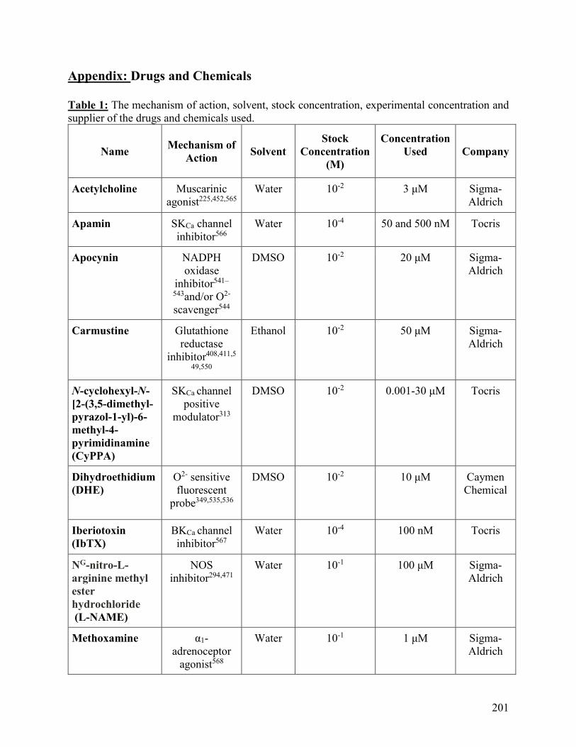
201
Appendix: Drugs and Chemicals
Table 1: The mechanism of action, solvent, stock concentration, experimental concentration and
supplier of the drugs and chemicals used.
Name Mechanism of
Action Solvent
Stock
Concentration
(M)
Concentration
Used
Company
Acetylcholine Muscarinic
agonist225,452,565
Water 10-2 3 μM Sigma-
Aldrich
Apamin SKCa channel
inhibitor566
Water 10-4 50 and 500 nM Tocris
Apocynin NADPH
oxidase
inhibitor541–
543and/or O2-
scavenger544
DMSO 10-2 20 μM Sigma-
Aldrich
Carmustine Glutathione
reductase
inhibitor408,411,5
49,550
Ethanol 10-2 50 μM Sigma-
Aldrich
N-cyclohexyl-N-
[2-(3,5-dimethyl-
pyrazol-1-yl)-6-
methyl-4-
pyrimidinamine
(CyPPA)
SKCa channel
positive
modulator313
DMSO 10-2 0.001-30 μM Tocris
Dihydroethidium
(DHE)
O2- sensitive
fluorescent
probe349,535,536
DMSO 10-2 10 μM Caymen
Chemical
Iberiotoxin
(IbTX)
BKCa channel
inhibitor567
Water 10-4 100 nM Tocris
NG-nitro-L-
arginine methyl
ester
hydrochloride
(L-NAME)
NOS
inhibitor294,471
Water 10-1 100 μM Sigma-
Aldrich
Methoxamine α1-
adrenoceptor
agonist568
Water 10-1 1 μM Sigma-
Aldrich

202
Nicotinamide-
adenine-
dinucleotide
phosphate
(NADPH)
Electron donor 385,471
10 mg/ml
NaHCO3
10-2 100 μM Sigma-
Aldrich
Nifedipine L-type VOCC
inhibitor569,570
DMSO 10-2 1 and 10 μM Sigma-
Aldrich
4-[[3-
(Trifluoromethyl
)phenyl]methyl]-
2H-1,4-
benzothiazin-
3(4H)-one
(NS 6180)
IKCa channel
inhibitor311
DMSO 10-2 1 μM Tocris
N'-[3,5-
Bis(trifluorometh
yl)phenyl]-N-[4-
bromo-2-(2H-
tetrazol-5-yl-
phenyl]thiourea
(NS 11021)
BKCa channel
activator571,572
DMSO 10-2 100 nM Tocris
Phenylephrine α1-
adrenoceptor
agonist568,573
Water 10-2, 10-3, 10-4
and 10-5
0.001- 100 μM Sigma-
Aldrich
Prazosin α1-
adrenoceptor
antagonist184
Water 10-2 1 μM Tocris
1H-
[1,2,4]Oxadiazolo
[4,3-
a]quinoxalin-1-
one (ODQ)
Soluble
guanylyl
cyclase
inhibitor574
DMSO 10-2 5 μM Tocris
Naphtho[1,2-
d]thiazol-2-
ylamine
(SKA-31)
IKCa channel
positive
modulator121
DMSO 10-2 0.001-30 μM Tocris
Sodium
Nitroprusside
NO donor575 Water 10-2 1 μM Sigma-
Aldrich
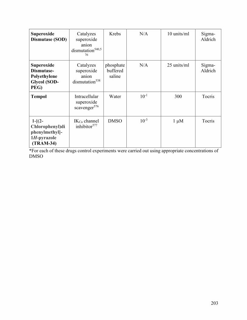
203
Superoxide
Dismutase (SOD)
Catalyzes
superoxide
anion
dismutation540,5
76
Krebs N/A 10 units/ml Sigma-
Aldrich
Superoxide
Dismutase-
Polyethylene
Glycol (SOD-
PEG)
Catalyzes
superoxide
anion
dismutation538
phosphate
buffered
saline
N/A 25 units/ml Sigma-
Aldrich
Tempol
Intracellular
superoxide
scavenger576
Water 10-1 300 Tocris
1-[(2-
Chlorophenyl)di
phenylmethyl]-
1H-pyrazole
(TRAM-34)
IKCa channel
inhibitor577
DMSO 10-3 1 μM Tocris
*For each of these drugs control experiments were carried out using appropriate concentrations of
DMSO
Related Documents