42 Winter 2011 • Volume 26 • Number 4 Abstract This article, the second in a two-part series, continues the discussion of a conservative, effective, and artistic philosophy for performing esthetic direct anterior composite restorations based on the principles of emulating the proper form, color, and function of natural dentition. This particular article outlines step-by-step procedural approaches to solving day-to-day anterior direct restorative challenges, including tooth preparation, artistic application, and how to create seamless transitions from tooth substance to the synthetic composite restoratives using correct finishing and polishing techniques. Step-by-Step Approaches for Anterior Direct Restorative Challenges Mastering Composite Artistry to Create Anterior Masterpieces—Part 2 Newton Fahl, Jr., DDS

Welcome message from author
This document is posted to help you gain knowledge. Please leave a comment to let me know what you think about it! Share it to your friends and learn new things together.
Transcript

42 Winter 2011 • Volume 26 • Number 4
Abstract
This article, the second in a two-part series, continues the discussion of a conservative,
effective, and artistic philosophy for performing esthetic direct anterior composite
restorations based on the principles of emulating the proper form, color, and
function of natural dentition. This particular article outlines step-by-step procedural
approaches to solving day-to-day anterior direct restorative challenges, including
tooth preparation, artistic application, and how to create seamless transitions from
tooth substance to the synthetic composite restoratives using correct finishing and
polishing techniques.
Step-by-Step Approaches for Anterior Direct Restorative ChallengesMasteringCompositeArtistrytoCreateAnteriorMasterpieces—Part2 Newton Fahl, Jr., DDS
43 Journal of Cosmetic Dentistry
Step-by-Step Approaches for Anterior Direct Restorative Challenges
Fahl
Clinical Direct Composite Placement Techniques
Two individual denture teeth mounted on an acrylic base were prepared and restored using the achromatic and chromatic enam-el approaches. The objective is to educate the reader on how to properly select and apply diverse restorative systems based on ideal optical and physical properties to produce similar color re-sults.1-3
As Part 1 of this article explained, the achromatic approach entails the use of non-VITA based enamel composites to modulate the chroma and value of the underlying artificial dentin, which provides the basic hue and facilitates the achievement of the final intended shade.4-7 Comparatively, the chromatic approach utilizes VITA-based (Vident; Brea, CA) enamel composite to provide the final desired hue and chroma, while allowing the use of effect achromatic enamels for minor color characterizations.4-6 Both techniques can be used predictably to promote lifelike restorations and elicit similar esthetic results (see sidebar, page 44).
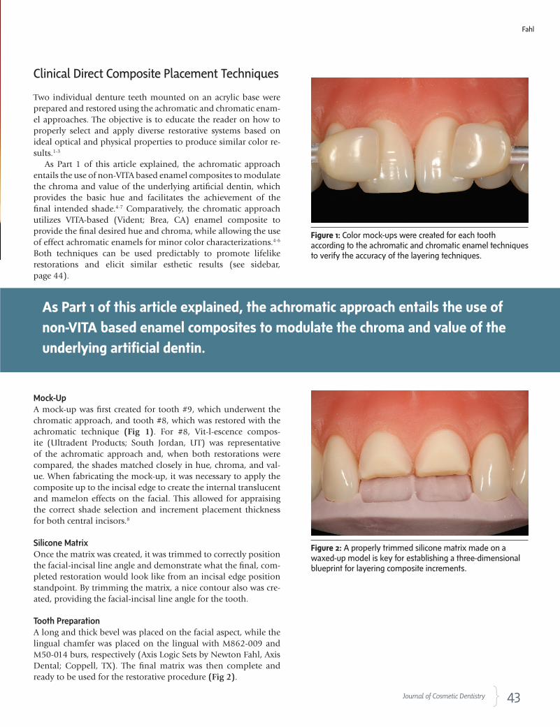
Mock-UpA mock-up was first created for tooth #9, which underwent the chromatic approach, and tooth #8, which was restored with the achromatic technique (Fig 1). For #8, Vit-l-escence compos-ite (Ultradent Products; South Jordan, UT) was representative of the achromatic approach and, when both restorations were compared, the shades matched closely in hue, chroma, and val-ue. When fabricating the mock-up, it was necessary to apply the composite up to the incisal edge to create the internal translucent and mamelon effects on the facial. This allowed for appraising the correct shade selection and increment placement thickness for both central incisors.8
Silicone MatrixOnce the matrix was created, it was trimmed to correctly position the facial-incisal line angle and demonstrate what the final, com-pleted restoration would look like from an incisal edge position standpoint. By trimming the matrix, a nice contour also was cre-ated, providing the facial-incisal line angle for the tooth.
Tooth PreparationA long and thick bevel was placed on the facial aspect, while the lingual chamfer was placed on the lingual with M862-009 and M50-014 burs, respectively (Axis Logic Sets by Newton Fahl, Axis Dental; Coppell, TX). The final matrix was then complete and ready to be used for the restorative procedure (Fig 2).
Figure 1:Colormock-upswerecreatedforeachtoothaccordingtotheachromaticandchromaticenameltechniquestoverifytheaccuracyofthelayeringtechniques.
Figure 2:Aproperlytrimmedsiliconematrixmadeonawaxed-upmodeliskeyforestablishingathree-dimensionalblueprintforlayeringcompositeincrements.
As Part 1 of this article explained, the achromatic approach entails the use of
non-VITA based enamel composites to modulate the chroma and value of the
underlying artificial dentin.

44 Winter 2011 • Volume 26 • Number 4
Protocol
Following tooth preparation, a single-component bond-ing agent (PQ1, Ultradent) was applied to the tooth sur-faces. Excess material was suctioned off the preparation area, and the bonding agent was cured with an LED cur-ing light (VALO, Ultradent) for 10 seconds.
Incremental BuildupTo begin building up the composite materials, it was first necessary to understand how far to apply the material. Using an explorer, the matrix was scratched along the lin-gual chamfer finish line until a white line could be seen. This provided the boundaries for the initial increment and prevented over-buildup of the composite.
Achromatic Enamel Approach—#8—Lingual Shelf ApplicationA pearl-amber shade of enamel composite (Vit-l-escence PA) was placed on #8 to begin the achromatic technique. The material was removed from the syringe and rolled into the shape of a ball. To achieve proper consistency, the material was patted and then worked as thinly as possible on the matrix. The matrix was then positioned onto the tooth, and the composite was tucked into the chamfered area with a contouring instrument.
A very low-viscosity, 45% filled wetting resin (Ultra-dent), was used to lubricate contouring instruments and artist brushes during the procedure. It is important to limit the use of this resin, however, as overuse diminishes the physical properties of the composites. With all instru-ments slightly lubricated, the composite material was pushed toward the incisal area, while making the mate-rial thinner or thicker as necessary to achieve the proper effects and esthetics (Fig 3). A flexible contouring instru-ment (IPC-L, Cosmedent; Chicago, IL) was used to slice off the excess material.
To gauge the thickness of the composite, the ma-trix was observed. Specifically, thickness was controlled based on the color seen underneath the matrix. When the proper thickness was reached, brushes (#1 and #3, Cosmedent) were used to smooth the compos-ite. While brushing, it was important to keep the ma-trix securely in position, without pressing too much or allowing the matrix to loosen. This was done so the facio-incisal line angle could be reestablished lat-er on. Once smoothed, the composite was cured for five seconds.
After curing, the matrix was removed to reveal an enamel composite lingual shelf. Demonstrating a thick-ness of 0.37 mm, the ideal average thinness for this ini-tial layer, desirable optical effects that mimicked those of natural enamel were produced. To clean excess material from the surrounding areas, a #12 surgical blade was used (Bard-Parker, Aspen Surgical; Caledonia, MI).
Color Map #8 - Achromatic Approach
1.Lingualshelflayer(PearlAmber-Vit-l-escence)
2.Dentinlayer(A3-Vit-l-escence)
3.DeepTranslucentEnamellayer(IrB-Vitalescence)
4.1stAchromaticEnamellayer(PearlFrost-Vit-l-escence)
5.2ndAchromaticEnamellayer(TransFrost-Vit-l-escence)
Color Map #9 - Chromatic Approach
1.Lingualshelflayer(Amber–VenusDiamond)
2.Dentinlayer(A3-SupremeUltra)
3.DeepTranslucentEnamellayer(Opal-EmpressDirect)
4.ChromaticEnamellayer(A2–EsteliteSigma)
5.1stAchromaticEnamellayer(IncisalMedium–RenamelMicrofill)
6.2ndAchromaticEnamellayer(TransFrost-Vit-l-escence)

45 Journal of Cosmetic Dentistry
Chromatic Enamel Approach—#9—Lingual Shelf ApplicationA milky-white, semi-translucent nano-hybrid universal composite (shade AM, Venus Diamond, Heraeus; South Bend, IN) was applied thinly to the matrix. The material was pressed and sculpted to shape, then tucked in. This composite was chosen based on its excellent handling characteristics and optical effects. Although it tends to be stiffer, it does not crack or tear and, when pressed, allows the operator to shape it as needed.
Using the IPC-L, the material was trimmed and cut back while being pushed toward the incisal. Like the oth-er central incisor, it also was necessary to keep thinning the material at this point in the procedure. The incisal em-brasure then was cleaned with the IPC-L where the con-tact areas were to be placed, followed by brushing of the chamfer area. The composite was then quickly “zapped” with the curing light to complete the lingual shelf. At this point in the procedure, the contacts are left slightly open, but the teeth should both demonstrate the same thick-ness and optical properties (Fig 4).
Achromatic Enamel Approach—#8—Artificial Dentin ApplicationShade A3 dentin (Vit-l-escence) was used to begin build-ing the dentin on #8. This particular composite required two increments to build the mamelon effects, with one layer placed initially to create a primary contour and an-other layer to complete the contour. Once cured, dentin placement was complete.
To establish the contours, it was crucial to carry over the bevel until the transition line was eliminated. The in-cisal portion of the dentin increment was pulled toward the bevel to create as much clearance as possible between the dentin and halo so that the mamelons could be re-fined and room was allowed for placement of translucent enamel shades later on. The dentin composite was bur-nished over the bevel until it disappeared, after which it was blended in. At this point, no ledge should be visible, and the dentin should be convex, not concave, following the natural histological boundaries of the dento-enamel junction.
A fine-tipped Hollenback #6 was used to gently cre-ate the mamelons of the dentin, followed by curing of the composite layer (Fig 5). At this time, the layer should curve toward the incisal, leaving room for translucent enamels over the incisal third. It was necessary to deter-mine the irregular pattern of the mamelons, which are uneven finger-like projections, and create subdivisions to replicate natural dentition. If the mamelons needed refin-ing, minute increments of resin would have been rolled into shape and placed where needed. The string-like pro-
Figure 3:Thelingualshelfmustbeanachromaticenamelnothickerthan0.3mm.
Figure 4:Oncecured,thelingualshelvesofbothcentralsdenoteamber-whitishnuancesthatreplicatetheopalescencepresentinnaturalenamel.
Figure 5: Afine-tippeddentalinstrumentwasusedtogentlycreatethedentinmamelons.
Fahl
47 Journal of Cosmetic Dentistry
jections of the mamelons were brought toward the halo area to complete mamelon creation.
To gauge the room required for the enamel, the con-tour was checked on the previously made wax-up. This enabled a preview of the contours of the cervical-third, middle-third, and incisal-third and also allowed visual-ization of the space required for the enamel composite resin.
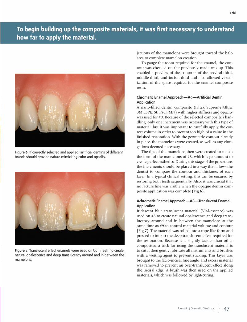
Chromatic Enamel Approach—#9—Artificial Dentin ApplicationA nano-filled dentin composite (Filtek Supreme Ultra, 3M ESPE; St. Paul, MN) with higher stiffness and opacity was used for #9. Because of the selected composite’s han-dling, only one increment was necessary with this type of material, but it was important to carefully apply the cor-rect volume in order to prevent too high of a value in the finished restoration. With the geometric contour already in place, the mamelons were created, as well as any elon-gations deemed necessary.
The tips of the mamelons then were created to match the form of the mamelons of #8, which is paramount to create perfect esthetics. During this stage of the procedure, the increments should be placed in a way that allows the dentist to compare the contour and thickness of each layer. In a typical clinical setting, this can be ensured by restoring both teeth sequentially. Also, it was crucial that no facture line was visible when the opaque dentin com-posite application was complete (Fig 6).
Achromatic Enamel Approach—#8—Translucent Enamel ApplicationIridescent blue translucent material (Vit-l-escence) was used on #8 to create natural opalescence and deep trans-lucency around and in between the mamelons at the same time as #9 to control material volume and contour (Fig 7). The material was rolled into a rope-like form and pressed to impart the deep translucent effect required for the restoration. Because it is slightly tackier than other composites, a trick for using the translucent material is to cut it then gently lubricate all instruments and brushes with a wetting agent to prevent sticking. This layer was brought to the facio-incisal line angle, and excess material was removed to prevent an over-translucent effect along the incisal edge. A brush was then used on the applied materials, which was followed by light-curing.
Figure 6:Ifcorrectlyselectedandapplied,artificialdentinsofdifferentbrandsshouldprovidenature-mimickingcolorandopacity.
Figure 7:Translucenteffectenamelswereusedonbothteethtocreatenaturalopalescenceanddeeptranslucencyaroundandinbetweenthemamelons.
To begin building up the composite materials, it was first necessary to understand how far to apply the material.
Fahl
48 Winter 2011 • Volume 26 • Number 4
Chromatic Enamel Approach—Tooth #9—Translucent Enamel ApplicationA nano-hybrid translucent Opal composite (IPS Empress Direct, Ivoclar Vivadent; Amherst, NY) was chosen for #9 to give the restoration a natural opalescence and deep translucency in between and around the dentin mam-elons at the same time as #8 to control material volume and contour. The material was rolled, cut, and pressed to fill the voids between the mamelons. Once the trans-lucent composite had been applied and brushed, it was light-cured.
Chromatic Enamel Approach—#9—Body Enamel ApplicationBecause the purpose of this article is to demonstrate how to achieve the same hue, chroma, and value using chro-matic and achromatic enamels, a VITA-based enamel had to be applied first on #9 to establish a three-dimensional color baseline, after which #8 would be modeled using a
non-VITA-based enamel. Application of body enamel be-gan on the left central as the final hue, chroma, and value for the cervical middle-thirds were created. As described, this body enamel produced the final desired shade of the restoration in a single layer of Vita-based enamel compos-ite. This technique is employed when the final hue and chroma of the restoration need to be achieved without the need for modulating the color of the underlying den-tin core.
A nano-filled composite with spherical submicron fill-ers (Estelite Sigma, Tokuyama America; Encinitas, CA) was chosen as the chromatic enamel for the restoration, as the spherical particles promote good handling char-acteristics and achieve an excellent polish. Additionally, the composite forms a lifelike blend with the tooth color underneath.
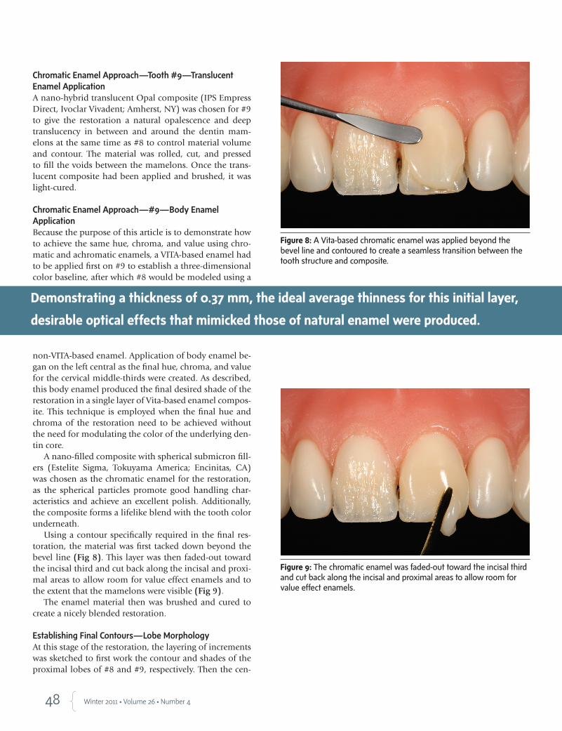
Using a contour specifically required in the final res-toration, the material was first tacked down beyond the bevel line (Fig 8). This layer was then faded-out toward the incisal third and cut back along the incisal and proxi-mal areas to allow room for value effect enamels and to the extent that the mamelons were visible (Fig 9).
The enamel material then was brushed and cured to create a nicely blended restoration.
Establishing Final Contours—Lobe MorphologyAt this stage of the restoration, the layering of increments was sketched to first work the contour and shades of the proximal lobes of #8 and #9, respectively. Then the cen-
Figure 8:AVita-basedchromaticenamelwasappliedbeyondthebevellineandcontouredtocreateaseamlesstransitionbetweenthetoothstructureandcomposite.
Figure 9:Thechromaticenamelwasfaded-outtowardtheincisalthirdandcutbackalongtheincisalandproximalareastoallowroomforvalueeffectenamels.
Demonstrating a thickness of 0.37 mm, the ideal average thinness for this initial layer,
desirable optical effects that mimicked those of natural enamel were produced.
49 Journal of Cosmetic Dentistry
ter lobes of both were restored in the same order. This technique will demonstrate the sequential application of distinct composite materials of similar optical properties.
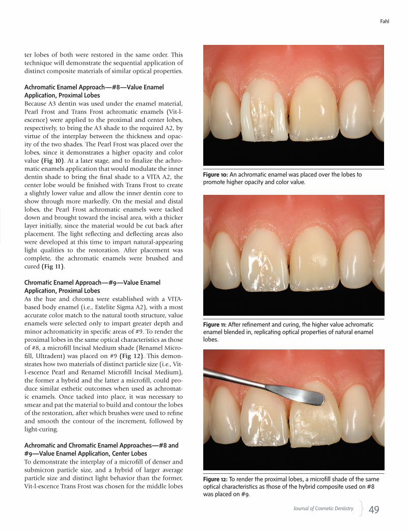
Achromatic Enamel Approach—#8—Value Enamel Application, Proximal LobesBecause A3 dentin was used under the enamel material, Pearl Frost and Trans Frost achromatic enamels (Vit-l-escence) were applied to the proximal and center lobes, respectively, to bring the A3 shade to the required A2, by virtue of the interplay between the thickness and opac-ity of the two shades. The Pearl Frost was placed over the lobes, since it demonstrates a higher opacity and color value (Fig 10). At a later stage, and to finalize the achro-matic enamels application that would modulate the inner dentin shade to bring the final shade to a VITA A2, the center lobe would be finished with Trans Frost to create a slightly lower value and allow the inner dentin core to show through more markedly. On the mesial and distal lobes, the Pearl Frost achromatic enamels were tacked down and brought toward the incisal area, with a thicker layer initially, since the material would be cut back after placement. The light reflecting and deflecting areas also were developed at this time to impart natural-appearing light qualities to the restoration. After placement was complete, the achromatic enamels were brushed and cured (Fig 11).
Chromatic Enamel Approach—#9—Value Enamel Application, Proximal LobesAs the hue and chroma were established with a VITA-based body enamel (i.e., Estelite Sigma A2), with a most accurate color match to the natural tooth structure, value enamels were selected only to impart greater depth and minor achromaticity in specific areas of #9. To render the proximal lobes in the same optical characteristics as those of #8, a microfill Incisal Medium shade (Renamel Micro-fill, Ultradent) was placed on #9 (Fig 12). This demon-strates how two materials of distinct particle size (i.e., Vit-l-escence Pearl and Renamel Microfill Incisal Medium), the former a hybrid and the latter a microfill, could pro-duce similar esthetic outcomes when used as achromat-ic enamels. Once tacked into place, it was necessary to smear and pat the material to build and contour the lobes of the restoration, after which brushes were used to refine and smooth the contour of the increment, followed by light-curing.
Achromatic and Chromatic Enamel Approaches—#8 and #9—Value Enamel Application, Center LobesTo demonstrate the interplay of a microfill of denser and submicron particle size, and a hybrid of larger average particle size and distinct light behavior than the former, Vit-l-escence Trans Frost was chosen for the middle lobes
Figure 10:Anachromaticenamelwasplacedoverthelobestopromotehigheropacityandcolorvalue.
Figure 11:Afterrefinementandcuring,thehighervalueachromaticenamelblendedin,replicatingopticalpropertiesofnaturalenamellobes.
Figure 12:Torendertheproximallobes,amicrofillshadeofthesameopticalcharacteristicsasthoseofthehybridcompositeusedon#8wasplacedon#9.
Fahl

51 Journal of Cosmetic Dentistry
of both central incisors (Figs 13 & 14). Trans Frost also was placed on the prox-imal lobes, between the vertical transi-tional line angles and the contact area with the adjacent teeth, where the value is usually slightly lower than the crest of the proximal lobes (Fig 15).
This last step determined center lobes of identical optical characteristics for both centrals, since Trans Frost was used on both; whereas the proximal lobes of both centrals, although similar in optical characteristics, were restored
with different materials. At this point, the proximal lobes were covered, but were no cause for concern. During fin-ishing, this layer would be removed to reach ideal volume and contour, and the main concern would be creating pri-mary anatomy. When completing the fi-nal shape, the underlying Estelite Sigma and proximal Renamel Microfill were reached. To complete this stage, the em-brasure forms also were sculpted, and the restoration was brought to an even and smooth contour with brushes, fol-
lowed by curing. The restorations on both teeth were then ready for finishing and polishing.
Emergence Profile and Facio-Incisal Line Angles—#8 and #9To achieve the proper incisal edge po-sition, a finishing disc (Sof-Lex, 3M ESPE) was run back and forth to remove any flashes. The matrix was placed back on the model, aligned, and made flush with the facial-incisal line angles of both central incisors.
Figures 13 & 14:Anon-VITA,moretranslucentachromatichybridcompositeenamelwaschosenforthemiddlelobesofbothcentralincisorstoallowmoredentinshow-through.
To establish the contours, it was crucial to carry over the bevel until the transition
line was eliminated.
Figure 15:Afterapplicationandlight-curingofchromaticandachromaticenamels,bothcentralsdepictsimilaropticalcharacteristics.
Figure 16:Toestablishnaturalfacialplanes,thetransitionallineangleswereevaluatedandthefacialplanesworkedwithfinishingdiscstoestablishtheprimaryanatomy.
Fahl
K N I G H T D E N T A L G R O U P
Helping You Keep Your Promise
The Extend-Plant Unit Priced Abutments & Crowns Include:
• Convenience – You don’t have to keep track of parts or place special orders.
• A custom milled abutment & final placement screw designed by our expert
technicians in your choice of 10 shades.
• Soft tissue model, analog and implant labor.
• Full-zirconia or Noble PFM crown (up to 2.5 grams of alloy included).
www.knightdentalgroup.comYour Removable Solution Specialists
© 2011 Knight Dental Group.
A Division of Knight Dental Group
The Extend-Plant product is compatible with systems from Nobel Biocare, Biomet 3i, Straumann, AstraTech and Sybron. The Extend™ Division from Knight works with all implant systems.
28659_knight_dental_r1_Layout 1 12/14/10 1:12 PM Page 1

52 Winter 2011 • Volume 26 • Number 4
To establish natural facial planes of #8 and #9, the transitional line angles were evaluated and the facial planes worked with the finishing disc from cervical to in-cisal in a single, continuous movement (Fig 16).
Vertical Transitional Line Angles—#8 and #9Once the required emergence profile was achieved, the transitional line angles were worked. Lines were drawn with a pencil to determine their ideal position, starting at the point angles and going upward according to a nat-ural ascending path. With a red disc (Sof-Lex) at a very low speed, and viewing the restorations from different perspectives, the embrasures were opened until symme-try was achieved (Fig 17). Any sharp transitions between aspects were softened with the disc at low speed. Straight line angles were and should be visible, even though the restorations are round.
The incisal embrasure was worked with the same disc
at a very low speed. Immediately after, the incisal embra-sures were worked to suit the desired shape based on the roundness or squareness of the restorations, which cre-ated symmetry.
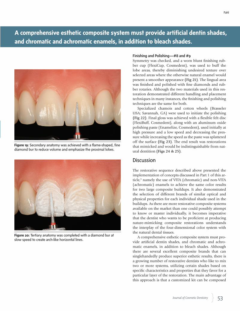
Secondary Anatomy—#8 and #9To finish the secondary anatomy, the line angles first were penciled in, and a long axis was drawn dividing the teeth into thirds, with anatomy mapping on the centrals (Fig 18). Finishing began with a long flame-shaped, fine diamond bur (F888-012, Axis Dental) in a swinging mo-tion on the center lobe to reduce volume and emphasize the proximal lobes (Fig 19). To redefine the lobes, the diamond bur was used to create delta-like developmen-tal grooves that narrowed toward the cervical area. A soft silicone carbide brush (Jiffy Brush, Ultradent) was used intermittently to buff the restorations in circular move-ments. This allowed for assessment of the surface texture achieved up to that point.
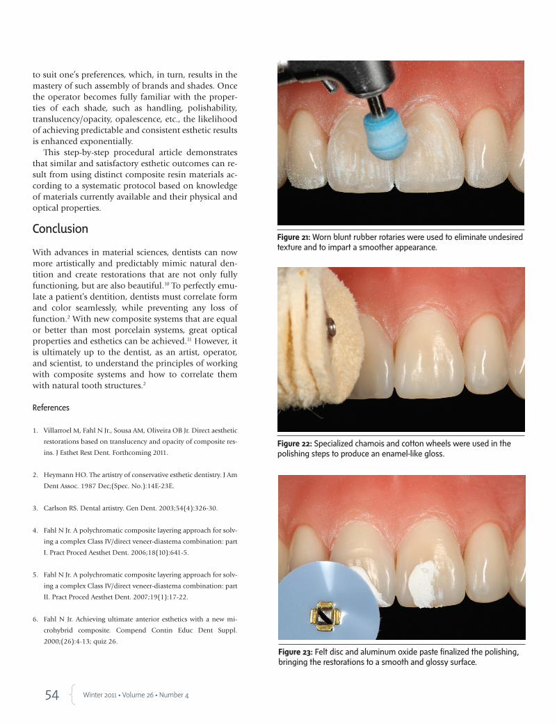
Tertiary Anatomy—#8 and #9After using the silicone carbide brush, it was discovered that some tertiary anatomy still needed to be completed. With the diamond bur at the slowest possible speed, the surface was blended with a back-and-forth horizontal motion (Fig 20). The silicone carbide brush was used again to brush and buff the surface and verify the level of micro texture achieved. The point angles were rounded off with polishing discs (Sof-Lex Orange and Yellow).
Figure 18:Anatomymappingwaspenciledinonthecentralstoaidinattainingpropertoothmorphologyduringfinishing.
Figure 17:Thetransitionallineangleswereworkedandtheembrasuresopeneduntilsymmetrywasachieved.
To redefine the lobes, the diamond bur was used to create delta-like developmental
grooves that narrowed toward the cervical area.
53 Journal of Cosmetic Dentistry
To redefine the lobes, the diamond bur was used to create delta-like developmental
grooves that narrowed toward the cervical area.
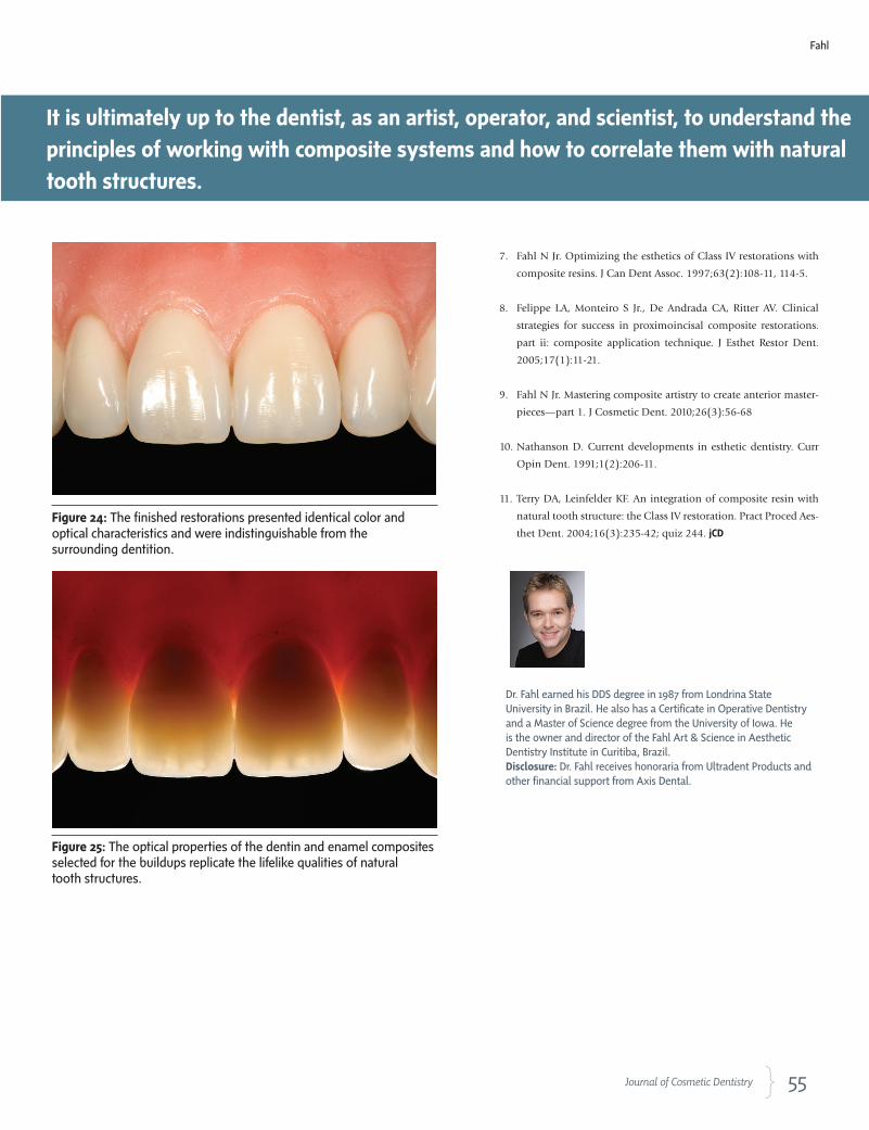
Finishing and Polishing—#8 and #9Symmetry was checked, and a worn blunt finishing rub-ber cup (FlexiCup, Cosmedent), was used to buff the lobe areas, thereby diminishing undesired texture over selected areas where the otherwise natural enamel would present a smoother appearance (Fig 21). The lingual area was finished and polished with fine diamonds and rub-ber rotaries. Although the two materials used in this res-toration demonstrated different handling and placement techniques in many instances, the finishing and polishing techniques are the same for both.
Specialized chamois and cotton wheels (Brasseler USA; Savannah, GA) were used to initiate the polishing (Fig 22). Final gloss was achieved with a flexible felt disc (FlexiBuff, Cosmedent), along with an aluminum oxide polishing paste (Enamelize, Cosmedent), used initially at high pressure and a low speed and decreasing the pres-sure while increasing the speed as the paste was splattered off the surface (Fig 23). The end result was restorations that mimicked and would be indistinguishable from nat-ural dentition (Figs 24 & 25).
Discussion
The restorative sequence described above presented the implementation of concepts discussed in Part 1 of this ar-ticle,9 namely the use of VITA (chromatic) and non-VITA (achromatic) enamels to achieve the same color results for two large composite buildups. It also demonstrated the selection of different brands of similar optical and physical properties for each individual shade used in the buildups. As there are more restorative composite systems available on the market than one could possibly attempt to know or master individually, it becomes imperative that the dentist who wants to be proficient at producing nature-mimicking composite restorations understands the interplay of the four-dimensional color system with the natural dental tissues.
A comprehensive esthetic composite system must pro-vide artificial dentin shades, and chromatic and achro-matic enamels, in addition to bleach shades. Although there are several excellent composite brands that can singlehandedly produce superior esthetic results, there is a growing number of restorative dentists who like to mix two or more systems, utilizing certain shades based on specific characteristics and properties that they favor for a particular layer of the restoration. The main advantage of this approach is that a customized kit can be composed
Figure 20:Tertiaryanatomywascompletedwithadiamondburatslowspeedtocreatearch-likehorizontallines.
Figure 19:Secondaryanatomywasachievedwithaflame-shaped,finediamondburtoreducevolumeandemphasizetheproximallobes.
A comprehensive esthetic composite system must provide artificial dentin shades,
and chromatic and achromatic enamels, in addition to bleach shades.
Fahl
54 Winter 2011 • Volume 26 • Number 4
to suit one’s preferences, which, in turn, results in the mastery of such assembly of brands and shades. Once the operator becomes fully familiar with the proper-ties of each shade, such as handling, polishability, translucency/opacity, opalescence, etc., the likelihood of achieving predictable and consistent esthetic results is enhanced exponentially.
This step-by-step procedural article demonstrates that similar and satisfactory esthetic outcomes can re-sult from using distinct composite resin materials ac-cording to a systematic protocol based on knowledge of materials currently available and their physical and optical properties.
Conclusion
With advances in material sciences, dentists can now more artistically and predictably mimic natural den-tition and create restorations that are not only fully functioning, but are also beautiful.10 To perfectly emu-late a patient’s dentition, dentists must correlate form and color seamlessly, while preventing any loss of function.2 With new composite systems that are equal or better than most porcelain systems, great optical properties and esthetics can be achieved.11 However, it is ultimately up to the dentist, as an artist, operator, and scientist, to understand the principles of working with composite systems and how to correlate them with natural tooth structures.2
References
1. Villarroel M, Fahl N Jr., Sousa AM, Oliveira OB Jr. Direct aesthetic
restorations based on translucency and opacity of composite res-
ins. J Esthet Rest Dent. Forthcoming 2011.
2. Heymann HO. The artistry of conservative esthetic dentistry. J Am
Dent Assoc. 1987 Dec;(Spec. No.):14E-23E.
3. Carlson RS. Dental artistry. Gen Dent. 2003;54(4):326-30.
4. Fahl N Jr. A polychromatic composite layering approach for solv-
ing a complex Class IV/direct veneer-diastema combination: part
I. Pract Proced Aesthet Dent. 2006;18(10):641-5.
5. Fahl N Jr. A polychromatic composite layering approach for solv-
ing a complex Class IV/direct veneer-diastema combination: part
II. Pract Proced Aesthet Dent. 2007;19(1):17-22.
6. Fahl N Jr. Achieving ultimate anterior esthetics with a new mi-
crohybrid composite. Compend Contin Educ Dent Suppl.
2000;(26):4-13; quiz 26.
Figure 21:Wornbluntrubberrotarieswereusedtoeliminateundesiredtextureandtoimpartasmootherappearance.
Figure 22:Specializedchamoisandcottonwheelswereusedinthepolishingstepstoproduceanenamel-likegloss.
Figure 23:Feltdiscandaluminumoxidepastefinalizedthepolishing,bringingtherestorationstoasmoothandglossysurface.
55 Journal of Cosmetic Dentistry
7. Fahl N Jr. Optimizing the esthetics of Class IV restorations with
composite resins. J Can Dent Assoc. 1997;63(2):108-11, 114-5.
8. Felippe LA, Monteiro S Jr., De Andrada CA, Ritter AV. Clinical
strategies for success in proximoincisal composite restorations.
part ii: composite application technique. J Esthet Restor Dent.
2005;17(1):11-21.
9. Fahl N Jr. Mastering composite artistry to create anterior master-
pieces—part 1. J Cosmetic Dent. 2010;26(3):56-68
10. Nathanson D. Current developments in esthetic dentistry. Curr
Opin Dent. 1991;1(2):206-11.
11. Terry DA, Leinfelder KF. An integration of composite resin with
natural tooth structure: the Class IV restoration. Pract Proced Aes-
thet Dent. 2004;16(3):235-42; quiz 244. jCD
Figure 25:Theopticalpropertiesofthedentinandenamelcompositesselectedforthebuildupsreplicatethelifelikequalitiesofnaturaltoothstructures.
Figure 24:Thefinishedrestorationspresentedidenticalcolorandopticalcharacteristicsandwereindistinguishablefromthesurroundingdentition.
Dr. Fahl earned his DDS degree in 1987 from Londrina State University in Brazil. He also has a Certificate in Operative Dentistry and a Master of Science degree from the University of Iowa. He is the owner and director of the Fahl Art & Science in Aesthetic Dentistry Institute in Curitiba, Brazil. Disclosure: Dr. Fahl receives honoraria from Ultradent Products and other financial support from Axis Dental.
It is ultimately up to the dentist, as an artist, operator, and scientist, to understand the
principles of working with composite systems and how to correlate them with natural
tooth structures.
Fahl
Related Documents


