201 Current Cardiology Reviews, 2011, 7, 201-212 1573-403X/11 $58.00+.00 © 2011 Bentham Science Publishers Stem Cell Therapy in Heart Diseases: A Review of Selected New Perspec- tives, Practical Considerations and Clinical Applications Eltyeb Abdelwahid 1, *, Tomasz Siminiak 2 , Luiz César Guarita-Souza 3 , Katherine Athayde Teixeira de Carvalho 4 . Pasquale Gallo 5,6 , Winston Shim 7 and Gianluigi Condorelli 5,6,8 1 CBRC, Massachusetts General Hospital/Harvard Medical School, Building 149, 13 th Street, Charlestown Massachu- setts, 02129, U.S.A; 2 Pozna University of Medical Sciences, Cardiac and Rehabilitation Hospital ul. Sanatoryjna 34 64- 600 Kowanówko k/Obornik Wlkp. Poland; 3 PUCPR - Experimental Laboratory of Cell Culture Institute of Biological and Health Sciences, CCBS, Brazil; 4 Pequeno Príncipe Faculty & The Pelé Pequeno Príncipe Institute- Child and Ado- lescent Health Research, Curitiba,Brazil; 5 Laboratory of Molecular Cardiology, San Raffaele Biomedical Science Foundation of Rome, IT; 6 IRCCS, Multimedica Hospital, Milan, IT; 7 National Heart Centre Singapore, Singapore; 8 Department of Medicine, University of California, San Diego, CA 92003 USA, Abstract: Degeneration of cardiac tissues is considered a major cause of mortality in the western world and is expected to be a greater problem in the forthcoming decades. Cardiac damage is associated with dysfunction and irreversible loss of cardiomyocytes. Stem cell therapy for ischemic heart failure is very promising approach in cardiovascular medicine. Ini- tial trials have indicated the ability of cardiomyocytes to regenerate after myocardial injury. These preliminary trials aim to translate cardiac regeneration strategies into clinical practice. In spite of advances, current therapeutic strategies to ischemic heart failure remain very limited. Moreover, major obstacles still need to be solved before stem cell therapy can be fully applied. This review addresses the current state of research and experimental data regarding embryonic stem cells (ESCs), myoblast transplantation, histological and functional analysis of transplantation of co-cultured myoblasts and mesenchymal stem cells, as well as comparison between mononuclear and mesenchymal stem cells in a model of myocar- dium infarction. We also discuss how research with stem cell transplantation could translate to improvement of cardiac function. Keywords: Heart, stem cells, transplantation, therapy. HUMAN EMBRYONIC STEM CELLS AS A SOURCE OF CARDIOMYOCYTES FOR CELL THERAPY AP- PLICATIONS: OBSTACLES TO OVERCOME In contrast to adult stem cells, embryonic stem cells (ESCs) have the potential to differentiate into tissue deriva- tives of all three embryonic germ layers and therefore are termed pluripotent. Cardiomyocytes (CMCs) have been ob- tained by all three types of murine embryo-derived stem cells: embryonic carcinoma (EC), embryonic stem (ES), and embryonic germ (EG) cells. In this chapter, we focus our attention on human ESCs (hESCs), due to their potential clinical application. hESC lines, isolated from the inner cell mass (ICM) of embryos can be propagated continuously in the undifferentiated state when grown on top of a mouse embryonic fibroblast (MEF) feeder layer. When removed from these conditions and grown in suspension, they begin to generate three-dimensional differentiating cell aggregates termed embryoid bodies (EBs). This in vitro differentiating system can be used to generate a plurality of tissue types. The ability of hESCs to differentiate into mature somatic cells was demonstrated using spontaneous and directed in vitro differentiation systems. So far, hESCs have been shown to differentiate into neuronal tissue [1], ß islet pancreatic *Address correspondence to this author at the CBRC, Massachusetts Gen- eral Hospital/Harvard Medical School, Building 149, 13 th Street, Char- lestown Massachusetts, 02129, USA; Tel/Fax: 617-321-5483; E-mail: [email protected] cells [2], hematopoietic progenitors [3], endothelial cells [4] and cardiac tissue [5,6]. Interesting data were obtained by the use of adult stem cell for cardiac repair [7-9]. Given the versatility of hESCs and the possibility of ob- taining beating CMCs from them, (Fig. 1) [6,10] they appear as the main candidate for cell-based applications for cardiac repair. In fact, hESCs apparently fulfill most, if not all, the properties of an ideal donor cell line [11] (Table 1). In the following sections, we will discuss briefly, but critically, the obstacles on the path to hESC-based cardiac therapy. A possible strategy for cell-replacement therapy would be to initially allow spontaneous differentiation of ESCs into multiple lineages in vitro followed by selective purification of the cardiomyogenic lineage isolated from embryoid bodies (Fig. 1). On this issue, Kehat et al. [5] show that transplanted hESC-derived CMCs substitute damaged pacemaker cells in a swine model of atrioventricular block, and are responsible for eliciting an ectopic rhythm compati- ble with the animal’s survival. Their results provide compel- ling evidence that this type of graft integrates electrome- chanically within the recipient tissue, as discussed exten- sively by Menaschè [12]. However, this is a relatively inefficient and haphazard process. We have to highlight that research on the exploita- tion of hESCs for cell-replacement therapy is still in its in- fancy, but the complex technical/technological problems are

Welcome message from author
This document is posted to help you gain knowledge. Please leave a comment to let me know what you think about it! Share it to your friends and learn new things together.
Transcript

201 Current Cardiology Reviews, 2011, 7, 201-212
1573-403X/11 $58.00+.00 © 2011 Bentham Science Publishers
Stem Cell Therapy in Heart Diseases: A Review of Selected New Perspec-tives, Practical Considerations and Clinical Applications
Eltyeb Abdelwahid1,*, Tomasz Siminiak
2, Luiz César Guarita-Souza
3, Katherine Athayde Teixeira
de Carvalho4. Pasquale Gallo
5,6, Winston Shim
7 and Gianluigi Condorelli
5,6,8
1CBRC, Massachusetts General Hospital/Harvard Medical School, Building 149, 13
th Street, Charlestown Massachu-
setts, 02129, U.S.A; 2Pozna University of Medical Sciences, Cardiac and Rehabilitation Hospital ul. Sanatoryjna 34 64-
600 Kowanówko k/Obornik Wlkp. Poland; 3PUCPR - Experimental Laboratory of Cell Culture Institute of Biological
and Health Sciences, CCBS, Brazil; 4Pequeno Príncipe Faculty & The Pelé Pequeno Príncipe Institute- Child and Ado-
lescent Health Research, Curitiba,Brazil; 5Laboratory of Molecular Cardiology, San Raffaele Biomedical Science
Foundation of Rome, IT; 6IRCCS, Multimedica Hospital, Milan, IT;
7National Heart Centre Singapore, Singapore;
8Department of Medicine, University of California, San Diego, CA 92003 USA,
Abstract: Degeneration of cardiac tissues is considered a major cause of mortality in the western world and is expected to
be a greater problem in the forthcoming decades. Cardiac damage is associated with dysfunction and irreversible loss of
cardiomyocytes. Stem cell therapy for ischemic heart failure is very promising approach in cardiovascular medicine. Ini-
tial trials have indicated the ability of cardiomyocytes to regenerate after myocardial injury. These preliminary trials aim
to translate cardiac regeneration strategies into clinical practice. In spite of advances, current therapeutic strategies to
ischemic heart failure remain very limited. Moreover, major obstacles still need to be solved before stem cell therapy can
be fully applied. This review addresses the current state of research and experimental data regarding embryonic stem cells
(ESCs), myoblast transplantation, histological and functional analysis of transplantation of co-cultured myoblasts and
mesenchymal stem cells, as well as comparison between mononuclear and mesenchymal stem cells in a model of myocar-
dium infarction. We also discuss how research with stem cell transplantation could translate to improvement of cardiac
function.
Keywords: Heart, stem cells, transplantation, therapy.
HUMAN EMBRYONIC STEM CELLS AS A SOURCE
OF CARDIOMYOCYTES FOR CELL THERAPY AP-
PLICATIONS: OBSTACLES TO OVERCOME
In contrast to adult stem cells, embryonic stem cells (ESCs) have the potential to differentiate into tissue deriva-tives of all three embryonic germ layers and therefore are termed pluripotent. Cardiomyocytes (CMCs) have been ob-tained by all three types of murine embryo-derived stem cells: embryonic carcinoma (EC), embryonic stem (ES), and embryonic germ (EG) cells. In this chapter, we focus our attention on human ESCs (hESCs), due to their potential clinical application. hESC lines, isolated from the inner cell mass (ICM) of embryos can be propagated continuously in the undifferentiated state when grown on top of a mouse embryonic fibroblast (MEF) feeder layer. When removed from these conditions and grown in suspension, they begin to generate three-dimensional differentiating cell aggregates termed embryoid bodies (EBs). This in vitro differentiating system can be used to generate a plurality of tissue types. The ability of hESCs to differentiate into mature somatic cells was demonstrated using spontaneous and directed in vitro differentiation systems. So far, hESCs have been shown to differentiate into neuronal tissue [1], ß islet pancreatic
*Address correspondence to this author at the CBRC, Massachusetts Gen-
eral Hospital/Harvard Medical School, Building 149, 13th Street, Char-lestown Massachusetts, 02129, USA; Tel/Fax: 617-321-5483;
E-mail: [email protected]
cells [2], hematopoietic progenitors [3], endothelial cells [4] and cardiac tissue [5,6].
Interesting data were obtained by the
use of adult stem cell for cardiac repair [7-9].
Given the versatility of hESCs and the possibility of ob-taining beating CMCs from them, (Fig. 1) [6,10] they appear as the main candidate for cell-based applications for cardiac repair. In fact, hESCs apparently fulfill most, if not all, the properties of an ideal donor cell line [11] (Table 1).
In the following sections, we will discuss briefly, but critically, the obstacles on the path to hESC-based cardiac therapy. A possible strategy for cell-replacement therapy would be to initially allow spontaneous differentiation of ESCs into multiple lineages in vitro followed by selective purification of the cardiomyogenic lineage isolated from embryoid bodies (Fig. 1). On this issue, Kehat et al.
[5] show
that transplanted hESC-derived CMCs substitute damaged pacemaker cells in a swine model of atrioventricular block, and are responsible for eliciting an ectopic rhythm compati-ble with the animal’s survival. Their results provide compel-ling evidence that this type of graft integrates electrome-chanically within the recipient tissue, as discussed exten-sively by Menaschè [12].
However, this is a relatively inefficient and haphazard process. We have to highlight that research on the exploita-tion of hESCs for cell-replacement therapy is still in its in-fancy, but the complex technical/technological problems are

202 Current Cardiology Reviews, 2011, Vol. 7, No. 3 Abdelwahid et al.
well worth overcoming when contemplating the benefits that this procedure may bring. Promising data has been obtained so far; hESC-based cell therapy will revolutionize medicine in the near future, offering therapeutical alternatives for treatment of severe degenerative disorders.
In point of fact, several obstacles still remain unsolved:
1) The yield of CMC production has to be dramatically im-proved. It is fundamental to work on the “ideal” culture conditions for CMCs differentiation. Unfortunately, the definition of strategies useful to the aim is not easy. The inherent differences between hESCs and their murine counterpart [5,12,13] necessitate the obligatory use of hESCs as a model; laws and ethical considerations place strong limitations to what can be done. A further compli-cation is represented by differences between the various
hESC lines [14-17] and their characterization which, to
date, has been unsystematic. It appears that each hESC line possesses a unique expression signature and distinct cardiomyogenic inclination [18]. Hence, it is probably unrealistic to assume that an approach designed to im-prove cardiac differentiation would be applicable to all hESC lines. Clearly, systematic characterization is neces-sary to identify sub-categories of hESC lines. As under-lined by Murdoch and co-workers [19], one possible so-lution to this problem is the establishment of national or international hESC banks which would allow comparable and detailed characterization of deposited cells and pro-vide scientists with all necessary information to choose the most suitable hESC line for their own research.
2) Stimuli useful for directing hESC through the cardiac lineage are still only being investigated [20-23] A meth-
Fig. (1). hESC propagation and in vitro differentiation into CMCs. hESC lines can be propagated continuously in the undifferentiated state
when grown on top of an MEF feeder layer. With the Kehat protocol‡ [5], when hESCs are removed from these conditions and grown in
suspension, they begin to generate three-dimensional differentiating cell aggregates termed embryoid bodies (EBs). Two weeks after plating
on gelatin coated plates, spontaneously contracting areas appear within the EBs. The Mummery protocol* [6], however, uses END-2 cells in
the place of MEFs as feeders for hESCs; within 2 weeks, spontaneously contracting areas appear in the hESC-colonies. (hESCs: human Em-
bryonic Stem Cells; MEFs: Mouse Embryo Fibroblasts; EBs: Embryoid Bodies; END2: visceral endoderm-like cells; CMCs: cardiomyo-
cytes)

Stem Cells and Heart Repair Current Cardiology Reviews, 2011, Vol. 7, No. 3 203
odic, combinatorial approach, using various stimuli (trans-stimuli, extra-cellular matrices, co-culture, physi-cal stimuli etc) could be the best way of directing the dif-ferentiation of stem cells in vitro in a cardiac stringent specific way. This speculation is supported by the fact that when in their natural milieu, cardiomyogenic differ-entiation of stem cells probably involves multiple signal-ing pathways. This may be mimicked in vitro with a combination of various methods that achieve a synergis-tic effect. In fact, in vitro derived prevascularized scaf-fold-free cardiac tissue patches from co-culture of car-diomyocytes, endothelial cells and fibroblasts were found to greatly improve cell viability post transplantation [24].
3) Culture media. For clinical applications, it is imperative to develop well-defined and efficient in vitro protocols for cardiomyogenic differentiation of stem cells, that util-ize chemically defined culture media supplemented with recombinant cytokines and growth factors. The main drawback of the actual xenosupport system is the risk of cross-transfer of animal pathogens that might hamper fu-ture clinical applications. It was recently shown that non-human sialic acid Neu5Gc, against which many humans have circulating antibodies, is incorporated into hES cells grown on mouse feeder layers [25]. The use of human plasma-derived serum [26] and development of a serum-free support system [27] and animal-free feeder layer consisting of human fetal fibroblasts and adult epithelial cells [17] or foreskin cells
[28] may provide an appropri-
ate solution to these risks. Nevertheless, in vitro up-scaling of clinical grade cell products essentially free of xenogenic products in compliance with good manufactur-ing practice (GMP) remains a significant hurdle [29]. Universal acceptable solutions to these challenges are needed to provide the stringent levels of safety and qual-ity control that would make the clinical applications of stem cell transplantation therapy realizable. Hopefully, this will be achieved in the near future.
4) Competency of derived CMCs in terms of excitation-contraction coupling. Another important issue is to what extent these cells can be considered mature CMCs in terms of excitation-contraction coupling. Indeed, hetero-genous electrophysiological properties have been demon-strated in CMCs derived from separate differentiation
methods within the same group [30]. This question can-not be accurately answered at the moment since the dif-ferentiation procedure has not been efficiently or even minimally standardized. However, some data [5] provide fairly convincing evidence that hESCs can integrate elec-trically with the recipient myocardium, suggesting that they are capable of contributing to the augmentation of pump function following injury.
5) Immune rejection has to be blocked. Upon differentia-tion, ES cells express molecules of the major histocompatibility complex (MHC), in particular MHC I, while MHC II expression levels are low or absent [31]. Thus, decreasing the expression of MHC I by genetic modification could improve immunologic tolerance. Alternatively, minimal but targeted conditioning of CD4 and CD8 T-cells may be an option to promote tolerance of embryonic stem cell-derived tissues [32].
On this issue, recent high-profile reports of the derivation of human embryonic stem cells from human blastocysts pro-duced by somatic cell nuclear transfer (SCNT) [1,33,34] have highlighted the possibility of making autologous cell lines specific to individual patients. Given the range of im-munophenotypes of hESC lines currently available, rejection of the differentiated cells by the host is a potentially serious problem. SCNT offers a means of circumventing this by producing embryonic stem cells of the same genotype as the donor. However, this technique is not without problems since it requires resetting of the gene expression program of a somatic cell to a state consistent with embryonic develop-ment [35].
Currently, the use of SCNT is under investigation from several points of view (ethical, scientific, techni-cal/technological) and has a promising potential for treat-ment of a variety of degenerative diseases. Furthermore, with the advent of other techniques such as xeno-free [36,37] and direct differentiation of resident cells to cardiomyoyctes [38] may offer additional and exciting avenues for autologous cell therapy in the future.
6) Tumorigenicity may be a problem, even when terminally differentiated CMCs are used for cell replacement. Im-plantation of undifferentiated ES cells leads to the forma-tion of benign teratomas in the recipients [34,35,39,40]
Table 1. hESCs Meeting the Need for Cell-based Applications for Cardiac Repair (hESC:human Embryonic Stem Cell; CMC
Cardiomyocytes; MHC:Major Histocompatibility Complex)

204 Current Cardiology Reviews, 2011, Vol. 7, No. 3 Abdelwahid et al.
As discussed by Authors [41,42], an ES-derived teratoma is not in essence malignant, but its natural propensity to grow makes it potentially dangerous when implanted into an individual and, as such, a crippling obstacle on the path to ES cell therapeutics. Recent experiments suggest that forma-tion of a teratoma may be dependent upon experimental con-ditions. Bjorklund et al. [43] have, for instance, shown that teratoma formation could be prevented in a majority of cases when pre-differentiated mouse ES cells were implanted into the rat brain at a very low density. Asano et al. [44] showed that ES cells implanted allogenically into a non-human pri-mate fetus in utero formed a teratoma when developing in a natural cavity, but conversely integrated normally in tissues when implanted within various organs. Teratoma formation does not appear, therefore, as an unavoidable consequence of ES cell implantation but rather as a phenomenon, the mecha-nisms of which require further investigation in order to iden-tify the safest procedures for clinical application. Tumori-genicity demands the use of an extensively characterized, pure, differentiated cell population. Negative selection of Oct4 (undifferentiation marker) expressing cells might be a solution. New strategies and methodologies need to be de-veloped to isolate the terminally differentiated cells. ES cell implants can be tagged with some kind of death signal in such a way that when they start to form tumors, or cause severe complications, they can be cleared from the body, leaving the host unaffected. Other safeguards proposed to purify cardiomyocytes such as flow cytometry cell sorting using cardiomyocyte-specific fluorescent dye [45] or cardiac plasma membrane surface marker [46] and other strategies reviewed elsewhere [47] would further enhance the safety profile of these exogenously derived cardiomyocytes. As yet, there is no validated solution to this problem.
CO-TRANSPLANTATION IN REPAIRING MYO-CARDIAL DAMAGE
Most studies with cell transplantation have been per-formed in animal models and patients with ischemic cardio-myopathy. Although results are promising, the most appro-priate cell for this therapy is still a matter of discussion. Skeletal myoblasts transplantation has been shown effective in experimental
[48-52] and clinical
[53,54] studies. They
differentiate into viable muscle fibers within the scared tis-sue but they lack morphological differentiation into cardio-myocytes and no intercalated discs develop between trans-planted cells and the native adult cardiomyocytes. On the other hand, adult stem cells are pluripotent [55], but some studies suggested of only an angiogenic potential [56]. In the same model of ischemic cardiomyopathy, but comparing the effects between both cells separately we found that skeletal myoblasts transplantation resulted in myogenesis and im-provement of ventricular function. In contrast, treatment with mesenchymal stem cells resulted in neoangiogenesis and no functional effect [57].
Manasché et al [58] demonstrated in a phase I clinical trial that skeletal myoblasts alone are able to improve ven-tricular function but with a high incidence of ventricular ar-rhythmias. One of the possible explanations is that when only new muscular fibres are provided (myoblast transplan-tation) these structures can become ischemic by the lack of vascularization and thus the tissue become more prone to
arrhythmias. As some authors suggest that bone marrow stem cells have only an angiogenic potential
[57] in a fibro-
sis, we have hypothesized that some problems could be eliminated providing contractile and angiogenic cells. The option for combined trasplantation of skeletal myoblasts and mesenchymal stem cells was based on pathophysiology of ischemic cardiomyopathy, characterized by chronic fibrosis and no vascularization of this region. This is the rationale for our studies with myoblasts and mesenchymal cells combined to get angiomuscular regeneration.
We performed one study [59] in a model of myocardial
infarction that observed increased ejection fraction after 30 days of both cells transplantation (myoblast and mesenchy-mal stem cells together) (24.03±8.68% to 31.77±9.06% p=0.011) and the difference was significant when this group was compared to control group at the same time. (31.77±9.06% vs 23.54±6.51% p=0.020) (Fig. 2). Histologi-cal evaluation was made by Gomori’s Trichrome and identi-fied cells with morphological characteristics of skeletal mus-cular fibers that colonize the region of fibrosis. The forma-tion of new blood vessels was also identified in this region however, the presence of neither muscle nor blood vessels was visualized in the region of myocardial fibrosis in control group (Fig. 3).
Fig. (2). Ejection Fraction(EF%) of left ventricle between two
groups and in the two periods of evaluation.
Fig. (3). New skeletal fibers (white arrows) and new vessels and
endothelial cells (black arrows) identified in a myocardial infarction
(MI) (Gomery’s Trichrome, x 200).

Stem Cells and Heart Repair Current Cardiology Reviews, 2011, Vol. 7, No. 3 205
Whether these same effects can be seen in other cardio-myopathies is still not known, so we performed one study on model of Chagasic cardiomyopathy [60]. We emphasize that in both described studies: Chagasic cardiomyopathy and myocardial infarction the co-transplantations included both cellular types co-cultured to allow in vitro interaction as re-ported previously [61].
The option for combined transplanta-
tion of skeletal myoblasts and mesenchymal bone marrow cells was based on pathophysiology of chagasic cardiomy-opathy, characterized by chronic inflammation, sites of fi-brosis and subendocardial ischemia. Cell transplantation in Chagasic model increased ejection fraction, reduced left ven-tricle volumes, both end systolic and diastolic (Table 2). His-tological evaluation was made by hematoxylin eosin and identified cells with morphological characteristics of skeletal muscular fibers that colonize the injured myocardium (Fig. 4).
Fig. (4). New skeletal fibers(white arrows) identified in an injured
myocardium (IM) of Chagas disease. (H&E, X200).
This effect on ventricular remodelling seems to be more related to the pathology and the way cells are transplanted into the heart than to the transplanted cell. Nevertheless,
transplantation of co-cultured myoblasts and mesenchymal stem cells to a rat model of post-infarction ventricular dys-function didn’t prevent ventricular remodelling despite of improvement in ventricular function [59]. Cells were in-jected only in anterior wall, differently from the current model where cells were injected in a more diffuse area (ante-rior and lateral wall). Further studies are necessary to evalu-ate whether the effect on ventricular remodelling is depend-ent on the model of cardiomyopathy and the way cells are deployed.
FUNCTIONAL COMPARISON BETWEEN MONO-NUCLEAR AND MESENCHYMAL STEM CELLS IN
A MODEL OF MYOCARDIUM INFARCTION
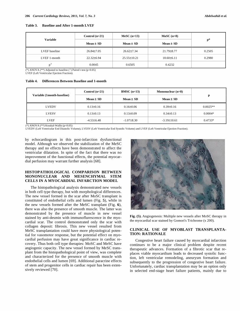
Experimental studies suggest that cardiac bone marrow stem cells (SC) transplantation can has a favorable impact on tissue perfusion. The strategy to repair the myocardial scar after infarction with SC has been proposed based on its ca-pacity to differentiate according to the environment and could give a new perspective for myocardial regeneration [62][55,62-64]. Studies in human beings have shown im-provement in clinical and functional cardiac status explained mainly by the angiogenic potential after stem cells transplan-tation [65-67]. Bone marrow stems cells are composed by mononuclear stem cells (MoSC) that contain hematopoietic stem cells with precursors of endothelial cells and mesen-chymal stem cells (MeSC). MeSC shows pluripotentiality for mesoderm-derived cells, but it is only 0,01- 0,05% of MoSC although they can be enriched based on the adhesion capac-ity of MeSC on tissue culture plate [67]. To compare the functional outcome of mononuclear stem cells (MoSC) and mesenchymal stem cell (MeSC) therapy after myocardial infarction in rats, we used several approaches. The flow cy-tometric analysis we used was CD45+ and CD 34+ for the MoSC and CD45- and CD34- for the MeSC. The MeSC was positive for vimentin in vitro. There was difference in base-line LVEF and LVEDV between all groups. After one month, LVEF decreased in the control group and remained unchanged in MoSC and MeSC groups. The myocardium was remodeled in all of the groups (Tables 3 and 4). Thus functional effectiveness was not demonstrated with both cell type therapy: MoSC and MeSC, when LVEF was analyzed
Table 2. Functional Evaluation of Cells Transplantation in a Model of Chagas Disease
Group Control Co-culture Difference Between Groups (p)
LVEDV. ml pre 0.69 ± 0.11 0.82 ± 0.07
LVEDV. ml post 0.73 ± 0.14 0.65 ± 0.14 0.0166
P 0.6311 0.0004
LVESV. ml pre 0.44 ± 0.08 0.56 ± 0.05
LVESV. ml post 0.46 ± 0.12 0.32 ± 0.09 0.0001
P 0.6523 <0.0001
EF% pre 36.74 ± 3.63 30.10 ± 5.71
EF% post 37.42 ± 6.66 51.76 ± 6.63 <0.0001
P 0.7684 <0.0001
206 Current Cardiology Reviews, 2011, Vol. 7, No. 3 Abdelwahid et al.
by echocardiogram in this post-infarction dysfunctional model. Although we observed the stabilization of the MeSC therapy and no effects have been demonstrated to affect the ventricular dilatation. In spite of the fact that there was no improvement of the functional effects, the potential myocar-dial perfusion may warrant further analysis [68].
HISTOPATHOLOGICAL COMPARISON BETWEEN
MONONUCLEAR AND MESENCHYMAL STEM
CELLS IN A MYOCARDIAL INFARCTION MODEL
The histopathological analysis demonstrated new vessels in both cell type therapy, but with morphological differences. The new vessel formed in the scar after MoSC transplant is constituted of endothelial cells and lumen (Fig. 5), while in the new vessels formed after the MeSC transplant (Fig. 6), there was also the presence of smooth muscle. The latter was demonstrated by the presence of muscle in new vessel stained by anti-desmin with immunoflurescence in the myo-cardial scar. The control demonstrated only the scar with collagen deposit: fibrosis. This new vessel resulted from MeSC transplantation could have more physiological poten-tial for vasomotor response, but the potential effect on myo-cardial perfusion may have great significance in cardiac re-covery. Thus both cell type therapies: MoSC and MeSC have angiogenic capacity. The new vessel formed by MeSC trans-plant from the histopathological point of view, was complete and characterized for the presence of smooth muscle with endothelial cells and lumen [69]. Additional paracrine effects of stem and progenitor cells in cardiac repair has been exten-sively reviewed [70].
Fig. (5). Angiogenesis: Multiple new vessels after MoSC therapy in
the myocardial scar stained by Gomori's Trichrome (x 200).
CLINICAL USE OF MYOBLAST TRANSPLANTA-TION: RATIONALE
Congestive heart failure caused by myocardial infarction continues to be a major clinical problem despite recent therapeutic advances. Formation of a fibrotic scar that re-places viable myocardium leads to decreased systolic func-tion, left ventricular remodeling, aneurysm formation and subsequently to the progression of congestive heart failure. Unfortunately, cardiac transplantation may be an option only in selected end-stage heart failure patients, mainly due to
Table 3. Baseline and After 1-month LVEF
Control (n=21) MeSC (n=13) MoSC (n=8)
Variable
Mean ± SD Mean ± SD Mean ± SD
p*
LVEF baseline 26.84±7.05 26.62±7.34 21.79±8.77 0.2505
LVEF 1-month 22.32±6.94 25.55±10.21 18.60±6.11 0.2980
p 0.0045 0.6505 0.4232
(*) ANOVA (**) Adjusted to baseline ( ) Paired t test (p<0.05)
LVEF (Left Ventricular Ejection Fraction).
Table 4. Differences Between Baseline and 1-month
Control (n=21) BMSC (n=13) Mononuclear (n=8)
Variable (1month-baseline)
Mean ± SD Mean ± SD Mean ± SD
p
LVEDV 0.13±0.16 0.16±0.06 0.39±0.16 0.0025**
LVESV 0.13±0.13 0.13±0.09 0.34±0.13 0.0004*
LVEF -4.53±6.48 -1.07±8.30 -3.19±10.61 0.4733*
(*) ANOVA (**) Kruskal-Wallis (p<0.05) LVEDV (Left Ventricular End Diastolic Volume), LVESV (Left Ventricular End Systolic Volume) and LVEF (Left Ventricular Ejection Fraction).

Stem Cells and Heart Repair Current Cardiology Reviews, 2011, Vol. 7, No. 3 207
Fig. (6). New vessel after MeSC in the myocardial scar stained by
Gomori's Trichrome (x 400).
organ shortage. Restoration of the total amount of contractile cells within the necrotic tissue as a result of cell transplanta-tion into myocardium have been widely studied in both ex-perimental and clinical conditions [71] Among variety of cell studied, autologous skeletal myoblasts, chronologically the first to enter the clinical arena, are one of the most encourag-ing cell source for cardiac repair because of their biologic properties and lack of ethical and immunological issues (Figs 7-9). Transplantation of totipotent stem cell types, including bone marrow derived stem cells, into a fibrous postinfarction scar may result in their differentiation into fibroblasts [71-73], therefore direct myocyte precursors, myoblasts, have been considered as possible cell sources in patients with chronic, postinfarction myocardial injury.
Numerous studies have explored different delivery tech-niques. As skeletal myoblasts do not extravasate, their poten-tial application in myocardial regeneration requires direct cell injection into the area of damaged myocardium. There-fore surgical approach (open-chest surgery) and several catheter-based methods had been proposed.
MYOBLAST TRANSPLANTATION DURING CAR-DIAC SURGERY
The initial clinical experience with autologous skeletal myoblast transplantation during open chest cardiac surgery was obtained by Ph. Menasché et.al., followed by phase one clinical trials performed both in Paris and in Poznan [58,74,75]. Both trials were done in patients in whom direct intramyocardial injections could be performed during coro-nary artery by-pass grafting (CABG). In the first clinical case described [58] several injections of the cell suspension into area of postinfarction injury within inferior wall of the left ventricle were performed. Five months after the proce-dure, a significant clinical status improvement was observed, including decrease of symptoms of heart failure by one NYHA class, an increase in segmental contractility and ejec-tion fraction seen on echocardiography as well as increase in tracer activity on positron emission tomography suggesting new onset metabolic activity in the previously non viable scar area [58]. An independent phase one clinical trial on autologous skeletal myoblast transplantation in patients un-dergoing CABG [71,74,75] was performed in Poznan. MI survivors scheduled for CABG, with an akinetic area of the left ventricle and no viable myocardium was detected by means of dobutamine stress echocardiography were included into the study. A skeletal myocardial biopsy was obtained in all patients from the vastus lateralis. The biopsy sample was digested and myoblasts (satellite cells) were isolated. The cells were cultured for three weeks to increase the number of cells to be implanted. A measurable increase in segmental contractility was seen in all patients 2 to 3 months after the procedure and this effect was maintained throughout a 36-months follow-up period. However, 3 years after combined myoblast transplantation and CABG, in almost every third case in the Poznan series, end diastolic left ventricular di-ameter increased, suggesting that cell transplantation did not prevented left ventricular remodeling or the number of cells transplanted was not sufficient to prevent left ventricular dilatation.
A US-based multicenter phase one clinical trial aimed at evaluation of myoblast transplantation during CABG has also been reported [76]. Eleven patients underwent myoblast
Fig. (7). Procedural steps of cell transplantation using trans-coronary venous approach. Left: coronary artery visualization in LAO 30 view; middle: administration of the contrast medium via a guiding catheter placed in the coronary sinus in LAO 30 view, note the balloon inflated at the tip of the guiding catheter to slow the contrast medium outflow; right: placement of TransAccess® catheter system in the anterior in-terventricular vein and injection of the cell suspension via the IntraLume® microcatheter into anterior wall myocardium – the arrow indicates the microcatheter tip.

208 Current Cardiology Reviews, 2011, Vol. 7, No. 3 Abdelwahid et al.
Fig. (9). Intravascular ultrasound image obtained from the cardiac
venous site. Please note the visibility of the pericardium (big ar-
rows) and the coronary artery parallel to the vein (small arrows),
enabling the orientation of TransAccess® catheter system.
transplantation combined with CABG. Echocardiography, PET and MRI scans showed evidence of increased viability in the area of grafted scar. Improvement of the mean ejection fraction from 22.7% to 35.9% has been observed. Another phase one study evaluating myoblast injections during CABG has been reported by Herreros et al. [77], suggesting safety and feasibility of the method as well as indicating its possible efficacy in increasing contractile left ventricular performance.
Cell transplantations during cardiac surgery has certain advantages, including easy access and good visualization of the target site as well as possible delivery of big amount of cells per unit area. However, direct transepicardial approach may cause additional risk related to the surgery. It must be underlined that possible candidates for cell transplantation usually have a history of multiple infarctions, with signifi-cant LV dysfunction and clinical symptoms of severe heart failure, being high-risk candidates for open-chest surgery.
Furthermore open-chest approach does not give free access to septal wall which is frequently inflicted by postinfarction injury.
Clear interpretation of clinical data obtained from trials evaluating cell transplantation during CABG is not possible. The effect of CABG and cellular transplantation performed at the same time cannot be easily distinguished. Despite the use of careful inclusion criteria aimed at selection of patients with no viable myocardium within the target postinfarction area, the possible effect of skeletal myoblast transplantation may be enhanced by myocardial revascularization. Large on-going and future clinical trials evaluating efficacy of myoblast injections during CABG may allow evaluating of the cell effect independently on restoration of blood flow. Nevertheless, a recently concluded randomized, placebo-controlled study of 97 patients, the myoblast autologous grafting in ischemic cardiomyopahty (MAGIC) trial failed to show improved LV function at 6 months after myoblast transplantation [78].
PERCUTANEOUS MYOBLAST TRANSPLANTATION
Myoblast transplantations, performed as a sole therapy during percutaneous procedures, may allow the evaluation of the cellular effect independent of revascularization. In addi-tion, it may enable repeated injections in patients with severe myocardial injury, since excessive numbers of transplanted cells in a single injection may result in a small percentage of survived and grafted cells. Initial experience has been ob-tained with cell injection into myocardium with the use of both endoventricular catheter systems [79] and transvenous approach [80,81].
The efficacy of cell transplantation may be very much dependent on the design of catheter system used [82,83]. Currently available endoventricular catheter systems, utilized for intramyocardial injections of therapeutic agents, have limited stability, since the injection pressure can cause ex-pulsion of the needle tip from the injection site. After needle withdrawal from a short perpendicular injection channel, therapeutic agent leakage back to the ventricle may occur. Such a back-flush of cells from the puncture site may cause the presence of graft cells in the systemic circulation and/or diminished number of cell delivered to the target area. Moreover, for endoventricular systems in which needle is directed perpendicularly to the inner surface of cardiac mus-
Fig. (8). Procedural steps of cell transplantation using trans-coronary venous approach. Left: coronary artery visualization in LAO 30 view;
middle: administration of the contrast medium via a non-occlusive guiding catheter placed in the coronary sinus; right: placement of
TransAccess® catheter system in the anterior interventricular vein and injection of the cell suspension via the IntraLume® microcatheter into
the septum – the arrow indicates the microcatheter tip.

Stem Cells and Heart Repair Current Cardiology Reviews, 2011, Vol. 7, No. 3 209
cle wall and does not follow the heart movement, thinned postinfarction scar is currently a relative contraindication.
Another catheter-based endovascular system for direct myocardial injection using IVUS guided needle punctures via the coronary venous system (the TransAccess®, Trans Vascular, Manlo Park, CA) has been recently developed [84,85]. The TransAccess® catheter is a monorail, compos-ite catheter system combining both a phased-array IVUS and a pre-shaped adjustable nitinol needle. After placing the TransAccess® system in the target coronary vein, through the coronary sinus, the system is oriented using the corre-sponding artery, pericardium and ventricular myocardium as landmarks with IVUS imaging. The nitinol needle is then extended into the myocardium and a microinfusion catheter (IntraLume™, TransVascular Inc.) is then advanced through the needle deep into the myocardium with simultaneous in-jection of the therapeutic agent.
The POZNAN trial [81] was performed as a phase one clinical trial for both the TransAccess® catheter system and for percutaneous autologous myoblast transplantations per-formed as a sole therapy. The trial has confirmed the feasi-bility of intramyocardial injections performed using the TransAccess® system. Precise advancement of the micro lumen catheter in the remote target area up to 4 cm deep within the injured myocardium was obtained. The use of both the anterior interventricular vein as well as the middle cardiac vein, parallel to the posterior descending coronary artery, has been shown to be feasible. In the POZNAN trial, in 4 cases the middle vein was used to advance the TransAc-cess® system closer to the apical segments of the left ventri-cle, as compared to cases using the anterior interventricular venous approach [81]. The lack of procedural success in one patient, related to the inability of appropriate positioning of the guiding catheter across the venous valve present at the bifurcation of the great cardiac vein, suggests the need for better coronary sinus guiding catheter design (10F guiding catheters were used). In remaining POZNAN trial patients, two to four intramyocardial injections 1.5-4.5 cm deep were performed in each patient, delivering up to 100 million cells in 0.6 – 2.5 ml of saline. During 6 months follow-up NYHA class improved in all patients and ejection fraction assessed by echocardiography increased 3-8 percent in 6 out of 9 cases [81,83].
Smits et al. [79] used transventricular approach to inject myoblast suspensions into the area of postinfarction injury. They observed increase in LV ejection fraction assessed by angiography at 3 month follow-up, but nucluear radiography and magnetic resonance failed to confirm this improvement. However, they recently published a very interesting study [86] have shown hemodynamic improvement evaluated by pressure-volume loops after follow-up for up to one year after percutaneous myoblast transplantation. 10-15 injections of autologous myoblasts using Myostar™ (Cordis, Warren, NJ) resulted in increased ejection fraction at 6 months, in-creased cardiac output, reduction of end-systolic volume and a trend towards improved stroke work. Another recently published study [86] decribes results of transventricular in-jections performed using fluoroscopy guided MyoCath™ catheter (Bioheart, Weston, FL) or the NOGA™-guided catheter system (Biosense-Webster, Waterloo, Belgium).
The study failed to show improvement in the ejection frac-tion but wall motion score index improved both at rest at under low-dose dobutamine. In addition, the result of the study indicate the possibility of arrhythmogenic effect of myoblast transplantation.
MYOBLAST-RELATED SAFETY ISSUES
Myocardial and skeletal muscle tissues differ signifi-cantly in their electromechanical properties. Cardiac cells, having special cell-cell junctions, even though separated one from another, act together synchronically. Junctions contain adherins and gap junctions for mechanical and electrical coupling. Cardiac tissue gap junctions contain connexin-43 transmembrane protein by which electrical current can be fast and freely conducted [3]. Although certain data suggest that skeletal myoblasts may acquire few characteristics of cardiomyocytes, generally it could be assumed that the grafted cells do not transdifferentiate and keep morphologi-cal and electrophysiological properties of skeletal muscle [87,88]. It is speculated that skeletal myoblasts (satellite cells) are not able to form intercellular junctions characteris-tic for cardiomyocytes. However, it was shown that the lack of junctions between grafted cells and host tissue do not pre-clude improvement in LV contractile function [87].
There is evidence that this positive effect of skeletal myoblasts on myocardial contractility seems to last over time and is correlated with the number of implanted cells [50].
Experimental studies performed on myocardial wound strips indicate that skeletal myoblast grafts do contract when ex-ogenously stimulated [49]. In addition, cardiomyocytes and skeletal myoblasts, when placed in co-culture, forms syn-chronous beating network [89]. It has been suggested that transplanted cells can contract synchronously even in the absence of connections between cells, because a simple stretch may initiate contraction [90]. This phenomenon is important due to the possibility of insulation of transplanted cells by scar tissue. The scar may form a physical barrier which impede electromechanical coupling.
It is speculated that the inability of skeletal myoblasts to transdifferentiate to cardiomyocytes and to form junctions with neighboring cells may be a substrate for ventricular re-entry arrhythmia. Indeed, current experimental and clinical data suggest a possibility of increased risk of arrhyth-mogenicity. In their first clinical series, Menasché at al. [58] implanted automatic internal cardioverter-defibrillators (AICD) in 4 out of 10 patients receiving autologous skeletal myoblast transplantations during CABG due to sustained episodes of ventricular tachycardia (VT). The possible ar-rhythmogenic effect have been also noticed in an endoven-tricular catheter-based trial which had to be temporarily stopped because of 2 sudden deaths, probably due to ar-rhythmia. In the Poznan CABG phase one experience [74,75], episodes of sustained ventricular tachycardia (VT) were observed in first 2 patients during early postoperative period, but prophylactic amiodarone administration in other patients prevented VT episodes and no amiodarone treatment was continued later then 6 weeks during follow-up. This corresponds to the experience of Menasche et al [58] Later during follow-up period only one of their four AICD patients experienced asymptomatic VT episodes [71]. On the other

210 Current Cardiology Reviews, 2011, Vol. 7, No. 3 Abdelwahid et al.
hand, observations from percutaneous series in the POZNAN trial, indicating successful prevention of cell transplantation-related ventricular arrhythmia by prophylactic amiodarone administration, suggest that AICD implantations may not necessarily needed in all patients that undergo myoblast transplantations [81,83].
Based on published data from clinical studies, it could be speculated that the possible arrhythmogenic effect of myoblast transplantation is noticed only in the initial weeks after the procedure. The possible arrhythmogenic effect of myoblast transplantation is more probably related to its me-chanics, including myocardial puncture and the inflamma-tory response to transplanted cells, some of which die after injection, than to possible problems with electromechanical coupling between newly developed myocytes and cardio-myocytes. Possible electromechanical coupling problems would result in late arrhythmia as cells differentiate (down-regulation of connexin-43 and N-cadherin), a situation that has not been observed in clinical trials so far.
At the current stage, only moderate numbers of patients that have undergone autologous skeletal myoblast transplan-tations, it is difficult to predict whether skeletal myoblasts are really arrhythmogenic, especially that patients with ischemic LV dysfunction frequently develop ventricular ar-rhythmia. Indeed, the experience of MAGIC trial [78] re-flected this trend whereby no significant difference in ven-tricular arrhythmias or major cardiac adverse events was detected between myoblast-treated patients and placebo con-trol group at 6-month follow-up. Nevertheless, future studies on cell transplantation in patients with postinfarction heart failure will have to focus on potential arrhythmogenic effect. Similarly, large phase two/three clinical trials are needed to assess the efficacy of myoblast transplantation in chronic postinfarction myocardial injury.
REFERENCES
[1] Reubinoff BE, Itsykson P, Turetsky T, et al. Neural progenitors
from human embryonic stem cells. Nat Biotechnol 2001; 19: 1134-40.
[2] Assady S, Maor G, Amit M, Itskovitz-Eldor J, Skorecki KL, Tzukerman M. Insulin production by human embryonic stem cells.
Diabetes 2001; 50: 1691-7. [3] Beyer EC, Paul DL, Goodenough DA. Connexin43: a protein from
rat heart homologous to a gap junction protein from liver. J Cell Biol 1987; 105: 2621-9.
[4] Levenberg S, Golub JS, Amit M, Itskovitz-Eldor J, Langer R. En-dothelial cells derived from human embryonic stem cells. Proc Natl
Acad Sci USA 2002; 99: 4391-6. [5] Kehat I, Khimovich L, Caspi O, et al. Electromechanical integra-
tion of cardiomyocytes derived from human embryonic stem cells. Nat Biotechnol 2004; 22: 1282-9.
[6] Mummery C, Ward D, van den Brink CE, et al. Cardiomyocyte differentiation of mouse and human embryonic stem cells. J Anat
2002; 200: 233-42. [7] Assmus B, Honold J, Schachinger V, Britten MB, Fischer-Rasokat
U, Lehmann R, Teupe C, Pistorius K, Martin H, Abolmaali ND, Tonn T, Dimmeler S, Zeiher AM. Transcoronary transplantation of
progenitor cells after myocardial infarction. N Engl J Med 2006; 355: 1222-32.
[8] Briguori C, Reimers B, Sarais C, et al. Direct intramyocardial percutaneous delivery of autologous bone marrow in patients with
refractory myocardial angina. Am Heart J 2006; 151: 674-80. [9] Condorelli G, Borello U, De Angelis L, Latronico M, Sirabella D,
Coletta M, Galli R, Balconi G, Follenzi A, Frati G, Cusella De An-gelis MG, Gioglio L, et al. Cardiomyocytes induce endothelial cells
to trans-differentiate into cardiac muscle: implications for myocar-
dium regeneration. Proc Natl Acad Sci USA 2001; 98: 10733-8. [10] Kehat I, Kenyagin-Karsenti D, Snir M, et al. Human embryonic
stem cells can differentiate into myocytes with structural and func-tional properties of cardiomyocytes. J Clin Invest 2001; 108: 407-
14. [11] Caspi O, Gepstein L. Potential applications of human embryonic
stem cell-derived cardiomyocytes. Ann N Y Acad Sci 2004; 1015: 285-98.
[12] Menasche P. Embryonic stem cells pace the heart. Nat Biotechnol 2004; 22: 1237-8.
[13] Odorico JS, Kaufman DS, Thomson JA. Multilineage differentia-tion from human embryonic stem cell lines. Stem Cells 2001; 19:
193-204. [14] Carpenter MK, Rosler ES, Fisk GJ, et al. Properties of four human
embryonic stem cell lines maintained in a feeder-free culture sys-tem. Dev Dyn 2004; 229: 243-58.
[15] Rosler ES, Fisk GJ, Ares X, et al. Long-term culture of human embryonic stem cells in feeder-free conditions. Dev Dyn 2004;
229: 259-74. [16] Abeyta MJ, Clark AT, Rodriguez RT, Bodnar MS, Pera RA, Firpo
MT. Unique gene expression signatures of independently-derived human embryonic stem cell lines. Hum Mol Genet 2004; 13: 601-8.
[17] Richards M, Fong CY, Chan WK, Wong PC, Bongso A. Human feeders support prolonged undifferentiated growth of human inner
cell masses and embryonic stem cells. Nat Biotechnol 2002; 20: 933-6.
[18] Pekkanen-Mattila M, Kerkela E, Tanskanen JM, et al. Substantial variation in the cardiac differentiation of human embryonic stem
cell lines derived and propagated under the same conditions--a comparison of multiple cell lines. Ann Med 2009; 41: 360-70.
[19] Stojkovic M, Lako M, Strachan T, Murdoch A. Derivation, growth and applications of human embryonic stem cells. Reproduction
2004; 128: 259-67. [20] Heng BC, Haider HK, Sim EK, Cao T, Tong GQ, Ng SC. Com-
ments about possible use of human embryonic stem cell-derived cardiomyocytes to direct autologous adult stem cells into the car-
diomyogenic lineage. Acta Cardiol 2005; 60: 7-12. [21] Heng BC, Cao T, Haider HK, Wang DZ, Sim EK, Ng SC. An
overview and synopsis of techniques for directing stem cell differ-entiation in vitro. Cell Tissue Res 2004; 315: 291-303.
[22] Heng BC, Cao T, Tong GQ, Ng SC. Potential utility of cell-permeable transcription factors to direct stem cell differentiation.
Stem Cells Dev 2004; 13: 460-2. [23] Heng BC, Haider HK, Sim EK, Cao T, Ng SC. Strategies for di-
recting the differentiation of stem cells into the cardiomyogenic lineage in vitro. Cardiovasc Res 2004; 62: 34-42.
[24] Stevens KR, Kreutziger KL, Dupras SK, et al. Physiological func-tion and transplantation of scaffold-free and vascularized human
cardiac muscle tissue. Proc Natl Acad Sci USA 2009; 106: 16568-73.
[25] Martin MJ, Muotri A, Gage F, Varki A. Human embryonic stem cells express an immunogenic nonhuman sialic acid. Nat Med
2005; 11: 228-32. [26] Ellerstrom C, Strehl R, Moya K, et al. Derivation of a xeno-free
human embryonic stem cell line. Stem Cells 2006; 24: 2170-6. [27] Rajala K, Hakala H, Panula S, et al. Testing of nine different xeno-
free culture media for human embryonic stem cell cultures. Hum Reprod 2007; 22: 1231-8.
[28] Amit M, Margulets V, Segev H, et al. Human feeder layers for human embryonic stem cells. Biol Reprod 2003; 68: 2150-6.
[29] Unger C, Skottman H, Blomberg P, Dilber MS, Hovatta O. Good manufacturing practice and clinical-grade human embryonic stem
cell lines. Hum Mol Genet 2008; 17: R48-53. [30] Pekkanen-Mattila M, Chapman H, Kerkela E, et al. Human embry-
onic stem cell-derived cardiomyocytes: demonstration of a portion of cardiac cells with fairly mature electrical phenotype. Exp Biol
Med (Maywood) 2010; 235: 522-30. [31] Drukker M, Katz G, Urbach A, et al. Characterization of the ex-
pression of MHC proteins in human embryonic stem cells. Proc Natl Acad Sci USA 2002; 99: 9864-9.
[32] Robertson NJ, Brook FA, Gardner RL, Cobbold SP, Waldmann H, Fairchild PJ. Embryonic stem cell-derived tissues are immunogenic
but their inherent immune privilege promotes the induction of tol-erance. Proc Natl Acad Sci USA 2007; 104: 20920-5.

Stem Cells and Heart Repair Current Cardiology Reviews, 2011, Vol. 7, No. 3 211
[33] Lanzendorf SE, Boyd CA, Wright DL, Muasher S, Oehninger S,
Hodgen GD. Use of human gametes obtained from anonymous do-nors for the production of human embryonic stem cell lines. Fertil
Steril 2001; 76: 132-7. [34] Hwang WS, Ryu YJ, Park JH, et al. Evidence of a pluripotent
human embryonic stem cell line derived from a cloned blastocyst. Science 2004; 303: 1669-74.
[35] Armstrong L, Lako M, Dean W, Stojkovic M. Epigenetic modifica-tion is central to genome reprogramming in somatic cell nuclear
transfer. Stem Cells 2006; 24: 805-14. [36] Rodriguez-Piza I, Richaud-Patin Y, Vassena R, et al. Reprogram-
ming of human fibroblasts to induced pluripotent stem cells under xeno-free conditions. Stem Cells 2010; 28: 36-44.
[37] Takahashi K, Tanabe K, Ohnuki M, et al. Induction of pluripotent stem cells from adult human fibroblasts by defined factors. Cell
2007; 131: 861-72. [38] Ieda M, Fu JD, Delgado-Olguin P, et al. Direct reprogramming of
fibroblasts into functional cardiomyocytes by defined factors. Cell 2010; 142: 375-86.
[39] Thomson JA, Itskovitz-Eldor J, Shapiro SS, et al. Embryonic stem cell lines derived from human blastocysts. Science 1998; 282:
1145-7. [40] Thomson JA, Kalishman J, Golos TG, et al. Isolation of a primate
embryonic stem cell line. Proc Natl Acad Sci USA 1995; 92: 7844-8.
[41] Doss MX, Koehler CI, Gissel C, Hescheler J, Sachinidis A. Em-bryonic stem cells: a promising tool for cell replacement therapy. J
Cell Mol Med 2004; 8: 465-73. [42] Mitjavila-Garcia MT, Simonin C, Peschanski M. Embryonic stem
cells: meeting the needs for cell therapy. Adv Drug Deliv Rev 2005; 57: 1935-43.
[43] Bjorklund LM, Sanchez-Pernaute R, Chung S, et al. Embryonic stem cells develop into functional dopaminergic neurons after
transplantation in a Parkinson rat model. Proc Natl Acad Sci USA 2002; 99: 2344-9.
[44] Asano T, Ageyama N, Takeuchi K, et al. Engraftment and tumor formation after allogeneic in utero transplantation of primate em-
bryonic stem cells. Transplantation 2003; 76: 1061-7. [45] Hattori F, Chen H, Yamashita H, et al. Nongenetic method for
purifying stem cell-derived cardiomyocytes. Nat Methods 2010; 7: 61-6.
[46] Van Hoof D, Dormeyer W, Braam SR, et al. Identification of cell surface proteins for antibody-based selection of human embryonic
stem cell-derived cardiomyocytes. J Proteome Res 2010; 9: 1610-8. [47] Hattori F, Fukuda K. Strategies for ensuring that regenerative car-
diomyocytes function properly and in cooperation with the host myocardium. Exp Mol Med 2010; 42: 155-65.
[48] Ghostine S, Carrion C, Souza LC, et al. Long-term efficacy of myoblast transplantation on regional structure and function after
myocardial infarction. Circulation 2002; 106: I131-6. [49] Murry CE, Wiseman RW, Schwartz SM, Hauschka SD. Skeletal
myoblast transplantation for repair of myocardial necrosis. J Clin Invest 1996; 98: 2512-23.
[50] Scorsin M, Hagege A, Vilquin JT, et al. Comparison of the effects of fetal cardiomyocyte and skeletal myoblast transplantation on
postinfarction left ventricular function. J Thorac Cardiovasc Surg 2000; 119: 1169-75.
[51] Taylor DA, Atkins BZ, Hungspreugs P, et al. Regenerating func-tional myocardium: improved performance after skeletal myoblast
transplantation. Nat Med 1998; 4: 929-33. [52] Dorfman J, Duong M, Zibaitis A, et al. Myocardial tissue engineer-
ing with autologous myoblast implantation. J Thorac Cardiovasc Surg 1998; 116: 744-51.
[53] Menasche P, Hagege AA, Vilquin JT, et al. Autologous skeletal myoblast transplantation for severe postinfarction left ventricular
dysfunction. J Am Coll Cardiol 2003; 41: 1078-83. [54] Chachques JC, Herreros J, Trainini J, et al. Autologous human
serum for cell culture avoids the implantation of cardioverter-defibrillators in cellular cardiomyoplasty. Int J Cardiol 2004; 95
Suppl 1: S29-33. [55] Verfaillie CM. Adult stem cells: assessing the case for pluripo-
tency. Trends Cell Biol 2002; 12: 502-8. [56] Guarita-Souza LC, Carvalho KA, Rebelatto C, et al. Cell transplan-
tation: differential effects of myoblasts and mesenchymal stem cells. Int J Cardiol 2006; 111: 423-9.
[57] Souza LC, Carvalho KA, Rebelatto C, et al. Combined transplanta-
tion of skeletal myoblasts and mesenchymal cells (cocultivation) in ventricular dysfunction after myocardial infarction. Arq Bras Car-
diol 2004; 83: 294-9; 288-93. [58] Menasche P, Hagege AA, Vilquin JT, et al. Autologous skeletal
myoblast transplantation for severe postinfarction left ventricular dysfunction. J Am Coll Cardiol 2003; 41: 1078-83.
[59] Guarita-Souza LC, Carvalho KA, Woitowicz V, et al. Simultane-ous autologous transplantation of cocultured mesenchymal stem
cells and skeletal myoblasts improves ventricular function in a murine model of Chagas disease. Circulation 2006; 114: I120-4.
[60] Carvalho KA, Guarita-Souza LC, Rebelatto CL, Set al. Could the coculture of skeletal myoblasts and mesenchymal stem cells be a
solution for postinfarction myocardial scar? Transplant Proc 2004; 36: 991-2.
[61] Perin EC, Dohmann HF, Borojevic R, et al. Transendocardial, autologous bone marrow cell transplantation for severe, chronic
ischemic heart failure. Circulation 2003; 107: 2294-302. [62] Orlic D, Kajstura J, Chimenti S, Bodine DM, Leri A, Anversa P.
Bone marrow stem cells regenerate infarcted myocardium. Pediatr Transplant 2003; 7 Suppl 3: 86-8.
[63] Wollert KC, Meyer GP, Lotz J, et al. Intracoronary autologous bone-marrow cell transfer after myocardial infarction: the BOOST
randomised controlled clinical trial. Lancet 2004; 364: 141-8. [64] Strauer BE, Brehm M, Zeus T, et al. Repair of infarcted myocar-
dium by autologous intracoronary mononuclear bone marrow cell transplantation in humans. Circulation 2002; 106: 1913-8.
[65] Silva GV, Litovsky S, Assad JA, et al. Mesenchymal stem cells differentiate into an endothelial phenotype, enhance vascular den-
sity, and improve heart function in a canine chronic ischemia model. Circulation 2005; 111: 150-6.
[66] Friedenstein AJ, Petrakova KV, Kurolesova AI, Frolova GP. Het-erotopic of bone marrow. Analysis of precursor cells for osteogenic
and hematopoietic tissues. Transplantation 1968; 6: 230-47. [67] Guarita-Souza LC, Carvalho KA, Simeone BR, Franscisco JC,
Miyague N, Olandoski M. Functional outcome of bone marrow stem cells: mononuclear versus mesenchymal stem cells after cellu-
lar therapy in myocardial scar in Wistar rats. Transplant Proc 2006; 38: 1953-4.
[68] Guarita-Souza LC,Carvalho KA, Rebelatto C, et al. Comparison of mononuclear and mesenchymal stem cell transplantation in myo-
cardium infarction. Brazilian Journal of Cardiovascular Surgery 2005; 20: 270-8.
[69] Siminiak T, Kurpisz M. Myocardial replacement therapy. Circula-tion 2003; 108: 1167-71.
[70] Gnecchi M, Zhang Z, Ni A, Dzau VJ. Paracrine mechanisms in adult stem cell signaling and therapy. Circ Res 2008; 103: 1204-19.
[71] Wang J-S., Shum-Tim D., Chedrawy E. Marrow stromal cells for cellular cardiomyoplasty: the importance of microenvironment for
milieu dependent differentiation. Circulation 2000; 102: II-683. [72] Bel A, Messas E, Agbulut O, et al. Transplantation of autologous
fresh bone marrow into infarcted myocardium: a word of caution. Circulation 2003; 108 Suppl 1: II247-52.
[73] Menasche P, Hagege AA, Scorsin M, et al. Myoblast transplanta-tion for heart failure. Lancet 2001; 357: 279-20.
[74] Siminiak T, Kalawski R, Kurpisz M. Myoblast transplantation in the treatment of postinfarction myocardial contractility impairment.
Kardiol Pol 2002; 56: 131-7. [75] Siminiak T, Kalawski R, Fiszer D, et al. Transplantation of autolo-
gous skeletal myoblasts in the treatment of patients with postinfarc-tion heart failure. Circulation 2002; 106: II-636.
[76] Dib N., McCarthy P., Campbell A. Safety and feasibility of autolo-gous myoblast transplantation in patients undergoing coronary ar-
tery bypass grafting: results from the United States experience. J Am Coll Cardiol 2003; 41 suppl.
[77] Herreros J, Prosper F, Perez A, et al. Autologous intramyocardial injection of cultured skeletal muscle-derived stem cells in patients
with non-acute myocardial infarction. Eur Heart J 2003; 24: 2012-20.
[78] Menasche P, Alfieri O, Janssens S, et al. The Myoblast Autologous Grafting in Ischemic Cardiomyopathy (MAGIC) trial: first random-
ized placebo-controlled study of myoblast transplantation. Circula-tion 2008; 117: 1189-200.
[79] Smits PC, van Geuns RJ, Poldermans D, et al. Catheter-based intramyocardial injection of autologous skeletal myoblasts as a

212 Current Cardiology Reviews, 2011, Vol. 7, No. 3 Abdelwahid et al.
primary treatment of ischemic heart failure: clinical experience
with six-month follow-up. J Am Coll Cardiol 2003; 42: 2063-9. [80] Siminiak T, Fiszer D, Jerzykowska O, Grygielska B, Kalmucki P,
Kurpisz M. Percutaneous autologous myoblast transplantation in the treatment of post-infarction myocardial contractility impair-
ment-report on two cases. Kardiol Pol 2003; 59: 492-501. [81] Siminiak T, Fiszer D, Jerzykowska O, et al. Percutaneous trans-
coronary-venous transplantation of autologous skeletal myoblasts in the treatment of post-infarction myocardial contractility impair-
ment: the POZNAN trial. Eur Heart J 2005; 26: 1188-95. [82] Sherman W, Martens TP, Viles-Gonzalez JF, Siminiak T. Catheter-
based delivery of cells to the heart. Nat Clin Pract Cardiovasc Med 2006; 3 Suppl 1: S57-64.
[83] Siminiak T, Kalmucki P, Kurpisz M. Autologous skeletal myoblasts for myocardial regeneration. J Interv Cardiol 2004; 17:
357-65. [84] Thompson CA, Nasseri BA, Makower J, et al. Percutaneous trans-
venous cellular cardiomyoplasty. A novel nonsurgical approach for myocardial cell transplantation. J Am Coll Cardiol 2003; 41: 1964-
71.
[85] Thompson CA, Oesterle SN. Biointerventional cardiology: the
future interface of interventional cardiovascular medicine and bio-engineering. Vasc Med 2002; 7: 135-40.
[86] Smits PC, Nienaber C, Colombo A, et al. Myocardial repair by percutaneous cell transplantation of autologous skeletal myoblast
as a stand alone procedure in post myocardial infarction chronic heart failure patients. EuroIntervention 2006; 1: 417-24.
[87] Pagani FD, DerSimonian H, Zawadzka A, et al. Autologous skele-tal myoblasts transplanted to ischemia-damaged myocardium in
humans. Histological analysis of cell survival and differentiation. J Am Coll Cardiol 2003; 41: 879-88.
[88] Taylor DA, Atkins BZ, Hungspreugs P, et al. Regenerating func-tional myocardium: improved performance after skeletal myoblast
transplantation. Nat Med 1998; 4: 929-33. [89] Hagege AA, Vilquin JT, Bruneval P, Menasche P. Regeneration of
the myocardium: a new role in the treatment of ischemic heart dis-ease? Hypertension 2001; 38: 1413-5.
[90] Menasche P, Hagege AA, Scorsin M, Pet al. Myoblast transplanta-tion for heart failure. Lancet 2001; 357: 279-80.
Received: October 24, 2010 Revised: January 31, 2011 Accepted: February 3, 2011
Related Documents