Welcome message from author
This document is posted to help you gain knowledge. Please leave a comment to let me know what you think about it! Share it to your friends and learn new things together.
Transcript


Stem Cell Biology and Regenerative Medicine
Series EditorKursad Turksen, [email protected]
For further volumes:http://www.springer.com/series/7896

wwwwwwwwwwwwwww

Alison L. AllanEditor
Cancer Stem Cells in Solid Tumors
EditorAlison L. Allan Depts. of Oncology and Anat. & Cell BiologySchulich School of Med. and Dent.University of Western OntarioLondon, Ontario, [email protected]
ISBN 978-1-61779-245-8 e-ISBN 978-1-61779-246-5DOI 10.1007/978-1-61779-246-5Springer New York Dordrecht Heidelberg London
Library of Congress Control Number: 2011932988
© Springer Science+Business Media, LLC 2011All rights reserved. This work may not be translated or copied in whole or in part without the written permission of the publisher (Humana Press, c/o Springer Science+Business Media, LLC, 233 Spring Street, New York, NY 10013, USA), except for brief excerpts in connection with reviews or scholarly analysis. Use in connection with any form of information storage and retrieval, electronic adaptation, computer software, or by similar or dissimilar methodology now known or hereafter developed is forbidden.The use in this publication of trade names, trademarks, service marks, and similar terms, even if they are not identified as such, is not to be taken as an expression of opinion as to whether or not they are subject to proprietary rights.
Printed on acid-free paper
Humana Press is part of Springer Science+Business Media (www.springer.com)
v
Preface
Recently, there has been increasing support for the “cancer stem cell” hypothesis, which postulates that cancer arises from a subpopulation of tumor-initiating cells or cancer stem cells (CSCs). There are currently two conflicting views that attempt to explain tumor formation. The classical stochastic model suggests that every cell within a tumor is a potential tumor-initiator, but that entry into the cell cycle is governed by a low probability of stochastic mutations. According to this model, it would be impossible to tell which cell initiated the tumor since each cell has an equal ability to be malignant. By contrast, the hierarchy theory (upon which the CSC hypothesis is based) proposes that only a subset of cells within a tumor is capable of initiating tumor growth, but that these cells all do so at a high frequency. According to this theory, it should be possible to identify and target the cells responsible for tumor initiation and progression because not all cells have the same phenotypic and func-tional characteristics.
While the idea of CSCs has been around for more than 100 years, evidence from the hematology field has now demonstrated the critical role of stem cells in hemato-logical malignancies and suggested that these same mechanisms could also be cen-tral to the initiation, progression, and treatment of solid cancers. Indeed, several pivotal studies have recently provided compelling evidence that these cells do exist in solid tumors of many types including breast, brain, colorectal, pancreas, prostate, melanoma, lung, ovarian, liver, and head and neck cancer. Furthermore, clinical and experimental studies have demonstrated that CSCs exhibit many classical properties of normal stem cells, including a high self-renewal capacity and the ability to gener-ate heterogeneous lineages; the requirement for a specific “niche”/microenvironment to grow; and an increased capacity for self-protection against harsh environments, toxins, and drugs.
This multi-authored volume focuses specifically on the role of CSCs in solid cancers. The authors are all active investigators with research programs related to oncology and/or stem cell biology, and are leaders in their field. Part I (Chap. 1) serves to introduce the concept of CSCs vs. normal stem cells, including a histori-cal perspective and the contributing lessons from leukemia. Part II (Chaps. 2–11) describes the identification and role of CSCs in various forms of solid cancer,

vi Preface
organized according to disease site. Part III (Chaps. 12–14) elaborates on molecular pathways that are involved in driving CSC function, with a particular focus on the convergence of embryonic and tumorigenic signaling pathways. Part IV (Chaps. 15–18) describes available model systems and modalities for studying CSC biol-ogy and therapeutic development, including in vitro and in vivo model systems and assays and imaging modalities. Part V (Chaps. 19–23) discusses the importance of CSCs for cancer management and treatment, including implications for prognosis, prediction, and treatment resistance. Finally, Part VI (Chap. 24) provides the con-cluding thoughts for the book, including consideration of the controversy sur-rounding the CSC hypothesis. The editor and the authors hope that this work will provide a comprehensive overview of this evolving and important field.
London, ON Alison L. AllanCanada

vii
Acknowledgments
I would like to express my gratitude to all of the authors for their scholarly efforts in summarizing the current literature in this rapidly evolving field. I would also like to thank Mindy Okura-Marszycki and Kursad Turksen for giving me the opportunity to edit this book, and acknowledge Vindra Dass and Renata Hutter for all of their help throughout the editorial and publication process. Finally, I am grateful to mem-bers of my own research group for their patience, contributions, helpful discussion, and continued hard work in this exciting area of research.

wwwwwwwwwwwwwww

ix
Contents
Part I Introduction to Cancer Stem Cells
1 Cancer Stem Cells: Historical Perspectives and Lessons from Leukemia ................................................................. 3Christopher R. Cogle
Part II Cancer Stem Cells in Solid Tumors
2 Cancer Stem Cells in Breast Cancer .................................................... 15Jenny E. Chu and Alison L. Allan
3 Cancer Stem Cells in Brain Cancer ..................................................... 37Xin Wang, Chitra Venugopal, and Sheila K. Singh
4 Cancer Stem Cells in Colorectal Cancer.............................................. 57Mauro Biffoni, Eros Fabrizi, and Lucia Ricci-Vitiani
5 Cancer Stem Cells in Pancreatic Cancer ............................................. 79Jorge Dorado, Alicia G. Serrano, and Christopher Heeschen
6 Cancer Stem Cells in Prostate Cancer ................................................. 99Paula Kroon, Davide Pellacani, Fiona M. Frame, Norman J. Maitland, and Anne T. Collins
7 Cancer Stem Cells in Melanoma .......................................................... 117Ping Jin, Qiuzhen Liu, Marianna Sabatino, David F. Stroncek, Francesco M. Marincola, and Ena Wang
8 Cancer Stem Cells in Lung Cancer ...................................................... 139Jun Shen and Feng Jiang
9 Cancer Stem Cells in Ovarian Cancer ................................................. 151Fang Fang, Curt Balch, Meng Li, Jay M. Pilrose, and Kenneth P. Nephew
x Contents
10 Cancer Stem Cells in Hepatocellular Cancer ...................................... 177Russell C. Langan and Itzhak Avital
11 Cancer Stem Cells in Head and Neck Cancer ..................................... 197Mark E.P. Prince and Samantha J. Davis
Part III Cancer Stem Cell Gene Expression and Mechanisms: Convergence of Embryonic and Tumorigenic Signaling Pathways
12 Relationship Between Regulatory Pathways in Pluripotent Stem Cells and Human Tumors ................................... 209Olga Gaidarenko and Yang Xu
13 Influence of the Embryonic Microenvironment on Tumor Progression ............................................................................ 223Daniela Quail, Meghan Taylor, Michael Jewer, and Lynne-Marie Postovit
14 The Epithelial-to-Mesenchymal Transition and Cancer Stem Cells ........................................................................... 243Jonas Fuxe
Part IV Model Systems for Studying Cancer Stem Cell Biology and Therapeutic Development
15 Application of Stem Cell Assays for the Characterization of Cancer Stem Cells .............................................................................. 259Pamela M. Willan and Gillian Farnie
16 Zebrafish as a Model to Study Stem Cells in Development, Disease, and Cancer .................................................. 283Viviana Anelli, Cristina Santoriello, and Marina C. Mione
17 Imaging Cancer Stem Cells ................................................................... 297Paula Foster
18 Mouse Models for Studying Normal and Cancer Stem Cells ............ 311David A. Hess
Part V Clinical and Therapeutic Implications of Cancer Stem Cells
19 Cancer Stem Cells and Disease Prognosis ........................................... 329Zeshaan A. Rasheed, Jeanne Kowalski, and William H. Matsui
20 Mechanisms of Radioresistance in Cancer Stem Cells ....................... 345Cleo Y-F Lee and Maximilian Diehn

xiContents
21 The Role of ABC Transporters in Cancer Stem Cell Drug Resistance ...................................................................................... 361Vera S. Donnenberg, Ludovic Zimmerlin, and Albert D. Donnenberg
22 Resistance to Endocrine Therapy in Breast Cancer: Are Breast Cancer Stem Cells Implicated? ......................................... 381Ciara S. O’Brien, Sacha J. Howell, Gillian Farnie, and Robert B. Clarke
23 Future Directions: Cancer Stem Cells as Therapeutic Targets .......................................................................... 403Alysha K. Croker and Alison L. Allan
Part VI Final Thoughts
24 Final Thoughts: Complexity and Controversy Surrounding the “Cancer Stem Cell” Paradigm ....................................................... 433Craig Gedye, Richard P. Hill, and Laurie Ailles
Index ................................................................................................................ 465

wwwwwwwwwwwwwww
xiii
Contributors
Laurie Ailles Ontario Cancer Institute, Campbell Family Institute of Cancer Research; and Departments of Medical Biophysics, University of Toronto, Toronto, ON, Canada
Alison L. Allan Departments of Oncology and Anatomy & Cell Biology, Schulich School of Medicine and Dentistry, University of Western Ontario, London, ON, Canada
London Regional Cancer Program, London Health Sciences Centre, London, ON, Canada
Viviana Anelli IFOM, The FIRC Institute of Molecular Oncology, Milan, Italy
Itzhak Avital National Cancer Institute (NIH), Surgery Branch, Bethesda, MD, USA
Curt Balch Medical Sciences Program, Indiana University, Bloomington, IN, USA
Indiana University Simon Cancer Center, Indianapolis, IN, USA
Mauro Biffoni Department of Hematology, Oncology and Molecular Medicine, Istituto Superiore di Sanità, Rome, Italy
Jenny E. Chu Department of Anatomy & Cell Biology, Schulich School of Medicine and Dentistry, University of Western Ontario, London, ON, Canada
Robert B. Clarke Breast Biology Group, School of Cancer and Enabling Sciences, Paterson Institute for Cancer Research, University of Manchester, Manchester, UK
Christopher R. Cogle Department of Medicine, Division of Hematology/ Oncology, University of Florida, Gainesville, FL, USA
Program in Stem Cell Biology and Regenerative Medicine, University of Florida, Gainesville, FL, USA
xiv Contributors
Anne T. Collins Cancer Research Unit, Department of Biology, University of York, York, UK
Alysha K. Croker Department of Anatomy & Cell Biology, Schulich School of Medicine and Dentistry, University of Western Ontario, London, ON, Canada
Samantha J. Davis Department of Otolaryngology-Head and Neck Surgery, University of Michigan, Ann Arbor, MI, USA
Maximilian Diehn Stanford Cancer Center, CA, USA
Stanford Institute for Stem Cell Biology and Regenerative Medicine, Stanford, CA, USA
Department of Radiation Oncology, Stanford University School of Medicine, Stanford, CA, USA
Albert D. Donnenberg Department of Medicine, Division of Hematology/ Oncology, University of Pittsburgh Cancer Institute and University of Pittsburgh School of Medicine, Pittsburgh, PA, USA
Vera S. Donnenberg Department of Cardiovascular Surgery, Division of Hematology/Oncology, University of Pittsburgh Cancer Institute and University of Pittsburgh School of Medicine, Pittsburgh, PA, USA
Jorge Dorado Clinical Research Programme, Stem Cells & Cancer Group, Spanish National Cancer Research Centre (CNIO), Madrid, Spain
Eros Fabrizi Department of Hematology, Oncology and Molecular Medicine, Istituto Superiore di Sanità, Rome, Italy
Fang Fang Medical Sciences Program, Indiana University, Bloomington, IN, USA
Gillian Farnie Cancer Stem Cell Research, University of Manchester, School of Cancer and Enabling Sciences, Paterson Institute for Cancer Research, Manchester, UK
Paula Foster Robarts Research Institute, London, ON, Canada
Department of Medical Biophysics, University of Western Ontario, London, ON, Canada
Fiona M. Frame Cancer Research Unit, Department of Biology, University of York, York, UK
Jonas Fuxe Department of Medical Biochemistry and Biophysics, Karolinska Institute, Stockholm, Sweden
Olga Gaidarenko Section of Molecular Biology, Division of Biological Sciences, University of California, San Diego, La Jolla, CA, USA

xvContributors
Craig Gedye Ontario Cancer Institute, Campbell Family Institute of Cancer Research, Toronto, ON, Canada
Christopher Heeschen Clinical Research Programme, Stem Cells & Cancer Group, Spanish National Cancer Research Centre (CNIO), Madrid, Spain
David A. Hess Department of Physiology & Pharmacology, The University of Western Ontario, London, ON, Canada
Vascular Biology Group, Krembil Centre for Stem Cell Biology, Robarts Research Institute, London, ON, Canada
Richard P. Hill Ontario Cancer Institute, Campbell Family Institute of Cancer Research, Departments of Medical Biophysics and Radiation Oncology, University of Toronto, Toronto, ON, Canada
Sacha J. Howell Department of Medical Oncology, The Christie NHS Foundation Trust, University of Manchester, Manchester, UK
Michael Jewer Department of Anatomy & Cell Biology, University of Western Ontario, London, ON, Canada
Feng Jiang Department of Pathology, University of Maryland School of Medicine, Baltimore, MD, USA
Ping Jin Cell Processing Section, Department of Transfusion Medicine, Clinical Center, National Institutes of Health, Bethesda, MD, USA
Jeanne Kowalski Winship Cancer Institute, Emory University, Atlanta, GA, USA
Paula Kroon Cancer Research Unit, Department of Biology, University of York, York, UK
Russell C. Langan Surgery Branch, National Cancer Institute, National Institute of Health, Bethesda, MD, USA
Cleo Y-F Lee Stanford Cancer Center, Stanford University School of Medicine, Stanford, CA, USA
Stanford Institute for Stem Cell Biology and Regenerative Medicine, Stanford University School of Medicine, Stanford, CA, USA
Meng Li Medical Sciences Program, Indiana University, Bloomington, IN, USA
Qiuzhen Liu Infectious Disease and Immunogenetics Section (IDIS), Department of Transfusion Medicine, Clinical Center, National Institutes of Health, Bethesda, MD, USA
Norman J. Maitland Cancer Research Unit, Department of Biology, University of York, York, UK
Francesco M. Marincola Infectious Disease and Immunogenetics Section (IDIS), Department of Transfusion Medicine, Clinical Center, National Institutes of Health, Bethesda, MD, USA

xvi Contributors
William H. Matsui The Sidney Kimmel Comprehensive Cancer Center, Johns Hopkins University School of Medicine, Baltimore, MD, USA
Marina C. Mione IFOM, The FIRC Institute of Molecular Oncology, Milan, Italy
Kenneth P. Nephew Medical Sciences Program, Indiana University, Bloomington, IN, USA
Indiana University Simon Cancer Center, Indianapolis, IN, USA
Ciara S. O’Brien Department of Medical Oncology, The Christie NHS Foundation Trust, University of Manchester, Manchester, UK
Davide Pellacani Cancer Research Unit, Department of Biology, University of York, York, UK
Jay M. Pilrose Medical Sciences Program, Indiana University, Bloomington, IN, USA
Lynne-Marie Postovit Department of Anatomy & Cell Biology, University of Western Ontario, London, ON, Canada
Mark E.P. Prince Department of Otolaryngology-Head and Neck Surgery, University of Michigan, Ann Arbor, MI, USA
Daniela Quail Department of Anatomy & Cell Biology, University of Western Ontario, London, ON, Canada
Zeshaan A. Rasheed The Sidney Kimmel Comprehensive Cancer Center, Johns Hopkins University School of Medicine, Baltimore, MD, USA
Lucia Ricci-Vitiani Department of Hematology, Oncology and Molecular Medicine, Istituto Superiore di Sanità, Rome, Italy
Marianna Sabatino Cell Processing Section, Department of Transfusion Medicine, Clinical Center, National Institutes of Health, Bethesda, MD, USA
Cristina Santoriello IFOM, The FIRC Institute of Molecular Oncology, Milan, Italy
Alicia G. Serrano Clinical Research Programme, Stem Cells & Cancer Group, Spanish National Cancer Research Centre (CNIO), Madrid, Spain
Jun Shen Department of Pathology, University of Maryland School of Medicine, Baltimore, MD, USA
Sheila K. Singh McMaster Stem Cell and Cancer Research Institute, McMaster University, Hamilton, ON, Canada
Departments of Surgery, Biochemistry & Biomedical Sciences, and Neuroscience, Faculty of Health Sciences, McMaster University, Hamilton, ON, Canada
xviiContributors
David F. Stroncek Cell Processing Section, Department of Transfusion Medicine, Clinical Center, National Institutes of Health, Bethesda, MD, USA
Meghan Taylor Department of Anatomy & Cell Biology, University of Western Ontario, London, ON, Canada
Chitra Venugopal McMaster Stem Cell and Cancer Research Institute, McMaster University, Hamilton, ON, Canada
Ena Wang Infectious Disease and Immunogenetics Section (IDIS), Department of Transfusion Medicine, Clinical Center, National Institutes of Health, Bethesda, MD, USA
Xin Wang McMaster Stem Cell and Cancer Research Institute, McMaster University, Hamilton, ON, Canada
Pamela M. Willan Cancer Stem Cell Research, University of Manchester, School of Cancer and Enabling Sciences, Paterson Institute for Cancer Research, Manchester, UK
Yang Xu Section of Molecular Biology, Division of Biological Sciences, University of California, San Diego, La Jolla, CA, USA
Ludovic Zimmerlin Department of Cardiovascular Surgery, Division of Hematology/Oncology, University of Pittsburgh Cancer Institute Pittsburgh, Pittsburgh, PA, USA

wwwwwwwwwwwwwww

Part IIntroduction to Cancer Stem Cells
3A.L. Allan (ed.), Cancer Stem Cells in Solid Tumors, Stem Cell Biology and Regenerative Medicine, DOI 10.1007/978-1-61779-246-5_1, © Springer Science+Business Media, LLC 2011
Abstract Cancer has a long history rooted in developmental biology. Early scientists regarded cancer as remnant embryonal tissues waiting to be provoked into a malignant state. Whereas this embryonal rest theory fits well with certain childhood cancers like teratocarcinomas, acquired cancers in adulthood require more explana-tion. Because of early advances in hematology and immunology, investigations of hematologic malignancies like leukemias have benefited from translated tech-nology. Seminal discoveries in leukemia stem cell biology are reviewed in this chapter. Some of these discoveries translate to novel opportunities for improved diagnostics and therapeutics. Importantly, several lessons in the leukemia stem cell experience are applicable to ongoing cancer stem cell investigations. These lessons are discussed relative to leukemia stem cells and with an eye toward defining and testing cancer stem cells in solid tumors.
Abbreviations
ABC ATP binding cassetteABL AblesonALDH Aldehyde dehydrogenaseALL Acute lymphoblastic leukemiaAML Acute myeloid leukemia
C.R. Cogle (*)Department of Medicine, Division of Hematology/Oncology, University of Florida, Gainesville, FL, USA
Program in Stem Cell Biology and Regenerative Medicine, University of Florida, Gainesville, FL, USA e-mail: [email protected]
Chapter 1Cancer Stem Cells: Historical Perspectives and Lessons from Leukemia
Christopher R. Cogle
4 C.R. Cogle
ATP Adenosine triphosphateBCR Breakpoint cluster regionCD Cluster of differentiationCML Chronic myeloid leukemiaENL Eleven nineteen leukemiaFISH Fluorescent in situ hybridizationMDR Multi-drug resistanceMLL Mixed lineage leukemiaMOZ Monocytic leukemia zinc finger proteinNOD/SCID Non-obese diabetic/severe combined immunodeficiencyNOG Non-obese diabetic/severe combined immunodeficiency/IL2 recep-
tor g-nullPCR Polymerase chain reactionTIF2 Transcriptional intermediary factor 2
1.1 Historical Postulates for the Stem Cell Basis of Cancer
Today, cancer stem cells are defined as “a small subset of cancer cells within a cancer that constitute a reservoir of self-sustaining cells with the exclusive ability to self-renew and to cause the heterogeneous lineages of cancer cells that com-prise the tumor” [1]. However, this idea that primitive cells can lead to cancer is not new.
The earliest reports of a cancer stem cell hypothesis appeared in the 1800s. Similarities between teratocarcinomas and the developing embryo led biologists to postulate that cancers arise from embryonic remnants in adults [2]. Certainly, the existence of teratocarcinomas which contain cells of all three germ layers and afflict young adults along midline migration pathways between gonads to brain endorses this embryonal rest theory. Subsequent investigators further developed this theory and suggested that adult tissues may contain embryonic remnants that are normally dormant but can become cancerous if provoked [3–5].
Whereas the embryonal rest hypothesis may explain teratocarcinomas, which primarily arise in children, the hypothesis requires more elaboration to understand the genesis of acquired cancers, which arise in adulthood and not necessarily along the midline. Given evidence for tissue-resident stem and progenitor cells in the adult, it is possible that these normally self-renewing and multi-lineage differentiat-ing stem cells may be provoked by carcinogens to acquire hallmark properties of cancer, including evasion of apoptosis, growth factor independence, self-renewal, tissue invasion, and sustained angiogenesis. Hematologic malignancies, which usu-ally arise in the seventh and eighth decades of life and which coincide with normal hematopoietic stem and progenitor cells, provide a clear opportunity to define adult cancer stem cells [6].
51 Cancer Stem Cells: Historical Perspectives and Lessons from Leukemia
1.2 History of Leukemia Stem Cells
The first reports of leukemia stem cells were in the 1930s when Furth and Kahn transplanted leukemia from one mouse to another via a single undifferentiated leu-kemia cell [7]. These experiments demonstrated that a self-renewing malignant hematopoietic stem cell was present; however, without the ability to characterize source cells or define progeny, no definite comment could be made about a hierarchy of malignant stem cells which exhibit the two cardinal features of stem cells: self-renewal and multi-lineage differentiation. Defining leukemia stem cells would come decades later, after advancements in immunology and cell sorting techniques.
The first detailed investigation for leukemia stem cells came in the 1990s out of John Dick’s laboratory [8, 9]. Taking cues from normal hematopoietic stem cell biology, these investigators identified a subpopulation of CD34+CD38− human acute myeloid leukemia (AML) cells that propagated colonies in culture and reca-pitulated human leukemia in immunocompromised mice. Using limiting dilution xenotransplant experiments, AML stem cells were estimated to exist at a frequency of 1 in 250,000 CD34+CD38− AML cells. In contrast, when these investigators xenografted more committed leukemia cells expressing a CD38+ phenotype, they were unable to recapitulate AML. Together, these experiments showed that AML stem cells were present, prospectively identifiable, and rare. Moreover, an AML hierarchy was apparent, with AML stem cells giving rise to terminally differenti-ated yet malignant progeny.
Studies subsequent to these seminal discoveries have shed new light on leukemia stem cells and serve as important lessons for the field of cancer stem cell biology.
1.3 Lesson: Normal Stem Cells Aren’t Always the Origin
The fact that AML stem cells can be enriched using the same selection strategy as normal hematopoietic stem cells (e.g., immunosorting for CD34+CD38−) suggests that leukemia stem cells may be a malignant transformation of normal stem cells. However, follow-up experiments of AML stem cells found that they do not express CD90 (Thy1), in contrast to normal hematopoietic stem cells, which do express Thy1 [10]. This finding begged the question of whether malignant transformation of normal hematopoietic stem cells results in loss of Thy1 expression, or whether hematopoietic progenitors lacking Thy1 are the target of malignant transformation into leukemia stem cells. The answer depends on the type of leukemia.
In leukemias that harbor the fusion oncogene BCR-ABL (which can be found in patients with chronic myeloid leukemia [CML], acute lymphoblastic leukemia [ALL] and AML with translocation of chromosomes 9 and 22), the cancer-initiating cell is believed to be at the level of the hematopoietic stem cell or higher. Forced expression of BCR-ABL in hematopoietic progenitor cells resulted in a proliferation of leukemia cells; however, the transformed hematopoietic progenitors could not
6 C.R. Cogle
self-renew and recapitulate disease [11]. In other types of leukemia, hematopoietic progenitors may serve as the origin for transformation. For example, forced expres-sion of oncogene fusions such as MLL-ENL or MOZ-TIF2, which can be found in patients with AML, endow hematopoietic progenitor cells with the ability to self-renew and differentiate [11, 12]. Together, these results show the heterogeneity of leukemia origin and may explain the heterogeneity in clinical behavior.
In context to cancer stem cells in solid tumors, the hunt for the source should not be restricted to the organ-resident stem cell. Candidates for oncogenic transformation should also include more committed tissue progenitor and differentiated cells, espe-cially in epithelial situations where field cancerization and dysplasia can be found.
1.4 Lesson: Don’t Underestimate the Microenvironment
In early leukemia stem cell experiments, when investigators replaced the severe combined immunodeficiency (SCID) mouse with the more immunocompromised non-obese diabetic (NOD)/scid strain, xenotransplanted human AML CD34+CD38− cells more readily repopulated secondary mice, thus demonstrating in vivo self-renewal typical of stem cells. Use of even more immunodeficient mice, such as NOD/scid/IL2R-g−/− (NOG) mice [13], resulted in even higher engraftment levels of human AML cells [14]. Moreover, in these NOG mice, consistent AML engraft-ment can be found in secondary and tertiary xenograft recipients. Interestingly, female NOG mice are more tolerant of AML stem cell engraftment than male mice [15]. Taken together, these data implicate the host microenvironment as a key factor in determining the presence and frequency of cancer stem cells. Careful consideration and scrutiny should be applied to the model system used to detect, quantify, and characterize putative cancer stem cells. Discoveries from one lab may not replicate in another lab simply due to differences in host model and/or manipulations of the host model. For example, conditioning transplant recipients with ionizing irradia-tion or antibodies to immune cells may enhance the gain when reading out putative cancer stem cell engraftment.
Although differences in the host microenvironment may complicate consensus on the definition of cancer stem cells, these differences may also be explored as opportunities to discover which situations support cancer survival. Once defined, these host microenvironmental factors may then be targeted as novel therapeutic strategies. For example, blood vessels in the bone marrow microenvironment are important for leukemia stem cell survival and proliferation [16–18]. Targeting these blood vessels in the microenvironment causes regression of leukemia and may be a promising therapeutic for patients with this cancer [19, 20]. As another example, given evidence of robust AML engraftment in severely deficient animals, host immune response to leukemia stem cells is likely important. In fact, leukemia stem cells were shown to over-express CD47, a surface protein that inhibits macrophage recognition [21]. Clinically, patients whose leukemia cells expressed high levels of CD47 had inferior outcomes after chemotherapy, which suggests the importance of
71 Cancer Stem Cells: Historical Perspectives and Lessons from Leukemia
macrophage immunosurveillance in leukemia [22]. Modulating host immune response to overcome leukemia’s evasion may therefore represent a novel potential therapeutic strategy.
1.5 Lesson: Surface Molecules Aren’t Just Markers
Immunophenotyping is a common method for identifying and selecting cancer stem cells after advancements in immunology and cell sorting technology (e.g., flow cytometry, magnetic separation). Increasingly, investigators have used the term “marker” to describe a unique surface molecule or constellation of surface mole-cules on putative cancer stem cells. However, the term “marker” is a restrictive term that disregards the molecule’s biological function.
As an example, the normal hematopoietic stem cell expresses CD44 receptors, which tether it to stromal adhesion molecules like hyaluronic acid, osteopontin, col-lagens, and matrix metalloproteinases. Leukemia stem cells also express CD44 iso-forms [23]. Recognizing that CD44 is more than a “marker” of leukemia stem cells, investigators have blocked CD44 stroma binding and found impairments in leuke-mogenesis. When BCR-ABL leukemia CD44 receptors were mutated, leukemia pro-liferation was inhibited. Furthermore, the application of blocking antibodies to CD44 inhibits leukemia stem cell engraftment [24].
1.6 Lesson: There May Be More Than One Cancer Stem Cell Population
Clear evidence shows that leukemia stem cells can be found in the CD34+CD38− subpopulation of leukemic bone marrow. However, there is also evidence that leuke-mia stem cells can be found in the CD34− subpopulation [25–27]. Whether leukemia stem cells lose CD34 expression after oncogenic transformation or whether CD34-negative leukemia stem cells represent transformation of a very primitive bone marrow–derived stem cell is yet to be defined.
Leukemia stem cells have also been defined by their functional characteristics. For example, aldehyde dehydrogenase (ALDH) is important for eliminating intrac-ellular toxins. Normal hematopoietic stem cells are known to have higher levels of this enzyme and can thereby be prospectively identified based on functional ALDH activity [28]. Taking cues from normal stem cell biology, leukemia investigators have reported enrichment of leukemia stem cells by selecting leukemic bone mar-row cells with high ALDH activity [29]. Another functional assay exploits the drug efflux capacity of stem cells. In normal stem cell biology, side-population cells, defined by their ability to efflux the DNA-binding dye Hoechst 33342, have shown self-renewal and multi-lineage differentiation [30, 31]. Following suit, leukemia
8 C.R. Cogle
investigators have also identified a small subpopulation of leukemia stem cells that reside within this side-population of leukemic bone marrow [32, 33].
At face value, these multiple and overlapping reports may suggest contradic-tions. But it is more likely that there are different leukemia stem cell populations for different types of leukemias. In addition, it has yet to be determined whether there are multiple leukemia stem cells within each patient’s leukemia.
1.7 Lesson: Treatment Failure May Be Due to Cancer Stem Cell Resistance
The identification of self-renewing leukemia stem cells that reside in protective microenvironments suggests that these cells may be sources of primary refractory and relapsed disease. If so, then these leukemia stem cells must be less sensitive to conventional therapies than their differentiated progeny.
Given the important role of multiple drug resistance (MDR) transporters in stem cells (a family of at least 48 human ATP binding cassette [ABC] transporters dis-covered to date), this mechanism has been suggested as cause for leukemia stem cell resistance to conventional chemotherapies [34]. In younger patients with AML, MDR1 is less frequent, which may explain better responses to therapy [35]. Administration of MDR inhibitors as adjuvant therapy does bring about improve-ments in remission rates [35, 36]. However, it is not clear whether the more effective response rates are due to MDR inhibition in leukemia stem cells and increased sen-sitivity to chemotherapy, or increases in circulating chemotherapy levels due to altered chemotherapy metabolism related to side effects of the MDR inhibitor.
For patients with CML, the BCR-ABL fusion oncogene can be targeted with the tyrosine kinase inhibitor, imatinib. Imatinib directly targets the BCR-ABL–encoded tyrosine kinase activity in CML leading to decreased proliferation of myeloid progenitors. However, despite cytogenetic responses measured by fluorescent in situ hybridization (FISH), molecular eradication of the disease measured by more sensi-tive quantitative polymerase chain reaction (PCR) is difficult to achieve and the current standard of care is to keep patients on imatinib indefinitely or until disease relapse or progression. The persistence of CML despite tyrosine kinase inhibitor therapy within imatinib is a result of resistance by quiescent CML stem cells [37]. Several strategies are now being developed to target resistant CML-initiating cells.
1.8 Conclusions
Traced back far enough, the roots of cancer can be found in developmental biol-ogy. From the embryonal rest theory, more detailed investigations of cancer have uncovered rare cancer stem cells with the potency to self-renew and differentiate.
91 Cancer Stem Cells: Historical Perspectives and Lessons from Leukemia
Because of advances in normal hematopoietic stem cell biology and immunology, significant progress has been made in defining leukemia stem cells. Translating technology from the normal to malignant setting has illuminated mechanisms of leukemogenesis, resistance to treatment, and relapse. This enlightened understand-ing empowers physician scientists to move beyond brute force cytotoxicity and closer to strategic strikes.
Several lessons stand out from the leukemia stem cell experience that are relevant to most cancer stem cell investigations. These lessons all have in common the central idea that cancer is a heterogeneous mixture of primitive and differentiated cells that each has multidirectional relationships with each other and the host microenviron-ment. The idea that multiple subpopulations enriching for cancer stem cells are sup-ported by many microenvironmental interactions is more likely than the concept of one cancer stem cell dependent on only one pathway. Certainly, it is easier to present and think about cancer stem cell data in one dimension, but creating new therapies and optimizing old ones will require us to broaden our scientific considerations.
References
1. Clarke MF, Dick JE, Dirks PB, Eaves CJ, Jamieson CH, Jones DL, Visvader J, Weissman IL, Wahl GM (2006) Cancer stem cells–perspectives on current status and future directions: Aacr workshop on cancer stem cells. Cancer Res 66 (19):9339–9344
2. Virchow R (1855) Editoral archiv fuer pathologische. Anatomie und Physiologie und fuer klinische Medizin 8:23
3. Cohnheim J (1867) Ueber entzundung und eiterung. Path Anat Physiol Klin Med 40:1–79 4. Durante F (1874) Nesso fisiopathologico tra la struttura dei nei materni e la genesi di alcuni
tumori maligni. Arch Memori ed Osservazioni di Chirugia Practica 11:217–226 5. Rotter W (1921) Histogenese der malignen geschwulste. Ztschr Krebsforschung 18:171–208 6. Hanahan D, Weinberg RA (2000) The hallmarks of cancer. Cell 100 (1):57–70 7. Furth J, Kahn M (1937) The transmission of leukemia of mice with a single cell. Am J Cancer
(31):276–282 8. Lapidot T, Sirard C, Vormoor J, Murdoch B, Hoang T, Caceres-Cortes J, Minden M, Paterson
B, Caligiuri MA, Dick JE (1994) A cell initiating human acute myeloid leukaemia after trans-plantation into scid mice. Nature 367 (6464):645–648
9. Bonnet D, Dick JE (1997) Human acute myeloid leukemia is organized as a hierarchy that originates from a primitive hematopoietic cell. Nat Med 3 (7):730–737
10. Blair A, Hogge DE, Ailles LE, Lansdorp PM, Sutherland HJ (1997) Lack of expression of thy-1 (cd90) on acute myeloid leukemia cells with long-term proliferative ability in vitro and in vivo. Blood 89 (9):3104–3112
11. Huntly BJ, Shigematsu H, Deguchi K, Lee BH, Mizuno S, Duclos N, Rowan R, Amaral S, Curley D, Williams IR, Akashi K, Gilliland DG (2004) Moz-tif2, but not bcr-abl, confers properties of leukemic stem cells to committed murine hematopoietic progenitors. Cancer Cell 6 (6):587–596
12. Cozzio A, Passegue E, Ayton PM, Karsunky H, Cleary ML, Weissman IL (2003) Similar mll-associated leukemias arising from self-renewing stem cells and short-lived myeloid progeni-tors. Genes Dev 17 (24):3029–3035. doi:10.1101/gad.1143403 17/24/3029 [pii]
13. Ito M, Hiramatsu H, Kobayashi K, Suzue K, Kawahata M, Hioki K, Ueyama Y, Koyanagi Y, Sugamura K, Tsuji K, Heike T, Nakahata T (2002) Nod/scid/gamma(c)(null) mouse: An excel-lent recipient mouse model for engraftment of human cells. Blood 100 (9):3175–3182
10 C.R. Cogle
14. Sanchez PV, Perry RL, Sarry JE, Perl AE, Murphy K, Swider CR, Bagg A, Choi JK, Biegel JA, Danet-Desnoyers G, Carroll M (2009) A robust xenotransplantation model for acute myeloid leukemia. Leukemia 23 (11):2109–2117. doi:leu2009143 [pii] 10.1038/leu.2009.143
15. Notta F, Doulatov S, Dick JE (2010) Engraftment of human hematopoietic stem cells is more efficient in female NOD/SCID/IL-2Rgc-null recipients. Blood 115 (18):3704–3707. doi:blood-2009-10-249326 [pii] 10.1182/blood-2009-10-249326
16. Hussong JW, Rodgers GM, Shami PJ (2000) Evidence of increased angiogenesis in patients with acute myeloid leukemia. Blood 95 (1):309–313
17. Fiedler W, Graeven U, Ergun S, Verago S, Kilic N, Stockschlader M, Hossfeld DK (1997) Vascular endothelial growth factor, a possible paracrine growth factor in human acute myeloid leukemia. Blood 89 (6):1870–1875
18. Schliemann C, Bieker R, Padro T, Kessler T, Hintelmann H, Buchner T, Berdel WE, Mesters RM (2006) Expression of angiopoietins and their receptor tie2 in the bone marrow of patients with acute myeloid leukemia. Haematologica 91 (9):1203–1211
19. Petit I, Karajannis MA, Vincent L, Young L, Butler J, Hooper AT, Shido K, Steller H, Chaplin DJ, Feldman E, Rafii S (2008) The microtubule-targeting agent ca4p regresses leukemic xeno-grafts by disrupting interaction with vascular cells and mitochondrial-dependent cell death. Blood 111 (4):1951–1961
20. Madlambayan GJ, Meacham AM, Hosaka K, Mir S, Jorgensen M, Scott EW, Siemann DW, Cogle CR (2010) Leukemia regression by vascular disruption and antiangiogenic therapy. Blood 116 (9):1539–1547. doi:blood-2009-06-230474 [pii] 10.1182/blood-2009-06-230474
21. Jaiswal S, Jamieson CH, Pang WW, Park CY, Chao MP, Majeti R, Traver D, van Rooijen N, Weissman IL (2009) Cd47 is upregulated on circulating hematopoietic stem cells and leukemia cells to avoid phagocytosis. Cell 138 (2):271–285. doi:S0092-8674(09)00651-5 [pii] 10.1016/j.cell.2009.05.046
22. Majeti R, Chao MP, Alizadeh AA, Pang WW, Jaiswal S, Gibbs KD, Jr., van Rooijen N, Weissman IL (2009) Cd47 is an adverse prognostic factor and therapeutic antibody target on human acute myeloid leukemia stem cells. Cell 138 (2):286–299. doi:S0092-8674(09)00650-3 [pii] 10.1016/j.cell.2009.05.045
23. Krause DS, Lazarides K, von Andrian UH, Van Etten RA (2006) Requirement for cd44 in homing and engraftment of bcr-abl-expressing leukemic stem cells. Nat Med 12 (10):1175–1180
24. Jin L, Hope KJ, Zhai Q, Smadja-Joffe F, Dick JE (2006) Targeting of cd44 eradicates human acute myeloid leukemic stem cells. Nat Med 12 (10):1167–1174
25. Terpstra W, Prins A, Ploemacher RE, Wognum BW, Wagemaker G, Lowenberg B, Wielenga JJ (1996) Long-term leukemia-initiating capacity of a cd34-subpopulation of acute myeloid leukemia. Blood 87 (6):2187–2194
26. Taussig DC, Vargaftig J, Miraki-Moud F, Griessinger E, Sharrock K, Luke T, Lillington D, Oakervee H, Cavenagh J, Agrawal SG, Lister TA, Gribben JG, Bonnet D (2010) Leukemia-initiating cells from some acute myeloid leukemia patients with mutated nucleophosmin reside in the cd34(−) fraction. Blood 115 (10):1976–1984. doi:blood-2009-02-206565 [pii] 10.1182/blood-2009-02-206565
27. Tanizaki R, Nomura Y, Miyata Y, Minami Y, Abe A, Hanamura A, Sawa M, Murata M, Kiyoi H, Matsushita T, Naoe T (2010) Irrespective of cd34 expression, lineage-committed cell frac-tion reconstitutes and re-establishes transformed philadelphia chromosome-positive leukemia in nod/scid/il-2rgammac−/− mice. Cancer Sci 101 (3):631–638. doi:CAS1440 [pii] 10.1111/j.1349-7006.2009.01440.x
28. Storms RW, Green PD, Safford KM, Niedzwiecki D, Cogle CR, Colvin OM, Chao NJ, Rice HE, Smith CA (2005) Distinct hematopoietic progenitor compartments are delineated by the expression of aldehyde dehydrogenase and cd34. Blood 106 (1):95–102
29. Ran D, Schubert M, Pietsch L, Taubert I, Wuchter P, Eckstein V, Bruckner T, Zoeller M, Ho AD (2009) Aldehyde dehydrogenase activity among primary leukemia cells is associated with stem cell features and correlates with adverse clinical outcomes. Exp Hematol 37 (12):1423–1434. doi:S0301-472X(09)00390-7 [pii] 10.1016/j.exphem.2009.10.001
111 Cancer Stem Cells: Historical Perspectives and Lessons from Leukemia
30. Goodell MA, Brose K, Paradis G, Conner AS, Mulligan RC (1996) Isolation and functional properties of murine hematopoietic stem cells that are replicating in vivo. J Exp Med 183 (4):1797–1806
31. Goodell MA, Rosenzweig M, Kim H, Marks DF, DeMaria M, Paradis G, Grupp SA, Sieff CA, Mulligan RC, Johnson RP (1997) Dye efflux studies suggest that hematopoietic stem cells expressing low or undetectable levels of cd34 antigen exist in multiple species. Nat Med 3 (12):1337–1345
32. Wulf GG, Wang RY, Kuehnle I, Weidner D, Marini F, Brenner MK, Andreeff M, Goodell MA (2001) A leukemic stem cell with intrinsic drug efflux capacity in acute myeloid leukemia. Blood 98 (4):1166–1173
33. Moshaver B, van Rhenen A, Kelder A, van der Pol M, Terwijn M, Bachas C, Westra AH, Ossenkoppele GJ, Zweegman S, Schuurhuis GJ (2008) Identification of a small subpopulation of candidate leukemia-initiating cells in the side population of patients with acute myeloid leukemia. Stem Cells 26 (12):3059–3067. doi:26/12/3059 [pii] 10.1634/stemcells.2007-0861
34. Donnenberg VS, Donnenberg AD (2005) Multiple drug resistance in cancer revisited: The cancer stem cell hypothesis. J Clin Pharmacol 45 (8):872–877
35. Leith CP, Kopecky KJ, Chen IM, Eijdems L, Slovak ML, McConnell TS, Head DR, Weick J, Grever MR, Appelbaum FR, Willman CL (1999) Frequency and clinical significance of the expression of the multidrug resistance proteins mdr1/p-glycoprotein, mrp1, and lrp in acute myeloid leukemia: A southwest oncology group study. Blood 94 (3):1086–1099
36. Chauncey TR, Rankin C, Anderson JE, Chen I, Kopecky KJ, Godwin JE, Kalaycio ME, Moore DF, Shurafa MS, Petersdorf SH, Kraut EH, Leith CP, Head DR, Luthardt FW, Willman CL, Appelbaum FR (2000) A phase i study of induction chemotherapy for older patients with newly diagnosed acute myeloid leukemia (aml) using mitoxantrone, etoposide, and the mdr modulator psc 833: A southwest oncology group study 9617. Leuk Res 24 (7):567–574
37. Jiang X, Zhao Y, Smith C, Gasparetto M, Turhan A, Eaves A, Eaves C (2007) Chronic myeloid leukemia stem cells possess multiple unique features of resistance to bcr-abl targeted thera-pies. Leukemia 21:926–935

Part IICancer Stem Cells in Solid Tumors
15A.L. Allan (ed.), Cancer Stem Cells in Solid Tumors, Stem Cell Biology and Regenerative Medicine, DOI 10.1007/978-1-61779-246-5_2, © Springer Science+Business Media, LLC 2011
Abstract Breast cancer is one of the leading causes of cancer-related deaths among women worldwide. While it is highly treatable during the primary stages, the dis-ease is often lethal if it successfully metastasizes. Breast cancer stem cells (CSCs) show distinct similarities to normal breast stem cells, have been shown to be the driving force behind primary tumorigenesis, and are postulated to be the cells responsible for metastasis. Many groups have used the CD44+CD24− and/or ALDH+ phenotype for breast CSC isolation; however, this definition does not apply to all breast cancers and needs further refining. As CSCs have been shown to be therapy resistant, identification of additional markers will aid in the isolation of a pure CSC population, which can then be used to elucidate effective treatments. This chapter will discuss normal breast stem cells, breast CSC identification, the relationship between normal mammary stem cells and breast CSCs, and the clinical implications of the CSC population in breast cancer.
Abbreviations
ABCG2 ATP-binding cassette sub-family G member 2ALDH Aldehyde dehydrogenase
A.L. Allan (*)Departments of Oncology and Anatomy & Cell Biology, Schulich School of Medicine and Dentistry, University of Western Ontario,London, ON, Canada
London Regional Cancer Program, London Health Sciences Centre, London, ON, Canada e-mail: [email protected]
Chapter 2Cancer Stem Cells in Breast Cancer
Jenny E. Chu and Alison L. Allan
16 J.E. Chu and A.L. Allan
BCRP1 Breast cancer resistance protein 1BMP Bone morphogenic proteinBRCA1 Breast cancer susceptibility geneCD Cluster of differentiationCSC Cancer stem cellCXCR4 Chemokine receptor 4DCIS Ductal carcinoma in situECM Extracellular matrixEGF Epidermal growth factorEGFR Epidermal growth factor receptorEpCAM Epithelial cell adhesion moleculeER Estrogen receptorESA Epithelial specific antigenHA Hyaluronic acidHER2 Human epidermal growth factor receptor 2HSC Hematopoietic stem cellIHC ImmunohistochemistryLCIS Lobular carcinoma in situLin LineageMaSC Mammary epithelial stem cellMDR1 Multi drug resistance pump 1MMTV Mouse mammary tumor virusNAD(P) Nicotinamide adenine dinucleotide (phosphate)NOD/SCID Non-obese diabetic/severe combined immune deficiencyPR Progesterone receptorRA Retinoic acidRAR Retinoic acid receptorRXR Retinoid X receptorSDF Stromal derived factorTGF-b Transforming growth factor betaT-IC Tumor-initiating cells
2.1 Breast Cancer
2.1.1 Statistics
Excluding nonmelanoma skin cancers, breast cancer is the most frequently diag-nosed cancer and the second highest cause of cancer-related deaths among both Canadian and American women [1, 2]. On a global scale, breast cancer is the most frequently diagnosed cancer and the leading cause of cancer death among females, accounting for 23% of total cancer cases and 14% of cancer deaths [3].
172 Cancer Stem Cells in Breast Cancer
2.1.2 Initiation and Disease Progression
Breast cancer originates from the transformation of breast epithelial cells found either lining the milk ducts or in the milk-producing lobules of the breast. Lobules and ducts are formed from three lineages of cells in two layers: the myoepithelial layer is common to both structures and forms the basal layer, while ductal epithelial cells line the ducts and alveolar epithelial cells synthesize the milk within the lobules [4, 5]. While still confined within the duct or lobule of origin, breast tumors are clas-sified as ductal carcinoma in situ (DCIS) or lobular carcinoma in situ (LCIS), respectively. When breast cancers are diagnosed in the in situ stage, treatments are highly effective (DCIS) if even necessary (LCIS) [6–8]. Prognosis worsens when the tumor invades adjacent tissues and gains the potential to metastasize. Metastatic disease is the aspect of breast cancer that is responsible for the majority of breast cancer-related deaths.
Breast cancer tumors exhibit two levels of heterogeneity: different tumor subtypes [9, 10] and functional differences at the cellular level within the tumor [11, 12]. Among patients and even among different tumors within the same patient, breast tumor subtype can vary in many ways: through histopathology (i.e., where the tumor is located and the type of cellular morphology), molecular pathology (ER/PR/HER2 status and other cellular markers), and through variability of genetic composition and expression (loss or gain of chromosomal material, oncogene expression, or mutation carriers) [9]. Through the use of gene expression analysis, six breast tumor subtypes have been identified, each having different characteristics and prognosis. These include two unique luminal subtypes (A and B); basal-like; HER2-overexpressing; normal breast-like; and the most recently identified, claudin-low subtype [13–15].
Cell populations that make up individual tumors are not homogenous, but are in fact functionally heterogeneous. The two categories consist of the tumor-initiating cells (T-ICs), capable of tumor propagation and maintenance due to their ability to self-renew, and terminally differentiated cells that are not capable of producing large amounts of progeny and are not capable of tumor propagation [12, 16, 17]. These observed levels of heterogeneity are accounted for by the cancer stem cell (CSC) hypothesis, which postulates that cancers are hierarchically organized stem-ming from progenitor cells, or CSCs [18]. The hierarchal nature of the tumors mir-rors that of the normal breast tissue for which a normal mammary epithelial stem cell (MaSC) has recently putatively been identified in human and murine tissues.
2.2 Normal Breast Organization and Mammary Stem Cells
Recent studies point strongly to the existence of both murine and human MaSCs. Indeed, the dynamic nature of breast development throughout life dictates the need for some type of long-lived progenitor capable of multiple types of differentiation with a large capacity for cellular proliferation. The breast undergoes restructuring
18 J.E. Chu and A.L. Allan
involving proliferation, remodeling, and differentiation in response to hormonal changes during embryogenesis, puberty and pregnancy [4]. A stem cell (defined as a cell capable of unlimited self-renewal and possessing the ability to produce at least one kind of differentiated progeny [19]) is likely the driving force behind this continual remodeling. Unlike pluripotent embryonic stem cells that are able to give rise to all cells of the body, these tissue-specific stem cells are multipotent – they are restricted to producing cells found within the breast tissue.
2.2.1 Support for Normal Murine Mammary Stem Cells
The first evidence of a potential mammary stem cell was observed by Deome et al. [20]. In their transplantation experiments, a sample of normal mammary tissue was implanted into a cleared mammary fat pad, resulting in outgrowths with normal mammary gland appearance. Further transplantation experiments demonstrated that single cells are capable of re-creating the entire heterogeneity of a mammary gland [21]. Single cell implantation experiments using sorted cells have verified that murine cells depleted of hematopoietic cells (Lin−) and expressing CD29 and/or CD49f in combination with CD24 are capable of self-renewal and differentiation into the breast cell lineages, forming a functional mammary gland [22, 23].
2.2.2 Support for Normal Human Mammary Stem Cells
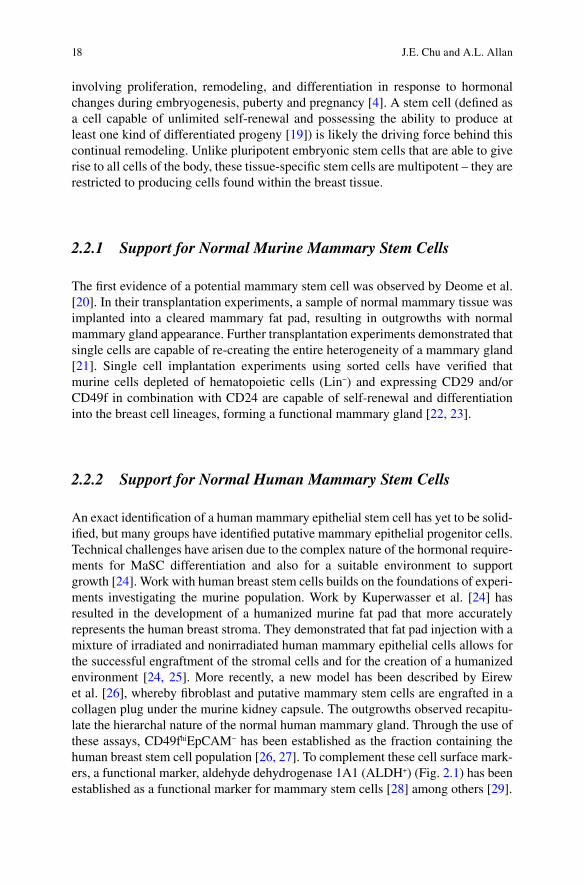
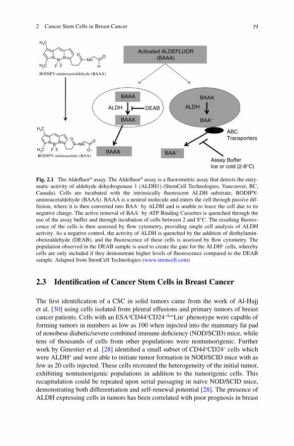
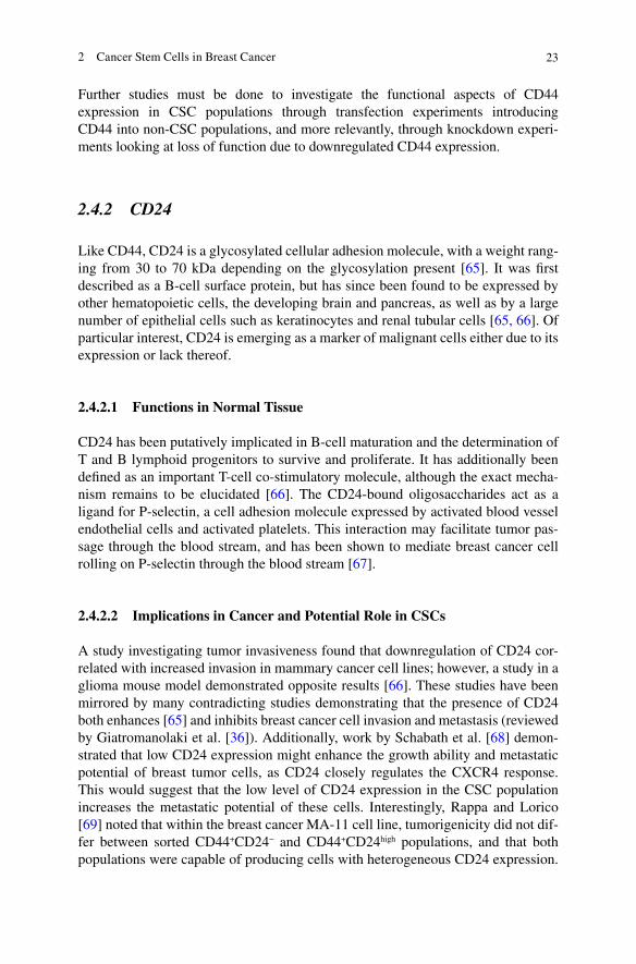
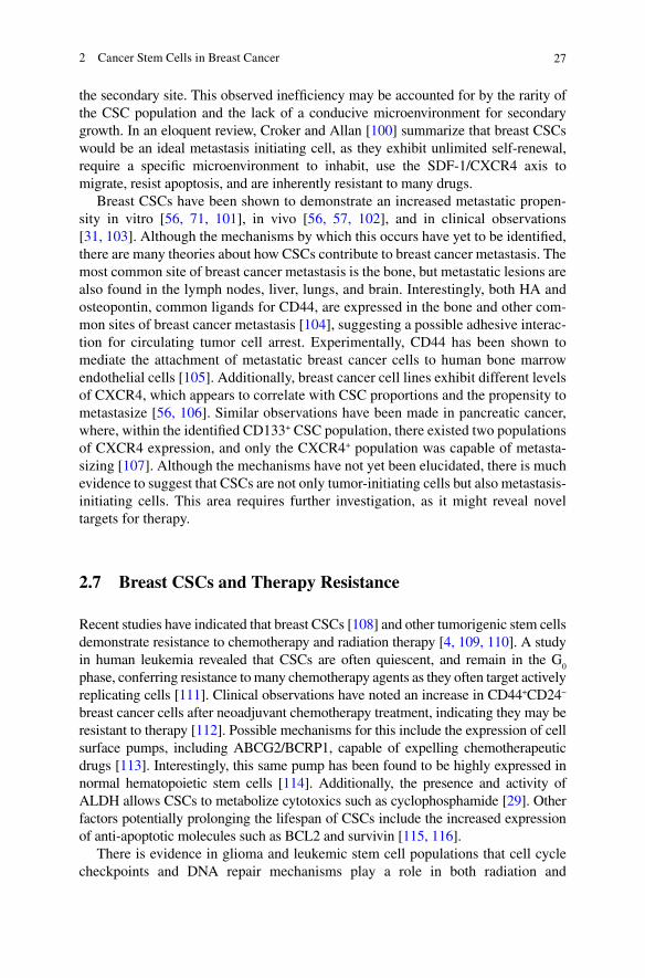
An exact identification of a human mammary epithelial stem cell has yet to be solid-ified, but many groups have identified putative mammary epithelial progenitor cells. Technical challenges have arisen due to the complex nature of the hormonal require-ments for MaSC differentiation and also for a suitable environment to support growth [24]. Work with human breast stem cells builds on the foundations of experi-ments investigating the murine population. Work by Kuperwasser et al. [24] has resulted in the development of a humanized murine fat pad that more accurately represents the human breast stroma. They demonstrated that fat pad injection with a mixture of irradiated and nonirradiated human mammary epithelial cells allows for the successful engraftment of the stromal cells and for the creation of a humanized environment [24, 25]. More recently, a new model has been described by Eirew et al. [26], whereby fibroblast and putative mammary stem cells are engrafted in a collagen plug under the murine kidney capsule. The outgrowths observed recapitu-late the hierarchal nature of the normal human mammary gland. Through the use of these assays, CD49fhiEpCAM− has been established as the fraction containing the human breast stem cell population [26, 27]. To complement these cell surface mark-ers, a functional marker, aldehyde dehydrogenase 1A1 (ALDH+) (Fig. 2.1) has been established as a functional marker for mammary stem cells [28] among others [29].
192 Cancer Stem Cells in Breast Cancer
2.3 Identification of Cancer Stem Cells in Breast Cancer
The first identification of a CSC in solid tumors came from the work of Al-Hajj et al. [30] using cells isolated from pleural effusions and primary tumors of breast cancer patients. Cells with an ESA+CD44+CD24−/lowLin− phenotype were capable of forming tumors in numbers as low as 100 when injected into the mammary fat pad of nonobese diabetic/severe combined immune deficiency (NOD/SCID) mice, while tens of thousands of cells from other populations were nontumorigenic. Further work by Ginestier et al. [28] identified a small subset of CD44+CD24− cells which were ALDH+ and were able to initiate tumor formation in NOD/SCID mice with as few as 20 cells injected. These cells recreated the heterogeneity of the initial tumor, exhibiting nontumorigenic populations in addition to the tumorigenic cells. This recapitulation could be repeated upon serial passaging in naïve NOD/SCID mice, demonstrating both differentiation and self-renewal potential [28]. The presence of ALDH expressing cells in tumors has been correlated with poor prognosis in breast
ABCTransporters
BAA−
BAA−
BAAA
O
O
O
FF
N NHH3C
H3C
N B
O
H
O
FF
N NHH3C
H3C
NB
BAAA
BAAA
Activated ALDEFLUOR(BAAA)
BAAA
ALDHALDH
BODIPY-aminoacetate (BAA)
BODIPY-aminoacetaldehyde (BAAA)
DEAB
Assay BufferIce or cold (2-8°C)
Fig. 2.1 The Aldefluor® assay. The Aldefluor® assay is a fluorometric assay that detects the enzy-matic activity of aldehyde dehydrogenase 1 (ALDH1) (StemCell Technologies, Vancouver, BC, Canada). Cells are incubated with the intrinsically fluorescent ALDH substrate, BODIPY-aminoacetaldehyde (BAAA). BAAA is a neutral molecule and enters the cell through passive dif-fusion, where it is then converted into BAA− by ALDH and is unable to leave the cell due to its negative charge. The active removal of BAA− by ATP Binding Cassettes is quenched through the use of the assay buffer and through incubation of cells between 2 and 8°C. The resulting fluores-cence of the cells is then assessed by flow cytometry, providing single cell analysis of ALDH activity. As a negative control, the activity of ALDH is quenched by the addition of diethylamin-obenzaldehyde (DEAB), and the fluorescence of these cells is assessed by flow cytometry. The population observed in the DEAB sample is used to create the gate for the ALDH+ cells, whereby cells are only included if they demonstrate higher levels of fluorescence compared to the DEAB sample. Adapted from StemCell Technologies (www.stemcell.com)
20 J.E. Chu and A.L. Allan
cancer patients [28, 31, 32]. Additionally, the CD44+CD24− population appears to be enriched in basal-like tumors (ER, PR, HER2 negative) and in BRCA1 tumors [33], both of which have been associated with poor patient prognosis [34, 35]. The presence of a CSC population has also been verified in breast cancer cell lines and primary tumor samples [36].
Due to the functional stem cell-like characteristics of these cells, the term “cancer stem cell” is a fitting descriptor. However, it does not mean that these cells are indeed stem cells re-wired, although they may be. A consensus on the definition of CSCs was created by the leaders in the field to be “a cell within a tumor that possess the capacity to self-renew and to cause the heterogeneous lineages of cancer cells that comprise the tumor” [37]. It is hypothesized that CSCs arise either from a normal tissue stem cell that has acquired mutations that make it tumorigenic or from a more differentiated progenitor or mature cell that has dedifferentiated and acquired the ability to self-renew in addition to the tumorigenic mutations. While the described phenotype is not an absolute definition of the breast CSC population, it provides a basis for further work.
2.4 Markers Used to Identify CSCs
In order to elucidate the functions and the populations of CSCs within solid tumors, the phenotypic definition of a CSC must first be established. Selectable markers are either found on the cell surface or confer functional properties that are characteris-tics of normal stem cells that have extended to malignant stem cell populations. As previously mentioned, the current definition of a breast CSC is CD44+CD24− and/or ALDH+. In the following section, these markers and other putative CSC markers will be discussed.
2.4.1 CD44
CD44 is a multifunctional cell membrane protein that plays a role in both cell–cell and cell–extracellular matrix (ECM) interactions primarily through the binding of hyaluronan (HA). Other ligands of CD44 include collagen, fibronectin, fibrino-gen, laminin, chondroitin sulfate, mucosal vascular addressin, serglycin, osteo-pontin, class II major histocompatability complex invariant chain, L-selectin, and E-selectin [38, 39]. As CD44 is widely expressed throughout the body, and its ligands are common, the successful binding of CD44 to its ligands often depends on an external stimulus. Alternative splicing and protein glycosylation gives rise to multiple CD44 isoforms that differ in size (85–230 kDa), functionality, and tissue localization [39, 40].
212 Cancer Stem Cells in Breast Cancer
2.4.1.1 Function in Normal Tissue
Work by Lesley et al. [41] has identified three states of CD44: active, inducible, and inactive. The activity is dictated by the glycosylation status of the protein: the active form is least glycosylated and constitutively binds HA; inducible CD44 is moder-ately glycosylated and requires activation by monoclonal antibodies, cytokines, growth factors, or phorbol ester; and inactive CD44, the most glycosylated, is unable to bind HA (reviewed by Naor et al. [38]). Adding additional variability, the types of glycosylation may vary from isoform to isoform, using side chains such as heparin sulfate and chondroitin sulfate, resulting not only in variations of molecular weight but also in differentially charged environments that affect CD44 function [42].
The human CD44 gene consists of 19 exons, the first 5 of which are constant [39]. The middle 9 exons (v2–v10) are variable regions which may be removed depending on the variant expressed. The next three exons (16–18) are constant, and the last two exons (19 and 20) are variable. Exons 1–17 encode the extracellular domain of the protein, while 18 encodes the transmembrane domain, and 19 and 20 encode the cytoplasmic tail [43]. Individual cells are capable of altering the splicing of CD44, allowing for much diversity. The standard form, CD44s, is the smallest of the isoforms (37 kDa unglycosylated; 80–100 kDa when glycosylated [42]), and was first identified on hematopoietic cells [44] and is therefore additionally termed hematopoietic CD44, or CD44H [38]. Further research has highlighted CD44s expression in a variety of tissues including the epidermis, liver, pancreas, lung, and central nervous system. The distribution of variant CD44 (CD44v) isoforms is much more restricted and apparently tissue specific (reviewed by Sneath [42]). Nomenclature for CD44v isoforms depends on the variant expressed. A CD44v expressing only variant exon 6 would be called CD44v6.
CD44 is involved in cell–ECM and cell–cell interactions. In cell–ECM interac-tions, CD44 functions through the binding of its previously mentioned ligands, which may facilitate cellular functions such as adhesion and migration. Additionally, CD44 binding of HA causes the internalization of the CD44–HA complex and the lysoso-mally facilitated degradation of HA [45]. In cell–cell interactions, CD44 allows for the aggregation of cells through the binding of exogenous or endogenous HA [42]. CD44s has also been implicated in the lymph node homing and activation of lympho-cytes through its binding of mucosal addressin. The standard and variant forms of CD44 are also involved in myelopoiesis and lymphopoiesis, angiogenesis, chemokine and growth factor presentation, and growth and apoptosis signaling [39, 42, 46].
In normal breast tissue, expression of CD44s and CD44v has been observed by immunohistochemistry (IHC) to be in the myoepithelial layer, while the remaining epithelial cells are CD44− [47–50]. Normal breast stromal elements have been observed to express only CD44s [47]. These IHC observations also apply to clinical tumor specimens, as high levels of mainly CD44v have been observed. The correla-tion between CD44 expression and patient prognosis varies from study to study, likely due to differences in technique, isoform, and the breast cancer population studied (reviewed by Herrera-Gayol and Jothy [51]).
22 J.E. Chu and A.L. Allan
2.4.1.2 First Implications in Cancer and Potential Role in CSCs
CD44 was first implicated in cancer when a nonmetastatic cell line acquired meta-static potential upon transfection with CD44v4-v7, a variant previously found to be expressed by a metastatic rat pancreatic adenocarcinoma. Studies have demon-strated that CD44s is involved in breast cancer cell adhesion, motility, and invasion; whereas CD44v6 is involved solely in cell motility [52]. CD44 most likely acts in tumorigenesis by allowing for more efficient colony formation through increased adhesion to its multitude of ligands in the surrounding environment, its ability to aggregate cells, its induction of cellular growth signals via intracellular signaling partners, and by facilitating the degradation of the surrounding ECM and basal lam-ina, allowing a path for cellular migration and tumor expansion (reviewed in [42, 51]). Notably, CD44 has been shown to interact with matrix metalloproteinases, acti-vating them and attaching them to the cell surface of tumor cells, thus enabling efficient tumor cell invasion through collagen IV [53, 54]. It is also thought that CD44 plays a distinct role in tumor metastasis; however, the absolute mechanism remains elusive due to the many isoforms and variable functions in different envi-ronments [53]. A possible component is revealed through the observation that CD44v4 has been shown to mediate breast cancer transendothelial metastasis through its binding to E-selectin [54]. Contradicting studies show that the presence of CD44s reduced metastasis, potentially explained through the masking of HA from other receptors [55].
The function of CD44 in breast CSCs has yet to be fully elucidated; however, it is likely that the molecule plays a role in enabling CSCs to be the metastasis-initiat-ing cells observed by Croker et al. [56] and Charafe-Jauffret et al. [31, 57]. Recent evidence has shown that CD44 plays a role in protection against apoptosis [58], an important characteristic for a tumor-initiating and metastasis-initiating cell. Additionally, CD44’s dual ability for cell–cell and cell–ECM adherence could con-fer an advantage for CSCs as they travel through the bloodstream and arrive at and enter their secondary site [53]. Within the last few years, much work has been done on the HA–CD44 interaction, revealing that it promotes growth through an EGFR-MAP/ERK (MEK)–dependent mechanism in head and neck cancer [59], and through a HER2-b-catenin–dependent manner in ovarian cancer [60]. In breast and ovarian cancers, the HA–CD44 interaction has been shown to activate transcription of Nanog (an embryonic stem cell transcription factor) transcription, which pro-ceeds to activate Rex1, SOX2, and Multi-drug resistance pump 1 (MDR1) [61], all stem cell-related products. These responses to HA-CD44 binding may provide insight into the observed properties of breast CSCs, especially with regard to their therapy resistance.
There is no distinct rule regarding CD44 isoforms and functions within cancer. In some cases, CD44 variants are involved in promoting malignancy, while in others it is the standard form [62]. A further exception to the rule is the observation that CD44 can in fact act as a metastasis suppressor, holding the tumor within the primary site [55, 63]. Diaz and colleagues suggest that the expression of CD44s in node-negative invasive cancer may be associated with increased disease-free survival [64].
232 Cancer Stem Cells in Breast Cancer
Further studies must be done to investigate the functional aspects of CD44 expression in CSC populations through transfection experiments introducing CD44 into non-CSC populations, and more relevantly, through knockdown experi-ments looking at loss of function due to downregulated CD44 expression.
2.4.2 CD24
Like CD44, CD24 is a glycosylated cellular adhesion molecule, with a weight rang-ing from 30 to 70 kDa depending on the glycosylation present [65]. It was first described as a B-cell surface protein, but has since been found to be expressed by other hematopoietic cells, the developing brain and pancreas, as well as by a large number of epithelial cells such as keratinocytes and renal tubular cells [65, 66]. Of particular interest, CD24 is emerging as a marker of malignant cells either due to its expression or lack thereof.
2.4.2.1 Functions in Normal Tissue
CD24 has been putatively implicated in B-cell maturation and the determination of T and B lymphoid progenitors to survive and proliferate. It has additionally been defined as an important T-cell co-stimulatory molecule, although the exact mecha-nism remains to be elucidated [66]. The CD24-bound oligosaccharides act as a ligand for P-selectin, a cell adhesion molecule expressed by activated blood vessel endothelial cells and activated platelets. This interaction may facilitate tumor pas-sage through the blood stream, and has been shown to mediate breast cancer cell rolling on P-selectin through the blood stream [67].
2.4.2.2 Implications in Cancer and Potential Role in CSCs
A study investigating tumor invasiveness found that downregulation of CD24 cor-related with increased invasion in mammary cancer cell lines; however, a study in a glioma mouse model demonstrated opposite results [66]. These studies have been mirrored by many contradicting studies demonstrating that the presence of CD24 both enhances [65] and inhibits breast cancer cell invasion and metastasis (reviewed by Giatromanolaki et al. [36]). Additionally, work by Schabath et al. [68] demon-strated that low CD24 expression might enhance the growth ability and metastatic potential of breast tumor cells, as CD24 closely regulates the CXCR4 response. This would suggest that the low level of CD24 expression in the CSC population increases the metastatic potential of these cells. Interestingly, Rappa and Lorico [69] noted that within the breast cancer MA-11 cell line, tumorigenicity did not dif-fer between sorted CD44+CD24− and CD44+CD24high populations, and that both populations were capable of producing cells with heterogeneous CD24 expression.
24 J.E. Chu and A.L. Allan
Whether or not CD24 is simply a marker of CSCs or actually plays a functional role in CSC cell behavior has yet to be established. However, the molecule plays a role in many functions that may influence tumorigenicity, and the functionality of this molecule in CSCs requires further study.
2.4.3 Lineage Markers
In the original identification of the breast CSC, cells positive for lineage markers CD2, CD3, CD10, CD16, CD18, CD31, CD64, and CD140b were discarded during flow cytometry in order to exclude normal human leukocytes, endothelial cells, mesothelial cells, and fibroblasts from the population being analyzed [30]. Work by Sheridan et al. has highlighted that CD10 is expressed on several breast cancer cell lines, and that perhaps CD10 should be excluded from the lineage criteria, as it has been defined as a marker of basal cells and might provide a further subdivision for the breast CSC population [70, 71].
2.4.4 Additional Cell Surface Markers
While the CD44+CD24− selection criterion appears to enrich the tumor-initiating capability of breast cancer cells, it is not a definitive identification of these cells, nor does it apply to all breast cancers. Thus, other groups have been investigating other potential markers to further narrow down the CSC phenotype.
As discussed previously, the mouse mammary stem cell markers have been established as Lin−CD29hiCD49fhi (a6-integrin) and human mammary stem cells putatively identified as CD49fhiEpCAM−. It is notable that a subpopulation in the human breast cancer line MCF-7 was recently identified as overexpressing a6-integrin. These cells were capable of propagation as mammospheres, resisted pro-apoptotic agents and exhibited increased tumorigenicity when compared to the whole popula-tion, and as few as 1,000 cells were capable of tumor formation. Furthermore, knockdown of a6-integrin caused the loss of mammosphere capability and tumori-genicity [72].
In mouse models, CD29 and CD61 have been highlighted as potential proteins active in driving luminal cell fate. Within the CD24+ population, CD29 differenti-ates between luminal committed (CD29low) and mammary stem cells (CD29high) [23]. The addition of CD61 allows for further division of the luminal committed cells into progenitors (CD61+) and mature differentiated cells (CD61−) [73]. Recent work in a mouse model of luminal breast cancer (MMTV-WNT1) demonstrated that the selec-tion of the CD61+ population resulted in a much more tumorigenic population when compared to the CD61− population [74].
Most recently, Meyer et al. [75] isolated a tumorigenic subset of CD44+ cells from ER-negative breast cancers and found that CD49fhiCD133/2hi cells exhibited
252 Cancer Stem Cells in Breast Cancer
xenograft-initiating capability, whereas the CD49fneg/lowCD133/2neg/low population did not. They noted that while this new population enriched for xenograft initiation in mouse mammary fat pads, capability varied between their samples. Additionally, other markers established as CSC markers for other cancers, such as CD133 (a marker for colon and brain cancer initiating cells [76, 77]), may be good candidates for further refining the breast CSC phenotype.
Although knowledge translation from murine models and from other cancers to breast cancer is anything but direct, results from these highlighted surface markers merit more investigation into their application on the human breast cancer front. Furthermore, the lack of identified markers for the human mammary gland stem cell highlights the need for more research and standardized assays in this area.
2.4.5 ALDH
A hallmark of cancer cells is the genomic instability that allows for the accrual of the multiple mutations necessary for a cell to become tumorigenic [78]. The addi-tional selection criterion afforded by the Aldefluor® assay (Fig. 2.1) provides quan-titative analysis of ALDH functionality within CSCs, and this is emerging as an important tool in the study of normal stem cells and CSCs. ALDH activity has been shown to be a functional marker of stem cells. As a result, it might be a common property of CSC populations across all subtypes of the cancer in question (unlike the CD44+CD24− phenotype). Interestingly, work by Ginestier et al. demonstrated that CD44+CD24−Lin−Aldefluor− cells were nontumorigenic [28], suggesting that the CD44+CD24−Lin− phenotype is itself heterogeneous and does not contain strictly CSCs.
The aldehyde dehydrogenases are a large family of enzymes responsible for the oxidation of aldehydes into their corresponding carboxylic acids in a NAD(P)+-dependent manner [79]. Different subfamilies are responsible for many functions in the body such as facilitation of retinoic acid biosynthesis, metabolizing cyclophos-phamides and its derivatives, and clearing toxic byproducts of reactive oxygen spe-cies [29, 80].
High ALDH activity has been used to isolate a variety of normal stem cells, most notably human hematopoietic (HSCs) [81, 82] and murine neural stem cells [83]. Additionally, ALDH activity has been reported to identify leukemic stem cells [84, 85], head and neck CSCs [86], colon CSCs [87], and normal and malignant breast epithelial stem cells [28]. Consequently, ALDH is emerging as an important marker of both normal and malignant stem cell populations. Gene expression studies in HSCs and IHC staining of normal and malignant breast tissue reveal that ALDH 1A1 is likely the isoform responsible for the observed ALDH activity within these stem cell populations [80].
In addition to the conferred resistance to cyclophosphamide and its derivatives, ALDH is responsible for the metabolism of retinal to retinoic acid (RA) [88, 89], and therefore plays an important role in cellular differentiation during development
26 J.E. Chu and A.L. Allan
[90, 91] and in stem cell self-protection from intracellular aldehydes for the duration of an organism’s life [29]. The formed RA can proceed to interact with nuclear retinoic acid receptors (RAR) and retinoid X receptors (RXR). RA–RAR interac-tions cause downstream effects on histone deacetylases, which control the epige-netic regulation of gene expression [92]. It is thought that this ALDH-dependent gene regulation and drug resistance play a role in creating the CSC phenotype.
2.5 Comparison of Breast CSCs and Normal Mammary Stem Cells
Although CSCs may arise from a normal tissue stem cell that has undergone cancer-ous mutations, CSCs may also arise from a more differentiated progenitor that has acquired self-renewal capabilities. Putative pathways involved in mammary stem cell self-renewal include LIF, Hedgehog, Wnt, Notch, TGFb, EGF, Prl/GH, and ER/PR (reviewed by Kalirai and Clarke [5]). Similarly, Notch, HOXB4, Wnt, and bone mor-phogenetic protein (BMP) signaling pathways are identified pathways regulating HSC self-renewal [90]. Notably, Notch has been identified as being upregulated in CD44+ populations of both normal and malignant breast cells [93], which may trans-late into an upregulation in the CD44+CD24− CSC population. Additionally, CD44+CD49fhiCD133/2hi cells demonstrated upregulation of Sox2, Bmi-1, and Nanog (transcription factors known to play key roles in the stem cell self-renewal process) [75]. Unfortunately, due to the complex nature of stem cell self-renewal, it is unlikely that a single pathway will be shown to be responsible for CSC self-renewal.
2.6 The Role of CSCs in Metastasis
Breast cancer is a highly treatable disease if caught in the primary stage; however, once the disease metastasizes, patient prognosis becomes much worse [94, 95]. The stepwise process of metastasis is well established, whereby cells must first escape from the primary tumor into the bloodstream and/or the lymphatic system via intra-vasation. Once in the circulation, the cells must survive until they reach a secondary site where they arrest and enter the tissue (extravasate). Tumor cells able to initiate and maintain colony growth in the secondary sites form micrometastases, which, fol-lowing angiogenesis, progress to macrometastases [94, 96, 97]. Although tumor cells may readily escape the primary tumor and enter circulation, production of sustain-able metastatic lesions is a highly inefficient process (reviewed by Hunter et al. [98]). This was exemplified by an in vivo videomicroscopy study by Luzzi et al. which reported that only 0.02% of melanoma cells injected to target the liver could success-fully complete the metastatic cascade [99]. Interestingly, this paper highlighted that not all metastatic stages are equally inefficient: the main inefficiencies occur during the initiation and maintenance of the metastatic lesions once tumor cells have reached
272 Cancer Stem Cells in Breast Cancer
the secondary site. This observed inefficiency may be accounted for by the rarity of the CSC population and the lack of a conducive microenvironment for secondary growth. In an eloquent review, Croker and Allan [100] summarize that breast CSCs would be an ideal metastasis initiating cell, as they exhibit unlimited self-renewal, require a specific microenvironment to inhabit, use the SDF-1/CXCR4 axis to migrate, resist apoptosis, and are inherently resistant to many drugs.
Breast CSCs have been shown to demonstrate an increased metastatic propen-sity in vitro [56, 71, 101], in vivo [56, 57, 102], and in clinical observations [31, 103]. Although the mechanisms by which this occurs have yet to be identified, there are many theories about how CSCs contribute to breast cancer metastasis. The most common site of breast cancer metastasis is the bone, but metastatic lesions are also found in the lymph nodes, liver, lungs, and brain. Interestingly, both HA and osteopontin, common ligands for CD44, are expressed in the bone and other com-mon sites of breast cancer metastasis [104], suggesting a possible adhesive interac-tion for circulating tumor cell arrest. Experimentally, CD44 has been shown to mediate the attachment of metastatic breast cancer cells to human bone marrow endothelial cells [105]. Additionally, breast cancer cell lines exhibit different levels of CXCR4, which appears to correlate with CSC proportions and the propensity to metastasize [56, 106]. Similar observations have been made in pancreatic cancer, where, within the identified CD133+ CSC population, there existed two populations of CXCR4 expression, and only the CXCR4+ population was capable of metasta-sizing [107]. Although the mechanisms have not yet been elucidated, there is much evidence to suggest that CSCs are not only tumor-initiating cells but also metastasis-initiating cells. This area requires further investigation, as it might reveal novel targets for therapy.
2.7 Breast CSCs and Therapy Resistance
Recent studies have indicated that breast CSCs [108] and other tumorigenic stem cells demonstrate resistance to chemotherapy and radiation therapy [4, 109, 110]. A study in human leukemia revealed that CSCs are often quiescent, and remain in the G
0
phase, conferring resistance to many chemotherapy agents as they often target actively replicating cells [111]. Clinical observations have noted an increase in CD44+CD24− breast cancer cells after neoadjuvant chemotherapy treatment, indicating they may be resistant to therapy [112]. Possible mechanisms for this include the expression of cell surface pumps, including ABCG2/BCRP1, capable of expelling chemotherapeutic drugs [113]. Interestingly, this same pump has been found to be highly expressed in normal hematopoietic stem cells [114]. Additionally, the presence and activity of ALDH allows CSCs to metabolize cytotoxics such as cyclophosphamide [29]. Other factors potentially prolonging the lifespan of CSCs include the increased expression of anti-apoptotic molecules such as BCL2 and survivin [115, 116].
There is evidence in glioma and leukemic stem cell populations that cell cycle checkpoints and DNA repair mechanisms play a role in both radiation and
28 J.E. Chu and A.L. Allan
chemotherapy resistance, and that these mechanisms may apply to breast CSCs [117–119]. Further, the observed radiotherapy resistance of CSCs may be due to the decreased levels of pro-oxidants in the CD44+CD24− population [120] or through Wnt/b-catenin pathway signaling [121].
These innate therapy resistance mechanisms make breast CSCs a difficult target to treat; however, their defined characteristics may provide the basis for new thera-pies. For example, deregulated pathways in breast cancer offer potential treatment options. However, the exact pathways responsible for the self-renewal of these cells have yet to be firmly established, and when they are, it is likely that they will heavily overlap with those used by normal stem cells, thus providing a barrier to treatment. Preclinical and Phase I clinical trials are underway targeting hedgehog, Notch, Akt, and CXCR1 [17]. Currently, high throughput screening is being used on cells sorted for CSC phenotypes, looking for small molecules, siRNA or lenti-viral shRNA that target the CSC population. The effects of therapy may be ana-lyzed in many ways including through changes in cellular growth [122], spheroid formation [123], migration [124], or through pathway-specific flow cytometry [125]. Until the biology of CSC therapy resistance is thoroughly understood, high throughput screening may provide the best hope of finding new therapies to target the CSC population.
2.8 Conclusions and Future Perspectives
While large steps have been made toward the absolute identification of the breast CSC, the definition still requires further refining. The CD44+CD24−and/or ALDH+ phenotype has allowed for the establishment of the presence of a CSC population; indeed, gene expression profiling based on stem and differentiated cell markers indicates that the CD44+ population is more stem-like and that the CD24+ popula-tion is more differentiated [101, 126]. Unfortunately, due to the vast heterogeneity observed between breast cancers, this phenotype does not extend to all cases, thus further markers need to be established.
CSCs exist both in primary tumors and in metastatic lesions where they appear to play a role in the initiation and maintenance of both tumors. When an unambigu-ous definition of the CSC phenotype is elucidated, further research should be done to define the role of the CSCs in metastasis, and to identify unique therapy targets, either based on cell surface markers or based on a functional target. Before work targeting CSCs can move forward, it is essential that the functional and cell surface characterization of CSCs is completed. Once a pure population is identified, scien-tists will then be able to generate novel treatment strategies that aim to eradicate the cells postulated to be responsible for tumor initiation, recurrence, and metastasis.
Acknowledgements We thank members of our laboratory and our collaborators for their research work and helpful discussions. The authors’ research on CSCs is supported by research grants from the Ontario Institute for Cancer Research (#08NOV230), and the Canada Foundation
292 Cancer Stem Cells in Breast Cancer
for Innovation (#13199) (to ALA). JEC is the recipient of scholarships from the Natural Sciences and Engineering Research Council of Canada (NSERC), the Canadian Institute of Health Research (CIHR) Strategic Training Program, and the Pamela Kohlemier Translational Breast Cancer Unit at the London Regional Cancer Program. ALA is supported by a CIHR New Investigator Award and an Early Researcher Award from the Ontario Ministry of Research and Innovation.
References
1. Canadian Cancer Society’s Steering Committee (2010) Canadian cancer statistics 2010. Toronto: Canadian Cancer Society
2. American Cancer Society (2010) Breast cancer facts & figures 2009–2010. American Cancer Society, Atlanta
3. Jemal A, Bray F, Center MM, Ferlay J, Ward E, Forman D (2011) Global cancer statistics. CA: Cancer J Clin 61:69–90. doi:10.3322/caac.20107
4. Kakarala M, Wicha MS (2008) Implications of the cancer stem-cell hypothesis for breast cancer prevention and therapy. J Clin Oncol 26 (17):2813–2820. doi:26/17/2813 [pii] 10.1200/JCO.2008.16.3931
5. Kalirai H, Clarke RB (2006) Human breast epithelial stem cells and their regulation. J Pathol 208 (1):7–16. doi:10.1002/path.1881
6. Sakorafas GH, Tsiotou AG (2000) Ductal carcinoma in situ (DCIS) of the breast: Evolving perspectives. Cancer Treat Rev 26 (2):103–125. doi:10.1053/ctrv.1999.0149 S0305-7372(99)90149 – 4 [pii]
7. Simpson PT, Gale T, Fulford LG, Reis-Filho JS, Lakhani SR (2003) The diagnosis and man-agement of pre-invasive breast disease: Pathology of atypical lobular hyperplasia and lobular carcinoma in situ. Breast Cancer Res 5 (5):258–262. doi:10.1186/bcr624
8. Anderson BO, Calhoun KE, Rosen EL (2006) Evolving concepts in the management of lobular neoplasia. J Natl Compr Canc Netw 4 (5):511–522
9. Sims AH, Howell A, Howell SJ, Clarke RB (2007) Origins of breast cancer subtypes and therapeutic implications. Nat Clin Pract Oncol 4 (9):516–525. doi:ncponc0908 [pii] 10.1038/ncponc0908
10. Polyak K (2007) Breast cancer: Origins and evolution. J Clin Invest 117 (11):3155–3163. doi: 10.1172/JCI33295
11. Visvader JE, Lindeman GJ (2008) Cancer stem cells in solid tumours: Accumulating evidence and unresolved questions. Nat Rev Cancer 8 (10):755–768. doi:nrc2499 [pii] 10.1038/nrc2499
12. Campbell LL, Polyak K (2007) Breast tumor heterogeneity: Cancer stem cells or clonal evolu-tion? Cell Cycle 6 (19):2332–2338. doi:4914 [pii]
13. Perou CM, Sorlie T, Eisen MB, van de Rijn M, Jeffrey SS, Rees CA, Pollack JR, Ross DT, Johnsen H, Akslen LA, Fluge O, Pergamenschikov A, Williams C, Zhu SX, Lonning PE, Borresen-Dale AL, Brown PO, Botstein D (2000) Molecular portraits of human breast tumours. Nature 406 (6797):747–752. doi:10.1038/35021093
14. Sorlie T, Perou CM, Tibshirani R, Aas T, Geisler S, Johnsen H, Hastie T, Eisen MB, van de Rijn M, Jeffrey SS, Thorsen T, Quist H, Matese JC, Brown PO, Botstein D, Eystein Lonning P, Borresen-Dale AL (2001) Gene expression patterns of breast carcinomas distinguish tumor subclasses with clinical implications. Proc Natl Acad Sci USA 98 (19):10869–10874. doi:10.1073/pnas.191367098 98/19/10869 [pii]
15. Herschkowitz JI, Simin K, Weigman VJ, Mikaelian I, Usary J, Hu Z, Rasmussen KE, Jones LP, Assefnia S, Chandrasekharan S, Backlund MG, Yin Y, Khramtsov AI, Bastein R, Quackenbush J, Glazer RI, Brown PH, Green JE, Kopelovich L, Furth PA, Palazzo JP, Olopade OI, Bernard PS, Churchill GA, Van Dyke T, Perou CM (2007) Identification of conserved gene expression features between murine mammary carcinoma models and human breast tumors. Genome Biol 8 (5):R76. doi:gb-2007-8-5-r76 [pii] 10.1186/gb-2007-8-5-r76
30 J.E. Chu and A.L. Allan
16. Lobo NA, Shimono Y, Qian D, Clarke MF (2007) The biology of cancer stem cells. Annu Rev Cell Dev Biol 23:675–699. doi:10.1146/annurev.cellbio.22.010305.104154
17. McDermott SP, Wicha MS (2010) Targeting breast cancer stem cells. Mol Oncol. doi:S1574-7891(10)00051-7 [pii] 10.1016/j.molonc.2010.06.005
18. Reya T, Morrison SJ, Clarke MF, Weissman IL (2001) Stem cells, cancer, and cancer stem cells. Nature 414 (6859):105–111. doi:10.1038/35102167 35102167 [pii]
19. Smith GH, Chepko G (2001) Mammary epithelial stem cells. Microsc Res Tech 52 (2):190–203. 20. Deome KB, Faulkin LJ, Jr., Bern HA, Blair PB (1959) Development of mammary tumors from
hyperplastic alveolar nodules transplanted into gland-free mammary fat pads of female c3h mice. Cancer Res 19 (5):515–520
21. Kordon EC, Smith GH (1998) An entire functional mammary gland may comprise the progeny from a single cell. Development 125 (10):1921–1930
22. Stingl J, Eirew P, Ricketson I, Shackleton M, Vaillant F, Choi D, Li HI, Eaves CJ (2006) Purification and unique properties of mammary epithelial stem cells. Nature 439 (7079): 993–997. doi:nature04496 [pii] 10.1038/nature04496
23. Shackleton M, Vaillant F, Simpson KJ, Stingl J, Smyth GK, Asselin-Labat ML, Wu L, Lindeman GJ, Visvader JE (2006) Generation of a functional mammary gland from a single stem cell. Nature 439 (7072):84–88. doi:nature04372 [pii] 10.1038/nature04372
24. Kuperwasser C, Chavarria T, Wu M, Magrane G, Gray JW, Carey L, Richardson A, Weinberg RA (2004) Reconstruction of functionally normal and malignant human breast tissues in mice. Proc Natl Acad Sci USA 101 (14):4966–4971. doi:10.1073/pnas.0401064101 0401064101 [pii]
25. Proia DA, Kuperwasser C (2006) Reconstruction of human mammary tissues in a mouse model. Nat Protoc 1 (1):206–214. doi:nprot.2006.31 [pii] 10.1038/nprot.2006.31
26. Eirew P, Stingl J, Raouf A, Turashvili G, Aparicio S, Emerman JT, Eaves CJ (2008) A method for quantifying normal human mammary epithelial stem cells with in vivo regenerative ability. Nat Med 14 (12):1384 –1389. doi:nm.1791 [pii] 10.1038/nm.1791
27. Lim E, Vaillant F, Wu D, Forrest NC, Pal B, Hart AH, Asselin-Labat ML, Gyorki DE, Ward T, Partanen A, Feleppa F, Huschtscha LI, Thorne HJ, Fox SB, Yan M, French JD, Brown MA, Smyth GK, Visvader JE, Lindeman GJ (2009) Aberrant luminal progenitors as the candidate target population for basal tumor development in brca1 mutation carriers. Nat Med 15 (8): 907–913. doi:nm.2000 [pii] 10.1038/nm.2000
28. Ginestier C, Hur MH, Charafe-Jauffret E, Monville F, Dutcher J, Brown M, Jacquemier J, Viens P, Kleer CG, Liu S, Schott A, Hayes D, Birnbaum D, Wicha MS, Dontu G (2007) Aldh1 is a marker of normal and malignant human mammary stem cells and a predictor of poor clinical outcome. Cell stem cell 1 (5):555–567
29. Moreb JS (2008) Aldehyde dehydrogenase as a marker for stem cells. Curr Stem Cell Res Ther 3 (4):237–246
30. Al-Hajj M, Wicha MS, Benito-Hernandez A, Morrison SJ, Clarke MF (2003) Prospective identification of tumorigenic breast cancer cells. Proceedings of the National Academy of Sciences of the United States of America 100 (7):3983–3988
31. Charafe-Jauffret E, Ginestier C, Iovino F, Tarpin C, Diebel M, Esterni B, Houvenaeghel G, Extra JM, Bertucci F, Jacquemier J, Xerri L, Dontu G, Stassi G, Xiao Y, Barsky SH, Birnbaum D, Viens P, Wicha MS (2010) Aldehyde dehydrogenase 1-positive cancer stem cells mediate metastasis and poor clinical outcome in inflammatory breast cancer. Clin Cancer Res 16 (1):45–55. doi:1078-0432.CCR-09-1630 [pii] 10.1158/1078-0432.CCR-09–1630
32. Yu P, Zhou L, Wang J, Jiang A, Li K (2010) Prognostic relevance of aldh1 in breast cancer: A clinicopathological study of 96 cases. The Chinese-German Journal of Clinical Oncology 9 (1):31–35. doi:10.1007/s10330-009-0178–4
33. Honeth G, Bendahl PO, Ringner M, Saal LH, Gruvberger-Saal SK, Lovgren K, Grabau D, Ferno M, Borg A, Hegardt C (2008) The cd44+/cd24− phenotype is enriched in basal-like breast tumors. Breast Cancer Res 10 (3):R53. doi:bcr2108 [pii] 10.1186/bcr2108
34. Cheang MC, Voduc D, Bajdik C, Leung S, McKinney S, Chia SK, Perou CM, Nielsen TO (2008) Basal-like breast cancer defined by five biomarkers has superior prognostic value than
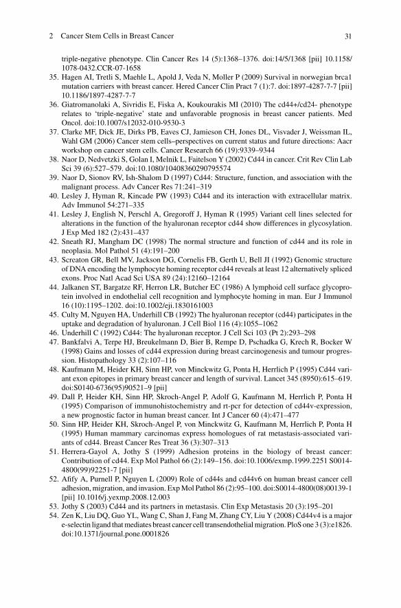
312 Cancer Stem Cells in Breast Cancer
triple-negative phenotype. Clin Cancer Res 14 (5):1368–1376. doi:14/5/1368 [pii] 10.1158/ 1078-0432.CCR-07-1658
35. Hagen AI, Tretli S, Maehle L, Apold J, Veda N, Moller P (2009) Survival in norwegian brca1 mutation carriers with breast cancer. Hered Cancer Clin Pract 7 (1):7. doi:1897-4287-7-7 [pii] 10.1186/1897-4287-7-7
36. Giatromanolaki A, Sivridis E, Fiska A, Koukourakis MI (2010) The cd44+/cd24- phenotype relates to ‘triple-negative’ state and unfavorable prognosis in breast cancer patients. Med Oncol. doi:10.1007/s12032-010-9530-3
37. Clarke MF, Dick JE, Dirks PB, Eaves CJ, Jamieson CH, Jones DL, Visvader J, Weissman IL, Wahl GM (2006) Cancer stem cells–perspectives on current status and future directions: Aacr workshop on cancer stem cells. Cancer Research 66 (19):9339–9344
38. Naor D, Nedvetzki S, Golan I, Melnik L, Faitelson Y (2002) Cd44 in cancer. Crit Rev Clin Lab Sci 39 (6):527–579. doi:10.1080/10408360290795574
39. Naor D, Sionov RV, Ish-Shalom D (1997) Cd44: Structure, function, and association with the malignant process. Adv Cancer Res 71:241–319
40. Lesley J, Hyman R, Kincade PW (1993) Cd44 and its interaction with extracellular matrix. Adv Immunol 54:271–335
41. Lesley J, English N, Perschl A, Gregoroff J, Hyman R (1995) Variant cell lines selected for alterations in the function of the hyaluronan receptor cd44 show differences in glycosylation. J Exp Med 182 (2):431–437
42. Sneath RJ, Mangham DC (1998) The normal structure and function of cd44 and its role in neoplasia. Mol Pathol 51 (4):191–200
43. Screaton GR, Bell MV, Jackson DG, Cornelis FB, Gerth U, Bell JI (1992) Genomic structure of DNA encoding the lymphocyte homing receptor cd44 reveals at least 12 alternatively spliced exons. Proc Natl Acad Sci USA 89 (24):12160–12164
44. Jalkanen ST, Bargatze RF, Herron LR, Butcher EC (1986) A lymphoid cell surface glycopro-tein involved in endothelial cell recognition and lymphocyte homing in man. Eur J Immunol 16 (10):1195–1202. doi:10.1002/eji.1830161003
45. Culty M, Nguyen HA, Underhill CB (1992) The hyaluronan receptor (cd44) participates in the uptake and degradation of hyaluronan. J Cell Biol 116 (4):1055–1062
46. Underhill C (1992) Cd44: The hyaluronan receptor. J Cell Sci 103 (Pt 2):293–298 47. Bankfalvi A, Terpe HJ, Breukelmann D, Bier B, Rempe D, Pschadka G, Krech R, Bocker W
(1998) Gains and losses of cd44 expression during breast carcinogenesis and tumour progres-sion. Histopathology 33 (2):107–116
48. Kaufmann M, Heider KH, Sinn HP, von Minckwitz G, Ponta H, Herrlich P (1995) Cd44 vari-ant exon epitopes in primary breast cancer and length of survival. Lancet 345 (8950):615–619. doi:S0140-6736(95)90521–9 [pii]
49. Dall P, Heider KH, Sinn HP, Skroch-Angel P, Adolf G, Kaufmann M, Herrlich P, Ponta H (1995) Comparison of immunohistochemistry and rt-pcr for detection of cd44v-expression, a new prognostic factor in human breast cancer. Int J Cancer 60 (4):471–477
50. Sinn HP, Heider KH, Skroch-Angel P, von Minckwitz G, Kaufmann M, Herrlich P, Ponta H (1995) Human mammary carcinomas express homologues of rat metastasis-associated vari-ants of cd44. Breast Cancer Res Treat 36 (3):307–313
51. Herrera-Gayol A, Jothy S (1999) Adhesion proteins in the biology of breast cancer: Contribution of cd44. Exp Mol Pathol 66 (2):149–156. doi:10.1006/exmp.1999.2251 S0014-4800(99)92251-7 [pii]
52. Afify A, Purnell P, Nguyen L (2009) Role of cd44s and cd44v6 on human breast cancer cell adhesion, migration, and invasion. Exp Mol Pathol 86 (2):95–100. doi:S0014-4800(08)00139-1 [pii] 10.1016/j.yexmp.2008.12.003
53. Jothy S (2003) Cd44 and its partners in metastasis. Clin Exp Metastasis 20 (3):195–201 54. Zen K, Liu DQ, Guo YL, Wang C, Shan J, Fang M, Zhang CY, Liu Y (2008) Cd44v4 is a major
e-selectin ligand that mediates breast cancer cell transendothelial migration. PloS one 3 (3):e1826. doi:10.1371/journal.pone.0001826
32 J.E. Chu and A.L. Allan
55. Lopez JI, Camenisch TD, Stevens MV, Sands BJ, McDonald J, Schroeder JA (2005) Cd44 attenuates metastatic invasion during breast cancer progression. Cancer Res 65 (15): 6755–6763. doi:65/15/6755 [pii] 10.1158/0008-5472.CAN-05-0863
56. Croker AK, Goodale D, Chu J, Postenka C, Hedley BD, Hess DA, Allan AL (2009) High aldehyde dehydrogenase and expression of cancer stem cell markers selects for breast cancer cells with enhanced malignant and metastatic ability. J Cell Mol Med 13 (8B):2236–2252
57. Charafe-Jauffret E, Ginestier C, Iovino F, Wicinski J, Cervera N, Finetti P, Hur MH, Diebel ME, Monville F, Dutcher J, Brown M, Viens P, Xerri L, Bertucci F, Stassi G, Dontu G, Birnbaum D, Wicha MS (2009) Breast cancer cell lines contain functional cancer stem cells with metastatic capacity and a distinct molecular signature. Cancer Res 69 (4):1302–1313. doi:0008-5472.CAN-08-2741 [pii] 10.1158/0008-5472.CAN-08-2741
58. Bourguignon LY, Spevak CC, Wong G, Xia W, Gilad E (2009) Hyaluronan-cd44 interaction with protein kinase c(epsilon) promotes oncogenic signaling by the stem cell marker nanog and the production of microrna-21, leading to down-regulation of the tumor suppressor protein pdcd4, anti-apoptosis, and chemotherapy resistance in breast tumor cells. J Biol Chem 284 (39):26533–26546. doi:M109.027466 [pii] 10.1074/jbc.M109.027466
59. Wang SJ, Bourguignon LY (2006) Hyaluronan and the interaction between cd44 and epider-mal growth factor receptor in oncogenic signaling and chemotherapy resistance in head and neck cancer. Arch Otolaryngol Head Neck Surg 132 (7):771–778. doi:132/7/771 [pii] 10.1001/archotol.132.7.771
60. Bourguignon LY, Peyrollier K, Gilad E, Brightman A (2007) Hyaluronan-cd44 interaction with neural wiskott-aldrich syndrome protein (n-wasp) promotes actin polymerization and erbb2 activation leading to beta-catenin nuclear translocation, transcriptional up-regulation, and cell migration in ovarian tumor cells. J Biol Chem 282 (2):1265–1280. doi:M604672200 [pii] 10.1074/jbc.M604672200
61. Bourguignon LY, Peyrollier K, Xia W, Gilad E (2008) Hyaluronan-cd44 interaction activates stem cell marker nanog, stat-3-mediated mdr1 gene expression, and ankyrin-regulated multi-drug efflux in breast and ovarian tumor cells. J Biol Chem 283 (25):17635–17651. doi:M800109200 [pii] 10.1074/jbc.M800109200
62. Sy MS, Guo YJ, Stamenkovic I (1991) Distinct effects of two cd44 isoforms on tumor growth in vivo. J Exp Med 174 (4):859–866
63. Naor D, Wallach-Dayan SB, Zahalka MA, Sionov RV (2008) Involvement of cd44, a molecule with a thousand faces, in cancer dissemination. Semin Cancer Biol 18 (4):260–267. doi:S1044-579X(08)00036-9 [pii] 10.1016/j.semcancer.2008.03.015
64. Diaz LK, Zhou X, Wright ET, Cristofanilli M, Smith T, Yang Y, Sneige N, Sahin A, Gilcrease MZ (2005) Cd44 expression is associated with increased survival in node-negative invasive breast carcinoma. Clin Cancer Res 11 (9):3309–3314. doi:11/9/3309 [pii] 10.1158/1078-0432.CCR-04-2184
65. Baumann P, Cremers N, Kroese F, Orend G, Chiquet-Ehrismann R, Uede T, Yagita H, Sleeman JP (2005) Cd24 expression causes the acquisition of multiple cellular properties associated with tumor growth and metastasis. Cancer Res 65 (23):10783–10793. doi:65/23/10783 [pii] 10.1158/0008-5472.CAN-05-0619
66. Lim SC (2005) Cd24 and human carcinoma: Tumor biological aspects. Biomed Pharmacother 59 Suppl 2:S351–354. doi:S0753-3322(05)80076-9 [pii]
67. Aigner S, Ramos CL, Hafezi-Moghadam A, Lawrence MB, Friederichs J, Altevogt P, Ley K (1998) Cd24 mediates rolling of breast carcinoma cells on p-selectin. FASEB J 12 (12):1241–1251
68. Schabath H, Runz S, Joumaa S, Altevogt P (2006) Cd24 affects cxcr4 function in pre-b lym-phocytes and breast carcinoma cells. J Cell Sci 119 (Pt 2):314–325. doi:jcs.02741 [pii] 10.1242/jcs.02741
69. Rappa G, Lorico A (2010) Phenotypic characterization of mammosphere-forming cells from the human ma-11 breast carcinoma cell line. Exp Cell Res 316 (9):1576–1586. doi:S0014-4827(10) 00013-3 [pii] 10.1016/j.yexcr.2010.01.012
70. Charafe-Jauffret E, Ginestier C, Monville F, Finetti P, Adelaide J, Cervera N, Fekairi S, Xerri L, Jacquemier J, Birnbaum D, Bertucci F (2006) Gene expression profiling of breast cell lines
332 Cancer Stem Cells in Breast Cancer
identifies potential new basal markers. Oncogene 25 (15):2273–2284. doi:1209254 [pii] 10.1038/sj.onc.1209254
71. Sheridan C, Kishimoto H, Fuchs RK, Mehrotra S, Bhat-Nakshatri P, Turner CH, Goulet R, Jr., Badve S, Nakshatri H (2006) Cd44+/cd24- breast cancer cells exhibit enhanced invasive prop-erties: An early step necessary for metastasis. Breast Cancer Res 8 (5):R59. doi:bcr1610 [pii] 10.1186/bcr1610
72. Cariati M, Naderi A, Brown JP, Smalley MJ, Pinder SE, Caldas C, Purushotham AD (2008) Alpha-6 integrin is necessary for the tumourigenicity of a stem cell-like subpopulation within the mcf7 breast cancer cell line. Int J Cancer 122 (2):298–304
73. Asselin-Labat ML, Sutherland KD, Barker H, Thomas R, Shackleton M, Forrest NC, Hartley L, Robb L, Grosveld FG, van der Wees J, Lindeman GJ, Visvader JE (2007) Gata-3 is an essential regulator of mammary-gland morphogenesis and luminal-cell differentiation. Nat Cell Biol 9 (2):201–209. doi:ncb1530 [pii] 10.1038/ncb1530
74. Vaillant F, Asselin-Labat ML, Shackleton M, Forrest NC, Lindeman GJ, Visvader JE (2008) The mammary progenitor marker cd61/beta3 integrin identifies cancer stem cells in mouse models of mammary tumorigenesis. Cancer Res 68 (19):7711–7717. doi:68/19/7711 [pii] 10.1158/0008-5472.CAN-08-1949
75. Meyer MJ, Fleming JM, Lin AF, Hussnain SA, Ginsburg E, Vonderhaar BK (2010) Cd44poscd49fhicd133/2hi defines xenograft-initiating cells in estrogen receptor-negative breast cancer. Cancer Res 70 (11):4624–4633. doi:0008-5472.CAN-09-3619 [pii] 10.1158/0008-5472.CAN-09-3619
76. O’Brien CA, Pollett A, Gallinger S, Dick JE (2007) A human colon cancer cell capable of initiating tumour growth in immunodeficient mice. Nature 445 (7123):106–110
77. Singh SK, Hawkins C, Clarke ID, Squire JA, Bayani J, Hide T, Henkelman RM, Cusimano MD, Dirks PB (2004) Identification of human brain tumour initiating cells. Nature 432 (7015):396-401. doi:nature03128 [pii] 10.1038/nature03128
78. Hanahan D, Weinberg RA (2000) The hallmarks of cancer. Cell 100 (1):57–70. doi:S0092-8674(00)81683-9 [pii]
79. Sladek NE (2003) Human aldehyde dehydrogenases: Potential pathological, pharmacological, and toxicological impact. J Biochem Mol Toxicol 17 (1):7–23. doi:10.1002/jbt.10057
80. Levi BP, Yilmaz OH, Duester G, Morrison SJ (2009) Aldehyde dehydrogenase 1a1 is dispens-able for stem cell function in the mouse hematopoietic and nervous systems. Blood 113 (8): 1670–1680. doi:blood-2008-05-156752 [pii] 10.1182/blood-2008-05-156752
81. Hess DA, Meyerrose TE, Wirthlin L, Craft TP, Herrbrich PE, Creer MH, Nolta JA (2004) Functional characterization of highly purified human hematopoietic repopulating cells isolated according to aldehyde dehydrogenase activity. Blood 104 (6):1648–1655. doi:10.1182/blood-2004-02-0448 2004-02-0448 [pii]
82. Storms RW, Trujillo AP, Springer JB, Shah L, Colvin OM, Ludeman SM, Smith C (1999) Isolation of primitive human hematopoietic progenitors on the basis of aldehyde dehydroge-nase activity. Proceedings of the National Academy of Sciences of the United States of America 96 (16):9118–9123
83. Corti S, Locatelli F, Papadimitriou D, Donadoni C, Salani S, Del Bo R, Strazzer S, Bresolin N, Comi GP (2006) Identification of a primitive brain-derived neural stem cell population based on aldehyde dehydrogenase activity. Stem Cells 24 (4):975–985. doi:2005-0217 [pii] 10.1634/stemcells.2005–0217
84. Cheung AM, Wan TS, Leung JC, Chan LY, Huang H, Kwong YL, Liang R, Leung AY (2007) Aldehyde dehydrogenase activity in leukemic blasts defines a subgroup of acute myeloid leukemia with adverse prognosis and superior nod/scid engrafting potential. Leukemia 21 (7):1423–1430
85. Pearce DJ, Taussig D, Simpson C, Allen K, Rohatiner AZ, Lister TA, Bonnet D (2005) Characterization of cells with a high aldehyde dehydrogenase activity from cord blood and acute myeloid leukemia samples. Stem Cells 23 (6):752–760
86. Clay MR, Tabor M, Owen JH, Carey TE, Bradford CR, Wolf GT, Wicha MS, Prince ME (2010) Single-marker identification of head and neck squamous cell carcinoma cancer stem cells with aldehyde dehydrogenase. Head Neck. doi:10.1002/hed.21315
34 J.E. Chu and A.L. Allan
87. Huang EH, Hynes MJ, Zhang T, Ginestier C, Dontu G, Appelman H, Fields JZ, Wicha MS, Boman BM (2009) Aldehyde dehydrogenase 1 is a marker for normal and malignant human colonic stem cells (sc) and tracks sc overpopulation during colon tumorigenesis. Cancer Res 69 (8):3382–3389. doi:0008–5472.CAN-08-4418 [pii] 10.1158/0008-5472.CAN-08-4418
88. Vasiliou V, Pappa A, Estey T (2004) Role of human aldehyde dehydrogenases in endo-biotic and xenobiotic metabolism. Drug Metab Rev 36 (2):279–299. doi:10.1081/DMR-120034001
89. Duester G (2001) Genetic dissection of retinoid dehydrogenases. Chem Biol Interact 130-132 (1-3):469–480. doi:S0009279700002921 [pii]
90. Chute JP, Muramoto GG, Whitesides J, Colvin M, Safi R, Chao NJ, McDonnell DP (2006) Inhibition of aldehyde dehydrogenase and retinoid signaling induces the expansion of human hematopoietic stem cells. Proc Natl Acad Sci USA 103 (31):11707–11712. doi:0603806103 [pii] 10.1073/pnas.0603806103
91. Appel B, Eisen JS (2003) Retinoids run rampant: Multiple roles during spinal cord and motor neuron development. Neuron 40 (3):461–464. doi:S0896627303006883 [pii]
92. Ginestier C, Wicinski J, Cervera N, Monville F, Finetti P, Bertucci F, Wicha MS, Birnbaum D, Charafe-Jauffret E (2009) Retinoid signaling regulates breast cancer stem cell differentia-tion. Cell Cycle 8 (20):3297–3302. doi:9761 [pii]
93. Farnie G, Clarke RB (2007) Mammary stem cells and breast cancer--role of notch signalling. Stem Cell Rev 3 (2):169–175. doi:SCR:3:2:169 [pii]
94. Chambers AF, Groom AC, MacDonald IC (2002) Dissemination and growth of cancer cells in metastatic sites. Nat Rev Cancer 2 (8):563–572
95. Dalerba P, Clarke MF (2007) Cancer stem cells and tumor metastasis: First steps into uncharted territory. Cell Stem Cell 1 (3):241–242. doi:S1934-5909(07)00131-2 [pii] 10.1016/j.stem.2007.08.012
96. Chambers AF, Naumov GN, Varghese HJ, Nadkarni KV, MacDonald IC, Groom AC (2001) Critical steps in hematogenous metastasis: An overview. Surgical Oncology Clinics of North America 10 (2):243–255, vii
97. Pantel K, Brakenhoff RH (2004) Dissecting the metastatic cascade. Nature Reviews Cancer 4 (6):448–456
98. Hunter KW, Crawford NP, Alsarraj J (2008) Mechanisms of metastasis. Breast Cancer Res 10 Suppl 1:S2. doi: bcr1988 [pii] 10.1186/bcr1988
99. Luzzi KJ, MacDonald IC, Schmidt EE, Kerkvliet N, Morris VL, Chambers AF, Groom AC (1998) Multistep nature of metastatic inefficiency: Dormancy of solitary cells after successful extravasation and limited survival of early micrometastases. Am J Pathol 153 (3):865–873
100. Croker AK, Allan AL (2008) Cancer stem cells: Implications for the progression and treat-ment of metastatic disease. J Cell Mol Med 12 (2):374–390
101. Liu R, Wang X, Chen GY, Dalerba P, Gurney A, Hoey T, Sherlock G, Lewicki J, Shedden K, Clarke MF (2007) The prognostic role of a gene signature from tumorigenic breast-cancer cells. N Engl J Med 356 (3):217–226. doi:356/3/217 [pii] 10.1056/NEJMoa063994
102. Ling LJ, Wang S, Liu XA, Shen EC, Ding Q, Lu C, Xu J, Cao QH, Zhu HQ, Wang F (2008) A novel mouse model of human breast cancer stem-like cells with high cd44+cd24−/lower phenotype metastasis to human bone. Chin Med J (Engl) 121 (20):1980–1986
103. Abraham BK, Fritz P, McClellan M, Hauptvogel P, Athelogou M, Brauch H (2005) Prevalence of cd44+/cd24−/low cells in breast cancer may not be associated with clinical outcome but may favor distant metastasis. Clin Cancer Res 11 (3):1154–1159. doi:11/3/1154 [pii]
104. Brown LF, Berse B, Van de Water L, Papadopoulos-Sergiou A, Perruzzi CA, Manseau EJ, Dvorak HF, Senger DR (1992) Expression and distribution of osteopontin in human tissues: Widespread association with luminal epithelial surfaces. Mol Biol Cell 3 (10):1169–1180
105. Draffin JE, McFarlane S, Hill A, Johnston PG, Waugh DJ (2004) Cd44 potentiates the adher-ence of metastatic prostate and breast cancer cells to bone marrow endothelial cells. Cancer Res 64 (16):5702–5711. doi:10.1158/0008-5472.CAN-04-0389 64/16/5702 [pii]
106. Dewan MZ, Ahmed S, Iwasaki Y, Ohba K, Toi M, Yamamoto N (2006) Stromal cell-derived factor-1 and cxcr4 receptor interaction in tumor growth and metastasis of breast cancer. Biomed Pharmacother 60 (6):273–276. doi:S0753-3322(06)00086-2 [pii] 10.1016/j.biopha. 2006.06.004
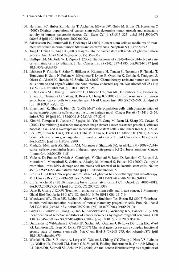
352 Cancer Stem Cells in Breast Cancer
107. Hermann PC, Huber SL, Herrler T, Aicher A, Ellwart JW, Guba M, Bruns CJ, Heeschen C (2007) Distinct populations of cancer stem cells determine tumor growth and metastatic activity in human pancreatic cancer. Cell Stem Cell 1 (3):313–323. doi:S1934-5909(07) 00066-5 [pii] 10.1016/j.stem.2007.06.002
108. Sakariassen PO, Immervoll H, Chekenya M (2007) Cancer stem cells as mediators of treat-ment resistance in brain tumors: Status and controversies. Neoplasia 9 (11):882–892
109. Tang C, Chua CL, Ang BT (2007) Insights into the cancer stem cell model of glioma tumori-genesis. Ann Acad Med Singapore 36 (5):352–357
110. Phillips TM, McBride WH, Pajonk F (2006) The response of cd24(−/low)/cd44+ breast can-cer-initiating cells to radiation. J Natl Cancer Inst 98 (24):1777–1785. doi:98/24/1777 [pii] 10.1093/jnci/djj495
111. Ishikawa F, Yoshida S, Saito Y, Hijikata A, Kitamura H, Tanaka S, Nakamura R, Tanaka T, Tomiyama H, Saito N, Fukata M, Miyamoto T, Lyons B, Ohshima K, Uchida N, Taniguchi S, Ohara O, Akashi K, Harada M, Shultz LD (2007) Chemotherapy-resistant human aml stem cells home to and engraft within the bone-marrow endosteal region. Nat Biotechnol 25 (11): 1315–1321. doi:nbt1350 [pii] 10.1038/nbt1350
112. Li X, Lewis MT, Huang J, Gutierrez C, Osborne CK, Wu MF, Hilsenbeck SG, Pavlick A, Zhang X, Chamness GC, Wong H, Rosen J, Chang JC (2008) Intrinsic resistance of tumori-genic breast cancer cells to chemotherapy. J Natl Cancer Inst 100 (9):672–679. doi:djn123 [pii] 10.1093/jnci/djn123
113. Engelmann K, Shen H, Finn OJ (2008) Mcf7 side population cells with characteristics of cancer stem/progenitor cells express the tumor antigen muc1. Cancer Res 68 (7):2419–2426. doi:68/7/2419 [pii] 10.1158/0008-5472.CAN-07-2249
114. Kim M, Turnquist H, Jackson J, Sgagias M, Yan Y, Gong M, Dean M, Sharp JG, Cowan K (2002) The multidrug resistance transporter abcg2 (breast cancer resistance protein 1) effluxes hoechst 33342 and is overexpressed in hematopoietic stem cells. Clin Cancer Res 8 (1):22–28
115. Lee CW, Simin K, Liu Q, Plescia J, Guha M, Khan A, Hsieh CC, Altieri DC (2008) A func-tional notch-survivin gene signature in basal breast cancer. Breast Cancer Res 10 (6):R97. doi:bcr2200 [pii] 10.1186/bcr2200
116. Madjd Z, Mehrjerdi AZ, Sharifi AM, Molanaei S, Shahzadi SZ, Asadi-Lari M (2009) Cd44+ cancer cells express higher levels of the anti-apoptotic protein bcl-2 in breast tumours. Cancer Immun 9:4. doi:090304 [pii]
117. Viale A, De Franco F, Orleth A, Cambiaghi V, Giuliani V, Bossi D, Ronchini C, Ronzoni S, Muradore I, Monestiroli S, Gobbi A, Alcalay M, Minucci S, Pelicci PG (2009) Cell-cycle restriction limits DNA damage and maintains self-renewal of leukaemia stem cells. Nature 457 (7225):51–56. doi:nature07618 [pii] 10.1038/nature07618
118. Frosina G (2009) DNA repair and resistance of gliomas to chemotherapy and radiotherapy. Mol Cancer Res 7 (7):989–999. doi:7/7/989 [pii] 10.1158/1541-7786.MCR-09-0030
119. Liu S, Wicha MS (2010) Targeting breast cancer stem cells. J Clin Oncol. 28: 4006–4012. doi:JCO.2009.27.5388 [pii] 10.1200/JCO.2009.27.5388
120. Dave B, Chang J (2009) Treatment resistance in stem cells and breast cancer. J Mammary Gland Biol Neoplasia 14 (1):79–82. doi:10.1007/s10911-009-9117-9
121. Woodward WA, Chen MS, Behbod F, Alfaro MP, Buchholz TA, Rosen JM (2007) Wnt/beta-catenin mediates radiation resistance of mouse mammary progenitor cells. Proc Natl Acad Sci USA 104 (2):618–623. doi:0606599104 [pii] 10.1073/pnas.0606599104
122. Gupta PB, Onder TT, Jiang G, Tao K, Kuperwasser C, Weinberg RA, Lander ES (2009) Identification of selective inhibitors of cancer stem cells by high-throughput screening. Cell 138 (4):645–659. doi:S0092-8674(09)00781-8 [pii] 10.1016/j.cell.2009.06.034
123. Diamandis P, Wildenhain J, Clarke ID, Sacher AG, Graham J, Bellows DS, Ling EK, Ward RJ, Jamieson LG, Tyers M, Dirks PB (2007) Chemical genetics reveals a complex functional ground state of neural stem cells. Nat Chem Biol 3 (5):268–273. doi:nchembio873 [pii] 10.1038/nchembio873
124. Wurdak H, Zhu S, Romero A, Lorger M, Watson J, Chiang CY, Zhang J, Natu VS, Lairson LL, Walker JR, Trussell CM, Harsh GR, Vogel H, Felding-Habermann B, Orth AP, Miraglia LJ, Rines DR, Skirboll SL, Schultz PG (2010) An rnai screen identifies trrap as a regulator of

36 J.E. Chu and A.L. Allan
brain tumor-initiating cell differentiation. Cell Stem Cell 6 (1):37–47. doi:S1934-5909(09)00575-X [pii] 10.1016/j.stem.2009.11.002
125. Krutzik PO, Nolan GP (2006) Fluorescent cell barcoding in flow cytometry allows high-throughput drug screening and signaling profiling. Nat Methods 3 (5):361–368. doi:nmeth872 [pii] 10.1038/nmeth872
126. Shipitsin M, Campbell LL, Argani P, Weremowicz S, Bloushtain-Qimron N, Yao J, Nikolskaya T, Serebryiskaya T, Beroukhim R, Hu M, Halushka MK, Sukumar S, Parker LM, Anderson KS, Harris LN, Garber JE, Richardson AL, Schnitt SJ, Nikolsky Y, Gelman RS, Polyak K (2007) Molecular definition of breast tumor heterogeneity. Cancer Cell 11 (3):259–273. doi:S1535-6108(07)00029-3 [pii] 10.1016/j.ccr.2007.01.013
37A.L. Allan (ed.), Cancer Stem Cells in Solid Tumors, Stem Cell Biology and Regenerative Medicine, DOI 10.1007/978-1-61779-246-5_3, © Springer Science+Business Media, LLC 2011
Abstract Several lines of evidence suggest that brain tumors arise from the transformation of a normal neural stem cell (NSC) or progenitor cell, which relies on the recognition of the many functional and genetic similarities shared by somatic stem cells and cancer cells. A minority population of human brain tumor initiating cells (BTICs) was identified through application of stem cell assays to brain tumors, and only these cells are able to recapitulate the patient tumor phenotype in an immu-nodeficient mouse model. Although the molecular mechanisms that regulate BTICs are still poorly understood, many of the signaling pathways that are active during development may be implicated as targets for transformation. BTICs have impor-tant implications for treatment, as these cells may evade current chemotherapy and radiotherapy. Further understanding of the role of BTICs in brain tumorigenesis may yield novel therapeutic agents aimed at targeting these rare cancer stem cells.
Abbreviations
AGAP2 GTPase-activating protein for ARF1 and ARF5APC Adenomatous polyposis colibFGF Basic fibroblast growth factor
S.K. Singh (*)McMaster Stem Cell and Cancer Research Institute, McMaster University, Hamilton, ON, Canada
Departments of Surgery, Biochemistry & Biomedical Sciences, and Neuroscience, Faculty of Health Sciences, McMaster University, Hamilton, ON, Canada e-mail: [email protected]
Chapter 3Cancer Stem Cells in Brain Cancer
Xin Wang, Chitra Venugopal, and Sheila K. Singh
38 X. Wang et al.
BMP Bone morphogenic proteinBTIC Brain tumor initiating cellBTSC Brain tumor stem cellCD Cluster of differentiationCDK Cyclin-dependent kinase inhibitorCENTG1 Centaurin, gamma1CNS Central nervous systemCSC Cancer stem cellEGF Epidermal growth factorEGFR Epidermal growth factor receptorGBM Glioblastoma multiformeHSC Hematopoietic stem cellLIF Leukemia inhibitory factorLSC Leukemic stem cellsMDM2 Murine double minute 2NOD/SCID Non-obese diabetic/severe combined immunodeficientNSC Neural stem cellPDGFRa Platelet-derived growth factor receptor alphaPTCH PatchedPTEN Phosphatase and tensin homologRB RetinoblastomaSFM Serum-free mediaShh Sonic hedgehogSTAT3 Signal transducer and activator of transcription 3TSM Tumor sphere media
3.1 Brain Tumor Initiating Cells: The Starting Line
3.1.1 Lessons from Leukemia
Brain tumors are typically comprised of morphologically diverse cells that express a variety of neural lineage markers. It is recognized that tumors with vastly different histology have a different prognosis, but often brain tumors that share similar mor-phology and phenotype can have a very different prognosis and response to treat-ment. The cancer stem cell (CSC) hypothesis [1], based on work in leukemia [2] and breast cancer [3], suggests that not all the cells in the tumor have the same ability to proliferate and maintain the growth of the tumor. Only a relatively small fraction of cells in the tumor, termed CSCs, possess an ability to extensively proliferate and self-renew. Most of the other tumor cells lose the ability to proliferate and self-renew and instead differentiate into tumor cells that become the phenotypic signa-ture of the tumor.
The CSC hypothesis is a variation on a theme first introduced more than 150 years ago by the pathologists Rudolph Virchow and Julius Cohnheim, both of whom observed histological similarities between primitive tumors such as teratocarcinomas
393 Cancer Stem Cells in Brain Cancer
and the developing fetus [4–6]. They postulated that cancer arises from the activation of dormant embryonic rests, or tissue remnants [5]. The tools to explore the hetero-geneous potential of cancer cells to self-renew emerged a century later, when Till, McCulloch and colleagues made the essential discovery that bone marrow contained single cells that could give rise to myeloerythroid colonies in the spleen. These colo-nies were clonal and self-renewing as well as radioprotected, and could reconstitute lethally irradiated mice [7–10]. The researchers applied their spleen colony-forming unit assay to myeloblastic leukemia, and isolated proliferative blast cells that were capable of self-renewal and abnormal patterns of differentiation [11–16]. These methods were adapted to allow for the assay of clonogenicity of human neoplastic cells in myelomonocytic leukemia [17], and later in solid cancers such as ovarian cancer [18]. With the advent of multiparameter fluorescent activated cell sorting and monoclonal antibodies, the purification of hematopoietic stem cells (HSCs) and their leukemic counterparts could be achieved [19–25] with prospective cell sorting combined with established in vitro clonogenic assays. To truly test the hypothesis that cancers arise from the clonal expansion of a single transformed stem cell, a func-tional in vivo xenotransplantation model was required to definitively identify the neoplastic clone exclusively capable of indefinite self-renewal in vivo. A remarkable series of experiments carried out by Dick and colleagues led to the identification and purification of leukemic stem cells (LSCs) capable of repopulating NOD/SCID (non-obese diabetic severe combined immunodeficient) mice [2, 26], laying the groundwork for the application of the CSC hypothesis to a broad range of cancers.
3.1.2 Lessons from Neural Stem Cells
Stem cells are functionally defined as self-renewing cells that exhibit multilineage differentiation [1, 27, 28]. Somatic stem cells are thought to self-renew to generate all the mature cell types of a particular tissue through proliferative expansion of progenitor cells followed by differentiation into mature cell types. The discovery that multipotential, self-renewing neural stem cells (NSCs) exist throughout life in the adult mammalian brain has only re-emerged in the recent past [29–31], reflecting a rediscovery of 1960s evidence that suggested that neurogenesis was occurring in the adult brain [32]. When multipotent NSCs were isolated from the mammalian neuroaxis more than a decade ago, culture conditions were developed that allowed embryonic Epidermal Growth Factor (EGF) responsive cells to proliferate as floating spheres (neurospheres), which could be easily manipulated for subsequent passage and differentiation [33]. Serum-free media (SFM) allowed for the maintenance of an undifferentiated state, and the addition of saturating concentrations of basic Fibroblast Growth Factor (bFGF) and EGF (20 ng/mL) induced the proliferation of multipotent, self-renewing, and expandable NSCs [34, 35]. This neurosphere cul-ture system and analysis to identify NSCs has permitted in vitro characterization of these cells, but in a retrospective fashion, as the multipotential floating clusters of cells are inferred to have been derived from clonal expansion of a single NSC. Prospective study of this cell has been previously limited by lack of cell surface
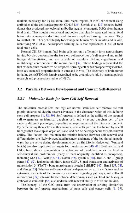
40 X. Wang et al.
markers necessary for its isolation, until recent reports of NSC enrichment using antibodies to the cell surface protein CD133 [36]. Uchida et al. [37] selected hybri-domas that produced monoclonal antibodies against clonogenic NSCs from human fetal brain. They sought monoclonal antibodies that cleanly separated human fetal brain into neurosphere-forming and non–neurosphere-forming fractions. They found that CD133 enriched highly for clonogenic human NSCs, in vitro and in vivo, identifying 95% of all neurosphere-forming cells that represented 1–6% of total fetal brain cells.
Normal CD133+ human fetal brain cells not only efficiently form neurospheres in vitro but also demonstrate the key stem cell properties of self-renewal and multi-lineage differentiation, and are capable of seamless lifelong engraftment and multilineage contribution to the mouse brain [37]. These findings represented the first evidence that the in vitro neurosphere-forming cell, when prospectively isolated, bore key stem cell properties both in vitro and in vivo. The discovery of brain tumor initiating cells (BTICs) is largely accredited to the groundwork laid by haematopoiesis research and prospective studies of NSCs.
3.2 Parallels Between Development and Cancer: Self-Renewal
3.2.1 Molecular Basis for Stem Cell Self-Renewal
The molecular mechanisms that regulate normal stem cell self-renewal are still poorly understood, despite recent advances in the characterization of this defining stem cell property [1, 38, 39]. Self-renewal is defined as the ability of the parental cell to generate an identical daughter cell, and a second daughter cell of the same or different phenotype, depending on requirements of the microenvironment. By perpetuating themselves in this manner, stem cells give rise to a hierarchy of cell lineages that make up an organ or tissue, and can be heterogeneous for self-renewal ability. The factors that maintain the relative balance between self-renewal and differentiation are likely dysregulated in cancer, and many of the key signaling path-ways that are active during development (such as Shh [Sonic Hedgehog], Wnt, and Notch) are also implicated as targets for transformation [40, 41]. Both normal and CSCs have shown upregulation or activation of candidate genes involved in self-renewal and proliferation (many of them originally identified as oncogenes), including Shh [42], Wnt [43, 44], Notch [45], cyclin E [46], Hox A and B group genes [47–52], leukemia inhibitory factor (LIF), Signal transducer and activator of transcription 3 (STAT3), bone morphogenic protein 2 (BMP2) [47], Bmi1 [53, 54], and Nanog [55]. Whereas self-renewal can be dependent on extrinsic factors such as cytokines, elements of the previously mentioned signaling pathways, and cell–cell interactions [39]; intrinsic transcriptional determinants such as Oct-4 and Nanog in embryonic stem cells [56] also underlie self-renewal ability in vitro and in vivo.
The concept of the CSC arose from the observation of striking similarities between the self-renewal mechanisms of stem cells and cancer cells [1, 57].
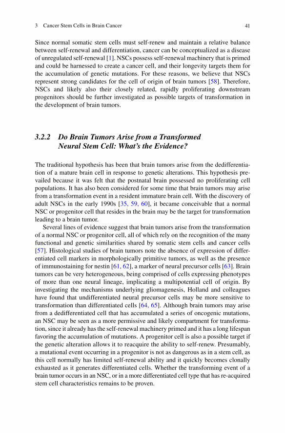
413 Cancer Stem Cells in Brain Cancer
Since normal somatic stem cells must self-renew and maintain a relative balance between self-renewal and differentiation, cancer can be conceptualized as a disease of unregulated self-renewal [1]. NSCs possess self-renewal machinery that is primed and could be harnessed to create a cancer cell, and their longevity targets them for the accumulation of genetic mutations. For these reasons, we believe that NSCs represent strong candidates for the cell of origin of brain tumors [58]. Therefore, NSCs and likely also their closely related, rapidly proliferating downstream progenitors should be further investigated as possible targets of transformation in the development of brain tumors.
3.2.2 Do Brain Tumors Arise from a Transformed Neural Stem Cell: What’s the Evidence?
The traditional hypothesis has been that brain tumors arise from the dedifferentia-tion of a mature brain cell in response to genetic alterations. This hypothesis pre-vailed because it was felt that the postnatal brain possessed no proliferating cell populations. It has also been considered for some time that brain tumors may arise from a transformation event in a resident immature brain cell. With the discovery of adult NSCs in the early 1990s [35, 59, 60], it became conceivable that a normal NSC or progenitor cell that resides in the brain may be the target for transformation leading to a brain tumor.
Several lines of evidence suggest that brain tumors arise from the transformation of a normal NSC or progenitor cell, all of which rely on the recognition of the many functional and genetic similarities shared by somatic stem cells and cancer cells [57]. Histological studies of brain tumors note the absence of expression of differ-entiated cell markers in morphologically primitive tumors, as well as the presence of immunostaining for nestin [61, 62], a marker of neural precursor cells [63]. Brain tumors can be very heterogeneous, being comprised of cells expressing phenotypes of more than one neural lineage, implicating a multipotential cell of origin. By investigating the mechanisms underlying gliomagenesis, Holland and colleagues have found that undifferentiated neural precursor cells may be more sensitive to transformation than differentiated cells [64, 65]. Although brain tumors may arise from a dedifferentiated cell that has accumulated a series of oncogenic mutations, an NSC may be seen as a more permissive and likely compartment for transforma-tion, since it already has the self-renewal machinery primed and it has a long lifespan favoring the accumulation of mutations. A progenitor cell is also a possible target if the genetic alteration allows it to reacquire the ability to self-renew. Presumably, a mutational event occurring in a progenitor is not as dangerous as in a stem cell, as this cell normally has limited self-renewal ability and it quickly becomes clonally exhausted as it generates differentiated cells. Whether the transforming event of a brain tumor occurs in an NSC, or in a more differentiated cell type that has re-acquired stem cell characteristics remains to be proven.
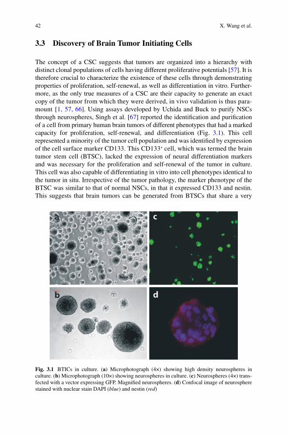
42 X. Wang et al.
3.3 Discovery of Brain Tumor Initiating Cells
The concept of a CSC suggests that tumors are organized into a hierarchy with distinct clonal populations of cells having different proliferative potentials [57]. It is therefore crucial to characterize the existence of these cells through demonstrating properties of proliferation, self-renewal, as well as differentiation in vitro. Further-more, as the only true measures of a CSC are their capacity to generate an exact copy of the tumor from which they were derived, in vivo validation is thus para-mount [1, 57, 66]. Using assays developed by Uchida and Buck to purify NSCs through neurospheres, Singh et al. [67] reported the identification and purification of a cell from primary human brain tumors of different phenotypes that had a marked capacity for proliferation, self-renewal, and differentiation (Fig. 3.1). This cell represented a minority of the tumor cell population and was identified by expression of the cell surface marker CD133. This CD133+ cell, which was termed the brain tumor stem cell (BTSC), lacked the expression of neural differentiation markers and was necessary for the proliferation and self-renewal of the tumor in culture. This cell was also capable of differentiating in vitro into cell phenotypes identical to the tumor in situ. Irrespective of the tumor pathology, the marker phenotype of the BTSC was similar to that of normal NSCs, in that it expressed CD133 and nestin. This suggests that brain tumors can be generated from BTSCs that share a very
Fig. 3.1 BTICs in culture. (a) Microphotograph (4×) showing high density neurospheres in culture. (b) Microphotograph (10×) showing neurospheres in culture. (c) Neurospheres (4×) trans-fected with a vector expressing GFP. Magnified neurospheres. (d) Confocal image of neurosphere stained with nuclear stain DAPI (blue) and nestin (red)
433 Cancer Stem Cells in Brain Cancer
similar phenotype. The discovery of a CSC in human adult gliomas extends the defi-nition of the BTSC to describe a class of cells that may drive tumorigenesis in an increasing number of brain tumors, both pediatric and adult.
As the true validation of CSCs rests in the establishment of an in vivo recapitula-tion of a tumor’s original patient phenotype, Singh et al. [68] developed a xenograft assay to identify human BTICs that had the capacity to initiate tumors in vivo. Corresponding to the in vitro data, only the CD133+ brain tumor fraction contained cells that were capable of tumor initiation in NOD/SCID mouse brains. Injection of as few as 100 CD133+ live cells produced a tumor that was serially transplantable and was a phenocopy of the patient’s original tumor, whereas injection of 105 live CD133− cells engrafted but did not cause a tumor. Together, these data indicate that the CD133+ human brain tumor cell fraction from adult and pediatric tumors of dif-ferent types contain BTICs which exclusively initiate tumor formation in immuno-deficient mice. Thus, BTICs possess all the key properties ascribed to a stem cell. These findings support the application of principles of leukemogenesis to solid tumors: namely, the principle that only a small subset of CSCs is enriched for clo-nogenic capacity, and that these cells alone are capable of tumor propagation. This work was corroborated by the results of several labs that found similar findings in various brain tumor models [69, 70]. Hemmati and colleagues applied the neuro-sphere assay to several childhood brain tumors including medulloblastoma and glioma, and found that these stem-like cells expressed high levels of stem cell genes such as CD133, Sox2, Musashi-1, and Bmi1 [70]. Yuan et al. [71] characterized CSC populations from adult glioblastoma , and Kelly et al. [72] identified GBM BTIC populations that proliferate independently of exogenous mitogens.
Since the discovery of BTICs, much work has been done to characterize these cells. Vescovi et al. [69] continued this work by characterizing BTICs through iso-lating clonogenic, neurosphere-forming progenitors from adult human glioblastoma multiforme (GBM). There is now strong evidence that the same key mechanisms that control the activity of normal neural progenitors are altered in brain tumors. Pathways that regulate neural stem-cell proliferation and cell-fate commitments such as Wnt-beta-catenin, Sonic hedgehog, Notch, and Bmi1 are aberrantly expressed in brain malignancies [69]. More recently, Phosphatase and Tensin Homolog (PTEN) deficiency has been documented as a potential molecular marker for self-renewing, tumor-initiating cells in glioblastoma [73]. The search is ongoing for novel BTIC markers that can further purify these populations.
3.4 The Search for BTIC Markers
CD133, or human prominin-1, is a 120 kDa, five-transmembrane cell surface pro-tein of unknown function originally shown to be a HSC marker, and is specifically associated with plasma membrane protrusions in embryonic, but not adult epithelia [74–76]. There are two isoforms that may be generally co-expressed; prominin-1 (AC133-1) mRNA is more prominent in fetal brain and adult skeletal muscle,
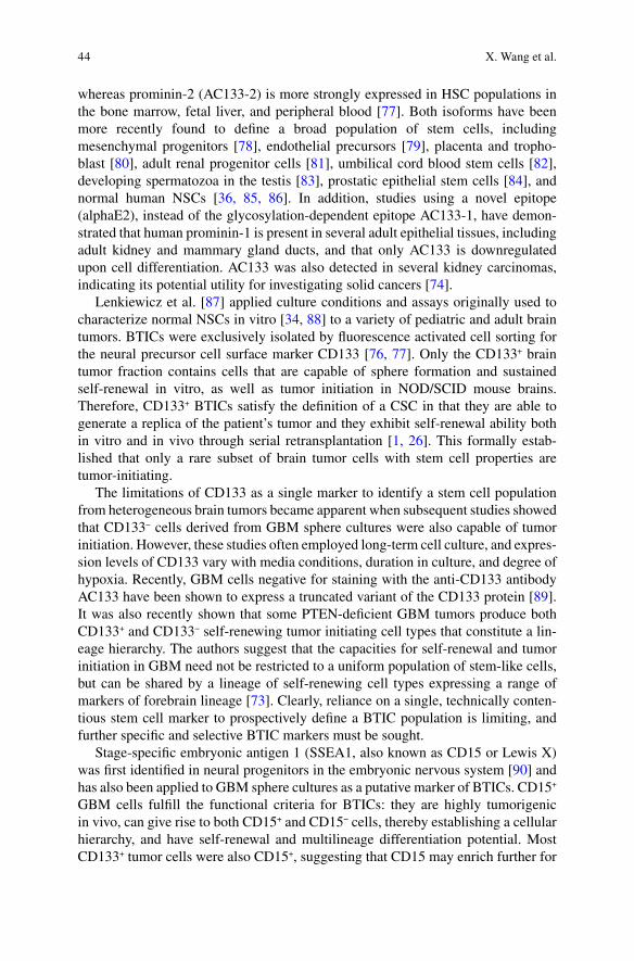
44 X. Wang et al.
whereas prominin-2 (AC133-2) is more strongly expressed in HSC populations in the bone marrow, fetal liver, and peripheral blood [77]. Both isoforms have been more recently found to define a broad population of stem cells, including mesenchymal progenitors [78], endothelial precursors [79], placenta and tropho-blast [80], adult renal progenitor cells [81], umbilical cord blood stem cells [82], developing spermatozoa in the testis [83], prostatic epithelial stem cells [84], and normal human NSCs [36, 85, 86]. In addition, studies using a novel epitope (alphaE2), instead of the glycosylation-dependent epitope AC133-1, have demon-strated that human prominin-1 is present in several adult epithelial tissues, including adult kidney and mammary gland ducts, and that only AC133 is downregulated upon cell differentiation. AC133 was also detected in several kidney carcinomas, indicating its potential utility for investigating solid cancers [74].
Lenkiewicz et al. [87] applied culture conditions and assays originally used to characterize normal NSCs in vitro [34, 88] to a variety of pediatric and adult brain tumors. BTICs were exclusively isolated by fluorescence activated cell sorting for the neural precursor cell surface marker CD133 [76, 77]. Only the CD133+ brain tumor fraction contains cells that are capable of sphere formation and sustained self-renewal in vitro, as well as tumor initiation in NOD/SCID mouse brains. Therefore, CD133+ BTICs satisfy the definition of a CSC in that they are able to generate a replica of the patient’s tumor and they exhibit self-renewal ability both in vitro and in vivo through serial retransplantation [1, 26]. This formally estab-lished that only a rare subset of brain tumor cells with stem cell properties are tumor-initiating.
The limitations of CD133 as a single marker to identify a stem cell population from heterogeneous brain tumors became apparent when subsequent studies showed that CD133− cells derived from GBM sphere cultures were also capable of tumor initiation. However, these studies often employed long-term cell culture, and expres-sion levels of CD133 vary with media conditions, duration in culture, and degree of hypoxia. Recently, GBM cells negative for staining with the anti-CD133 antibody AC133 have been shown to express a truncated variant of the CD133 protein [89]. It was also recently shown that some PTEN-deficient GBM tumors produce both CD133+ and CD133− self-renewing tumor initiating cell types that constitute a lin-eage hierarchy. The authors suggest that the capacities for self-renewal and tumor initiation in GBM need not be restricted to a uniform population of stem-like cells, but can be shared by a lineage of self-renewing cell types expressing a range of markers of forebrain lineage [73]. Clearly, reliance on a single, technically conten-tious stem cell marker to prospectively define a BTIC population is limiting, and further specific and selective BTIC markers must be sought.
Stage-specific embryonic antigen 1 (SSEA1, also known as CD15 or Lewis X) was first identified in neural progenitors in the embryonic nervous system [90] and has also been applied to GBM sphere cultures as a putative marker of BTICs. CD15+ GBM cells fulfill the functional criteria for BTICs: they are highly tumorigenic in vivo, can give rise to both CD15+ and CD15− cells, thereby establishing a cellular hierarchy , and have self-renewal and multilineage differentiation potential. Most CD133+ tumor cells were also CD15+, suggesting that CD15 may enrich further for
453 Cancer Stem Cells in Brain Cancer
BTICs in human GBMs [91]. Since its initial identification, CD15 has also been characterized in medulloblastomas; CD15+ cells have a unique expression profile with increased proliferation and decreased tendency to undergo apoptosis and dif-ferentiation [92, 93].
Other putative BTIC candidate markers include the RNA binding protein Mushashi-1 and the transcription factor Sox2. Musashi-1 is an evolutionally con-served marker for central nervous system (CNS) progenitor cells including NSCs [94], and was later shown to also be expressed in tumor spheres [70]. Sox2 is a key transcription factor that maintains the proliferation of NSCs, inhibits neuronal fate commitment, and may also represent glial tumor precursor cells [95].
3.5 Molecular Genetics of Brain Tumors: Disruption of Signaling Pathways Regulating Growth and Development May Predispose to BTIC Generation
Brain tumors are comprised of cells that can resemble any of the normal neural cell lineages that compose the brain: astrocytes, neurons, oligodendrocytes, and ependymal cells. The tumors that recapitulate these lineages include GBM, medullo-blastoma, oligodendroglioma, and ependymoma (Fig. 3.2). Our understanding of the genetic and epigenetic pathogenic events of these tumors has advanced consid-erably toward a molecular reclassification of brain tumors that will transform clinical medicine [96].
GBM, the most frequent brain tumor, is a highly malignant astrocytic tumor that usually occurs in the cerebral hemispheres of adults, and can occur in young chil-dren and infants as well. Its growth is rapid and infiltrative, and diagnostic patho-logical features include nuclear pleomorphism, microvascular proliferation, and necrosis. Many genes involved in control of proliferation, cell cycle and apoptosis have been implicated in its pathogenesis, including epidermal growth factor recep-tor (EGFR), p53, murine double minute 2 (MDM2), PTEN, and platelet-derived growth factor receptor (PDGFR) [97]. Interestingly, these genes are distinctly dys-regulated, depending on whether the glioblastoma arises de novo (primary GBM) or from a pre-existing lower grade glioma (secondary GBM) [98]. Since gliomagene-sis and progression from low- to high-grade gliomas can be seen as a process of multistep carcinogenesis in secondary gliomas, certain genetic alterations involved in both low- and high-grade gliomas (such as loss of p53 or NF1) can be seen as tumor initiating events [99]. Later events in gliomagenesis, such as CDK4 amplifi-cation or loss of retinoblastoma (RB) gene expression, could be part of a tumor progression pathway. In primary GBM, several different mechanisms disrupt the RB and p53 tumor suppressor gene pathways, respectively, with loss of the genes that encode INK4A and ARF [100]. Also in support of the multistep carcinogenesis model of gliomagenesis is the fact that any of these mutations created singly in astrocytoma mouse models (p53 loss, PDGFRa overexpression) are insufficient to
46 X. Wang et al.
incur tumor growth [101]. It is therefore likely that multiple genetic or epigenetic events accumulate in target cells and cooperatively induce transformation.
GBM is the first cancer with comprehensive genomic profiles mapped by The Cancer Genome Atlas (TCGA) project. It was found that GBM alterations tend to occur within specific functional modules, and that two of the largest modules involve signaling via Rb, p53, PI3K, and receptor protein kinases. New candidate drivers were also identified in GBM, including AGAP2/CENTG1(GTPase-activating pro-tein for ARF1 and ARF5/Centaurin, gamma1), a putative oncogene and activator of the PI3K pathway, as well as three additional significantly altered modules includ-ing one involved in microtubule organization [102].
Medulloblastoma is a malignant embryonal tumor of the cerebellum that mani-fests largely in children, and has a dominant pattern of neuronal differentiation. Many developmental signaling pathways, such as Shh and Wnt, have been impli-cated in its pathogenesis [1, 97]. Mutations in the Shh pathway, which regulates the growth of normal NSCs and cerebellar granule cell precursors, have been shown to convey predisposition to medulloblastomas in both mice and humans [103–106]. The Wnt pathway, critical for self-renewal of hematopoietic, epithelial, and likely NSCs [43], is also activated in a subset of medulloblastomas that harbor mutations
Fig. 3.2 Comparison between the role of stem cells in normal development and tumorigenesis. (a) Neural stem cells (NSC) give rise to early and late progenitors and depending on its niche and extrinsic factors may differentiate into any of the three neural lineages, oligodendrocyte, astrocyte, and neuron. (b) In a tumorigenesis model, cancer stem cells may arise from the transformation of normal NSC or an early progenitor. With accumulation of genetic changes, enhanced self-renewal and proliferation is seen. Many genetic alterations such as copy number changes, overexpression or deletion, as indicated by the asterisk, are observed and ultimately contribute to tumor formation

473 Cancer Stem Cells in Brain Cancer
in b-catenin, axin, or APC (adenomatous polyposis coli) [107]. Another gene recently shown to be critical for maintenance of self-renewal of NSCs, HSCs, and LSCs is Bmi1 [53, 54], which is overexpressed in human medulloblastomas in conjunction with the Shh pathway receptor Patched (PTCH) [108]. Downstream events of the mutations in these self-renewal pathways in medulloblastoma may lead to repression of RB and p53, potentially disturbing the balance of prolifera-tion and differentiation in cerebellar precursor cells to incur tumorigenesis. We have also shown that Bmi1 plays an important role in BTIC-driven tumorigenesis in human medulloblastoma [109].
Oligodendrogliomas and oligoastrocytomas are diffusely infiltrating tumors occurring predominantly in adults and are composed of cells morphologically resembling oligodendrocytes and astrocytes. They can be induced experimentally with chemical carcinogens such as ethynitrosourea, and often bear loss of heterozy-gosity on chromosome 19q [98]. The basic helix-loop-helix transcription factor Olig2, which is involved in oligodendroglial specification [110] is expressed highly in oligodendrogliomas and oligoastrocytomas, and may serve as a tumor biomarker or play a pivotal role in tumor development [111, 112]. It was shown that p21(WAF1/CIP1) is directly repressed by Olig2 in neural progenitors and gliomas, indicating that Olig2-regulated lineage-restricted pathway is critical for proliferation of nor-mal and tumorigenic CNS stem cells [113]. Ependymomas are slowly growing, insidious tumors thought to arise from the ependymal lining of the cerebral ventri-cles and spinal canal of children and adults, and can also occur as an anaplastic variant [97]. Genetic pathways underlying ependymoma pathogenesis remain elu-sive, and the only consistent cytogenetic event occurring in this tumor is loss of chromosome 22, and potential NF2 mutations [114]. CSCs which were isolated from ependymomas, showed a radial glia phenotype and produced tumors when orthotopically transplanted in mice. Thus, restricted populations of radial glia cells can be candidate stem cells of the different subgroups of ependymoma, and they support a general hypothesis that subgroups of the same histologic tumor type are produced by different populations of progenitor cells in the source tissue [115]. New insights into the causes and potential therapeutics of brain tumors have arisen from recognized defects in signaling pathways that govern cell growth, differentia-tion, and death in normal brain development [97]. If brain tumors represent develop-ment gone awry, the underpinnings of brain tumorigenesis may lie in normal neurogenesis.
3.6 Controversies in BTIC Identification and Propagation
3.6.1 Divergence in BTIC Culture Methods
The neurosphere assay and culture conditions originally described by Reynolds and Weiss [34] represent the gold standard method for induction and maintenance of multipotent, self-renewing, and expandable stem cells from both normal and
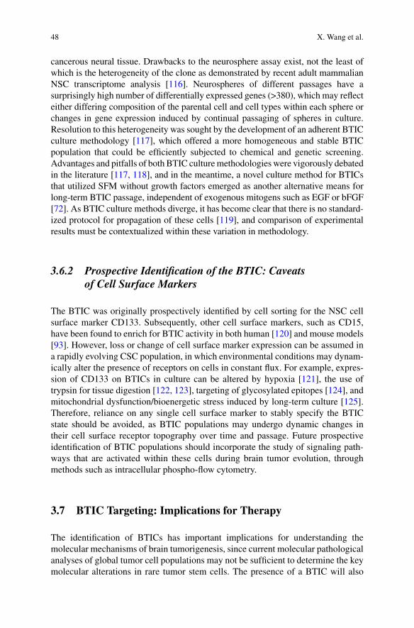
48 X. Wang et al.
cancerous neural tissue. Drawbacks to the neurosphere assay exist, not the least of which is the heterogeneity of the clone as demonstrated by recent adult mammalian NSC transcriptome analysis [116]. Neurospheres of different passages have a surprisingly high number of differentially expressed genes (>380), which may reflect either differing composition of the parental cell and cell types within each sphere or changes in gene expression induced by continual passaging of spheres in culture. Resolution to this heterogeneity was sought by the development of an adherent BTIC culture methodology [117], which offered a more homogeneous and stable BTIC population that could be efficiently subjected to chemical and genetic screening . Advantages and pitfalls of both BTIC culture methodologies were vigorously debated in the literature [117, 118], and in the meantime, a novel culture method for BTICs that utilized SFM without growth factors emerged as another alternative means for long-term BTIC passage, independent of exogenous mitogens such as EGF or bFGF [72]. As BTIC culture methods diverge, it has become clear that there is no standard-ized protocol for propagation of these cells [119], and comparison of experimental results must be contextualized within these variation in methodology.
3.6.2 Prospective Identification of the BTIC: Caveats of Cell Surface Markers
The BTIC was originally prospectively identified by cell sorting for the NSC cell surface marker CD133. Subsequently, other cell surface markers, such as CD15, have been found to enrich for BTIC activity in both human [120] and mouse models [93]. However, loss or change of cell surface marker expression can be assumed in a rapidly evolving CSC population, in which environmental conditions may dynam-ically alter the presence of receptors on cells in constant flux. For example, expres-sion of CD133 on BTICs in culture can be altered by hypoxia [121], the use of trypsin for tissue digestion [122, 123], targeting of glycosylated epitopes [124], and mitochondrial dysfunction/bioenergetic stress induced by long-term culture [125]. Therefore, reliance on any single cell surface marker to stably specify the BTIC state should be avoided, as BTIC populations may undergo dynamic changes in their cell surface receptor topography over time and passage. Future prospective identification of BTIC populations should incorporate the study of signaling path-ways that are activated within these cells during brain tumor evolution, through methods such as intracellular phospho-flow cytometry.
3.7 BTIC Targeting: Implications for Therapy
The identification of BTICs has important implications for understanding the molecular mechanisms of brain tumorigenesis, since current molecular pathological analyses of global tumor cell populations may not be sufficient to determine the key molecular alterations in rare tumor stem cells. The presence of a BTIC will also
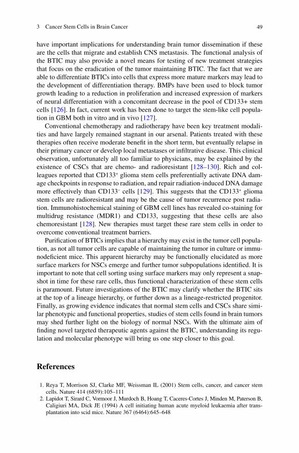
493 Cancer Stem Cells in Brain Cancer
have important implications for understanding brain tumor dissemination if these are the cells that migrate and establish CNS metastasis. The functional analysis of the BTIC may also provide a novel means for testing of new treatment strategies that focus on the eradication of the tumor maintaining BTIC. The fact that we are able to differentiate BTICs into cells that express more mature markers may lead to the development of differentiation therapy. BMPs have been used to block tumor growth leading to a reduction in proliferation and increased expression of markers of neural differentiation with a concomitant decrease in the pool of CD133+ stem cells [126]. In fact, current work has been done to target the stem-like cell popula-tion in GBM both in vitro and in vivo [127].
Conventional chemotherapy and radiotherapy have been key treatment modali-ties and have largely remained stagnant in our arsenal. Patients treated with these therapies often receive moderate benefit in the short term, but eventually relapse in their primary cancer or develop local metastases or infiltrative disease. This clinical observation, unfortunately all too familiar to physicians, may be explained by the existence of CSCs that are chemo- and radioresistant [128–130]. Rich and col-leagues reported that CD133+ glioma stem cells preferentially activate DNA dam-age checkpoints in response to radiation, and repair radiation-induced DNA damage more effectively than CD133− cells [129]. This suggests that the CD133+ glioma stem cells are radioresistant and may be the cause of tumor recurrence post radia-tion. Immunohistochemical staining of GBM cell lines has revealed co-staining for multidrug resistance (MDR1) and CD133, suggesting that these cells are also chemoresistant [128]. New therapies must target these rare stem cells in order to overcome conventional treatment barriers.
Purification of BTICs implies that a hierarchy may exist in the tumor cell popula-tion, as not all tumor cells are capable of maintaining the tumor in culture or immu-nodeficient mice. This apparent hierarchy may be functionally elucidated as more surface markers for NSCs emerge and further tumor subpopulations identified. It is important to note that cell sorting using surface markers may only represent a snap-shot in time for these rare cells, thus functional characterization of these stem cells is paramount. Future investigations of the BTIC may clarify whether the BTIC sits at the top of a lineage hierarchy, or further down as a lineage-restricted progenitor. Finally, as growing evidence indicates that normal stem cells and CSCs share simi-lar phenotypic and functional properties, studies of stem cells found in brain tumors may shed further light on the biology of normal NSCs. With the ultimate aim of finding novel targeted therapeutic agents against the BTIC, understanding its regu-lation and molecular phenotype will bring us one step closer to this goal.
References
1. Reya T, Morrison SJ, Clarke MF, Weissman IL (2001) Stem cells, cancer, and cancer stem cells. Nature 414 (6859):105–111
2. Lapidot T, Sirard C, Vormoor J, Murdoch B, Hoang T, Caceres-Cortes J, Minden M, Paterson B, Caligiuri MA, Dick JE (1994) A cell initiating human acute myeloid leukaemia after trans-plantation into scid mice. Nature 367 (6464):645–648
50 X. Wang et al.
3. Al-Hajj M, Wicha MS, Benito-Hernandez A, Morrison SJ, Clarke MF (2003) Prospective identification of tumorigenic breast cancer cells. Proc Natl Acad Sci USA 100 (7): 3983–3988
4. Huntly BJ, Gilliland DG (2005) Leukaemia stem cells and the evolution of cancer-stem-cell research. Nat Rev Cancer 5 (4):311–321
5. Virchow R (1855) Editorial. Virchows Arch Pathol Anat Physiol Klin Med 3:23 6. Cohnheim J (1867) Ueber entzundung und eiterung. Path Anat Physiol Klin Med 40:1–79 7. Till JE, McCulloch EA, Siminovitch L (1964) A stochastic model of stem cell proliferation,
based on the growth of spleen colony-forming cells. Proc Natl Acad Sci USA 51:29–36 8. McCulloch EA, Siminovitch L, Till JE (1964) Spleen-colony formation in anemic mice of
genotype ww. Science 144:844–846 9. McCulloch EA, Till JE (1964) Proliferation of hemopoietic colony-forming cells transplanted
into irradiated mice. Radiat Res 22:383–397 10. Becker AJ, Mc CE, Till JE (1963) Cytological demonstration of the clonal nature of spleen
colonies derived from transplanted mouse marrow cells. Nature 197:452–454 11. McCulloch EA, Buick RN, Till JE (1978) Normal and leukemic hemopoiesis compared.
Cancer 42 (2 Suppl):845–853 12. Minden MD, Till JE, McCulloch EA (1978) Proliferative state of blast cell progenitors in acute
myeloblastic leukemia (AML). Blood 52 (3):592–600 13. Senn JS, McCulloch EA, Till JE (1967) Comparison of colony-forming ability of normal and
leukaemic human marrow in cell culture. Lancet 2 (7516):597–598 14. Buick RN, Minden MD, McCulloch EA (1979) Self-renewal in culture of proliferative blast
progenitor cells in acute myeloblastic leukemia. Blood 54 (1):95–104 15. Buick RN, Till JE, McCulloch EA (1977) Colony assay for proliferative blast cells circulating
in myeloblastic leukaemia. Lancet 1 (8016):862–863 16. Chang LJ, Till JE, McCulloch EA (1980) The cellular basis of self renewal in culture by
human acute myeloblastic leukemia blast cell progenitors. J Cell Physiol 102 (2):217–222 17. Metcalf D, Moore MA, Warner NL (1969) Colony formation in vitro by myelomonocytic
leukemic cells. J Natl Cancer Inst 43 (4):983–1001 18. Buick RN, MacKillop WJ (1981) Measurement of self-renewal in culture of clonogenic cells
from human ovarian carcinoma. Br J Cancer 44 (3):349–355 19. Worton RG, McCulloch EA, Till JE (1969) Physical separation of hemopoietic stem cells from
cells forming colonies in culture. J Cell Physiol 74 (2):171–182 20. Weissman IL (2002) The road ended up at stem cells. Immunol Rev 185:159–174 21. Spangrude GJ, Aihara Y, Weissman IL, Klein J (1988) The stem cell antigens Sca-1 and Sca-2
subdivide thymic and peripheral T lymphocytes into unique subsets. J Immunol 141 (11): 3697–3707
22. Spangrude GJ, Heimfeld S, Weissman IL (1988) Purification and characterization of mouse hematopoietic stem cells. Science 241 (4861):58–62
23. Muller-Sieburg CE, Whitlock CA, Weissman IL (1986) Isolation of two early B lymphocyte progenitors from mouse marrow: A committed pre-pre-B cell and a clonogenic Thy-1-lo hematopoietic stem cell. Cell 44 (4):653–662
24. Ezine S, Weissman IL, Rouse RV (1984) Bone marrow cells give rise to distinct cell clones within the thymus. Nature 309 (5969):629–631
25. Baum CM, Weissman IL, Tsukamoto AS, Buckle AM, Peault B (1992) Isolation of a candi-date human hematopoietic stem-cell population. Proc Natl Acad Sci USA 89 (7):2804–2808
26. Bonnet D, Dick JE (1997) Human acute myeloid leukemia is organized as a hierarchy that originates from a primitive hematopoietic cell. Nat Med 3 (7):730–737
27. Fuchs E, Segre JA (2000) Stem cells: A new lease on life. Cell 100 (1):143–155 28. Weissman IL (2000) Stem cells: Units of development, units of regeneration, and units in
evolution. Cell 100 (1):157–168 29. Gage FH (2000) Mammalian neural stem cells. Science 287 (5457):1433–1438 30. Gage FH (2002) Neurogenesis in the adult brain. J Neurosci 22 (3):612–613 31. Temple S (2001) The development of neural stem cells. Nature 414 (6859):112–117
513 Cancer Stem Cells in Brain Cancer
32. Gross CG (2000) Neurogenesis in the adult brain: Death of a dogma. Nat Rev Neurosci 1 (1): 67–73
33. Reynolds BA, Tetzlaff W, Weiss S (1992) A multipotent EGF-responsive striatal embryonic progenitor cell produces neurons and astrocytes. J Neurosci 12 (11):4565–4574
34. Reynolds BA, Weiss S (1992) Generation of neurons and astrocytes from isolated cells of the adult mammalian central nervous system. Science 255 (5052):1707–1710
35. Reynolds BA, Weiss S (1996) Clonal and population analyses demonstrate that an EGF-responsive mammalian embryonic CNS precursor is a stem cell. Dev Biol 175 (1):1–13
36. Uchida N, Buck DW, He D, Reitsma MJ, Masek M, Phan TV, Tsukamoto AS, Gage FH, Weissman IL (2000) Direct isolation of human central nervous system stem cells. Proc Natl Acad Sci USA 97 (26):14720–14725
37. Uchida N, Buck DW, He D, Reitsma MJ, Masek M, Phan TV, Tsukamoto AS, Gage FH, Weissman IL (2000) Direct isolation of human central nervous system stem cells. Proc Natl Acad Sci USA 97 (26):14720–14725
38. Lessard J, Faubert A, Sauvageau G (2004) Genetic programs regulating HSC specification, maintenance and expansion. Oncogene 23 (43):7199–7209
39. Sauvageau G, Iscove NN, Humphries RK (2004) In vitro and in vivo expansion of hematopoi-etic stem cells. Oncogene 23 (43):7223–7232
40. Gilbertson RJ (2004) Medulloblastoma: Signalling a change in treatment. Lancet Oncol 5 (4): 209–218
41. Maher EA, Furnari FB, Bachoo RM, Rowitch DH, Louis DN, Cavenee WK, DePinho RA (2001) Malignant glioma: Genetics and biology of a grave matter. Genes Dev 15 (11):1311–1333. doi:10.1101/gad.891601
42. Bhardwaj G, Murdoch B, Wu D, Baker DP, Williams KP, Chadwick K, Ling LE, Karanu FN, Bhatia M (2001) Sonic hedgehog induces the proliferation of primitive human hematopoietic cells via BMP regulation. Nat Immunol 2 (2):172–180
43. Reya T, Duncan AW, Ailles L, Domen J, Scherer DC, Willert K, Hintz L, Nusse R, Weissman IL (2003) A role for wnt signalling in self-renewal of haematopoietic stem cells. Nature 423 (6938):409–414
44. Spink KE, Polakis P, Weis WI (2000) Structural basis of the axin-adenomatous polyposis coli interaction. EMBO J 19 (10):2270–2279
45. Calvi LM, Adams GB, Weibrecht KW, Weber JM, Olson DP, Knight MC, Martin RP, Schipani E, Divieti P, Bringhurst FR, Milner LA, Kronenberg HM, Scadden DT (2003) Osteoblastic cells regulate the haematopoietic stem cell niche. Nature 425 (6960):841–846
46. Bhat KM, Apsel N (2004) Upregulation of mitimere and nubbin acts through cyclin e to confer self-renewing asymmetric division potential to neural precursor cells. Development 131 (5):1123–1134
47. Tanaka TS, Kunath T, Kimber WL, Jaradat SA, Stagg CA, Usuda M, Yokota T, Niwa H, Rossant J, Ko MS (2002) Gene expression profiling of embryo-derived stem cells reveals can-didate genes associated with pluripotency and lineage specificity. Genome Res 12 (12): 1921–1928
48. Bijl J, Sauvageau M, Thompson A, Sauvageau G (2005) High incidence of proviral integra-tions in the hoxa locus in a new model of E2a-pbx1-induced B-cell leukemia. Genes Dev 19 (2): 224–233
49. Beslu N, Krosl J, Laurin M, Mayotte N, Humphries KR, Sauvageau G (2004) Molecular interactions involved in hoxb4-induced activation of HSC self-renewal. Blood 104 (8): 2307–2314
50. Krosl J, Austin P, Beslu N, Kroon E, Humphries RK, Sauvageau G (2003) In vitro expansion of hematopoietic stem cells by recombinant tat-hoxb4 protein. Nat Med 9 (11):1428–1432
51. Krosl J, Beslu N, Mayotte N, Humphries RK, Sauvageau G (2003) The competitive nature of HOXB4-transduced HSC is limited by PBX1: The generation of ultra-competitive stem cells retaining full differentiation potential. Immunity 18 (4):561–571
52. Krosl JK, Faubert A, Sauvageau G (2004) Molecular basis for stem-cell self-renewal. Hematol J 5 Suppl 3:S118–121
52 X. Wang et al.
53. Molofsky AV, Pardal R, Iwashita T, Park IK, Clarke MF, Morrison SJ (2003) Bmi 1 dependence distinguishes neural stem cell self-renewal from progenitor proliferation. Nature 425 (6961): 962–967
54. Lessard J, Sauvageau G (2003) Bmi 1 determines the proliferative capacity of normal and leukaemic stem cells. Nature 423 (6937):255–260
55. Wang Z, Lin H (2004) Nanos maintains germline stem cell self-renewal by preventing differ-entiation. Science 303 (5666):2016–2019
56. Chambers I, Smith A (2004) Self-renewal of teratocarcinoma and embryonic stem cells. Oncogene 23 (43):7150–7160
57. Pardal R, Clarke M, Morrison S (2003) Applying the principles of stem-cell biology to cancer. Nature Reviews Cancer 3:895–902
58. Passegue E, Jamieson CH, Ailles LE, Weissman IL (2003) Normal and leukemic hematopoi-esis: Are leukemias a stem cell disorder or a reacquisition of stem cell characteristics? Proc Natl Acad Sci USA 100 Suppl 1:11842–11849
59. Lois C, Alvarez-Buylla A (1993) Proliferating subventricular zone cells in the adult mammalian forebrain can differentiate into neurons and glia. Proc Natl Acad Sci USA 90 (5):2074–2077
60. Kilpatrick TJ, Bartlett PF (1993) Cloning and growth of multipotential neural precursors: Requirements for proliferation and differentiation. Neuron 10 (2):255–265
61. Dahlstrand J, Collins VP, Lendahl U (1992) Expression of the class vi intermediate filament nestin in human central nervous system tumors. Cancer Res 52 (19):5334–5341
62. Tohyama T, Lee VM, Rorke LB, Marvin M, McKay RD, Trojanowski JQ (1992) Nestin expression in embryonic human neuroepithelium and in human neuroepithelial tumor cells. Lab Invest 66 (3):303–313
63. Lendahl U, Zimmerman LB, McKay RD (1990) CNS stem cells express a new class of inter-mediate filament protein. Cell 60 (4):585–595
64. Holland EC, Hively WP, DePinho RA, Varmus HE (1998) A constitutively active epidermal growth factor receptor cooperates with disruption of G1 cell-cycle arrest pathways to induce glioma-like lesions in mice. Genes Dev 12 (23):3675–3685
65. Holland EC, Celestino J, Dai C, Schaefer L, Sawaya RE, Fuller GN (2000) Combined activa-tion of ras and akt in neural progenitors induces glioblastoma formation in mice. Nat Genet 25 (1):55–57
66. Dick JE (2003) Breast cancer stem cells revealed. Proc Natl Acad Sci USA 100 (7):3547–3549 67. Singh SK, Clarke ID, Terasaki M, Bonn VE, Hawkins C, Squire J, Dirks PB (2003) Identification
of a cancer stem cell in human brain tumors. Cancer Res 63 (18):5821–5828 68. Singh SK, Hawkins C, Clarke ID, Squire JA, Bayani J, Hide T, Henkelman RM, Cusimano
MD, Dirks PB (2004) Identification of human brain tumour initiating cells. Nature 432 (7015):396–401
69. Vescovi AL, Galli R, Reynolds BA (2006) Brain tumour stem cells. Nat Rev Cancer 6 (6): 425–436. doi:nrc1889 [pii] 10.1038/nrc1889
70. Hemmati HD, Nakano I, Lazareff JA, Masterman-Smith M, Geschwind DH, Bronner-Fraser M, Kornblum HI (2003) Cancerous stem cells can arise from pediatric brain tumors. Proc Natl Acad Sci USA 100 (25):15178–15183
71. Yuan X, Curtin J, Xiong Y, Liu G, Waschsmann-Hogiu S, Farkas DL, Black KL, Yu JS (2004) Isolation of cancer stem cells from adult glioblastoma multiforme. Oncogene 23 (58):9392–9400
72. Kelly JJ, Stechishin O, Chojnacki A, Lun X, Sun B, Senger DL, Forsyth P, Auer RN, Dunn JF, Cairncross JG, Parney IF, Weiss S (2009) Proliferation of human glioblastoma stem cells occurs independently of exogenous mitogens. Stem Cells (Dayton, Ohio) 27 (8):1722–1733
73. Chen R, Nishimura MC, Bumbaca SM, Kharbanda S, Forrest WF, Kasman IM, Greve JM, Soriano RH, Gilmour LL, Rivers CS, Modrusan Z, Nacu S, Guerrero S, Edgar KA, Wallin JJ, Lamszus K, Westphal M, Heim S, James CD, VandenBerg SR, Costello JF, Moorefield S, Cowdrey CJ, Prados M, Phillips HS (2010) A hierarchy of self-renewing tumor-initiating cell types in glioblastoma. Cancer Cell 17 (4):362–375. doi:S1535-6108(10)00065-6 [pii] 10.1016/j.ccr.2009.12.049
533 Cancer Stem Cells in Brain Cancer
74. Florek M, Haase M, Marzesco AM, Freund D, Ehninger G, Huttner WB, Corbeil D (2005) Prominin-1/CD133, a neural and hematopoietic stem cell marker, is expressed in adult human differentiated cells and certain types of kidney cancer. Cell Tissue Res 319 (1):15–26
75. Miraglia S, Godfrey W, Yin AH, Atkins K, Warnke R, Holden JT, Bray RA, Waller EK, Buck DW (1997) A novel five-transmembrane hematopoietic stem cell antigen: Isolation, character-ization, and molecular cloning. Blood 90 (12):5013–5021
76. Yin AH, Miraglia S, Zanjani ED, Almeida-Porada G, Ogawa M, Leary AG, Olweus J, Kearney J, Buck DW (1997) AC133, a novel marker for human hematopoietic stem and progenitor cells. Blood 90 (12):5002–5012
77. Yu Y, Flint A, Dvorin EL, Bischoff J (2002) AC133-2, a novel isoform of human AC133 stem cell antigen. J Biol Chem 277 (23):20711–20716
78. Jones EA, Kinsey SE, English A, Jones RA, Straszynski L, Meredith DM, Markham AF, Jack A, Emery P, McGonagle D (2002) Isolation and characterization of bone marrow multipoten-tial mesenchymal progenitor cells. Arthritis Rheum 46 (12):3349–3360
79. Salven P, Mustjoki S, Alitalo R, Alitalo K, Rafii S (2003) VEGFR-3 and CD133 identify a population of CD34+ lymphatic/vascular endothelial precursor cells. Blood 101 (1): 168–172
80. Potgens AJ, Bolte M, Huppertz B, Kaufmann P, Frank HG (2001) Human trophoblast contains an intracellular protein reactive with an antibody against CD133–a novel marker for tropho-blast. Placenta 22 (7):639–645
81. Bussolati B, Bruno S, Grange C, Buttiglieri S, Deregibus MC, Cantino D, Camussi G (2005) Isolation of renal progenitor cells from adult human kidney. Am J Pathol 166 (2):545–555
82. Bonanno G, Perillo A, Rutella S, De Ritis DG, Mariotti A, Marone M, Meoni F, Scambia G, Leone G, Mancuso S, Pierelli L (2004) Clinical isolation and functional characterization of cord blood CD133+ hematopoietic progenitor cells. Transfusion 44 (7):1087–1097
83. Fargeas CA, Joester A, Missol-Kolka E, Hellwig A, Huttner WB, Corbeil D (2004) Identification of novel prominin-1/CD133 splice variants with alternative c-termini and their expression in epididymis and testis. J Cell Sci 117 (Pt 18):4301–4311
84. Richardson GD, Robson CN, Lang SH, Neal DE, Maitland NJ, Collins AT (2004) CD133, a novel marker for human prostatic epithelial stem cells. J Cell Sci 117 (Pt 16):3539–3545
85. Tamaki S, Eckert K, He D, Sutton R, Doshe M, Jain G, Tushinski R, Reitsma M, Harris B, Tsukamoto A, Gage F, Weissman I, Uchida N (2002) Engraftment of sorted/expanded human central nervous system stem cells from fetal brain. J Neurosci Res 69 (6):976–986
86. Corbeil D, Roper K, Weigmann A, Huttner WB (1998) AC133 hematopoietic stem cell antigen: Human homologue of mouse kidney prominin or distinct member of a novel protein family? Blood 91 (7):2625–2626
87. Lenkiewicz M, Li N, Singh SK (2009) Culture and isolation of brain tumor initiating cells. Curr Protoc Stem Cell Biol Chapter 3:Unit3 3. doi:10.1002/9780470151808.sc0303s11
88. Tropepe V, Sibilia M, Ciruna BG, Rossant J, Wagner EF, van der Kooy D (1999) Distinct neural stem cells proliferate in response to EGF and FGF in the developing mouse telencepha-lon. Dev Biol 208 (1):166–188
89. Osmond TL, Broadley KW, McConnell MJ (2010) Glioblastoma cells negative for the anti-CD133 antibody AC133 express a truncated variant of the CD133 protein. Int J Mol Med 25 (6): 883–888
90. Capela A, Temple S (2006) Lex is expressed by principle progenitor cells in the embryonic nervous system, is secreted into their environment and binds wnt-1. Dev Biol 291 (2):300–313. doi:S0012-1606(05)00909-7 [pii] 10.1016/j.ydbio.2005.12.030
91. Son MJ, Woolard K, Nam DH, Lee J, Fine HA (2009) SSEA-1 is an enrichment marker for tumor-initiating cells in human glioblastoma. Cell Stem Cell 4 (5):440–452. doi:S1934-5909(09)00104-0 [pii] 10.1016/j.stem.2009.03.003
92. Read TA, Fogarty MP, Markant SL, McLendon RE, Wei Z, Ellison DW, Febbo PG, Wechsler-Reya RJ (2009) Identification of CD15 as a marker for tumor-propagating cells in a mouse model of medulloblastoma. Cancer Cell 15 (2):135–147. doi:S1535-6108(08)00435-2 [pii] 10.1016/j.ccr.2008.12.016
54 X. Wang et al.
93. Ward RJ, Lee L, Graham K, Satkunendran T, Yoshikawa K, Ling E, Harper L, Austin R, Nieuwenhuis E, Clarke ID, Hui CC, Dirks PB (2009) Multipotent CD15+ cancer stem cells in patched-1-deficient mouse medulloblastoma. Cancer Res 69 (11):4682–4690. doi:69/11/4682 [pii] 10.1158/0008-5472.CAN-09-0342
94. Kaneko Y, Sakakibara S, Imai T, Suzuki A, Nakamura Y, Sawamoto K, Ogawa Y, Toyama Y, Miyata T, Okano H (2000) Musashi1: An evolutionally conserved marker for CNS progenitor cells including neural stem cells. Dev Neurosci 22 (1–2):139–153. doi:dne22139 [pii]
95. Phi JH, Park SH, Kim SK, Paek SH, Kim JH, Lee YJ, Cho BK, Park CK, Lee DH, Wang KC (2008) Sox2 expression in brain tumors: A reflection of the neuroglial differentiation path-way. Am J Surg Pathol 32 (1):103–112. doi:10.1097/PAS.0b013e31812f6ba6 00000478-200801000-00015 [pii]
96. Mischel PS, Cloughesy TF, Nelson SF (2004) DNA-microarray analysis of brain cancer: Molecular classification for therapy. Nat Rev Neurosci 5 (10):782–792
97. Wechsler-Reya R, Scott MP (2001) The developmental biology of brain tumors. Annu Rev Neurosci 24:385–428
98. Zhu Y, Parada LF (2002) The molecular and genetic basis of neurological tumours. Nat Rev Cancer 2 (8):616–626
99. Holland EC (2001) Gliomagenesis: Genetic alterations and mouse models. Nat Rev Genet 2 (2): 120–129
100. Ichimura K, Schmidt EE, Goike HM, Collins VP (1996) Human glioblastomas with no alter-ations of the CDKN2A (p16INK4a, MTS1) and CDK4 genes have frequent mutations of the retinoblastoma gene. Oncogene 13 (5):1065–1072
101. Yahanda AM, Bruner JM, Donehower LA, Morrison RS (1995) Astrocytes derived from p53-deficient mice provide a multistep in vitro model for development of malignant gliomas. Mol Cell Biol 15 (8):4249–4259
102. Cerami E, Demir E, Schultz N, Taylor BS, Sander C (2010) Automated network analysis identifies core pathways in glioblastoma. PLoS One 5 (2):e8918. doi:10.1371/journal.pone.0008918
103. Hahn H, Wojnowski L, Specht K, Kappler R, Calzada-Wack J, Potter D, Zimmer A, Muller U, Samson E, Quintanilla-Martinez L (2000) Patched target Igf2 is indispensable for the forma-tion of medulloblastoma and rhabdomyosarcoma. J Biol Chem 275 (37):28341–28344
104. Goodrich LV, Milenkovic L, Higgins KM, Scott MP (1997) Altered neural cell fates and medulloblastoma in mouse patched mutants. Science 277 (5329):1109–1113
105. Raffel C, Jenkins RB, Frederick L, Hebrink D, Alderete B, Fults DW, James CD (1997) Sporadic medulloblastomas contain ptch mutations. Cancer Res 57 (5):842–845
106. Taylor MD, Liu L, Raffel C, Hui CC, Mainprize TG, Zhang X, Agatep R, Chiappa S, Gao L, Lowrance A, Hao A, Goldstein AM, Stavrou T, Scherer SW, Dura WT, Wainwright B, Squire JA, Rutka JT, Hogg D (2002) Mutations in sufu predispose to medulloblastoma. Nat Genet 31 (3):306–310
107. Wechsler-Reya RJ (2003) Analysis of gene expression in the normal and malignant cerebel-lum. Recent Prog Horm Res 58:227–248
108. Leung C, Lingbeek M, Shakhova O, Liu J, Tanger E, Saremaslani P, Van Lohuizen M, Marino S (2004) Bmi1 is essential for cerebellar development and is overexpressed in human medulloblastomas. Nature 428 (6980):337–341
109. Wang X, Venugopal C, Manoranjan B, McFarlane N, O’Farrell E, Nolte S, Gunnarsson T, Hollenberg R, Kwiecien J, Northcott P, Taylor MD, Hawkins C, Singh SK (2011) Sonic hedgehog regulates Bmi1 in human medulloblastoma brain tumor-initiating cells. Oncogene. 2011 Jun 20. doi: 10.1038/onc.2011.232 [Epub ahead of print]
110. Lu QR, Park JK, Noll E, Chan JA, Alberta J, Yuk D, Alzamora MG, Louis DN, Stiles CD, Rowitch DH, Black PM (2001) Oligodendrocyte lineage genes (olig) as molecular markers for human glial brain tumors. Proc Natl Acad Sci USA 98 (19):10851–10856
111. Mokhtari K, Paris S, Aguirre-Cruz L, Privat N, Criniere E, Marie Y, Hauw JJ, Kujas M, Rowitch D, Hoang-Xuan K, Delattre JY, Sanson M (2005) Olig2 expression, GFAP, p53 and 1p loss analysis contribute to glioma subclassification. Neuropathol Appl Neurobiol 31 (1):62–69
553 Cancer Stem Cells in Brain Cancer
112. Marie Y, Sanson M, Mokhtari K, Leuraud P, Kujas M, Delattre JY, Poirier J, Zalc B, Hoang-Xuan K (2001) Olig2 as a specific marker of oligodendroglial tumour cells. Lancet 358 (9278):298–300
113. Ligon KL, Huillard E, Mehta S, Kesari S, Liu H, Alberta JA, Bachoo RM, Kane M, Louis DN, Depinho RA, Anderson DJ, Stiles CD, Rowitch DH (2007) Olig2-regulated lineage-restricted pathway controls replication competence in neural stem cells and malignant glioma. Neuron 53 (4):503–517. doi:S0896-6273(07)00029-3 [pii] 10.1016/j.neuron.2007.01.009
114. Ebert C, von Haken M, Meyer-Puttlitz B, Wiestler OD, Reifenberger G, Pietsch T, von Deimling A (1999) Molecular genetic analysis of ependymal tumors. Nf2 mutations and chromosome 22q loss occur preferentially in intramedullary spinal ependymomas. Am J Pathol 155 (2):627–632
115. Taylor MD, Poppleton H, Fuller C, Su X, Liu Y, Jensen P, Magdaleno S, Dalton J, Calabrese C, Board J, Macdonald T, Rutka J, Guha A, Gajjar A, Curran T, Gilbertson RJ (2005) Radial glia cells are candidate stem cells of ependymoma. Cancer Cell 8 (4):323–335. doi:S1535-6108(05)00270-9 [pii] 10.1016/j.ccr.2005.09.001
116. Sievertzon M, Wirta V, Mercer A, Meletis K, Erlandsson R, Wikstrom L, Frisen J, Lundeberg J (2005) Transcriptome analysis in primary neural stem cells using a tag cdna amplification method. BMC Neurosci 6 (1):28
117. Pollard SM, Yoshikawa K, Clarke ID, Danovi D, Stricker S, Russell R, Bayani J, Head R, Lee M, Bernstein M, Squire JA, Smith A, Dirks P (2009) Glioma stem cell lines expanded in adherent culture have tumor-specific phenotypes and are suitable for chemical and genetic screens. Cell Stem Cell 4 (6):568–580. doi:S1934-5909(09)00149-0 [pii] 10.1016/j.stem.2009.03.014
118. Reynolds BA, Vescovi AL (2009) Brain cancer stem cells: Think twice before going flat. Cell stem cell 5 (5):466–467; author reply 468–469
119. Chaichana K, Zamora-Berridi G, Camara-Quintana J, Quinones-Hinojosa A (2006) Neurosphere assays: Growth factors and hormone differences in tumor and nontumor studies. Stem Cells 24 (12):2851–2857. doi:2006–0399 [pii] 10.1634/stemcells.2006-0399
120. Mao XG, Zhang X, Xue XY, Guo G, Wang P, Zhang W, Fei Z, Zhen HN, You SW, Yang H (2009) Brain tumor stem-like cells identified by neural stem cell marker CD15. Transl Oncol 2 (4):247–257
121. Soeda A, Park M, Lee D, Mintz A, Androutsellis-Theotokis A, McKay RD, Engh J, Iwama T, Kunisada T, Kassam AB, Pollack IF, Park DM (2009) Hypoxia promotes expansion of the cd133-positive glioma stem cells through activation of HIF-1alpha. Oncogene 28 (45):3949–3959. doi:onc2009252 [pii] 10.1038/onc.2009.252
122. Fukuchi Y, Nakajima H, Sugiyama D, Hirose I, Kitamura T, Tsuji K (2004) Human placenta-derived cells have mesenchymal stem/progenitor cell potential. Stem Cells 22 (5):649–658. doi:10.1634/stemcells.22-5-649 22/5/649 [pii]
123. Schwab KE, Hutchinson P, Gargett CE (2008) Identification of surface markers for prospec-tive isolation of human endometrial stromal colony-forming cells. Hum Reprod 23 (4):934–943. doi:den051 [pii] 10.1093/humrep/den051
124. Bidlingmaier S, Zhu X, Liu B (2008) The utility and limitations of glycosylated human CD133 epitopes in defining cancer stem cells. J Mol Med 86 (9):1025–1032. doi:10.1007/s00109-008-0357-8
125. Griguer CE, Oliva CR, Gobin E, Marcorelles P, Benos DJ, Lancaster JR, Jr., Gillespie GY (2008) CD133 is a marker of bioenergetic stress in human glioma. PLoS One 3 (11):e3655. doi:10.1371/journal.pone.0003655
126. Piccirillo SG, Reynolds BA, Zanetti N, Lamorte G, Binda E, Broggi G, Brem H, Olivi A, Dimeco F, Vescovi AL (2006) Bone morphogenetic proteins inhibit the tumorigenic potential of human brain tumour-initiating cells. Nature 444 (7120):761–765. doi:nature05349 [pii] 10.1038/nature05349
127. Campos B, Wan F, Farhadi M, Ernst A, Zeppernick F, Tagscherer KE, Ahmadi R, Lohr J, Dictus C, Gdynia G, Combs SE, Goidts V, Helmke BM, Eckstein V, Roth W, Beckhove P, Lichter P, Unterberg A, Radlwimmer B, Herold-Mende C (2010) Differentiation therapy exerts antitumor effects on stem-like glioma cells. Clin Cancer Res 16 (10):2715–2728. doi:1078–0432.CCR-09-1800 [pii] 10.1158/1078-0432.CCR-09-1800

56 X. Wang et al.
128. Nakai E, Park K, Yawata T, Chihara T, Kumazawa A, Nakabayashi H, Shimizu K (2009) Enhanced mdr1 expression and chemoresistance of cancer stem cells derived from glioblas-toma. Cancer Invest 27 (9):901–908. doi:10.3109/07357900801946679 [pii] 10.3109/ 07357900801946679
129. Bao S, Wu Q, McLendon RE, Hao Y, Shi Q, Hjelmeland AB, Dewhirst MW, Bigner DD, Rich JN (2006) Glioma stem cells promote radioresistance by preferential activation of the DNA damage response. Nature 444 (7120):756–760. doi:nature05236 [pii] 10.1038/nature05236
130. Tamura K, Aoyagi M, Wakimoto H, Ando N, Nariai T, Yamamoto M, Ohno K (2010) Accumulation of cd133-positive glioma cells after high-dose irradiation by gamma knife surgery plus external beam radiation. J Neurosurg. doi:10.3171/2010.2.JNS091607
57A.L. Allan (ed.), Cancer Stem Cells in Solid Tumors, Stem Cell Biology and Regenerative Medicine, DOI 10.1007/978-1-61779-246-5_4, © Springer Science+Business Media, LLC 2011
Abstract Colorectal cancer (CRC) is the third most common form of cancer and the second cause of cancer-related death in the Western world. Despite advances in diagnosis, surgery, and new targeted agents for CRC, only a modest improvement in mortality has occurred for advanced disease. A growing body of evidence supports the idea that human cancers arise from a rare population of cells with stem cell-like properties which would be the pathological counterpart of the normal epithelial stem cell. These “cancer stem cells” (CSCs), firstly identified in hematologic malig-nancies, have been recently isolated in several solid tumors including CRC. The hypothesis that only a subset of cells drives tumor formation in CRC raises ques-tions as to whether current therapies are able to efficiently eradicate the CSC popu-lation. This chapter will discuss different aspects of stem cell biology in the context of CRC that may contribute to understanding the mechanisms responsible for tumor development and therapy resistance.
Abbreviations
ALDH1 Aldehyde dehydrogenase 1APC Adenomatous polyposis coliAscl2 Achaete scute-like 2bHLH Basic helix-loop-helixBMP Bone morphogenetic proteinBMPR Bone morphogenetic protein receptorCBC Crypt base columnar cells
L. Ricci-Vitiani (*) Department of Hematology, Oncology and Molecular Medicine, Istituto Superiore di Sanità, Rome, Italye-mail: [email protected]
Chapter 4Cancer Stem Cells in Colorectal Cancer
Mauro Biffoni, Eros Fabrizi, and Lucia Ricci-Vitiani

58 M. Biffoni et al.
CD Cluster of differentiationCK20 Cytokeratin 20COX Cytochrome C oxidaseCRC Colorectal cancerCSC Cancer stem cellEGF Epidermal growth factorEGFR Epidermal growth factor receptorEpCAM Epithelial cell adhesion moleculeEphB Ephrin B receptorESA Epithelial surface antigenFAP Familial adenomatous polyposisGFP Green fluorescent proteinHNPCC Hereditary non-polyposis colon cancerIL-4 Interleukin-4JPS Juvenile polyposis syndromeMLH1 MutL homolog 1mPAS Mild periodic acid-Schiff reagent stainingMsi-1 Musashi-1NOD/SCID Non-obese diabetic/severe combined immunodeficiencyOAT O-acetyl transferaseOLFM4 Olfactomedin-4PDK1 Phosphoinositide-dependent kinase-1PI3K Phosphatidylinositol 3-kinasePIP
2 Phosphatidylinositol biphosphate
PIP3 Phosphatidylinositol triphosphate
PTEN Phosphatase and tensin homologRBP-J Recombination signal-binding protein 1 for J-kappaSC Stem cellVEGF Vascular endothelial growth factor
4.1 From Normal Crypt Organization to Colorectal Cancer Development
4.1.1 The Intestinal Epithelium
The mammalian intestinal tube consists of the small intestine (duodenum, jejunum, and ileum) and the large intestine or colon, and is lined by a single layer of epithelial cells called the mucosa. The intestinal epithelium has a well-defined architecture where proliferation and differentiation take place along organized structures. The simple columnar epithelium of the small intestine is folded to form a number of flask-shaped mucosal invaginations known as crypts of Lieberkühn and finger-shaped luminal protrusions termed villi. These structures generate a large surface

594 Cancer Stem Cells in Colorectal Cancer
area, allowing efficient absorption of nutrients from the intestinal lumen [1]. The colon lacks villi and has a flat surface mucosa [2]. The crypt is the proliferative compartment of the intestinal epithelium, every crypt has a monoclonal origin and multipotent stem cells warrant the turnover of the epithelial cells (Fig. 4.1). Multipotent stem cells slowly proliferate, undergoing asymmetric division such that they generate daughter cells with different fates: one remains quiescent as a stem cell, whereas the other expands to produce a progeny of cells committed to differentiate. Epithelial cells produced in the lower cryptal region migrate up the crypt onto an adjacent villus in coherent columns, where they perform their function before being shed into the lumen. In the colon, cells migrate to the intercrypt surface at the top of the colonic crypt. In both the small intestine and colon, cells differentiate into three functional epithelial lineages: the predominant enterocyte with absorptive function, enteroendocrine cells specialized in secretion of peptide hormones, and the mucous-secreting goblet cells. A fourth differentiated type, the Paneth cell, is functionally similar to a neutrophil, resides at the bottom of crypt, and secretes antimicrobial agents [3–6].
Cells of the enterocyte lineage divide several more times as they migrate up the crypts, and as they migrate onto the villi, differentiate further into the highly polarized mature
Fig. 4.1 Morphological unit of the small (left) and large (right) intestine epithelium. Few stem cells located at the base of the crypt, interspersed with Paneth cells in the small intestine, produce transit amplifying cells which migrate through the crypt walls and generate the large number of differentiated enterocytes and goblet cells lining the intestinal lumen and, in the small intestine, the villi. Different signaling pathways (middle panel) are modulated and play a major role during this course which is schematically summarized at the bottom of the figure

60 M. Biffoni et al.
absorptive cells that express all the transport proteins and enzymes characteristic of those cells. In the crypt–villus units of the small intestine, both absorptive and goblet cells migrate outwards with a turnover time of about 3 days, whereas Paneth cells migrate inwards and their turnover time is about 15 days.
4.1.2 Morphogenetic Pathways in Intestinal Development and Homeostasis
The gastrointestinal tract is one of the most dynamically self-renewing tissues in the adult mammal. The epithelium undergoes virtually complete self-renewal every 2–7 days. The homeostasis of the intestine depends on a fine-tuned interaction of epithe-lial cells and underlying mesenchymal cells, and is coordinated by a relatively small number of highly evolutionary conserved signaling pathways whose deregulation may lead to pathological conditions, including cancer [7]. The molecular definition of these pathways has received a great impulse by studies aimed at defining the genetic background of familial syndromes, which account for 5–10% of colorectal carcinoma cases, such as Familial Adenomatous Polyposis (FAP), Juvenile Polyposis Syndrome (JPS), and Cowden Syndrome [8].
FAP is an autosomal dominantly inherited disease characterized by the develop-ment of multiple bowel adenomas in the second and third decades of life. Although these benign tumors are not individually life threatening, their large number virtu-ally guarantees that some will progress to an invasive lesion if patients do not undergo a prophylactic colectomy [8]. In FAP, approximately 80% of patients display truncating mutations of the APC (adenomatous polyposis coli) gene, a critical component of the Wnt pathway. APC encodes a protein that is part of a complex that binds b-catenin, targeting it for degradation. In the absence of binding and degrada-tion by this complex, b-catenin translocates to the nucleus and activates multiple transcription factors responsible for proliferation, differentiation, migration, and apoptosis, including cyclin D1 and c-myc.
There is strong genetic evidence that the components of the so-called “canonical” Wnt pathway play a critical role in the regulation of proliferation in the stem cell compartment of the intestinal crypt. Progenitors at the bottom of the intestinal crypt accumulate nuclear b-catenin [9]. Moreover, mice bearing loss-of-function mutations in key players of the transcriptional program controlled by Wnt (i.e., b-catenin, Tcf-4) or mice with over-expression of Wnt inhibitors such as Dickkopf fail to develop colonic crypts due to a complete loss of proliferation in the crypt compartment. This supports the hypothesis that the Wnt signaling path-way is a dominant force in controlling the proliferative activity in the intestinal crypt [10–12].
The identification of many different Wnt target genes indicates how the Wnt signaling pathway is involved in the crypt stem cell compartment regulation, and
614 Cancer Stem Cells in Colorectal Cancer
also shows that Wnt signaling has different effects in different cell types, depending on their localization along the crypt axis. Among Wnt targets, B subclass eph-rins and their tyrosine kinase receptors have been recently shown to coordinate migration and proliferation in the intestinal stem cell niche [13]. These receptors allow the correct positioning of epithelial cells in a Wnt gradient along the crypt–villus axis as well as the positioning of the Paneth cells at the bottom of the crypt [14].
The Wnt cascade interplays with the Notch pathway to maintain undifferenti-ated, proliferating cells in normal crypts and adenomas [15]. Notch signaling is known to control cell fate decisions in the development of many tissues. The Notch genes encode single pass transmembrane receptors that interact with transmem-brane ligands on adjacent cells. Engagement of the receptor by its ligands Delta or Jagged induce its proteolytic cleavage by g-secretase. A cleaved fragment of Notch (NCID) translocates into the nucleus and acts as a transcription factor after dimeriza-tion with the DNA binding protein RBP-J/CSL. The best characterized Notch target genes are the bHLH hairy/enhancer of split (Hes) transcription regulators, which in turn activate factors involved in the control of proliferation and differentiation [16]. Knock-down of RBP-J or Hes-1 as well as treatment with g-secretase inhibitors leads to an increased number of secretory epithelial cells [15, 17, 18]. Moreover, inducible gut-specific Notch-mutant mice have shown that Notch is important for maintaining the proliferative crypt compartment [19], confirming a role of Notch signaling in triggering proliferation of crypt progenitor cells in the transit-amplifying units. Conversely, a regulated reduction of Notch signaling in cooperation with the activation of specific bHLH factors repressed by Notch induces specific differentia-tion into the intestinal epithelial lineages.
Another important regulator of the intestinal homeostasis is the phosphati-dylinositol 3-kinase (PI3K), a major player of the PTEN-PI3K-Akt pathway. PI3K is composed by the p110 catalytic subunit and the regulatory subunit p85. Upon binding, p110 is activated and phosphorylates its substrate leading to the activa-tion of the kinase Akt by PDK1. The major negative regulator of the PI3K path-way is the phosphatase PTEN, which inhibits Akt function through the reconversion of phosphatidylinositol triphosphate (PIP
3) to phosphatidylinositol biphosphate
(PIP2) [20]. PI3K is activated in many different human tumors, including 40% of
CRC [21], probably related to its important role in promoting cell survival and proliferation in cooperation with the Wnt pathway. Indeed, one of the targets of Akt phosphorylation is b-catenin at Ser552; thus Akt may induce nuclear accu-mulation of b-catenin and enhance its transcriptional activity. PTEN mutations are responsible for 80% of cases of Cowden disease, characterized by hamartoma-tous intestinal polyps with epithelial and stromal involvement. The relationship between PTEN inactivation and intestinal polyposis has been recently elucidated in a model of PTEN-deficient mice that showed an excess of intestinal stem cells able to initiate de novo crypt formation, suggesting that the PTEN-PI3K-Akt pathway probably governs stem cell activation by regulating nuclear localization of b-catenin [22].
62 M. Biffoni et al.
The study of JPS has disclosed another crucial link between molecular genetics and developmental biology, revealing the importance of the bone morphogenetic protein (BMP) pathway. BMPs bind to receptor types I or II (BMPR1 or BMPR2), thus leading to the phosphorylation of the intracellular signal transducing factors SMAD1, 5 or 8, which then form a heterodimer with SMAD4, translocate to the nucleus, and act as transcriptional activators [23]. JPS is an autosomal-dominant gastrointestinal condition that predisposes to hamartomatous gastrointestinal polyp formation, which can turn into malignant lesions in approximately 20% of cases [24]. Germline mutations in the SMAD4 gene have been found in 15–20% of cases, and mutations in the BMPR1A gene in 25–40% of cases. Moreover, it has been shown in mice that conditional inactivation of BMPR1A as well as ectopic expression of Noggin or Gremlin (negative modulators of BMP signaling) results in an expansion of the stem and progenitor cell populations and in the formation of numerous ecto-pic crypt units, eventually leading to intestinal polyposis resembling human JPS [24, 25]. Considering that BMP stabilizes PTEN and leads to reduced Akt activity and subsequent reduction of b-catenin nuclear accumulation, the regulation of BMP signaling in the intestinal epithelium may contribute to the central role of the Wnt pathway in intestinal homeostasis [26]. In the intestine, BMP4 is secreted by intravillus stromal cells and BMPR1 is expressed in all intestinal epithelial cells [24, 25], suggesting that alterations in intestinal stem cell microenvironment might influence normal development and tumorigenicity.
4.1.3 Intestinal Epithelial Stem Cells
Stem cells (SCs) are defined as undifferentiated, primitive cells that persist through-out the lifetime of an organism due to their ability to maintain themselves (self-renewal) and to generate all the differentiated types of the pertinent tissue (multipotency). Despite the significant progress made in recent years in the field of stem cell biology, the identification, isolation, and characterization of SCs of the intes-tinal crypt remains elusive. Many obstacles have hindered the identification of intestinal SCs, including the lack of clonogenic and reconstitution assays, the complexity of the crypt structure that limits the retrieval of putative SCs from their niche (where they are interspersed among more differentiated daughter cells), and the absence of reliable markers. In 1974, Cheng and Leblond formulated the “Unitarian hypothesis,” according to which all of the terminally differentiated cell repertoire in the intestinal crypts are derived from a single multipotent SC located at the bottom of the crypt where cellular migration originates [27–29]. Clonality studies in humans, mainly relying on natural mutations and polymorphisms, confirm the clonal origin of the crypt cell population. One such mutation was described in the gene coding for the enzyme O-acetyl transferase (OAT), responsible for O-acetylation of the sialic acid in goblet cell mucus. Mild periodic acid-Schiff reagent (mPAS) staining, which marks non–O-acetylated mucus, has shown that crypts from heterozygous (OAT+/−) individuals do not stain with mPAS unless further
634 Cancer Stem Cells in Colorectal Cancer
mutation causes the loss of the remaining allele. When loss of heterozygosity occurs, the whole crypt is progressively colonized by the progeny of mutant cells [30]. The frequency of positive crypts is increased after irradiation and the time required for “chimeric” crypts to become uniform (“clonal stabilization time”) is about 1 year [31]. Additional more convincing evidence for the clonality of human colonic crypts came from the work of Novelli et al., in which in situ hybridization analysis of the Y chromosome was performed on rare patients with XO/XY mosaicism who have undergone colectomy for FAP. None of the 12,000 crypts analyzed showed coexis-tence of Y chromosome positive and Y chromosome negative cells [32]. Further studies have demonstrated that the age-dependent accumulation of mitochondrial DNA (mtDNA) mutations in human colonic crypt SCs results in a significant bio-chemical defect in cytochrome C oxidase (COX) activity in their progeny. A num-ber of crypts were uniformly negative for COX activity; however, a few crypts were found to have ribbons of COX-deficient cells moving from the bottom to top of the crypt, suggesting that one of the multipotent SCs within the niche has acquired enough mtDNA mutations to result in a functional deficit. More recently, the ability of a single mutated SC to repopulate a crypt has been confirmed by two-color enzyme histochemistry that simultaneously detects mitochondrial COX and the nuclear DNA-encoded succinate dehydrogenase [33].
Despite the fact that the “Unitarian concept” has been well documented in the mouse small intestine, the exact identity of the intestinal SCs has proven to be con-troversial over the last 30 years. During this time, many studies have been performed in order to indirectly localize intestinal SCs by using techniques such as long-term retention of labeled DNA [34] or transgenic mice expressing histone H2B-green fluorescent protein (GFP) [35]. Both approaches were aimed at identifying infre-quently cycling cells and were based on the “immortal strand hypothesis” formu-lated by Cairns in 1975. According to this theory, SCs selectively retain their original DNA strands, while donating the newly synthesized DNA strand to their daughter cells. However, this hypothesis is currently the subject of controversy due to the demonstrated absence of asymmetric genetic material segregation in hematopoietic SCs that represent the milestones of SC identification [36].
The studies performed in the last 3 decades to identify intestinal SCs have led to the formulation of two different models, known, respectively, as the “+4 position” and the “stem cell zone” models. Both models are based on the assumption that every crypt contains approximately 4–6 independent SCs. According to the “+4 position” model, the crypt is essentially a tube of proliferating cells bounded from below by non-cycling Paneth cells, and SCs are located just above the Paneth cells at the so-called +4 position relative to the crypt bottom [37, 38]. A more recent model, “the stem cell zone,” originated from the identification of a unique cell type population of small undifferentiated cycling cells, interspersed within the Paneth cells, termed crypt base columnar (CBC) cells, that are believed to be the true intes-tinal SCs [27, 39, 40]. Definitive proof for either model has proven elusive due to the lack of specific markers for these cells. This is different than other organ systems such as the hematopoietic system [41] or mammary gland [42], in which the recog-nition of specific cell surface markers has allowed the identification of SCs.
64 M. Biffoni et al.
Several molecules have been proposed as markers of SCs in the intestine including the mammalian neural stem cell marker musashi-1 (Msi-1). Msi-1 is a RNA-binding protein whose function has been characterized in asymmetric division during neuronal development in Drosophila melanogaster [43]. Msi-1 was expressed in putative SCs in the neonatal and adult murine intestinal crypts [44]. Moreover, immunohistochemical analysis performed in normal human colon crypts revealed that the majority of cells expressing Msi-1 reside in the lower region of the crypt, which corresponds to the expected position of the colonic SCs [45]. However, immunoreactivity was also observed above the bottom of the crypt, suggesting that Msi-1 is still expressed by early transient amplifying progenitor cells. The ability of Msi-1 to up-regulate the expression of the transcriptional repressor Hes-1 led to the evaluation of Hes1 and Msi-1 co-expression in the mouse small intestine epithe-lium [46]. These two potential SC markers were co-expressed by the putative SCs at the crypt base, although Hes1 was expressed by a broader population of cells. However, an “ideal” SC marker would be a surface molecule suitable for selecting viable cells to test in functional repopulating assays and both these markers cannot be identified in intact cells. Other biomarkers have been evaluated to distinguish the SC population within the intestine, such as members of the integrin superfamily of heterodimeric transmembrane glycoproteins. The members of this superfamily (as well as their receptors) define basement membrane function and activate the cellular signaling pathways controlling epithelial cell survival, proliferation, and differentiation [47]. Integrin subunits have been identified as SC markers in the epidermis [48], and testes [49] and have been recently been suggested as markers on intestinal clonogenic cells on the bases of the expression of the a
2b
1 integrin in the
epithelial cells at the base of the crypts in the human small intestine [50].More recently, Eph-B receptors have been described as important regulators of
migration and proliferation in the intestinal epithelium. The expression of both Eph-B2 and Eph-B3 tyrosine kinase receptors has been reported at the bottom of the crypt in mouse colon [13]. Inhibition of Eph-B2/Eph-B3 signaling has shown to reduce the number of proliferating cells without altering the SC number, suggesting that Eph-B receptors are unlikely to be independent biomarkers of colonic SCs. Conversely, a more promising intestinal SC marker might be polycomb protein Bmi-1, known to be involved in the self-renewal of hematopoietic and neural stem cells [51]. Bmi-1 has recently been reported to be expressed within the bottom of the crypts in the small intestine mainly by the cells at the +4 position [52]. The long-term label retaining cells located in a four-cell annulus at the crypt base were long considered as intestinal SCs. However, this hypothesis was recently challenged when, during a study aimed at determining the genetic program deregulated in APC-mutant human colon cancer cells, Barker et al. selected several Wnt target genes with a restricted crypt expression [53]. Among these, Lgr5 has been proposed as a biomarker of intestinal SCs. The Lgr5 gene encodes a seven-transmembrane, leucine-rich repeat containing G-protein–coupled receptor, also known as Gpr49. Despite being predicted to be a receptor for a peptide ligand, its function is currently unknown. In situ hybridization on mouse small intestine revealed that Lgr5 expression
654 Cancer Stem Cells in Colorectal Cancer
is restricted to cycling CBC cells and it has been demonstrated that Lgr5-expressing cells differentiate into the expected functional lineages of the colonic epithelium. Importantly, Lgr5 positive cells appear to fulfill the major criteria which define SCs in that they are both self-maintaining and multipotent. Indeed, more recently, it has been shown that single sorted Lgr5+ cells are able to establish long-term cultures and to generate crypt–villus organoid, without requiring a mesenchymal niche [54]. These cultures can be established and maintained in a serum-free medium contain-ing a defined set of growth factors including R-spondin 1, Noggin, and epidermal growth factor (EGF). Gene expression profiling of Lgr5+ epithelial cells isolated from the bottom of murine small intestinal crypts led to the identification of a gene signature for these cells [55]. Not surprisingly, many genes on the list were previ-ously identified as Wnt-dependent genes such as the transcription factor Achaete Scute-Like 2 (Ascl2). The achaete-scute genes are essential for the differentiation of the central as well as the peripheral nervous system and are the best known tar-gets of the Notch pathway [56]. In the adult intestinal epithelium, Ascl2 controls Lgr5 SC fate and misexpression of Ascl2 gene in non-stem cells results in crypt hyperplasia and in the formation of crypt-like invaginations on villi.
By in situ hybridization experiments, olfactomedin-4 (OLFM4) was also identi-fied as a highly specific and robust marker for Lgr5+ cells, even though its expres-sion was not under the control of Wnt. The OLFM-4 gene encodes a secreted molecule with unknown function, originally cloned from human myeloblasts [57], which is enriched in human colon crypts [58]. Due to the very low expression levels of Lgr5, OLFM-4 has been recently proposed as a more faithful SC marker highly expressed in CBC cells in human small intestine and colon [59].
4.1.4 Mutational Events in Colon Tumorigenesis
The gastrointestinal tract is one of the most rapidly proliferating tissues in the body with differentiated cells undergoing continuous replacement. Intestinal cells are also exposed to a hostile environment as they come into close contact with numerous toxins and carcinogens contained in digested foods. Thus, it is not surprising that there is a high cancer prevalence in the gastrointestinal epithe-lium which has become important for the understanding of cancer biology. Moreover, clinical and histopathological data have suggested that most, if not all, malignant CRC arise from benign tumors [60]. In 1990, Fearon and Vogelstein proposed a model of successive genetic changes leading to CRC, the so-called “adenoma–carcinoma sequence.” In the original proposal, they stressed that mutational activation of a number of genes was essential for the development of CRC, but more than 10 years of research were needed to elucidate the function of the key genes involved in the model. Studies on the familial colonic cancer syndromes including FAP and hereditary non-polyposis colon cancer (HNPCC, also known as the Lynch syndrome) massively contributed to the understanding
66 M. Biffoni et al.
of intestinal tumor initiation including the confirmation that many colonic adenocarcinomas arise from adenomas [8]. The hereditary nature of FAP was recognized at the end of nineteenth century; however, it was not until 1986 that a deletion of chromosome 5q was observed in a FAP patient [61]. Several years later, the tumor suppressor gene APC was mapped in the deleted region and identified as the initial mutation involved in the adenoma–carcinoma progression [62, 63]. Further studies revealed that mutations in APC are also found in 63% of sporadic adenomas [64] and up to 80% of sporadic colorectal cancers [65]. This observation led to the definition of a “gatekeeper” function for APC in the con-trol of normal epithelial cell proliferation required for intestine homeostasis. A mutation of the gatekeeper leads to a permanent imbalance of cell division over cell death. Conversely, mutations of other genes in the presence of a normal gatekeeper function would not able to induce a sustainable growth perturbation. APC mutations typically affect the central domain of the protein containing the binding site for b-catenin, and thus determine the increase of nuclear b-catenin and the transcriptional activation of specific target genes, such as the oncogene c-myc [66, 67]. Furthermore, approximately 10% of CRCs carry activating mutations in the highly conserved serine/threonine residues of b-catenin, which are required for recognition and degradation of the protein [68]. Other hereditary bowel can-cer syndromes have been used for the identification of an alternative pathogenic mechanism for colon tumorigenesis. HNPCC is a condition that predisposes patients to cancers of the colon, endometrium, and several other extracolonic sites without prior formation of polyps [69]. The use of microsatellite markers linked to HNPCC susceptibility to demonstrate allelic losses in this syndrome led to the identification of new microsatellite alleles in HNPCC tumor cells never observed in patient’s normal cells. These new alleles were present in every di- and tri-nucleotide repeat examined, suggesting a genome-wide instability due to defects in DNA mismatch repair genes which normally recognize and repair sin-gle base pair and larger mismatches during DNA replication. The observation that 90% of HNPCC patients carry mutations of the mismatch repair genes hMSH2 and MLH1 led to the definition of the “caretaker” function [70, 71]. Taken together, the studies performed on FAP and HNPCC patients demonstrate the importance of both “gatekeeper” and “caretaker” gene functions. FAP results from an increased rate of tumor initiation due to the altered gatekeeper function of APC that leads to the development of numerous benign tumors. Each of these benign tumors slowly progresses to a malignancy, requiring the sequential accu-mulation of mutations in other genes including kRAS and p53. Thus, FAP can be considered as a disease of tumor initiation. In contrast, the mismatch repair defect in HNPCC results in an enhanced rate of mutation that greatly accelerates tumor progression. Interestingly, FAP and HNPCC patients both develop CRC at a median age of 42 years, suggesting that initiation and progression are the cardinal features leading to malignancy and that once one of these is hereditarily acquired, a similar time is needed to accumulate the other involved in either initiation or progression.
674 Cancer Stem Cells in Colorectal Cancer
4.2 Colorectal Cancer Stem Cells: The Driving Force Behind Tumor Formation
The parallel evolution of knowledge concerning normal and tumor development in mouse models as well as in human CRC highlights the idea that cancer may be regarded as a disease of dysregulated intestinal SC homeostasis. Indeed, several lines of evidence support the SC origin of CRC, above all, the observation that dif-ferentiated epithelial cells have a short lifespan, whereas normal intestinal SCs are long lived and have more opportunity to accumulate mutations that give rise to a malignant phenotype. Moreover, studies on CRC pathogenesis have widely demon-strated that the most common mutations observed in patients involve pathways that also play a crucial role in intestinal ontogenesis.
For many years, the observation that tumors are composed of a heterogeneous population of cells differing in morphology, marker expression, proliferation ability, and tumorigenic potential has been explained on the basis of the “stochastic model” of tumor development. According to this traditional model, every tumor cell is equally capable of initiating neoplastic growth such that stochastic genetic events and microenvironmental influence lead to clonal selection. However, this theory has been recently challenged by the new hierarchical “cancer stem cell” (CSC) model, which suggests that within a tumor, only a small fraction of cells with stem cell-like properties (including the ability to self-renew and differentiate) possesses cancer-initiating potential and are therefore able to initiate and sustain tumors with hetero-geneous histology.
Although normal colonic SCs have long been believed to be the logical origin of CRC, only recently has the development of new experimental methods facilitated the identification and isolation of this tumorigenic population of CSCs. The gold standard for ascertaining the existence of a subpopulation of CSCs within a tumor is the demonstration that these cells are able to initiate a tumor in mouse xenograft models and morphologically and histologically reproduce the parental tumor. However, similar to SCs, the identification and isolation of putative CSC subpopu-lations requires the definition of specific cell surface biomarkers which can be used to enrich a subfractionated population for cancer-initiating activity (Table 4.1). Several recent studies have evaluated the functionality of specific CRC-SC bio-markers. In 2007, the first two studies have suggested that the tumorigenic cell population of CRC can be isolated by means of the positive expression of the surface molecule CD133 [72, 73], which was originally classified as a marker of primitive hematopoietic and neural stem cells [74, 75]. CD133, also known as Prominin-1, is a cholesterol-interacting pentaspan, glycosylated membrane protein located in the apical plasma membrane protrusions of epithelial structures such as epithelial microvilli and epididymal ductal epithelial sterocilia [76]. Due to its location, a functional role was ascribed to CD133 as an “organizer” of the plasma membrane topology [77]. Interactions between CD133 and cholesterol within membrane micro-domains suggested that CD133 might also be important in maintaining an appropriate
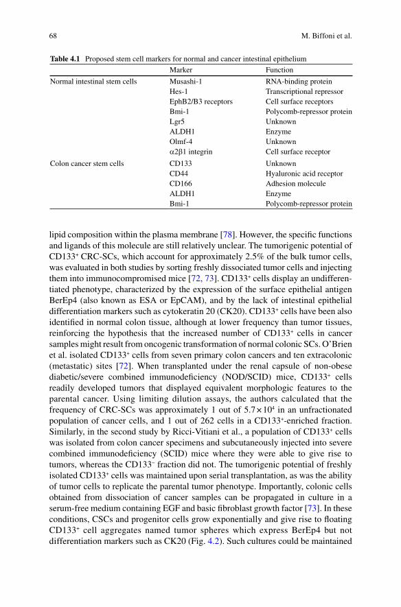
68 M. Biffoni et al.
lipid composition within the plasma membrane [78]. However, the specific functions and ligands of this molecule are still relatively unclear. The tumorigenic potential of CD133+ CRC-SCs, which account for approximately 2.5% of the bulk tumor cells, was evaluated in both studies by sorting freshly dissociated tumor cells and injecting them into immunocompromised mice [72, 73]. CD133+ cells display an undifferen-tiated phenotype, characterized by the expression of the surface epithelial antigen BerEp4 (also known as ESA or EpCAM), and by the lack of intestinal epithelial differentiation markers such as cytokeratin 20 (CK20). CD133+ cells have been also identified in normal colon tissue, although at lower frequency than tumor tissues, reinforcing the hypothesis that the increased number of CD133+ cells in cancer samples might result from oncogenic transformation of normal colonic SCs. O’Brien et al. isolated CD133+ cells from seven primary colon cancers and ten extracolonic (metastatic) sites [72]. When transplanted under the renal capsule of non-obese diabetic/severe combined immunodeficiency (NOD/SCID) mice, CD133+ cells readily developed tumors that displayed equivalent morphologic features to the parental cancer. Using limiting dilution assays, the authors calculated that the frequency of CRC-SCs was approximately 1 out of 5.7 × 10⁴ in an unfractionated population of cancer cells, and 1 out of 262 cells in a CD133+-enriched fraction. Similarly, in the second study by Ricci-Vitiani et al., a population of CD133+ cells was isolated from colon cancer specimens and subcutaneously injected into severe combined immunodeficiency (SCID) mice where they were able to give rise to tumors, whereas the CD133− fraction did not. The tumorigenic potential of freshly isolated CD133+ cells was maintained upon serial transplantation, as was the ability of tumor cells to replicate the parental tumor phenotype. Importantly, colonic cells obtained from dissociation of cancer samples can be propagated in culture in a serum-free medium containing EGF and basic fibroblast growth factor [73]. In these conditions, CSCs and progenitor cells grow exponentially and give rise to floating CD133+ cell aggregates named tumor spheres which express BerEp4 but not differentiation markers such as CK20 (Fig. 4.2). Such cultures could be maintained
Table 4.1 Proposed stem cell markers for normal and cancer intestinal epithelium
Marker Function
Normal intestinal stem cells Musashi-1 RNA-binding proteinHes-1 Transcriptional repressorEphB2/B3 receptors Cell surface receptorsBmi-1 Polycomb-repressor proteinLgr5 UnknownALDH1 EnzymeOlmf-4 Unknowna2b1 integrin Cell surface receptor
Colon cancer stem cells CD133 UnknownCD44 Hyaluronic acid receptorCD166 Adhesion moleculeALDH1 EnzymeBmi-1 Polycomb-repressor protein
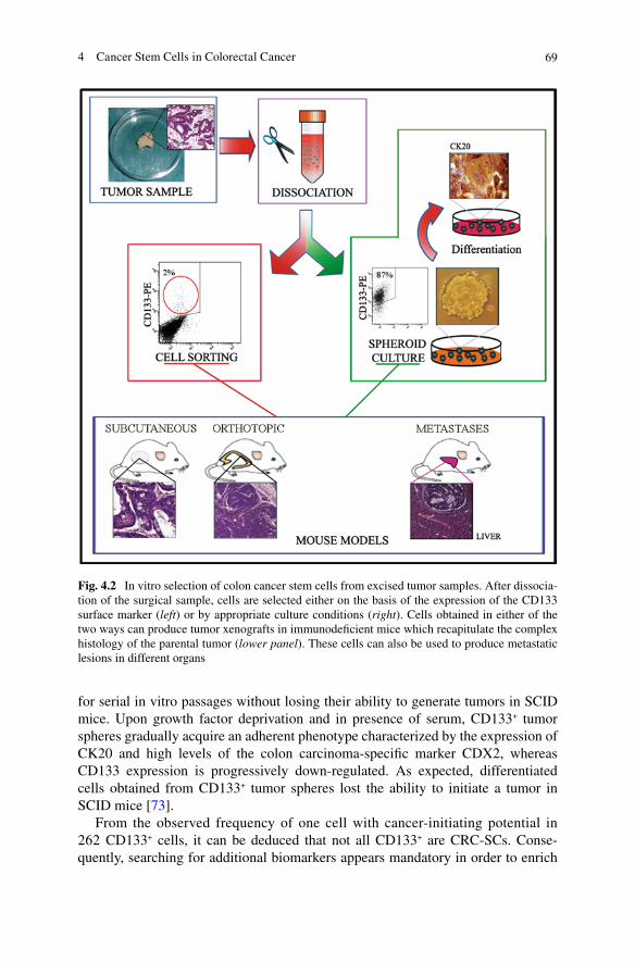
694 Cancer Stem Cells in Colorectal Cancer
Fig. 4.2 In vitro selection of colon cancer stem cells from excised tumor samples. After dissocia-tion of the surgical sample, cells are selected either on the basis of the expression of the CD133 surface marker (left) or by appropriate culture conditions (right). Cells obtained in either of the two ways can produce tumor xenografts in immunodeficient mice which recapitulate the complex histology of the parental tumor (lower panel). These cells can also be used to produce metastatic lesions in different organs
for serial in vitro passages without losing their ability to generate tumors in SCID mice. Upon growth factor deprivation and in presence of serum, CD133+ tumor spheres gradually acquire an adherent phenotype characterized by the expression of CK20 and high levels of the colon carcinoma-specific marker CDX2, whereas CD133 expression is progressively down-regulated. As expected, differentiated cells obtained from CD133+ tumor spheres lost the ability to initiate a tumor in SCID mice [73].
From the observed frequency of one cell with cancer-initiating potential in 262 CD133+ cells, it can be deduced that not all CD133+ are CRC-SCs. Conse-quently, searching for additional biomarkers appears mandatory in order to enrich
70 M. Biffoni et al.
the CRC-SC population. To this end, Dalerba et al. proposed CD44 and EpCAM as CRC-SC–specific markers, with further enrichment by CD166 [79]. Subcutaneous injection of purified CD44+/EpCAMhigh cells into NOD/SCID mice resulted in high-frequency generation of tumor xenograft. In contrast, CD44−/EpCAMlow cells lack tumor-initiating activity [79]. Further subfractionation of the CD44+/EpCAMhigh cell population using the mesenchymal stem cell marker CD166 increased the success of tumor xenograft. However, immunohistochemical analysis of normal colonic epithelium shows that CD44 expression occurs not only in the stem cell compartment at the crypt bottom but also in cells within the proliferative compart-ment; thus the specificity of CD44 for colonic SC remains to be determined. More recently, aldehyde dehydrogenase 1 (ALDH1) has been proposed as a promising new marker for normal and malignant human colonic SCs [80]. ALDH is a detoxi-fying enzyme that oxidizes intracellular aldehydes and thereby confers resistance to alkylating agents [81]. ALDH also converts retinol to retinoic acid, a modulator of cell proliferation. Moreover, ALDH1 has been described as highly expressed in embry-onal tissues as well as in adult SCs isolated from bone marrow, brain, and breast. As few as 25 ALDH1+ cancer cells, isolated from CRC specimens by flow cytometry, were able to generate tumor xenografts. Notably, a subsequent isolation of cancer cells using a second marker (CD44 and CD133 serially) produced a modest further enrichment of tumor-initiating ability [80].
Significant controversy exists, however, over the functional role of these CRC-SC markers. Major questions have been raised regarding CD133. Studies using a trans-genic mouse model in which the CD133 promoter drove LacZ reporter expression demonstrated that CD133 was expressed by both mature and undifferentiated colonic epithelial cells, suggesting that CD133 is not restricted to SC compartment. Moreover, in primary human colon cancer specimens, CD133 was expressed in most of the tumor cell population and sorting of CD133+ cells from liver metastasis of colon cancer demonstrated that CD133 high- and low-expressing cells could gen-erate tumors in NOD/SCID mice [82]. Regardless of the ongoing debate regarding CD133 as a CRC-SC marker and the lack of evidence for a functional role in tum-origenesis, growing evidence supports the clinical significance of this molecule in CRC. Indeed, Horst et al. have recently shown that CD133 expression correlates with poor prognosis and it is an independent prognostic marker for low survival in CRC [83]. The combined evaluation of CD133 and nuclear b-catenin can identify high-risk cases of low-stage CRC [84], and longer relapse-free interval with an increased overall survival has been observed in patients with lower levels of CD133 [85]. Moreover, a recent study comparing the expression of CD133, CD44, and CD166 (markers that have been associated with CRC-SCs) revealed that the expres-sion of CD133 correlates with that of CD166, whereas neither correlates with CD44. The authors also showed that CD133 is the best single marker to predict poor patient survival [86] whereas the combination of the three markers allows stratification of patients into high-, intermediate-, and low-risk classes. To verify the clinical rele-vance of CD133 in CRC metastasis, CD133 expression has been evaluated in a matched case–control collection of 54 pairs of CRC patients with and without synchro-nous liver metastasis showing a strong correlation between high CD133 expression and
714 Cancer Stem Cells in Colorectal Cancer
synchronous liver metastasis. However, no effect was observed in colon cancer cell lines after CD133 knocking down on proliferation, migration, invasion, and colony formation, suggesting that CD133 may be a marker with high prognostic impact for CRC, without relevant functional role as a determinant of tumor progression [87].
Taken together, these data confirm that the identification of biomarkers for CRC-SCs will enable greater understanding of the mechanisms underlying tumor growth and progression. Studies performed on mouse models have provided a great contri-bution to the identification of the CRC-SC population. Indeed, Barker et al. recently provided strong support for the hypothesis that the origin of intestinal cancers is from Lgr5+ CBC cells. The deletion of APC in Lgr5 expressing cells leads to their transformation within days. Transformed SCs remain located at the crypt bottom while feeding a growing microadenoma that develops into a macroscopic adenoma within 3–5 weeks. These data suggest that Lgr5 may mark not only normal intestinal SCs but also the small population of CSCs [88]. Using knock-in LacZ reporter mice within the Prominin-1 (Prom1) locus, Zhu et al. have shown that Prom1+ cells, located at the base of the crypts in the small intestine, co-express Lgr5, generate the entire intestinal epithelium, and are susceptible to neoplastic transformation [89]. Lgr5 was markedly over-expressed in the majority of advanced human CRCs com-pared with normal mucosal tissue [90]. As expected, in situ hybridization analysis confirmed the expression of Lgr5 in CBC cells in both small intestine and colon. This Lgr5 expression, which was variable among CRC cases, correlated significantly with lymphatic and vascular invasion, lymph node metastasis and tumor stage, sug-gesting the involvement of this marker in tumor progression. A similar correlation has been described for the “stemness” gene Bmi1, confirming that cells responsible for colon tumorigenesis and colon ontogenesis share common markers [91].
4.3 Therapeutic Implications of the CRC-SC Model
The CSC model has major implications for the development of new and more effective therapeutic strategies aimed at targeting and eradicating the tumor SC population. At present, anticancer therapies for CRC include surgery, radiation, chemotherapy, and anti-VEGF or EGFR monoclonal antibodies. Regardless of the therapeutic approach, none of these treatment modalities is curative in most of advanced cancer cases. One of the major concerns surrounding the use of cytotoxic agents is that they are designed to target the most rapidly dividing cells, which represent the majority of the tumor cell population, thus resulting in a remarkable but frequently transient clinical remission. Failure of conventional treatment options to eliminate the CSC compartment might result in tumor relapse and, more impor-tantly, in the proliferation of therapy-resistant and more aggressive tumor cells, which ultimately reduce patient survival. Indeed, it has been shown that CRC-SCs are enriched in residual tumors following conventional chemotherapy regimens, and remain capable of rapidly regenerating the tumor from which they were derived [92]. According to the CSC hypothesis, it was expected that tumor-initiating cells may
72 M. Biffoni et al.
display resistance to cytotoxic therapy, permitting the repopulation of treated tumors. Many mechanisms may contribute to the development of therapeutic resis-tance, including the stochastic selection of resistant genetic subclones, microenvi-ronmental factors, and cell extrinsic factors. CSCs are relatively quiescent and this allows them to escape from chemotherapeutic regimens that typically target actively cycling cells. Moreover, CSCs share signaling pathways (i.e., Wnt, Hedgehog, and Notch) with their normal counterparts that regulate self-renewal of the normal colonic SC population and whose deregulation can lead to tumor development. Similarly to normal SCs, CSCs have been proposed to exhibit high-level expression of multidrug transporter family genes, likely resulting in more efficient efflux of chemotherapeutic drugs and innate multidrug resistance [93]. Thus, an efficient therapeutic approach would require the identification of distinctive molecular pathways active in CSCs and should identify agents that can block CSC proliferation without or minimally affecting normal tissues. Together with intrinsic factors, the microenvironment or niche may influence the ability of CSCs to proliferate, migrate, and/or invade. The niche is an anchoring site for CSCs and adhesion molecules or microenvironmental soluble molecules (including growth factors and cytokines), and these can significantly contribute to therapy resistance. In line with this hypoth-esis, it has been recently demonstrated that the production of interleukin-4 (IL-4) by CD133+ CRC-SCs promote tumor resistance to the chemotherapeutic agents 5-fluorouracil and oxaliplatin. On the basis of this finding, a new therapeutic strategy can be devised in order to sensitize CRC-SCs to chemotherapy through the targeting of IL-4 [94]. Thus, in addition to its impact on our understanding of the efficacy of available therapies, the CSC model has an impact on the identification of future therapeutic targets.
To study new approaches to develop drugs that target CSCs, Boman et al. used computer modeling [95]. They demonstrated that an exponential increase in both SC and non-SC populations in CRC development involves an enhanced symmetric SC division. This finding suggests that any systemic therapy designed to effectively treat CRC and other cancers must control or eliminate symmetrical CSC division in tumors, while minimally affecting normal SCs. Thus, a systematic approach to identify and challenge the CSC survival machinery would be mandatory in order to develop novel and more efficient SC-targeted therapies. Genome-wide analyses of cancer have revealed the existence of a vast genetic variation among individual tumors, which makes the use of an exclusively genomic approach to cancer biology extremely complex. On the other hand, it is increasingly clear that tumors share common features at the protein pathway level, suggesting that a pathway-oriented perspective may represent the most effective approach to drug discovery and ther-apy. In a recent study, Fang et al. generated CD133+ tumor sphere cultures from several colon cancer specimens and performed mass-spectrometry–based quantita-tive proteomics in order to identify cell surface proteins enriched on cultured tumor cells [96]. These cells retain the expression of cell surface markers such as CD133, CD166, CD44, and EpCAM as well as other stem cell–associated proteins includ-ing nestin, Bmi1, and Msi-1, thus confirming the value of this in vitro model for biological analysis of CSC populations and for drug screening experiments.
734 Cancer Stem Cells in Colorectal Cancer
Finally, most of the currently available mouse models of CRC are based on chemically induced tumors, genetically engineered animals, and tumor implants in immunocompromised mice, but none of them faithfully replicates all aspects of human tumor development. CRC-SCs represent an excellent tool for the preclinical evaluation of new anticancer therapies both in vitro and in vivo, where they generate xenografts that phenocopy the human tumor of origin [97] (Fig. 4.2). Reliable mouse models of human CRC are essential to understand the mechanisms underly-ing tumor development or pathogenesis and for the preclinical evaluation of new therapies. CSCs both as a theoretical model and as innovative “reagents” could contribute to a significant advancement in cancer research.
References
1. Bannister LH (1995) Alimentary system. In: Williams PL (ed) Gray’s Anatomy, 38th edn. Churchill Livingstone, London
2. Scoville DH, Sato T, He XC, Li L (2008) Current view: intestinal stem cells and signaling. Gastroenterology 134 (3):849–864
3. Potten CS, Booth C, Pritchard DM (1997) The intestinal epithelial stem cell: the mucosal governor. International Journal of Experimental Pathology 78 (4):219–243
4. Booth C, Potten CS (2000) Gut instincts: thoughts on intestinal epithelial stem cells. J Clin Invest 105 (11):1493–1499
5. Marshman E, Booth C, Potten CS (2002) The intestinal epithelial stem cell. Bioessays 24 (1): 91–98
6. Radtke F, Clevers H (2005) Self-renewal and cancer of the gut: two sides of a coin. Science 307 (5717):1904–1909
7. Sancho E, Batlle E, Clevers H (2004) Signaling pathways in intestinal development and can-cer. Annual Review of Cell and Developmental Biology 20:695–723
8. Lynch HT, de la Chapelle A (2003) Hereditary colorectal cancer. N Engl J Med 348 (10):919–932 9. van de Wetering M, Sancho E, Verweij C, de Lau W, Oving I, Hurlstone A, van der Horn K,
Batlle E, Coudreuse D, Haramis AP, Tjon-Pon-Fong M, Moerer P, van den Born M, Soete G, Pals S, Eilers M, Medema R, Clevers H (2002) The beta-catenin/TCF-4 complex imposes a crypt progenitor phenotype on colorectal cancer cells. Cell 111 (2):241–250
10. Korinek V, Barker N, Moerer P, van Donselaar E, Huls G, Peters PJ, Clevers H (1998) Depletion of epithelial stem-cell compartments in the small intestine of mice lacking Tcf-4. Nat Genet 19 (4):379–383
11. Pinto D, Gregorieff A, Begthel H, Clevers H (2003) Canonical Wnt signals are essential for homeostasis of the intestinal epithelium. Genes Dev 17 (14):1709–1713
12. Kuhnert F, Davis CR, Wang HT, Chu P, Lee M, Yuan J, Nusse R, Kuo CJ (2004) Essential requirement for Wnt signaling in proliferation of adult small intestine and colon revealed by adenoviral expression of Dickkopf-1. Proc Natl Acad Sci USA 101 (1):266–271
13. Holmberg J, Genander M, Halford MM, Anneren C, Sondell M, Chumley MJ, Silvany RE, Henkemeyer M, Frisen J (2006) EphB receptors coordinate migration and proliferation in the intestinal stem cell niche. Cell 125 (6):1151–1163
14. Batlle E, Henderson JT, Beghtel H, van den Born MM, Sancho E, Huls G, Meeldijk J, Robertson J, van de Wetering M, Pawson T, Clevers H (2002) Beta-catenin and TCF mediate cell positioning in the intestinal epithelium by controlling the expression of EphB/ephrinB. Cell 111 (2):251–263
15. van Es JH, van Gijn ME, Riccio O, van den Born M, Vooijs M, Begthel H, Cozijnsen M, Robine S, Winton DJ, Radtke F, Clevers H (2005) Notch/gamma-secretase inhibition turns
74 M. Biffoni et al.
proliferative cells in intestinal crypts and adenomas into goblet cells. Nature 435 (7044):959–963
16. Bray SJ (2006) Notch signalling: a simple pathway becomes complex. Nat Rev Mol Cell Biol 7 (9):678–689
17. Jensen J, Pedersen EE, Galante P, Hald J, Heller RS, Ishibashi M, Kageyama R, Guillemot F, Serup P, Madsen OD (2000) Control of endodermal endocrine development by Hes-1. Nat Genet 24 (1):36–44
18. van Es JH, Clevers H (2005) Notch and Wnt inhibitors as potential new drugs for intestinal neoplastic disease. Trends Mol Med 11 (11):496–502
19. Riccio O, van Gijn ME, Bezdek AC, Pellegrinet L, van Es JH, Zimber-Strobl U, Strobl LJ, Honjo T, Clevers H, Radtke F (2008) Loss of intestinal crypt progenitor cells owing to inacti-vation of both Notch1 and Notch2 is accompanied by derepression of CDK inhibitors p27Kip1 and p57Kip2. EMBO Rep 9 (4):377–383
20. Cully M, You H, Levine AJ, Mak TW (2006) Beyond PTEN mutations: the PI3K pathway as an integrator of multiple inputs during tumorigenesis. Nat Rev Cancer 6 (3):184–192
21. Parsons DW, Wang TL, Samuels Y, Bardelli A, Cummins JM, DeLong L, Silliman N, Ptak J, Szabo S, Willson JK, Markowitz S, Kinzler KW, Vogelstein B, Lengauer C, Velculescu VE (2005) Colorectal cancer: mutations in a signalling pathway. Nature 436 (7052):792
22. He XC, Yin T, Grindley JC, Tian Q, Sato T, Tao WA, Dirisina R, Porter-Westpfahl KS, Hembree M, Johnson T, Wiedemann LM, Barrett TA, Hood L, Wu H, Li L (2007) PTEN-deficient intestinal stem cells initiate intestinal polyposis. Nat Genet 39 (2):189–198
23. von Bubnoff A, Cho KW (2001) Intracellular BMP signaling regulation in vertebrates: path-way or network? Dev Biol 239 (1):1–14
24. Haramis AP, Begthel H, van den Born M, van Es J, Jonkheer S, Offerhaus GJ, Clevers H (2004) De novo crypt formation and juvenile polyposis on BMP inhibition in mouse intestine. Science 303 (5664):1684–1686
25. He XC, Zhang J, Tong WG, Tawfik O, Ross J, Scoville DH, Tian Q, Zeng X, He X, Wiedemann LM, Mishina Y, Li L (2004) BMP signaling inhibits intestinal stem cell self-renewal through suppression of Wnt-beta-catenin signaling. Nat Genet 36 (10):1117–1121
26. Waite KA, Eng C (2003) From developmental disorder to heritable cancer: it’s all in the BMP/TGF-beta family. Nat Rev Genet 4 (10):763–773
27. Cheng H, Leblond CP (1974) Origin, differentiation and renewal of the four main epithelial cell types in the mouse small intestine. V. Unitarian Theory of the origin of the four epithelial cell types. The American Journal of Anatomy 141 (4):537–561
28. Cheng H, Leblond CP (1974) Origin, differentiation and renewal of the four main epithelial cell types in the mouse small intestine. I. Columnar cell. The American Journal of Anatomy 141 (4):461–479
29. Cheng H, Leblond CP (1974) Origin, differentiation and renewal of the four main epithelial cell types in the mouse small intestine. III. Entero-endocrine cells. The American Journal of Anatomy 141 (4):503–519
30. Fuller CE, Davies RP, Williams GT, Williams ED (1990) Crypt restricted heterogeneity of goblet cell mucus glycoprotein in histologically normal human colonic mucosa: a potential marker of somatic mutation. Br J Cancer 61 (3):382–384
31. Campbell F, Williams GT, Appleton MA, Dixon MF, Harris M, Williams ED (1996) Post-irradiation somatic mutation and clonal stabilisation time in the human colon. Gut 39 (4): 569–573
32. Preston SL, Wong WM, Chan AO, Poulsom R, Jeffery R, Goodlad RA, Mandir N, Elia G, Novelli M, Bodmer WF, Tomlinson IP, Wright NA (2003) Bottom-up histogenesis of colorectal adenomas: origin in the monocryptal adenoma and initial expansion by crypt fission. Cancer Res 63 (13):3819–3825
33. Taylor RW, Barron MJ, Borthwick GM, Gospel A, Chinnery PF, Samuels DC, Taylor GA, Plusa SM, Needham SJ, Greaves LC, Kirkwood TB, Turnbull DM (2003) Mitochondrial DNA mutations in human colonic crypt stem cells. J Clin Invest 112 (9):1351–1360
34. Potten CS, Hume WJ, Reid P, Cairns J (1978) The segregation of DNA in epithelial stem cells. Cell 15 (3):899–906
754 Cancer Stem Cells in Colorectal Cancer
35. Tumbar T, Guasch G, Greco V, Blanpain C, Lowry WE, Rendl M, Fuchs E (2004) Defining the epithelial stem cell niche in skin. Science 303 (5656):359–363
36. Kiel MJ, He S, Ashkenazi R, Gentry SN, Teta M, Kushner JA, Jackson TL, Morrison SJ (2007) Haematopoietic stem cells do not asymmetrically segregate chromosomes or retain BrdU. Nature 449 (7159):238–242
37. Potten CS, Kovacs L, Hamilton E (1974) Continuous labelling studies on mouse skin and intestine. Cell and Tissue Kinetics 7 (3):271–283
38. Potten CS (1977) Extreme sensitivity of some intestinal crypt cells to X and gamma irradia-tion. Nature 269 (5628):518–521
39. Bjerknes M, Cheng H (1981) The stem-cell zone of the small intestinal epithelium. III. Evidence from columnar, enteroendocrine, and mucous cells in the adult mouse. The American Journal of Anatomy 160 (1):77–91
40. Bjerknes M, Cheng H (1999) Clonal analysis of mouse intestinal epithelial progenitors. Gastroenterology 116 (1):7–14
41. Kiel MJ, Yilmaz OH, Iwashita T, Terhorst C, Morrison SJ (2005) SLAM family receptors distinguish hematopoietic stem and progenitor cells and reveal endothelial niches for stem cells. Cell 121 (7):1109–1121
42. Shackleton M, Vaillant F, Simpson KJ, Stingl J, Smyth GK, Asselin-Labat ML, Wu L, Lindeman GJ, Visvader JE (2006) Generation of a functional mammary gland from a single stem cell. Nature 439 (7072):84–88
43. Nakamura M, Okano H, Blendy JA, Montell C (1994) Musashi, a neural RNA-binding protein required for Drosophila adult external sensory organ development. Neuron 13 (1):67–81
44. Potten CS, Booth C, Tudor GL, Booth D, Brady G, Hurley P, Ashton G, Clarke R, Sakakibara S, Okano H (2003) Identification of a putative intestinal stem cell and early lineage marker; musashi-1. Differentiation 71 (1):28–41
45. Nishimura S, Wakabayashi N, Toyoda K, Kashima K, Mitsufuji S (2003) Expression of Musashi-1 in human normal colon crypt cells: a possible stem cell marker of human colon epithelium. Dig Dis Sci 48 (8):1523–1529
46. Kayahara T, Sawada M, Takaishi S, Fukui H, Seno H, Fukuzawa H, Suzuki K, Hiai H, Kageyama R, Okano H, Chiba T (2003) Candidate markers for stem and early progenitor cells, Musashi-1 and Hes1, are expressed in crypt base columnar cells of mouse small intestine. FEBS Lett 535 (1-3):131–135
47. Juliano RL, Varner JA (1993) Adhesion molecules in cancer: the role of integrins. Curr Opin Cell Biol 5 (5):812–818
48. Jones PH, Watt FM (1993) Separation of human epidermal stem cells from transit amplifying cells on the basis of differences in integrin function and expression. Cell 73 (4):713–724
49. Shinohara T, Avarbock MR, Brinster RL (1999) Beta1- and alpha6-integrin are surface mark-ers on mouse spermatogonial stem cells. Proc Natl Acad Sci USA 96 (10):5504–5509
50. Fujimoto K, Beauchamp RD, Whitehead RH (2002) Identification and isolation of candidate human colonic clonogenic cells based on cell surface integrin expression. Gastroenterology 123 (6):1941–1948
51. van der Lugt NM, Domen J, Linders K, van Roon M, Robanus-Maandag E, te Riele H, van der Valk M, Deschamps J, Sofroniew M, van Lohuizen M, et al. (1994) Posterior transformation, neurological abnormalities, and severe hematopoietic defects in mice with a targeted deletion of the bmi-1 proto-oncogene. Genes Dev 8 (7):757–769
52. Sangiorgi E, Capecchi MR (2008) Bmi1 is expressed in vivo in intestinal stem cells. Nat Genet 40 (7):915–920
53. Barker N, van Es JH, Kuipers J, Kujala P, van den Born M, Cozijnsen M, Haegebarth A, Korving J, Begthel H, Peters PJ, Clevers H (2007) Identification of stem cells in small intestine and colon by marker gene Lgr5. Nature 449 (7165):1003–1007
54. Sato T, Vries RG, Snippert HJ, van de Wetering M, Barker N, Stange DE, van Es JH, Abo A, Kujala P, Peters PJ, Clevers H (2009) Single Lgr5 stem cells build crypt-villus structures in vitro without a mesenchymal niche. Nature 459 (7244):262–265
55. van der Flier LG, van Gijn ME, Hatzis P, Kujala P, Haegebarth A, Stange DE, Begthel H, van den Born M, Guryev V, Oving I, van Es JH, Barker N, Peters PJ, van de Wetering M,
76 M. Biffoni et al.
Clevers H (2009) Transcription factor achaete scute-like 2 controls intestinal stem cell fate. Cell 136 (5):903–912
56. Calleja M, Renaud O, Usui K, Pistillo D, Morata G, Simpson P (2002) How to pattern an epithelium: lessons from achaete-scute regulation on the notum of Drosophila. Gene 292 (1-2):1–12
57. Zhang J, Liu WL, Tang DC, Chen L, Wang M, Pack SD, Zhuang Z, Rodgers GP (2002) Identification and characterization of a novel member of olfactomedin-related protein family, hGC-1, expressed during myeloid lineage development. Gene 283 (1-2):83–93
58. Kosinski C, Li VS, Chan AS, Zhang J, Ho C, Tsui WY, Chan TL, Mifflin RC, Powell DW, Yuen ST, Leung SY, Chen X (2007) Gene expression patterns of human colon tops and basal crypts and BMP antagonists as intestinal stem cell niche factors. Proc Natl Acad Sci USA 104 (39):15418–15423
59. van der Flier LG, Haegebarth A, Stange DE, van de Wetering M, Clevers H (2009) OLFM4 is a robust marker for stem cells in human intestine and marks a subset of colorectal cancer cells. Gastroenterology 137 (1):15–17
60. Morson BC (1974) Evolution of cancer of the colon and rectum. Proc Inst Med Chic 30 (4): 145–148
61. Herrera L, Kakati S, Gibas L, Pietrzak E, Sandberg AA (1986) Gardner syndrome in a man with an interstitial deletion of 5q. Am J Med Genet 25 (3):473–476
62. Groden J, Thliveris A, Samowitz W, Carlson M, Gelbert L, Albertsen H, Joslyn G, Stevens J, Spirio L, Robertson M, et al. (1991) Identification and characterization of the familial adenom-atous polyposis coli gene. Cell 66 (3):589–600
63. Kinzler KW, Nilbert MC, Su LK, Vogelstein B, Bryan TM, Levy DB, Smith KJ, Preisinger AC, Hedge P, McKechnie D, et al. (1991) Identification of FAP locus genes from chromosome 5q21. Science 253 (5020):661–665
64. Powell SM, Zilz N, Beazer-Barclay Y, Bryan TM, Hamilton SR, Thibodeau SN, Vogelstein B, Kinzler KW (1992) APC mutations occur early during colorectal tumorigenesis. Nature 359 (6392):235–237
65. Miyoshi Y, Nagase H, Ando H, Horii A, Ichii S, Nakatsuru S, Aoki T, Miki Y, Mori T, Nakamura Y (1992) Somatic mutations of the APC gene in colorectal tumors: mutation cluster region in the APC gene. Hum Mol Genet 1 (4):229–233
66. Albuquerque C, Breukel C, van der Luijt R, Fidalgo P, Lage P, Slors FJ, Leitao CN, Fodde R, Smits R (2002) The ‘just-right’ signaling model: APC somatic mutations are selected based on a specific level of activation of the beta-catenin signaling cascade. Hum Mol Genet 11 (13):1549–1560
67. Sansom OJ, Meniel VS, Muncan V, Phesse TJ, Wilkins JA, Reed KR, Vass JK, Athineos D, Clevers H, Clarke AR (2007) Myc deletion rescues Apc deficiency in the small intestine. Nature 446 (7136):676–679
68. Morin PJ, Sparks AB, Korinek V, Barker N, Clevers H, Vogelstein B, Kinzler KW (1997) Activation of beta-catenin-Tcf signaling in colon cancer by mutations in beta-catenin or APC. Science 275 (5307):1787–1790
69. Lynch HT, Smyrk T, Lynch JF (1996) Overview of natural history, pathology, molecular genet-ics and management of HNPCC (Lynch Syndrome). Int J Cancer 69 (1):38–43
70. Bronner CE, Baker SM, Morrison PT, Warren G, Smith LG, Lescoe MK, Kane M, Earabino C, Lipford J, Lindblom A, et al. (1994) Mutation in the DNA mismatch repair gene homologue hMLH1 is associated with hereditary non-polyposis colon cancer. Nature 368 (6468):258–261
71. Leach FS, Nicolaides NC, Papadopoulos N, Liu B, Jen J, Parsons R, Peltomaki P, Sistonen P, Aaltonen LA, Nystrom-Lahti M, et al. (1993) Mutations of a mutS homolog in hereditary nonpolyposis colorectal cancer. Cell 75 (6):1215–1225
72. O’Brien CA, Pollett A, Gallinger S, Dick JE (2007) A human colon cancer cell capable of initiating tumour growth in immunodeficient mice. Nature 445 (7123):106–110
73. Ricci-Vitiani L, Lombardi DG, Pilozzi E, Biffoni M, Todaro M, Peschle C, De Maria R (2007) Identification and expansion of human colon-cancer-initiating cells. Nature 445 (7123): 111–115
774 Cancer Stem Cells in Colorectal Cancer
74. Yin AH, Miraglia S, Zanjani ED, Almeida-Porada G, Ogawa M, Leary AG, Olweus J, Kearney J, Buck DW (1997) AC133, a novel marker for human hematopoietic stem and progenitor cells. Blood 90 (12):5002–5012
75. Uchida N, Buck DW, He D, Reitsma MJ, Masek M, Phan TV, Tsukamoto AS, Gage FH, Weissman IL (2000) Direct isolation of human central nervous system stem cells. Proc Natl Acad Sci USA 97 (26):14720–14725
76. Corbeil D, Roper K, Hellwig A, Tavian M, Miraglia S, Watt SM, Simmons PJ, Peault B, Buck DW, Huttner WB (2000) The human AC133 hematopoietic stem cell antigen is also expressed in epithelial cells and targeted to plasma membrane protrusions. J Biol Chem 275 (8): 5512–5520
77. Corbeil D, Roper K, Fargeas CA, Joester A, Huttner WB (2001) Prominin: a story of choles-terol, plasma membrane protrusions and human pathology. Traffic 2 (2):82–91
78. Roper K, Corbeil D, Huttner WB (2000) Retention of prominin in microvilli reveals distinct cholesterol-based lipid micro-domains in the apical plasma membrane. Nat Cell Biol 2 (9): 582–592
79. Dalerba P, Dylla SJ, Park IK, Liu R, Wang X, Cho RW, Hoey T, Gurney A, Huang EH, Simeone DM, Shelton AA, Parmiani G, Castelli C, Clarke MF (2007) Phenotypic characterization of human colorectal cancer stem cells. Proc Natl Acad Sci USA 104 (24):10158–10163
80. Huang EH, Hynes MJ, Zhang T, Ginestier C, Dontu G, Appelman H, Fields JZ, Wicha MS, Boman BM (2009) Aldehyde dehydrogenase 1 is a marker for normal and malignant human colonic stem cells (SC) and tracks SC overpopulation during colon tumorigenesis. Cancer Res 69 (8):3382–3389
81. Yoshida A, Rzhetsky A, Hsu LC, Chang C (1998) Human aldehyde dehydrogenase gene family. Eur J Biochem 251 (3):549–557
82. Shmelkov SV, Butler JM, Hooper AT, Hormigo A, Kushner J, Milde T, St Clair R, Baljevic M, White I, Jin DK, Chadburn A, Murphy AJ, Valenzuela DM, Gale NW, Thurston G, Yancopoulos GD, D’Angelica M, Kemeny N, Lyden D, Rafii S (2008) CD133 expression is not restricted to stem cells, and both CD133+ and CD133- metastatic colon cancer cells initiate tumors. J Clin Invest 118 (6):2111–2120
83. Horst D, Kriegl L, Engel J, Kirchner T, Jung A (2008) CD133 expression is an independent prognostic marker for low survival in colorectal cancer. Br J Cancer 99 (8):1285–1289
84. Horst D, Kriegl L, Engel J, Jung A, Kirchner T (2009) CD133 and nuclear beta-catenin: the marker combination to detect high risk cases of low stage colorectal cancer. Eur J Cancer 45 (11):2034–2040
85. Artells R, Moreno I, Diaz T, Martinez F, Gel B, Navarro A, Ibeas R, Moreno J, Monzo M (2010) Tumour CD133 mRNA expression and clinical outcome in surgically resected colorectal cancer patients. Eur J Cancer 46 (3):642–649
86. Horst D, Kriegl L, Engel J, Kirchner T, Jung A (2009) Prognostic significance of the cancer stem cell markers CD133, CD44, and CD166 in colorectal cancer. Cancer Invest 27 (8): 844–850
87. Horst D, Scheel SK, Liebmann S, Neumann J, Maatz S, Kirchner T, Jung A (2009) The cancer stem cell marker CD133 has high prognostic impact but unknown functional relevance for the metastasis of human colon cancer. J Pathol 219 (4):427–434
88. Barker N, Ridgway RA, van Es JH, van de Wetering M, Begthel H, van den Born M, Danenberg E, Clarke AR, Sansom OJ, Clevers H (2009) Crypt stem cells as the cells-of-origin of intestinal cancer. Nature 457 (7229):608–611
89. Zhu L, Gibson P, Currle DS, Tong Y, Richardson RJ, Bayazitov IT, Poppleton H, Zakharenko S, Ellison DW, Gilbertson RJ (2009) Prominin 1 marks intestinal stem cells that are susceptible to neoplastic transformation. Nature 457 (7229):603–607
90. Uchida H, Yamazaki K, Fukuma M, Yamada T, Hayashida T, Hasegawa H, Kitajima M, Kitagawa Y, Sakamoto M (2010) Overexpression of leucine-rich repeat-containing G protein-coupled receptor 5 in colorectal cancer. Cancer Sci 101 (7):1731–1737
91. Du J, Li Y, Li J, Zheng J (2010) Polycomb group protein Bmi1 expression in colon cancers predicts the survival. Med Oncol 27:1273–1276
78 M. Biffoni et al.
92. Dylla SJ, Beviglia L, Park IK, Chartier C, Raval J, Ngan L, Pickell K, Aguilar J, Lazetic S, Smith-Berdan S, Clarke MF, Hoey T, Lewicki J, Gurney AL (2008) Colorectal cancer stem cells are enriched in xenogeneic tumors following chemotherapy. PLoS One 3 (6):e2428
93. Hirschmann-Jax C, Foster AE, Wulf GG, Nuchtern JG, Jax TW, Gobel U, Goodell MA, Brenner MK (2004) A distinct “side population” of cells with high drug efflux capacity in human tumor cells. Proc Natl Acad Sci USA 101 (39):14228–14233
94. Todaro M, Alea MP, Di Stefano AB, Cammareri P, Vermeulen L, Iovino F, Tripodo C, Russo A, Gulotta G, Medema JP, Stassi G (2007) Colon cancer stem cells dictate tumor growth and resist cell death by production of interleukin-4. Cell Stem Cell 1 (4):389–402
95. Boman BM, Wicha MS, Fields JZ, Runquist OA (2007) Symmetric division of cancer stem cells–a key mechanism in tumor growth that should be targeted in future therapeutic approaches. Clin Pharmacol Ther 81 (6):893–898
96. Fang DD, Kim YJ, Lee CN, Aggarwal S, McKinnon K, Mesmer D, Norton J, Birse CE, He T, Ruben SM, Moore PA (2010) Expansion of CD133(+) colon cancer cultures retaining stem cell properties to enable cancer stem cell target discovery. Br J Cancer 102 (8):1265–1275
97. Baiocchi M, Biffoni M, Ricci-Vitiani L, Pilozzi E, De Maria R (2010) New models for can-cer research: human cancer stem cell xenografts. Current Opinion in Pharmacology 10 (4):380–384
79A.L. Allan (ed.), Cancer Stem Cells in Solid Tumors, Stem Cell Biology and Regenerative Medicine, DOI 10.1007/978-1-61779-246-5_5, © Springer Science+Business Media, LLC 2011
Abstract Over the past decade, increasing evidence has suggested that stem cells play a crucial role not only in the generation of complex multicellular organisms but also in the development and progression of malignant diseases. It has now been shown that many tumors harbor a subset of distinct cancer cells that bear stem cell characteristics and, therefore, these cells are termed cancer stem cells (CSCs) or tumor-initiating cells. CSCs are hypothesized to be exclusively responsible for tumor initiation, propagation, and metastasis. Indeed, it has been shown that human pancreatic CSCs contain a subpopulation of so-called migrating CSCs character-ized by CXCR4 co-expression. Only these cells are capable of escaping the primary tumor and metastasizing to distant sites. Clinically even more important, however, is the observation that CSCs are highly resistant to chemo- and radiotherapy. Laboratories around the world are now aiming to further characterize these cells in hopes of identifying novel treatment modalities to conquer pancreatic cancer.
Abbreviations
ABC ATP binding cassetteALDH1 Aldehyde dehydrogenase 1a1Arx Aristalless-related homeoboxCAC Centroacinar cellsCD Cluster of differentiationCDKN2A Cyclin-dependent kinase inhibitor 2A
C. Heeschen (*)Clinical Research Programme, Stem Cells & Cancer Group, Spanish National Cancer Research Centre (CNIO), Madrid, Spaine-mail: [email protected]
Chapter 5Cancer Stem Cells in Pancreatic Cancer
Jorge Dorado, Alicia G. Serrano, and Christopher Heeschen
80 J. Dorado et al.
CSC Cancer stem cellCXCR4 CXC chemokine receptor 4EMT Epithelial-to-mesenchymal transitionEpCAM Epithelial cell adhesion moleculeHes1 Hairy enhancer of split 1IL-4 Interleukin 4IPMN Intraductal mucinous neoplasmKlf4 Krueppel-like factor 4MDR1 Multi-drug resistance 1mTOR Mammalian target of rapamycinNF-kB Nuclear factor kappa light chain enhancer of activated B cellsNgn3 Neurogenin 3PanIN Pancreatic intraepithelial neoplasiaPax4 Paired box gene 4PDAC Pancreatic ductal adenocarcinomaPdx1 Pancreatic and duodenal homeobox 1PTEN Phosphatase and tensin homologPtf1 Pancreas-specific transcription factor 1RBP-Jk Recombination signal-binding protein 1 for J-kappaSDF-1 Stromal-derived factor 1Shh Sonic hedgehogSox2 Sex determining region Y-box 2SP Side populationTP53 Tumor protein 53 geneZEB1 Zinc finger E-box-binding homeobox 1
5.1 Introduction
Pancreatic ductal adenocarcinoma is the deadliest solid cancer and currently the fourth most frequent cause for cancer-related deaths. Pancreatic cancer is character-ized by late diagnosis due to lack of early symptoms, extensive metastasis, and high resistance to chemotherapy and radiation. Despite increasing research activities in the field of pancreatic tumor and vascular biology, there has been very little substan-tial therapeutic progress regarding clinical endpoints over the past decades (Fig. 5.1). One of the more recent therapeutic advancements involving introduction of the anti-metabolite gemcitabine in 1997 has improved clinical response in terms of pain reduction and weight loss [1]. However, with a 5-year survival rate of 1–4% and a median survival period of 4–6 months, the prognosis of patients with pancreatic cancer remains extremely poor [2–7]. The addition of the only other approved agent, erlotinib, to gemcitabine has not resulted in major improvement in survival [8]. Therefore, elucidation of the mechanisms governing pancreas biology and their deregulation during tumorigenesis is of crucial importance for the development of more effective therapies.
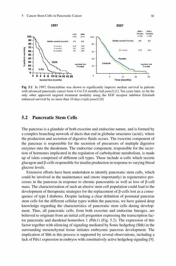
815 Cancer Stem Cells in Pancreatic Cancer
5.2 Pancreatic Stem Cells
The pancreas is a glandule of both exocrine and endocrine nature, and is formed by a complex branching network of ducts that end in globular structures (acini), where the production and secretion of digestive fluids occurs. The exocrine component of the pancreas is responsible for the secretion of precursors of multiple digestive enzymes into the duodenum. The endocrine component, responsible for the secre-tion of hormones implicated in the regulation of carbohydrate metabolism, is made up of islets comprised of different cell types. These include a-cells which secrete glucagon and b-cells responsible for insulin production in response to varying blood glucose levels.
Extensive efforts have been undertaken to identify pancreatic stem cells, which could be involved in the maintenance and (more importantly) in regenerative pro-cesses in the pancreas in response to chronic pancreatitis as well as loss of b-cell mass. The characterization of such an elusive stem cell population could lead to the development of therapeutic strategies for the replacement of b-cells lost as a conse-quence of type I diabetes. Despite lacking a clear definition of postnatal pancreas stem cells for the different cellular types within the pancreas, we have gained deep knowledge regarding the characteristics of pancreatic stem cells during develop-ment. Thus, all pancreatic cells, from both exocrine and endocrine lineages, are believed to originate from an initial cell progenitor expressing the transcription fac-tor pancreatic and duodenal homeobox 1 (Pdx1) (Fig. 5.2). The expression of this factor together with silencing of signaling mediated by Sonic hedgehog (Shh) in the surrounding mesenchymal tissue initiates embryonic pancreas development. The implication of Shh in this process is supported by several observations, including a lack of Pdx1 expression in embryos with constitutively active hedgehog signaling [9].
Fig. 5.1 In 1997, Gemcitabine was shown to significantly improve median survival in patients with advanced pancreatic cancer from 4.4 to 5.6 months (left panel) [1]. Ten years later, so far the only other approved targeted treatment modality using the EGF receptor inhibitor Erlotinib enhanced survival by no more than 10 days (right panel) [8]
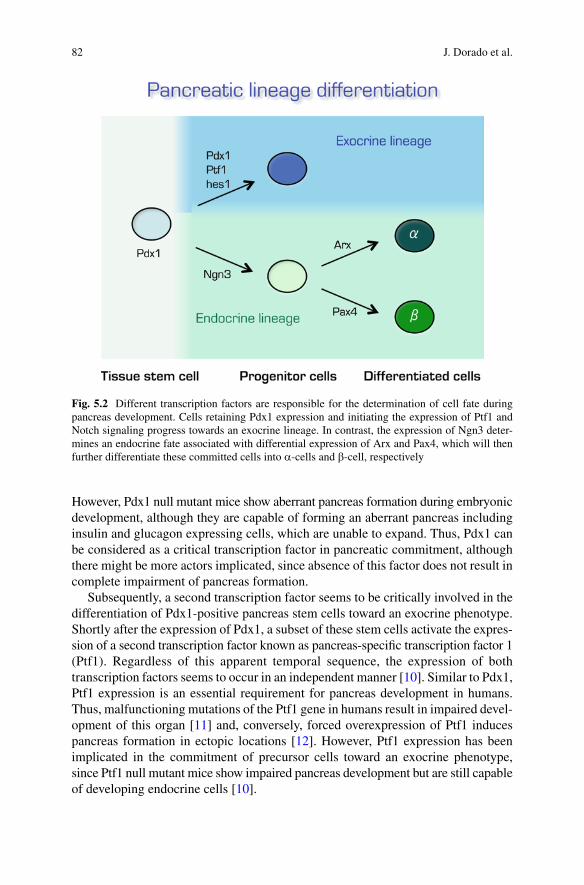
82 J. Dorado et al.
However, Pdx1 null mutant mice show aberrant pancreas formation during embryonic development, although they are capable of forming an aberrant pancreas including insulin and glucagon expressing cells, which are unable to expand. Thus, Pdx1 can be considered as a critical transcription factor in pancreatic commitment, although there might be more actors implicated, since absence of this factor does not result in complete impairment of pancreas formation.
Subsequently, a second transcription factor seems to be critically involved in the differentiation of Pdx1-positive pancreas stem cells toward an exocrine phenotype. Shortly after the expression of Pdx1, a subset of these stem cells activate the expres-sion of a second transcription factor known as pancreas-specific transcription factor 1 (Ptf1). Regardless of this apparent temporal sequence, the expression of both transcription factors seems to occur in an independent manner [10]. Similar to Pdx1, Ptf1 expression is an essential requirement for pancreas development in humans. Thus, malfunctioning mutations of the Ptf1 gene in humans result in impaired devel-opment of this organ [11] and, conversely, forced overexpression of Ptf1 induces pancreas formation in ectopic locations [12]. However, Ptf1 expression has been implicated in the commitment of precursor cells toward an exocrine phenotype, since Ptf1 null mutant mice show impaired pancreas development but are still capable of developing endocrine cells [10].
Fig. 5.2 Different transcription factors are responsible for the determination of cell fate during pancreas development. Cells retaining Pdx1 expression and initiating the expression of Ptf1 and Notch signaling progress towards an exocrine lineage. In contrast, the expression of Ngn3 deter-mines an endocrine fate associated with differential expression of Arx and Pax4, which will then further differentiate these committed cells into a-cells and b-cell, respectively
835 Cancer Stem Cells in Pancreatic Cancer
In addition, commitment toward an exocrine fate seems to be potentiated through signaling of the surrounding mesenchyma on Pdx1-positive cells, enhancing Notch signaling mediated by its downstream target Hes1 (hairy enhancer of split 1) and inhibiting expression of the pro-endocrine differentiation factor Neurogenin 3 (Ngn3) [13]. Thus, null mutant mice for both the Notch ligand delta-like or for the Notch target RBP-Jk transcription factor are enriched in cells of the endocrine lineage and Hes1 null mice display severe hypoplasia of the pancreas as a result of lack of exo-crine progenitor cells [13]. Determination of endocrine fate is induced by expression of the transcription factor Ngn3. In fact, Ngn3-positive cells represent the origin of all the heterogeneity of pancreatic endocrine cells [14]. Thus, a- and b-cells will arise from Ngn3-positive cells, although they will be generated in a different ratio. In early pancreatic development during mouse embryogenesis, the vast majority of cells derived from Ngn3-positive cells are glucagon-secreting a-cells, providing a rationale for the observation that Pdx1-Ngn3 forced expression primarily leads to the develop-ment of glucagon cells [15]. Subsequently, Pdx1 is then downregulated in a-cells as they progress toward a non-epithelial phenotype in a process that strongly resembles the epithelial-to-mesenchymal transition (EMT). Conversely, b-cells retain Pdx1 expression while remaining in rather low numbers as compared to glucagon-secreting cells until later in development, when an amplification of the pool of b-cells occurs, together with branching morphogenesis and acinar cell differentiation [14, 16].
Commitment toward a- or b-cell fate seems to depend on the mutually exclusive action of the transcription factors Aristalless-related homeobox (Arx) and paired box gene 4 (Pax4). Expression of Arx might induce formation of a-cells, since deletion of this gene results in impaired generation of this cell type [17], whereas Pax4 appears to be responsible for b-cell formation [18]. The existence of different sequential progenitor cells raises the question of whether these cells can also be reverted to a less differentiated phenotype in order to give rise to a broader number of cell types (plasticity). However, accumulating evidence suggests that b-cells are differentiated cells with very limited expansion capability. In fact, most b-cells seem to originate from a pool of already existing b-cells precursors rather than from expansion of ancient b-cells [19]. Notch is not capable of reverting mature endocrine cells toward a progenitor-like state [20]. In contrast, Ngn3-positive cells demonstrate greater plas-ticity, since they can be reverted to a ductal progenitor phenotype [21].
5.3 Cell of Origin for Pancreatic Cancer
The cell from which human pancreatic ductal adenocarcinoma originates still remains elusive. One of the hypotheses that have been proposed for tumor initiation in solid organs is the malignant transformation of stem cells resident in the normal tissue. These cells are intrinsically endowed with the capacity of self-renewal, and would therefore only need to accumulate sequential mutations to undergo malignant trans-formation and originate a tumor. Indeed, this hypothesis has just recently been validated for intestinal cancer [22]. The fact that adult pancreatic stem cells, while
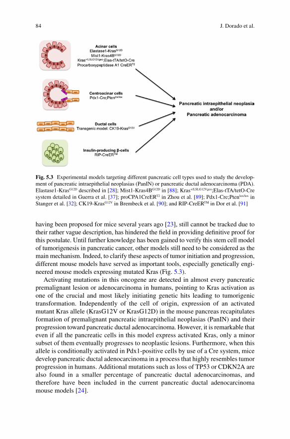
84 J. Dorado et al.
having been proposed for mice several years ago [23], still cannot be tracked due to their rather vague description, has hindered the field in providing definitive proof for this postulate. Until further knowledge has been gained to verify this stem cell model of tumorigenesis in pancreatic cancer, other models still need to be considered as the main mechanism. Indeed, to clarify these aspects of tumor initiation and progression, different mouse models have served as important tools, especially genetically engi-neered mouse models expressing mutated Kras (Fig. 5.3).
Activating mutations in this oncogene are detected in almost every pancreatic premalignant lesion or adenocarcinoma in humans, pointing to Kras activation as one of the crucial and most likely initiating genetic hits leading to tumorigenic transformation. Independently of the cell of origin, expression of an activated mutant Kras allele (KrasG12V or KrasG12D) in the mouse pancreas recapitulates formation of premalignant pancreatic intraepithelial neoplasias (PanIN) and their progression toward pancreatic ductal adenocarcinoma. However, it is remarkable that even if all the pancreatic cells in this model express activated Kras, only a minor subset of them eventually progresses to neoplastic lesions. Furthermore, when this allele is conditionally activated in Pdx1-positive cells by use of a Cre system, mice develop pancreatic ductal adenocarcinoma in a process that highly resembles tumor progression in humans. Additional mutations such as loss of TP53 or CDKN2A are also found in a smaller percentage of pancreatic ductal adenocarcinomas, and therefore have been included in the current pancreatic ductal adenocarcinoma mouse models [24].
Fig. 5.3 Experimental models targeting different pancreatic cell types used to study the develop-ment of pancreatic intraepithelial neoplasias (PanIN) or pancreatic ductal adenocarcinoma (PDA). Elastase1-KrasG12D described in [28]; Mist1-Kras4BG12D in [88]; Kras+/LSLG12Vgeo;Elas-tTA/tetO-Cre system detailed in Guerra et al. [37]; proCPA1CreERT2 in Zhou et al. [89]; Pdx1-Cre;Ptenlox/lox in Stanger et al. [32]; CK19-KrasG12V in Brembeck et al. [90]; and RIP-CreERTM in Dor et al. [91]
855 Cancer Stem Cells in Pancreatic Cancer
Considering the fact that pancreatic ductal adenocarcinoma has a ductal morphology and that its gene expression pattern is similar to that of ductal cells, it is tempting to affirm that a ductal cell would be the target for the tumorigenic transformation. Unfortunately, the poor performance of currently available ductal promoters for in vivo mouse models renders this hypothesis difficult to prove, and the evidence obtained so far is not yet conclusive. Specifically, expression of KrasG12V under the control of the ductal promoter cytokeratin-19 in a transgenic mouse model produces no apparent malignant phenotype. However, despite the ductal histology of pancreatic ductal ade-nocarcinoma, the lesions, which can be appreciated at the earliest stages of tumorigen-esis, are actually embedded in islets mainly formed by clusters of a- and b-cells. This observation raises the possibility that transdifferentiation of b-cells may be the root of pancreatic cancer, a hypothesis which is further supported by the observation that chemical depletion of b-cells impairs tumor initiation. Indeed prior evidence suggests that transdifferentiation may occur in the pancreas, since markers of foregut differen-tiation are expressed in some premalignant pancreatic lesions [25]. While tracing experiments indicate that b-cells do not contribute to the generation of cells with aci-nar or ductal phenotype during tumorigenesis [26], a new mouse model of Kras activa-tion in b-cells has provided new insights. Exclusive Kras activation in b-cells was not sufficient for transformation of these cells in unchallenged mice, although previous induction of pancreatitis did lead to the development of exocrine neoplasia [27].
During the early stages of pancreatic tumorigenesis, progression of acinar cells toward a ductal phenotype is also frequently detected, expanding the possibility toward acinar cells as the tumor-initiating cells. Mouse studies using the acinar-specific promoter of the Elastase gene have revealed that conditional activation of Kras exclusively in acinar cells results in tumors of mixed acinar and ductal mor-phology [28]. Moreover, Notch signaling cooperates with Kras activation during tumor initiation and progression [29]. Another observation pointing to acinar cells as the pancreatic cell type where tumorigenesis may initiate has been the recent discovery of a Bmi1-positive population within the acinar subset of cells, which is capable of maintaining pancreatic cell homeostasis [30]. Finally, centroacinar cells (CAC) have emerged as another candidate cell type for driving tumorigen-esis. These cells are located at the junction of ductal and acinar compartments. The fact that Notch signaling and its target gene Hes1 remain active in these cells during adulthood [30, 31], together with the observation that Notch signaling maintains an undifferentiated state during pancreas embryogenesis [21], has lead to the hypothesis that these CACs are possible targets for tumor-initiating events. Consistent with this hypothesis, it has been observed that different Notch signal-ing mediators, including Hes1, are overexpressed in pancreatic ductal adenocarci-noma. Furthermore, specific deletion of PTEN gene in pancreatic tissue leads to expansion of CACs and eventual progression to carcinoma, pointing to these cells as the origin of tumorigenic processes within this organ in mice [32]. However, in this model, mice develop tumors morphologically different from pancreatic ductal adenocarcinoma and more related to human intraductal mucinous neoplasm (IPMN), an infrequent premalignant lesion in ductal cells than can progress to PDAC pancreatic ductal adenocarcinoma.
86 J. Dorado et al.
5.4 The Role of Chronic Pancreatitis
Pancreatitis is an inflammatory response of the pancreas toward autoimmune antibodies and liberation of pro-enzymatic content of the exocrine pancreas following external injury or damage caused by xenobiotics such as caerulein [33]. There is growing evidence that this inflammatory state facilitates pancreatic tumorigenesis, indicating that the physiological context can exert a strong influence on the suscep-tibility of a cell toward transforming events. Consistently, chronic pancreatitis has been identified as a prominent risk factor for pancreatic cancer in humans. This link between pancreatitis and human pancreatic cancer has been well documented in several epidemiological studies [34–36]. Accumulating experimental evidence from mouse models now also supports this notion. An inflammatory stimulus was necessary for the induction of tumorigenesis through activation of Kras in acinar cells of adult mice, which otherwise are refractory to this oncogenic input [37]. Interestingly, this process was not mediated by the pro-inflammatory transcription factor NF-kB, although such a mechanism has been demonstrated to be operative for other similar processes such as colitis-associated colon carcinogenesis [38].
Pancreatitis has not only been shown to have synergistic effects for promoting malignant transformation, but it has also been implicated in mobilization of tissue progenitor cells and induction of their proliferative capacity. Specifically, partial duct ligation of the pancreas results in activation of b-cell progenitors together with their expansion [39]. In a similar manner, pancreatic injury leading to pancreatitis has been demonstrated to affect the endocrine status of insulin-secreting cells, enabling them to behave as starting points for exocrine neoplasias [27]. Since, under normal conditions, pancreatic stem cells are likely to constitute only a minor population that can hardly be detected, experimental induction of chronic pancreatitis may increase the number of these cells facilitating their detection, characterization, and further investigation. Therefore, future studies should address the possibility of pancreatic stem cells being expanded in response to pancreatitis, rendering them more susceptible to trans-forming events and their subsequent conversion into cancer stem cells (CSCs) as the proposed root of pancreatic cancer as discussed in the following section.
5.5 Pancreatic Cancer Stem Cells
The CSC hypothesis is receiving increasing interest within the field of pancreatic cancer as well as other malignancies, since it also provides a rationale for the high resistance to chemotherapy leading to relapse of disease after treatment. In this context, a thorough understanding of the biological characteristics of CSCs will be crucial for their better identification, for their tracking during treatment and for the develop-ment of new therapies directed against these cells as the putative root of the tumor. To date, pancreatic CSC markers remain poorly defined. The first evidence for a distinct CSC population in pancreatic cancer was provided by Li and colleagues [40].
875 Cancer Stem Cells in Pancreatic Cancer
The authors identified a highly tumorigenic CD44+CD24+EpCAM+ subpopulation using a xenograft model of immunocompromised mice for primary human pancre-atic ductal adenocarcinoma. In contrast to their CD44−CD24−EpCAM− counterparts, these CD44+CD24+EpCAM+ cells were able to form tumors at low numbers and displayed typical stem cell features such as self-renewal, activation of developmental signaling pathways (Shh), generation of differentiated progeny, and the ability to recapitulate the phenotype of the parental tumor from which they were derived [40]. Interestingly, the finding that tumorigenicity in pancreatic cancer is confined to CD44+CD24+ cells is in stark contrast to the original findings in breast cancer, where only CD44+CD24−/low cells were tumorigenic [41]. However, these different findings have now been extended to other tumor entities such as ovarian cancer [42].
In a second study, Hermann et al. showed that CD133 expression in freshly iso-lated primary human pancreatic cancers discriminated for cells with capacity for self-renewal, sphere formation, and, most importantly, in vivo tumorigenicity upon serial transplantation [43]. Although CD133+ cells show some overlap with the CD44+CD24+EpCAM+ subpopulation, these data indicate that the putative CSCs isolated by different research groups are not identical. Further studies will be required in order to determine whether these markers (CD44+CD24+EpCAM+ and CD133+) define two distinct pancreatic CSC populations, or whether the use of a combination of these markers confers a higher enrichment in pancreatic tumor-ini-tiating cells (Fig. 5.4).
More recently, additional markers have also been associated with pancreatic CSCs. Specifically, aldehyde dehydrogenase 1a1 (ALDH1) has been shown by several groups to label tumorigenic cells in pancreatic cancer [44–46]. However, although ALDH1 may indeed enrich for a tumorigenic population within the tumor tissue, ALDH1 has also been found to be abundantly expressed in normal pancreas tissue [47]. Therefore, ALDH1 can be best used for tumors whose corre-sponding normal tissues express ALDH1 in relatively restricted or limited levels such as breast, lung, ovarian, or colon cancer. Since the currently available cell surface markers are merely enriching for CSC populations, and therefore their use remains controversial, functional assays such as sphere-formation capacity in vitro and tumorigenicity in vivo are becoming even more important for the identification and subsequent characterization of CSCs and may also serve as a platform to find better CSC markers.
The main stem cell properties of CSCs are self-renewal and differentiation to generate the heterogeneous cancer cell population within a tumor, a process which is also recapitulated in metastatic spread. Metastasis is the major cause of death in pancreatic cancer patients and currently there is no effective treatment available for this deadly disease. Importantly, not all cells within a tumor (or even within the CSC population) possess the same metastatic potential, and only a small subset of cells is directed through lymphatic or blood vessels toward specific secondary sites to form metastases. In order to be able to establish secondary lesions, the migrating cells would require similar features to the cells initiating the primary tumor. For this rea-son, CSCs were proposed to represent the only cell population capable of spreading and giving rise to metastases. Indeed, Hermann et al. for the first time identified two

88 J. Dorado et al.
distinct subsets of CSCs based on the expression of the chemokine receptor CXCR4 in pancreatic cancer [43]. CXCR4 is a chemokine receptor responding to chemotactic gradients of its specific ligand stromal cell derived factor 1 (SDF-1) that was originally found to be responsible for leukocyte and hematopoietic progenitor cell homing. Both are also obligatory players in the maintenance of pan-creatic duct survival, proliferation, and migration during pancreatic organogenesis and regeneration [48]. Emerging evidence suggests that CXCR4 plays a pivotal role in the metastatic process of different tumor entities toward a gradient of SDF-1, which is highly expressed in secondary sites usually associated with metastasis such as lymph nodes, lung, liver, and bone marrow [49, 50].
Hermann et al. identified a “stationary” population expressing CD133, but not CXCR4, which is responsible for the initiation and maintenance of the primary tumor, and a “migrating” and highly metastatic population characterized by co-expression of CD133 and CXCR4. Only CD133+CXCR4+ cells had metastatic potential, while depletion of the CSC population for CD133+CXCR4+ cells completely abrogated
Fig. 5.4 Distinct populations of cancer stem cells (CSCs) in pancreatic cancer. In addition to the tumor resident EpCAM+, CD44+, CD24+ [40] and/or CD133+ [43] CSCs, a subpopulation of migrating CSCs, identified by CD133+ and CXCR4+ expression [43], can be detected in the inva-sive front in the pancreas as well as in the circulating blood. Typically, metastatic lesions in pan-creatic cancer are found in organs with strong expression of stromal-derived factor-1 (SDF-1), the specific ligand for CXCR4. Detection of these circulating CSC could serve as prognostic and therapeutic biomarkers
895 Cancer Stem Cells in Pancreatic Cancer
the usually strong metastatic phenotype of the implanted tumors. Consequently, pharmacological inhibition of the CXCR4 receptor by AMD3100 also prevented the metastatic activity of transplanted CSCs. These data provide convincing evidence for a crucial role of the SDF-1/CXCR4 axis in metastasis. Since most cancers initially spread to local lymph nodes long before solid organ colonization, the lymphatic system and lymph node metastases also need to be investigated for the presence and contribution of migrating CSCs. Indeed, Hermann et al. also found significantly higher numbers of CD133+CXCR4+ migrating CSCs in patients with lymph node metastasis (pN1+), demonstrating a close clinical correlation between migrating CSCs and advanced disease [43]. A different study by Nakata et al. suggested that CCR7, another chemokine receptor (also known as BLR2 or CD197), is also associated with lymph node metastasis in pancreatic cancer and, based on multi-variate survival analysis, could serve as an independent prognostic factor [51].
CSCs may acquire a migrating phenotype through EMT in primary tumors, since the mesenchymal phenotype is usually associated with strong migration capacity while maintaining stemness, thus allowing the production of progenies during metastasis. Recently, Wellner et al. showed in pancreatic and colon cancer that the EMT-activator ZEB1 represents an important promoter of metastasis by suppress-ing E-cadherin. Furthermore, the stem cell phenotype was maintained by suppres-sion of miR-200 family members that usually target stem cell factors such as Sox2 and Klf4 [52]. Together, these results suggest that the metastatic process is not ran-dom, but rather regulated by specific mechanisms related to the expression of adhe-sion molecules, chemokine receptors, and their respective ligands. Whether this is a reversible process in pancreatic cancer remains to be determined. Indeed, Hermann et al. did not find any evidence for the generation of CD133+CXCR4+ from CD133+CXCR4− cells in the utilized model systems [43].
5.6 Therapeutic Implications of Pancreatic CSCs
Several studies have now shown that standard therapy has limited or no significant effect on CSCs, and in fact may enrich for these populations due to the elimination of more differentiated cells [43, 53, 54]. For this reason, it is important to identify new therapeutic approaches that can selectively eliminate this population and thus improve cancer treatment response. It has been consistently demonstrated that treatment of fresh and in vivo expanded patient-derived pancreatic cancer cells with the first-line chemotherapeutic agent gemcitabine preferentially targets more differentiated tumor cells, with a resulting enrichment of CD133+ cells in which the tumorigenic population is contained. Similarly, gemcitabine treatment of orthotopi-cally xenografted human tumors is merely effective in controlling tumor growth and prolonging survival, but does not affect CSCs as the putative root of the tumor [43, 55]. The basis of resistance to chemotherapy in this population is most likely linked to their quiescence, enhanced anti-apoptotic mechanisms [56], increased repair of DNA after damage, and by the presence of membrane transporters that
90 J. Dorado et al.
pump drugs out of these cells [57]; and this way the CSC population is protected from damage caused by external agents.
The functional identification of so-called side population (SP) cells has been linked to CSC in head and neck cancer [58–60] and many other types of cancer [61–65], including those of the gastrointestinal system [66]. This population shows the ability to efflux the fluorescent dye Hoechst 33342, producing a char-acteristic profile in flow cytometry analyses. This ability has been attributed to the expression of ABC transporters, in particular ABCG2 and MDR1, and has been related to tumor-initiating cells [62, 63, 67, 68]. Moreover, this efflux capacity may well be responsible for the resistance to some chemotherapeutic agents [69]. In the case of gemcitabine, a nucleoside analogue, it has also been suggested that the cause of resistance may be an increase in the expression of anti-apoptotic genes such as Bcl-X
L that can allow the incorporation of normal nucleosides but
not toxic analogues [70]. Thus, withdrawal of gemcitabine treatment usually results in a rapid relapse of tumor growth and increased aggressiveness of the disease. Many investigators have already identified a “side population” in cultured pancreatic cancer cell lines [69] and fresh human pancreatic ductal adenocarci-noma samples [71], but to date, no data have been published demonstrating a direct relationship between side population cells and tumorigenicity in human pancreatic cancer specimens.
Although the identification of reliable pancreatic CSC markers and subsequent targeting of these cells will be a critical step toward improved treatment modalities for this devastating disease, more general approaches are also being developed. Telomeres play an essential role in the regulation of the lifespan of human cells and telomere elongation is usually mediated by telomerase. However, with increasing age, telomeres progressively shorten and also contribute to organismal ageing by limiting the proliferative capacity of stem cells. In contrast to normal somatic cells, telomerase appear also to be strongly activated in numerous cancer types, contribut-ing to cell immortality and tumor growth [72, 73]. Growth-deregulated cells during tumorigenesis would rapidly deplete telomeres, leading to senescence and subse-quent cell death unless telomerase or some other mechanism of telomere elongation is highly active in these cells. Indeed, despite increased expression of telomerase, tumors often still have significantly shorter telomeres as compared to normal somatic cells. Importantly, recent studies suggested that CSCs also express high levels of telomerase despite the quiescence of at least a subpopulation of these cells [74–77]. Therefore telomerase, which is essential for tumor progression, appears to be a criti-cal marker in many cancers and most likely for all cancer cells despite the inherent cellular heterogeneity of solid tumors. Because of this, telomerase inhibition has emerged as an almost universal tumor target. It has been reported that the combina-tion of standard chemotherapy with telomerase inhibitors is more effective for solid tumors such as prostate cancer [78]. Several therapeutic approaches for telomerase inhibition are now being developed and tested in solid tumors including pancreatic cancer, although most of the candidate molecules are still in preclinical develop-ment [76]. Most importantly, whether this new treatment modality will also be capable of eliminating CSCs still remains to be determined.
915 Cancer Stem Cells in Pancreatic Cancer
Extensive investigations concerning the development of the pancreas and global genomic analysis of human pancreatic ductal adenocarcinomas [79] have revealed the importance of several targetable stem cell regulatory pathways. The Sonic Hedgehog (Shh) pathway has been shown to be critical for the development of the pancreas [80] and also appears to play a role in the maintenance and progression of pancreatic cancer [24, 81]. More recently, this pathway has now also been consid-ered as a crucial element for the maintenance of CSCs. Inhibition of Shh signaling increased survival in a mouse model of pancreatic cancer [82], and therapeutic blockade of this pathway in a xenograft model induced tumor regression and decreased the CSC content [45]. Moreover, Feldmann and colleagues [44] also showed that Hedgehog blockade abrogates pancreatic cancer metastases, a process which has been linked to the evasion of migrating CSCs from the primary tumor, as explained above. Surprisingly, however, Mueller et al. have recently shown that nei-ther Shh inhibition alone nor as a supplement to chemotherapy were capable of effectively diminishing the CSC pool [55]. Only the combined inhibition of Shh and mTOR (mammalian target of rapamycin) together with chemotherapy reduced the number of CSCs to virtually undetectable levels in vitro and in vivo. Most impor-tantly, in vivo administration of this triple combination in mice with established patient-derived pancreatic tumors was reasonably tolerated and translated into sig-nificantly prolonged long-term survival. Therefore, the combined blockade of Shh and mTOR signaling together with standard chemotherapy may provide the basis for the development of a novel therapeutic strategy to improve the devastating prog-nosis of patients with pancreatic cancer.
The Notch pathway is known to be critical in general developmental patterning and in cell fate determination. The main Notch pathway-mediated effect involves the ability to restrain differentiation and maintain cells in a precursor state. In the pancreas, Notch signaling modulates the differentiation of progenitors under physi-ological conditions [21]. Components of this pathway seem to be upregulated in invasive pancreatic cancer and precursor lesions [83] and there is a synergy between reactivation of Notch signaling and expression of Kras, leading to the formation and progression of early precursor lesions (PanIN) [29]. In addition, Notch1 downregu-lation has been shown to inhibit cell growth in pancreatic cancer cells [84] and more recently Plentz et al. showed that Notch blockade by gamma-secretase inhibition restrains tumor progression in a mouse model of pancreatic cancer [85]. Further studies are still needed to test whether inhibition of the Notch pathway is targeting all cancer cells including CSCs.
5.7 Summary and Perspectives
According to the cancer progression model postulated by Fearon and Vogelstein in 1990, at least 4–5 genetic events are required for the progression from normal epithe-lium to carcinoma [86]. Due to their very long lifespan, stem cells would represent a rather likely target for the accumulation of these genetic events. However,
92 J. Dorado et al.
irrespective of their actual cell of origin, CSCs seem to harbor mechanisms protecting them from standard therapy. While CSCs have been demonstrated to be responsible for therapy resistance in glioblastoma and pancreatic cancer [43, 53, 54], further evidence now points to similar mechanisms in colon CSCs. Todaro and colleagues have shown that CD133+ colon CSC produce interleukin-4 (IL-4) in an autocrine manner, a feature that seems to protect them from chemotherapy, but which can be overcome by co-treatment with IL-4 inhibiting antibodies [87]. Therefore, it appears reasonable to conclude that there is sufficient evidence now for the existence of CSCs or tumor-initiating cells in several epithelial tumors, and that these CSCs pose a significant threat to the patient via their resistance to standard therapies. To further foster our understanding of CSC biology, synergy between development of novel probes such as nanoparticles and corresponding imaging modalities will be of paramount importance in building strategies for robust and efficient tracking and validation of CSCs and their niche, both under in vitro and in vivo conditions. These studies will pave the way to better elucidate the underlying regulatory mechanisms of CSCs and develop platforms for targeted theragnostics. Evidence is accumulating for putative therapeutic approaches to overcome these resistance mechanisms, thus promoting the search for new and better clinical therapies based on the CSC concept, which may eventually help improving the prognosis of patients suffering from this deadly disease.
References
1. Burris HA, III, Moore MJ, Andersen J, Green MR, Rothenberg ML, Modiano MR, Cripps MC, Portenoy RK, Storniolo AM, Tarassoff P, Nelson R, Dorr FA, Stephens CD, Von Hoff DD (1997) Improvements in survival and clinical benefit with gemcitabine as first-line therapy for patients with advanced pancreas cancer: A randomized trial. J Clin Oncol 15 (6):2403–2413
2. Ahlgren JD (1996) Chemotherapy for pancreatic carcinoma. Cancer 78 (3 Suppl):654–663 3. Jemal A, Siegel R, Xu J, Ward E (2010) Cancer statistics, 2010. CA Cancer J Clin 60 (5):
277–300. doi:caac.20073 [pii] 10.3322/caac.20073 4. Philip PA, Mooney M, Jaffe D, Eckhardt G, Moore M, Meropol N, Emens L, O’Reilly E, Korc
M, Ellis L, Benedetti J, Rothenberg M, Willett C, Tempero M, Lowy A, Abbruzzese J, Simeone D, Hingorani S, Berlin J, Tepper J (2009) Consensus report of the national cancer institute clinical trials planning meeting on pancreas cancer treatment. J Clin Oncol 27 (33):5660–5669. doi:JCO.2009.21.9022 [pii] 10.1200/JCO.2009.21.9022
5. Rosenberg L (1997) Treatment of pancreatic cancer. Promises and problems of tamoxifen, somatostatin analogs, and gemcitabine. Int J Pancreatol 22 (2):81–93
6. Rothenberg ML, Moore MJ, Cripps MC, Andersen JS, Portenoy RK, Burris HA, III, Green MR, Tarassoff PG, Brown TD, Casper ES, Storniolo AM, Von Hoff DD (1996) A phase ii trial of gemcitabine in patients with 5-fu-refractory pancreas cancer. Ann Oncol 7 (4):347–353
7. Warshaw AL, Fernandez-del Castillo C (1992) Pancreatic carcinoma. N Engl J Med 326 (7): 455–465
8. Moore MJ, Goldstein D, Hamm J, Figer A, Hecht JR, Gallinger S, Au HJ, Murawa P, Walde D, Wolff RA, Campos D, Lim R, Ding K, Clark G, Voskoglou-Nomikos T, Ptasynski M, Parulekar W (2007) Erlotinib plus gemcitabine compared with gemcitabine alone in patients with advanced pancreatic cancer: A phase iii trial of the national cancer institute of canada clinical trials group. J Clin Oncol 25 (15):1960–1966. doi:JCO.2006.07.9525 [pii] 10.1200/JCO.2006.07.9525
935 Cancer Stem Cells in Pancreatic Cancer
9. Hebrok M, Kim SK, Melton DA (1998) Notochord repression of endodermal sonic hedgehog permits pancreas development. Genes Dev 12 (11):1705–1713
10. Lin JW, Biankin AV, Horb ME, Ghosh B, Prasad NB, Yee NS, Pack MA, Leach SD (2004) Differential requirement for ptf1a in endocrine and exocrine lineages of developing zebrafish pancreas. Dev Biol 274 (2):491–503
11. Sellick GS, Barker KT, Stolte-Dijkstra I, Fleischmann C, Coleman RJ, Garrett C, Gloyn AL, Edghill EL, Hattersley AT, Wellauer PK, Goodwin G, Houlston RS (2004) Mutations in PTF1A cause pancreatic and cerebellar agenesis. Nat Genet 36 (12):1301–1305. doi:ng1475 [pii] 10.1038/ng1475
12. Fukuda A, Kawaguchi Y, Furuyama K, Kodama S, Horiguchi M, Kuhara T, Koizumi M, Boyer DF, Fujimoto K, Doi R, Kageyama R, Wright CV, Chiba T (2006) Ectopic pancreas formation in Hes1 - knockout mice reveals plasticity of endodermal progenitors of the gut, bile duct, and pancreas. J Clin Invest 116 (6):1484–1493. doi:10.1172/JCI27704
13. Duvillie B, Attali M, Bounacer A, Ravassard P, Basmaciogullari A, Scharfmann R (2006) The mesenchyme controls the timing of pancreatic beta-cell differentiation. Diabetes 55 (3):582–589
14. Gu G, Dubauskaite J, Melton DA (2002) Direct evidence for the pancreatic lineage: NGN3+ cells are islet progenitors and are distinct from duct progenitors. Development 129 (10):2447–2457
15. Schwitzgebel VM, Scheel DW, Conners JR, Kalamaras J, Lee JE, Anderson DJ, Sussel L, Johnson JD, German MS (2000) Expression of neurogenin3 reveals an islet cell precursor population in the pancreas. Development 127 (16):3533–3542
16. Zhou Q, Brown J, Kanarek A, Rajagopal J, Melton DA (2008) In vivo reprogramming of adult pancreatic exocrine cells to beta-cells. Nature 455 (7213):627–632
17. Collombat P, Mansouri A, Hecksher-Sorensen J, Serup P, Krull J, Gradwohl G, Gruss P (2003) Opposing actions of Arx and Pax4 in endocrine pancreas development. Genes Dev 17 (20): 2591–2603. doi:10.1101/gad.269003 17/20/2591 [pii]
18. Sosa-Pineda B (2004) The gene Pax4 is an essential regulator of pancreatic beta-cell develop-ment. Mol Cells 18 (3):289–294. doi:790 [pii]
19. Dor Y, Melton DA (2004) How important are adult stem cells for tissue maintenance? Cell Cycle 3 (9):1104-1106. doi:1096 [pii]
20. Fujikura J, Hosoda K, Iwakura H, Tomita T, Noguchi M, Masuzaki H, Tanigaki K, Yabe D, Honjo T, Nakao K (2006) Notch/Rbp-j signaling prevents premature endocrine and ductal cell differentiation in the pancreas. Cell Metab 3 (1):59–65. doi:S1550-4131(05)00379-7 [pii] 10.1016/j.cmet.2005.12.005
21. Murtaugh LC, Stanger BZ, Kwan KM, Melton DA (2003) Notch signaling controls multiple steps of pancreatic differentiation. Proc Natl Acad Sci USA 100 (25):14920–14925
22. Zhu L, Gibson P, Currle DS, Tong Y, Richardson RJ, Bayazitov IT, Poppleton H, Zakharenko S, Ellison DW, Gilbertson RJ (2009) Prominin 1 marks intestinal stem cells that are suscep-tible to neoplastic transformation. Nature 457 (7229):603–607. doi:nature07589 [pii] 10.1038/nature07589
23. Oshima Y, Suzuki A, Kawashimo K, Ishikawa M, Ohkohchi N, Taniguchi H (2007) Isolation of mouse pancreatic ductal progenitor cells expressing CD133 and c-Met by flow cytometric cell sorting. Gastroenterology 132 (2):720–732
24. Morton JP, Mongeau ME, Klimstra DS, Morris JP, Lee YC, Kawaguchi Y, Wright CV, Hebrok M, Lewis BC (2007) Sonic hedgehog acts at multiple stages during pancreatic tumorigenesis. Proc Natl Acad Sci USA 104 (12):5103–5108
25. Prasad NB, Biankin AV, Fukushima N, Maitra A, Dhara S, Elkahloun AG, Hruban RH, Goggins M, Leach SD (2005) Gene expression profiles in pancreatic intraepithelial neoplasia reflect the effects of hedgehog signaling on pancreatic ductal epithelial cells. Cancer Res 65 (5):1619–1626. doi:65/5/1619 [pii] 10.1158/0008-5472.CAN-04-1413
26. Strobel O, Dor Y, Alsina J, Stirman A, Lauwers G, Trainor A, Castillo CF, Warshaw AL, Thayer SP (2007) In vivo lineage tracing defines the role of acinar-to-ductal transdifferentia-tion in inflammatory ductal metaplasia. Gastroenterology 133 (6):1999–2009
27. Gidekel Friedlander SY, Chu GC, Snyder EL, Girnius N, Dibelius G, Crowley D, Vasile E, DePinho RA, Jacks T (2009) Context-dependent transformation of adult pancreatic cells by
94 J. Dorado et al.
oncogenic K-Ras. Cancer Cell 16 (5):379–389. doi:S1535-6108(09)00338-9 [pii] 10.1016/j.ccr.2009.09.027
28. Grippo PJ, Nowlin PS, Demeure MJ, Longnecker DS, Sandgren EP (2003) Preinvasive pancreatic neoplasia of ductal phenotype induced by acinar cell targeting of mutant Kras in transgenic mice. Cancer Res 63 (9):2016–2019
29. De La OJ, Murtaugh LC (2009) Notch and Kras in pancreatic cancer: At the crossroads of mutation, differentiation and signaling. Cell Cycle 8 (12):1860–1864. doi:8744 [pii]
30. Sangiorgi E, Capecchi MR (2009) Bmi1 lineage tracing identifies a self-renewing pancreatic acinar cell subpopulation capable of maintaining pancreatic organ homeostasis. Proc Natl Acad Sci USA 106 (17):7101–7106. doi:0902508106 [pii] 10.1073/pnas.0902508106
31. Stanger BZ, Dor Y (2006) Dissecting the cellular origins of pancreatic cancer. Cell Cycle 5 (1):43–46. doi:2291 [pii]
32. Stanger BZ, Stiles B, Lauwers GY, Bardeesy N, Mendoza M, Wang Y, Greenwood A, Cheng KH, McLaughlin M, Brown D, Depinho RA, Wu H, Melton DA, Dor Y (2005) Pten constrains centroacinar cell expansion and malignant transformation in the pancreas. Cancer Cell 8 (3): 185–195. doi:S1535-6108(05)00236-9 [pii] 10.1016/j.ccr.2005.07.015
33. Hebrok M, Kim SK, St Jacques B, McMahon AP, Melton DA (2000) Regulation of pancreas development by hedgehog signaling. Development 127 (22):4905–4913
34. Malka D, Hammel P, Maire F, Rufat P, Madeira I, Pessione F, Levy P, Ruszniewski P (2002) Risk of pancreatic adenocarcinoma in chronic pancreatitis. Gut 51 (6):849–852
35. Rebours V, Levy P, Mosnier JF, Scoazec JY, Soubeyrand MS, Flejou JF, Turlin B, Hammel P, Ruszniewski P, Bedossa P, Couvelard A (2010) Pathology analysis reveals that dysplastic pancreatic ductal lesions are frequent in patients with hereditary pancreatitis. Clin Gastroenterol Hepatol 8 (2):206–212. doi:S1542-3565(09)00892-1 [pii] 10.1016/j.cgh.2009.09.009
36. Raimondi S, Maisonneuve P, Lowenfels AB (2009) Epidemiology of pancreatic cancer: An overview. Nat Rev Gastroenterol Hepatol 6 (12):699–708. doi:nrgastro.2009.177 [pii] 10.1038/nrgastro.2009.177
37. Guerra C, Schuhmacher AJ, Canamero M, Grippo PJ, Verdaguer L, Perez-Gallego L, Dubus P, Sandgren EP, Barbacid M (2007) Chronic pancreatitis is essential for induction of pancreatic ductal adenocarcinoma by K-Ras oncogenes in adult mice. Cancer Cell 11 (3):291–302
38. Greten FR, Eckmann L, Greten TF, Park JM, Li ZW, Egan LJ, Kagnoff MF, Karin M (2004) IKKbeta links inflammation and tumorigenesis in a mouse model of colitis-associated cancer. Cell 118 (3):285–296. doi:10.1016/j.cell.2004.07.013 S0092867404006713 [pii]
39. Xu X, Dhoker J, Stange G, Bonne S, Deleu N, Xiao X, Vandecasteele M, Mellitzer G, Ling Z, Pipeleers D (2008) B cells can be generated from endogenous progenitors in injured adult mouse pancreas. Cell 132 (2):197–207. doi:10.1016/j.cell.2007.12.015
40. Li C, Heidt D, Dalerba P, Burant C, Zhang L, Adsay V, Wicha M, Clarke M, Simeone D (2007) Identification of pancreatic cancer stem cells. Cancer Research 67 (3):1030–1037. doi:10.1158/0008-5472.CAN-06-2030
41. Al-Hajj M, Wicha MS, Benito-Hernandez A, Morrison S, Clarke MF (2003) Prospective iden-tification of tumorigenic breast cancer cells. Proc Natl Acad Sci USA 100 (7):3983–3988. doi:10.1073/pnas.0530291100
42. Gao MQ, Choi YP, Kang S, Youn JH, Cho NH (2010) CD24(+) cells from hierarchically orga-nized ovarian cancer are enriched in cancer stem cells. Oncogene 29 (18):2672–2680. doi:onc201035 [pii] 10.1038/onc.2010.35
43. Hermann PC, Huber SL, Herrler T, Aicher A, Ellwart JW, Guba M, Bruns CJ, Heeschen C (2007) Distinct populations of cancer stem cells determine tumor growth and metastatic activ-ity in human pancreatic cancer. Cell Stem Cell 1 (3):313–323. doi:S1934-5909(07)00066-5 [pii] 10.1016/j.stem.2007.06.002
44. Feldmann G, Dhara S, Fendrich V, Bedja D, Beaty R, Mullendore M, Karikari C, Alvarez H, Iacobuzio-Donahue C, Jimeno A, Gabrielson KL, Matsui W, Maitra A (2007) Blockade of hedgehog signaling inhibits pancreatic cancer invasion and metastases: A new paradigm for combination therapy in solid cancers. Cancer Res 67 (5):2187–2196
45. Jimeno A, Feldmann G, Suarez-Gauthier A, Rasheed Z, Solomon A, Zou GM, Rubio-Viqueira B, Garcia-Garcia E, Lopez-Rios F, Matsui W, Maitra A, Hidalgo M (2009) A direct pancreatic
955 Cancer Stem Cells in Pancreatic Cancer
cancer xenograft model as a platform for cancer stem cell therapeutic development. Mol Cancer Ther 8 (2):310–314. doi:1535-7163.MCT-08-0924 [pii] 10.1158/1535-7163.MCT-08-0924
46. Rasheed ZA, Yang J, Wang Q, Kowalski J, Freed I, Murter C, Hong SM, Koorstra JB, Rajeshkumar NV, He X, Goggins M, Iacobuzio-Donahue C, Berman DM, Laheru D, Jimeno A, Hidalgo M, Maitra A, Matsui W (2010) Prognostic significance of tumorigenic cells with mesenchymal features in pancreatic adenocarcinoma. J Natl Cancer Inst 102 (5):340–351. doi:djp535 [pii] 10.1093/jnci/djp535
47. Deng S, Yang X, Lassus H, Liang S, Kaur S, Ye Q, Li C, Wang LP, Roby KF, Orsulic S, Connolly DC, Zhang Y, Montone K, Butzow R, Coukos G, Zhang L (2010) Distinct expres-sion levels and patterns of stem cell marker, aldehyde dehydrogenase isoform 1 (ALDH1), in human epithelial cancers. PLoS One 5 (4):e10277. doi:10.1371/journal.pone.0010277
48. Kayali AG, Van Gunst K, Campbell IL, Stotland A, Kritzik M, Liu G, Flodstrom-Tullberg M, Zhang YQ, Sarvetnick N (2003) The stromal cell-derived factor-1alpha/CXCR4 ligand-receptor axis is critical for progenitor survival and migration in the pancreas. J Cell Biol 163 (4): 859–869. doi:10.1083/jcb.200304153 jcb.200304153 [pii]
49. Kucia M, Reca R, Miekus K, Wanzeck J, Wojakowski W, Janowska-Wieczorek A, Ratajczak J, Ratajczak MZ (2005) Trafficking of normal stem cells and metastasis of cancer stem cells involve similar mechanisms: Pivotal role of the SDF-1-CXCR4 axis. Stem Cells 23 (7):879–894
50. Murakami T, Maki W, Cardones AR, Fang H, Tun Kyi A, Nestle FO, Hwang ST (2002) Expression of CXC chemokine receptor-4 enhances the pulmonary metastatic potential of murine B16 melanoma cells. Cancer Res 62 (24):7328–7334
51. Nakata B, Fukunaga S, Noda E, Amano R, Yamada N, Hirakawa K (2008) Chemokine recep-tor CCR7 expression correlates with lymph node metastasis in pancreatic cancer. Oncology 74 (1-2):69–75. doi:000139126 [pii] 10.1159/000139126
52. Wellner U, Schubert J, Burk UC, Schmalhofer O, Zhu F, Sonntag A, Waldvogel B, Vannier C, Darling D, zur Hausen A, Brunton VG, Morton J, Sansom O, Schuler J, Stemmler MP, Herzberger C, Hopt U, Keck T, Brabletz S, Brabletz T (2009) The EMT-activator ZEB1 pro-motes tumorigenicity by repressing stemness-inhibiting micrornas. Nat Cell Biol 11 (12): 1487–1495. doi:ncb1998 [pii] 10.1038/ncb1998
53. Bao S, Wu Q, Mclendon R, Hao Y, Shi Q, Hjelmeland A, Dewhirst M, Bigner D, Rich J (2006) Glioma stem cells promote radioresistance by preferential activation of the DNA damage response. Nature 444 (7120):756–760. doi:10.1038/nature05236
54. Mueller MT, Hermann PC, Witthauer J, Rubio-Viqueira B, Leicht SF, Huber S, Ellwart JW, Mustafa M, Bartenstein P, D’Haese JG, Schoenberg MH, Berger F, Jauch KW, Hidalgo M, Heeschen C (2009) Combined targeted treatment to eliminate tumorigenic cancer stem cells in human pancreatic cancer. Gastroenterology 137 (3):1102–1113. doi:S0016-5085(09)00901-9 [pii] 10.1053/j.gastro.2009.05.053
55. Mueller MT, Hermann PC, Witthauer J, Rubio-Viqueira B, Leicht SF, Huber S, Ellwart JW, Mustafa M, Bartenstein P, D’Haese JG, Schoenberg MH, Berger F, Hidalgo M, Heeschen C (2009) Combined targeted treatment to eliminate tumorigenic cancer stem cells in human pancreatic cancer. Gastroenterology. doi:S0016-5085(09)00901-9 [pii] 10.1053/j.gastro. 2009.05.053
56. Visvader JE, Lindeman GJ (2008) Cancer stem cells in solid tumours: Accumulating evidence and unresolved questions. Nat Rev Cancer 8 (10):755–768. doi:nrc2499 [pii] 10.1038/nrc2499
57. Goodell MA, Brose K, Paradis G, Conner AS, Mulligan RC (1996) Isolation and functional properties of murine hematopoietic stem cells that are replicating in vivo. J Exp Med 183 (4): 1797–1806
58. Chen CY, Chiou SH, Huang CY, Jan CI, Lin SC, Tsai ML, Lo JF (2009) Distinct population of highly malignant cells in a head and neck squamous cell carcinoma cell line established by xeno-graft model. J Biomed Sci 16:100. doi:1423–0127-16-100 [pii] 10.1186/1423-0127-16-100
59. Prince M, Sivanandan R, Kaczorowski A, Wolf G, Kaplan M, Dalerba P, Weissman I, Clarke M, Ailles L (2007) Identification of a subpopulation of cells with cancer stem cell properties in head and neck squamous cell carcinoma. Proceedings of the National Academy of Sciences 104 (3):973–978. doi:10.1073/pnas.0610117104
96 J. Dorado et al.
60. Zhang P, Zhang Y, Mao L, Zhang Z, Chen W (2009) Side population in oral squamous cell carcinoma possesses tumor stem cell phenotypes. Cancer Lett 277 (2):227–234. doi:S0304-3835(08)00946-4 [pii] 10.1016/j.canlet.2008.12.015
61. Harris MA, Yang H, Low BE, Mukherjee J, Guha A, Bronson RT, Shultz LD, Israel MA, Yun K (2008) Cancer stem cells are enriched in the side population cells in a mouse model of glioma. Cancer Res 68 (24):10051–10059. doi:68/24/10051 [pii] 10.1158/0008-5472.CAN-08-0786
62. Hirschmann-Jax C, Foster AE, Wulf GG, Nuchtern JG, Jax TW, Gobel U, Goodell MA, Brenner MK (2004) A distinct “Side population” Of cells with high drug efflux capacity in human tumor cells. Proc Natl Acad Sci USA 101 (39):14228–14233
63. Ho MM, Ng AV, Lam S, Hung JY (2007) Side population in human lung cancer cell lines and tumors is enriched with stem-like cancer cells. Cancer Res 67 (10):4827–4833
64. Murase M, Kano M, Tsukahara T, Takahashi A, Torigoe T, Kawaguchi S, Kimura S, Wada T, Uchihashi Y, Kondo T, Yamashita T, Sato N (2009) Side population cells have the characteris-tics of cancer stem-like cells/cancer-initiating cells in bone sarcomas. Br J Cancer 101 (8): 1425–1432. doi:6605330 [pii] 10.1038/sj.bjc.6605330
65. Wu C, Alman BA (2008) Side population cells in human cancers. Cancer Lett 268 (1):1–9. doi:S0304-3835(08)00223-1 [pii] 10.1016/j.canlet.2008.03.048
66. Haraguchi N, Utsunomiya T, Inoue H, Tanaka F, Mimori K, Barnard GF, Mori M (2006) Characterization of a side population of cancer cells from human gastrointestinal system. Stem Cells 24 (3):506–513. doi:2005-0282 [pii] 10.1634/stemcells.2005-0282
67. Bleau AM, Huse JT, Holland EC (2009) The ABCG2 resistance network of glioblastoma. Cell Cycle 8 (18):2936–2944. doi:9504 [pii]
68. Dean M (2009) Abc transporters, drug resistance, and cancer stem cells. J Mammary Gland Biol Neoplasia 14 (1):3–9. doi:10.1007/s10911-009-9109-9
69. Zhou J, Wang CY, Liu T, Wu B, Zhou F, Xiong JX, Wu HS, Tao J, Zhao G, Yang M, Gou SM (2008) Persistence of side population cells with high drug efflux capacity in pancreatic cancer. World J Gastroenterol 14 (6):925–930
70. Shi X, Liu S, Kleeff J, Friess H, Buchler MW (2002) Acquired resistance of pancreatic cancer cells towards 5-fluorouracil and gemcitabine is associated with altered expression of apoptosis-regulating genes. Oncology 62 (4):354–362. doi:ocl62354 [pii]
71. Sergeant G, Vankelecom H, Gremeaux L, Topal B (2009) Role of cancer stem cells in pancre-atic ductal adenocarcinoma. Nat Rev Clin Oncol 6 (10):580–586. doi:nrclinonc.2009.127 [pii] 10.1038/nrclinonc.2009.127
72. Hiyama E, Hiyama K (2003) Telomerase as tumor marker. Cancer Lett 194 (2):221–233. doi:S0304383502007097 [pii]
73. Kim NW, Piatyszek MA, Prowse KR, Harley CB, West MD, Ho PL, Coviello GM, Wright WE, Weinrich SL, Shay JW (1994) Specific association of human telomerase activity with immortal cells and cancer. Science 266 (5193):2011–2015
74. Armanios M, Greider CW (2005) Telomerase and cancer stem cells. Cold Spring Harb Symp Quant Biol 70:205–208. doi:10.1101/sqb.2005.70.030
75. Bhagwandin VJ, Shay JW (2009) Pancreatic cancer stem cells: Fact or fiction? Biochim Biophys Acta 1792 (4):248–259. doi:S0925-4439(09)00040-4 [pii] 10.1016/j.bbadis.2009.02.007
76. Harley CB (2008) Telomerase and cancer therapeutics. Nat Rev Cancer 8 (3):167–179. doi:nrc2275 [pii] 10.1038/nrc2275
77. Phatak P, Cookson JC, Dai F, Smith V, Gartenhaus RB, Stevens MF, Burger AM (2007) Telomere uncapping by the G-quadruplex ligand RHPS4 inhibits clonogenic tumour cell growth in vitro and in vivo consistent with a cancer stem cell targeting mechanism. Br J Cancer 96 (8):1223–1233. doi:6603691 [pii] 10.1038/sj.bjc.6603691
78. Marian CO, Shay JW (2009) Prostate tumor-initiating cells: A new target for telomerase inhi-bition therapy? Biochim Biophys Acta 1792 (4):289–296. doi:S0925-4439(09)00046-5 [pii] 10.1016/j.bbadis.2009.02.012
79. Jones S, Zhang X, Parsons DW, Lin JC, Leary RJ, Angenendt P, Mankoo P, Carter H, Kamiyama H, Jimeno A, Hong SM, Fu B, Lin MT, Calhoun ES, Kamiyama M, Walter K, Nikolskaya T, Nikolsky Y, Hartigan J, Smith DR, Hidalgo M, Leach SD, Klein AP, Jaffee EM, Goggins M,
975 Cancer Stem Cells in Pancreatic Cancer
Maitra A, Iacobuzio-Donahue C, Eshleman JR, Kern SE, Hruban RH, Karchin R, Papadopoulos N, Parmigiani G, Vogelstein B, Velculescu VE, Kinzler KW (2008) Core signaling path-ways in human pancreatic cancers revealed by global genomic analyses. Science 321 (5897):1801–1806. doi:1164368 [pii] 10.1126/science.1164368
80. Ingham PW, McMahon AP (2001) Hedgehog signaling in animal development: Paradigms and principles. Genes Dev 15 (23):3059–3087. doi:10.1101/gad.938601
81. Bailey JM, Mohr AM, Hollingsworth MA (2009) Sonic hedgehog paracrine signaling regu-lates metastasis and lymphangiogenesis in pancreatic cancer. Oncogene 28 (40):3513–3525. doi:onc2009220 [pii] 10.1038/onc.2009.220
82. Olive KP, Jacobetz MA, Davidson CJ, Gopinathan A, McIntyre D, Honess D, Madhu B, Goldgraben MA, Caldwell ME, Allard D, Frese KK, Denicola G, Feig C, Combs C, Winter SP, Ireland-Zecchini H, Reichelt S, Howat WJ, Chang A, Dhara M, Wang L, Ruckert F, Grutzmann R, Pilarsky C, Izeradjene K, Hingorani SR, Huang P, Davies SE, Plunkett W, Egorin M, Hruban RH, Whitebread N, McGovern K, Adams J, Iacobuzio-Donahue C, Griffiths J, Tuveson DA (2009) Inhibition of hedgehog signaling enhances delivery of chemotherapy in a mouse model of pancreatic cancer. Science 324 (5933):1457–1461. doi:1171362 [pii] 10.1126/science.1171362
83. Miyamoto Y, Maitra A, Ghosh B, Zechner U, Argani P, Iacobuzio-Donahue CA, Sriuranpong V, Iso T, Meszoely IM, Wolfe MS, Hruban RH, Ball DW, Schmid RM, Leach SD (2003) Notch mediates TGF alpha-induced changes in epithelial differentiation during pancreatic tumori-genesis. Cancer Cell 3 (6):565–576
84. Wang Z, Zhang Y, Banerjee S, Li Y, Sarkar FH (2006) Notch-1 down-regulation by curcumin is associated with the inhibition of cell growth and the induction of apoptosis in pancreatic cancer cells. Cancer 106 (11):2503–2513. doi:10.1002/cncr.21904
85. Plentz R, Park JS, Rhim AD, Abravanel D, Hezel AF, Sharma SV, Gurumurthy S, Deshpande V, Kenific C, Settleman J, Majumder PK, Stanger BZ, Bardeesy N (2009) Inhibition of gamma-secretase activity inhibits tumor progression in a mouse model of pancreatic ductal adenocarcinoma. Gastroenterology 136 (5):1741–1749.e1746. doi:S0016-5085(09)00014-6 [pii] 10.1053/j.gastro.2009.01.008
86. Fearon ER, Vogelstein B (1990) A genetic model for colorectal tumorigenesis. Cell 61 (5): 759–767. doi:0092-8674(90)90186-I [pii]
87. Todaro M, Zerilli M, Ricci-Vitiani L, Bini M, Perez Alea M, Maria Florena A, Miceli L, Condorelli G, Bonventre S, Di Gesu G, De Maria R, Stassi G (2006) Autocrine production of interleukin-4 and interleukin-10 is required for survival and growth of thyroid cancer cells. Cancer Res 66 (3):1491–1499
88. Tuveson DA, Zhu L, Gopinathan A, Willis NA, Kachatrian L, Grochow R, Pin CL, Mitin NY, Taparowsky EJ, Gimotty PA, Hruban RH, Jacks T, Konieczny SF (2006) Mist1-KrasG12D knock-in mice develop mixed differentiation metastatic exocrine pancreatic carcinoma and hepatocellu-lar carcinoma. Cancer Res 66 (1):242–247. doi:66/1/242 [pii] 10.1158/0008-5472.CAN-05-2305
89. Zhou Q, Law AC, Rajagopal J, Anderson WJ, Gray PA, Melton DA (2007) A multipotent progenitor domain guides pancreatic organogenesis. Dev Cell 13 (1):103–114. doi:S1534-5807(07)00231-6 [pii] 10.1016/j.devcel.2007.06.001
90. Brembeck FH, Schreiber FS, Deramaudt TB, Craig L, Rhoades B, Swain G, Grippo P, Stoffers DA, Silberg DG, Rustgi AK (2003) The mutant K-Ras oncogene causes pancreatic periductal lymphocytic infiltration and gastric mucous neck cell hyperplasia in transgenic mice. Cancer Res 63 (9):2005–2009
91. Dor Y, Brown J, Martinez OI, Melton DA (2004) Adult pancreatic beta-cells are formed by self-duplication rather than stem-cell differentiation. Nature 429 (6987):41–46
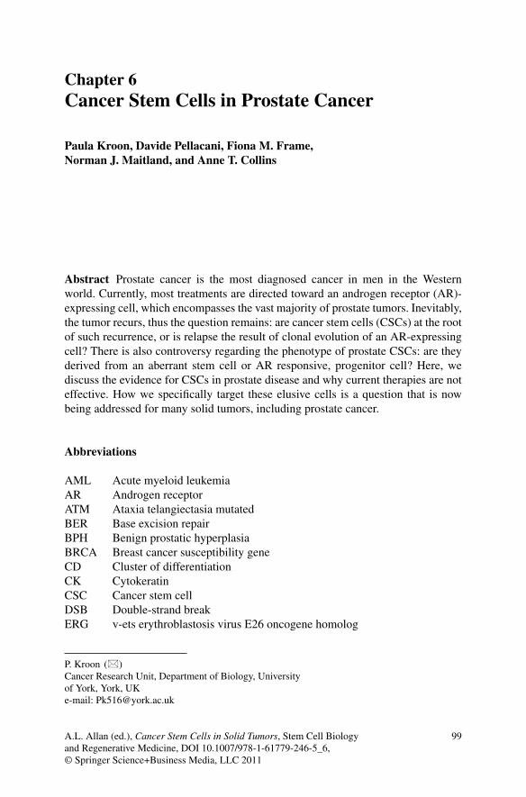
99A.L. Allan (ed.), Cancer Stem Cells in Solid Tumors, Stem Cell Biology and Regenerative Medicine, DOI 10.1007/978-1-61779-246-5_6, © Springer Science+Business Media, LLC 2011
Abstract Prostate cancer is the most diagnosed cancer in men in the Western world. Currently, most treatments are directed toward an androgen receptor (AR)-expressing cell, which encompasses the vast majority of prostate tumors. Inevitably, the tumor recurs, thus the question remains: are cancer stem cells (CSCs) at the root of such recurrence, or is relapse the result of clonal evolution of an AR-expressing cell? There is also controversy regarding the phenotype of prostate CSCs: are they derived from an aberrant stem cell or AR responsive, progenitor cell? Here, we discuss the evidence for CSCs in prostate disease and why current therapies are not effective. How we specifically target these elusive cells is a question that is now being addressed for many solid tumors, including prostate cancer.
Abbreviations
AML Acute myeloid leukemiaAR Androgen receptorATM Ataxia telangiectasia mutatedBER Base excision repairBPH Benign prostatic hyperplasiaBRCA Breast cancer susceptibility geneCD Cluster of differentiationCK CytokeratinCSC Cancer stem cellDSB Double-strand breakERG v-ets erythroblastosis virus E26 oncogene homolog
P. Kroon (*) Cancer Research Unit, Department of Biology, University of York, York, UKe-mail: [email protected]
Chapter 6Cancer Stem Cells in Prostate Cancer
Paula Kroon, Davide Pellacani, Fiona M. Frame, Norman J. Maitland, and Anne T. Collins
100 P. Kroon et al.
ETS Erythroblast transformation-specificGSTP1 Glutathione S-transferase P1HR Homologous recombinationhTERT Human telomerase reverse transciptaseIL-6 Interleukin-6JAK Janus kinaseMLH1 MutL homolog 1MMR Mismatch repairNER Nucleotide excision repairPAP Prostate acid phosphatasePIA Proliferative inflammatory atrophyPIN Prostatic intraepithelial neoplasiaPKB Protein kinase BPSA Prostate-specific antigenSTAT Signal transduction and activator of transcriptionTMPRSS2 Transmembrane protease, serine 2UGM Urogenital sinus mesenchyme
6.1 Anatomy and Development of the Human Prostate
The prostate is located toward the base of the bladder surrounding the urethra. Its main function is to produce hormones and secrete proteins for semen production and is therefore essential for the reproductive system. It also functions as an endo-crine gland, metabolizing the rapid conversion of testosterone to dihydrotestoster-one, which is a more effective androgen [1].
The human adult prostate is a complex tubulo-alveolar gland composed of an epithelial parenchyma embedded within a connective tissue matrix. The epithelial cells are arranged in glands composed of ducts that branch out from the urethra and terminate into acini. It is a heterogeneous organ, and can be divided into central, transition, and peripheral zones [2]. The majority of prostate cancers arise in the peripheral zone (70%) compared to 20% in the transition zone and 10% within the central zone, whereas benign prostatic hyperplasia (BPH) mainly occurs within the transitional zone [3].
The development of the human prostate begins during the ninth week of embryo-genesis [4], in response to testosterone stimulation, with the outgrowth of epithelial buds from the urogenital sinus epithelium into the surrounding urogenital sinus mesenchyme (UGM) [5]. These epithelial buds form ducts that elongate, branch out, and terminate into acini. From the 20th week of gestation up to puberty, the immature prostatic acini and ducts are lined by multiple layers of immature cells with round nuclei and very little cytoplasm. In the immature epithelium, cytokera-tins (CK) of simple and stratified epithelium are expressed (primary cytokeratins; numbers 8, 18, and 19 and the large molecular weight forms; numbers 4, 5, 6, 7, 10, 11, 14, 15) [6]. Postnatal development includes a period of growth during the
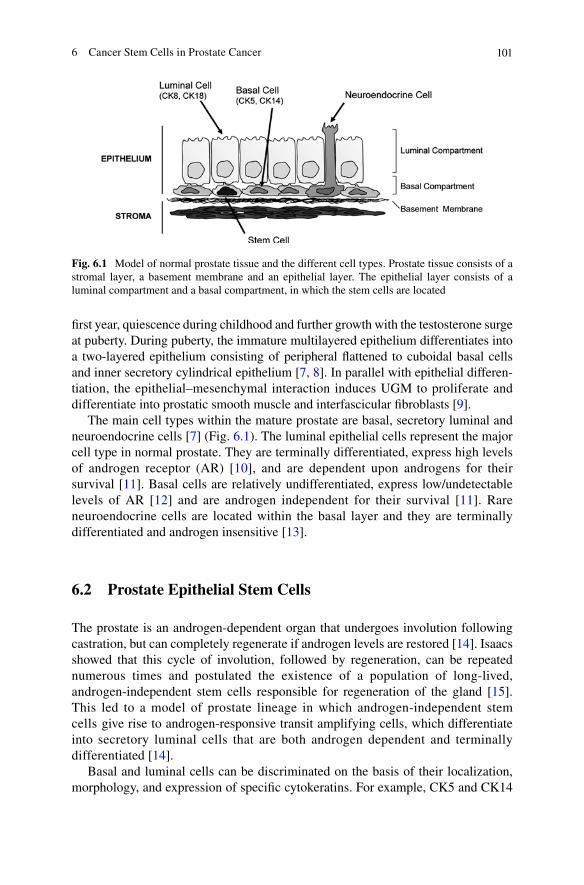
1016 Cancer Stem Cells in Prostate Cancer
first year, quiescence during childhood and further growth with the testosterone surge at puberty. During puberty, the immature multilayered epithelium differentiates into a two-layered epithelium consisting of peripheral flattened to cuboidal basal cells and inner secretory cylindrical epithelium [7, 8]. In parallel with epithelial differen-tiation, the epithelial–mesenchymal interaction induces UGM to proliferate and differentiate into prostatic smooth muscle and interfascicular fibroblasts [9].
The main cell types within the mature prostate are basal, secretory luminal and neuroendocrine cells [7] (Fig. 6.1). The luminal epithelial cells represent the major cell type in normal prostate. They are terminally differentiated, express high levels of androgen receptor (AR) [10], and are dependent upon androgens for their survival [11]. Basal cells are relatively undifferentiated, express low/undetectable levels of AR [12] and are androgen independent for their survival [11]. Rare neuroendocrine cells are located within the basal layer and they are terminally differentiated and androgen insensitive [13].
6.2 Prostate Epithelial Stem Cells
The prostate is an androgen-dependent organ that undergoes involution following castration, but can completely regenerate if androgen levels are restored [14]. Isaacs showed that this cycle of involution, followed by regeneration, can be repeated numerous times and postulated the existence of a population of long-lived, androgen-independent stem cells responsible for regeneration of the gland [15]. This led to a model of prostate lineage in which androgen-independent stem cells give rise to androgen-responsive transit amplifying cells, which differentiate into secretory luminal cells that are both androgen dependent and terminally differentiated [14].
Basal and luminal cells can be discriminated on the basis of their localization, morphology, and expression of specific cytokeratins. For example, CK5 and CK14
Fig. 6.1 Model of normal prostate tissue and the different cell types. Prostate tissue consists of a stromal layer, a basement membrane and an epithelial layer. The epithelial layer consists of a luminal compartment and a basal compartment, in which the stem cells are located
102 P. Kroon et al.
are expressed by basal cells, whereas the luminal cells of the prostate predominantly express CK8 and CK18 [16]. Keratin expression patterns in the prostate have provided evidence of epithelial cells that are phenotypically intermediate between basal and luminal cells. Cells have been identified in the luminal layer that express both CK5 and CK18, while some basal cells lack CK14 expression but express low levels of CK18 and CK5 [17–19]. These results indicate that basal and luminal cells are linked in a hierarchical pathway.
Although the overall organization of the murine prostate differs from that of the human gland, studying the murine prostate provides a unique opportunity to study the biology of the prostate. It has been shown that the proliferating cells are located at the tips of ducts [20] and also that prostatic stem cells may be located in the distal region [21]. However, quiescent cells were subsequently shown to be located in the proximal region of the duct nearest the urethra. These cells also have a high prolifera-tive potential and are capable of reconstituting large, branched glandular structures in collagen gels [22]. Tsujimura and co-workers proposed that the stem cells migrate distally toward the proliferating tips where they terminally differentiate [22].
The proposal that prostate stem cells are located within the basal layer of epithe-lial cells is supported by evidence provided by Signoretti et al., who showed that p63, which is expressed by basal cells [23], is essential for normal prostate develop-ment in the mouse [24]. By histological examination, they found that newborn p63(−/−) male mice do not develop a prostate, suggesting that p63 is necessary for the formation of ducts or epithelial budding structures [24].
Recently, using the murine hematopoietic stem cell marker Sca-1 [25], it was shown that Sca-1+ prostate cells can self-renew (in a sphere-forming assay) for several generations. Moreover, Sca-1+ cells can differentiate in vivo to produce prostatic tubule structures containing basal and luminal cells. Sca-1+ cells are also localized to the basal cell layer within the proximal region of the murine prostate [26]. Leong and colleagues also showed enrichment of stem cells within the proximal region of the mouse prostate. They determined that lin−/Sca-1+/CD133+/CD44+/CD177+ cells (localized to the basal compartment of mouse prostate) can generate a prostate after transplantation in vivo [27]. The regenerated prostate had a branching morphology with epithelial tubules composed of basal, luminal, and neuroendo-crine cells. Nonetheless, there is still some controversy as to whether stem cells are located within the basal layer. The Nkx3.1 gene regulates prostate epithelial differ-entiation, and is expressed within the luminal cells and rare basal cells in the mouse prostate. Expression is rapidly lost after castration and is restored following prostate regeneration when androgen levels are restored. Wang et al. [28] showed that in the castrate-resistant state, Nkx3.1 expression is restricted to the luminal cells and only those genetically marked. They observed that Nkx3.1-marked luminal cells were able to give rise to both basal and luminal cells following androgen-induced regeneration.
In the human prostate, several studies have revealed that prostatic basal cells can differentiate into luminal cells in vitro [29, 30]. Basal epithelial cells, isolated on the basis of high surface expression of a
2b
1-integrin, are clonogenic in vitro [31, 32]
and have the potential to regenerate a fully differentiated human prostate epithelium
1036 Cancer Stem Cells in Prostate Cancer
in vivo [31]. Use of the CD133 antigen, which was first identified as a marker for human hematopoietic stem cells [33], further enriched for the stem cell popula-tion [34]. The cells expressing CD133 are restricted to the a
2b
1hi population and are
located within the basal layer. Richardson and colleagues showed that these a2b
1hi/
CD133+ cells had a greater colony-forming ability and proliferative potential in vitro than a
2b
1hi/CD133− cells. Moreover, when grafted together with prostate stromal
cells into nude mice (which is necessary to produce a functional and morphological differentiated prostate [35]), a
2b
1hi/CD133+ cells generated prostatic acini, unlike
the a2b
1hi/CD133− cells [34].
The identification and characterization of stem cells in the normal prostate is important, because they may represent a major target for carcinogenesis as well as a potential source of BPH [36]. It was hypothesized in the 1960s that cancers exist in a hierarchy consisting of cells with different proliferative potentials [37, 38]. The cancer stem cell (CSC) hypothesis presumes that the bulk population of cancerous cells arise from CSCs [39], defined as a rare population of cells that maintain the rest of the population. Normal stem cells and CSCs have shared properties, such as the capacity to self-renew and differentiate to give rise to multi-cellular lineages [40]. These properties are important for CSCs to maintain and spread the tumor.
6.3 Prostate Cancer
Prostate cancer is a major health problem as it is the most commonly diagnosed cancer in men in the Western world. It is mainly detected in men after the age of 50, at which time one in three men suffer from symptoms related to BPH [41]. Prostate cancer is thought to arise from high-grade prostate intraepithelial neoplastic lesions (PIN) [42, 43], although proliferative inflammatory atrophy (PIA) may also play a role [44]. Certain environmental factors, such as diet, are thought to have a role in the development of prostate cancer [45]. Risk increases with age, as the largest number of cases are diagnosed within the age range of 72–74, but ethnicity and family history are also thought to play a role in the development of the disease [46]. The most common diagnostic test to detect prostate cancer is blood prostate-specific antigen (PSA) levels, as PSA increases with prostate cancer, but can also rise with non-malignant growth of the prostate [47]. However, to complete the diagnosis of prostate cancer, a biopsy is necessary to assess histology using the Gleason grading system [48].
When the cancer is confined to the prostate gland, the disease can be treated with surgery, radiation therapy (brachytherapy), or cryotherapy [49]. For elderly men who have no symptoms at diagnosis, and have a relatively short life expectancy, symptoms are controlled as they occur; this is called “active surveillance” or “watchful waiting” [50]. For patients with metastatic prostate cancer, the widely used treatment remains androgen ablation therapy, as homeostasis of the prostate gland is dependent upon androgens [51, 52]. Androgen ablation reduces tumor growth but ultimately, in most cases, the therapy fails and the prostate cells become castrate resistant [53]. That the tumor initially responds well to androgen ablation
104 P. Kroon et al.
therapy is not surprising, as the main cell type found within prostate carcinomas is the AR-positive secretory luminal cell [54]. These cells express the AR and will be sensitive to the therapy. Treatment failure can be explained by the presence of tumor-initiating cells that are independent of androgens for their survival [55]. CSCs, isolated from patient samples, were found to be AR-negative [55]. It has also been suggested that cancer-initiating cells are more resistant to radiation [56] and chemotherapy [57]. These studies suggest that the cancer-initiating cells, also known as CSCs, are not affected by conventional therapies and therefore can be the cause of recurrence and/or spread of the tumor. Therefore, it is important to develop new, more effective therapies that will specifically target this population.
6.4 Prostate Cancer Stem Cells
The origin of prostate CSCs is still controversial. As prostate cancer mainly consists of luminal cells [54], it has been the prevailing view that the AR-expressing luminal cells are the tumor-initiating cells [reviewed in 58]. The observation that telomerase is expressed within the luminal compartment in high-grade PIN, thus extending the lifespan of these cells, has added weight to this proposal [59]. Others have suggested that an intermediate cell, which expresses both basal and luminal keratin markers, could give rise to prostate cancer [18]. However, it is more plausible that normal tissue stem cells are the targets for transformation given their longevity. This has been definitively demonstrated by Bonnet and Dick [60] who showed that the tumor-initiating cells in acute myeloid leukemia (AML) shared cell surface markers with normal hematopoietic stem cells. More recently, Barker and colleagues showed that crypt stem cells are the cells of origin of intestinal cancer [61]. There are several lines of evidence that support the proposal that prostate CSCs arise from normal stem cells. Metastases often include rare cells that are phenotypically undif-ferentiated, expressing basal cell markers, such as cytokeratins 5 and 14 [62, 63]. Advanced prostate cancers can respond to low levels of androgens, but the castrate-resistant state results from clonal expansion of androgen-independent cells that are present at a frequency of 1 per 105–106 androgen-responsive cells [64].
CSCs share numerous markers with normal stem cells. More recent work from our laboratory compared isolated populations, from primary prostate cancers, for clono-genic potential. We found that only the most primitive cells (CD44+/CD133+/a
2b
1hi),
which were identical phenotypically to normal prostate stem cells, could self-renew in vitro [55]. Moreover, under differentiating conditions, AR+/PAP+/CK18+ luminal cells could be identified in these cultures, suggesting that they were derived from the more primitive population. In support of this finding, the CD44+ population from xenograft tumors and cell lines has enhanced proliferative potential and tumor-initiating ability in vivo compared to CD44− cells [65]. The CD44+ cells are likewise AR− and express higher mRNA levels of stemness genes, such as OCT3/4 and BMI 1. Using clonally derived human prostate cancer epithelial cells expressing human telomerase reverse transcriptase (hTERT), Gu and co-workers [66] demonstrated that these lines
1056 Cancer Stem Cells in Prostate Cancer
could regenerate tumors in mice that resembled the original patient tumor with respect to Gleason score. The tumors contained luminal, basal, and neuroendocrine cells, implying that the clone of origin could differentiate into the epithelial cell lin-eages of the prostate. In this case, the tumor-initiating cell was AR− and p63− and expressed the stem cell genes Oct-4, Nanog, Sox2, nestin, CD44, CD133, and c-kit. Moreover, Sca-1 sorted cells, enriched for cells with prostate-regenerating activity, showed evidence of basal and luminal lineage.
A recurrent genomic alteration in prostate cancer is the expression of TMPRSS2-ETS fusion genes [67], with TMPRSS2-ERG being the most frequently detected [68]. The presence of the fusion is associated with PSA biochemical failure [68] and occurs with a frequency of approximately 50% [67]. Identification of the TMPRSS2-ETS fusion gene in approximately 20% of PIN lesions suggests that it is an early event in prostate tumorigenesis [69] and our recent findings that TMPRSS2-ERG is expressed in a
2b
1hi/CD133+ cells from prostate tumors [70] supports the hypothesis
that the cell of origin of prostate cancer is a stem cell.Gene expression studies on populations of prostate CSCs were carried out by
Birnie et al. [70]. The resulting gene signature provided clear evidence of different gene sets expressed in the CSCs, their amplifying progeny, and their normal equiva-lents. Functional annotation of the CSC signature led to the identification of four main pathways: (1) JAK-STAT signaling; (2) cell adhesion and extracellular matrix interaction; (3) focal adhesion signaling, and (4) Wnt signaling. Verification that the cultures used in this study were tumorigenic came from the identification of the TMPRSS2-ERG translocation [67, 70].
6.5 Molecular Mechanisms Regulating Prostate Cancer Stem Cells
Similar pathways are involved in maintaining tumors and stem cells. For example, Wnt [71] and JAK-STAT signaling [72, 73] have been linked to stemness and malig-nancy, as have epigenetic mechanisms.
6.5.1 Wnt/b-Catenin Signaling
The Wnt/b-catenin signaling pathway plays an important role in multiple devel-opmental events during embryogenesis, but it has also been implicated in adult tissue homeostasis [reviewed in 74] and cancer [75]. Wnt signaling can induce cell proliferation and self-renewal of adult hematopoietic stem cells [76], and in the intestinal epithelium it is important for the maintenance of stem cells [77]. Mutations that lead to constitutively active Wnt signaling are implicated in prostate cancer, where mutations of b-catenin are the most frequent [78, 79]. b-catenin increases AR transcription in a ligand-dependent manner [80], and it has been suggested that
106 P. Kroon et al.
there is a crosstalk between Wnt and androgen signaling in prostate cancer [81]. Yang et al. suggested that excessive free b-catenin, which occurs during prostate cancer progression, might maintain or even increase AR activity when androgen levels are low [81].
6.5.2 JAK-STAT Signaling
Normally, the human body only produces high levels of IL-6 as part of an inflam-matory response. However, IL-6 is also elevated in the serum of patients with meta-static prostate cancer [82] and it acts as a positive regulator of prostate cancer cell growth [83]. It has also been shown that STAT3 is constitutively activated in pros-tate cancer tissue and high levels of STAT3 activation are associated with higher Gleason grade tumors [72]. Interestingly, activation of STAT3 is also important for maintenance of stem cell self-renewal and the undifferentiated state of embryonic stem cells [73], as well as glioblastoma stem cells [84]. Preliminary data from our group indicate that prostate CSCs secrete more IL-6 compared to the progenitor population and receptor levels are highest within the CSCs (unpublished data). Further investigation will determine whether the JAK-STAT signaling pathway is indeed important for maintaining (cancer) stemness.
6.5.3 Epigenetic Deregulation of Prostate Cancer Hierarchy
Epigenetic alteration of stem cells has been hypothesized to have an important role in prostate tumorigenesis; being involved both in the formation of prostate CSCs and their therapy resistant features [85, 86]. However, no direct study has confirmed epigenetic deregulation of prostate CSCs.
Epigenetic regulation of gene expression is defined as a heritable change in gene expression that does not involve changes in the DNA sequence [87] and includes DNA methylation, chromatin structure (mainly determined by histone posttranslational modifications) and small non-coding RNAs. Epigenetic mecha-nisms are both heritable and dynamic, allowing for fine regulation of gene expres-sion throughout all the different cell types [88]. This regulation plays a crucial role in the maintenance of the hierarchical structure of tissues, being involved in both maintenance of stemness and fate determination of stem cells [89–93]. Moreover, compounds that inhibit DNA methylation or histone deacetylation can induce cell differentiation [92, 94]. In the prostate, the key epigenetic mechanisms responsible for the maintenance of hierarchy have not yet been identified. However, studies have been performed to identify genes that are differentially expressed in prostate stem and committed cells [70, 95], from which crucial epigenetic pathways can be identified for further study. Moreover, it is known that histone deacetylase inhibi-tors can induce differentiation to a neuroendocrine phenotype in prostate cells [96, 97], emphasizing the importance of epigenetic mechanisms in the mainte-nance of lineage determination.
1076 Cancer Stem Cells in Prostate Cancer
Disruption of epigenetic mechanisms has been found in all cancer types and, together with genetic changes, plays a key role in cancer initiation and progression [98]. In the last few years, much effort has been put into defining the epigenetic alterations present in prostate cancer. This led to the identification of hundreds of hypermethylated genes, of which GSTP1 is the most studied [99], and alteration of chromatin structure through alteration of many histone-modifying enzymes and chromatin-associated proteins. In fact, global patterns of histone modification are linked to the risk of prostate cancer recurrence [100, 101]. However, these studies do not take into account the hierarchical structure present in cancer. In fact, the models typically used are cell lines adapted to culture conditions with crucial modification of epigenetic regulation [102] or tissues considered as a homogeneous population.
As previously discussed, epigenetic mechanisms are crucial for the maintenance of the correct hierarchical structure in normal tissues, suggesting that self-renewal and multipotency are, at least partially, under the control of epigenetic regulation. It has been proposed that disruption of this control may result in formation of self-renewing malignant stem cells [103], generating a deregulation of the hierarchical system, which ultimately leads to cancer [86]. Little is known about epigenetic deregulation of hierarchical structure in prostate cancer, but it undoubtedly plays a crucial role in prostate cancer development. Prostate cancer is characterized by an expansion of the luminal compartment [54], with a clear imbalance of the differen-tiation process that leads to the accumulation of aberrantly differentiated luminal cancer cells. Interestingly, this process is accompanied by epigenetic deregulation of genes that are usually expressed only in more undifferentiated cells. GSTP1 is only expressed in the basal compartment of normal prostate [104] and it is fre-quently downregulated by hypermethylation in prostate cancer [99]. Moreover, CD44, one of the molecules used as a marker to enrich for both benign and malig-nant prostate basal cells [58], is also downregulated by hypermethylation in the majority of prostate cancers [105]. These studies suggest that deregulation of epige-netic control is accompanying, or even driving, the expansion of the luminal compartment in prostate cancer, clearly indicating epigenetic deregulation of the hierarchy in prostate cancer. With the ability to now isolate different cell types, including prostate CSCs, it will be possible to elucidate the role of these epigenetic mechanisms in a cell-specific manner.
6.6 Prostate CSCs and Therapy Resistance
Existing therapies such as androgen ablation or radiation have been successful in reducing the bulk of cells within prostate tumors. These forms of therapies target the AR-expressing population and proliferating cells, respectively, but in most cases the tumors recur, which suggests that the tumor-initiating cells are a reservoir for recurrent disease following therapy, as they are more resistant to therapies currently used [58].
In the last decade, there has been an explosion in the number of papers published on CSCs. We are now reaching a consensus that CSCs must be taken into
108 P. Kroon et al.
consideration when designing therapies, particularly with tumors that are prone to relapse [57, 106–110]. However, before targeting prostate CSCs specifically, there needs to be proof that they are resistant to current therapies and preferably elucida-tion of the mechanism of resistance. Ultimately, we have to design diagnostic test(s) to determine whether, when the CSC component of a tumor is eliminated, there is a resulting tumor eradication or cure [111, 112].
Very little is known about therapy resistance of prostate CSCs, and most studies have been based on the use of cancer cell lines and tissue sections. However, increasing numbers of studies are now using primary epithelial cells from patient samples, a focus of which is the response of prostate cells to DNA damage caused by radiotherapy or chemotherapeutic agents. In terms of DNA repair, homologous recombination (HR), nucleotide excision repair (NER), base excision repair (BER), and mismatch repair (MMR) have all been examined. There are reduced levels of MMR proteins, including hMLH1 and hMSH2, in various prostate cancer cell lines [113]. More significantly, there are defects in MMR in prostate tumor foci as indicated by the absence of PMS1 and PMS2 proteins [114]. In contrast, another study found increased levels of PMS2 in recurrent prostate cancer patients and suggested this to be of use as a marker with prognostic potential [115]. Increased expression of this protein has been associated with increased mutation frequency and resistance to apoptosis. We can take from these studies that an increase or decrease in repair proteins has the potential to cause mutations that may be involved in tumor progression.
Combining radiotherapy with inhibitors of DNA repair has been explored by Bristow et al. [116]. Mutations in BRCA1 and BRCA2, key proteins in the double-strand break (DSB) response, are found in familial prostate cancers [117, 118]. Cells with these mutations are defective in DSB repair and are more sensitive to radiation [119]. Other DSB response proteins such as ATM (whose expression is increased in prostate tumors) and p53 are frequently mutated in prostate cancer [120, 121]. Mutations of Chk2 have also been observed [117]. These proteins are involved in cell cycle checkpoints, abrogation of which can lead to radioresistance and metastasis [116]. An altered BER pathway and response to oxidative stress have also been implicated in prostate cancer [114, 122–124]. With the study of CSCs, the prostate field can follow the lead of other fields. CD133+ cells from hepatocellular carcinoma are resistant to doxorubicin and fluorouracil, which is due to expression of bcl-2, Akt, and PKB; components of an anti-apoptotic sur-vival pathway [125]. Glioma stem cells are resistant to chemotherapeutic agents [126] and have increased activation of DNA damage checkpoints and more effi-cient DNA repair in response to irradiation, with inhibition of Chk1 and Chk2 kinase restoring radiosensitivity [127].
These studies on cell lines and whole populations of primary epithelial cells can be used as a basis for studies on prostate CSCs, as we now have the ability to isolate these cells and analyze their response, which is likely to be significantly different to the more differentiated cells. Ultimately, in cancer cells, there is an upset in control of DNA repair and cell cycle checkpoints, and depending on the mutation the cells may be either more sensitive to treatment or more resistant to treatment. Therefore, it is imperative to explore the specific response of prostate CSCs to
1096 Cancer Stem Cells in Prostate Cancer
different treatments, in order to manipulate therapy. This would allow for prediction of success of certain therapies and also for manipulation of treatments to exploit defects in the prostate CSCs.
6.7 Conclusions and Future Perspectives
The identification and isolation of prostate CSCs was a major breakthrough in the understanding of prostate cancer progression and relapse following therapy [55]. To be able to study prostate CSCs is very important and increases hope of identify-ing a prostate CSC-specific target, but it is also very challenging. The main reason for this is that this population is very rare within the bulk of tumor cells (0.01%), and therefore only a few techniques are suitable for studying such a small cell num-ber. However, we are able to expand these cells in culture, which can be used to study and have a better understanding of these prostate CSCs. Ultimately, it would be desirable to have a treatment specifically for the prostate CSCs that could be used in combination with androgen ablation to reduce tumor mass [62]. This will require further development of primary epithelial cell culture models and assays to detect stem cell-specific targeting. Novel treatments could include DNA repair inhibitors, inhibitors of anti-apoptotic proteins, and inhibitors of ABC transporters. With all these options, minimizing toxicity and maximizing patient benefit would be para-mount. Ultimately, if the CSCs are responsible for the recurrence following therapy and metastasis, then their elimination is the best route to a longer lasting, or even permanent cure.
References
1. Kumar VL, Majumder PK (1995) Prostate gland: structure, functions and regulation. Int Urol Nephrol 27 (3):231–243
2. McNeal JE (1988) Normal histology of the prostate. Am J Surg Pathol 12 (8):619–633 3. McNeal JE, Redwine EA, Freiha FS, Stamey TA (1988) Zonal distribution of prostatic adeno-
carcinoma. Correlation with histologic pattern and direction of spread. Am J Surg Pathol 12 (12):897–906
4. Kellokumpu-Lehtinen P, Santti R, Pelliniemi LJ (1979) Early cytodifferentiation of human prostatic urethra and Leydig cells. Anat Rec 194 (3):429–443. doi:10.1002/ar.1091940309
5. Cunha GR, Donjacour AA, Cooke PS, Mee S, Bigsby RM, Higgins SJ, Sugimura Y (1987) The endocrinology and developmental biology of the prostate. Endocr Rev 8 (3):338–362
6. Wernert N, Seitz G, Achtstatter T (1987) Immunohistochemical investigation of different cytokeratins and vimentin in the prostate from the fetal period up to adulthood and in prostate carcinoma. Pathol Res Pract 182 (5):617–626
7. Aumuller G (1991) Postnatal development of the prostate. Bull Assoc Anat (Nancy) 75 (229):39–42
8. Kellokumpu-Lehtinen P, Santti RS, Pelliniemi LJ (1981) Development of human fetal prostate in culture. Urol Res 9 (2):89–98
9. Cunha GR, Battle E, Young P, Brody J, Donjacour A, Hayashi N, Kinbara H (1992) Role of epithelial-mesenchymal interactions in the differentiation and spatial organization of visceral smooth muscle. Epithelial Cell Biol 1 (2):76–83
110 P. Kroon et al.
10. Sar M, Lubahn DB, French FS, Wilson EM (1990) Immunohistochemical localization of the androgen receptor in rat and human tissues. Endocrinology 127 (6):3180–3186
11. Kyprianou N, Isaacs JT (1988) Activation of programmed cell death in the rat ventral prostate after castration. Endocrinology 122 (2):552–562
12. Bonkhoff H, Remberger K (1993) Widespread distribution of nuclear androgen receptors in the basal cell layer of the normal and hyperplastic human prostate. Virchows Arch A Pathol Anat Histopathol 422 (1):35–38
13. Bonkhoff H, Stein U, Remberger K (1995) Endocrine-paracrine cell types in the prostate and prostatic adenocarcinoma are postmitotic cells. Hum Pathol 26 (2):167–170
14. Isaacs JT, Coffey DS (1989) Etiology and disease process of benign prostatic hyperplasia. Prostate Suppl 2:33–50
15. Isaacs TJ (1985) Control of cell proliferation and cell death in the normal and neoplastic prostate: a stem cell model. Rodgers CH, Coffey DS, Gunha G, Grayhack JT, Hinman Jr F, Horton R, editors, Benign prostatic hyperplasia, Department of Health and Human Services, NIH, Washington (DC) No 87–2881:85–94
16. Sherwood ER, Theyer G, Steiner G, Berg LA, Kozlowski JM, Lee C (1991) Differential expression of specific cytokeratin polypeptides in the basal and luminal epithelia of the human prostate. Prostate 18 (4):303–314
17. van Leenders G, Dijkman H, Hulsbergen-van de Kaa C, Ruiter D, Schalken J (2000) Demonstration of intermediate cells during human prostate epithelial differentiation in situ and in vitro using triple-staining confocal scanning microscopy. Lab Invest 80 (8):1251–1258
18. Verhagen AP, Ramaekers FC, Aalders TW, Schaafsma HE, Debruyne FM, Schalken JA (1992) Colocalization of basal and luminal cell-type cytokeratins in human prostate cancer. Cancer Res 52 (22):6182–6187
19. Xue Y, Smedts F, Debruyne FM, de la Rosette JJ, Schalken JA (1998) Identification of interme-diate cell types by keratin expression in the developing human prostate. Prostate 34 (4):292–301. doi:10.1002/(SICI)1097-0045(19980301)34:4<292::AID-PROS7>3.0.CO;2-J [pii]
20. Sugimura Y, Cunha GR, Donjacour AA, Bigsby RM, Brody JR (1986) Whole-mount autora-diography study of DNA synthetic activity during postnatal development and androgen-induced regeneration in the mouse prostate. Biol Reprod 34 (5):985–995
21. Kinbara H, Cunha GR, Boutin E, Hayashi N, Kawamura J (1996) Evidence of stem cells in the adult prostatic epithelium based upon responsiveness to mesenchymal inductors. Prostate 29 (2): 107–116. doi:10.1002/(SICI)1097-0045(199608)29:2<107::AID-PROS6>3.0.CO;2-C [pii] 10.1002/(SICI)1097-0045(199608)29:2<107::AID-PROS6>3.0.CO;2-C
22. Tsujimura A, Koikawa Y, Salm S, Takao T, Coetzee S, Moscatelli D, Shapiro E, Lepor H, Sun TT, Wilson EL (2002) Proximal location of mouse prostate epithelial stem cells: a model of prostatic homeostasis. J Cell Biol 157 (7):1257–1265. doi:10.1083/jcb.200202067 jcb.200202067 [pii]
23. Yang A, Kaghad M, Wang Y, Gillett E, Fleming MD, Dotsch V, Andrews NC, Caput D, McKeon F (1998) p63, a p53 homolog at 3q27-29, encodes multiple products with transacti-vating, death-inducing, and dominant-negative activities. Mol Cell 2 (3):305–316. doi:S1097-2765(00)80275-0 [pii]
24. Signoretti S, Waltregny D, Dilks J, Isaac B, Lin D, Garraway L, Yang A, Montironi R, McKeon F, Loda M (2000) p63 is a prostate basal cell marker and is required for prostate development. Am J Pathol 157 (6):1769–1775
25. Spangrude GJ, Heimfeld S, Weissman IL (1988) Purification and characterization of mouse hematopoietic stem cells. Science 241 (4861):58–62
26. Lawson DA, Xin L, Lukacs RU, Cheng D, Witte ON (2007) Isolation and functional char-acterization of murine prostate stem cells. Proc Natl Acad Sci USA 104 (1):181–186. doi:0609684104 [pii] 10.1073/pnas.0609684104
27. Leong KG, Wang BE, Johnson L, Gao WQ (2008) Generation of a prostate from a single adult stem cell. Nature 456 (7223):804–808. doi:nature07427 [pii] 10.1038/nature07427
28. Wang X, Kruithof-de Julio M, Economides KD, Walker D, Yu H, Halili MV, Hu YP, Price SM, Abate-Shen C, Shen MM (2009) A luminal epithelial stem cell that is a cell of origin for prostate cancer. Nature 461 (7263):495–500. doi:nature08361 [pii] 10.1038/nature08361
1116 Cancer Stem Cells in Prostate Cancer
29. Liu AY, True LD, LaTray L, Nelson PS, Ellis WJ, Vessella RL, Lange PH, Hood L, van den Engh G (1997) Cell-cell interaction in prostate gene regulation and cytodifferentiation. Proc Natl Acad Sci USA 94 (20):10705–10710
30. Robinson EJ, Neal DE, Collins AT (1998) Basal cells are progenitors of luminal cells in primary cultures of differentiating human prostatic epithelium. Prostate 37 (3):149–160. doi:10.1002/(SICI)1097-0045(19981101)37:3<149::AID-PROS4>3.0.CO;2-E [pii]
31. Collins AT, Habib FK, Maitland NJ, Neal DE (2001) Identification and isolation of human prostate epithelial stem cells based on alpha(2)beta(1)-integrin expression. J Cell Sci 114 (Pt 21):3865–3872
32. Hudson DL, O’Hare M, Watt FM, Masters JR (2000) Proliferative heterogeneity in the human prostate: evidence for epithelial stem cells. Lab Invest 80 (8):1243–1250
33. Yin AH, Miraglia S, Zanjani ED, Almeida-Porada G, Ogawa M, Leary AG, Olweus J, Kearney J, Buck DW (1997) AC133, a novel marker for human hematopoietic stem and progenitor cells. Blood 90 (12):5002–5012
34. Richardson GD, Robson CN, Lang SH, Neal DE, Maitland NJ, Collins AT (2004) CD133, a novel marker for human prostatic epithelial stem cells. J Cell Sci 117 (Pt 16):3539–3545. doi:10.1242/jcs.01222 jcs.01222 [pii]
35. Lang SH, Stark M, Collins A, Paul AB, Stower MJ, Maitland NJ (2001) Experimental prostate epithelial morphogenesis in response to stroma and three-dimensional matrigel culture. Cell Growth Differ 12 (12):631–640
36. De Marzo AM, Nelson WG, Meeker AK, Coffey DS (1998) Stem cell features of benign and malignant prostate epithelial cells. J Urol 160 (6 Pt 2):2381–2392
37. Bruce WR, Van Der Gaag H (1963) A Quantitative Assay for the Number of Murine Lymphoma Cells Capable of Proliferation in Vivo. Nature 199:79–80
38. Southam C, Brunschwig A. (1961) Quantitative studies of autotransplantation of human cancer. Cancer 14:461–563
39. Hamburger AW, Salmon SE (1977) Primary bioassay of human tumor stem cells. Science 197 (4302):461–463
40. Wicha MS, Liu S, Dontu G (2006) Cancer stem cells: an old idea--a paradigm shift. Cancer Res 66 (4):1883–1890; discussion 1895–1886. doi:66/4/1883 [pii] 10.1158/0008-5472.CAN-05-3153
41. Rizzo S, Attard G, Hudson DL (2005) Prostate epithelial stem cells. Cell Prolif 38 (6):363–374. doi:CPR356 [pii] 10.1111/j.1365-2184.2005.00356.x
42. Simpson RJ (1997) Benign prostatic hyperplasia. Br J Gen Pract 47 (417):235–240 43. Zynger DL, Yang X (2009) High-grade prostatic intraepithelial neoplasia of the prostate: the
precursor lesion of prostate cancer. Int J Clin Exp Pathol 2 (4):327–338 44. De Marzo AM, Marchi VL, Epstein JI, Nelson WG (1999) Proliferative inflammatory atrophy
of the prostate: implications for prostatic carcinogenesis. Am J Pathol 155 (6):1985–1992 45. Snowdon DA, Phillips RL, Choi W (1984) Diet, obesity, and risk of fatal prostate cancer. Am
J Epidemiol 120 (2):244–250 46. Gronberg H (2003) Prostate cancer epidemiology. Lancet 361 (9360):859–864. doi:S0140-
6736(03)12713-4 [pii] 10.1016/S0140-6736(03)12713-4 47. Carter HB, Pearson JD, Metter EJ, Brant LJ, Chan DW, Andres R, Fozard JL, Walsh PC (1992)
Longitudinal evaluation of prostate-specific antigen levels in men with and without prostate disease. JAMA 267 (16):2215–2220
48. Gleason DF (1966) Classification of prostatic carcinomas. Cancer Chemother Rep 50 (3): 125–128
49. Walsh PC, DeWeese TL, Eisenberger MA (2007) Clinical practice. Localized prostate cancer. N Engl J Med 357 (26):2696–2705. doi:357/26/2696 [pii] 10.1056/NEJMcp0706784
50. Wu H, Sun L, Moul JW, Wu HY, McLeod DG, Amling C, Lance R, Kusuda L, Donahue T, Foley J, Chung A, Sexton W, Soderdahl D (2004) Watchful waiting and factors predictive of secondary treatment of localized prostate cancer. J Urol 171 (3):1111–1116. doi:10.1097/01.ju.0000113300.74132.8b
51. Balk SP (2002) Androgen receptor as a target in androgen-independent prostate cancer. Urology 60 (3 Suppl 1):132–138; discussion 138–139. doi:S0090429502015935 [pii]
112 P. Kroon et al.
52. Huggins C, Hodges CV (1941) Studies on prostatic cancer. I. The effect of estrogen and andro-gen injection on serum phosphatases in metastatic carcinoma of the prostate. Cancer Res 1:293
53. Feldman BJ, Feldman D (2001) The development of androgen-independent prostate cancer. Nat Rev Cancer 1 (1):34–45. doi:10.1038/35094009
54. Nagle RB, Ahmann FR, McDaniel KM, Paquin ML, Clark VA, Celniker A (1987) Cytokeratin characterization of human prostatic carcinoma and its derived cell lines. Cancer Res 47 (1): 281–286
55. Collins AT, Berry PA, Hyde C, Stower MJ, Maitland NJ (2005) Prospective identification of tumorigenic prostate cancer stem cells. Cancer Res 65 (23):10946–10951. doi:65/23/10946 [pii] 10.1158/0008-5472.CAN-05-2018
56. Phillips TM, McBride WH, Pajonk F (2006) The response of CD24(−/low)/CD44+ breast cancer-initiating cells to radiation. J Natl Cancer Inst 98 (24):1777–1785. doi:98/24/1777 [pii] 10.1093/jnci/djj495
57. Dean M, Fojo T, Bates S (2005) Tumour stem cells and drug resistance. Nat Rev Cancer 5 (4): 275–284. doi:nrc1590 [pii] 10.1038/nrc1590
58. Maitland NJ, Collins AT (2008) Prostate cancer stem cells: a new target for therapy. J Clin Oncol 26 (17):2862–2870. doi:26/17/2862 [pii] 10.1200/JCO.2007.15.1472
59. Meeker AK, Hicks JL, Platz EA, March GE, Bennett CJ, Delannoy MJ, De Marzo AM (2002) Telomere shortening is an early somatic DNA alteration in human prostate tumorigenesis. Cancer Res 62 (22):6405–6409
60. Bonnet D, Dick JE (1997) Human acute myeloid leukemia is organized as a hierarchy that originates from a primitive hematopoietic cell. Nat Med 3 (7):730–737
61. Barker N, Ridgway RA, van Es JH, van de Wetering M, Begthel H, van den Born M, Danenberg E, Clarke AR, Sansom OJ, Clevers H (2009) Crypt stem cells as the cells-of-origin of intestinal cancer. Nature 457 (7229):608–611. doi:nature07602 [pii] 10.1038/nature07602
62. Lang SH, Frame FM, Collins AT (2009) Prostate cancer stem cells. J Pathol 217 (2):299–306. doi:10.1002/path.2478
63. Liu AY, Nelson PS, van den Engh G, Hood L (2002) Human prostate epithelial cell-type cDNA libraries and prostate expression patterns. Prostate 50 (2):92–103. doi:10.1002/pros.10036 [pii]
64. Craft N, Chhor C, Tran C, Belldegrun A, DeKernion J, Witte ON, Said J, Reiter RE, Sawyers CL (1999) Evidence for clonal outgrowth of androgen-independent prostate cancer cells from androgen-dependent tumors through a two-step process. Cancer Res 59 (19):5030–5036
65. Patrawala L, Calhoun T, Schneider-Broussard R, Li H, Bhatia B, Tang S, Reilly JG, Chandra D, Zhou J, Claypool K, Coghlan L, Tang DG (2006) Highly purified CD44+ prostate cancer cells from xenograft human tumors are enriched in tumorigenic and metastatic progenitor cells. Oncogene 25 (12):1696–1708. doi:1209327 [pii] 10.1038/sj.onc.1209327
66. Gu G, Yuan J, Wills M, Kasper S (2007) Prostate cancer cells with stem cell characteristics reconstitute the original human tumor in vivo. Cancer Res 67 (10):4807–4815. doi:67/10/4807 [pii] 10.1158/0008-5472.CAN-06-4608
67. Tomlins SA, Rhodes DR, Perner S, Dhanasekaran SM, Mehra R, Sun XW, Varambally S, Cao X, Tchinda J, Kuefer R, Lee C, Montie JE, Shah RB, Pienta KJ, Rubin MA, Chinnaiyan AM (2005) Recurrent fusion of TMPRSS2 and ETS transcription factor genes in prostate cancer. Science 310 (5748):644–648. doi:310/5748/644 [pii] 10.1126/science.1117679
68. Demichelis F, Rubin MA (2007) TMPRSS2-ETS fusion prostate cancer: biological and clinical implications. J Clin Pathol 60 (11):1185–1186. doi:60/11/1185 [pii] 10.1136/jcp.2007.046557
69. Cerveira N, Ribeiro FR, Peixoto A, Costa V, Henrique R, Jeronimo C, Teixeira MR (2006) TMPRSS2-ERG gene fusion causing ERG overexpression precedes chromosome copy number changes in prostate carcinomas and paired HGPIN lesions. Neoplasia 8 (10):826–832. doi:10.1593/neo.06427
70. Birnie R, Bryce SD, Roome C, Dussupt V, Droop A, Lang SH, Berry PA, Hyde CF, Lewis JL, Stower MJ, Maitland NJ, Collins AT (2008) Gene expression profiling of human prostate can-cer stem cells reveals a pro-inflammatory phenotype and the importance of extracellular matrix interactions. Genome Biol 9 (5):R83. doi:gb-2008-9-5-r83 [pii] 10.1186/gb-2008-9-5-r83
1136 Cancer Stem Cells in Prostate Cancer
71. Fodde R, Brabletz T (2007) Wnt/beta-catenin signaling in cancer stemness and malignant behavior. Curr Opin Cell Biol 19 (2):150–158. doi:S0955-0674(07)00021-X [pii] 10.1016/j.ceb.2007.02.007
72. Mora LB, Buettner R, Seigne J, Diaz J, Ahmad N, Garcia R, Bowman T, Falcone R, Fairclough R, Cantor A, Muro-Cacho C, Livingston S, Karras J, Pow-Sang J, Jove R (2002) Constitutive activation of Stat3 in human prostate tumors and cell lines: direct inhibition of Stat3 signaling induces apoptosis of prostate cancer cells. Cancer Res 62 (22):6659–6666
73. Stuhlmann-Laeisz C, Lang S, Chalaris A, Krzysztof P, Enge S, Eichler J, Klingmuller U, Samuel M, Ernst M, Rose-John S, Scheller J (2006) Forced dimerization of gp130 leads to constitutive STAT3 activation, cytokine-independent growth, and blockade of differentiation of embryonic stem cells. Mol Biol Cell 17 (7):2986–2995. doi:E05-12-1129 [pii] 10.1091/mbc.E05-12-1129
74. Logan CY, Nusse R (2004) The Wnt signaling pathway in development and disease. Annu Rev Cell Dev Biol 20:781–810. doi:10.1146/annurev.cellbio.20.010403.113126
75. Polakis P (2000) Wnt signaling and cancer. Genes Dev 14 (15):1837–1851 76. Reya T, Duncan AW, Ailles L, Domen J, Scherer DC, Willert K, Hintz L, Nusse R, Weissman
IL (2003) A role for Wnt signalling in self-renewal of haematopoietic stem cells. Nature 423 (6938):409–414. doi:10.1038/nature01593 nature01593 [pii]
77. Fevr T, Robine S, Louvard D, Huelsken J (2007) Wnt/beta-catenin is essential for intestinal homeostasis and maintenance of intestinal stem cells. Mol Cell Biol 27 (21):7551–7559. doi:MCB.01034-07 [pii] 10.1128/MCB.01034-07
78. Chesire DR, Ewing CM, Sauvageot J, Bova GS, Isaacs WB (2000) Detection and analysis of beta-catenin mutations in prostate cancer. Prostate 45 (4):323–334. doi:10.1002/1097-0045(20001201)45:4<323::AID-PROS7>3.0.CO;2-W [pii]
79. Voeller HJ, Truica CI, Gelmann EP (1998) Beta-catenin mutations in human prostate cancer. Cancer Res 58 (12):2520–2523
80. Truica CI, Byers S, Gelmann EP (2000) Beta-catenin affects androgen receptor transcriptional activity and ligand specificity. Cancer Res 60 (17):4709–4713
81. Yang F, Li X, Sharma M, Sasaki CY, Longo DL, Lim B, Sun Z (2002) Linking beta-catenin to androgen-signaling pathway. J Biol Chem 277 (13):11336–11344. doi:10.1074/jbc.M111962200 M111962200 [pii]
82. Twillie DA, Eisenberger MA, Carducci MA, Hseih WS, Kim WY, Simons JW (1995) Interleukin-6: a candidate mediator of human prostate cancer morbidity. Urology 45 (3): 542–549. doi:S0090-4295(99)80034-X [pii] 10.1016/S0090-4295(99)80034-X
83. Culig Z, Steiner H, Bartsch G, Hobisch A (2005) Interleukin-6 regulation of prostate cancer cell growth. J Cell Biochem 95 (3):497–505. doi:10.1002/jcb.20477
84. Sherry MM, Reeves A, Wu JK, Cochran BH (2009) STAT3 is required for proliferation and maintenance of multipotency in glioblastoma stem cells. Stem Cells 27 (10):2383–2392. doi:10.1002/stem.185
85. Cooper CS, Foster CS (2009) Concepts of epigenetics in prostate cancer development. Br J Cancer 100 (2):240–245. doi:6604771 [pii] 10.1038/sj.bjc.6604771
86. Feinberg AP, Ohlsson R, Henikoff S (2006) The epigenetic progenitor origin of human cancer. Nat Rev Genet 7 (1):21–33. doi:nrg1748 [pii] 10.1038/nrg1748
87. Jaenisch R, Bird A (2003) Epigenetic regulation of gene expression: how the genome inte-grates intrinsic and environmental signals. Nat Genet 33 Suppl:245–254. doi:10.1038/ng1089 ng1089 [pii]
88. Li X, Zhao X (2008) Epigenetic regulation of mammalian stem cells. Stem Cells Dev 17 (6): 1043–1052. doi:10.1089/scd.2008.0036
89. Ansel KM, Lee DU, Rao A (2003) An epigenetic view of helper T cell differentiation. Nat Immunol 4 (7):616–623. doi:10.1038/ni0703-616 ni0703-616 [pii]
90. Fan G, Martinowich K, Chin MH, He F, Fouse SD, Hutnick L, Hattori D, Ge W, Shen Y, Wu H, ten Hoeve J, Shuai K, Sun YE (2005) DNA methylation controls the timing of astrogliogenesis through regulation of JAK-STAT signaling. Development 132 (15):3345–3356. doi:132/15/3345 [pii] 10.1242/dev.01912
114 P. Kroon et al.
91. Hsieh J, Gage FH (2004) Epigenetic control of neural stem cell fate. Curr Opin Genet Dev 14 (5):461–469. doi:10.1016/j.gde.2004.07.006 S0959-437X(04)00117-0 [pii]
92. Roloff TC, Nuber UA (2005) Chromatin, epigenetics and stem cells. Eur J Cell Biol 84 (2–3):123–135
93. Tagoh H, Melnik S, Lefevre P, Chong S, Riggs AD, Bonifer C (2004) Dynamic reorganiza-tion of chromatin structure and selective DNA demethylation prior to stable enhancer complex formation during differentiation of primary hematopoietic cells in vitro. Blood 103 (8): 2950–2955. doi:10.1182/blood-2003-09-3323 2003-09-3323 [pii]
94. Hsieh J, Nakashima K, Kuwabara T, Mejia E, Gage FH (2004) Histone deacetylase inhibition-mediated neuronal differentiation of multipotent adult neural progenitor cells. Proc Natl Acad Sci USA 101 (47):16659–16664. doi:0407643101 [pii] 10.1073/pnas.0407643101
95. Shepherd CJ, Rizzo S, Ledaki I, Davies M, Brewer D, Attard G, de Bono J, Hudson DL (2008) Expression profiling of CD133+ and CD133- epithelial cells from human prostate. Prostate 68 (9):1007–1024. doi:10.1002/pros.20765
96. Frigo DE, McDonnell DP (2008) Differential effects of prostate cancer therapeutics on neuroendocrine transdifferentiation. Mol Cancer Ther 7 (3):659–669. doi:7/3/659 [pii] 10.1158/1535-7163.MCT-07-0480
97. Valentini A, Biancolella M, Amati F, Gravina P, Miano R, Chillemi G, Farcomeni A, Bueno S, Vespasiani G, Desideri A, Federici G, Novelli G, Bernardini S (2007) Valproic acid induces neuroendocrine differentiation and UGT2B7 up-regulation in human prostate carcinoma cell line. Drug Metab Dispos 35 (6):968–972. doi:dmd.107.014662 [pii] 10.1124/dmd.107.014662
98. Schulz WA, Hatina J (2006) Epigenetics of prostate cancer: beyond DNA methylation. J Cell Mol Med 10 (1):100–125. doi:010.001.08 [pii]
99. Nakayama M, Gonzalgo ML, Yegnasubramanian S, Lin X, De Marzo AM, Nelson WG (2004) GSTP1 CpG island hypermethylation as a molecular biomarker for prostate cancer. J Cell Biochem 91 (3):540–552. doi:10.1002/jcb.10740
100. Ellinger J, Kahl P, von der Gathen J, Rogenhofer S, Heukamp LC, Gütgemann I, Walter B, Hofstädter F, Büttner R, Müller SC, Bastian PJ, von Ruecker A (2010) Global levels of histone modifications predict prostate cancer recurrence. Prostate 70 (1):61–69. doi:10.1002/pros.21038
101. Seligson DB, Horvath S, Shi T, Yu H, Tze S, Grunstein M, Kurdistani SK (2005) Global histone modification patterns predict risk of prostate cancer recurrence. Nature 435 (7046):1262–1266. doi:nature03672 [pii] 10.1038/nature03672
102. Antequera F, Boyes J, Bird A (1990) High levels of de novo methylation and altered chromatin structure at CpG islands in cell lines. Cell 62 (3):503–514. doi:0092-8674(90)90015-7 [pii]
103. Baylin SB, Ohm JE (2006) Epigenetic gene silencing in cancer – a mechanism for early onco-genic pathway addiction? Nat Rev Cancer 6 (2):107–116. doi:nrc1799 [pii] 10.1038/nrc1799
104. Moskaluk CA, Duray PH, Cowan KH, Linehan M, Merino MJ (1997) Immunohistochemical expression of pi-class glutathione S-transferase is down-regulated in adenocarcinoma of the prostate. Cancer 79 (8):1595–1599. doi:10.1002/(SICI)1097-0142(19970415)79:8<1595::AID-CNCR23>3.0.CO;2-S [pii]
105. Lou W, Krill D, Dhir R, Becich MJ, Dong JT, Frierson HF, Jr., Isaacs WB, Isaacs JT, Gao AC (1999) Methylation of the CD44 metastasis suppressor gene in human prostate cancer. Cancer Res 59 (10):2329–2331
106. Eyler CE, Rich JN (2008) Survival of the fittest: cancer stem cells in therapeutic resistance and angiogenesis. J Clin Oncol 26 (17):2839–2845. doi:26/17/2839 [pii] 10.1200/JCO.2007.15.1829
107. Fabian A, Barok M, Vereb G, Szollosi J (2009) Die hard: are cancer stem cells the Bruce Willises of tumor biology? Cytometry A 75 (1):67–74. doi:10.1002/cyto.a.20690
108. Frank NY, Schatton T, Frank MH (2010) The therapeutic promise of the cancer stem cell concept. J Clin Invest 120 (1):41–50. doi:41004 [pii] 10.1172/JCI41004
1156 Cancer Stem Cells in Prostate Cancer
109. Lee JT, Herlyn M (2007) Old disease, new culprit: tumor stem cells in cancer. J Cell Physiol 213 (3):603–609. doi:10.1002/jcp.21252
110. Winquist RJ, Boucher DM, Wood M, Furey BF (2009) Targeting cancer stem cells for more effective therapies: Taking out cancer’s locomotive engine. Biochem Pharmacol 78 (4): 326–334. doi:S0006-2952(09)00196-8 [pii] 10.1016/j.bcp.2009.03.020
111. Drewa T, Styczynski J (2008) Can conception of prostate cancer stem cells influence treat-ment dedicated to patients with disseminated disease? Med Hypotheses 71 (5):694–699. doi:S0306-9877(08)00290-9 [pii] 10.1016/j.mehy.2008.06.021
112. Woodward WA, Bristow RG, Clarke MF, Coppes RP, Cristofanilli M, Duda DG, Fike JR, Hambardzumyan D, Hill RP, Jordan CT, Milas L, Pajonk F, Curran WJ, Dicker AP, Chen Y (2009) Radiation Therapy Oncology Group translational research program stem cell sympo-sium: incorporating stem cell hypotheses into clinical trials. Int J Radiat Oncol Biol Phys 74 (5):1580–1591. doi:S0360-3016(09)00507-0 [pii] 10.1016/j.ijrobp.2009.03.047
113. Yeh CC, Lee C, Dahiya R (2001) DNA mismatch repair enzyme activity and gene expression in prostate cancer. Biochem Biophys Res Commun 285 (2):409–413. doi: 10.1006/bbrc.2001.5187 S0006-291X(01)95187-3 [pii]
114. Chen L, Elahi A, Pow-Sang J, Lazarus P, Park J (2003) Association between polymorphism of human oxoguanine glycosylase 1 and risk of prostate cancer. J Urol 170 (6 Pt 1):2471–2474. doi:10.1097/01.ju.0000087498.23008.bb
115. Norris AM, Gentry M, Peehl DM, D’Agostino R, Jr., Scarpinato KD (2009) The elevated expression of a mismatch repair protein is a predictor for biochemical recurrence after radical prostatectomy. Cancer Epidemiol Biomarkers Prev 18 (1):57–64. doi:18/1/57 [pii] 10.1158/ 1055-9965.EPI-08-0377
116. Bristow RG, Ozcelik H, Jalali F, Chan N, Vesprini D (2007) Homologous recombination and prostate cancer: a model for novel DNA repair targets and therapies. Radiother Oncol 83 (3):220–230. doi:S0167-8140(07)00156-9 [pii] 10.1016/j.radonc.2007.04.016
117. Dong JT (2006) Prevalent mutations in prostate cancer. J Cell Biochem 97 (3):433–447. doi:10.1002/jcb.20696
118. Levy-Lahad E, Friedman E (2007) Cancer risks among BRCA1 and BRCA2 mutation carriers. Br J Cancer 96 (1):11–15. doi:6603535 [pii] 10.1038/sj.bjc.6603535
119. Farmer H, McCabe N, Lord CJ, Tutt AN, Johnson DA, Richardson TB, Santarosa M, Dillon KJ, Hickson I, Knights C, Martin NM, Jackson SP, Smith GC, Ashworth A (2005) Targeting the DNA repair defect in BRCA mutant cells as a therapeutic strategy. Nature 434 (7035):917–921. doi:nature03445 [pii] 10.1038/nature03445
120. Angele S, Falconer A, Foster CS, Taniere P, Eeles RA, Hall J (2004) ATM protein overex-pression in prostate tumors: possible role in telomere maintenance. Am J Clin Pathol 121 (2):231–236. doi:10.1309/JTKG-GGKU-RFX3-XMGT
121. Cuddihy AR, Bristow RG (2004) The p53 protein family and radiation sensitivity: Yes or no? Cancer Metastasis Rev 23 (3–4):237–257. doi:10.1023/B:CANC.0000031764.81141.e4 5273262 [pii]
122. Rybicki BA, Conti DV, Moreira A, Cicek M, Casey G, Witte JS (2004) DNA repair gene XRCC1 and XPD polymorphisms and risk of prostate cancer. Cancer Epidemiol Biomarkers Prev 13 (1):23–29
123. Trzeciak AR, Nyaga SG, Jaruga P, Lohani A, Dizdaroglu M, Evans MK (2004) Cellular repair of oxidatively induced DNA base lesions is defective in prostate cancer cell lines, PC-3 and DU-145. Carcinogenesis 25 (8):1359–1370. doi:10.1093/carcin/bgh144 bgh144 [pii]
124. Xu J, Zheng SL, Turner A, Isaacs SD, Wiley KE, Hawkins GA, Chang BL, Bleecker ER, Walsh PC, Meyers DA, Isaacs WB (2002) Associations between hOGG1 sequence variants and prostate cancer susceptibility. Cancer Res 62 (8):2253–2257
125. Ma S, Lee TK, Zheng BJ, Chan KW, Guan XY (2008) CD133+ HCC cancer stem cells confer chemoresistance by preferential expression of the Akt/PKB survival pathway. Oncogene 27 (12):1749–1758. doi:1210811 [pii] 10.1038/sj.onc.1210811

116 P. Kroon et al.
126. Murat A, Migliavacca E, Gorlia T, Lambiv WL, Shay T, Hamou MF, de Tribolet N, Regli L, Wick W, Kouwenhoven MC, Hainfellner JA, Heppner FL, Dietrich PY, Zimmer Y, Cairncross JG, Janzer RC, Domany E, Delorenzi M, Stupp R, Hegi ME (2008) Stem cell-related “self-renewal” signature and high epidermal growth factor receptor expression associated with resistance to concomitant chemoradiotherapy in glioblastoma. J Clin Oncol 26 (18): 3015–3024. doi:26/18/3015 [pii] 10.1200/JCO.2007.15.7164
127. Bao S, Wu Q, McLendon RE, Hao Y, Shi Q, Hjelmeland AB, Dewhirst MW, Bigner DD, Rich JN (2006) Glioma stem cells promote radioresistance by preferential activation of the DNA damage response. Nature 444 (7120):756–760. doi:nature05236 [pii] 10.1038/nature05236
117A.L. Allan (ed.), Cancer Stem Cells in Solid Tumors, Stem Cell Biology and Regenerative Medicine, DOI 10.1007/978-1-61779-246-5_7, © Springer Science+Business Media, LLC 2011
Abstract Malignant melanoma is a significant health problem worldwide. Disease relapse due to the heterogeneity and instability of cancer cells may explain the persistence of disease in spite of primary response to therapy. Recent progress in cancer research suggests that melanomas, similar to other solid tumors, contain a subpopulation of cells which have unlimited self-renewal capability directly descending from the original founder cell and characterized by relatively stable genetic properties throughout disease evolution. This model also applies to the development of metastasis and may be responsible for drug resistance and cancer recurrence. These cells with tumor-initiating ability are termed cancer stem cells (CSCs). CSCs as well as tumor cells interact with their microenvironment (niche) to modulate the malignant phenotype. This chapter provides an overview of melanoma stem cell characterization and the interactions between melanoma stem cells and their niche.
Abbreviations
ABC ATP-binding cassettebFGF Basic fibroblast growth factorBMP Bone morphogenic proteinBRAF B-Raf proto-oncogene serine/threonine-protein kinaseCD Cluster of differentiationCMC Circulating melanoma cell
E. Wang (*) Infectious Disease and Immunogenetics Section (IDIS), Department of Transfusion Medicine, Clinical Center, National Institutes of Health, 10 Center Drive, Bethesda, MD 20892-1184, USA e-mail: [email protected]
Chapter 7Cancer Stem Cells in Melanoma
Ping Jin, Qiuzhen Liu, Marianna Sabatino, David F. Stroncek, Francesco M. Marincola, and Ena Wang
118 P. Jin et al.
CSC Cancer stem cellCXCR4 Chemokine receptor 4DTC Disseminated tumor cellECM Extracellular matrixERK Extracellular signal-regulated kinaseGAP-43 Growth-associated protein-43GFP Green fluorescent proteinHER2 Human epidermal growth factor receptor 2HSC Hematopoietic stem cellhTERT Human telomerase reverse transcriptaseIGF Insulin-like growth factorIL InterleukinMAGE Melanoma antigen geneMART1 Melanoma antigen recognized by T-cellsMCAM Melanoma cell adhesion moleculeMCP-1 Monocyte chemoattractant protein-1MEK Map kinase kinaseMITF Microphthalmia-associated transcription factorMLL Mixed lineage leukemiaMMP Matrix metalloproteinaseMMTV Mouse mammary tumor virusMSC Mesenchymal stem cellmTOR Mammalian target of rapamycinNCAM Neural cell adhesion moleculeNES NestinNK Natural killerNOD/SCID Non-obese diabetic/Severe combined immune deficiencyPDGF Platelet-derived growth factorPTEN Phosphatase and tensin homologRANTES Regulated on activation normal T cell expressed and secretedSCA Sphere cell formation assaySCF Stem cell factorSDF Stromal derived factorSP Side populationSSEA Stage-specific embryonic antigenTA Transit amplifyingTGF-b Transforming growth factor betaTIE1 Tyrosine kinase with immunoglobulin-like and EGF-like
domains 1TRAIL Tumor necrosis factor-related apoptosis-inducing ligandVEGFR Vascular endothelial growth factor receptorVWF von Willebrand factor
1197 Cancer Stem Cells in Melanoma
7.1 Introduction
Malignant melanoma is a disease with a very poor survival rate. Its incidence has increased 3–7% on average over several decades. In the US, the lifetime risk of melanoma in the year 2000 was estimated at 1 in 75 persons [1]. Patients with advanced disease have a poor prognosis with a reported median survival ranging between 3 and 11 months. Biological therapies including immune therapy with systemic administration of high-dose interleukin-2, interferon-alpha, antigen-specific immunization, and chemotherapy with dacarbazine or temozolomide can induce objective tumor regression in only 5–20% of patients [2, 3]. Adoptive transfer of autologous tumor-infiltrating lymphocytes following myeloid lympho-ablation has been reported to induce objective tumor regression in approximately 60% of patients [4–6]. However, these responses do not result, in most cases, in an overall survival benefit as the large majority of patients die with relapsing disease that is often resistant to further therapy.
It has been hypothesized that the stubborn recurrence of cancer following a primary response to treatment is due to the survival of a subset of cancer cells that display an intrinsic resistance to treatment-induced cell death [7, 8]. The existence of cancer stem cells (CSCs), characterized by a less differentiated status, lower immunogenicity and resistance to immune rejection [9] may be the source of cancer relapse and resistance to therapy [8, 10]. It is important to note that the term CSCs is more of a functional definition created to define a subgroup of cancer cells which can self-renew, initiate tumors, and differentiate into a heterogeneous progeny that partially maintains similarity to the original tissue from which they derived. Different from normal stem cells, CSCs share the accumulated genetic instability responsible for cancer development and acquire the genetic alterations required to promote the malignant process. As a result of the genetic and epigenetic changes, different subsets of cancer-initiating cells can be identified [11]. The concept of CSCs and the hierarchical model of tumorigenesis have implications that may help advance the understanding of tumor biology and the development of more effective anti-cancer treatments. Thus, the development of cancer therapy has expanded from targeting a population of cells derived from a stochastic model of chance variation to targeting cells transformed after a single or few random mutations followed by subsequent clonal selection and perpetuation. In this chapter, we review the charac-terization of melanoma CSCs and their biology as well as the interaction between melanoma and their microenvironment/niche.
7.2 Melanoma Genesis
Stem cells of the melanocytic lineage derive from the neural crest and migrate to the hair follicle or the basal layer of the epidermis during embryonic development. There, they remain in a quiescent state or asymmetrically divide when needed, with one
120 P. Jin et al.
remaining a steady-state stem cell while the other one becomes a transit amplifying (TA) cell that proliferates and eventually results in a progeny of differentiated melano-cytes. This asymmetrical proliferation property is a unique biological characteristic for stem cells and is the main mechanism of homeostasis and tissue repair. TA melano-cytes further differentiate into pigmented melanocytes which are interspersed among keratinocytes at a constant ratio of approximately 1:35, forming an “epidermal melanin unit” [12]. TA melanocytes maintain a partial self-renewal capability and can return to a quiescent state in the hair bulge area if the original stem cells are missing. Although they share similar properties, TA cells are different from the originating stem cells [13]. In contrast to TA cells, melanocyte stem cells globally suppress transcription, including that of melanocytic genes, but express totipotent embryonic stem cell markers (i.e., nestin, slug, snail, twist, sow-9, bmp4, Nanog, and Oct4), which are less consis-tently expressed by TA cells [12, 14, 15]. Furthermore, melanocyte stem cells can differentiate in appropriate conditions not only into melanocytes but also into neu-ronal and smooth muscle cells, thus demonstrating their potential plasticity.
Cancer derives from the accumulation of genetic and epigenetic alterations. Mutations of critical growth regulatory genes contribute to its initiation and pro-gression [16, 17]. Ras/Raf/MEK/ERK signaling is one of the most critical signaling pathways for melanoma proliferation, and hyper-activation of ERK is found in 90% of melanomas. BRAF mutations are found in 50–70% of melanomas and drive ERK signaling activation. Besides this common initiation mechanism, the transforming cell needs to accumulate other genetic and epigenetic changes to develop its full malignant potential, and this process may take years or even decades. There are two main models to explain how transformed cells retain their genetic code while at the same time sequentially accumulating further genetic mutations that could be relevant or irrelevant to their survival: one is a long-term survival of the founder(s) cell, and another is the continuous passage of genetic alterations through serial cell divisions that proceed vertically generation by generation. Because of their intrinsic long-term survival in the host and ability to generate a progeny, melanocyte adult stem cells and TA melanocytes are the critical target cells for melanoma develop-ment since adult melanocytes are less likely to survival long enough to accumulate the required repertoire of genetic alterations for a full-fledge transformation [18]. Mutated melanocyte stem cells transform, therefore, into melanoma stem cells and pass their self-renewal capacity to transformed stem cells [18].
CSCs derived from normal stem cells would be expected to bear markers similar to those borne by the latter, whereas CSCs derived from differentiated cells might have differentiation markers. In fact, CSCs identified in several kind of cancers share several phenotypic characteristics with their normal counterparts [19], and mouse leukemias induced by the fusion gene products MLL-AF9 and MOA-TIR2 contain leukemogenic cells with a phenotype closer to differentiated hematopoietic cells than HSCs [20]. A subtype of human acute myeloid leukemia cells that carry the hematopoietic stem cell phenotype CD34+CD38− can initiate the disease when engrafted in SCID mice [21]. However, this may not always be the case. In mouse models, mammary CSCs display lower expression of CD29 compared to normal mouse mammary stem cells [22]. As we will see later, in melanoma, this question remains open.
1217 Cancer Stem Cells in Melanoma
It has been suggested that reversal of epigenetic changes and genetic alterations could allow terminally differentiated cells to dedifferentiate back into stem cells. It has been documented that melanocytic quail cells can dedifferentiate into multi-potent stem cells [23]. Furthermore, cultured differentiated normal melanocytes can be transformed into melanoma stem cells by introducing oncogenes [24, 25]. However, the question remains as to whether those populations of differen-tiated melanocytes contain a small percentage of normal melanocytic stem cells or TAs that could account for their plasticity. The clarification of this point is difficult, since the cancer genome may contain genetic patterns characterized by sporadic genetic alterations which do not necessarily contribute to malignant transforma-tion and are due instead to stochastic accumulation of mutations related to the genetic instability of cancer cells. These “irrelevant” genetic patterns confuse the understanding of progression according to the CSC hypothesis, since it is difficult to sort variable phenotypes resulting from random occurrences from an orderly progression. Genomic and functional genomic analysis of metachronous melanoma metastases from a single patient, who underwent repeated treatments and experi-enced several recurrences over a decade, demonstrated that all metastases shared a unique genetic pattern derived from the original progenitor cell, while each metastasis displayed unique genetic alternations which appeared and disappeared in time without following a sequential pattern [26, 27]. Thus, only a small propor-tion of the genetic (and consequently) cellular make up of cancer is due to rele-vant alterations driving its malignant behavior but such specific mutational drivers may be difficult to identify unless the long-term progression of a disease can be followed.
It may be that the driving genetics of cancer are regulated by key transcription factors that control the pluripotent state [28, 29]. Mouse and human somatic cells can be reprogrammed to a pluripotent-like state by ectopic expression of various proteins such as OCT4, SOX2, KLF4 and c-MYC, NANOG, and LIN28, and some-times only a combination of two such as Oct4 and Sox2 can be sufficient [30–35]. Yet, successful reprogramming may include sequential epigenetic alterations in culture similar to those that accumulate during normal stem cell development. Thus, both genetic and epigenetic changes are essential to the development of melanoma and the discrimination between a pure normal stem cell and a CSC compared to pluripotent-like phenotypes of differentiated cells may be difficult to completely define as they overlap in a continuum spectrum of genetic and epigenetic alterations of hierarchically decreasing relevance.
7.3 Melanoma Stem Cell Markers and Their Limitations
In many cases, CSC marker profiles are similar to those of their normal counterpart stem cells. For example, both human mammary stem cells and mammary CSCs lack CD24 expression [19, 36, 37]. Similarly, human acute myeloid leukemia stem cells and normal hematopoietic stem cells are enriched in the CD34+CD38− fraction of the bone marrow [21]. However, the markers of normal melanocyte stem cells have
122 P. Jin et al.
not yet been identified, and therefore the markers for melanoma stem cells are deduced according to knowledge about common stem cell markers and common methods for identifying CSCs that are used in other cancer systems (i.e., sphere cell formation assays (SCA), cancer initiation properties, and side population (SP) identification using Hoechst 33342 stain) [38]. As we will see later, the adoption of neuronal crest markers may have led to the identification of CD271 as a useful melanoma stem cell marker [39].
Biomarker analysis using the SCA assay has demonstrated that melanoma spheres are negative for embryonic, endothelial, neural, and hematopoietic stem cells markers, such as SSEA-3, TRA-1-80, TRA-1-60, vWF, CD31, CD34, and VEGFR2, GAP-43, CD56/NCAM, and CD3, 4, 8, and 45; and positive for melanoma-associated markers such as MCAM, Sox10 and MITF [40]. This study also found that melanoma spheres are enriched for CD20+ positive cells. Since CD20 is present in 20% of human melanoma specimens, it is possible that this marker represents a subpopulation of melanoma-initiating cells. Na et al. [41] found that melanoma sphere cells from WM-266-4, a highly metastatic melanoma cell line, expressed stem cell markers such as ABCG2, Bmi1, WNT5A, CD133, Nestin, SCF, prox1, and VEGFR3. However, they could not demonstrate different tumorigenicity between WM-266-4 sphere-forming cells and the non-sphere counterparts, potentially because the WM-266-4 cell line is characterized by inherently high tumorigenicity.
7.4 Tumorigenic Potential of Melanoma Stem Cells
Because of the absence of credible markers that identify melanoma stem cells, testing their tumor-initiating capability has become the ultimate technique to demonstrate their most important characteristic: the ability to efficiently self-renew. Dou et al. identified a subpopulation (SP) of cells from B16F10 mouse melanoma cells with high expression of CD44+CD133+CD24+ that possess stronger tumorigenic potential in C57BL/6 mice compared to non-SP B16F10. Melanoma formed in 7 out of 8 mice injected with 3 × 10⁴ SP- B16F10, while only 3 in 8 mice formed melanoma in the non-SP-B16F10 group [42].
Monzani et al. [43] demonstrated that a distinct subset of CD133+ cells existed in seven human melanoma specimens, which ranged in frequency from 0.2 to 0.8%. By injecting one NOD-SCID mouse with 1 × 10⁵ CD133+ melanoma cells on one side and the same number of CD133− melanoma cells in other, they found that tumor occurred only in the CD133+ injected side. To further study CD133+ melanoma cell tumorigenicity, they investigated the WM115 melanoma cell line (which is 100% positive for CD133+ cells), and found that WM155 possess many properties of stem cells, such as expression of neurogenic markers and ability to differentiate into various mesenchymal lineages as adiposities. Moreover, WM155 cells could grow as spheres in serum-free media. More importantly, when injected in immunodeficient mice, they formed tumors which included a progeny of differ-entiated CD133− cells.
1237 Cancer Stem Cells in Melanoma
CD133 is a common marker for normal stem cells and some CSCs [44], and has also been used as a marker for melanoma stem cell identification. CD133+ melanoma cells not only have enhanced tumorigenic potential in mice but also express higher levels of angiogenic and lymphangiogenic genes which are related to melanoma initiation and metastasis [43]. Klein et al. [45] observed that CD133+ melanoma cells overexpress CD166 and nestin compared to melanocytic cells in nevi. On the other hand, the multi-drug resistance gene, a member of the ABC transporter family, has been reported to be enriched in melanoma sphere cells, which represent 1.3–9.7% of the entire melanoma population [46]. The multi-drug resistance-expressing cells also express stem cell markers such as ABCB5, Nanog, and hTERT, but are negative for CD133. Schatton et al. [47] suggested that ABCB5, an ABC transporter that mediates doxorubicin drug resistance in cancer, is a melanoma stem cell marker and showed that its expression correlates with clinical progression of melanoma. This marker was expressed by 1.6–20.4% cells in melanoma speci-mens, and cells bearing this marker were more effective in initiating tumors in immune deficient mice. ABCB5+ or ABCB5− melanoma cells isolated from patients displayed significantly different levels of tumorigenicity; 14/23 mice formed tumors when ABCB+ cells were injected compared to only 1/23 mice in ABCB5− group. ABCB5+ cell-derived xenografts re-established tumor heterogeneity and contained both ABCG5+ and ABCG5− progenies. The tumorigenic competence of ABCG5+ cells could be inhibited by anti-ABCB5 antibody. Histologically, ABCB5+ cells correlated with non-pigmented, undifferentiated regions of human samples, whereas pigmentation was more frequent in areas where ABCB5− cells were more abundant. ABCB5+ cells also expressed other melanoma progression-related markers, such as TIE1, CD144, CD133, and BMPR1. However, when purified, the ABCB5+ population did not lead invariably to tumor formation. This suggests that not every ABCB5+ cell represents a melanoma stem cell, and other factors may be necessary to achieve the complete stem cell phenotype, although ABCB5 may represent an essential component of the melanoma stem cell repertoire. This asso-ciation between expression of multi-drug resistance-associated genes and mela-noma stem cells may represent a useful marker for targeted therapy, and may have significant implications regarding their responsiveness to therapy [8].
Recently, Boiko et al. [39] reported that melanoma stem cells can be isolated prospectively according to the expression of the biomarker CD271, which has been used successfully to sort neural crest stem cells. It was observed that sorted CD271+ melanoma cells that were re-suspended in matrigel and implanted into T-, B-, and nature killer-deficient Rag2−/− gc−/− mice resulted in xenograft tumor formation in 90% of injection sites, while CD271− subsets did not. CD271+ melanoma cells lacked expression of the melanoma differentiation antigens TYR, MART1, and MAGE, which may partially explain the ineffectiveness or brief responses of antigen-specific T-cell therapies. To date this is the most convincing characteriza-tion of melanoma stem cells and it may be used in the future to further analyze subcategories of melanoma and their responsiveness to treatment.
Quintana et al. [48] significantly reduced the number of cells needed for xeno-graft initiation by improving the conditions favoring engraftment. They used highly
124 P. Jin et al.
immune compromised NOD/SCID IL-2Rg−/− mice and infused cells within a matrigel that favored the growth of melanoma cells. With this model, they demon-strated that melanoma cells co-injected with matrigel grow faster than when injected alone. Moreover, they did not observe a substantial difference in tumorigenicity between cells bearing or lacking stem cell markers such as CD133, CD166, CD20, ABCG5. In fact, they were unable to identify any marker characteristic of melanoma-initiating ability. This study demonstrated that the number of cells needed to propagate melanoma is determined to a great extent by the environment in which cells are placed and not the frequency of CSCs. Moreover, this study suggests that any single cell in a melanoma population can form xenografts and, therefore, tumorigenic cells might be more common in melanoma than previously believed. This finding also shows that unlimited proliferation is an intrinsic property of all cancer cells and each cell maintains similar growth kinetics in favorable envi-ronmental conditions. Overall, this study questions some of the methods used to study CSCs/melanoma stem cells, and suggests that a bias may be imposed in the characterization of self-renewal properties by providing an environment that may not be representative of the natural conditions in human subjects. It suggests that one of the most important components for promoting tumor initiation and perhaps facilitating tumor metastasis is a favorable microenvironment or niche.
7.5 Plasticity of Melanoma Stem Cells
One reason that may account for the large variation of melanoma stem cell frequency in different studies might be due to their plasticity in switching phenotypes under different conditions. It has been observed that pathological confirmed melanoma can redirect its differentiation into chondrosarcoma [49] and melanoma derived from neuroglioma (unpublished clinical data). Highly aggressive melanoma cells have molecular signatures that are reminiscent of pluripotent stem cells [50, 51]. It has been reported that melanoma cells can switch reversibly between more and less pigmented states [52], and inter-conversion between proliferative and invasive states has been reported in primary tumors [53]. Inter-conversion has recently been reported between tumorigenic and non-tumorigenic cells in vivo using intra-vital imaging methods combined with a reporter construct [54]. A subpopulation of cells containing little or no pigment and high levels of Brn2:GFP expression have been shown to metastasize to secondary sites and lose the characteristics of the primary tumor, implying switching between states as melanoma cells metastasize. Pinner et al. also demonstrated that mela-noma cells can switch in both directions between high- and low-pigment states. Therefore, a cell which is non-tumorigenic in one context could be tumorigenic in another context. The majority of melanoma cells might be in a state of TA and share some degree of self-renewal potential. They can, however, be easily dedifferenti-ated back to a melanoma stem cell state in favorable environmental conditions [13]. Held et al. [55] identified three subsets of melanoma cells in three different
1257 Cancer Stem Cells in Melanoma
melanoma mouse models that could be divided by surface markers as well as function: a CD34−p75− subset representing stem cells, a CD34+p75− subset repre-senting TA cells (called “intermediate cells” by these investigators), and a CD34−p75+ subset representing differentiated cells. Tumor formation occurred at a high rate when CD34+p75− melanoma cells were injected, while intermediate and low rates of growth were observed when CD34−p75− or CD34−p75+ cells were, respectively, injected. Interestingly, individual xenografts derived from CD34−p75− cells (TA cells) recaptured cellular heterogeneity, whereas CD34+p75− melanoma stem cells underwent self-renewal only and remained homogeneous. This study suggests that TA cells can reverse to the melanoma stem cell state, and that tumor formation is not initiated by a single subset of CSCs.
The plasticity and TA dedifferentiation of melanoma cells may also contribute to the variation in melanoma stem cell biomarkers. Melanoma cells cultured in vitro are heterogeneous even when derived from a single cell expansion [56].Therefore, a great degree of heterogeneity exists in long-term dense cultures that may confuse the detection of the conversion between melanoma stem cells into TA cells and vice versa. It will be important to evaluate the stability of the immune phenotype in CSCs (and melanoma stem cells) over time to develop more confidence in the significance of their marker expression as stable predictors of self-renewal capacity among a continuously evolving and chaotic cancer cell population. If some markers prove to be transiently expressed, prospective isolation of CSCs will be an approach of limited validity.
7.6 Metastasis and Cancer Stem Cells
Metastasis is a fundamental characteristic of cancer. However, the targeted organ of cancer in some degree depends on the cancer tissue of origin. Not every cell in a tumor has the ability to metastasize to other organs. Similarly, the majority of circulating tumor cells are incapable of forming metastases, and it is possible that only CSCs can give rise to metastases. Using a combination of in vivo video-microscopy and immunohistochemical staining, it was observed that 80% of intra-portally injected B16F1 melanoma cells can survive and extravasate by day 3, but only one out of 40 survived cells formed micrometastases and 1 in 100 microme-tastases continued to grow into macrometastases [57]. Expression analysis of the stem cell markers nestin (NES) and CD133 on circulating melanoma cells (CMC) revealed that there are less than 1% of CMCs double positive for CD133 and NES. However, NES-positive cells represent 18% of the CMC (median percentage) inde-pendent of the absolute number of CMC, and are significantly correlated with tumor burden and number of metastases [58]. This suggests that cancer metastatic capability depends not only on multiple factors involving tumor cell growth, survival, angio-genesis, and invasion but also (and most importantly) on the microenvironment at an ectopic site, which is crucial for efficient tumor cell proliferation.
126 P. Jin et al.
It has been hypothesized that primary tumors may influence the development of a niche even before they metastasize [59, 60]. Gene expression analysis of ovarian cancer revealed that histological normal sub-peritonea stromal tissues in ovarian cancer patients share the same gene expression signature as the cancer itself, suggesting that the stroma may facilitate regional spread of ovarian cancer [60]. Notwithstanding ectopic site contribution to metastasis, it has been reported that cancer cells may improve the efficiency of metastasis formation by recruiting mesenchymal and endothelial cells from the bone marrow, the niche for hematopoietic stem cells [61–63]. Moreover, fibroblasts cooperate actively in cancer development and progression within the niche [64]. It has also been observed that in BALB-neuT mice transgenic for transforming rat Her-2/neu and in the MMTV-polyomavirus middle T transgenic mice model, disseminated tumor cells (DTC) (CK+ and HER-2+) become detectable in bone marrow as early as 4–9 weeks of age when the most meticulous analysis of the mammary gland could detect areas of only atypical ductal hyperplasia, suggesting an early spread of cancer through the migration of CSC-like progenitors in the bone marrow [65]. Moreover, those dis-seminated tumor cells in bone marrow do not significantly increase in number during tumor growth and progression, suggesting a quiescent phenotype and asymmetrical self-renewal in the niche. Those DTCs in bone marrow have also been identified in breast cancer patient at different stages [65]. These observa-tions suggest that migrating cancer cells leading to cancer metastasis act like stem cells and have tropism to their niche. Whether this concept applies to melanoma remains to be determined.
7.7 Melanoma Cancer Stem Cells and Microenvironment/Niche
7.7.1 Cancer Stem Cells and Microenvironment/Niche
To effectively exert their self-renewal and generation of differentiated progeny properties, both CSCs and normal stem cells require a favorable surrounding environment commonly referred to as the “niche.” In 1978, Schofield proposed the “niche” hypothesis to describe the physiological microenvironment within which stem cells reside to maintain their stemness [66]. Increasing evidence supports the theory that the tumor microenvironment plays a major role in all phases of tumorigenesis, including initiation, progression, maintenance, and metastasis. It may also influence the outcome of therapy in several cancers including melanoma [67].
Niches are specific anatomical locations that provide a nurturing microenvi-ronment for stem cells to grow. By nourishing stem cells, the niche protects them from apoptosis and regulates the differentiation of their progeny. Components of a niche include fibroblasts, endothelial cells, and extracellular matrix (ECM); and each different stage of stem cells possesses a distinct relationship with its own niche cell population [68]. Stem cells, their progeny cells, and other cells in the
1277 Cancer Stem Cells in Melanoma
niche work together as a functional unit and stem cells cannot function and/or function less effectively in the absence of a niche [69]. The preferential homing of stem cells into the bone marrow niche has been observed in mouse models after marrow ablation and the infusion of candidate hematopoietic stem cells (HSCs). Those infused HSCs not only homed to the bone marrow but also reconstituted the entire hematopoietic system for the lifetime of the animal. The success of this model depended upon the preferential homing of candidate HSCs to the ablated marrow, which is their natural niche. It would be logical that in solid tissues, cancer cells with stem cell properties could reconstitute the structure of the tissue and niche of their normal residence and exert their multi-potent functions. However, little is known about the requirements for a conductive microenvironment for the development of CSC niches, although niches have been well characterized in different model systems of normal stem cells [70].
Attempts to create a self-organizing niche in mice that could favor the establish-ment of CSC-initiated tumors have been made by co-infusing potential “helper” cells [61]. Infusing breast cancer cells together with human mesenchymal cells can greatly reduce the number of cells needed to initiate xenografts in mice, suggesting that the co-injected cells may provide a necessary component to develop a “niche-like” environment in the mouse recipient [61]. In fact, as is the case for a niche, the cancer microenvironment is characterized by an intricate network of distinct supporting cells such as fibroblasts, endothelial cells, macrophages, mesenchymal stem cells, and immune-infiltrating cells as well as their products such as cytokines and receptors. However, the putative CSC niche remains different from normal stem cell niches that support a steady-state number of stem cells and their progeny with a characteristically large degree of heterogeneity. This balance in the niche of normal tissues maintains an organized structure where the self-renewal capabilities of stem cells are highly regulated. On the contrary, the cancer microenvironment has no capacity to control the growth and differentiation of CSCs into their progeny or to regulate self-renewal of CSC progeny. Thus, it may be unrealistic to attempt to reconstruct a CSC niche in animals since such a niche may not truly exist in the cancer-bearing status in humans. It could be hypothesized that in primary tumors, the tissue niches that are responsible for normal stem cell growth and behavior may nurture in part the early CSCs. However in metastases, migrating CSCs may be able to prime the targeted tissue and re-establish a surrogate niche that allows growth and differentiation, although this niche may be highly likely not to contain the complete repertoire of factors that regulate the function of a normal stem cell niche.
7.7.2 Melanoma and Microenvironment/Niche
The melanoma microenvironment/niche includes ECM, fibroblasts, microvasculature, infiltrating immune cells, growth factors, and cytokines. Melanoma cells actively interact with their microenvironment through the direct cell–cell and cell–matrix contact and secreted growth factors and cytokines. The development of melanoma
128 P. Jin et al.
involves the interaction of environmental, genetic, and host factors. Under normal tissue homeostasis, melanocytes in the skin dwell on the basement membrane and in the hair follicles in close contact with keratinocytes, which play a regulatory role through an intricate system of growth factors and cell adhesion molecules such as E-cadherin, P-cadherin, desmoglein, and connexins [71]. To succeed in development and progression, melanoma cells need to override these regulatory mechanisms. Loss of dendrite formation is common in these autonomous melanoma cells. Normal melanocytes cultured in vitro in the absence of keratinocytes display altered genetic profiles similar to those observed in melanoma, suggesting an important homeo-static role of keratinocytes in normal conditions [72]. Cadherins are a family of transmembrane proteins. Melanoma cells escape keratinocyte control by changing cadherin expression via down-regulating E-cadherin and up-regulating N-cadherin, which allows melanoma cells to interact with other N-cadherin–expressing cells such as fibroblasts and endothelial cells [71].
Fibroblasts are the main cellular component of the tumor stroma, comprising an integral component of the tumor. In melanoma, tumor-associated fibroblasts generate ECM components and secrete growth factors such as bFGF, IGF-1, and TGF-b into the tumor microenvironment. The resident fibroblasts or circulating mesenchymal stem cells derived from bone marrow are recruited to the tumor stroma and are then stimulated by melanoma cells to proliferate and transform into myofibroblasts or fibrocytes. Melanoma and stromal cells carry on a con-tinuous cross-talk. Melanoma cells secrete PDGF which stimulates fibroblast to secrete IGF-1. IGF-1 in turn stimulates melanoma proliferation and activates fibroblasts to release bFGF and endothelin to promote melanoma growth [73]. Melanoma microvasculature is derived from the sprouting of local vessels. Angiogenesis in melanoma is stimulated through autocrine and paracrine growth factors such as VEGF, bFGF, PDGF, and TGF-a and b. Significantly increased expression of VEGF and bFGF in melanoma is associated with reorganization of the ECM, enhanced secretion of matrix metalloproteinase (MMPs) which digest ECM, and stimulation of tumor-associated fibroblast and endothelial cell proliferation [74].
7.7.3 Bone Marrow-Derived Mesenchymal Stem Cells (MSC) and Tumor Microenvironment
Mesenchymal stem cells are typically characterized by their ability to differentiate into variety of cell types, including osteoblasts, chondrocytes, adipocytes, etc. In the bone marrow, they provide the microenvironmental regulation that control HSC quiescence and proliferation. MSCs have been attracting lots of attention recently in the tumor biology and tumor therapy field because of their ability to give rise to bone, cartilage, fat, and muscle; their role in inflammation and tissue repair; and their potential role in cancer progression.
1297 Cancer Stem Cells in Melanoma
MSCs can be recruited by tumors [75, 76]. The relationship between MSCs and tumor cells is twofold. Primary and metastatic tumors actively attract MSC from the bone marrow where they become tumor-associated fibroblasts and contribute to the tumor microenvironment, affecting tumor cell survival, angiogenesis, immune function, and establishment of distant metastasis [77]. Two potential roles for MSCs in metastasis have been recognized; including their ability to colonize metastatic tumors and their ability to promote the metastatic behavior of malignant cells in the primary tumor. MSCs also attract tumor cells to the bone marrow, support their growth, and support their survival during chemotherapy which underline possible mechanisms for the high frequency of bone metastasis.
The mechanisms involved in the recruitment of MSCs into tumors exhibit significant overlap with the mechanisms involved with migration and activation of inflammatory cells in the tissue repair process. The angiogenic molecule VEGF can induce the homing of MSCs to tumor sites in murine glioma models [78]. In addi-tion, tumor-derived cytokines such as TGF-b, IL8, EGF. HGF bFGF, and PDGF also function as chemoattractants to recruit MSCs into tumor sites. Additionally, a number of chemokines and their receptors have been implicated in MSC homing, although their contribution is not clear. Along with soluble growth factors and chemokines, ECM proteases that are activated at injury sites can contribute to attracting MSCs [78].
Together with other cells such as myofibroblasts, endothelial cells, and immune cells, MSCs incorporate into the tumor and contribute to the tumor microenviron-ment. MSCs also can secrete some important inflammatory cytokines that affect tumor cells and immune cells, such as IL-6, IL-10, CCL5/RANTES, and VEGF [79]. The immune modulatory function of MSCs influence tumor development by inhibiting T-cell proliferation, dendritic cell maturation, and NK and B-cell activation, as well as simultaneously increasing regulatory T-cell (Treg) numbers [80]. In a pre-clinical study, co-injection of MSC allowed B16 melanoma cells to grow in mice with an allogeneic background, avoiding a vigorous immune rejection response [81].
In addition migration to tumor sites, MSCs contribute to a pro-tumorigenic environment in the bone marrow. Here, MSCs produce chemoattractants such as SDF-1 and MCP-1 that not only attract and retain HSCs but also are potent chemoat-tractants for circulating tumor cells in the bloodstream. In the bone marrow, tumor cells can interact with MSCs and their progeny through adhesion-dependent and adhesion-independent mechanisms [77]. Through the SDF-1/CXCR4 axis, MSCs have been suggested to mediate chemotaxis of CD34+ acute myelogenous leukemia cells, and to play an important role in the homing of these cells to the bone marrow microenvironment [82]. Therefore, the preferential homing of potential CSCs to the bone marrow via the production of SDF-1 has been proposed as a mechanism of chemoresistance in different hematological malignancies [82]. Adhesion-independent mechanisms of interaction between tumor cells and MSCs also play an important role in bone marrow and bone metastasis. Both myeloma and neuro-blastoma models have shown that IL-6 appears at the center of the interaction between tumor cells and MSC in the bone marrow microenvironment, acting as a potent pro-tumorigenic factor [77].

130 P. Jin et al.
Taking together, MSCs can migrate to the primary tumor and sites of metastasis, and can be recruited by tumors to become part of tumor microenvironment and modulate the immune reaction in tumor biology. MSCs can also attract tumor cells into the bone marrow in order to retain and protect them from chemotherapy.
7.8 Cancer Stem Cells and Drug Design for Melanoma Treatment
Melanoma is characterized by a peculiar resistance to chemotherapy. One reason could be a particular resistance of melanoma stem cells to standard treatment and/or immunotherapy. As previously discussed, CSCs and melanoma stem cells might maintain properties of normal stem cells that include their multiple self-protective mechanisms including drug resistance.
7.8.1 Limitations of CSC-Based Drug Discovery
It is possible that the successful treatment of cancer rests on the use of multiple therapeutic approaches targeting different cell types within the same cancer popula-tion. Cancer as a functional unit includes CSCs, TAs, and differentiated cancer cells. Each one of these cell types may have a different sensitivity to drugs. If an agent is effective against CSCs, because of the previously discussed plasticity of the system, TAs if resistant to the same treatment may restore the CSC populations and the same could happen in the other direction. Therefore, testing drug effectiveness cannot be limited to the elimination of CSCs.
Drug discovery relies heavily on the sensitivity of cancer cell lines in vitro. These cell lines are almost all monoclonal and they may not recapitulate the complexity of the tumor in vivo, where the various subpopulations of cancer cells ranging from CSCs to differentiated cancer cells may be more representative. This could partially explain the drug resistance heterogeneity observed using different cultured cell lines which represent only a subpopulation of CSCs. Similarly, in vivo xenograft models may not predict drug efficacy because models may not fully represent the niche-like environment that fosters cancer growth in humans which involves complex interac-tions between CSCs and other cells and its protective effects against therapy. Thus, an ex vivo primary tumor cell model may better test drug effects. Drug evaluation studies performed on primary human glioblastomas seem to have greater accuracy in predicting treatment results in a preclinical setting [83].
Thus, there is no good experimental model to study therapeutics for indi-vidual components of the different tumor populations. Mechanisms of asymmetric division, dedifferentiation into TAs, and their self-renewal capability need to be better understood before a rational approach can be applied for the identification of effective drugs. This applies to melanoma as well as many other cancers.
1317 Cancer Stem Cells in Melanoma
7.8.2 Targeting Pathways Regulating CSCs Growth
Many pathways of self-renewal involved in the propagation of CSCs appear to be shared by their normal counterparts, raising the possibility that therapies which target CSCs may also damage normal stem cells. Therefore, it is important to iden-tify unique targets not shared with normal stem cells; some of which have been identified. For example, leukemic stem cells have lost PTEN tumor suppressor activity which promotes their self renewal, while hematopoietic stem cells employ different mechanisms for their survival. Rapamycin, which targets mTOR, eradi-cates leukemia-initiating cells in mice and restores normal HSC function [84, 85]. Moreover, Parthenolide selectively targets human leukemia stem cells, but not normal stem or progenitor cells [86]. Unfortunately, to date no pathways which are specific to melanoma stem cells compared to normal stem cells have been identified with the exception of Notch signaling which seems to be required for maintenance of the melanoma stem cell niche.
7.8.3 Drug Delivery via Mesenchymal Stem Cells
Because MSCs can home to tumors and metastatic sites, they can be considered as novel cell-based delivery agents to cancer [87, 88]. Recently, some preclinical models tested the efficacy of engineered MSCs to systemically deliver pro-drug activating enzymes or cytokines with anti-cancer activities to the primary tumor and metastatic sites. For example, the systemic administration of MSCs engineered to express recombinant TRAIL in brain glioma-bearing mice has an anti-tumor effect [89], and co-injection of human prostate cancer cells with adipose tissue-derived MSCs engineered to express the suicide gene cytosine deaminase induce complete tumor regression upon treatment of mice with the pro-drug 5 fluoro-cytosine [90]. However, to translate these observations to human clinical trials will require convincing evidence that MSCs can effectively colonize primary tumor and metastatic sites in cancer patients.
7.9 Conclusions
Since our last review of melanoma stem cells [91], progress has been made in the characterization of these cells particularly at the basic experimental level. From the inception of the CSCs hypothesis more than 100 years ago [92], evidence has grown that supports the existence of a subpopulation of cells within the tumor that is responsible for tumorigenesis, tumor maintenance, growth, and metastasis. As previously discussed, if CSCs bear dramatically different biological properties compared with the rest of the cancer cells, it is possible to explain the poor effectiveness of current therapies by the fact that most were developed by testing their activity against the bulk of cancer cells independent of functional subsets.
132 P. Jin et al.
However, many questions remain. Most of the reported characterizations of CSCs, including melanoma stem cells, rest on the expression of surface markers; the ability to form spheres; and the capacity for self-renewal by initiating tumors in immunode-ficient mice. These arbitrary criteria may suffer some limitations. The markers used to isolate CSCs are not unique to these cells and are often expressed by somatic cells in normal tissues [93]. Their expression can be modulated by different experimental and environmental conditions; for example hypoxia can induce increased expression of stem cell-like surface markers and interfere with the gene expression machinery of cancer cells [94]. Thus, surface markers may not be considered accurate indexes capable of identifying a pure population of CSCs, but they could be more realisti-cally applied to enrich for a specific population bearing stem cell-like properties that could be then tested for their ability to initiate tumors in animals. These in vivo assays and their results can be difficult to interpret because of extremely variability due to the experimental conditions and the host microenvironment [95]. The ability of tumor cells to survive and regenerate in xenografts may be unrelated to stem cell-like features but instead may be due to random alterations in the regulation of apoptotic pathways, cell cycle regulation, or altered methylation patterns.
Research on melanoma stem cells suffers the same limitations experienced in other cancer models and may be additionally hampered by the high plasticity of this cancer, its unpredictable behavior, and its unique resistance to most therapies. As other aspects of cancer biology are being better understood including the under-standing of the leading driver pathways that stimulate its growth and of the immune biology responsible for its survival/rejection, it is becoming clear that combination therapies may represent the most rational approach to treatment [96, 97]. Most thera-pies look to simultaneously target different pathways related to a stable phenotype of melanoma that are studied in its globality. It is possible that another level of complex-ity should be added to the algorithm used to design anti-melanoma therapy by con-sidering a plastic interaction of different cell populations within each tumor that may differently respond to the treatments. For instance, immune therapy should consider alternate target antigens unrelated to tissue differentiation such as cancer testis anti-gens [98], whose expression is increasingly stabilized in the later stages of cancer progression or mutated neo antigens associated with the oncogenic process and most likely expressed by CSCs [99, 100]. However, it should be kept in mind that even these antigens may or may not represent good targets for melanoma stem cell due to a different sensitivity to cytotoxicity [101] and to an intrinsic down-regulation of some of them [39]. Similarly, chemotherapy should target pathways that are less strictly associated with the division rapidly dividing differentiated melanoma cells but more closely related to the metabolism of resting melanoma stem cells [102].
References
1. Beddingfield FC, III (2003) The melanoma epidemic: res ipsa loquitur. Oncologist 8:459–465. 2. Gogas HJ, Kirkwood JM, Sondak VK (2007) Chemotherapy for metastatic melanoma: time
for a change? Cancer 109:455–464.
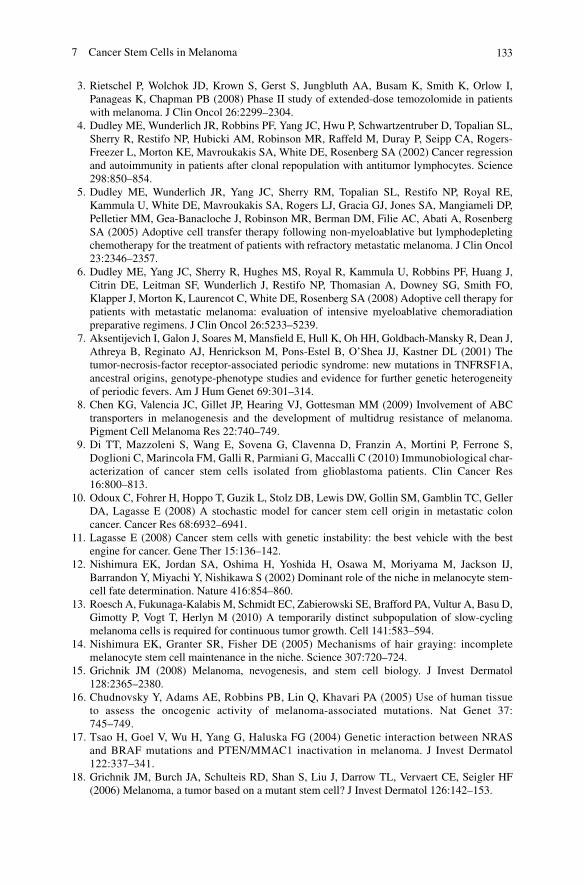
1337 Cancer Stem Cells in Melanoma
3. Rietschel P, Wolchok JD, Krown S, Gerst S, Jungbluth AA, Busam K, Smith K, Orlow I, Panageas K, Chapman PB (2008) Phase II study of extended-dose temozolomide in patients with melanoma. J Clin Oncol 26:2299–2304.
4. Dudley ME, Wunderlich JR, Robbins PF, Yang JC, Hwu P, Schwartzentruber D, Topalian SL, Sherry R, Restifo NP, Hubicki AM, Robinson MR, Raffeld M, Duray P, Seipp CA, Rogers-Freezer L, Morton KE, Mavroukakis SA, White DE, Rosenberg SA (2002) Cancer regression and autoimmunity in patients after clonal repopulation with antitumor lymphocytes. Science 298:850–854.
5. Dudley ME, Wunderlich JR, Yang JC, Sherry RM, Topalian SL, Restifo NP, Royal RE, Kammula U, White DE, Mavroukakis SA, Rogers LJ, Gracia GJ, Jones SA, Mangiameli DP, Pelletier MM, Gea-Banacloche J, Robinson MR, Berman DM, Filie AC, Abati A, Rosenberg SA (2005) Adoptive cell transfer therapy following non-myeloablative but lymphodepleting chemotherapy for the treatment of patients with refractory metastatic melanoma. J Clin Oncol 23:2346–2357.
6. Dudley ME, Yang JC, Sherry R, Hughes MS, Royal R, Kammula U, Robbins PF, Huang J, Citrin DE, Leitman SF, Wunderlich J, Restifo NP, Thomasian A, Downey SG, Smith FO, Klapper J, Morton K, Laurencot C, White DE, Rosenberg SA (2008) Adoptive cell therapy for patients with metastatic melanoma: evaluation of intensive myeloablative chemoradiation preparative regimens. J Clin Oncol 26:5233–5239.
7. Aksentijevich I, Galon J, Soares M, Mansfield E, Hull K, Oh HH, Goldbach-Mansky R, Dean J, Athreya B, Reginato AJ, Henrickson M, Pons-Estel B, O’Shea JJ, Kastner DL (2001) The tumor-necrosis-factor receptor-associated periodic syndrome: new mutations in TNFRSF1A, ancestral origins, genotype-phenotype studies and evidence for further genetic heterogeneity of periodic fevers. Am J Hum Genet 69:301–314.
8. Chen KG, Valencia JC, Gillet JP, Hearing VJ, Gottesman MM (2009) Involvement of ABC transporters in melanogenesis and the development of multidrug resistance of melanoma. Pigment Cell Melanoma Res 22:740–749.
9. Di TT, Mazzoleni S, Wang E, Sovena G, Clavenna D, Franzin A, Mortini P, Ferrone S, Doglioni C, Marincola FM, Galli R, Parmiani G, Maccalli C (2010) Immunobiological char-acterization of cancer stem cells isolated from glioblastoma patients. Clin Cancer Res 16:800–813.
10. Odoux C, Fohrer H, Hoppo T, Guzik L, Stolz DB, Lewis DW, Gollin SM, Gamblin TC, Geller DA, Lagasse E (2008) A stochastic model for cancer stem cell origin in metastatic colon cancer. Cancer Res 68:6932–6941.
11. Lagasse E (2008) Cancer stem cells with genetic instability: the best vehicle with the best engine for cancer. Gene Ther 15:136–142.
12. Nishimura EK, Jordan SA, Oshima H, Yoshida H, Osawa M, Moriyama M, Jackson IJ, Barrandon Y, Miyachi Y, Nishikawa S (2002) Dominant role of the niche in melanocyte stem-cell fate determination. Nature 416:854–860.
13. Roesch A, Fukunaga-Kalabis M, Schmidt EC, Zabierowski SE, Brafford PA, Vultur A, Basu D, Gimotty P, Vogt T, Herlyn M (2010) A temporarily distinct subpopulation of slow-cycling melanoma cells is required for continuous tumor growth. Cell 141:583–594.
14. Nishimura EK, Granter SR, Fisher DE (2005) Mechanisms of hair graying: incomplete melanocyte stem cell maintenance in the niche. Science 307:720–724.
15. Grichnik JM (2008) Melanoma, nevogenesis, and stem cell biology. J Invest Dermatol 128:2365–2380.
16. Chudnovsky Y, Adams AE, Robbins PB, Lin Q, Khavari PA (2005) Use of human tissue to assess the oncogenic activity of melanoma-associated mutations. Nat Genet 37: 745–749.
17. Tsao H, Goel V, Wu H, Yang G, Haluska FG (2004) Genetic interaction between NRAS and BRAF mutations and PTEN/MMAC1 inactivation in melanoma. J Invest Dermatol 122:337–341.
18. Grichnik JM, Burch JA, Schulteis RD, Shan S, Liu J, Darrow TL, Vervaert CE, Seigler HF (2006) Melanoma, a tumor based on a mutant stem cell? J Invest Dermatol 126:142–153.
134 P. Jin et al.
19. Klonisch T, Wiechec E, Hombach-Klonisch S, Ande SR, Wesselborg S, Schulze-Osthoff K, Los M (2008) Cancer stem cell markers in common cancers – therapeutic implications. Trends Mol Med 14:450–460.
20. Huntly BJ, Shigematsu H, Deguchi K, Lee BH, Mizuno S, Duclos N, Rowan R, Amaral S, Curley D, Williams IR, Akashi K, Gilliland DG (2004) MOZ-TIF2, but not BCR-ABL, confers properties of leukemic stem cells to committed murine hematopoietic progenitors. Cancer Cell 6:587–596.
21. Lapidot T, Sirard C, Vormoor J, Murdoch B, Hoang T, Caceres-Cortes J, Minden M, Paterson B, Caligiuri MA, Dick JE (1994) A cell initiating human acute myeloid leukaemia after trans-plantation into SCID mice. Nature 367:645–648.
22. Zhang M, Behbod F, Atkinson RL, Landis MD, Kittrell F, Edwards D, Medina D, Tsimelzon A, Hilsenbeck S, Green JE, Michalowska AM, Rosen JM (2008) Identification of tumor-initiating cells in a p53-null mouse model of breast cancer. Cancer Res 68:4674–4682.
23. Real C, Glavieux-Pardanaud C, Le Douarin NM, Dupin E (2006) Clonally cultured differenti-ated pigment cells can dedifferentiate and generate multipotent progenitors with self-renewing potential. Dev Biol 300:656–669.
24. Herlyn M, Thurin J, Balaban G, Bennicelli JL, Herlyn D, Elder DE, Bondi E, Guerry D, Nowell P, Clark WH, Koprowski H (1985) Characteristics of cultured human melanocytes isolated from different stages of tumor progression. Cancer Res 45:5670–5676.
25. Herlyn M, Clark WH, Rodeck U, Mancianti ML, Jambrosic J, Koprowski H (1987) Biology of tumor progression in human melanocytes. Lab Invest 56:461–474.
26. Wang E, Voiculescu S, Le Poole IC, el Gamil M, Li X, Sabatino M, Robbins PF, Nickoloff BJ, Marincola FM (2006) Clonal persistence and evolution during a decade of recurrent melanoma. J Invest Dermatol 126:1372–1377.
27. Sabatino M, Zhao Y, Voiculescu S, Monaco A, Robbins PF, Nickoloff BJ, Karai L, Selleri S, Maio M, Selleri S, Marincola FM, Wang E (2008) Conservation of a core of genetic alterations over a decade of recurrent melanoma supports the melanoma stem cell hypothesis. Cancer Res 68:222–231.
28. Takahashi K, Yamanaka S (2006) Induction of pluripotent stem cells from mouse embryonic and adult fibroblast cultures by defined factors. Cell 126:663–676.
29. Okada M, Oka M, Yoneda Y (2010) Effective culture conditions for the induction of pluri-potent stem cells. Biochim Biophys Acta 1800(9):956–63.
30. Okita K, Ichisaka T, Yamanaka S (2007) Generation of germline-competent induced pluri-potent stem cells. Nature 448:313–317.
31. Maherali N, Sridharan R, Xie W, Utikal J, Eminli S, Arnold K, Stadtfeld M, Yachechko R, Tchieu J, Jaenisch R, Plath K, Hochedlinger K (2007) Directly reprogrammed fibro-blasts show global epigenetic remodeling and widespread tissue contribution. Cell Stem Cell 1:55–70.
32. Wernig M, Meissner A, Foreman R, Brambrink T, Ku M, Hochedlinger K, Bernstein BE, Jaenisch R (2007) In vitro reprogramming of fibroblasts into a pluripotent ES-cell-like state. Nature 448:318–324.
33. Yu J, Vodyanik MA, Smuga-Otto K, Ntosiewicz-Bourget J, Frane JL, Tian S, Nie J, Jonsdottir GA, Ruotti V, Stewart R, Slukvin II, Thomson JA (2007) Induced pluripotent stem cell lines derived from human somatic cells. Science 318:1917–1920.
34. Lowry WE, Richter L, Yachechko R, Pyle AD, Tchieu J, Sridharan R, Clark AT, Plath K (2008) Generation of human induced pluripotent stem cells from dermal fibroblasts. Proc Natl Acad Sci USA 105:2883–2888.
35. Huangfu D, Osafune K, Maehr R, Guo W, Eijkelenboom A, Chen S, Muhlestein W, Melton DA (2008) Induction of pluripotent stem cells from primary human fibroblasts with only Oct4 and Sox2. Nat Biotechnol 26:1269–1275.
36. Al Hajj M, Wicha MS, Benito-Hernandez A, Morrison SJ, Clarke MF (2003) Prospective identification of tumorigenic breast cancer cells. Proc Natl Acad Sci USA 100:3983–3988.
37. Shackleton M, Vaillant F, Simpson KJ, Stingl J, Smyth GK, Asselin-Labat ML, Wu L, Lindeman GJ, Visvader JE (2006) Generation of a functional mammary gland from a single stem cell. Nature 439:84–88.
1357 Cancer Stem Cells in Melanoma
38. Goodell MA, Brose K, Paradis G, Conner AS, Mulligan RC (1996) Isolation and functional properties of murine hematopoietic stem cells that are replicating in vivo. J Exp Med 183:1797–1806.
39. Boiko AD, Razorenova OV, Van de RM, Swetter SM, Johnson DL, Ly DP, Butler PD, Yang GP, Joshua B, Kaplan MJ, Longaker MT, Weissman IL (2010) Human melanoma-initiating cells express neural crest nerve growth factor receptor CD271. Nature 466:133–137.
40. Fang D, Nguyen TK, Leishear K, Finko R, Kulp AN, Hotz S, Van Belle PA, Xu X, Elder DE, Herlyn M (2005) A tumorigenic subpopulation with stem cell properties in melanomas. Cancer Res 65:9328–9337.
41. Na YR, Seok SH, Kim DJ, Han JH, Kim TH, Jung H, Lee BH, Park JH (2009) Isolation and characterization of spheroid cells from human malignant melanoma cell line WM-266-4. Tumour Biol 30:300–309.
42. Dou J, Li Y, Zhao F, Hu W, Wen P, Tang Q, Chu L, Wang Y, Cao M, Jiang C, Gu N (2009) Identification of tumor stem-like cells in a mouse myeloma cell line. Cell Mol Biol (Noisy-le-grand) 55 Suppl:OL1151–OL1160.
43. Monzani E, Facchetti F, Galmozzi E, Corsini E, Benetti A, Cavazzin C, Gritti A, Piccinini A, Porro D, Santinami M, Invernici G, Parati E, Alessandri G, La Porta CA (2007) Melanoma contains CD133 and ABCG2 positive cells with enhanced tumourigenic potential. Eur J Cancer 43:935–946.
44. Mizrak D, Brittan M, Alison MR (2008) CD133: molecule of the moment. J Pathol 214:3–9. 45. Klein WM, Wu BP, Zhao S, Wu H, Klein-Szanto AJ, Tahan SR (2007) Increased expression of
stem cell markers in malignant melanoma. Mod Pathol 20:102–107. 46. Keshet GI, Goldstein I, Itzhaki O, Cesarkas K, Shenhav L, Yakirevitch A, Treves AJ, Schachter
J, Amariglio N, Rechavi G (2008) MDR1 expression identifies human melanoma stem cells. Biochem Biophys Res Commun 368:930–936.
47. Schatton T, Murphy GF, Frank NY, Yamaura K, Waaga-Gasser AM, Gasser M, Zhan Q, Jordan S, Duncan LM, Weishaupt C, Fuhlbrigge RC, Kupper TS, Sayegh MH, Frank MH (2008) Identification of cells initiating human melanomas. Nature 451:345–349.
48. Quintana E, Shackleton M, Sabel MS, Fullen DR, Johnson TM, Morrison SJ: Efficient tumour formation by single human melanoma cells. Nature 2008, 456:593–598.
49. Wang E, Miller LD, Ohnmacht GA, Mocellin S, Petersen D, Zhao Y, Simon R, Powell JI, Asaki E, Alexander HR, Duray PH, Herlyn M, Restifo NP, Liu ET, Rosenberg SA, Marincola FM (2002) Prospective molecular profiling of subcutaneous melanoma metastases suggests classifiers of immune responsiveness. Cancer Res 62:3581–3586.
50. Bittner M, Meltzer P, Chen Y, Jiang E, Seftor E, Hendrix M, Radmacher M, Simon R, Yakhini Z, Ben-Dor A, Sampas N, Dougherty E, Wang E, Marincola F, Gooden C, Lueders J, Glatfelter A, Pollock P, Carpten J, Gillanders E, Leja D, Dietrich K, Beaudry C, Berens M, Alberts D, Sondak V (2000) Molecular classification of cutaneous malignant melanoma by gene expres-sion: shifting from a countinuous spectrum to distinct biologic entities. Nature 406:536–840.
51. Hendrix MJ, Seftor EA, Seftor RE, Kasemeier-Kulesa J, Kulesa PM, Postovit LM (2007) Reprogramming metastatic tumour cells with embryonic microenvironments. Nat Rev Cancer 7:246–255.
52. Bennett DC (1983) Differentiation in mouse melanoma cells: initial reversibility and an on-off stochastic model. Cell 34:445–453.
53. Hoek KS, Eichhoff OM, Schlegel NC, Dobbeling U, Kobert N, Schaerer L, Hemmi S, Dummer R (2008) In vivo switching of human melanoma cells between proliferative and invasive states. Cancer Res 68:650–656.
54. Pinner S, Jordan P, Sharrock K, Bazley L, Collinson L, Marais R, Bonvin E, Goding C, Sahai E (2009) Intravital imaging reveals transient changes in pigment production and Brn2 expression during metastatic melanoma dissemination. Cancer Res 69:7969–7977.
55. Held MA, Curley DP, Dankort D, McMahon M, Muthusamy V, Bosenberg MW (2010) Characterization of melanoma cells capable of propagating tumors from a single cell. Cancer Res 70:388–397.
56. Lee JT, Herlyn M (2007) Microenvironmental influences in melanoma progression. J Cell Biochem 101:862–872.
136 P. Jin et al.
57. Luzzi KJ, MacDonald IC, Schmidt EE, Kerkvliet N, Morris VL, Chambers AF, Groom AC (1998) Multistep nature of metastatic inefficiency: dormancy of solitary cells after successful extravasation and limited survival of early micrometastases. Am J Pathol 153:865–873.
58. Fusi A, Reichelt U, Busse A, Ochsenreither S, Rietz A, Maisel M, Keilholz U (2011) Expression of the Stem Cell Markers Nestin and CD133 on Circulating Melanoma Cells. J Invest Dermatol 131:487–494.
59. Kaplan RN, Riba RD, Zacharoulis S, Bramley AH, Vincent L, Costa C, MacDonald DD, Jin DK, Shido K, Kerns SA, Zhu Z, Hicklin D, Wu Y, Port JL, Altorki N, Port ER, Ruggero D, Shmelkov SV, Jensen KK, Rafii S, Lyden D (2005) VEGFR1-positive haematopoietic bone marrow progenitors initiate the pre-metastatic niche. Nature 438:820–827.
60. Wang E, Ngalame Y, Panelli MC, Deavers M, Mueller P, Ju W, Savary C, Nguyen-Jackson H, Freedman RS, Marincola FM (2005) Peritoneal and sub-peritoneal stroma may facilitate regional spread of ovarian cancer. Clin Cancer Res 11:113–122.
61. Orimo A, Gupta PB, Sgroi DC, Arenzana-Seisdedos F, Delaunay T, Naeem R, Carey VJ, Richardson AL, Weinberg RA (2005) Stromal fibroblasts present in invasive human breast carcinomas promote tumor growth and angiogenesis through elevated SDF-1/CXCL12 secretion. Cell 121:335–348.
62. Karnoub AE, Dash AB, Vo AP, Sullivan A, Brooks MW, Bell GW, Richardson AL, Polyak K, Tubo R, Weinberg RA (2007) Mesenchymal stem cells within tumour stroma promote breast cancer metastasis. Nature 449:557–563.
63. Alphonso A, Alahari SK (2009) Stromal cells and integrins: conforming to the needs of the tumor microenvironment. Neoplasia 11:1264–1271.
64. Hendrix MJ, Seftor EA, Hess AR, Seftor RE (2003) Vasculogenic mimicry and tumour-cell plasticity: lessons from melanoma. Nat Rev Cancer 3:411–421.
65. Husemann Y, Geigl JB, Schubert F, Musiani P, Meyer M, Burghart E, Forni G, Eils R, Fehm T, Riethmuller G, Klein CA (2008) Systemic spread is an early step in breast cancer. Cancer Cell 13:58–68.
66. Schofield R (1978) The relationship between the spleen colony-forming cell and the haemopoietic stem cell. Blood Cells 4:7–25.
67. Villanueva J, Herlyn M (2008) Melanoma and the tumor microenvironment. Curr Oncol Rep 10:439–446.
68. Baguley BC (2006) Tumor stem cell niches: a new functional framework for the action of anticancer drugs. Recent Pat Anticancer Drug Discov 1:121–127.
69. Voog J, Jones DL (2010) Stem cells and the niche: a dynamic duo. Cell Stem Cell 6:103–115. 70. LaBarge MA (2010) The difficulty of targeting cancer stem cell niches. Clin Cancer Res
16:3121–3129. 71. Haass NK, Smalley KS, Li L, Herlyn M (2005) Adhesion, migration and communication in
melanocytes and melanoma. Pigment Cell Res 18:150–159. 72. Ch’ng S, Tan ST (2009) Genetics, cellular biology and tumor microenvironment of melanoma.
Front Biosci 14:918–928. 73. Ruiter D, Bogenrieder T, Elder D, Herlyn M (2002) Melanoma-stroma interactions: structural
and functional aspects. Lancet Oncol 3:35–43. 74. Mahabeleshwar GH, Byzova TV (2007) Angiogenesis in melanoma. Semin Oncol 34:555–565. 75. Ishii G, Sangai T, Oda T, Aoyagi Y, Hasebe T, Kanomata N, Endoh Y, Okumura C, Okuhara Y,
Magae J, Emura M, Ochiya T, Ochiai A (2003). Bone-marrow-derived myofibroblasts contribute to the cancer-induced stromal reaction. Biochem Biophys Res Commun 309:232–240.
76. Direkze NC, Hodivala-Dilke K, Jeffery R, Hunt T, Poulsom R, Oukrif D, Alison MR, Wright NA (2004) Bone marrow contribution to tumor-associated myofibroblasts and fibroblasts. Cancer Res 64:8492–8495.
77. Bergfeld SA, DeClerck YA (2010) Bone marrow-derived mesenchymal stem cells and the tumor microenvironment. Cancer Metastasis Rev 29:249–261.
78. Schichor C, Birnbaum T, Etminan N, Schnell O, Grau S, Miebach S, Aboody K, Padovan C, Straube A, Tonn JC, Goldbrunner R (2006) Vascular endothelial growth factor A contributes to glioma-induced migration of human marrow stromal cells (hMSC). Exp Neurol 199:301–310.
1377 Cancer Stem Cells in Melanoma
79. Coffelt SB, Marini FC, Watson K, Zwezdaryk KJ, Dembinski JL, LaMarca HL, Tomchuck SL, Honer zu BK, Danka ES, Henkle SL, Scandurro AB (2009) The pro-inflammatory peptide LL-37 promotes ovarian tumor progression through recruitment of multipotent mesenchymal stromal cells. Proc Natl Acad Sci USA 106:3806–3811.
80. Sotiropoulou PA, Papamichail M (2007) Immune properties of mesenchymal stem cells. Methods Mol Biol 407:225–243.
81. Djouad F, Plence P, Bony C, Tropel P, Apparailly F, Sany J, Noel D, Jorgensen C (2003) Immunosuppressive effect of mesenchymal stem cells favors tumor growth in allogeneic animals. Blood 102:3837–3844.
82. Kim CH, Broxmeyer HE (1998) In vitro behavior of hematopoietic progenitor cells under the influence of chemoattractants: stromal cell-derived factor-1, steel factor, and the bone marrow environment. Blood 91:100–110.
83. Szakacs G, Paterson JK, Ludwig JA, Booth-Genthe C, Gottesman MM (2006) Targeting multidrug resistance in cancer. Nat Rev Drug Discov 5:219–234.
84. Yilmaz OH, Valdez R, Theisen BK, Guo W, Ferguson DO, Wu H, Morrison SJ (2006) Pten dependence distinguishes haematopoietic stem cells from leukaemia-initiating cells. Nature 441:475–482.
85. Rossi DJ, Weissman IL: Pten, tumorigenesis, and stem cell self-renewal (2006) Cell 125:229–231. 86. Hassane DC, Guzman ML, Corbett C, Li X, Abboud R, Young F, Liesveld JL, Carroll M,
Jordan CT (2008) Discovery of agents that eradicate leukemia stem cells using an in silico screen of public gene expression data. Blood 111:5654–5662.
87. Fritz V, Jorgensen C (2008) Mesenchymal stem cells: an emerging tool for cancer targeting and therapy. Curr Stem Cell Res Ther 3:32–42.
88. Hall B, Dembinski J, Sasser AK, Studeny M, Andreeff M, Marini F (2007) Mesenchymal stem cells in cancer: tumor-associated fibroblasts and cell-based delivery vehicles. Int J Hematol 86:8–16.
89. Studeny M, Marini FC, Dembinski JL, Zompetta C, Cabreira-Hansen M, Bekele BN, Champlin RE, Andreeff M (2004) Mesenchymal stem cells: potential precursors for tumor stroma and targeted-delivery vehicles for anticancer agents. J Natl Cancer Inst 96:1593–1603.
90. Cavarretta IT, Altanerova V, Matuskova M, Kucerova L, Culig Z, Altaner C (2010) Adipose tissue-derived mesenchymal stem cells expressing prodrug-converting enzyme inhibit human prostate tumor growth. Mol Ther 18:223–231.
91. Sabatino M, Stroncek DF, Klein H, Marincola FM, Wang E (2009) Stem cells in melanoma development. Cancer Lett 279(2):119–25.
92. Wicha MS, Liu S, Dontu G (2006) Cancer stem cells: an old idea--a paradigm shift. Cancer Res, 66:1883–1890.
93. Clarke MF, Dick JE, Dirks PB, Eaves CJ, Jamieson CH, Jones DL, Visvader J, Weissman IL, Wahl GM (2006) Cancer stem cells--perspectives on current status and future directions: AACR Workshop on cancer stem cells. Cancer Res 66:9339–9344.
94. Greijer AE, van der Groep P, Kemming D, Shvarts A, Semenza GL, Meijer GA, van de Wiel MA, Belien JA, van Diest PJ, van der Wall E (2005) Up-regulation of gene expression by hypoxia is mediated predominantly by hypoxia-inducible factor 1 (HIF-1). J Pathol 206:291–304.
95. Kelly PN, Dakic A, Adams JM, Nutt SL, Strasser A (2007) Tumor growth need not be driven by rare cancer stem cells. Science 317:337.
96. Ascierto PA, Kirkwood JM (2008) Adjuvant therapy of melanoma with interferon: lessons of the past decade. J Transl Med 6:62.
97. Ascierto PA, Streicher HZ, Sznol M (2010) Melanoma: a model for testing new agents in combination therapies. J Transl Med 8:38.
98. Costa FF, Le BK, Brodin B (2007) Concise review: cancer/testis antigens, stem cells, and cancer. Stem Cells 25:707–711.
99. Robbins PF, el-Gamil M, Li YF, Kawakami Y, Loftus D, Appella E, Rosenberg SA (1996) A mutated beta-catenin gene encodes a melanoma-specific antigen recognized by tumor infil-trating lymphocytes. J Exp Med 183:1185–1192.

138 P. Jin et al.
100. Lennerz V, Fatho M, Gentilini C, Frye RA, Lifke A, Ferel D, Wolfel C, Huber C, Wolfel T (2005) The response of autologous T cells to a human melanoma is dominated by mutated neoantigens. Proc Natl Acad Sci USA 102:16013–16018.
101. Schatton T, Schutte U, Frank NY, Zhan Q, Hoerning A, Robles SC, Zhou J, Hodi FS, Spagnoli GC, Murphy GF, Frank MH (2010) Modulation of T-cell activation by malignant melanoma initiating cells. Cancer Res 70:697–708.
102. Al-Hajj M (2007) Cancer stem cells and oncology therapeutics. Curr Opin Oncol 19:61–64.
139A.L. Allan (ed.), Cancer Stem Cells in Solid Tumors, Stem Cell Biology and Regenerative Medicine, DOI 10.1007/978-1-61779-246-5_8, © Springer Science+Business Media, LLC 2011
Abstract Lung cancer is the most common cancer worldwide, accounting for 1.2 million new cases annually. Furthermore, it is the most lethal of all cancers. A major challenge in treating this deadliest form of malignancy is the intrinsic resis-tance to conventional therapies. It is believed that cancer stem/progenitor cells are responsible for the sustained growth, survival, and invasion of tumors. Therefore, identifying lung cancer stem cells (CSCs) and studying the biologic functions necessary for their existence within lung tumors will provide new clinical approaches with the goal of improving clinical outcomes of the disease. This chapter will summarize our understanding of the identification of cancer stem cells in lung tumors, molecular mechanisms, and associated pathways that operate within cancer stem cells of lung tumors, and potential applications in clinic settings. We will also discuss future perspectives in lung cancer stem cell research.
Abbreviations
ABCG2 ATP-binding cassette sub-family G member 2AC AdenocarcinomaALDH1 Aldehyde dehydrogenase 1ASCL Achaete scute-likeBAC Bronchioalveolar carcinomasbHLH Basic helix-loop-helixCCSP Clara cell secretory protein
F. Jiang (*)Department of Pathology, University of Maryland School of Medicine,Baltimore, MD, USAe-mail: [email protected]
Chapter 8Cancer Stem Cells in Lung Cancer
Jun Shen and Feng Jiang
140 J. Shen and F. Jiang
CD Cluster of differentiationCSC Cancer stem cellEPC Endothelial progenitor cellHh HedgehogHSC Hematopoietic stem cellMDR1 Multi-drug resistance protein 1mTOR Mammalian target of rapamycinNSCLC Non small-cell lung cancerPNEC Pulmonary neuroendocrine cellsPTCH PatchedSCC Squamous cell carcinomaSCID Severe combined immunodeficiencySCLC Small-cell lung cancerSDF-1 Stromal-derived factor 1SMO SmoothenedSP Side populationSSEA Stage-specific embryonic antigenTAC Transit amplifying cellTRA Tumor rejection antigen
8.1 Introduction
Lung cancer, the leading cause of cancer death worldwide, is comprised of four major histological types: small-cell lung cancer (SCLC) and three types of non–small-cell lung cancer (NSCLC) including squamous cell carcinoma (SCC), adeno-carcinoma (AC), and large-cell carcinoma [1, 2]. Despite recent treatment advances, including modernization of drug cocktails and radiotherapeutic regimens over the past half century, the 5-year survival rate of patients with NSCLC is only 15%. Therefore, there is an urgent need to better understand the key molecular events driving lung tumorigenesis, such that we can find more effective ways for its prevention, diagnosis, prognosis, and treatment.
Accumulating evidence suggests that stem cells and cancer are inextricably linked, and perceived wisdom is that the process of carcinogenesis initially affects normal stem cells or their closely related progenitors. For instance, in animal models of intestinal cancer, a direct involvement of stem cells in adenoma forma-tion has well been demonstrated [3]. Furthermore, during the progression of the tumor, neoplastic stem cells may evolve to maintain tumor growth. Many other types of solid tumors also have a population of self-renewing and/or expanding stem cells: cancer stem cells (CSCs). The CSC hypothesis provides an explanation for the origins of tumor self-renewal and heterogeneity [4, 5]. There are two components of the CSC hypothesis [6]. The first is that cancers directly arise from stem cells that have acquired sufficient oncogenic mutations for transformation [7]. Therefore, the
1418 Cancer Stem Cells in Lung Cancer
tumor cell of origin, referred to as a tumor-initiating cell, is likely a stem or progenitor cell that is already capable of self-renewal and differentiation. The second compo-nent of the hypothesis is that tumor progression could be driven by a subpopulation of self-renewing tumor cells. This vision is supported by the observation that most tumors are comprised of functionally heterogeneous cell subpopulations, including a population that differs in their ability for limitless proliferative potential and repopulation ability [8].
Lung cancer is a complex network consisting of cells at various stages of dif-ferentiation, neovascular structures, reactive inflammatory cells, recruited cells, and infiltrated parenchyma that interact within the tumor mass. Based on the CSC hypothesis, a lung tumor might be driven and maintained by antigenically distinct subpopulations of perpetually self-renewing CSCs that give rise to transit-amplifying cells (TACs) and terminally differentiated cells. Therefore, like normal cell popula-tions, lung tumors may have a hierarchical structure. Adherents of the hypothesis imply that the bulk of the solid tumor is thus not the clinical problem, and that iden-tifying CSCs and the associated factors that regulate CSCs’ behavior may have an enormous bearing on the way we treat the deadliest form of malignancy in the clinical setting. The clinical implications of a tumorigenic hierarchy thus seem to be obvious, considering that therapies targeting the rapid reduction of tumor size in the lungs are not selected for their discriminatory ability to treat tumor-initiating cell subpopulation. When a therapy fails to kill all self-renewing lung CSCs, residual surviving CSCs will be able to repopulate the disease, leading to relapse of the tumors of the lungs.
Evidence for the existence of clonogenic cells in the lungs was first described in 1982 [9]. In this study, a small population of cells (<1.5%) isolated from surgically resected tumors from patients diagnosed with AC and SCLC of the lungs were able to form colonies in a soft agar cloning assay [9]. However, comparatively less is known about the biology of lung CSCs compared with other solid tumor stem cells. Considering lung cancer is the most common lethal type of cancer in the world [10], there is a pressing need for the development of new therapeutic agents that better manage the progression of highly aggressive lung cancer cells. Research in the area of lung CSCs might provide a new paradigm leading to improved therapies for the disease. This chapter will summarize our understanding of the cellular and molec-ular mechanisms that operate within CSCs or initiating cells of lung tumors and their potential applications in clinical practice.
8.2 Identification and Isolation of Lung Cancer CSCs
Because many of the molecules expressed by normal stem cells may also be found in their malignant counterparts, considerable effort has been made in the search for “markers” of CSCs based on the currently used markers for stem cells (Table 8.1).
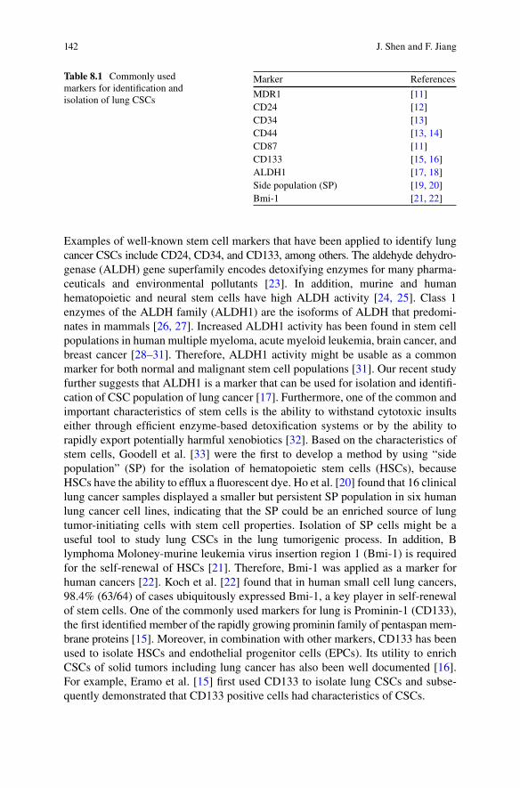
142 J. Shen and F. Jiang
Examples of well-known stem cell markers that have been applied to identify lung cancer CSCs include CD24, CD34, and CD133, among others. The aldehyde dehydro-genase (ALDH) gene superfamily encodes detoxifying enzymes for many pharma-ceuticals and environmental pollutants [23]. In addition, murine and human hematopoietic and neural stem cells have high ALDH activity [24, 25]. Class 1 enzymes of the ALDH family (ALDH1) are the isoforms of ALDH that predomi-nates in mammals [26, 27]. Increased ALDH1 activity has been found in stem cell populations in human multiple myeloma, acute myeloid leukemia, brain cancer, and breast cancer [28–31]. Therefore, ALDH1 activity might be usable as a common marker for both normal and malignant stem cell populations [31]. Our recent study further suggests that ALDH1 is a marker that can be used for isolation and identifi-cation of CSC population of lung cancer [17]. Furthermore, one of the common and important characteristics of stem cells is the ability to withstand cytotoxic insults either through efficient enzyme-based detoxification systems or by the ability to rapidly export potentially harmful xenobiotics [32]. Based on the characteristics of stem cells, Goodell et al. [33] were the first to develop a method by using “side population” (SP) for the isolation of hematopoietic stem cells (HSCs), because HSCs have the ability to efflux a fluorescent dye. Ho et al. [20] found that 16 clinical lung cancer samples displayed a smaller but persistent SP population in six human lung cancer cell lines, indicating that the SP could be an enriched source of lung tumor-initiating cells with stem cell properties. Isolation of SP cells might be a useful tool to study lung CSCs in the lung tumorigenic process. In addition, B lymphoma Moloney-murine leukemia virus insertion region 1 (Bmi-1) is required for the self-renewal of HSCs [21]. Therefore, Bmi-1 was applied as a marker for human cancers [22]. Koch et al. [22] found that in human small cell lung cancers, 98.4% (63/64) of cases ubiquitously expressed Bmi-1, a key player in self-renewal of stem cells. One of the commonly used markers for lung is Prominin-1 (CD133), the first identified member of the rapidly growing prominin family of pentaspan mem-brane proteins [15]. Moreover, in combination with other markers, CD133 has been used to isolate HSCs and endothelial progenitor cells (EPCs). Its utility to enrich CSCs of solid tumors including lung cancer has also been well documented [16]. For example, Eramo et al. [15] first used CD133 to isolate lung CSCs and subse-quently demonstrated that CD133 positive cells had characteristics of CSCs.
Marker References
MDR1 [11]CD24 [12]CD34 [13]CD44 [13, 14]CD87 [11]CD133 [15, 16]ALDH1 [17, 18]Side population (SP) [19, 20]Bmi-1 [21, 22]
Table 8.1 Commonly used markers for identification and isolation of lung CSCs
1438 Cancer Stem Cells in Lung Cancer
8.3 CSCs Play Important Roles in Lung Tumorigenesis
Using the Aldefluor® assay followed by fluorescence-activated cell sorting analysis, we isolated lung cancer cells that had high ALDH1 activity [17]. ALDH1 positive lung cancer cells displayed in vitro features of CSCs, including enhanced capacity for proliferation, self-renewal, and differentiation; resistance to chemotherapy; and expression of the CSC surface marker CD133 [17]. In vivo experiments showed that the ALDH1 positive cells could generate tumors that recapitulate the heterogeneity of the parental cancer cells. Furthermore, immunohistochemical analysis of 303 clinical specimens from three independent cohorts of lung cancer patients and controls showed that expression of ALDH1 was positively correlated with the stage and grade of lung tumors, and related to a poor prognosis for the patients with early-stage lung cancer. ALDH1 is therefore a lung tumor stem cell–associated marker. These findings offer an important new tool for the study of lung CSCs, and provide a potential prognostic factor and therapeutic target for treatment of patients with lung cancer [17].
CD133 positive lung cancer cells have also been considered as CSCs in the lungs. However, as experienced for other tumors, somewhat disparate results have been reported. For example, from both the NSCLC A549 cell line and the SCLC H466 cell line, CD133 negative cells were as proficient as CD133 positive cells at colony forming ability in vitro and tumorigenic capacity in nude mice in vivo [34]. On the other hand, it has been shown that in NSCLC, 1 × 10³ CD133 positive cells could form tumors in SCID mice; however, 1 × 10⁴ CD133 negative cells could not [35]. CD133 positive cells also show enhanced expression of Oct-4 and ABCG2, and small interfering RNA (siRNA) knockdown of Oct-4 blocked clonogenicity and enhanced chemosensitivity. Expression of Oct-4 was observed in bronchioalveolar carcinomas (BACs), but the immunohistochemical expression data were uncon-vincing in comparison with the seminoma positive control [36].
Because CSCs are resistant to chemotherapeutic drugs, the perceived chemore-sistance of CSCs has been investigated. For example, CSCs have been enriched in NSCLC cell lines treated with cisplatin and doxorubicin [37]. 5 × 10³ drug selected cells regularly formed tumors in SCID mice. These cells expressed CD133, CD117, and the embryonic stem cell markers stage-specific embryonic antigen 3 (SSEA-3), tumor rejection antigen TRA1-81, Oct-4, and nuclear b-catenin. In the A549 cell line, the SP fraction was 5.2%; however, after drug treatment, this was increased to 35%. These studies clearly indicated that drug resistance and lung CSCs are heavily entwined. Furthermore, in the A549 NSCLC cell line, a large (24%) SP has been found. The SP cells displayed enhanced resistance to doxorubicin and methotrexate related to ABCG2 activity [38]. In the SCLC H-146 cell line, a “migratory” SP has been observed which is able to migrate toward conditioned media from hypoxic bone marrow–derived stromal cells, probably due to release of stromal cell derived factor 1 (SDF-1). This fraction was also enriched for CSCs in nude mice in vivo [39]. The b-helix-loop-helix (bHLH) transcription factor achaete scute complex homologue 1 (ASCL1/mammalian achaete scute homolog 1 [Mash1]) is essential
144 J. Shen and F. Jiang
for neuroendocrine development, and it appears that both CD133 and ALDH1 are directly regulated by ASCL1 [18]. In SCLC, there might be a relatively abundant CD133highASCL1highALDH high subpopulation enriched for CSCs, with as few as 1 × 10³ of these cells being capable of forming rapidly growing aggressive tumors in nude mice. In a number of SCLC cell lines, a subpopulation (1–4%) of urokinase plasminogen activator-positive cells was found. The positive cells were more resis-tant to traditional chemotherapies such as 5-fluorouracil, cisplatin, and etoposide. The resistance might be associated with enhanced MDR1 (ABCB1) activity and CD44 expression [11]. Ling et al. [40] showed that a serum-free system for primary neonatal pulmonary cells can support the growth of Oct-4+ epithelial colonies. Furthermore, besides Oct-4, these cells also expressed other stem cell markers such as SSEA-1, stem cell antigen 1 (Sca-1), and CCSP. These cells could be maintained for several weeks in primary cultures and undergo terminal differentiation to AT1 and AT2-like pneumocytes. They further demonstrated these Oct-4+ cells were located at the bronchoalveolar junction of neonatal lung. Taken together, all these findings provide strong evidence that CSCs play key role in the development and progression of lung cancer.
8.4 Cancer Stem Cell-Related Pathways of Self-Renewal in Lung Cancer
Stem cell self-renewal is a tightly controlled process governed by both signals from the stem cell niche as well as deliberate and regulated control of key developmental pathways. Examples of such pathways are Wnt, Hedgehog, and Notch signaling pathways [41]. Because CSCs comprise the self-renewing component of tumors, it is hypothesized that the same pathways that govern normal stem cell self-renewal could also govern CSC self-renewal. However, self-renewal in tumorigenesis is achieved by the deregulation of these key pathways. [4]. It is imperative to identify and understand the deregulated pathways involved in lung CSCs, because future development of potential therapeutic approaches to target these altered pathways in tumors might provide an important strategy for treating tumors that are often intrac-table to conventional therapy alone [42]. We will review three major pathways whose deregulations are involved in lung CSCs.
8.4.1 Wnt/b-Catenin Signaling
The Wnt/b-catenin pathway was originally found to play an important role in the regulation of HSC self-renewal [43]. Reynolds et al. [43] showed that activated Wnt/b-catenin signaling in the developing lung coincided with an expansion of BASCs and attenuated bronchiolar differentiation. On the other hand, conditional Cre-mediated deletion of Catnb (which encodes b-catenin) had no appreciable effect
1458 Cancer Stem Cells in Lung Cancer
on the repair and maintenance of the bronchiolar epithelium. These observations suggest that the role of Wnt/b-catenin signaling in lung stem cell self-renewal might be niche specific [44]. In lung cancer, evidence of activated Wnt signaling suggests aberrant Wnt signaling may be important for lung tumorigenesis [45, 46]. Furthermore, inhibition of Wnt signaling by a Wnt-2 monoclonal antibody resulted in the induction of apoptosis in NSCLC cells [47]. Taken together, deregulation of the Wnt signaling pathway clearly promotes lung carcinogenesis and stem cell self-renewal, making the Wnt signaling pathway an appealing target for the devel-opment of novel therapies for lung cancer.
8.4.2 Hedgehog Signaling
The Hedgehog (Hh) signaling pathway is activated when one of three extracellular Hh ligands (sonic hedgehog, desert hedgehog, and Indian hedgehog) binds to and inactivates its receptor patched (PTCH). The Hh signaling pathway is a key devel-opmental pathway required for proper embryogenesis [35]. In the developing lungs, activated Hh signaling is involved in pulmonary cell fate determination and branching morphogenesis [36]. Aberrations in expression and activation of this pathway could lead to deformations in development, and hence contribute to tumorigenesis [37]. For example, during lung epithelial regeneration after injury, activated Hh signaling is observed in regions of repair and in pulmonary neuroendocrine stem cell niches [38] and cyclopamine-mediated suppression of aberrantly active Hh signaling in some SCLCs resulted in a dramatic drop in cell viability and tumorigenicity [39]. These findings suggest that SCLC is a malignancy that arises from a population of self-renewing pulmonary neuroendocrine cells (PNECs) that retain active Hh signaling as well as primitive neuroendocrine features. Furthermore, the observa-tion implies that therapeutic targeting of the Hh signaling pathway may suppress stem-like tumor cell self-renewal [40]. The increasing evidence for the use of Hh signaling in tumor cell maintenance and cancer stem cell self-renewal has provoked the development of better and more specific inhibitors of the Hh pathway, some of which are currently in clinical trials for SCLC [41, 48, 49].
8.4.3 Notch Signaling
The Notch signaling pathway is involved in cell fate determination, organogenesis, and tissue homeostasis. Notch-mediated cell–cell interactions dictate the preserva-tion or differentiation of stem cells [50]. Activation of Notch signaling begins when membrane-bound Notch ligands bind to receptors on adjacent cells. Upon binding, the intracellular domain of the receptor is cleaved by gamma-secretase, allowing for the activation of downstream targets, such as the inhibitory basic helix-loop-helix transcription factor Hes1 [51]. In lung development, Notch signaling appears
146 J. Shen and F. Jiang
to be required for determining proximal and distal lung epithelial cell fates [52]. Forcing activation of Notch signaling in the developing lung tissue either through the ectopic expression of intracellular Notch domains or through gamma-secretase activation can result in the accumulation of distal airway stem cell differentiation [53]. Although elevated Notch signaling transcripts have been described in NSCLC, the role of Notch in tumor maintenance remains largely unknown. Suppression of Notch signaling in some NSCLC cells by treatment with a gamma-secretase inhibitor induced cell death and decreased tumor growth in mice [54]. In contrast, another study showed that activation of Notch signaling in A549 cells through overexpression of Notch1 resulted in a decrease in proliferation and tumorigenic growth in mice [55]. The apparent discrepancy between these results may be due to the perturbations of Notch signaling via different Notch receptors. However, although it is not yet clear how Notch signaling functions for self-renewal of lung CSCs, several reports suggest that Notch signaling components are expressed in putative lung CSC populations and are required for tumor initiation capacity [56].
Finally, it should be emphasized that for treatment of CSCs to have any signifi-cant future impact on the overall survival of patients with lung cancer, the underlying molecular signaling that drives tumorigenicity in these cells must be elucidated in much greater detail than is currently known.
8.5 Potential Applications of CSCs for Lung Cancer Treatment
As described above, Wnt signaling, Hedgehog signaling, Notch/Delta signaling, mTOR, ABC transporters, and the stem cell niche may provide a variety of drug-gable targets related to CSCs. The below examples could demonstrate the potential in developing innovative treatments of lung cancer in future.
In lung tissues, key regulators of stem cell renewal appear to be members of the Polycomb group protein family of transcriptional repressors, including Bmi-1, polyhomeotic-like 1, and melanoma surface molecular 18. Bmi-1 is a downstream target of the morphogen Sonic hedgehog (Shh) through the latter’s activation of the glioma-associated oncogene homolog (Gli) family of transcription factors. Shh acts on the receptor complex of patched (PTCH) and smoothened (SMO), blocking the restraining influence of PTCH on SMO, resulting in SMO signaling that activates the Gli family of transcription factors, thus activating target genes like Bmi-1. Inhibiting the action of SMO with the antagonist cyclopamine is a highly effective strategy against some cancers. It has been shown that almost all SCLCs ubiqui-tously express Bmi-1 [22], and antisense Bmi-1 RNA therapy reduces proliferation of A549 cells [57].
Another example is that of the Notch family of receptors, which are critical for stem cell self-renewal. Engagement of ligands of the delta and jagged families could cause cleavage of the intracellular portion of Notch and its translocation to the nucleus where it binds to the transcription factor chisel, changing it from a tran-scriptional repressor to an activator. Interestingly, cleavage of the intracellular
1478 Cancer Stem Cells in Lung Cancer
portion of Notch is mediated by the g-secretase protease complex. Therefore, the use of g-secretase inhibitors may have promising utility in cancers where Notch signaling is inappropriately inactivated [58]. Furthermore, various pathways acti-vated in human lung cancer converge on mTOR, with obvious therapeutic possi-bilities for the disease. Apart from these renewal and proliferation pathways, there are many other potential molecular targets relating to lung CSCs. For instance, because lung tumors have SP fractions, targeting ABC transporter activity might be a useful strategy for overcoming chemoresistance and directly eradicating stem cells [37, 59].
Techniques that could be applied for the potential targets include but are not limited to those described below. Antibody-based targeting of CSCs exploiting the overexpression of molecules such as CD133 is possible approach. Small molecule therapeutics that target growth factors, growth factor receptors, and their kinases and more specific tyrosine kinase inhibitors are gaining widespread usage in lung cancer treatment. RNA interference will become an increasingly useful strategy. In fact, targeting Oct-4 by using the technique in lung cancer has been shown to increase apoptosis of the cells [60]. Additionally, some new therapies for lung can-cer are designed to make cells more sensitive to induced cell death, while others target self-renewal pathways. Although it is not clear whether these therapies spe-cifically target lung cancer stem cells, in other cancers such as colon cancer [61], siRNA targeted to putative stem cell molecules like CD44 and Musashi-1 have been highly effective in blocking growth of xenografted tumors [61, 62].
8.6 Future Perspectives
The development of effective, safe CSC-based therapies for the treatment of lung cancer remains a tantalizing prospect [63]. However, before considering and initi-ating any possibility of CSC-based treatments, emphasis must be placed on under-standing events occurring frequently in CSCs and their local microenvironment during cancer progression, and the molecular mechanisms involved in their resis-tance to current chemotherapeutics. The fact that CSCs and normal adult stem cells utilize common molecular machinery and have similar protein expression profiles is potentially the most challenging hurdle to overcome in the development of therapies to target CSCs while sparing normal stem cells. Therefore, more precise methods and specific markers need to be developed to discriminate CSCs from normal stem cells. Furthermore, it is unknown whether CSCs originate from pluripotent adult stem cells or more differentiated committed stem/progenitor cells. If CSCs originate from pluripotent adult stem cells, CSCs may be able to differentiate into all pathological types of lung cancer with more primitive charac-teristics. However, if CSCs come from more differentiated committed stem/progenitor cells, their fate will already be determined as it can only differentiate along a particular lineage. To answer this question, the lineage tracing technique should be used to identify the cell of origin. Niches along with trachea for normal
148 J. Shen and F. Jiang
adult stem cells may be harnessed by CSCs, since aberrant signals within the niches are utilized for abnormal proliferation of CSCs. Therefore, there may be a possibil-ity that lineage differentiation is already defined by the remaining extinctive niches with potential reactivation of a particular signal pathway. Successful answering of these and other questions will undoubtedly have profound implications for the treat-ment of lung cancer and ultimately decrease lethality from the disease.
References
1. Jemal A et al (2008) Cancer statistics, 2008. CA Cancer J Clin 58(2):71–96 2. Potti A et al (2006) A genomic strategy to refine prognosis in early-stage non-small-cell lung
cancer. N Engl J Med 355(6):570–580 3. Barker N et al (2009) Crypt stem cells as the cells-of-origin of intestinal cancer. Nature 457
(7229):608–611 4. Reya T, et al (2001) Stem cells, cancer, and cancer stem cells. Nature 414(6859):105–111 5. Blagosklonny MV (2007) Cancer stem cell and cancer stemloids: from biology to therapy.
Cancer Biol Ther 6(11):1684–1690 6. Clarke MF, Fuller M (2006) Stem cells and cancer: two faces of eve. Cell 124(6):1111–1115 7. Wicha MS, Liu S, Dontu G (2006) Cancer stem cells: an old idea--a paradigm shift. Cancer
Res 66(4):1883–1890 8. Passegué E, Wagner EF, Weissman IL (2004) JunB deficiency leads to a myeloproliferative
disorder arising from hematopoietic stem cells. Cell 119(3):431–443 9. Carney DN (1982) Demonstration of the stem cell nature of clonogenic tumor cells from lung
cancer patients. Stem Cells 1(3):149–164 10. Minna JD, Roth JA, Gazdar AF (2002) Focus on lung cancer. Cancer Cell 1(1):49–52 11. Gutova M et al (2007) Identification of uPAR-positive chemoresistant cells in small cell lung
cancer. PLoS One 2(2):e243 12. Lee HJ et al (2010) CD24, a Novel Cancer Biomarker, Predicting Disease-Free Survival of
Non-small Cell Lung Carcinomas: A Retrospective Study of Prognostic Factor Analysis from the Viewpoint of Forthcoming (Seventh) New TNM Classification. J Thorac Oncol 5(5): 649–657
13. Liu D et al (2008) Multiparametric flow cytometry analyzes the expressions of immunopheno-type CD133, CD34, CD44 in lung cancer naive cells. Sichuan Da Xue Xue Bao Yi Xue Ban 39(5):827–831
14. Shimada Y et al (2009) Expression of podoplanin, CD44, and p63 in squamous cell carcinoma of the lung. Cancer Sci 100(11):2054–2059
15. Eramo A (2008) Identification and expansion of the tumorigenic lung cancer stem cell popula-tion. Cell Death Differ 15(3):504–514
16. Vroling L et al (2010) CD133+ circulating haematopoietic progenitor cells predict for response to sorafenib plus erlotinib in non-small cell lung cancer patients. Br J Cancer 102(2):268–275
17. Jiang F et al (2009) Aldehyde dehydrogenase 1 is a tumor stem cell-associated marker in lung cancer. Mol Cancer Res 7(3):330–338
18. Jiang T et al (2009) Achaete-scute complex homologue 1 regulates tumor-initiating capacity in human small cell lung cancer. Cancer Res 69(3):845–854
19. Hirschmann-Jax C et al (2004) A distinct “side population” of cells with high drug efflux capacity in human tumor cells. Proc Natl Acad Sci USA 101(39):14228–14233
20. Ho MM et al (2007) Side population in human lung cancer cell lines and tumors is enriched with stem-like cancer cells. Cancer Res 67(10):4827–4833
1498 Cancer Stem Cells in Lung Cancer
21. Katoh Y, Katoh M (2009) Hedgehog target genes: mechanisms of carcinogenesis induced by aberrant hedgehog signaling activation. Curr Mol Med 9(7):873–886
22. Koch LK et al (2008) Stem cell marker expression in small cell lung carcinoma and developing lung tissue. Hum Pathol 39(11):1597–1605
23. Douville J, Beaulieu R, Balicki D (2009) ALDH1 as a Functional Marker of Cancer Stem and Progenitor Cells. Stem Cells Dev 18:17–26
24. Armstrong L et al (2004) Phenotypic characterization of murine primitive hematopoietic progenitor cells isolated on basis of aldehyde dehydrogenase activity. Stem Cells 22(7):1142–1151
25. Chute JP et al (2006) Inhibition of aldehyde dehydrogenase and retinoid signaling induces the expansion of human hematopoietic stem cells. Proc Natl Acad Sci USA. 103(31):11707–11712
26. Hess DA et al (2006) Selection based on CD133 and high aldehyde dehydrogenase activity isolates long-term reconstituting human hematopoietic stem cells. Blood 107(5):2162–2169
27. Hess DA et al (2008) Widespread nonhematopoietic tissue distribution by transplanted human progenitor cells with high aldehyde dehydrogenase activity. Stem Cells 26(3):611–620
28. Pearce DJ et al (2005) Characterization of cells with a high aldehyde dehydrogenase activity from cord blood and acute myeloid leukemia samples. Stem Cells 23(6):752–760
29. Ginestier C et al (2007) ALDH1 is a marker of normal and malignant human mammary stem cells and a predictor of poor clinical outcome. Cell Stem Cell 1(5):555–567
30. Feldmann G et al (2007) Blockade of hedgehog signaling inhibits pancreatic cancer invasion and metastases: a new paradigm for combination therapy in solid cancers. Cancer Res 67(5): 2187–2196
31. Balicki D (2007) Moving forward in human mammary stem cell biology and breast cancer prognostication using ALDH1. Cell Stem Cell 1(5):485–487
32. Giangreco A et al (2004) Molecular phenotype of airway side population cells. Am J Physiol Lung Cell Mol Physiol 286(4):L624-630
33. Goodell MA et al (1996) Isolation and functional properties of murine hematopoietic stem cells that are replicating in vivo. J Exp Med 183(4):1797–1806
34. Meng X et al (2009) Both CD133+ and CD133- subpopulations of A549 and H446 cells contain cancer-initiating cells. Cancer Sci 100(6):1040–1046
35. Chen YC et al (2008) Oct-4 expression maintained cancer stem-like properties in lung cancer-derived CD133-positive cells. PLoS One 3(7):e2637
36. Karoubi G (2009) OCT4 expression in human non-small cell lung cancer: implications for therapeutic intervention. Interact Cardiovasc Thorac Surg 8(4):393–397
37. Levina V et al (2008) Drug-selected human lung cancer stem cells: cytokine network, tumori-genic and metastatic properties. PLoS One 3(8):e3077
38. Sung J M, et al (2008) Characterization of a stem cell population in lung cancer A549 cells. Biochem Biophys Res Commun 371(1):163–167
39. Das B et al (2008) Hypoxia enhances tumor stemness by increasing the invasive and tumori-genic side population fraction. Stem Cells 26(7):1818–1830
40. Ling TY et al (2006) Identification of pulmonary Oct-4+ stem/progenitor cells and demonstra-tion of their susceptibility to SARS coronavirus (SARS-CoV) infection in vitro. Proc Natl Acad Sci USA 103(25):9530–9535
41. Sullivan JP, Minna JD, Shay JW (2010) Evidence for self-renewing lung cancer stem cells and their implications in tumor initiation, progression, and targeted therapy. Cancer Metastasis Rev 29(1):61–72
42. Al-Hajj M, Clarke MF (2004) Self-renewal and solid tumor stem cells. Oncogene 23(43): 7274–7282
43. Reynolds SD et al (2008) Conditional stabilization of beta-catenin expands the pool of lung stem cells. Stem Cells 26(5):1337–1346
44. Zemke AC et al (2009) Beta-Catenin is not necessary for maintenance or repair of the bron-chiolar epithelium. Am J Respir Cell Mol Biol 41(5):535–543
45. Lemjabbar-Alaoui H et al (2006) Wnt and Hedgehog are critical mediators of cigarette smoke-induced lung cancer. PLoS One 1:e93
150 J. Shen and F. Jiang
46. Uematsu K, et al (2007) Targeting the Wnt signaling pathway with dishevelled and cisplatin synergistically suppresses mesothelioma cell growth. Anticancer Res 27(6B):4239–4242
47. You L et al (2004) Inhibition of Wnt-2-mediated signaling induces programmed cell death in non-small-cell lung cancer cells. Oncogene 23(36):6170–6174
48. Watkins DN et al (2003) Hedgehog signalling within airway epithelial progenitors and in small-cell lung cancer. Nature 422(6929):313–317
49. Vestergaard J et al (2006) Hedgehog signaling in small-cell lung cancer: frequent in vivo but a rare event in vitro. Lung Cancer 52(3):281–290
50. Grandbarbe L et al (2003) Delta-Notch signaling controls the generation of neurons/glia from neural stem cells in a stepwise process. Development 130(7):1391–1402
51. Artavanis-Tsakonas S, Rand MD, Lake RJ (1999) Notch signaling: cell fate control and signal integration in development. Science 284(5415):770–776
52. Collins BJ, Kleeberger W, Ball DW (2004) Notch in lung development and lung cancer. Semin Cancer Biol 14(5):357–364
53. Guseh JS et al (2009) Notch signaling promotes airway mucous metaplasia and inhibits alveolar development. Development 136(10):1751–1759
54. Konishi J et al (2007) Gamma-secretase inhibitor prevents Notch3 activation and reduces proliferation in human lung cancers. Cancer Res 67(17):8051–8057
55. Zheng Q et al (2007). Notch signaling inhibits growth of the human lung adenocarcinoma cell line A549. Oncol Rep 17(4):847–852
56. Bertolini G et al (2009) Highly tumorigenic lung cancer CD133+ cells display stem-like fea-tures and are spared by cisplatin treatment. Proc Natl Acad Sci USA 106(38):16281–16286
57. Yu Q et al (2007) Antisense RNA-mediated suppression of Bmi-1 gene expression inhibits the proliferation of lung cancer cell line A549. Oligonucleotides 17(3):327–335
58. Grosveld GC (2009) Gamma-secretase inhibitors: Notch so bad. Nat Med 15(1):20–21 59. Subramanian J, Govindan R (2008) Small cell, big problem! Stem cells, root cause? Clin Lung
Cancer 9(5):252–253 60. Hu T et al (2008) Octamer 4 small interfering RNA results in cancer stem cell-like cell apop-
tosis. Cancer Res 68(16):6533–6540 61. Misra S et al (2009) Delivery of CD44 shRNA/nanoparticles within cancer cells: perturbation
of hyaluronan/CD44v6 interactions and reduction in adenoma growth in Apc Min/+ MICE. J Biol Chem 284(18):12432–12446
62. Sureban SM et al (2008) Knockdown of RNA binding protein musashi-1 leads to tumor regres-sion in vivo. Gastroenterology 134(5):1448–1458
63. Kakarala M, Wicha MS (2008) Implications of the cancer stem-cell hypothesis for breast cancer prevention and therapy. J Clin Oncol 26(17):2813–2820
151A.L. Allan (ed.), Cancer Stem Cells in Solid Tumors, Stem Cell Biology and Regenerative Medicine, DOI 10.1007/978-1-61779-246-5_9, © Springer Science+Business Media, LLC 2011
Abstract Ovarian cancer causes more deaths than any other gynecologic malignancy. Five-year survival rates have only marginally improved over the past 3 decades, with progression to drug resistance remaining the major therapeutic barrier. Similar to a number of other carcinomas, recent reports suggest that ovarian tumors may exhibit a hierarchical organization of cell types, with tumor development and progression driven by “cancer stem cells” that are inefficiently targeted by conven-tional therapies. This chapter will focus on the cancer stem cell (CSC) hypothesis as it may relate to ovarian cancer, examine reports of ovarian cancer stem cells (OCSCs), and discuss potentially improved therapeutic strategies based on the specific targeting of these tumor progenitors.
Abbreviations
ABC Adenosine triphosphate binding cassetteALDH Aldehyde dehydrogenaseATRA All-trans retinoic acidBCRP Breast cancer resistance proteinBRCA Breast cancer susceptibility geneCD Cluster of differentiationCSC Cancer stem cellDNMT DNA methyltransferasesDTEP Drug-tolerant expanded persisters
K.P. Nephew (*) Medical Sciences Program, Indiana University,Bloomington, IN, USA
Indiana University Simon Cancer Center, Indianapolis, IN, USAe-mail: [email protected]
Chapter 9Cancer Stem Cells in Ovarian Cancer
Fang Fang, Curt Balch, Meng Li, Jay M. Pilrose, and Kenneth P. Nephew
152 F. Fang et al.
DTP Drug-tolerant persistersEMT Epithelial-to-mesenchymal transitionEOC Epithelial ovarian cancerERK Extracellular receptor kinaseFACS Fluorescence activated cell sortingFAK Focal adhesion kinaseFTE Fallopian tube epitheliaHDAC Histone deacetylaseHOX HomeoboxIFN-a Interferon-alphaIL InterleukinMDR Multidrug resistanceMyD88 Myeloid differentiation factor 88NF-kB Nuclear factor kappa light chain enhancer of activated B cellsNICD Notch intracellular domainOCSC Ovarian cancer stem cellOS Overall survivalOSE Ovarian surface epitheliumPI3K Phosphatidylinositol 3-kinaseSCF Stem cell factorSHH Sonic hedgehogSP Side populationSTIC Serous tubal intraepithelial carcinomasTGF-b Transforming growth factor betauPA Urokinase plasminogen activatorWnt Wingless
9.1 Ovarian Cancer Biology and Pathology
Globally, ovarian cancer is the seventh leading cause of total cancer-related death, claiming 125,000 lives per year [1]. In the United States, an estimated 21,550 women will be diagnosed with, and more than 14,600 women die from, ovarian cancer in 2009 [2]. It is estimated that one woman in 70 will develop ovarian cancer during her lifetime, and the lifetime risk of death for all woman is 1 in 98 [1].
Epithelial ovarian cancer (EOC) comprises the majority (over 80%) of malignant ovarian tumors in adult women and is further subclassified into serous, mucinous, endometrioid, clear cell, transitional cell, squamous, mixed, and undifferentiated subtypes [3]. Of these, the serous subtype is the most common (over 60%) and consequently, in most contexts, the generic term “ovarian cancer” refers to serous EOC. Unfortunately, serous EOC is also the most lethal subtype, generally revealing no symptoms until late in its course [4, 5], resulting in an overall 5-year survival rate of 46%. This ranges from 70 to 90% for early stage I–II disease, compared with 30.6% at the advanced stages (III–IV), which constitute the majority (over 75%) of
1539 Cancer Stem Cells in Ovarian Cancer
initial presentations [6]. The current standard treatment, comprised of cytoreductive surgery followed by a platinum/taxane-based regimen, results in clinical complete remissions in approximately 70% of patients [7, 8]. However, for the majority of those initially responsive patients, chemoresistant tumor recurrence is common, at which point the disease is essentially terminal, as current second-line therapies are largely ineffective [9]. Thus, similar to most metastatic malignancies, drug resistance represents the major therapeutic barrier to the effective treatment of EOC [10].
Although no direct cause(s) of EOC have been discovered, various hypotheses for transformation have been put forth, including ovulation-related wound healing, ovulation-associated inflammation, and prolonged exposure to gonadotropins (pituitary hormones that regulate the estrous cycle) [11–13]. These hypotheses are not mutually exclusive, and all are consistent with established EOC risk reduction factors, including multiple pregnancies, contraceptive use, and younger age group (thus having less cumulative ovulations and exposure to pituitary gonadotropins) [14–16].
While no precursor lesion for serous EOC has been identified, it has traditionally been hypothesized that tumors arise from the ovarian surface epithelium (OSE) and/or cortical inclusion cysts (stroma-entrapped OSE cells), possibly formed during ovulation, that are highly exposed to estrogen [3]. However, more recent hypotheses speculate that EOC can arise from secondary Müllerian tissues (remnants of the embryonic female Müllerian duct) or from the secretory epithelium of the fallopian tube fimbria, based on similarities in gene expression (specifically, overexpression of mutant p53) and the frequent detection of fibrial carcinomas in BRCA1/BRCA2-carrying women undergoing prophaylactic salpingo-oophorectomies [17, 18]. A growing consensus, however, is that serous EOC can likely arise from any of these tissues (OSE, fimbrial epithelia, or secondary Müllerian structures) [19–21], and ongoing studies of ovarian cancer stem cells (OCSCs) may provide more definitive insight into origin(s) of this lethal gynecologic malignancy.
Similar to most epithelial malignancies, ovarian cancer metastasis can occur by direct extension into nearby organs, including other reproductive structures, and less frequently, the rectum or bladder [22]. However, hematogenous metastasis to distant organs is exceedingly rare, and unlike other carcinomas, tumor extension is followed by dissemination (“seeding”) into the peritoneal cavity, with frequent involvement of pelvic lymph nodes [23, 24]. Individual tumor cells frequently form multiaggregate “spheroids,” likely for the purpose of immunoevasion [22]. Peritoneal ascites act as a fluidic carrier for detached metastatic cells or spheroids, which upon attachment to the mesothelial cell monolayer typically disaggregate into individual cells with an invasive and motile phenotype, followed by implantation into the peritoneal lining [25, 26]. Peritoneal implantation is mediated by protein-mediated cellular attachment to extracellular matrix components, such as integrin/fibronectin interactions or binding to the glycosaminoglycan hyaluronate by its receptor, CD44. Additionally, reports of CD44 as an OCSC marker (see below) raise a number of questions regarding possible signal transduction propagated by this specific protein/polysaccharide (i.e., CD44/hyaluronan) interaction.
154 F. Fang et al.
9.2 Isolation and Characterization of Ovarian Cancer Stem Cells
The existence of CSCs was first demonstrated in hematologic malignancies [27] and more recently in solid tumors [28, 29], including EOC [30–44]. Putative OCSCs have now been isolated from a number of sources, including established EOC cell lines, ascites, and primary tumors. Similar to the isolation of other tumor stem cells, enrichment of OCSCs relies on various phenotypes likely shared with normal stem cells, including the ability to form anchorage-independent spherical aggregates, express stem cell markers, undergo membrane efflux, display distinct surface proteins, form clones in culture, and exhibit greatly enhanced tumor-forming ability [45, 46].
9.2.1 Isolation of Putative OCSCs from Established Ovarian Cancer Cell Cultures
A number of studies have now been performed demonstrating the presence of a subpopulation of tumorigenic stem-like cells in cultures of established cancer cell lines. While all of the above-mentioned methods have been used to identify CSCs from cell cultures, the most widely used technique exploits the membrane efflux phenotype, by fluorescence-activated cell sorting (FACS) of cells capable of expel-ling a specific fluorophore (“side population” or SP cells), based on early studies of hematopoietic stem cells [47]. One of the most commonly used fluorescent reagents for SP isolation is the DNA-binding molecule Hoechst 33342 [48]. In ovarian cancer, SP cells have been identified in a number of cell culture lines, including the human lines OVCAR3, IGROV-1, SKOV3, and the mouse lines MOVCAR-7 and 4306 [38, 42]. In the latter study, mouse SP cells demonstrated stem cell proper-ties not present in non-SP cells; including self-renewal, generation of non-SP cells (i.e., differentiation), and shorter tumor latency periods [42]. SP cells are character-ized by their expression of ATP-binding-cassette (ABC) transporters including ABCG2 [49] and the multidrug resistance-associated transporter (MDR1) [50], membrane pumps that also mediate efflux of chemotherapeutics and other anti-cancer agents, thus contributing significantly to drug resistance [51]. However, in another study, Patrawala et al. demonstrated that while SP cells purified from various human EOC cell lines (including the SKOV-3 line) were more tumorigenic than their non-SP counterparts, ABCG2+ and ABCG2− cells were similarly tumorigenic, and both overexpressed various “stemness” genes [52]. Moesele et al. further demonstrated that SP cells had a higher proliferation rate, reduced apoptosis, increased tumor-forming ability (based on tumor growth time and the reduced necessary number of engrafted cells), and interestingly, were also highly sensitive to treatment with interferon-a (IFN-a) [40]. Moreover, IFN-a treatment of purified SP cells was associated with a distinct change in their transcriptional profiles [40],
1559 Cancer Stem Cells in Ovarian Cancer
while in an orthotopic mouse EOC model, intraperitoneal delivery of a lentiviral human IFN-a gene construct caused more regression of isogenic tumors having a large SP fraction than tumors with low SP levels [40]. Additionally, the ABCG2/BCRP1 gene, first isolated from drug-resistant human tumor cell lines [53–55], has been reported to be more highly expressed in SP than in non-SP cells, supporting the concept that CSCs (including OCSCs) are more drug-resistant than their more differentiated progeny cells [52, 56, 57].
9.2.2 Isolation of Ovarian Cancer Stem Cells from Ascites
The first report by Bapat et al. of the isolation and identification of OCSCs from EOC patients described two ascites-derived clones able to form multiaggregate, anchorage-independent spheres in culture, and serially propagate xenograft tumors (i.e., re-isolation of stem cells capable of tumorigenesis in a newly engrafted animal) in nude mice that were histopathologically similar to their parental tumors [33]. A follow-up study by the same group demonstrated that in one of these ascites-derived clones, overexpression of mediators of the epithelial-to-mesenchymal tran-sition (EMT, a facilitator of metastasis) was associated with chemotherapy and radiotherapy resistance [58]. In another study, SP cells isolated from ovarian cancer patient ascites were demonstrated by immunohistochemistry to express the stem cell markers Oct4, Nanog, Stellar and ABCG2/BCRP1, as compared to non-SP cells, while also exhibiting greater proliferation rates and tumor multiplicity in xenografted animals [39].
9.2.3 Isolation of Ovarian Cancer Stem Cells from Primary Ovarian Tumors
While sphere-forming and clonogenicity assays have been used to isolate CSCs from solid tumors, the use of cell surface markers or stem cell gene reporter assays has been employed most extensively. A number of surface markers have now been used to isolate OCSCs from primary patient ovarian carcinomas (Table 9.1); most of these are “cluster of differentiation” (CD) markers originally used for the identification of hematopoietic cells of distinct lineages and levels of differentiation [59].
9.2.3.1 CD117
The c-kit proto-oncogene CD117, encoding a tyrosine kinase receptor, is expressed in many normal and cancerous tissues, and has also been used to isolate
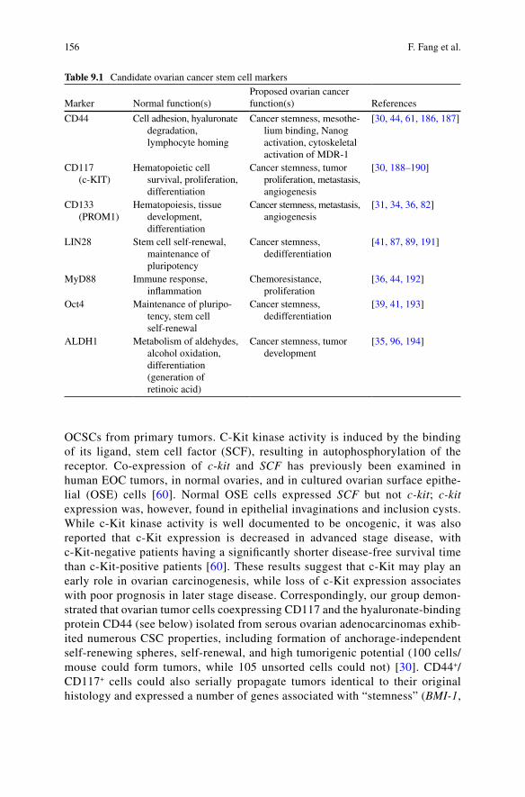
156 F. Fang et al.
OCSCs from primary tumors. C-Kit kinase activity is induced by the binding of its ligand, stem cell factor (SCF), resulting in autophosphorylation of the receptor. Co-expression of c-kit and SCF has previously been examined in human EOC tumors, in normal ovaries, and in cultured ovarian surface epithe-lial (OSE) cells [60]. Normal OSE cells expressed SCF but not c-kit; c-kit expression was, however, found in epithelial invaginations and inclusion cysts. While c-Kit kinase activity is well documented to be oncogenic, it was also reported that c-Kit expression is decreased in advanced stage disease, with c-Kit-negative patients having a significantly shorter disease-free survival time than c-Kit-positive patients [60]. These results suggest that c-Kit may play an early role in ovarian carcinogenesis, while loss of c-Kit expression associates with poor prognosis in later stage disease. Correspondingly, our group demon-strated that ovarian tumor cells coexpressing CD117 and the hyaluronate-binding protein CD44 (see below) isolated from serous ovarian adenocarcinomas exhib-ited numerous CSC properties, including formation of anchorage-independent self-renewing spheres, self-renewal, and high tumorigenic potential (100 cells/mouse could form tumors, while 105 unsorted cells could not) [30]. CD44+/CD117+ cells could also serially propagate tumors identical to their original histology and expressed a number of genes associated with “stemness” (BMI-1,
Table 9.1 Candidate ovarian cancer stem cell markers
Marker Normal function(s)Proposed ovarian cancer function(s) References
CD44 Cell adhesion, hyaluronate degradation, lymphocyte homing
Cancer stemness, mesothe-lium binding, Nanog activation, cytoskeletal activation of MDR-1
[30, 44, 61, 186, 187]
CD117 (c-KIT)
Hematopoietic cell survival, proliferation, differentiation
Cancer stemness, tumor proliferation, metastasis, angiogenesis
[30, 188–190]
CD133 (PROM1)
Hematopoiesis, tissue development, differentiation
Cancer stemness, metastasis, angiogenesis
[31, 34, 36, 82]
LIN28 Stem cell self-renewal, maintenance of pluripotency
Cancer stemness, dedifferentiation
[41, 87, 89, 191]
MyD88 Immune response, inflammation
Chemoresistance, proliferation
[36, 44, 192]
Oct4 Maintenance of pluripo-tency, stem cell self-renewal
Cancer stemness, dedifferentiation
[39, 41, 193]
ALDH1 Metabolism of aldehydes, alcohol oxidation, differentiation (generation of retinoic acid)
Cancer stemness, tumor development
[35, 96, 194]
1579 Cancer Stem Cells in Ovarian Cancer
SCF, OCT4, NES, NOTCH1, and NANOG) [30]. Those highly tumorigenic cells were also found to express the membrane efflux pump ABCG2, and demonstrated enhanced resistance to the conventional EOC chemotherapies cisplatin or paclitaxel [30].
9.2.3.2 CD44
The hyaluron receptor CD44, a single chain transmembrane glycoprotein widely expressed in both epithelial and nonepithelial tissues [61], plays a role in numer-ous physiological processes including cell–cell and cell–matrix interactions, cell adhesion, and cell migration [62]. CD44, however, has also been implicated in tumor progression, and its interaction with hyaluronan has been shown to play a role in the onset of drug resistance [63–66], and in EOC progression CD44 expres-sion has been correlated with the multidrug resistance proteins MDR1, MRP2, and the invasion mediator uPA [67]. Moreover, EGF family member activation of oncogenic ErbB2-ERK signaling was found to result in hyaluronan synthase phos-phorylation/activation, with the subsequent upregulation of hyaluronan leading to CD44-mediated ovarian cancer progression [68]. The hyaluronan-CD44 interac-tion has also been demonstrated to facilitate cytoskeletal protein binding to the multidrug resistance protein MDR1 to augment drug efflux [69], and hyaluronan-based prodrugs against CD44 have demonstrated antitumor and antimetastasis activity in vivo [70]. Paradoxically, however, CD44 expression has been found to correlate with well-differentiated, early-stage ovarian tumors and greater survival [71]; however, the specific CD44 isoform that was analyzed may contribute to these differences.
In coexpression studies with CD133 (see below), it was found that while CD133high and CD133−/low cells expressed similar CD44 levels, in the CD133high cells, CD44 demonstrated physical interactions with transporters, receptor tyrosine kinases, and the metastasis/invasion-associated plasma membrane protein emmprin (CD147) [72].
9.2.3.3 CD133
In addition to CD44 and CD117, CD133 (Prominin-1, formerly known as AC133), a plasma membrane glycoprotein and marker of neural stem cells [73], has now been described as a common CSC marker for numerous malignancies, including brain [74, 75], pancreas [56], liver [76], melanoma [77], prostate [78, 79], and colon [80, 81]. In ovarian cancer, it was also reported that CD133-positive EOC cell lines, primary tumor cells, and patient ascites-derived cells were more platinum-resistant than CD133-negative cells, in addition to forming more aggressive tumor grafts at lower inoculums [34]. In the latter study, CD133+ cells sorted from primary ovarian carcinomas were more clonogenic in culture and more proliferative than
158 F. Fang et al.
CD133− cells. Moreover, CD133+ cells were found significantly more frequently in ovarian carcinomas than in normal ovaries, benign ovarian tumors, or omental lesions [34], while also showing increased tumorigenic capacity and recapitu-lation of their original tumor phenotype [34]. CD133+ cells were also shown to differentiate into CD133− cells, with CD133 silenced in progeny cells by DNA methylation [31]. In another study, however, while CD133+ cells were found to interact with OCSCs, these cells were not tumorigenic, although they could facilitate tumor development by augmenting vasculogenesis (thus suggesting the CD133+ cells to be OCSC-associated endothelial stem cells) [82]. Additionally, CD133 expres-sion was found nonpredictive of patient response to treatment, time to progression (TTP), clinical prognosis, or overall survival (OS) [83]. Consequently, the precise role of this glycoprotein in ovarian tumors remains somewhat uncertain.
9.2.3.4 MyD88
Myeloid differentiation factor 88 (MyD88) is a critical component of the toll-like receptor pathway (associated with immune response) and an activator of the proto-oncogenic NF-kB signaling pathway [44]. In ovarian cancer, Alvero et al. isolated CD44+/MyD88+ cells from solid ovarian tumors and ascites that demonstrated constitutive NF-kB activity, cytokine and chemokine production, a high capacity for DNA repair, and resistance to conventional chemotherapies [44]. Gene array analysis demonstrated that MyD88 was exclusively expressed in CD44+ EOC cells, while 10% of all genes examined were differentially expressed between CD44+ and CD44− cells, including genes related to apoptosis, signal transduction, and cell differentiation [44].
9.2.3.5 CD24
CD24 is a cell surface molecule upregulated in a large number of human malig-nancies, and in EOC, its expression has been correlated with poor prognosis [84]. CD24 has also been used as a marker to identify breast and pancreatic CSCs [85, 86], and CD24+ ovarian tumor cells were reported to possess various stem-like characteristics, including quiescence, chemoresistance, self-renewal, and differen-tiation. In addition, low (5 × 10³) numbers of CD24+ cells were capable of xenograft formation in nude mice, while equal numbers of CD24− cells remained nontumori-genic. CD24+ cells were also found to overexpress the stem cell markers NES, CTNNBIP1, BMI-1, OCT4, NOTCH1, and NOTCH4, while underexpressing CDH1, as compared to CD24− cells [37].
9.2.3.6 LIN28 and OCT4
The microRNA-binding protein Lin28 and the pluripotency-associated transcrip-tion factor Oct4 have also been linked to the initiation and/or progression of EOC.
1599 Cancer Stem Cells in Ovarian Cancer
In previous studies of “induced pluripotency,” LIN28 and OCT4 coexpression with two other transcription factors was capable of eliciting “dedifferentiating” epidermal cells to an embryonic-like, pluripotent phenotype [87]. Lin28 is also known to inhibit processing of the tumor-suppressing microRNA let-7, whose downregula-tion is associated with poor prognosis and advanced stage of EOC [88]. Analogously, upregulation of (the let-7 antagonist) LIN28 associated with higher-grade EOC and was also found capable of transforming fibroblasts and other normal cells [89]. Peng and colleagues also reported that a subpopulation of EOC cells and patient tumors coexpresses LIN28 and OCT4, and that in tumors their coexpression cor-related with advanced tumor grade, thus suggesting that LIN28 and OCT4 co-expression may be a genotype of OCSCs [41].
9.2.3.7 ALDH1
Aldehyde dehydrogenase-1 (ALDH1), a detoxifying enzyme responsible for the oxidation of intracellular aldehydes, has been reported to play a role in the early differentiation of stem cells through its oxidation of retinol to retinoic acid [90–95]. Additionally, several groups have now shown ALDH1 expression to be a prognostic marker for a number of epithelial cancers [96–102]. Deng et al. [35] analyzed ALDH1 expression in 24 types of normal tissues and a large collection of epithelial tumor specimens, in addition to a transgenic EOC mouse model and murine EOC cell lines. ALDH1 expression, while minimal in ovarian cancer cells and tumors, was significantly associated with poor clinical outcomes in serous EOC patients [35]. This finding, however, was contradicted by another study of 266 serous and 176 nonserous EOC patients, in which ALDH1 expression correlated with favorable prognosis [99]. These discrepancies may indicate that the prognostic value of ALDH1 is tumor subtype-specific [99].
9.3 Origin of Ovarian Cancer Stem Cells
As mentioned in Sect. 9.1, a precursor lesion for serous EOC has yet to be iden-tified, and it is unclear whether this disease originates from the ovarian surface epithelium (OSE) or from the epithelium of neighboring reproductive structures [103]. Epithelial cells from the three most common types of EOC tumors (endo-metrioid, mucinous, and serous) are morphologically identical to epithelia of the endometrium, endocervix, and fallopian tubes, respectively [104]. Because no ovarian structures have an epithelial lining similar to any of the aforementioned tissues, it has been hypothesized that EOC tumors may originate from those nono-varian tissues which (unlike the ovary) are embryologically developed from the Müllerian ducts [105]. During embryonic development, the cervix and uterus are formed from the Müllerian ducts fusing, while the fallopian tubes form from an unfused portion of the ducts [106]. Support for this possible origin of EOC is
160 F. Fang et al.
found in two types of benign cysts found on the ovarian surface: inclusion cysts, which are lined by epithelial cells resembling the OSE, and metaplastic cysts, which are lined by cells identical to the epithelia of the nonovarian structures discussed above [107].
Gene expression analyses may further resolve this point-of-origin question. In particular, expression of the development-associated HOX gene family has shown that endometrioid, mucinous, and serous ovarian tumors express the same HOX genes as normal endometrioid, endocervix, and fallopian tube epithelia, respectively [108]. Within the fallopian tube, the structure implicated in the “shedding” of tumor cells onto the ovarian surface is the fimbria (Latin for “fringe”), an entity located at the distal end of the fallopian tube that during ovulation, is hormonally induced to rub the surface of the ovary in a sweeping motion, allowing extraction of the ovum into the tube. One compelling argument for a fimbrial origin for EOC is that overexpression of mutant p53 (vaguely des-ignated as a “p53 signature”), a defining characteristic of fimbrial serous tubal intraepithelial carcinomas (STICs), is typically also found in high-grade (but not low-grade) serous EOCs [18]. Microarray gene expression studies comparing distal STICs, fallopian tube epithelia (FTE), and serous EOCs demonstrated indistinguishable expression profiles in BRCA-mutation-carrying women, impli-cating the FTE as a precursor to serous EOC [109]. Pax8, another developmen-tally associated transcription factor, is also expressed in mucinous, clear cell, serous EOCs, and nonciliated cells present in ovarian inclusion cysts; however, PAX8 is not expressed in normal OSE cells [110].
In contrast to the hypothesis of the FTE as a precursor for serous EOC, the current argument for the OSE as an origin of serous EOC is that the Müllerian-like features of those malignant cells develop within inclusion cysts (stroma-entrapped OSE) following exposure to high levels of female hormones in that microenvi-ronment [111]. Consequently, however, that hypothesis suggests that serous EOC cells are more differentiated than their cell(s) of origin, and this type of tumor progression (i.e., increased differentiation) runs counter to the known characteris-tics of all nonovarian carcinomas [112].
It has also been suggested that various CSC attributes can be conferred to normal/precancerous cells during drug treatment. A recent study by Sharma et. al. demonstrated that treating lung cancer cells with normal first-line anticancer drugs resulted in the transient expression of stem cell markers; however, these stem-like “drug-tolerant persisters” (DTPs) did not proliferate. However, while DTP cells were relatively quiescent, a transient subpopulation of these, designated “drug-tolerant expanded persister” (DTEP) cells (which did not express the stem cell markers) proliferated normally and possessed significantly greater cisplatin resis-tance than the original tumor cells [113]. Moreover, DTEP cells could be ablated by inhibition of histone deacetylases or a histone demethylase [113], demonstrating that chromatin-targeting agents might preferentially target cancer stem-like cells. In a related study, our group recently showed that in an EOC model of platinum resistance, drug resistance positively correlates with a linear increase in the total genomic number of hypermethylated gene promoters, while drug sensitivity was
1619 Cancer Stem Cells in Ovarian Cancer
subsequently restored by inhibitors of DNA methylation [114]. These results provide additional support for the association of epigenome alterations with chemo-therapy resistance and/or cancer stemness.
9.4 Therapeutic Approaches for Eradicating Ovarian Cancer Stem Cells
As a consequence of the CSC theory, it is hypothesized that tumors that initially undergo complete remission, but subsequently relapse to a completely refractory state (e.g., ovarian cancer), are more likely to possess CSCs than tumors that do not respond well to primary therapy [115]. In this model, chemotherapeutics preferen-tially target the rapidly proliferating cells (presumably, CSC progeny cells) that comprise the bulk of the tumor, causing tumor regression, but fail to eradicate drug-resistant CSCs. Consequently, therapies are needed that target both the small percentage of tumorigenic progenitors as well as the more rapidly proliferating nontumorigenic progeny that comprise that bulk of the tumor [116]. Such potential CSC-targeted therapies can be categorized into four general classes: (1) elimination, (2) differentiation, (3) stem cell niche modification, and (4) epigenetic.
9.4.1 Elimination Therapies Targeting Cancer Stemness-Related Pathways
9.4.1.1 PI3K/Akt Signaling
One cascade upregulated in numerous solid cancers, possibly contributing to tumor initiation (and thus an attractive target for cancer therapeutics), is the phos-phatidylinositol kinase-3 (PI3K)/Akt mitogenic signaling pathway [117]. Two potential Akt signaling inhibitors, daidzein-daunomycin and N-t-Boc-daidzein, were derived to from the promising cancer chemopreventative phytoestrogen daidzein, which is relatively unstable [118]. Daidzein-daunomycin was reported to improve therapeutic response in an animal EOC model [119], while N-t-Boc-daidzein could decrease the number of OCSCs isolated from patient ascites. N-t-Boc-daidzein was also found to elicit apoptosis of ascites-derived mature EOC primary cell lines, in a dose- and time-dependent manner, due in part to the degradation of Akt [120]. In other cancers, Akt inhibitors were found effective in targeting CD133+ hepatocellular, CD133+ glioblastoma, and CD133+/CD44+ prostate tumor-initiating cells [121–123]. In an impressive recent study of breast cancer, an antagonist of the IL8 receptor CXCR1 was demonstrated to reduce the number of ALDH+ breast CSCs, followed by massive apoptosis of the remaining bulk of the tumor; and that antagonist was demonstrated to inhibit focal adhesion
162 F. Fang et al.
kinase (FAK) signaling through Akt [124]. Together, these reports strongly implicate the PI3K/Akt cascade in CSC maintenance and self-renewal.
9.4.1.2 Sonic Hedgehog Pathway
Another signal cascade implicated in playing a role in cancer stemness is the Sonic hedgehog (SHH) embryogenesis-associated pathway. SHH signaling, initiated by SHH binding to its and its receptor, Patched-1, is a crucial mediator of cell fate during early mammalian development [125]. However, SHH deregulation has been hypothesized to contribute to CSC self-renewal, and therefore represents an attractive target for cancer therapy [126, 127]. In support of such an approach, it was demonstrated that cyclopamine, a naturally occurring alkaloid also found to be specific SHH pathway inhibitor, strongly inhibited the proliferation and clonogenic growth of ovarian tumor cells in vitro, while also arresting ovarian tumor growth, in vivo [128]. However, another study demonstrated minimal SHH signaling in ovarian cancer [129], leaving the specific role of this pathway in EOC largely unresolved.
9.4.1.3 Notch
The Notch pathway is a cell–cell contact signaling cascade intimately involved in normal development and tissue renewal [130]. Signal transduction occurs when a surface Notch ligand on one cell activates its receptor on a contiguous cell, resulting in cleavage of the Notch intracellular domain (NICD) [130]. The NICD then relo-cates from the cytoplasm to the nucleus, resulting in gene transactivation via its interaction with the transcription factor CBF (C element-binding factor) [130]. However, Notch dysregulation has also been implicated in maintenance of the CSC phenotype, and a number of specific Notch inhibitors are currently in various phases of development [131]. In ovarian cancer, various Notch pathway members are over-expressed in tumors, but not in adenomas [132]. Correspondingly, significant Notch signaling has also been observed in EOC cell lines and 76% of EOC patient tumors [133]. Moreover, Notch1 was found overexpressed in candidate OCSCs, as compared to the bulk population of tumor cells or OCSCs placed under differenti-ating conditions [30], while Notch3 amplification in EOC tumors was found to mediate their proliferation and survival [134]. Together, these and other reports strongly implicate Notch in ovarian tumorigenesis and OCSC maintenance, making this an attractive therapeutic target.
9.4.1.4 Wingless (Wnt) Signaling
The Wnt pathway is essential for embryonic morphogenesis and body axis speci-fication and tissue homeostasis, due to its regulation of self-renewal of normal
1639 Cancer Stem Cells in Ovarian Cancer
stem cells [135]. Signal propagation occurs upon the binding of Wnt ligand to its receptor, Frizzled, resulting in a cascade that leads to nuclear translocation of beta-catenin, which upon binding to its transactivational cofactor, TCF, induces a number of protooncogenes (including MYC) [135]. Consequently, similar to other embryonic signaling pathways, Wnt dysregulation is also associated with carcino-genesis and tumor progression [135, 136]. In one EOC study, Rask et al. demon-strated increased expression of components of the Wnt pathway in malignant EOC tumors, as compared to normal ovarian tissues [137]. Towards the targeting of Wnt signaling as an effective cancer therapy, two small molecules (ZTM000990 and PKF118-310), were identified in a high-throughput screen (based on the structure of the beta-catenin/TCF complex) to target the canonical Wnt signaling cascade [138]. Additionally, anti-Wnt1 and anti-Wnt2 monoclonal antibodies were found to be potent inducers of apoptosis in melanoma, mesothelioma, and melanoma cells [139]. With further pharmacologic optimization, these small molecules or antibodies targeting the Wnt signaling pathway could represent effective ovarian cancer therapeutics.
9.4.2 Differentiation Therapies Targeting Cancer Stem Cells
Another potential approach to CSC targeting is the use of differentiating agents, which presumably would alter the embryo-like CSC phenotype toward that of its normal, mature tissue; it is hypothesized that disruption of the abovementioned self-renewal pathways might serve this purpose. Various differentiating agents have now been examined with varying degrees of success, including dietary polyphenols and phytoestrogens, and vitamin D3. However, the agent best studied (and to date, the most successful) differentiating agent is all-trans retinoic acid (ATRA), which has demonstrated impressive effectiveness against acute promyelocytic leukemia, head/neck squamous carcinomas, thyroid cancer, and in combination with inter-feron-g, neuroblastoma cells [140–143]. In two serous EOC cell lines, ATRA was also demonstrated to alter cell morphology relative to that of differentiated epithe-lial cells, in addition to strongly inhibiting cell proliferation [144]. With regard to CSC differentiation, our group showed that OCSCs are more resistant to cisplatin and paclitaxel, but could be resensitized to both agents under differentiating condi-tions [30]. In a separate study, we also demonstrated that histone deacetylase inhib-itors (a type of epigenetic therapy) induced morphological changes and epithelial differentiation markers in a platinum-resistant EOC cell line [145]. In a series of striking studies, differentiation of highly aggressive melanoma cells to normal melanocytes has also been demonstrated. Following plating the melanoma cells atop an embryonic stem cell-derived extracellular matrix, the reciprocal placement of melanocytes onto a melanoma-derived microenvironment resulted in restoration of the aggressive malignant phenotype [146, 147]. As normal differentiation is governed by epigenomic changes (see below), it is strongly believed that CSCs also possess a type of “epigenetic plasticity” capable of altering their degree of

164 F. Fang et al.
differentiation (and thus their malignant phenotype) [148, 149]. Together, these results support the idea that differentiation therapy, possibly facilitated by epigenetic therapies of OCSCs, has strong potential as an effective therapeutic approach.
9.4.3 Destruction or Alteration of the Cancer Stem Cell Niche
In vivo, stem cell self-renewal and differentiation are tightly controlled by a complex niche that physically harbors those cells in an anatomically well-defined location within a tissue (reviewed in [150]), and there is increasing evidence that the microen-vironment regulates tissue specificity and contributes significantly to tumorigenesis (reviewed in [151]). The extracellular environment provides the structural platform necessary for cell growth and intercellular communication; analogously, various growth factors and chemokines may enhance tumor cell proliferation and invasion [151]. Conversely, the tumor microenviroment may also stimulate production of antiangiogenic proteins and inhibitors of matrix metalloproteases that can obstruct tumorigenesis [152]. In brain cancer, it was found that CD133+/Nestin+ CSCs reside in a paravascular niche, and that inhibition of angiogenesis, via EGF signaling disruption or inhibition of vascular endothelial growth factor (VEGF), eradicated those self-renewing cells [153, 154]. Moreover, as noted previously, a number of studies have now demonstrated differentiation-associated “reprogramming” of aggressive melanoma and breast cancer cells into normal epithelial cells by culturing them in an embryonic stem cell–derived microenvironment [146]. Those studies demonstrate that even advanced stage cancers (likely having increased numbers of CSCs) exhibit a phenotypic “plasticity” for differentiation that is governed by epig-enomic changes. In particular, the embryonic microenvironmental signaling mole-cule responsible for melanoma cell reprogramming was found to be an inhibitor of the embryonic morphogen Nodal, a member of the TGF-beta family [146]. Nodal was later found to effect “vascular mimicry,” formation of tube-like structures capable of perfusing the tumor [155, 156] (thus possibly similar to brain CSC perivascular niche). In ovarian cancer specifically, components of the secondary Müllerian system (paraovarian/paratubal cysts, rete ovarii, endosalpingiosis, endo-metriosis, and endomucinosis) may similarly provide a source of cells and/or signaling molecules that contribute to the different histologic types of ovarian malignancies [103]. Analogously, a recent study suggested Müllerian inhibiting substance (MIS) as a possible adjuvant to conventional ovarian cancer chemotherapeutics that targets putative OCSCs, as MIS treatment inhibited proliferation of both SP and non-SP cells, while conventional chemotherapies primarily arrested non-SP cells [42].
9.4.4 Epigenetic Therapies
Epigenetic alterations have been demonstrated to govern gene expression both in embryonic and tissue stem cells, and thus likely play an important role in the
1659 Cancer Stem Cells in Ovarian Cancer
tumorigenic potential (and differentiation prevention) in OCSCs. These alterations include methylation of deoxycytosine, as well as numerous modifications of histones that regulate distinct gene expression patterns that allow for specific cell or organotypic phenotypes [157]. It is also well established that epigenetics contrib-utes significantly to ovarian tumorigenesis [158], likely in association with its intri-cate role in differentiation.
It is hypothesized that ovarian tumor chemotherapy resistance results largely from the repression of tumor suppressor genes (specifically, chemotherapy-response genes) by DNA methylation [159–161]. In contrast to DNA mutations and dele-tions, however, aberrant gene-repressive epigenetic modifications are potentially reversible, by epigenetic therapies, including inhibitors of DNA methyltransferases (DNMTs) or various histone-modifying enzymes (reviewed in [161]). Although epigenetic monotherapies have shown little activity against solid tumors [162–164] including ovarian cancer [161, 165], preclinical studies of DNMT inhibitors by our group [114, 166] and others [167–170] have demonstrated potent resensitization of drug-resistant EOC cells and xenograft tumors to conventional chemotherapies, possibly by differentiation or apoptosis of CSCs [159, 164]. Interestingly, it was also demonstrated that DNMT inhibitor treatment induced silenced SFRP5 (encoding an endogenous Wnt pathway inhibitor), similarly chemosensitizing drug-resistant EOC cells and xenografts [171].
Based on the abovementioned studies demonstrating chemotherapy sensitization of resistant EOC cells and tumors, several such combinatorial regimens are now being examined in cancer clinical trials [160, 161]. Our group recently completed a Phase I trial (NCT00477386, Study ID 0704-07, www.clinicaltrials.gov) using a DNMT inhibitor, decitabine (Dacogen; Eisai, Inc., Tokyo, Japan) combined with the standard chemotherapeutic, carboplatin, hypothesizing that low-dose decitabine may derepress silenced tumor suppressors to chemosensitize platinum-resistant EOCs [172]. In that study, a low dose (10 mg/m2) of decitabine was administered for 5 consecutive days, followed by carboplatin (AUC5) administered 3 days later, with each treatment cycle lasting 28 days. That regimen demonstrated bioactivity in vivo, as assessed by decreased methylation of DNA repetitive elements (in patients’ peripheral blood cells) and of specific genes (in patients’ sera). Out of 10 patients, we observed 1 complete response, while 4 patients experienced stable disease for at least 6 months; although interestingly the complete response did not occur prior to eight treatment cycles [172]. While we cannot draw conclusions based on this small patient sample size, it is interesting that breast CSCs induce tumorigenesis only after a latency of 6 months, and it is conceivable that drug-associated hypomethyla-tion of CSCs would similarly require an extended period based on the slow division of those cells and their delayed response in other carcinomas [29, 173].
Since histone deacetylation is another transcriptional silencing mechanism in ovarian cancer, HDAC inhibitors (HDACIs) can also relieve epigenetic gene repression, and these agents also exert anticancer effects by inhibiting the deacety-lation of nonhistone proteins [174]. Similar to DNA methylation inhibitors, HDACIs are most promising in combination with conventional agents, and studies by our group [145] and others [175–180] have demonstrated chemosensitizing effects on
166 F. Fang et al.
drug-resistant ovarian cancer cells and tumors. HDACIs have also been effectively combined with a differentiating [181] or a “death receptor” ligand [182] in ovarian cancer cell studies, similar demonstrating additive or synergistic effects. It is also possible that HDACIs might also have direct effects on CSCs. In one study, a subpopulation of rapidly proliferating, drug-resistant lung cancer cells could be eliminated by HDACI treatment [113], while an HDACI/imatinib combination was demonstrated to target chronic myelogenous leukemia stem cells [183]. In other studies, an HDACI was found to block self-renewal and aggregation of breast cancer spheroids [184], while another HDACI suppressed expression of the stemness gene NANOG in embryonic carcinoma cells, resulting in loss of a stem cell “gene signature” [185]. Thus this class of epigenetic agents holds promise for the treatment of drug-resistant ovarian cancer, possibly via their effects on ovarian CSCs.
9.5 Conclusions
Based on several studies to date, it is highly likely that ovarian tumors, similar to numerous other solid cancers, possess a hierarchy of cell types, with tumor initia-tion, progression, and chemoresistance driven by a distinct subpopulation of malignant progenitor cells. A further understanding of these “cancer stem cells” holds promise for the design of strategies toward their eradication, possibly by epigenetically inducing their differentiation or elimination via blockade of specific pathways essential for maintenance of their tumor propagating and undif-ferentiated phenotype.
References
1. American Cancer Society. Cancer Facts & Figures 2009, Atlanta, GA: Amercian Cancer Society; 2009. Available at www.cancer.org/downloads/STT/2008CAFFfinalsecured.pdf.
2. Jemal A, Siegel R, Ward E, Hao Y, Xu J, Thun MJ (2009) Cancer statistics, 2009. CA Cancer J Clin 59 (4):225–249.
3. Bell DA (2005) Origins and molecular pathology of ovarian cancer. Mod Pathol 18 Suppl 2:S19–32.
4. Bankhead CR, Collins C, Stokes-Lampard H, Rose P, Wilson S, Clements A, Mant D, Kehoe ST, Austoker J (2008) Identifying symptoms of ovarian cancer: a qualitative and quantitative study. BJOG 115 (8):1008–1014.
5. Twombly R (2007) Cancer killer may be “silent” no more. J Natl Cancer Inst 99 (18):1359–1361. 6. Goff BA, Mandel L, Muntz HG, Melancon CH (2000) Ovarian carcinoma diagnosis. Cancer
89 (10):2068–2075. 7. Agarwal R, Kaye SB (2003) Ovarian cancer: strategies for overcoming resistance to chemo-
therapy. Nat Rev Cancer 3 (7):502–516. 8. Armstrong DK, Bundy B, Wenzel L, Huang HQ, Baergen R, Lele S, Copeland LJ, Walker JL,
Burger RA (2006) Intraperitoneal cisplatin and paclitaxel in ovarian cancer. N Engl J Med 354 (1):34–43.
1679 Cancer Stem Cells in Ovarian Cancer
9. Clarke-Pearson DL (2009) Clinical practice. Screening for ovarian cancer. N Engl J Med 361 (2):170–177.
10. Ozols RF (2005) Treatment goals in ovarian cancer. Int J Gynecol Cancer 15 Suppl 1:3–11. 11. Choi JH, Wong AS, Huang HF, Leung PC (2007) Gonadotropins and ovarian cancer. Endocr
Rev 28 (4):440–461. 12. Murdoch WJ, McDonnel AC (2002) Roles of the ovarian surface epithelium in ovulation and
carcinogenesis. Reproduction 123 (6):743–750. 13. Ness RB, Cottreau C (1999) Possible role of ovarian epithelial inflammation in ovarian cancer.
J Natl Cancer Inst 91 (17):1459–1467. 14. Fleming JS, Beaugie CR, Haviv I, Chenevix-Trench G, Tan OL (2006) Incessant ovulation,
inflammation and epithelial ovarian carcinogenesis: revisiting old hypotheses. Mol Cell Endocrinol 247 (1-2):4–21.
15. Holschneider CH, Berek JS (2000) Ovarian cancer: epidemiology, biology, and prognostic factors. Semin Surg Oncol 19 (1):3–10.
16. Auersperg N, Wong AS, Choi KC, Kang SK, Leung PC (2001) Ovarian surface epithelium: biology, endocrinology, and pathology. Endocr Rev 22 (2):255–288.
17. Levanon K, Crum C, Drapkin R (2008) New insights into the pathogenesis of serous ovarian cancer and its clinical impact. J Clin Oncol 26 (32):5284–5293.
18. Lee Y, Miron A, Drapkin R, Nucci MR, Medeiros F, Saleemuddin A, Garber J, Birch C, Mou H, Gordon RW, Cramer DW, McKeon FD, Crum CP (2007) A candidate precursor to serous carcinoma that originates in the distal fallopian tube. J Pathol 211 (1):26–35.
19. Tuma RS (2010) Origin of ovarian cancer may have implications for screening. J Natl Cancer Inst 102 (1):11–13.
20. Kurman RJ, Shih Ie M (2010) The origin and pathogenesis of epithelial ovarian cancer: a proposed unifying theory. Am J Surg Pathol 34 (3):433–443.
21. Landen CN, Jr., Birrer MJ, Sood AK (2008) Early events in the pathogenesis of epithelial ovarian cancer. J Clin Oncol 26 (6):995–1005.
22. Tan DS, Agarwal R, Kaye SB (2006) Mechanisms of transcoelomic metastasis in ovarian cancer. Lancet Oncol 7 (11):925–934.
23. Amadori D, Sansoni E, Amadori A (1997) Ovarian cancer: natural history and metastatic pattern. Front Biosci 2:g8–10.
24. Hacker NF, Valmadre S, Robertson G (2008) Management of retroperitoneal lymph nodes in advanced ovarian cancer. Int J Gynecol Cancer 18 Suppl 1:7–10.
25. Burleson KM, Casey RC, Skubitz KM, Pambuccian SE, Oegema TR, Jr., Skubitz AP (2004) Ovarian carcinoma ascites spheroids adhere to extracellular matrix components and mesothe-lial cell monolayers. Gynecol Oncol 93 (1):170–181.
26. Burleson KM, Boente MP, Pambuccian SE, Skubitz AP (2006) Disaggregation and invasion of ovarian carcinoma ascites spheroids. J Transl Med 4:6.
27. Bonnet D, Dick JE (1997) Human acute myeloid leukemia is organized as a hierarchy that originates from a primitive hematopoietic cell. Nat Med 3 (7):730–737.
28. Polyak K, Hahn WC (2006) Roots and stems: stem cells in cancer. Nat Med 12 (3):296–300. 29. Dalerba P, Cho RW, Clarke MF (2007) Cancer stem cells: models and concepts. Annu Rev
Med 58:267–284. 30. Zhang S, Balch C, Chan MW, Lai HC, Matei D, Schilder JM, Yan PS, Huang TH, Nephew KP
(2008) Identification and characterization of ovarian cancer-initiating cells from primary human tumors. Cancer Res 68 (11):4311–4320.
31. Baba T, Convery PA, Matsumura N, Whitaker RS, Kondoh E, Perry T, Huang Z, Bentley RC, Mori S, Fujii S, Marks JR, Berchuck A, Murphy SK (2009) Epigenetic regulation of CD133 and tumorigenicity of CD133+ ovarian cancer cells. Oncogene 28 (2):209–218.
32. Bapat SA (2010) Human Ovarian Cancer Stem Cells. Reproduction 140:33–41. 33. Bapat SA, Mali AM, Koppikar CB, Kurrey NK (2005) Stem and progenitor-like cells
contribute to the aggressive behavior of human epithelial ovarian cancer. Cancer Res 65 (8): 3025–3029.
168 F. Fang et al.
34. Curley MD, Therrien VA, Cummings CL, Sergent PA, Koulouris CR, Friel AM, Roberts DJ, Seiden MV, Scadden DT, Rueda BR, Foster R (2009) CD133 expression defines a tumor initiating cell population in primary human ovarian cancer. Stem Cells 27 (12):2875–2883.
35. Deng S, Yang X, Lassus H, Liang S, Kaur S, Ye Q, Li C, Wang LP, Roby KF, Orsulic S, Connolly DC, Zhang Y, Montone K, Butzow R, Coukos G, Zhang L (2010) Distinct expres-sion levels and patterns of stem cell marker, aldehyde dehydrogenase isoform 1 (ALDH1), in human epithelial cancers. PLoS One 5 (4):e10277.
36. Fong MY, Kakar SS (2010) The role of cancer stem cells and the side population in epithelial ovarian cancer. Histol Histopathol 25 (1):113–120.
37. Gao MQ, Choi YP, Kang S, Youn JH, Cho NH (2010) CD24(+) cells from hierarchically organized ovarian cancer are enriched in cancer stem cells. Oncogene 29:2972–2980.
38. Gao Q, Geng L, Kvalheim G, Gaudernack G, Suo Z (2009) Identification of cancer stem-like side population cells in ovarian cancer cell line OVCAR-3. Ultrastruct Pathol 33 (4):175–181.
39. Hu L, McArthur C, Jaffe RB (2010) Ovarian cancer stem-like side-population cells are tumourigenic and chemoresistant. Br J Cancer 102 (8):1276–1283.
40. Moserle L, Indraccolo S, Ghisi M, Frasson C, Fortunato E, Canevari S, Miotti S, Tosello V, Zamarchi R, Corradin A, Minuzzo S, Rossi E, Basso G, Amadori A (2008) The side population of ovarian cancer cells is a primary target of IFN-alpha antitumor effects. Cancer Res 68 (14): 5658–5668.
41. Peng S, Maihle NJ, Huang Y (2010) Pluripotency factors Lin28 and Oct4 identify a sub-population of stem cell-like cells in ovarian cancer. Oncogene 29 (14):2153–2159.
42. Szotek PP, Pieretti-Vanmarcke R, Masiakos PT, Dinulescu DM, Connolly D, Foster R, Dombkowski D, Preffer F, Maclaughlin DT, Donahoe PK (2006) Ovarian cancer side popula-tion defines cells with stem cell-like characteristics and Mullerian Inhibiting Substance respon-siveness. Proc Natl Acad Sci USA 103 (30):11154–11159.
43. Wani AA, Sharma N, Shouche YS, Bapat SA (2006) Nuclear-mitochondrial genomic profiling reveals a pattern of evolution in epithelial ovarian tumor stem cells. Oncogene 25 (47): 6336–6344.
44. Alvero AB, Chen R, Fu HH, Montagna M, Schwartz PE, Rutherford T, Silasi DA, Steffensen KD, Waldstrom M, Visintin I, Mor G (2009) Molecular phenotyping of human ovarian cancer stem cells unravels the mechanisms for repair and chemoresistance. Cell Cycle 8 (1):158–166.
45. Lobo NA, Shimono Y, Qian D, Clarke MF (2007) The biology of cancer stem cells. Annu Rev Cell Dev Biol 23:675–699.
46. Visvader JE, Lindeman GJ (2008) Cancer stem cells in solid tumours: accumulating evidence and unresolved questions. Nat Rev Cancer 8 (10):755–768.
47. Kim M, Turnquist H, Jackson J, Sgagias M, Yan Y, Gong M, Dean M, Sharp JG, Cowan K (2002) The multidrug resistance transporter ABCG2 (breast cancer resistance protein 1) effluxes Hoechst 33342 and is overexpressed in hematopoietic stem cells. Clin Cancer Res 8 (1): 22–28.
48. Goodell MA, Brose K, Paradis G, Conner AS, Mulligan RC (1996) Isolation and functional properties of murine hematopoietic stem cells that are replicating in vivo. J Exp Med 183 (4): 1797–1806.
49. Zhou S, Schuetz JD, Bunting KD, Colapietro AM, Sampath J, Morris JJ, Lagutina I, Grosveld GC, Osawa M, Nakauchi H, Sorrentino BP (2001) The ABC transporter Bcrp1/ABCG2 is expressed in a wide variety of stem cells and is a molecular determinant of the side-population phenotype. Nat Med 7 (9):1028–1034.
50. Hirschmann-Jax C, Foster AE, Wulf GG, Nuchtern JG, Jax TW, Gobel U, Goodell MA, Brenner MK (2004) A distinct “side population” of cells with high drug efflux capacity in human tumor cells. Proc Natl Acad Sci USA 101 (39):14228–14233.
51. Sharom FJ (2008) ABC multidrug transporters: structure, function and role in chemoresis-tance. Pharmacogenomics 9 (1):105–127.
52. Patrawala L, Calhoun T, Schneider-Broussard R, Zhou J, Claypool K, Tang DG (2005) Side population is enriched in tumorigenic, stem-like cancer cells, whereas ABCG2+ and ABCG2- cancer cells are similarly tumorigenic. Cancer Res 65 (14):6207–6219.
1699 Cancer Stem Cells in Ovarian Cancer
53. Doyle LA, Yang W, Abruzzo LV, Krogmann T, Gao Y, Rishi AK, Ross DD (1998) A multidrug resistance transporter from human MCF-7 breast cancer cells. Proc Natl Acad Sci USA 95 (26):15665–15670.
54. Allikmets R, Schriml LM, Hutchinson A, Romano-Spica V, Dean M (1998) A human placenta-specific ATP-binding cassette gene (ABCP) on chromosome 4q22 that is involved in multidrug resistance. Cancer Res 58 (23):5337–5339.
55. Miyake K, Mickley L, Litman T, Zhan Z, Robey R, Cristensen B, Brangi M, Greenberger L, Dean M, Fojo T, Bates SE (1999) Molecular cloning of cDNAs which are highly overex-pressed in mitoxantrone-resistant cells: demonstration of homology to ABC transport genes. Cancer Res 59 (1):8–13.
56. Olempska M, Eisenach PA, Ammerpohl O, Ungefroren H, Fandrich F, Kalthoff H (2007) Detection of tumor stem cell markers in pancreatic carcinoma cell lines. Hepatobiliary Pancreat Dis Int 6 (1):92–97.
57. Lou H, Dean M (2007) Targeted therapy for cancer stem cells: the patched pathway and ABC transporters. Oncogene 26 (9):1357–1360.
58. Kurrey NK, Jalgaonkar SP, Joglekar AV, Ghanate AD, Chaskar PD, Doiphode RY, Bapat SA (2009) Snail and slug mediate radioresistance and chemoresistance by antagonizing p53-mediated apoptosis and acquiring a stem-like phenotype in ovarian cancer cells. Stem Cells 27 (9):2059–2068.
59. Zola H, Swart B, Nicholson I, Aasted B, Bensussan A, Boumsell L, Buckley C, Clark G, Drbal K, Engel P, Hart D, Horejsi V, Isacke C, Macardle P, Malavasi F, Mason D, Olive D, Saalmueller A, Schlossman SF, Schwartz-Albiez R, Simmons P, Tedder TF, Uguccioni M, Warren H (2005) CD molecules 2005: human cell differentiation molecules. Blood 106 (9): 3123–3126.
60. Tonary AM, Macdonald EA, Faught W, Senterman MK, Vanderhyden BC (2000) Lack of expression of c-KIT in ovarian cancers is associated with poor prognosis. Int J Cancer 89 (3):242–250.
61. Ponta H, Wainwright D, Herrlich P (1998) The CD44 protein family. Int J Biochem Cell Biol 30 (3):299–305.
62. Lesley J, Hyman R, Kincade PW (1993) CD44 and its interaction with extracellular matrix. Adv Immunol 54:271–335.
63. Fraser JR, Laurent TC, Laurent UB (1997) Hyaluronan: its nature, distribution, functions and turnover. J Intern Med 242 (1):27–33.
64. Ropponen K, Tammi M, Parkkinen J, Eskelinen M, Tammi R, Lipponen P, Agren U, Alhava E, Kosma VM (1998) Tumor cell-associated hyaluronan as an unfavorable prognostic factor in colorectal cancer. Cancer Res 58 (2):342–347.
65. Setala LP, Tammi MI, Tammi RH, Eskelinen MJ, Lipponen PK, Agren UM, Parkkinen J, Alhava EM, Kosma VM (1999) Hyaluronan expression in gastric cancer cells is associated with local and nodal spread and reduced survival rate. Br J Cancer 79 (7-8):1133–1138.
66. Anttila MA, Tammi RH, Tammi MI, Syrjanen KJ, Saarikoski SV, Kosma VM (2000) High levels of stromal hyaluronan predict poor disease outcome in epithelial ovarian cancer. Cancer Res 60 (1):150–155.
67. Chen H, Hao J, Wang L, Li Y (2009) Coexpression of invasive markers (uPA, CD44) and multiple drug-resistance proteins (MDR1, MRP2) is correlated with epithelial ovarian cancer progression. Br J Cancer 101 (3):432–440.
68. Bourguignon LY, Gilad E, Peyrollier K (2007) Heregulin-mediated ErbB2-ERK signaling activates hyaluronan synthases leading to CD44-dependent ovarian tumor cell growth and migration. J Biol Chem 282 (27):19426–19441.
69. Bourguignon LY, Peyrollier K, Gilad E, Brightman A (2007) Hyaluronan-CD44 interaction with neural Wiskott-Aldrich syndrome protein (N-WASP) promotes actin polymerization and ErbB2 activation leading to beta-catenin nuclear translocation, transcriptional up-regulation, and cell migration in ovarian tumor cells. J Biol Chem 282 (2):1265–1280.
70. Auzenne E, Ghosh SC, Khodadadian M, Rivera B, Farquhar D, Price RE, Ravoori M, Kundra V, Freedman RS, Klostergaard J (2007) Hyaluronic acid-paclitaxel: antitumor efficacy against CD44(+) human ovarian carcinoma xenografts. Neoplasia 9 (6):479–486.
170 F. Fang et al.
71. Sillanpaa S, Anttila MA, Voutilainen K, Tammi RH, Tammi MI, Saarikoski SV, Kosma VM (2003) CD44 expression indicates favorable prognosis in epithelial ovarian cancer. Clin Cancer Res 9 (14):5318–5324.
72. Slomiany MG, Dai L, Tolliver LB, Grass GD, Zeng Y, Toole BP (2009) Inhibition of func-tional hyaluronan-CD44 interactions in CD133-positive primary human ovarian carcinoma ells by small hyaluronan oligosaccharides. Clin Cancer Res 15 (24):7593–7601.
73. Uchida N, Buck DW, He D, Reitsma MJ, Masek M, Phan TV, Tsukamoto AS, Gage FH, Weissman IL (2000) Direct isolation of human central nervous system stem cells. Proc Natl Acad Sci USA 97 (26):14720–14725.
74. Singh SK, Hawkins C, Clarke ID, Squire JA, Bayani J, Hide T, Henkelman RM, Cusimano MD, Dirks PB (2004) Identification of human brain tumour initiating cells. Nature 432 (7015):396–401.
75. Liu G, Yuan X, Zeng Z, Tunici P, Ng H, Abdulkadir IR, Lu L, Irvin D, Black KL, Yu JS (2006) Analysis of gene expression and chemoresistance of CD133+ cancer stem cells in glioblas-toma. Mol Cancer 5:67.
76. Yin S, Li J, Hu C, Chen X, Yao M, Yan M, Jiang G, Ge C, Xie H, Wan D, Yang S, Zheng S, Gu J (2007) CD133 positive hepatocellular carcinoma cells possess high capacity for tumori-genicity. Int J Cancer 120 (7):1444–1450.
77. Monzani E, Facchetti F, Galmozzi E, Corsini E, Benetti A, Cavazzin C, Gritti A, Piccinini A, Porro D, Santinami M, Invernici G, Parati E, Alessandri G, La Porta CA (2007) Melanoma contains CD133 and ABCG2 positive cells with enhanced tumourigenic potential. Eur J Cancer 43 (5):935–946.
78. Collins AT, Berry PA, Hyde C, Stower MJ, Maitland NJ (2005) Prospective identification of tumorigenic prostate cancer stem cells. Cancer Res 65 (23):10946–10951.
79. Miki J, Furusato B, Li H, Gu Y, Takahashi H, Egawa S, Sesterhenn IA, McLeod DG, Srivastava S, Rhim JS (2007) Identification of putative stem cell markers, CD133 and CXCR4, in hTERT-immortalized primary nonmalignant and malignant tumor-derived human prostate epithelial cell lines and in prostate cancer specimens. Cancer Res 67 (7): 3153–3161.
80. O’Brien CA, Pollett A, Gallinger S, Dick JE (2007) A human colon cancer cell capable of initiating tumour growth in immunodeficient mice. Nature 445 (7123):106–110.
81. Ricci-Vitiani L, Lombardi DG, Pilozzi E, Biffoni M, Todaro M, Peschle C, De Maria R (2007) Identification and expansion of human colon-cancer-initiating cells. Nature 445 (7123):111–115.
82. Kusumbe AP, Mali AM, Bapat SA (2009) CD133-expressing stem cells associated with ovar-ian metastases establish an endothelial hierarchy and contribute to tumor vasculature. Stem Cells 27 (3):498–508.
83. Ferrandina G, Martinelli E, Petrillo M, Prisco MG, Zannoni G, Sioletic S, Scambia G (2009) CD133 antigen expression in ovarian cancer. BMC Cancer 9:221.
84. Kristiansen G, Denkert C, Schluns K, Dahl E, Pilarsky C, Hauptmann S (2002) CD24 is expressed in ovarian cancer and is a new independent prognostic marker of patient survival. Am J Pathol 161 (4):1215–1221.
85. Al-Hajj M, Wicha MS, Benito-Hernandez A, Morrison SJ, Clarke MF (2003) Prospective identification of tumorigenic breast cancer cells. Proc Natl Acad Sci USA 100 (7): 3983–3988.
86. Li C, Heidt DG, Dalerba P, Burant CF, Zhang L, Adsay V, Wicha M, Clarke MF, Simeone DM (2007) Identification of pancreatic cancer stem cells. Cancer Res 67 (3):1030–1037.
87. Yu J, Vodyanik MA, Smuga-Otto K, Antosiewicz-Bourget J, Frane JL, Tian S, Nie J, Jonsdottir GA, Ruotti V, Stewart R, Slukvin, II, Thomson JA (2007) Induced pluripotent stem cell lines derived from human somatic cells. Science 318 (5858):1917–1920.
88. Shell S, Park SM, Radjabi AR, Schickel R, Kistner EO, Jewell DA, Feig C, Lengyel E, Peter ME (2007) Let-7 expression defines two differentiation stages of cancer. Proc Natl Acad Sci USA 104 (27):11400–11405.
1719 Cancer Stem Cells in Ovarian Cancer
89. Viswanathan SR, Powers JT, Einhorn W, Hoshida Y, Ng TL, Toffanin S, O’Sullivan M, Lu J, Phillips LA, Lockhart VL, Shah SP, Tanwar PS, Mermel CH, Beroukhim R, Azam M, Teixeira J, Meyerson M, Hughes TP, Llovet JM, Radich J, Mullighan CG, Golub TR, Sorensen PH, Daley GQ (2009) Lin28 promotes transformation and is associated with advanced human malignancies. Nat Genet 41 (7):843–848.
90. Kastan MB, Schlaffer E, Russo JE, Colvin OM, Civin CI, Hilton J (1990) Direct demonstra-tion of elevated aldehyde dehydrogenase in human hematopoietic progenitor cells. Blood 75 (10):1947–1950.
91. Storms RW, Trujillo AP, Springer JB, Shah L, Colvin OM, Ludeman SM, Smith C (1999) Isolation of primitive human hematopoietic progenitors on the basis of aldehyde dehydroge-nase activity. Proc Natl Acad Sci USA 96 (16):9118–9123.
92. Hess DA, Wirthlin L, Craft TP, Herrbrich PE, Hohm SA, Lahey R, Eades WC, Creer MH, Nolta JA (2006) Selection based on CD133 and high aldehyde dehydrogenase activity iso-lates long-term reconstituting human hematopoietic stem cells. Blood 107 (5):2162–2169.
93. Cai J, Cheng A, Luo Y, Lu C, Mattson MP, Rao MS, Furukawa K (2004) Membrane proper-ties of rat embryonic multipotent neural stem cells. J Neurochem 88 (1):212–226.
94. Hess DA, Meyerrose TE, Wirthlin L, Craft TP, Herrbrich PE, Creer MH, Nolta JA (2004) Functional characterization of highly purified human hematopoietic repopulating cells isolated according to aldehyde dehydrogenase activity. Blood 104 (6):1648–1655.
95. Armstrong L, Stojkovic M, Dimmick I, Ahmad S, Stojkovic P, Hole N, Lako M (2004) Phenotypic characterization of murine primitive hematopoietic progenitor cells isolated on basis of aldehyde dehydrogenase activity. Stem Cells 22 (7):1142–1151.
96. Ginestier C, Hur MH, Charafe-Jauffret E, Monville F, Dutcher J, Brown M, Jacquemier J, Viens P, Kleer CG, Liu S, Schott A, Hayes D, Birnbaum D, Wicha MS, Dontu G (2007) ALDH1 is a marker of normal and malignant human mammary stem cells and a predictor of poor clinical outcome. Cell Stem Cell 1 (5):555–567.
97. Morimoto K, Kim SJ, Tanei T, Shimazu K, Tanji Y, Taguchi T, Tamaki Y, Terada N, Noguchi S (2009) Stem cell marker aldehyde dehydrogenase 1-positive breast cancers are character-ized by negative estrogen receptor, positive human epidermal growth factor receptor type 2, and high Ki67 expression. Cancer Sci 100 (6):1062–1068.
98. Jiang F, Qiu Q, Khanna A, Todd NW, Deepak J, Xing L, Wang H, Liu Z, Su Y, Stass SA, Katz RL (2009) Aldehyde dehydrogenase 1 is a tumor stem cell-associated marker in lung cancer. Mol Cancer Res 7 (3):330–338.
99. Chang B, Liu G, Xue F, Rosen DG, Xiao L, Wang X, Liu J (2009) ALDH1 expression correlates with favorable prognosis in ovarian cancers. Mod Pathol 22 (6):817–823.
100. Rasheed ZA, Yang J, Wang Q, Kowalski J, Freed I, Murter C, Hong SM, Koorstra JB, Rajeshkumar NV, He X, Goggins M, Iacobuzio-Donahue C, Berman DM, Laheru D, Jimeno A, Hidalgo M, Maitra A, Matsui W (2010) Prognostic significance of tumorigenic cells with mesenchymal features in pancreatic adenocarcinoma. J Natl Cancer Inst 102 (5): 340–351.
101. Su Y, Qiu Q, Zhang X, Jiang Z, Leng Q, Liu Z, Stass SA, Jiang F (2010) Aldehyde dehydro-genase 1 A1-positive cell population is enriched in tumor-initiating cells and associated with progression of bladder cancer. Cancer Epidemiol Biomarkers Prev 19 (2):327–337.
102. Charafe-Jauffret E, Ginestier C, Iovino F, Tarpin C, Diebel M, Esterni B, Houvenaeghel G, Extra JM, Bertucci F, Jacquemier J, Xerri L, Dontu G, Stassi G, Xiao Y, Barsky SH, Birnbaum D, Viens P, Wicha MS (2010) Aldehyde dehydrogenase 1-positive cancer stem cells mediate metastasis and poor clinical outcome in inflammatory breast cancer. Clin Cancer Res 16 (1):45–55.
103. Dubeau L (2008) The cell of origin of ovarian epithelial tumours. Lancet Oncol 9 (12): 1191–1197.
104. Cho KR, Shih Ie M (2009) Ovarian cancer. Annu Rev Pathol 4:287–313. 105. Rodriguez M, Dubeau L (2001) Ovarian tumor development: insights from ovarian embryo-
genesis. Eur J Gynaecol Oncol 22 (3):175–183.
172 F. Fang et al.
106. Behringer RR, Finegold MJ, Cate RL (1994) Mullerian-inhibiting substance function during mammalian sexual development. Cell 79 (3):415–425.
107. Byskov AG (1982) Primordial germ cells and regulation of meiosis. In: Reproduction in mammals. I. Germ cells and fertilization. Austin CR and Short RV, eds. Cambridge University Press, London:16.
108. Cheng W, Liu J, Yoshida H, Rosen D, Naora H (2005) Lineage infidelity of epithelial ovarian cancers is controlled by HOX genes that specify regional identity in the reproductive tract. Nat Med 11 (5):531–537.
109. Tone AA, Begley H, Sharma M, Murphy J, Rosen B, Brown TJ, Shaw PA (2008) Gene expression profiles of luteal phase fallopian tube epithelium from BRCA mutation carriers resemble high-grade serous carcinoma. Clin Cancer Res 14 (13):4067–4078.
110. Bowen NJ, Logani S, Dickerson EB, Kapa LB, Akhtar M, Benigno BB, McDonald JF (2007) Emerging roles for PAX8 in ovarian cancer and endosalpingeal development. Gynecol Oncol 104 (2):331–337.
111. Dubeau L (1999) The cell of origin of ovarian epithelial tumors and the ovarian surface epithelium dogma: does the emperor have no clothes? Gynecol Oncol 72 (3):437–442.
112. Thiery JP, Acloque H, Huang RY, Nieto MA (2009) Epithelial-mesenchymal transitions in development and disease. Cell 139 (5):871–890.
113. Sharma SV, Lee DY, Li B, Quinlan MP, Takahashi F, Maheswaran S, McDermott U, Azizian N, Zou L, Fischbach MA, Wong KK, Brandstetter K, Wittner B, Ramaswamy S, Classon M, Settleman J (2010) A chromatin-mediated reversible drug-tolerant state in cancer cell sub-populations. Cell 141 (1):69–80.
114. Li M, Balch C, Montgomery JS, Jeong M, Chung JH, Yan P, Huang TH, Kim S, Nephew KP (2009) Integrated analysis of DNA methylation and gene expression reveals specific signaling pathways associated with platinum resistance in ovarian cancer. BMC Med Genomics 2:34.
115. Dean M, Fojo T, Bates S (2005) Tumour stem cells and drug resistance. Nat Rev Cancer 5 (4): 275–284.
116. Jordan CT (2009) Cancer stem cells: controversial or just misunderstood? Cell Stem Cell 4 (3): 203–205.
117. Vivanco I, Sawyers CL (2002) The phosphatidylinositol 3-Kinase AKT pathway in human cancer. Nat Rev Cancer 2 (7):489–501.
118. Ungar Y, Oluwatooyin F, Shimoni E (2003) Thermal Stability of Genistein and Daidzein and Its Effect on Their Antioxidant Activity. J Agric Food Chem 51 (15):4394–4399.
119. Somjen D, Katzburg S, Nevo N, Gayer B, Hodge RP, Renevey MD, Kalchenko V, Meshorer A, Stern N, Kohen F (2008) A daidzein-daunomycin conjugate improves the therapeutic response in an animal model of ovarian carcinoma. J Steroid Biochem Mol Biol 110 (1-2):144–149.
120. Green JM, Alvero AB, Kohen F, Mor G (2009) 7-(O)-Carboxymethyl daidzein conjugated to N-t-Boc-hexylenediamine: a novel compound capable of inducing cell death in epithelial ovarian cancer stem cells. Cancer Biol Ther 8 (18):1747–1753.
121. Ma S, Lee TK, Zheng BJ, Chan KW, Guan XY (2008) CD133+ HCC cancer stem cells confer chemoresistance by preferential expression of the Akt/PKB survival pathway. Oncogene 27 (12):1749–1758.
122. Eyler CE, Foo WC, LaFiura KM, McLendon RE, Hjelmeland AB, Rich JN (2008) Brain cancer stem cells display preferential sensitivity to Akt inhibition. Stem Cells 26 (12):3027–3036.
123. Dubrovska A, Kim S, Salamone RJ, Walker JR, Maira SM, Garcia-Echeverria C, Schultz PG, Reddy VA (2009) The role of PTEN/Akt/PI3K signaling in the maintenance and viability of prostate cancer stem-like cell populations. Proc Natl Acad Sci USA 106 (1):268–273.
124. Ginestier C, Liu S, Diebel ME, Korkaya H, Luo M, Brown M, Wicinski J, Cabaud O, Charafe-Jauffret E, Birnbaum D, Guan JL, Dontu G, Wicha MS (2010) CXCR1 blockade selectively targets human breast cancer stem cells in vitro and in xenografts. J Clin Invest 120 (2):485–497.
1739 Cancer Stem Cells in Ovarian Cancer
125. Chari NS, McDonnell TJ (2007) The sonic hedgehog signaling network in development and neoplasia. Adv Anat Pathol 14 (5):344–352.
126. Ischenko I, Seeliger H, Schaffer M, Jauch KW, Bruns CJ (2008) Cancer stem cells: how can we target them? Curr Med Chem 15 (30):3171–3184.
127. Dean M (2006) Cancer stem cells: redefining the paradigm of cancer treatment strategies. Mol Interv 6 (3):140–148.
128. Bhattacharya R, Kwon J, Ali B, Wang E, Patra S, Shridhar V, Mukherjee P (2008) Role of hedgehog signaling in ovarian cancer. Clin Cancer Res 14 (23):7659–7666.
129. Yang L, He J, Huang S, Zhang X, Bian Y, He N, Zhang H, Xie J (2009) Activation of hedge-hog signaling is not a frequent event in ovarian cancers. Mol Cancer 8:112.
130. Kopan R, Ilagan MX (2009) The canonical Notch signaling pathway: unfolding the activation mechanism. Cell 137 (2):216–233.
131. Pannuti A, Foreman K, Rizzo P, Osipo C, Golde T, Osborne B, Miele L (2010) Targeting notch to target cancer stem cells. Clin Cancer Res 16 (12):3141–3152.
132. Hopfer O, Zwahlen D, Fey MF, Aebi S (2005) The Notch pathway in ovarian carcinomas and adenomas. Br J Cancer 93 (6):709–718.
133. Rose SL, Kunnimalaiyaan M, Drenzek J, Seiler N (2010) Notch 1 signaling is active in ovarian cancer. Gynecol Oncol 117 (1):130–133.
134. Park JT, Li M, Nakayama K, Mao TL, Davidson B, Zhang Z, Kurman RJ, Eberhart CG, Shih Ie M, Wang TL (2006) Notch3 gene amplification in ovarian cancer. Cancer Res 66 (12):6312–6318.
135. Klaus A, Birchmeier W (2008) Wnt signalling and its impact on development and cancer. Nat Rev Cancer 8 (5):387–398.
136. Clevers H (2006) Wnt/beta-catenin signaling in development and disease. Cell 127 (3):469–480.
137. Rask K, Nilsson A, Brannstrom M, Carlsson P, Hellberg P, Janson PO, Hedin L, Sundfeldt K (2003) Wnt-signalling pathway in ovarian epithelial tumours: increased expression of beta-catenin and GSK3beta. Br J Cancer 89 (7):1298–1304.
138. Lepourcelet M, Chen YN, France DS, Wang H, Crews P, Petersen F, Bruseo C, Wood AW, Shivdasani RA (2004) Small-molecule antagonists of the oncogenic Tcf/beta-catenin protein complex. Cancer Cell 5 (1):91–102.
139. Barker N, Clevers H (2006) Mining the Wnt pathway for cancer therapeutics. Nat Rev Drug Discov 5 (12):997–1014.
140. Wuarin L, Verity MA, Sidell N (1991) Effects of interferon-gamma and its interaction with retinoic acid on human neuroblastoma differentiation. Int J Cancer 48 (1):136–141.
141. Sell S (2004) Stem cell origin of cancer and differentiation therapy. Crit Rev Oncol Hematol 51 (1):1–28.
142. Hansen LA, Sigman CC, Andreola F, Ross SA, Kelloff GJ, De Luca LM (2000) Retinoids in chemoprevention and differentiation therapy. Carcinogenesis 21 (7):1271–1279.
143. Leszczyniecka M, Roberts T, Dent P, Grant S, Fisher PB (2001) Differentiation therapy of human cancer: basic science and clinical applications. Pharmacol Ther 90 (2-3):105–156.
144. Caliaro MJ, Marmouget C, Guichard S, Mazars P, Valette A, Moisand A, Bugat R, Jozan S (1994) Response of four human ovarian carcinoma cell lines to all-trans retinoic acid: relationship with induction of differentiation and retinoic acid receptor expression. Int J Cancer 56 (5):743–748.
145. Yang YT, Balch C, Kulp SK, Mand MR, Nephew KP, Chen CS (2009) A rationally designed histone deacetylase inhibitor with distinct antitumor activity against ovarian cancer. Neoplasia 11 (6):552–563, 553 p following 563.
146. Hendrix MJ, Seftor EA, Seftor RE, Kasemeier-Kulesa J, Kulesa PM, Postovit LM (2007) Reprogramming metastatic tumour cells with embryonic microenvironments. Nat Rev Cancer 7 (4):246–255.
147. Seftor EA, Brown KM, Chin L, Kirschmann DA, Wheaton WW, Protopopov A, Feng B, Balagurunathan Y, Trent JM, Nickoloff BJ, Seftor RE, Hendrix MJ (2005) Epigenetic
174 F. Fang et al.
transdifferentiation of normal melanocytes by a metastatic melanoma microenvironment. Cancer Res 65 (22):10164–10169.
148. Lotem J, Sachs L (2006) Epigenetics and the plasticity of differentiation in normal and cancer stem cells. Oncogene 25 (59):7663–7672.
149. Feinberg AP (2007) Phenotypic plasticity and the epigenetics of human disease. Nature 447 (7143):433–440.
150. Kobel S, Lutolf M (2010) High-throughput methods to define complex stem cell niches Biotechniques 48(4):ix–xxii.
151. Bissell MJ, Labarge MA (2005) Context, tissue plasticity, and cancer: are tumor stem cells also regulated by the microenvironment? Cancer Cell 7 (1):17–23.
152. Hamano Y, Zeisberg M, Sugimoto H, Lively JC, Maeshima Y, Yang C, Hynes RO, Werb Z, Sudhakar A, Kalluri R (2003) Physiological levels of tumstatin, a fragment of collagen IV alpha3 chain, are generated by MMP-9 proteolysis and suppress angiogenesis via alphaV beta3 integrin. Cancer Cell 3 (6):589–601.
153. Calabrese C, Poppleton H, Kocak M, Hogg TL, Fuller C, Hamner B, Oh EY, Gaber MW, Finklestein D, Allen M, Frank A, Bayazitov IT, Zakharenko SS, Gajjar A, Davidoff A, Gilbertson RJ (2007) A perivascular niche for brain tumor stem cells. Cancer Cell 11 (1):69–82.
154. Yang ZJ, Wechsler-Reya RJ (2007) Hit ‘em where they live: targeting the cancer stem cell niche. Cancer Cell 11 (1):3–5.
155. Hendrix MJ, Seftor EA, Hess AR, Seftor RE (2003) Vasculogenic mimicry and tumour-cell plasticity: lessons from melanoma. Nat Rev Cancer 3 (6):411–421.
156. McAllister JC, Zhan Q, Weishaupt C, Hsu MY, Murphy GF (2010) The embryonic morpho-gen, Nodal, is associated with channel-like structures in human malignant melanoma xeno-grafts. J Cutan Pathol 37 Suppl 1:19–25.
157. Jablonka E, Lamb MJ (2002) The changing concept of epigenetics. Ann N Y Acad Sci 981:82–96.
158. Balch C, Fang F, Matei DE, Huang TH, Nephew KP (2009) Minireview: epigenetic changes in ovarian cancer. Endocrinology 150 (9):4003–4011.
159. Balch C, Huang TH, Brown R, Nephew KP (2004) The epigenetics of ovarian cancer drug resistance and resensitization. Am J Obstet Gynecol 191 (5):1552–1572.
160. Balch C, Matei D, Huang TH-M, Nephew KP (2010) Role of epigenomics in ovarian and endometrial cancers. Epigenomics 2 (3):419–447.
161. Matei DE, Nephew KP (2010) Epigenetic therapies for chemoresensitization of epithelial ovarian cancer. Gynecol Oncol 116 (2):195–201.
162. Balch C, Montgomery JS, Paik HI, Kim S, Huang TH, Nephew KP (2005) New anti-cancer strategies: epigenetic therapies and biomarkers. Front Biosci 10:1897–1931.
163. Lyko F, Brown R (2005) DNA methyltransferase inhibitors and the development of epige-netic cancer therapies. J Natl Cancer Inst 97 (20):1498–1506.
164. Jones PA, Baylin SB (2007) The epigenomics of cancer. Cell 128 (4):683–692. 165. Modesitt SC, Sill M, Hoffman JS, Bender DP (2008) A phase II study of vorinostat in
the treatment of persistent or recurrent epithelial ovarian or primary peritoneal carcinoma: a Gynecologic Oncology Group study. Gynecol Oncol 109 (2):182–186.
166. Balch C, Yan P, Craft T, Young S, Skalnik DG, Huang TH, Nephew KP (2005) Antimitogenic and chemosensitizing effects of the methylation inhibitor zebularine in ovarian cancer. Mol Cancer Ther 4 (10):1505–1514.
167. Plumb JA, Strathdee G, Sludden J, Kaye SB, Brown R (2000) Reversal of drug resistance in human tumor xenografts by 2¢-deoxy-5-azacytidine-induced demethylation of the hMLH1 gene promoter. Cancer Res 60 (21):6039–6044.
168. Staub J, Chien J, Pan Y, Qian X, Narita K, Aletti G, Scheerer M, Roberts LR, Molina J, Shridhar V (2007) Epigenetic silencing of HSulf-1 in ovarian cancer:implications in chemore-sistance. Oncogene 26 (34):4969–4978.
169. Steele N, Finn P, Brown R, Plumb JA (2009) Combined inhibition of DNA methylation and histone acetylation enhances gene re-expression and drug sensitivity in vivo. Br J Cancer 100 (5):758–763.
1759 Cancer Stem Cells in Ovarian Cancer
170. Strathdee G, MacKean MJ, Illand M, Brown R (1999) A role for methylation of the hMLH1 promoter in loss of hMLH1 expression and drug resistance in ovarian cancer. Oncogene 18 (14):2335–2341.
171. Su HY, Lai HC, Lin YW, Liu CY, Chen CK, Chou YC, Lin SP, Lin WC, Lee HY, Yu MH (2010) Epigenetic silencing of SFRP5 is related to malignant phenotype and chemoresistance of ovarian cancer through Wnt signaling pathway. Int J Cancer 127 (3):555–567.
172. Fang F, Balch C, Schilder J, Breen T, Zhang S, Shen C, Li L, Kulesavage C, Snyder AJ, Nephew KP, Matei DE (2010) A phase I and pharmacodynamic study of decitabine in com-bination with carboplatin in patients with recurrent, platinum-resistant, epithelial ovarian cancer. Cancer 116:4043–4053.
173. Jimeno A, Feldmann G, Suarez-Gauthier A, Rasheed Z, Solomon A, Zou GM, Rubio-Viqueira B, Garcia-Garcia E, Lopez-Rios F, Matsui W, Maitra A, Hidalgo M (2009) A direct pancreatic cancer xenograft model as a platform for cancer stem cell therapeutic development. Mol Cancer Ther 8 (2):310–314.
174. Minucci S, Pelicci PG (2006) Histone deacetylase inhibitors and the promise of epigenetic (and more) treatments for cancer. Nat Rev Cancer 6 (1):38–51.
175. Chobanian NH, Greenberg VL, Gass JM, Desimone CP, Van Nagell JR, Zimmer SG (2004) Histone deacetylase inhibitors enhance paclitaxel-induced cell death in ovarian cancer cell lines independent of p53 status. Anticancer Res 24 (2B):539–545.
176. Lin CT, Lai HC, Lee HY, Lin WH, Chang CC, Chu TY, Lin YW, Lee KD, Yu MH (2008) Valproic acid resensitizes cisplatin-resistant ovarian cancer cells. Cancer Sci 99 (6): 1218–1226.
177. Muscolini M, Cianfrocca R, Sajeva A, Mozzetti S, Ferrandina G, Costanzo A, Tuosto L (2008) Trichostatin A up-regulates p73 and induces Bax-dependent apoptosis in cisplatin-resistant ovarian cancer cells. Mol Cancer Ther 7 (6):1410–1419.
178. Nawrocki ST, Carew JS, Douglas L, Cleveland JL, Humphreys R, Houghton JA (2007) Histone deacetylase inhibitors enhance lexatumumab-induced apoptosis via a p21Cip1-dependent decrease in survivin levels. Cancer Res 67 (14):6987–6994.
179. Ozaki K, Kishikawa F, Tanaka M, Sakamoto T, Tanimura S, Kohno M (2008) Histone deacetylase inhibitors enhance the chemosensitivity of tumor cells with cross-resistance to a wide range of DNA-damaging drugs. Cancer Sci 99 (2):376–384.
180. Strait KA, Warnick CT, Ford CD, Dabbas B, Hammond EH, Ilstrup SJ (2005) Histone deacetylase inhibitors induce G2-checkpoint arrest and apoptosis in cisplatinum-resistant ovarian cancer cells associated with overexpression of the Bcl-2-related protein Bad. Mol Cancer Ther 4 (4):603–611.
181. Zuco V, Benedetti V, De Cesare M, Zunino F (2010) Sensitization of ovarian carcinoma cells to the atypical retinoid ST1926 by the histone deacetylase inhibitor, RC307: enhanced DNA damage response. Int J Cancer 126 (5):1246–1255.
182. Park SJ, Kim MJ, Kim HB, Sohn HY, Bae JH, Kang CD, Kim SH (2009) Trichostatin A sensitizes human ovarian cancer cells to TRAIL-induced apoptosis by down-regulation of c-FLIPL via inhibition of EGFR pathway. Biochem Pharmacol 77 (8):1328–1336.
183. Zhang B, Strauss AC, Chu S, Li M, Ho Y, Shiang KD, Snyder DS, Huettner CS, Shultz L, Holyoake T, Bhatia R (2010) Effective targeting of quiescent chronic myelogenous leukemia stem cells by histone deacetylase inhibitors in combination with imatinib mesylate. Cancer Cell 17 (5):427–442.
184. Robertson FM, Woodward WA, Pickei R, Ye Z, Bornmann W, Pal A, Peng Z, Hall CS, Cristofanilli M (2010) Suberoylanilide hydroxamic acid blocks self-renewal and homotypic aggregation of inflammatory breast cancer spheroids. Cancer 116 (11 Suppl): 2760–2767.
185. You JS, Kang JK, Seo DW, Park JH, Park JW, Lee JC, Jeon YJ, Cho EJ, Han JW (2009) Depletion of embryonic stem cell signature by histone deacetylase inhibitor in NCCIT cells: involvement of Nanog suppression. Cancer Res 69 (14):5716–5725.
186. Sneath RJ, Mangham DC (1998) The normal structure and function of CD44 and its role in neoplasia. Mol Pathol 51 (4):191–200.
176 F. Fang et al.
187. Bourguignon LY, Peyrollier K, Xia W, Gilad E (2008) Hyaluronan-CD44 interaction activates stem cell marker Nanog, Stat-3-mediated MDR1 gene expression, and ankyrin-regulated multidrug efflux in breast and ovarian tumor cells. J Biol Chem 283 (25): 17635–17651.
188. Parrott JA, Kim G, Skinner MK (2000) Expression and action of kit ligand/stem cell factor in normal human and bovine ovarian surface epithelium and ovarian cancer. Biol Reprod 62 (6):1600–1609.
189. Murdoch C, Muthana M, Coffelt SB, Lewis CE (2008) The role of myeloid cells in the promotion of tumour angiogenesis. Nat Rev Cancer 8 (8):618–631.
190. Inoue M, Kyo S, Fujita M, Enomoto T, Kondoh G (1994) Coexpression of the c-kit receptor and the stem cell factor in gynecological tumors. Cancer Res 54 (11):3049–3053.
191. Bussing I, Slack FJ, Grosshans H (2008) let-7 microRNAs in development, stem cells and cancer. Trends Mol Med 14 (9):400–409.
192. Han J (2006) MyD88 beyond Toll. Nat Immunol 7 (4):370–371. 193. Chambers I, Tomlinson SR (2009) The transcriptional foundation of pluripotency.
Development 136 (14):2311–2322. 194. Yoshida A, Rzhetsky A, Hsu LC, Chang C (1998) Human aldehyde dehydrogenase gene
family. Eur J Biochem 251 (3):549–557.
177A.L. Allan (ed.), Cancer Stem Cells in Solid Tumors, Stem Cell Biology and Regenerative Medicine, DOI 10.1007/978-1-61779-246-5_10, © Springer Science+Business Media, LLC 2011
Abstract Hepatocellular carcinoma (HCC) is the fifth most common cancer worldwide and typically portends a poor prognosis with a median survival ranging from 6 to 16 months. In the United States, a total of 24,120 new cases of primary liver cancers and 18,910 deaths are projected to occur in 2010. Associated factors potentially contributing to this abysmal prognosis include delayed diagnosis, under-lying cirrhosis, and resistance to chemotherapy. Recently, compelling evidence has emerged in support of the cancer stem cell (CSC) hypothesis for many solid organ cancers including hepatocellular cancer (HCC). CSCs are postulated to account for tumor initiation, therapeutic resistance, and relapse following surgery or therapy. Identification, proper characterization, and understanding the biology of the HCC-derived CSCs (HCSCs) are imperative for improving early detection and treatment outcomes. If proven correct, the CSC hypothesis may herald a paradigm shift in the treatment of this deadly disease. This chapter summarizes the differences between HCSCs and normal liver stem cells through state-of-the-art identification and char-acterization, and then assesses the clinical correlation and potential novel therapeutic strategies based on HCSCs.
Abbreviations
ABCG2 ATP-binding cassette sub-family G member 2AFP Alpha-fetoproteinALDH Aldehyde dehydrogenaseCD Cluster of differentiation
I. Avital (*)National Cancer Institute (NIH), Surgery Branch, Bethesda, MD, USAe-mail: [email protected]
Chapter 10Cancer Stem Cells in Hepatocellular Cancer
Russell C. Langan and Itzhak Avital
178 R.C. Langan and I. Avital
CK CytokeratinCSC Cancer stem cellCYP Cytochrome P450EpCAM Epithelial cell adhesion moleculeESA Epithelial specific antigenFAH Fumarylacetoacetate hydrolaseHCA Hepatocellular adenomaHCC Hepatocellular cancerHCSC Hepatocellular cancer stem cellHTAC Hepatocellular transiently amplifying cellICAM Intercellular adhesion moleculeIL InterleukinLRCC Label retaining cancer cellMDR Multi drug resistance pumpNCAM Neural cell adhesion moleculeNOD/SCID Non-obese diabetic/severe combined immune deficiencySC Stem cellSCF Stem cell factorSMO SmoothenedSP Side populationSTAT3 Signal transducer and activator of transcription 3TACSTD1 Tumor-associated calcium signal transducer 1TBRII TGF-beta type II receptorTGF-b Transforming growth factor beta
10.1 Introduction to Hepatic Stem Cells
Hepatocellular carcinoma (HCC) is the fifth most common cancer worldwide and typically portends a poor prognosis with a median survival ranging from 6 to 16 months [1]. In the United States, a total of 24,120 new cases of liver and intrahepatic bile duct cancer and 18,910 deaths are projected to occur in 2010 [2]. Systemic therapy for HCC is of limited efficacy [3]. The precise cell of origin of HCC is unknown. Currently, the cancer stem cell (CSC) hypothesis posits that HCC might be derived from liver stem cells or be driven by stem-like cancer cells. In order to elucidate the mechanisms of hepatocarcinogenesis and design more effective thera-pies, identification of the cell of origin of HCC or the hepatocellular cancer stem cell (HCSC) is of paramount importance.
Potential properties that define CSCs are: (1) self-renewal; (2) the capacity for differentiation, which allows for the recapitulation of all cell types of the original tumor; and (3) tumor-initiating capacity, which is the ability to propagate tumors when transplanted into a separate environment; and possibly, asymmetric cell division via non-random chromosomal co-segregation [4]. Investigators have been using
17910 Cancer Stem Cells in Hepatocellular Cancer
these properties and various cell-membrane markers for the isolation and testing of the HCSC. Below, we will begin with a discussion of normal hepatic stem cells followed by the potential role of stem cells in hepatocarcinogenesis and finally discuss potential therapeutic targets.
10.1.1 Hepatic Stem and Progenitor cells
Hepatoblasts (bipotential liver progenitors) originate during embryogenesis and differentiate into hepatocytes and cholangiocytes [5, 6]. Human fetal liver contains epithelial cell adhesion molecule (EpCAM)/CD326+ cells divided into two groups: hepatoblasts expressing intercellular adhesion molecule-1 (ICAM-1), alpha-fetoprotein (AFP), albumin CK19, and CD133; and another population negative for ICAM-1, AFP, and albumin but positive for CD133, CK19, and NCAM [7, 8]. Transplantation of this cell population into livers of NOD/SCID mice results in differentiation into human liver tissue [8]. Recent data have shown that liver stem cells exist both in fetal and in adult livers [7]. They are able to compensate for a daily loss of up to 3% of the parenchymal cell mass after injury [9–12]. However, they might represent transiently amplifying cells or mature hepatocytes rather than true liver stem cells.
10.1.2 Oval Cells and Human Liver Progenitors
Oval cells (rodent liver progenitors) can differentiate into hepatocytes and cholangi-ocytes [13–15]. Oval cells have appeared in the periportal spaces after treatment with carcinogens, and subsequently repopulated the injured liver. Oval cells may have been the first evidence for the HCSC. In humans, cells with similar character-istics have been identified in chronic inflammatory conditions and HCC. These cells were named hepatic progenitors or small hepatocytes, and they reside within termi-nal branches of the biliary system in the Canals of Hering [6]. When mature hepa-tocytes and cholangiocytes are damaged, these hepatic progenitors are activated (termed ductular reaction) [6]. They express markers of hepatocytic (albumin) and cholangiocytic (CK19) differentiation, and can differentiate into hepatocytes or cholangiocytes.
10.1.3 Liver Progenitors in Benign Liver Diseases Associated with Malignancy
Important risk factors for the development of HCC are chronic inflammatory condi-tions such as hepatitis and cirrhosis characterized by progenitor cell activation [16]. When the ability of mature hepatocytes or transiently amplifying cells to proliferate
180 R.C. Langan and I. Avital
is impaired, liver regeneration is initiated by liver progenitors [17–22]. This activation is correlated with the severity of the hepatic injury [12, 23–27]. However, evidence for a continuum represented by the presence of liver stem cells in the transition from ductular reaction metaplasia dysplasia adenoma to hepatocellular carcinoma is not robust at this time.
10.1.4 Liver Progenitors in Hepatocellular Adenoma
Hepatocellular adenoma (HCA) is a benign tumor. Approximately 10–20% of all reported surgical specimens of HCA have been observed to contain foci of HCC [28, 29]. Libbrecht et al. found liver progenitors in 5/10 patients with HCA. They identified a population of cells consisting of intermediate cells which were pheno-typically balanced between liver progenitors and hepatocytes (expressing CK7, CK19, Chromogranin-A, and OV-6) [27]. These studies suggest that liver stem cells contribute not only to chronic liver disease but also to benign liver tumors and, thus, may be involved in the adenoma–carcinoma transition, and potentially may assist in early detection of HCC.
10.1.5 Liver Progenitors in Hepatocellular Carcinoma
Some HCC tumors have characteristics consistent with both HCC and cholangio-carcinoma. CK7 and CK19 expression is correlated with biliary differentiation, while CD34 and CD117 are associated with hepatocytic differentiation. Yamamoto et al. found among 217 HCCs that CK7, CK19, and CD117 were expressed by 40, 10, and 1% of cells, respectively, and none were positive for CD34, suggesting that some HCCs may have been derived from bipotential liver progenitors [30]. Several studies have demonstrated tropism of liver stem cells to HCC [11, 12]. Yao et al. found that 28–50% of HCCs expressed markers associated with liver progenitor cells [1]. Tumors with these characteristics had an inferior prognosis [1, 6]. Further, Lee et al. demonstrated that a subset of HCC, which carried a poor prognosis, had a genetic signature consistent with liver progenitor cell origins [31]. However, the question as to whether these cells are dedifferentiated hepatocytes or transformed progenitor cells remains open.
10.2 The Liver Stem Cell Niche
Normal stem cells (SC) reside in a restricted microenvironment, the stem cell niche [32, 33]. The SC niche is a location where SCs are kept in an undifferentiated state [34–36]. However, in response to specific signals (such as tissue injury), stem

18110 Cancer Stem Cells in Hepatocellular Cancer
cells exit the niche and differentiate. In humans, the liver stem cell niche is thought to be localized to the Canals of Hering at the terminal branches of the biliary tree [18, 37]. It is hypothesized that SCs in the niche and hepatocellular transiently amplifying cells (HTAC) are in balance. Disturbances in this balance (i.e., in chronic inflammatory diseases) can disrupt the physical niche resulting in the SC exiting the niche prematurely with subsequent aberrant differentiation and eventual cancer formation [38].
10.3 The Side Population (SP)
In 1996, Goodell et al. used the capacity of cells to efflux Hoechst 33342 dye via the ABCG2 pump to isolate hematopoietic SCs [39]. This population of cells was named the side population (SP). Subsequently, a SP was identified in several solid cancers and was hypothesized to contain stem-like cancer cells. Further testing showed that SP cells were able to generate both SP and non-SP cells recapitulating the parent tumor, while non-SP cells can only generate non-SP cells. Importantly, SP cells are more tumorigenic in vivo [40]. The SP is a helpful tool for SC studies when there is a lack of other specific markers. It should be noted that some oval cells and HCC cells highly express ABC transporters such as the ABCG2 [41].
10.3.1 The Side Population in Normal Livers
Asakura et al. reported on SP cells (45% CD45-positive) in normal livers with stem cell capacity [42]. Hussain et al. isolated CD45-negative SP cells (0.01% of the non-parenchymal cells) from human livers. They showed that SP cells could differentiate into mature hepatocytes expressing lipofuscin pigment, HepPar, CK8, human albumin, CK18, P450 enzyme CYP2B6, and a1-anti-trypsin [43]. In rodents, hepatic-SP cells were able to regenerate livers (hepatocytes and cholangiocytes) undergoing chemical injury. Overall, the SP cells comprise approximately 1% of the parenchymal liver cell mass. They express CD34 (4%), c-Kit (12%), Sca-1 (50%), and thy-1 (50%), markers which have all previously been described as liver and hematopoietic SC markers. Interestingly, the SP cells were negative for liver maturation markers fumarylacetoacetate (FAH), the biliary marker CK19, and A6.
10.3.2 The Side Population in Human HCC
In human HCC cell lines, approximately 0.25–3.2% of cells have been observed to exhibit the SP phenotype [7, 44]. These SP cells resemble stem cells: small, quiescent, immature, highly tumorigenic, and expressing low levels of hepatocyte
182 R.C. Langan and I. Avital
differentiation markers. However, there is a paucity of data on SP cells from fresh tumors. The SP phenotype is dependent on the expression of the ABCG2 pump. Zen et al. reported on the expression of ABCG2 in human normal livers (n = 5), low grade (n = 10), and high-grade dysplastic nodules, and two HCC cell lines (n = 15). ABCG2-positive cells were found to be concentrated around the peri-portal area of dysplastic nodules, and scattered or in clusters within foci of HCC [45]. ABCG2-positive cells could generate both ABCG2-positive and negative cells, while ABCG2-negative cells generated only ABCG2-negative cells [45]. ABCG2-positive cells expressed progenitor markers (AFP and CK19) while ABCG2-negative cells highly expressed albumin. Note, these results are consis-tent with the SP phenotype but are not equivalent to SP cells.
10.3.3 The Side Population in Human HCC: Self-Renewal and Tumor Initiation Capacity
Our group and others have tested the tumor-initiating capacity of human SP cells derived from HCC. Chiba et al. compared the tumor initiation capacity of SP and non-SP cells (HuH7 and PLC/PRF/5) [7]. They reported that 1 × 10³ SP cells and 1 × 10⁶ non-SP cells were required to initiate tumors, respectively. After serial xenotransplantation, SP cells generated both SP and non-SP cells while the non-SP cells generated only non-SP cells. Other investigators demonstrated that SP cells have further stem cell-like characteristics, including self-renewal, high clonogenicity and chemoresistance [46, 47].
10.3.4 The Side Population in Human HCC: Therapeutic Resistance
Chemoresistance of CSCs possibly contained within the SP fraction of HCC may be related to the expression of the ABCG2 transporter. Until recently, doxorubicin was the first-line chemotherapeutic agent for HCC. SP cells were found to be less sensi-tive to doxorubicin (a known ABCG2 substrate) than non-SP cells [44, 48]. However, SP cells demonstrated more variable resistance to other chemotherapeutic agents (Gemcitabine and 5-flurouracil) which are not substrates of the ABCG2 transporter. Therefore, although the ABCG2 transporter may be associated with treatment failure, it is not the definitive answer to the potential chemoresistance exhibited by the SP. Fan et al. studied the anti-apoptotic mechanisms of SP cells in two cell lines (MHCC97 and hHCC), which contained 0.25 and 0.5% SP cells, respectively [49]. Following apoptotic conditions, SP cells demonstrated greater proliferative capacity
18310 Cancer Stem Cells in Hepatocellular Cancer
that was correlated with inhibition of Bax and upregulation of Bcl-2, potentially indicating that SP cells have an efficient anti-apoptotic mechanism that may account for the relative therapy resistance [49].
10.4 Experimental Considerations for Stem-Like Cells in HCC
The concept of the CSC in hepatocellular carcinoma is not a new idea. In the past, prior to the recent interest in the CSC hypothesis, mouse models of hepatocarcino-genesis suggested the role of stem cells in HCC [4]. There are three cell types in the adult liver that can potentially undergo malignant transformation resulting in HCC: hepatocytes, cholangiocytes, and hepatic progenitors. Mature hepatocytes have a lifespan of over a year and near infinite capacity to proliferate (approximately 69 doubling times); as such, they potentially live long enough to propagate transforma-tion events [9, 37]. In 1997, Overturf et al. demonstrated in serial transplantation experiments that hepatocytes have the characteristics of longevity, including the extensive capacity to proliferate and self-renew (clonogenic) both of which are funda-mental stem cell properties. Hence, mature hepatocytes, with their innate stem-like traits, could be one type of stem-like cancer cells, i.e., a dedifferentiated mature hepatocyte. As stated above, extensive SC/progenitor cell proliferation has been observed in preneoplastic, inflammatory conditions of the liver, and the extent of progenitor cell proliferation correlated with the severity of the underlying liver insult. Also, following malignant conversion, a substantial number of HCCs dem-onstrate bipotential characteristics, i.e., tumor cells co-express biliary and hepatic markers such as CK7, CK19, AFP, and albumin. The presence of these traits was associated with a more aggressive phenotype and a worse overall outcome [7]. Therefore, the question remains as to what the relationship is between liver stem cells and HCC. Is it a liver stem cell that gives rise to HCC or do some HCC cells behave like stem cells but themselves are not derived from stem cells?
Currently, there are two general approaches for the isolation of CSC: the anti-genic approach that use mainly cell surface markers to label putative CSCs, and the functional approach. The antigenic approach uses known stem cell markers and it is the most commonly reported approach. The problem with this approach is that studies report on different alleged CSC phenotypes from the same cancer. The func-tion of these cells seems to differ from study to study, in particular their ability to reconstitute tumors after xenotransplantation. The clinical problem with this approach is such that targeting these cells will also target normal stem cells. In con-tradistinction, the functional approach uses basic stem cell functions to define the putative CSC populations; the theory being that it is truly their function that makes these stem cells unique. One such approach was reported by Hari et al., who isolated live putative HCC-derived CSCs by their ability to retain DNA labels and divide asymmetrically with non-random chromosomal cosegregation (LRCC, label retaining cancer cells) [50].
184 R.C. Langan and I. Avital
10.4.1 Markers for Hepatocellular Cancer Stem Cells
10.4.1.1 HCC-Derived CD133-Positive Cells
CD133 is a potential cell surface marker for HCSC. CD133-positive cells derived from HCC have been shown to exhibit greater tumorigenicity when compared to their negative counterparts (CD133-negative cells). CD133-1 (the glycosylated epitope of AC133) was first identified in 1977 within primitive hematopoietic stem cells as a subset of CD34-positive cells derived from human fetal bone marrow and liver [51, 52]. Subsequently, CD133 was found to be expressed by various normal primitive cells of hematopoietic, neural, and endothelial lineages [52]. It is a 120 kD cell surface glycoprotein with five transmembrane domains. It is hypothesized that CD133 participates in organization of plasma membrane topology, and its expres-sion sustains the stem cell phenotype. CD133-positive cells derived from hematopoi-etic and somatic tissue have shown multipotential differentiation capabilities. These cells could differentiate into myogenic, endothelial, keratinocytic, cardiac, renal, prostatic, neural, islet, pancreatic, and liver lineages [53–56]. However, its true scope of function is yet to be elucidated [51, 57, 58].
CD133-positive cells have now been detected in several poorly differentiated human cancer cell lines derived from lung, prostate, brain, pancreatic, colon, and breast carcinomas. It was not detected in similar more differentiated cell lines, indi-cating that its expression might depend on the degree of cellular differentiation [59]. Subsequently, CD133 was reported to be a marker of putative solid organ CSCs in several organ systems (brain, prostate, colon, melanoma, pancreas, and liver) [60–66]. These CD133-positive cells were designated as putative solid organ CSCs because they were capable of self-renewal, tumor initiation, multilineage differen-tiation, and recapitulation of the original tumor phenotype in vivo, unlike their CD133-negative counterparts.
When specifically analyzing normal livers, CD133 is not detected by immunos-taining; however, its messenger RNA can be detected by northern blot analysis. Honor et al. identified putative liver stem cells as blast-like cells in human livers after massive hepatic necrosis. These cells were CD133- and CD117- (SCF/stem cell factor/c-KIT) positive, and CD34−, CD45− and tryptase-negative [67]. Ma et al. reported that Prominin-1, the mouse orthologue of human CD133, is highly upregulated during liver regeneration [62]. These findings suggested that CD133 expression was associated with putative liver stem cells following liver injury.
Several reports have confirmed the high tumor-initiating capabilities of CD133-positive cells isolated from liver cancers. Xenograft transplantation experiments showed that CD133-positive cells are capable of initiating tumors in NOD/SCID mice, while CD133-negative cells failed to generate tumors to the same degree. Yin et al. transplanted cells both intraperitonealy and intrahepatically, and observed that 2 × 10⁶ unsorted cells were required to grow a tumor intraperitoneally at 1 month [68]. In comparison, as few as 100 CD133-positive cells generated tumors in the abdominal cavity in 3/5 mice, and none were seen in the CD133-negative population.
18510 Cancer Stem Cells in Hepatocellular Cancer
Intrahepatically, 2,000 CD133-positive cells in 4/6 animals were required to grow tumors; while none grew after transplantation of CD133-negative cells [68]. However, reported results in the literature vary widely; 1 × 10⁶ CD133-positive cells from the HuH7 cell line, and as few as 1 × 10³ CD133-positive cells from the SMMC7721 cell line were able to initiate tumors in NOD/SCID mice [68, 69]. Suetsugu et al. reported that both CD133-positive and -negative cells initiated tumors in NOD/SCID mice, although CD133-negative cells induced only very small tumors [69].
Although CD133-positive cells have shown large variability in their tumor-initiating characteristics, Suetsugu et al. reported that HuH7 CD133-positive cells were able to regenerate and recapitulate the parent tumor. CD133-positive cells were able to regenerate both the CD133-negative and CD133-positive cells both in vitro (after only 7 days in culture) and in vivo. In addition, HuH7 CD133-positive cells had a significantly higher proliferative capacity [69].
Ma et al. examined stem-like properties other then tumor initiation capacity of CD133-positive cells. They reported that CD133-positive cells demonstrated significantly upregulated expression of Wnt/b-catenin, Notch, Hedgehog/SMO, Bmi, and Oct3/4 when compared to CD133-negative cells. These genes participate in pathways that govern stem cell pluripotency, proliferation, self-renewal, and differentiation and will be discussed in further detail below [62, 70]. Inhibition of the Notch pathway resulted in an approximately fivefold decrease in the CD133-positive population and an almost complete elimination of the side population [71].
As previously stated, it has been postulated that solid organ CSCs are resistant to chemotherapy. CD133-positive cells have been reported to be resistant to both chemotherapy and radiotherapy in several different models of cancer [72, 73]. Ma et al. tested HuH7 and PLC8024 CD133-positive cells with agents that are used clinically to treat HCC (doxorubicin and 5-fluorouracil). Treatment of unsorted cell populations resulted in significant enrichment of the CD133-positive subpopula-tions. The resistance to doxorubicin and 5-fluorouracil was based on the preferential activation of the Akt/PKB and Bcl-2 survival pathways [74]. Both pathways are thought to be pivotal in cytotoxic drug-mediated apoptosis in HCC [75]. Furthermore, inactivation of the Akt/PKB pathway by an AKT1 inhibitor abolished the preferen-tial survival of CD133-positive HCC cells.
The variable results (and in certain reports, contradicting results) relating particularly to the tumor-initating capacity of CD133-positive cells suggests that the population of HCSCs is widely heterogeneous. Thus, CD133 as a marker of HCSC may not be specific or sensitive enough for clinical studies but might be a useful tool for preclinical investigations. It also highlights the need for better HCSC markers.
10.4.1.2 HCC-Derived EpCAM-Positive Cells
Epithelial Cell Adhesion Molecule (EpCAM; also known as ESA or TACSTD1) is a cell surface molecule expressed by several epithelial stem cells and by most

186 R.C. Langan and I. Avital
epithelial cells. EpCAM is now believed to be a CSC marker for a number of malignancies including liver cancer [4]. EpCAM-positive cells from two separate HCC cell lines have displayed CSC properties. When compared to their EpCAM-negative counterparts, these cells were more tumorigenic in vivo and they formed more spheres in anchorage-independent growth trials [76]. Isolated EpCAM-positive cells in HCC exhibited stem cell gene expression profiles via cluster analysis tested on the human stem cell pluripotency array, as compared to EpCAM-negative cells, which much closer resembled the mature hepatocyte subtype of HCC [4]. Furthermore, using primary liver tumor samples, it was found that increased EpCAM-positivity (likely caused by Wnt signaling) increased the tumorigenicity of these cells, whereas blocking EpCAM had the opposite effect on tumorigenicity [76]. This suggests that classification of HCC patients via the EpCAM marker may have prognostic significance [4].
10.4.1.3 HCC-Derived CD90-Positive Cells
CD90 is a mesenchymal stem cell marker and is expressed by hepatic stem/progenitor cells during liver development, but infrequently expressed in adult liver [6]. Although it is expressed on HCC less than CD133, 0–2.5% of HCC cells have been reported to be positive for CD90 [75]. In these samples, it was found that the CD90+/CD45− population was highly tumorigenic. Note, this expression was absent in normal and cirrhotic livers [76]. HCC CD90-positive cells exhibit stem cell-like properties including propagation of tumorigenicity after secondary transplantation into SCID mice [6]. Prognostically, CD90 may serve as an early tumor detection marker for potential early diagnosis of HCC [6]. Interestingly, the results of CD90 and EpCAM studies on standard cancer cell lines have been reproduced in fresh human HCC tissue. These data provide supporting evidence for the presence of putative hepatic CSCs in human patients with HCC [76].
10.4.2 Genomics and Signaling Pathways in Hepatocarcinogenesis
Over the last decade, there have been rapid advances in microarray technologies. Through these advances, microarray analysis of the liver has uncovered a number of molecular signatures, signaling pathways, and gene sets associated with hepatocar-cinogenesis. How these exactly relate to liver stem cells waits to be elucidated. That being said, we now have data showing how many of the dysregulated pathways in HCC are involved in stem cell maintenance and self-renewal, including Wnt, Notch, Hedgehog, TGF-b, and IL-6. The relationship of HCC to these pathways may suggest the stem cell origin of HCC. Further evidence supporting the stem cell origin of HCC has recently been provided by comparative genomic investigations. Gene expression data from rat fetal hepatoblasts and adult hepatocytes were integrated with gene expression data from human HCC. The HCCs, which shared expression
18710 Cancer Stem Cells in Hepatocellular Cancer
data with fetal hepatoblasts (including hepatic oval cell markers), were profoundly different from other prognostic subtypes of HCC [4]. In addition, these HCCs were associated with a worse prognosis, and classification of HCC based on gene expres-sion of EpCam and AFP revealed distinct HCC subtypes. The specific phenotype of EpCam+/AFP+ was associated with poor survival and was characterized by activa-tion of WNT/b-catenin and TGF-b [4]. From this we can derive further support for the hypothesis that HCC is a disease characterized by a hepatic progenitor cell origin/CSC interplay. The pathway analysis findings (presented below) in HCC support the idea that molecular heterogeneity of HCC originates in the CSC compartment [76]. Therefore, these pathways could potentially serve as novel prog-nostic biomarkers and represent potential targets for novel therapeutic strategies.
10.4.2.1 Wnt Signaling
Extrapolating data from embryonic development, it has been found that Wnt signal-ing is involved in cell survival, proliferation, and cell fate, and plays a crucial role in stem and progenitor cell expansion. Disruption of Wnt signaling can result from both genetic and epigenetic changes and is frequently found in cancer, specifically colon and HCC [76]. In the colon, Wnt signaling is activated in the colonic crypts and keeps cells in a proliferative state [6]. Increased Wnt activity leads to enlarged crypts and intestinal tumors, whereas inhibition of Wnt leads to loss of the stem cell compartment all together [6]. Therefore, we know that Wnt signaling is essential for maintenance of the stem cell compartment in the colonic crypts. Up to 40% of HCCs analyzed have mutations and deletions in this pathway leading to over-expression of b-catenin [6]. However, not all studies demonstrate a correlation between elevated b-catenin and expression of its transcriptional targets in HCC, indicating that these target genes are also regulated by alternative pathways [1]. The effects of Wnt signaling on stem cells are modulated through the association with other signaling pathways such as Notch, Hedgehog, and TGF-b. Disruption by mutational and non-mutational events such as “cross-talk” with TGF-b is seen in 30% of all HCCs and emphasizes the magnitude of this pathway in hepatocarcino-genesis [76]. Elevated expression of Wnt has been found in CD133+ and EpCAM+ liver CSCs along with SP cells [76]. Exactly how the Wnt pathway promotes stem cell self-renewal and its involvement in HCC is still unclear. In select situations, Notch acts jointly with Wnt to sustain stem cell proliferation and is essential for the differentiation of specific cell types [6]. It may be the interplay between Wnt and its associated pathways that determines whether stem cells self-renew or differentiate. That being said, the Wnt pathway may have therapeutic potential in the future being that it is a central pathway for putative HCC-derived stem cells.
10.4.2.2 TGF-b Signaling
Recent evidence suggests a critical role for TGF-b signaling in both foregut cancer suppression and normal gut endoderm development. The data suggest a dual role in
188 R.C. Langan and I. Avital
liver tumor suppression as well as the transition of stem cells to a progenitor and fully differentiated phenotype [1]. Theory suggests that TGF-b is important for the transition of stem cells into progenitor cells with an ultimate conversion to a fully differentiated liver or biliary phenotype [1]. TGF-b appears to have its most promi-nent role at the interface between development and cancer in liver and foregut epithelial cells [6]. Specifically, Smad signaling is crucial for embryonic hepatocyte proliferation as well as the formation of gastrointestinal cancers [6]. Also, the formation of bile ducts can be upregulated by treatment with TGF-b in liver explant cultures [1]. The addition of TGF-b in haploinsufficiency studies causes an increase in Smad levels which leads to the formation of a limiting plate and bile ducts [1].
In many gastrointestinal tumors analyzed, it has been found that there is a disrup-tion of at least one of the TGF-b signaling components [1]. Yao et al. believe that tumors arise in organs lacking crucial differentiating factors such as Smad2 and Smad3. This occurs at the progenitor cell to transitional cell stage or at the stage when stem cells divide into progenitor cells which then further develop into immature epithelial cells. Thus carcinogenesis is potentially favored by a lack of TGF-b–driven epithelial differentiation [1]. Essentially, TGF-b signaling may help to discriminate between normal stem cells and CSCs and help identify a human progenitor cell pool and other pathways that become activated in cancer stem/progenitor cells [1].
Disrupted TGF-b signaling has been observed in potential HCC-derived CSCs. Some investigators suggested that lack of responsiveness to the TGF-b signaling pathway in liver stem cells leads to the generation of liver CSCs [76]. This was also observed in the EpCAM+ putative liver CSC [76]. Loss of expression of certain components of the TGF-b pathway such as TBRII, ELF, and Smad4 in cells that express stem cell markers such as Nanog, STAT3, and Oct3/4 could represent a prognostic event in HCC [6]. Furthermore, genetic studies in mice suggest that loss of ELF/TGF-b signaling and an increase in STAT3 contribute to the transformation of a normal hepatic stem cell to putative CSC [6]. However, the low numbers of stem cells and difficulties in isolation have precluded clear delineation of stages of differentiation [6]. There is therefore a need to define clear experimental condi-tions to show the role of all of these markers in the stage of stem cell to a differentiated hepatocyte or HCC [6].
10.4.2.3 Notch Signaling
The Notch pathway is involved in numerous cell processes including differentia-tion, cell fate, proliferation, apoptosis, and cellular adhesion [76]. In the liver, Notch is involved in the coordination of biliary cell differentiation and morphogenesis. Disruption of Notch signaling has been recognized in several HCC samples studied [76]. Moreover, activation of the Notch pathway has been demonstrated in putative HCSC and in HCC-derived CD133-positive cancer cells as compared to CD133-negative cells [62, 76]. The exact role of Notch in hepatocarcinogenesis demands further delineation.
18910 Cancer Stem Cells in Hepatocellular Cancer
10.4.2.4 Hedgehog Signaling
Hedgehog signaling is crucial in cellular processes associated with stem cell physiology. Upregulation of this pathway has been observed in a number of cancers including hepatobiliary cancers, specifically in the CD44/24/EpCAM-positive pancreatic CSCs at the invasive front of tumors [76]. Hedgehog signaling was inves-tigated in HCC, and it was observed that upregulation of genes involved in this pathway was found in the highly tumorigenic CD133+ HCSCs [76]. Potentially, in the future, this may provide targets for treatment of HCC via targeting pathways more specific for HCSC. However, currently, how this pathway relates to HCC, HCSC, and its clinical implications have yet to be elucidated.
10.4.2.5 MYC
It is known that the proto-oncogene MYC is involved in regulation of approximately 15% of all genes [75]. MYC can be activated by numerous pathways, including Wnt and Hedgehog. Over-expression or modifications in MYC are seen in a plethora of cancers [76]. Although MYC has an eclectic role in general cellular processes, it is also involved in stem cell pluripotency. Over-expression of MYC has been seen in side population cells of colon cancer, and knockdown of MYC caused cell cycle arrest and apoptosis in the SP cells [76]. Studies have now found that MYC is involved in the malignant transformation of hepatocarcinogenesis in murine and human models [76]. Using transcriptomic analysis to compare hepatic dysplasia, cirrhosis, and early HCC, the MYC-associated genes were found to be activated only in early HCC [4]. This therefore alludes to the involvement of MYC in driving conversion of preneoplastic lesions to malignancy and discriminating between preneoplastic lesions and early HCC [4]. That being said, the exact role in liver CSCs is not fully understood. An important confounding issue is the dual oncogenic and pro-apoptotic characteristics of MYC; therefore, targeting of MYC should be approached with caution [76].
10.5 Potential Therapeutic Implications
Understanding the potential mechanisms of liver progenitor cells and liver CSCs as the cells of origin of HCC is essential to design novel therapeutic approaches. If proven true, the CSC hypothesis and HCSCs will alter the way we treat HCC and may herald a paradigm shift in this deadly disease. Currently, we believe that the majority of cancer therapies address the highly proliferative cells within a tumor mass but not the tumor-initiating cells (solid organ CSCs) which are thought to be quiescent. When targeting the bulk of the tumor, one may often achieve tumor shrinkage but without specific therapies against the tumor-initiating cells (HCSC), rates of recurrence are high.
Clinical complete response to conventional chemotherapy in HCC is extremely rare. The relative resistance and recurrence rate in HCC after chemotherapy

190 R.C. Langan and I. Avital
suggests that a subpopulation of cells exists that are highly resistant and potentially relatively dormant. However, the relative quiescence of HCSCs is a much debated subject, suggesting that different mechanisms impart chemotherapy resistance on HCSC. These need to be further studied to potentially target HCSCs. Using blood born malignancies as a comparative example, it was found that approximately 70% of liver CSCs survive cell cycle–dependent cytotoxic treatment, whereas the leukemic stem cells can be eradicated [7]. Furthermore, since large populations of HCC SP cells are in the G
0 phase, it is conceivable that these cells are also resistant
to cell cycle–specific agents [7]. The ABC transporters represent an important protective mechanism. It has been demonstrated in several cancers including HCC that there exists a subpopulation of cells that highly express the MDR/ABC family of genes (SP cells) [77]. High drug efflux capacity through ABC transporters is one of the most striking characteristics of SP cells [7]. Therefore, strategies for tumor eradication must look outside of chemotherapeutics.
Inhibitors of pathways that cause therapy resistance would be an optimal target for tumor eradication. One particular strategy would be to target the stem cell niche since it is known to be the specific microenvironment in which stem cells reside and assists in self-renewal and reproduction [1]. The goal of targeting the niche would be to change the fate of stem cells. It is known that human embryonic stem cell–derived fibroblast-like cells provide a supportive environment for stem cells through insulin-like growth factor 2 [1]. Targeting insulin-like growth factor 2 may therefore manipulate the microenvironment. How this applies to clinical practice remains to be elucidated [1, 6].
The inhibition of specific HCSC pathways also shows promise. It has previously been shown that activation of the Akt/PKB and Bcl-2 pathways contributes to chemoresistance in CD133-positive HCC cells [7]. Following treatment with an Akt1 inhibitor, the previously chemoresistant CD133 cells became sensitized to 5-flurouricil. CD133-derived HCC cells also harbor strong aldehyde dehydrogenase (ALDH) enzymatic activity. ALDH is a detoxifying enzyme which eliminates toxic byproducts of reactive oxygen species and is a marker of both normal cells and CSCs [7]. It was subsequently found that ALDH-positive cells are resistant to alky-lating agents. Therefore, an inhibitor of ALDH may show clinical significance in the future. In pancreatic cancer, cyclopamine (a small molecule hedgehog inhibitor) reduces ALDH and is currently in phase II trials for pancreatic cancer [6]. Targeting of the Notch pathway through gamma secretase inhibitors may decrease tumor growth by inhibiting CSC self-renewal [6]. Similarly, blocking IL-6 signaling may show promise in HCC therapy. IL-6 may be linked to the self-renewal of hepatocel-lular stem cells; therefore, blocking IL-6 may inhibit this pathway [6].
A separate approach to tumor eradication would be differentiation therapy. Theory states that the tumorigenicity of CSCs is determined partly by self-renewal [6]. Therefore, it is presumed that the differentiation of a CSC results in suppression of carcinogenesis. In transgenic mice where c-Myc was conditionally regulatable, c-Myc expression induced HCC. However, following inactivation of c-Myc, the HCC cells lost their neoplastic properties and differentiated into hepatocytes and cholangiocytes [78]. These mice then showed decreased tumor burden which was
19110 Cancer Stem Cells in Hepatocellular Cancer
associated with improved survival [78]. Interferon-alpha has also been shown to expedite differentiation into hepatocytes and cholangiocytes in oval cell lines and may have a role in the treatment of HCC CSCs [7].
With the recent attention to monoclonal antibody therapy as a treatment modality for cancer, attention has been placed on CD44 with regard to hepatocellular carci-noma. CD44 is a receptor for hyaluronic acid and osteopontin and is expressed by hepatocellular stem cells, leukemia stem cells, and other CSCs [7]. Anti-CD44 treatment in a xenograft mouse model of human acute myelogenous leukemia (AML) was found to eradicate leukemia stem cells without disturbing normal stem cells. The antibody diminished the capacity of leukemia stem cells to hone to their supportive microenvironment and also promoted a terminal differentiation of leukemia stem cells in vivo [7]. Extrapolating these data to hepatocellular cancer, one may postulate that the administration of a CD44 antibody might be a promising CSC therapy in HCC while not effecting normal hepatic stem cells.
In summary, clear identification and definition of markers specific to stages of CSC formation such as cell surface markers (CD90, CD133, CD45), stem cell markers (Nanog, Oct3/4, STAT3), and pathways (TGF-b/Smad/ELF) are crucial for the development of CSC focused treatments.
References
1. Yao Z, Mishra L (2009) Cancer stem cells and hepatocellular carcinoma. Cancer Biol Ther 8 (18):1691–1698. doi:9843 [pii]
2. Jemal A, Siegel R, Xu J, Ward E (2010) Cancer statistics, 2010. CA Cancer J Clin 60 (5):277–300. doi:caac.20073 [pii] 10.3322/caac.20073
3. Llovet JM, Ricci S, Mazzaferro V, Hilgard P, Gane E, Blanc JF, de Oliveira AC, Santoro A, Raoul JL, Forner A, Schwartz M, Porta C, Zeuzem S, Bolondi L, Greten TF, Galle PR, Seitz JF, Borbath I, Haussinger D, Giannaris T, Shan M, Moscovici M, Voliotis D, Bruix J (2008) Sorafenib in advanced hepatocellular carcinoma. N Engl J Med 359 (4):378–390. doi:359/4/378 [pii] 10.1056/NEJMoa0708857
4. Marquardt JU, Thorgeirsson SS (2010) Stem cells in hepatocarcinogenesis: evidence from genomic data. Semin Liver Dis 30 (1):26–34. doi:10.1055/s-0030-1247130
5. Shiojiri N, Lemire JM, Fausto N (1991) Cell lineages and oval cell progenitors in rat liver development. Cancer Res 51 (10):2611–2620
6. Mishra L, Banker T, Murray J, Byers S, Thenappan A, He AR, Shetty K, Johnson L, Reddy EP (2009) Liver stem cells and hepatocellular carcinoma. Hepatology 49 (1):318–329. doi:10.1002/hep.22704
7. Chiba T, Kamiya A, Yokosuka O, Iwama A (2009) Cancer stem cells in hepatocellular carci-noma: Recent progress and perspective. Cancer Lett 286 (2):145–153. doi:S0304-3835(09)00304-8 [pii] 10.1016/j.canlet.2009.04.027
8. Schmelzer E, Zhang L, Bruce A, Wauthier E, Ludlow J, Yao HL, Moss N, Melhem A, McClelland R, Turner W, Kulik M, Sherwood S, Tallheden T, Cheng N, Furth ME, Reid LM (2007) Human hepatic stem cells from fetal and postnatal donors. J Exp Med 204 (8):1973–1987. doi:jem.20061603 [pii] 10.1084/jem.20061603
9. Overturf K, Dhalimy M, Ou CN, Finegold M, Grompe M (1997) Serial transplantation reveals the stem-cell-like regenerative potential of adult mouse hepatocytes. Am J Pathol 151 (5): 1273–1280
192 R.C. Langan and I. Avital
10. Nowak MA, Bonhoeffer S, Hill AM, Boehme R, Thomas HC, McDade H (1996) Viral dynamics in hepatitis B virus infection. Proc Natl Acad Sci USA 93 (9):4398–4402
11. Zhong XG, He S, Yin W, Deng JY, Cheng B (2007) Selective tropism of liver stem cells to hepatocellular carcinoma in vivo. World J Gastroenterol 13 (28):3886–3891
12. Roskams T (2003) Progenitor cell involvement in cirrhotic human liver diseases: from contro-versy to consensus. J Hepatol 39 (3):431–434. doi:S0168827803003337 [pii]
13. Farber E (1956) Carcinoma of the liver in rats fed ethionine. AMA Arch Pathol 62 (6): 445–453
14. Alison MR, Lovell MJ (2005) Liver cancer: the role of stem cells. Cell Prolif 38 (6):407–421. doi:CPR354 [pii] 10.1111/j.1365-2184.2005.00354.x
15. Lowes KN, Croager EJ, Olynyk JK, Abraham LJ, Yeoh GC (2003) Oval cell-mediated liver regeneration: Role of cytokines and growth factors. J Gastroenterol Hepatol 18 (1):4–12. doi:2906 [pii]
16. Roskams T, Desmet V (1998) Ductular reaction and its diagnostic significance. Semin Diagn Pathol 15 (4):259–269
17. Bird TG, Lorenzini S, Forbes SJ (2008) Activation of stem cells in hepatic diseases. Cell Tissue Res 331 (1):283–300. doi:10.1007/s00441-007-0542-z
18. Piscaglia AC, Shupe TD, Petersen BE, Gasbarrini A (2007) Stem cells, cancer, liver, and liver cancer stem cells: finding a way out of the labyrinth. Curr Cancer Drug Targets 7 (6):582–590
19. Avital I, Inderbitzin D, Aoki T, Tyan DB, Cohen AH, Ferraresso C, Rozga J, Arnaout WS, Demetriou AA (2001) Isolation, characterization, and transplantation of bone marrow-derived hepatocyte stem cells. Biochem Biophys Res Commun 288 (1):156–164. doi:10.1006/bbrc.2001.5712 S0006-291X(01)95712-2 [pii]
20. Roskams T, Yang SQ, Koteish A, Durnez A, DeVos R, Huang X, Achten R, Verslype C, Diehl AM (2003) Oxidative stress and oval cell accumulation in mice and humans with alcoholic and nonalcoholic fatty liver disease. Am J Pathol 163 (4):1301–1311
21. Roskams TA, Libbrecht L, Desmet VJ (2003) Progenitor cells in diseased human liver. Semin Liver Dis 23 (4):385–396. doi:10.1055/s-2004-815564
22. Falkowski O, An HJ, Ianus IA, Chiriboga L, Yee H, West AB, Theise ND (2003) Regeneration of hepatocyte ‘buds’ in cirrhosis from intrabiliary stem cells. J Hepatol 39 (3):357–364. doi:S016882780300309X [pii]
23. Santoni-Rugiu E, Jelnes P, Thorgeirsson SS, Bisgaard HC (2005) Progenitor cells in liver regeneration: molecular responses controlling their activation and expansion. APMIS 113 (11–12):876–902. doi:APMapm_386 [pii] 10.1111/j.1600-0463.2005.apm_386.x
24. Wiemann SU, Satyanarayana A, Tsahuridu M, Tillmann HL, Zender L, Klempnauer J, Flemming P, Franco S, Blasco MA, Manns MP, Rudolph KL (2002) Hepatocyte telomere shortening and senescence are general markers of human liver cirrhosis. FASEB J 16 (9): 935–942. doi:10.1096/fj.01-0977com 16/9/935 [pii]
25. Marshall A, Rushbrook S, Davies SE, Morris LS, Scott IS, Vowler SL, Coleman N, Alexander G (2005) Relation between hepatocyte G1 arrest, impaired hepatic regeneration, and fibrosis in chronic hepatitis C virus infection. Gastroenterology 128 (1):33–42. doi:S0016508504017457 [pii]
26. Lowes KN, Brennan BA, Yeoh GC, Olynyk JK (1999) Oval cell numbers in human chronic liver diseases are directly related to disease severity. Am J Pathol 154 (2):537–541
27. Libbrecht L, Desmet V, Van Damme B, Roskams T (2000) Deep intralobular extension of human hepatic ‘progenitor cells’ correlates with parenchymal inflammation in chronic viral hepatitis: can ‘progenitor cells’ migrate? J Pathol 192 (3):373–378. doi:10.1002/1096-9896-(2000)9999:9999<::AID-PATH700>3.0.CO;2–5 [pii] 10.1002/1096-9896(2000)9999:9999<::AID-PATH700>3.0.CO;2–5
28. Tesluk H, Lawrie J (1981) Hepatocellular adenoma. Its transformation to carcinoma in a user of oral contraceptives. Arch Pathol Lab Med 105 (6):296–299
29. Micchelli ST, Vivekanandan P, Boitnott JK, Pawlik TM, Choti MA, Torbenson M (2008) Malignant transformation of hepatic adenomas. Mod Pathol 21 (4):491–497. doi:modpathol20088 [pii] 10.1038/modpathol.2008.8
19310 Cancer Stem Cells in Hepatocellular Cancer
30. Yamamoto T, Uenishi T, Ogawa M, Ichikawa T, Hai S, Sakabe K, Tanaka S, Kato H, Mikami S, Ikebe T, Tanaka H, Ito S, Kaneda K, Hirohashi K, Kubo S (2005) Immunohistologic attempt to find carcinogenesis from hepatic progenitor cell in hepatocellular carcinoma. Dig Surg 22 (5):364–370. doi:DSU2005022005364 [pii] 10.1159/000090515
31. Lee JS, Heo J, Libbrecht L, Chu IS, Kaposi-Novak P, Calvisi DF, Mikaelyan A, Roberts LR, Demetris AJ, Sun Z, Nevens F, Roskams T, Thorgeirsson SS (2006) A novel prognostic subtype of human hepatocellular carcinoma derived from hepatic progenitor cells. Nat Med 12 (4):410–416. doi:nm1377 [pii] 10.1038/nm1377
32. Moore KA, Lemischka IR (2006) Stem cells and their niches. Science 311 (5769):1880–1885. doi:311/5769/1880 [pii] 10.1126/science.1110542
33. Schofield R (1978) The relationship between the spleen colony-forming cell and the hae-mopoietic stem cell. Blood Cells 4 (1–2):7–25
34. Ohlstein B, Kai T, Decotto E, Spradling A (2004) The stem cell niche: theme and variations. Curr Opin Cell Biol 16 (6):693–699. doi:S0955-0674(04)00139-5 [pii] 10.1016/j.ceb.2004.09.003
35. Lensch MW, Daheron L, Schlaeger TM (2006) Pluripotent stem cells and their niches. Stem Cell Rev 2 (3):185–201. doi:SCR:2:3:185 [pii] 10.1007/s12015-006-0047-2
36. Lamprecht J (1990) Symmetric and asymmetric cell division in rat corneal epithelium. Cell Tissue Kinet 23 (3):203–216
37. Roskams T (2006) Liver stem cells and their implication in hepatocellular and cholangiocar-cinoma. Oncogene 25 (27):3818–3822
38. Potten CS, Loeffler M (1990) Stem cells: attributes, cycles, spirals, pitfalls and uncertainties. Lessons for and from the crypt. Development 110 (4):1001–1020
39. Goodell MA, Brose K, Paradis G, Conner AS, Mulligan RC (1996) Isolation and functional properties of murine hematopoietic stem cells that are replicating in vivo. J Exp Med 183 (4): 1797–1806
40. Zhang N, Li R, Tao KS, Cao DY, Ti ZY, Ding R, Cai L, Zhang FQ, Dou KF (2010) Characterization of a stem-like population in hepatocellular carcinoma MHCC97 cells. Oncol Rep 23 (3):827–831
41. Shimano K, Satake M, Okaya A, Kitanaka J, Kitanaka N, Takemura M, Sakagami M, Terada N, Tsujimura T (2003) Hepatic oval cells have the side population phenotype defined by expression of ATP-binding cassette transporter ABCG2/BCRP1. Am J Pathol 163 (1):3–9
42. Asakura A, Rudnicki MA (2002) Side population cells from diverse adult tissues are capable of in vitro hematopoietic differentiation. Exp Hematol 30 (11):1339–1345
43. Hussain SZ, Strom SC, Kirby MR, Burns S, Langemeijer S, Ueda T, Hsieh M, Tisdale JF (2005) Side population cells derived from adult human liver generate hepatocyte-like cells in vitro. Dig Dis Sci 50 (10):1755–1763
44. Haraguchi N, Utsunomiya T, Inoue H, Tanaka F, Mimori K, Barnard GF, Mori M (2006) Characterization of a side population of cancer cells from human gastrointestinal system. Stem Cells 24 (3):506–513. doi:2005–0282 [pii] 10.1634/stemcells.2005-0282
45. Zen Y, Fujii T, Yoshikawa S, Takamura H, Tani T, Ohta T, Nakanuma Y (2007) Histological and culture studies with respect to ABCG2 expression support the existence of a cancer cell hierarchy in human hepatocellular carcinoma. Am J Pathol 170 (5):1750–1762. doi:170/5/1750 [pii] 10.2353/ajpath.2007.060798
46. Chiba T, Kita K, Zheng YW, Yokosuka O, Saisho H, Iwama A, Nakauchi H, Taniguchi H (2006) Side population purified from hepatocellular carcinoma cells harbors cancer stem cell-like properties. Hepatology 44 (1):240–251
47. Shi GM, Xu Y, Fan J, Zhou J, Yang XR, Qiu SJ, Liao Y, Wu WZ, Ji Y, Ke AW, Ding ZB, He YZ, Wu B, Yang GH, Qin WZ, Zhang W, Zhu J, Min ZH, Wu ZQ (2008) Identification of side population cells in human hepatocellular carcinoma cell lines with stepwise metastatic poten-tials. J Cancer Res Clin Oncol 134 (11):1155–1163. doi:10.1007/s00432-008-0407-1
48. Haraguchi N, Inoue H, Tanaka F, Mimori K, Utsunomiya T, Sasaki A, Mori M (2006) Cancer stem cells in human gastrointestinal cancers. Hum Cell 19 (1):24–29. doi:HUC [pii] 10.1111/j.1749-0774.2005.00004.x
194 R.C. Langan and I. Avital
49. Fan J, Li R, Zhang R, Liu HL, Zhang N, Zhang FQ, Dou KF (2007) Effect of Bcl-2 and Bax on survival of side population cells from hepatocellular carcinoma cells. World J Gastroenterol 13 (45):6053–6059
50. Hari D, Xin HW, Jaiswal K, Wiegand G, Kim BK, Ambe C, Burka D, Koizumi T, Ray S, Garfield S, Thorgeirsson S, Avital I (2011) Isolation of live label-retaining cells and cells undergoing asymmetric cell division via nonrandom chromosomal cosegregation from human cancer. Stem Cells Dev
51. Miraglia S, Godfrey W, Yin AH, Atkins K, Warnke R, Holden JT, Bray RA, Waller EK, Buck DW (1997) A novel five-transmembrane hematopoietic stem cell antigen: isolation, character-ization, and molecular cloning. Blood 90 (12):5013–5021
52. Yin AH, Miraglia S, Zanjani ED, Almeida-Porada G, Ogawa M, Leary AG, Olweus J, Kearney J, Buck DW (1997) AC133, a novel marker for human hematopoietic stem and progenitor cells. Blood 90 (12):5002–5012
53. Torrente Y, Belicchi M, Sampaolesi M, Pisati F, Meregalli M, D’Antona G, Tonlorenzi R, Porretti L, Gavina M, Mamchaoui K, Pellegrino MA, Furling D, Mouly V, Butler-Browne GS, Bottinelli R, Cossu G, Bresolin N (2004) Human circulating AC133(+) stem cells restore dystrophin expression and ameliorate function in dystrophic skeletal muscle. J Clin Invest 114 (2): 182–195. doi:10.1172/JCI20325
54. Bonanno G, Mariotti A, Procoli A, Corallo M, Rutella S, Pessina G, Scambia G, Mancuso S, Pierelli L (2007) Human cord blood CD133+ cells immunoselected by a clinical-grade appa-ratus differentiate in vitro into endothelial- and cardiomyocyte-like cells. Transfusion 47 (2): 280–289. doi:TRF01104 [pii] 10.1111/j.1537-2995.2007.01104.x
55. Bussolati B, Bruno S, Grange C, Buttiglieri S, Deregibus MC, Cantino D, Camussi G (2005) Isolation of renal progenitor cells from adult human kidney. Am J Pathol 166 (2):545–555. doi:166/2/545 [pii]
56. am Esch JS, II, Knoefel WT, Klein M, Ghodsizad A, Fuerst G, Poll LW, Piechaczek C, Burchardt ER, Feifel N, Stoldt V, Stockschlader M, Stoecklein N, Tustas RY, Eisenberger CF, Peiper M, Haussinger D, Hosch SB (2005) Portal application of autologous CD133+ bone marrow cells to the liver: a novel concept to support hepatic regeneration. Stem Cells 23 (4):463–470. doi:23/4/463 [pii] 10.1634/stemcells.2004-0283
57. Shmelkov SV, St Clair R, Lyden D, Rafii S (2005) AC133/CD133/Prominin-1. Int J Biochem Cell Biol 37 (4):715–719. doi:S1357-2725(04)00350-4 [pii] 10.1016/j.biocel.2004.08.010
58. Neuzil J, Stantic M, Zobalova R, Chladova J, Wang X, Prochazka L, Dong L, Andera L, Ralph SJ (2007) Tumour-initiating cells vs. cancer ‘stem’ cells and CD133: what’s in the name? Biochem Biophys Res Commun 355 (4):855–859. doi:S0006-291X(07)00232-X [pii] 10.1016/j.bbrc.2007.01.159
59. Yu Y, Flint A, Dvorin EL, Bischoff J (2002) AC133-2, a novel isoform of human AC133 stem cell antigen. J Biol Chem 277 (23):20711–20716
60. Singh SK, Clarke ID, Terasaki M, Bonn VE, Hawkins C, Squire J, Dirks PB (2003) Identification of a cancer stem cell in human brain tumors. Cancer Res 63 (18):5821–5828
61. Collins AT, Berry PA, Hyde C, Stower MJ, Maitland NJ (2005) Prospective identification of tumorigenic prostate cancer stem cells. Cancer Res 65 (23):10946–10951
62. Ma S, Chan KW, Hu L, Lee TK, Wo JY, Ng IO, Zheng BJ, Guan XY (2007) Identification and characterization of tumorigenic liver cancer stem/progenitor cells. Gastroenterology 132 (7): 2542–2556
63. Olempska M, Eisenach PA, Ammerpohl O, Ungefroren H, Fandrich F, Kalthoff H (2007) Detection of tumor stem cell markers in pancreatic carcinoma cell lines. Hepatobiliary Pancreat Dis Int 6 (1):92–97
64. Ricci-Vitiani L, Lombardi DG, Pilozzi E, Biffoni M, Todaro M, Peschle C, De Maria R (2007) Identification and expansion of human colon-cancer-initiating cells. Nature 445 (7123): 111–115
65. Richardson GD, Robson CN, Lang SH, Neal DE, Maitland NJ, Collins AT (2004) CD133, a novel marker for human prostatic epithelial stem cells. J Cell Sci 117 (Pt 16):3539–3545
19510 Cancer Stem Cells in Hepatocellular Cancer
66. Frank NY, Margaryan A, Huang Y, Schatton T, Waaga-Gasser AM, Gasser M, Sayegh MH, Sadee W, Frank MH (2005) ABCB5-mediated doxorubicin transport and chemoresistance in human malignant melanoma. Cancer Res 65 (10):4320–4333
67. Craig CE, Quaglia A, Selden C, Lowdell M, Hodgson H, Dhillon AP (2004) The histopathology of regeneration in massive hepatic necrosis. Semin Liver Dis 24 (1):49–64
68. Yin S, Li J, Hu C, Chen X, Yao M, Yan M, Jiang G, Ge C, Xie H, Wan D, Yang S, Zheng S, Gu J (2007) CD133 positive hepatocellular carcinoma cells possess high capacity for tumori-genicity. Int J Cancer 120 (7):1444–1450. doi:10.1002/ijc.22476
69. Suetsugu A, Nagaki M, Aoki H, Motohashi T, Kunisada T, Moriwaki H (2006) Characterization of CD133+ hepatocellular carcinoma cells as cancer stem/progenitor cells. Biochem Biophys Res Commun 351 (4):820–824
70. Beachy PA, Karhadkar SS, Berman DM (2004) Tissue repair and stem cell renewal in carcino-genesis. Nature 432 (7015):324–331
71. Fan X, Matsui W, Khaki L, Stearns D, Chun J, Li YM, Eberhart CG (2006) Notch pathway inhibition depletes stem-like cells and blocks engraftment in embryonal brain tumors. Cancer Res 66 (15):7445–7452
72. Liu G, Yuan X, Zeng Z, Tunici P, Ng H, Abdulkadir IR, Lu L, Irvin D, Black KL, Yu JS (2006) Analysis of gene expression and chemoresistance of CD133+ cancer stem cells in glioblas-toma. Mol Cancer 5:67
73. Hambardzumyan D, Squatrito M, Holland EC (2006) Radiation resistance and stem-like cells in brain tumors. Cancer Cell 10 (6):454–456
74. Shmelkov SV, Meeus S, Moussazadeh N, Kermani P, Rashbaum WK, Rabbany SY, Hanson MA, Lane WJ, St Clair R, Walsh KA, Dias S, Jacobson JT, Hempstead BL, Edelberg JM, Rafii S (2005) Cytokine preconditioning promotes codifferentiation of human fetal liver CD133+ stem cells into angiomyogenic tissue. Circulation 111 (9):1175–1183
75. Fabregat I, Roncero C, Fernandez M (2007) Survival and apoptosis: a dysregulated balance in liver cancer. Liver Int 27 (2):155–162. doi:LIV1409 [pii] 10.1111/j.1478-3231.2006.01409.x
76. Marquardt JU, Factor VM, Thorgeirsson SS (2010) Epigenetic regulation of cancer stem cells in liver cancer: current concepts and clinical implications. J Hepatol 53 (3):568–577. doi:S0168-8278(10)00476-9 [pii] 10.1016/j.jhep.2010.05.003
77. Donnenberg VS, Donnenberg AD (2005) Multiple drug resistance in cancer revisited: the can-cer stem cell hypothesis. J Clin Pharmacol 45 (8):872–877
78. Shachaf CM, Kopelman AM, Arvanitis C, Karlsson A, Beer S, Mandl S, Bachmann MH, Borowsky AD, Ruebner B, Cardiff RD, Yang Q, Bishop JM, Contag CH, Felsher DW (2004) MYC inactivation uncovers pluripotent differentiation and tumour dormancy in hepatocellular cancer. Nature 431 (7012):1112–1117. doi:nature03043 [pii] 10.1038/nature03043
197A.L. Allan (ed.), Cancer Stem Cells in Solid Tumors, Stem Cell Biology and Regenerative Medicine, DOI 10.1007/978-1-61779-246-5_11, © Springer Science+Business Media, LLC 2011
Abstract Each year, malignancies of the head and neck account for approximately 500,000 new cases cancer diagnoses worldwide. The prognosis for patients affected by head and neck cancer has remained largely unchanged in recent years, despite significant advances in the understanding of tumor biology and etiologic factors. This is largely due to the fact that two-thirds of patients present with disease that has already spread regionally or metastasized. Unfortunately, for patients presenting with advanced-stage disease, complete cure is often not possible with our current treatment modalities. The consistently poor prognosis of head and neck cancer patients underscores the need for a better understanding of tumor biology and how to target malignant cells. The re-emergence of the cancer stem cell (CSC) hypothesis offers hope in this area. This chapter reviews the current knowledge about CSCs in head and neck cancer, including markers used for CSC identification and isolation, as well as their potential clinical implications.
Abbreviations
ABCG2 ATP-binding cassette sub-family G member 2ALDH Aldehyde dehydrogenaseATP Adenosine triphosphateBCRP1 Breast cancer resistance protein 1CD Cluster of differentiationCSC Cancer stem cellCXCR1 Chemokine receptor 1
M.E.P. Prince (*) Department of Otolaryngology-Head and Neck Surgery, University of Michigan, Ann Arbor, MI, USAe-mail: [email protected]
Chapter 11Cancer Stem Cells in Head and Neck Cancer
Mark E.P. Prince and Samantha J. Davis
198 M.E.P. Prince and S.J. Davis
DEAB DiethylaminobenzaldehydeESA Epithelial specific antigenHNSCC Head and neck squamous cell carcinomaIL InterleukinMDR1 Multidrug resistance pump 1NOD/SCID Nonobese diabetic/severe combined immune deficiencySP Side population
11.1 Introduction
Each year, malignancies of the head and neck account for approximately 500,000 new cases cancer diagnoses worldwide [1]. The prognosis for patients affected by head and neck cancer has remained largely unchanged in recent years, despite significant advances in the understanding of tumor biology and etiologic factors. In the U.S., the overall 5-year survival for patients with cancer of the oral cavity or oropharynx is around 59%. This statistic reflects the fact that two-thirds of patients present with disease that has already spread regionally or metastasized [2]. Unfortunately, for patients presenting with advanced stage disease, complete cure is often not possible with our current treatment modalities. The consistently poor prognosis of head and neck cancer patients underscores the need for a better understanding of tumor biology and how to target malignant cells.
Although introduced over a century ago, the cancer stem cell (CSC) hypothesis has only recently gained a strong foothold in the research community. Advances in stem cell biology have revealed that most adult tissues contain a stem cell population, and many markers have been identified as being characteristic of a stem cell pheno-type. In the field of oncology, it is now widely accepted that most, if not all, cancers arise from a small population of cells within a given tissue that have a unique set of characteristics. These characteristics include (a) the ability to self-renew in order to preserve a “stem cell” population; and (b) the ability to produce differentiated progeny, thereby forming a heterogeneous tumor. Both of these requirements are fulfilled by the process of asymmetric division, when a progenitor cell divides not to form two identical daughter cells, but rather to form another progenitor cell and a differenti-ated daughter cell. While normal stem cells are usually quiescent until signaled to divide by some insult or growth factor, CSCs have undergone some transformation, the result of which is deregulated self-renewal [3].
Initially, research into CSC biology focused almost exclusively on hematopoietic malignancies. This was largely due to the ease of obtaining samples and existing knowledge regarding cell markers that define the lineage of normal blood cells [4]. The first CSCs isolated from a solid tumor were derived from breast cancer [5]. Since then, CSCs have been identified in brain, colon, pancreatic, prostate, and head and neck cancers, among others [6]. Several markers have been shown to designate a stem-like phenotype in more than one type of solid tumor, and CSC research continues at an ever-increasing pace as oncologists in one field discover new avenues to explore from researchers in another area.
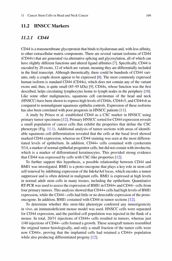
19911 Cancer Stem Cells in Head and Neck Cancer
11.2 HNSCC Markers
11.2.1 CD44
CD44 is a transmembrane glycoprotein that binds to hyaluronan and, with less affinity, to other extracellular matrix components. There are several variant isoforms of CD44 (CD44v) that are generated via alternative splicing and glycosylation, all of which can have slightly different functions and altered ligand affinities [7]. Specifically, CD44 is encoded by 20 exons, 12 of which are variant, meaning they are differentially included in the final transcript. Although theoretically, there could be hundreds of CD44 vari-ants, only a couple dozen appear to be expressed [8]. The most commonly expressed human isoform is standard CD44 (CD44s), which does not contain any of the variant exons and, thus, is quite small (85–95 kDa) [9]. CD44s, whose function was the first described, helps circulating lymphocytes home to lymph nodes in the periphery [10]. Like some other malignancies, squamous cell carcinomas of the head and neck (HNSCC) have been shown to express high levels of CD44s, CD44v5, and CD44v6 as compared to nonmalignant squamous epithelia controls. Expression of these isoforms has also been correlated with poor prognosis in HNSCC patients [11].
A study by Prince et al. established CD44 as a CSC marker in HNSCC using primary tumor specimens [12]. Primary HNSCC sorted for CD44 expression reveals a small population of cancer cells that exhibit the properties that define the CSC phenotype (Fig. 11.1). Additional analysis of tumor sections with areas of identifi-able squamous cell differentiation revealed that the cells at the basal level showed marked CD44 expression, whereas no CD44 staining was seen at the most differen-tiated levels of epithelium. In addition, CD44+ cells costained with cytokeratin 5/14, a marker of normal epithelial progenitor cells, but did not costain with involucrin, which is a marker of differentiated keratinocytes. This provided strong evidence that CD44 was expressed by cells with CSC-like properties [12].
To further support this hypothesis, a possible relationship between CD44 and BMI1 was investigated. BMI1 is a proto-oncogene that plays a key role in stem cell self-renewal by inhibiting expression of the Ink4a/Arf locus, which encodes a tumor suppressor and is often deleted in malignant cells. BMI1 is expressed at high levels in normal adult stem cells in many tissues, including the epithelium. Quantitative RT-PCR was used to assess the expression of BMI1 in CD44+ and CD44− cells from four primary tumors. This analysis showed that CD44+ cells had high levels of BMI1 expression, while the CD44− cells had little or no detectable expression of the proto-oncogene. In addition, BMI1 costained with CD44 in tumor sections [12].
To determine whether this stem-like phenotype conferred any tumorigenicity in vivo, an immunodeficient mouse model was used. HNSCC cells were separated for CD44 expression, and the purified cell population was injected in the flank of a mouse. In total, 20/31 injections of CD44+ cells resulted in tumors, whereas just 1/40 injections of CD44− cells formed a growth. These xenograft tumors resembled the original tumor histologically, and only a small fraction of the tumor cells were now CD44+, proving that the implanted cells had retained a CD44+ population while also producing differentiated progeny [12].
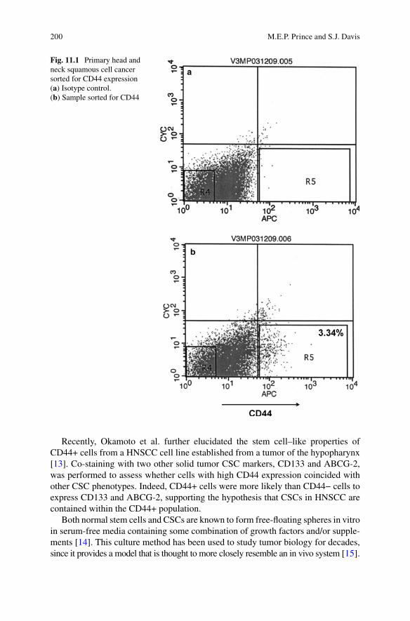
200 M.E.P. Prince and S.J. Davis
Recently, Okamoto et al. further elucidated the stem cell–like properties of CD44+ cells from a HNSCC cell line established from a tumor of the hypopharynx [13]. Co-staining with two other solid tumor CSC markers, CD133 and ABCG-2, was performed to assess whether cells with high CD44 expression coincided with other CSC phenotypes. Indeed, CD44+ cells were more likely than CD44− cells to express CD133 and ABCG-2, supporting the hypothesis that CSCs in HNSCC are contained within the CD44+ population.
Both normal stem cells and CSCs are known to form free-floating spheres in vitro in serum-free media containing some combination of growth factors and/or supple-ments [14]. This culture method has been used to study tumor biology for decades, since it provides a model that is thought to more closely resemble an in vivo system [15].
Fig. 11.1 Primary head and neck squamous cell cancer sorted for CD44 expression (a) Isotype control. (b) Sample sorted for CD44
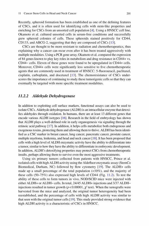
20111 Cancer Stem Cells in Head and Neck Cancer
Recently, spheroid formation has been established as one of the defining features of CSCs, and it is often used for identifying cells with stem-like properties and enriching for CSCs from an unsorted cell population [4]. Using a HNSCC cell line, Okamoto et al. cultured unsorted cells in serum-free conditions and successfully grew spheroid colonies of cells. These spheroids stained positively for CD44, CD133, and ABCG-2, suggesting that they are composed of CSCs [13].
CSCs are thought to be more resistant to radiation and chemotherapeutics, thus explaining why a cancer can recur even after it has been treated aggressively with multiple modalities. Using a PCR gene array, Okamoto et al. compared the expression of 84 genes known to play key roles in metabolism and drug resistance in CD44+ vs. CD44− cells. Eleven of these genes were found to be upregulated in CD44+ cells. Moreover, CD44+ cells were significantly less sensitive to five chemotherapeutic agents that are commonly used in treatment of HNSCC: 5-fluorouracil, paclitaxel, cisplatin, carboplatin, and docetaxel [13]. The chemoresistance of CSCs under-scores the importance of continuing to study these tumorigenic cells so that they can eventually be targeted with more specific treatment modalities.
11.2.2 Aldehyde Dehydrogenase
In addition to exploiting cell surface markers, functional assays can also be used to isolate CSCs. Aldehyde dehydrogenase (ALDH) is an intracellular enzyme that detoxi-fies aldehydes through oxidation. In humans, there are at least 13 different genes that encode various ALDH isotypes [16]. Research in the field of embryology has shown that ALDH plays a well-defined role in early organogenesis via signaling through the retinoic acid pathway [17]. In addition, it helps cells metabolize both endogenous and exogenous toxins, protecting them and allowing them to thrive. ALDH has been identi-fied as a CSC marker in breast cancer, lung cancer, pancreatic cancer, prostate cancer, multiple myeloma, leukemia, and head and neck cancer [18]. It has been proposed that cells with a high level of ALDH enzymatic activity have the ability to differentiate into a tumor, similar to how they have the ability to differentiate in embryonic development. In addition, ALDH’s detoxifying properties may protect CSCs from chemotherapeutic insults, perhaps allowing them to survive even the most aggressive treatments.
Using six primary tumors collected from patients with HNSCC, Prince et al. isolated cells with high ALDH activity using the Aldefluor enzymatic assay (StemCo Biomedical, Durham, NC) followed by flow cytometry [19]. The ALDH+ cells made up a small percentage of the total population (<10%), and the majority of these cells (50–75%) also expressed high levels of CD44 (Fig. 11.2). To test the ability of these cells to form tumors in vivo, NOD/SCID mice were injected with either ALDH+ or ALDH− cells. In total, 24/45 ALDH+ injections and 3/37 ALDH− injections resulted in tumor growth (p < 0.00001, c2 test). When the xenografts were harvested from the mice and analyzed, the original tumor heterogeneity had been reestablished, and the percentage of cells with high ALDH activity was similar to that seen with the original tumor cells [19]. This study provided strong evidence that high ALDH activity is a characteristic of CSCs in HNSCC.
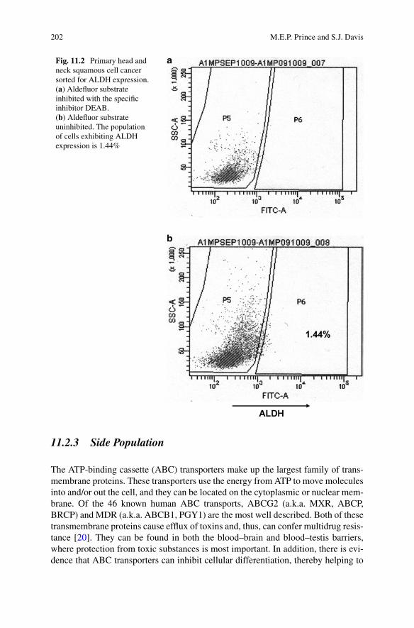
202 M.E.P. Prince and S.J. Davis
11.2.3 Side Population
The ATP-binding cassette (ABC) transporters make up the largest family of trans-membrane proteins. These transporters use the energy from ATP to move molecules into and/or out the cell, and they can be located on the cytoplasmic or nuclear mem-brane. Of the 46 known human ABC transports, ABCG2 (a.k.a. MXR, ABCP, BRCP) and MDR (a.k.a. ABCB1, PGY1) are the most well described. Both of these transmembrane proteins cause efflux of toxins and, thus, can confer multidrug resis-tance [20]. They can be found in both the blood–brain and blood–testis barriers, where protection from toxic substances is most important. In addition, there is evi-dence that ABC transporters can inhibit cellular differentiation, thereby helping to
Fig. 11.2 Primary head and neck squamous cell cancer sorted for ALDH expression. (a) Aldefluor substrate inhibited with the specific inhibitor DEAB. (b) Aldefluor substrate uninhibited. The population of cells exhibiting ALDH expression is 1.44%
20311 Cancer Stem Cells in Head and Neck Cancer
maintain a stem cell phenotype. Side populations (SPs) of cells with highly active ABC transporters have been identified using a Hoechst 33342 dye exclusion assay. Cells able to pump out the dye will appear as an unstained fraction in flow cytometric analysis. These SPs are thought to be enriched in stem cells, as they have been shown to express markers of a stem-like phenotype [21]. SPs have been reported in several tumor types, both solid and hematopoietic. Although these cell populations do not appear to be enriched in CSCs, SP cells have still proven to be highly tumorigenic [22].
11.3 Future Directions
While the markers studied in HNSCC thus far have allowed for separation of a cell fraction that contains a large number of highly tumorigenic cells, it is likely that no single marker will be specific enough to select for a pure CSC subpopulation. Therefore, it is imperative to identify new properties of CSCs so that, by combining these characteristics, a profile of sorts can be used to isolate and target these high-risk cells. In breast cancer, the stem cell subpopulation has been isolated by identi-fying cells with high CD44 expression in combination with low CD24 expression. The CD44+/CD24− population formed tumors very efficiently, while the CD44+/CD24+ cells did not [5]. Similarly, CSCs from colon cancers were characterized by expression of multiple markers: CD44+/CD166+/ESA+ [23].
Costaining has also been used with the Aldefluor assay to narrow in on the CSC population. In breast cancer cells, it was recently reported that CXCR1, one of the receptors for IL-8, is necessary for reproducing the original tumor heterogeneity in a mouse xenograft model. Aldefluor+/CXCR1+ cells isolated from human breast cancer cell lines were able to reconstitute the original tumor cell population, while Aldefluor+/CXCR1− cells formed a tumor but remained Aldefluor+/CXCR1−. Moreover, the CSCs expressing CXCR1 had a higher rate of metastasis after intra-ventricular injection into NOD/SCID mice [24].
It is important to keep in mind that each tumor type is distinct from any other, and the corresponding CSC population will likely have a unique gene expression profile. Nevertheless, it is reasonable to explore avenues that have proven fruitful in other areas of CSC research, at least as a starting point. Improved understanding of HNSCC tumor biology, therefore, will likely rely on the use of multiple CSC markers in the future.
11.4 Clinical Implications
According to the CSC theory of tumorigenesis, a cancer can only be completely eradicated by killing cells with a stem cell–like phenotype. This may explain why many malignancies recur after being treated with multiple modalities that debulk the tumor, even to the point of appearing eradicated on imaging, but do not specifically
204 M.E.P. Prince and S.J. Davis
target the CSC subpopulation. Several studies have shown that CSCs are less sensitive to chemotherapy agents and radiation in several tumor types, though some argue that the data are still inconclusive [25, 26].
Regardless of their sensitivity or resistance, CSCs are believed to be responsible overall for the persistence or distant spread of disease in patients who have previously undergone treatment for their cancer. Upregulated detoxifying mechanisms, such as overexpression of ALDH and ABC transporters, likely confer a great deal of resis-tance to chemotherapeutics. In addition, CSCs are relatively quiescent compared to the rest of the tumor population and have enhanced DNA repair mechanisms, which may make them less likely to succumb to the cytotoxic hits induced by radiation or drugs [27, 28].
CSCs are thought to be preferentially localized in well-vascularized areas of tumor, and the use of antiangiogenic drugs in combination with classical chemotherapeu-tics have been shown to kill more CSCs than does chemotherapy alone [27]. Other potential mechanisms of targeting stem-like cells include the use of small molecule inhibitors, monoclonal antibodies, and micro-RNAs aimed at pathways known to be exploited in CSCs. This has shown promise in breast CSCs, in which CXCR1 signaling has been blocked using both a small molecule inhibitor, repertaxin, and a monoclonal, resulting in a drastic reduction in the CSC population [24]. Since many of the known CSC cell surface markers, including CD44, are also present on the surface of normal human cells, targets specific to the stem-like phenotype must be identified to refine our treatments and reduce toxicity to healthy tissue. While many obstacles and unanswered questions still stand in way of developing targeted anti-CSC therapies for HNSCC, recent advances in the field of tumor biology have given us hope that such a goal is within reach.
References
1. Parkin DM, Bray F, Ferlay J, Pisani P (2005) Global cancer statistics, 2002. CA Cancer J Clin 55 (2):74–108.
2. Jemal A, Siegel R, Ward E, Murray T, Xu J, Thun MJ (2007) Cancer statistics, 2007. CA Cancer J Clin 57 (1):43–66.
3. Wicha MS, Liu S, Dontu G (2006) Cancer stem cells: an old idea–a paradigm shift. Cancer Res 66 (4):1883-1890; discussion 1895–1886.
4. Clarke MF, Dick JE, Dirks PB, Eaves CJ, Jamieson CH, Jones DL, Visvader J, Weissman IL, Wahl GM (2006) Cancer stem cells--perspectives on current status and future directions: AACR Workshop on cancer stem cells. Cancer Res 66 (19):9339–9344.
5. Al-Hajj M, Wicha MS, Benito-Hernandez A, Morrison SJ, Clarke MF (2003) Prospective iden-tification of tumorigenic breast cancer cells. Proc Natl Acad Sci USA 100 (7):3983–3988.
6. Cho RW, Clarke MF (2008) Recent advances in cancer stem cells. Curr Opin Genet Dev 18 (1): 48–53.
7. Rudzki Z, Jothy S (1997) CD44 and the adhesion of neoplastic cells. Mol Pathol 50 (2):57–71 8. Naor D, Wallach-Dayan SB, Zahalka MA, Sionov RV (2008) Involvement of CD44, a mole-
cule with a thousand faces, in cancer dissemination. Semin Cancer Biol 18 (4):260–267. 9. Naor D, Sionov RV, Ish-Shalom D (1997) CD44: structure, function, and association with the
malignant process. Adv Cancer Res 71:241–319.
20511 Cancer Stem Cells in Head and Neck Cancer
10. Gallatin WM, Weissman IL, Butcher EC (1983) A cell-surface molecule involved in organ-specific homing of lymphocytes. Nature 304 (5921):30–34.
11. Herold-Mende C, Seiter S, Born AI, Patzelt E, Schupp M, Zoller J, Bosch FX, Zoller M (1996) Expression of CD44 splice variants in squamous epithelia and squamous cell carcinomas of the head and neck. J Pathol 179 (1):66–73.
12. Prince ME, Sivanandan R, Kaczorowski A, Wolf GT, Kaplan MJ, Dalerba P, Weissman IL, Clarke MF, Ailles LE (2007) Identification of a subpopulation of cells with cancer stem cell properties in head and neck squamous cell carcinoma. Proc Natl Acad Sci USA 104 (3):973–978.
13. Okamoto A, Chikamatsu K, Sakakura K, Hatsushika K, Takahashi G, Masuyama K (2009) Expansion and characterization of cancer stem-like cells in squamous cell carcinoma of the head and neck. Oral Oncol 45 (7):633–639.
14. Hirschhaeuser F, Menne H, Dittfeld C, West J, Mueller-Klieser W, Kunz-Schughart LA (2010) Multicellular tumor spheroids: An underestimated tool is catching up again. J Biotechnol 148:3–15.
15. Mueller-Klieser W (2000) Tumor biology and experimental therapeutics. Crit Rev Oncol Hematol 36 (2-3):123–139.
16. Sophos NA, Vasiliou V (2003) Aldehyde dehydrogenase gene superfamily: the 2002 update. Chem Biol Interact 143-144:5–22.
17. Duester G (2008) Retinoic acid synthesis and signaling during early organogenesis. Cell 134 (6): 921–931.
18. Saini V, Shoemaker RH (2010) Potential for therapeutic targeting of tumor stem cells. Cancer Sci 101 (1):16–21.
19. Clay MR, Tabor M, Owen JH, Carey TE, Bradford CR, Wolf GT, Wicha MS, Prince ME (2010) Single-marker identification of head and neck squamous cell carcinoma cancer stem cells with aldehyde dehydrogenase. Head Neck 32:1195–1201.
20. Dean M, Hamon Y, Chimini G (2001) The human ATP-binding cassette (ABC) transporter superfamily. J Lipid Res 42 (7):1007–1017.
21. Hadnagy A, Gaboury L, Beaulieu R, Balicki D (2006) SP analysis may be used to identify cancer stem cell populations. Exp Cell Res 312 (19):3701–3710.
22. Moserle L, Ghisi M, Amadori A, Indraccolo S (2010) Side population and cancer stem cells: therapeutic implications. Cancer Lett 288 (1):1–9.
23. Dalerba P, Dylla SJ, Park IK, Liu R, Wang X, Cho RW, Hoey T, Gurney A, Huang EH, Simeone DM, Shelton AA, Parmiani G, Castelli C, Clarke MF (2007) Phenotypic characterization of human colorectal cancer stem cells. Proc Natl Acad Sci USA 104 (24):10158–10163.
24. Ginestier C, Liu S, Diebel ME, Korkaya H, Luo M, Brown M, Wicinski J, Cabaud O, Charafe-Jauffret E, Birnbaum D, Guan JL, Dontu G, Wicha MS (2010) CXCR1 blockade selectively tar-gets human breast cancer stem cells in vitro and in xenografts. J Clin Invest 120 (2):485–497.
25. Vlashi E, McBride WH, Pajonk F (2009) Radiation responses of cancer stem cells. J Cell Biochem 108 (2):339–342.
26. Baumann M, Krause M, Hill R (2008) Exploring the role of cancer stem cells in radioresis-tance. Nat Rev Cancer 8 (7):545–554.
27. Ghotra VP, Puigvert JC, Danen EH (2009) The cancer stem cell microenvironment and anti-cancer therapy. Int J Radiat Biol 85 (11):955–962.
28. Mimeault M, Hauke R, Batra SK (2008) Recent advances on the molecular mechanisms involved in the drug resistance of cancer cells and novel targeting therapies. Clin Pharmacol Ther 83 (5):673–691.

Part IIICancer Stem Cell Gene
Expression and Mechanisms: Convergence of Embryonic and
Tumorigenic Signaling Pathways
209A.L. Allan (ed.), Cancer Stem Cells in Solid Tumors, Stem Cell Biology and Regenerative Medicine, DOI 10.1007/978-1-61779-246-5_12, © Springer Science+Business Media, LLC 2011
Abstract Embryonic stem cells have the ability to undergo unlimited self-renewal and retain the pluripotent capacity to differentiate into all of the cell types of the body. Their pluripotency is maintained by the core transcription factors Nanog, Oct4, and Sox2, as well as signaling pathways, which include Wnt, FGF, and TGFb. Recent studies have shown that somatic cells can be reprogrammed into the pluripo-tent state with defined reprogramming factors, including Oct4, Sox2, Nanog, and c-Myc. Considering the oncogenic potential of the reprogramming factors, the induced pluripotency mimics the cellular transformation process and is inhibited by the tumor suppressor p53. Since the Wnt/FGF/TGFb pathways are implicated in human cancers and pluripotency/reprogramming factors Oct4/Sox2/Nanog/c-Myc/Lin28 are overexpressed in some human cancers, there is a possibility that dedif-ferentiation is a mechanism for the generation of tumor-initiating cells in human cancer. Therefore, there appears to be a strong link between the regulatory pathways in pluripotent stem cells and human tumors.
Abbreviations
ALL Acute lymphoblastic leukemiaAML1-ETO Acute myelogenous leukemia (AML) with t(8;21)(q22;q22)APC Adenomatous polyposis coliBMP Bone morphogenic proteinCKI Cyclin-dependent kinase inhibitorsERK Extracellular signal-regulated kinase
Y. Xu (*)Section of Molecular Biology, Division of Biological Sciences, University of California, San Diego, La Jolla, CA, USAe-mail: [email protected]
Chapter 12Relationship Between Regulatory Pathways in Pluripotent Stem Cells and Human Tumors
Olga Gaidarenko and Yang Xu
210 O. Gaidarenko and Y. Xu
ES Embryonic stemFGF Basic fibroblast growth factorFGFR Basic fibroblast growth factor receptorGSK Glycogen synthase kinasehESC Human embryonic stem cellHSC Hematopoietic stem cellICM Inner cell massiPSC Induced pluripotent stem cellsKlf4 Kruppel-like factor 4LDL Low density lipoproteinMAPK Mitogen-activated protein kinasemiRNA MicroRNAPcG Polycomb groupPI3K Phosphoinositide 3-kinasePIP Phosphotidylinositol 4,5-bisphosphatePKC Protein kinase CPLC Phospholipase CPLZF Promyelocytic leukemia zinc fingerRAR Retinoic acid receptorSSEA Stage-specific embryonic antigenTCF/LEF T-cell factor/lymphoid enhancer factorTGF-b Transforming growth factor
12.1 Regulatory Pathways in Embryonic Stem Cells
Mammalian embryonic stem (ES) cells are derived from the inner cell mass (ICM) of the pre-implantation or peri-implantation blastocyst. The typical stage of isolation for human ES cells (hESCs) is from day 5 to 6 blastocysts. These cells are karyotypically normal, have high levels of telomerase activity, are capa-ble of prolonged self-renewal in culture, and are pluripotent. As such, they are able to give rise to all cell types found in the human body, derived from all three embryonic germ layers: endoderm, mesoderm, and ectoderm. This capacity is maintained even after repeated passaging in long-term, and potentially indefinite, culture [1–5].
Pluripotency, self-renewal, and differentiation of ES cells are regulated by a com-plex network that includes transcription factors, signaling pathways, micro-RNAs (miRNAs), and epigenetics. Pluripotency-associated transcription factors such as Oct4, Nanog, and Sox2 function in concert to maintain ES cells in their pluripotent, self-renewing state. They repress the transcription of genes involved in differentia-tion and activate those that are important for stem cell characteristics.
Oct4 is a POU transcription factor that is expressed in blastomeres, germ cells, and cells of early embryos. It is encoded by the Pou5f1 gene and is also referred to as Oct3. It is essential for the establishment of a pluripotent cell population in
21112 Relationship Between Regulatory Pathways in Pluripotent Stem Cells…
the blastocyst ICM. Precise regulation of Oct4 levels is crucial to ES cells, as its downregulation results in dedifferentiation into trophectoderm, whereas less than a twofold increase in its expression leads to differentiation into primitive endoderm and mesoderm [6, 7]. Nanog is a homeodomain transcription factor whose expres-sion in the developing mammalian embryo is restricted to the transient ICM popula-tion from which ES cells can be established. Its expression is downregulated rapidly during further differentiation from ICM and is tightly associated with and required for pluripotency [8–10]. Sox2 belongs to the SRY-related HMG box (Sox) family of transcription factors. Its pattern of expression during early embryogenesis is similar to that of Oct4. It is a major transcriptional regulator involved in the perpetuation of the self-renewing, pluripotent state of ES cells. At present, it remains less exten-sively studied than Oct4 and Nanog [11, 12].
Oct4, Nanog, and Sox2 co-occupy the promoters of at least 353 genes in hESCs, some of which are active in ES cells, while others are not. The transcriptionally inactive population is enriched for transcription factor genes that are involved in developmental processes such as differentiation. The transcriptionally active genes include Oct4, Nanog, and Sox2 themselves, as well as other transcription factors and components of signaling pathways that have been implicated in the self-renewal and pluripotency. The promoter co-occupancy is regulatory in nature, promoting stem cell programming while suppressing developmental processes [13]. In addi-tion to regulating the expression of protein-coding genes, Oct4, Sox2, and Nanog occupy the promoter regions of some miRNAs that are preferentially expressed in ES cells. The miRNA expression profiles differ for undifferentiated ES cells, ES cells undergoing differentiation, and fully differentiated cells. Thus, miRNAs par-ticipate in the network that regulates “stemness” and differentiation [14]. Of note is the miR-302-367 cluster in hESCs, whose transcription is regulated by Oct4, Sox2, and Nanog. Its expression is restricted to pluripotent ES cells and is repressed upon differentiation. Much remains to be understood about the mechanisms of its action in ES cells; however, evidence exists that implicates it in apoptosis, differentiation, cell cycle regulation, and maintenance of the stem cell state [15]. As differentiation ensues, pluripotency factors themselves become targets of miRNA-mediated silenc-ing. As hESCs differentiate, miR-145 serves to downregulate Oct4, Sox2, and Klf4, another transcription factor of importance to pluripotency. Deficiency in miR-145 results in impaired differentiation and elevated levels of the three aforementioned transcription factors [16].
Epigenetics also play a prominent role in maintaining the stem cell fate, as well as during differentiation. In hESCs, a variety of genes that function in differentia-tion, development, and transcription are repressed by polycomb group (PcG) pro-teins that posttranslationally modify histones. A significant portion of these genes are also occupied by the three key transcription factors, Oct4, Sox2, and Nanog. This repression is alleviated for the majority of the targets during differentiation of ES cells. Conversely, Oct4, Sox2, and Nanog themselves become repressed by PcG proteins as they become downregulated during differentiation. CpG methylation is also involved in the epigenetic regulation of “stemness,” and a significant subpopu-lation of PcG bound regions are within 1 kb of CpG islands [17, 18].
212 O. Gaidarenko and Y. Xu
12.2 Similarities Between ES cells and Cancer Cells
Cancer cells have striking similarities to ES cells. For instance, most cancers can overcome their proliferative limit by expression of telomerase, which is also active in ES cells, but not in normal somatic cells [19, 20]. Other cancer cells rely on an alternative lengthening of telomeres (ALT) mechanism for telomere maintenance [21]. However it is managed, maintained telomeres are necessary for the immortalization of cancer cells as well as the self-renewal of ES cells. Additionally, numerous genes that are reversibly repressed by the PcG proteins in ES cells are permanently silenced by promoter methylation in cancer cells [22]. Cancer cells and hESCs have common miRNA expression patterns [23]. Some cancer types such as germinomas, semino-mas, dysgerminomas, and embryonal carcinomas are able to give rise to various somatic and extraembryonic tissues, thus exhibiting pluripotency [24]. Various sig-naling pathways of importance to the maintenance of ES cells also play a role in driving tumorigenesis, and these are described below.
12.2.1 Wnt Signaling
Wnt signaling occurs via canonical and non-canonical pathways. Canonical Wnt signaling is involved in the regulatory circuitry of ES cells, maintenance of adult stem cells, and, if gone awry, tumorigenesis. It proceeds via stabilization of b-catenin, which then translocates to the nucleus and activates the expression of genes that are constitutively bound by Tcf/Lef proteins. In the absence of b-catenin/Wnt signaling, the expression of those genes is repressed. The Wnt signaling cascade begins when Wnt binds two receptors on the cell surface: a serpentine receptor belonging to the Frizzled family and Lrp5/6, an LDL receptor family member. This binding inhibits the activity of the destruction complex which otherwise acts upon b-catenin. The destruction complex has two scaffolding proteins, axin and adenomatous polyposis coli (APC) that bind to b-catenin, whose N-terminus is then phosphorylated by two kinases of the destruction complex, CKI and GSK3. This phosphorylation recruits an E3 ubiquitin ligase, which targets b-catenin to the proteasome for degradation [25–29].
It has been demonstrated that stimulation of the Wnt canonical pathway is suf-ficient for the maintenance of self-renewal in human, as well as mouse, ES cells. When Wnt signaling is activated in ES cells experimentally in a variety of ways, these cells are able to resist differentiation, remain pluripotent, and maintain the expression of Oct4, Nanog, and Rex-1 transcription factors associated with pluripo-tency [30, 31]. The mechanism of Wnt action in hESCs remains to be elucidated. However, studies in mouse ES cells suggest potential roles that Wnt signaling may play in hESCs, if conserved across species. In mouse ES cells, Tcf3 has been found to have genome-wide promoter co-occupancy with Oct4 and Nanog. It occupies its own promoter, along with the promoters of the Oct4, Nanog, and Sox2 genes, thus
21312 Relationship Between Regulatory Pathways in Pluripotent Stem Cells…
participating in an autoregulatory loop with them. In the absence of Wnt signaling, Tcf3 plays a repressive role; when the pathway is active, Tcf3-bound genes are activated, supporting pluripotency and self-renewal in ES cells [29].
Wnt signaling is involved in the maintenance of adult stem/progenitor cells in a variety of tissues, such as the colorectal crypts and the hematopoietic system. Aberrations in the pathway result in neoplastic malignancies. In the small intestine, Tcf-4 is responsible for crypt cell maintenance [32]. Tcf-4 target genes are consti-tutively activated in colon carcinomas that harbor mutations in b-catenin or APC genes. Thus, colorectal cancer cells share the gene expression program specified by Tcf-4 with the crypt stem and progenitor cells. Activating mutations of the Wnt pathway components are typically an early transforming event in colorectal cancers [28, 32–35].
There is also mounting evidence that Wnts, produced by hematopoietic stem cells (HSCs) as well as their microenvironment, are important for the maintenance of the HSCs and their niche [28, 36–41]. Various cancers of the blood have been found to have inappropriately active Wnt signaling. For instance, self-renewing granulocyte–macrophage progenitors from patients with chronic myelogenous leukemia have abnormally high levels of nuclear b-catenin [42]. In acute myelogenous leukemia (AML), the associated translocation products (AML1-ETO, RARa, and PLZF-RARa) encode transcription factors that activate Wnt signaling via induction of b-catenin expression as well as expression of its homologue plakoglobin (which can also interact with TCF/LEF proteins and activate targets of Wnt signaling) [43–45]. E2A-Pbx1, the product of the t(1;19) chromosomal translocation found in a signifi-cant portion of pre-B acute lymphoblastoid leukemias (ALL), transcriptionally acti-vates a Wnt gene. It is likely that this Wnt promotes the development of such pre-B ALL cancers via an autocrine mechanism [46]. Additionally, Wnt signaling has been shown to be important for the growth of multiple myeloma cells [47].
12.2.2 FGF Signaling
At present, the human fibroblast growth factor (FGF) family boasts at least 22 known members that share 13–71% sequence identity. FGFs interact with four tyrosine kinase receptors; FGFR1-4. Alternative splicing of FGFR1-3 results in a diverse array of tissue-specific receptors with varying affinities for different FGF family members. FGFs also interact with heparin or heparin sulfate proteoglycans via a region distinct from the one that engages FGFRs. This interaction facilitates FGF binding of FGFRs and subsequent initiation of signal transduction. Once the FGF ligand is bound, FGFRs dimerize and autophosphorylate, primed to activate downstream signaling pathways such as Ras-Raf-MAPK, PLC(gamma)-PKC, and PI3K-Akt [48–52].
FGF2, also known as basic FGF or bFGF, is typically included in hESC media and has been demonstrated to maintain hESCs in an undifferentiated state even in the absence of feeder cells or feeder-conditioned media [53–55]. Inhibition of FGFRs in
214 O. Gaidarenko and Y. Xu
hESCs results in suppressed activation of downstream kinases and leads to rapid differentiation [56]. Although the mechanisms by which FGF2 maintains hESC pluripotency remain to be fully elucidated, several groups have found evidence that the MAPK pathway plays a role in the maintenance of pluripotency. When this path-way is activated downstream of FGFR, ERK is phosphorylated. It then translocates to the nucleus and phosphorylates various transcription factors, including c-Jun, c-Fos, and c-Myc, the latter being one of the factors originally employed by Yamanaka et al. in generating iPS cells [52, 57]. Inhibition of the MAPK pathway in hESCs results in the downregulation of pluripotency markers such as Oct4, Nanog, and SSEA-4, and ultimately, differentiation [51, 52, 58, 59]. The PI3K/AKT pathway also appears to be involved in the maintenance of pluripotency in hESCs, as its inhibition leads to differentiation [52, 58]. Once PI3K is activated, it phophorylates PIP2, generating PIP3, a second messenger that then communicates with AKT and recruits it to the plasma membrane. At the membrane, AKT is phosphorylated, and thus activated, by PDKI, which enables it to activate its downstream targets. Of note is the ability of this pathway to stimulate the Wnt/b-catenin signaling. Additionally, the ERK/MAPK pathway appears to lie downstream of PI3K/AKT [52, 53].
Various cancers also utilize FGF signaling to carry out important malignant behaviors such as increased proliferation, angiogenesis, invasiveness, metastasis, and resistance to chemo- and radiotherapy. For example, prostate cancer has increased levels of FGF2, as well as FGF1, FGF6, and FGF8, and expresses all four FGFR types [48]. Levels of FGF2 are elevated in esophageal and gastroesophageal junction adenocarcinomas, and overexpression of FGF2 is correlated with tumor recurrence in patients with esophageal cancer [59, 60]. FGF2 confers apoptotic resistance to small cell lung cancer cells, and, if present at elevated serum levels, is a predictor of a poor clinical outcome [61]. The FGFs may function in an autocrine or paracrine manner. In addition to FGFs produced by cancer cells themselves, cancer tissues may have increased access to FGFs due to their enhanced release from the extracellular matrix [48]. Another way cancers make use of this pathway is through activating mutations in FGFR genes. Mutations resulting in constitutively active FGFR3 are frequently found in bladder and cervical carcinomas [62]. Likewise, 12% of endometrial carcinomas feature constitutively activated mutant FGFR2 [63]. Additionally, certain cancers such as gastric cancer and oral squamous cell carcinoma have amplification and overexpression of FGFRs, which also leads to aberrant FGF signaling [64, 65].
12.2.3 TGFb /Activin/Nodal Signaling
Another ES cell pathway that is utilized by cancer cells proceeds via Activin/Nodal signaling. Activin and Nodal are both related to the transforming growth factor-b (TGF-b) family. The Activin signaling cascade is initiated when this secreted peptide binds its cognate heterodimeric receptor complex, bringing together two transmem-brane serine/threonine kinase receptor types: type I and type II. The type II receptor
21512 Relationship Between Regulatory Pathways in Pluripotent Stem Cells…
phosphorylates the type I receptor, which in turn phosphorylates Smad2 and Smad3. The activated Smads translocate to the nucleus where they are able to bind DNA directly. Smads associate with a variety of DNA-binding cofactors in order to fully activate the transcription of their target genes. This allows for flexibility and versa-tility of the pathway [66–69]. Nodal signaling proceeds analogously [70].
This signaling pathway has been shown to be important for sustaining self-renewal and pluripotency in hESCs [71], and its ability to maintain hESCs is augmented by cooperation with FGF signaling [72]. Smad 2/3 are able to bind the Nanog gene directly and maintain its expression [73, 74]. Nodal is secreted by the hESCs them-selves, providing an autocrine mechanism for reinforcing their pluripotent state. Along with it, they secrete Lefty, a Nodal inhibitor, thus keeping the signaling path-way regulated. Nodal expression is rapidly downregulated upon differentiation [71, 72, 75, 76]. Activin A, secreted by mouse embryonic fibroblasts (MEFs) which are used as feeder layers for hESC culture, is a major regulator of pluripotency and self-renewal of hESCs. It is able to stimulate the expression of Nanog, Oct4, bFGF, FGF8, Wnt3, and Nodal. Additionally, it suppresses BMP signaling, which promotes hESC differentiation [77, 78].
The TGFb/Activin/Nodal Signaling pathway also plays a role in various cancers, albeit a complex, context-dependent one. At the onset of cancer development, it has an inhibitory role. However, as the disease becomes more mature, the pathway may become oncogenic, promoting invasion and metastasis. In epithelial cells, this usually proceeds by increased ligand secretion coupled with decreased or altered cancer cell responsiveness to the tumor-suppressive effects of the pathway. The majority of human tumors possess functional TGFb signaling. Ligand secretion by tumor cells also affects the microenvironmental interacting cells, such as the stroma [68, 79–82]. High levels of TGFb1 correlate with poor clinical outcome in patients with a variety of cancers, such as colorectal, lung, and prostate [82–86]. Elevated expression of Activin A is observed in a variety of carcinomas, including esopha-geal, prostate, and pancreatic [87–90]. Aggressive melanoma and breast carcinoma cells express Nodal, but not its inhibitor Lefty, whose expression is restricted to hESCs, thus allowing for uncontrolled signaling to maintain their multipotent, dedi-fferentiated phenotype [75, 91].
12.3 Induced Pluripotent Stem Cells (iPSCs) and Cancer Cells
Pluripotency is not necessarily a property that is restricted to cells of the early embryo and lost irreversibly upon differentiation. Terminally differentiated adult cells can be reprogrammed back to the pluripotent state by forced expression of transcription fac-tors that maintain the “stemness” of ES cells. Such cells are referred to as induced pluripotent stem or iPS cells. The first iPSCs were reported in 2006, obtained by expressing Oct4, Sox2, c-Myc and Klf4 in mouse fibroblasts. The reprogrammed cells expressed a variety of ES cell markers. Like ES cells, they formed teratomas comprising all three germ layers (ectoderm, mesoderm, and endoderm) when injected
216 O. Gaidarenko and Y. Xu
into nude mice, and were able to contribute to developing embryos after injection into blastocysts [92]. Just a year later, two groups obtained human iPS cells, one using the same four factors, the other substituting Nanog and Lin28 for c-Myc and Klf4. The gene expression profiles, surface antigens, and epigenetic status of genes involved in pluripotency were very similar, but not identical, between iPSCs and ES cells [56, 93]. Since then, various groups have reported new strategies for cell repro-gramming, employing different methods, factors, and chemicals intended to promote or improve the process. Human somatic cells from all three germ layers, including hepatocytes (endoderm), hematopoetic progenitors (mesoderm), and neural stem cells (ectoderm), have been successfully reprogrammed into iPS cells [56, 92–96].
Induction of pluripotency with the defined reprogramming factors, most of which have oncogenic properties, provides an additional link between pluripotency and cancer [97]. Additionally, there is evidence that, during reprogramming, iPSCs aquire epigenetic changes that are associated with cancer [98, 99]. Findings that the tumor suppressor p53 also inhibits pluripotency induction further underscore the potential transforming nature of the reprogramming process [97, 100]. The presence of tumor-initiating cells in some human cancers has been suggested to be a primary cause for drug resistance and metastasis [91, 101–104]. These tumor-initiating cells are thought to possess some stem cell/progenitor characteristics, such as the ability to self-renew and the potential to differentiate into multiple lineages. The fact that many human cancers overexpress some of the reprogramming factors, including Oct4, Sox2, Nanog, Lin28, and c-Myc, raises the possibility that the dedifferentiation process within the tumor cells could lead to dynamic generation of tumor-initiating cells [24, 105–112]. Therefore, there appears to be a strong link between the regula-tory pathways in pluripotent stem cells and human tumors.
References
1. Evans MJ, Kaufman MH (1981) Establishment in culture of pluripotential cells from mouse embryos. Nature 292: 154–156
2. Thomson JA, Itzkovitz-Eldor J, Shapiro SS, Waknitz MA, Swiergiel JJ, Marshall VS, Jones JM (1998) Embryonic stem cell lines derived from human blastocysts. Science 282: 1145–1147
3. Bongso A, Fong CY, Ng SC, Ratnam S (1994) Isolation and culture of inner cell mass cells from human blastocysts. Hum Reprod 9: 2110–2117
4. Conley BJ, Young JC, Trounson AO, Mollard R. (2004) Derivation, propagation and differen-tiation of human embryonic stem cells. Int J Biochem Cell Biol 36: 555–567
5. Amit M, Carpenter MK, Inokuma MS, Chiu C, Harris CP, Waknitz MA, Itskovitz-Eldor J, Thomson JA (2000) Clonally Derived Human Embryonic Stem Cell Lines Maintain Pluripotency and Proliferative Potential for Prolonged Periods of Culture. Dev Biol 227: 271–278
6. Nichols J, Zevnik B, Anastassiadis K, Niwa H, Klewe-Nebenius D, Chambers I, Scholer H, Smith A (1998) Formation of pluripotent stem cells in the mammalian embryo depends on the POU transcription factor Oct4. Cell 95: 379–391
7. Niwa H, Miyazaki J, Smith AG (2000) Quantitative expression of Oct-3/4 defines differentia-tion, dedifferentiation or self-renewal of ES cells. Nat Genet 24: 372–376
21712 Relationship Between Regulatory Pathways in Pluripotent Stem Cells…
8. Chambers I, Colby D, Robertson M, Nichols J, Lee S, Tweedie S, Smith A (2003) Functional expression cloning of Nanog, a pluripotency sustaining factor in embryonic stem cells. Cell 113: 643–655
9. Mitsui K, Tokuzawa Y, Itoh H, Segawa K, Murakami M, Takahashi K, Maruyama M, Maeda M, Yamanaka S (2003)The homeoprotein Nanog is required for maintenance of pluripotency in mouse epiblast and ES cells. Cell 113: 631–642
10. Hyslop L, Stojkovic M, Armstrong L, Walter T, Stojkovic P, Przyborski S, Herbert M, Murdoch A, Strachan T, Lako M (2005) Downregulation of NANOG Induces Differentiation of Human Embryonic Stem Cells to Extraembryonic Lineages. Stem Cells 23: 1035–1043
11. Do H, Lee W, Lim H, Oh J, Kim D, Kim J, Kim T, Kim J (2009) Two potent transactivation domains in the C-terminal region of human NANOG mediate transcriptional activation in human embryonic carcinoma cells. J Cell Biochem 106: 1079–1089
12. Chew J, Loh Y, Zhang W, Chen X, Tam W, Yeap L, Li P, Ang Y, Lim B, Robson P, Ng H (2005) Reciprocal Transcriptional Regulation of Pou5f1 and Sox2 via the Oct4/Sox2 Complex in Embryonic Stem Cells. Mol Cell Biol 25: 6031–6046
13. Boyer LA, Lee TI, Cole MF, Johnstone SE, Levine SS, Zucker JP, Guenther MG, Kumar RM, Murray HL, Jenner RG, Gifford DK, Melton DA, Jaenisch R, Young RA (2005) Core tran-scriptional regulatory circuitry in human embryonic stem cells. Cell 122: 947–956
14. Lakshmipathy U, Love B, Goff LA, Jornsten R, Graichen R, Hart RP, Chesnut JD (2007) MicroRNA expression pattern of undifferentiated and differentiated human embryonic stem cells. Stem Cells Dev 16: 1003–1016
15. Barroso-del Jesus A, Lucena-Aguilar G, Menendez P (2009) The miR-302-367 cluster as a potential stemness regulator in ESCs. Cell Cycle 8: 394–398
16. Xu N, Papagiannakopoulos T, Pan G, Thomson JA, Kosik KS (2009) MicroRNA-145 regulates OCT4, SOX2, and KLF4 and represses pluripotency in human embryonic stem cells. Cell 137: 647–658
17. Lee TI, Jenner RG, Boyer LA, Guenther MG, Levine SS, Kumar RM, Chevalier B, Johnstone SE, Cole MF, Isono K, Koseki H, Fuchikami T, Abe K, Murray HL, Zucker JP, Yuan B, Bell GW, Herbolsheimer E, Hannett NM, Sun K, Odom DT, Otte AP, Volkert TL, Bartel DP, Melton DA, Gifford DK, Jaenisch R, Young RA (2006) Control of developmental regulators by Polycomb in human embryonic stem cells. Cell 125: 301–313
18. Pan G, Tian S, Nie J, Yang C, Ruotti V, Wei H, Jonsdottir GA, Stewart R, Thomson JA (2007) Whole-Genome Analysis of Histone H3 Lysine 4 and Lysine 27 Methylation in Human Embryonic Stem Cells. Cell Stem Cell 1: 299–312
19. Shay JW, Bacchetti S (1997) A survey of telomerase activity in human cancer. Eur J Cancer 33: 787–791
20. Kim NW, Piatyszek MA, Prowse KR, Harley CB, West MD, Ho PLC, Coviello GM, Wright WE, Weinrich SL, Shay JW (1994) Specific association of human telomerase activity with immortal cells and cancer. Science 266: 2011–2015
21. Muntoni A, Reddel RR (2005) The first molecular details of ALT in human tumor cells. Hum Mol Genet 14(2): R191-196
22. Widschwendter M, Fiegl H, Egle D, Mueller-Holzner E, Spizzo G, Marth C, Weisenberger DJ, Campan M, Young J, Jacobs I, Laird PW (2007) Epigenetic stem cell signature in cancer. Nat Genet 39: 157–158
23. Navarro A, Monzó M (2010) MicroRNAs in Human Embryonic and Cancer Stem Cells. Yonsei Med J 51: 622–632
24. Looijenga LHJ, Stoop H, de Leeuw HPJC, de Gouveia Brazao CA, Gillis AJM, van Roozendaal KEP, van Zoelen EJJ, Weber RFA, Wolfenbuttel KP, van Dekken H, Honecker F, Bokemeyer C, Perlman EJ, Schneider DT, Kononen J, Sauter G, Oosterhuis JW (2003) POU5F1 (OCT3/4) Identifies Cells with Pluripotent Potential in Human Germ Cell Tumors. Cancer Research 63: 2244–2250
25. Behrens J, von Kries JP, Kuhl M, Bruhn L, Wedlich D, Grosschedl R, Birchmeier W (1996) Functional interaction of beta-catenin with the transcription factor LEF-1. Nature 382: 638–642
218 O. Gaidarenko and Y. Xu
26. Zeng X, Huang H, Tamai K, Zhang X, Harada Y, Yokota C, Almeida K, Wang J, Doble B, Woodgett J, Wynshaw-Boris A, Hsieh J, He X (2008) Initiation of Wnt signaling: control of Wnt coreceptor Lrp6 phosphorylation/activation via frizzled, dishevelled and axin functions. Development 135: 367–375
27. Brantjes H, Roose J, van de Wetering M, Clevers H (2001) All Tcf HMG box transcription factors interact with Groucho-related co-repressors. Nucleic Acids Res 29: 1410–1419
28. Reya T, Clevers H (2005) Wnt signalling in stem cells and cancer. Nature 434: 843–850 29. Cole MF, Johnstone SE, Newman JJ, Kagey MH, Young RA(2008) Tcf3 is an integral com-
ponent of the core regulatory circuitry of embryonic stem cells. Genes Dev 22: 746–755 30. Sato N, Meijer L, Skaltsounis L, Greengard P, Brivanlou AH (2004) Maintenance of pluripo-
tency in human and mouse embryonic stem cells through activation of Wnt signaling by a pharmacological GSK-3-specific inhibitor. Nat Med 10: 55–63
31. Pereira L, Yi F, Merrill BJ (2006) Repression of Nanog Gene Transcription by Tcf3 Limits Embryonic Stem Cell Self-Renewal. Mol Cell Biol 26: 7479–7491
32. Korinek V, Barker N, Moerer P, van Donselaar E, Huls G, Peters PJ, Clevers H (1998) Depletion of epithelial stem-cell compartments in the small intestine of mice lacking Tcf-4. Nat Genet 19: 379–383
33. van de Wetering M, Sancho E, Verweij C, de Lau W, Oving I, Hurlstone A, van der Horn K, Batlle E, Coudreuse D, Haramis A, Tjon-Pon-Fong M, Moerer P, van den Born M, Soete G, Pals S, Eilers M, Medema R, Clevers H (2002) The beta-catenin/TCF-4 complex imposes a crypt progenitor phenotype on colorectal cancer cells. Cell 111: 241–250
34. Morin PJ, Sparks AB, Korinek V, Barker N, Clevers H, Vogelstein B, Kinzler KW (1997) Activation of beta-catenin-Tcf signaling in colon cancer by mutations in beta-catenin or APC. Science 275: 1787–1790
35. Korinek V, Barker N, Morin PJ, van Wichen D, de Weger R, Kinzler KW, Vogelstein B, Clevers (1997) Constitutive transcriptional activation by a beta-catenin-Tcf complex in APC−/− colon carcinoma. Science 275: 1784–1787
36. Rattis FM, Voermans C, Reya T (2004) Wnt signaling in the stem cell niche. Curr Opin Hematol 11: 88–94
37. Reya T, Duncan AW, Ailles L, Domen J, Scherer DC, Willert K, Hintz L, Nusse R, Weismann IL (2003) A role for Wnt signalling in self-renewal of haematopoietic stem cells. Nature 423: 409–414
38. Murdoch B, Chadwick K, Martin M, Shojaei F, Shah KV, Gallacher L, Moon RT, Bhatia M (2003) Wnt-5A augments repopulating capacity and primitive hematopoietic development of human blood stem cells invivo. Proc Natl Acad Sci USA 100: 3422–3427
39. Austin TW, Solar GP, Ziegler FC, Liem L, Matthews WA (1997) Role for the Wnt Gene Family in Hematopoiesis: Expansion of Multilineage Progenitor Cells. Blood 89: 3624–3635
40. Van Den Berg DJ, Sharma AK, Bruno E, Hoffman R (1998) Role of Members of the Wnt Gene Family in Human Hematopoiesis. Blood 92: 3189–3202
41. Willert K, Brown JD, Danenberg E, Duncan AW, Weissman IL, Reya T, Yates JRIII, Nusse R (2003) Wnt proteins are lipid-modified and can act as stem cell growth factors. Nature 423: 448–452
42. Jamieson CHM, Ailles LE, Dylla SJ, Muijtjens M, Jones C, Zehnder JL, Gotlib J, Li K, Manz MG, Keating A, Sawyers CL, Weissman IL (2004) Granulocyte-macrophage progenitors as candidate leukemic stem cells in blast-crisis CML. N Engl J Med 351: 657–667
43. Müller-Tidow C, Steffen B, Cauvet T, Tickenbrock L, Ji P, Diedrichs S, Sargin B, Kohler G, Stelljes M, Puccetti E, Ruthardt M, de Vos S, Hiebert SW, Koeffler HP, Berdel WE, Serve H (2004) Translocation products in acute myeloid leukemia activate the Wnt signaling pathway in hematopoietic cells. Mol Cell Biol 24: 2890–2904
44. Zheng X, Beissert T, Kukoc-Zivojnov N, Puccetti E, Altschmied J, Strolz C, Boehrer S, Gul H, Schneider O, Ottmann OG, Hoelzer D, Henschler R, Ruthardt M (2004) Gamma-catenin con-tributes to leukemogenesis induced by AML-associated translocation products by increasing the self-renewal of very primitive progenitor cells. Blood 103: 3535–3543
21912 Relationship Between Regulatory Pathways in Pluripotent Stem Cells…
45. Maeda O, Usami N, Kondo M, Takahashi M, Goto H, Shimokata K, Kusugami K, Sekido Y (2004) Plakoglobin (gamma-catenin) has TCF/LEF family-dependent transcriptional activity in beta-catenin-deficient cell line. Oncogene 23: 964–972
46. McWhirter JR, Neuteboom STC, Wancewicz EV, Monia BP, Downing JR, Murre C (1999) Oncogenic homeodomain transcription factor E2A-Pbx1 activates a novel WNT gene in pre-B acute lymphoblastoid leukemia. Proc Natl Acad Sci USA 96: 11464–11469
47. Derksen PWB, Tjin E, Meijer HP, Klok MD, Mac Gillavry HD, van Oers MHJ, Lokhorst HM, Bloem AC, Clevers H, Nusse R, van der Neut R, Spaargaren M, Pals ST (2004) Illegitimate WNT signaling promotes proliferation of multiple myeloma cells. Proc Natl Acad Sci USA 101: 6122–6127
48. Kwabi-Addo B, Ozen M, Ittmann M (2004) The role of fibroblast growth factors and their receptors in prostate cancer. Endocr Relat Cancer 11: 709–724
49. Eswarakumar VP, Lax I, Schlessinger J (2005) Cellular signaling by fibroblast growth factor receptors. Cytokine Growth Factor Rev 16: 139–149
50. Ornitz DM, Itoh N (2001) Fibroblast growth factors. Genome Biol 2(3): reviews3005 51. Dvorak P, Hampl A (2005) Basic fibroblast growth factor and its receptors in human embryonic
stem cells. Folia Histochem Cytobiol 43: 203–208 52. Ding VMY, Ling L, Natarajan S, Yap MGS, Cool SM, Choo ABH (2010) FGF-2 modulates
Wnt signaling in undifferentiated hESC and iPS cells through activated PI3-K/GSK3beta sig-naling. J Cell Physiol 225: 417–428
53. Xu R, Peck RM, Li DS, Feng X, Ludwig T, Thomson JA (2005) Basic FGF and suppression of BMP signaling sustain undifferentiated proliferation of human ES cells. Nat Methods 2: 185–190
54. Xu C. Rosler E, Jiang J, Lebkowski JS, Gold JD, O’Sullivan C, Delavan-Boorsma K, Mok M, Bronstein A, Carpenter MK (2005) Basic fibroblast growth factor supports undifferentiated human embryonic stem cell growth without conditioned medium. Stem Cells 23: 315–323
55. Levenstein ME, Ludwig TE, Xu R, Llanas RA, VanDenHeuvel-Kramer K, Manning D, Thomson JA (2006) Basic fibroblast growth factor support of human embryonic stem cell self-renewal. Stem Cells 24: 568–574
56. Takahashi K, Tanabe K, Ohnuki M, Narita M, Ichisaka T, Tomoda K, Yamanaka S (2007) Induction of Pluripotent Stem Cells from Adult Human Fibroblasts by Defined Factors. Cell 131: 861–872
57. Armstrong L, Hughes O, Yung S, Hyslop L, Stewart R, Wappler I, Peters H, Walter T, Stojkovic P, Evans J, Stojkovic M, Lako M (2006) The role of PI3K/AKT, MAPK/ERK and NFkappabeta signalling in the maintenance of human embryonic stem cell pluripotency and viability high-lighted by transcriptional profiling and functional analysis. Hum Mol Genet 15: 1894–1913
58. Li J, Wang G, Wang C, Zhao Y, Zhang H, Tan Z, Song Z, Ding M, Deng H (2007) MEK/ERK signaling contributes to the maintenance of human embryonic stem cell self-renewal. Differentiation 75: 299–307
59. Lord RVN, Park JM, Wickramasinghe K, DeMeester SR, Oberg S, Salonga D, Singer J, Peters JH, Danenberg KD, DeMeester TR, Danenberg PV (2003) Vascular endothelial growth factor and basic fibroblast growth factor expression in esophageal adenocarcinoma and Barrett esophagus. J Thorac Cardiovasc Surg 125: 246–253
60. Barclay C, Li AW, Geldenhuys L, Baguma-Nibasheka M, Porter GA, Veugelers PJ, Murphy PR, Casson AG (2005) Basic fibroblast growth factor (FGF-2) overexpression is a risk factor for esophageal cancer recurrence and reduced survival, which is ameliorated by coexpression of the FGF-2 antisense gene. Clin Cancer Res 11: 7683–7691
61. Pardo OE, Wellbrock C, Khanzada UK, Aubert M, Arozarena I, Davidson S, Bowen F, Parker PJ, Filonenko VV, Gout IT, Sebire N, Marais R, Downward J, Seckl MJ (2006) FGF-2 protects small cell lung cancer cells from apoptosis through a complex involving PKC, B-Raf and S6K2. EMBO J 25: 3078–3088
62. Cappellen D, De Oliveira C, Ricol D, de Medina SGD, Bourdin J, Sastre-Garau X, Chopin D, Thiery JP, Radvanyi F (1999) Frequent activating mutations of FGFR3 in human bladder and cervix carcinomas. Nat Genet 23: 18–20
220 O. Gaidarenko and Y. Xu
63. Dutt A, Salvesen HB, Chen T, Ramos AH, Onofrio RC, Hatton C, Nicoletti R, Winckler W, Grewal R, Hanna M, Wyhs N, Ziaugra L, Richter DJ, Trovik J, Engelsen IB, Stefansson IM, Fennell, Cibulskis K, Zody MC, Akslen LA, Gabriel S, Wong K, Sellers WR, Meyerson M, Greulich H (2008) Drug-sensitive FGFR2 mutations in endometrial carcinoma. Proc Natl Acad Sci USA 105: 8713–8717
64. Kunii K, Davis L, Gorenstein J, Hatch H, Yashiro M, Di Bacco A, Elbi C, Lutterbach B (2008) FGFR2-amplified gastric cancer cell lines require FGFR2 and Erbb3 signaling for growth and survival. Cancer Res 68: 2340–2348
65. Freier K, Schwaenen C, Sticht C, Flechtenmacher C, Muhling J, Hofele C, Radlwimmer B, Lichter P, Joos S (2007) Recurrent FGFR1 amplification and high FGFR1 protein expression in oral squamous cell carcinoma (OSCC). Oral Oncol 43: 60–66
66. Massagué J, Gomis RR (2006) The logic of TGFbeta signaling. FEBS Lett 580: 2811–2820 67. Feng X, Derynck R (2005) Specificity and versatility in TGF-beta signaling through Smads.
Annu Rev Cell Dev Biol 21: 659–693 68. Tsuchida K, Nakatani M, Hitachi K, Uezumi A, Sunada Y, Ageta H, Inokuchi K (2009) Activin
signaling as an emerging target for therapeutic interventions. Cell Communication and Signaling 7: 15
69. Shi Y, Massagué J (2003) Mechanisms of TGF-beta signaling from cell membrane to the nucleus. Cell 113: 685–700
70. Schier AF, Shen MM (2000) Nodal signalling in vertebrate development. Nature 403: 385–389 71. Vallier L, Reynolds D, Pedersen RA (2004) Nodal inhibits differentiation of human embryonic
stem cells along the neuroectodermal default pathway. Dev Biol 275: 403–421 72. Vallier L, Alexander M, Pedersen RA (2005) Activin/Nodal and FGF pathways cooperate to
maintain pluripotency of human embryonic stem cells. J Cell Sci 118: 4495–4509 73. Vallier L, Mendjan S, Brown S, Chng Z, Teo A, Smithers LE, Trotter MWB, Cho CHH,
Martinez A, Rugg-Gunn P, Brons G, Pedersen RA (2009) Activin/Nodal signalling maintains pluripotency by controlling Nanog expression. Development 136: 1339–1349
74. Xu R, Sampsell-Barron TL, Gu F, Root S, Peck RM, Pan G, Yu J, Antosiewicz-Bourget J, Tian S, Stewart R, Thomson JA (2008) NANOG is a direct target of TGFbeta/activin-mediated SMAD signaling in human ESCs. Cell Stem Cell 3: 196–206
75. Postovit L, Margaryan NV, Seftor EA, Kirschmann DA, Lipavsky A, Wheaton WW, Abbott DE, Seftor REB, Hendrix MJC (2008) Human embryonic stem cell microenvironment sup-presses the tumorigenic phenotype of aggressive cancer cells. Proc Natl Acad Sci USA 105: 4329–4334
76. James D, Levine AJ, Besser D, Hemmati-Brivanlou A (2005) TGFbeta/activin/nodal signaling is necessary for the maintenance of pluripotency in human embryonic stem cells. Development 132: 1273–1282
77. Beattie GM, Lopez AD, Bucay N, Hinton A, Firpo MT, King CC, Hayek A (2005) Activin A maintains pluripotency of human embryonic stem cells in the absence of feeder layers. Stem Cells 23: 489–495
78. Xiao L, Yuan X, Sharkis SJ (2006) Activin A maintains self-renewal and regulates fibroblast growth factor, Wnt, and bone morphogenic protein pathways in human embryonic stem cells. Stem Cells 24: 1476–1486
79. Roberts AB, Wakefield LM (2003) The two faces of transforming growth factor beta in car-cinogenesis. Proc Natl Acad Sci USA (2003) 100: 8621–8623
80. Wakefield LM, Roberts AB (2002) TGF-beta signaling: positive and negative effects on tum-origenesis. Curr Opin Genet Dev 12: 22–29
81. Akhurst RJ, Derynck R (2001) TGF-beta signaling in cancer--a double-edged sword. Trends Cell Biol 11: S44-51
82. Barcellos-Hoff MH, Akhurst RJ (2009) Transforming growth factor-beta in breast cancer: too much, too late. Breast Cancer Res 11: 202
83. Tsushima H, Kawata S, Tamura S, Ito N, Shirai Y, Kiso S, Imai Y, Shimomukai H, Nomura Y, Matsuda Y, Matsuzawa Y (1996) High levels of transforming growth factor beta 1 in patients with colorectal cancer: association with disease progression. Gastroenterology 110: 375–382
22112 Relationship Between Regulatory Pathways in Pluripotent Stem Cells…
84. Wikström P, Stattin P, Franck-Lissbrant I, Damber JE, Bergh A (1998) Transforming growth factor beta1 is associated with angiogenesis, metastasis, and poor clinical outcome in prostate cancer. Prostate 37: 19–29
85. Adler HL, McCurdy MA, Kattan MW, Timme TL, Scardino PT, Thompson TC (1999) Elevated levels of circulating interleukin-6 and transforming growth factor-beta1 in patients with metastatic prostatic carcinoma. J Urol 161: 182–187
86. Hasegawa Y, Takanashi S, Kanehira Y, Tsushima T, Imai T, Okumura K (2001) Transforming growth factor-beta1 level correlates with angiogenesis, tumor progression, and prognosis in patients with nonsmall cell lung carcinoma. Cancer 91: 964–971
87. Kleeff J, Ishiwata T, Friess H, Büchler MW, Korc M (1998) Concomitant over-expression of activin/inhibin beta subunits and their receptors in human pancreatic cancer. Int J Cancer 77: 860–868
88. Yoshinaga K, Yamashita K, Mimori K, Tanaka F, Inoue H, Mori M (2008) Activin a causes cancer cell aggressiveness in esophageal squamous cell carcinoma cells. Ann Surg Oncol 15: 96–103
89. Yoshinaga K, Mimori K, Yamashita K, Utsunomiya T, Inoue H, Mori M (2003) Clinical significance of the expression of activin A in esophageal carcinoma. Int J Oncol 22: 75–80
90. Thomas TZ, Wang H, Niclasen P, O’Bryan MK, Evans LW, Groome NP, Pedersen J, Risnridger GP (1997) Expression and localization of activin subunits and follistatins in tis-sues from men with high grade prostate cancer. J Clin Endocrinol Metab 82: 3851–3858
91. Topczewska JM, Postovit L, Margaryan NV, Sam A, Hess AR, Wheaton WW, Nickoloff BJ, Topczewski J, Hendrix MJC (2006) Embryonic and tumorigenic pathways converge via Nodal signaling: role in melanoma aggressiveness. Nat Med 12: 925–932
92. Takahashi K, Yamanaka S (2006) Induction of pluripotent stem cells from mouse embryonic and adult fibroblast cultures by defined factors. Cell 126: 663–676
93. Yu J, Vodyanik MA, Smuga-Otto K, Antosiewicz-Bourget J, Frane JL, Tian S, Nie J, Jonsdottir GA, Ruotti V, Stewart R, Slukvin II, Thomson JA (2007) Induced pluripotent stem cell lines derived from human somatic cells. Science 318: 1917–1920
94. Liu H, Ye Z, Kim Y, Sharkis S, Jang Y (2010) Generation of endoderm-derived human induced pluripotent stem cells from primary hepatocytes. Hepatology 51: 1810–1819
95. Ye Z, Zhan H, Mali P, Dowey S, Williams DM, Jang YY, Dand CV, Spivak JL, Moliterno AR, Cheng L (2009) Human induced pluripotent stem cells from blood cells of healthy donors and patients with acquired blood disorders. Blood 114: 5473–5480
96. Kim JB, Greber B, Arauzo-Bravo MJ, Meyer J, Park KI, Zaehres H, Scholer HR (2009) Direct reprogramming of human neural stem cells by OCT4. Nature 461: 649–643
97. Zhao T, Xu Y (2010) p53 and stem cells: new developments and new concerns. Trends Cell Biol 20: 170–175
98. Doi A, Park I, Wen B, Murakami P, Aryee MJ, Irizarry R, Brian H, Ladd-Acosta C, Rho J, Loewer S, Miller J, Schlaeger T, Daley GQ, Feinberg AP (2009) Differential methylation of tissue- and cancer-specific CpG island shores distinguishes human induced pluripotent stem cells, embryonic stem cells and fibroblasts. Nat Genet 41: 1350–1353
99. Ohm JE, Mali P, Van Neste L, Berman DM, Liang L, Pandiyan K, Briggs KJ, Zhang W, Argani P, Simons B, Yu W, Matsui W, Van Criekinge W, Rassool FV, Zambidis E, Schuebel KE, Cope L, Yen J, Mohammad HP, Cheng L, Baylin SB (2010) Cancer-related epigenome changes associated with reprogramming to induced pluripotent stem cells. Cancer Res 70: 7662–7673
100. Deng W, Xu Y (2009) Genome integrity: linking pluripotency and tumorgenicity. Trends Genet 25: 425–427
101. Dean M, Fojo T, Bates S (2005) Tumour stem cells and drug resistance. Nat Rev Cancer 5: 275–284
102. Lewis MT, Wicha MS (2009) Tumor-initiating cells and treatment resistance: how goes the war? J Mammary Gland Biol 14: 1–2
103. Hermann PC, Huber SL, Heeschen C (2008) Metastatic cancer stem cells: a new target for anti-cancer therapy? Cell Cycle 7: 188–193
222 O. Gaidarenko and Y. Xu
104. Dalerba P, Clarke MF (2007) Cancer stem cells and tumor metastasis: first steps into uncharted territory. Cell Stem Cell 1: 241–242
105. Ge N, Lin H, Xiao X, Guo L, Xu H, Wang X, Jin T, Cai X, Liang Y, Hu W, Kang T (2010) Prognostic significance of Oct4 and Sox2 expression in hypopharyngeal squamous cell car-cinoma. J Transl Med 8: 94
106. Chiou S, Yu C, Huang C, Lin S, Liu C, Tsai T, Chou S, Chien C, Ku H, Lo J (2008) Positive correlations of Oct-4 and Nanog in oral cancer stem-like cells and high-grade oral squamous cell carcinoma. Clin Cancer Res 14: 4085–4095
107. Monk M, Holding C (2001) Human embryonic genes re-expressed in cancer cells. Oncogene 20: 8085–8091
108. Godmann M, Gashaw I, Eildermann K, Schweyer S, Bergmann M, Skotheim RI, Behr R (2009) The pluripotency transcription factor Krüppel-like factor 4 is strongly expressed in intratubular germ cell neoplasia unclassified and seminoma. Mol Hum Reprod 15: 479–488
109. Ulbright TM (2005) Germ cell tumors of the gonads: a selective review emphasizing problems in differential diagnosis, newly appreciated, and controversial issues. Mod Pathol 18: S61-79
110. West JA, Viswanathan SR, Yabuuchi A, Cunniff K, Takeuchi A, Park I, Sero JE, Zhu H, Perez-Atayde A, Frazier AL, Surani MA, Daley GQ (2009) A role for Lin28 in primordial germ cell development and germ cell malignancy. Nature 460: 909–913
111. McNeil CM, Sergio CM, Anderson LR, Inman CK, Eggleton SA, Murphy NC, Millar EKA, Crea P, Kench JG, Alles MC, Gardiner-Garden M, Ormandy CJ, Butt AJ, Henshall SM, Musgrove EA, Sutherland RL (2006) c-Myc overexpression and endocrine resistance in breast cancer. J Steroid Biochem Mol Biol 102: 147–155
112. Stearns D, Chaudhry A, Abel TW, Burger PC, Dang CV, Eberhart CG (2006) c-Myc Overexpression Causes Anaplasia in Medulloblastoma. Cancer Research 66: 673–681
223A.L. Allan (ed.), Cancer Stem Cells in Solid Tumors, Stem Cell Biology and Regenerative Medicine, DOI 10.1007/978-1-61779-246-5_13, © Springer Science+Business Media, LLC 2011
Abstract Recent advancements in stem cell biology have revealed remarkable plasticity in cell fate specification. For example, fully differentiated somatic cells can be reprogrammed to pluripotent stem cells following the expression of specific stem cell associated transcription factors. This extraordinary process of cellular repro-gramming or dedifferentiation shares many similarities with tumor progression, such that cancer cells often acquire stem cell-like plasticity concomitant with metastatic disease. Evidence suggests that cancer cells co-opt stem cell associated signaling factors to sustain plasticity. However, in contrast to normal stem cells, which have a complement of inhibitors and activators of pluripotency, cancer cells lack this critical balance. Here, we describe stem cell associated proteins and microenvironments that sustain and promote cellular plasticity in embryonic and neoplastic populations. We also review evidence that embryonic microenvironments may be capitalized upon to rebalance cancer cells toward a benign well-differentiated phenotype.
Abbreviations
ADAM Disintegrin and metalloproteinaseALK Activin-like kinase receptor type IATP Adenosine triphosphateBCR-ABL Breakpoint cluster region-abelsonCBF-1 C-promoter binding factor 1CD Cluster of differentiationChIP Chromatin immunoprecipitation
L.-M. Postovit (*)Department of Anatomy & Cell Biology, University of Western Ontario, London, ON, Canadae-mail: [email protected]
Chapter 13Influence of the Embryonic Microenvironment on Tumor Progression
Daniela Quail, Meghan Taylor, Michael Jewer, and Lynne-Marie Postovit
224 D. Quail et al.
DAPT N-[N-(3,5-difluorophenacetyl)-l-alanyl]-S-phenylglycine t-butyl esterEGF-CFC Epidermal growth factor-cripto FRL1 crypticEPO ErythropoietinFOXH1 Forkhead box HIGdf-1 Growth differentiation factor-1GLUT Glucose transportersGPI Glycosyl-phosphatidylinositolGsc GoosecoidHIF Hypoxia-inducible factorHPV Human papilloma virushESCs Human embryonic stem cellsINK4 Inhibitor of cyclin-dependent kinase 4iPSC Induced pluripotent stem cellsMAML Mastermind/LagNICD Notch intracellular domainNOD/SCID Non-obese diabetic/Severe combined immune deficiencyPI3K Phosphoinositol-3-kinaseRB RetinoblastomasiRNA Small interfering RNAT-ALL T-acute lymphoblastic leukemiaVEGF Vascular endothelial growth factor
13.1 Introduction: Cancer as a Disease of Development Undone
With few exceptions, all cells in an organism share the same DNA code; yet, different cell types have vastly different gene expression profiles. Such phenotypic diversity is dictated during development by microenvironmental mediators such as morphogen gradients and oxygen availability. The instructive cues of the embryonic program culminate in epigenetic alterations in cell signaling and DNA structure (i.e., DNA methylation). The result of embryogenesis is an organism made of tissues and cell types that maintain homeostatic balance concomitant with exquisite structure–function relationships. In cancer, the embryonic program is “undone,” resulting in the manifestation of stem cell-like characteristics (Fig. 13.1) [1]. Alternatively, some cancers may arise from resident stem cell populations [2]. This stem cell-like nature of cancer is correlated with metastatic progression, resistance to therapy, and a poor clinical prognosis. Hence, understanding and targeting the molecular mediators of tumor cell plasticity could be of tremendous therapeutic value.
Recent advancements in the area of stem cell biology have revealed that somatic cells can be reprogrammed to pluripotent stem cells following the expression of the embryonic transcription factors Oct-4, Klf-4, c-Myc, and Sox-2 or Oct-4, Nanog, Sox2, and Lin28 [3, 4]. These groundbreaking studies have illuminated the epige-netic plasticity of the genome and have generated a powerful tool with which to
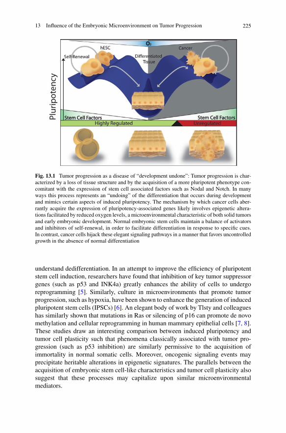
22513 Influence of the Embryonic Microenvironment on Tumor Progression
Fig. 13.1 Tumor progression as a disease of “development undone”: Tumor progression is char-acterized by a loss of tissue structure and by the acquisition of a more pluripotent phenotype con-comitant with the expression of stem cell associated factors such as Nodal and Notch. In many ways this process represents an “undoing” of the differentiation that occurs during development and mimics certain aspects of induced pluripotency. The mechanism by which cancer cells aber-rantly acquire the expression of pluripotency-associated genes likely involves epigenetic altera-tions facilitated by reduced oxygen levels, a microenvironmental characteristic of both solid tumors and early embryonic development. Normal embryonic stem cells maintain a balance of activators and inhibitors of self-renewal, in order to facilitate differentiation in response to specific cues. In contrast, cancer cells hijack these elegant signaling pathways in a manner that favors uncontrolled growth in the absence of normal differentiation
understand dedifferentiation. In an attempt to improve the efficiency of pluripotent stem cell induction, researchers have found that inhibition of key tumor suppressor genes (such as p53 and INK4a) greatly enhances the ability of cells to undergo reprogramming [5]. Similarly, culture in microenvironments that promote tumor progression, such as hypoxia, have been shown to enhance the generation of induced pluripotent stem cells (IPSCs) [6]. An elegant body of work by Tlsty and colleagues has similarly shown that mutations in Ras or silencing of p16 can promote de novo methylation and cellular reprogramming in human mammary epithelial cells [7, 8]. These studies draw an interesting comparison between induced pluripotency and tumor cell plasticity such that phenomena classically associated with tumor pro-gression (such as p53 inhibition) are similarly permissive to the acquisition of immortality in normal somatic cells. Moreover, oncogenic signaling events may precipitate heritable alterations in epigenetic signatures. The parallels between the acquisition of embryonic stem cell-like characteristics and tumor cell plasticity also suggest that these processes may capitalize upon similar microenvironmental mediators.
226 D. Quail et al.
13.2 Cellular Mediators of Plasticity
A number of cell-derived proteins have been shown to promote cellular plasticity in both normal and neoplastic stem cell populations. Moreover, many of these factors work together in co-ordinated regulatory networks. Two such proteins are Nodal and Notch.
13.2.1 Nodal
Nodal is a member of the transforming growth factor beta (TGF-b) superfamily. During embryogenesis and in cancer, Nodal confers its signal as a homodimer by binding to activin-like kinase receptor type I (ALK4/7) and type II (ActRIIB). Upon activating this receptor complex (ALK receptor complex), SMAD2 (and possibly SMAD3) are phosphorylated intracellularly and interact with SMAD4 before trans-locating to the nucleus [9]. In the nucleus, transcription factors such as forkhead box HI (FOXH1) are activated to increase the expression of Nodal responsive genes such as Goosecoid (Gsc) and Lefty. Nodal also induces its own expression, thereby creating a positive feedback loop [10, 11]. The specificity of Nodal is further estab-lished by the epidermal growth factor-cripto FRL1 cryptic (EGF-CFC) family co-receptor, Cripto-1 [10, 12]. Cripto-1 has an N-terminal signal peptide, an EGF-like domain which directly interacts with Nodal, a conserved cysteine-rich (CFC) domain which interacts with ALK4, and a hydrophobic C-terminal glycosyl- phosphatidylinositol (GPI) anchor [13]. The adjacent positioning of the EGF-like domain and the CFC domain helps bring Nodal into proximity with ALK4 to facili-tate enhanced binding [13]. Nodal signaling is inhibited spatially and temporally during development by proteins such as Lefty A, Lefty B, and Cerberus [11, 14]. These inhibitors are transcribed in response to Nodal signaling, and act as a negative feedback mechanism to control Nodal localization and action in the developing embryo [9]. In particular, Lefty is also regulated by alternate SMAD pathways, Wnt, and Oct3/4 signaling, and is upregulated during differentiation events [15]. Lefty inhibits Nodal signaling through interactions with Nodal and/or Cripto-1 that prevent activation of the ALK receptor complex [9].
The primary role of Nodal during embryonic development is to establish anterior–posterior axis patterning and left–right asymmetry [9]. Nodal is first expressed in the mouse epiblast shortly after implantation, and is maintained and enhanced by auto-regulation [16]. Convertases expressed in the adjacent extraembryonic ectoderm pro-cess Nodal predominantly in the proximal epiblast [9, 17]. Activated Nodal signaling subsequently induces Lefty1 and cerberus-like (cerl) in the distal visceral endoderm, which later becomes the anterior visceral endoderm [9]. Eventually, Nodal signaling becomes restricted by Lefty1 and cerl to the proximal posterior region of the epiblast, where the embryonic ectoderm and primitive endoderm are developing, and where the primitive streak will form [12, 14, 18–20]. As development proceeds and cells undergo gastrulation, Nodal becomes restricted to the node at the anterior of the primitive streak, hence the name “Nodal” [9]. The node initiates left–right axis
22713 Influence of the Embryonic Microenvironment on Tumor Progression
formation [19, 21, 22]. Nodal and growth differentiation factor-1 (Gdf-1) from the ventral node pattern the left side of the embryo through interactions with Cryptic in lateral plate mesoderm [14]. On the right side of the embryo, Nodal inhibitors such as Cerl and Lefty, and physical leftward flow from cilia restrict Nodal signaling [14]. During somitogenesis, Nodal becomes more specifically restricted to mesoderm cells on the left side of the embryo, and is downregulated with differentiation until it is no longer present at approximately 8 days post fertilization [20].
Several studies with human embryonic stem cells have sought to elucidate the role of Nodal in human development. Vallier et al. [23] showed that Nodal signaling maintained pluripotency in human embryonic stem cells (hESCs) through SMAD2/3-induced activation of Nanog transcription. In turn, Nanog was shown to interact with SMAD2/3 to limit transcriptional activity of the Nodal signaling pathway, and inhibit endoderm differentiation [23]. Several studies have also shown that Nodal/Activin inhibition in human embryonic stem cells by receptor inhibition with SB431542 induces neuroectoderm specification [23–25]. Together, these studies exemplify the role of Nodal in maintaining pluripotency by inhibiting differentiation into neuroectoderm and mesendoderm lineages in hESCs.
Recent studies have demonstrated that Nodal is also expressed in several cancers, and that this expression is correlated with disease progression. Indeed, Nodal expres-sion has been described in testicular cancer, glioma, melanoma, breast cancer, prostate cancer, and endometrial cancer lesions [10, 26–31]. Studies suggest that Nodal plays a pro-tumorigenic role in many of these cancers. For example, when Nodal signaling was inhibited with a small molecule inhibitory drug (SB431542) or morpholino oligo-nucleotides in melanoma cells, there was a marked reduction in cellular invasion and tumor formation [10]. A recent study demonstrated that Nodal similarly promotes tumor growth, invasion, and dedifferentiation in glioma cells [27]. In another study, Adkins et al. [26] determined that inhibition of Cripto–Nodal signaling via an anti-EGF antibody (A27.F6.1) was able to inhibit tumor growth of NCCIT testicular cancer cells in nude mice. Ongoing studies have recently correlated Nodal expression with tumor progression in prostate cancer [30]. Indeed, compared to poorly aggressive LNCaP prostate cancer cells that express low levels of Nodal, aggressive DU145 pros-tate cancer cells express high levels of Nodal, and undergo anchorage-independent growth and invasion in vitro [30]. Furthermore, transfection of LNCaP cells with a Nodal expression vector increases clonogenicity in vitro [30].
Nodal signaling has also been described in endometrial cancer [31]. Normally, the female endometrium undergoes constant remodeling and turnover throughout the men-strual cycle. In cancer, regulatory mechanisms of these remodeling events go amiss. One study by Papageorgiou et al. [31] showed that Nodal and its co-receptor, Cripto, were expressed during normal proliferative phases of the menstrual cycle in stromal and epithelial cells. Interestingly, patient biopsies of endometrial carcinoma that ranged from Grade 1 to Grade 3 in severity showed a positive correlation between Nodal/Cripto expression and cancer progression [31]. Lefty, a potent inhibitor of Nodal signaling in embryonic stem cells, was absent in all endometrial cancer biopsies [31]. These results are important for understanding normal mechanisms of proliferation in the endometrium, and aberrant mechanisms of endometrial carcinoma progression.
228 D. Quail et al.
Recent studies have demonstrated a pro-metastatic role for Nodal and Cripto in breast cancer. Tissue microarray analyses of human breast tissue samples revealed a positive correlation between Nodal and breast cancer progression [29]. Furthermore, Nodal was completely absent in normal breast tissue samples from these experiments. When MDA-MB-231 cells were treated with Lefty from hESCs, invasion and clonogenicity was reduced concomitant with a downregulation of Nodal gene and protein expression [29]. Recently, Meyer et al. [32] demonstrated that CD44+/CD24+ breast cancer cell populations are able to convert to CD44+/CD24− breast cancer stem cells (and vice versa) both in vitro and in vivo. This suggests that differ-entiated cells outside the breast cancer stem cell subpopulation exhibit a dynamic plastic phenotype. It was found that the central regulator of this dynamic phenotypic switching was the Activin/Nodal pathway. When the ALK receptor was inhibited with SB431542 in either of these populations, phenotypic switching was significantly impaired, implying an important role for Nodal-associated signaling pathways in mediating plasticity in cancer.
Of note, although studies have described the presence of Nodal and Cripto in several cancers, none have reported the concomitant expression of Nodal inhibitors such as Lefty. In fact, studies have shown that breast cancers, melanomas, and endo-metrial cancers do not express the Lefty proteins [29, 31]. This is in sharp contrast with hESCs, where Lefty is among the mostly highly expressed genes [15]. Hence, it is possible that Nodal-associated increases in tumorigenic phenotypes are due to an imbalance in this signaling pathway, rather than its presence per se.
13.2.2 Notch
Signaling between Notch receptors and their ligands is known to play a role in a wide variety of cellular processes including stem cell maintenance, cell specification, differentiation, proliferation, and apoptosis [33]. There are four known mammalian Notch receptors (Notch 1–4) and five ligands (Jagged1, Jagged2, Delta1, Delta3, and Delta4) [34]. The Notch receptors are activated by binding to ligands that are expressed on adjacent cells, exposing a site in the extracellular portion of the transmembrane domain for disintegrin and metalloproteinase (ADAM) mediated cleavage [35]. The Notch intracellular domain (NICD) is subsequently released as a consequence of a second proteolytic cleavage mediated by g-secretase [36]. The liberated NICD trans-locates to the nucleus where it interacts with C-promoter binding factor 1 (CBF-1), suppressor of hairless (Su(H)), lin-12, and glp-1 (Lag-1) (CSL), a DNA-binding tran-scription factor that normally inhibits transcription by interacting with co-repressor proteins [37]. The NICD competes with these repressor proteins to form a NICD-CSL complex which recruits the Mastermind/Lag (MAML) co-activator to form a tran-scriptional activation complex [38]. This complex initiates the transcription of target genes such as Hes and Hey [39] that have been shown to prevent the transcription of lineage-specific genes including Myo-D and Mash-1 [34, 40–42].
The role of Notch in the maintenance of the undifferentiated state is generally thought to arise from its involvement in maintaining the balance between the
22913 Influence of the Embryonic Microenvironment on Tumor Progression
progenitor cell pool and differentiating cells [43, 44]. Studies using constitutively active NICD in both frog and chicken show that increased Notch signaling results in decreased neurogenesis, whereas blocking this pathway leads to a depletion of the pool of progenitor cells and unbalanced neurogenesis [45, 46]. In addition to contrib-uting to stem cell maintenance, Notch has also been found to be involved in binary cell fate decisions through lateral and inductive signaling [47]. Lateral signaling was first elucidated in Drosophila, where it was determined that neuronal precursor cells, which have the capacity to differentiate into either neuronal cells or epidermal cells, initially express both Notch and its ligand, but over time exclusively express one or the other [47]. The Notch-expressing cell, which receives its signal upon binding neighboring receptors, adopts an epidermal cell fate, whereas the cells that exclu-sively express ligand pursue a neuronal cell fate [48]. Inductive signaling occurs between two developmentally distinct cells that express exclusively either receptor or ligand [49]. An example of inductive signaling is seen in mouse thymic epithelial cells: Populations that express Notch1 are able to induce early lymphocyte precur-sors to adopt a T-cell fate as soon as they enter the thymus, whereas populations that lack Notch1 adopt the B-cell fate as a default pathway [50]. Hence, Notch is a factor that has a notable influence over stem cell maintenance and cell fate decisions, which have the potential to become deregulated in the pathological state of cancer.
Just as Notch signaling has been demonstrated to play a role in maintaining cells in a proliferative and undifferentiated state, it has been postulated that its role in cancer is to prevent cells from responding to differentiation cues that they may receive from their immediate environment [51, 52]. How Notch contributes to the tumorigenic process is perhaps the least known aspect of Notch signaling. Depending on the tumor type, Notch can either promote or limit tumor growth through its effects on differentiation, cellular metabolism, cell cycle progression, and possibly self-renewal and immune function [53]. Much of this depends on cellular context and crosstalk with other signal transduction pathways. For example, NOTCH1 prevents cellular proliferation in normal epithelia by increased expression of the cell cycle regulator WAF1; however, it does not appear to play the same role in HPV-containing cervical cancer cells, primarily because RB and WAF1 are inactivated by HPV oncoprotein E7 [54–57]. Furthermore, it has recently been shown that Notch signaling may have a role in the maintenance of normal and malignant stem cells [58–61].
Oncogenic Notch signaling is most clearly understood in T-acute lymphoblastic leukemia (T-ALL), an aggressive neoplasm of immature T-cells [47]. NOTCH1 was first identified by its involvement in a chromosomal translocation, and more recently, has been found to have two types of activating mutations, at least one of which is found in 55–60% cases of human T-ALL [62, 63]. Beyond T-ALL, there is increasing evidence that Notch signals are oncogenic in other cellular contexts, particularly breast cancer and melanoma [64]. The expression of Notch receptors is upregulated in human breast cancers, and the expression of ligands such as Jagged1 correlates with a more aggressive phenotype in both breast and prostate cancer [64–67]. Also, low levels of the Notch antagonist, Numb, correlate with both high levels of Notch signaling and sensitivity to g-secretase inhibitors [68]. Another report on the role of Numb reveals that Notch1 signaling is increased in a variety of molecularly different breast cancers, and that with enforced expression of Numb, such cancers are reverted
230 D. Quail et al.
to a more benign phenotype [69]. In primary human melanomas, expression of Notch1 promotes cancer progression, concomitant with an upregulation of Notch receptors and their downstream target genes [70–72]. Furthermore, in both breast cancer and melanoma cells, an interplay between increased Notch activity and acti-vation of the P13K-Akt pathway involved in cellular metabolism has also been established; however, the specifics of the interplay are still not understood [72]. As is the case with normal Notch signaling, the role of Notch in cancer is known to be somewhat dependent on context. In many cases, Notch can alternatively serve as a tumor suppressor as opposed to an oncogene [73]. In breast cancer, Notch2 is known to function as a tumor suppressor, whereas the other Notch receptors func-tion as oncogenes [73]. Furthermore, Notch1 knockout mice develop basal cell carcinoma-like lesions, and mice with dominant negative MAML1 develop cutaneous squamous cell carcinomas [74].
A complexity between the Notch and Nodal signaling pathways has recently emerged, providing further evidence for co-opted stem cell pathways in cancer that may interact in a similar manner to their normal stem cell context [75]. Mouse embryos lacking functional components of the Notch pathway exhibited disorga-nized left–right asymmetry patterning, a phenomenon traditionally attributed to Nodal signaling [40, 41]. Analysis of the NDE region upstream from the Nodal gene locus lead to the identification of two binding sites for CSL, the primary transcrip-tional mediator of Notch signaling [40, 41]. This region has been shown to be respon-sive to Notch signaling in mouse embryos [40, 41]. Furthermore, Baf60c, a subunit of the Swi/Snf-like BAF chromatin remodeling complex, is essential for the integra-tion of Notch signaling components for transcriptional activation of Nodal in the mouse node [76]. These studies revealed a role for the Notch pathway working upstream of Nodal expression in left–right asymmetry patterning in mice. Furthermore, it was determined that inhibiting Notch in metastatic melanoma cells with a g-secretase inhibitor (DAPT) or by Notch4-specific small interfering RNA (siRNA) results in a decrease in Nodal expression [77]. Moreover, these studies indicate that human Nodal, like mouse Nodal, is upregulated by Notch signaling in cancer cells [77]. Recently, more complexity has emerged in the crosstalk between the Notch and Nodal signaling pathways, with the finding that the Nodal co-receptor Cripto-1 is able to facilitate the posttranslational maturation of Notch receptors [78, 79]. The similar appearance of Notch and Nodal signaling in breast cancer and melanoma, as well as their interaction in development, demonstrates a recapitulation of the interac-tion found in a normal stem cell context and indicates a way in which the interplay between two stem cell pathways may be mediating a stem cell phenotype in cancer.
13.3 Oxygen as a Regulator of Cellular Plasticity
Cell-derived factors potentiate the acquisition and maintenance of self-renewal and plasticity. However, such parameters do not operate autonomously; rather, they are dynamically regulated by alterations in the external milieu. As a solid tumor grows,
23113 Influence of the Embryonic Microenvironment on Tumor Progression
the rate of cell proliferation exceeds that of normal tissue growth and consequently interferes with the ability of the existing vasculature to supply growth factors, nutrients, and oxygen, and to remove waste products from the cells [80]. This results in areas within the tumor that have pathologically low levels of oxygen, low glucose levels, and low extracellular pH [80, 81]. These phenomena result in heterogeneous populations of cells within the tumor, and furthermore, differences between tumors [80]. Such aspects of the external tumor environment have been extensively linked to tumor progression and metastatic disease, and thus are important considerations when investigating the role of cancer stem cells within a complex and deregulated microenvironment.
Oxygen availability dramatically alters the expression profile and behavior of cells. Hypoxia, which is defined as a state of low oxygen tension that falls below critical levels, initiates complex and specialized responses at the molecular, cellular, tissue, and organismal level so as to re-establish oxygen homeostasis and minimize the detrimental effects of low oxygen [81]. The oxygen concentrations in mamma-lian tissues can range from 150 mmHg in the upper airways to 5 mmHg in tissues such as the retina [82, 83]. The first model of tumor hypoxia proposed by Thomlinson and Gray [84] suggested that as a result of diffusion-limited hypoxia (a consequence of tumor cells being sufficiently distant from the vasculature), cells within a tumor could remain viable in chronic levels of hypoxia for a few hours to a few days [85, 86]. More recently, studies of blood flow and oxygen levels in animal tumors have suggested a model of perfusion-limited hypoxia (acute or fluctuating hypoxia), in which perfusion of blood vessels is dynamic and can result in transient periods of severe hypoxia within the tumor [87–89]. As a result, it is now believed that the oxygenation levels within a tumor can vary, not just by the diffusion to cells that are distant from the vasculature but also by the fluctuating levels of blood flowing through the vessels to the tumor [90]. In response to hypoxia, there is reduced oxi-dative phosphorylation in the mitochondria, and those activities that require large amounts of ATP, such as ATP-dependent ion channel function and protein transla-tion, are repressed [91]. Moreover, glycolysis becomes immediately upregulated to compensate for the lower-than-normal ATP production in the cell [91]. Generally, most transcription is repressed during times of hypoxia; however, transcription of subsets of genes, including vascular endothelial growth factor (VEGF), erythropoe-itin (EPO), and glucose transporters (GLUT1, GLUT3) are increased dramatically [81, 92]. At the tissue level, angiogenesis and EPO concentrations are enhanced in hypoxia to increase the oxygen delivery to affected areas [81, 93]. Specifically, the development of tumor hypoxia is fundamentally linked to the formation of neovas-culature by the process of angiogenesis.
Intratumoral oxygen tensions are strikingly similar to the low levels of oxygen experienced in early development [94]. This correlation suggests that specific path-ways regulated by low oxygen in stem cells may also manifest in cancer. Early embryonic development occurs in a low oxygen environment (1–2%), and increased oxygen levels are detrimental to the proper execution of early developmental events and stem cell differentiation [95, 96]. Also, many human stem cells inhabit hypoxic niches throughout the body in adult life. For example, bone marrow–derived stem
232 D. Quail et al.
cells reside in a microenvironment that has been estimated to have a relatively low oxygen tension (1–2%) [97]. When these cells are cultured ex vivo, they exhibit greater proliferation and ability to reconstitute the bone marrow of NOD/SCID mice if they are cultured in hypoxia vs. atmospheric oxygen [97, 98]. Other stem cells have also been reported to thrive in a hypoxic environment, such that their prolifera-tion and maintenance of stem cell identity is promoted [99, 100]. Neuronal stem cells exhibit increased proliferation and preferential differentiation into certain cell types when cultured in hypoxic conditions (1–5%) over normoxic conditions [99, 100]. Furthermore, culturing hESCs in hypoxic conditions (3–5%) results in main-tenance of an undifferentiated state, whereas culture at atmospheric oxygen causes differentiation of the cells [101]. Exposure to hypoxia also supports cancer stem cells [80]. For example, recent studies demonstrated that hypoxia promotes CD133-positive cancer stem cell populations in glioblastomas [102]. Furthermore, breast cancer cells have been shown to decrease estrogen receptor expression, and increase cytokertain 19 (an epithelial stem cell marker), following 3 days of exposure to 1% oxygen. Neuroblastoma cells exposed to hypoxia similarly assume a dedifferentiated phenotype, characterized by a reduction in neuronal/neuroendocrine markers such as dHAND, concomitant with an enhancement of markers including cKit and Notch-1, which characterize neural crest progenitors [103]. The parallel between the oxygen-mediated regulation of cell fate in stem cells and cancer cells is strong, supporting the notion that low oxygen has the ability to promote stem-like phenotypes in cancer, ultimately leading to metastatic spread.
Several studies have demonstrated that hypoxia promotes pluripotency and self-renewal in stem cells, and that it characterizes several stem cell niches [101, 104]. Hypoxia is similarly able to sustain cancer cell immortality and plasticity [94]. An important protein that has been found to be upregulated by hypoxia is the embry-onic stem cell associated transcription factor Oct4 [105]. Oct4 is expressed in the embryonic stem cells of the inner cell mass of the blastocyst [106]. This pivotal protein is downregulated in somatic cells around the time of gastrulation, but is retained in primordial germ cells and in some adult stem cell populations [106]. A recent study revealed a significant link between hypoxia inducible transcription factors (HIFs) and Oct4 expression during development. In this study, a HIF-2a gene “knock-in” mouse was generated on a HIF-1a deficient background. Surprisingly, this knock-in did not rescue the effects of the HIF-1a knockout. Rather, the mice showed gross developmental abnormalities and embryonic lethality between E3.5 and E7.5 (earlier than HIF-1a deficient embryos alone which died at E9.5–E10.5). These HIF-2a knock-in mice had an enhanced expression of Oct4 [105]. ChIP anal-yses further revealed that HIF-2a binds to hypoxic regulatory regions in the mouse Oct4 promoter, and that this ability is not shared by HIF-1a [105]. Hence, by preventing terminal differentiation, the HIF-2 induced expression of Oct4 resulted in embryonic lethality. Of note, Oct4 expression is found in numerous tumor cell types, as well as human adult stem cells, and increased expression of Oct4 in embryonic stem cells causes them to form tumors in a dose-dependent manner [106, 107]. Thus, it is quite plausible that hypoxia promotes and/or sustains stem cell phenotypes and tumorige-nicity via the HIF-2a mediated upregulation of Oct4.
23313 Influence of the Embryonic Microenvironment on Tumor Progression
An equally impressive finding is the discovery that HIF proteins also regulate the activity of c-Myc, an oncogene of central importance in many cancers and in embryological development [108]. HIF-1a was found to antagonize c-Myc activity by competing for binding to its transcription factor Sp1 in hypoxia, and thus inhibiting c-Myc dependent cell cycle progression [109]. However, HIF-2a had the opposite effect, and was able to promote cell cycle progression and also contribute to onco-genic transformation by enhancing the transcriptional effects of c-Myc [110]. This resulted in the activation and repression of c-Myc target genes in multiple cancer cell lines, mouse embryonic fibroblasts, and embryonic stem cell lines [110]. It is important to note that Oct4 and c-Myc are two of the four genes that were shown to be sufficient to generate iPSCs and therefore, it is highly probable that their hypoxia-induced expression in cancer plays a pivotal role in the development of a cancer stem cell phenotype [4, 111]. Furthermore, this provides a clear example of how hypoxia contributes to a poorly differentiated phenotype and the existence of stem-like cancer cells.
As mentioned previously, Notch is another stem cell factor known to play a role in cancer. Of note, hypoxia upregulates the activity of Notch signaling in a HIF-1 dependent manner [112]. Specifically, HIF-1a binds and stabilizes the cleaved NICD, thereby increasing the efficiency of NICD binding to CSL and initiating transcription of Notch target genes [112]. The interaction of HIF-1a with Notch was shown to inhibit neuronal and myogenic differentiation, and HIF-1a deficient mouse embryonic fibroblasts showed a decrease in activation of Notch target genes under hypoxia [112]. Furthermore, the interaction between Notch and HIF-1a shows that hypoxia is able to increase Notch activity and contribute to the maintenance of pluripotency in embryonic stem cells [113]. As previously described, Notch signaling is also involved in various forms of cancer. Hence, hypoxia may promote cancer stem cell phenotypes via the activation of Notch.
Notch is also known to regulate the potent stem cell factor Nodal in an embryonic environment, and is thought to have a similar activating effect in cancer. The relation-ship between Notch and Nodal is further supported by the finding that both converge on hypoxia and HIF-1a signaling. Preliminary studies done in our laboratory suggest that Nodal is regulated by low oxygen, and that hypoxia exerts its influence through HIF signaling [114]. When poorly metastatic breast cancer and melanoma cell lines (MCF-7, T47D, C81-61) that do not endogenously express Nodal were exposed to 0.5% oxygen for 48 hours, induction of Nodal mRNA expression and protein expression was observed. By examining the role of the HIF pathway using HIF-1a-specific siRNA in hypoxia and introducing at HIF-1a containing expression vector in normoxia, it was also determined that hypoxia-induced Nodal expression is dependent on the HIF-1a pathway. As discussed above, it was determined in embry-onic studies that Notch binds to the NDE region of the Nodal gene. In this respect, it is possible that Notch, through its activation by hypoxia and HIF-1a, may be contributing to the undifferentiated state of cancer cells by inducing the expression of Nodal.
234 D. Quail et al.
13.4 Targeting Tumor Plasticity: Restoring the Embryonic Program
The plasticity that allows cancer to progress also presents a unique and powerful target for treatment. There are two main anti-cancer strategies that capitalize on the embryonic microenvironment: (1) Normal embryonic niches, containing a balance of activators and inhibitors of stem cell fate, can be used to restore balance to aber-rant signaling in cancer cells. As a consequence, the cancer cells would either stop growing due to an inhibition of self-renewal cues or reprogram toward a more dif-ferentiated phenotype. (2) Cancer cells can be fully reprogrammed back to an induced pluripotent stem cell, and then differentiated into a cell with a relatively normal epi-genetic status and phenotype. Such a cell could then be targeted for treatment.
Numerous studies have revealed that embryonic microenvironments reduce tum-origenicity by inducing senescence and differentiation in cancer cells. For example, Pierce and Wells discovered that B16 murine melanoma cells are unable to form tumors and appear to differentiate toward a neuronal phenotype following exposure to microenvironmental factors derived from the embryonic skin of a developing mouse [115]. A number of zebrafish models have confirmed these findings. For example, extracts derived from zebrafish embryos have been shown to inhibit proliferation and induce apoptosis in several cancer cell types [116]. Furthermore, following transplan-tation into zebrafish embryos, metastatic melanoma cells lay dormant and are unable to form tumors [117]. Interestingly, this phenomenon is unique to the embryonic zebrafish, as human melanoma cells transplanted into zebrafish 2 days after fertiliza-tion (after morphogenesis and organogenesis are complete) form tumors and even induce angiogenesis [117]. Bissell and colleagues documented that Rous sarcoma virus, which causes a rapidly growing tumor when injected into hatched chicks, is non-tumorigenic when injected into 4-day-old chick embryos, despite viral replica-tion and v-src oncogene activation [118]. In another set of experiments, transplanta-tion of melanoma cells in ovo adjacent to host chick premigratory neural crest cells caused the transplanted melanoma cells to respond to neural crest cues by populating structures such as the brachial arches, sympathetic ganglia, and dorsal root, in a manner similar to neural crest cells [119]. Interestingly, a subpopulation of melanoma cells that invaded the chick periphery was reprogrammed to express the melanocyte-associated protein Mart-1/Melan-A, thus confirming that melanoma cells can respond to devel-opmental cues by undergoing differentiation [119].
As an extension to these findings, an in vitro 3D model was developed to examine whether the microenvironment of hESCs could similarly reprogram metastatic phe-notypes [29, 120]. In this model, hESCs were cultured on 3D extracellular matrices and were removed, thereby generating a cell autonomous hESC conditioned envi-ronment. Utilizing this approach, exposure of melanoma and breast cancer cells to extracellular matrices conditioned by hESCs (CMTX) resulted in reduced in vivo tumorigenicity concomitant with increased apoptosis [29]. More recently, a study demonstrated that conditioned medium from hESCs reduces the proliferation of ovarian, breast, and prostate cancer cells, but does not affect normal fibroblasts [121].
23513 Influence of the Embryonic Microenvironment on Tumor Progression
Collectively, these studies suggest that hESC-derived factors have anti-tumor activi-ties. In an effort to isolate factor(s) that confer such properties, separation of condi-tioned medium has been conducted, and anti-tumorigenic entities appear to be enriched in low molecular weight (<10 kDa), heat stable fractions [121]. The identi-ties of such factors have not, however, been described. Using the 3D CMTX model, Lefty was isolated as a protein with major anti-tumorigenic properties, largely due to its ability to inhibit Nodal signaling [29]. Of note, the hESC-derived CMTX was able to inhibit tumorigenic phenotypes even when Lefty was depleted, suggesting that other factors are also involved in this complex process [29]. Proteomic analyses of the hESC microenvironment, combined with functional assays, are vital to dis-covering which proteins in the hESC milieu may be harnessed for the treatment of cancers [122].
A number of studies have demonstrated that embryonic microenvironments can inhibit the tumorigenicity of cancer cells through the process of reprogramming followed by differentiation. One of the first examples was presented by Mintz and Illmensee [123]. In this seminal study, embryonal carcinoma cells were injected into blastocysts. Surprisingly, these cancer cells did not form tumors, nor did they mitigate embryological development. Rather, they partook in the development of all tissues, inclusive of the germ line. In corroboration with these findings, Jaenisch and colleagues demonstrated that nuclear transplantation of a RAS-inducible melanoma nucleus into an oocyte leads to the reprogramming of the melanoma genome, giving rise to ESCs with the capacity to differentiate into cell types such as melanocytes and fibroblasts [124]. While predisposing mice to RAS-induced tumors later in life, this reprogramming event illustrated the ability of the oocyte environment to epige-netically reprogram cancers to a well-differentiated phenotype. More recently, a number of studies have shown that cancer cells can be programmed to iPSCs, despite karyotypic abnormalities. For example, iPSCs have been generated from leukemic cells, colon cancer cells, and melanoma [125–127]. Notably, iPSCs were generated from human chronic myeloid leukemia cells containing the Philadelphia transloca-tion (BCR-Abl). Prior to reprogramming, these cells had acquired resistance to Gleevec; however, hematopoietic cells differentiated from the iPSCs had regained sensitivity to this drug [125]. These findings point to the epigenetic nature of many phenomena associated with cancer progression, such as drug resistance. Moreover, they suggest that reprogramming followed by differentiation is a powerful strategy for the normalization and treatment of advanced cancers.
13.5 Conclusions
Tumor progression is characterized by a loss of differentiation and by the acquisition of an embryonic stem cell-like gene expression pattern (summarized in Fig. 13.1) [1, 128]. Several stem cell–derived proteins such as Nodal and the Notch receptors facilitate this transition. The mechanism by which cancer cells aberrantly acquire the expression of such pluripotency-associated genes is not clearly understood, but
236 D. Quail et al.
likely involves epigenetic alterations induced by biophysical parameters such as reduced oxygenation. Normal embryonic stem cells maintain a balance of activators and inhibitors of self-renewal, in order to facilitate differentiation in response to specific cues. For example, hESC populations express high levels of both Nodal and the Nodal inhibitors Lefty and Cerl [29, 129]. In contrast, cancer cells hijack these elegant signaling pathways in a manner that favors uncontrolled growth in the absence of normal differentiation. This imbalance of stem cell associated cues in cancer presents a unique and powerful target for treatment. Indeed, mounting evi-dence suggests that normal embryonic niches, containing a balance of activators and inhibitors of stem cell fate, can be harnessed to reprogram cancer cells toward a more differentiated phenotype. This approach of re-establishing normal develop-mental signaling may be of great therapeutic value for the prevention and eradication of cancer stem cell-like populations.
References
1. Hendrix MJ, Seftor EA, Seftor RE, Kasemeier-Kulesa J, Kulesa PM, Postovit LM (2007l) Reprogramming metastatic tumour cells with embryonic microenvironments. Nat Rev Cancer 7:246–255
2. Visvader JE, Lindeman GJ (2006) Mammary stem cells and mammopoiesis. Cancer Res 66:9798–9801
3. Yu J, Vodyanik MA, Smuga-Otto K, Antosiewicz-Bourget J, Frane JL, Tian S, Nie J, Jonsdottir GA, Ruotti V, Stewart R, Slukvin II, Thomson JA (2007) Induced pluripotent stem cell lines derived from human somatic cells. Science 318:1917–1920
4. Takahashi K, Tanabe K, Ohnuki M, Narita M, Ichisaka T, Tomoda K, Yamanaka S (2007) Induction of pluripotent stem cells from adult human fibroblasts by defined factors. Cell 131:861–872
5. Utikal J, Polo JM, Stadtfeld M, Maherali N, Kulalert W, Walsh RM, Khalil A, Rheinwald JG, Hochedlinger K (2009) Immortalization eliminates a roadblock during cellular reprogram-ming into iPS cells. Nature 460:1145–1148
6. Yoshida Y, Takahashi K, Okita K, Ichisaka T, Yamanaka S (2009) Hypoxia enhances the generation of induced pluripotent stem cells. Cell Stem Cell 5:237–241
7. Dumont N, Wilson MB, Crawford YG, Reynolds PA, Sigaroudinia M, Tlsty TD (2008) Sustained induction of epithelial to mesenchymal transition activates DNA methylation of genes silenced in basal-like breast cancers. Proc Natl Acad Sci USA 105:14867–14872
8. Reynolds PA, Sigaroudinia M, Zardo G, Wilson MB, Benton GM, Miller CJ, Hong C, Fridlyand J, Costello JF, Tlsty TD (2006) Tumor suppressor p16INK4A regulates polycomb-mediated DNA hypermethylation in human mammary epithelial cells. J Biol Chem 281:24790–24802
9. Schier AF (2003) Nodal signaling in vertebrate development. Annu Rev Cell Dev Biol 19:589–621
10. Topczewska JM, Postovit LM, Margaryan NV, Sam A, Hess AR, Wheaton WW, Nickoloff BJ, Topczewski J, Hendrix MJ (2006) Embryonic and tumorigenic pathways converge via Nodal signaling: role in melanoma aggressiveness. Nat Med 12:925–932
11. Schier AF (2009) Nodal morphogens. Cold Spring Harb Perspect Biol 1:a003459 12. Strizzi L, Postovit LM, Margaryan NV, Seftor EA, Abbott DE, Seftor RE, Salomon DS,
Hendrix MJ (2008) Emerging roles of nodal and Cripto-1: from embryogenesis to breast cancer progression. Breast Dis 29:91–-103
13. Minchiotti G (2005) Nodal-dependant Cripto signaling in ES cells: from stem cells to tumor biology. Oncogene 24:5668–5675
23713 Influence of the Embryonic Microenvironment on Tumor Progression
14. Constam DB (2009) Running the gauntlet: an overview of the modalities of travel employed by the putative morphogen Nodal. Curr Opin Genet Dev 19:302–307
15. Tabibzadeh S, Hemmati-Brivanlou A (2006) Lefty at the crossroads of “stemness” and differ-entiative events. Stem Cells 24:1998–2006
16. Ang SL, Constam DB (2004) A gene network establishing polarity in the early mouse embryo. Semin Cell Dev Biol 15:555–561
17. Beck S, Le Good JA, Guzman M, Ben HN, Roy K, Beermann F, Constam DB (2002) Extraembryonic proteases regulate Nodal signalling during gastrulation. Nat Cell Biol 4:981–985
18. Constam DB (2009) Riding shotgun: a dual role for the epidermal growth factor-Cripto/FRL-1/Cryptic protein Cripto in Nodal trafficking. Traffic 10:783–791
19. Zhou X, Sasaki H, Lowe L, Hogan BL, Kuehn MR (1993) Nodal is a novel TGF-beta-like gene expressed in the mouse node during gastrulation. Nature 361:543–547
20. Collignon J, Varlet I, Robertson EJ (1996) Relationship between asymmetric nodal expression and the direction of embryonic turning. Nature 381:155–158
21. Smith JC (1995) Mesoderm-inducing factors and mesodermal patterning. Curr Opin Cell Biol 7:856–861
22. Takaoka K, Yamamoto M, Shiratori H, Meno C, Rossant J, Saijoh Y, Hamada H (2006) The mouse embryo autonomously acquires anterior-posterior polarity at implantation. Dev Cell 10:451–459
23. Vallier L, Mendjan S, Brown S, Chng Z, Teo A, Smithers LE, Trotter MW, Cho CH, Martinez A, Rugg-Gunn P, Brons G, Pedersen RA (2009) Activin/Nodal signalling maintains pluripotency by controlling Nanog expression. Development 136:1339–1349
24. Smith JR, Vallier L, Lupo G, Alexander M, Harris WA, Pedersen RA (2008) Inhibition of Activin/Nodal signaling promotes specification of human embryonic stem cells into neuroec-toderm. Dev Biol 313:107–117
25. Patani R, Compston A, Puddifoot CA, Wyllie DJ, Hardingham GE, Allen ND, Chandran S (2009) Activin/Nodal inhibition alone accelerates highly efficient neural conversion from human embryonic stem cells and imposes a caudal positional identity. PLoS One 4:e7327
26. Adkins HB, Bianco C, Schiffer SG, Rayhorn P, Zafari M, Cheung AE, Orozco O, Olson D, De LA, Chen LL, Miatkowski K, Benjamin C, Normanno N, Williams KP, Jarpe M, LePage D, Salomon D, Sanicola M (2003) Antibody blockade of the Cripto CFC domain suppresses tumor cell growth in vivo. J Clin Invest 112:575–587
27. Lee CC, Jan HJ, Lai JH, Ma HI, Hueng DY, Gladys Lee YC, Cheng YY, Liu LW, Wei HW, Lee HM (2010) Nodal promotes growth and invasion in human gliomas. Oncogene 29:3110–3123
28. Yu L, Harms PW, Pouryazdanparast P, Kim DS, Ma L, Fullen DR (2010) Expression of the embryonic morphogen Nodal in cutaneous melanocytic lesions. Mod Pathol 23:1209–1214
29. Postovit LM, Margaryan NV, Seftor EA, Kirschmann DA, Lipavsky A, Wheaton WW, Abbott DE, Seftor RE, Hendrix MJ (2008) Human embryonic stem cell microenvironment suppresses the tumorigenic phenotype of aggressive cancer cells. Proc Natl Acad Sci USA 105:4329–4334
30. Lawrence M. (2009) Crosstalk between developmental and tumour-specific signalling path-ways: Kallikrein-related serine peptidases and Nodal in prostate cancer. PhD thesis, QIT, Brisbane, Australia
31. Papageorgiou I, Nicholls PK, Wang F, Lackmann M, Makanji Y, Salamonsen LA, Robertson DM, Harrison CA (2009) Expression of nodal signalling components in cycling human endo-metrium and in endometrial cancer. Reprod Biol Endocrinol 7:122
32. Meyer MJ, Fleming JM, Ali MA, Pesesky MW, Ginsburg E, Vonderhaar BK (2009) Dynamic regulation of CD24 and the invasive, CD44posCD24neg phenotype in breast cancer cell lines. Breast Cancer Res. 11:R82
33. Lai EC (2004) Notch signaling: control of cell communication and cell fate. Development 131:965–973
34. Bray SJ (2006) Notch signalling: a simple pathway becomes complex. Nat Rev Mol Cell Biol 7:678–689
238 D. Quail et al.
35. Nichols JT, Miyamoto A, Olsen SL, D’Souza B, Yao C, Weinmaster G (2007) DSL ligand endocytosis physically dissociates Notch1 heterodimers before activating proteolysis can occur. J Cell Biol 176:445–458
36. Fortini ME (2002) Gamma-secretase-mediated proteolysis in cell-surface-receptor signalling. Nat Rev Mol Cell Biol 3:673–684
37. Mumm JS, Kopan R (2000) Notch signaling: from the outside in. Dev Biol 228:151–165 38. Nam Y, Aster JC, Blacklow SC (2002) Notch signaling as a therapeutic target. Curr Opin
Chem Biol 6:501–509 39. Miele L, Golde T, Osborne B (2006) Notch signaling in cancer. Curr Mol Med 6:905–918 40. Raya A, Kawakami Y, Rodriguez-Esteban C, Buscher D, Koth CM, Itoh T, Morita M, Raya
RM, Dubova I, Bessa JG, de la Pompa JL, Belmonte JC (2003) Notch activity induces Nodal expression and mediates the establishment of left-right asymmetry in vertebrate embryos. Genes Dev 17:1213–1218
41. Krebs LT, Iwai N, Nonaka S, Welsh IC, Lan Y, Jiang R, Saijoh Y, O’Brien TP, Hamada H, Gridley T (2003) Notch signaling regulates left-right asymmetry determination by inducing Nodal expression. Genes Dev 17:1207–1212
42. Iso T, Kedes L, Hamamori Y (2003) HES and HERP families: multiple effectors of the Notch signaling pathway. J Cell Physiol 194:237–255
43. Lewis J (1996) Neurogenic genes and vertebrate neurogenesis. Curr Opin Neurobiol 6:3–10 44. Lewis J (1998) Notch signalling. A short cut to the nucleus. Nature 393:304–305 45. Chitnis A, Henrique D, Lewis J, Ish-Horowicz D, Kintner C (1995) Primary neurogenesis in
Xenopus embryos regulated by a homologue of the Drosophila neurogenic gene Delta. Nature 375:761–766
46. Henrique D, Hirsinger E, Adam J, Le R, I, Pourquie O, Ish-Horowicz D, Lewis J (1997) Maintenance of neuroepithelial progenitor cells by Delta-Notch signalling in the embryonic chick retina. Curr Biol 7:661–670
47. Radtke F, Raj K (2003) The role of Notch in tumorigenesis: oncogene or tumour suppressor? Nat Rev Cancer 3:756–767
48. Kimble J, Simpson P (1997) The LIN-12/Notch signaling pathway and its regulation. Annu Rev Cell Dev Biol 13:333–361
49. Artavanis-Tsakonas S, Matsuno K, Fortini ME (1995) Notch signaling. Science 268:225–232 50. Pear WS, Radtke F (2003) Notch signaling in lymphopoiesis. Semin Immunol 15:69–79 51. Milner LA, Bigas A, Kopan R, Brashem-Stein C, Bernstein ID, Martin DI (1996) Inhibition
of granulocytic differentiation by mNotch1. Proc Natl Acad Sci USA 93:13014–13019 52. Milner LA, Bigas A (1999) Notch as a mediator of cell fate determination in hematopoiesis:
evidence and speculation. Blood 93:2431–2448 53. Roy M, Pear WS, Aster JC (2007) The multifaceted role of Notch in cancer. Curr Opin Genet
Dev 17:52–59 54. Rangarajan A, Syal R, Selvarajah S, Chakrabarti O, Sarin A, Krishna S (2001) Activated
Notch1 signaling cooperates with papillomavirus oncogenes in transformation and generates resistance to apoptosis on matrix withdrawal through PKB/Akt. Virology 286:23–30
55. Munger K, Basile JR, Duensing S, Eichten A, Gonzalez SL, Grace M, Zacny VL (2001) Biological activities and molecular targets of the human papillomavirus E7 oncoprotein. Oncogene 20:7888–7898
56. Helt AM, Funk JO, Galloway DA (2002) Inactivation of both the retinoblastoma tumor sup-pressor and p21 by the human papillomavirus type 16 E7 oncoprotein is necessary to inhibit cell cycle arrest in human epithelial cells. J Virol 76:10559–10568
57. Westbrook TF, Nguyen DX, Thrash BR, McCance DJ (2002) E7 abolishes raf-induced arrest via mislocalization of p21(Cip1). Mol Cell Biol 22:7041–7052
58. Axelson H, Fredlund E, Ovenberger M, Landberg G, Pahlman S (2005) Hypoxia-induced dedifferentiation of tumor cells--a mechanism behind heterogeneity and aggressiveness of solid tumors. Semin Cell Dev Biol 16:554–563
59. Dontu G, Jackson KW, McNicholas E, Kawamura MJ, Abdallah WM, Wicha MS (2004) Role of Notch signaling in cell-fate determination of human mammary stem/progenitor cells. Breast Cancer Res 6:R605–R615
23913 Influence of the Embryonic Microenvironment on Tumor Progression
60. Hitoshi S, Alexson T, Tropepe V, Donoviel D, Elia AJ, Nye JS, Conlon RA, Mak TW, Bernstein A, van der KD (2002) Notch pathway molecules are essential for the maintenance, but not the generation, of mammalian neural stem cells. Genes Dev 16:846–858
61. Hitoshi S, Seaberg RM, Koscik C, Alexson T, Kusunoki S, Kanazawa I, Tsuji S, van der KD (2004) Primitive neural stem cells from the mammalian epiblast differentiate to definitive neural stem cells under the control of Notch signaling. Genes Dev 18:1806–1811
62. Strizzi L, Hardy KM, Seftor EA, Costa FF, Kirschmann DA, Seftor RE, Postovit LM, Hendrix MJ (2009) Development and cancer: at the crossroads of Nodal and Notch signaling. Cancer Res 69:7131–7134
63. Abbott DE, Bailey CM, Postovit LM, Seftor EA, Margaryan NV, Hendrix MJ (2008) The epigenetic influence of tumor and embryonic microenvironments: How different are they? Cancer Microenvironment 1:13–21.
64. Weijzen S, Rizzo P, Braid M, Vaishnav R, Jonkheer SM, Zlobin A, Osborne BA, Gottipati S, Aster JC, Hahn WC, Rudolf M, Siziopikou K, Kast WM, Miele L (2002) Activation of Notch-1 signaling maintains the neoplastic phenotype in human Ras-transformed cells. Nat Med 8:979–986
65. Reedijk M, Odorcic S, Chang L, Zhang H, Miller N, McCready DR, Lockwood G, Egan SE (2005) High-level coexpression of JAG1 and NOTCH1 is observed in human breast cancer and is associated with poor overall survival. Cancer Res 65:8530–8537
66. Santagata S, Demichelis F, Riva A, Varambally S, Hofer MD, Kutok JL, Kim R, Tang J, Montie JE, Chinnaiyan AM, Rubin MA, Aster JC (2004) JAGGED1 expression is associated with prostate cancer metastasis and recurrence. Cancer Res 64:6854–6857
67. Bismar TA, Demichelis F, Riva A, Kim R, Varambally S, He L, Kutok J, Aster JC, Tang J, Kuefer R, Hofer MD, Febbo PG, Chinnaiyan AM, Rubin MA (2006) Defining aggressive prostate cancer using a 12-gene model. Neoplasia 8:59–68
68. Pece S, Serresi M, Santolini E, Capra M, Hulleman E, Galimberti V, Zurrida S, Maisonneuve P, Viale G, Di Fiore PP (2004) Loss of negative regulation by Numb over Notch is relevant to human breast carcinogenesis. J Cell Biol 167:215–221
69. Stylianou S, Clarke RB, Brennan K (2006) Aberrant activation of notch signaling in human breast cancer. Cancer Res 66:1517–1525
70. Balint K, Xiao M, Pinnix CC, Soma A, Veres I, Juhasz I, Brown EJ, Capobianco AJ, Herlyn M, Liu ZJ (2005) Activation of Notch1 signaling is required for beta-catenin-mediated human primary melanoma progression. J Clin Invest 115:3166–3176
71. Hoek K, Rimm DL, Williams KR, Zhao H, Ariyan S, Lin A, Kluger HM, Berger AJ, Cheng E, Trombetta ES, Wu T, Niinobe M, Yoshikawa K, Hannigan GE, Halaban R (2004) Expression profiling reveals novel pathways in the transformation of melanocytes to melanomas. Cancer Res 64:5270–5282
72. Liu ZJ, Xiao M, Balint K, Smalley KS, Brafford P, Qiu R, Pinnix CC, Li X, Herlyn M (2006) Notch1 signaling promotes primary melanoma progression by activating mitogen-activated protein kinase/phosphatidylinositol 3-kinase-Akt pathways and up-regulating N-cadherin expression. Cancer Res 66:4182–4190
73. O’Neill CF, Urs S, Cinelli C, Lincoln A, Nadeau RJ, Leon R, Toher J, Mouta-Bellum C, Friesel RE, Liaw L (2007) Notch2 signaling induces apoptosis and inhibits human MDA-MB-231 xenograft growth. Am J Pathol 171:1023–1036
74. Nicolas M, Wolfer A, Raj K, Kummer JA, Mill P, van NM, Hui CC, Clevers H, Dotto GP, Radtke F (2003) Notch1 functions as a tumor suppressor in mouse skin. Nat Genet 33:416-421
75. Dreesen O, Brivanlou AH (2007) Signaling pathways in cancer and embryonic stem cells. Stem Cell Rev 3:7–17
76. Takeuchi JK, Lickert H, Bisgrove BW, Sun X, Yamamoto M, Chawengsaksophak K, Hamada H, Yost HJ, Rossant J, Bruneau BG (2007) Baf60c is a nuclear Notch signaling component required for the establishment of left-right asymmetry. Proc Natl Acad Sci USA 104:846–851
77. Postovit LM, Seftor EA, Seftor RE, Hendrix MJ (2007) Targeting Nodal in malignant mela-noma cells. Expert Opin Ther Targets 11:497–505
78. Watanabe K, Nagaoka T, Lee JM, Bianco C, Gonzales M, Castro NP, Rangel MC, Sakamoto K, Sun Y, Callahan R, Salomon DS (2009) Enhancement of Notch receptor maturation and signaling sensitivity by Cripto-1. J Cell Biol 187:343–353
240 D. Quail et al.
79. Watanabe K, Kondo K, Takeuchi N, Okano H, Yamasoba T (2007) Musashi-1 expression in postnatal mouse olfactory epithelium. Neuroreport 18:641–644
80. Lunt SJ, Chaudary N, Hill RP (2009) The tumor microenvironment and metastatic disease. Clin Exp Metastasis 26:19–34
81. Postovit LM, Sullivan R, Adams MA, Graham CH (2005) Nitric oxide signalling and cellular adaptations to changes in oxygenation. Toxicology 208:235–248
82. Nathan AT, Singer M (1999) The oxygen trail: tissue oxygenation. Br Med Bull 55:96–108 83. Birol G, Wang S, Budzynski E, Wangsa-Wirawan ND, Linsenmeier RA (2007) Oxygen dis-
tribution and consumption in the macaque retina. Am J Physiol Heart Circ Physiol 293:H1696–H1704
84. Thomlinson RH, Gray LH (1955) The histological structure of some human lung cancers and the possible implications for radiotherapy. Br J Cancer 9:539–549
85. Franko AJ, Sutherland RM (1978) Rate of death of hypoxic cells in multicell spheroids. Radiat Res 76:561–572
86. Durand RE, Raleigh JA (1998) Identification of nonproliferating but viable hypoxic tumor cells in vivo. Cancer Res 58:3547–3550
87. Bennewith KL, Durand RE (2004) Quantifying transient hypoxia in human tumor xenografts by flow cytometry. Cancer Res 64:6183–6189
88. Lanzen J, Braun RD, Klitzman B, Brizel D, Secomb TW, Dewhirst MW (2006) Direct dem-onstration of instabilities in oxygen concentrations within the extravascular compartment of an experimental tumor. Cancer Res 66:2219–2223
89. Brurberg KG, Graff BA, Olsen DR, Rofstad EK (2004) Tumor-line specific pO2 fluctuations
in human melanoma xenografts. Int J Radiat Oncol Biol Phys 58:403–409 90. Vaupel P, Hockel M (2000) Blood supply, oxygenation status and metabolic micromilieu of
breast cancers: characterization and therapeutic relevance. Int J Oncol 17:869–879 91. Hochachka PW, Lutz PL (2001) Mechanism, origin, and evolution of anoxia tolerance in
animals. Comp Biochem Physiol B Biochem Mol Biol 130:435–459 92. Le QT, Denko NC, Giaccia AJ (2004) Hypoxic gene expression and metastasis. Cancer
Metastasis Rev 23:293–310 93. Semenza GL (2003) Targeting HIF-1 for cancer therapy. Nat Rev Cancer 3:721–732 94. Keith B, Simon MC (2007) Hypoxia-inducible factors, stem cells, and cancer. Cell 129:465–472 95. Genbacev O, Zhou Y, Ludlow JW, Fisher SJ (1997) Regulation of human placental development
by oxygen tension. Science 277:1669–1672 96. Jauniaux E, Watson A, Burton G (2001) Evaluation of respiratory gases and acid-base gradients
in human fetal fluids and uteroplacental tissue between 7 and 16 weeks’ gestation. Am J Obstet Gynecol 184:998–1003
97. Cipolleschi MG, Dello SP, Olivotto M (1993) The role of hypoxia in the maintenance of hematopoietic stem cells. Blood 82:2031–2037
98. Danet GH, Pan Y, Luongo JL, Bonnet DA, Simon MC (2003) Expansion of human SCID-repopulating cells under hypoxic conditions. J Clin Invest 112:126–135
99. Morrison SJ, Csete M, Groves AK, Melega W, Wold B, Anderson DJ (2000) Culture in reduced levels of oxygen promotes clonogenic sympathoadrenal differentiation by isolated neural crest stem cells. J Neurosci 20:7370–7376
100. Studer L, Csete M, Lee SH, Kabbani N, Walikonis J, Wold B, McKay R (2000) Enhanced proliferation, survival, and dopaminergic differentiation of CNS precursors in lowered oxygen. J Neurosci 20:7377–7383
101. Ezashi T, Das P, Roberts RM (2005) Low O2 tensions and the prevention of differentiation of
hES cells. Proc Natl Acad Sci USA 102:4783–4788 102. Soeda A, Park M, Lee D, Mintz A, Androutsellis-Theotokis A, McKay RD, Engh J, Iwama T,
Kunisada T, Kassam AB, Pollack IF, Park DM (2009) Hypoxia promotes expansion of the CD133-positive glioma stem cells through activation of HIF-1alpha. Oncogene 28:3949–3959
103. Pietras A, Gisselsson D, Ora I, Noguera R, Beckman S, Navarro S, Pahlman S (2008) High levels of HIF-2alpha highlight an immature neural crest-like neuroblastoma cell cohort located in a perivascular niche. J Pathol 214:482–488
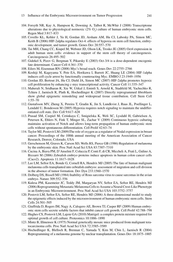
24113 Influence of the Embryonic Microenvironment on Tumor Progression
104. Forsyth NR, Kay A, Hampson K, Downing A, Talbot R, McWhir J (2008) Transcriptome alterations due to physiological normoxic (2% O
2) culture of human embryonic stem cells.
Regen Med 3:817–833 105. Covello KL, Kehler J, Yu H, Gordan JD, Arsham AM, Hu CJ, Labosky PA, Simon MC,
Keith B (2006) HIF-2alpha regulates Oct-4: effects of hypoxia on stem cell function, embry-onic development, and tumor growth. Genes Dev 20:557–570
106. Tai MH, Chang CC, Kiupel M, Webster JD, Olson LK, Trosko JE (2005) Oct4 expression in adult human stem cells: evidence in support of the stem cell theory of carcinogenesis. Carcinogenesis 26:495–502
107. Gidekel S, Pizov G, Bergman Y, Pikarsky E (2003) Oct-3/4 is a dose-dependent oncogenic fate determinant. Cancer Cell 4:361–370
108. Eilers M, Eisenman RN (2008) Myc’s broad reach. Genes Dev 22:2755–2766 109. Koshiji M, Kageyama Y, Pete EA, Horikawa I, Barrett JC, Huang LE (2004) HIF-1alpha
induces cell cycle arrest by functionally counteracting Myc. EMBO J 23:1949–1956 110. Gordan JD, Bertout JA, Hu CJ, Diehl JA, Simon MC (2007) HIF-2alpha promotes hypoxic
cell proliferation by enhancing c-myc transcriptional activity. Cancer Cell 11:335–347 111. Maherali N, Sridharan R, Xie W, Utikal J, Eminli S, Arnold K, Stadtfeld M, Yachechko R,
Tchieu J, Jaenisch R, Plath K, Hochedlinger K (2007) Directly reprogrammed fibroblasts show global epigenetic remodeling and widespread tissue contribution. Cell Stem Cell 1:55–70
112. Gustafsson MV, Zheng X, Pereira T, Gradin K, Jin S, Lundkvist J, Ruas JL, Poellinger L, Lendahl U, Bondesson M (2005) Hypoxia requires notch signaling to maintain the undiffer-entiated cell state. Dev Cell 9:617–628
113. Prasad SM, Czepiel M, Cetinkaya C, Smigielska K, Weli SC, Lysdahl H, Gabrielsen A, Petersen K, Ehlers N, Fink T, Minger SL, Zachar V (2009) Continuous hypoxic culturing maintains activation of Notch and allows long-term propagation of human embryonic stem cells without spontaneous differentiation. Cell Prolif 42:63–74
114. Taylor MJ, Postovit LM (2009)The role of oxygen as a regulator of Nodal expression in breast cancer. Proceedings of the 100th annual meeting of the American Association of Cancer Research, Denver, Colorado, USA
115. Gerschenson M, Graves K, Carson SD, Wells RS, Pierce GB (1986) Regulation of melanoma by the embryonic skin. Proc Natl Acad Sci USA 83:7307–7310
116. Cucina A, Biava PM, D’Anselmi F, Coluccia P, Conti F, di CR, Miccheli A, Frati L, Gulino A, Bizzarri M (2006) Zebrafish embryo proteins induce apoptosis in human colon cancer cells (Caco2). Apoptosis 11:1617–1628
117. Lee LM, Seftor EA, Bonde G, Cornell RA, Hendrix MJ (2005) The fate of human malignant melanoma cells transplanted into zebrafish embryos: assessment of migration and cell division in the absence of tumor formation. Dev Dyn 233:1560–1570
118. Dolberg DS, Bissell MJ (1984) Inability of Rous sarcoma virus to cause sarcomas in the avian embryo. Nature 309:552–556
119. Kulesa PM, Kasemeier JC, Teddy JM, Margaryan NV, Seftor EA, Seftor RE, Hendrix MJ (2006) Reprogramming Metastatic Melanoma Cells to Assume a Neural Crest-Like Phenotype in an Embryonic Microenvironment. Proc Natl Acad Sci USA 103:3752–3757
120. Postovit LM, Seftor EA, Seftor RE, Hendrix MJ (2006) A three-dimensional model to study the epigenetic effects induced by the microenvironment of human embryonic stem cells. Stem Cells 24:501–505
121. Giuffrida D, Rogers IM, Nagy A, Calogero AE, Brown TJ, Casper RF (2009) Human embry-onic stem cells secrete soluble factors that inhibit cancer cell growth. Cell Prolif 42:788–798
122. Hughes CS, Postovit LM, Lajoie GA (2010) Matrigel: a complex protein mixture required for optimal growth of cell culture. Proteomics 10:1886–1890
123. Mintz B, Illmensee K (1975) Normal genetically mosaic mice produced from malignant tera-tocarcinoma cells. Proc Natl Acad Sci USA 72:3585–3589
124. Hochedlinger K, Blelloch R, Brennan C, Yamada Y, Kim M, Chin L, Jaenisch R (2004) Reprogramming of a melanoma genome by nuclear transplantation. Genes Dev 18:1875–1885

242 D. Quail et al.
125. Carette JE, Pruszak J, Varadarajan M, Blomen VA, Gokhale S, Camargo FD, Wernig M, Jaenisch R, Brummelkamp TR (2010) Generation of iPSCs from cultured human malignant cells. Blood 115:4039–4042
126. Miyoshi N, Ishii H, Nagai K, Hoshino H, Mimori K, Tanaka F, Nagano H, Sekimoto M, Doki Y, Mori M (2010) Defined factors induce reprogramming of gastrointestinal cancer cells. Proc Natl Acad Sci USA 107:40–45
127. Utikal J, Maherali N, Kulalert W, Hochedlinger K (2009) Sox2 is dispensable for the repro-gramming of melanocytes and melanoma cells into induced pluripotent stem cells. J Cell Sci 122:3502–3510
128. Ben-Porath I, Thomson MW, Carey VJ, Ge R, Bell GW, Regev A, Weinberg RA (2008) An embryonic stem cell-like gene expression signature in poorly differentiated aggressive human tumors. Nat Genet 40:499–507
129. Bendall SC, Stewart MH, Menendez P, George D, Vijayaragavan K, Werbowetski-Ogilvie T, Ramos-Mejia V, Rouleau A, Yang J, Bosse M, Lajoie G, Bhatia M (2007) IGF and FGF cooperatively establish the regulatory stem cell niche of pluripotent human cells in vitro. Nature 448:1015–1021

243A.L. Allan (ed.), Cancer Stem Cells in Solid Tumors, Stem Cell Biology and Regenerative Medicine, DOI 10.1007/978-1-61779-246-5_14, © Springer Science+Business Media, LLC 2011
Abstract The epithelial-to-mesenchymal transition (EMT) is a developmental process which is reactivated during carcinoma progression, providing tumor cells with enhanced migratory properties, the capacity to invade the stroma, and the ability to metastasize. Tumor cells undergoing EMT also acquire stem cell characteristics, suggesting that there is crosstalk between pathways promoting EMT and self-renewal, and that the EMT process contributes to the generation of cancer stem cells. This chapter summarizes findings pointing to molecular links between EMT and cancer stem cells. The focus is crosstalk between signaling by the transforming growth factor-beta (TGF-b)/Smad pathway, a major inducer of EMT, and stem cell pathways including Wnt, Ras, Hedgehog, and Notch. Finally, the existence of EMT/stem cell niches in tumors where cooperative signaling between TGF-b and self-renewal pathways is activated is discussed.
Abbreviations
ALDH1 Aldehyde dehydrogenase 1AP-1 Activator protein-1APC Adenomatous polyposis colibHLH Basic helix-loop-helixCAR Coxsackie- and adenovirus receptorCD Cluster of differentiationEMT Epithelial-to-mesenchymal transitionEpR Epithelial repressorsEPSC EMT promoting Smad complexes
J. Fuxe (*)Department of Medical Biochemistry and Biophysics, Karolinska Institute, Stockholm, Swedene-mail: [email protected]
Chapter 14The Epithelial-to-Mesenchymal Transition and Cancer Stem Cells
Jonas Fuxe
244 J. Fuxe
ERK Extracellular signal-regulated kinaseERa Estrogen receptor-aGSK-3b Glycogen synthase kinase-3betaHIF-1a Hypoxia-inducible factor 1 alphaHMGA2 High mobility group A2LDL Low density lipoproteinLEF Lymphoid enhancer factorMAPK Mitogen-activated protein kinaseMeA Mesenchymal activatorsMMP Matrix metalloproteinaseNF B Nuclear factor kappa-light-chain-enhancer of activated B cellsPAI-1 Plasminogen activator inhibitor 1PI3K Phosphoinositol-3-kinaseRTK Receptor tyrosine kinaseSp1 Specificity protein 1TCF T cell factorTGF-b Transforming growth factor betaTNF-a Tumor necrosis factor alphaa-SMA Alpha smooth muscle actinb-Cat Beta Catenin
14.1 Epithelial-to-Mesenchymal Transition in Tumor Metastasis
Epithelial-to-mesenchymal transition (EMT) is transdifferentiation process whereby epithelial cells acquire mesenchymal features, including the capacity to migrate to dis-tant sites. EMT is important for various stages of development including gastrulation, neural crest formation, and heart development [1]. In addition, EMT is reactivated in pathological conditions such as organ fibrosis and cancer. Tumor epithelial cells under-going EMT lose the expression of epithelial proteins involved in cell–cell interactions [2]. As a consequence, tumor cells can detach from the primary tumor. In parallel, EMT cells gain expression of mesenchymal cytoskeletal proteins such as vimentin and alpha-smooth muscle actin (a-SMA). This provides tumor cells with the capacity to invade the surrounding stroma and subsequently spread via blood and lymphatic vessels to distant sites [2]. Thus, activation of an EMT program in tumor cells constitutes a switch that converts benign tumors into invasive and metastatic counterparts [1, 3–5].
14.1.1 Consequences of EMT in Cancer
14.1.1.1 Loss of Cell–Cell Adhesion: Detachment from the Primary Tumor
Loss of cell–cell adhesion is a hallmark of EMT in cancer [1, 4]. Components of intercellular junctions including E-cadherin (adherens junctions) and the tight junction
24514 The Epithelial-to-Mesenchymal Transition and Cancer Stem Cells
proteins claudins, occludin, and the coxsackie and adenovirus receptor (CAR) are downregulated during EMT. These proteins are essential for the establishment of epithelial cell–cell interactions and for the barrier function and integrity of all epithe-lial cell layers. During EMT, transcription factors acting as repressors are activated and bind to specific DNA sequences in the gene promoters of these junction proteins. As a result, the genes are turned off.
Various EMT promoting transcriptional repressors of junction proteins have been identified, including members of the Snail, Zeb, and Twist families [6]. These transcriptional repressors recognize and bind a core 5¢-CACCTG-3¢ motif (E-box) within gene promoters. Snail1 was the first transcription factor identified to repress E-cadherin through direct binding to these E-boxes [7, 8]. Subsequently, additional transcription factors including Snail2 (Slug) [9], members of the bHLH family including Twist, E47 (TCF3), and TCF4 (E2-2) [6, 10, 11], and the two zinc-finger E-box binding homeobox factors Zeb1 (dEF1 or Tcf8) and Zeb2 (SIP1) [12, 13] have been identified as repressors of the E-cadherin promoter.
Overexpression of Snail1 results in EMT and the expression of other repressors, suggesting that Snail1 induces an EMT program [6, 14]. Snail1 expression in human cancer is confined to tumor cells at the invasive front [15–17]. However, Snail1 expression in normal adult tissues is limited and generally absent in mesenchymal cells [16]. The precise contribution of Snail, Zeb1/2, and bHLH factors to the repres-sion of E-cadherin during EMT is not well understood. Twist promotes metastatic properties in breast tumor cells and stem-like properties in epithelial cells [11, 18, 19], and its expression is associated with high-grade ductal carcinomas and poor prognosis [20, 21]. Twist also promotes cellular migration, invasion, and resistance to paclitaxel treatment in breast cancer cells [22]. Twist is induced by hypoxia or overexpression of hypoxia inducible factor-1 alpha (HIF-1a), showing a link between the tumor microenvironment and the expression of EMT promoting tran-scription factors [23]. Twist can also be upregulated by Wnt signaling in mammary epithelial cells [24].
14.1.1.2 Activation of Mesenchymal Genes: Gain of Migratory Capacity
In parallel to inactivation of epithelial genes, EMT is characterized by activation of genes encoding mesenchymal proteins including N-cadherin, vimentin, fibronectin, alpha smooth muscle actin (a-SMA), the plasminogen activator inhibitor (PAI-1), and matrix metalloproteases (MMPs) [1, 4]. These proteins are involved in organization of the cytoskeleton during cell movement and in interactions with the extracellular matrix, therefore providing cells with migratory properties.
Various transcription factors acting as activators of mesenchymal genes dur-ing EMT have been identified. The activator protein-1 (AP-1) transcription fac-tor, which is formed by Jun-Jun homodimers or Jun-Fos/Fra-2 heterodimers, induces the expression of Snail1, Snail2, and vimentin during EMT in colon cancer cells [25, 26]. AP-1 also promotes MMP expression, cancer cell invasion and metastasis [27]. Inhibition of AP-1 blocks EMT in human keratinocytes [28]. b-catenin is a transcription factor sequestered at adherens junctions through
246 J. Fuxe
interaction with E-cadherin in normal epithelial cells, but upon activation of Wnt signaling, b-catenin translocates to the nucleus where it interacts with TCF4/LEF transcription factors to induce the expression of EMT-related genes includ-ing Snail2, fibronectin, and vimentin [29–31]. b-catenin also interacts with Snail and other EMT-related factors, such as HIF-1a, Foxo3a, Foxo4, and estrogen receptor alpha (ERa) suggesting that b-catenin complexes play important roles in EMT [32–35]. Nuclear factor kappa-light-chain-enhancer of activated B cells (NF-kB), which plays a key role in regulating immune responses, induces EMT in mammary epithelial cells by activating Snail1 and other mesenchymal genes [36]. Specificity protein 1 (Sp1) is a transcriptional activator required for TGF-b induced expression of vimentin and EMT in pancreatic cancer cells [37]. Sp1 seems also to be involved in the induction of MMP-9 in Snail expressing cells [37, 38].
14.2 EMT and Cancer Stem Cells
Due to the role of EMT in promoting tumor cell invasiveness and the capacity of cancer stem cells to metastasize to distant sites, it was proposed that EMT might provide a link between metastasis and cancer stem cells [39, 40]. Indeed, such a link was identified by results showing that cells undergoing EMT (by Twist/Snail/TGF-b) acquire a CD44high/CD24low signature, a feature of breast cancer stem cells showing a unique ability to form tumors in xenograft models [18, 41]. Moreover, tumor cells undergoing EMT were found to form mammospheres, differentiate into cells of different lineages (i.e., myoepithelial or luminal epithelial cells), and recon-stitute a heterogeneous tumor, thus displaying many properties of stem cells [18]. Subsequently, it was shown that tumor cells induced to undergo EMT by activation of the Ras-MAPK pathway display stem-like properties and a CD44low/CD24high signature [19].
Further studies have reinforced a link between EMT and cancer stem cells. The capacity of mammary stem cells to form mammospheres is inhibited by overexpression of miR200c, a microRNA which turns off the EMT program [42]. Circulating tumor cells from breast cancer patients classified as non-responders to chemotherapy display both EMT and cancer stem cell signatures and express aldehyde dehydrogenase isoform 1 (ALDH1), a stem cell marker in breast, colon, lung, and head and neck carcinomas [43, 44]. Metaplastic breast cancer, a rare and aggressive form of human breast cancer, displays both EMT and cancer stem cell properties [45]. Similarly, human breast cancer cell lines with enhanced invasive properties classified as “Basal B” cell lines due to their basal-like/mesenchymal features show both an EMT and a cancer stem cell-like gene expression profile, including the CD44low/CD24high signature [46]. Further- more, the EMT promoting transcription factors Snail1 and Snail2 (Slug) induce expression of genes promoting cellular stemness including Nanog, KLF4, and TCF4 [47].
24714 The Epithelial-to-Mesenchymal Transition and Cancer Stem Cells
14.2.1 Signaling Pathways in EMT
TGF-b is a major inducer of EMT during development and is overexpressed in many types of human cancer, suggesting a role for TGF-b as an inducer of EMT in tumors [48, 49]. Paradoxically, TGF-b also has anti-proliferative tumor suppressive effects and inactivating mutations or epigenetic silencing of various components of the TGF-b signaling pathway predisposes tissues to cancer and inflammation, indi-cating that the capacity of TGF-b to induce EMT is contextual [50, 51]. TGF-b cooperates with pathways including Wnt, Hedgehog, Notch, and Ras to induce EMT. Interestingly, these are pathways involved in the induction and maintenance of stem cell niches. Thus, co-activation of TGF-b and stem cell pathways may shift the cellular response to TGF-b toward EMT. An explanation for this may lie in the subtle design of the TGF-b signaling pathway.
14.2.1.1 TGF-b / Smad Signaling in EMT
TGF-b binding to its receptors leads to phosphorylation and activation of downstream effectors of the Smad family [52, 53]. The receptor-activated Smad2 and Smad3 (R-Smads) become phosphorylated, associate with cytoplasmic Smad4 (co-Smad) and translocate to the nucleus where Smad complexes regulate transcription of target genes through interaction with specific binding motifs in gene regulatory regions [50]. However, Smad transcription factors have low affinity for DNA and need to interact with cofactors to achieve high affinity and selectivity for target genes. Through this delicate design of the Smad signaling pathway, cells read TGF-b signals differently and the outcome of the TGF-b response ultimately depends on the acces-sibility of Smad cofactors [50].
Smad signaling is essential for TGF-b–induced EMT [51]. Renal tubular epithelial cells deficient in Smad3 fail to undergo EMT and keratinocytes derived from Smad3−/− mice show reduced migration in response to TGF-b [54, 55]. Knockdown of Smad4 through RNA-interference or dominant negative approaches prevents E-cadherin repres-sion upon TGF-b treatment [17, 56–59]. Smad4 deficiency also suppresses fibrotic type I collagen synthesis in vitro [57], and leads to decreased bone metastasis in vivo [56]. Furthermore, Smad4 promotes tumor cell invasion in advanced pancreatic tumors [60].
14.2.1.2 Wnt Signaling in EMT
Wnt signaling regulates stem cell renewal and is implicated in the induction of EMT in cancer. Overexpression of Wnt ligands or silencing of endogenous Wnt inhibitors has been reported in numerous types of human cancer including colon, breast, mela-noma, and prostate carcinomas, and has been linked to EMT [61–66].
Activation of Wnt signaling is initiated by binding of Wnt ligands (typically Wnt1 or Wnt3a) to cell surface receptors composed by Frizzled (Fzd) and the LDL receptor-related proteins LRP5 or LRP6. Signaling from the receptor complex
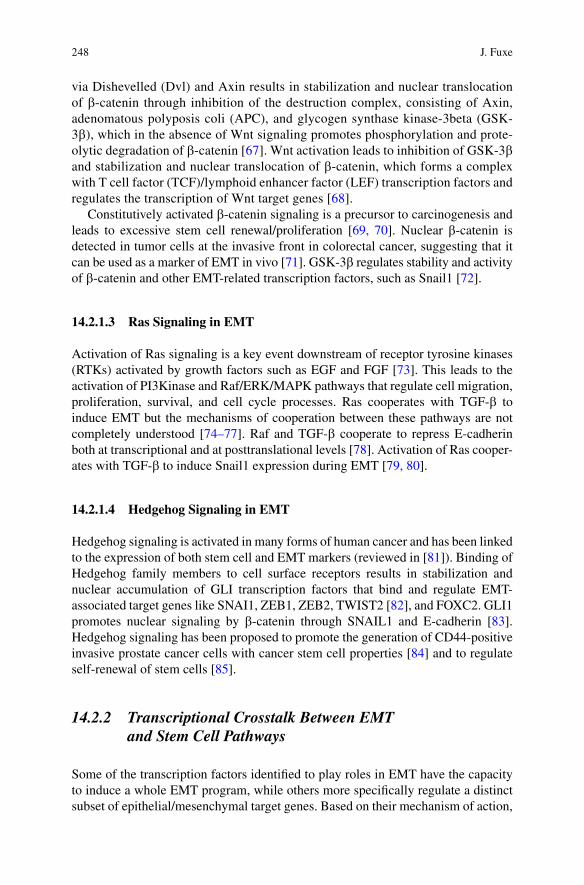
248 J. Fuxe
via Dishevelled (Dvl) and Axin results in stabilization and nuclear translocation of b-catenin through inhibition of the destruction complex, consisting of Axin, adenomatous polyposis coli (APC), and glycogen synthase kinase-3beta (GSK-3b), which in the absence of Wnt signaling promotes phosphorylation and prote-olytic degradation of b-catenin [67]. Wnt activation leads to inhibition of GSK-3b and stabilization and nuclear translocation of b-catenin, which forms a complex with T cell factor (TCF)/lymphoid enhancer factor (LEF) transcription factors and regulates the transcription of Wnt target genes [68].
Constitutively activated b-catenin signaling is a precursor to carcinogenesis and leads to excessive stem cell renewal/proliferation [69, 70]. Nuclear b-catenin is detected in tumor cells at the invasive front in colorectal cancer, suggesting that it can be used as a marker of EMT in vivo [71]. GSK-3b regulates stability and activity of b-catenin and other EMT-related transcription factors, such as Snail1 [72].
14.2.1.3 Ras Signaling in EMT
Activation of Ras signaling is a key event downstream of receptor tyrosine kinases (RTKs) activated by growth factors such as EGF and FGF [73]. This leads to the activation of PI3Kinase and Raf/ERK/MAPK pathways that regulate cell migration, proliferation, survival, and cell cycle processes. Ras cooperates with TGF-b to induce EMT but the mechanisms of cooperation between these pathways are not completely understood [74–77]. Raf and TGF-b cooperate to repress E-cadherin both at transcriptional and at posttranslational levels [78]. Activation of Ras cooper-ates with TGF-b to induce Snail1 expression during EMT [79, 80].
14.2.1.4 Hedgehog Signaling in EMT
Hedgehog signaling is activated in many forms of human cancer and has been linked to the expression of both stem cell and EMT markers (reviewed in [81]). Binding of Hedgehog family members to cell surface receptors results in stabilization and nuclear accumulation of GLI transcription factors that bind and regulate EMT-associated target genes like SNAI1, ZEB1, ZEB2, TWIST2 [82], and FOXC2. GLI1 promotes nuclear signaling by b-catenin through SNAIL1 and E-cadherin [83]. Hedgehog signaling has been proposed to promote the generation of CD44-positive invasive prostate cancer cells with cancer stem cell properties [84] and to regulate self-renewal of stem cells [85].
14.2.2 Transcriptional Crosstalk Between EMT and Stem Cell Pathways
Some of the transcription factors identified to play roles in EMT have the capacity to induce a whole EMT program, while others more specifically regulate a distinct subset of epithelial/mesenchymal target genes. Based on their mechanism of action,
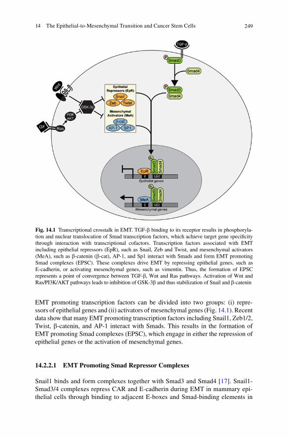
24914 The Epithelial-to-Mesenchymal Transition and Cancer Stem Cells
EMT promoting transcription factors can be divided into two groups: (i) repre- ssors of epithelial genes and (ii) activators of mesenchymal genes (Fig. 14.1). Recent data show that many EMT promoting transcription factors including Snail1, Zeb1/2, Twist, b-catenin, and AP-1 interact with Smads. This results in the formation of EMT promoting Smad complexes (EPSC), which engage in either the repression of epithelial genes or the activation of mesenchymal genes.
14.2.2.1 EMT Promoting Smad Repressor Complexes
Snail1 binds and form complexes together with Smad3 and Smad4 [17]. Snail1-Smad3/4 complexes repress CAR and E-cadherin during EMT in mammary epi-thelial cells through binding to adjacent E-boxes and Smad-binding elements in
Fig. 14.1 Transcriptional crosstalk in EMT. TGF-b binding to its receptor results in phosphoryla-tion and nuclear translocation of Smad transcription factors, which achieve target gene specificity through interaction with transcriptional cofactors. Transcription factors associated with EMT including epithelial repressors (EpR), such as Snail, Zeb and Twist, and mesenchymal activators (MeA), such as b-catenin (b-cat), AP-1, and Sp1 interact with Smads and form EMT promoting Smad complexes (EPSC). These complexes drive EMT by repressing epithelial genes, such as E-cadherin, or activating mesenchymal genes, such as vimentin. Thus, the formation of EPSC represents a point of convergence between TGF-b, Wnt and Ras pathways. Activation of Wnt and Ras/PI3K/AKT pathways leads to inhibition of GSK-3b and thus stabilization of Snail and b-catenin
250 J. Fuxe
the gene promoters (SBE) [17]. Zeb2 was initially characterized as a Smad-binding protein [86] and Zeb1 also interacts with R-Smads [87]. The specific role of Snail1-Smad3/4 vs. Zeb1/2-Smad3/4 complexes during EMT is not clear. Snail1 is more rapidly induced upon TGF-b stimulation compared to Zeb1/2 and it is therefore possible that Snail1-Smad3/4 complexes facilitate recruitment of Zeb1/2 to CAR and E-cadherin promoters.
14.2.2.2 EMT Promoting Smad Activator Complexes
b-catenin-Smad2 complexes are formed during TGF-b–induced EMT in alveolar epithelial cells and promote transcriptional activation of mesenchymal genes like a-SMA and PAI-1 [88]. Formation of b-catenin–Smad2 complexes is dependent on a3b1-integrin–mediated phosphorylation of b-catenin, which releases b-catenin from its interaction with E-cadherin. b-catenin-Smad3/4 complexes stabilize and promote nuclear translocation and transcriptional activity of b-catenin [89]. Silencing of Smad4 in pancreatic carcinoma cells leads to decreased b-catenin levels and signaling activity, suggesting a role for b-catenin-Smad3/4 complexes in controlling EMT in tumor cells [90]. Smad3-AP-1 and Smad3-Sp1/Sp3 complexes regulate the expression of vimentin in myogenic cells in response to TGF-b1 [91]. Smad3 and Jun proteins cooperate to activate AP-1–dependent promoters [92]. Sp1-Smad3 complexes activate the plasminogen activator inhibitor-1 (PAI-1) pro-moter in response to TGF-b [93]. Sp1-Smad3 complexes also induce the expression of the TGF-b receptor endoglin [94], the type alpha2 (I) collagen [95], and the type VII collagen [96] in response to TGF-b.
Finally, Smad proteins can interact with other cofactors to induce EMT activa-tors. It has been found that high mobility group A2 (HMGA2), a non-histone chro-matin modifier, regulates Snail1 expression through interaction with Smads [97]. HMGA2 is required for TGF-b–induced EMT in mammary epithelial cells [98] and maintains oncogenic Ras-induced EMT in human pancreatic cells [99].
14.3 EMT/Cancer Stem Cell Niches in Solid Tumors
As discussed, the proposed link between EMT and cancer stem cells has gained sup-port by experimental data showing crosstalk between EMT and stem cell pathways. Some data hint that crosstalk between these pathways may specifically occur in certain niches within tumor tissues. For example, Wnt signaling is specifically active in stem cell/progenitor cell niches in adult tissues where it maintains self-renewal [100]. If such niches are hit by carcinogenic events, such as oncogenic activation of Ras, a foundation for the induction of EMT may be created during tumor develop-ment. This would imply that progenitor cells are more sensitive to EMT stimuli compared to more differentiated epithelial cells. Thus, the invasive drive of tumors may be affected by the state of differentiation of the tumor originating cells.
25114 The Epithelial-to-Mesenchymal Transition and Cancer Stem Cells
Inflammation represents another niche within the tumor microenvironment and is linked to EMT and tumor cell invasion [101–103]. Immune cells such as mac-rophages and T-lymphocytes infiltrating the tumor stroma contribute to EMT by secreting TGF-b, other cytokines, proteases, and growth factors. Macrophages can activate Wnt signaling in gastric tumor cells via secretion of TNF-a [104]. CD8 T cells can induce an EMT/stem cell phenotype in breast cancer cells upon co-culture, suggesting that immune responses may contribute to the generation of breast cancer stem cells [105]. In combination with intrinsic mutagenic events, the inflammatory milieu of the tumor microenvironment may provide a fundament for crosstalk between EMT and stem cell pathways.
References
1. Thiery JP, Acloque H, Huang RY, Nieto MA (2009) Epithelial-mesenchymal transitions in development and disease. Cell 139 (5):871–890.
2. Thiery JP, Sleeman JP (2006) Complex networks orchestrate epithelial-mesenchymal transi-tions. Nat Rev Mol Cell Biol 7 (2):131–142.
3. Christofori G (2006) New signals from the invasive front. Nature 441 (7092):444–450. 4. Kalluri R, Weinberg RA (2009) The basics of epithelial-mesenchymal transition. J Clin Invest
119 (6):1420–1428. 5. Thompson EW, Newgreen DF, Tarin D (2005) Carcinoma invasion and metastasis: a role for
epithelial-mesenchymal transition? Cancer Res 65 (14):5991–5995; discussion 5995. 6. Peinado H, Olmeda D, Cano A (2007) Snail, Zeb and bHLH factors in tumour progression: an
alliance against the epithelial phenotype? Nat Rev Cancer 7 (6):415–428. 7. Batlle E, Sancho E, Franci C, Dominguez D, Monfar M, Baulida J, Garcia De Herreros A
(2000) The transcription factor snail is a repressor of E-cadherin gene expression in epithelial tumour cells. Nat Cell Biol 2 (2):84–89.
8. Cano A, Perez-Moreno MA, Rodrigo I, Locascio A, Blanco MJ, del Barrio MG, Portillo F, Nieto MA (2000) The transcription factor snail controls epithelial-mesenchymal transitions by repressing E-cadherin expression. Nat Cell Biol 2 (2):76–83.
9. Hajra KM, Chen DY, Fearon ER (2002) The SLUG zinc-finger protein represses E-cadherin in breast cancer. Cancer Res 62 (6):1613–1618.
10. Perez-Moreno MA, Locascio A, Rodrigo I, Dhondt G, Portillo F, Nieto MA, Cano A (2001) A new role for E12/E47 in the repression of E-cadherin expression and epithelial-mesenchymal transi-tions. J Biol Chem 276 (29):27424–27431.
11. Yang J, Mani SA, Donaher JL, Ramaswamy S, Itzykson RA, Come C, Savagner P, Gitelman I, Richardson A, Weinberg RA (2004) Twist, a master regulator of morphogenesis, plays an essential role in tumor metastasis. Cell 117 (7):927–939.
12. Comijn J, Berx G, Vermassen P, Verschueren K, van Grunsven L, Bruyneel E, Mareel M, Huylebroeck D, van Roy F (2001) The two-handed E box binding zinc finger protein SIP1 downregulates E-cadherin and induces invasion. Mol Cell 7 (6):1267–1278.
13. Eger A, Aigner K, Sonderegger S, Dampier B, Oehler S, Schreiber M, Berx G, Cano A, Beug H, Foisner R (2005) DeltaEF1 is a transcriptional repressor of E-cadherin and regulates epithelial plasticity in breast cancer cells. Oncogene 24 (14):2375–2385.
14. De Craene B, Gilbert B, Stove C, Bruyneel E, van Roy F, Berx G (2005) The transcription factor snail induces tumor cell invasion through modulation of the epithelial cell differentiation program. Cancer Res 65 (14):6237–6244.
15. Franci C, Gallen M, Alameda F, Baro T, Iglesias M, Virtanen I, Garcia de Herreros A (2009) Snail1 protein in the stroma as a new putative prognosis marker for colon tumours. PLoS One 4 (5):e5595.
252 J. Fuxe
16. Franci C, Takkunen M, Dave N, Alameda F, Gomez S, Rodriguez R, Escriva M, Montserrat-Sentis B, Baro T, Garrido M, Bonilla F, Virtanen I, Garcia de Herreros A (2006) Expression of Snail protein in tumor-stroma interface. Oncogene 25 (37):5134–5144.
17. Vincent T, Neve EP, Johnson JR, Kukalev A, Rojo F, Albanell J, Pietras K, Virtanen I, Philipson L, Leopold PL, Crystal RG, de Herreros AG, Moustakas A, Pettersson RF, Fuxe J (2009) A SNAIL1-SMAD3/4 transcriptional repressor complex promotes TGF-beta mediated epithelial- mesenchymal transition. Nat Cell Biol 11 (8):943–950.
18. Mani SA, Guo W, Liao MJ, Eaton EN, Ayyanan A, Zhou AY, Brooks M, Reinhard F, Zhang CC, Shipitsin M, Campbell LL, Polyak K, Brisken C, Yang J, Weinberg RA (2008) The epi-thelial-mesenchymal transition generates cells with properties of stem cells. Cell 133 (4):704–715.
19. Morel AP, Lievre M, Thomas C, Hinkal G, Ansieau S, Puisieux A (2008) Generation of breast cancer stem cells through epithelial-mesenchymal transition. PLoS One 3 (8):e2888.
20. Martin TA, Goyal A, Watkins G, Jiang WG (2005) Expression of the transcription factors snail, slug, and twist and their clinical significance in human breast cancer. Ann Surg Oncol 12 (6):488–496.
21. Mironchik Y, Winnard PT, Jr., Vesuna F, Kato Y, Wildes F, Pathak AP, Kominsky S, Artemov D, Bhujwalla Z, Van Diest P, Burger H, Glackin C, Raman V (2005) Twist overexpression induces in vivo angiogenesis and correlates with chromosomal instability in breast cancer. Cancer Res 65 (23):10801–10809.
22. Cheng GZ, Chan J, Wang Q, Zhang W, Sun CD, Wang LH (2007) Twist transcriptionally up-regulates AKT2 in breast cancer cells leading to increased migration, invasion, and resis-tance to paclitaxel. Cancer Res 67 (5):1979–1987.
23. Yang MH, Wu KJ (2008) TWIST activation by hypoxia inducible factor-1 (HIF-1): implica-tions in metastasis and development. Cell Cycle 7 (14):2090–2096.
24. Howe LR, Watanabe O, Leonard J, Brown AM (2003) Twist is up-regulated in response to Wnt1 and inhibits mouse mammary cell differentiation. Cancer Res 63 (8):1906–1913.
25. Andreolas C, Kalogeropoulou M, Voulgari A, Pintzas A (2008) Fra-1 regulates vimentin dur-ing Ha-RAS-induced epithelial mesenchymal transition in human colon carcinoma cells. Int J Cancer 122 (8):1745–1756.
26. Lemieux E, Bergeron S, Durand V, Asselin C, Saucier C, Rivard N (2009) Constitutively active MEK1 is sufficient to induce epithelial-to-mesenchymal transition in intestinal epithe-lial cells and to promote tumor invasion and metastasis. Int J Cancer 125 (7):1575–1586.
27. Rivat C, Le Floch N, Sabbah M, Teyrol I, Redeuilh G, Bruyneel E, Mareel M, Matrisian LM, Crawford HC, Gespach C, Attoub S (2003) Synergistic cooperation between the AP-1 and LEF-1 transcription factors in activation of the matrilysin promoter by the src oncogene: impli-cations in cellular invasion. Faseb J 17 (12):1721–1723.
28. Davies M, Robinson M, Smith E, Huntley S, Prime S, Paterson I (2005) Induction of an epi-thelial to mesenchymal transition in human immortal and malignant keratinocytes by TGF-beta1 involves MAPK, Smad and AP-1 signalling pathways. Journal of Cellular Biochemistry 95 (5):918–931.
29. Conacci-Sorrell M, Simcha I, Ben-Yedidia T, Blechman J, Savagner P, Ben-Ze’ev A (2003) Autoregulation of E-cadherin expression by cadherin-cadherin interactions: the roles of beta-catenin signaling, Slug, and MAPK. J Cell Biol 163 (4):847–857.
30. Gilles C, Polette M, Mestdagt M, Nawrocki-Raby B, Ruggeri P, Birembaut P, Foidart JM (2003) Transactivation of vimentin by beta-catenin in human breast cancer cells. Cancer Res 63 (10):2658–2664.
31. Gradl D, Kuhl M, Wedlich D (1999) The Wnt/Wg signal transducer beta-catenin controls fibronectin expression. Mol Cell Biol 19 (8):5576–5587.
32. Essers MA, de Vries-Smits LM, Barker N, Polderman PE, Burgering BM, Korswagen HC (2005) Functional interaction between beta-catenin and FOXO in oxidative stress signaling. Science 308 (5725):1181–1184.
33. Kaidi A, Williams AC, Paraskeva C (2007) Interaction between beta-catenin and HIF-1 pro-motes cellular adaptation to hypoxia. Nat Cell Biol 9 (2):210–217.
25314 The Epithelial-to-Mesenchymal Transition and Cancer Stem Cells
34. Kouzmenko AP, Takeyama K, Ito S, Furutani T, Sawatsubashi S, Maki A, Suzuki E, Kawasaki Y, Akiyama T, Tabata T, Kato S (2004) Wnt/beta-catenin and estrogen signaling converge in vivo. J Biol Chem 279 (39):40255–40258.
35. Stemmer V, de Craene B, Berx G, Behrens J (2008) Snail promotes Wnt target gene expression and interacts with beta-catenin. Oncogene 27 (37):5075–5080.
36. Huber MA, Azoitei N, Baumann B, Grunert S, Sommer A, Pehamberger H, Kraut N, Beug H, Wirth T (2004) NF-kappaB is essential for epithelial-mesenchymal transition and metastasis in a model of breast cancer progression. J Clin Invest 114 (4):569–581.
37. Jungert K, Buck A, von Wichert G, Adler G, Konig A, Buchholz M, Gress TM, Ellenrieder V (2007) Sp1 is required for transforming growth factor-beta-induced mesenchymal transition and migration in pancreatic cancer cells. Cancer Res 67 (4):1563–1570.
38. Jorda M, Olmeda D, Vinyals A, Valero E, Cubillo E, Llorens A, Cano A, Fabra A (2005) Upregulation of MMP-9 in MDCK epithelial cell line in response to expression of the Snail transcription factor. J Cell Sci 118 (Pt 15):3371–3385.
39. Brabletz T, Jung A, Spaderna S, Hlubek F, Kirchner T (2005) Opinion: migrating cancer stem cells - an integrated concept of malignant tumour progression. Nat Rev Cancer 5 (9):744–749.
40. Brabletz T, Hlubek F, Spaderna S, Schmalhofer O, Hiendlmeyer E, Jung A, Kirchner T (2005) Invasion and metastasis in colorectal cancer: epithelial-mesenchymal transition, mesenchymal-epithelial transition, stem cells and beta-catenin. Cells Tissues Organs 179 (1-2):56–65.
41. Al-Hajj M, Wicha MS, Benito-Hernandez A, Morrison SJ, Clarke MF (2003) Prospective iden-tification of tumorigenic breast cancer cells. Proc Natl Acad Sci USA 100 (7):3983–3988.
42. Shimono Y, Zabala M, Cho RW, Lobo N, Dalerba P, Qian D, Diehn M, Liu H, Panula SP, Chiao E, Dirbas FM, Somlo G, Pera RA, Lao K, Clarke MF (2009) Downregulation of miRNA-200c links breast cancer stem cells with normal stem cells. Cell 138 (3):592–603.
43. Aktas B, Tewes M, Fehm T, Hauch S, Kimmig R, Kasimir-Bauer S (2009) Stem cell and epi-thelial-mesenchymal transition markers are frequently overexpressed in circulating tumor cells of metastatic breast cancer patients. Breast Cancer Res 11 (4):R46.
44. Douville J, Beaulieu R, Balicki D (2008) ALDH1 as a Functional Marker of Cancer Stem and Progenitor Cells. Stem Cells Dev.
45. Hennessy BT, Gonzalez-Angulo AM, Stemke-Hale K, Gilcrease MZ, Krishnamurthy S, Lee JS, Fridlyand J, Sahin A, Agarwal R, Joy C, Liu W, Stivers D, Baggerly K, Carey M, Lluch A, Monteagudo C, He X, Weigman V, Fan C, Palazzo J, Hortobagyi GN, Nolden LK, Wang NJ, Valero V, Gray JW, Perou CM, Mills GB (2009) Characterization of a naturally occurring breast cancer subset enriched in epithelial-to-mesenchymal transition and stem cell characteristics. Cancer Res 69 (10):4116–4124.
46. Blick T, Hugo H, Widodo E, Waltham M, Pinto C, Mani SA, Weinberg RA, Neve RM, Lenburg ME, Thompson EW (2010) Epithelial mesenchymal transition traits in human breast cancer cell lines parallel the CD44(hi/)CD24 (lo/-) stem cell phenotype in human breast cancer. J Mammary Gland Biol Neoplasia 15 (2):235–252.
47. Kurrey NK, Jalgaonkar SP, Joglekar AV, Ghanate AD, Chaskar PD, Doiphode RY, Bapat SA (2009) Snail and slug mediate radioresistance and chemoresistance by antagonizing p53-mediated apoptosis and acquiring a stem-like phenotype in ovarian cancer cells. Stem Cells 27 (9):2059–2068.
48. Massague J (2008) TGFbeta in Cancer. Cell 134 (2):215–230. 49. Xu J, Lamouille S, Derynck R (2009) TGF-beta-induced epithelial to mesenchymal transition.
Cell Res 19 (2):156–172. 50. Massague J (2000) How cells read TGF-beta signals. Nat Rev Mol Cell Biol 1 (3):169–178. 51. Pardali K, Moustakas A (2007) Actions of TGF-beta as tumor suppressor and pro-metastatic
factor in human cancer. Biochim Biophys Acta 1775 (1):21–62. 52. Feng XH, Derynck R (2005) Specificity and versatility in tgf-beta signaling through Smads.
Annual Review of Cell and Developmental Biology 21:659–693. 53. Massague J, Seoane J, Wotton D (2005) Smad transcription factors. Genes & Development 19
(23):2783–2810.
254 J. Fuxe
54. Sato M, Muragaki Y, Saika S, Roberts AB, Ooshima A (2003) Targeted disruption of TGF-beta1/Smad3 signaling protects against renal tubulointerstitial fibrosis induced by unilateral ure-teral obstruction. J Clin Invest 112 (10):1486–1494.
55. Ashcroft GS, Yang X, Glick AB, Weinstein M, Letterio JL, Mizel DE, Anzano M, Greenwell-Wild T, Wahl SM, Deng C, Roberts AB (1999) Mice lacking Smad3 show accelerated wound healing and an impaired local inflammatory response. Nat Cell Biol 1 (5):260–266.
56. Deckers M, van Dinther M, Buijs J, Que I, Lowik C, van der Pluijm G, ten Dijke P (2006) The tumor suppressor Smad4 is required for transforming growth factor beta-induced epithelial to mesenchymal transition and bone metastasis of breast cancer cells. Cancer Res 66 (4): 2202–2209.
57. Kaimori A, Potter J, Kaimori JY, Wang C, Mezey E, Koteish A (2007) Transforming growth factor-beta1 induces an epithelial-to-mesenchymal transition state in mouse hepatocytes in vitro. J Biol Chem 282 (30):22089–22101.
58. Takano S, Kanai F, Jazag A, Ijichi H, Yao J, Ogawa H, Enomoto N, Omata M, Nakao A (2007) Smad4 is essential for down-regulation of E-cadherin induced by TGF-beta in pancreatic can-cer cell line PANC-1. J Biochem 141 (3):345–351.
59. Valcourt U, Kowanetz M, Niimi H, Heldin CH, Moustakas A (2005) TGF-beta and the Smad signaling pathway support transcriptomic reprogramming during epithelial-mesenchymal cell transition. Mol Biol Cell 16 (4):1987–2002.
60. Bardeesy N, Cheng KH, Berger JH, Chu GC, Pahler J, Olson P, Hezel AF, Horner J, Lauwers GY, Hanahan D, DePinho RA (2006) Smad4 is dispensable for normal pancreas development yet critical in progression and tumor biology of pancreas cancer. Genes Dev 20 (22):3130–3146.
61. DiMeo TA, Anderson K, Phadke P, Fan C, Perou CM, Naber S, Kuperwasser C (2009) A novel lung metastasis signature links Wnt signaling with cancer cell self-renewal and epithelial-mesenchymal transition in basal-like breast cancer. Cancer Res 69 (13):5364–5373.
62. Gauger KJ, Hugh JM, Troester MA, Schneider SS (2009) Down-regulation of sfrp1 in a mam-mary epithelial cell line promotes the development of a cd44high/cd24low population which is invasive and resistant to anoikis. Cancer Cell Int 9:11.
63. McCoy EL, Iwanaga R, Jedlicka P, Abbey NS, Chodosh LA, Heichman KA, Welm AL, Ford HL (2009) Six1 expands the mouse mammary epithelial stem/progenitor cell pool and induces mammary tumors that undergo epithelial-mesenchymal transition. J Clin Invest 119 (9):2663–2677.
64. McDonald SL, Silver A (2009) The opposing roles of Wnt-5a in cancer. Br J Cancer 101 (2):209–214.
65. Polakis P (2007) The many ways of Wnt in cancer. Curr Opin Genet Dev 17 (1):45–51. 66. Su HY, Lai HC, Lin YW, Liu CY, Chen CK, Chou YC, Lin SP, Lin WC, Lee HY, Yu MH
(2009) Epigenetic silencing of SFRP5 is related to malignant phenotype and chemoresistance of ovarian cancer through Wnt signaling pathway. Int J Cancer.
67. MacDonald BT, Tamai K, He X (2009) Wnt/beta-catenin signaling: components, mechanisms, and diseases. Dev Cell 17 (1):9–26.
68. Cadigan KM, Peifer M (2009) Wnt signaling from development to disease: insights from model systems. Cold Spring Harb Perspect Biol 1 (2):a002881.
69. Fuchs E (2009) The tortoise and the hair: slow-cycling cells in the stem cell race. Cell 137 (5):811–819.
70. Morin PJ, Sparks AB, Korinek V, Barker N, Clevers H, Vogelstein B, Kinzler KW (1997) Activation of beta-catenin-Tcf signaling in colon cancer by mutations in beta-catenin or APC. Science 275 (5307):1787–1790.
71. Schmalhofer O, Brabletz S, Brabletz T (2009) E-cadherin, beta-catenin, and ZEB1 in malig-nant progression of cancer. Cancer Metastasis Rev 28 (1–2):151–166.
72. Zhou BP, Deng J, Xia W, Xu J, Li YM, Gunduz M, Hung MC (2004) Dual regulation of Snail by GSK-3beta-mediated phosphorylation in control of epithelial-mesenchymal transition. Nat Cell Biol 6 (10):931–940.
73. Karnoub AE, Weinberg RA (2008) Ras oncogenes: split personalities. Nat Rev Mol Cell Biol 9 (7):517–531.
25514 The Epithelial-to-Mesenchymal Transition and Cancer Stem Cells
74. Safina AF, Varga AE, Bianchi A, Zheng Q, Kunnev D, Liang P, Bakin AV (2009) Ras alters epithelial-mesenchymal transition in response to TGFbeta by reducing actin fibers and cell-matrix adhesion. Cell Cycle 8 (2):284–298.
75. Janda E, Lehmann K, Killisch I, Jechlinger M, Herzig M, Downward J, Beug H, Grunert S (2002) Ras and TGF[beta] cooperatively regulate epithelial cell plasticity and metastasis: dis-section of Ras signaling pathways. J Cell Biol 156 (2):299–313.
76. Ellenrieder V, Hendler SF, Boeck W, Seufferlein T, Menke A, Ruhland C, Adler G, Gress TM (2001) Transforming growth factor beta1 treatment leads to an epithelial-mesenchymal trans-differentiation of pancreatic cancer cells requiring extracellular signal-regulated kinase 2 acti-vation. Cancer Res 61 (10):4222–4228.
77. Oft M, Akhurst RJ, Balmain A (2002) Metastasis is driven by sequential elevation of H-ras and Smad2 levels. Nat Cell Biol 4 (7):487–494.
78. Janda E, Nevolo M, Lehmann K, Downward J, Beug H, Grieco M (2006) Raf plus TGFbeta-dependent EMT is initiated by endocytosis and lysosomal degradation of E-cadherin. Oncogene 25 (54):7117–7130.
79. Horiguchi K, Shirakihara T, Nakano A, Imamura T, Miyazono K, Saitoh M (2009) Role of Ras signaling in the induction of snail by transforming growth factor-beta. J Biol Chem 284 (1):245–253.
80. Peinado H, Quintanilla M, Cano A (2003) Transforming growth factor beta-1 induces snail transcription factor in epithelial cell lines: mechanisms for epithelial mesenchymal transitions. J Biol Chem 278 (23):21113–21123.
81. Katoh Y, Katoh M (2009) Hedgehog target genes: mechanisms of carcinogenesis induced by aberrant hedgehog signaling activation. Current Molecular Medicine 9 (7):873–886.
82. Li X, Deng W, Nail CD, Bailey SK, Kraus MH, Ruppert JM, Lobo-Ruppert SM (2006) Snail induction is an early response to Gli1 that determines the efficiency of epithelial transforma-tion. Oncogene 25 (4):609–621.
83. Li X, Deng W, Lobo-Ruppert SM, Ruppert JM (2007) Gli1 acts through Snail and E-cadherin to promote nuclear signaling by beta-catenin. Oncogene 26 (31):4489–4498.
84. Klarmann GJ, Hurt EM, Mathews LA, Zhang X, Duhagon MA, Mistree T, Thomas SB, Farrar WL (2009) Invasive prostate cancer cells are tumor initiating cells that have a stem cell-like genomic signature. Clin Exp Metastasis 26 (5):433–446.
85. Molofsky AV, Pardal R, Morrison SJ (2004) Diverse mechanisms regulate stem cell self-renewal. Curr Opin Cell Biol 16 (6):700–707.
86. Verschueren K, Remacle JE, Collart C, Kraft H, Baker BS, Tylzanowski P, Nelles L, Wuytens G, Su MT, Bodmer R, Smith JC, Huylebroeck D (1999) SIP1, a novel zinc finger/homeodo-main repressor, interacts with Smad proteins and binds to 5¢-CACCT sequences in candidate target genes. J Biol Chem 274 (29):20489–20498.
87. Postigo AA (2003) Opposing functions of ZEB proteins in the regulation of the TGFbeta/BMP signaling pathway. EMBO J 22 (10):2443–2452.
88. Kim Y, Kugler MC, Wei Y, Kim KK, Li X, Brumwell AN, Chapman HA (2009) Integrin alpha3beta1-dependent beta-catenin phosphorylation links epithelial Smad signaling to cell contacts. J Cell Biol 184 (2):309–322.
89. Zhang M, Wang M, Tang X, Li TF, Zhang Y, Chen D (2010) Smad3 prevents beta-catenin degradation and facilitates beta-catenin nuclear translocation in chondrocytes. J Biol Chem.
90. Romero D, Iglesias M, Vary CP, Quintanilla M (2008) Functional blockade of Smad4 leads to a decrease in beta-catenin levels and signaling activity in human pancreatic carcinoma cells. Carcinogenesis 29 (5):1070–1076.
91. Wu Y, Zhang X, Salmon M, Lin X, Zehner ZE (2007) TGFbeta1 regulation of vimentin gene expression during differentiation of the C2C12 skeletal myogenic cell line requires Smads, AP-1 and Sp1 family members. Biochimica et Biophysica Acta 1773 (3):427–439.
92. Verrecchia F, Vindevoghel L, Lechleider RJ, Uitto J, Roberts AB, Mauviel A (2001) Smad3/AP-1 interactions control transcriptional responses to TGF-beta in a promoter-specific manner. Oncogene 20 (26):3332–3340.
256 J. Fuxe
93. Datta PK, Blake MC, Moses HL (2000) Regulation of plasminogen activator inhibitor-1 expression by transforming growth factor-beta -induced physical and functional interactions between smads and Sp1. J Biol Chem 275 (51):40014–40019.
94. Botella LM, Sanchez-Elsner T, Rius C, Corbi A, Bernabeu C (2001) Identification of a critical Sp1 site within the endoglin promoter and its involvement in the transforming growth factor-beta stimulation. J Biol Chem 276 (37):34486–34494.
95. Poncelet AC, Schnaper HW (2001) Sp1 and Smad proteins cooperate to mediate transforming growth factor-beta 1-induced alpha 2(I) collagen expression in human glomerular mesangial cells. J Biol Chem 276 (10):6983–6992.
96. Naso M, Uitto J, Klement JF (2003) Transcriptional control of the mouse Col7a1 gene in keratinocytes: basal and transforming growth factor-beta regulated expression. J Invest Dermatol 121 (6):1469–1478.
97. Thuault S, Tan EJ, Peinado H, Cano A, Heldin CH, Moustakas A (2008) HMGA2 and Smads co-regulate SNAIL1 expression during induction of epithelial-to-mesenchymal transition. J Biol Chem 283 (48):33437–33446.
98. Thuault S, Valcourt U, Petersen M, Manfioletti G, Heldin CH, Moustakas A (2006) Transforming growth factor-beta employs HMGA2 to elicit epithelial-mesenchymal transi-tion. J Cell Biol 174 (2):175–183.
99. Watanabe S, Ueda Y, Akaboshi S, Hino Y, Sekita Y, Nakao M (2009) HMGA2 maintains oncogenic RAS-induced epithelial-mesenchymal transition in human pancreatic cancer cells. Am J Pathol 174 (3):854–868.
100. Reya T, Clevers H (2005) Wnt signalling in stem cells and cancer. Nature 434 (7035):843–850.
101. Coussens LM, Werb Z (2002) Inflammation and cancer. Nature 420 (6917):860–867. 102. Wu Y, Zhou BP (2009) Inflammation: a driving force speeds cancer metastasis. Cell Cycle 8
(20):3267–3273. 103. Lopez-Novoa JM, Nieto MA (2009) Inflammation and EMT: an alliance towards organ fibrosis
and cancer progression. EMBO Mol Med 1 (6–7):303–314. 104. Oguma K, Oshima H, Aoki M, Uchio R, Naka K, Nakamura S, Hirao A, Saya H, Taketo MM,
Oshima M (2008) Activated macrophages promote Wnt signalling through tumour necrosis factor-alpha in gastric tumour cells. EMBO J 27 (12):1671–1681.
105. Santisteban M, Reiman JM, Asiedu MK, Behrens MD, Nassar A, Kalli KR, Haluska P, Ingle JN, Hartmann LC, Manjili MH, Radisky DC, Ferrone S, Knutson KL (2009) Immune-induced epithelial to mesenchymal transition in vivo generates breast cancer stem cells. Cancer Res 69 (7):2887–2895.

Part IVModel Systems for Studying
Cancer Stem Cell Biology and Therapeutic Development
259A.L. Allan (ed.), Cancer Stem Cells in Solid Tumors, Stem Cell Biology and Regenerative Medicine, DOI 10.1007/978-1-61779-246-5_15, © Springer Science+Business Media, LLC 2011
Abstract Cancer stem cells (CSCs) have been defined as a population of cells capable of initiating a tumor and more recently for causing tumor recurrence, attrib-uted so by their ability to preferentially survive current therapeutic strategies. The elimination of CSCs is therefore thought to be crucial for the improvement of long-term patient survival. Consequently, the ability to specifically investigate the CSC population and compare its biological functions to the non-CSC component of the tumor will be an essential step toward this goal. This chapter discusses the current methodologies that are widely used to identify and characterize CSCs, with an emphasis on recent developments in the breast cancer field.
Abbreviations
ABCG2 ATP-binding cassette sub-family G member 2ALDH Aldehyde dehydrogenaseAML Acute myeloid leukemiaBRCA1 Breast cancer susceptibility geneCD Cluster of differentiationCFSE Carboxyfluorescein diacetate succinimidyl esterCNS Central nervous systemCSC Cancer stem cellCXCR4 Chemokine receptor 4DNER Delta/notch-like EGF repeat containing protein DCIS Ductal carcinoma in situDLL1 Delta-like-1
G. Farnie (*)Cancer Stem Cell Research, University of Manchester, School of Cancer and Enabling Sciences, Paterson Institute for Cancer Research, Manchester, UKe-mail: [email protected]
Chapter 15Application of Stem Cell Assays for the Characterization of Cancer Stem Cells
Pamela M. Willan and Gillian Farnie
260 P.M. Willan and G. Farnie
DNER Delta/notch-like EGF repeat containing proteinEGF Epidermal growth factorEGFR Epidermal growth factor receptorER Estrogen receptorESA Epithelial specific antigenFACS Fluorescence activated cell sortingIL InterleukinMaSC Mammary epithelial stem cellMDR1 Multi drug resistance pump 1NOD/SCID Non-obese diabetic/Severe combined immune deficiencyPR Progesterone receptorPROCR Protein C receptorSA-bgal Senescence-associated b-galactosidaseSP Side populationTGF-b Transforming growth factor betaUV UltravioletWT Wildtype
15.1 Introduction
In normal tissue, a cellular hierarchy exists where a normal adult stem cell gives rise to progenitor and fully differentiated lineages of the normal tissue. It is now sug-gested that cancer also has a cellular hierarchy, where the cancer stem cells (CSCs) are the cells capable of producing heterogeneous tumors. CSCs are also termed tumor-initiating cells and can be defined as having a number of distinct properties: they have a selective capacity to initiate tumors and drive neoplastic growth; a capacity for endless self renewal; and the potential to give rise to more mature non-stem cell cancer progeny via differentiation. The CSC hypothesis suggests that every tumor contains a cellular component of CSCs, which retain the key stem cell properties to initiate and drive tumorigenesis [1].
It is widely accepted that like normal tissue, cancers are composed of morpho-logically and phenotypically heterogeneous cell populations [2, 3], and it has become increasingly more evident that to eliminate recurrence and improve survival in can-cer patients, choosing the correct cells within the cancer to target will be crucial. Studies of CSC populations within a number of different cancers have proposed that CSCs are resistant to current anticancer therapies such as radiotherapy and chemo-therapy [4–8]. One of the most prominent studies in breast cancer used in vitro and in vivo techniques to assess the number of CSCs and determined that biopsies taken from breast cancer patients after 6 weeks of chemotherapy treatment had an increased number of CSCs when compared with biopsies before treatment [6].
There has long been a movement toward a more targeted approach to cancer treatment to improve patient survival. A crucial step will be the ability to identify and characterize CSCs in the laboratory using both in vitro and in vivo assays, thus
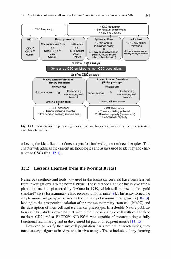
26115 Application of Stem Cell Assays for the Characterization of Cancer Stem Cells
Fig. 15.1 Flow diagram representing current methodologies for cancer stem cell identification and characterization
allowing the identification of new targets for the development of new therapies. This chapter will address the current methodologies and assays used to identify and char-acterize CSCs (Fig. 15.1).
15.2 Lessons Learned from the Normal Breast
Numerous methods and tools now used in the breast cancer field have been learned from investigations into the normal breast. These methods include the in vivo trans-plantation method pioneered by DeOme in 1959, which still represents the “gold standard” assay for mammary gland reconstitution in mice [9]. This assay forged the way to numerous groups discovering the clonality of mammary outgrowths [10–13], leading to the prospective isolation of the mouse mammary stem cell (MaSC) and the description of their cell surface marker phenotype. In a double Nature publica-tion in 2006, studies revealed that within the mouse a single cell with cell surface markers CD24medSca-1lowCD29highCD49fhigh was capable of reconstituting a fully functional mammary gland in the cleared fat pad of a recipient mouse [14, 15].
However, to verify that any cell population has stem cell characteristics, they must undergo rigorous in vitro and in vivo assays. These include colony forming
262 P.M. Willan and G. Farnie
assays determining the differentiation potential of the sorted cells, nonadherent mammosphere assays, and in vivo repopulating experiments with a particular emphasis on serial transplantation to assess repopulating capacity (reviewed in detail in [16, 17]). Many of these assays and ideas from normal breast stem cells now have been applied to breast cancer studies, and are reviewed later.
15.3 In Vitro Methods of Identification and Isolation of CSCs
15.3.1 Side Population
A side population (SP) is defined by its ability to efflux the dye Hoechst 33342 due to the high expression of ATP-binding cassette (ABC) transporter family members such as MDR1 and ABGC2 [18, 19]. First described in 1997, the SP population was found to contain hematopoietic stem cells from bone marrow [20]. Moreover, numerous studies have found a SP population within cancers including the skin [21], lung [22], brain [23], and breast [24]. SP cells from the mouse mammary gland were found to contain cells capable of regenerating a functional mammary gland system in a cleared fat pad [25], and this was also true in the human where the SP cells contained breast stem cells [26–29]. In MCF7 breast cancer cells, the SP popu-lation (0.2%) had a greater tumorigenic capacity than the non-SP fraction when determined by tumor formation subcutaneously in nonobese diabetic/severe com-bined immunodeficiency (NOD/SCID) mice. The MCF7 SP also expressed higher levels of Notch1 and b-catenin mRNA compared with the non-SP population, sug-gesting that the SP cancer cells have some intrinsic properties of stem cells [24]. A recent study has also reported that SP cells from human primary breast cancers and cell lines have tumor initiating properties [30]. Further analysis showed that the SP was predominantly found in luminal cancers but was not concomitant with other tumor initiating phenotypes such as CD44+CD24−/low and aldehyde dehydrogenase (ALDH)+, which are mainly found in the basal subtypes.
However, Hoechst 33342 is highly toxic, more so to non-SP cells, which do not efflux the dye, and thus presents difficulties in interpreting in vivo studies where non-SP cells appear less tumorigenic than SP cells in immunodeficient mice. Some studies have also indicated that non-SP cells can form tumors just as readily as SP cells, examples of which can be seen in mesothelioma and thyroid cancer cell lines [31, 32]. The toxicity issues surrounding Hoechst and the fact that studies have shown that the SP did not exclusively contain the tumorigenic cells have caused the SP technique to fall out of favor. However, with the emer-gence of new dyes that are not toxic such as calcein and DyeCycle Violet, which are also effluxed by cells expressing ABGC2, there may be more conclusive evi-dence emerging which is not influenced by Hoechst toxicity or UV damage dur-ing analysis [33, 34].
26315 Application of Stem Cell Assays for the Characterization of Cancer Stem Cells
15.3.2 Cell Surface Markers
Fluorescence activated cell sorting (FACS) can allow the measurement and isolation of populations of cells with differential expression of single or multiple cell surface markers. The rationale for using cell surface markers to identify CSCs has been largely based on the known and shared markers of normal stem cells. In leukemia, the cell surface markers CD34+CD38− have been successfully used [35, 36], and in gliomas CD133 also enriched for CSCs that were capable of tumor initiation [37, 38]. In the breast MaSC, surface markers are not as well defined and although the markers in the mouse have been described, the translation from mouse to human is not always consistent. To date there are a number of cell surface markers that have been used in the identification of CSCs, some of which are highlighted below and summarized in Table 15.1.
A seminal paper published in 2003 showed the ability of as few as 200 passaged or 1,000 unpassaged ESA (epithelial specific antigen)+CD44+CD24−/low Lineage− cells to give rise to tumors that could be serially transplanted in NOD/SCID mice [39]. These cells had extensive proliferative potential and had the ability to recapitu-late the entire heterogeneity of the initial tumor. The cell surface makers ESA+ CD44+CD24−/low have been used in a number of studies to enrich for CSCs, which have greater tumor initiating capacity, although the expression is highly heteroge-neous within the different subtypes of breast cancer. Sorting for these cell surface markers selected for CSCs within breast cancer cell lines too, enriching tumor initi-ating cells in SUM159, SUM1315, SUM149 [5], and MCF7 cells [40] similar to that found in primary tumors [39]. These cells were capable of recapitulating the tumor heterogeneity in vivo and were enriched for mammosphere formation in vitro.
A study in 2008 utilized the CD44+CD24−/low phenotype and showed that biopsies taken from invasive breast cancer patients after 12 weeks of chemotherapy treat-ment had a greater number of CD44+CD24−/low cells compared with the pretreatment biopsy. These results and additional data showing post-biopsy samples had an increased potential for sphere formation in vitro and tumor initiation in vivo indi-cated that cells with the phenotype CD44+CD24−/low were not only more tumorigenic but were also preferentially surviving chemotherapy treatment [6].
In contrast, a number of other studies have not favored this cell surface marker combination. For example, a study profiling eight breast cancer cell lines with known CSC markers CD44, CD24, CD133, PROCR, ABGC2, CXCR4, ESA, and ALDH concluded that CD44+CD24−/low and ALDH+ were not universal markers of CSC isolation using a soft agar culture to measure tumorigenicity. They concluded that a combination of ESA+PROCR+ could define an uncharacterized type of breast cancer stem cell from both primary and breast cancer cell lines [41]. In particular, the study shows the MDA-MB-231 cell line, which had a 90% population of CD44+CD24−/low cells, was further enriched for tumorigenic cells by selecting the ESA+PROCR+ vs. ESA−PROCR−. However, previous enrichment for tumor initiating cells within this cell line used a triple marker selection ESA+CD44+CD24−/low indicating that the selection for ESA+ cells may have been the enriching marker in this cell line [5].
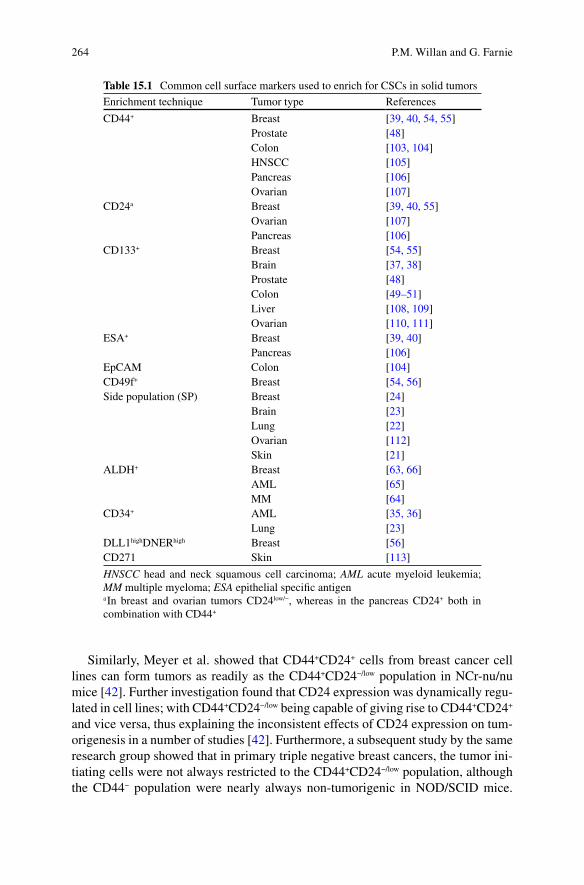
264 P.M. Willan and G. Farnie
Similarly, Meyer et al. showed that CD44+CD24+ cells from breast cancer cell lines can form tumors as readily as the CD44+CD24−/low population in NCr-nu/nu mice [42]. Further investigation found that CD24 expression was dynamically regu-lated in cell lines; with CD44+CD24−/low being capable of giving rise to CD44+CD24+ and vice versa, thus explaining the inconsistent effects of CD24 expression on tum-origenesis in a number of studies [42]. Furthermore, a subsequent study by the same research group showed that in primary triple negative breast cancers, the tumor ini-tiating cells were not always restricted to the CD44+CD24−/low population, although the CD44− population were nearly always non-tumorigenic in NOD/SCID mice.
Table 15.1 Common cell surface markers used to enrich for CSCs in solid tumors
Enrichment technique Tumor type References
CD44+ Breast [39, 40, 54, 55]Prostate [48]Colon [103, 104]HNSCC [105]Pancreas [106]Ovarian [107]
CD24a Breast [39, 40, 55]Ovarian [107]Pancreas [106]
CD133+ Breast [54, 55]Brain [37, 38]Prostate [48]Colon [49–51]Liver [108, 109]Ovarian [110, 111]
ESA+ Breast [39, 40]Pancreas [106]
EpCAM Colon [104]CD49f+ Breast [54, 56]Side population (SP) Breast [24]
Brain [23]Lung [22]Ovarian [112]Skin [21]
ALDH+ Breast [63, 66]AML [65]MM [64]
CD34+ AML [35, 36]Lung [23]
DLL1highDNERhigh Breast [56]CD271 Skin [113]
HNSCC head and neck squamous cell carcinoma; AML acute myeloid leukemia; MM multiple myeloma; ESA epithelial specific antigena In breast and ovarian tumors CD24low/−, whereas in the pancreas CD24+ both in combination with CD44+
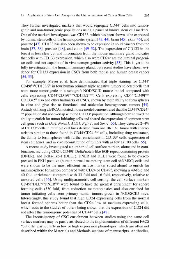
26515 Application of Stem Cell Assays for the Characterization of Cancer Stem Cells
They further investigated markers that would segregate CD44+ cells into tumori-genic and non-tumorigenic populations using a panel of known stem cell markers. One of the markers investigated was CD133, which has been shown to be expressed by normal stem cells of the hematopoietic system [43, 44], brain [45], skin [46], and prostate [47]. CD133 has also been shown to be expressed in solid cancers from the brain [37, 38], prostate [48], and colon [49–52]. The expression of CD133 in the breast is less clear cut and information from the mouse mammary gland indicates that cells with CD133 expression, which also were CD24+ are the luminal progeni-tor cells and not capable of in vivo stem/progenitor activity [53]. This is yet to be fully investigated in the human mammary gland, but recent studies have shown evi-dence for CD133 expression in CSCs from both mouse and human breast cancer [54, 55].
For example, Meyer et al. have demonstrated that triple staining for CD44+ CD49fhighCD133/2hi in four human primary triple negative tumors selected cells that were more tumorigenic in a xenograft NOD/SCID mouse model compared with cells expressing CD44+CD49f−/lowCD133/2−/low. Cells expressing CD44+CD49fhigh CD133/2hi also had other hallmarks of CSCs, shown by their ability to form spheres in vitro and give rise to functional and molecular heterogeneous tumors [54]. A study utilizing a BRCA mutated mouse model demonstrated that the CD44+CD24−/
low population did not overlap with the CD133+ population, although both showed the ability to enrich for tumor initiating cells and shared the expression of common stem cell genes such as Oct4, Notch1, Aldh1, Fgfr 1, and Sox-1 [55]. They detected 2–4% of CD133+ cells in multiple cell lines derived from one BRCA1 tumor with charac-teristics similar to those found in CD44+CD24−/low cells, including drug resistance, the ability to form spheres with further enrichment in CD133+ cells, expression of stem cell genes, and in vivo reconstitution of tumors with as few as 100 cells [55].
A recent study investigated a number of cell surface markers alone and in com-bination, including CD24, CD49f, Delta/notch-like EGF repeat containing protein (DNER), and Delta-like-1 (DLL1). DNER and DLL1 were found to be overex-pressed in PKH positive (human normal mammary stem cell shNMSC) cells and were shown to be the most efficient surface marker (used alone) to enrich for mammosphere formation compared with CD24 or CD49f, showing a 49-fold and 40-fold enrichment compared with 33-fold and 16-fold, respectively, relative to unsorted cells [56]. Using multiparametric cell sorting, the cell surface markers CD49f+DLLhighDNERhigh were found to have the greatest enrichment for sphere forming cells (530-fold) from reduction mammoplasties and also enriched for tumor initiating cells from primary human tumors grown in NOD/SCID mice. Interestingly, this study found that high CD24 expressing cells from the normal breast formed spheres better than the CD24 low or medium expressing cells, which adds to the studies of others being shown that the expression of CD24 did not affect the tumorigenic potential of CD44+ cells [42].
The inconsistency of CSC enrichment between studies using the same cell surface markers may be partly attributed to the implementation of different FACS “cut offs” particularly in low or high expression phenotypes, which are often not described within the Materials and Methods sections of manuscripts. Antibodies,
266 P.M. Willan and G. Farnie
particularly for ESA, also vary between publications. Clones recognizing different epitopes on the ESA antibody do not always mark the same population of cells (data not shown) and should be taken into account if trying to compare the enrichment ability of different studies seemingly using the same cell surface markers.
A number of different cell surface marker combinations have been shown to enrich for breast CSCs; however, the limitation of our assays to “read out” CSCs must be taken into account particularly in the instance of human xenograft systems. The current methodologies used to create a “humanized” environment within the mouse may influence the cells ability to form a tumor, therefore generating data that may not translate into the human. This will be discussed later in the chapter.
15.3.3 ALDEFLUOR Assay
The aldehyde dehydrogenase (ALDH) family of enzymes have important functions in the development of epithelial homeostasis, and as a result deregulation of this class of enzymes has been implicated in multiple cancers [57]. The Aldefluor assay is based on the enzymatic function of ALDH1, a detoxifying enzyme responsible for the oxidation of intracellular aldehydes [58]. ALDH is suggested to have a role in early differentiation of stem cells via its role in oxidizing retinol to retinoic acid [59]. High activity of ALDH has been found in hematopoietic and neural stem/progenitor cells [60–62], and when used to select for human normal MaSCs only ALDH+ cells had the capacity to reconstitute a mammary tree in a humanized cleared mammary fat pad [63]. ALDH+ cells within malignancies also enrich for the CSC population of multiple myeloma and acute myeloid leukemia (AML) [64, 65]. The Aldefluor assay is therefore thought to be an almost universal marker of stem cell activity in both normal and cancer tissue.
Ginestier and colleagues utilized this assay to enrich for CSCs from xenotrans-plants generated from human invasive ductal carcinomas and found that three can-cers had an ALDH+ population representing 3–10% of the total cell population. These xenografts were serially passaged in vivo using limiting dilution of ALDH+, ALDH−, and unsorted cells, and at each passage only the ALDH+ population formed tumors, even when implanted in low numbers (500 cells). The ALDH+ population showed consistencies with stem cell characteristics, generating tumors that reca-pitulated the phenotypic heterogeneity of the initial tumor. Measurement of ALDH populations revealed that a similar ratio of ALDH+ and ADLH− were found in tumors grown from an ALDH+ population, suggesting that the ALDH+ cells were capable of self renewing into both ALDH+ and ALDH− cells [63]. Further investiga-tion of the overlap of the ALDH+ population and the previously described CD44+CD24−/low Lineage− population [39] revealed that within the xenografted tumors both populations are represented (6.08 and 4.34%, respectively), and that the overlap of ALDH+ and CD44+CD24−/low Lineage− expressing cells from three xeno-grafts ranged between 0.1 and 1.2%. Cells bearing both CSC phenotypes were
26715 Application of Stem Cell Assays for the Characterization of Cancer Stem Cells
highly tumorigenic and generated a tumor with a few as 20 cells. However, cells with an ALDH− and CD44+CD24−/low Lineage− were not tumorigenic, even when implanting up to 5,000 cells per fat pad [63].
ALDH+ cells have also been discovered in 23 out of 33 breast cancer cell lines, representing the spectrum of molecular subtypes, in which populations ranged from 0.2 to ~100%. Further analysis showed that all of the 16 basal/mesenchymal breast cancer cell lines contained an ALDH+ population, whereas 7 out of 12 luminal cell lines did not have detectable ALDH+ cells. In vivo xeno-transplantation studies using three estrogen receptor a (ER)-negative breast cell lines (MDA-MB- 435, SUM159 and BrCa-MZ-01) showed that in two of the cell lines (MDA-MB-435, SUM159) only the ALDH+ cells had tumor generating capacity, which was main-tained through serial passage. However, ALDH− cells from the BrCa-MZ-01 cell line were also shown to be capable of generating tumors but, in contrast to the ALDH+ tumors, the ALDH− tumors had slower growth, only contained ALDH− cells and failed to reform tumors after 3 serial passages, suggesting that the ALDH− cell population may contain progenitor cells, which are able to undergo limited growth but not self renewal [66]. Another group investigating ER negative breast cancer cell lines have confirmed these findings, showing ALDH+CD44+CD24− (MDA-MB-231) and ALDH+CD44+CD133+(MDA-MB-468) cells demonstrated increased colony formation, migration, and invasion in vitro compared to ALDH−CD44low/− cells. Furthermore, in vivo experiments demonstrated that following tail vein or mam-mary fat pad injection of cells into NOD/SCID/IL2gamma receptor null mice, ALDH+CD44+CD24− and ALDH+CD44+CD133+ cells had enhanced tumorigenicity and metastasis relative to ALDH−CD44low/− expressing cells [67].
In contrast, a recent study in melanoma describes the capacity for ALDH+ and ALDH− subpopulations isolated from different xenografts and two patient biopsies to form tumors in NOD/SCID/IL2gamma receptor null mice. Both ALDH+ and ALDH− cells derived from the xenografts or patient biopsies were capable of effi-cient formation of first generation tumors following injection of as few as 100 or 2,000 cells, respectively. Tumor growth rate was also not dependent on ALDH expression and both ALDH+ and ALDH− could be serially transplanted in vivo. The only difference seen between the two experimental groups was that the ALDH+ cells could reestablish tumor heterogeneity, showing a mixed phenotype where 20–40% of the melanoma cells did not show ALDH activity, whereas the ALDH− tumors remained ALDH− [68]. Similarly, in a lung cancer cell line H225, both ALDHbright and ALDHlow populations could generate tumors in recipient NOD/SCID mice, which were capable of serial passage. The in vivo data also demonstrated that the tumors formed from ALDHbright cell had a significantly slower growth rate [69].
These data demonstrate that the Aldefluor assay can recognize a subpopulation of cells showing ALDH activity, which are more tumorigenic in vivo. However, there are conflicting studies, which indicate ALDH alone may not be sufficient for CSC selection in other cancers, and within breast cancer studies further limitations are apparent as the studies demonstrating the tumorigenic capacity of ALDH+ cells have predominantly utilized ER negative cell lines and primary samples. ER+ cell lines have shown little or no ALDH+ cells indicating that Aldefluor will not be a
268 P.M. Willan and G. Farnie
universal marker for all molecular subtypes of breast cancer, although further stud-ies are needed to confirm this. In spite of this, studies in breast cancer combining other CSC surface markers with Aldefluor have improved tumorigenic enrichment and this combination may prove to be a better strategy for enriching for CSCs.
15.3.4 Immunohistochemical Staining for CSC Markers
The ability to immunohistochemically stain in situ for CSCs would be an ideal methodology to enable the assessment of CSCs within large cohorts of patients with known survival, recurrence, and metastasis, and would allow the assessment of CSCs in samples from clinical trials without the need for in vitro or in vivo growth assays. Unfortunately, to date no specific marker or markers have been found to specifically pinpoint the CSCs, but a number of groups have investigated the expres-sion of ALDH1 as a surrogate for the ALDH activity measured via the Aldefluor assay and have seen some success. Positive ALDH1 protein expression in two cohorts of breast cancer patient samples (n = 136 and n = 341) had a significant asso-ciation with poor overall survival, and within the larger cohort ALDH1 expression was an independent prognostic factor when compared with tumor size, lymph node metastasis, grade, ER, PR, Ki67, and ErbB2 receptor status [63].
A retrospective study of 109 inflammatory breast cancers also reported that ALDH1 expression correlated with the development of distant metastasis and with decreased survival [70]. Characterization of ALDH1 expression in 203 breast can-cer patients revealed that 10% were found to be ALDH1 positive and these cancers were significantly more likely to be ER−, PR−, ErbB2+, and Ki67+ [71], reiterating that ALDH1 expression or activity may be predominantly observed in ER− breast cancers. However, ALDH1 expression to identify CSCs is also contentious as there are studies in colorectal cancer showing ALDH1 expression has no correlation with prognosis [72], and in ovarian cancer ALDH1 expression correlates with favorable prognosis [73].
Other markers used for in situ staining in the breast are the markers CD44+CD24−/low. How effective these markers are at identifying the CSCs is not clear, as expression of these markers in formalin fixed paraffin embedded sections may be very different in comparison to cell surface expression levels seen in FACS analysis studies. A retrospective study of 240 breast cancers stained for CD44 and CD24 demon-strated that CD44+/CD24− cells were detected in 31% of the tumors. The CD44+/CD24− phenotype was most common in the basal-like subgroup (ER−, PR−, ErbB2−, CK5/14+, and EGFR+) and was found in 94% of BRCA1 hereditary tumors [74]. This suggests that immunohistochemical staining for CD44+CD24−/low may not mark the CSCs in all breast cancer subtypes. Furthermore, another study in 122 breast cancer patients showed that the CD44+CD24−/low phenotype was neither associated with overall survival nor clinicopathological characteristics [75].
Overall the expression markers being used have some ability to predict for over-all survival, and in the case of ALDH1 in breast cancer can also predict for response
26915 Application of Stem Cell Assays for the Characterization of Cancer Stem Cells
to chemotherapy [76]. However, to advance the detection of CSCs in situ, there needs to be further characterization of CSCs to find new markers or possibly com-bining multiple known markers to improve current strategies.
15.3.5 PKH26 Labeling of CSCs
Long-term label retention is used frequently for the identification of stem cells and has been successful in identifying MaSCs [25, 77, 78]. BrdU labeling has some disadvantages as it is known to be toxic [79–81] and does not allow the tracking of live cells. However, other labeling dyes such as CFSE and PKH26 can be measured in live cells, thus allowing more complex functional analysis of CSCs including their division and proliferation. PKH26 is a fluorescent dye that binds to cell mem-branes and segregates in daughter cells after each cell division, such that the inten-sity of staining correlates inversely, at the single cell level, with the number of previous cell divisions [82]. The hematopoietic field has used this dye to identify long-term and short term repopulating cells in blood [82, 83] and to follow the pro-liferation of leukemic cells from AML patients [84]. PKHHi cells isolated from mammosphere cultures from human normal breast and implanted into humanised cleared fat pads of NOD/SCID mice had the capacity to reconstitute normal mam-mary epithelium. Conversely, the PKH− cells could not reconstitute the mammary gland even when high concentrations of cells were injected [56], indicating the PKHHi cells are enriched for MaSCs. Cells capable of forming a mammosphere from human or murine normal or breast cancer tissue have also been shown to retain PHK labeling [40, 56, 85]. Furthermore, in a panel of human tumor cell lines including ovarian, acute promyelocytic leukemia, glioma, lung, and breast, PKH26 labeling experiments showed that label retaining PKHHi cells have a greater colony forming capacity in vitro and a high tumorigenic potential in vivo compared with PKH− cells, which lacked both colony and tumor forming capabilities [86]. PKHHi cells were also found to express the stem cell markers Oct4, Nestin, Nanog, and Bmi, whereas the PKH− cell lacked all four markers, further indicating that label retaining PKHHi cells have CSC characteristics. To investigate the effect of chemotherapy on CSCs, xenograft tumors formed from PKHHi ovarian cells were treated with five doses of paclitaxel over a 6-week time period, and during this time tumors stopped growing and reduced marginally in size. Analysis of the tumors show that paclitaxel treat-ment increased the number of PKHHi cells, suggesting a treatment induced enrich-ment of CSCs and highlighting the need for targeting CSCs in the clinic [86].
PKH26 can also be used as a tool to analyse symmetric or asymmetric division. Cicalese and colleagues investigated modes of self-renewing divisions by time-lapse video microscopy by plating PKHHi cells from mammospheres grown from wild type (WT) mouse mammary gland or from tumor (ErbB2) mammary tissue into methylcellulose and monitoring these cells for 7 days, at 1 h intervals. The first division was defined as asymmetric if one of the first-generation daughter cells remained quiescent, whereas the other divided further, giving rise to a total of five
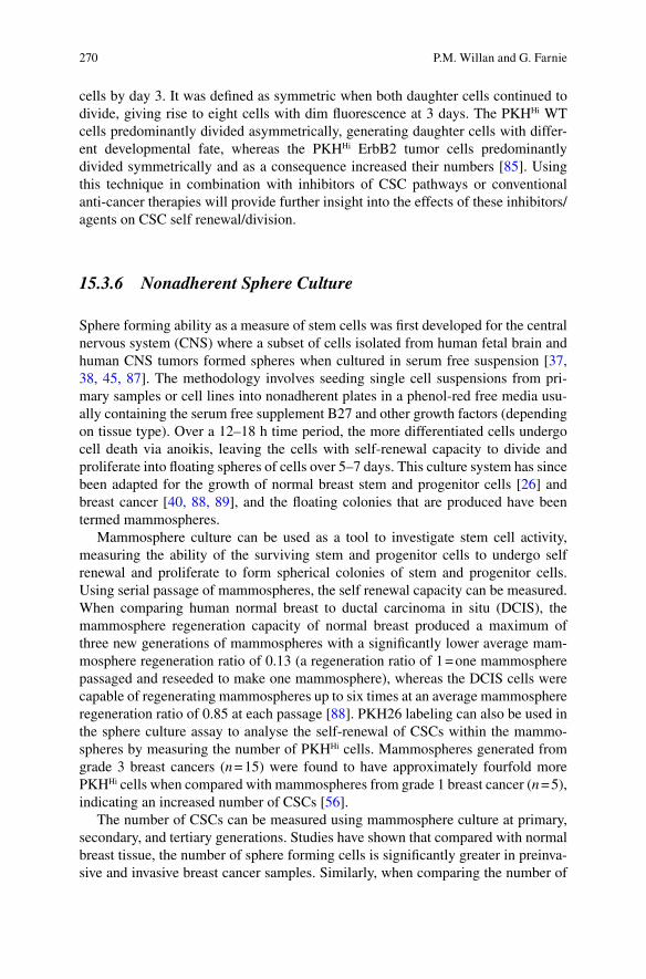
270 P.M. Willan and G. Farnie
cells by day 3. It was defined as symmetric when both daughter cells continued to divide, giving rise to eight cells with dim fluorescence at 3 days. The PKHHi WT cells predominantly divided asymmetrically, generating daughter cells with differ-ent developmental fate, whereas the PKHHi ErbB2 tumor cells predominantly divided symmetrically and as a consequence increased their numbers [85]. Using this technique in combination with inhibitors of CSC pathways or conventional anti-cancer therapies will provide further insight into the effects of these inhibitors/agents on CSC self renewal/division.
15.3.6 Nonadherent Sphere Culture
Sphere forming ability as a measure of stem cells was first developed for the central nervous system (CNS) where a subset of cells isolated from human fetal brain and human CNS tumors formed spheres when cultured in serum free suspension [37, 38, 45, 87]. The methodology involves seeding single cell suspensions from pri-mary samples or cell lines into nonadherent plates in a phenol-red free media usu-ally containing the serum free supplement B27 and other growth factors (depending on tissue type). Over a 12–18 h time period, the more differentiated cells undergo cell death via anoikis, leaving the cells with self-renewal capacity to divide and proliferate into floating spheres of cells over 5–7 days. This culture system has since been adapted for the growth of normal breast stem and progenitor cells [26] and breast cancer [40, 88, 89], and the floating colonies that are produced have been termed mammospheres.
Mammosphere culture can be used as a tool to investigate stem cell activity, measuring the ability of the surviving stem and progenitor cells to undergo self renewal and proliferate to form spherical colonies of stem and progenitor cells. Using serial passage of mammospheres, the self renewal capacity can be measured. When comparing human normal breast to ductal carcinoma in situ (DCIS), the mammosphere regeneration capacity of normal breast produced a maximum of three new generations of mammospheres with a significantly lower average mam-mosphere regeneration ratio of 0.13 (a regeneration ratio of 1 = one mammosphere passaged and reseeded to make one mammosphere), whereas the DCIS cells were capable of regenerating mammospheres up to six times at an average mammosphere regeneration ratio of 0.85 at each passage [88]. PKH26 labeling can also be used in the sphere culture assay to analyse the self-renewal of CSCs within the mammo-spheres by measuring the number of PKHHi cells. Mammospheres generated from grade 3 breast cancers (n = 15) were found to have approximately fourfold more PKHHi cells when compared with mammospheres from grade 1 breast cancer (n = 5), indicating an increased number of CSCs [56].
The number of CSCs can be measured using mammosphere culture at primary, secondary, and tertiary generations. Studies have shown that compared with normal breast tissue, the number of sphere forming cells is significantly greater in preinva-sive and invasive breast cancer samples. Similarly, when comparing the number of
27115 Application of Stem Cell Assays for the Characterization of Cancer Stem Cells
spheres forming from grade 1 or grade 3 preinvasive or invasive breast cancer samples, the grade 3 samples produced greater numbers of mammospheres [56, 88]. Cells that survive the sphere culture conditions and are capable of initiating a mam-mosphere have been found to predominantly express CSC cell surface markers ESA+CD44+CD24−/low and have been shown to be ALDH+. In both instances, if cells are FACS sorted for these CSC markers they can enrich for mammosphere initiating cells between 2 and 12-fold and 10-fold, respectively [40, 63].
Cells can also be enriched for mammosphere and tumor initiating cells by seed-ing cells into the mammosphere culture system but instead of leaving them to form spheres over 5–7 days the cells are harvested after ~16 h. At this timepoint, 85% of MCF7 cells have died by anoikis and the remaining viable cells have been shown to be 5.7-fold enriched for mammosphere formation and 12-fold enriched for tumor initiating cells after implantation into NOD/SCID mice when compared with mono-layer cells [40]. The link between mammosphere initiating cells and tumor initiating cells was corroborated in primary human breast cancers when the growth rate and numbers of CSCs (measured by PKH) from human breast xenografts were found to be comparable regardless of whether the cells were directly implanted or were cells derived from mammospheres generated from the same patient samples [56].
Mammosphere culture is extremely useful for primary culture where only small numbers of cells are available and FACs staining for CSC markers is not feasible. Additionally, due to some controversy over which cell surface markers or labels enrich for CSCs and the suggestion that some markers may not identify CSCs from different molecular subtypes of breast cancer, studies have shown that mammo-spheres can be formed from cell lines and primary tissues from all molecular sub-types, allowing the measurement of CSC numbers regardless of origin of tissue.
15.3.7 Holoclone Formation
Another successful method of CSC identification is based on their ability to form dense colonies of a specific morphology, called holoclones [90, 91]. The three types of colony formed are as follows: holoclones containing the most undifferentiated cells including the CSCs; meroclones containing a mixture of progenitor cells and more differentiated cells; and paraclones which are fully differentiated. Holoclones consist of tightly packed small cells, paraclones contain larger cells with smaller cell numbers, and meroclones are an intermediate between the two.
Studies in a prostate cancer cell line PC3 have shown that the holoclones were negative for senescence-associated b-galactosidase (SA-bgal) and cells were homogenously small. In contrast, most cells in paraclones were positive for SA-bgal and cells were large and flat, suggesting that cells within paraclones were mostly senescent and nonproliferative [92]. Holoclones obtained from different cancer cell lines, including head and neck squamous cell carcinomas and breast cancer have also been shown to have cells with cancer stem cell characteristics [90–92]. When three independent holoclones from PC3 cells were harvested and injected into
272 P.M. Willan and G. Farnie
NOD/SCID mice at 1,000 or 10,000 cells, all initiated a tumor 80–100% of the time. In contrast, meroclones (n = 2) or paraclones were not capable of initiating any tumors even after 5–6 months with 10,000 or 10,00,000 cells injected. The holo-clone derived tumors had the capacity for serial transplantation, and analysis of the cells generated from the tumors demonstrated that all three colony types could be formed, indicating that holoclones can regenerate or maintain the clonal heteroge-neity in vivo [92].
Holoclone formation is a valuable in vitro technique for measuring CSCs, although, as with the mammosphere technique, combining holoclone formation with in vivo tumor growth aids the definition of the colonies measured. In addition, holoclone formation does not require specialized equipment or media and can be use with relatively low numbers of cells.
15.4 Assessing Biological Properties of CSCs Using Gene Array Analysis
Once CSCs have been enriched using the in vitro assays discussed earlier, gene expression arrays can be used to compare enriched CSC vs. non-CSC populations or normal tissue. These types of studies can identify differentially expressed genes within the CSC population, which may lead to the discovery of new biological pro-cesses (Table 15.2). Genes must then be subjected to revalidation using both the in vitro assays described above and ultimately in the in vivo models which are dis-cussed next, to establish the functional effects of the genes identified.
Table 15.2 Differential gene expression in CSC vs. non-CSC populations
Tumor type Enrichment technique Examples of genes of interest found References
Colon CD133high vs. CD133high-depleted cells (HCT116 cell lines)
988 differentially expressed genes including FOXO3, NFKB1A, IL-8, Shh and Oct4
[114]
Breast CD44+CD24−/low breast cancer cells vs. normal breast epithelium (human primary tissue)
Prognostic 186 gene signature, including CASP8, BCL2, LRP2, MAPK14, CXCL2, MMP7, RAD23B and ERBB4
[93]
ALDH+ vs. ALDH− cells from 8 breast cancer cell lines
413 differentially expressed genes including CXCR1/IL-8RA, Notch2, RAD51L1 and FBX021
[66]
CD44+ or PROCR+ vs. CD24+ cells in normal and breast cancer tissue
BMI1, Gli1, Gli2, and TGF-b [95]
Prostate CD133+/a2b1hi vs. CD133−/a2b1low human primary prostate cancer
518 differentially expressed genes including NFKB1, IL6, ITGAV and IFNGR1
[115]
Lung SP and non-SP cells (A549 cell line)
Genes included AKR1C1/AKR1C2, TM4SF1, NR0B1, ABCG2 and IL6R
[116]
27315 Application of Stem Cell Assays for the Characterization of Cancer Stem Cells
The comparison of CSCs enriched using cell surface markers CD44+CD24−/low from 6 human breast cancer patients vs. normal breast epithelium from 3 mammoplasties revealed 186 genes that were differentially expressed in the CSC population [93]. When compared with published breast cancer gene signatures, a significant associa-tion between the CSC signature and worse overall survival and metastasis-free sur-vival was found, suggesting more aggressive tumors may contain a higher percentage of CSCs [93]. When gene expression profiles of ALDH+ vs. ALDH− cell popula-tions (using the Aldefluor assay) from 8 breast cancer cells lines were compared, 413 genes were found to be differentially expressed [66]. Some of these genes were validated including CXCR1, which was found to be up-regulated in CSCs, and results show that inhibition of the IL-8 receptor CXCR1 in three breast cancer cell lines could selectively deplete the CSC population (ALDH+ cells) and reduce tumor growth in vivo [66, 94]. CSCs enriched using CD44 from human normal breast and breast cancers samples (CD44+) were compared with CD24+ expressing non-CSC population. Profiles indicated that cells within the CSC enriched population expressed stem/progenitor-associated genes such as BMI1, Gli1, and Gli2 whereas the non-CSC population expressed differentiation associated genes such as GATA3 and ERS1 [95]. This CSC gene expression signature also correlated with patient clinical outcome and specifically identified the TGF-b pathway to be activated within the CSC population, and inhibition of this pathway in vitro led to differentia-tion [95]. These data demonstrate that enrichment of CSCs can facilitate the discov-ery of biological properties of CSCs that are unique and may not have otherwise been revealed.
15.5 In Vivo Models for the Identification and Characterization of CSCs
The in vitro methodologies described are the foundation of identifying CSCs, but ultimately to validate these findings CSCs need to be tested in a human xenograft model. It is defined that a true CSC should be a cell that can reconstitute, in a recipi-ent animal, a tumor identical to the original tumor in the patient, which can be seri-ally xenotransplanted indefinitely. To date this xenograft model is the best experimental strategy to mimic tumors in human patients, allowing the primary tumor xenografts to grow in the presence of vasculature and stroma, which are not as easily mimicked in vitro. However, depending on the injection location, there may be differences in the mouse stromal environment compared to the human, for example the murine mammary fat pad was found to have an inferior stromal envi-ronment resulting in poor human mammary gland development. A major improve-ment to the cleared fat pad environment was published in 2004, where human mammary fibroblasts were implanted into cleared fat pad of NOD/SCID mice, cre-ating a “humanized” fat pad resulting in the improved implantation of normal MaSCs [96]. These humanizing techniques have also been used for the improved implantation of human breast cancer cells [5, 6, 39, 56, 63, 70]; however, published
274 P.M. Willan and G. Farnie
human xenograft models of human breast cancer used to investigate CSCs have used different variations of implantation techniques and mouse strains (Table 15.3).
Even though the xenotransplantation method is deemed as the “gold standard” to measure CSCs, there are still many limitations that may ultimately influence tumor transplantation and these have been highlighted in a number of different cancers. With regards to immunity, estimated CSC frequencies may vary with the immune status of tumor xenotransplantation recipients [35, 36, 97, 98]. In studies of human AML, 2 × 10⁵ CD34+CD38− cells were required to initiate leukemia in SCID mice [36] compared with 5 × 10³ CD34+CD38− cells (a 40-fold reduction) in more severely immunocompromised NOD/SCID mice [35]. These studies also demonstrated that CSC phenotype and function was dependent on the recipients’ immune status in AML, as the CD34+CD38− AML cells could be serially passaged in NOD/SCID mice [35] but failed to do so in SCID hosts [36]. Similar variability has been found in human melanomas, where the CSC frequency (measured by tumor initiation) was enriched from ~1 in 105 in NOD/SCID mice to 1 in 5.5 × 10³ in IL2Rg−/− NOD/SCID recipients, which calculates as an 18-fold enrichment [97].
This also raises controversy about the CSC model, as the frequency of CSC num-bers in the published melanoma model was 1 in 4 using the IL2Rg−/− NOD/SCID mice. However, by definition the CSC model does not dictate the number of CSCs (rather just the presence of a hierarchy), and this study did not directly address CSC functions such as self-renewal and differentiation capacity in serial xenotransplanta-tion experiments. This study and others, however, do highlight the complications of the tumor microenvironment, which also include nonimmune host factors [99]. This has been highlighted in the breast with the humanization of the murine mammary fat pad improving implantation of human normal and breast cancer cells [39, 56, 63, 69, 70, 96] but also in other cancers such as glioblastoma [100]. This study demonstrated that intracranial orthotopic inoculation of stem-like glioblastoma cells enabled con-sistent tumor formation; however, this was significantly reduced when cells were injected subcutaneously [100]. CD133+Nestin+ brain CSCs were also found to reside within a perivascular niche and within proximity to endothelial cells [101], and increasing the number of cografted endothelial cells in orthotopic human brain xeno-grafts caused expansion of the CSC population and increased tumor initiation and growth [101]. A recent publication also described that the engraftment of human hematopoietic stem cells is more efficient in female IL2Rg−/− NOD/SCID mice and that secondary transplantation from primary recipients also indicated that females more efficiently supported the self-renewal of human hematopoietic stem cells. This suggests that sex-associated factors may play a role in the survival, proliferation, and self renewal of human hematopoietic stem cells in xenograft models and as such the recipient sex should be carefully monitored in the future experimental design [102]. In addition, the coadministration of tumor growth promoting factors, such as extra-cellular matrix component laminin, can enable tumor cells, which would not in the absence of laminin initiate tumors, to contribute to experimental tumor formation [97]. These results emphasize the importance of CSC interactions with the tumor host microenvironment and therefore should be considered for the design of effective and clinically relevant assays to allow accurate CSC identification and targeting.

Tabl
e 15
.3
Hum
an x
enog
raft
mod
els
of b
reas
t can
cer
depi
ctin
g th
e va
ried
met
hodo
logi
es to
ass
ess
CSC
s nu
mbe
r an
d tu
mor
igen
icity
CSC
isol
atio
n
met
hod
# C
SC o
r fo
ld
enri
chm
ent
EC
MC
ell t
ype
Est
roge
n
pelle
tSi
te o
f
inje
ctio
nH
uman
izat
ion
of f
at
pad
Oth
er a
dditi
ons
Mou
se s
trai
n
used
Ref
eren
ces
Lim
iting
dilu
tion
G1,
1:6
8,00
0H
uman
pri
mar
y br
east
can
cer
Non
-IR
& I
R H
MF
w
ith b
reas
t ca
ncer
cel
ls
5 w
eek
old
N
OD
/SC
ID[5
6]G
3, 1
:21,
000
–+
Mam
mar
y
glan
d2
× 1
05 non
IR
HM
F
CD
44+C
D49
fHi
CD
133/
2Hi
Mor
e
tum
orig
enic
+H
uman
pri
mar
y br
east
can
cer
+M
amm
ary
gl
and
Non
ei.p
eto
posi
de 5
day
s pr
ior
to c
ell
inje
ctio
n
4–8
wee
k ol
d N
OD
/SC
ID[5
4]
ESA
+C
D44
+
CD
24lo
w/−
100
fold
–H
uman
bre
ast
canc
er c
ell
lines
+Su
bcut
aneo
usN
/A–
6 w
eek
old
N
OD
/SC
ID[4
0]
16 h
Mam
mo-
sp
here
cul
ture
12 f
old
–H
uman
bre
ast
canc
er c
ell
lines
+Su
bcut
aneo
usN
/A–
6 w
eek
old
N
OD
/SC
ID[4
0]
ESA
+C
D44
+
CD
24lo
w/−
100
fold
–H
uman
pri
mar
y br
east
can
cer
+M
amm
ary
gl
and
Non
ei.p
eto
posi
de 5
day
s pr
ior
to c
ell
inje
ctio
n
8 w
eek
old
N
OD
/SC
ID[3
9]
PRO
CR
+E
SA+
Mor
e
tum
orig
enic
+H
uman
bre
ast
canc
er c
ell
lines
–M
amm
ary
gl
and
Non
eC
AFs
add
ed 1
:1
with
bre
ast
canc
er c
ells
NO
D/S
CID
[41]
AL
DH
+M
ore
tu
mor
igen
ic+
Hum
an b
reas
t ca
ncer
cel
l lin
es
+M
amm
ary
gl
and
1:1
IR a
nd n
on-I
R
HM
F in
mat
rige
l (5
× 1
04 )
–N
OD
/SC
ID[6
6]
AL
DH
+M
ore
tu
mor
igen
ic+
Hum
an p
rim
ary
brea
st c
ance
r+
Mam
mar
y
glan
d1:
1 IR
and
non
-IR
H
MF
in m
atri
gel
(5 ×
104 )
–N
OD
/SC
ID[6
3]
AL
DH
Hi C
D44
+
CD
133+
Mor
e tu
mor
i-ge
nic
&
met
asta
tic
–H
uman
bre
ast
canc
er c
ell
lines
–M
amm
ary
gl
and
Non
e–
7–10
wee
k ol
d N
OD
/SC
ID/
IL2g
−/−
[67]
EC
M e
xtra
cel
lula
r m
atri
x (M
atri
gel)
; IR
irr
adia
ted;
HM
F h
uman
mam
mar
y fib
robl
asts
; C
AF
s ca
ncer
ass
ocia
ted
fibro
blas
ts;
NO
D/S
CID
non
obes
e di
abet
ic/s
ever
e co
mbi
ned
imm
unod
efici
ency
276 P.M. Willan and G. Farnie
15.6 Conclusions
With the growing evidence that cancers have a hierarchical population and that cells capable of initiating tumors (CSCs) also have the ability to avoid current therapeutic strategies, the ability to identify and characterize CSCs will be critical to the future development of therapeutic targets for many cancers. Therefore, the impact of both in vitro and in vivo CSC assays and their robustness in identifying CSCs will be very important. Identifying CSCs is very challenging and there is not, as yet, one definitive assay that will consistently identify CSCs in all cancers. However, the development of xenograft models that fully mimic the tumor’s original micro-environment will aid the advancement of the field. With the combination of multiple in vitro assays and retransplantation xenograft models already available, studies should lead to a better understanding of the molecular mechanisms controlling self-renewal and differentiation. These studies may reveal whether CSCs from different types of cancers or from different subgroups within cancer types have similar CSC markers and biological function, knowledge of which will also be essential if the ultimate aim of eliminating CSCs is to be achieved.
Acknowledgments PW and GF are funded by Breast Cancer Campaign 2008MAYSF01.
References
1. Reya T, Morrison SJ, Clarke MF, Weissman IL (2001) Stem cells, cancer, and cancer stem cells. Nature 414 (6859):105–111
2. Fidler IJ, Hart IR (1982) Biological diversity in metastatic neoplasms: origins and implica-tions. Science 217 (4564):998–1003
3. Hanahan D, Weinberg RA (2000) The hallmarks of cancer. Cell 100 (1):57–70 4. Bao S, Wu Q, McLendon RE, Hao Y, Shi Q, Hjelmeland AB, Dewhirst MW, Bigner DD, Rich
JN (2006) Glioma stem cells promote radioresistance by preferential activation of the DNA damage response. Nature 444 (7120):756–760
5. Fillmore CM, Kuperwasser C (2008) Human breast cancer cell lines contain stem-like cells that self-renew, give rise to phenotypically diverse progeny and survive chemotherapy. Breast Cancer Res 10 (2):R25
6. Li X, Lewis MT, Huang J, Gutierrez C, Osborne CK, Wu MF, Hilsenbeck SG, Pavlick A, Zhang X, Chamness GC, Wong H, Rosen J, Chang JC (2008) Intrinsic resistance of tumori-genic breast cancer cells to chemotherapy. J Natl Cancer Inst 100 (9):672–679
7. Phillips TM, McBride WH, Pajonk F (2006) The response of CD24(−/low)/CD44+ breast cancer-initiating cells to radiation. J Natl Cancer Inst 98 (24):1777–1785
8. Piccirillo SG, Reynolds BA, Zanetti N, Lamorte G, Binda E, Broggi G, Brem H, Olivi A, Dimeco F, Vescovi AL (2006) Bone morphogenetic proteins inhibit the tumorigenic potential of human brain tumor-initiating cells. Nature 444 (7120):761–765
9. Deome KB, Faulkin LJ, Jr., Bern HA, Blair PB (1959) Development of mammary tumors from hyperplastic alveolar nodules transplanted into gland-free mammary fat pads of female C3H mice. Cancer Res 19 (5):515–520
10. Daniel CW, De Ome KB, Young JT, Blair PB, Faulkin LJ, Jr. (1968) The in vivo life span of normal and preneoplastic mouse mammary glands: a serial transplantation study. Proc Natl Acad Sci USA 61 (1):53–60

27715 Application of Stem Cell Assays for the Characterization of Cancer Stem Cells
11. Hoshino K, Gardner WU (1967) Transplantability and life span of mammary gland during serial transplantation in mice. Nature 213 (5072):193–194
12. Kordon EC, Smith GH (1998) An entire functional mammary gland may comprise the progeny from a single cell. Development 125 (10):1921–1930
13. Smith GH (1996) Experimental mammary epithelial morphogenesis in an in vivo model: evi-dence for distinct cellular progenitors of the ductal and lobular phenotype. Breast Cancer Res Treat 39 (1):21–31
14. Shackleton M, Vaillant F, Simpson KJ, Stingl J, Smyth GK, Asselin-Labat ML, Wu L, Lindeman GJ, Visvader JE (2006) Generation of a functional mammary gland from a single stem cell. Nature 439 (7072):84–88
15. Stingl J, Eirew P, Ricketson I, Shackleton M, Vaillant F, Choi D, Li HI, Eaves CJ (2006) Purification and unique properties of mammary epithelial stem cells. Nature 439 (7079):993–997
16. Stingl J (2009) Detection and analysis of mammary gland stem cells. J Pathol 217 (2): 229–241
17. Visvader JE (2009) Keeping abreast of the mammary epithelial hierarchy and breast tumori-genesis. Genes Dev 23 (22):2563–2577
18. Zhou S, Schuetz JD, Bunting KD, Colapietro AM, Sampath J, Morris JJ, Lagutina I, Grosveld GC, Osawa M, Nakauchi H, Sorrentino BP (2001) The ABC transporter Bcrp1/ABCG2 is expressed in a wide variety of stem cells and is a molecular determinant of the side-population phenotype. Nat Med 7 (9):1028–1034
19. Wu C, Alman BA (2008) Side population cells in human cancers. Cancer Lett 268 (1):1–9 20. Goodell MA, Rosenzweig M, Kim H, Marks DF, DeMaria M, Paradis G, Grupp SA, Sieff CA,
Mulligan RC, Johnson RP (1997) Dye efflux studies suggest that hematopoietic stem cells expressing low or undetectable levels of CD34 antigen exist in multiple species. Nat Med 3 (12):1337–1345
21. Larderet G, Fortunel NO, Vaigot P, Cegalerba M, Maltere P, Zobiri O, Gidrol X, Waksman G, Martin MT (2006) Human side population keratinocytes exhibit long-term proliferative poten-tial and a specific gene expression profile and can form a pluristratified epidermis. Stem Cells 24 (4):965–974
22. Majka SM, Beutz MA, Hagen M, Izzo AA, Voelkel N, Helm KM (2005) Identification of novel resident pulmonary stem cells: form and function of the lung side population. Stem Cells 23 (8):1073–1081
23. Kim M, Morshead CM (2003) Distinct populations of forebrain neural stem and progenitor cells can be isolated using side-population analysis. J Neurosci 23 (33):10703–10709
24. Patrawala L, Calhoun T, Schneider-Broussard R, Zhou J, Claypool K, Tang DG (2005) Side population is enriched in tumorigenic, stem-like cancer cells, whereas ABCG2+ and ABCG2- cancer cells are similarly tumorigenic. Cancer Res 65 (14):6207–6219
25. Welm BE, Tepera SB, Venezia T, Graubert TA, Rosen JM, Goodell MA (2002) Sca-1(pos) cells in the mouse mammary gland represent an enriched progenitor cell population. Dev Biol 245 (1):42–56
26. Dontu G, Abdallah WM, Foley JM, Jackson KW, Clarke MF, Kawamura MJ, Wicha MS (2003) In vitro propagation and transcriptional profiling of human mammary stem/progenitor cells. Genes Dev 17 (10):1253–1270
27. Clayton H, Titley I, Vivanco M (2004) Growth and differentiation of progenitor/stem cells derived from the human mammary gland. Exp Cell Res 297 (2):444–460
28. Clarke RB, Spence K, Anderson E, Howell A, Okano H, Potten CS (2005) A putative human breast stem cell population is enriched for steroid receptor-positive cells. Dev Biol 277 (2): 443–456
29. Alvi AJ, Clayton H, Joshi C, Enver T, Ashworth A, Vivanco MM, Dale TC, Smalley MJ (2003) Functional and molecular characterisation of mammary side population cells. Breast Cancer Res 5 (1):R1–8
278 P.M. Willan and G. Farnie
30. Nakanishi T, Chumsri S, Khakpour N, Brodie AH, Leyland-Jones B, Hamburger AW, Ross DD, Burger AM (2010) Side-population cells in luminal-type breast cancer have tumor-initiating cell properties, and are regulated by HER2 expression and signalling. Br J Cancer 102 (5):815–826
31. Kai K, D’Costa S, Yoon BI, Brody AR, Sills RC, Kim Y (2010) Characterization of side population cells in human malignant mesothelioma cell lines. Lung Cancer 70:146–151
32. Mitsutake N, Iwao A, Nagai K, Namba H, Ohtsuru A, Saenko V, Yamashita S (2007) Characterization of side population in thyroid cancer cell lines: cancer stem-like cells are enriched partly but not exclusively. Endocrinology 148 (4):1797–1803
33. Allen JE, Hart LS, Dicker DT, Wang W, El-Deiry WS (2009) Visualization and enrichment of live putative cancer stem cell populations following p53 inactivation or Bax deletion using non-toxic fluorescent dyes. Cancer Biol Ther 8 (22):2194–2205
34. Mathew G, Timm EA, Jr., Sotomayor P, Godoy A, Montecinos VP, Smith GJ, Huss WJ (2009) ABCG2-mediated DyeCycle Violet efflux defined side population in benign and malignant prostate. Cell Cycle 8 (7):1053–1061
35. Bonnet D, Dick JE (1997) Human acute myeloid leukemia is organized as a hierarchy that originates from a primitive hematopoietic cell. Nat Med 3 (7):730–737
36. Lapidot T, Sirard C, Vormoor J, Murdoch B, Hoang T, Caceres-Cortes J, Minden M, Paterson B, Caligiuri MA, Dick JE (1994) A cell initiating human acute myeloid leukaemia after trans-plantation into SCID mice. Nature 367 (6464):645–648
37. Singh SK, Clarke ID, Terasaki M, Bonn VE, Hawkins C, Squire J, Dirks PB (2003) Identification of a cancer stem cell in human brain tumors. Cancer Res 63 (18):5821–5828
38. Singh SK, Hawkins C, Clarke ID, Squire JA, Bayani J, Hide T, Henkelman RM, Cusimano MD, Dirks PB (2004) Identification of human brain tumor initiating cells. Nature 432 (7015): 396–401
39. Al-Hajj M, Wicha MS, Benito-Hernandez A, Morrison SJ, Clarke MF (2003) Prospective iden-tification of tumorigenic breast cancer cells. Proc Natl Acad Sci USA 100 (7):3983–3988
40. Harrison H, Farnie G, Howell SJ, Rock RE, Stylianou S, Brennan KR, Bundred NJ, Clarke RB (2010) Regulation of breast cancer stem cell activity by signaling through the Notch4 receptor. Cancer Res 70 (2):709–718
41. Hwang-Verslues WW, Kuo WH, Chang PH, Pan CC, Wang HH, Tsai ST, Jeng YM, Shew JY, Kung JT, Chen CH, Lee EY, Chang KJ, Lee WH (2009) Multiple lineages of human breast can-cer stem/progenitor cells identified by profiling with stem cell markers. PLoS One 4 (12):e8377
42. Meyer MJ, Fleming JM, Ali MA, Pesesky MW, Ginsburg E, Vonderhaar BK (2009) Dynamic regulation of CD24 and the invasive, CD44posCD24neg phenotype in breast cancer cell lines. Breast Cancer Res 11 (6):R82
43. Yin AH, Miraglia S, Zanjani ED, Almeida-Porada G, Ogawa M, Leary AG, Olweus J, Kearney J, Buck DW (1997) AC133, a novel marker for human hematopoietic stem and progenitor cells. Blood 90 (12):5002–5012
44. Miraglia S, Godfrey W, Yin AH, Atkins K, Warnke R, Holden JT, Bray RA, Waller EK, Buck DW (1997) A novel five-transmembrane hematopoietic stem cell antigen: isolation, character-ization, and molecular cloning. Blood 90 (12):5013–5021
45. Uchida N, Buck DW, He D, Reitsma MJ, Masek M, Phan TV, Tsukamoto AS, Gage FH, Weissman IL (2000) Direct isolation of human central nervous system stem cells. Proc Natl Acad Sci USA 97 (26):14720–14725
46. Ito Y, Hamazaki TS, Ohnuma K, Tamaki K, Asashima M, Okochi H (2007) Isolation of murine hair-inducing cells using the cell surface marker prominin-1/CD133. J Invest Dermatol 127 (5):1052–1060
47. Richardson GD, Robson CN, Lang SH, Neal DE, Maitland NJ, Collins AT (2004) CD133, a novel marker for human prostatic epithelial stem cells. J Cell Sci 117 (Pt 16):3539–3545
48. Collins AT, Berry PA, Hyde C, Stower MJ, Maitland NJ (2005) Prospective identification of tumorigenic prostate cancer stem cells. Cancer Res 65 (23):10946–10951
49. O’Brien CA, Pollett A, Gallinger S, Dick JE (2007) A human colon cancer cell capable of initiating tumor growth in immunodeficient mice. Nature 445 (7123):106–110
27915 Application of Stem Cell Assays for the Characterization of Cancer Stem Cells
50. Ricci-Vitiani L, Lombardi DG, Pilozzi E, Biffoni M, Todaro M, Peschle C, De Maria R (2007) Identification and expansion of human colon-cancer-initiating cells. Nature 445 (7123): 111–115
51. Todaro M, Mileidys Perez Alea, Anna B. Di Stefano, Patrizia Cammareri, Louis Vermeulen, Flora Iovino, Claudio Tripodo, Antonio Russo, Gaspare Gulotta, Jan Paul Medema, Stassi G (2004) Colon Cancer Stem Cells Dictate Tumor Growth and Resist Cell Death by Production of Interleukin-4. Cell Stem Cell 1:389–402
52. Neuzil J, Stantic M, Zobalova R, Chladova J, Wang X, Prochazka L, Dong L, Andera L, Ralph SJ (2007) Tumor-initiating cells vs. cancer ‘stem’ cells and CD133: what’s in the name? Biochem Biophys Res Commun 355 (4):855–859
53. Sleeman KE, Kendrick H, Robertson D, Isacke CM, Ashworth A, Smalley MJ (2007) Dissociation of estrogen receptor expression and in vivo stem cell activity in the mammary gland. J Cell Biol 176 (1):19–26
54. Meyer MJ, Fleming JM, Lin AF, Hussnain SA, Ginsburg E, Vonderhaar BK (2010) CD44posCD49fhiCD133/2hi defines xenograft-initiating cells in estrogen receptor-negative breast cancer. Cancer Res 70 (11):4624–4633
55. Wright MH, Calcagno AM, Salcido CD, Carlson MD, Ambudkar SV, Varticovski L (2008) Brca1 breast tumors contain distinct CD44+/CD24- and CD133+ cells with cancer stem cell characteristics. Breast Cancer Res 10 (1):R10
56. Pece S, Tosoni D, Confalonieri S, Mazzarol G, Vecchi M, Ronzoni S, Bernard L, Viale G, Pelicci PG, Di Fiore PP (2010) Biological and molecular heterogeneity of breast cancers cor-relates with their cancer stem cell content. Cell 140 (1):62–73
57. Marchitti SA, Brocker C, Stagos D, Vasiliou V (2008) Non-P450 aldehyde oxidizing enzymes: the aldehyde dehydrogenase superfamily. Expert Opin Drug Metab Toxicol 4 (6):697–720
58. Duester G (2000) Families of retinoid dehydrogenases regulating vitamin A function: produc-tion of visual pigment and retinoic acid. Eur J Biochem 267 (14):4315–4324
59. Chute JP, Muramoto GG, Whitesides J, Colvin M, Safi R, Chao NJ, McDonnell DP (2006) Inhibition of aldehyde dehydrogenase and retinoid signaling induces the expansion of human hematopoietic stem cells. Proc Natl Acad Sci USA 103 (31):11707–11712
60. Armstrong L, Stojkovic M, Dimmick I, Ahmad S, Stojkovic P, Hole N, Lako M (2004) Phenotypic characterization of murine primitive hematopoietic progenitor cells isolated on basis of aldehyde dehydrogenase activity. Stem Cells 22 (7):1142–1151
61. Corti S, Locatelli F, Papadimitriou D, Donadoni C, Salani S, Del Bo R, Strazzer S, Bresolin N, Comi GP (2006) Identification of a primitive brain-derived neural stem cell population based on aldehyde dehydrogenase activity. Stem Cells 24 (4):975–985
62. Hess DA, Meyerrose TE, Wirthlin L, Craft TP, Herrbrich PE, Creer MH, Nolta JA (2004) Functional characterization of highly purified human hematopoietic repopulating cells isolated according to aldehyde dehydrogenase activity. Blood 104 (6):1648–1655
63. Ginestier C, Hur MH, Charafe-Jauffret E, Monville F, Dutcher J, Brown M, Jacquemier J, Viens P, Kleer CG, Liu S, Schott A, Hayes D, Birnbaum D, Wicha MS, Dontu G (2007) ALDH1 Is a Marker of Normal and Malignant Human Mammary Stem Cells and a Predictor of Poor Clinical Outcome. Cell Stem Cell (1):555–567
64. Matsui W, Huff CA, Wang Q, Malehorn MT, Barber J, Tanhehco Y, Smith BD, Civin CI, Jones RJ (2004) Characterization of clonogenic multiple myeloma cells. Blood 103 (6):2332–2336
65. Pearce DJ, Taussig D, Simpson C, Allen K, Rohatiner AZ, Lister TA, Bonnet D (2005) Characterization of cells with a high aldehyde dehydrogenase activity from cord blood and acute myeloid leukemia samples. Stem Cells 23 (6):752–760
66. Charafe-Jauffret E, Ginestier C, Iovino F, Wicinski J, Cervera N, Finetti P, Hur MH, Diebel ME, Monville F, Dutcher J, Brown M, Viens P, Xerri L, Bertucci F, Stassi G, Dontu G, Birnbaum D, Wicha MS (2009) Breast cancer cell lines contain functional cancer stem cells with metastatic capacity and a distinct molecular signature. Cancer Res 69 (4):1302–1313
67. Croker AK, Goodale D, Chu J, Postenka C, Hedley BD, Hess DA, Allan AL (2009) High aldehyde dehydrogenase and expression of cancer stem cell markers selects for breast cancer cells with enhanced malignant and metastatic ability. J Cell Mol Med 13 (8B):2236–2252
280 P.M. Willan and G. Farnie
68. Prasmickaite L, Engesaeter BO, Skrbo N, Hellenes T, Kristian A, Oliver NK, Suo Z, Maelandsmo GM (2010) Aldehyde dehydrogenase (ALDH) activity does not select for cells with enhanced aggressive properties in malignant melanoma. PLoS One 5 (5):e10731
69. Ucar D, Cogle CR, Zucali JR, Ostmark B, Scott EW, Zori R, Gray BA, Moreb JS (2009) Aldehyde dehydrogenase activity as a functional marker for lung cancer. Chem Biol Interact 178 (1–3):48–55
70. Charafe-Jauffret E, Ginestier C, Iovino F, Tarpin C, Diebel M, Esterni B, Houvenaeghel G, Extra JM, Bertucci F, Jacquemier J, Xerri L, Dontu G, Stassi G, Xiao Y, Barsky SH, Birnbaum D, Viens P, Wicha MS (2010) Aldehyde dehydrogenase 1-positive cancer stem cells mediate metastasis and poor clinical outcome in inflammatory breast cancer. Clin Cancer Res 16 (1):45–55
71. Morimoto K, Kim SJ, Tanei T, Shimazu K, Tanji Y, Taguchi T, Tamaki Y, Terada N, Noguchi S (2009) Stem cell marker aldehyde dehydrogenase 1-positive breast cancers are characterized by negative estrogen receptor, positive human epidermal growth factor receptor type 2, and high Ki67 expression. Cancer Sci 100 (6):1062–1068
72. Lugli A, Iezzi G, Hostettler I, Muraro MG, Mele V, Tornillo L, Carafa V, Spagnoli G, Terracciano L, Zlobec I (2010) Prognostic impact of the expression of putative cancer stem cell markers CD133, CD166, CD44s, EpCAM, and ALDH1 in colorectal cancer. Br J Cancer 103: 382–390
73. Chang B, Liu G, Xue F, Rosen DG, Xiao L, Wang X, Liu J (2009) ALDH1 expression corre-lates with favorable prognosis in ovarian cancers. Mod Pathol 22 (6):817–823
74. Honeth G, Bendahl PO, Ringner M, Saal LH, Gruvberger-Saal SK, Lovgren K, Grabau D, Ferno M, Borg A, Hegardt C (2008) The CD44+/CD24- phenotype is enriched in basal-like breast tumors. Breast Cancer Res 10 (3):R53
75. Abraham BK, Fritz P, McClellan M, Hauptvogel P, Athelogou M, Brauch H (2005) Prevalence of CD44+/CD24-/low cells in breast cancer may not be associated with clinical outcome but may favor distant metastasis. Clin Cancer Res 11 (3):1154–1159
76. Tanei T, Morimoto K, Shimazu K, Kim SJ, Tanji Y, Taguchi T, Tamaki Y, Noguchi S (2009) Association of breast cancer stem cells identified by aldehyde dehydrogenase 1 expression with resistance to sequential Paclitaxel and epirubicin-based chemotherapy for breast cancers. Clin Cancer Res 15 (12):4234–4241
77. Smith GH (2005) Label-retaining epithelial cells in mouse mammary gland divide asymmetri-cally and retain their template DNA strands. Development 132 (4):681–687
78. Zeps N, Bentel JM, Papadimitriou JM, D’Antuono MF, Dawkins HJ (1998) Estrogen receptor-negative epithelial cells in mouse mammary gland development and growth. Differentiation 62 (5):221–226
79. Ashman CR, Kaufman ER, Davidson RL (1985) Bromodeoxyuridine mutagenesis and deoxy-ribonucleotide pool imbalance in mammalian cells. Basic Life Sci 31:391–408
80. Brown EH, Schildkraut CL (1979) Perturbation of growth and differentiation of Friend murine erythroleukemia cells by 5-bromodeoxyuridine incorporation in early S phase. J Cell Physiol 99 (2):261–278
81. Biswas DK, Hartigan JA, Pichler MH (1984) Identification of DNA sequence responsible for 5-bromodeoxyuridine-induced gene amplification. Science 225 (4665):941–943
82. Lanzkron SM, Collector MI, Sharkis SJ (1999) Hematopoietic stem cell tracking in vivo: a comparison of short-term and long-term repopulating cells. Blood 93 (6):1916–1921
83. Hendrikx PJ, Martens CM, Hagenbeek A, Keij JF, Visser JW (1996) Homing of fluorescently labeled murine hematopoietic stem cells. Exp Hematol 24 (2):129–140
84. Boutonnat J, Faussat AM, Marie JP, Bignon J, Wdzieczak-Bakala J, Barbier M, Thierry J, Ronot X, Colle PE (2005) Usefulness of PKH fluorescent labelling to study leukemic cell pro-liferation with various cytostatic drugs or acetyl tetrapeptide–AcSDKP. BMC Cancer 5:120
85. Cicalese A, Bonizzi G, Pasi CE, Faretta M, Ronzoni S, Giulini B, Brisken C, Minucci S, Di Fiore PP, Pelicci PG (2009) The tumor suppressor p53 regulates polarity of self-renewing divi-sions in mammary stem cells. Cell 138 (6):1083–1095
28115 Application of Stem Cell Assays for the Characterization of Cancer Stem Cells
86. Kusumbe AP, Bapat SA (2009) Cancer stem cells and aneuploid populations within develop-ing tumors are the major determinants of tumor dormancy. Cancer Res 69 (24):9245–9253
87. Hemmati HD, Nakano I, Lazareff JA, Masterman-Smith M, Geschwind DH, Bronner-Fraser M, Kornblum HI (2003) Cancerous stem cells can arise from pediatric brain tumors. Proc Natl Acad Sci USA 100 (25):15178–15183
88. Farnie G, Clarke RB, Spence K, Pinnock N, Brennan K, Anderson NG, Bundred NJ (2007) Novel cell culture technique for primary ductal carcinoma in situ: role of Notch and epider-mal growth factor receptor signaling pathways. J Natl Cancer Inst 99 (8):616–627
89. Ponti D, Costa A, Zaffaroni N, Pratesi G, Petrangolini G, Coradini D, Pilotti S, Pierotti MA, Daidone MG (2005) Isolation and in vitro propagation of tumorigenic breast cancer cells with stem/progenitor cell properties. Cancer Res 65 (13):5506–5511
90. Mackenzie IC (2005) Retention of stem cell patterns in malignant cell lines. Cell Prolif 38 (6):347–355
91. Locke M, Heywood M, Fawell S, Mackenzie IC (2005) Retention of intrinsic stem cell hier-archies in carcinoma-derived cell lines. Cancer Res 65 (19):8944–8950
92. Li H, Chen X, Calhoun-Davis T, Claypool K, Tang DG (2008) PC3 human prostate carci-noma cell holoclones contain self-renewing tumor-initiating cells. Cancer Res 68 (6):1820–1825
93. Liu R, Wang X, Chen GY, Dalerba P, Gurney A, Hoey T, Sherlock G, Lewicki J, Shedden K, Clarke MF (2007) The prognostic role of a gene signature from tumorigenic breast-cancer cells. N Engl J Med 356 (3):217–226
94. Ginestier C, Liu S, Diebel ME, Korkaya H, Luo M, Brown M, Wicinski J, Cabaud O, Charafe-Jauffret E, Birnbaum D, Guan JL, Dontu G, Wicha MS (2010) CXCR1 blockade selectively targets human breast cancer stem cells in vitro and in xenografts. J Clin Invest 120 (2):485–497
95. Shipitsin M, Campbell LL, Argani P, Weremowicz S, Bloushtain-Qimron N, Yao J, Nikolskaya T, Serebryiskaya T, Beroukhim R, Hu M, Halushka MK, Sukumar S, Parker LM, Anderson KS, Harris LN, Garber JE, Richardson AL, Schnitt SJ, Nikolsky Y, Gelman RS, Polyak K (2007) Molecular definition of breast tumor heterogeneity. Cancer Cell 11 (3):259–273
96. Kuperwasser C, Chavarria T, Wu M, Magrane G, Gray JW, Carey L, Richardson A, Weinberg RA (2004) Reconstruction of functionally normal and malignant human breast tissues in mice. Proc Natl Acad Sci USA 101 (14):4966–4971
97. Quintana E, Shackleton M, Sabel MS, Fullen DR, Johnson TM, Morrison SJ (2008) Efficient tumor formation by single human melanoma cells. Nature 456 (7222):593–598
98. Schatton T, Murphy GF, Frank NY, Yamaura K, Waaga-Gasser AM, Gasser M, Zhan Q, Jordan S, Duncan LM, Weishaupt C, Fuhlbrigge RC, Kupper TS, Sayegh MH, Frank MH (2008) Identification of cells initiating human melanomas. Nature 451 (7176):345–349
99. Scadden DT (2006) The stem-cell niche as an entity of action. Nature 441 (7097): 1075–1079
100. Galli R, Binda E, Orfanelli U, Cipelletti B, Gritti A, De Vitis S, Fiocco R, Foroni C, Dimeco F, Vescovi A (2004) Isolation and characterization of tumorigenic, stem-like neural precursors from human glioblastoma. Cancer Res 64 (19):7011–7021
101. Bao S, Wu Q, Sathornsumetee S, Hao Y, Li Z, Hjelmeland AB, Shi Q, McLendon RE, Bigner DD, Rich JN (2006) Stem cell-like glioma cells promote tumor angiogenesis through vascu-lar endothelial growth factor. Cancer Res 66 (16):7843–7848
102. Notta F, Doulatov S, Dick JE (2010) Engraftment of human hematopoietic stem cells is more efficient in female NOD/SCID/IL-2Rgc-null recipients. Blood 115 (18):3704–3707
103. Dylla SJ, Beviglia L, Park IK, Chartier C, Raval J, Ngan L, Pickell K, Aguilar J, Lazetic S, Smith-Berdan S, Clarke MF, Hoey T, Lewicki J, Gurney AL (2008) Colorectal cancer stem cells are enriched in xenogeneic tumors following chemotherapy. PLoS One 3 (6):e2428
104. Dalerba P, Dylla SJ, Park IK, Liu R, Wang X, Cho RW, Hoey T, Gurney A, Huang EH, Simeone DM, Shelton AA, Parmiani G, Castelli C, Clarke MF (2007) Phenotypic characterization of human colorectal cancer stem cells. Proc Natl Acad Sci USA 104 (24):10158–10163
282 P.M. Willan and G. Farnie
105. Prince ME, Sivanandan R, Kaczorowski A, Wolf GT, Kaplan MJ, Dalerba P, Weissman IL, Clarke MF, Ailles LE (2007) Identification of a subpopulation of cells with cancer stem cell properties in head and neck squamous cell carcinoma. Proc Natl Acad Sci USA 104 (3):973–978
106. Li C, Heidt DG, Dalerba P, Burant CF, Zhang L, Adsay V, Wicha M, Clarke MF, Simeone DM (2007) Identification of pancreatic cancer stem cells. Cancer Res 67 (3):1030–1037
107. Shi MF, Jiao J, Lu WG, Ye F, Ma D, Dong QG, Xie X (2010) Identification of cancer stem cell-like cells from human epithelial ovarian carcinoma cell line. Cell Mol Life Sci 67:3915–3925
108. Ma S, Chan KW, Hu L, Lee TK, Wo JY, Ng IO, Zheng BJ, Guan XY (2007) Identification and characterization of tumorigenic liver cancer stem/progenitor cells. Gastroenterology 132 (7):2542–2556
109. Yin S, Li J, Hu C, Chen X, Yao M, Yan M, Jiang G, Ge C, Xie H, Wan D, Yang S, Zheng S, Gu J (2007) CD133 positive hepatocellular carcinoma cells possess high capacity for tumori-genicity. Int J Cancer 120 (7):1444–1450
110. Ferrandina G, Bonanno G, Pierelli L, Perillo A, Procoli A, Mariotti A, Corallo M, Martinelli E, Rutella S, Paglia A, Zannoni G, Mancuso S, Scambia G (2008) Expression of CD133-1 and CD133-2 in ovarian cancer. Int J Gynecol Cancer 18 (3):506–514
111. Baba T, Convery PA, Matsumura N, Whitaker RS, Kondoh E, Perry T, Huang Z, Bentley RC, Mori S, Fujii S, Marks JR, Berchuck A, Murphy SK (2009) Epigenetic regulation of CD133 and tumorigenicity of CD133+ ovarian cancer cells. Oncogene 28 (2):209–218
112. Szotek PP, Pieretti-Vanmarcke R, Masiakos PT, Dinulescu DM, Connolly D, Foster R, Dombkowski D, Preffer F, Maclaughlin DT, Donahoe PK (2006) Ovarian cancer side popu-lation defines cells with stem cell-like characteristics and Mullerian Inhibiting Substance responsiveness. Proc Natl Acad Sci USA 103 (30):11154–11159
113. Boiko AD, Razorenova OV, van de Rijn M, Swetter SM, Johnson DL, Ly DP, Butler PD, Yang GP, Joshua B, Kaplan MJ, Longaker MT, Weissman IL (2010) Human melanoma-ini-tiating cells express neural crest nerve growth factor receptor CD271. Nature 466 (7302):133–137
114. Botchkina IL, Rowehl RA, Rivadeneira DE, Karpeh MS, Jr., Crawford H, Dufour A, Ju J, Wang Y, Leyfman Y, Botchkina GI (2009) Phenotypic subpopulations of metastatic colon cancer stem cells: genomic analysis. Cancer Genomics Proteomics 6 (1):19–29
115. Birnie R, Bryce SD, Roome C, Dussupt V, Droop A, Lang SH, Berry PA, Hyde CF, Lewis JL, Stower MJ, Maitland NJ, Collins AT (2008) Gene expression profiling of human prostate cancer stem cells reveals a pro-inflammatory phenotype and the importance of extracellular matrix interactions. Genome Biol 9 (5):R83
116. Seo DC, Sung JM, Cho HJ, Yi H, Seo KH, Choi IS, Kim DK, Kim JS, El-Aty AA, Shin HC (2007) Gene expression profiling of cancer stem cell in human lung adenocarcinoma A549 cells. Mol Cancer 6:75
283A.L. Allan (ed.), Cancer Stem Cells in Solid Tumors, Stem Cell Biology and Regenerative Medicine, DOI 10.1007/978-1-61779-246-5_16, © Springer Science+Business Media, LLC 2011
Abstract Throughout the lifespan of an organism, stem cells help maintain normal homeostasis by choosing one of the several alternate fates: self-renewal, differentia-tion, senescence, or death. They are fundamental to development, but are also sus-ceptible to generate disease and especially cancer. This chapter will review the information available to date on the study of stem cell biology and dysfunction using the zebrafish, an optically transparent vertebrate that is gaining increasing attention in experimental and preclinical disease studies. The first section will review studies that identified tissue-specific stem cells in developing zebrafish. The second section will deal with reports of stem cell involvement in regeneration and in diseases other than cancer. The last two sections will describe the increasing number of zebrafish cancer models and studies leading to the identification of cancer stem cells (CSCs) in these models, as well as the use of the zebrafish for heterologous CSC studies.
Abbreviations
AGM Aorta-gonad-mesonephronAPC Adenomatous polyposis coliCHT Caudal hematopoietic tissueCNS Central nervous systemCSC Cancer stem cellDA Dorsal aortadpf Days postfertilizationEGFR Epidermal growth factor receptorERMS Embryonal rhabdomyosarcoma
M.C. Mione (*)IFOM, The FIRC Institute of Molecular Oncology, Milan, Italye-mail: [email protected]
Chapter 16Zebrafish as a Model to Study Stem Cells in Development, Disease, and Cancer
Viviana Anelli, Cristina Santoriello, and Marina C. Mione
284 V. Anelli et al.
GFP Green fluorescent proteinGSEA Gene set enrichment analysishpf Hours post fertilizationHSC Hematopoietic stem cellICM Inner cell massMPD Myeloproliferative disorderMPNST Malignant peripheral nerve sheath tumorsMSC Melanocyte stem cellNSC Neural stem cellPGE2 Prostaglandin E2RBI Rostral blood islandRFP Red fluorescent proteinRMS RhabdomyosarcomaRUNX1 Runt-related transcription factorT-ALL T-cell acute lymphoblastic leukemiaVZ Ventricular zone
16.1 Stem Cells in Development
There are three areas where tissue-specific stem cells have been thoroughly studied in developing zebrafish: the hematopoietic system, the nervous system, and the pig-ment cells (melanocytes).
16.1.1 Hematopoietic Stem Cells
The hematopoietic system has evolved to ensure nutrient supply and protection from external challenges in multicellular organisms. The blood is composed of a large variety of mature cell types with different life spans that need to be constantly replenished from a pool of hematopoietic stem cells (HSCs), and this process is known as hematopoiesis.
During embryonic development, the major site of hematopoiesis shifts from one organ to another in a dynamic temporal spatial manner. Indeed, in most vertebrates, hematopoiesis occurs in sequential waves, termed primitive and definitive. For example, in mammals there is first a transient primitive wave of hematopoiesis, dur-ing which the HSCs appear in the blood islands in the extraembryonic yolk sac, giving rise to erythrocytes and macrophages [1]. The successive definitive wave starts intraembryonically in the aorta-gonad-mesonephron (AGM) region, giving rise to all different blood lineages. Subsequently, HSCs born from the AGM migrate into the fetal liver where they will proliferate and ultimately seed the bone marrow, which is the adult hematopoietic organ in mammals [2].
In zebrafish, there are waves of hematopoiesis, which occur in a spatial and temporal sequence unique to this vertebrate model. The primitive or embryonic
28516 Zebrafish as a Model to Study Stem Cells in Development, Disease, and Cancer
hematopoietic wave occurs in two intraembryonic sites: (1) in the ventral meso-derm-derived tissue called the intermediate cell mass (ICM); and (2) in the rostral blood island (RBI) arising from the cephalic mesoderm [3]. During this wave the anterior part of embryo generates myeloid cells, while the posterior part generates mostly erythrocytes and some myeloid cells. From 24 h postfertilization, these primitive blood cells start to circulate through the embryo. Subsequently, the defini-tive HSCs emerge from the ventral wall of the dorsal aorta (DA) [4–6] and these HSCs migrate to the posterior region in the tail called the caudal hematopoietic tis-sue (CHT) [7]. From 3dpf lymphopoiesis initiates in the thymus. By 4dpf HSCs seed the kidney marrow, which is equivalent to the bone marrow in mammals. Although mammals and zebrafish show differences between the two hematopoietic waves, they share similar genetic programs. Definitive hematopoiesis requires the expression of transcription factors such as runx1 and c-myb in mammals as well as in zebrafish. Excellent reviews on zebrafish hematopoiesis [3, 8] provide a detailed account of the similarities and differences between the two species.
16.1.1.1 Common Precursors for Hematopoietic and Endothelial Lineages
Several observations in the mouse related to the close time and space development of hematopoietic and endothelial cells raised the possibility of a common mesoder-mal progenitor, or of a hemogenic endothelium [9]. Now it is widely accepted that hemogenic endothelial cells of the DA are the source of HSCs within the AGM region. In fact, recent studies in zebrafish where they directly imaged the generation of HSCs using a combination of fluorescent transgene reporters and confocal time-lapse imaging have shown that HSCs arise from the hemogenic endothelium lining the ventral wall of the DA, indicating that the cellular processes underlying the generation of these multipotent progenitors are similar in fish and mammals [10, 11]. Moreover, it has been shown using the transcription factor runx1 as early marker of very early HSC commitment that it is possible to visualize cells acquiring hemo-genic properties and emerging as presumptive HSCs [12].
16.1.1.2 Zebrafish Shine a Light on HSCs
Large scale forward genetic screens in Boston and Tuebingen have generated numer-ous zebrafish blood mutants. More than 40 mutants with hematopoietic defects were identified, and many of these mutants (but not all) have been characterized and have been useful in elucidating specific signaling pathways important for HSCs [3].
These mutants also represent useful animal models for human diseases. Since HSC transplantation is an effective treatment for blood diseases, autoimmune diseases, leukemia, and lymphoma, improving the efficiency of homing and engraftment of HSCs is crucial. Using the zebrafish as a “platform” for chemical genetic screening aimed at identifying new pathways modulating definitive HSC formation, it was possible to discover that prostaglandin E2 (PGE2) is an important and conserved
286 V. Anelli et al.
regulator of HSC number [13]. These results have been confirmed in vitro and in vivo in a murine model, and prostaglandins are currently in clinical trials as a drug useful to enhance hematopoietic stem engraftment after bone marrow depletion (personal communication) [13].
16.1.2 Neural Stem Cells and Adult Neurogenesis
Vertebrate neurogenesis occurs during embryogenesis as well as in adults. The dis-covery of new neurons being generated in the adult brain opened up the way to new therapeutic treatments for brain injury and neurological diseases based on cell replacement therapy.
In mammals, neurogenesis occurs in two areas of the brain: (1) in the anterior part of the subventricular zone of the lateral ventricle; and (2) in the subgranular zone of the dentate gyrus [14]. In the first area, immature neurons migrate rostrally to the olfactory bulb; in the second, newly generated cells migrate to the granule cell layer of the hippocampus. In contrast, in teleosts and more specifically in zebrafish, there are dozens of such areas in the brain with high mitotic activity, including regions homologous to the olfactory bulb and hippocampus in mammals. In particu-lar, 16 distinct proliferative zones have been described in the whole brain mostly located in the ventricular surface, but also deeper in the brain parenchyma [15]. The persistence of such proliferative areas indicates the presence of stem cells. In par-ticular, the presence of different types of proliferating cells within the proliferative domain has been reported: these include cells that once generated, migrate from their place of birth, and cells that show an active cycling behavior and remain in place. This last population of cells represents the neuronal stem cell pool and shows ability to self renew and to retain fluorescent labeling [16].
There are a limited number of new neurons generated during adulthood, and this number must be tightly controlled by the balance between quiescent cells and neu-ral stem cells (NSCs) recruited into the cell cycle. The signals that instruct NSCs to exit from a quiescent state are unknown, but there is evidence in mice of an involve-ment of Notch signaling [17], where it has been observed that cell cycle reentry of ependymal cells after injury is blocked upon Notch activation. In zebrafish, it has been shown that Notch activity controls the balance between quiescent and prolifer-ating NSCs. In particular, radial glial cells lining the ventricular zone (VZ) have been seen to switch back and forth between a quiescent and proliferating state, in response to changes in Notch activity levels [18]. The authors propose that dividing progenitors use Notch to impose transitory quiescence to their neighboring glia. Moreover, it has been suggested that Notch (via the ligand Jagged) is important for the maintenance and differentiation of neural progenitors cells during late neuro-genesis in the zebrafish embryo [19]. Thus, it appears that Notch signaling, in mouse as well as in zebrafish, is involved in the mechanisms controlling the frequency of adult stem cell recruitment.
28716 Zebrafish as a Model to Study Stem Cells in Development, Disease, and Cancer
Stem cells have also been identified in the adult retina of the zebrafish [20]. These cells support the multi lineage retinal progenitors in the developing, adult, and regenerating teleost retina and have features of neuroepithelial cells [21].
16.1.3 Melanocyte Stem Cell and Regeneration
An example of an adult stem cell that maintains specific cells is the melanocyte stem cell (MSC). A way to analyze the mechanisms underlying adult stem cell formation and regulation is to study regeneration of melanocytes. The zebrafish has been used extensively to study melanocyte regeneration during larval stages and in adults, following fin amputation [22]. During embryonic development, melano-cytes first appear at 24hpf and by 3dpf the pigment pattern is fully established, and comprise a precise number of melanocytes (460). After 2 weeks, there is a new wave of melanocyte production, which will give rise to the typical striped pattern of the adult fish.
In recent years, some researchers have developed a protocol for ablating larval melanocytes [23] using the chemical MoTP, which was originally identified during a small molecule screen for drugs that affect zebrafish development [24]. The use of this protocol in combination with a zebrafish mutant called picasso, which has defi-cits in forming new melanocytes after metamorphosis, thus shedding light on the nature of adult MSCs.
Indeed it has been suggested that already during larval stages, there is a quiescent population of adult MSCs, dependent on epidermal growth factor receptor (EGFR)-3b (erbb3b), which give rise to the adult melanocyte [25]. The researchers therefore speculated that erbb3b is required for the development of adult melano-cytes, and it is responsible for generating a niche where MSCs remain quiescent till metamorphosis.
A different approach to study regeneration is to look at melanocyte appearance following fin amputation. In particular, studies on fin regeneration propose the existence of two waves of melanocyte regeneration, the first or primary is depen-dent on the kit receptor tyrosine kinase, and the second one is kit independent [22]. These studies support a model of two distinct populations of regenerating melano-cytes and probably of different populations of MSCs [25]. In a recent elegant study, the number of MSCs in the adult caudal fin has been estimated to be less than 10 cells [26].
Therefore, the use of zebrafish pigment mutants has been crucial for the under-standing of basic melanocyte development and melanocytes stem cells. There are more zebrafish mutants that have been generated during forward genetic screens, and they have not yet been fully characterized or identified. The characterization/identification of these mutants in combination with the use of chemical biology applied to the zebrafish will help to clarify many aspects of adult MSC biology.
288 V. Anelli et al.
16.2 Stem Cells in Tissue Regeneration and Disease
Tissue-specific stem cells are very important for the homeostasis of an organism. Physiological aging, pathological aging, and regeneration are all conditions where tissue stem cells are called upon for extra work. Failure of stem cells to meet an increased demand will result in disease.
Pioneer studies on the involvement of tissue stem cells in disease in zebrafish have been concentrated on regeneration studies. The zebrafish shares with urodele amphibians an enhanced regenerative capacity, and cells ranging from CNS neurons to differentiated cardiomyocytes as well as entire organs (i.e., fins and scales) can regenerate to perfection in just a few days [27]. There is consensus on the involve-ment of stem cells in repairing missing tissue or damaged organs in both zebrafish and mammals. However, what is not clear (and is intensely investigated) is the source of the stem cells that repair the injury in the different tissues and the molecu-lar, genetic, and epigenetic steps that are necessary for regeneration to occur. Thanks to its increased regenerative abilities, the zebrafish is starting to provide answers to these questions, in at least two areas: heart and fin regeneration.
16.2.1 Heart Regeneration and Stem Cells
Following the initial report on adult heart regeneration in zebrafish [28], the source of regenerating cardiomyocytes has been actively sought using a variety of lineage labeling approaches [29, 30]. The question of whether the regenerative response is sustained by quiescent cardiac progenitor cells or terminally differentiated cardio-myocytes has obtained different answers. Using the different stability of red fluores-cent protein (RFP) and green fluorescent protein (GFP) driven by the cardiomyocyte specific promoter (cml2), Lepilina et al. [29] claimed that new cardiomyocytes are generated by progenitor cells, lending support to the hypothesis that stem cells may play an important role in heart regeneration. On the contrary, Jopling et al. [30] used a cre/lox system to demonstrate that upon ventricular resection, terminally differen-tiated cardiomyocytes undergo limited de-differentiation and re-enter the cell cycle. This latter report argues against a contribution of stem cells to heart regeneration in adult zebrafish.
16.2.2 Fin Regeneration and Stem Cells
The regeneration of a complex organ such as the adult fin composed of epithelial tissue, pigment cells, blood vessels, peripheral nerves and bony rays proceeds through the formation of multiple blastema, transient structures of undifferentiated
28916 Zebrafish as a Model to Study Stem Cells in Development, Disease, and Cancer
and proliferating cells. The existence of tissue specific stem cells to sustain blast-ema formation has been postulated; however, the origin of the lineages that are generated by blastemal cells is still unknown. The results obtained by several labo-ratories point to the origin of blastemal cells from de-differentiated fibroblasts sit-ting in the inter-ray region (as suggested by [28]). By contrast, for at least one of the populations of regenerating cells in amputated fins, the melanocytes, a discrete group of MSCs has been clearly demonstrated [30].
16.2.3 Stem Cells in Physiological and Pathological Aging
A reduction in the number or properties of tissue stem cells has been hypothesized as an important mechanism in physiological aging [31]. The zebrafish is a diurnal vertebrate showing progressive aging [32], and is a good model to study aging mechanisms and the behavior of tissue stem cells with aging. A number of mutants have been identified that show premature senescence phenotypes [33], and it would be interesting to investigate whether tissue stem cells are affected in these mutants. A recently described genetic screen for retinal degeneration phenotypes [34] is likely to uncover some of the genes that are important for retinal stem cell activity in aged zebrafish. Similarly, chemical screens for hair cell loss or protection in neu-romasts of the lateral line in aging zebrafish [35] may identify drugs that help pre-vent sensory neuron progenitor/stem cell loss during aging.
In a zebrafish model of Costello syndrome, a rare disease resulting from de novo germline activating mutations of HRAS, we found that adult progenitor/stem cells in the brain and heart undergo oncogene induced senescence, a process that ham-pers their ability to proliferate or differentiate, leading to severe impairment of the functions of these two organs [36]. This is one example of the involvement of stem cells in pathological aging and in diseases with a degenerative component. It is highly probable that many more degenerative processes will be related to patholo-gies of the stem cell compartment in the coming years.
16.3 Zebrafish Cancer Models
The zebrafish has long been used as a model organism for the identification of genes required for early vertebrate development [37]. However, while keeping zebrafish in the laboratory environment, researchers have observed different diseases in adults, including cancer. Studies on the latter revealed that zebrafish spontaneously develop almost any type of tumor (reviewed in [38]). The most common target tissues for spontaneous tumors are the testis, gut, thyroid, liver, peripheral nerve, connective tissue, and ultimobranchial gland. Cancer progression in these animals recapitulates many aspects of human disease. After this discovery, several approaches have been developed to induce tumors in zebrafish, including chemical treatment, forward and
290 V. Anelli et al.
reverse genetic screens, transplantation of mammalian cancer cells, and ectopic expression of transgenes. Although many zebrafish cancer types have been gener-ated using all these techniques, this section will focus on the more recent zebrafish cancer models, expressing mammalian oncogenes in specific cells.
A powerful demonstration of the oncogenic activity of a human protein modeled in zebrafish was the phenotype generated by expressing human RUNX1-CBF2T1 cDNA in zebrafish embryos in 2002 [6]. However, although this fusion protein is frequently found in human acute myeloid leukemia, the fish showed circulation defects, hemorrhages, abnormal vascular development, and defective hematopoie-sis, but not leukemia. To overcome this problem, Langenau and colleagues expressed mouse c-myc under control of the zebrafish Rag2 promoter, leading to a clonally derived T-cell acute lymphoblastic leukemia in transgenic zebrafish [39]. The Rag2 promoter is expressed in immature T- and B-cell lineages, olfactory rosettes, sperm, and skeletal musculature. After injection of the transgene at the one-cell stage, 6% of injected fish develop tumors, showing distended abdominal cavities and splayed eyes due to retro-orbital infiltration by malignant cells. Interestingly, through the construction of a chimeric transgene expressing c-myc fused to GFP, it was possible to visualize and discover that leukemic cells arose in the thymus, spread locally into gill arches and retro-orbital soft tissue, and then disseminated into skeletal muscle and abdominal organs. However, as leukemia after germline transmission of the transgene was lethal before reproductive age, the line could only be propagated via in vitro fertilization. To overcome this problem, the same group generated a conditional transgenic zebrafish by using Cre/lox tech-nology. After injection of Cre mRNA into one-cell-stage embryos, T-cell acute lymphoblastic leukemia (T-ALL) developed that recapitulates the human disease both molecularly and pathologically [39]. This transgenic strategy was subse-quently improved by crossing the conditional line to a transgenic line expressing Cre under a heat-shock promoter, making it possible to express the oncogene at a specific time in the adult zebrafish.
After generation of the leukemia model, several transgenic models for solid tumors have also been developed. In 2005, a zebrafish model of RAS-induced embryonal rhabdomyosarcoma (ERMS) in which animals develop externally visi-ble tumors by 10 days of life was generated by injecting zebrafish embryos with a human KRASG12-containing plasmid under the promoter Rag2 [40]. These zebrafish tumors express clinical diagnostic markers of human rhabdomyosarcoma (RMS) and are morphologically similar to human ERMS. Microarray analysis and gene set enrichment analysis (GSEA) revealed that zebrafish RMS is similar to the human embryonal subtype of disease but not the alveolar subtype. Tumors were highly invasive, being found in the intestine, liver, kidney, and testis. Interestingly, dual fluorescently labeled RMS were created that allowed for the identification of dis-crete subpopulations of cells (cancer stem cells, CSCs) within the tumor mass based on muscle differentiation status that will be discussed in the next paragraph. Also in this type of tumor, a heat shock inducible Cre/lox-mediated transgenic approach was used in which activated human KRASG12 was conditionally induced within transgenic animals by heat shock treatment [41]. Using this system, four types of
29116 Zebrafish as a Model to Study Stem Cells in Development, Disease, and Cancer
tumors and hyperplasia were generated: RMS, myeloproliferative disorder (MPD), intestinal hyperplasia, and malignant peripheral nerve sheath tumors (MPNST); and all these RAS-induced zebrafish diseases are morphologically and molecularly sim-ilar to those described in humans.
Pancreatic neuroendocrine tumors in zebrafish were developed in the Look labo-ratory in 2005 by targeted expression of the human MYCN transgene under the con-trol of the myod promoter. These zebrafish develop carcinomas between 3 and 6 months of age, and the tumors express insulin and histologically resemble pancreatic neuroendocrine carcinomas [42]. More recently, by using a BAC transgene under the regulation of ptf1a regulatory elements, GFP fused to oncogenic KRAS was expressed in the developing zebrafish pancreas [42, 43]. Pancreatic progenitor cells expressing the transgene underwent normal specification and migration, but failed to differentiate. The block in differentiation resulted in the abnormal persistence of an undifferenti-ated progenitor pool and was associated with the subsequent formation of invasive pancreatic cancer, showing several features common to the human disease.
Another tumor that has been extensively studied in zebrafish, thanks to availabil-ity of many transgenic models, is melanoma. The first transgenic melanoma model was generated in 2005 by Patton and colleagues through the injection of a transgene containing the most common BRAF mutant form (V600E) under the mitfa promoter [44]. In these fish, focal sites of melanocyte proliferation (designated “fish-nevi”) were clearly evident by 8 weeks in 10% of fish. However, melanocytes in F-nevi appeared well differentiated, not dysplastic and without evidence of local tissue invasion. After injection of mitfa-BRAFV600E into zebrafish embryos harboring homozygous missense mutation of the p53 gene, by 4 months of age half of these animals developed malignant melanoma that was highly invasive, with nuclear pleomorphism similar to human melanoma [44]. A more recent melanoma model was developed by Santoriello and colleagues [45] through the generation of trans-genic lines specifically expressing oncogenic human HRAS in the melanocytic lin-eage using the combinatorial Gal4-UAS system, an expression system in which ectopically expressed Gal4 activates the transcription of a reporter gene that is downstream of an upstream activation sequence (UAS). In this model, when trans-genic fish expressing Gal4 containing GAL4 under the control of kita promoter were crossed with reporter fish containing the constitutive active mutant form of HRAS (G12V), they develop melanoma by 1–3 months of age, without the need of coactivating mutations in tumor suppressors. Interestingly, analysis of the methyla-tion status of histones showed that, like in human melanoma, important epigenetic changes occur that could be responsible for the global repression of gene expression observed in the zebrafish melanoma [46]. Moreover, the larvae show a hyper-pig-mentation phenotype as the earliest evidence of abnormal melanocyte growth, offer-ing the advantage of a larval phenotype suitable for large scale drug and genetic screens. The Gal4-UAS system has been adopted by several zebrafish laboratories and hundreds of specific enhancer-trap lines have been generated. These lines can be crossed with reporter lines expressing different oncogenes thus generating differ-ent combinations of oncogenes and cell types, with the possibility of targeting stem cells, progenitors, and differentiated cells.
292 V. Anelli et al.
In conclusion, the zebrafish cancer model show several features in common with the human disease and represent a useful tool for advancing and understanding the biology of the human disease.
16.4 Zebrafish and Cancer Stem Cells
A growing number of studies provide supporting evidence for the existence within tumors of cells with tumor-initiating abilities, the cancer initiating cells. Cancer initiating cells may evolve in CSCs, a population of hierarchically superior cancer cells that maintain the ability to generate the diversity of cancer cells (multipoten-tiality), self renew and possess properties that prevent their eradication by com-monly used therapies. Not all cancer cells can sustain cancer development as demonstrated by the most commonly used in vivo assays for CSC identification, i.e., the transplantation and limiting dilution assays. CSCs have been identified both in hematopoietic malignancies and in solid tumors, as described in the other chapters of this book. In zebrafish, CSCs have been demonstrated through trans-plantation assays in hematopoietic malignancies [47], in RMS [40], in hepatocel-lular carcinoma [48], and in melanoma [49]. The use of transplantation and limiting dilution assays as gold standard tests for the presence and number of CSCs in zebrafish will take advantage of the development of clonal zebrafish that will per-mit cancer cell transplantation in syngeneic individuals without the need for immu-nosuppression [48].
Instead of being a stable population of cells, CSCs could represent a temporary state acquired in turns by different groups of cancer cells, and, as recently shown for melanoma, the properties of CSCs could derive from the transient expression of certain genes, or more likely by the transient epigenetic landscape of a group of cells [50]. The dynamic status of CSCs could justify the difficulties in identifying these elusive cells in solid tumors and the problems in eradicating them. The zebrafish may provide additional tools to identify the transient status of CSCs through different fluorescent reporters; careful design of probe-sets will be impor-tant and may be followed by genetic or molecular manipulation of the CSC determinant(s). Besides CSCs being responsible for tumor growth, metastasis for-mation and resistance to therapy, another important aspect is the identification of cancer initiating cells in different types of solid tumors. Many animal models of cancer are based on transgenic lines where the oncogene expression is driven by a specific promoter in a cell/tissue and time-specific manner. However, there are a limited number of cell types targeted by specific promoters and the process of gen-erating the models is lengthy, especially for the mouse, further limiting the number of combinations of cancer relevant factors used in these models. The zebrafish has the potential to bridge this gap, as the generation of cancer model is somewhat quicker and more flexible than in the mouse. Moreover, studies where cancer initiat-ing cells have been unequivocally identified have shown that a deep relationship
29316 Zebrafish as a Model to Study Stem Cells in Development, Disease, and Cancer
exists between the development of normal cell lineage and the CSC lineages that sustain tumor development in a particular organ, thus confirming that the study of normal developmental processes will also shine a light on cancer. Cancer initiating cells have been identified and targeted in transgenic zebrafish models of hematopoi-etic malignancies (reviewed in [51]) and shown to correspond to the same cells giving rise to different types of leukemia in humans. The origin of pancreatic ade-nocarcinoma from ductal or acinar cells has been investigated by the Leach labora-tory [43, 52] using a ptf1a promoter, but the identification of CSCs in pancreatic adenocarcinoma is still unclear. In the gut, the pivotal role of the tumor suppressor APC in regulating cancer initiation by preventing global hypomethylation (which characterizes the initial stages of transformation) has been recently described by Rai et al. [53]. Cancer initiating cells seem to correspond to intestinal stem cells residing at the bases of the intestinal cryptae [54]. Melanoma initiating cells have been identified as cells that express the melanoblast/melanocyte-specific transcrip-tion factor mitfa [44, 55] but also (and more efficiently) as their precursors express-ing kita [45].
Transgenic zebrafish expressing a fluorescent reporter or a fluorescent oncogene are being used to compare the same cell lineage in normal development or in trans-formation and cancer, and we will soon be able to follow the transformation events in real time in vivo. The ease of generating a large number of transgenic lines and the advantages of the zebrafish as an ideal model to study developmental processes will turn out to be a winning point in the study of cancer initiating cells and CSCs in vivo. Finally, the zebrafish has been used to study CSCs from human tumor sam-ples and primary cancer cell lines, by serving as a recipient of transplanted cells [49]. Although providing a new platform for the analysis of CSCs, some aspects of the procedure still require optimization and tool development [56].
16.5 Conclusions
In complex organisms that have evolved to support a life-span of several years, stem cells are fundamental to maintain tissue homeostasis and ensure repair and regen-eration. At the same time, stem cells have become susceptible to diseases that are more severe than those affecting replaceable progenitors or differentiated cells. Some of these diseases such as cancer may be able to generate their own stem cells, which will ensure perpetuation of the disease status. The parallels between develop-ment and disease for stem cell biology are clear. For this reason, the zebrafish, a transparent vertebrate which is widely used for genetic and in vivo imaging of tis-sue morphogenesis and regeneration, can provide a unique angle to study normal and pathological stem cell biology. An increasing number of scientific reports on stem cells in zebrafish organogenesis and disease modeling document the potential of this model for the study of stem cell and cancer biology.
294 V. Anelli et al.
References
1. Palis J, Yoder MC (2001) Yolk-sac hematopoiesis: the first blood cells of mouse and man. Exp Hematol 29: 927–936.
2. Cumano A, Godin I (2007) Ontogeny of the hematopoietic system. Annu Rev Immunol 25: 745–785.
3. de Jong JL, Zon LI (2005) Use of the zebrafish system to study primitive and definitive hematopoiesis. Annu Rev Genet 39: 481–501.
4. Thompson MA, Ransom DG, Pratt SJ, MacLennan H, Kieran MW, Detrich HW 3rd, Vail B, Huber TL, Paw B, Brownlie AJ, Oates AC, Fritz A, Gates MA, Amores A, Bahary N, Talbot WS, Her H, Beier DR, Postlethwait JH, Zon LI (1998) The cloche and spadetail genes differ-entially affect hematopoiesis and vasculogenesis. Dev Biol 197: 248–269.
5. Burns CE, DeBlasio T, Zhou Y, Zhang J, Zon L, Nimer SD (2002) Isolation and characteriza-tion of runxa and runxb, zebrafish members of the runt family of transcriptional regulators. Exp Hematol 30: 1381–1389.
6. Kalev-Zylinska ML, Horsfield JA, Flores MV, Postlethwait JH, Vitas MR, Baas AM, Crosier PS, Crosier KE (2002) Runx1 is required for zebrafish blood and vessel development and expression of a human RUNX1-CBF2T1 transgene advances a model for studies of leukemo-genesis. Development 129: 2015–2030.
7. Murayama E, Kissa K, Zapata A, Mordelet E, Briolat V, Lin HF, Handin RI, Herbomel P (2006) Tracing hematopoietic precursor migration to successive hematopoietic organs during zebrafish development. Immunity 25: 963–975.
8. Orkin SH, Zon LI (2008) SnapShot: hematopoiesis. Cell 132: 712. 9. Boisset JC, van Cappellen W, Andrieu-Soler C, Galjart N, Dzierzak E, Robin C (2010) In vivo
imaging of haematopoietic cells emerging from the mouse aortic endothelium. Nature 464: 116–120.
10. Bertrand JY, Chi NC, Santoso B, Teng S, Stainier DY Traver D (2010) Haematopoietic stem cells derive directly from aortic endothelium during development. Nature 464: 108–111.
11. Kissa K, Herbomel P (2010) Blood stem cells emerge from aortic endothelium by a novel type of cell transition. Nature 464: 112–115.
12. Lam EY, Hall CJ, Crosier PS, Crosier KE, Flores MV (2010) Live imaging of Runx1 expres-sion in the dorsal aorta tracks the emergence of blood progenitors from endothelial cells. Blood 116: 909–914.
13. North TE, Goessling W, Walkley CR, Lengerke C, Kopani KR, Lord AM, Weber GJ, Bowman TV, Jang IH, Grosser T, Fitzgerald GA, Daley GQ, Orkin SH, Zon LI (2007) Prostaglandin E2 regulates vertebrate haematopoietic stem cell homeostasis. Nature 447: 1007–1011.
14. Pozniak CD, Pleasure SJ (2006) Genetic control of hippocampal neurogenesis. Genome Biol 7: 207. 15. Adolf B, Chapouton P, Lam CS, Topp S, Tannhauser B, Strähle U, Götz M, Bally-Cuif L
(2006) Conserved and acquired features of adult neurogenesis in the zebrafish telencephalon. Dev Biol 295: 278–293.
16. Grandel H, Kaslin J, Ganz J, Wenzel I, Brand M (2006) Neural stem cells and neurogenesis in the adult zebrafish brain: origin, proliferation dynamics, migration and cell fate. Dev Biol 295: 263–277.
17. Carlen M, Meletis K, Goritz C, Darsalia V, Evergren E, Tanigaki K, Amendola M, Barnabé-Heider F, Yeung MS, Naldini L, Honjo T, Kokaia Z, Shupliakov O, Cassidy RM, Lindvall O, Frisén J (2009) Forebrain ependymal cells are Notch-dependent and generate neuroblasts and astrocytes after stroke. Nat Neurosci 12: 259–267.
18. Chapouton P, Skupien P, Hesl B, Coolen M, Moore JC, Madelaine R, Kremmer E, Faus-Kessler T, Blader P, Lawson ND, Bally-Cuif L (2010) Notch activity levels control the balance between quiescence and recruitment of adult neural stem cells. J Neurosci 30: 7961–7974.
19. Gwak JW, Kong HJ, Bae YK, Kim MJ, Lee J, Park JH, Yeo SY (2010) Proliferating neural progenitors in the developing CNS of zebrafish require Jagged2 and Jagged1b. Mol Cells 30: 155–159.
29516 Zebrafish as a Model to Study Stem Cells in Development, Disease, and Cancer
20. Wehman AM, Staub W, Meyers JR, Raymond PA, Baier H (2005) Genetic dissection of the zebrafish retinal stem-cell compartment. Dev Biol 281: 53–65.
21. Raymond PA, Barthel LK, Bernardos RL, Perkowski JJ (2006) Molecular characterization of retinal stem cells and their niches in adult zebrafish. BMC Dev Biol 6: 36.
22. Rawls JF, Johnson SL (2000) Zebrafish kit mutation reveals primary and secondary regulation of melanocyte development during fin stripe regeneration. Development 127: 3715–3724.
23. Yang CT, Johnson SL (2006) Small molecule-induced ablation and subsequent regeneration of larval zebrafish melanocytes. Development 133: 3563–3573.
24. Peterson RT, Link BA, Dowling JE, Schreiber SL (2000) Small molecule developmental screens reveal the logic and timing of vertebrate development. Proc Natl Acad Sci USA 97: 12965–12969.
25. Hultman KA, Budi EH, Teasley DC, Gottlieb AY, Parichy DM, Johnson SL (2009) Defects in ErbB-dependent establishment of adult melanocyte stem cells reveal independent origins for embryonic and regeneration melanocytes. PLoS Genet 5: e1000544.
26. Tu S, Johnson SL (2010) Clonal analyses reveal roles of organ founding stem cells, melano-cyte stem cells and melanoblasts in establishment, growth and regeneration of the adult zebrafish fin. Development 137: 3931–3939.
27. Gurley KA, Rink JC, Sanchez Alvarado A (2008) Beta-catenin defines head versus tail identity during planarian regeneration and homeostasis. Science 319: 323–327.
28. Poss KD, Wilson LG, Keating MT (2002) Heart regeneration in zebrafish. Science 298: 2188–2190.
29. Lepilina A, Coon AN, Kikuchi K, Holdway JE, Roberts RW, Burns CG, Poss KD (2006) A dynamic epicardial injury response supports progenitor cell activity during zebrafish heart regeneration. Cell 127: 607–619.
30. Jopling C, Sleep E, Raya M, Marti M, Raya A, Belmonte JC (2010) Zebrafish heart regenera-tion occurs by cardiomyocyte dedifferentiation and proliferation. Nature 464: 606–609.
31. Rossi DJ, Jamieson CH, Weissman IL (2008) Stems cells and the pathways to aging and can-cer. Cell 132: 681–696.
32. Kishi S, Slack BE, Uchiyama J, Zhdanova IV (2009) Zebrafish as a genetic model in biological and behavioral gerontology: where development meets aging in vertebrates–a mini-review. Gerontology 55: 430–441.
33. Kishi S, Bayliss PE, Uchiyama J, Koshimizu E, Qi J, Nanjappa P, Imamura S, Islam A, Neuberg D, Amsterdam A, Roberts TM (2008) The identification of zebrafish mutants showing altera-tions in senescence-associated biomarkers. PLoS Genet 4: e1000152.
34. Li L, Li Y, Chen D, Shao J, Li X, Xu C (2010) Fishing for age-related visual system mutants: behavioral screening of retinal degeneration genes in zebrafish. Curr Aging Sci 3: 43–45.
35. Coffin AB, Ou H, Owens KN, Santos F, Simon JA, Rubel EW, Raible DW (2010) Chemical screening for hair cell loss and protection in the zebrafish lateral line. Zebrafish 7: 3–11.
36. Santoriello C, Deflorian G, Pezzimenti F, Kawakami K, Lanfrancone L, d’Adda di Fagagna F, Mione M (2009) Expression of H-RASV12 in a zebrafish model of Costello syndrome causes cellular senescence in adult proliferating cells. Dis Model Mech 2: 56–67.
37. Kimmel CB (1989) Genetics and early development of zebrafish. Trends Genet 5: 283–288. 38. Amatruda JF, Patton EE (2008) Genetic models of cancer in zebrafish. Int Rev Cell Mol Biol
271: 1–34. 39. Langenau DM, Feng H, Berghmans S, Kanki JP, Kutok JL, Look AT (2005) Cre/lox-regulated
transgenic zebrafish model with conditional myc-induced T cell acute lymphoblastic leuke-mia. Proc Natl Acad Sci USA 102: 6068–6073.
40. Langenau DM, Keefe MD, Storer NY, Guyon JR, Kutok JL, Le X, Goessling W, Neuberg DS, Kunkel LM, Zon LI (2007) Effects of RAS on the genesis of embryonal rhabdomyosarcoma. Genes Dev 21: 1382–1395.
41. Lee JA, Cole GJ (2007) Generation of transgenic zebrafish expressing green fluorescent pro-tein under control of zebrafish amyloid precursor protein gene regulatory elements. Zebrafish 4: 277–286.
296 V. Anelli et al.
42. Yang HW, Kutok JL, Lee NH, Piao HY, Fletcher CD, Kanki JP, Look AT (2004) Targeted expression of human MYCN selectively causes pancreatic neuroendocrine tumors in trans-genic zebrafish. Cancer Res 64: 7256–7262.
43. Park SW, Davison JM, Rhee J, Hruban RH, Maitra A, Leach SD (2008) Oncogenic KRAS induces progenitor cell expansion and malignant transformation in zebrafish exocrine pan-creas. Gastroenterology 134: 2080–2090.
44. Patton EE, Widlund HR, Kutok JL, Kopani KR, Amatruda JF, Murphey RD, Berghmans S, Mayhall EA, Traver D, Fletcher CD, Aster JC, Granter SR, Look AT, Lee C, Fisher DE, Zon LI (2005) BRAF mutations are sufficient to promote nevi formation and cooperate with p53 in the genesis of melanoma. Curr Biol 15: 249–254.
45. Santoriello S GE, Anelli V, Distel M, Kelly A, Köster RW, Hurlstone A, Mione M (2010) Kita driven expression of oncogenic HRAS leads to early onset and highly penetrant melanoma in zebrafish. PLoS One 5: e15170.
46. Anelli V, Santoriello C, Distel M, Koster RW, Ciccarelli FD, Mione M (2009) Global repres-sion of cancer gene expression in a zebrafish model of melanoma is linked to epigenetic regu-lation. Zebrafish 6: 417–424.
47. Smith AC, Raimondi AR, Salthouse CD, Ignatius MS, Blackburn JS, Mizgirev IV, Storer NY, de Jong JL, Chen AT, Zhou Y, Revskoy S, Zon LI, Langenau DM (2010) High-throughput cell transplantation establishes that tumor-initiating cells are abundant in zebrafish T-cell acute lymphoblastic leukemia. Blood 115: 3296–3303.
48. Mizgirev I, Revskoy S (2010) Generation of clonal zebrafish lines and transplantable hepatic tumors. Nat Protoc 5: 383–394.
49. Taylor AM, Zon LI (2009) Zebrafish tumor assays: the state of transplantation. Zebrafish 6: 339–346.
50. Roesch A, Fukunaga-Kalabis M, Schmidt EC, Zabierowski SE, Brafford PA, Vultur A, Basu D, Gimotty P, Vogt T, Herlyn M (2010) A temporarily distinct subpopulation of slow-cycling melanoma cells is required for continuous tumor growth. Cell 141: 583–594.
51. Payne E, Look T (2009) Zebrafish modelling of leukaemias. Br J Haematol 146: 247–256. 52. Davison JM, Woo Park S, Rhee JM, Leach SD (2008) Characterization of Kras-mediated pan-
creatic tumorigenesis in zebrafish. Methods Enzymol 438: 391–417. 53. Rai K, Sarkar S, Broadbent TJ, Voas M, Grossmann KF, Nadauld LD, Dehghanizadeh S,
Hagos FT, Li Y, Toth RK, Chidester S, Bahr TM, Johnson WE, Sklow B, Burt R, Cairns BR, Jones DA (2010) DNA demethylase activity maintains intestinal cells in an undifferentiated state following loss of APC. Cell 142: 930–942.
54. Faro A, Boj SF, Clevers H (2009) Fishing for intestinal cancer models: unraveling gastrointes-tinal homeostasis and tumorigenesis in zebrafish. Zebrafish 6: 361–376.
55. Michailidou C, Jones M, Walker P, Kamarashev J, Kelly A, Hurlstone AF (2009) Dissecting the roles of Raf- and PI3K-signalling pathways in melanoma formation and progression in a zebrafish model. Dis Model Mech 2: 399–411.
56. Dovey MC, Zon LI (2009) Defining cancer stem cells by xenotransplantation in zebrafish. Methods Mol Biol 568: 1–5.
297A.L. Allan (ed.), Cancer Stem Cells in Solid Tumors, Stem Cell Biology and Regenerative Medicine, DOI 10.1007/978-1-61779-246-5_17, © Springer Science+Business Media, LLC 2011
Abstract With increasing evidence of a role for cancer stem cells (CSC) in tumor initiation, proliferation, and metastasis, and a multitude of advanced imaging tech-nologies being developed for noninvasive in vivo cell tracking, the need for imaging studies with a focus on monitoring the fate of CSCs in vivo appears clear. Preclinical investigations of CSCs would benefit from techniques that could dynamically moni-tor cells from their earliest appearance in tissues and throughout the processes of tumor development and metastasis in entire organs or animals. Traditionally, the assays used to identify and examine CSC are labor-intensive, time-consuming, inva-sive, and provide little information on the dynamics of cancer cells in vivo. CSC studies should take advantage of advanced imaging technology to increase our understanding of the CSC model, dormancy, tumor growth, and metastasis. With the ability to reliably track the metastasis and proliferation of small numbers of cancer cells, and specific subsets of cancer cells, will come new knowledge of the behavior of these cells in a relatively undisturbed environment.
Abbreviations
18FDG 18-Fluoro-2-deoxyglucose2D Two-dimensional3D Three-dimensionalBLI Bioluminescence imagingCCD Charge-coupled deviceCD Cluster of differentiation
P. Foster (*)Robarts Research Institute, London, ON, Canada
Department of Medical Biophysics, University of Western Ontario, London, ON, Canadae-mail: [email protected]
Chapter 17Imaging Cancer Stem Cells
Paula Foster
298 P. Foster
cODC Carboxyl-terminal degron of ornithine decarboxylaseCSC Cancer stem cellCT Computed tomographyCu-64 Copper 64FI Fluorescence imagingFITC Fluorescein isothiocyanateGFP Green fluorescence proteinGRE Gradient echoHSV1-TK Herpes simplex virus type 1 thymidine kinaseMRI Magnetic resonance imagingNIR Near-infraredPET Positron emission tomographyPTSM Pyruvalde-hyde-bis (N4-methylthiosemicarbazone)RFP Red fluorescence proteinSE Spin echoSPECT Single photon emission computed tomographySPIO Superparamagnetic iron oxide
17.1 Introduction
With increasing evidence of a role for cancer stem cells (CSC) in tumor initiation, proliferation, and metastasis, and a multitude of advanced imaging technologies being developed for noninvasive in vivo cell tracking, the need for imaging studies with a focus on monitoring the fate of CSCs in vivo appears clear. Preclinical inves-tigations of CSCs would benefit from techniques that could dynamically monitor cells from their earliest appearance in tissues and throughout the processes of tumor development and metastasis in entire organs or animals. The optimal techniques would be minimally or noninvasive, sensitive to small numbers of cells, and able to assess cell distribution throughout the organ at early stages, to differentiate between dormant and proliferating cells, and to follow the growth of tumors over time.
A number of imaging modalities can be used to detect cancer cells in vivo in preclinical animal models. These include optical techniques [1], computed tomog-raphy (CT) [2], ultrasound (US) [3], nuclear imaging techniques such as positron emission tomography (PET) [4] and single photon emission computed tomography (SPECT) [5], and magnetic resonance imaging (MRI) [6]. Each of these modalities has certain advantages and limitations for in vivo cell tracking of CSCs. To image CSCs, one of two approaches may be taken. First, CSCs could be isolated and prelabeled with a probe or reporter in vitro, prior to their injection or implantation into preclinical models. Second, CSCs could be imaged after in vivo labeling through the administration of a targeted or specific CSC probe (Fig. 17.1). The choice of which imaging modality to use depends mainly on the spatial resolution and sensitivity required, the depth of the tissue, and the prospective for clinical translation.
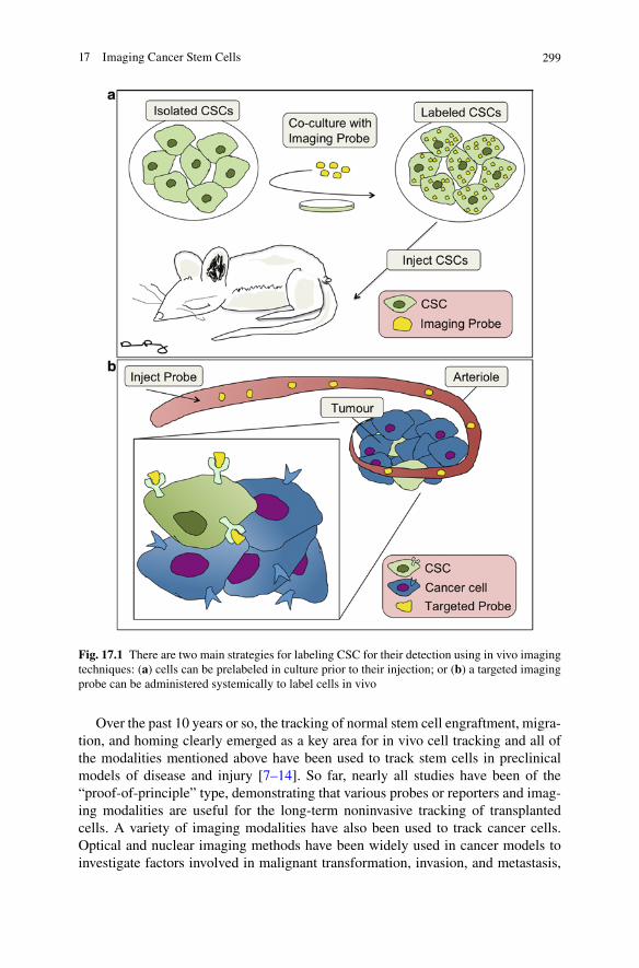
29917 Imaging Cancer Stem Cells
Over the past 10 years or so, the tracking of normal stem cell engraftment, migra-tion, and homing clearly emerged as a key area for in vivo cell tracking and all of the modalities mentioned above have been used to track stem cells in preclinical models of disease and injury [7–14]. So far, nearly all studies have been of the “proof-of-principle” type, demonstrating that various probes or reporters and imag-ing modalities are useful for the long-term noninvasive tracking of transplanted cells. A variety of imaging modalities have also been used to track cancer cells. Optical and nuclear imaging methods have been widely used in cancer models to investigate factors involved in malignant transformation, invasion, and metastasis,
Fig. 17.1 There are two main strategies for labeling CSC for their detection using in vivo imaging techniques: (a) cells can be prelabeled in culture prior to their injection; or (b) a targeted imaging probe can be administered systemically to label cells in vivo
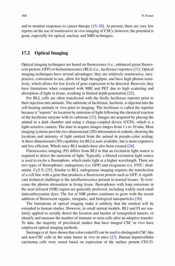
300 P. Foster
and to monitor responses to cancer therapy [15–20]. At present, there are very few reports on the use of noninvasive in vivo imaging of CSCs; however, the potential is great, especially for optical, nuclear, and MRI techniques.
17.2 Optical Imaging
Optical imaging techniques are based on fluorescence (i.e., enhanced green fluores-cent protein; GFP) or bioluminescence (BLI) (i.e., luciferase) reporters [21]. Optical imaging techniques have several advantages: they are relatively noninvasive, inex-pensive, convenient to use, allow for high throughput, and have high photon sensi-tivity, which allows for low levels of gene expression to be detected. However, they have limitations when compared with MRI and PET due to high scattering and absorption of light in tissue, resulting in limited depth penetration [22].
For BLI, cells are often transfected with the firefly luciferase reporter prior to their injection into animals. The substrate of luciferase, luciferin, is injected into the cell-bearing animals in vivo prior to imaging. The luciferase is called the reporter because it “reports” its location by emission of light following the chemical reaction of the luciferase enzyme with its substrate [23]. Images are acquired by placing the animal in a dark chamber and using a charge-coupled device (CCD), which is a light-sensitive camera. The time to acquire images ranges from 1 s to 10 min. Most imaging systems provide two-dimensional (2D) information in rodents, showing the locations and intensity of light emitted from the animal in pseudo-color scaling. A three-dimensional (3D) capability for BLI is now available, but is more expensive and less efficient. Whole mice BLI models have also been created [24].
Fluorescence imaging (FI) differs from BLI in that an excitation light source is required to detect the emission of light. Typically, a filtered excitation light source is used to excite a fluorophore, which emits light at a higher wavelength. There are two types of fluorophores: endogenous (i.e. GFP) and exogenous (i.e. FITC, rhod-amine, Cy5.5) [25]. Similar to BLI, endogenous imaging requires the transfection of a cell line with a gene that produces a fluorescent protein such as GFP. A signifi-cant technical challenge is the autofluorescence present in normal tissues. To over-come the photon attenuation in living tissue, fluorophores with long emissions in the near-infrared (NIR) region are generally preferred, including widely used small indocarbocyanine dyes. The list of NIR probes continues to grow with the recent addition of fluorescent organic, inorganic, and biological nanoparticles [26].
The limitations of optical imaging make it unlikely that the method will be extended to human studies. However, in small animal models, BLI and FI are rou-tinely applied to serially detect the location and burden of xenografted tumors, or identify and measure the number of immune or stem cells after an adoptive transfer. To date, the majority of preclinical studies that have imaged CSC in vivo have employed optical imaging methods.
Suetsugu et al. have shown that color-coded FI can be used to distinguish CSC-like and non-CSC cells in the same tumor in vivo in mice [27]. Human hepatocellular carcinoma cells were sorted based on expression of the surface protein CD133.
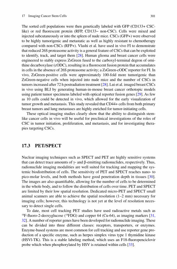
30117 Imaging Cancer Stem Cells
The sorted cell populations were then genetically labeled with GFP (CD133+ CSC-like) or red fluorescent protein (RFP, CD133− non-CSC). Cells were mixed and injected subcutaneously or into the spleen of nude mice. CSCs (GFP+) were observed to be highly tumorigenic and metastatic as well as highly resistant to chemotherapy compared with non-CSCs (RFP+). Vlashi et al. have used in vivo FI to demonstrate that reduced 26S proteasome activity is a general feature of CSCs that can be exploited to identify, track, and target them [28]. Human glioma and breast cancer cells were engineered to stably express ZsGreen fused to the carboxyl-terminal degron of orni-thine decarboxylase (cODC), resulting in a fluorescent fusion protein that accumulates in cells in the absence of 26S proteasome activity; a ZsGreen-cODC reporter for FI. In vivo, ZsGreen-positive cells were approximately 100-fold more tumorigenic than ZsGreen-negative cells when injected into nude mice and the number of CSCs in tumors increased after 72 h postradiation treatment [28]. Lui et al. imaged breast CSCs in vivo using BLI by generating human-in-mouse breast cancer orthotopic models using patient tumor specimens labeled with optical reporter fusion genes [29]. As few as 10 cells could be detected in vivo, which allowed for the early visualization of tumor growth and metastasis. This study revealed that CD44+ cells from both primary breast tumors and lung metastases are highly enriched for tumor-initiating cells.
These optical imaging studies clearly show that the ability to distinguish stem-like cancer cells in vivo will be useful for preclinical investigations of the roles of CSC in tumor initiation, proliferation, and metastasis, and for investigating thera-pies targeting CSCs.
17.3 PET/SPECT
Nuclear imaging techniques such as SPECT and PET are highly sensitive systems that can detect trace amounts of g- and b-emitting radionuclides, respectively. Thus, radionuclide imaging modalities are well suited for tracking and mapping the sys-temic biodistribution of cells. The sensitivity of PET and SPECT reaches nano- to pico-molar levels, and both methods have good penetration depth in tissues [30]. The images are also quantifiable, allowing for the number of cells to be determined in the whole body, and to follow the distribution of cells over time. PET and SPECT are limited by their low spatial resolution. Dedicated micro-PET and SPECT small animal scanners are able to achieve the spatial resolution (1–2 mm) necessary for imaging cells; however, this technology is not yet at the level of resolution neces-sary to detect single cells.
To date, most cell tracking PET studies have used radioactive metals such as 18F-fluoro-2-deoxyglucose (18FDG) and copper 64 (Cu-64), as imaging markers [31, 32]. A number of reporter genes have been developed for radionuclide imaging. These can be divided into three different classes: receptors, transporters, or enzymes. Enzyme-based systems are most common for cell tracking and use reporter gene pro-duction of a specific enzyme, such as herpes simplex virus type 1 thymidine kinase (HSVl-TK). This is a stable labeling method, which uses an F18-fluoropenciclovir probe which when phosphorylated by HSV is retained within cells [33].

302 P. Foster
The alternative to the reporter gene system is direct labeling of cells. Cells are incubated with the radioactive tracer, allowing the lipophilic molecules to diffuse across the cell membrane, and the isotope becomes trapped [34]. Following incuba-tion, the cells are washed to remove any unbound activity, and the cells injected into the host. Cu-64 can be delivered into cells via a lipophilic redox-active carrier molecule pyruvalde-hyde-bis (N4-methylthiosemicarbazone) (PTSM). Cu-64 is one of the longer-lived PET radionuclides, with a half-life of 12.7 h, allowing labeled cells to be tracked for 2–3 days [35]. Although direct cell labeling has many attractive features, the drawbacks include radiotoxicity effects, loss of label from cells, dilution of signal from cell division, and lack of information on cell function or viability.
Yoshii et al. showed that in a mouse colon carcinoma model (colon-26), Cu-64-ATSM localized preferentially in tumor regions with a high density of CD133+ cells with characteristics of CSCs [36]. Most nuclear imaging studies are performed with the addition of either MRI or CT so that the low resolution PET or SPECT images can be superimposed onto high resolution anatomical images.
17.4 Magnetic Resonance Imaging
MRI can produce images with high spatial resolution and exquisite soft tissue contrast. Current micro-MRI technology can achieve three-dimensional spatial resolutions on the order of tens of microns [37]. MRI uses no ionizing radiation and is considered safe and noninvasive. Cellular MRI is a relatively young field of imaging research that combines the ability to obtain high resolution MR data with the use of magnetic contrast agents for labeling specific cells, thereby enhancing their detection [38, 39]. Superparamagnetic iron oxide (SPIO) nano-particles represent a class of magnetic contrast agents used for cellular MRI that exhibit extremely high relaxivity [40]. A variety of iron oxide-based labels are now available [38]. Iron-labeled cells are usually imaged using either gradient echo (GRE) or spin echo (SE) sequences [41, 42]. The presence of the magnetic label causes a distortion in the magnetic field and leads to abnormal signal hypo-intensities in T2 or T2* sensitive images. Areas containing iron labeled cells appear as regions of low signal intensity, creating negative contrast. The large magnetic susceptibility of these particles affects an area much larger than the actual particle size. This effect is known as a “blooming artifact,” and leads to an exaggeration of the region occupied by iron oxide [43]. SPIO has been used to label and track a wide variety of cell types including T-lymphocytes [44, 45], macrophages [46, 47], pancreatic islets [48, 49], cancer cells [50, 51], and stem cells [42, 52] with minimal impact on cell function over a period of several weeks and over multiple cell divisions.
Pushing the limits of detection of SPIO-labeled cells has become an active area of research in the field of cellular MRI. This has been driven by interest in the fol-lowing: (i) tracking the migration of small numbers of cells; (ii) detecting cells with
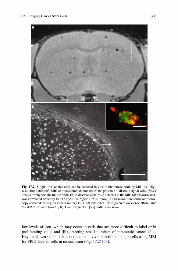
30317 Imaging Cancer Stem Cells
low levels of iron, which may occur in cells that are more difficult to label or in proliferating cells; and (iii) detecting small numbers of metastatic cancer cells. Heyn et al. were first to demonstrate the in vivo detection of single cells using MRI for SPIO-labeled cells in mouse brain (Fig. 17.2) [53].
Fig. 17.2 Single iron-labeled cells can be detected in vivo in the mouse brain by MRI. (a) High resolution (100 mm3) MRI of mouse brain demonstrates the presence of discrete signal voids (black arrow) throughout the mouse brain. (b) A discrete signal void detected in the MRI (black arrow in a) was correlated optically to a DiI positive region (white arrow). High resolution confocal micros-copy revealed this region to be a solitary DiI (red) labeled cell with green fluorescence attributable to GFP expression (inset of b). From Heyn et al. [53], with permission
304 P. Foster
The use of iron nanoparticles and MRI for cancer cell tracking has some limitations. These include relatively low sensitivity and difficulty in quantifying the signal loss caused by iron labeled cells. In addition, iron-labeled cells, which die in vivo, may be engulfed by bystander cells in tissue and this may confuse the image interpretation. The dilution of iron particles with cell division is also a drawback, since eventually in proliferating cells the iron label is diluted to a level below the detection threshold of MRI. MRI has yet to be used to track the fate of individual CSCs; however, the potential for this is clear. The fact that quiescent cancer cells retain the iron label for long periods compared with pro-liferating cells may represent a novel in vivo assay for differentiating between cancer cell types with various metastatic potentials.
17.5 Multimodality Imaging
It is becoming quite common to see multiple imaging modalities used in a comple-mentary fashion to acquire multilayered information. The goal of multimodality imaging is to combine the best features of separate modalities. For instance, high-resolution anatomical images acquired with MRI or CT are often combined with functional or metabolic imaging such as PET or SPECT. Many studies have shown the benefits of using multiple imaging modalities to achieve different, complemen-tary information about the fate of cells in vivo [54–59]. Along with this has come the development of multi-modality imaging probes [60–63].
Garzia et al. used PET/CT to investigate the relationship between miRNAs tar-geting the Notch pathway and medulloblastoma (MB) tumors. Notch regulates a subset of the MB cells that have stem-cell-like properties (CD44+) and can promote tumor growth. They showed that miR-199b-5p expression correlates with metasta-sis spread and that in a xenograft model, MB tumor burden can be reduced, indicat-ing the use of miR199b-5p as an adjuvant therapy for the improvement of anti-cancer MB treatments [64].
Today hybrid multi-modality imaging systems are being developed that will allow the simultaneous acquisition of different types of images in the same animal during the same imaging session. These systems include integrated PET/CT, SPECT/CT, and PET/MRI. PET/CT studies are now quite common in cancer patients. A number of groups are pushing the frontier of PET/MRI, which will provide high resolution morphological, molecular, and functional information [65, 66].
An important recent development is the concept of multimodality fusion reporter systems. Ray et al. [67] and Ponomarv et al. [68] have described triple modality reporter genes for whole mouse body fluorescent, bioluminescent, and nuclear imaging. Fusion reporters have the potential to accelerate translational cancer research and will be important for defining the potential roles of each modality in specific applications.
30517 Imaging Cancer Stem Cells
17.6 Cell Detection Thresholds: A Comparison of MRI and PET
Radionuclide imaging techniques are generally considered to have superior sensi-tivity compared with MRI. This argument is based on the detectable concentration of the probe, which is ~10−12 M for PET vs. ~10−5 M for MRI [69]. Although the sensitivity of PET and SPECT tracers expressed in terms of concentration of the probe is an appropriate convention when the target is large (on the order of the reso-lution of a PET and SPECT scan), it may not be appropriate for cases in which the target is microscopic. Such a case arises when cells (especially small numbers of cells or single cells) are imaged. In this case, the probe is concentrated within a small volume within the voxel (the cell). For these microscopic targets, increasing image resolution would result in a significant increase in the “apparent concentra-tion” of probe (number of moles of probe in a cell/voxel volume). For MRI, the high resolution of this technique results in a pessimistic estimate of sensitivity when expressed in terms of concentration, while the intrinsic low resolution of PET and SPECT techniques provide an apparent advantage. This advantage, however, would quickly deteriorate if the resolution of PET or SPECT could approach the resolution of MR. For the specific application of detecting a microscopic target, such as a cell, a proper calculation of sensitivity would involve a comparison of the number of moles of tracer that must be loaded into a cell to permit its detection.
An analysis by Heyn et al. indicates that on a mole basis, the detection threshold of Fe in SPIO is in fact not very different from that of PET tracers [70]. The detec-tion of cells labeled with 64Cu allows for the in vivo detection of a few hundred cells (~10−17 mol of 64Cu per cell) or ~10−15 mol of 64Cu (1 fmol) in a 10 microl voxel volume using a small animal PET scanner [71]. Heyn showed that for a typical MRI microimaging acquisition with an image SNR of 60 and 100 mm isotropic resolu-tion, the minimum detectable amount of iron is approximately 1.3 pg Fe/cell, which corresponds to a detection threshold of 2.33 × 10⁻¹⁴ mol of Fe (23.3 fmol) [70]. Stated this way, the sensitivity levels of MRI and PET for SPIO and radionuclide tracer, respectively, are not very different after all.
17.7 Implications of Imaging for CSC Detection
Traditionally, the assays used to identify and examine CSC are labor-intensive, time-consuming, invasive, and provide little information on the dynamics of cancer cells in vivo. CSC studies should take advantage of advanced imaging technology to increase our understanding of the CSC model, dormancy, tumor growth, and metastasis. With the ability to reliably track the metastasis and proliferation of small numbers of cancer cells and specific subsets of cancer cells, will come new knowl-edge of the behavior of these cells in a relatively undisturbed environment.
306 P. Foster
References
1. Pierce MC, Javier DJ, Richards-Kortum R (2008) Optical contrast agents and imaging systems for detection and diagnosis of cancer. Int J Cancer 123(9):1979–1990.
2. Rodt T, von Falck C, Halter R, Ringe K, Shin HO, Galanski M, Borlak M (2009) In vivo microCT quantification of lung tumor growth in SPC-raf transgenic mice. J Front Biosci 14:1939–1944.
3. Rychak JJ, Graba J, Cheung AM, Mystry BS, Lindner JR, Kerbel RS, Foster FS (2007) Microultrasound molecular imaging of vascular endothelial growth factor receptor 2 in a mouse model of tumor angiogenesis. Mol Imaging 6(5):289–296.
4. Herschman HR (2004) PET reporter genes for noninvasive imaging of gene therapy, cell track-ing and transgenic analysis Crit Rev Oncol Hematol 51(3):191–204.
5. Emonts P, Bourgeois P, Lemort M, Flamen P (2009) Functional imaging of head and neck cancers. Curr Opin Oncol 21(3):212–217.
6. Bernas LM, Foster PJ, Rutt BK (2010) Imaging iron-loaded mouse glioma tumors with bSSFP at 3 T. Magn Reson Med 64(1):23–31.
7. Bulte JW, Douglas T, Witwer B, Zhang SC, Strable E, Lewis BK, Zywicke H, Miller B, van Gelderen P, Moskowitz BM, Duncan ID, Frank JA (2001) Magnetodendrimers allow endo-somal magnetic labeling and in vivo tracking of stem cells. Nat Biotechnol 19(12):1141–1147.
8. Hinds KA, Hill JM, Shapiro EM, Laukkanen MO, Silva AC, Combs CA, Varney TR, Balaban RS, Koretsky AP, Dunbar CE (2003) Highly efficient endosomal labeling of progenitor and stem cells with large magnetic particles allows magnetic resonance imaging of single cells. Blood 102(3): 867–872.
9. Stroh A, Faber C, Neuberger T, Lorenz P, Sieland K, Jakob PM, Webb A, Pilgrimm H, Schober R, Pohl EE, Zimmer C (2005) In vivo detection limits of magnetically labeled embryonic stem cells in the rat brain using high-field (17.6 T) magnetic resonance imaging. Neuroimage 24(3):635–645.
10. Boddington S, Henning TD, Sutton EJ, Daldrup-Link HE (2008) Labeling stem cells with fluorescent dyes for non-invasive detection with optical imaging. J Vis Exp 2(14): 686. doi: 10.3791/686.
11. Narsinh KH, Cao F, Wu JC (2009) Molecular imaging of human embryonic stem cells. Methods Mol Biol 515:13–32.
12. Gera A, Steinberg GK, Guzman R (2010) In vivo neural stem cell imaging: current modalities and future directions. Regen Med 5(1):73–86.
13. Huang J, Lee CC, Sutcliffe JL, Cherry SR, Tarantal AF (2008) Radiolabeling rhesus monkey CD34+ hematopoietic and mesenchymal stem cells with 64Cu-pyruvaldehyde-bis(N4-methylthiosemicarbazone) for microPET imaging. Mol Imaging 7(1):1–11.
14. Gyöngyösi M, Blanco J, Marian T, Trón L, Petneházy O, Petrasi Z, Hemetsberger R, Rodriguez J, Font G, Pavo IJ, Kertész I, Balkay L, Pavo N, Posa A, Emri M, Galuska L, Kraitchman DL, Wojta J, Huber K, Glogar D (2008) Serial noninvasive in vivo positron emission tomographic tracking of percutaneously intramyocardially injected autologous porcine mesenchymal stem cells modified for transgene reporter gene expression. Circ Cardiovasc Imaging 1(2):94–103.
15. Hong H, Yang Y, Zhang Y, Cai W (2010) Non-invasive cell tracking in cancer and cancer therapy. Curr Top Med Chem 10(12):1237–1248.
16. Bading JR, Shields AF (2008) Imaging of cell proliferation: status and prospects. J Nucl Med 49 Suppl 2:64S-80S.
17. Wang H, Liu B, Tian JH, Xu BX, Guan ZW, Qu BL, Liu CB, Wang RM, Chen YM, Zhang JM. (2010) Monitoring early responses to irradiation with dual-tracer micro-PET in dual-tumor bearing mice. World J Gastroenterol 16(43):5416–5423.
18. Capala J, Bouchelouche K (2010) Molecular imaging of HER2-positive breast cancer: a step toward an individualized ‘image and treat’ strategy. Curr Opin Oncol 22(6):559–66.
30717 Imaging Cancer Stem Cells
19. Kienast Y, von Baumgarten L, Fuhrmann M, Klinkert WE, Goldbrunner R, Herms J, Winkler F (2010) Real-time imaging reveals the single steps of brain metastasis formation. Nat Med 16(1):116–122.
20. Nogawa M, Yuasa T, Kimura S, Kuroda J, Sato K, Segawa H, Yokota A, Maekawa T (2005) Monitoring luciferase-labeled cancer cell growth and metastasis in different in vivo models. Cancer Lett 217(2):243–253.
21. Ntziachristos V (2010) Going deeper than microscopy: the optical imaging frontier in biology. Nat Methods 7(8):603–614.
22. Klerk CP, Overmeer RM, Niers TM, Versteeg HH, Richel DJ, Buckle T, Van Noorden CJ, van Tellingen O (2007) Validity of bioluminescence measurements for noninvasive in vivo imag-ing of tumor load in small animals. Biotechniques 43(1 Suppl):7–13.
23. Dothager RS, Flentie K, Moss B, Pan MH, Kesarwala A, Piwnica-Worms D (2009) Advances in bioluminescence imaging of live animal models. Curr Opin Biotechnol 20(1):45–53.
24. Sadikot RT, Blackwell TS (2008) Bioluminescence: imaging modality for in vitro and in vivo gene expression. Methods Mol Biol 477:383–394.
25. Zhou L, El-Deiry WS (2009) Multispectral fluorescence imaging. J Nucl Med 50(10): 1563–1566.
26. Shin D, Vigneswaran N, Gillenwater A, Richards-Kortum R (2010) Advances in fluorescence imaging techniques to detect oral cancer and its precursors. Future Oncol 6(7):1143–1154.
27. Suetsugu A, Osawa Y, Nagaki M, Moriwaki H, Saji S, Bouvet M, Hoffman RM (2010) Simultaneous color-coded imaging to distinguish cancer “stem-like” and non-stem cells in the same tumor. J Cell Biochem 111(4):1035–1041.
28. Vlashi E, Kim K, Lagadec C, Donna LD, McDonald JT, Eghbali M, Sayre JW, Stefani E, McBride W, Pajonk F (2009) In vivo imaging, tracking, and targeting of cancer stem cells. J Natl Cancer Inst 101(5):350–359.
29. Liu H, Patel MR, Prescher JA, Patsialou A, Qian D, Lin J, Wen S, Chang YF, Bachmann MH, Shimono Y, Dalerba P, Adorno M, Lobo N, Bueno J, Dirbas FM, Goswami S, Somlo G, Condeelis J, Contag CH, Gambhir SS, Clarke MF (2010) Cancer stem cells from human breast tumors are involved in spontaneous metastases in orthotopic mouse models. Proc Natl Acad Sci USA 107(42):18115–18120.
30. Rowland DJ, Cherry SR (2008) Small-animal preclinical nuclear medicine instrumentation and methodology. Semin Nucl Med 38(3):209–222.
31. Acton PD, Zhou R (2005) Imaging reporter genes for cell tracking with PET and SPECT. Q J Nucl Med Mol Imaging 49(4):349–360.
32. Thompson M, Wall DM, Hicks RJ, Prince HM (2005) In vivo tracking for cell therapies. Q J Nucl Med Mol Imaging 49(4):339–348.
33. Hsieh CH, Chen FD, Wang HE, Hwang JJ, Chang CW, Lee YJ, Gelovani JG, Liu RS (2008) Generation of destabilized herpes simplex virus type 1 thymidine kinase as transcription reporter for PET reporter systems in molecular genetic imaging. J Nucl Med 49(1):142–150.
34. Patel D, Kell A, Simard B, Xiang B, Lin HY, Tian G (2011) The cell labeling efficacy, cytotox-icity and relaxivity of copper-activated MRI/PET imaging contrast agents. Biomaterials 32(4): 1167–76.
35. Cai H, Li Z, Huang CW, Shahinian AH, Wang H, Park R, Conti PS (2010) Evaluation of cop-per-64 labeled AmBaSar conjugated cyclic RGD peptide for improved MicroPET imaging of integrin alphavbeta3 expression. Bioconjug Chem 21(8):1417–1424.
36. Yoshii Y, Furukawa T, Kiyono Y, Watanabe R, Waki A, Mori T, Yoshii H, Oh M, Asai T, Okazawa H, Welch MJ, Fujibayashi Y (2010) Copper-64-diacetyl-bis (N4-methylthiosemicarbazone) accumulates in rich regions of CD133+ highly tumorigenic cells in mouse colon carcinoma. Nucl Med Biol 37(4):395–404.
37. Degen CL, Poggio M, Mamin HJ, Rettner CT, Rugar D (2009) Nanoscale magnetic resonance imaging. Proc Natl Acad Sci USA 106(5):1313–1317.
38. Modo M, Hoehn M, and Bulte JW (2005) Cellular MR imaging. Mol Imaging 4: 143–164. 39. Corot C, Robert P, Idee JM, Port M (2006) Recent advances in iron oxide nanocrystal technol-
ogy for medical imaging. Adv Drug Deliv Rev 58: 1471–1504.
308 P. Foster
40. Wang YX, Hussain SM, Krestin GP (2001) Superparamagnetic iron oxide contrast agents: physicochemical characteristics and applications in MR imaging. Eur Radiol 11(11): 2319–2331.
41. Thorek DL, Chen AK, Czupryna J, Tsourkas A (2006) Superparamagnetic iron oxide nanopar-ticle probes for molecular imaging. Ann Biomed Eng 34:23–38.
42. Frank JA, Anderson SA, Kalsih H, Jordan EK, Lewis BK, Yocum GT, Arbab AS (2004) Methods for magnetically labeling stem and other cells for detection by in vivo magnetic reso-nance imaging. Cytotherapy 6: 621–625.
43. Bulte JW, Kraitchman DL (2004) Iron oxide MR contrast agents for molecular and cellular imaging. NMR Biomed 17(7):484–499.
44. Shapiro, EM, Medford-Davis LN, Fahny TM, Dunbar CE, Koretsky AP (2007) Antibody-mediated cell labeling of peripheral T cells with micron-sized iron oxide particles (MPIOs) allows single cell detection by MRI. Contrast Media Mol Imaging 2:147–153.
45. Medarova Z, Tsai S, Evgenov N, Santamaria P, Moore A (2008) In vivo imaging of a diabeto-genic CD8+ T cell response during type 1 diabetes progression. Magn Reson Med 59(4):712–720.
46. Mun HS, Kang HJ, Lim KH, Sohn JY, Chang H, Lee KG, Lee JS (2008) Graft rejection in the xenogeneic transplantation of mice: diagnosis with in vivo MR imaging using the homing trait of macrophages. Xenotransplantation 15(4):218–224.
47. Oweida AJ, Dunn EA, Karlik SJ, Dekaban GA, Foster PJ (2007) Iron-oxide labeling of hematogenous macrophages in a model of experimental autoimmune encephalomyelitis and the contribution to signal loss in fast imaging employing steady state acquisition (FIESTA) images. J Magn Reson Imaging 26(1):144–151.
48. Evgenov NV, Medarova Z, Pratt J, Pantazopoulos P, Leyting S, Bonner-Weir S, Moore A (2006) In vivo imaging of immune rejection in transplanted pancreatic islets. Diabetes 55(9):2419–2428.
49. Tai JH, Foster P, Rosales A, Feng B, Hasilo C, Martinez V, Ramadan S, Snir J, Melling CW, Dhanvantari S, Rutt B, White DJ (2006) Imaging islets labeled with magnetic nanoparticles at 1.5 Tesla. Diabetes 55(11):2931–2938.
50. Heyn C, Ronald JA, Ramadan SS, Snir JA, Barry AM, MacKenzie LT, Mikulis DJ, Palmieri D, Bronder JL, Steeg PS (2006) In vivo MRI of cancer cell fate at the single-cell level in a mouse model of breast cancer metastasis to the brain. Magn Reson Med 56: 1001–1010.
51. Foster PJ, Dunn EA, Karl KE, Snir JA, Nycz CM, Harvey AJ, Pettis RJ (2008) Cellular mag-netic resonance imaging: in vivo imaging of melanoma cells in lymph nodes of mice. Neoplasia 10(3):207–216.
52. Niemeyer M, Oostendorp RA, Kremer M, Hippauf S, Jacobs VR, Baurecht H, Ludwig G, Piontek G, Bekker-Ruz V, Timmer S, Rummeny EJ, Kiechle M, Beer AJ (2010) Non-invasive tracking of human haemopoietic CD34(+) stem cells in vivo in immunodeficient mice by using magnetic resonance imaging. Eur Radiol 20(9):2184–2193.
53. Heyn C, Ronald JA, Mackenzie LT, MacDonald IC, Chambers AF, Rutt BK, and Foster PJ (2006) In vivo magnetic resonance imaging of single cells in mouse brain with optical valida-tion. Magn Reson Med 55: 23–29.
54. Nahrendorf M, Zhang H, Hembrador S, Panizzi P, Sosnovik DE, Aikawa E, Libby P, Swirski FK, Weissleder R (2008) Nanoparticle PET-CT imaging of macrophages in inflammatory ath-erosclerosis. Circulation 117(3):379–387.
55. Sosnovik DE, Nahrendorf M, Deliolanis N, Novikov M, Aikawa E, Josephson L, Rosenzweig A, Weissleder R, Ntziachristos V (2007) Fluorescence tomography and magnetic resonance imaging of myocardial macrophage infiltration in infarcted myocardium in vivo. Circulation 115(11):1384–1391.
56. Chen R, Parry JJ, Akers WJ, Berezin MY, El Naqa IM, Achilefu S, Edwards WB, Rogers BE (2010) Multimodality imaging of gene transfer with a receptor-based reporter gene. J Nucl Med 51(9):1456–1463.
57. Liang M, Liu X, Cheng D, Liu G, Dou S, Wang Y, Rusckowski M, Hnatowich DJ (2010) Multimodality nuclear and fluorescence tumor imaging in mice using a streptavidin nanopar-ticle. Bioconjug Chem 21(7):1385–1388.
30917 Imaging Cancer Stem Cells
58. Tu C, Ma X, Pantazis P, Kauzlarich SM, Louie AY (2010) Paramagnetic, silicon quantum dots for magnetic resonance and two-photon imaging of macrophages. J Am Chem Soc 132(6):2016–2023.
59. Higuchi T, Anton M, Dumler K, Seidl S, Pelisek J, Saraste A, Welling A, Hofmann F, Oostendorp RA, Gansbacher B, Nekolla SG, Bengel FM, Botnar RM, Schwaiger M (2009) Combined reporter gene PET and iron oxide MRI for monitoring survival and localization of transplanted cells in the rat heart. J Nucl Med 50(7):1088–1094.
60. Kim HS, Cho HR, Choi SH, Woo JS, Moon WK (2010) In vivo imaging of tumor transduced with bimodal lentiviral vector encoding human ferritin and green fluorescent protein on a 1.5T clinical magnetic resonance scanner. Cancer Res 70(18):7315–7324.
61. Ogawa M, Regino CA, Seidel J, Green MV, Xi W, Williams M, Kosaka N, Choyke PL, Kobayashi H (2009) Dual-modality molecular imaging using antibodies labeled with activat-able fluorescence and a radionuclide for specific and quantitative targeted cancer detection. Bioconjug Chem 20(11):2177–2184.
62. Veiseh O, Sun C, Fang C, Bhattarai N, Gunn J, Kievit F, Du K, Pullar B, Lee D, Ellenbogen RG, Olson J, Zhang M (2009) Specific targeting of brain tumors with an optical/magnetic reso-nance imaging nanoprobe across the blood-brain barrier. Cancer Res 69(15):6200–6207.
63. Gindy ME, Prud’homme RK (2009) Multifunctional nanoparticles for imaging, delivery and targeting in cancer therapy. Expert Opin Drug Deliv 6(8):865–878.
64. Garzia L, Andolfo I, Cusanelli E, Marino N, Petrosino G, De Martino D, Esposito V, Galeone A, Navas L, Esposito S, Gargiulo S, Fattet S, Donofrio V, Cinalli G, Brunetti A, Vecchio LD, Northcott PA, Delattre O, Taylor MD, Iolascon A, Zollo M (2009) MicroRNA-199b-5p impairs cancer stem cells through negative regulation of HES1 in medulloblastoma. PLoS One 4(3):e4998.
65. Wehrl HF, Sauter AW, Judenhofer MS, Pichler BJ (2010) Combined PET/MR imaging– technology and applications. Technol Cancer Res Treat 9(1):5–20.
66. Hawkes RC, Fryer TD, Siegel S, Ansorge RE, Carpenter TA (2010) Preliminary evaluation of a combined microPET-MR system. Technol Cancer Res Treat 9(1):53–60.
67. Ray P, De A, Min JJ, Tsien RY, Gambhir SS (2004) Imaging tri-fusion multimodality reporter gene expression in living subjects. Cancer Res 64(4):1323–1330.
68. Ponomarev V, Doubrovin M, Serganova I, Vider J, Shavrin A, Beresten T, Ivanova A, Ageyeva L, Tourkova V, Balatoni J, Bornmann W, Blasberg R, Gelovani Tjuvajev J (2004) A novel tri-ple-modality reporter gene for whole-body fluorescent, bioluminescent, and nuclear noninva-sive imaging. Eur J Nucl Med Mol Imaging 31(5):740–751.
69. Phelps ME (1991) PET: a biological imaging technique. Neurochem Res 16:929–940. 70. Heyn C, Bowen CV, Rutt BK, Foster PJ (2005) Detection threshold of single SPIO-labeled
cells with FIESTA. Magn Reson Med 53(2):312–320. 71. Adonai N, Nguyen KN, Walsh J, Iyer M, Toyokuni T, Phelps ME, McCarthy T, McCarthy DW,
Gambhir SS (2002) Ex vivo cell labeling with 64Cu-pyruvaldehyde-bis(N4-methylthiosemi-carbazone) for imaging cell trafficking in mice with positron-emission tomography. Proc Natl Acad Sci USA 99:3030–3035.

311A.L. Allan (ed.), Cancer Stem Cells in Solid Tumors, Stem Cell Biology and Regenerative Medicine, DOI 10.1007/978-1-61779-246-5_18, © Springer Science+Business Media, LLC 2011
Abstract Adult stem and progenitor cell functions have traditionally been studied by transplantation into immune-deficient mice. Over the years, the mouse strains capable of accepting human cells and models used to study regenerative or tumorigenic pro-cesses have grown in number and complexity. Because of these developments, it is now possible to study the establishment and metastasis of malignant human cancer cells, and this has generated an explosion of studies identifying cancer cells with robust tumor-initiating potential. In hematopoietic malignancies and some solid tumor types, the cells driving tumor growth, angiogenic vessel formation, and metastasis share char-acteristics with human stem cells; namely self-renewal and the ability to differentiate into multiple cell types. As our knowledge of the mechanisms of human tumor progres-sion expands, we will hopefully be able to employ humanized mouse models to study cancer biology and to develop novel anti-cancer therapies.
Abbreviations
ALDH Aldehyde dehydrogenaseAML Acute myeloid leukemiaBM Bone marrowCSC Cancer stem cellFACS Fluorescent activated cell sortingHSC Hematopoietic stem cell
D.A. Hess (*)Department of Physiology & Pharmacology, The University of Western Ontario, London, ON, Canada
Vascular Biology Group, Krembil Centre for Stem Cell Biology, Robarts Research Institute, London, ON, Canadae-mail: [email protected]
Chapter 18Mouse Models for Studying Normal and Cancer Stem Cells
David A. Hess
312 D.A. Hess
HSPCs Hematopoietic stem and progenitor cellsIHC ImmunohistochemistryIL InterleukinIL-2Rg Interleukin-2 receptor common gamma chainNK Natural killer cellNOD Nonobese diabeticNOD/SCID Nonobese diabetic plus severe combined immunodeficientSCID Severe combined immunodeficientSL-IC SCID leukemia-initiating cellsSP Side populationSRC NOD/SCID repopulating cellUCB Umbilical cord bloodb2M Beta-2-microglobullin
18.1 Introduction
Immunodeficient mouse xenograft models have been used extensively in recent years to establish the fundamentals of human stem cell biology. Classically, intravenously transplanted human hematopoietic stem cells (HSC) have been shown to home to the murine bone marrow (BM) and differentiate into multilineage progeny following isolation, purification, and/or ex vivo manipulation [1–7]. Starting in the early 1990s, John Dick and collaborators also identified human cells that can establish human myeloid leukemia in transplanted murine recipients [8–12]. These groundbreaking findings were made possible by the development of the first and second generation immune deficient murine hosts, namely the severe combined immune deficient (SCID) and nonobese diabetic (NOD) plus severe combined immune deficient (NOD/SCID) recipients. Throughout the 1990s, the NOD/SCID recipient served as the “gold standard” for the study of normal human hematopoiesis and leukemogenesis in vivo.
The number and quality of immune-deficient murine models for xenotransplanation has grown immensely over the past 10 years. The explosive growth in this field is due in part to the limitations of the NOD/SCID model and the lack of in vitro assays to model complex biological processes such as human hematopoiesis, the regeneration or repair of damaged organs and tissues, and tumor development. Recently, improved immunodeficient murine strains, such as the NOD/SCID beta2-microglobulin (b2M) null [13–15] and NOD/SCID IL-2 receptor common gamma chain (IL-2Rg) null mice [16–20], have increased the survival and engraftment of human cells after transplantation. These novel strains permit extensive human cell engraftment without rejection due to reduced innate immunity (NOD mutation), complete T- and B-lymphocyte deficiency (SCID mutation), and reduced NK-cell function (b2M or IL-2Rg mutation) [20]. These strains are commercially available and provide trans-planted human cells the highest rate of survival in xenograft models developed to date. Although these strains do not represent an exhaustive list of the immunodefi-cient mice described in the literature, this chapter will focus on the recent progress
31318 Mouse Models for Studying Normal and Cancer Stem Cells
made with commonly-used human-into-mouse xenotransplantation models to study the biology of normal human stem cells and tumor initiating cells from both hematopoietic and solid tumors.
18.2 Mouse Models to Study Normal Human Stem Cells
Although large animal models exist for the study of human xenografts in utero [21–24] or for studying nonhuman primate stem cells [25–28], the use of the immune-deficient mouse as a recipient of human stem cell grafts has emerged as the most common and cost effective strategy. The first immune-deficient mouse characterized was the athymic nude mouse described by Isaason and Cattanach in 1962. Early trans-plantation studies with the T-lymphocyte-deficient nude mice were disappointing, as these mice failed to support the growth of transferred human hematopoietic BM stem cells [29]. Although the nude mouse has been used to study murine tumor growth due to its hairless phenotype, the immunodeficiency elicited in this strain is insufficient for xenotransplantation studies using human HSC and poorly permits human tumor establishment and metastasis unless highly aggressive and/or large numbers of tumor cells are implanted. Thus, to effectively study complex biological processes such as human hematopoiesis or tumor biology in vivo, a systematic progression of genetic modifications was required to develop the modern immunodeficient host mouse, and these are described later. Table 18.1 also provides a summary of the immunodeficient mouse strains commonly used for human stem cell transplantation studies, and contrasts the advantages and disadvantages of each model common to experimental use.
18.2.1 The Severe Combined Immunodeficient Mouse
The ability to study human hematopoiesis in a murine model was greatly facilitated by the discovery of mice with the severe combined immunodeficiency (SCID) muta-tion [30]. SCID mice have defects in T- and B-lymphocyte development due to a mutation in the gene for DNA-dependent protein kinase on chromosome 16 (PrkdcSCID). DNA-dependent protein kinase is required for successful rearrangement of the T-cell receptor and immunoglobulin gene segments, leaving the mice unable to produce functional T- and B-lymphocytes. Because DNA-dependent protein kinase is a DNA-proofreading enzyme, the SCID mutation also confers extreme sensitivity to radio-therapy, due to the inability of sublethally irradiated cells to mediate repair of double stranded breaks. Nonetheless, the SCID mouse was the first mouse to support the survival of human neoplasms [31] and human hematopoietic cells [32, 33]. One of the drawbacks to using the SCID mouse was low-level human cell engraftment due to elevated natural killer (NK) cell activity and innate immunity conferred by the background CB17 strain. Therefore, this strain is acceptable for the implantation of solid fetal liver or thymus (SCID/hu model) or sections of tumors, but human peripheral blood and BM cell suspensions (hu-PBL-model) were rapidly recognized
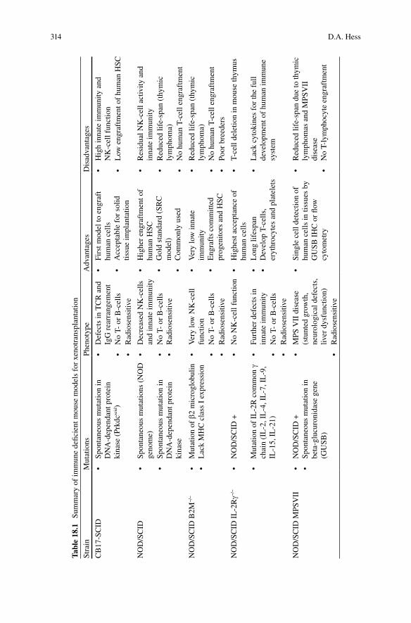
314 D.A. Hess
Tabl
e 18
.1
Sum
mar
y of
imm
une
defic
ient
mou
se m
odel
s fo
r xe
notr
ansp
lant
atio
n
Stra
inM
utat
ions
Phen
otyp
eA
dvan
tage
sD
isad
vant
ages
CB
17-S
CID
•Sp
onta
neou
sm
utat
ion
in
DN
A-d
epen
dant
pro
tein
ki
nase
(Pr
kdcsc
id)
•D
efec
tsin
TC
Ra
nd
IgG
rea
rran
gem
ent
•Fi
rstm
odel
toe
ngra
ft
hum
an c
ells
•A
ccep
tabl
efo
rso
lid
tissu
e im
plan
tatio
n
•H
igh
inna
teim
mun
itya
nd
NK
-cel
l fun
ctio
n•
No
T-o
rB
-cel
ls•
Low
eng
raft
men
tof
hum
anH
SC•
Rad
iose
nsiti
ve
NO
D/S
CID
•Sp
onta
neou
sm
utat
ions
(N
OD
ge
nom
e)•
Dec
reas
edN
K-c
ells
an
d in
nate
imm
unity
•H
ighe
ren
graf
tmen
tof
hum
an H
SC•
Gol
dst
anda
rd(
SRC
m
odel
)•
Com
mon
lyu
sed
•R
esid
ualN
K-c
ella
ctiv
itya
nd
inna
te im
mun
ity•
Spon
tane
ous
mut
atio
nin
D
NA
-dep
enda
nt p
rote
in
kina
se
•N
oT-
or
B-c
ells
•R
adio
sens
itive
•R
educ
edli
fe-s
pan
(thy
mic
ly
mph
oma)
•N
ohu
man
T-c
elle
ngra
ftm
ent
NO
D/S
CID
B2M
−/−
•M
utat
ion
ofb
2 m
icro
glob
ulin
•L
ack
MH
Cc
lass
Ie
xpre
ssio
n•
Ver
ylo
wN
K-c
ell
func
tion
•V
ery
low
inna
te
imm
unity
•E
ngra
fts
com
mitt
ed
prog
enito
rs a
nd H
SC
•R
educ
edli
fe-s
pan
(thy
mic
ly
mph
oma)
•N
oT-
or
B-c
ells
•N
ohu
man
T-c
elle
ngra
ftm
ent
•R
adio
sens
itive
•Po
orb
reed
ers
NO
D/S
CID
IL
-2Rg−
/−•
NO
D/S
CID
+•
No
NK
-cel
lfun
ctio
n•
Hig
hest
acc
epta
nce
of
hum
an c
ells
•T-
cell
dele
tion
inm
ouse
thym
us
•M
utat
ion
ofI
L-2
Rc
omm
ong
ch
ain
(IL
-2, I
L-4
, IL
-7, I
L-9
, IL
-15,
IL
-21)
•Fu
rthe
rde
fect
sin
in
nate
imm
unity
•L
ong
lifes
pan
•D
evel
opT
-cel
ls,
eryt
hroc
ytes
and
pla
tele
ts
•L
ack
cyto
kine
sfo
rth
efu
llde
velo
pmen
t of
hum
an im
mun
e sy
stem
•N
oT-
or
B-c
ells
•R
adio
sens
itive
NO
D/S
CID
MPS
VII
•N
OD
/SC
ID+
•Sp
onta
neou
sm
utat
ion
in
beta
-glu
curo
nida
se g
ene
(GU
SB)
•M
PSV
IId
isea
se
(stu
nted
gro
wth
, ne
urol
ogic
al d
efec
ts,
liver
dys
func
tion)
•Si
ngle
cel
ldet
ectio
nof
hu
man
cel
ls in
tiss
ues
by
GU
SB I
HC
or
flow
cy
tom
etry
•R
educ
edli
fe-s
pan
due
toth
ymic
ly
mph
omas
and
MPS
VII
di
seas
e•
No
T-ly
mph
ocyt
een
graf
tmen
t•
Rad
iose
nsiti
ve
31518 Mouse Models for Studying Normal and Cancer Stem Cells
as foreign and lysed by murine NK-cells. In addition, the SCID model does not support T-cell development in vivo, and transferred human T-cells become profoundly anergic due to incompatability with the murine thymus and a lack of human antigen-presenting cells [34]. Thus, enhanced human cell engraftment and models for the generation of human T-lymphocytes in immune-deficient mice were still sought.
18.2.2 The Nonobese Diabetic SCID Mouse (NOD/SCID)
To circumvent the high NK-cell activity inherent in the SCID mouse, Len Shultz and colleagues at the Jackson Laboratories (Bar Harbour, ME) crossed the SCID muta-tion onto various strains of inbred mice with known defects in innate immunity. The NOD mouse background, best known as a model of spontaneous autoimmune diabe-tes, possessed defects in macrophage and NK-cell functions [35, 36]. The result was the generation of the most prevalently used immunodeficient mouse strain, the non-obese diabetic SCID (NOD/SCID) mouse. The combined immunodeficiency pro-duced an improved recipient with excellent capacity to engraft purified human stem cells for the study of hematopoietic development after transplantation. In addition, NOD/SCID mice were excellent breeders, with large and robust litters. However, these mice required housing in a clean barrier facility and required sterile animal handling to prevent untoward infections due to their extreme immune deficiencies.
The NOD/SCID model is by far the most widely used xenograft recipient to date. Sublethally irradiated (300–350 cGy) NOD/SCID mice have been extensively used to demonstrate multilineage human hematiopoietic reconstitution after the trans-plantation of human BM- or UCB-derived HSCs purified using cell surface markers such as CD34 [2, 3, 22, 37] and CD133 [38–40]; using conserved stem cell func-tions such as Hoescht dye efflux pumps (side population [SP] cells)[41, 42], and high aldehyde dehydrogenase (ALDH) activity; or a combination of both strategies [43]. Dick and collaborators were the first to identify a novel human hematopoietic stem or progenitor cell, termed the NOD/SCID repopulating cell (SRC), present in the CD34+CD38− cell fraction, that was capable of multilineage repopulation in the BM of sublethally irradiated NOD/SCID mice [3, 6]. Using limiting dilution analysis and Poisson statistics, the frequency of this novel cell type was functionally defined in human UCB at 1 SRC in 617 CD34+CD38− cells [3], indicating that the CD34+CD38− cell population was functionally heterogeneous. The same group was the first to discover a new class of SRC capable of low level engraftment originated from the human CD34− cell fraction [2], supporting the dogma that a single trans-planted murine CD34− cell could facilitate multilineage reconstitution in vivo [44]. Subsequently, Fujisaki et al. [45] confirmed these findings, and Goodell et al. [42] purified a SP of CD34− cells that excluded Hoescht 33342 dye and possessed recon-stituting capacity. Nakamura et al. [46] demonstrated that CD34− cells were highly quiescent, and that prestimulation of cultured human CD34− cells with cytokines prior to transplantation induced CD34 expression and increased their capacity for homing and engraftment. Finally, Dao et al. [47] used serial transplantation to show
316 D.A. Hess
the reversibility of CD34 expression within the human stem and progenitor cell compartments.
However, in all these of these studies, differentiated human blood cell production was skewed to the B-lymphoid and myeloid compartments, as the NOD/SCID thy-mic microenvironment was not able to fully support T-lymphocyte development under normal transplant conditions in vivo [48]. The lack of T-cell specific engraft-ment is one of the major limitations of the traditional NOD/SCID model, and is potentially due to residual NK-cell activity and incompatibility with the hypertrophic murine thymus in NOD/SCID mice [49]. In addition to the lack of human T-lymphocyte development in vivo, another major drawback of the NOD/SCID model is their shortened lifespan (4–6 months) due to the presence of the Emv-30 provirus integrated into the NOD genome, which can cause breakthrough of lethal thymic lymphomas in mice with SCID mutation [50]. The duration of experiments that can be performed using 8–10-week old NOD/SCID mice is therefore limited to 8–12 weeks posttransplantation, preventing proper analysis of long-term engraft-ment. Furthermore, unlike murine donor/murine recipient transplantation systems [44], engraftment and multilineage differentiation after injection of a single human HSC has never been achieved using xenograft models. Therefore, to address the abil-ity of human cells to self-renew using the NOD/SCID model, retroviral or lentiviral marking of transplanted cell clones, the retrieval of human cells from primary recipi-ents, and clonal analysis of short-term vs. long-term hematopoietic propagation after serial transplantation in secondary and tertiary recipients is required [51–53].
18.2.3 The NOD/SCID Beta2 Migroglobulin Null Mouse (NOD/SCID b 2M)
Second generation immunodeficient mice have recently been extended from the NOD/SCID background to circumvent the residual NK-cell function, the lack of T-cell engraftment, and the shortened lifespan characteristic of the NOD/SCID strain. The first strain developed was the NOD/SCID b2 microglobulin null mouse (NOD/SCID b2M) characterized in 1997 [54]. These mice lacked MHC class I expression on hematopoietic effector cells due to the targeted deletion of the b2M gene, demonstrated virtually no NK-cell function, and were very permissive for xenograft acceptance [15, 55]. Committed human hematopoietic progenitors (CD34+CD38+ cells) as well as primitive HSCs (CD34+CD38− cells) can engraft NOD/SCID b2M mice [14, 56]. Although early reports described the survival of CD4+ T-cells after adoptive transfer of human peripheral blood mononuclear cells (PBMC) [54], T-lymphocyte development from uncommitted human progenitor cells in NOD/SCID b2M null mice remains inconsistent. Unfortunately, the NOD/SCID b2M null strain has a lifespan that is even shorter than the parental NOD/SCID, potentially due to the duplication of the Emv30 provirus to additional chromosomes, or perhaps due to more profound NK-cell deficiency. In our experience with this strain, breeding can be problematic because female mice can die from thymomas prior to weaning
31718 Mouse Models for Studying Normal and Cancer Stem Cells
of the first or second litters, and pups may require fostering to more stable immune competent mothers. Until the recent development of the NOD/SCID IL-2Rg null mice, NOD/SCID b2M null mice provided the highest and most reproducible human cell engraftment, but were still limited by the lack of functional T-cell development and severely shortened lifespan.
18.2.4 The NOD/SCID IL-2 Receptor g Null Mouse (NOD/SCID IL-2Rg )
The long-standing hurdles of residual NK-cell activity and shortened life-span were finally overcome by the recent production of two related and highly immunodeficient mouse strains: the NOD/Shi-SCID IL-2Rg null mouse developed in Japan [57], and the NOD/SCID IL-2Rg null mouse developed at the Jackson Laboratories, USA [20]. Using different NOD/SCID inbred strains as background, these groups used targeted mutation of the IL-2Rg chain locus, resulting in a truncated version [57] or complete absence of the IL-2R common g chain protein [58]. The IL-2Rg chain is a common component of the receptors for multiple cytokines, and is therefore required for high-affinity signaling via IL-2, IL-4, IL-7, IL-9, IL-15, and IL-21 receptor complexes [59]. The lack of functional receptors for the pleiotropic cytokines results in severe impair-ments in innate immune cell function, and completely prevents T- and B-lymphocyte as well as NK-cell development. Interestingly, the NOD/SCID IL-2Rg null mouse does not develop thymic lymphomas common to the parental NOD/SCID strain, sug-gesting cytokine dependence on IL-2R common g chain signaling in the development of Emv30 lymphomas. Thus, the NOD/SCID IL-2Rg null mouse permits reproducible and increased human cell engraftment using smaller numbers of HSCs [20].
NOD/SCID IL-2Rg null mice have also demonstrated the differentiation of CD34+ stem cells into a complete human immune system, including the generation of plate-lets and erythrocytes [20], and human T-cell populations [58, 60, 61]. Recently, McDermott et al. compared the overall engraftment and multilineage differentiation of near-limiting doses of lineage-depleted human UCB by direct intrafemoral injec-tion into the parental NOD/SCID mouse or into the NOD/SCID IL-2Rg null strain. Indeed, the NOD/SCID IL-2Rg null mice generated moderately higher engraftment levels in the murine BM and improved engraftment in peripheral tissues such as the spleen and T-cells in the thymus. Overall, the NOD/SCID IL-2Rg null mouse was 3.6-fold more sensitive in detecting human repopulating cells compared with the NOD/SCID strain. Interestingly, the NOD/SCID IL-2Rg null females exhibited higher engraftment at limiting cell doses [62]. Given the advancements from the parental NOD/SCID strain, there is little doubt that the NOD/SCID IL-2Rg null mouse will replace the NOD/SCID as the “gold standard” for normal human hematopoietic and cancer stem cell (CSC) research.
The ultimate goal of achieving a xenotransplanted mouse with a fully functional human immune system remains a work in progress. Humanized NOD/SCID IL-2Rg null mice do not possess fully functional human immune systems with defects in
318 D.A. Hess
T-lymphocyte survival and function due to aberrant positive and negative selection in the murine thymus, and a lack of human-specific cytokine stimulation and adhesion molecules resulting in decreased T-cell-dependent antibody responses [58, 63]. Although solutions to these constraints are currently under active investigation [64], humanized mice with or without adoptively transferred human tumor burden repre-sent great promise as future models for preclinical testing of drug- or cell-based therapies prior to advancement into clinical trials.
18.2.5 The NOD/SCID Mucopolysaccharidosis Type VII Mouse (NOD/SCID MPSVII)
Traditional murine xenograft models do not permit easy detection of nonhematopoietic progenitor cells in solid tissues. To overcome this issue, Mark Sands and colleagues have backcrossed the NOD/SCID mouse with the mucopolysacharidosis type VII (MPSVII) mouse [65]. MPSVII is a rare but lethal lysosomal storage disease in humans caused by a deficiency in b-glucuronidase activity, a ubiquitous enzyme found in all human cell types including malignant cells. Thus, the NOD/SCID MPSVII mouse is an immune deficient recipient where transplanted human cells can be efficiently tracked using a colorimetric or fluorescent substrate for GUSB [66]. Individual transplanted cells stand out vividly against the background and GUSB negative murine tissues. This model also enables analysis of cell interactions in situ and detection of cell surface marker coexpression by fluorescence activated cell sorting (FACS) or immunohis-tochemistry (IHC). We have used this model to document previously unrecognized distribution of nonhematopoietic (CD45−) cell types engrafted in multiple murine tissues after intravenous transplantation of human umbilical cord blood (UCB) ALDH-expressing cells [66]. More recently, we have employed the NOD/SCID MPSVII mouse to track tumor progression and metastasis of single cells to the lung after fat pad injection of ALDH-expressing, tumor-initiating human breast cancer cells [67].
18.3 Models to Study Cancer Stem Cells
A controversial issue in cancer research is the identification of cell types capable of initiating and sustaining the growth of tumors in vivo. Solving this controversy depends on determining whether every cell within a neoplasm is capable of initiat-ing and sustaining tumor growth, or whether only an infrequent subset of cells (so-called “cancer stem cells”) is responsible for maintenance of the tumor. Indeed, the existence of CSCs and the CSC hypothesis was proposed almost 50 years ago [68], and provided a potential explanation for the origin of tumors within humans. Unfortunately, this field had to await the development of modern immune deficient mice to investigate the behavior of human tumor-initiating cells in vivo. It is not surprising that the best evidence supporting the existence of human CSCs has origi-nated from the study of hematologic malignancies [9–12, 69, 70] and various solid tumors [71–75] using NOD/SCID mice. However, recent reports describing increased

31918 Mouse Models for Studying Normal and Cancer Stem Cells
tumor-initiation in leukemia [16], and tumor formation by a single human melanoma cell in NOD/SCID IL-2Rg null mice [76], have rekindled controversy surrounding the CSC hypothesis.
18.3.1 Cancer Stem Cells in Hematopoietic Malignancies
Seminal studies using serial transplantation of human acute myeloid leukemia (AML) cells into SCID [11], or NOD/SCID mice [10], contributed to the hypothesis that only rare cells, termed SCID leukemia-initiating cells (SL-IC), were capable of tumor initiation, maintenance, and self-renewal. In addition, SL-IC could be prospectively purified by selection of CD34+CD38− cells from the AML blast population, and like the same phenotype in normal HSC, the CD34+CD38− cells were the only cells capable of regrowing leukemia in recipient mice [10]. These studies were the first in a series from several groups that compared normal and malignant hematopoiesis [77–80] and ruled out the stochastic possibility that each tumor cell has a low but equal probability of forming new tumors. In contrast, AML could be organized as a hierarchy of distinct, functionally heterogenous cells, most of which have a limited proliferative potential, and only a small subset of which have the ability to initiate and sustain new tumor growth [81]. These studies provided the first direct evidence for the CSC hypothesis. Recently, lentiviral gene marking to track the clonal func-tion of SL-IC following serial transplantation into NOD/SCID mice has shown dis-tinct heterogeneity in the ability to propagate cancer in secondary and tertiary recipients, suggesting the existence of distinct classes of SL-IC with differing self-renewal capacity [8, 12]. Thus, the hierarchal nature of human AML has been exquisitely established using the NOD/SCID model.
The implications of the CSC hypothesis are critical for the design of future treat-ments for AML and other hematopoietic malignancies. Several studies have shown that in contrast to leukemic blasts studied in vitro, the SL-IC are highly quiescent [82–84], making them difficult to target with traditional chemotherapeutic agents that target highly proliferative blasts. Therefore, survival and subsequent proliferative acti-vation of quiescent leukemic stem cells may explain the rate of relapse associated with AML. Although putative leukemic stem cells share many characteristics with normal HSC, the recent discovery of leukemia specific stem cell markers, such as the IL-3 receptor a chain (CD123) [80], may allow for prospective isolation and elucidation of their unique properties as therapeutic targets. Furthermore, agents and therapeutic approaches can be tested for proof-of-principal in humanized leukemic mice, providing insights into leukemia progression potentially relevant to other human cancers.
18.3.2 Cancer Stem Cells in Solid Tumors
Many of the properties and concepts of CSCs first described in human leukemia have recently been extended to other cancer cell types including solid tumors of the breast [74], brain [72, 73], pancreas [75], and large intestine [71]. Interestingly,
320 D.A. Hess
all of these initial discoveries were assayed using the NOD/SCID mouse recipient. Human breast cancer is one of the best-studied examples. Similar to studies with AML, primary breast cancer tumor initiating cells were found to be rare and distinct from the bulk population by the unique CD44+CD24− cell pheno-type [74]. Based on the pioneering biochemical work by Sladek and colleagues in the 1990s [85–88], Ginestier et al. used ALDH-activity to identify normal and tumorigenic mammary epithelial cells capable of self-renewal and of generating tumors that recapitulate the heterogeneity of the parental tumor [89]. More recently, we have employed the NOD/SCID MPSVII mouse to track tumor pro-gression and demonstrate enhanced metastastatic ability of ALDH-expressing CD44+ tumor-initiating human breast cancer cells [67]. We are currently using this novel model to study angiogenic processes in breast cancer tumor vascular-ization. Future studies using the NOD/SCID IL-2Rg null mouse will undoubtedly uncover more detailed information aiding in our understanding of tumorigenesis all the way from initiation to metastasis.
Not all solid tumor subtypes demonstrate the characteristics of CSC hypothesis. In a recent study, Morrison and colleagues have shown that the injection of unfrac-tionated melanoma cells with matrigel scaffold support into the NOD/SCID IL-2Rg null mouse can increase the detection of tumorigenic melanoma cells in vivo. In fact, single cell transplantation using this model system resulted in tumor estab-lishment in 27% of recipients, demonstrating that not all tumor initiating cells were common in some solid tumors, and that the transplant conditions and immune deficient host greatly influence tumor-initiating cell frequency [76]. Furthermore, the phenotypic heterogeneity between tumorigenic melanoma cells may not be hierarchal in nature [90]. Nonetheless, it is clear that the NOD/SCID IL-2Rg null should replace the NOD/SCID mouse as the model of choice for human tumor initiating studies.
18.4 Conclusions and Future Perspectives
Based on the sequential improvements in immune deficient animal models, and due to advances in stem cell isolation and propagation technologies, our knowl-edge surrounding normal and malignant stem cells has grown immensely in recent years. In human AML and perhaps certain types of solid cancers, rare tumor- initiating cells or CSCs are biologically distinct from the bulk of a heterogenous tumor. Current treatment regimes for these diseases rely on the inaccurate prin-ciple that tumors are homogeneous mixtures of cells with aberrant proliferative potential. As our understanding of tumor biology advances, new therapies must be designed to target these rare cancer initiating cells as well as proliferative pro-genitors to effectively eradicate and prevent disease relapse. There is no doubt that proof-of-principal testing of novel therapeutic stategies will benefit from pre-clinical studies using humanized immune deficient mice as a recipient prior to clinical trials.
32118 Mouse Models for Studying Normal and Cancer Stem Cells
References
1. Bhatia M, Bonnet D, Kapp U, Wang JC, Murdoch B, Dick JE (1997) Quantitative analysis reveals expansion of human hematopoietic repopulating cells after short-term ex vivo culture. J Exp Med 186 (4):619–624.
2. Bhatia M, Bonnet D, Murdoch B, Gan OI, Dick JE (1998) A newly discovered class of human hematopoietic cells with SCID-repopulating activity. Nat Med 4 (9):1038–1045.
3. Bhatia M, Wang JC, Kapp U, Bonnet D, Dick JE (1997) Purification of primitive human hematopoietic cells capable of repopulating immune-deficient mice. Proc Natl Acad Sci USA 94 (10):5320–5325.
4. Danet GH, Pan Y, Luongo JL, Bonnet DA, Simon MC (2003) Expansion of human SCID-repopulating cells under hypoxic conditions. J Clin Invest 112 (1):126–135
5. Weissman IL (2000) Stem cells: units of development, units of regeneration, and units in evolution. Cell 100 (1):157–168.
6. Larochelle A, Vormoor J, Hanenberg H, Wang JC, Bhatia M, Lapidot T, Moritz T, Murdoch B, Xiao XL, Kato I, Williams DA, Dick JE (1996) Identification of primitive human hematopoi-etic cells capable of repopulating NOD/SCID mouse bone marrow: implications for gene therapy. Nat Med 2 (12):1329–1337.
7. Vormoor J, Lapidot T, Pflumio F, Risdon G, Patterson B, Broxmeyer HE, Dick JE (1994) Immature human cord blood progenitors engraft and proliferate to high levels in severe combined immunodeficient mice. Blood 83 (9):2489–2497.
8. Barabe F, Kennedy JA, Hope KJ, Dick JE (2007) Modeling the initiation and progression of human acute leukemia in mice. Science 316 (5824):600–604.
9. Jin L, Hope KJ, Zhai Q, Smadja-Joffe F, Dick JE (2006) Targeting of CD44 eradicates human acute myeloid leukemic stem cells. Nat Med 12 (10):1167–1174.
10. Bonnet D, Dick JE (1997) Human acute myeloid leukemia is organized as a hierarchy that originates from a primitive hematopoietic cell. Nat Med 3 (7):730–737.
11. Lapidot T, Sirard C, Vormoor J, Murdoch B, Hoang T, Caceres-Cortes J, Minden M, Paterson B, Caligiuri MA, Dick JE (1994) A cell initiating human acute myeloid leukaemia after trans-plantation into SCID mice. Nature 367 (6464):645–648.
12. Hope KJ, Jin L, Dick JE (2004) Acute myeloid leukemia originates from a hierarchy of leukemic stem cell classes that differ in self-renewal capacity. Nat Immunol 5 (7):738–743.
13. Kawano N, Ishikawa F, Shimoda K, Yasukawa M, Nagafuji K, Miyamoto T, Baba E, Tanaka T, Yamasaki S, Gondo H, Otsuka T, Ohshima K, Shultz LD, Akashi K, Harada M (2005) Efficient engraftment of primary adult T-cell leukemia cells in newborn NOD/SCID/beta2-microglobulin(null) mice. Leukemia 19 (8):1384–1390.
14. Glimm H, Eisterer W, Lee K, Cashman J, Holyoake TL, Nicolini F, Shultz LD, von Kalle C, Eaves CJ (2001) Previously undetected human hematopoietic cell populations with short-term repopulating activity selectively engraft NOD/SCID-beta2 microglobulin-null mice. J Clin Invest 107 (2):199–206.
15. Kollet O, Peled A, Byk T, Ben-Hur H, Greiner D, Shultz L, Lapidot T (2000) beta2 microglob-ulin-deficient (B2m(null)) NOD/SCID mice are excellent recipients for studying human stem cell function. Blood 95 (10):3102–3105.
16. Agliano A, Martin-Padura I, Mancuso P, Marighetti P, Rabascio C, Pruneri G, Shultz LD, Bertolini F (2008) Human acute leukemia cells injected in NOD/LtSz-scid/IL-2Rgamma null mice generate a faster and more efficient disease compared to other NOD/scid-related strains. Int J Cancer 123 (9):2222–2227.
17. le Viseur C, Hotfilder M, Bomken S, Wilson K, Rottgers S, Schrauder A, Rosemann A, Irving J, Stam RW, Shultz LD, Harbott J, Jurgens H, Schrappe M, Pieters R, Vormoor J (2008) In childhood acute lymphoblastic leukemia, blasts at different stages of immunophenotypic mat-uration have stem cell properties. Cancer Cell 14 (1):47–58.
18. Kong Y, Yoshida S, Saito Y, Doi T, Nagatoshi Y, Fukata M, Saito N, Yang SM, Iwamoto C, Okamura J, Liu KY, Huang XJ, Lu DP, Shultz LD, Harada M, Ishikawa F (2008)
322 D.A. Hess
CD34+CD38+CD19+ as well as CD34+CD38-CD19+ cells are leukemia-initiating cells with self-renewal capacity in human B-precursor ALL. Leukemia 22 (6):1207–1213.
19. Ishikawa F, Yoshida S, Saito Y, Hijikata A, Kitamura H, Tanaka S, Nakamura R, Tanaka T, Tomiyama H, Saito N, Fukata M, Miyamoto T, Lyons B, Ohshima K, Uchida N, Taniguchi S, Ohara O, Akashi K, Harada M, Shultz LD (2007) Chemotherapy-resistant human AML stem cells home to and engraft within the bone-marrow endosteal region. Nat Biotechnol 25 (11):1315–1321.
20. Ishikawa F, Yasukawa M, Lyons B, Yoshida S, Miyamoto T, Yoshimoto G, Watanabe T, Akashi K, Shultz LD, Harada M (2005) Development of functional human blood and immune systems in NOD/SCID/IL2 receptor {gamma} chain(null) mice. Blood 106 (5):1565–1573.
21. Zanjani ED, Almeida-Porada G, Ascensao JL, MacKintosh FR, Flake AW (1997) Transplantation of hematopoietic stem cells in utero. Stem Cells 15 Suppl 1:79–92; discussion 93. doi:10.1002/stem.5530150812.
22. Civin CI, Almeida-Porada G, Lee MJ, Olweus J, Terstappen LW, Zanjani ED (1996) Sustained, retransplantable, multilineage engraftment of highly purified adult human bone marrow stem cells in vivo. Blood 88 (11):4102–4109.
23. Almeida-Porada GD, Hoffman R, Manalo P, Gianni AM, Zanjani ED (1996) Detection of human cells in human/sheep chimeric lambs with in vitro human stroma-forming potential. Exp Hematol 24 (3):482–487.
24. Zanjani ED, Almeida-Porada G, Flake AW (1995) Retention and multilineage expression of human hematopoietic stem cells in human-sheep chimeras. Stem Cells 13 (2):101–111.
25. Dunbar CE (2004) Stem and progenitor cell therapies: insights from non-human primate models. Cytotherapy 6 (6):586–588.
26. Hu J, Dunbar CE (2002) Update on hematopoietic stem cell gene transfer using non-human primate models. Curr Opin Mol Ther 4 (5):482–490.
27. Shi PA, Hematti P, von Kalle C, Dunbar CE (2002) Genetic marking as an approach to studying in vivo hematopoiesis: progress in the non-human primate model. Oncogene 21 (21):3274–3283.
28. Dunbar CE, Takatoku M, Donahue RE (2001) The impact of ex vivo cytokine stimulation on engraftment of primitive hematopoietic cells in a non-human primate model. Ann N Y Acad Sci 938:236–244; discussion 244–235.
29. Ganick DJ, Sarnwick RD, Shahidi NT, Manning DD (1980) Inability of intravenously injected monocellular suspensions of human bone marrow to establish in the nude mouse. Int Arch Allergy Appl Immunol 62 (3):330–333.
30. Bosma GC, Custer RP, Bosma MJ (1983) A severe combined immunodeficiency mutation in the mouse. Nature 301 (5900):527–530.
31. Custer RP, Bosma GC, Bosma MJ (1985) Severe combined immunodeficiency (SCID) in the mouse. Pathology, reconstitution, neoplasms. Am J Pathol 120 (3):464–477
32. Mosier DE, Gulizia RJ, Baird SM, Wilson DB (1989) On the SCIDs? Nature 338 (6212):211. 33. Mosier DE, Gulizia RJ, Baird SM, Wilson DB (1988) Transfer of a functional human immune
system to mice with severe combined immunodeficiency. Nature 335 (6187):256–259. 34. Tary-Lehmann M, Saxon A (1992) Human mature T cells that are anergic in vivo prevail in
SCID mice reconstituted with human peripheral blood. J Exp Med 175 (2):503–516. 35. Prochazka M, Gaskins HR, Shultz LD, Leiter EH (1992) The nonobese diabetic scid mouse:
model for spontaneous thymomagenesis associated with immunodeficiency. Proc Natl Acad Sci USA 89 (8):3290–3294.
36. Shultz LD, Schweitzer PA, Christianson SW, Gott B, Schweitzer IB, Tennent B, McKenna S, Mobraaten L, Rajan TV, Greiner DL, et al. (1995) Multiple defects in innate and adaptive immunologic function in NOD/LtSz-scid mice. J Immunol 154 (1):180–191.
37. Civin CI, Strauss LC, Brovall C, Fackler MJ, Schwartz JF, Shaper JH (1984) Antigenic analysis of hematopoiesis. III. A hematopoietic progenitor cell surface antigen defined by a monoclonal antibody raised against KG-1a cells. J Immunol 133 (1):157–165.
38. Yin AH, Miraglia S, Zanjani ED, Almeida-Porada G, Ogawa M, Leary AG, Olweus J, Kearney J, Buck DW (1997) AC133, a novel marker for human hematopoietic stem and progenitor cells. Blood 90 (12):5002–5012.
32318 Mouse Models for Studying Normal and Cancer Stem Cells
39. de Wynter EA, Buck D, Hart C, Heywood R, Coutinho LH, Clayton A, Rafferty JA, Burt D, Guenechea G, Bueren JA, Gagen D, Fairbairn LJ, Lord BI, Testa NG (1998) CD34+AC133+ cells isolated from cord blood are highly enriched in long-term culture-initiating cells, NOD/SCID-repopulating cells and dendritic cell progenitors. Stem Cells 16 (6):387–396.
40. Gallacher L, Murdoch B, Wu DM, Karanu FN, Keeney M, Bhatia M (2000) Isolation and characterization of human CD34(−)Lin(−) and CD34(+)Lin(−) hematopoietic stem cells using cell surface markers AC133 and CD7. Blood 95 (9):2813–2820.
41. Goodell MA, Brose K, Paradis G, Conner AS, Mulligan RC (1996) Isolation and functional properties of murine hematopoietic stem cells that are replicating in vivo. J Exp Med 183 (4):1797–1806.
42. Goodell MA, Rosenzweig M, Kim H, Marks DF, DeMaria M, Paradis G, Grupp SA, Sieff CA, Mulligan RC, Johnson RP (1997) Dye efflux studies suggest that hematopoietic stem cells expressing low or undetectable levels of CD34 antigen exist in multiple species. Nat Med 3 (12):1337–1345.
43. Hess DA, Wirthlin L, Craft TP, Herrbrich PE, Hohm SA, Lahey R, Eades WC, Creer MH, Nolta JA (2006) Selection based on CD133 and high aldehyde dehydrogenase activity isolates long-term reconstituting human hematopoietic stem cells. Blood 107 (5):2162–2169.
44. Osawa M, Hanada K, Hamada H, Nakauchi H (1996) Long-term lymphohematopoietic reconstitution by a single CD34-low/negative hematopoietic stem cell. Science 273 (5272): 242–245.
45. Fujisaki T, Berger MG, Rose-John S, Eaves CJ (1999) Rapid differentiation of a rare subset of adult human lin(−)CD34(−)CD38(−) cells stimulated by multiple growth factors in vitro. Blood 94 (6):1926–1932.
46. Nakamura Y, Ando K, Chargui J, Kawada H, Sato T, Tsuji T, Hotta T, Kato S (1999) Ex vivo generation of CD34(+) cells from CD34(−) hematopoietic cells. Blood 94 (12):4053–4059.
47. Dao MA, Arevalo J, Nolta JA (2003) Reversibility of CD34 expression on human hematopoi-etic stem cells that retain the capacity for secondary reconstitution. Blood 101 (1):112–118.
48. Kerre TC, De Smet G, De Smedt M, Zippelius A, Pittet MJ, Langerak AW, De Bosscher J, Offner F, Vandekerckhove B, Plum J (2002) Adapted NOD/SCID model supports development of phenotypically and functionally mature T cells from human umbilical cord blood CD34(+) cells. Blood 99 (5):1620–1626.
49. Plum J, De Smedt M, Verhasselt B, Kerre T, Vanhecke D, Vandekerckhove B, Leclercq G (2000) Human T lymphopoiesis. In vitro and in vivo study models. Ann N Y Acad Sci 917:724–731.
50. Serreze DV, Leiter EH, Hanson MS, Christianson SW, Shultz LD, Hesselton RM, Greiner DL (1995) Emv30null NOD-scid mice. An improved host for adoptive transfer of autoimmune diabetes and growth of human lymphohematopoietic cells. Diabetes 44 (12):1392–1398.
51. Mazurier F, Gan OI, McKenzie JL, Doedens M, Dick JE (2004) Lentivector-mediated clonal tracking reveals intrinsic heterogeneity in the human hematopoietic stem cell compartment and culture-induced stem cell impairment. Blood 103 (2):545–552.
52. Mazurier F, Doedens M, Gan OI, Dick JE (2003) Rapid myeloerythroid repopulation after intrafemoral transplantation of NOD-SCID mice reveals a new class of human stem cells. Nat Med 9 (7):959–963.
53. McKenzie JL, Gan OI, Doedens M, Wang JC, Dick JE (2006) Individual stem cells with highly variable proliferation and self-renewal properties comprise the human hematopoietic stem cell compartment. Nat Immunol 7 (11):1225–1233.
54. Christianson SW, Greiner DL, Hesselton RA, Leif JH, Wagar EJ, Schweitzer IB, Rajan TV, Gott B, Roopenian DC, Shultz LD (1997) Enhanced human CD4+ T cell engraftment in beta2-microglobulin-deficient NOD-scid mice. J Immunol 158 (8):3578–3586.
55. Kollet O, Spiegel A, Peled A, Petit I, Byk T, Hershkoviz R, Guetta E, Barkai G, Nagler A, Lapidot T (2001) Rapid and efficient homing of human CD34(+)CD38(−/low)CXCR4(+) stem and progenitor cells to the bone marrow and spleen of NOD/SCID and NOD/SCID/B2m(null) mice. Blood 97 (10):3283–3291.
56. Glimm H, Schmidt M, Fischer M, Klingenberg S, Lange W, Waller CF, Eaves CJ, von Kalle C (2005) Evidence of similar effects of short-term culture on the initial repopulating activity of
324 D.A. Hess
mobilized peripheral blood transplants assessed in NOD/SCID-beta2microglobulin(null) mice and in autografted patients. Exp Hematol 33 (1):20–25.
57. Yahata T, Ando K, Nakamura Y, Ueyama Y, Shimamura K, Tamaoki N, Kato S, Hotta T (2002) Functional human T lymphocyte development from cord blood CD34+ cells in nonobese diabetic/Shi-scid, IL-2 receptor gamma null mice. J Immunol 169 (1):204–209.
58. Shultz LD, Lyons BL, Burzenski LM, Gott B, Chen X, Chaleff S, Kotb M, Gillies SD, King M, Mangada J, Greiner DL, Handgretinger R (2005) Human lymphoid and myeloid cell develop-ment in NOD/LtSz-scid IL2R gamma null mice engrafted with mobilized human hemopoietic stem cells. J Immunol 174 (10):6477–6489.
59. Sugamura K, Asao H, Kondo M, Tanaka N, Ishii N, Ohbo K, Nakamura M, Takeshita T (1996) The interleukin-2 receptor gamma chain: its role in the multiple cytokine receptor complexes and T cell development in XSCID. Annu Rev Immunol 14:179–205.
60. Watanabe S, Terashima K, Ohta S, Horibata S, Yajima M, Shiozawa Y, Dewan MZ, Yu Z, Ito M, Morio T, Shimizu N, Honda M, Yamamoto N (2007) Hematopoietic stem cell-engrafted NOD/SCID/IL2Rgamma null mice develop human lymphoid systems and induce long-lasting HIV-1 infection with specific humoral immune responses. Blood 109 (1):212–218.
61. Ito M, Hiramatsu H, Kobayashi K, Suzue K, Kawahata M, Hioki K, Ueyama Y, Koyanagi Y, Sugamura K, Tsuji K, Heike T, Nakahata T (2002) NOD/SCID/gamma (c)(null) mouse: an excellent recipient mouse model for engraftment of human cells. Blood 100 (9):3175–3182. doi:10.1182/blood-2001-12-0207.
62. McDermott SP, Eppert K, Lechman ER, Doedens M, Dick JE (2010) Comparison of human cord blood engraftment between immunocompromised mouse strains. Blood 116 (2):193–200.
63. Shultz LD, Ishikawa F, Greiner DL (2007) Humanized mice in translational biomedical research. Nat Rev Immunol 7 (2):118–130.
64. Shultz LD, Saito Y, Najima Y, Tanaka S, Ochi T, Tomizawa M, Doi T, Sone A, Suzuki N, Fujiwara H, Yasukawa M, Ishikawa F (2010) Generation of functional human T-cell subsets with HLA-restricted immune responses in HLA class I expressing NOD/SCID/IL2r gamma(null) humanized mice. Proc Natl Acad Sci USA 107 (29):13022–13027.
65. Hofling AA, Vogler C, Creer MH, Sands MS (2003) Engraftment of human CD34+ cells leads to widespread distribution of donor-derived cells and correction of tissue pathology in a novel murine xenotransplantation model of lysosomal storage disease. Blood 101 (5):2054–2063.
66. Hess DA, Craft TP, Wirthlin L, Hohm S, Zhou P, Eades WC, Creer MH, Sands MS, Nolta JA (2008) Widespread nonhematopoietic tissue distribution by transplanted human progenitor cells with high aldehyde dehydrogenase activity. Stem Cells 26 (3):611–620.
67. Croker AK, Goodale D, Chu J, Postenka C, Hedley BD, Hess DA, Allan AL (2009) High aldehyde dehydrogenase and expression of cancer stem cell markers selects for breast cancer cells with enhanced malignant and metastatic ability. J Cell Mol Med 13 (8B):2236–2252.
68. Bruce WR, Van Der Gaag H (1963) A Quantitative Assay for the Number of Murine Lymphoma Cells Capable of Proliferation in Vivo. Nature 199:79–80.
69. Sirard C, Lapidot T, Vormoor J, Cashman JD, Doedens M, Murdoch B, Jamal N, Messner H, Addey L, Minden M, Laraya P, Keating A, Eaves A, Lansdorp PM, Eaves CJ, Dick JE (1996) Normal and leukemic SCID-repopulating cells (SRC) coexist in the bone marrow and periph-eral blood from CML patients in chronic phase, whereas leukemic SRC are detected in blast crisis. Blood 87 (4):1539–1548.
70. Wang JC, Lapidot T, Cashman JD, Doedens M, Addy L, Sutherland DR, Nayar R, Laraya P, Minden M, Keating A, Eaves AC, Eaves CJ, Dick JE (1998) High level engraftment of NOD/SCID mice by primitive normal and leukemic hematopoietic cells from patients with chronic myeloid leukemia in chronic phase. Blood 91 (7):2406–2414.
71. O’Brien CA, Pollett A, Gallinger S, Dick JE (2007) A human colon cancer cell capable of initiating tumour growth in immunodeficient mice. Nature 445 (7123):106–110.
72. Singh SK, Hawkins C, Clarke ID, Squire JA, Bayani J, Hide T, Henkelman RM, Cusimano MD, Dirks PB (2004) Identification of human brain tumour initiating cells. Nature 432 (7015):396–401.
73. Singh SK, Clarke ID, Terasaki M, Bonn VE, Hawkins C, Squire J, Dirks PB (2003) Identification of a cancer stem cell in human brain tumors. Cancer Res 63 (18):5821–5828.
32518 Mouse Models for Studying Normal and Cancer Stem Cells
74. Al-Hajj M, Wicha MS, Benito-Hernandez A, Morrison SJ, Clarke MF (2003) Prospective iden-tification of tumorigenic breast cancer cells. Proc Natl Acad Sci USA 100 (7):3983–3988.
75. Li C, Heidt DG, Dalerba P, Burant CF, Zhang L, Adsay V, Wicha M, Clarke MF, Simeone DM (2007) Identification of pancreatic cancer stem cells. Cancer Res 67 (3):1030–1037.
76. Quintana E, Shackleton M, Sabel MS, Fullen DR, Johnson TM, Morrison SJ (2008) Efficient tumour formation by single human melanoma cells. Nature 456 (7222):593–598.
77. Blair A, Sutherland HJ (2000) Primitive acute myeloid leukemia cells with long-term prolif-erative ability in vitro and in vivo lack surface expression of c-kit (CD117). Exp Hematol 28 (6):660–671.
78. Blair A, Hogge DE, Sutherland HJ (1998) Most acute myeloid leukemia progenitor cells with long-term proliferative ability in vitro and in vivo have the phenotype CD34(+)/CD71(−)/HLA-DR. Blood 92 (11):4325–4335.
79. Blair A, Hogge DE, Ailles LE, Lansdorp PM, Sutherland HJ (1997) Lack of expression of Thy-1 (CD90) on acute myeloid leukemia cells with long-term proliferative ability in vitro and in vivo. Blood 89 (9):3104–3112.
80. Jordan CT, Upchurch D, Szilvassy SJ, Guzman ML, Howard DS, Pettigrew AL, Meyerrose T, Rossi R, Grimes B, Rizzieri DA, Luger SM, Phillips GL (2000) The interleukin-3 receptor alpha chain is a unique marker for human acute myelogenous leukemia stem cells. Leukemia 14 (10):1777–1784.
81. Sutherland HJ, Blair A, Zapf RW (1996) Characterization of a hierarchy in human acute myeloid leukemia progenitor cells. Blood 87 (11):4754–4761.
82. Jordan CT (2002) Unique molecular and cellular features of acute myelogenous leukemia stem cells. Leukemia 16 (4):559–562.
83. Guzman ML, Upchurch D, Grimes B, Howard DS, Rizzieri DA, Luger SM, Phillips GL, Jordan CT (2001) Expression of tumor-suppressor genes interferon regulatory factor 1 and death-associated protein kinase in primitive acute myelogenous leukemia cells. Blood 97 (7):2177–2179.
84. Guan Y, Gerhard B, Hogge DE (2003) Detection, isolation, and stimulation of quiescent primi-tive leukemic progenitor cells from patients with acute myeloid leukemia (AML). Blood 101 (8):3142–3149.
85. Ginestier C, Liu S, Wicha MS (2009) Getting to the root of BRCA1-deficient breast cancer. Cell Stem Cell 5 (3):229–230.
86. Sladek NE, Kollander R, Sreerama L, Kiang DT (2002) Cellular levels of aldehyde dehydro-genases (ALDH1A1 and ALDH3A1) as predictors of therapeutic responses to cyclophosph-amide-based chemotherapy of breast cancer: a retrospective study. Rational individualization of oxazaphosphorine-based cancer chemotherapeutic regimens. Cancer Chemother Pharmacol 49 (4):309–321.
87. Sreerama L, Sladek NE (1994) Identification of the class-3 aldehyde dehydrogenases present in human MCF-7/0 breast adenocarcinoma cells and normal human breast tissue. Biochem Pharmacol 48 (3):617–620.
88. Sreerama L, Sladek NE (1994) Identification of a methylcholanthrene-induced aldehyde dehy-drogenase in a human breast adenocarcinoma cell line exhibiting oxazaphosphorine-specific acquired resistance. Cancer Res 54 (8):2176–2185.
89. Ginestier C, Hur MH, Charafe-Jauffret E, Monville F, Dutcher J, Brown M, Jacquemier J, Viens P, Kleer CG, Liu S, Schott A, Hayes D, Birnbaum D, Wicha MS, Dontu G (2007) ALDH1 is a marker of normal and malignant human mammary stem cells and a predictor of poor clinical outcome. Cell Stem Cell 1 (5):555–567.
90. Quintana E, Shackleton M, Foster HR, Fullen DR, Sabel MS, Johnson TM, Morrison SJ (2010) Phenotypic heterogeneity among tumorigenic melanoma cells from patients that is reversible and not hierarchically organized. Cancer Cell 18 (5):510–523.

Part VClinical and Therapeutic
Implications of Cancer Stem Cells
329A.L. Allan (ed.), Cancer Stem Cells in Solid Tumors, Stem Cell Biology and Regenerative Medicine, DOI 10.1007/978-1-61779-246-5_19, © Springer Science+Business Media, LLC 2011
Abstract Cancer stem cells (CSCs) represent distinct tumor cells defined by their capacity for tumor formation, self-renewal, and differentiation. In addition, the CSC hypothesis has been extended to suggest that specific tumor cell populations are also responsible for distinct clinical scenarios such as initial tumor formation, disease relapse following initial therapy, and cancer progression including the transforma-tion of indolent to aggressive disease in hematologic malignancies and the develop-ment of metastatic disease in solid tumors. However, several questions regarding CSCs remain the subject of intense debate, including their actual clinical relevance and/or whether the eradication of CSCs will actually improve patient outcomes. In this chapter, we will review strategies to identify CSCs and evidence that they play a role in disease prognosis, progression, and therapeutic resistance; as well as discuss potential barriers in designing and interpreting clinical trials studying CSC targeting therapies.
Abbreviations
ABCG ATP-binding cassette subfamily GALDH Aldehyde dehydrogenaseALL Acute lymphoblastic leukemiaAML Acute myeloid leukemiaBCR-ABL Breakpoint cluster region-abelsonCD Cluster of differentiation
W.H. Matsui (*)The Sidney Kimmel Comprehensive Cancer Center, Johns Hopkins University School of Medicine,Baltimore, MD, USAe-mail: [email protected]
Chapter 19Cancer Stem Cells and Disease Prognosis
Zeshaan A. Rasheed, Jeanne Kowalski, and William H. Matsui
330 Z.A. Rasheed et al.
CML Chronic myeloid leukemiaCR Complete responseCSC Cancer stem cellCXCR4 Chemokine receptor 4EMT Epithelial-to-mesenchymal transitionHER2 Human epidermal growth factor receptor 2IGS Invasiveness gene signatureIHC ImmunohistochemistryNHL Non-Hodgkin’s lymphomaOS Overall survivalRECIST Response evaluation criteria in solid tumorsSCID Severe combined immune deficiencyTGF-b Transforming growth factor betaTIC Tumor-initiating cells
19.1 Introduction
Cancer stem cells (CSCs) represent distinct tumor cells defined by their capacity for tumor formation, self-renewal, and differentiation [1]. The CSC concept dates back several decades and was initially proposed to explain observations that only a minority of malignant cells from both hematologic malignancies and solid tumors are tumorigenic [2–4]. In the early 1990s, several groups subsequently demonstrated that phenotypically primitive hematopoietic cells in chronic (CML) and acute myeloid leukemias (AML) were capable of propagating disease in vitro in long-term cultures or in vivo in severe combined immuno deficiency (SCID) mice [5–10]. These important studies established that tumorigenic potential was restricted to distinct cells within an individual tumor. Moreover, these unique functional properties have suggested that the development of strategies capable of inhibiting CSCs may ulti-mately improve clinical outcomes.
The CSC hypothesis has been extended to suggest that specific tumor cell popu-lations are also responsible for distinct clinical scenarios, such as initial tumor for-mation, disease relapse following initial therapy, and cancer progression including the transformation of indolent to aggressive disease in hematologic malignancies and the development of metastatic disease in solid tumors. However, several ques-tions regarding CSCs remain the subject of intense debate, including their actual clinical relevance and/or whether the eradication of CSCs will actually improve patient outcomes. In this chapter, we will initially review strategies to identify CSCs and evidence that they play a role in disease progression and therapeutic resistance. We then discuss how the CSC hypothesis may potentially explain why most cancers remain incurable despite numerous advances in anticancer treatments. Finally, we will review studies that provide the initial clinical evidence that CSCs may have prognostic value and discuss potential barriers in designing and interpreting clinical trials studying CSC targeting therapies.
33119 Cancer Stem Cells and Disease Prognosis
19.2 CSC Identification Strategies and Complexity
The fixed relationship between cancer cell phenotype and function was initially demonstrated through studies in which phenotypically distinct cells were prospec-tively isolated and then found to give rise to tumor growth in the ectopic setting. Several strategies have been used to identify phenotypic markers capable of dis-criminating CSCs from nonclonogenic tumor cells. In some diseases, CSCs share markers with normal stem cells and/or progenitors from the corresponding tissue. For example, in AML and CML, leukemia-initiating cells phenotypically resemble normal hematopoietic stem cells [5, 11], and in several types of brain tumors, CSCs express surface antigens that characterize normal neural stem cells [12, 13]. In mul-tiple myeloma, malignant plasma cells constitute the tumor bulk, but CSCs have been found to resemble normal memory B cells that represent a self-renewing com-partment in the humoral immune system [14]. Unfortunately, specific surface anti-gens marking stem cells have not been identified in most normal organs; thus, this strategy cannot be applied to many cancers. The prospective isolation of CSCs from all tumor types has been difficult because of the lack of a universal CSC marker. However, the cell surface antigens CD44 and CD133 have been used to identify CSCs from a number of unrelated malignancies [15–21], suggesting that CSCs from different organs may express shared phenotypes.
The understanding of CSC biology has been further complicated by the identi-fication of multiple CSC phenotypes for a specific tumor type, such as CD44+CD24+, CD133+, and aldehyde dehydrogenase (ALDH+) cells in pancreatic cancer [17, 18, 22]; CD44+CD24−/low and ALDH+ cells in breast cancer [15, 23]; CD133+ and SSEA-1+ cells in glioblastoma [12, 24]; and CD20+, ABCB5+, CD271+ in mela-noma [25–27]. The tumor-initiating cell (TIC) frequency in each of these different populations is markedly higher compared with bulk tumor cells [28], but little is known about how these populations are related to one another. Furthermore, it is unclear whether each CSC population has distinct functional features (such as migratory or invasive potential) in addition to their defining tumor initiating capacity (see later).
19.3 Role of CSCs in Disease Relapse and Progression
CSCs were initially identified through their enhanced tumorigenicity, but growing evidence suggests that CSCs have other functional capabilities that also dictate clinical outcomes. Disease relapse implies that clonogenic CSCs capable of tumor regrowth are relatively drug resistant, and several studies have begun to elucidate the mechanisms involved in this process. In multiple myeloma, CSCs have been found to express several intrinsic properties that promote the resistance of normal stem cells to toxic injury [29]. These include increased expression of membrane-bound drug transporters and intracellular detoxification enzymes that mediate drug
332 Z.A. Rasheed et al.
efflux and metabolism, respectively. In addition, CSCs in mantle cell non-Hodgkin’s lymphoma (NHL) have been found to be relatively quiescent, and this property may promote drug resistance to cytotoxic agents that are dependent on cell cycle pro-gression for their activity or by decreasing the expression of proteins or pathways inhibited by targeted therapies [29, 30]. In glioblastoma and breast cancer, CSCs have been found to be relatively radioresistant due to the increased activity of DNA repair pathways compared with the tumor bulk [31, 32]. In addition to these in vitro studies, CSCs have been found to be relatively resistant in vivo. Two studies using mouse xenograft models of colorectal and pancreatic cancer showed that tumors are enriched in CSCs following conventional chemotherapy, suggesting that they are relatively drug resistant in vivo [33, 34]. In a clinical study of patients with breast cancer, both the frequency of CSCs and the clonogenic growth potential of tumors were increased after treatment with conventional chemotherapy [35]. Therefore, CSCs have been found to be relatively resistant compared with bulk tumor cells in vitro, in vivo, and in the clinical setting.
CSCs have also been implicated as mediators of disease progression. In pancreatic adenocarcinoma, a subset of CSCs expressing chemokine receptor 4 (CXCR4) were found to be more invasive and mediated metastasis formation in an animal model [17]. In another study, ALDH+ pancreatic CSCs were more frequently identified in metastatic lesions compared with matched primary tumors and were more invasive in vitro [22]. Likewise, ALDH+ prostate CSCs were more fre-quently found in bone metastases and more invasive in an animal model [36]. A number of studies have recently identified a relationship between CSCs and the epithelial-to-mesenchymal transition (EMT), a proposed mediator of meta-static disease [37]. The link between EMT and CSCs was initially suggested by studies demonstrating that the ectopic expression of the Snail or Twist transcrip-tion factors in immortalized mammary epithelial cells induced EMT as well as the capacity to form mammospheres in vitro or tumors in vivo [38]. In pancreatic cancer, studies have similarly found an association between chemoresistance, the mesenchymal phenotype, and CSCs [39]. The link between CSCs and EMT has been further strengthened by multiple findings showing that a number of factors, including TWIST1, ZEB1, ZEB2, the transforming growth factor beta (TGF-b) pathway, and microRNAs are able to regulate both EMT and CSC function [40–42]. CSCs have also been implicated in the progression of hematologic malignancies. During the transition from chronic phase to blast crisis in CML, the CSC phenotype may change as clonogenic precursors in blast crisis express myeloid markers compared to chronic phase leukemic stem cells that lack markers of lineage commitment [43]. Moreover, this transition may be mediated by the activation of specific cellular pathways (such as Wnt and Notch signaling) that confer self-renewal on a previously self-limited cell population [43, 44]. Therefore, the biology of CSCs is likely to be considerably more complex than simply mediating tumor formation and growth and may be dictated by the precise clinical situation studied.

33319 Cancer Stem Cells and Disease Prognosis
19.4 Clinical Response Does Not Always Correlate with Survival, a Clinical Paradox that May Be Explained by CSCs
The functional studies described earlier suggest that the CSC hypothesis has major implications for basic tumor biology, but it may also provide an explanation for sev-eral dilemmas in clinical oncology. The ultimate goal of anticancer therapy is to improve overall survival (OS) across patients with a specific disease. However, in individual patients, the efficacy of a specific therapy is usually judged by monitoring changes in tumor burden, and in several instances disease response has little impact on OS. Three clinical scenarios in which the paradox between tumor response and OS is evident are disease relapse months to years after achieving an initial complete response (CR) (e.g., small cell lung cancer); a failure of immediate treatment to improve survival compared with patients receiving delayed therapy (e.g., indolent NHL); and similar survival rates despite improved response rates for patients treated with high-dose chemotherapy (e.g., multiple myeloma and breast cancer). Studies utilizing syngeneic transplants of murine cancers [2, 45–49] and xenotransplantation of human tumors in immunocompromised mice [25, 28, 50] have shown that CSCs are rare in some diseases, but this may not be universally so in others, such as malig-nant melanoma. However, it is possible that each of these clinical situations can be explained by the rarity of CSCs since disease response primarily reflects short-term changes in bulk tumor cells, whereas long-term outcomes such as disease relapse and progression may be dictated by resistant and clonogenic CSCs.
Small cell lung cancer is an excellent example in which relapse almost uniformly occurs after achieving a CR. Thirty to 40% of patients with limited- and extensive-stage disease achieve a CR after chemotherapy with or without radiation, but 2-year OS rates are only 40 and 5% for patients with limited-stage and extensive-stage disease, respectively [51]. Likewise, adults with precursor B cell acute lymphoblastic leukemia (ALL) have CR rates of 60–80%, but the 2-year survival is only 35–40% [52]. These findings are not limited to treatments with standard cytotoxic agents, as the treatment of chronic myeloid leukemia (CML) with the Breakpoint cluster region-abelson (BCR-ABL) tyrosine kinase inhibitor imatinib produces 5-year complete cytogenetic response rates of 87% [53], but it does not appear that these responses are durable as relapse occurs in practically all patients when the drug is discontinued [54, 55].
The diagnosis of cancer leads to immediate treatment in most patients. However, in patients with advanced stage indolent follicular NHL, long-term studies have compared immediate treatment with chemotherapy or radiation to delaying treat-ment until symptoms develop. Even in patients achieving a CR with early treatment, OS rates are equivalent between the two groups [56]. Therefore, a watchful waiting approach is preferred in these patients since the median time to treatment is 2–3 years, but one-third of patients never required therapy (half died of other causes and half remained progression free after 10 years) [56, 57].
334 Z.A. Rasheed et al.
Dose intensification can improve response rates in most diseases including the plasma cell malignancy multiple myeloma. Autologous stem cell transplantation can dramatically improve CR rates, but it is unclear whether this approach definitively improves OS, as clinical trials comparing transplantation to standard chemotherapy have produced conflicting results [58]. In patients with high-risk AML based upon the presence of complex cytogenetics, the use of dose-intensive chemotherapeutic regimens can produce better CR rates but minimally impact OS [59]. Likewise, when compared with conventional doses of chemotherapy, high-dose chemotherapy followed by autologous stem cell transplantation soon after a complete or partial remission with conventional chemotherapy does not improve survival in women with metastatic breast cancer [60].
19.5 CSCs Can Be Used as Biomarkers to Predict Patient Outcomes
These scenarios suggest that the inhibition of CSCs will improve long-term outcomes, including OS. However, they fail to provide actual proof that the CSC hypothesis is clinically relevant. To demonstrate that CSCs are clinically important, the ideal study would completely eradicate CSC function that results in improved long-term clinical outcomes, but such studies have yet to be completed. Alternatively, if CSCs truly drive the natural history of tumors, including disease relapse or progression, then the quantification of their frequency or functional capabilities should correlate with clinical outcomes. Accordingly, CSCs may serve as biomarkers to predict long-term clinical outcomes including OS. A number of studies have addressed this possibility and can be broadly categorized as follows: (1) those correlating the fre-quency of CSCs in pathologic specimens with clinical outcomes; (2) those develop-ing a prognostic gene expression profile based on the isolation of CSCs; and (3) those identifying a prognostic “stem-like” gene expression profile of the bulk tumor population.
Correlation of patient outcomes with the frequency of CSCs has been examined in a number of malignancies using immunohistochemistry (IHC) or functional assays. Abraham et al. used IHC to stain for CD44+CD24−/low cells and found that breast tumors with a high percentage of CSCs were associated with higher rates of distant metastases, although there was no correlation with OS [61]. The frequency of CD133+ CSCs assessed by IHC in glioblastoma was also associated with worse progression-free and OS [62]. IHC analysis of ALDH has also been performed in a number of malignances and found to be associated with worse clinical outcomes in patients with breast carcinomas, prostate cancer, pancreatic adenocarcinoma, and AML [22, 23, 63–65].
Functional quantification of CSC activity has also been correlated with disease prognosis. The engraftment of AML in immunocompromised mice has been found to correlate with OS [66]. Similarly, the in vitro quantification of neurosphere for-mation and in vivo tumor formation in mice has been found to correlate with patient
33519 Cancer Stem Cells and Disease Prognosis
outcomes [67]. Interestingly, neurosphere formation correlated with worse OS and earlier time to tumor progression, whereas xenograft formation correlated solely with earlier time to tumor progression. Therefore, it is likely that the specific assay itself will be an important factor in predicting outcomes based on CSCs.
The initial study examining the relationship between gene expression of isolated CSCs and patient outcomes was carried out in breast cancer. Here, Liu et al. compared the gene expression profiles of CD44+CD24−/low breast cancer cells and normal breast epithelium [68]. This resulted in the generation of a 186-gene “invasiveness” gene signature (IGS) that was significantly associated with worse overall and metastasis-free survival in patients with localized disease. Interestingly, the IGS was also associated with worse prognosis in patients with medulloblastoma, lung cancer, and prostate cancer. Shipitsin et al. also used a microarray strategy to com-pare the gene expression of CD44+CD24−/low and CD44−CD24+ cells from normal and malignant breast cells [69]. They found that tumors sharing similar gene expres-sion patterns to CD44+CD24−/low cells were associated with worse clinical outcomes. In another study Stevenson et al. used lung adenocarcinoma cells to develop an “embryonic stemness” gene signature that also correlated with OS and resistance to cisplatin [70]. Together these studies indicate that the expression of genes indicative of CSCs may be able to predict clinical outcomes, either because CSCs are increased in these tumors or the overall tumor takes on CSC characteristics.
19.6 Development of CSC Targeting Strategies
CSCs may be resistant to conventional therapies; however, several cellular pathways, such as Hedgehog and Notch signaling, that are required for normal embryonic development have been found to regulate the function and maintenance of CSCs [44, 71, 72]. A number of preclinical studies have identified novel agents capable of targeting these pathways and abolishing the function of CSCs in pancreatic adeno-carcinoma, breast cancer, glioblastoma, and some B cell malignancies [30, 34, 73–76]. Several clinical trials based on these preclinical findings are now underway, and the improvement in clinical outcomes, especially long-term endpoints such as OS, may provide the most definitive proof that the CSC hypothesis is clinically relevant.
19.6.1 Designing and Interpreting Clinical Trials to Evaluate CSC Targeting Therapies
Although results from clinical trials explicitly targeting CSCs are not yet available, several clinical experiences may provide insights into the potential outcomes and responses observed. In breast cancer, HER2/neu has been found to play a role in CSC
336 Z.A. Rasheed et al.
self-renewal [35, 77], and a study examining lapatinib, a tyrosine kinase inhibitor of HER2/neu signaling, has been found to increase OS without significantly improving response rates [78]. Therefore, it is likely that changes in tumor bulk will be inade-quate endpoints in CSC targeting trials. In CML, tyrosine kinase inhibitors such as imatinib have emerged as the standard of care due to their relative safety and ability to produce rapid disease responses [53], but prior to the introduction of these agents, most patients were treated with alpha-interferon. Interestingly, responses to interferon are typically slow with a median time to the best response of nearly 2 years [79]. However, CRs achieved by interferon may be durable following discontinuation of the drug, in stark contrast to imatinib [55, 79]. Laboratory studies have provided an expla-nation for these findings as interferon can eradicate CML CSCs, whereas imatinib primarily inhibits differentiated tumor cells and progenitors [79–81].
Several endpoints are available to measure efficacy in clinical trials, including tumor response rate (i.e., tumor regression based on Response Evaluation Criteria in Solid Tumors [RECIST]), progression-free survival, metastasis-free survival, relapse-free survival, and OS. The ultimate goal of drug development is to improve OS, but undertaking trials that measure this endpoint are not always feasible because of the large sample sizes and long periods of follow-up that are required. Therefore, Phase II trials in oncology have traditionally relied on short-term measurements of tumor response that are meant to act as surrogates for long-term patient outcomes. Since CSCs represent a minority of all tumor cells in most diseases studied thus far, a major challenge in these CSC targeting trials is the ability to detect efficacy against cells that represent a small proportion of the total tumor burden. However, the use of endpoints that reflect the functional capabilities of CSCs should be used. For example, a potential clinical trial could first “debulk” patients with conventional therapy then examine the activity of a CSC-targeting agent by measuring time to relapse and OS.
Since CSCs may have other properties besides tumorigenic potential, their specific functions must also be considered to ensure that CSC targeting therapies are studied in the proper clinical context using the appropriate endpoints. If the primary function of a particular CSC is to mediate metastasis, then the optimal clinical scenario to test a CSC targeting agent might be when a patient presents with localized (non-metastatic) disease, following induction therapy with conventional chemotherapy and/or radiotherapy, or in the context of adjuvant therapy. In these settings, it would be most appropriate to measure relapse-free or metastasis-free survival in addition to OS. Alternatively, if the predominant property of a particular CSC is therapeutic resistance, then a novel anti-CSC therapy might be studied in combination with a known cytoreductive therapy. In this setting, tumor response rates may be appropriate primary endpoints along with progression free survival.
Another important consideration in developing clinical trials is to identify surrogate endpoints capable of detecting the inhibition or reduction of CSCs early in the course of treatment, since these may be informative in early phase trials inadequately powered to evaluate survival. Several novel functional assays have been developed during the efforts to identify and characterize CSCs, and it is possible that these assays can act as surrogates within CSC targeting clinical trials. Huff et al. have
33719 Cancer Stem Cells and Disease Prognosis
demonstrated that the serial measurement of in vitro clonogenic tumor growth from patients with multiple myeloma correlates with response to therapy and predicted clinical relapse [82]. Therefore, serial functional assessment of CSCs may provide a dynamic endpoint to monitor CSC-based clinical trials.
19.7 Conclusions
The ever-increasing number of reports regarding CSCs has significantly increased the complexity of our understanding of both their basic and clinical biology. In addi-tion to their defining characteristic of tumorigenicity, a number of properties such as drug resistance and migratory and invasive potential are now attributed to CSCs that suggest a primary role in disease relapse and progression. Although these findings have driven attempts to develop novel therapies, they fail to prove that the CSC hypothesis is clinically relevant. An observed improvement in OS resulting from the documented inhibition of CSCs would be the most definitive means of demonstrating that CSCs are clinically important, but challenges exist in identifying the proper endpoints to clinically assess therapies targeting a minority (in most cancers) of tumor cells. Therefore, the development of novel clinical trial designs and biomarker strategies is likely to be just as (or even more) important than the development of the drugs themselves.
References
1. Clarke MF, Dick JE, Dirks PB, Eaves CJ, Jamieson CH, Jones DL, Visvader J, Weissman IL, Wahl GM (2006) Cancer stem cells – perspectives on current status and future directions: AACR Workshop on cancer stem cells. Cancer Res 66 (19):9339–9344
2. Bruce WR, Van Der Gaag H (1963) A Quantitative Assay for the Number of Murine Lymphoma Cells Capable of Proliferation in Vivo. Nature 199:79–80
3. Hamburger AW, Salmon SE (1977) Primary bioassay of human tumor stem cells. Science 197 (4302):461–463
4. Park CH, Bergsagel DE, McCulloch EA (1971) Mouse myeloma tumor stem cells: a primary cell culture assay. J Natl Cancer Inst 46 (2):411–422
5. Lapidot T, Sirard C, Vormoor J, Murdoch B, Hoang T, Caceres-Cortes J, Minden M, Paterson B, Caligiuri MA, Dick JE (1994) A cell initiating human acute myeloid leukaemia after trans-plantation into SCID mice. Nature 367 (6464):645–648
6. Bedi A, Zehnbauer BA, Collector MI, Barber JP, Zicha MS, Sharkis SJ, Jones RJ (1993) BCR-ABL gene rearrangement and expression of primitive hematopoietic progenitors in chronic myeloid leukemia. Blood 81 (11):2898–2902
7. Verfaillie CM, McCarthy JB, McGlave PB (1992) Mechanisms underlying abnormal traffick-ing of malignant progenitors in chronic myelogenous leukemia. Decreased adhesion to stroma and fibronectin but increased adhesion to the basement membrane components laminin and collagen type IV. The Journal of clinical investigation 90 (4):1232–1241. doi:10.1172/JCI115985
8. Udomsakdi C, Eaves CJ, Swolin B, Reid DS, Barnett MJ, Eaves AC (1992) Rapid decline of chronic myeloid leukemic cells in long-term culture due to a defect at the leukemic stem cell
338 Z.A. Rasheed et al.
level. Proceedings of the National Academy of Sciences of the United States of America 89 (13):6192–6196
9. Sirard C, Lapidot T, Vormoor J, Cashman JD, Doedens M, Murdoch B, Jamal N, Messner H, Addey L, Minden M, Laraya P, Keating A, Eaves A, Lansdorp PM, Eaves CJ, Dick JE (1996) Normal and leukemic SCID-repopulating cells (SRC) coexist in the bone marrow and peripheral blood from CML patients in chronic phase, whereas leukemic SRC are detected in blast crisis. Blood 87 (4):1539–1548
10. Wang JC, Lapidot T, Cashman JD, Doedens M, Addy L, Sutherland DR, Nayar R, Laraya P, Minden M, Keating A, Eaves AC, Eaves CJ, Dick JE (1998) High level engraftment of NOD/SCID mice by primitive normal and leukemic hematopoietic cells from patients with chronic myeloid leukemia in chronic phase. Blood 91 (7):2406–2414
11. Civin CI, Strauss LC, Brovall C, Fackler MJ, Schwartz JF, Shaper JH (1984) Antigenic analysis of hematopoiesis. III. A hematopoietic progenitor cell surface antigen defined by a monoclonal antibody raised against KG-1a cells. J Immunol 133 (1):157–165
12. Singh SK, Hawkins C, Clarke ID, Squire JA, Bayani J, Hide T, Henkelman RM, Cusimano MD, Dirks PB (2004) Identification of human brain tumour initiating cells. Nature 432 (7015):396–401
13. Uchida N, Buck DW, He D, Reitsma MJ, Masek M, Phan TV, Tsukamoto AS, Gage FH, Weissman IL (2000) Direct isolation of human central nervous system stem cells. Proceedings of the National Academy of Sciences of the United States of America 97 (26):14720–14725
14. Matsui W, Huff CA, Wang Q, Malehorn MT, Barber J, Tanhehco Y, Smith BD, Civin CI, Jones RJ (2004) Characterization of clonogenic multiple myeloma cells. Blood 103 (6):2332–2336
15. Al-Hajj M, Wicha MS, Benito-Hernandez A, Morrison SJ, Clarke MF (2003) Prospective identification of tumorigenic breast cancer cells. Proceedings of the National Academy of Sciences of the United States of America 100 (7):3983-3988. doi:10.1073/pnas.0530291100 0530291100 [pii]
16. Dalerba P, Dylla SJ, Park IK, Liu R, Wang X, Cho RW, Hoey T, Gurney A, Huang EH, Simeone DM, Shelton AA, Parmiani G, Castelli C, Clarke MF (2007) Phenotypic characterization of human colorectal cancer stem cells. Proceedings of the National Academy of Sciences of the United States of America 104 (24):10158–10163. doi:0703478104 [pii] 10.1073/pnas.0703478104
17. Hermann PC, Huber SL, Herrler T, Aicher A, Ellwart JW, Guba M, Bruns CJ, Heeschen C (2007) Distinct populations of cancer stem cells determine tumor growth and metastatic activity in human pancreatic cancer. Cell stem cell 1 (3):313–323
18. Li C, Heidt DG, Dalerba P, Burant CF, Zhang L, Adsay V, Wicha M, Clarke MF, Simeone DM (2007) Identification of pancreatic cancer stem cells. Cancer Res 67 (3):1030–1037
19. Prince ME, Sivanandan R, Kaczorowski A, Wolf GT, Kaplan MJ, Dalerba P, Weissman IL, Clarke MF, Ailles LE (2007) Identification of a subpopulation of cells with cancer stem cell properties in head and neck squamous cell carcinoma. Proceedings of the National Academy of Sciences of the United States of America 104 (3):973–978. doi:0610117104 [pii] 10.1073/pnas.0610117104
20. Ricci-Vitiani L, Lombardi DG, Pilozzi E, Biffoni M, Todaro M, Peschle C, De Maria R (2007) Identification and expansion of human colon-cancer-initiating cells. Nature 445 (7123): 111–115
21. Suva ML, Riggi N, Stehle JC, Baumer K, Tercier S, Joseph JM, Suva D, Clement V, Provero P, Cironi L, Osterheld MC, Guillou L, Stamenkovic I (2009) Identification of cancer stem cells in Ewing’s sarcoma. Cancer Res 69 (5):1776–1781
22. Rasheed ZA, Yang J, Wang Q, Kowalski J, Freed I, Murter C, Hong SM, Koorstra JB, Rajeshkumar NV, He X, Goggins M, Iacobuzio-Donahue C, Berman DM, Laheru D, Jimeno A, Hidalgo M, Maitra A, Matsui W (2010) Prognostic significance of tumorigenic cells with mesenchymal features in pancreatic adenocarcinoma. J Natl Cancer Inst 102 (5):340–351. doi:djp535 [pii] 10.1093/jnci/djp535
23. Ginestier C, Hur MH, Charafe-Jauffret E, Monville F, Dutcher J, Brown M, Jacquemier J, Viens P, Kleer CG, Liu S, Schott A, Hayes D, Birnbaum D, Wicha MS, Dontu G (2007)
33919 Cancer Stem Cells and Disease Prognosis
ALDH1 Is a Marker of Normal and Malignant Human Mammary Stem Cells and a Predictor of Poor Clinical Outcome. Cell stem cell 1 (5):555–567
24. Son MJ, Woolard K, Nam DH, Lee J, Fine HA (2009) SSEA-1 is an enrichment marker for tumor-initiating cells in human glioblastoma. Cell stem cell 4 (5):440–452. doi:S1934- 5909(09)00104-0 [pii] 10.1016/j.stem.2009.03.003
25. Boiko AD, Razorenova OV, van de Rijn M, Swetter SM, Johnson DL, Ly DP, Butler PD, Yang GP, Joshua B, Kaplan MJ, Longaker MT, Weissman IL (2010) Human melanoma-initiating cells express neural crest nerve growth factor receptor CD271. Nature 466 (7302):133–137
26. Fang D, Nguyen TK, Leishear K, Finko R, Kulp AN, Hotz S, Van Belle PA, Xu X, Elder DE, Herlyn M (2005) A tumorigenic subpopulation with stem cell properties in melanomas. Cancer Res 65 (20):9328–9337
27. Schatton T, Murphy GF, Frank NY, Yamaura K, Waaga-Gasser AM, Gasser M, Zhan Q, Jordan S, Duncan LM, Weishaupt C, Fuhlbrigge RC, Kupper TS, Sayegh MH, Frank MH (2008) Identification of cells initiating human melanomas. Nature 451 (7176):345–349. doi:nature06489 [pii] 10.1038/nature06489
28. Ishizawa K, Rasheed ZA, Karisch R, Wang Q, Kowalski J, Susky E, Pereira K, Karamboulas C, Moghal N, Rajeshkumar NV, Hidalgo M, Tsao M, Ailles L, Waddell TK, Maitra A, Neel BG, Matsui W (2010) Tumor-Initiating Cells Are Rare in Many Human Tumors. Cell stem cell 7 (3):279–282. doi:S1934-5909(10)00401-7 [pii] 10.1016/j.stem.2010.08.009
29. Matsui W, Wang Q, Barber JP, Brennan S, Smith BD, Borrello I, McNiece I, Lin L, Ambinder RF, Peacock C, Watkins DN, Huff CA, Jones RJ (2008) Clonogenic multiple myeloma progeni-tors, stem cell properties, and drug resistance. Cancer Res 68 (1):190–197
30. Brennan SK, Meade B, Wang Q, Merchant AA, Kowalski J, Matsui W (2010) Mantle cell lymphoma activation enhances bortezomib sensitivity. Blood. 116:4185-91. doi:blood-2010-02-268375 [pii]
31. Bao S, Wu Q, McLendon RE, Hao Y, Shi Q, Hjelmeland AB, Dewhirst MW, Bigner DD, Rich JN (2006) Glioma stem cells promote radioresistance by preferential activation of the DNA damage response. Nature 444 (7120):756–760
32. Diehn M, Cho RW, Lobo NA, Kalisky T, Dorie MJ, Kulp AN, Qian D, Lam JS, Ailles LE, Wong M, Joshua B, Kaplan MJ, Wapnir I, Dirbas FM, Somlo G, Garberoglio C, Paz B, Shen J, Lau SK, Quake SR, Brown JM, Weissman IL, Clarke MF (2009) Association of reactive oxy-gen species levels and radioresistance in cancer stem cells. Nature 458 (7239):780–783. doi:nature07733 [pii] 10.1038/nature07733
33. Dylla SJ, Beviglia L, Park IK, Chartier C, Raval J, Ngan L, Pickell K, Aguilar J, Lazetic S, Smith-Berdan S, Clarke MF, Hoey T, Lewicki J, Gurney AL (2008) Colorectal cancer stem cells are enriched in xenogeneic tumors following chemotherapy. PLoS ONE 3 (6):e2428
34. Jimeno A, Feldmann G, Suarez-Gauthier A, Rasheed Z, Solomon A, Zou GM, Rubio-Viqueira B, Garcia-Garcia E, Lopez-Rios F, Matsui W, Maitra A, Hidalgo M (2009) A direct pancreatic cancer xenograft model as a platform for cancer stem cell therapeutic development. Mol Cancer Ther 8 (2):310–314
35. Li X, Lewis MT, Huang J, Gutierrez C, Osborne CK, Wu MF, Hilsenbeck SG, Pavlick A, Zhang X, Chamness GC, Wong H, Rosen J, Chang JC (2008) Intrinsic resistance of tumori-genic breast cancer cells to chemotherapy. J Natl Cancer Inst 100 (9):672–679. doi:djn123 [pii] 10.1093/jnci/djn123
36. van den Hoogen C, van der Horst G, Cheung H, Buijs JT, Lippitt JM, Guzman-Ramirez N, Hamdy FC, Eaton CL, Thalmann GN, Cecchini MG, Pelger RC, van der Pluijm G (2010) High aldehyde dehydrogenase activity identifies tumor-initiating and metastasis-initiating cells in human prostate cancer. Cancer Res 70 (12):5163–5173. doi:0008-5472.CAN-09-3806 [pii] 10.1158/0008-5472.CAN-09-3806
37. Thiery JP (2002) Epithelial-mesenchymal transitions in tumour progression. Nature reviews 2 (6):442–454
38. Mani SA, Guo W, Liao M-J, Eaton EN, Ayyanan A, Zhou AY, Brooks M, Reinhard F, Zhang CC, Shipitsin M, Campbell LL, Polyak K, Brisken C, Yang J, Weinberg RA (2008) The Epithelial-
340 Z.A. Rasheed et al.
Mesenchymal Transition Generates Cells with Properties of Stem Cells. Cell 133 (4): 704–715
39. Shah AN, Summy JM, Zhang J, Park SI, Parikh NU, Gallick GE (2007) Development and characterization of gemcitabine-resistant pancreatic tumor cells. Annals of surgical oncology 14 (12):3629–3637
40. Wellner U, Schubert J, Burk UC, Schmalhofer O, Zhu F, Sonntag A, Waldvogel B, Vannier C, Darling D, zur Hausen A, Brunton VG, Morton J, Sansom O, Schuler J, Stemmler MP, Herzberger C, Hopt U, Keck T, Brabletz S, Brabletz T (2009) The EMT-activator ZEB1 promotes tumorigenic-ity by repressing stemness-inhibiting microRNAs. Nat Cell Biol 11 (12):1487–1495. doi:ncb1998 [pii] 10.1038/ncb1998
41. Shimono Y, Zabala M, Cho RW, Lobo N, Dalerba P, Qian D, Diehn M, Liu H, Panula SP, Chiao E, Dirbas FM, Somlo G, Pera RA, Lao K, Clarke MF (2009) Downregulation of miRNA-200c links breast cancer stem cells with normal stem cells. Cell 138 (3):592–603. doi:S0092- 8674(09)00850-2 [pii] 10.1016/j.cell.2009.07.011
42. Brabletz S, Brabletz T The ZEB/miR-200 feedback loop--a motor of cellular plasticity in development and cancer? EMBO reports 11 (9):670-677. doi:embor2010117 [pii] 10.1038/embor.2010.117
43. Jamieson CH, Ailles LE, Dylla SJ, Muijtjens M, Jones C, Zehnder JL, Gotlib J, Li K, Manz MG, Keating A, Sawyers CL, Weissman IL (2004) Granulocyte-macrophage progenitors as candidate leukemic stem cells in blast-crisis CML. The New England journal of medicine 351 (7):657–667. doi:10.1056/NEJMoa040258 351/7/657 [pii]
44. Rice KN, Jamieson CH (2010) Molecular pathways to CML stem cells. Int J Hematol 91 (5):748–752. doi:10.1007/s12185-010-0615-8
45. Curtis SJ, Sinkevicius KW, Li D, Lau AN, Roach RR, Zamponi R, Woolfenden AE, Kirsch DG, Wong K-K, Kim CF (2010) Primary Tumor Genotype Is an Important Determinant in Identification of Lung Cancer Propagating Cells. Cell stem cell 7 (1):127–133
46. Guo W, Lasky JL, Chang CJ, Mosessian S, Lewis X, Xiao Y, Yeh JE, Chen JY, Iruela-Arispe ML, Varella-Garcia M, Wu H (2008) Multi-genetic events collaboratively contribute to Pten-null leukaemia stem-cell formation. Nature 453 (7194):529–533. doi:nature06933 [pii] 10.1038/nature06933
47. Neering SJ, Bushnell T, Sozer S, Ashton J, Rossi RM, Wang PY, Bell DR, Heinrich D, Bottaro A, Jordan CT (2007) Leukemia stem cells in a genetically defined murine model of blast-crisis CML. Blood 110 (7):2578–2585. doi:blood-2007-02-073031 [pii] 10.1182/blood-2007-02-073031
48. Zhang M, Behbod F, Atkinson RL, Landis MD, Kittrell F, Edwards D, Medina D, Tsimelzon A, Hilsenbeck S, Green JE, Michalowska AM, Rosen JM (2008) Identification of tumor-initiating cells in a p53-null mouse model of breast cancer. Cancer Res 68 (12):4674–4682. doi:68/12/4674 [pii]10.1158/0008-5472.CAN-07-6353
49. Kelly PN, Dakic A, Adams JM, Nutt SL, Strasser A (2007) Tumor growth need not be driven by rare cancer stem cells. Science 317 (5836):337. doi:317/5836/337 [pii]10.1126/science.1142596
50. Quintana E, Shackleton M, Sabel MS, Fullen DR, Johnson TM, Morrison SJ (2008) Efficient tumour formation by single human melanoma cells. Nature 456 (7222):593–598
51. Pujol JL, Carestia L, Daures JP (2000) Is there a case for cisplatin in the treatment of small-cell lung cancer? A meta-analysis of randomized trials of a cisplatin-containing regimen versus a regimen without this alkylating agent. British journal of cancer 83 (1):8–15. doi:S00070 92000911649 [pii] 10.1054/bjoc.2000.1164
52. Gaynor J, Chapman D, Little C, McKenzie S, Miller W, Andreeff M, Arlin Z, Berman E, Kempin S, Gee T, et al. (1988) A cause-specific hazard rate analysis of prognostic factors among 199 adults with acute lymphoblastic leukemia: the Memorial Hospital experience since 1969. J Clin Oncol 6 (6):1014–1030
53. Druker BJ, Guilhot F, O’Brien SG, Gathmann I, Kantarjian H, Gattermann N, Deininger MW, Silver RT, Goldman JM, Stone RM, Cervantes F, Hochhaus A, Powell BL, Gabrilove JL, Rousselot P, Reiffers J, Cornelissen JJ, Hughes T, Agis H, Fischer T, Verhoef G, Shepherd J,
34119 Cancer Stem Cells and Disease Prognosis
Saglio G, Gratwohl A, Nielsen JL, Radich JP, Simonsson B, Taylor K, Baccarani M, So C, Letvak L, Larson RA (2006) Five-year follow-up of patients receiving imatinib for chronic myeloid leukemia. The New England journal of medicine 355 (23):2408–2417. doi:355/23/2408 [pii] 10.1056/NEJMoa062867
54. Chen Y, Peng C, Sullivan C, Li D, Li S (2010) Critical molecular pathways in cancer stem cells of chronic myeloid leukemia. Leukemia 24 (9):1545–1554. doi:leu2010143 [pii] 10.1038/leu.2010.143
55. Rousselot P, Huguet F, Rea D, Legros L, Cayuela JM, Maarek O, Blanchet O, Marit G, Gluckman E, Reiffers J, Gardembas M, Mahon FX (2007) Imatinib mesylate discontinuation in patients with chronic myelogenous leukemia in complete molecular remission for more than 2 years. Blood 109 (1):58–60. doi:blood-2006-03-011239 [pii] 10.1182/blood-2006-03-011239
56. Ardeshna KM, Smith P, Norton A, Hancock BW, Hoskin PJ, MacLennan KA, Marcus RE, Jelliffe A, Vaughan G, Hudson, Linch DC (2003) Long-term effect of a watch and wait policy versus immediate systemic treatment for asymptomatic advanced-stage non-Hodgkin lymphoma: a randomised controlled trial. Lancet 362 (9383):516–522. doi:S0140673603141104 [pii]
57. Brice P, Bastion Y, Lepage E, Brousse N, Haioun C, Moreau P, Straetmans N, Tilly H, Tabah I, Solal-Celigny P (1997) Comparison in low-tumor-burden follicular lymphomas between an initial no-treatment policy, prednimustine, or interferon alfa: a randomized study from the Groupe d’Etude des Lymphomes Folliculaires. Groupe d’Etude des Lymphomes de l’Adulte. J Clin Oncol 15 (3):1110–1117
58. Koreth J, Cutler CS, Djulbegovic B, Behl R, Schlossman RL, Munshi NC, Richardson PG, Anderson KC, Soiffer RJ, Alyea EP, 3rd (2007) High-dose therapy with single autologous transplantation versus chemotherapy for newly diagnosed multiple myeloma: A systematic review and meta-analysis of randomized controlled trials. Biol Blood Marrow Transplant 13 (2):183–196. doi:S1083-8791(06)00644-6 [pii] 10.1016/j.bbmt.2006.09.010
59. Schoch C, Haferlach T, Haase D, Fonatsch C, Loffler H, Schlegelberger B, Staib P, Sauerland MC, Heinecke A, Buchner T, Hiddemann W (2001) Patients with de novo acute myeloid leu-kaemia and complex karyotype aberrations show a poor prognosis despite intensive treatment: a study of 90 patients. Br J Haematol 112 (1):118–126. doi:bjh2511 [pii]
60. Stadtmauer EA, O’Neill A, Goldstein LJ, Crilley PA, Mangan KF, Ingle JN, Brodsky I, Martino S, Lazarus HM, Erban JK, Sickles C, Glick JH (2000) Conventional-dose chemotherapy com-pared with high-dose chemotherapy plus autologous hematopoietic stem-cell transplantation for metastatic breast cancer. Philadelphia Bone Marrow Transplant Group. The New England journal of medicine 342 (15):1069–1076. doi:10.1056/NEJM200004133421501
61. Abraham BK, Fritz P, McClellan M, Hauptvogel P, Athelogou M, Brauch H (2005) Prevalence of CD44+/CD24-/low cells in breast cancer may not be associated with clinical outcome but may favor distant metastasis. Clin Cancer Res 11 (3):1154–1159. doi:11/3/1154 [pii]
62. Zeppernick F, Ahmadi R, Campos B, Dictus C, Helmke BM, Becker N, Lichter P, Unterberg A, Radlwimmer B, Herold-Mende CC (2008) Stem cell marker CD133 affects clinical outcome in glioma patients. Clin Cancer Res 14 (1):123–129
63. Charafe-Jauffret E, Ginestier C, Iovino F, Tarpin C, Diebel M, Esterni B, Houvenaeghel G, Extra JM, Bertucci F, Jacquemier J, Xerri L, Dontu G, Stassi G, Xiao Y, Barsky SH, Birnbaum D, Viens P, Wicha MS (2010) Aldehyde Dehydrogenase 1-Positive Cancer Stem Cells Mediate Metastasis and Poor Clinical Outcome in Inflammatory Breast Cancer. Clin Cancer Res. 16:45–55
64. Li T, Su Y, Mei Y, Leng Q, Leng B, Liu Z, Stass SA, Jiang F (2009) ALDH1A1 is a marker for malignant prostate stem cells and predictor of prostate cancer patients’ outcome. Lab Invest 90 (2):234–244. doi:labinvest2009127 [pii] 10.1038/labinvest.2009.127
65. Ran D, Schubert M, Pietsch L, Taubert I, Wuchter P, Eckstein V, Bruckner T, Zoeller M, Ho AD (2009) Aldehyde dehydrogenase activity among primary leukemia cells is associated with stem cell features and correlates with adverse clinical outcomes. Exp Hematol 37 (12):1423–1434. doi:S0301-472X(09)00390-7 [pii] 10.1016/j.exphem.2009.10.001
66. Pearce DJ, Taussig D, Zibara K, Smith LL, Ridler CM, Preudhomme C, Young BD, Rohatiner AZ, Lister TA, Bonnet D (2006) AML engraftment in the NOD/SCID assay reflects the out-
342 Z.A. Rasheed et al.
come of AML: implications for our understanding of the heterogeneity of AML. Blood 107 (3):1166–1173. doi:2005-06-2325 [pii] 10.1182/blood-2005-06-2325
67. Laks DR, Masterman-Smith M, Visnyei K, Angenieux B, Orozco NM, Foran I, Yong WH, Vinters HV, Liau LM, Lazareff JA, Mischel PS, Cloughesy TF, Horvath S, Kornblum HI (2009) Neurosphere formation is an independent predictor of clinical outcome in malignant glioma. Stem Cells 27 (4):980–987. doi:10.1002/stem.15
68. Liu R, Wang X, Chen GY, Dalerba P, Gurney A, Hoey T, Sherlock G, Lewicki J, Shedden K, Clarke MF (2007) The prognostic role of a gene signature from tumorigenic breast-cancer cells. The New England journal of medicine 356 (3):217–226
69. Shipitsin M, Campbell LL, Argani P, Weremowicz S, Bloushtain-Qimron N, Yao J, Nikolskaya T, Serebryiskaya T, Beroukhim R, Hu M, Halushka MK, Sukumar S, Parker LM, Anderson KS, Harris LN, Garber JE, Richardson AL, Schnitt SJ, Nikolsky Y, Gelman RS, Polyak K (2007) Molecular definition of breast tumor heterogeneity. Cancer cell 11 (3):259–273. doi:S1535-6108(07)00029-3 [pii] 10.1016/j.ccr.2007.01.013
70. Stevenson M, Mostertz W, Acharya C, Kim W, Walters K, Barry W, Higgins K, Tuchman SA, Crawford J, Vlahovic G, Ready N, Onaitis M, Potti A (2009) Characterizing the Clinical Relevance of an Embryonic Stem Cell Phenotype in Lung Adenocarcinoma. Clin Cancer Res 15 (24):7553–7561. doi:1078-0432.CCR-09-1939 [pii] 10.1158/1078-0432.CCR-09-1939
71. Peacock CD, Wang Q, Gesell GS, Corcoran-Schwartz IM, Jones E, Kim J, Devereux WL, Rhodes JT, Huff CA, Beachy PA, Watkins DN, Matsui W (2007) Hedgehog signaling main-tains a tumor stem cell compartment in multiple myeloma. Proceedings of the National Academy of Sciences of the United States of America 104 (10):4048–4053
72. Zhao C, Chen A, Jamieson CH, Fereshteh M, Abrahamsson A, Blum J, Kwon HY, Kim J, Chute JP, Rizzieri D, Munchhof M, VanArsdale T, Beachy PA, Reya T (2009) Hedgehog signalling is essential for maintenance of cancer stem cells in myeloid leukaemia. Nature 458 (7239):776–779. doi:nature07737 [pii] 10.1038/nature07737
73. Bar EE, Chaudhry A, Lin A, Fan X, Schreck K, Matsui W, Piccirillo S, Vescovi AL, DiMeco F, Olivi A, Eberhart CG (2007) Cyclopamine-mediated hedgehog pathway inhibition depletes stem-like cancer cells in glioblastoma. Stem Cells 25 (10):2524–2533. doi:2007-0166 [pii] 10.1634/stemcells.2007-0166
74. Feldmann G, Dhara S, Fendrich V, Bedja D, Beaty R, Mullendore M, Karikari C, Alvarez H, Iacobuzio-Donahue C, Jimeno A, Gabrielson KL, Matsui W, Maitra A (2007) Blockade of Hedgehog Signaling Inhibits Pancreatic Cancer Invasion and Metastases: A New Paradigm for Combination Therapy in Solid Cancers. Cancer Res 67 (5):2187–2196. doi:10.1158/0008-5472.can-06-3281
75. Brennan SK, Wang Q, Tressler R, Harley C, Go N, Bassett E, Huff CA, Jones RJ, Matsui W (2010) Telomerase inhibition targets clonogenic multiple myeloma cells through telomere length-dependent and independent mechanisms. PLoS ONE 5 (9):e12487. doi:10.1371/journal.pone.0012487
76. Rajeshkumar NV, Rasheed ZA, Garcia-Garcia E, Lopez-Rios F, Fujiwara K, Matsui WH, Hidalgo M (2010) A combination of DR5 agonistic monoclonal antibody with gemcitabine targets pancreatic cancer stem cells and results in long-term disease control in human pancre-atic cancer model. Mol Cancer Ther 9 (9):2582–2592. doi:1535-7163.MCT-10-0370 [pii] 10.1158/1535-7163.MCT-10-0370
77. Farnie G, Clarke RB, Spence K, Pinnock N, Brennan K, Anderson NG, Bundred NJ (2007) Novel cell culture technique for primary ductal carcinoma in situ: role of Notch and epidermal growth factor receptor signaling pathways. J Natl Cancer Inst 99 (8):616–627. doi:99/8/616 [pii] 10.1093/jnci/djk133
78. Geyer CE, Forster J, Lindquist D, Chan S, Romieu CG, Pienkowski T, Jagiello-Gruszfeld A, Crown J, Chan A, Kaufman B, Skarlos D, Campone M, Davidson N, Berger M, Oliva C, Rubin SD, Stein S, Cameron D (2006) Lapatinib plus capecitabine for HER2-positive advanced breast cancer. The New England journal of medicine 355 (26):2733–2743. doi:355/26/2733 [pii] 10.1056/NEJMoa064320

34319 Cancer Stem Cells and Disease Prognosis
79. Bonifazi F, de Vivo A, Rosti G, Guilhot F, Guilhot J, Trabacchi E, Hehlmann R, Hochhaus A, Shepherd PC, Steegmann JL, Kluin-Nelemans HC, Thaler J, Simonsson B, Louwagie A, Reiffers J, Mahon FX, Montefusco E, Alimena G, Hasford J, Richards S, Saglio G, Testoni N, Martinelli G, Tura S, Baccarani M (2001) Chronic myeloid leukemia and interferon-alpha: a study of complete cytogenetic responders. Blood 98 (10):3074–3081
80. Angstreich GR, Matsui W, Huff CA, Vala MS, Barber J, Hawkins AL, Griffin CA, Smith BD, Jones RJ (2005) Effects of imatinib and interferon on primitive chronic myeloid leukaemia pro-genitors. Br J Haematol 130 (3):373–381. doi:BJH5606 [pii] 10.1111/j.1365-2141.2005.05606.x
81. Bhatia R, Holtz M, Niu N, Gray R, Snyder DS, Sawyers CL, Arber DA, Slovak ML, Forman SJ (2003) Persistence of malignant hematopoietic progenitors in chronic myelogenous leukemia patients in complete cytogenetic remission following imatinib mesylate treatment. Blood 101 (12):4701–4707. doi:10.1182/blood-2002-09-2780 2002-09-2780 [pii]
82. Huff CA, Wang Q, Rogers K, Jung M, Bolanos-Meade J, Borrello I, Jones R, Matsui W (2008). AACR Meeting Abstracts, LB-87.
345A.L. Allan (ed.), Cancer Stem Cells in Solid Tumors, Stem Cell Biology and Regenerative Medicine, DOI 10.1007/978-1-61779-246-5_20, © Springer Science+Business Media, LLC 2011
Abstract Cancer stem cells (CSCs) have recently been identified and characterized in many types of solid tumors and may contribute to treatment failure since they have been shown to be relatively resistant to conventional therapies. Recent data suggest that both intrinsic and extrinsic determinants confer radioresistance to CSCs through a variety of mechanisms including high DNA repair capabilities, lower cellular reactive oxygen species (ROS) levels, induced autophagy, activation of sur-vival signaling pathways, and the influence of the microenvironment in hypoxic regions of tumors. In this chapter, we review long-established mechanisms for tumor radioresistance and describe recent findings indicating that CSCs may also contribute to treatment failure following radiotherapy. In addition, we examine the mechanisms that appear to govern radioresistance in CSCs and discuss potential approaches to overcoming them.
Abbreviations
ALDH Aldehyde dehydrogenaseAML Acute myelogenous leukemiaATM Ataxia telangiectasia mutatedCSC Cancer stem cellDNA Deoxyribonucleic acidDNA-PKcs DNA-dependent protein kinase catalytic subunitGSH Glutathione
M. Diehn (*)Stanford Cancer Center, CA, USA
Stanford Institute for Stem Cell Biology and Regenerative Medicine, Stanford, CA, USA
Department of Radiation Oncology, Stanford University School of Medicine, Stanford, CA, USAe-mail: [email protected]
Chapter 20Mechanisms of Radioresistance in Cancer Stem Cells
Cleo Y-F Lee and Maximilian Diehn
346 C.Y-F Lee and M. Diehn
GSI Gamma-secretaseHIF Hypoxia-inducible factorHR Homologous recombinationhTERT Human telomerase reverse transcriptaseLin LineageLSC Leukemia stem cellMMTV Murine mammary tumor virusmRNA Messenger ribonucleic acidNHEJ Nonhomologous end-joiningNICD Notch intracellular domainNOD Nonobese diabetesNTC Nontumorigenic cellPTEN Phosphatase and tensin homologqRT-PCR Quantitative reverse transcription polymerase chain reactionROS Reactive oxygen speciesSCID Severe combined immunodeficiencyshRNA Short hairpin ribonucleic acidTSC Tumor stem-like cellWnt Wingless typeXRCC4 X-ray repair complementing defective repair in Chinese hamster cells 4
20.1 The Cancer Stem Cell Theory
In recent years, application of the principles and techniques of normal stem cell biology to the study of cancer has led to the identification and isolation of cancer stem cells (CSCs) from many human malignances. CSCs have the capacity to both proliferate indefinitely (self-renew) and to give rise to phenotypically distinct daughter cells that are unable to form new tumors (i.e., nontumorigenic cells [NTCs]) (Fig. 20.1a). In many tumors, CSCs appear to make up a minority subpopulation of cancer cells, although this is not an absolute requirement of the CSC hypothesis [1]. The existence of CSCs has critical implications for cancer therapy; hence, elimination of CSCs is likely critical for complete disease eradication (Fig. 20.1b).
CSCs were first identified and characterized in hematopoietic malignancies [2, 3]. In acute myelogenous leukemia (AML), only the CD34+CD38− leukemia stem cell (LSC) subpopoulation can lead to the engraftment of human leukemia in immunodeficient mice, whereas the CD34−CD38+ population, which represents the majority of leukemic blasts, is unable to transplant the disease [3, 4]. Subsequently, CSCs were discovered in many solid tumors including cancers of the brain [5], breast [6], head and neck [7], liver [8], pancreas [9], colon [10], bladder [11], and prostate [12]. CSCs in solid tumors were first documented in human breast cancer, where the CD44+CD24−/lowLineage− subpopulation was shown to be able to reconstitute human breast cancer in immunocompromised mice. Importantly, the resulting xeno-grafts recapitulated the morphology and phenotypic heterogeneity of the parental tumor [6]. As few as 100 CD44+CD24−/lowLin− cells were able to cause tumor formation
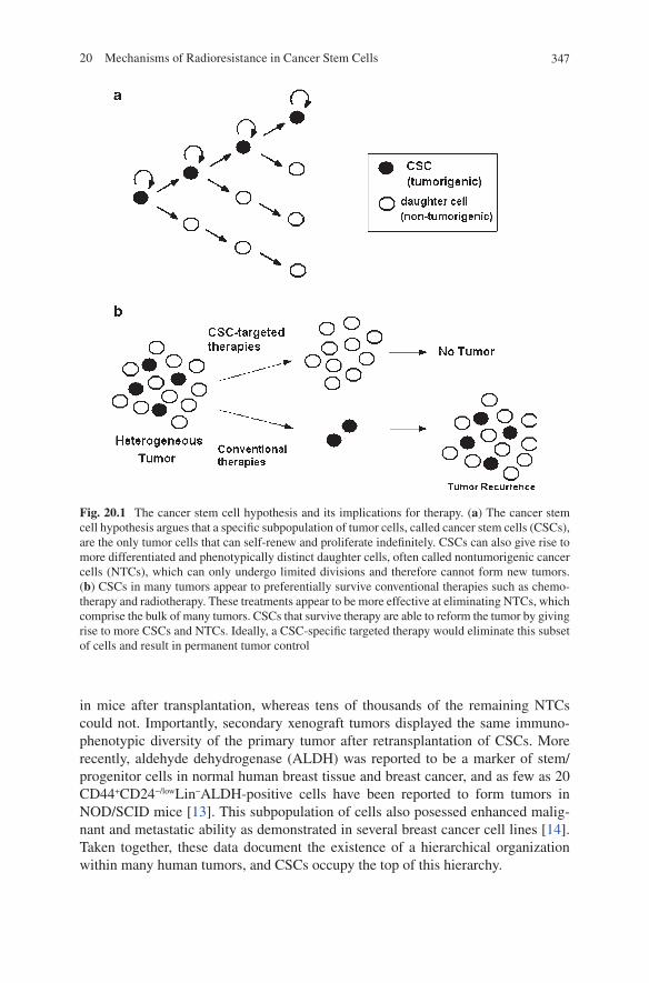
34720 Mechanisms of Radioresistance in Cancer Stem Cells
in mice after transplantation, whereas tens of thousands of the remaining NTCs could not. Importantly, secondary xenograft tumors displayed the same immuno-phenotypic diversity of the primary tumor after retransplantation of CSCs. More recently, aldehyde dehydrogenase (ALDH) was reported to be a marker of stem/progenitor cells in normal human breast tissue and breast cancer, and as few as 20 CD44+CD24−/lowLin−ALDH-positive cells have been reported to form tumors in NOD/SCID mice [13]. This subpopulation of cells also posessed enhanced malig-nant and metastatic ability as demonstrated in several breast cancer cell lines [14]. Taken together, these data document the existence of a hierarchical organization within many human tumors, and CSCs occupy the top of this hierarchy.
Fig. 20.1 The cancer stem cell hypothesis and its implications for therapy. (a) The cancer stem cell hypothesis argues that a specific subpopulation of tumor cells, called cancer stem cells (CSCs), are the only tumor cells that can self-renew and proliferate indefinitely. CSCs can also give rise to more differentiated and phenotypically distinct daughter cells, often called nontumorigenic cancer cells (NTCs), which can only undergo limited divisions and therefore cannot form new tumors. (b) CSCs in many tumors appear to preferentially survive conventional therapies such as chemo-therapy and radiotherapy. These treatments appear to be more effective at eliminating NTCs, which comprise the bulk of many tumors. CSCs that survive therapy are able to reform the tumor by giving rise to more CSCs and NTCs. Ideally, a CSC-specific targeted therapy would eliminate this subset of cells and result in permanent tumor control
348 C.Y-F Lee and M. Diehn
20.2 The 4 R’s of Radiobiology
Radiotherapy is one of the most commonly employed and effective cancer therapies currently used in the clinic. Ionizing radiation causes cell killing when cells fail to repair the damaged components that are critical for the survival of the cell. Of the several potential molecular targets of radiation damage, deoxyribonucleic acid (DNA) has been shown to represent the critical target and DNA damage is closely correlated with cell lethality [15, 16]. Cells that are inherently deficient in DNA repair pathways or inhibited in their ability to repair DNA damage exhibit distinc-tively high radiosensitivity [17, 18]. There are two types of radiation-induced DNA damage: direct and indirect interactions. In the direct interaction, ionizing photons interact directly with the DNA molecule itself to cause damage [19], whereas in the indirect interaction (which predominates), water molecules are ionized to form free radicals that are highly reactive and capable of causing DNA damage [20]. In general, the success of fractionated courses of radiation therapy for cancer depends on four factors (“the 4 R’s”): repair of radiation-induced DNA damage, repopulation of cells, redistribution of cells in the cell cycle, and reoxygenation.
20.2.1 Repair
Both direct and indirect ionizing radiation can cause single-stranded or double-stranded breaks in DNA. On the one hand, single-stranded DNA breaks are relatively easy to repair since the broken strand can be repaired using the other strand as a template. Hence, single-stranded breaks are not primarily related to cell death by radiation. On the other hand, double-stranded DNA breaks are considered the most important con-tributors to radiation cytotoxicity and these are more difficult to repair [21]. There are two main DNA repair pathways for double strand breaks in mammalian cells: homolo-gous recombination (HR), which occurs primarily during S and G2 phases and requires an intact sister chromatid; and nonhomologous end-joining (NHEJ), which is of sig-nificantly lower fidelity since it functions by indiscriminately rejoining broken double-stranded DNA lesions. NHEJ is thought to be the more commonly employed repair pathway. The first event to occur in NHEJ after radiation-induced DNA damage is the recognition of Ku70/80 end binding proteins to the DNA lesions followed by the recruitment and phosphorylation of histone H2AX by ataxia telangiectasia mutated (ATM) and DNA-dependent protein kinase catalytic subunit (DNA-PKcs) repair pro-teins, resulting in the rejoining of the broken ends by X-ray repair complementing defective repair in Chinese hamster cells 4 (XRCC4) and Ligase 4 [22].
20.2.2 Redistribution
The radiosensitivity of cells changes as they progress through the different stages of the cell cycle. Cells in late S-phase are relatively resistant to radiation, whereas cells
34920 Mechanisms of Radioresistance in Cancer Stem Cells
in G2/M are preferentially killed during fractionated radiation therapy [23, 24]. Resistant cells in the S-phase can redistribute to other phases of the cell cycle in between fractions of radiation therapy to become more radiosensitive.
20.2.3 Repopulation
Repopulation of tumor cells is thought to be the most common reason for treatment failure of conventional fractionated radiation therapy [25, 26]. Radiation therapy is currently most frequently administered in daily fractions of 1.8–2.0 Gray for courses extending up to 7 or 8 weeks. The reason for this fractionated scheduling is to allow normal cells to repopulate after surviving radiation therapy, thereby reducing unde-sired toxicities [27]. However, at the same time, fractionated treatment regimens also give the surviving tumor cells a chance to divide, thus increasing the number of tumor cells that need to be eliminated and therefore reducing the effectiveness of a given total dose of radiation therapy.
20.2.4 Reoxygenation
Oxygen is the most potent modifier of radiation sensitivity, and tumor cells in hypoxic niches are 2–3 times more radioresistant than nonhypoxic cells [28]. High levels of hypoxia have been correlated with poor prognosis and accompanying local recurrence or systemic dissemination of disease in human tumors [29–32]. During fractionated radiotherapy, tumors can become reoxygenated through various mech-anisms. As the tumor reduces in size during the course of radiotherapy, tumor cells previously located within hypoxic regions may now be juxtaposed to nearby blood vessels as a result of reorganization of the surrounding vasculature and may thus be exposed to higher oxygen tensions [33]. This reoxygenation process occurs between fractions of radiotherapy and renders the tumor cells more radiosensitive to subse-quent fractions.
20.3 Radioresistance in Cancer Stem Cells
Even before the relatively recent focus on CSCs, radiobiologists studied clonogenic tumor cells and their radiation resistance properties through the use of local control assays. Since by definition, CSCs are the only cells that can lead to significant regrowth of tumors, in vivo local control assays test the ability of CSCs to survive a given therapy. Using such assays, it was shown that CSC content and intrinsic radio-sensitivity measured by in vivo and in vitro clonogenic assays directly correlated with tumor radioresistance [34–37]. These data suggested that CSC radioresistance
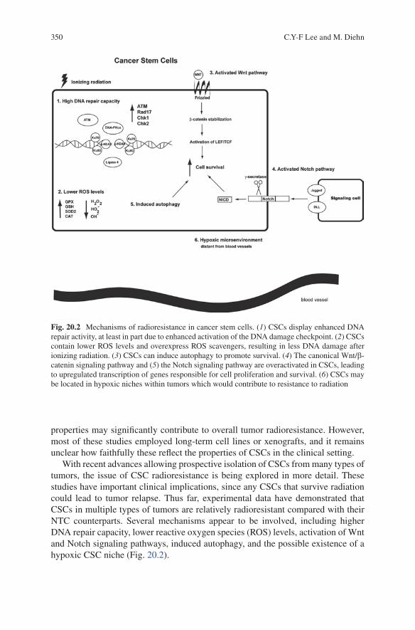
350 C.Y-F Lee and M. Diehn
properties may significantly contribute to overall tumor radioresistance. However, most of these studies employed long-term cell lines or xenografts, and it remains unclear how faithfully these reflect the properties of CSCs in the clinical setting.
With recent advances allowing prospective isolation of CSCs from many types of tumors, the issue of CSC radioresistance is being explored in more detail. These studies have important clinical implications, since any CSCs that survive radiation could lead to tumor relapse. Thus far, experimental data have demonstrated that CSCs in multiple types of tumors are relatively radioresistant compared with their NTC counterparts. Several mechanisms appear to be involved, including higher DNA repair capacity, lower reactive oxygen species (ROS) levels, activation of Wnt and Notch signaling pathways, induced autophagy, and the possible existence of a hypoxic CSC niche (Fig. 20.2).
Fig. 20.2 Mechanisms of radioresistance in cancer stem cells. (1) CSCs display enhanced DNA repair activity, at least in part due to enhanced activation of the DNA damage checkpoint. (2) CSCs contain lower ROS levels and overexpress ROS scavengers, resulting in less DNA damage after ionizing radiation. (3) CSCs can induce autophagy to promote survival. (4) The canonical Wnt/b-catenin signaling pathway and (5) the Notch signaling pathway are overactivated in CSCs, leading to upregulated transcription of genes responsible for cell proliferation and survival. (6) CSCs may be located in hypoxic niches within tumors which would contribute to resistance to radiation
35120 Mechanisms of Radioresistance in Cancer Stem Cells
20.3.1 Enhanced DNA Repair in CSCs
One recurring mechanism of radioresistance found in CSCs is increased DNA repair capacity. Bao et al. demonstrated that glioma stem cells appear to be relatively radioresistance due to preferential activation of the DNA damage checkpoint responses and increased DNA repair ability [38]. The authors found that tumor cells expressing the glioma CSC marker CD133 were enriched after radiation in vitro and in vivo. CD133+ tumor cells activated the DNA damage checkpoint and repaired DNA damage more effectively than CD133− tumor cells from both human glioma xenografts and primary glioblastoma specimens. Activating phosphorylation of the ATM, RAD17, CHK1, and CHK2 proteins was significantly higher in CD133+ tumor cells than in CD133− cells. These observations were further confirmed recently in atypical teratoid/rhabdoid tumor (AT/RT), which is a rare and aggressive pediatric brain tumor [39]. Increased phosphorylation of p-ATM, p-RAD17, and p-CHK2 was observed in CD133+ tumor cells when compared with CD133− cells after ionizing radiation, indicating that CD133+ AT/RT cells exhibited greater checkpoint activation in response to DNA damage. Similarly, CD133+ Daoy medulloblastoma cells from an established cell line showed consistently more effec-tive repair of sublethal DNA damage and were more resistant to radiation than the CD133− cells [40]. In addition, RAD51, a protein involved in strand pairing during HR of DNA DSBs, has recently been reported to be overexpressed in ALDH+ CSCs in breast cancer cell lines, suggesting that Chk1-dependent HR may play an impor-tant role in DNA repair in CSCs [41]. Recently, using the p53 null mammary tumor mode, Zhang et al. demonstrated that Lin−CD29HiCD24Hi CSCs are intrinsically more radioresistant than corresponding NTC subpopulations (Lin−CD29HiCD24Lo, Lin−CD29LoCD24Hi, and Lin−CD29LoCD24Lo) as evidenced by the increased DNA damage repair responses within the CSCs [42]. Taken together, these results suggest that CSCs in at least some tumors are relatively resistant to ionizing radiation through enhanced DNA damage repair compared with NTCs. These data suggest that targeting the DNA damage checkpoint response pathway could sensitize CSCs to radiation therapy.
Although there is evidence for enhanced DNA repair in CSCs from some tumors, CSC or CSC-like cells did not show similar capabilities in all systems. For example, Phillips et al. examined radioresistance of CSC-like cells from a breast cancer cell line and did not find clear evidence of enhanced DNA repair rates [43]. Similarly, CSCs from primary murine breast tumors arising in MMTV-Wnt-1 mice do not appear to have enhanced DNA repair capabilities [44]. Ropolo et al. examined five stem and nonstem glioma cell lines and reported enhanced activation of CHK1 and CHK2 kinases in unirradiated CD133+ cells when compared with CD133− cells [45], suggesting an elevated basal activity of DNA damage responses in CD133+ cells. However, they found no change in DNA base excision, single-strand break repair, or resolution of pH2AX nuclear foci in CD133+ cells in vitro. In addition, McCord et al. showed that CD133+ glioblastoma tumor stem-like cells (TSCs) from recently established neurosphere cultures were not always more radioresistant than
352 C.Y-F Lee and M. Diehn
the CD133− cells from the same cultures, suggesting that the relative radioresistance of CD133+ cells may depend on the tumor from which they are isolated. Furthermore, they found CD133+ cells were more radiosensitive when compared with three estab-lished glioma cell lines as evidenced by the defective DNA damage response using neutral comet assay and the reduced presence of H2AX and RADd51 foci in CD133+ TSCs [46]. These data suggest that enhanced DNA repair is not a universal property of CSCs from all tumors.
20.3.2 Low ROS Levels in CSCs
Another mechanism of radioresistance in solid tumors is the maintenance of lower levels of ROS and enhanced levels of ROS scavengers, since the major mechanism of cell killing by ionizing radiation is through ROS mediators [21]. Our group has recently demonstrated that normal mammary epithelial stem cells contain lower concentrations of ROS than their more mature progeny cells, and that the CSC sub-population in human and murine breast tumors maintains lower ROS levels than NTCs [44]. CSCs from breast tumors arising in MMTV-Wnt-1 mice, characterized by the Thy+CD24+Lin− immunophenotype, developed less DNA damage after radia-tion. Furthermore, we found a twofold enrichment of CSCs when compared with NTCs after in vivo irradiation. Both human and mouse breast CSCs displayed increased expression of genes involved in free radical scavenging and single-cell qRT-PCR analysis showed a significant overexpression of glutathione (GSH) bio-synthesis genes such as Gclm and Gss by CSCs compared with NTCs. GSH is a critical intracellular free radical scavenger. Similar findings were reported in a study by Phillips et al. using the CSC-like cells from the MCF-7 cell line [43]. Specifically, they demonstrated that CD24−/lowCD44+-enriched breast CSC-like cells, propagated as mammosphere cultures, were more resistant to radiation than NTCs, which were grown as monolayer cultures, and that CSC-like cells were enriched during frac-tionated courses of radiation [43]. Lower concentrations of ROS and minimal phos-phorylation of H2AX after irradiation were observed in the CSC-like cells. Similarly, Sca1+ progenitor cells from the COMMA-Db-geo cell line developed less H2AX foci immediately after irradiation, suggesting differences in initial levels of DNA damage [47]. These results indicate that CSCs in some tumors develop lower levels of DNA damage immediately following radiation, which appears to be at least in part due to enhanced expression of ROS scavengers.
20.3.3 Activation of Wnt Signaling Pathway in CSCs
It has been shown that radiotherapy resistance can also be mediated through activation of the Wnt/b-catenin signaling pathway, which has been implicated in stem cell survival [48, 49]. Woodward et al. reported an enrichment of Sca1+ progenitor cells
35320 Mechanisms of Radioresistance in Cancer Stem Cells
in mammary epithelial cells isolated from MMTV-Wnt-1 transgenic mice after in vivo radiation and observed that b-catenin was selectively activated in Sca1+ cells but not in Sca1− cells in response to radiation [50]. Furthermore, real-time PCR analysis revealed elevated messenger ribonucleic acid (mRNA) levels of survivin, a bifunctional member of the inhibitor of apoptosis gene family, in Sca1+ after radiation, suggesting that overexpression of b-catenin may enhance cell survival through the regulation of survivin [51, 52]. These observations indicate that disrupting the Wnt/b-catenin pathway may radiosensitize CSCs.
20.3.4 Hypoxic Microenvironment/Niche
The tumor microenvironment is dynamic and heterogeneous, with ever-changing pH, oxygen concentrations, nutrient supplies, and growth factor levels [53], which may profoundly influence the activity of CSCs. Prolonged or chronic hypoxia is regarded as “diffusion-limited” hypoxia in which tumor cells are located far away from the vascular supply, whereas acute hypoxia in tumors is considered as “perfusion-limited” hypoxia, in which tumor oxygenation fluctuates over time [54–56]. Hypoxia in tumors is a well-established poor prognostic indicator as it promotes aggressive transformation of tumor cells leading to enhanced metastatic potential [57–59], and it is well recognized that hypoxic tumor cells are more radioresistant than nonhy-poxic cells [60]. Hypoxia has been shown to promote an immature stem cell-like phenotype in neuroblastoma cells and a dedifferentiated phenotype in ductal breast carcinoma in situ, thereby reverting tumor cells to become more “stem cell-like” [61]. Another recent study showed that a highly tumorigenic subpopulation of cells from several solid tumor cell lines, including neuroblastoma, rhabdomyosarcoma, and small-cell lung cancer, are preferentially located in hypoxic regions of solid tumors [62]. Furthermore, CSCs in medulloblastoma cells cultured under hypoxic conditions have been reported to upregulate the expression of the CSC marker CD133 [63]. However, CSCs may not reside in hypoxic niches in all tumors. Calabrese et al. showed that Nestin+CD133+ brain CSCs are located in a perivascular niche where they directly interact with endothelial cells [64]. Thus, in some (but not all) tumors, CSCs may be enriched in hypoxic regions, which could contribute to their in vivo radioresistance.
Besides the direct protection of CSCs residing in hypoxic niches by the lack of oxygen, hypoxia could also influence CSC maintenance through the regulation of hypoxia-inducible factors (HIFs) [65]. Li et al. recently demonstrated that HIF2a and multiple HIF-regulated genes are preferentially expressed in glioma CSCs when compared with NTCs and normal neural progenitors [66], and that targeting HIFs in glioma CSCs using lentivirus-mediated shRNAs inhibited self-renewal, prolifera-tion, and survival of CSCs in vitro and reduced the tumorigenic potential of CSCs in vivo. These results suggest that HIFs play an important role in CSCs and may contribute to tumor resistance to radiation.
354 C.Y-F Lee and M. Diehn
20.3.5 Notch Pathway Activation in CSCs
The Notch signaling pathway plays an important role in cell fate determination in neural, hematopoietic, and embryonic stem cells as well as in CSCs [67]. In mam-mals, there are four Notch receptors (Notch 1–4) and five ligands (Jagged-1 and -2, and Delta-like-1, -3, and -4) [68]. Upon activation through ligand binding, Notch receptors undergo sequential proteolytic cleavage resulting in the release and nuclear translocation of the intracellular domains of Notch receptors (NICDs), which subse-quently leads to activation of Notch-regulated transcription [69]. The g-secretase com-plex is required for Notch activation [70, 71], and inhibitors of g-secretase (GSIs) have been used to inhibit Notch signaling in vitro and in vivo. A recent study by Wang et al. demonstrated evidence for Notch-mediated radioresistance in glioma CSCs [72]. It was shown that inhibition of the Notch signaling pathway by GSIs rendered glioma CSCs more sensitive to radiation, and that GSIs impaired clonogenic survival of glioma CSCs but not NTCs through the downregulation of Akt activity.
20.3.6 Induced Autophagy
Autophagy is a “self-cannibalizing” process involving the degradation of long-lived proteins and cytoplasmic organelles, which are then recycled to macromolecules to maintain cellular homeostasis [73]. It has been shown to be a method of cell death distinct from apoptosis that can contribute to the killing of cancer cells by cytotoxic therapies. Somewhat counter-intuitively, autophagy also appears to lead to radiore-sistance in some systems, potentially by acting as a stress response system. Induced autophagy may be a mechanism that contributes to radioresistance in CSCs as dem-onstrated by Lomonaco et al. in CD133+ glioma stem cells [74]. In this study, a greater degree of autophagy was induced in CD133+ glioma stem cells when com-pared with CD133− cells, and higher levels of autophagy-related proteins such as LC3, ATG5 and ATG12 were expressed in CD133+ cells. Inhibition of autophagy by bafilomycin A1, which inhibits autophagy by disrupting fusion between the autopha-gosome and lyosome, and targeting beclin1 and ATG5 genes by shRNAs enhanced radiosensitivity of CD133+ cells to radiation. Although these data will need to be confirmed in other studies, they suggest that induced autophagy may contribute to radioresistance of CSCs.
20.4 Overcoming Radioresistance in CSCs
Recently gained insights into CSC radioresistance mechanisms provide novel opportunities for the development of CSC-specific radiation sensitizers. A number of such strategies have shown promising results in preclinical models. For instance, CSCs can be radiosensitized by targeting DNA damage repair pathways as demonstrated
35520 Mechanisms of Radioresistance in Cancer Stem Cells
by Bao et al. in gliomas [38]. By pretreating tumor cells with debromohymenialdisine (DBH), an inhibitor of checkpoint kinases CHK1 and CHK2 [75], resistance of CD133+ tumor cells to radiation was disrupted. Our group recently demonstrated that pharmacological inhibition of free radical scavengers by pretreating mouse mammary CSCs with the GSH synthesis inhibitor L-S,R-buthionine sulphoximine (BSO) radiosensitized these cells, suggesting that targeting of ROS defenses in CSCs could be a useful therapeutic strategy.
Another potential approach to overcoming radioresistance in CSCs is through modulation of the Akt signaling pathway. In p53 null mammary tumors, it has recently been shown that inhibiting the Akt pathway with perifosine significantly reduces the self-renewal ability of Lin−CD29HCD24H CSCs and sensitizes them to radiation treatment in vitro [42]. Perifosine appears to block DNA damage responses in CSCs, indicating that inhibitors of Akt pathway could be utilized to selectively radiosensitize CSCs.
On the one hand, inhibition of the Notch signaling pathway is also a promising approach to sensitize CSCs to radiation therapy. Mammosphere-forming cells derived from ductal carcinoma in situ have been shown to exhibit diminished self-renewal capacity when the Notch signaling pathway is inhibited [76]. On the other hand, activation of Notch pathway through overexpression of NICD1 promoted the formation of neurospheres in a glioma cell line [77]. Blockade of the Notch pathway using pharmacologic GSIs has also been reported to preferentially reduce the CD133+ stem-like cell subpopulation in medulloblastoma cell lines by decreasing their tumorigenic capacities [78]. More recently, Wang et al. showed that GSIs significantly reduced the clonogenic ability and increased cell death of CD133+ glioma CSCs after radiation treatment [72]. Additionally, overexpression of the constitutively active intracellular domains of Notch1 or Notch2 (NICD1 or NICD2) abolished the radiosensitizing effect of GSIs. In a xenograft mouse model, knockdown of Notch1 or Notch2 using shRNAs dramatically decreased tumor incidence, indicating that glioma CSCs were dependent on Notch signaling and could be radiosensitized by inhibition of the Notch pathway.
Oncolytic virotherapy is a newly emerging approach to specifically target and kill tumor cells, and similar targeting strategies could also be applied to targeting CSCs. It has recently been shown that radioresistant esophageal CSC-like cells exhibited high telomerase activity and that a tumor-specific replicating adenoviral vector Ad/TRAIL-E1, which carries the TRAIL and E1A genes under the control of the human telomerase reverse transcriptase (hTERT) promoter, preferentially targeted and eliminated these esophageal CSCs without causing collateral damage to normal tissues in a xenograft mouse model [79].
Development of anti-CSC therapies should take into consideration the mainte-nance and preservation of normal stem cell functions. It will be important to identify molecular pathways that are distinctively different between CSC and normal stem cells so that anticancer drugs or radiosensitizers could be utilized to target CSCs specifically without causing a significant adverse effect on normal stem cells. Along these lines, it has been shown that after phosphatase and tensin homolog (PTEN) deletion, LSCs were able to reconstitute irradiated mice while normal hematopoietic
356 C.Y-F Lee and M. Diehn
stem cells could not, and that rapamycin treatment not only eliminated the LSC population but also restored normal hematopoietic stem cell function [80]. These data demonstrated that mechanistic differences between CSCs and normal stem cells can be utilized to target CSCs specifically while leaving normal stem cells unharmed.
20.5 Conclusions
A number of mechanisms contributing to radioresistance in CSCs have recently been uncovered, including high DNA repair capacity, overexpression of free radical scavengers, activation of WNT and Notch signaling pathways, induced autophagy, and preferential presence in hypoxic regions of tumors. Taken together, these studies suggest that the particular resistance mechanism(s) present in CSCs of a given tumor may vary. Thus, it remains to be seen which mechanisms are the most common and if there are possible interactions between some of them.
Further investigation of the mechanisms and signaling pathways involved in CSC resistance to radiation therapy will allow the development of novel, CSC-targeted radiosensitizers. As such, future clinical protocols should incorporate char-acterization of CSCs and design patient-specific treatment regimens to improve the effectiveness of anticancer therapies. In addition, CSC-specific endpoints for treat-ment response need to be established, since currently tumor response is most often determined by a reduction in the size of a tumor. Since CSCs appear to be a minority population in many tumors, gross shrinkage will mostly reflect killing of NTCs rather than CSCs. Assessing the response of CSCs to anticancer therapies would be greatly aided by the development of molecular imaging tools that can locate and track CSCs in vivo [81]. Such technology would also be particularly helpful in the optimization of radiotherapy planning, as it might allow a “dose painting” approach to be utilized that could deliver higher doses to CSC-rich regions of tumors [82, 83]. In summary, developing a deeper understanding of CSC biology and the mechanisms governing radioresistance in these cell subpopulations will allow the development of CSC-specific radiosensitizers and will hopefully lead to improved treatment outcomes for cancer patients.
References
1. Clarke MF, Dick JE, Dirks PB et al (2006) Cancer stem cells – perspectives on current status and future directions: AACR Workshop on cancer stem cells. Cancer Res 66:9339–9344.
2. Baum CM, Weissman IL, Tsukamoto AS et al (1992) Isolation of a candidate human hematopoietic stem-cell population. Proc Natl Acad Sci USA 89:2804–2808.
3. Bonnet D, Dick JE (1997) Human acute myeloid leukemia is organized as a hierarchy that originates from a primitive hematopoietic cell. Nat Med 3:730–737.
4. Lapidot T, Sirard C, Vormoor J et al (1994) A cell initiating human acute myeloid leukaemia after transplantation into SCID mice. Nature 367:645–648.
35720 Mechanisms of Radioresistance in Cancer Stem Cells
5. Singh SK, Hawkins C, Clarke ID et al (2004) Identification of human brain tumour initiating cells. Nature 432:396–401.
6. Al-Hajj M, Wicha MS, Benito-Hernandez A et al (2003) Prospective identification of tumori-genic breast cancer cells. Proc Natl Acad Sci USA 100:3983–3988.
7. Prince ME, Sivanandan R, Kaczorowski A et al (2007) Identification of a subpopulation of cells with cancer stem cell properties in head and neck squamous cell carcinoma. Proc Natl Acad Sci USA 104:973–978.
8. Yang ZF, Ho DW, Ng MN et al (2008) Significance of CD90+ cancer stem cells in human liver cancer. Cancer Cell 13:153–166.
9. Hermann PC, Huber SL, Herrler T et al (2007) Distinct populations of cancer stem cells deter-mine tumor growth and metastatic activity in human pancreatic cancer. Cell Stem Cell 1:313–323.
10. Dalerba P, Dylla SJ, Park IK et al (2007) Phenotypic characterization of human colorectal cancer stem cells. Proc Natl Acad Sci USA 104:10158–10163.
11. Chan KS, Espinosa I, Chao M et al (2009) Identification, molecular characterization, clinical prognosis, and therapeutic targeting of human bladder tumor-initiating cells. Proc Natl Acad Sci USA 106:14016–14021.
12. Collins AT, Berry PA, Hyde C et al (2005) Prospective identification of tumorigenic prostate cancer stem cells. Cancer Res 65:10946–10951.
13. Ginestier C, Hur MH, Charafe-Jauffret E et al (2007) ALDH1 is a marker of normal and malig-nant human mammary stem cells and a predictor of poor clinical outcome. Cell Stem Cell 1:555–567.
14. Croker AK, Goodale D, Chu J et al (2009) High aldehyde dehydrogenase and expression of cancer stem cell markers selects for breast cancer cells with enhanced malignant and meta-static ability. J Cell Mol Med 13:2236–2252.
15. Nunez MI, McMillan TJ, Valenzuela MT et al (1996) Relationship between DNA damage, rejoining and cell killing by radiation in mammalian cells. Radiother Oncol 39:155–165.
16. Radford IR (1986) Evidence for a general relationship between the induced level of DNA double-strand breakage and cell-killing after X-irradiation of mammalian cells. Int J Radiat Biol Relat Stud Phys Chem Med 49:611–620.
17. Dikomey E, Dahm-Daphi J, Brammer I et al (1998) Correlation between cellular radiosensitivity and non-repaired double-strand breaks studied in nine mammalian cell lines. Int J Radiat Biol 73:269–278.
18. Kemp LM, Sedgwick SG, Jeggo PA (1984) X-ray sensitive mutants of Chinese hamster ovary cells defective in double-strand break rejoining. Mutat Res 132:189–196.
19. Brenner DJ, Ward JF (1992) Constraints on energy deposition and target size of multiply dam-aged sites associated with DNA double-strand breaks. Int J Radiat Biol 61:737–748.
20. Jonah CD (1995) A short history of the radiation chemistry of water. Radiat Res 144: 141–147.
21. Powell S, McMillan TJ (1990) DNA damage and repair following treatment with ionizing radiation. Radiother Oncol 19:95–108.
22. Yano K, Morotomi-Yano K, Adachi N et al (2009) Molecular mechanism of protein assembly on DNA double-strand breaks in the non-homologous end-joining pathway. J Radiat Res (Tokyo) 50:97–108.
23. Pawlik TM, Keyomarsi K (2004) Role of cell cycle in mediating sensitivity to radiotherapy. Int J Radiat Oncol Biol Phys 59:928–942.
24. Withers HR (1975) Cell cycle redistribution as a factor in multifraction irradiation. Radiology 114:199–202.
25. Bese NS, Sut PA, Ober A (2005) The effect of treatment interruptions in the postoperative irradiation of breast cancer. Oncology 69:214–223.
26. Withers HR, Maciejewski B, Taylor JM et al (1988) Accelerated repopulation in head and neck cancer. Front Radiat Ther Oncol 22:105–110.
27. Kim JJ, Tannock IF (2005) Repopulation of cancer cells during therapy: an important cause of treatment failure. Nat Rev Cancer 5:516–525.
358 C.Y-F Lee and M. Diehn
28. Thomlinson RH, Gray LH (1955) The histological structure of some human lung cancers and the possible implications for radiotherapy. Br J Cancer 9:539–549.
29. Brizel DM, Scully SP, Harrelson JM et al (1996) Tumor oxygenation predicts for the likelihood of distant metastases in human soft tissue sarcoma. Cancer Res 56:941–943.
30. Brizel DM, Sibley GS, Prosnitz LR et al (1997) Tumor hypoxia adversely affects the prognosis of carcinoma of the head and neck. Int J Radiat Oncol Biol Phys 38:285–289.
31. Fyles A, Milosevic M, Hedley D et al (2002) Tumor hypoxia has independent predictor impact only in patients with node-negative cervix cancer. J Clin Oncol 20:680–687.
32. Nordsmark M, Bentzen SM, Rudat V et al (2005) Prognostic value of tumor oxygenation in 397 head and neck tumors after primary radiation therapy. An international multi-center study. Radiother Oncol 77:18–24.
33. Fowler JF (1991) Rapid repopulation in radiotherapy: a debate on mechanism. The phantom of tumor treatment--continually rapid proliferation unmasked. Radiother Oncol 22:156–158.
34. Baumann M, Dubois W, Suit HD (1990) Response of human squamous cell carcinoma xeno-grafts of different sizes to irradiation: relationship of clonogenic cells, cellular radiation sensi-tivity in vivo, and tumor rescuing units. Radiat Res 123:325–330.
35. Baumann M, Krause M, Hill R (2008) Exploring the role of cancer stem cells in radioresis-tance. Nat Rev Cancer 8:545–554.
36. Hill RP, Milas L (1989) The proportion of stem cells in murine tumors. Int J Radiat Oncol Biol Phys 16:513–518.
37. Yaromina A, Krause M, Thames H et al (2007) Pre-treatment number of clonogenic cells and their radiosensitivity are major determinants of local tumour control after fractionated irradia-tion. Radiother Oncol 83:304–310.
38. Bao S, Wu Q, McLendon RE et al (2006) Glioma stem cells promote radioresistance by pref-erential activation of the DNA damage response. Nature 444:756–760.
39. Chiou SH, Kao CL, Chen YW et al (2008) Identification of CD133-positive radioresistant cells in atypical teratoid/rhabdoid tumor. PLoS One 3:e2090.
40. Blazek ER, Foutch JL, Maki G (2007) Daoy medulloblastoma cells that express CD133 are radioresistant relative to CD133- cells, and the CD133+ sector is enlarged by hypoxia. Int J Radiat Oncol Biol Phys 67:1–5.
41. Charafe-Jauffret E, Ginestier C, Iovino F et al (2009) Breast cancer cell lines contain func-tional cancer stem cells with metastatic capacity and a distinct molecular signature. Cancer Res 69:1302–1313.
42. Zhang M, Atkinson RL, Rosen JM (2010) Selective targeting of radiation-resistant tumor-initiating cells. Proc Natl Acad Sci USA 107:3522–3527.
43. Phillips TM, McBride WH, Pajonk F (2006) The response of CD24(−/low)/CD44+ breast cancer-initiating cells to radiation. J Natl Cancer Inst 98:1777–1785.
44. Diehn M, Cho RW, Lobo NA et al (2009) Association of reactive oxygen species levels and radioresistance in cancer stem cells. Nature 458:780–783.
45. Ropolo M, Daga A, Griffero F et al (2009) Comparative analysis of DNA repair in stem and nonstem glioma cell cultures. Mol Cancer Res 7:383–392.
46. McCord AM, Jamal M, Williams ES et al (2009) CD133+ glioblastoma stem-like cells are radiosensitive with a defective DNA damage response compared with established cell lines. Clin Cancer Res 15:5145–5153.
47. Chen MS, Woodward WA, Behbod F et al (2007) Wnt/beta-catenin mediates radiation resis-tance of Sca1+ progenitors in an immortalized mammary gland cell line. J Cell Sci 120: 468–477.
48. Li Y, Welm B, Podsypanina K et al (2003) Evidence that transgenes encoding components of the Wnt signaling pathway preferentially induce mammary cancers from progenitor cells. Proc Natl Acad Sci USA 100:15853–15858.
49. Theodosiou NA, Tabin CJ (2003) Wnt signaling during development of the gastrointestinal tract. Dev Biol 259:258–271.
50. Woodward WA, Chen MS, Behbod F et al (2007) WNT/beta-catenin mediates radiation resis-tance of mouse mammary progenitor cells. Proc Natl Acad Sci USA 104:618–623.
35920 Mechanisms of Radioresistance in Cancer Stem Cells
51. Kim PJ, Plescia J, Clevers H et al (2003) Survivin and molecular pathogenesis of colorectal cancer. Lancet 362:205–209.
52. Yang D, Welm A, Bishop JM (2004) Cell division and cell survival in the absence of survivin. Proc Natl Acad Sci USA 101:15100–15105.
53. Vaupel P, Mayer A (2007) Hypoxia in cancer: significance and impact on clinical outcome. Cancer Metastasis Rev 26:225–239.
54. Brurberg KG, Gaustad JV, Mollatt CS et al (2008) Temporal heterogeneity in blood supply in human tumor xenografts. Neoplasia 10:727–735.
55. Brurberg KG, Thuen M, Ruud EB et al (2006) Fluctuations in pO2 in irradiated human mela-noma xenografts. Radiat Res 165:16–25.
56. Cardenas-Navia LI, Mace D, Richardson RA et al (2008) The pervasive presence of fluctuating oxygenation in tumors. Cancer Res 68:5812–5819.
57. Chan DA, Giaccia AJ (2007) Hypoxia, gene expression, and metastasis. Cancer Metastasis Rev 26:333–339.
58. Gort EH, Groot AJ, van der Wall E et al (2008) Hypoxic regulation of metastasis via hypoxia-inducible factors. Curr Mol Med 8:60–67.
59. Lunt SJ, Chaudary N, Hill RP (2009) The tumor microenvironment and metastatic disease. Clin Exp Metastasis 26:19–34.
60. Wright EA, Howard-Flanders P (1957) The influence of oxygen on the radiosensitivity of mammalian tissues. Acta radiol 48:26–32.
61. Axelson H, Fredlund E, Ovenberger M et al (2005) Hypoxia-induced dedifferentiation of tumor cells--a mechanism behind heterogeneity and aggressiveness of solid tumors. Semin Cell Dev Biol 16:554–563.
62. Das B, Tsuchida R, Malkin D et al (2008) Hypoxia enhances tumor stemness by increasing the invasive and tumorigenic side population fraction. Stem Cells 26:1818–1830.
63. Platet N, Liu SY, Atifi ME et al (2007) Influence of oxygen tension on CD133 phenotype in human glioma cell cultures. Cancer Lett 258:286–290.
64. Calabrese C, Poppleton H, Kocak M et al (2007) A perivascular niche for brain tumor stem cells. Cancer Cell 11:69–82.
65. Heddleston JM, Li Z, Lathia JD et al (2010) Hypoxia inducible factors in cancer stem cells. Br J Cancer 102:789–795.
66. Li Z, Bao S, Wu Q et al (2009) Hypoxia-inducible factors regulate tumorigenic capacity of glioma stem cells. Cancer Cell 15:501–513.
67. Bolos V, Blanco M, Medina V et al (2009) Notch signalling in cancer stem cells. Clin Transl Oncol 11:11–19.
68. Wu J, Bresnick EH (2007) Bare rudiments of notch signaling: how receptor levels are regu-lated. Trends Biochem Sci 32:477–485.
69. Miele L, Golde T, Osborne B (2006) Notch signaling in cancer. Curr Mol Med 6:905–918. 70. Armogida M, Petit A, Vincent B et al (2001) Endogenous beta-amyloid production in preseni-
lin-deficient embryonic mouse fibroblasts. Nat Cell Biol 3:1030–1033. 71. Huppert SS, Le A, Schroeter EH et al (2000) Embryonic lethality in mice homozygous for a
processing-deficient allele of Notch1. Nature 405:966–970. 72. Wang J, Wakeman TP, Lathia JD et al (2010) Notch promotes radioresistance of glioma stem
cells. Stem Cells 28:17–28. 73. Levine B, Klionsky DJ (2004) Development by self-digestion: molecular mechanisms and
biological functions of autophagy. Dev Cell 6:463–477. 74. Lomonaco SL, Finniss S, Xiang C et al (2009) The induction of autophagy by gamma-radia-
tion contributes to the radioresistance of glioma stem cells. Int J Cancer 125:717–722. 75. Curman D, Cinel B, Williams DE et al (2001) Inhibition of the G2 DNA damage checkpoint
and of protein kinases Chk1 and Chk2 by the marine sponge alkaloid debromohymenialdisine. J Biol Chem 276:17914–17919.
76. Farnie G, Clarke RB, Spence K et al (2007) Novel cell culture technique for primary ductal carcinoma in situ: role of Notch and epidermal growth factor receptor signaling pathways. J Natl Cancer Inst 99:616–627.

360 C.Y-F Lee and M. Diehn
77. Zhang XP, Zheng G, Zou L et al (2008) Notch activation promotes cell proliferation and the formation of neural stem cell-like colonies in human glioma cells. Mol Cell Biochem 307: 101–108.
78. Fan X, Matsui W, Khaki L et al (2006) Notch pathway inhibition depletes stem-like cells and blocks engraftment in embryonal brain tumors. Cancer Res 66:7445–7452.
79. Zhang X, Komaki R, Wang L et al (2008) Treatment of radioresistant stem-like esophageal cancer cells by an apoptotic gene-armed, telomerase-specific oncolytic adenovirus. Clin Cancer Res 14:2813–2823.
80. Yilmaz OH, Valdez R, Theisen BK et al (2006) Pten dependence distinguishes haematopoietic stem cells from leukaemia-initiating cells. Nature 441:475–482.
81. Vlashi E, Kim K, Lagadec C et al (2009) In vivo imaging, tracking, and targeting of cancer stem cells. J Natl Cancer Inst 101:350–359.
82. Bentzen SM (2005) Theragnostic imaging for radiation oncology: dose-painting by numbers. Lancet Oncol 6:112–117.
83. Ling CC, Humm J, Larson S et al (2000) Towards multidimensional radiotherapy (MD-CRT): biological imaging and biological conformality. Int J Radiat Oncol Biol Phys 47:551–560.
361A.L. Allan (ed.), Cancer Stem Cells in Solid Tumors, Stem Cell Biology and Regenerative Medicine, DOI 10.1007/978-1-61779-246-5_21, © Springer Science+Business Media, LLC 2011
Abstract There can be no cure for epithelial cancer until there is a significant therapeutic index that separates the most therapy-resistant cells within tumors, and the cells that mediate functions essential to life. The cancer stem cell paradigm explains tumor heterogeneity and has led to the hypothesis that therapy resistance originates in the mechanisms by which normal tissue stem cells protect themselves from toxic insults. However, therapeutic index is not merely the ratio of cancer stem cell kill to normal stem cell kill. It is further compounded by the fact that a small number of clonogenic cancer cells need only to survive to perpetuate the neoplasm, whereas vital tissue functions cannot be disrupted for long without lethal conse-quences. In this chapter, we review the concepts of tumor grade and therapeutic index; maximal tolerated dose; innate vs. acquired multiple drug resistance (MDR); tumor heterogeneity; and the role of ABC transporters in multipotent, therapy resistant, clonogenic cancer stem-like cells in the context of therapeutic use of MDR inhibitors to change the maximal tolerated dose of currently used antineoplastic agents.
Abbreviations
ABC ATP binding cassette transportersATP Adenosine triphosphateBCRP Breast cancer resistance proteinCD Cluster of differentiationCFTR Cystic fibrosis transmembrane conductance regulatorCSA Cyclosporin A
V.S. Donnenberg (*) Department of Cardiovascular Surgery, Division of Hematology/Oncology, University of Pittsburgh Cancer Institute and University of Pittsburgh School of Medicine, Pittsburgh, PA, USAe-mail: [email protected]
Chapter 21The Role of ABC Transporters in Cancer Stem Cell Drug Resistance
Vera S. Donnenberg, Ludovic Zimmerlin, and Albert D. Donnenberg
362 V.S. Donnenberg et al.
ED50
Effective Dose50
EpCAM Epithelial cell adhesion moleculeHh HedgehogLD
50 Lethal Dose
50
MDR Multiple drug resistanceMHC Major histocompatibility complexMRD Minimal residual diseaseMRP Multidrug resistance proteinMTD Maximum tolerated doseNCI National Cancer InstitutePI Propidium iodideR123 Rhodamine 123SHh Sonic hedgehogSP Side populationSP1 Sphingosine-1-phosphateTIL Tumor infiltrating lymphocytes
21.1 Introduction
Lack of success in the eradication of epithelial cancers may be attributed to the fail-ure to identify tumor specific targets, together with the tumor’s ability to hijack many normal stem cell-like properties, including dormancy, self-renewal, self-protection, and telomere maintenance. Despite the success of current therapies in eliminating bulky disease and rapidly proliferating cells, the same therapies often spare this self-renewing, self protected stem-like compartment, missing the tumor reservoir respon-sible for recurrent disease and metastasis. Depending on the aggressiveness of the particular neoplasm, the resistant compartment may be dormant or proliferative. In the first case, long-term remission may be obtained, but in the latter, extreme drug resistance is encountered at the time of diagnosis. Recent advances in the under-standing of ABC transporter expression, activity, and regulation in normal tissue stem cells as well as in tumors provide a basis for revisiting the process of oncogen-esis, tumor heterogeneity, and drug resistance in the context of anticancer therapy.
ABC transporters (ATP binding cassette transporters) are highly conserved and represent a major protective mechanism for highly differentiated barrier tissues as well as adult tissue stem cells. Multiple drug resistance (MDR) mediated by their activity [1] was initially discovered in tumors and was recognized as an impediment to cancer therapy [2, 3]. Further, the significant redundancy in substrate specificity among the individual transporters, the significant homology between eukaryotic and mammalian transporters, and the lack of a lethal phenotype with specific ABC knock-outs demonstrates the essential nature and redundancy of these proteins for cellular transport and trafficking [4–6].

36321 The Role of ABC Transporters in Cancer Stem Cell Drug Resistance
More recently, intrinsic MDR transport has been shown to constitute a normal physiologic protective mechanism shared by long-lived stem cells in a variety of tissues [7–12]. Survival of tissue stem cells is essential for tissue maintenance and repair, and constitutive (as opposed to substrate-induced) MDR activity is thought to be one of several protective mechanisms by which normal tissue stem cells guard themselves from toxic insults, including damage caused by antineoplastic therapy [13]. The notion that the cancer-initiating cell may retain or reacquire constitutive drug resistance predicts the persistence of a therapy resistant stem cell-like fraction following apparently successful cytotoxic cancer therapy, even in the face of marked shrinkage or disappearance of measurable tumor.
Understanding the central role played by MDR transporters in the protection and self-renewal of normal and cancer stem cells may allow us to identify differences that can be exploited therapeutically. Recognizing that normal stem cells in indi-vidual tissues differ with respect to damage tolerance and degree of multipotential-ity may translate into differential drug susceptibility and metastatic potentials of cancer stem cells, depending on the tissue of origin. For example, hematopoietic stem cells responsible for bone marrow maintenance and regeneration after toxic injury are far more susceptible to radiotherapy [14, 15] and chemotherapy than their epithelial counterparts, despite constitutive MDR expression among the most primi-tive and multipotent hematopoietic progenitors [7, 8].
In this chapter, we will discuss the role of ABC transporters as a barrier to ther-apy as well as their role in transport of physiological biomolecules involved in sig-naling, differentiation, proliferation, and migration.
21.2 Discovery of MDR Transporters
Resistance to chemotherapy was recognized as an impediment to efficacious cancer treatment in the earliest stages of anticancer drug development [16]. Further, cancer cell lines selected for resistance to specific compounds also demonstrated “cross-resistance” to a broad spectrum of structurally unrelated agents [2, 3]. The advent of molecular approaches enabled the isolation of the genes for MDR. Roninson and colleagues isolated two genes, now recognized as the hamster homologs of the human MDR1 and MDR2 genes [17–19]. In 1986, Gros and colleagues transfected the MDR1 gene (P-glycoprotein, [ABCB1]) into drug sensitive hamster cells and showed that gene duplication or mutations were not required for the acquisition of the multidrug resistant phenotype [20]. ABCB1 did not account for all forms of MDR and additional transporters were identified, among them ABCG2, first described as mitoxantrone resistance [21, 22] breast cancer resistance protein (BCRP). ABCC1, also known as multiple resistance protein 1 [23, 24], was also recognized early as a transporter with activity over a range of substrates. Today, the ABC transporter family is the largest of all transmembrane protein families, utilizing
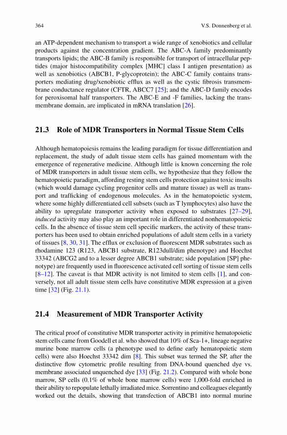
364 V.S. Donnenberg et al.
an ATP-dependent mechanism to transport a wide range of xenobiotics and cellular products against the concentration gradient. The ABC-A family predominantly transports lipids; the ABC-B family is responsible for transport of intracellular pep-tides (major histocompatibility complex [MHC] class I antigen presentation) as well as xenobiotics (ABCB1, P-glycoprotein); the ABC-C family contains trans-porters mediating drug/xenobiotic efflux as well as the cystic fibrosis transmem-brane conductance regulator (CFTR, ABCC7 [25]; and the ABC-D family encodes for peroxisomal half transporters. The ABC-E and -F families, lacking the trans-membrane domain, are implicated in mRNA translation [26].
21.3 Role of MDR Transporters in Normal Tissue Stem Cells
Although hematopoiesis remains the leading paradigm for tissue differentiation and replacement, the study of adult tissue stem cells has gained momentum with the emergence of regenerative medicine. Although little is known concerning the role of MDR transporters in adult tissue stem cells, we hypothesize that they follow the hematopoietic paradigm, affording resting stem cells protection against toxic insults (which would damage cycling progenitor cells and mature tissue) as well as trans-port and trafficking of endogenous molecules. As in the hematopoietic system, where some highly differentiated cell subsets (such as T lymphocytes) also have the ability to upregulate transporter activity when exposed to substrates [27–29], induced activity may also play an important role in differentiated nonhematopoietic cells. In the absence of tissue stem cell specific markers, the activity of these trans-porters has been used to obtain enriched populations of adult stem cells in a variety of tissues [8, 30, 31]. The efflux or exclusion of fluorescent MDR substrates such as rhodamine 123 (R123, ABCB1 substrate, R123dull/dim phenotype) and Hoechst 33342 (ABCG2 and to a lesser degree ABCB1 substrate; side population [SP] phe-notype) are frequently used in fluorescence activated cell sorting of tissue stem cells [8–12]. The caveat is that MDR activity is not limited to stem cells [1], and con-versely, not all adult tissue stem cells have constitutive MDR expression at a given time [32] (Fig. 21.1).
21.4 Measurement of MDR Transporter Activity
The critical proof of constitutive MDR transporter activity in primitive hematopoietic stem cells came from Goodell et al. who showed that 10% of Sca-1+, lineage negative murine bone marrow cells (a phenotype used to define early hematopoietic stem cells) were also Hoechst 33342 dim [8]. This subset was termed the SP, after the distinctive flow cytometric profile resulting from DNA-bound quenched dye vs. membrane associated unquenched dye [33] (Fig. 21.2). Compared with whole bone marrow, SP cells (0.1% of whole bone marrow cells) were 1,000-fold enriched in their ability to repopulate lethally irradiated mice. Sorrentino and colleagues elegantly worked out the details, showing that transfection of ABCB1 into normal murine
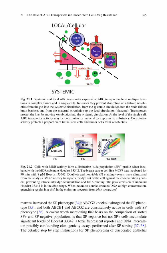
36521 The Role of ABC Transporters in Cancer Stem Cell Drug Resistance
Fig. 21.1 Systemic and local ABC transporter expression. ABC transporters have multiple func-tions in complex tissues and in single cells. In tissues they prevent absorption of substrate xenobi-otics from the gut into the systemic circulation, from the systemic circulation into the brain (blood brain barrier), and from the maternal circulation to the fetal circulation (placenta). Transporters protect the liver by moving xenobiotics into the systemic circulation. At the level of the single cell, ABC transporter activity may be constitutive or induced by exposure to substrates. Constitutive activity protects a proportion of tissue stem cells and tumor cells from xenobiotics
Fig. 21.2 Cells with MDR activity form a distinctive “side population (SP)” profile when incu-bated with the MDR substrate Hoechst 33342. The breast cancer cell line MCF7 was incubated for 90 min with 8 mM Hoechst 33342. Doublets and nonviable (PI staining) events were eliminated from the analysis. MDR activity transports the dye out of the cell against the concentration gradi-ent, preventing intracellular dye accumulation and DNA binding. The peak emission of unbound Hoechst 33342 is in the blue range. When bound to double stranded DNA at high concentration, quenching results in a shift in the emission spectrum from blue toward red
marrow increased the SP phenotype [34]; ABCG2 knockout abrogated the SP pheno-type [35]; and both ABCB1 and ABCG2 are constitutively active in cells with SP phenotype [36]. A caveat worth mentioning that bears on the comparison of sorted SP+ and SP negative populations is that SP negative but not SP+ cells accumulate significant levels of Hoechst 33342, a toxic fluorescent reporter and DNA intercala-tor, possibly confounding clonogenicity assays performed after SP sorting [37, 38]. The detailed step by step instructions for SP phenotyping of dissociated epithelial
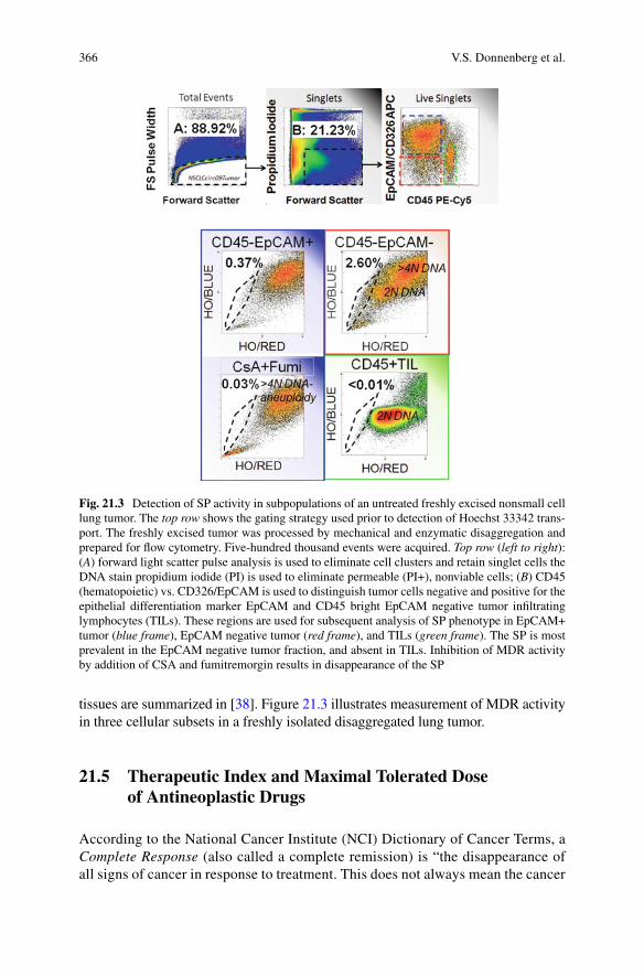
366 V.S. Donnenberg et al.
tissues are summarized in [38]. Figure 21.3 illustrates measurement of MDR activity in three cellular subsets in a freshly isolated disaggregated lung tumor.
21.5 Therapeutic Index and Maximal Tolerated Dose of Antineoplastic Drugs
According to the National Cancer Institute (NCI) Dictionary of Cancer Terms, a Complete Response (also called a complete remission) is “the disappearance of all signs of cancer in response to treatment. This does not always mean the cancer
Fig. 21.3 Detection of SP activity in subpopulations of an untreated freshly excised nonsmall cell lung tumor. The top row shows the gating strategy used prior to detection of Hoechst 33342 trans-port. The freshly excised tumor was processed by mechanical and enzymatic disaggregation and prepared for flow cytometry. Five-hundred thousand events were acquired. Top row (left to right): (A) forward light scatter pulse analysis is used to eliminate cell clusters and retain singlet cells the DNA stain propidium iodide (PI) is used to eliminate permeable (PI+), nonviable cells; (B) CD45 (hematopoietic) vs. CD326/EpCAM is used to distinguish tumor cells negative and positive for the epithelial differentiation marker EpCAM and CD45 bright EpCAM negative tumor infiltrating lymphocytes (TILs). These regions are used for subsequent analysis of SP phenotype in EpCAM+ tumor (blue frame), EpCAM negative tumor (red frame), and TILs (green frame). The SP is most prevalent in the EpCAM negative tumor fraction, and absent in TILs. Inhibition of MDR activity by addition of CSA and fumitremorgin results in disappearance of the SP
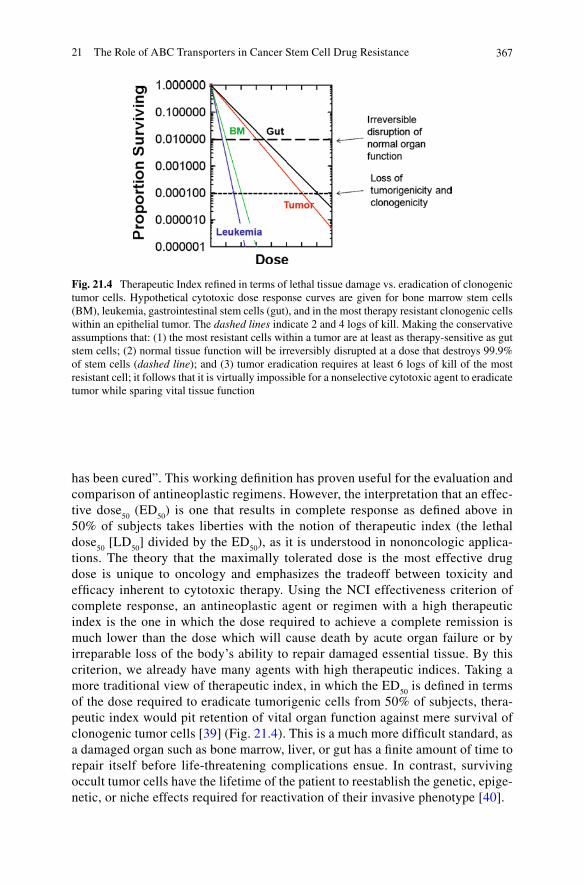
36721 The Role of ABC Transporters in Cancer Stem Cell Drug Resistance
has been cured”. This working definition has proven useful for the evaluation and comparison of antineoplastic regimens. However, the interpretation that an effec-tive dose
50 (ED
50) is one that results in complete response as defined above in
50% of subjects takes liberties with the notion of therapeutic index (the lethal dose
50 [LD
50] divided by the ED
50), as it is understood in nononcologic applica-
tions. The theory that the maximally tolerated dose is the most effective drug dose is unique to oncology and emphasizes the tradeoff between toxicity and efficacy inherent to cytotoxic therapy. Using the NCI effectiveness criterion of complete response, an antineoplastic agent or regimen with a high therapeutic index is the one in which the dose required to achieve a complete remission is much lower than the dose which will cause death by acute organ failure or by irreparable loss of the body’s ability to repair damaged essential tissue. By this criterion, we already have many agents with high therapeutic indices. Taking a more traditional view of therapeutic index, in which the ED
50 is defined in terms
of the dose required to eradicate tumorigenic cells from 50% of subjects, thera-peutic index would pit retention of vital organ function against mere survival of clonogenic tumor cells [39] (Fig. 21.4). This is a much more difficult standard, as a damaged organ such as bone marrow, liver, or gut has a finite amount of time to repair itself before life-threatening complications ensue. In contrast, surviving occult tumor cells have the lifetime of the patient to reestablish the genetic, epige-netic, or niche effects required for reactivation of their invasive phenotype [40].
Fig. 21.4 Therapeutic Index refined in terms of lethal tissue damage vs. eradication of clonogenic tumor cells. Hypothetical cytotoxic dose response curves are given for bone marrow stem cells (BM), leukemia, gastrointestinal stem cells (gut), and in the most therapy resistant clonogenic cells within an epithelial tumor. The dashed lines indicate 2 and 4 logs of kill. Making the conservative assumptions that: (1) the most resistant cells within a tumor are at least as therapy-sensitive as gut stem cells; (2) normal tissue function will be irreversibly disrupted at a dose that destroys 99.9% of stem cells (dashed line); and (3) tumor eradication requires at least 6 logs of kill of the most resistant cell; it follows that it is virtually impossible for a nonselective cytotoxic agent to eradicate tumor while sparing vital tissue function
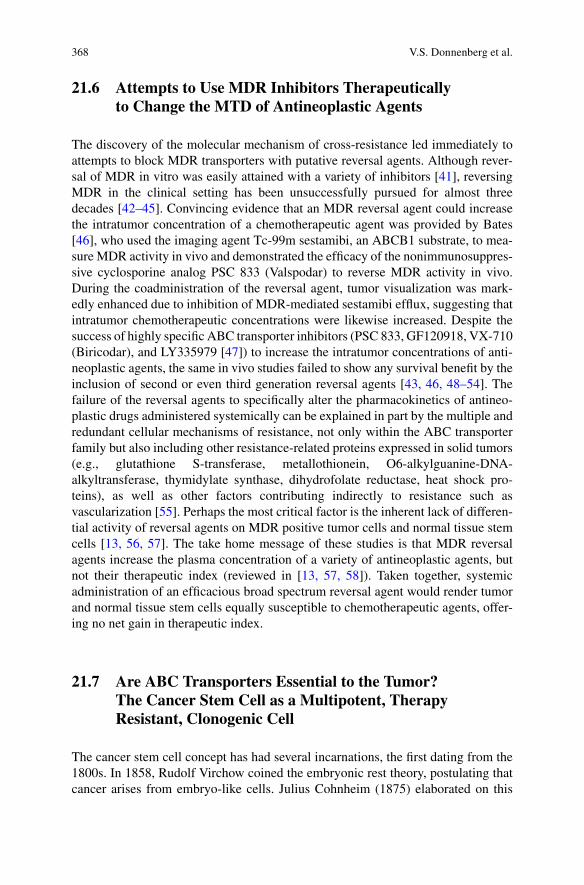
368 V.S. Donnenberg et al.
21.6 Attempts to Use MDR Inhibitors Therapeutically to Change the MTD of Antineoplastic Agents
The discovery of the molecular mechanism of cross-resistance led immediately to attempts to block MDR transporters with putative reversal agents. Although rever-sal of MDR in vitro was easily attained with a variety of inhibitors [41], reversing MDR in the clinical setting has been unsuccessfully pursued for almost three decades [42–45]. Convincing evidence that an MDR reversal agent could increase the intratumor concentration of a chemotherapeutic agent was provided by Bates [46], who used the imaging agent Tc-99m sestamibi, an ABCB1 substrate, to mea-sure MDR activity in vivo and demonstrated the efficacy of the nonimmunosuppres-sive cyclosporine analog PSC 833 (Valspodar) to reverse MDR activity in vivo. During the coadministration of the reversal agent, tumor visualization was mark-edly enhanced due to inhibition of MDR-mediated sestamibi efflux, suggesting that intratumor chemotherapeutic concentrations were likewise increased. Despite the success of highly specific ABC transporter inhibitors (PSC 833, GF120918, VX-710 (Biricodar), and LY335979 [47]) to increase the intratumor concentrations of anti-neoplastic agents, the same in vivo studies failed to show any survival benefit by the inclusion of second or even third generation reversal agents [43, 46, 48–54]. The failure of the reversal agents to specifically alter the pharmacokinetics of antineo-plastic drugs administered systemically can be explained in part by the multiple and redundant cellular mechanisms of resistance, not only within the ABC transporter family but also including other resistance-related proteins expressed in solid tumors (e.g., glutathione S-transferase, metallothionein, O6-alkylguanine-DNA-alkyltransferase, thymidylate synthase, dihydrofolate reductase, heat shock pro-teins), as well as other factors contributing indirectly to resistance such as vascularization [55]. Perhaps the most critical factor is the inherent lack of differen-tial activity of reversal agents on MDR positive tumor cells and normal tissue stem cells [13, 56, 57]. The take home message of these studies is that MDR reversal agents increase the plasma concentration of a variety of antineoplastic agents, but not their therapeutic index (reviewed in [13, 57, 58]). Taken together, systemic administration of an efficacious broad spectrum reversal agent would render tumor and normal tissue stem cells equally susceptible to chemotherapeutic agents, offer-ing no net gain in therapeutic index.
21.7 Are ABC Transporters Essential to the Tumor? The Cancer Stem Cell as a Multipotent, Therapy Resistant, Clonogenic Cell
The cancer stem cell concept has had several incarnations, the first dating from the 1800s. In 1858, Rudolf Virchow coined the embryonic rest theory, postulating that cancer arises from embryo-like cells. Julius Cohnheim (1875) elaborated on this
36921 The Role of ABC Transporters in Cancer Stem Cell Drug Resistance
idea, proposing that tumors derive from residual embryonic cells, which lie dormant through development and become cancerous upon activation (reviewed by Sell [59]). Fiala revived this notion in 1968 [60], hypothesizing the cancer-initiating cell to be a “stem cell unable to differentiate.” Interest in the relationships between can-cer and our modern notion of tissue stem cells was rekindled by Weissman [61], who first isolated the multipotential hematopoietic progenitor cell [62]. One of the defining characteristics of adult tissue stem cells is the fact that, at any given time, a proportion of them are constitutively resistant to xenobiotics, including most che-motherapeutic agents. Understanding the central role that MDR transporters play in protecting normal adult tissue stem cells allows us to extrapolate to the clonogenic, drug resistant cell subpopulations within a tumor and add new insights that may prove relevant to explaining treatment failure, late recurrence, metastasis, and tis-sue-specific differences in cancer incidence.
Our own experience leads us to believe that, as in normal tissues, clonogenicity (self-renewal or tumorigenicity), and tumor resistance to therapy are correlated but physiologically uncoupled (Fig. 21.5). Therapies that target proliferating cells, enzymes, growth receptors, adherence molecules, or signaling molecules in meta-bolically active cancer cells may be highly effective at debulking tumors and dis-patching tumorigenic cells without the stem cell-like property of self-protection, but
Fig. 21.5 MDR activity (SP phenotype) and tumorigenicity in two untreated nonsmall cell lung cancers. In the first case, MDR activity is rare and is correlated with clonogenicity with the MDR+ fraction comprising a small subset of tumorigenic cells. In the second case, the MDR+ fraction is prevalent and is uncorrelated with tumorigenicity. In both cases, an MDR+ clonogenic population is present, which would predictably be therapy resistant
370 V.S. Donnenberg et al.
they will consistently fail to eradicate the tumor cell fraction that constitutively shares protective mechanisms with normal stem cell counterparts. Working in a human breast cancer xenograft model, we demonstrated that MDR activity is not an intrinsic characteristic of a unique tumor stem cell compartment. Both CD45−CD44+CD90+ABCG2+ cells and CD45−CD44+CD90+ABCG2− cells were tumorigenic and serially transplantable. Further, the ABCG2+ phenotype appears to be inducible in ABCG2− clonogenic tumor cells, since tumors derived from the ABCG2− fraction gave rise to a substantial proportion of CD90+ ABCG2+ cells. We interpret this data to indicate that, in the absence of drug-induced selective pressure, there is a bidirectional or stochastic relationship between drug resistance and drug sensitivity among clonogenic tumor cells. This relationship is contrary to a rigid hierarchical model in which protected stem cells give rise to sensitive progenitor cells, but is consistent with observations that conditionally differentiated cells, such as those found in the normal airway [9], liver [63], and pancreas [64] can assume a self-renewing and self-protected phenotype after injury. Cancers that respond to therapy initially may appear to acquire drug resistance during the course of treat-ment, while other cancers may appear to be intrinsically resistant. Our interpretation of the cancer stem cell paradigm stipulates that at any given time a proportion of self-renewing stem-like tumor cells has innate drug resistance by virtue of its stem cell-like phenotype [13, 65, 66]. Acquired drug resistance in more differentiated cancer cells, through gene amplification or rearrangement, may contribute to an aggressive phenotype, but is not the primary reason for cancer recurrence or spread after therapy. Taken together, it seems that the multiple drug-resistant clonogenic tumor cell is a moving target, since resistance and tumorigenicity are uncoupled and resistance can be modulated in the absence of therapy mediated selective pressure. Thus, it is unlikely that any one phenotypically defined subset will encompass all tumorigenic, therapy resistant SP-like cells, which have the potential to metastasize and reactivate [67].
21.8 Other Physiologic Roles of ABC Transporters
For decades we have focused on ABC transporter drug efflux activities to determine the cause of treatment failure. However, significant correlative evidence suggests that in addition to their roles in drug transport, the same transporters are involved in intracellular and extracellular molecule trafficking (reviewed in [58]). Although confounded by the fact that ABC transporters confer resistance of cancer cells to apoptosis in the context of antineoplastic therapy, several studies provide evidence that ABC transporters promote cell survival independent of their ability to limit cytotoxic drug exposure. The earliest report from Johnstone et al. demonstrated that ABC transporter inhibition sensitized cells to FAS-induced cell death via FAS–FAS ligand interaction [68, 69]. Further, increased expression of ABCB1 (P-glycoprotein) delayed apoptosis in normal as well as malignant cells [70, 71] through the sphingomyelin-ceramide pathway. In neuroblastoma, down-regulation
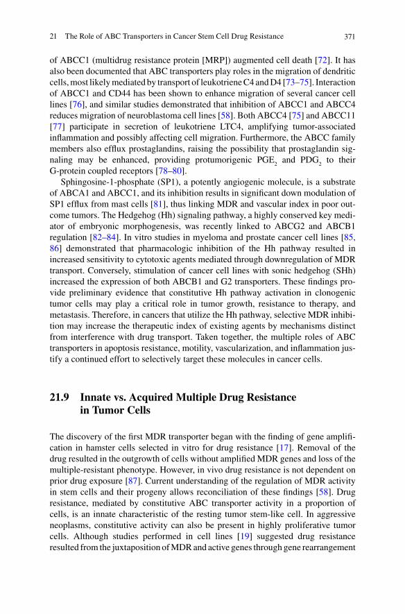
37121 The Role of ABC Transporters in Cancer Stem Cell Drug Resistance
of ABCC1 (multidrug resistance protein [MRP]) augmented cell death [72]. It has also been documented that ABC transporters play roles in the migration of dendritic cells, most likely mediated by transport of leukotriene C4 and D4 [73–75]. Interaction of ABCC1 and CD44 has been shown to enhance migration of several cancer cell lines [76], and similar studies demonstrated that inhibition of ABCC1 and ABCC4 reduces migration of neuroblastoma cell lines [58]. Both ABCC4 [75] and ABCC11 [77] participate in secretion of leukotriene LTC4, amplifying tumor-associated inflammation and possibly affecting cell migration. Furthermore, the ABCC family members also efflux prostaglandins, raising the possibility that prostaglandin sig-naling may be enhanced, providing protumorigenic PGE
2 and PDG
2 to their
G-protein coupled receptors [78–80].Sphingosine-1-phosphate (SP1), a potently angiogenic molecule, is a substrate
of ABCA1 and ABCC1, and its inhibition results in significant down modulation of SP1 efflux from mast cells [81], thus linking MDR and vascular index in poor out-come tumors. The Hedgehog (Hh) signaling pathway, a highly conserved key medi-ator of embryonic morphogenesis, was recently linked to ABCG2 and ABCB1 regulation [82–84]. In vitro studies in myeloma and prostate cancer cell lines [85, 86] demonstrated that pharmacologic inhibition of the Hh pathway resulted in increased sensitivity to cytotoxic agents mediated through downregulation of MDR transport. Conversely, stimulation of cancer cell lines with sonic hedgehog (SHh) increased the expression of both ABCB1 and G2 transporters. These findings pro-vide preliminary evidence that constitutive Hh pathway activation in clonogenic tumor cells may play a critical role in tumor growth, resistance to therapy, and metastasis. Therefore, in cancers that utilize the Hh pathway, selective MDR inhibi-tion may increase the therapeutic index of existing agents by mechanisms distinct from interference with drug transport. Taken together, the multiple roles of ABC transporters in apoptosis resistance, motility, vascularization, and inflammation jus-tify a continued effort to selectively target these molecules in cancer cells.
21.9 Innate vs. Acquired Multiple Drug Resistance in Tumor Cells
The discovery of the first MDR transporter began with the finding of gene amplifi-cation in hamster cells selected in vitro for drug resistance [17]. Removal of the drug resulted in the outgrowth of cells without amplified MDR genes and loss of the multiple-resistant phenotype. However, in vivo drug resistance is not dependent on prior drug exposure [87]. Current understanding of the regulation of MDR activity in stem cells and their progeny allows reconciliation of these findings [58]. Drug resistance, mediated by constitutive ABC transporter activity in a proportion of cells, is an innate characteristic of the resting tumor stem-like cell. In aggressive neoplasms, constitutive activity can also be present in highly proliferative tumor cells. Although studies performed in cell lines [19] suggested drug resistance resulted from the juxtaposition of MDR and active genes through gene rearrangement
372 V.S. Donnenberg et al.
[88, 89] or chromosomal activation [90, 91], such transforming events are not essential for acquisition of the MDR phenotype in vivo. Like its normal counterpart, a proportion of stem-like cancer cells express constitutive MDR activity, which is evident prior to drug-imposed selective pressure. Even when therapy succeeds in eliminating the bulk of proliferating cells, this population may survive as occult disease and reactivate months or years later. Additionally, selective pressure imposed by chemotherapy leads both to mutation and secondary genetic changes, including MDR upregulation in the bulky tumor [13, 92]. If these changes occur in self-renewing proliferating cells, the result is an aggressive therapy-resistant tumor. Therefore, MDR activity, whether constitutive in resting tumor cells, induced in active tumor cells, or amplified by genetic alterations, remains a major barrier to therapy.
21.10 Tumor Grade and Therapeutic Index
According to the NCI, “Tumor grade is a system used to classify cancer cells in terms of how abnormal they look under a microscope and how quickly the tumor is likely to grow and spread. Many factors are considered when determining tumor grade, including the structure and growth pattern of the cells. The specific factors used to determine tumor grade vary with each type of cancer.” Like normal tissues, tumors are heterogeneous with respect to stromal cells, vascular cells, infiltrating immune cells, and the differentiation state of the epithelial cells themselves. In low-grade cancers, many of the tumor cells may be nonclonogenic progeny of clono-genic cells. A tumor cell does not have to derive from a mutated stem cell to acquire stem cell attributes such as self-protection and self-renewal. Mutation and epige-netic reprogramming can produce any phenotype that offers a selective advantage to the cancer cell, including expression or reexpression of stem cell associated genes. Our interpretation of the cancer stem cell paradigm posits that a fraction of cancer cells evade therapy and reactivate because they have retained or reexpressed multi-ple mechanisms by which adult tissue stem cells protect themselves from environ-mental stressors. As such, stemness may be central not only to the tumor’s seemingly limitless capacity for growth, but also for the fundamental lack of therapeutic index separating essential tissue stem cells and the most therapy-resistant tumor cells [13]. The stem cell paradigm makes intuitive sense in low-grade well differentiated tumors [65], where the majority of tumor cells are nonclonogenic progeny with distorted but recognizable architectural features and protein expression reflecting their tissue of origin [93]. In such cases, analysis of dissociated tumor examining millions of cells reveals a minor subpopulation of proliferating progenitor cells and, rarer still, a population of stem cell marker positive resting cells [66], a subset of which will also express constitutive MDR and detoxifying enzyme activity (e.g., aldehyde dehydrogenase). Low-grade tumors are often slow growing, but the reten-tion of a dysregulated differentiation pattern ensures a low therapeutic index between the stem-like cells in the tumor and normal tissue stem cells. This differentiation

37321 The Role of ABC Transporters in Cancer Stem Cell Drug Resistance
process is less well-defined in high grade neoplasms, which retain stem cell-like self-renewal while acquiring the proliferative capacity of the progenitor. In the absence of stem-like self-protection, these cancers may be highly responsive to therapy, as is the case with certain high grade non-Hodgkin’s lymphomas [94]. If, however, these proliferating cells retain or have acquired the self-protective mecha-nisms of the tissue stem cell, the result is highly aggressive, therapy resistant disease.
21.11 Implications for Therapy
Unlike bone marrow or mobilized peripheral blood stem cell rescue following dose intensive therapy in hematopoietic malignancies, no analogous ability now exists to rescue nonhematopoietic tissue stem cells following stem cell ablative therapy. Given the similarities between the sensitivities of stem-like cells within a tumor and normal tissue stem cells, the inescapable conclusion is that systemic cytotoxic ther-apies are doomed to fail, because regimens that spare resting normal stem cells will also likely spare resting tumor stem-like cells. Successful therapies will require the identification of biological and immunological differences between the tumor and the normal stem cells [13, 39, 40].
Considering the parameters that comprise therapeutic index for antineoplastic therapy, we are struck by the similarities between the protective mechanisms consti-tutively expressed in normal tissue stem cells and those retained or acquired by tumor cell subsets. Sensitizing tumor cells with MDR reversal agents has been tested in Phase I clinical trials [54], but has been unsuccessful precisely because it decreased the ED
50 but fails to improve the therapeutic index. The identical scenario
will be true if therapies are targeted against other shared protective mechanisms (i.e., stem cell niche interactions, detoxifying enzymes) [95]. The most fertile approaches for increasing the true therapeutic index, particularly for metastatic or recurrent cancers, may require investigation of the properties of the cells which survive therapy, whether frankly resistant tumor or minimal residual disease (MRD). It is by the comparison of these cells and their normal tissue counterparts that unique therapeutic targets may be discovered and modalities developed to drive a wedge between residual tumor and normal tissue stem cells. Several paradigms exist in which stem-like attributes of tumors may be specifically targeted. These include the following: (1) Physically targeting reversal agents to tumor cells (antibodies, prod-rugs processed by tumor cells); (2) Taking advantage of tumor associated leaky vasculature to selectively deliver therapeutic compounds; (3) Mobilizing stem-like cells out of their protective niche, increasing their susceptibility to therapy; and (4) Finding targets unique to the tumor that are upstream in the regulation of MDR.
Acknowledgments This project was supported by grants BC032981 and BC044784 from the Department of Defense and with the generous support of the Hillman Foundation and the Glimmer of Hope Foundation. Vera S. Donnenberg is the recipient of a Department of Defense Era of Hope Scholar Award.
374 V.S. Donnenberg et al.
References
1. Leslie EM, Deeley RG, Cole SP (2005) Multidrug resistance proteins: role of P-glycoprotein, MRP1, MRP2, and BCRP (ABCG2) in tissue defense. Toxicol Appl Pharmacol 204 (3):216–237
2. Biedler JL, Riehm H (1970) Cellular resistance to actinomycin D in Chinese hamster cells in vitro: cross-resistance, radioautographic, and cytogenetic studies. Cancer Res 30 (4): 1174–1184
3. Ling V, Thompson LH (1974) Reduced permeability in CHO cells as a mechanism of resis-tance to colchicine. J Cell Physiol 83 (1):103–116
4. Borst P, Elferink RO (2002) Mammalian ABC transporters in health and disease. Annu Rev Biochem 71:537-592. doi:10.1146/annurev.biochem.71.102301.093055 102301.093055 [pii]
5. Dean M, Hamon Y, Chimini G (2001) The human ATP-binding cassette (ABC) transporter superfamily. J Lipid Res 42 (7):1007–1017
6. Sharom FJ (2008) ABC multidrug transporters: structure, function and role in chemoresis-tance. Pharmacogenomics 9 (1):105-127. doi:10.2217/14622416.9.1.105
7. Udomsakdi C, Eaves CJ, Sutherland HJ, Lansdorp PM (1991) Separation of functionally dis-tinct subpopulations of primitive human hematopoietic cells using rhodamine-123. Exp Hematol 19 (5):338–342
8. Goodell MA, Brose K, Paradis G, Conner AS, Mulligan RC (1996) Isolation and functional properties of murine hematopoietic stem cells that are replicating in vivo. J Exp Med 183 (4): 1797–1806
9. Giangreco A, Shen H, Reynolds SD, Stripp BR (2004) Molecular phenotype of airway side population cells. Am J Physiol Lung Cell Mol Physiol 286 (4):L624–630
10. Chen J, Hersmus N, Van Duppen V, Caesens P, Denef C, Vankelecom H (2005) The adult pituitary contains a cell population displaying stem/progenitor cell and early embryonic char-acteristics. Endocrinology 146 (9):3985–3998
11. He DN, Qin H, Liao L, Li N, Zhu WM, Yu BJ, Wu X, Zhao RC, Li JS (2005) Small intestinal organoid-derived SP cells contribute to repair of irradiation-induced skin injury. Stem Cells Dev 14 (3):285–291
12. Riou L, Bastos H, Lassalle B, Coureuil M, Testart J, Boussin FD, Allemand I, Fouchet P (2005) The telomerase activity of adult mouse testis resides in the spermatogonial alpha6-integrin-positive side population enriched in germinal stem cells. Endocrinology 146 (9):3926–3932
13. Donnenberg VS, Donnenberg AD (2005) Multiple drug resistance in cancer revisited: the can-cer stem cell hypothesis. Journal of Clinical Pharmacology 45 (8):872–877
14. Bond VP, Fliedner TM, Archambeau JO (1965) Mammalian radiation lethality; a disturbance in cellular kinetics. Academic, New York
15. Quastler H (1945) Studies on roentgen death in mice. I. Survival time and dosage. Am J Roentgenol Radium Therapy 54 (149):455
16. Kessel D, Botterill V, Wodinsky I (1968) Uptake and retention of daunomycin by mouse leu-kemic cells as factors in drug response. Cancer Res 28 (5):938–941
17. Roninson IB, Abelson HT, Housman DE, Howell N, Varshavsky A (1984) Amplification of specific DNA sequences correlates with multi-drug resistance in Chinese hamster cells. Nature 309 (5969):626–628
18. Gros P, Croop J, Roninson I, Varshavsky A, Housman DE (1986) Isolation and characteriza-tion of DNA sequences amplified in multidrug-resistant hamster cells. Proc Natl Acad Sci USA 83 (2):337–341
19. Roninson IB, Chin JE, Choi KG, Gros P, Housman DE, Fojo A, Shen DW, Gottesman MM, Pastan I (1986) Isolation of human MDR DNA sequences amplified in multidrug-resistant KB carcinoma cells. Proc Natl Acad Sci USA 83 (12):4538–4542
20. Gros P, Ben Neriah YB, Croop JM, Housman DE (1986) Isolation and expression of a complementary DNA that confers multidrug resistance. Nature 323 (6090):728–731. doi:10.1038/323728a0

37521 The Role of ABC Transporters in Cancer Stem Cell Drug Resistance
21. Doyle LA, Ross DD (2003) Multidrug resistance mediated by the breast cancer resistance protein BCRP (ABCG2). Oncogene 22 (47):7340–7358. doi:10.1038/sj.onc.1206938 1206938 [pii]
22. Harker WG, Slade DL, Dalton WS, Meltzer PS, Trent JM (1989) Multidrug resistance in mitoxantrone-selected HL-60 leukemia cells in the absence of P-glycoprotein overexpression. Cancer Res 49 (16):4542–4549
23. Leslie EM, Deeley RG, Cole SP (2001) Toxicological relevance of the multidrug resistance protein 1, MRP1 (ABCC1) and related transporters. Toxicology 167 (1):3–23. doi:S0300-483X(01)00454-1 [pii]
24. Doyle LA, Yang W, Abruzzo LV, Krogmann T, Gao Y, Rishi AK, Ross DD (1998) A multidrug resistance transporter from human MCF-7 breast cancer cells. Proc Natl Acad Sci USA 95 (26):15665–15670
25. Deeley RG, Cole SP (2006) Substrate recognition and transport by multidrug resistance pro-tein 1 (ABCC1). FEBS Lett 580 (4):1103-1111. doi:S0014-5793(05)01505-X [pii] 10.1016/j.febslet.2005.12.036
26. Visser WF, van Roermund CW, Ijlst L, Waterham HR, Wanders RJ (2007) Metabolite transport across the peroxisomal membrane. Biochem J 401 (2):365-375. doi:BJ20061352 [pii] 10.1042/BJ20061352
27. Chaudhary PM, Mechetner EB, Roninson IB (1992) Expression and activity of the multidrug resistance P-glycoprotein in human peripheral blood lymphocytes. Blood 80 (11):2735–2739
28. Donnenberg VS, Burckart GJ, Griffith BP, Jain AB, Zeevi A, Donnenberg AD (2001) P-glycoprotein (P-gp) is upregulated in peripheral T-cell subsets from solid organ transplant recipients. J Clin Pharmacol 41 (12):1271–1279
29. Donnenberg VS, Burckart GJ, Zeevi A, Griffith BP, Iacono A, McCurry KR, Wilson JW, Donnenberg AD (2004) P-glycoprotein activity is decreased in CD4+ but not CD8+ lung allograft-infiltrating T cells during acute cellular rejection. Transplantation 77 (11): 1699–1706
30. Chaudhary PM, Roninson IB (1991) Expression and activity of P-glycoprotein, a multidrug efflux pump, in human hematopoietic stem cells. Cell 66 (1):85–94
31. Bertoncello I, Williams B (2004) Hematopoietic stem cell characterization by Hoechst 33342 and rhodamine 123 staining. Methods Mol Biol 263:181–200
32. Donnenberg VS, Luketich JD, Landreneau RJ, DeLoia JA, Basse P, Donnenberg AD (2006) Tumorigenic epithelial stem cells and their normal counterparts. Ernst Schering Foundation Symposium Proceedings 5:245–263
33. Ibrahim SF, Diercks AH, Petersen TW, van den Engh G (2007) Kinetic analyses as a critical parameter in defining the side population (SP) phenotype. Experimental Cell Research 313 (9):1921–1926
34. Bunting KD, Zhou S, Lu T, Sorrentino BP (2000) Enforced P-glycoprotein pump function in murine bone marrow cells results in expansion of side population stem cells in vitro and repop-ulating cells in vivo. Blood 96 (3):902–909
35. Zhou S, Morris JJ, Barnes Y, Lan L, Schuetz JD, Sorrentino BP (2002) Bcrp1 gene expression is required for normal numbers of side population stem cells in mice, and confers relative protection to mitoxantrone in hematopoietic cells in vivo. Proc Natl Acad Sci USA 99 (19):12339–12344. doi:10.1073/pnas.192276999 192276999 [pii]
36. Zhou S, Schuetz JD, Bunting KD, Colapietro AM, Sampath J, Morris JJ, Lagutina I, Grosveld GC, Osawa M, Nakauchi H, Sorrentino BP (2001) The ABC transporter Bcrp1/ABCG2 is expressed in a wide variety of stem cells and is a molecular determinant of the side-population phenotype. Nat Med 7 (9):1028-1034. doi:10.1038/nm0901-1028 nm0901-1028 [pii]
37. Zimmerlin L, Donnenberg VS, Donnenberg AD (2011) Rare Event Detection and Analysis in Flow Cytometry: Bone marrow mesenchymal stem cells, breast cancer stem/progenitor cells in malignant effusions, and pericytes in disaggregated adipose tissue. In: Hawley TS, Hawley RG (eds) Flow Cytometry Protocols, vol 699. Methods in Molecular Biology, 3rd edn. Humana Press, New York
38. Donnenberg VS, Meyer EM, Donnenberg AD (2009) Measurement of Multiple Drug Resistance Transporter Activity in Putative Cancer Stem/Progenitor Cells. In: Yu J (ed)
376 V.S. Donnenberg et al.
Methods in Molecular Biology, vol 568 Cancer Stem Cells. Humana Press, Springer, New York, pp 261–279
39. Donnenberg VS, Donnenberg AD (2009) Therapeutic Index and the Cancer Stem Cell Paradigm. In: Bagley R, Teicher B (eds) Stem Cells and Cancer Series: Cancer Drug Discovery and Development. Springer, Humana Press, New York
40. Dingli D, Michor F (2006) Successful Therapy Must Eradicate Cancer Stem Cells. Stem Cells 24 (12):2603–2610. doi:10.1634/stemcells.2006-0136
41. Twentyman PR, Fox NE, Bleehen NM (1986) Drug resistance in human lung cancer cell lines: cross-resistance studies and effects of the calcium transport blocker, verapamil. Int J Radiat Oncol Biol Phys 12 (8):1355–1358
42. Merry S, Courtney ER, Fetherston CA, Kaye SB, Freshney RI (1987) Circumvention of drug resistance in human non-small cell lung cancer in vitro by verapamil. Br J Cancer 56 (4):401–405
43. Fojo A, Hamilton TC, Young RC, Ozols RF (1987) Multidrug resistance in ovarian cancer. Cancer 60 (8 Suppl):2075–2080
44. Teodori E, Dei S, Martelli C, Scapecchi S, Gualtieri F (2006) The functions and structure of ABC transporters: implications for the design of new inhibitors of Pgp and MRP1 to control multidrug resistance (MDR). Curr Drug Targets 7 (7):893–909
45. Szakacs G, Paterson JK, Ludwig JA, Booth-Genthe C, Gottesman MM (2006) Targeting mul-tidrug resistance in cancer. Nat Rev Drug Discov 5 (3):219-234. doi:nrd1984 [pii] 10.1038/nrd1984
46. Chen CC, Meadows B, Regis J, Kalafsky G, Fojo T, Carrasquillo JA, Bates SE (1997) Detection of in vivo P-glycoprotein inhibition by PSC 833 using Tc-99m sestamibi. Clin Cancer Res 3 (4):545–552
47. Dean M, Fojo T, Bates S (2005) Tumour stem cells and drug resistance. Nat Rev Cancer 5 (4):275–284. doi:nrc1590 [pii] 10.1038/nrc1590
48. Kaye SB (2008) Reversal of drug resistance in ovarian cancer: where do we go from here? J Clin Oncol 26 (16):2616-2618. doi:26/16/2616 [pii] 10.1200/JCO.2008.16.2123
49. Lhomme C, Joly F, Walker JL, Lissoni AA, Nicoletto MO, Manikhas GM, Baekelandt MM, Gordon AN, Fracasso PM, Mietlowski WL, Jones GJ, Dugan MH (2008) Phase III study of valspodar (PSC 833) combined with paclitaxel and carboplatin compared with paclitaxel and carboplatin alone in patients with stage IV or suboptimally debulked stage III epithelial ovar-ian cancer or primary peritoneal cancer. J Clin Oncol 26 (16):2674-2682. doi:26/16/2674 [pii] 10.1200/JCO.2007.14.9807
50. Greenberg PL, Lee SJ, Advani R, Tallman MS, Sikic BI, Letendre L, Dugan K, Lum B, Chin DL, Dewald G, Paietta E, Bennett JM, Rowe JM (2004) Mitoxantrone, etoposide, and cytara-bine with or without valspodar in patients with relapsed or refractory acute myeloid leukemia and high-risk myelodysplastic syndrome: a phase III trial (E2995). J Clin Oncol 22 (6):1078–1086. doi:10.1200/JCO.2004.07.048 JCO.2004.07.048 [pii]
51. Ruff P, Vorobiof DA, Jordaan JP, Demetriou GS, Moodley SD, Nosworthy AL, Werner ID, Raats J, Burgess LJ (2009) A randomized, placebo-controlled, double-blind phase 2 study of docetaxel compared to docetaxel plus zosuquidar (LY335979) in women with metastatic or locally recurrent breast cancer who have received one prior chemotherapy regimen. Cancer Chemother Pharmacol 64 (4):763–768. doi:10.1007/s00280-009-0925-9
52. Rustum YM, Radel S, Campbell J, Mayhew E (1986) Approaches to overcome in vivo anti-cancer drug resistance. Prog Clin Biol Res 223:187–202
53. Dey S, Ramachandra M, Pastan I, Gottesman MM, Ambudkar SV (1997) Evidence for two nonidentical drug-interaction sites in the human P-glycoprotein. Proc Natl Acad Sci USA 94 (20):10594–10599
54. Lum BL, Fisher GA, Brophy NA, Yahanda AM, Adler KM, Kaubisch S, Halsey J, Sikic BI (1993) Clinical trials of modulation of multidrug resistance. Pharmacokinetic and pharmaco-dynamic considerations. Cancer 72 (11 Suppl):3502–3514
55. Volm M (1998) Multidrug resistance and its reversal. Anticancer Res 18 (4C):2905-2917 56. Schinkel AH, Smit JJ, van Tellingen O, Beijnen JH, Wagenaar E, van Deemter L, Mol CA, van
der Valk MA, Robanus-Maandag EC, te Riele HP, et al. (1994) Disruption of the mouse
37721 The Role of ABC Transporters in Cancer Stem Cell Drug Resistance
MDR1a P-glycoprotein gene leads to a deficiency in the blood-brain barrier and to increased sensitivity to drugs. Cell 77 (4):491–502. doi:0092-8674(94)90212-7 [pii]
57. Tan B, Piwnica-Worms D, Ratner L (2000) Multidrug resistance transporters and modulation. Curr Opin Oncol 12 (5):450–458
58. Fletcher JI, Haber M, Henderson MJ, Norris MD (2010) ABC transporters in cancer: more than just drug efflux pumps. Nat Rev Cancer 10 (2):147-156. doi:nrc2789 [pii] 10.1038/nrc2789
59. Sell S (2004) Stem cell origin of cancer and differentiation therapy. Critical Reviews in Oncology/Hematology 51 (1):1–28
60. Fiala S (1968) The cancer cell as a stem cell unable to differentiate. A theory of carcinogenesis. Neoplasma 15 (6):607–622
61. Reya T, Morrison SJ, Clarke MF, Weissman IL (2001) Stem cells, cancer, and cancer stem cells. Nature 414 (6859):105–111
62. Spangrude GJ, Heimfeld S, Weissman IL (1988) Purification and characterization of mouse hematopoietic stem cells. Science 241 (4861):58–62
63. Cereghini S, Yaniv M, Cortese R (1990) Hepatocyte dedifferentiation and extinction is accom-panied by a block in the synthesis of mRNA coding for the transcription factor HNF1/LFB1. EMBO Journal 9 (7):2257–2263
64. Rulifson IC, Karnik SK, Heiser PW, ten Berge D, Chen H, Gu X, Taketo MM, Nusse R, Hebrok M, Kim SK (2007) Wnt signaling regulates pancreatic beta cell proliferation. Proceedings of the National Academy of Sciences of the United States of America 104 (15):6247–6252
65. Donnenberg VS, Landreneau RJ, Donnenberg AD (2007) Tumorigenic stem and progenitor cells: Implications for the therapeutic index of anti-cancer agents. J Control Release 122 (3):385–391. doi:S0168-3659(07)00231-3 [pii] 10.1016/j.jconrel.2007.05.005
66. Donnenberg VS, Donnenberg AD, Zimmerlin L, Landreneau RJ, Bhargava R, Wetzel RA, Basse P, Brufsky AM (2010) Localization of CD44 and CD90 positive cells to the invasive front of breast tumors. Cytometry B Clin Cytom. 78(5):287–301. doi:10.1002/cyto.b.20530
67. Zimmerlin L, Donnenberg AD, Rubin JP, Basse P, Landreneau RJ, Donnenberg VS (2011) Regenerative Therapy and Cancer: In Vitro and In Vivo Studies of the Interaction Between Adipose-Derived Stem Cells and Breast Cancer Cells from Clinical Isolates. Tissue Eng Part A. 17:93–106. doi:10.1089/ten.TEA.2010.0248
68. Smyth MJ, Krasovskis E, Sutton VR, Johnstone RW (1998) The drug efflux protein, P-glycoprotein, additionally protects drug-resistant tumor cells from multiple forms of cas-pase-dependent apoptosis. Proc Natl Acad Sci USA 95 (12):7024–7029
69. Johnstone RW, Cretney E, Smyth MJ (1999) P-glycoprotein protects leukemia cells against caspase-dependent, but not caspase-independent, cell death. Blood 93 (3):1075–1085
70. Pallis M, Russell N (2000) P-glycoprotein plays a drug-efflux-independent role in augmenting cell survival in acute myeloblastic leukemia and is associated with modulation of a sphingo-myelin-ceramide apoptotic pathway. Blood 95 (9):2897–2904
71. Bezombes C, Maestre N, Laurent G, Levade T, Bettaieb A, Jaffrezou JP (1998) Restoration of TNF-alpha-induced ceramide generation and apoptosis in resistant human leukemia KG1a cells by the P-glycoprotein blocker PSC833. FASEB J 12 (1):101–109
72. Kuss BJ, Corbo M, Lau WM, Fennell DA, Dean NM, Cotter FE (2002) In vitro and in vivo downregulation of MRP1 by antisense oligonucleotides: a potential role in neuroblastoma therapy. Int J Cancer 98 (1):128–133. doi:10.1002/ijc.10159 [pii]
73. Robbiani DF, Finch RA, Jager D, Muller WA, Sartorelli AC, Randolph GJ (2000) The leukot-riene C(4) transporter MRP1 regulates CCL19 (MIP-3beta, ELC)-dependent mobilization of dendritic cells to lymph nodes. Cell 103 (5):757–768. doi:S0092-8674(00)00179-3 [pii]
74. Randolph GJ, Sanchez-Schmitz G, Angeli V (2005) Factors and signals that govern the migra-tion of dendritic cells via lymphatics: recent advances. Springer Semin Immunopathol 26 (3):273–287. doi:10.1007/s00281-004-0168-0
75. Rius M, Hummel-Eisenbeiss J, Keppler D (2008) ATP-dependent transport of leukotrienes B4 and C4 by the multidrug resistance protein ABCC4 (MRP4). J Pharmacol Exp Ther 324 (1):86–94. doi:jpet.107.131342 [pii] 10.1124/jpet.107.131342
378 V.S. Donnenberg et al.
76. Miletti-Gonzalez KE, Chen S, Muthukumaran N, Saglimbeni GN, Wu X, Yang J, Apolito K, Shih WJ, Hait WN, Rodriguez-Rodriguez L (2005) The CD44 receptor interacts with P-glycoprotein to promote cell migration and invasion in cancer. Cancer Res 65 (15):6660–6667. doi:65/15/6660 [pii] 10.1158/0008-5472.CAN-04-3478
77. Chen ZS, Guo Y, Belinsky MG, Kotova E, Kruh GD (2005) Transport of bile acids, sulfated steroids, estradiol 17-beta-D-glucuronide, and leukotriene C4 by human multidrug resistance protein 8 (ABCC11). Mol Pharmacol 67 (2):545-557. doi:mol.104.007138 [pii] 10.1124/mol.104.007138
78. Reid G, Wielinga P, Zelcer N, van der Heijden I, Kuil A, de Haas M, Wijnholds J, Borst P (2003) The human multidrug resistance protein MRP4 functions as a prostaglandin efflux transporter and is inhibited by nonsteroidal antiinflammatory drugs. Proc Natl Acad Sci USA 100 (16):9244–9249. doi:10.1073/pnas.1033060100 1033060100 [pii]
79. de Waart DR, Paulusma CC, Kunne C, Oude Elferink RP (2006) Multidrug resistance associ-ated protein 2 mediates transport of prostaglandin E2. Liver Int 26 (3):362-368. doi:LIV1234 [pii] 10.1111/j.1478-3231.2005.01234.x
80. Rius M, Thon WF, Keppler D, Nies AT (2005) Prostanoid transport by multidrug resistance protein 4 (MRP4/ABCC4) localized in tissues of the human urogenital tract. J Urol 174 (6):2409–2414. doi:S0022-5347(01)69040-2 [pii] 10.1097/01.ju.0000180411.03808.cb
81. Hannun YA, Obeid LM (2008) Principles of bioactive lipid signalling: lessons from sphingo-lipids. Nat Rev Mol Cell Biol 9 (2):139–150. doi:nrm2329 [pii] 10.1038/nrm2329
82. Lum L, Beachy PA (2004) The Hedgehog response network: sensors, switches, and routers. Science 304 (5678):1755–1759. doi:10.1126/science.1098020 304/5678/1755 [pii]
83. Curran T, Ng JMY (2008) Cancer: Hedgehog’s other great trick. Nature 455 (7211):293-294 84. Rubin LL, de Sauvage FJ (2006) Targeting the Hedgehog pathway in cancer. Nature Rev Drug
Discov 5:1026–1033 85. Sims-Mourtada J, Izzo JG, Ajani J, Chao KS (2007) Sonic Hedgehog promotes multiple drug
resistance by regulation of drug transport. Oncogene 26 (38):5674–5679 86. Peacock CD, Wang Q, Gesell GS, Corcoran-Schwartz IM, Jones E, Kim J, Devereux WL,
Rhodes JT, Huff CA, Beachy PA, Watkins DN, Matsui W (2007) Hedgehog signaling main-tains a tumor stem cell compartment in multiple myeloma. Proceedings of the National Academy of Sciences of the United States of America 104 (10):4048–4053
87. Shoemaker RH, Curt GA, Carney DN (1983) Evidence for multidrug-resistant cells in human tumor cell populations. Cancer Treat Rep 67 (10):883–888
88. Knutsen T, Mickley LA, Ried T, Green ED, du Manoir S, Schrock E, Macville M, Ning Y, Robey R, Polymeropoulos M, Torres R, Fojo T (1998) Cytogenetic and molecular character-ization of random chromosomal rearrangements activating the drug resistance gene, MDR1/P-glycoprotein, in drug-selected cell lines and patients with drug refractory ALL. Genes Chromosomes Cancer 23 (1):44-54. doi:10.1002/(SICI)1098-2264(199809)23:1<44::AID-GCC7>3.0.CO;2-6 [pii]
89. Mickley LA, Spengler BA, Knutsen TA, Biedler JL, Fojo T (1997) Gene rearrangement: a novel mechanism for MDR-1 gene activation. J Clin Invest 99 (8):1947-1957. doi:10.1172/JCI119362
90. Raguz S, Tamburo De Bella M, Tripuraneni G, Slade MJ, Higgins CF, Coombes RC, Yague E (2004) Activation of the MDR1 upstream promoter in breast carcinoma as a surrogate for metastatic invasion. Clin Cancer Res 10 (8):2776–2783
91. Wang YC, Juric D, Francisco B, Yu RX, Duran GE, Chen GK, Chen X, Sikic BI (2006) Regional activation of chromosomal arm 7q with and without gene amplification in taxane-selected human ovarian cancer cell lines. Genes, Chromosomes and Cancer 45 (4):365–374
92. Leonard GD, Fojo T, Bates SE (2003) The role of ABC transporters in clinical practice. Oncologist 8 (5):411–424
93. Huntly BJP, Gilliland DG (2005) Cancer biology: Summing up cancer stem cells. Nature 435 (7046):1169–1170

37921 The Role of ABC Transporters in Cancer Stem Cell Drug Resistance
94. Steward WP, Todd ID, Harris M, Jones JM, Blackledge G, Wagstaff J, Anderson H, Wilkinson PM, Crowther D (1984) A multivariate analysis of factors affecting survival in patients with high-grade histology non-Hodgkin’s lymphoma. Eur J Cancer Clin Oncol 20 (7):881–889
95. Ceelen WP, Morris S, Paraskeva P, Pattyn P (2007) Surgical trauma, minimal residual disease and locoregional cancer recurrence. Cancer Treatment & Research 134:51–69
381A.L. Allan (ed.), Cancer Stem Cells in Solid Tumors, Stem Cell Biology and Regenerative Medicine, DOI 10.1007/978-1-61779-246-5_22, © Springer Science+Business Media, LLC 2011
Abstract From a developmental point of view, tumors can be seen as aberrant versions of their tissue of origin. For example, tumors often partially retain differ-entiation markers of their tissue of origin and there is evidence that they contain cancer stem cells (CSCs) that drive tumorigenesis. This chapter summarizes current evidence that breast CSCs may partly explain endocrine resistance in breast cancer. In normal breast, the stem cells are known to possess a basal phenotype and to be mainly estrogen receptor-a-negative (ER−). If the hierarchy in breast cancer reflects this, the breast CSC may be endocrine resistant because it expresses very little ER and can only respond to treatment by virtue of paracrine influences of neighboring, differentiated ER+ tumor cells. As we learn more about CSCs, differentiation, and the expression and function of the ER in these cells in diverse breast tumor sub-types, it is hoped that our understanding will lead to new modalities to overcome the problem of endocrine resistance in the clinic.
Abbreviations
ALDH1 Aldehyde dehydrogenase 1AML Acute myeloid leukemiaASCO American Society of Clinical OncologyAZA 5-aza-2¢-deoxycytidineBrdU 5-bromo-2-deoxyuridineCD Cluster of differentiationCK Cytokeratin
R.B. Clarke (*)Breast Biology Group, School of Cancer and Enabling Sciences, Paterson Institute for Cancer Research, University of Manchester, Wilmslow Road, Manchester, M21 9WP, UKe-mail: [email protected]
Chapter 22Resistance to Endocrine Therapy in Breast Cancer: Are Breast Cancer Stem Cells Implicated?
Ciara S. O’Brien, Sacha J. Howell, Gillian Farnie, and Robert B. Clarke
382 C.S. O’Brien et al.
CSC Cancer stem-like cellsDCIS Ductal carcinoma in situDNMT DNA methyltransferaseDTC Disseminated tumor cellsEGFR Epidermal growth factor receptorEMT Epithelial-to-mesenchymal transitionEpCAM Epithelial cell adhesion moleculeER Estrogen receptor aESA Epithelial specific antigenFACS Fluorescence activated cell sortingGSI Gamma secretase inhibitorHDAC Histone deacetylaseHER2 Human epidermal growth factor receptor 2HMEC Human mammary epithelial cellsIGFR Insulin-like growth factorIKK I kappa B kinaseIL InterleukinLin LineageLN Lymph nodeLTED Long-term estrogen deprivedMAPK Mitogen-activated protein (MAP) kinaseMDSK Methylation specific digital karyotypingMMP Matrix metalloproteinaseMVD Microvessel densityNOD/SCID Nonobese diabetic/severe combined immune deficiencyNS NeurospherePgR Progesterone receptorPI3K Phosphoinositide 3-kinasePROCR Protein C receptorRANKL Receptor activator for nuclear factor k B ligandSAGE Serial analysis of gene expressionTAM-R Tamoxifen treatedTGF-b Transforming growth factor betaTSA Trichostatin AVEGF Vascular endothelial growth factorVPA Valproate
22.1 Introduction
From a developmental point of view, tumors can be seen as aberrant versions of their tissue of origin. Certainly, tumors often partially retain differentiation mark-ers of their tissue of origin. In normal development, adult tissues such as the
38322 Resistance to Endocrine Therapy in Breast Cancer
mammary epithelium are derived from tissue-specific stem cells, which can be identified by specific cell surface markers and enriched using antibodies and flow cytometry before transplantation into new host animals to confirm that they can regenerate mammary epithelial tissue [1, 2]. In human leukemia, an infrequent population of stem-like cells with a surface-marker phenotype similar to normal hematopoietic stem cells has been shown to transfer the disease into immune-deficient mice, supporting the idea that these cancers contain their own stem cell population [3]. Accumulating evidence supports the concept that epithelial and other solid tumors are aberrantly developed tissues containing a developmental hierarchy that includes cancer stem-like cells (CSCs) and more differentiated pro-genitor cells. The frequency of this CSC population has been hotly disputed, ranging from very infrequent in leukemia (0.02%) to very frequent (10–25%) in some transgenic models of lymphoma and human melanomas [3–5]. It appears that CSC frequency in breast tumors may very well depend on tumor grade, stage, and molecular subtype [6–8]. A significant role for the stromal microenvironment in determining CSC frequency also appears to be emerging [5]. However, there is no doubt that the evidence that CSCs are responsible for tumorigenesis and can-cer recurrence is becoming increasingly solid and needs to be considered for therapeutic decision-making in the clinic. Indeed, analysis of drug compounds which may target cancer stem cells therapeutically has already begun [9, 10]. In terms of clinical trials of novel therapies, it will be important to determine bio-markers for breast CSCs so that their successful targeting can be assessed. In this chapter, we will address the likely contribution of CSCs in resistance to breast cancer treatment, in particular endocrine therapies, and explore the potential for targeting CSCs to resensitize them to treatment.
22.2 Cancer Stem-Like Cells (CSCs)
There is now a large body of evidence to show that leukemia originates from an infrequent leukemic stem-like cell. The first evidence for such CSCs described a small but variable proportion of human acute myeloid leukemia (AML) cells, which could be identified and purified with the cell surface markers CD34+CD38−. These cells were found to be the only cells capable of transferring AML from human patients to NOD/SCID mice, providing evidence that not all AML cells have in vivo clonogenic capacity and that only the small subset of CSCs was capable of regener-ating the cancer [3]. Many groups have extrapolated the CSC hypothesis from the hematopoietic system to solid cancers, and although the evidence for CSCs in solid cancers is in its infancy compared with the hematopoietic field, the body of support-ing data is growing rapidly. Cells with CSC characteristics from human brain tumors ( glioblastomas) were first isolated using a clonogenic sphere culture technique to produce the so-called neurospheres (NSs) [11, 12]. These NS cells are highly enriched for the cell surface marker CD133 and nestin (a neural stem cell marker),
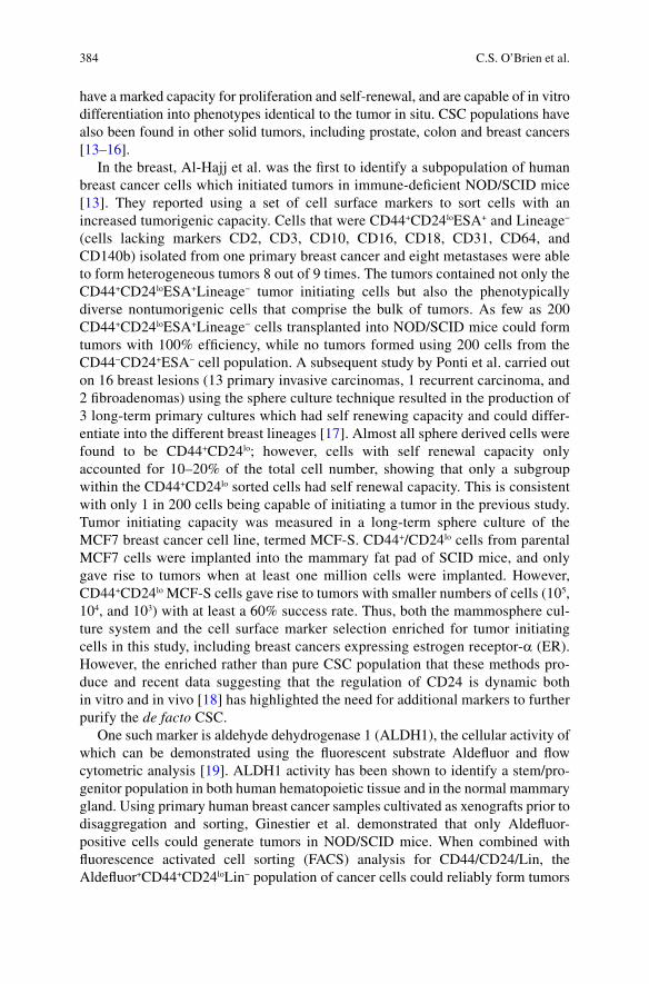
384 C.S. O’Brien et al.
have a marked capacity for proliferation and self-renewal, and are capable of in vitro differentiation into phenotypes identical to the tumor in situ. CSC populations have also been found in other solid tumors, including prostate, colon and breast cancers [13–16].
In the breast, Al-Hajj et al. was the first to identify a subpopulation of human breast cancer cells which initiated tumors in immune-deficient NOD/SCID mice [13]. They reported using a set of cell surface markers to sort cells with an increased tumorigenic capacity. Cells that were CD44+CD24loESA+ and Lineage− (cells lacking markers CD2, CD3, CD10, CD16, CD18, CD31, CD64, and CD140b) isolated from one primary breast cancer and eight metastases were able to form heterogeneous tumors 8 out of 9 times. The tumors contained not only the CD44+CD24loESA+Lineage− tumor initiating cells but also the phenotypically diverse nontumorigenic cells that comprise the bulk of tumors. As few as 200 CD44+CD24loESA+Lineage− cells transplanted into NOD/SCID mice could form tumors with 100% efficiency, while no tumors formed using 200 cells from the CD44−CD24+ESA− cell population. A subsequent study by Ponti et al. carried out on 16 breast lesions (13 primary invasive carcinomas, 1 recurrent carcinoma, and 2 fibroadenomas) using the sphere culture technique resulted in the production of 3 long-term primary cultures which had self renewing capacity and could differ-entiate into the different breast lineages [17]. Almost all sphere derived cells were found to be CD44+CD24lo; however, cells with self renewal capacity only accounted for 10–20% of the total cell number, showing that only a subgroup within the CD44+CD24lo sorted cells had self renewal capacity. This is consistent with only 1 in 200 cells being capable of initiating a tumor in the previous study. Tumor initiating capacity was measured in a long-term sphere culture of the MCF7 breast cancer cell line, termed MCF-S. CD44+/CD24lo cells from parental MCF7 cells were implanted into the mammary fat pad of SCID mice, and only gave rise to tumors when at least one million cells were implanted. However, CD44+CD24lo MCF-S cells gave rise to tumors with smaller numbers of cells (105, 104, and 103) with at least a 60% success rate. Thus, both the mammosphere cul-ture system and the cell surface marker selection enriched for tumor initiating cells in this study, including breast cancers expressing estrogen receptor-a (ER). However, the enriched rather than pure CSC population that these methods pro-duce and recent data suggesting that the regulation of CD24 is dynamic both in vitro and in vivo [18] has highlighted the need for additional markers to further purify the de facto CSC.
One such marker is aldehyde dehydrogenase 1 (ALDH1), the cellular activity of which can be demonstrated using the fluorescent substrate Aldefluor and flow cytometric analysis [19]. ALDH1 activity has been shown to identify a stem/pro-genitor population in both human hematopoietic tissue and in the normal mammary gland. Using primary human breast cancer samples cultivated as xenografts prior to disaggregation and sorting, Ginestier et al. demonstrated that only Aldefluor-positive cells could generate tumors in NOD/SCID mice. When combined with fluorescence activated cell sorting (FACS) analysis for CD44/CD24/Lin, the Aldefluor+CD44+CD24loLin− population of cancer cells could reliably form tumors
38522 Resistance to Endocrine Therapy in Breast Cancer
with as few as 20 cells in the innoculum, whereas 50,000 Aldefluor−CD44+CD24loLin− cells failed to form tumors [20]. However, recent data suggest that the Aldefluor assay may be less effective at discriminating CSC in breast cancer cell lines of luminal type [21].
There is also emerging evidence that some breast cancer cell lines will provide valuable and reliable models of tumor hierarchies containing CSCs with both cell sorting and xenografting being demonstrated from infrequent cell populations expressing markers such as CD44, Cytokeratin 5/6 (CK5/6), and ALDH1 [21–23]. A common theme of many investigations into CSCs is that they have inherent resis-tance to chemotherapy and radiotherapy. This is proposed to be due to mechanisms such as more efficient DNA damage checkpoints and survival pathways compared with more differentiated tumor cell populations [24, 25]. Endocrine therapy remains a pivotal treatment for breast cancers which express the ER. However, despite initial response to endocrine therapy, 25% of patients with early breast cancer and all patients with metastatic disease will eventually relapse [26]. In the following sec-tions, we will focus on how breast CSCs may have inherent resistance to endocrine therapies for a variety of reasons including their basal-like phenotype and the path-ways that determine their stem cell-like behavior.
22.3 Steroid Hormones and the Cellular Hierarchy of the Normal Breast
The rudimentary mammary gland matures at puberty and functionally differenti-ates during pregnancy, lactation, and menopause due to the influence of epider-mal growth factors and steroid hormones such as estrogens and progesterones [27–30]. This developmental plasticity at tissue level suggests a stem cell popula-tion within the mammary gland, which renews and differentiates to form a cel-lular hierarchy according to highly regulated functional cues. Human embryonic post mortem studies show absent expression of ER before 30 weeks gestation, although rudimentary mammary development commences from week 12 [31]. Moreover, ER knockout mice show no development of the breast beyond the rudi-mentary ductal structures of early gestation [32]. By contrast, in the mature human mammary gland, 10–20% of luminal epithelial cells coexpress ER and the progesterone receptor (PgR) [33, 34]. Progesterone receptor (PgR) expression is positively regulated by ER. Interestingly, ER+ cells in mature mammary glands of both mice and humans do not actively divide but are in proximity to mitotic cells [33, 34]. This would suggest a model whereby ER expression in the normal mam-mary gland is closely linked to a differentiated cell phenotype with limited repli-cative capacity.
Recent studies in mice and humans have suggested a more complicated role for the progesterone receptor in the postpubertal mammary gland. Beleut et al. [35] observed progesterone to be the main driver of alveolar proliferation in the adult murine mammary epithelium. Progesterone but not estrogen stimulation in
386 C.S. O’Brien et al.
ovariectomized mice led to two waves of cellular proliferation, measured by BrdU incorporation labeling. The first small proliferative peak occurred in PgR+ cells within the first 24 h and was driven by cyclin D1. This was followed by a second and larger wave of proliferation in PgR− cells, which was dependent on RANKL (receptor activator for nuclear factor k B ligand). The authors postulated that PgR+ cells may play a progenitor role in the postpubertal mammary gland, which is in contrast to the rudimentary mammary epithelium during early development [35].
The mouse mammary stem cell population characterized by expression of the markers CD29hi(b1 integrin)/CD24+/Lin− [1] consists of less than 0.01% cells expressing ER [36, 37]. Interestingly, epidermal growth factor receptor (EGFR) was found to be expressed in CD29hi(b1 integrin)/CD24+/Lin− cells, although expression of PR and erbB2/HER2 receptor was absent [36]. A further murine study defined the cellular hierarchy further, separating the luminal compartment by expression of Sca1, CD133, CD24, and ER [38]. ER rich CD133+/Sca1+/CD24hi cells were weakly proliferative whereas the milk-protein rich, ER low population of CD133−/Sca1−/CD24hi cells showed high proliferative capacity.
In a study using normal human tissue derived from mammoplasties, Raouf et al. [39] defined bipotent progenitor cells, luminal committed progenitor cells, and dif-ferentiated luminal cells by surface marker expression and subsequently assessed gene expression in each population. The cell sorting methods enriched for primitive bipotent cells (EpCAM+/CD49fhi/CALLA (CD10)+/Thy1+/CD133−) at a purity of 45 ± 3% (containing 57% of all bipotent cells) and luminal-restricted progenitors (EpCAM+/CD49f+/MUC1+/CD133+/CD10−/Thy1−) at a purity of 32 ± 3% (contain-ing 96% of all luminal progenitor cells). Transcriptional profiling revealed ERlo/PgRhi expression in the bipotent cell population compared to ERhi/PgRlo expression in the luminal committed progenitor population of the normal human breast, in an analogous manner to recent murine data [35]. These findings concur with the work of Shipitsin et al. [40], who determined ERlo expression of the stem cell population; albeit defined by an alternative cell marker methodology (CD44+/PROCR+/CD24lo).
Finally, in exciting recent work Asselin-Labat et al. [37] have investigated the role of steroid hormones in murine mammary stem cell function. Whilst demonstrat-ing the adult mammary stem cell to be ER− and PgR−, they observed that sensitivity to steroid hormones still remained, as ovariectomy diminished the stem cell pool, whereas pregnancy transiently increased stem cell number by 11-fold in a RANKL-dependent manner. This links the increased breast cancer risk associated with preg-nancy and cumulative estrogen exposure with the mammary stem cell field.
22.4 Breast Cancer Stem Cells and Endocrine Resistance
In contrast to the normal mammary gland, actively dividing ER+ cells are prominent in breast hyperplasia and breast cancers. The levels of ER and PR expression are predictive of treatment response rates to endocrine therapy and distinguish luminal
38722 Resistance to Endocrine Therapy in Breast Cancer
A tumors, which are highly ER+ and PR+, from luminal B and HER-2 tumors, which have lower ER expression, do not express PR, and coexpress other growth factor receptors such as EGFR and erbB2 [41, 42]. Intrinsic and acquired resistance to endocrine therapy remains a significant cause of disease relapse and mortality in ER+ breast cancers [43, 44].
22.4.1 EGFR Pathway
Enhanced interaction between estrogen receptor signaling and growth factor tyrosine kinase pathways such as EGFR, HER2/erbB2, and IGFR mediates resis-tance to endocrine therapy. For example, EGFR1 expression is inversely correlated with that of the ER and coexpression of both receptors confers relative resistance to endocrine therapy compared with tumors not expressing EGFR1 [42, 45]. A similar inverse expression relationship occurs between ER and erbB2/HER2. Tamoxifen-resistant MCF7 breast cancer cells show a five to tenfold increase in mRNA and protein expression of erbB2/HER2 and the EGFR receptor compared with sensitive MCF7 cells [46]. Similarly, resistance to fulvestrant and aromatase inhibitors can also be mediated by upregulation of the erbB2 pathway [47]. Long-term stimulation of the EGFR and HER2/erbB2 pathways in endocrine resistant cancer cells down-regulates the ER. Ligand-independent activation of ER may be mediated by growth factor or intracellular kinase phosphorylation of the AF1 domain of ER, for example at serine 118 or 167 [48, 49] by mitogen-activated pro-tein (MAP) kinase (MAPK), phosphoinositide 3-kinase (PI3K), AKT and Src kinase [48, 50, 51], thus allowing expression of estrogen regulated gene products despite endocrine therapy. In two models of acquired endocrine resistance (long-term estrogen deprived [LTED] and tamoxifen treated [TAM-R] cells), treatment with the dual EGFR inhibitor Lapatinib restored endocrine sensitivity [52]. In TAM-R cells, Lapatinib treatment led to reactivation of ER activity. In contrast, in LTED cells, which were exquisitely sensitive to estrogen stimulation at baseline, Lapatinib suppressed ER transcriptional activity. Intriguingly, the recent EGF30008 trial [53] demonstrated a significant improvement in progression free survival in HER2− patients with low ER expression, who had relapsed within 6 months of tamoxifen discontinuation with dual treatment with letrozole (an aromatase inhibi-tor) combined with Lapatinib (13.6 months) compared with letrozole alone (6.7 months p < 0.005).
The acquisition of enhanced EGFR/erbB2 pathway signaling in ER+ breast cancer with tamoxifen resistance potentially results from selection of a more stem-like phenotype. Expression of EGFR has been demonstrated in stem cells of the normal mammary gland in mice and humans [36, 54]. This is in contrast to ER, which is predominately expressed in more differentiated luminal cells [34, 38, 40]. In malig-nant CSCs, Farnie et al. [6] showed activation of the EGFR pathway in ductal car-cinoma in situ (DCIS) of the breast. Inhibition with gefitinib, an EGFR pathway inhibitor, significantly reduced mammosphere formation in vitro.
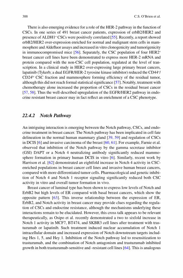
388 C.S. O’Brien et al.
There is also emerging evidence for a role of the HER-2 pathway in the function of CSCs. In one series of 491 breast cancer patients, expression of erbB2/HER2 and presence of ALDH1+ CSCs were positively correlated [55]. Recently, a report showed erbB2/HER2 over-expression enriched for normal and malignant stem cells in mam-mosphere and Aldefluor assays and increased in vitro clonogenicity and tumorigenicity in immunocompromised mice [56]. Separately, the CSC population of four HER2+ breast cancer cell lines have been demonstrated to express more HER-2 mRNA and protein compared with the non-CSC cell population, regulated at the level of tran-scription. In a clinical study in HER2 over-expressing large primary breast cancers, lapatinib (Tykerb; a dual EGFR/HER-2 tyrosine kinase inhibitor) reduced the CD44+/CD24lo CSC fraction and mammosphere forming efficiency of the residual tumor, although this did not reach formal statistical significance [57]. Notably, treatment with chemotherapy alone increased the proportion of CSCs in the residual breast cancer [57, 58]. Thus the well-described upregulation of the EGFR/HER2 pathway in endo-crine resistant breast cancer may in fact reflect an enrichment of a CSC phenotype.
22.4.2 Notch Pathway
An intriguing interaction is emerging between the Notch pathway, CSCs, and endo-crine treatment in breast cancer. The Notch pathway has been implicated in cell fate delineation in the normal human mammary gland [39, 59] and regulation of CSCs in DCIS [6] and invasive carcinoma of the breast [60, 61]. For example, Farnie et al. observed that inhibition of the Notch pathway by the gamma secretase inhibitor (GSI) DAPT or a Notch 4 neutralizing antibody significantly reduced mammo-sphere formation in primary human DCIS in vitro [6]. Similarly, recent work by Harrison et al. [62] demonstrated an eightfold increase in Notch 4 activity in CSC-enriched populations in breast cancer cell lines and invasive human breast cancers, compared with more differentiated tumor cells. Pharmacological and genetic inhibi-tion of Notch 4 and Notch 1 receptor signaling significantly reduced both CSC activity in vitro and overall tumor formation in vivo.
Breast cancer of luminal type has been shown to express low levels of Notch and ErbB2 but high levels of ER compared with basal breast cancers, which show the opposite pattern [63]. This inverse relationship between the expression of ER, ErbB2, and Notch activity in breast cancer may provide clues regarding the regula-tion of CSCs and endocrine resistance, although the mechanisms underlying these interactions remain to be elucidated. However, this cross talk appears to be relevant therapeutically, as Osipo et al. recently demonstrated a two to sixfold increase in Notch 1 activity in MCF7, BT474, and SKBR3 cell lines after treatment with tras-tuzumab or lapatinib. Such treatment induced nuclear accumulation of Notch 1 intracellular domain and increased expression of Notch downstream targets includ-ing Hes 1, 5, and Hey 1. Inhibition of the Notch pathway led to resensitization to trastuzumab, and the combination of Notch antagonism and trastuzumab inhibited growth in both trastuzumab-sensitive and -resistant cell lines [64]. This is analogous
38922 Resistance to Endocrine Therapy in Breast Cancer
to data reporting resensitization to docetaxel and doxorubicin chemotherapy in breast cancer after RNAi mediated knockdown of Notch 1 [65].
Estrogen signaling also down-regulates Notch signaling. Rizzo et al. demon-strated estradiol-induced reduction in the expression and activation of Notch 4 and Notch 1 in T47D and MCF7 cell lines. This reduction in Notch activity could be abrogated by tamoxifen and fulvestrant [63]. In a mouse xenotransplantation assay using the BT474 cell line, tumors were treated with tamoxifen alone or in combina-tion with a GSI. Combination therapy was significantly superior to the use of tamox-ifen alone, and the authors concluded that tamoxifen antagonism of the estrogen stimulus leads to reactivation of the Notch signaling pathway and promotion of proliferation and survival. More recently, a potential mechanism of direct transcrip-tional crosstalk between Notch 1 and ER target genes has been described via a nuclear I kappa B kinase (IKK)-dependent pathway [66]. However, further investi-gations will have to be carried out to determine whether this effect is on a cellular population level or specifically mediated by the CSC population.
22.4.3 Cellular Hierarchy of Breast Cancer and ER Expression
One mechanism of resistance to ER targeted endocrine therapy may be the presence of an ER−, treatment-resistant CSC population with the capacity to differentiate and produce treatment sensitive ER+ luminal cancer cells. One prediction that follows from this proposed mechanism is that after endocrine treatment, there would remain a resistant population of ER−/lo progenitor-like cells to seed relapse and metastases despite endocrine therapy.
In primary human breast cancer samples, Shipitsin et al. [40] used transcriptional profiling to characterize CD44+/PROCR+ stem cells and CD24+ luminal type cells from the same donor. This group showed that CD44+/PROCR+ cells in breast can-cers were enriched for stem cell markers and for gene expression related to cell motility and angiogenesis. Interestingly, malignant CD44+ cells were ERlo in a simi-lar manner to CD44+ cells from normal mammoplasty specimens in this report and in a study by Fillmore and Kupperwasser [22, 40].
A recent study has also demonstrated the presence of rare steroid receptor nega-tive CD44+ cells present in the ER+ breast cancer cell line T47D [23]. The size of this CD44+/CK5+/ER−/PgR− population did not proportionally increase with expan-sion of the rest of the tumor population, and this infrequent ER− cell type was observed in both in vitro clonogenic and in vivo tumorigenic assays, whereas the bulk of the tumor consisted of proliferative CD44−/CK5−/ER+/PgR+ cells. Notably, an intermediate CK5−/ER−/PgR+ cell population was demonstrable in in-vitro col-ony assays when treated with progesterone. These defined populations within an ER+ cell line appear to mimic the cellular hierarchy of steroid receptor transcript expression in the normal breast as shown by Connie Eaves’ [39] and Cathrin Brisken’s groups [35]. It is intriguing to speculate that there may be a role for the PgR in a putative progenitor cell population in cancer.
390 C.S. O’Brien et al.
Such findings might be consistent with a model in which an ER− stem cell generates a cellular hierarchy at a metastatic site comparable to the hierarchy of the primary tumor. Endocrine therapy in resistant patients may enrich for the CSC population in an analogous manner to the effects of chemotherapy or radiotherapy [57, 58, 67], leading to eventual relapse. Certainly recent data suggests residual cancer cells after letrozole therapy are enriched in the CD44hi/CD24lo phenotype, have enhanced mammosphere forming capacity, and express mesenchymal type markers including vimentin and MMP2 [25]. In contrast, ER+ breast cancers with a gene expression signature similar to mammosphere derived gene sets were more frequently low grade (p < 0.001) and of luminal A subtype (p < 0.001), with a supe-rior overall survival rate over 10 years (HR 0.24 95% CI 0.11–0.52) [68]. From this study, the authors contend that survival from ER+ breast cancer is largely governed by cellular proliferation rather than cancer stem cell activity. However, as the mam-mospheres in this study were harvested on day 7, it is possible that the cancer stem cell gene expression signature may be obscured by that of the larger population of differentiated daughter cells within the mammosphere colony at this time.
22.4.4 Cancer Stem Cells, Mesenchymal Phenotype, and Endocrine Resistance
Recent work by Weinberg’s group [69] has linked the mesenchymal cell phenotype to stem cells in normal tissue and to CSCs. Immortalized human mammary epithe-lial cells (HMECs) induced to undergo epithelial-to-mesenchymal transition (EMT) exhibited stem cell markers and had increased capacity to form mammospheres enriched in stem cells. Similarly, stem cells isolated from normal and cancerous human and mouse mammary glands demonstrated markers of a mesenchymal phe-notype normally apparent in EMT. This included up-regulation of the transcription factors Snail and Slug and also the transforming growth factor beta (TGF-b) signaling pathway, which has been previously implicated in stem cell function [40].
Metastatic potential has long been associated with the loss of markers of the epithelial cell phenotype and the acquisition of basal/mesenchymal properties. Interestingly, recent analysis of a panel of breast cancer cell lines of luminal, inter-mediate, and basal phenotypes has shown a significantly increased fraction of CSCs (defined by CD44+/CD24lo/ESA+ expression) in basal type breast cancers compared with hormone sensitive luminal cancers (2.5 vs. 0.5% p < 0.0001) [22]. Furthermore, a positive correlation was shown between CSC number and cell line tumorigenicity in vivo [22].
Endocrine resistant ER+ breast cancers are reported to gain a more basal pheno-type, for example, reduction in E-cadherin expression [70] and enhanced motility and invasion by upregulation of Src kinase [71, 72], NF-kB [73] and CD44 [74, 75]. As ER negatively regulates the expression of the key transcription factors regulating EMT such as Snail and Slug [76, 77], a functionally redundant ER in endocrine resis-tant breast cancer might therefore promote a more mesenchymal stem-cell-like
39122 Resistance to Endocrine Therapy in Breast Cancer
phenotype. Neoadjuvant letrozole therapy in a series of 36 patients [25] induced a comparative mesenchymal phenotype and claudin low signature [78] of cells remaining after systemic therapy. The residual cells showed enhanced expression of vimentin, fibronectin, and Snail; whereas expression of E-cadherin was diminished consistent with the acquisition of mesenchymal characteristics postendocrine therapy.
22.4.5 Epigenetic Regulation of the Cellular Hierarchy
Gene-expression profiling of breast cancer has demonstrated at least six distinct molecular subtypes, including basal, erbB2, luminal B, luminal A, normal-like and claudin-low [41, 78, 79]. These subtypes probably represent a differentiation spectrum comparable to the developmental hierarchy of the breast, with poorly dif-ferentiated ER-negative basal type at one extreme to well-differentiated luminal A type at the other. As such, these subtypes may derive from a cell of origin at a dif-ferent stage of the developmental hierarchy [80] and reflect the hormone and growth factor sensitivity of that distinct cell. Prolonged endocrine therapy may lead to the reacquisition of a more primitive cancer cell phenotype with intrinsic resistance to hormone manipulation.
A recent study elegantly demonstrates that targeted epigenetic modification of the genome has an important role to play in cell-fate determination in the cellular hierarchy of the human mammary gland and breast cancer [81]. Using MDSK (methylation specific digital karyotyping) and SAGE (serial analysis of gene expres-sion) techniques, adult mammary stem cells (CD44+) and breast cancer stem cells (CD44+) were compared and contrasted to more lineage committed (CD24+) cells. Normal adult mammary SCs and CSCs showed comparable genomic hypo-methy-lation of transcription factors implicated in stem cell function such as HOXA10, FOXC1, and TCF3 compared with the more highly methylated progenitor and lin-eage committed cells. Forced expression of FOXC1 in differentiated mammary cells, where FOXC1 is normally methylated, led to the reacquisition of a progenitor-like phenotype. This suggests an important role for epigenetic modification in cell fate specification and function of normal and cancer stem cells, which in the future may be amenable to therapeutic targeting.
Acquired endocrine resistance may thus result from an alteration in cancer phe-notype between the primary tumor and the metastases to a more stem-like hormone insensitive cellular identity; however, the evidence for this remains circumstantial. In one series of 200 patients, 19.5% of metastases were found to be ER− in the pres-ence of an ER+ primary breast cancer, and these findings have been replicated in another smaller study [82]. Fehm et al. have shown that in 88 patients with ER+ primary breast cancers, 76 had only ER− disseminated tumor cells (DTC) in the bone marrow [83]. These data raise the possibility that the ER− CSC is responsible for tumor metastasis and that cell surface phenotype of such cells facilitates com-munication with a stromal niche that enables intravasation and metastatic growth. It is worth noting that ER is lost completely in only 20% of metastases from ER+
392 C.S. O’Brien et al.
primary cancers, suggesting that the ER− DTCs isolated by Fehm et al. may undergo differentiation into tumors that can be subsequently defined as ER+. Up to half of metastatic tumors, which continue to express ER, show no functional inhibition by endocrine agents. Interestingly, aberrant methylation of ER and PgR promoters has been observed in up to 40% of hormone receptor negative breast cancers [84, 85], and epigenetic modifications have been shown in tamoxifen resistance [86]. Forced reexpression of ER by therapeutic demethylation may thus lead to the intriguing possibility of reacquisition of endocrine sensitivity in these malignancies and we will discuss this possibility further later in this review.
22.4.6 The Stem Cell Niche and Its Influence on Resistance to Endocrine Therapy
CSCs are associated with an increased invasive and metastatic/migratory phenotype [87–89]. Cells isolated as CSCs by virtue of ALDH1+ and/or CD44+/CD24lo demon-strated increased metastasis from primary tumors in NOD/SCID/IL2g receptor null mice [87]. Such augmented invasive and metastatic phenotypes are also seen in endocrine resistant breast cancer cell lines [50, 90, 91]. These cell lines exhibit over-expression of EGFR and the c-MET receptor through which they derive prolif-erative and migratory/invasive signals from stromal derived ligand secretion. Significantly, such resistant cells also overexpress CD44, the adhesion of which to bone marrow derived endothelial cells is enhanced by stromal derived HGF in vitro [75, 92]. Thus, adaptive endocrine resistance in cell lines is associated with a meta-static and stem cell-like phenotype.
Using human breast cancer cell lines in a murine model, it was demonstrated that CD44 was sparsely expressed in primary tumor cells but homogeneously over-expressed in cells transiting the lymphatics and populating lymph nodes (LNs) [93]. The authors hypothesized that CD44 expression targeted tumor cells for metastasis to, and uptake in the LN, although induction of CD44 expression by interaction of the epithelial cells with the LN stromal cells is also a possibility. The CD44 express-ing cells were relatively insensitive to the effects of estradiol and estradiol with-drawal despite ER expression levels comparable to those seen in the primary tumors [93, 94]. The same group have also recently shown that a small subpopulation of the cells expressing CD44 express CK5 but not ER or PgR, and are resistant to both endocrine and chemotherapy [95]. Thus, the LN and stromal microenvironments may be responsible for maintenance of the CSC phenotype and suppression of estrogen sensitivity in such cells.
Supporting the former hypotheses are recent data from Farmer et al. demonstrat-ing a stroma-related gene signature in primary breast cancers [96]. This signature was associated with the presence of a reactive stroma, and predicted for resistance to neo-adjuvant chemotherapy. Importantly, the stroma-related signature demon-strated a pattern of expression similar to that of mammospheres, suggesting that the
39322 Resistance to Endocrine Therapy in Breast Cancer
stroma may support the CSC phenotype and promote resistance to therapy. As the signature was only tested in ER negative tumors, the relevance to luminal tumors and endocrine resistance is unknown; however, such analyses are eagerly awaited.
Another emerging target that is likely to have an impact on CSCs is anti-angio-genic therapy, since evidence is accumulating that both tissue stem cells and CSCs preferentially associate with blood vessels. For example, in oligodendrogliomas and glioblastomas, there is a direct correlation between nestin positive CSCs and microvessel density (MVD) [97]. This study also reported that CSCs preferentially associate with CD34+ capillaries in vivo (in tumor sections) and endothelial vascu-lar tubes in a basement membrane (Matrigel) culture assay in vitro compared with non-CSCs. In a prior report, this association had been shown to be secondary to CSC secretion of vascular endothelial growth factor (VEGF), which directly stim-ulates endothelial cell growth [98]. Currently, there is little data to support or refute the existence of a vascular niche for the breast CSC and further investigation is required.
22.4.7 Differentiation Agents and Endocrine Treatment
There is evidence to show that histone deacetlylation and DNA methylation plays a key role in inactivation of ER gene expression. In ER− breast cancer cells, studies have demonstrated that the ER CpG promoter is occupied by abundant HDAC1 and HDAC2 [99, 100]. Similarly, DNA methylation has also been reported to be enhanced in ER− breast cancer cells [101]. Investigation of de novo ER gene methy-lation in vitro showed DNA methyltransferase 1 (DNMT1) levels were significantly elevated in ER− breast cancer cell lines compared with their ER+ counterparts [85]. Furthermore, recent research into cell type specific DNA methylation patterns revealed that progenitors were hypomethylated compared with differentiated cells in the human normal breast and breast cancer [81]. The role of epigenetic modifica-tion in regulation of ER expression and cell fate in breast cancer may provide a therapeutic targeting strategy for ER− breast cancer patients.
Epigenetic therapies such as histone deacetylase (HDAC) and DNMT inhibitors have shown considerable promise in the treatment of hematological malignancies [102], and trials are ongoing in solid cancers. Cell line studies have shown that func-tional ER gene expression can be induced by pharmacological administration of the DNMT inhibitor 5-aza-2¢-deoxycytidine (AZA) and a HDAC inhibitor trichostatin A (TSA) [103–105]. Furthermore, combination AZA and TSA treatment acts synergisti-cally to induce reexpression of ER in ER− breast cancer cells [106]. A recent preclini-cal xenograft model has demonstrated that ER− MDA-MB-435 cells treated with AZA and TSA reexpressed functional ER, which by itself caused a significant reduction in tumor growth. In addition, after ovarian ablation to mimic endocrine treatment, there was a further reduction in tumor growth [107]. Finally, using the clinically available HDAC inhibitor sodium valproate (VPA), Fortunati et al. [108] were also able to
394 C.S. O’Brien et al.
restore ER transcriptional activity to MDA-MB-231 cells and furthermore convey tamoxifen sensitivity to the previously tamoxifen insensitive HDAC treated cells using in vitro assays.
An inverse relationship between ER and EGFR expression has been well docu-mented in breast cancer cell lines. Using the HDAC inhibitor vorinostat, the ER− cell lines MDA-MB-231 and MDA-MB-468 cells exhibit ER gene expression and reduced EGFR expression. Reduction in EGFR expression led to reduced EGFR signaling and PAK1 expression levels [109]. Interestingly, immunohistochemical analysis of PAK1 showed significantly increased expression in breast cancers from hormone resistant patients [110–112].
HDAC inhibitors are being used in a number of ongoing clinical trials including a Phase II trial evaluating vorinostat in ER positive patients with metastatic breast can-cer who failed prior aromatase inhibitor therapy and up to three chemotherapy regimes [113]. A report of preliminary findings presented at the American Society of Clinical Oncology (ASCO) 2008 meeting showed that out of the 17 enrolled patients, 21% had a partial response and 29% had stable disease after treatment with vorinostat 400 mg daily for 3 of 4 weeks and tamoxifen 20 mg daily, continuously. These find-ings suggest that the addition of an HDAC inhibitor to tamoxifen in patients who have failed prior aromatase inhibitors or adjuvant tamoxifen may restore hormone sensi-tivity. The in vitro studies would also suggest that HDAC inhibitors in combination with endocrine inhibitors may be highly applicable to ER− breast cancers as well.
22.5 Concluding Remarks
In this chapter, we have summarized current evidence that supports our understand-ing of CSCs to explain endocrine resistance in breast cancer. The biology of breast CSCs is becoming better characterized, and the data suggest that they may be resis-tant to several forms of cancer therapy through diverse mechanisms. In terms of responsiveness to endocrine therapy, we can learn about CSC biology and hierar-chies in breast cancer (Fig. 22.1) by examining what is known about the develop-mental hierarchy of the normal breast epithelium. In normal breast, the stem cells are known to possess a basal phenotype and to be mainly ER−. If the hierarchy in breast cancer reflects this, the breast CSC may be endocrine resistant because it expresses very little ER and can only respond to treatment by virtue of paracrine influences of neighboring, differentiated ER+ tumor cells. Normal breast epithelial stem cells are highly dependent on EGFR and other growth factor receptors, and it may be that the observed increased growth factor receptor expression in resistant breast cancers reflects an increased proportion of stem-like cells selected by endo-crine therapies. There is evidence from a number of studies that breast CSCs are ER−, which would support this view. CSCs also express mesenchymal proteins, which are suppressed by ER expression, further indicating the mutual exclusion between ER+ cells and CSCs. It is likely that this is regulated at the epigenetic level, and differences in DNA methylation and chromatin organization can be observed
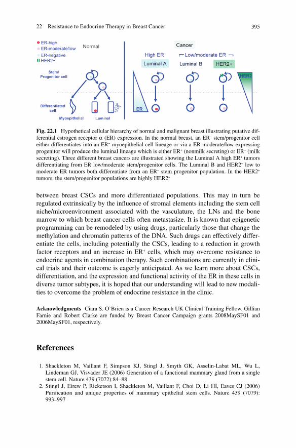
39522 Resistance to Endocrine Therapy in Breast Cancer
between breast CSCs and more differentiated populations. This may in turn be regulated extrinsically by the influence of stromal elements including the stem cell niche/microenvironment associated with the vasculature, the LNs and the bone marrow to which breast cancer cells often metastasize. It is known that epigenetic programming can be remodeled by using drugs, particularly those that change the methylation and chromatin patterns of the DNA. Such drugs can effectively differ-entiate the cells, including potentially the CSCs, leading to a reduction in growth factor receptors and an increase in ER+ cells, which may overcome resistance to endocrine agents in combination therapy. Such combinations are currently in clini-cal trials and their outcome is eagerly anticipated. As we learn more about CSCs, differentiation, and the expression and functional activity of the ER in these cells in diverse tumor subtypes, it is hoped that our understanding will lead to new modali-ties to overcome the problem of endocrine resistance in the clinic.
Acknowledgments Ciara S. O’Brien is a Cancer Research UK Clinical Training Fellow. Gillian Farnie and Robert Clarke are funded by Breast Cancer Campaign grants 2008MaySF01 and 2006MaySF01, respectively.
References
1. Shackleton M, Vaillant F, Simpson KJ, Stingl J, Smyth GK, Asselin-Labat ML, Wu L, Lindeman GJ, Visvader JE (2006) Generation of a functional mammary gland from a single stem cell. Nature 439 (7072):84–88
2. Stingl J, Eirew P, Ricketson I, Shackleton M, Vaillant F, Choi D, Li HI, Eaves CJ (2006) Purification and unique properties of mammary epithelial stem cells. Nature 439 (7079): 993–997
Fig. 22.1 Hypothetical cellular hierarchy of normal and malignant breast illustrating putative dif-ferential estrogen receptor a (ER) expression. In the normal breast, an ER− stem/progenitor cell either differentiates into an ER− myoepithelial cell lineage or via a ER moderate/low expressing progenitor will produce the luminal lineage which is either ER+ (nonmilk secreting) or ER− (milk secreting). Three different breast cancers are illustrated showing the Luminal A high ER+ tumors differentiating from ER low/moderate stem/progenitor cells. The Luminal B and HER2+ low to moderate ER tumors both differentiate from an ER− stem progenitor population. In the HER2+ tumors, the stem/progenitor populations are highly HER2+
396 C.S. O’Brien et al.
3. Bonnet D, Dick JE (1997) Human acute myeloid leukemia is organized as a hierarchy that originates from a primitive hematopoietic cell. Nat Med 3 (7):730–737
4. Kelly PN, Dakic A, Adams JM, Nutt SL, Strasser A (2007) Tumor growth need not be driven by rare cancer stem cells. Science 317 (5836):337
5. Quintana E, Shackleton M, Sabel MS, Fullen DR, Johnson TM, Morrison SJ (2008) Efficient tumor formation by single human melanoma cells. Nature 456 (7222):593–598
6. Farnie G, Clarke RB, Spence K, Pinnock N, Brennan K, Anderson NG, Bundred NJ (2007) Novel cell culture technique for primary ductal carcinoma in situ: role of Notch and epidermal growth factor receptor signaling pathways. J Natl Cancer Inst 99 (8):616-627
7. Park SY, Lee HE, Li H, Shipitsin M, Gelman R, Polyak K (2010) Heterogeneity for stem cell-related markers according to tumor subtype and histologic stage in breast cancer. Clin Cancer Res 16 (3):876–887
8. Pece S, Tosoni D, Confalonieri S, Mazzarol G, Vecchi M, Ronzoni S, Bernard L, Viale G, Pelicci PG, Di Fiore PP (2010) Biological and molecular heterogeneity of breast cancers cor-relates with their cancer stem cell content. Cell 140 (1):62–73
9. Gupta PB, Onder TT, Jiang G, Tao K, Kuperwasser C, Weinberg RA, Lander ES (2009) Identification of selective inhibitors of cancer stem cells by high-throughput screening. Cell 138 (4):645–659
10. Kakarala M, Brenner DE, Korkaya H, Cheng C, Tazi K, Ginestier C, Liu S, Dontu G, Wicha MS (2010) Targeting breast stem cells with the cancer preventive compounds curcumin and piperine. Breast Cancer Res Treat 122 (3):777–85
11. Singh SK, Clarke ID, Terasaki M, Bonn VE, Hawkins C, Squire J, Dirks PB (2003) Identification of a cancer stem cell in human brain tumors. Cancer Res 63 (18):5821–5828
12. Singh SK, Hawkins C, Clarke ID, Squire JA, Bayani J, Hide T, Henkelman RM, Cusimano MD, Dirks PB (2004) Identification of human brain tumor initiating cells. Nature 432 (7015):396–401
13. Al-Hajj M, Wicha MS, Benito-Hernandez A, Morrison SJ, Clarke MF (2003) Prospective iden-tification of tumorigenic breast cancer cells. Proc Natl Acad Sci USA 100 (7):3983–3988
14. Collins AT, Berry PA, Hyde C, Stower MJ, Maitland NJ (2005) Prospective identification of tumorigenic prostate cancer stem cells. Cancer Res 65 (23):10946–10951
15. O’Brien CA, Pollett A, Gallinger S, Dick JE (2007) A human colon cancer cell capable of initiating tumor growth in immunodeficient mice. Nature 445 (7123):106–110
16. Ricci-Vitiani L, Lombardi DG, Pilozzi E, Biffoni M, Todaro M, Peschle C, De Maria R (2007) Identification and expansion of human colon-cancer-initiating cells. Nature 445 (7123):111–115
17. Ponti D, Costa A, Zaffaroni N, Pratesi G, Petrangolini G, Coradini D, Pilotti S, Pierotti MA, Daidone MG (2005) Isolation and in vitro propagation of tumorigenic breast cancer cells with stem/progenitor cell properties. Cancer Res 65 (13):5506–5511
18. Meyer MJ, Fleming JM, Ali MA, Pesesky MW, Ginsburg E, Vonderhaar BK (2009) Dynamic regulation of CD24 and the invasive, CD44posCD24neg phenotype in breast cancer cell lines. Breast Cancer Res 11 (6):R82
19. Storms RW, Trujillo AP, Springer JB, Shah L, Colvin OM, Ludeman SM, Smith C (1999) Isolation of primitive human hematopoietic progenitors on the basis of aldehyde dehydroge-nase activity. Proc Natl Acad Sci USA 96 (16):9118–9123
20. Ginestier C, Hur MH, Charafe-Jauffret E, Monville F, Dutcher J, Brown M, Jacquemier J, Viens P, Kleer CG, Liu S, Schott A, Hayes D, Birnbaum D, Wicha MS, Dontu G (2007) ALDH1 Is a Marker of Normal and Malignant Human Mammary Stem Cells and a Predictor of Poor Clinical Outcome. Cell Stem Cell 1 (5):555–567
21. Charafe-Jauffret E, Ginestier C, Iovino F, Wicinski J, Cervera N, Finetti P, Hur MH, Diebel ME, Monville F, Dutcher J, Brown M, Viens P, Xerri L, Bertucci F, Stassi G, Dontu G, Birnbaum D, Wicha MS (2009) Breast cancer cell lines contain functional cancer stem cells with metastatic capacity and a distinct molecular signature. Cancer Res 69 (4):1302–1313
22. Fillmore CM, Kuperwasser C (2008) Human breast cancer cell lines contain stem-like cells that self-renew, give rise to phenotypically diverse progeny and survive chemotherapy. Breast Cancer Res 10 (2):R25
39722 Resistance to Endocrine Therapy in Breast Cancer
23. Horwitz KB, Dye WW, Harrell JC, Kabos P, Sartorius CA (2008) Rare steroid receptor- negative basal-like tumorigenic cells in luminal subtype human breast cancer xenografts. Proc Natl Acad Sci USA 105 (15):5774–5779
24. Zhang M, Atkinson RL, Rosen JM (2010) Selective targeting of radiation-resistant tumor-ini-tiating cells. Proc Natl Acad Sci USA 107 (8):3522–3527
25. Creighton CJ, Li X, Landis M, Dixon JM, Neumeister VM, Sjolund A, Rimm DL, Wong H, Rodriguez A, Herschkowitz JI, Fan C, Zhang X, He X, Pavlick A, Gutierrez MC, Renshaw L, Larionov AA, Faratian D, Hilsenbeck SG, Perou CM, Lewis MT, Rosen JM, Chang JC (2009) Residual breast cancers after conventional therapy display mesenchymal as well as tumor-initiating features. Proc Natl Acad Sci USA 106 (33):13820–13825
26. Howell A, Wardley AM (2005) Overview of the impact of conventional systemic therapies on breast cancer. Endocr Relat Cancer 12 Suppl 1:S9–S16
27. Mallepell S, Krust A, Chambon P, Brisken C (2006) Paracrine signaling through the epithelial estrogen receptor alpha is required for proliferation and morphogenesis in the mammary gland. Proc Natl Acad Sci USA 103 (7):2196–2201
28. Brisken C, Kaur S, Chavarria TE, Binart N, Sutherland RL, Weinberg RA, Kelly PA, Ormandy CJ (1999) Prolactin controls mammary gland development via direct and indirect mechanisms. Dev Biol 210 (1):96–106
29. Coleman S, Silberstein GB, Daniel CW (1988) Ductal morphogenesis in the mouse mammary gland: evidence supporting a role for epidermal growth factor. Dev Biol 127 (2):304–315
30. Brisken C, Park S, Vass T, Lydon JP, O’Malley BW, Weinberg RA (1998) A paracrine role for the epithelial progesterone receptor in mammary gland development. Proc Natl Acad Sci USA 95 (9):5076–5081
31. Keeling JW, Ozer E, King G, Walker F (2000) Oestrogen receptor alpha in female fetal, infant, and child mammary tissue. J Pathol 191 (4):449–451
32. Korach KS, Couse JF, Curtis SW, Washburn TF, Lindzey J, Kimbro KS, Eddy EM, Migliaccio S, Snedeker SM, Lubahn DB, Schomberg DW, Smith EP (1996) Estrogen receptor gene disruption: molecular characterization and experimental and clinical phenotypes. Recent Prog Horm Res 51:159-186; discussion 186–158
33. Russo J, Ao X, Grill C, Russo IH (1999) Pattern of distribution of cells positive for estrogen receptor alpha and progesterone receptor in relation to proliferating cells in the mammary gland. Breast Cancer Res Treat 53 (3):217–227
34. Clarke RB, Howell A, Potten CS, Anderson E (1997) Dissociation between steroid receptor expression and cell proliferation in the human breast. Cancer Res 57 (22):4987–4991
35. Beleut M, Rajaram RD, Caikovski M, Ayyanan A, Germano D, Choi Y, Schneider P, Brisken C (2010) Two distinct mechanisms underlie progesterone-induced proliferation in the mammary gland. Proc Natl Acad Sci USA 107 (7):2989–2994
36. Asselin-Labat ML, Shackleton M, Stingl J, Vaillant F, Forrest NC, Eaves CJ, Visvader JE, Lindeman GJ (2006) Steroid hormone receptor status of mouse mammary stem cells. J Natl Cancer Inst 98 (14):1011–1014
37. Asselin-Labat ML, Vaillant F, Sheridan JM, Pal B, Wu D, Simpson ER, Yasuda H, Smyth GK, Martin TJ, Lindeman GJ, Visvader JE (2010) Control of mammary stem cell function by ste-roid hormone signalling. Nature 465(7299):798–802
38. Sleeman KE, Kendrick H, Robertson D, Isacke CM, Ashworth A, Smalley MJ (2007) Dissociation of estrogen receptor expression and in vivo stem cell activity in the mammary gland. J Cell Biol 176 (1):19–26
39. Raouf A, Zhao Y, To K, Stingl J, Delaney A, Barbara M, Iscove N, Jones S, McKinney S, Emerman J, Aparicio S, Marra M, Eaves C (2008) Transcriptome analysis of the normal human mammary cell commitment and differentiation process. Cell Stem Cell 3 (1):109–118
40. Shipitsin M, Campbell LL, Argani P, Weremowicz S, Bloushtain-Qimron N, Yao J, Nikolskaya T, Serebryiskaya T, Beroukhim R, Hu M, Halushka MK, Sukumar S, Parker LM, Anderson KS, Harris LN, Garber JE, Richardson AL, Schnitt SJ, Nikolsky Y, Gelman RS, Polyak K (2007) Molecular definition of breast tumor heterogeneity. Cancer Cell 11 (3):259–273
41. Sorlie T, Tibshirani R, Parker J, Hastie T, Marron JS, Nobel A, Deng S, Johnsen H, Pesich R, Geisler S, Demeter J, Perou CM, Lonning PE, Brown PO, Borresen-Dale AL, Botstein D

398 C.S. O’Brien et al.
(2003) Repeated observation of breast tumor subtypes in independent gene expression data sets. Proc Natl Acad Sci USA 100 (14):8418–8423
42. Dowsett M, Houghton J, Iden C, Salter J, Farndon J, A’Hern R, Sainsbury R, Baum M (2006) Benefit from adjuvant tamoxifen therapy in primary breast cancer patients according oestrogen receptor, progesterone receptor, EGF receptor and HER2 status. Ann Oncol 17 (5):818–826
43. Howell A, Cuzick J, Baum M, Buzdar A, Dowsett M, Forbes JF, Hoctin-Boes G, Houghton J, Locker GY, Tobias JS (2005) Results of the ATAC (Arimidex, Tamoxifen, Alone or in Combination) trial after completion of 5 years’ adjuvant treatment for breast cancer. Lancet 365 (9453):60–62
44. Gelber RD, Cole BF, Goldhirsch A, Rose C, Fisher B, Osborne CK, Boccardo F, Gray R, Gordon NH, Bengtsson NO, Sevelda P (1996) Adjuvant chemotherapy plus tamoxifen com-pared with tamoxifen alone for postmenopausal breast cancer: meta-analysis of quality-adjusted survival. Lancet 347 (9008):1066–1071
45. Giltnane JM, Ryden L, Cregger M, Bendahl PO, Jirstrom K, Rimm DL (2007) Quantitative measurement of epidermal growth factor receptor is a negative predictive factor for tamoxifen response in hormone receptor positive premenopausal breast cancer. J Clin Oncol 25 (21):3007–3014
46. Knowlden JM, Hutcheson IR, Jones HE, Madden T, Gee JM, Harper ME, Barrow D, Wakeling AE, Nicholson RI (2003) Elevated levels of epidermal growth factor receptor/c-erbB2 het-erodimers mediate an autocrine growth regulatory pathway in tamoxifen-resistant MCF-7 cells. Endocrinology 144 (3):1032–1044
47. Massarweh S, Osborne CK, Jiang S, Wakeling AE, Rimawi M, Mohsin SK, Hilsenbeck S, Schiff R (2006) Mechanisms of tumor regression and resistance to estrogen deprivation and fulvestrant in a model of estrogen receptor-positive, HER-2/neu-positive breast cancer. Cancer Res 66 (16):8266–8273
48. Pancholi S, Lykkesfeldt A, Hilmi C, Banerjee S, Leary A, Drury S, Johnston S, Dowsett M, Martin LA (2008) ERBB2 influences the subcellular localization of the estrogen receptor in tamoxifen-resistant MCF-7 cells leading to the activation of AKT and p90RSK. Endocr Relat Cancer 15(4):985–1002.
49. Sarwar N, Kim JS, Jiang J, Peston D, Sinnett HD, Madden P, Gee JM, Nicholson RI, Lykkesfeldt AE, Shousha S, Coombes RC, Ali S (2006) Phosphorylation of ERalpha at serine 118 in pri-mary breast cancer and in tamoxifen-resistant tumors is indicative of a complex role for ERalpha phosphorylation in breast cancer progression. Endocr Relat Cancer 13 (3):851–861
50. Hiscox S, Morgan L, Green TP, Barrow D, Gee J, Nicholson RI (2006) Elevated Src activity promotes cellular invasion and motility in tamoxifen resistant breast cancer cells. Breast Cancer Res Treat 97 (3):263–274
51. Campbell RA, Bhat-Nakshatri P, Patel NM, Constantinidou D, Ali S, Nakshatri H (2001) Phosphatidylinositol 3-kinase/AKT-mediated activation of estrogen receptor alpha: a new model for anti-estrogen resistance. J Biol Chem 276 (13):9817–9824
52. Leary AF, Drury S, Detre S, Pancholi S, Lykkesfeldt AE, Martin LA, Dowsett M, Johnston SR (2010) Lapatinib restores hormone sensitivity with differential effects on estrogen receptor signaling in cell models of human epidermal growth factor receptor 2-negative breast cancer with acquired endocrine resistance. Clin Cancer Res 16 (5):1486–1497
53. Johnston S, Pippen J, Jr., Pivot X, Lichinitser M, Sadeghi S, Dieras V, Gomez HL, Romieu G, Manikhas A, Kennedy MJ, Press MF, Maltzman J, Florance A, O’Rourke L, Oliva C, Stein S, Pegram M (2009) Lapatinib combined with letrozole versus letrozole and placebo as first-line therapy for postmenopausal hormone receptor-positive metastatic breast cancer. J Clin Oncol 27 (33):5538–5546
54. Hebbard L, Steffen A, Zawadzki V, Fieber C, Howells N, Moll J, Ponta H, Hofmann M, Sleeman J (2000) CD44 expression and regulation during mammary gland development and function. J Cell Sci 113 ( Pt 14):2619–2630
55. Ginestier C, Hur MH, Charafe-Jauffret E, Monville F, Dutcher J, Brown M, Jacquemier J, Viens P, Kleer CG, Liu S, Schott A, Hayes D, Birnbaum D, Wicha MS, Dontu G (2007)
39922 Resistance to Endocrine Therapy in Breast Cancer
ALDH1 is a marker of normal and malignant human mammary stem cells and a predictor of poor clinical outcome. Cell Stem Cell 1 (5):555–567
56. Korkaya H, Paulson A, Iovino F, Wicha MS (2008) HER2 regulates the mammary stem/pro-genitor cell population driving tumorigenesis and invasion. Oncogene 27 (47):6120–6130
57. Li X, Lewis MT, Huang J, Gutierrez C, Osborne CK, Wu MF, Hilsenbeck SG, Pavlick A, Zhang X, Chamness GC, Wong H, Rosen J, Chang JC (2008) Intrinsic resistance of tumori-genic breast cancer cells to chemotherapy. J Natl Cancer Inst 100 (9):672–679
58. Yu F, Yao H, Zhu P, Zhang X, Pan Q, Gong C, Huang Y, Hu X, Su F, Lieberman J, Song E (2007) let-7 regulates self renewal and tumorigenicity of breast cancer cells. Cell 131 (6):1109–1123
59. Yalcin-Ozuysal O, Fiche M, Guitierrez M, Wagner KU, Raffoul W, Brisken C (2010) Antagonistic roles of Notch and p63 in controlling mammary epithelial cell fates. Cell Death Differ 17(10):1600–12
60. Dontu G, Jackson KW, McNicholas E, Kawamura MJ, Abdallah WM, Wicha MS (2004) Role of Notch signaling in cell-fate determination of human mammary stem/progenitor cells. Breast Cancer Res 6 (6):R605–615
61. Stylianou S, Clarke RB, Brennan K (2006) Aberrant activation of notch signaling in human breast cancer. Cancer Res 66 (3):1517–1525
62. Harrison H, Farnie G, Howell SJ, Rock RE, Stylianou S, Brennan KR, Bundred NJ, Clarke RB (2010) Regulation of breast cancer stem cell activity by signaling through the Notch4 receptor. Cancer Res 70 (2):709–718
63. Rizzo P, Miao H, D’Souza G, Osipo C, Song LL, Yun J, Zhao H, Mascarenhas J, Wyatt D, Antico G, Hao L, Yao K, Rajan P, Hicks C, Siziopikou K, Selvaggi S, Bashir A, Bhandari D, Marchese A, Lendahl U, Qin JZ, Tonetti DA, Albain K, Nickoloff BJ, Miele L (2008) Cross-talk between notch and the estrogen receptor in breast cancer suggests novel therapeutic approaches. Cancer Res 68 (13):5226–5235
64. Osipo C, Patel P, Rizzo P, Clementz AG, Hao L, Golde TE, Miele L (2008) ErbB-2 inhibition activates Notch-1 and sensitizes breast cancer cells to a gamma-secretase inhibitor. Oncogene 27 (37):5019–5032
65. Zang S, Chen F, Dai J, Guo D, Tse W, Qu X, Ma D, Ji C (2010) RNAi-mediated knockdown of Notch-1 leads to cell growth inhibition and enhanced chemosensitivity in human breast cancer. Oncol Rep 23 (4):893–899
66. Hao L, Rizzo P, Osipo C, Pannuti A, Wyatt D, Cheung LW, Sonenshein G, Osborne BA, Miele L (2010) Notch-1 activates estrogen receptor-alpha-dependent transcription via IKKalpha in breast cancer cells. Oncogene 29 (2):201–213
67. Phillips TM, McBride WH, Pajonk F (2006) The response of CD24(−/low)/CD44+ breast cancer-initiating cells to radiation. J Natl Cancer Inst 98 (24):1777–1785
68. Kok M, Koornstra RH, Margarido TC, Fles R, Armstrong NJ, Linn SC, Van’t Veer LJ, Weigelt B (2009) Mammosphere-derived gene set predicts outcome in patients with ER-positive breast cancer. J Pathol 218 (3):316–326
69. Mani SA, Guo W, Liao MJ, Eaton EN, Ayyanan A, Zhou AY, Brooks M, Reinhard F, Zhang CC, Shipitsin M, Campbell LL, Polyak K, Brisken C, Yang J, Weinberg RA (2008) The epi-thelial-mesenchymal transition generates cells with properties of stem cells. Cell 133 (4):704–715
70. Hiscox S, Jiang WG, Obermeier K, Taylor K, Morgan L, Burmi R, Barrow D, Nicholson RI (2006) Tamoxifen resistance in MCF7 cells promotes EMT-like behaviour and involves modu-lation of beta-catenin phosphorylation. Int J Cancer 118 (2):290–301
71. Hiscox S, Jordan NJ, Morgan L, Green TP, Nicholson RI (2007) Src kinase promotes adhe-sion-independent activation of FAK and enhances cellular migration in tamoxifen-resistant breast cancer cells. Clin Exp Metastasis 24 (3):157–167
72. Hiscox S, Jordan NJ, Smith C, James M, Morgan L, Taylor KM, Green TP, Nicholson RI (2008) Dual targeting of Src and ER prevents acquired antihormone resistance in breast cancer cells. Breast Cancer Res Treat 115(1):57–67
400 C.S. O’Brien et al.
73. Zhou Y, Yau C, Gray JW, Chew K, Dairkee SH, Moore DH, Eppenberger U, Eppenberger-Castori S, Benz CC (2007) Enhanced NF kappa B and AP-1 transcriptional activity associated with antiestrogen resistant breast cancer. BMC Cancer 7:59
74. Borley AC, Hiscox S, Gee J, Smith C, Shaw V, Barrett-Lee P, Nicholson RI (2008) Anti-oestrogens but not oestrogen deprivation promote cellular invasion in intercellular adhesion-deficient breast cancer cells. Breast Cancer Res 10 (6):R103
75. Hiscox S, Jiang WG (1997) Regulation of endothelial CD44 expression and endothelium-tumor cell interactions by hepatocyte growth factor/scatter factor. Biochem Biophys Res Commun 233 (1):1–5
76. Dhasarathy A, Kajita M, Wade PA (2007) The transcription factor snail mediates epithelial to mesenchymal transitions by repression of estrogen receptor-alpha. Mol Endocrinol 21 (12):2907–2918
77. Ye Y, Xiao Y, Wang W, Yearsley K, Gao JX, Barsky SH (2008) ERalpha suppresses slug expression directly by transcriptional repression. Biochem J 416 (2):179–187
78. Herschkowitz JI, Simin K, Weigman VJ, Mikaelian I, Usary J, Hu Z, Rasmussen KE, Jones LP, Assefnia S, Chandrasekharan S, Backlund MG, Yin Y, Khramtsov AI, Bastein R, Quackenbush J, Glazer RI, Brown PH, Green JE, Kopelovich L, Furth PA, Palazzo JP, Olopade OI, Bernard PS, Churchill GA, Van Dyke T, Perou CM (2007) Identification of conserved gene expression features between murine mammary carcinoma models and human breast tumors. Genome Biol 8 (5):R76
79. Perou CM, Sorlie T, Eisen MB, van de Rijn M, Jeffrey SS, Rees CA, Pollack JR, Ross DT, Johnsen H, Akslen LA, Fluge O, Pergamenschikov A, Williams C, Zhu SX, Lonning PE, Borresen-Dale AL, Brown PO, Botstein D (2000) Molecular portraits of human breast tumors. Nature 406 (6797):747–752
80. Sims AH, Howell A, Howell SJ, Clarke RB (2007) Origins of breast cancer subtypes and therapeutic implications. Nat Clin Pract Oncol 4 (9):516–525
81. Bloushtain-Qimron N, Yao J, Snyder EL, Shipitsin M, Campbell LL, Mani SA, Hu M, Chen H, Ustyansky V, Antosiewicz JE, Argani P, Halushka MK, Thomson JA, Pharoah P, Porgador A, Sukumar S, Parsons R, Richardson AL, Stampfer MR, Gelman RS, Nikolskaya T, Nikolsky Y, Polyak K (2008) Cell type-specific DNA methylation patterns in the human breast. Proc Natl Acad Sci USA 105 (37):14076–14081
82. Lower EE, Glass EL, Bradley DA, Blau R, Heffelfinger S (2005) Impact of metastatic estrogen receptor and progesterone receptor status on survival. Breast Cancer Res Treat 90 (1):65-70
83. Fehm T, Krawczyk N, Solomayer EF, Becker-Pergola G, Durr-Storzer S, Neubauer H, Seeger H, Staebler A, Wallwiener D, Becker S (2008) ERalpha-status of disseminated tumor cells in bone marrow of primary breast cancer patients. Breast Cancer Res 10 (5):R76
84. Lapidus RG, Ferguson AT, Ottaviano YL, Parl FF, Smith HS, Weitzman SA, Baylin SB, Issa JP, Davidson NE (1996) Methylation of estrogen and progesterone receptor gene 5’ CpG islands correlates with lack of estrogen and progesterone receptor gene expression in breast tumors. Clin Cancer Res 2 (5):805–810
85. Ottaviano YL, Issa JP, Parl FF, Smith HS, Baylin SB, Davidson NE (1994) Methylation of the estrogen receptor gene CpG island marks loss of estrogen receptor expression in human breast cancer cells. Cancer Res 54 (10):2552–2555
86. Badia E, Oliva J, Balaguer P, Cavailles V (2007) Tamoxifen resistance and epigenetic modifi-cations in breast cancer cell lines. Curr Med Chem 14 (28):3035–3045
87. Croker AK, Goodale D, Chu J, Postenka C, Hedley BD, Hess DA, Allan AL (2009) High aldehyde dehydrogenase and expression of cancer stem cell markers selects for breast cancer cells with enhanced malignant and metastatic ability. J Cell Mol Med 13(8B):2236–52
88. Sheridan C, Kishimoto H, Fuchs RK, Mehrotra S, Bhat-Nakshatri P, Turner CH, Goulet R, Jr., Badve S, Nakshatri H (2006) CD44+/CD24- breast cancer cells exhibit enhanced invasive properties: an early step necessary for metastasis. Breast Cancer Res 8 (5):R59
89. Ouhtit A, Abd Elmageed ZY, Abdraboh ME, Lioe TF, Raj MH (2007) In vivo evidence for the role of CD44s in promoting breast cancer metastasis to the liver. Am J Pathol 171 (6):2033–2039
40122 Resistance to Endocrine Therapy in Breast Cancer
90. Hiscox S, Jordan NJ, Jiang W, Harper M, McClelland R, Smith C, Nicholson RI (2006) Chronic exposure to fulvestrant promotes overexpression of the c-Met receptor in breast can-cer cells: implications for tumor-stroma interactions. Endocr Relat Cancer 13 (4):1085–1099
91. Hiscox S, Morgan L, Barrow D, Dutkowskil C, Wakeling A, Nicholson RI (2004) Tamoxifen resistance in breast cancer cells is accompanied by an enhanced motile and invasive pheno-type: inhibition by gefitinib (‘Iressa’, ZD1839). Clin Exp Metastasis 21 (3):201–212
92. Mine S, Fujisaki T, Kawahara C, Tabata T, Iida T, Yasuda M, Yoneda T, Tanaka Y (2003) Hepatocyte growth factor enhances adhesion of breast cancer cells to endothelial cells in vitro through up-regulation of CD44. Exp Cell Res 288 (1):189–197
93. Harrell JC, Dye WW, Allred DC, Jedlicka P, Spoelstra NS, Sartorius CA, Horwitz KB (2006) Estrogen receptor positive breast cancer metastasis: altered hormonal sensitivity and tumor aggressiveness in lymphatic vessels and lymph nodes. Cancer Res 66 (18):9308–9315
94. Harrell JC, Dye WW, Harvell DM, Pinto M, Jedlicka P, Sartorius CA, Horwitz KB (2007) Estrogen insensitivity in a model of estrogen receptor positive breast cancer lymph node metastasis. Cancer Res 67 (21):10582–10591
95. Kabos P, Haughian JM, Wang X, Dye WW, Finlayson C, Elias A, Horwitz KB, Sartorius CA (2010) Cytokeratin 5 positive cells represent a steroid receptor negative and therapy resistant subpopulation in luminal breast cancers. Breast Cancer Res Treat; Published Online 28 July 2010
96. Farmer P, Bonnefoi H, Anderle P, Cameron D, Wirapati P, Becette V, Andre S, Piccart M, Campone M, Brain E, Macgrogan G, Petit T, Jassem J, Bibeau F, Blot E, Bogaerts J, Aguet M, Bergh J, Iggo R, Delorenzi M (2009) A stroma-related gene signature predicts resistance to neoadjuvant chemotherapy in breast cancer. Nat Med 15 (1):68–74
97. Calabrese C, Poppleton H, Kocak M, Hogg TL, Fuller C, Hamner B, Oh EY, Gaber MW, Finklestein D, Allen M, Frank A, Bayazitov IT, Zakharenko SS, Gajjar A, Davidoff A, Gilbertson RJ (2007) A perivascular niche for brain tumor stem cells. Cancer Cell 11 (1):69–82
98. Bao S, Wu Q, Sathornsumetee S, Hao Y, Li Z, Hjelmeland AB, Shi Q, McLendon RE, Bigner DD, Rich JN (2006) Stem cell-like glioma cells promote tumor angiogenesis through vascular endothelial growth factor. Cancer Res 66 (16):7843–7848
99. Sharma D, Blum J, Yang X, Beaulieu N, Macleod AR, Davidson NE (2005) Release of methyl CpG binding proteins and histone deacetylase 1 from the Estrogen receptor alpha (ER) pro-moter upon reactivation in ER-negative human breast cancer cells. Mol Endocrinol 19 (7):1740–1751
100. Zhou Q, Atadja P, Davidson NE (2007) Histone deacetylase inhibitor LBH589 reactivates silenced estrogen receptor alpha (ER) gene expression without loss of DNA hypermethyla-tion. Cancer Biol Ther 6 (1):64–69
101. Cameron EE, Bachman KE, Myohanen S, Herman JG, Baylin SB (1999) Synergy of dem-ethylation and histone deacetylase inhibition in the re-expression of genes silenced in cancer. Nat Genet 21 (1):103–107
102. Lyko F, Brown R (2005) DNA methyltransferase inhibitors and the development of epigenetic cancer therapies. J Natl Cancer Inst 97 (20):1498–1506
103. Bovenzi V, Momparler RL (2001) Antineoplastic action of 5-aza-2’-deoxycytidine and his-tone deacetylase inhibitor and their effect on the expression of retinoic acid receptor beta and estrogen receptor alpha genes in breast carcinoma cells. Cancer Chemother Pharmacol 48 (1):71–76
104. Ferguson AT, Lapidus RG, Baylin SB, Davidson NE (1995) Demethylation of the estrogen receptor gene in estrogen receptor-negative breast cancer cells can reactivate estrogen recep-tor gene expression. Cancer Res 55 (11):2279–2283
105. Yang X, Ferguson AT, Nass SJ, Phillips DL, Butash KA, Wang SM, Herman JG, Davidson NE (2000) Transcriptional activation of estrogen receptor alpha in human breast cancer cells by histone deacetylase inhibition. Cancer Res 60 (24):6890–6894
106. Yang X, Phillips DL, Ferguson AT, Nelson WG, Herman JG, Davidson NE (2001) Synergistic activation of functional estrogen receptor (ER)-alpha by DNA methyltransferase and histone deacetylase inhibition in human ER-alpha-negative breast cancer cells. Cancer Res 61 (19):7025–7029
402 C.S. O’Brien et al.
107. Fan J, Yin WJ, Lu JS, Wang L, Wu J, Wu FY, Di GH, Shen ZZ, Shao ZM (2008) ER alpha negative breast cancer cells restore response to endocrine therapy by combination treatment with both HDAC inhibitor and DNMT inhibitor. J Cancer Res Clin Oncol 134 (8):883–890
108. Fortunati N, Bertino S, Costantino L, De Bortoli M, Compagnone A, Bandino A, Catalano MG, Boccuzzi G (2010) Valproic acid restores ER alpha and antiestrogen sensitivity to ER alpha-negative breast cancer cells. Mol Cell Endocrinol 314 (1):17–22
109. Zhou Q, Shaw PG, Davidson NE (2008) Inhibition of histone deacetylase suppresses EGF signaling pathways by destabilizing EGFR mRNA in ER-negative human breast cancer cells. Breast Cancer Res Treat 117(2):443–51
110. Rayala SK, Molli PR, Kumar R (2006) Nuclear p21-activated kinase 1 in breast cancer packs off tamoxifen sensitivity. Cancer Res 66 (12):5985–5988
111. Balasenthil S, Sahin AA, Barnes CJ, Wang RA, Pestell RG, Vadlamudi RK, Kumar R (2004) p21-activated kinase-1 signaling mediates cyclin D1 expression in mammary epithelial and cancer cells. J Biol Chem 279 (2):1422–1428
112. Holm C, Rayala S, Jirstrom K, Stal O, Kumar R, Landberg G (2006) Association between Pak1 expression and subcellular localization and tamoxifen resistance in breast cancer patients. J Natl Cancer Inst 98 (10):671–680
113. Munster PN, Lacevic M, Schmitt M, Bicaku E, Marchion D, Stephens A, Sullivan L, Minton S (2008) Phase II trial of vorinostat, a histone deacetylase inhibitor to restore the hormone sensitivity to the anti-estrogen tamoxifen in patients with advanced breast cancer having failed prior aromatase inhibitor therapy. J Clin Oncol; 26 (May 20 suppl):abstr 3501
403A.L. Allan (ed.), Cancer Stem Cells in Solid Tumors, Stem Cell Biology and Regenerative Medicine, DOI 10.1007/978-1-61779-246-5_23, © Springer Science+Business Media, LLC 2011
Abstract Although cancer therapies are becoming steadily more effective, the reality is that none of our current therapies are effective at curing the disease. Almost all cancers will inevitably relapse, and the relapsed tumor will usually be more aggressive and more resistant to current cancer therapies. The cancer stem cell (CSC) hypothesis suggests that a small population of CSCs is inherently resistant to many forms of cancer therapy, and is therefore the cause of tumor relapse. In this chapter, we review the implications of the CSC hypothesis as it relates to therapy resistance. We discuss normal stem cell pathways that are up-regulated in cancers as a way to target these cancers therapeutically, as well as the idea of differentiation therapy in the context of CSCs.
Abbreviations
AATP AML-associated translocation productALDH Aldehyde dehydrogenaseAML Acute myeloid leukemiaAPL Acute promyelocytic leukemiaAra-C CytarabineATP Adenosine triphosphateATRA All-trans retinoic acid
A.L. Allan (*) Departments of Oncology and Anatomy & Cell Biology, Schulich School of Medicine and Dentistry, University of Western Ontario, London, ON, Canada
London Regional Cancer Program, London Health Sciences Centre, London, ON, Canadae-mail: [email protected]
Chapter 23Future Directions: Cancer Stem Cells as Therapeutic Targets
Alysha K. Croker and Alison L. Allan
404 A.K. Croker and A.L. Allan
Bcl-2 B-cell lymphoma-2BCRP1 Breast cancer resistance protein-1C/EBP CCAAT enhancer binding proteinCAK Cyclin-dependent kinase activating kinaseCD Cluster of differentiationCML Chronic myeloid leukemiaCSC Cancer stem cellCXCR4 C-X-C chemokine receptor type 4DEAB DiethylaminobenzaldehydeEGFR Epidermal growth factor receptorEMT Epithelial-to-mesenchymal transitionERa Estrogen receptor-aGBM GlioblastomaGli Glioma-associated oncogene homolog 1GSI Gamma (g) secretase inhibitorGy GrayHER-2 Human epidermal growth receptor-2Hh HedgehogHSC Hematopoietic stem cellLSC Leukemic stem cellMAT-1 Menage-a-trois-1Mcl-1 Myeloid cell leukemia-1MDR1 Multidrug resistance protein 1MGMT O6-methylguanine DNA methyltransferaseNAD Nicotinamide adenine dinucleotideN-CoR Nuclear receptor corepressorNFkB Nuclear factor kappa-light-chain-enhancer of activated B cellsNICD Notch intracellular domainPI3K Phosphoinositol-3-kinasePLZF Promyelocytic leukemia zinc fingerPML Promyelocytic leukemiaPTCH1 Patched homolog-1RA Retinoic acidRAR Retinoic acid receptorROS Reactive oxygen speciesRT Radiation therapyRXR Retinoic X receptorSC Stem cellSCF Stem cell factorSHH Sonic hedgehogSMO SmoothenedSMRT Silencing mediator for retinoid and thyroid hormone receptorSTAT Signal transducer and activator of transcriptionVEGF Vascular endothelial growth factor

40523 Future Directions: Cancer Stem Cells as Therapeutic Targets
23.1 Introduction
It is estimated that North Americans have an approximate 40% risk of developing cancer in their lifetimes [1]. With many cancers, early detection and timely treat-ment with existing therapies can successfully reduce morbidity and mortality, such that a little less than half of patients diagnosed with cancer will actually die of the disease [1]. Depending on the type and severity of the cancer, current therapeutic options include surgery, radiation therapy (RT), and systemic therapies such as cyto-toxic chemotherapy and/or hormonal therapy [2, 3]. More recently, several promising molecular targeted agents have been approved for use in the clinic, including targeting of Her-2 with Herceptin® (trastuzumab; breast cancer) [4], targeting of vascular endothelial growth factor (VEGF) with Avastin® (bevacizumab; colorectal and lung cancer) [5, 6], targeting of the epidermal growth factor receptor (EGFR) with Iressa® (gefitinib; lung cancer) [7], targeting of the EGFR and Her-2 with Tykerb® (Lapatinib; breast cancer) [8, 9], and targeting the BCR-ABL oncoprotein with Gleevec® (Imatinib; chronic myelogenous leukemia) [10–12].
Despite these promising advances, therapy failure due to resistant cancer cells remains a devastating reality, especially in the metastatic setting. For example, even after complete remission in response to therapy, less than 20% of patients with metastatic breast cancer will remain disease-free for more than 5 years [13]. There are a number of mechanisms that attempt to explain therapy resistance. In response to therapy, cells can increase their expression of drug pumps or detoxification enzymes to either pump out or detoxify chemotherapeutic agents. Additionally, cancer cells have been shown to increase their activation of DNA repair pathways, activate antiapoptotic or pro-survival pathways (i.e., nuclear factor kappa-light-chain-enhancer of activated B cells [NFkB], PI3K/Akt), and/or disrupt apoptotic signaling pathways (i.e., p53) [14].
The classical theory of therapy resistance involves cells developing acquired immunity following a particular therapy [15, 16] (Fig. 23.1). For example, if a patient received the chemotherapeutic agent paclitaxel, the initial therapy may shrink the bulk of the tumor because the paclitaxel-naive cancer cells would succumb to the therapy; however, because the cancer cells are inherently highly mutagenic, a subset of these cells could mutate in response to the paclitaxel therapy, thus becoming resistant to the mechanisms by which paclitaxel kills cancer cells. It is impossible to tell whether this mutation happens in one cell or multiple cells, or how the cells become resistant to the therapy. In this way, it is possible that the cells that are resistant to paclitaxel may still be sensitive to a different type of chemotherapeutic agent because of the different mechanisms of action of the two chemotherapeutic drugs (i.e., doxorubicin acts by intercalating into DNA, while paclitaxel stabilizes micro-tubules) [15, 17]. However, it is also entirely possible that after exposure to one chemotherapeutic, cells may have developed a way to combat not only just the mechanism of action of that particular drug, but also those of many other chemo-therapeutics and thus become multidrug resistant. In this case, many patients would fail no matter what chemotherapeutic agent was used, and the same may hold true with targeted therapies.
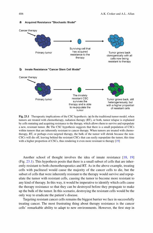
406 A.K. Croker and A.L. Allan
Another school of thought involves the idea of innate resistance [18, 19] (Fig. 23.1). This hypothesis posits that there is a small subset of cells that are inher-ently resistant to both chemotherapeutics and RT. As in the above example, treating cells with paclitaxel would cause the majority of the cancer cells to die, but the subset of cells that were inherently resistant to the therapy would survive and repop-ulate the tumor with resistant cells, causing the tumor to become more resistant to any kind of therapy. In this way, it would be imperative to identify which cells cause the therapy resistance so that they can be destroyed before they propagate to make up the bulk of the tumor. In this scenario, destroying the resistant cells would be the only way to eradicate the patient’s disease.
Targeting resistant cancer cells remains the biggest barrier we face in successfully treating cancer. The most frustrating thing about therapy resistance is the cancer cells’ remarkable ability to adapt to new environments. However, the first step in
Fig. 23.1 Therapeutic implications of the CSC hypothesis. (a) In the traditional tumor model, when tumors are treated with chemotherapy, radiation therapy (RT), or both, tumor relapse is explained by cells mutating and acquiring resistance to the therapy, which allows them to survive and repopulate a new, resistant tumor. (b) The CSC hypothesis suggests that there is a small population of CSCs within tumors that are inherently resistant to cancer therapy. When tumors are treated with chemo-therapy, RT, or perhaps even targeted therapy, the bulk of the tumor will shrink because the non-CSCs will die off, leaving behind the resistant CSCs that can easily repopulate the tumor, this time with a higher proportion of CSCs, thus rendering it even more resistant to therapy [19]
40723 Future Directions: Cancer Stem Cells as Therapeutic Targets
any kind of long and difficult campaign is to identify the proper questions that will lead to useful answers. A good place to start may be asking which cells are the resistant cells in a tumor. If it is possible to know which cells may become and/or are inherently resistant to therapy before the therapy even begins, then perhaps the therapy could be altered, or those resistant cells specifically targeted, in order to sensitize the cancer to cytotoxic therapy, radiation, or targeted therapy. The emer-gence of the cancer stem cell (CSC) hypothesis (predicting that a small subpopulation of “stem-like” cells are responsible for initiating and maintaining cancer growth) may hold promise in terms of new approaches to cancer therapy. This hypothesis also suggests that CSCs may be the resistant cells in a tumor because they retain normal stem cell (SC) self-protection mechanisms. If this is the case, then how can we target these therapy resistant CSCs? Does it matter whether cells are intrinsically resistant or whether this therapy resistance develops over time by natural selection and mutation in response to a particular form of therapy? How can we target multiple drug resistance pathways at the same time? These questions and others will be the focus of this chapter, which will address the therapeutic implications of CSCs in the context of future directions for cancer therapy.
23.2 Therapeutic Implications of CSCs
The CSC hypothesis could have broad therapeutic implications. Since normal SCs are resiliently resistant to many forms of cellular insult including traditional chemo-therapy and radiotherapy, then it would make sense that the CSC population within a tumor would also be at least somewhat inherently resistant to many cancer thera-pies [20, 21]. If this is true, then cancer relapse could easily be explained as follows: When a tumor is initially treated with chemotherapy or radiotherapy, this would cause the bulk of the tumor to shrink, since the non-CSCs which make up the bulk of the tumor would die. However, the treatment would leave behind a small population of resistant CSCs, which could easily repopulate the tumor and be responsible for tumor relapse.
23.2.1 Cytotoxic Therapy
Cancer cells most often become resistant to chemotherapeutic agents by inducing expression of ATP-dependent drug pumps, which actively transport toxic sub-stances out of cells, and can lead to multidrug resistance in many cancer cells [22–24]. There is evidence to suggest that the CSC compartment of many tumors inherently expresses high levels of drug resistance proteins compared with the rest of the cells in the tumor. For example, CD133+ CSCs within glioblastomas (GBM) were shown to be more resistant to chemotherapy, likely due to the high expression of breast cancer resistance protein-1 (BCRP1) and O6-methylguanine DNA
408 A.K. Croker and A.L. Allan
methyltransferase (MGMT) that was observed in the CSC population [23]. In another GBM study, researchers found that the CSCs within the tumor had both increased levels of multidrug resistance protein 1 (MDR1) and increased resis-tance to chemotherapy [24]. In pancreatic tumors, it was observed that CD44+ CSCs were responsible for gemcitabine resistance. Furthermore, CD44+ breast cancer cells have been shown to preferentially survive chemotherapy compared with the non stem-like cancer cells [22]. Interestingly, it has been shown that the CD44 receptor actually interacts with MDR-1, indicating that CD44 itself may actively contribute to drug resistance of CSCs in various tumor types [25].
In addition to a decreased sensitivity to chemotherapeutic agents due to a high expression of drug resistance genes, expression of both Hedgehog (Hh) and Bmi-1 have been identified in breast CSCs [26]. In many types of normal SCs, Hh signaling is essential for promoting SC self-renewal and proliferation. Hh signaling also increases Bmi-1 expression, and Bmi-1 has been shown to play an important role in the regulation of self-renewal of haematopoietic SCs and neuronal SCs [27]. The cytotoxic agent cyclopamine exerts its effect on cancer cells by binding to and inhibiting Smoothened (Smo), which inhibits the growth of tumors with activated Hh signaling [28, 29]. Studies have demonstrated that xenograft tumors resulting from injection of mice with DU-145 and PC-3 prostate cancer cells can be virtually eliminated by treatment with cyclopamine [20, 30]. It is possible that this is a result of the drug being able to inhibit CSC self-renewal, and hence the overall growth of the tumor.
In brain cancer, intracranial GBM and medulloblastoma xenografts treated with a C-X-C chemokine receptor type 4 (CXCR4) antagonist (AMD3100) showed reduced cell growth and increased tumor cell apoptosis [31]. Furthermore, Jin et al. were able to show that by targeting CD44, leukemic stem cells (LSCs) can be eliminated in an acute myeloid leukemia (AML) model. The authors hypothesized that this result was due to interference with transport to SC-supportive microenvironmental niches and/or alteration of CSC fate toward differentiation [32]. Other studies have shown that treatment of prostate and breast cancer cell lines with a siRNA against CD44 can decrease cancer cell adhesion to bone marrow endothelial cells [33]. This could reduce cellular ability to migrate and invade tissues, and further supports the idea of using a CD44 blocker to target CSCs in cancer therapy.
23.2.2 Radiation Therapy
A serious clinical problem associated with fractionated RT is accelerated repopula-tion, or the increase in rate of growth as a result of time between treatments. During accelerated repopulation, each day of a treatment gap reduces the efficacy of RT by about 0.6 Gy, making it one of the major reasons for local failure of RT [34, 35]. This may potentially be explained by the CSC compartment within tumors. CSCs are believed to be resistant to RT by preferentially up-regulating their DNA proofreading mechanisms to avoid cellular death due to DNA damage [35, 36].
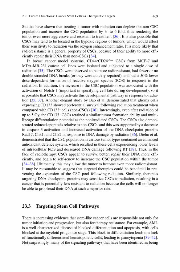
40923 Future Directions: Cancer Stem Cells as Therapeutic Targets
Studies have shown that treating a tumor with radiation can deplete the non-CSC population and increase the CSC population by 3- to 5-fold, thus rendering the tumor even more aggressive and resistant to treatment [36]. It is also possible that CSCs may tend to be located in the hypoxic regions of tumors, which would affect their sensitivity to radiation via the oxygen enhancement ratio. It is more likely that radioresistance is a general property of CSCs, because of their ability to more effi-ciently repair their DNA than non-CSCs [34].
In breast cancer model systems, CD44+CD24–/low CSCs from MCF-7 and MDA-MB-231 cancer cell lines were isolated and subjected to a single dose of radiation [35]. The CSCs were observed to be more radioresistant, had fewer or no double stranded DNA breaks (or they were quickly repaired), and had a 50% lower dose-dependent formation of reactive oxygen species (ROS) in response to the radiation. In addition, the increase in the CSC population was associated with the activation of Notch-1 (important in specifying cell fate during development), so it is possible that CSCs may activate this developmental pathway in response to radia-tion [35, 37]. Another elegant study by Bao et al. demonstrated that glioma cells expressing CD133 showed preferential survival following radiation treatment when compared with CD133− cells (non-CSCs) [36]. Interestingly, even after radiation of up to 5 Gy, the CD133+ CSCs retained a similar tumor formation ability and multi-lineage differentiation potential as the nonirradiated CSCs. The CSCs also demon-strated reduced apoptosis relative to non-CSCs, and this was supported by a decrease in caspase-3 activation and increased activation of the DNA checkpoint proteins Rad17, Chk1, and Chk2 in response to DNA damage by radiation [36]. Diehn et al. demonstrated that the CSC population in various tumor types contained an enhanced antioxidant defence system, which resulted in these cells experiencing lower levels of intracellular ROS and decreased DNA damage following RT [38]. Thus, in the face of radiotherapy, CSCs appear to survive better, repair their DNA more effi-ciently, and begin to self-renew to increase the CSC population within the tumor [34–38]. Ultimately, this may allow the tumor to become even more radioresistant. It may be reasonable to suggest that targeted therapies could be beneficial in pre-venting the expansion of the CSC pool following radiation. Similarly, therapies targeting DNA checkpoint proteins may sensitize CSCs to radiation, resulting in a cancer that is potentially less resistant to radiation because the cells will no longer be able to proofread their DNA at such a superior rate.
23.3 Targeting Stem Cell Pathways
There is increasing evidence that stem-like cancer cells are responsible not only for tumor initiation and progression, but also for therapy resistance. For example, AML is a well-characterized disease of blocked differentiation and apoptosis, with cells blocked at the myeloid progenitor stage. This block in differentiation leads to a lack of functionally differentiated hematopoietic cells, leading to pancytopenia [39–42]. Not surprisingly, many of the signaling pathways that have been identified as being
410 A.K. Croker and A.L. Allan
able to maintain normal hematopoietic stem cells (HSCs) ex vivo have also been identified as being deregulated in leukemia; most notably the Wnt/b-catenin, STAT3 and 5, Notch-1, and myeloid cell leukemia-1 (Mcl-1) signaling pathways [41, 43–45]. If activation of normal SC machinery in cancer cells contributes to or causes therapy resistance by inducing the CSC phenotype, then perhaps by inhibiting the normal SC pathways that are activated in cancer, we might be able to eliminate the CSC population, thereby sensitizing the cancer to conventional chemotherapy and RT. This section will discuss potential pathways that may be targeted for this purpose.
23.3.1 Hedgehog Signaling
Hh signaling is an ancient, highly conserved developmental pathway that has critical functions in embryonic development, particularly in relation to the epithelial-to-mesenchymal (EMT) transition [46]. Upon binding a Hh ligand, Patched homolog-1 (PTCH1) is internalized and inactivated so that the endogenous agonist of Smo accumulates in the cytoplasm and activates Smo [47]. This results in the translocation of Smo from the endosome membrane to the plasma membrane. Smo then activates the intracellular signaling molecule Fused, causing the release of the Gli family of transcription factors (Gli1-3), which can then translocate into the nucleus and acti-vate gene transcription. Transcriptional targets of Gli-1 include genes that control cell adhesion, cell cycle, signal transduction, vascularization, and apoptosis [47] (Fig. 23.2a).
In the adult, Hh signaling is important for maintaining and regulating SCs in many normal tissues [46–48]. For example, in haematopoiesis, Hh signaling has been shown to play a role in the regulation of stem and progenitor cell expansion as evidenced by experiments showing that the loss of Smo impairs HSC self-renewal [48]. Furthermore, downstream Hh signaling leads to entry into the cell cycle, inhi-bition of apoptosis, maintenance of self-renewal, regulation of tissue SC differentia-tion, and modulation of tissue polarity [48]. Sonic Hedgehog (SHH) has been shown to promote proliferation by opposing signals for growth arrest. For example, in retinogenesis, Hh activation accelerates G
1 and G
2 phases of the cell cycle without
affecting the duration of S or M phases, whereas Hh inhibition leads to cells spending more time in the G
1 and G
2 phases [47]. In fact, there is evidence suggesting that
SHH signaling may act upstream of other pathways that regulate SC self-renewal. For example, it was shown that the activation of SHH pathway is required for Notch signaling during retinal development, and SHH signaling has been shown to act upstream of Notch during arterial endothelial cell differentiation in mice [47].
Because of the role that Hh signaling plays in SC self-renewal and proliferation (as well as other normal SC features), it is possible that Hh signaling may be an interesting therapeutic target in many cancers, as blocking Hh signaling may cause a reduction in the CSC population. In fact, it has been estimated that up to 1/3 of all tumors have aberrant Hh signaling, and constitutive activation of the SHH signaling pathway has been shown to lead to some cancers [46]. Moreover, SHH-neutralizing
41123 Future Directions: Cancer Stem Cells as Therapeutic Targets
Fig. 23.2 Stem cell signaling pathways identified in cancer. (a) Hedgehog pathway. Binding of the Hh ligand to the PTCH1 receptor causes Smo to be released from the endosome membrane so that it can be expressed at the plasma membrane. Smo then activates the intracellular signaling molecule fused, causing the release of the Gli family of transcription factors (Gli1-3) which can then translocate to the nucleus and activate gene transcription. (b) Notch pathway. Notch ligands bind to either jagged or delta-like membrane receptors, which leads to cleavage of the receptor by ADAM and g-secretase, resulting in the release of the notch intracellular domain (NICD). The NICD can then translocate to the nucleus where it recruits various co-activators and co-repressors leading to activation of target genes. (c) Wnt/b-catenin pathway. Wnt binds the Frizzled receptor, which causes a signaling cascade ending with b-catenin translocating to the nucleus and activating transcription. (d) C-kit/SCF pathway. When stem cell factor (SCF) binds kit, this causes a homodi-merization of two kit molecules, which in turn, structurally changes the receptor resulting in acti-vation of the kit kinase domain. Phosphorylated tyrosine residues on kit serve as binding sites for various cell signaling proteins, including members of the PI3K/Akt, MAPK and the JAK/STAT pathways [47, 55, 65, 68]
412 A.K. Croker and A.L. Allan
antibodies have been shown to inhibit tumor cell growth while exogenously added SHH ligand was shown to stimulate tumor cell growth [47]. There was also an inter-esting negative relationship with estrogen receptor-a (ERa) and Gli-1 expression, where low levels of Gli-1 were found in breast cancers expressing high levels of ERa [46]. Strongly ERa+ breast cancers are generally not very aggressive, and are usually highly treatable [49]. This suggests that Gli-1 expression in breast cancer may lead to a more aggressive tumor. In fact, in vitro studies have demonstrated that stable transfection of Gli-1 into ERa, estrogen-dependent cell lines increased cell growth in estrogen-deficient medium through induction of cell cycle progression [46]. Furthermore, inhibition of Hh signaling was shown to not only inhibit the expansion of LSCs, but also to induce apoptosis in the LSC compartment [50, 51]. In fact, loss of Smo was able to completely abrogate the transplantability of chronic myeloid leukemia (CML) LSCs; whereas constitutive activation of Smo increased the numbers of LSCs and accelerated leukemic disease [51]. Interestingly, high levels of Smo were found specifically in LSCs and not in normal HSCs, suggesting that inhibiting Smo may be an excellent therapeutic target to eliminate the LSC pool [50]. Dierks et al. found that the loss of Smo in regular hematopoiesis had no significant impact on the regeneration of hematopoiesis except for the nearly complete loss of CD8+ T-cells and reduction in the number of short-term repopulating HSCs; however, the long-term repopulating cells in the bone marrow seem to be independent of Hh signaling [50].
In addition to affecting the CSC population, it seems that Hh signaling can also promote both chemo- and radio-resistance in the CSC compartment of leukemia and various solid tumors [48, 52, 53]. For example, it has been shown that Hh promotes cancer cell survival via Bcl-2 [52]. The Gli-1 and Gli-2 transcription factors actually bind to sequences in the Bcl-2 promoter in epithelial cells and induce transcription. When medulloblastoma cells were treated with cyclopamine, a Hh antagonist, lower levels of Bcl-2 were noted, as well as increased cellular apoptosis [52]. It has also been shown that inhibition of Hh signaling significantly decreased drug resistance in CD34+ LSCs to cytarabine (Ara-C), the chemotherapeutic of choice for many leukemias [48]. Furthermore, SHH signaling not only opposes apoptosis, but also actively promotes multidrug resistance by regulating drug trafficking. Studies done by Sims-Mourtada et al. demonstrated that stimulation of cells with SHH ligand resulted in increased expression of both MDR1 and BCRP, whereas blockade of SHH activation by cyclopamine or a Gli-1 specific siRNA resulted in decreased expression of these transporters [53]. Finally, it seems that Hh signaling also plays a role in cancer metastasis. Feldmann et al. were able to show that Hh-dependent pancreatic tumor cells treated with cyclopamine showed a greater than 500-fold reduction in the number of invading and migrating cells, which correlated to a signifi-cantly lower rate of metastasis in vivo. Furthermore, cyclopamine treatment also caused a reduction in the aldehyde dehydrogenase (ALDH)hi CSC population, and a significant decrease in therapy resistance [54].
It seems that targeting the Hh pathway may provide therapeutic benefit on many levels. First, inhibiting Hh signaling can decrease or even kill the CSC population within both solid tumors and leukemia [46, 47, 50, 51]. In doing so, these cancers may become more sensitive to therapy simply due to the decrease in the number of
41323 Future Directions: Cancer Stem Cells as Therapeutic Targets
stem-like resistant cancer cells. Furthermore, Hh signaling has been shown to actively promote therapy resistance by increasing expressing of drug resistance proteins, so inhibiting this pathway may, again, enhance the effectiveness of current chemotherapeutics [48, 50–53]. Finally, the role that Hh signaling plays in cancer metastasis is interesting and important since all our current cancer therapies fail in the metastatic setting [54]. In this way, Hh signaling inhibitors may prove to be extremely valuable players in combination with conventional therapies to eradicate many cancers.
23.3.2 Notch Signaling
The Notch signaling pathway is another interesting therapeutic target because of the role it plays in proliferation, apoptosis, and maintenance of the SC state in many normal tissues [55, 56]. Ligands bind to either Jagged or Delta-like membrane receptors, which leads to cleavage of the receptor. This results in the release of the Notch intracellular domain (NICD), which translocates to the nucleus where it recruits various coactivators and corepressors leading to activation of target genes, and finally, degradation of the Notch IC domain [55] (Fig. 23.2b). Interestingly, it has been shown that the inappropriate activation of Notch results in signals that stimulate proliferation, restrict differentiation, and prevent apoptosis, potentially leading to cancer [57]. Further studies actually demonstrated that Notch signaling plays a critical role in GBM CSC self-renewal mediated by endothelial cells, and inhibiting Notch signaling results in a significant reduction in not only the number of CSCs, but also decreass their tumorigenicity [55]. Furthermore, inhibition of Notch-1 or Notch-2 inhibited xenograft tumor formation of GBM cells [56]. Notch signaling has also been shown to play a role in radioresistance and in promoting a hypoxia-resistant phenotype [56–58], and it has been shown that g-secretase inhibitors (GSIs) (which inhibit Notch signaling) impair cell growth and survival after radiation [56]. Interestingly, Notch does not protect tumor cells from radiation by altering the DNA damage response, but instead by promoting radioresistance through the prosurvival regulation of PI3K/Akt and Mcl-1 [56].
In breast cancer, there is an interesting relationship between Notch and HER2 expression, where HER2-overexpressing cells display activated Notch signaling, and inhibition of Notch signaling using siRNA or a GSI results in down-regulation of HER2 expression [59, 60]. This also results in decreased tumorsphere-forming ability, suggesting that inhibiting Notch and down-regulating HER2 decreases the CSC population [60]. This would make sense since it has been shown that HER2 over-expression increases the stem/progenitor population of both normal and malig-nant mammary cells [61]. In this study, Korkaya et al. found that HER2 over-expression increased the normal mammary SC pool and caused a 4- to 5-fold increase in ALDH+ breast cancer cells (CSCs). Furthermore, HER2 over-expression increased the expression of numerous SC-related genes (i.e., Oct3/4, Notch1/2, Jag1, and Gli1). Finally, Herceptin, which is used to successfully treat HER2 over-expressing tumors, reduced the ALDH+ CSC population by approximately 50% [61].
414 A.K. Croker and A.L. Allan
23.3.3 Wnt/b-Catenin Signaling
The Wnt/b-catenin pathway plays a well characterized role during embryogenesis, and is implicated in the survival of normal SCs [62–65]. Similarly, the Wnt/b-catenin pathway has been shown to play a role in maintaining the CSC population in CML, as well as a variety of solid tumors including breast, melanoma, colon, and liver [62, 63]. In mouse mammary progenitor cells, treatment with ionizing radiation caused an increase in the progenitor pool, a phenomena that was enhanced by b-catenin stabilization in these cells. Furthermore, the radioresistance was mediated through the Wnt/b-catenin pathway, indicating that this pathway may also be involved in radioresistance [64]. It has also been shown that activated Wnt/b-catenin in lung adenocarcinoma enhanced tumor cell proliferation, clone formation, drug resistance, and the up-regulation of Oct-4, a primitive SC marker [63] (Fig. 23.2c).
23.3.4 Snail/Slug Signaling
Snail and Slug are zinc-finger transcription factors that have been shown to play an important role in wound healing, SC protection from DNA damage, and regulation of EMT [66]. The stem cell factor (SCF) and c-Kit pathways, which are essential for the formation of HSCs and other SCs during embryonic development, have been shown to activate Slug [67, 68]. In this way, the SCF/c-Kit/Slug pathway was shown to be absolutely necessary for SC survival in the bone marrow following lethal irradiation in a mouse model [67]. Similarly, Slug has been shown to mediate radio-protection and enhance survival of progenitor cells in ovarian cancer through acti-vation of the SCF/c-Kit pathways, and an increase in both Snail and Slug expression is seen in ovarian cells treated with chemotherapy, indicating that these proteins may be involved in chemoprotection of these cells [66]. Further evidence of this was seen in studies done by Catalano et al., which demonstrated that the SCF/c-Kit/Slug pathway actively mediates multidrug resistance in malignant mesothelioma cells [14]. When c-Kit expression was knocked down, this increased tumor cell sensitivity to various chemotherapeutic agents in multidrug resistant sublines, and forced expression of SCF/c-Kit signaling in a way that was sufficient to lead to multidrug resistance in parental lines. These processes were shown to be mediated by Slug [14] (Fig. 23.2d).
23.4 Differentiation Therapy
If it is true that the CSC pool within a tumor is responsible for therapy resistance and that this is because of the cells’ inherent SC nature, then the idea of differentia-tion therapy is an interesting one. Differentiation therapy would not kill the CSCs directly, but it does have the potential to restrain their self-renewal capacity, and perhaps increase the efficacy of more conventional therapies (such as chemotherapy),
41523 Future Directions: Cancer Stem Cells as Therapeutic Targets
which are often most effective in differentiated cells. Furthermore, differentiation agents often have less toxicity than conventional cancer treatments [69, 70].
23.4.1 Retinoid Signaling and Differentiation Therapy
The retinoic acid receptor (RAR) signaling pathway is a well-characterized differ-entiation pathway in many developmental systems, although it is best described in the hematopoietic system [44, 45, 71–73]. Briefly, aldehyde dehyogenase (ALDH) catalyzes the reaction of retinol to retinoic acid (RA), which then binds to a RAR (usually RARa or RARb). RA binding causes a conformational change to the RAR, which facilitates the release of a repressor protein and binding of an enhancer pro-tein (i.e., C/EBPa and C/EBPb), and allows transcription of RARa/b and targeting of differentiation proteins [74–80] (Fig. 23.3). The transcription factor acute myel-ogenous leukemia-2 (AML2) has also been shown to play a role in HSC differentia-tion and is regulated by RA through RARa-dependent signaling [81].
Blockage of this pathway using either RAR or retinoic X receptor (RXR) inhibi-tors has been shown to inhibit HSC differentiation [75, 78, 82]. Furthermore, the inhibition of ALDH has been shown to delay the differentiation of HSCs, probably by decreasing the amount of available RA [83, 84], although it has also been shown that ALDH can maintain CD34+ HSC activity and prevent apoptosis through RA signaling [74]. This may have something to do with retinoic acid receptor-g (RARg), a member of the RAR family (commonly involved in SC differentiation), as it has been shown to be important in maintaining the HSC phenotype. In fact, when RARg was silenced, it led to a reduction of HSCs in vivo [84, 85]. RA is not isotype spe-cific, so it will bind to RARa, RARb, and RARg with the same ease [86]. In this way, the type and number of RARs expressed in specific cells may contribute to how the cells respond to RA. This demonstrates the complicated nature of retinoic signaling and highlights the importance of gaining a better understanding of this process.
23.4.2 Acute Promyelocytic Leukemia (APL): The Differentiation Therapy Success Story
Many AMLs are initiated by translocation events that fuse proteins involved in cell differentiation, apoptosis/cell survival, cell cycle control, and DNA-binding pro-teins [87–90]. The AML-associated translocation products (AATPs) have been shown to activate Wnt signaling by increasing g-catenin expression, leading to SC self-renewal and accelerated cell cycle progression [90], indicating that these fusion proteins may work to give AML cells their blocked differentiation phenotype. Further support of this is found in the observation that many of the AATPs involve RARa (i.e., PML:RARa and PLZF:RARa), which plays a major role in myeloid differentiation of HSCs [91–94].
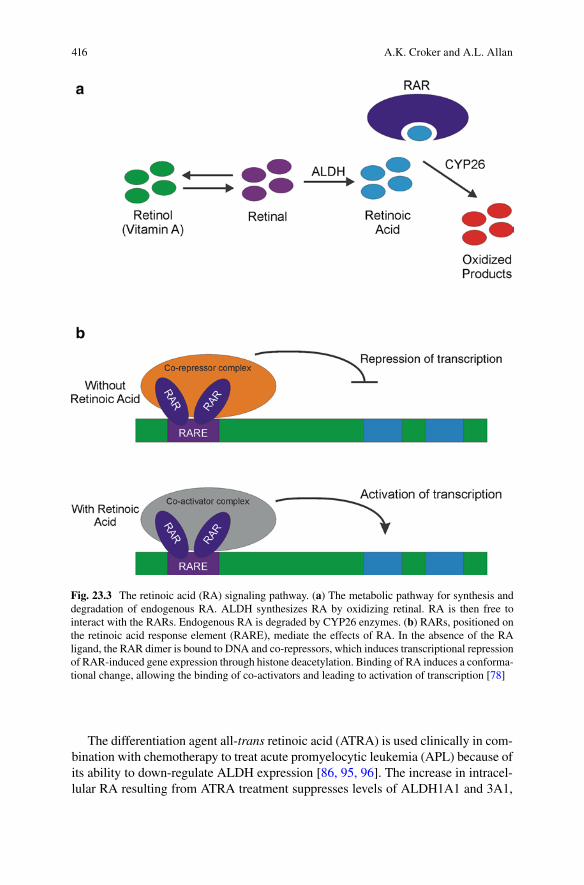
416 A.K. Croker and A.L. Allan
Fig. 23.3 The retinoic acid (RA) signaling pathway. (a) The metabolic pathway for synthesis and degradation of endogenous RA. ALDH synthesizes RA by oxidizing retinal. RA is then free to interact with the RARs. Endogenous RA is degraded by CYP26 enzymes. (b) RARs, positioned on the retinoic acid response element (RARE), mediate the effects of RA. In the absence of the RA ligand, the RAR dimer is bound to DNA and co-repressors, which induces transcriptional repression of RAR-induced gene expression through histone deacetylation. Binding of RA induces a conforma-tional change, allowing the binding of co-activators and leading to activation of transcription [78]
The differentiation agent all-trans retinoic acid (ATRA) is used clinically in com-bination with chemotherapy to treat acute promyelocytic leukemia (APL) because of its ability to down-regulate ALDH expression [86, 95, 96]. The increase in intracel-lular RA resulting from ATRA treatment suppresses levels of ALDH1A1 and 3A1,
41723 Future Directions: Cancer Stem Cells as Therapeutic Targets
driving differentiation of the malignant promyelocytes into mature neutrophils and causing enhanced sensitivity to chemotherapy [97, 98]. PML:RARa AML cells exert their pathogenic activity by recruiting histone deacetylases through nuclear receptor corepressor (N-CoR) and silencing mediator for retinoid and thyroid hormone receptor (SMRT), activating the cyclin-dependent kinase activating kinase (CAK) complex to hyperphosphorylate RARa, and blocking terminal differentiation and apoptosis [99, 100]. Treatment with ATRA induces ubiquitination-proteolysis of Menage-a-trois-1 (MAT-1), which results in a decrease of CAK-induced phosphory-lation of RARa, promoting granulocytic differentiation [76, 100]. ATRA has there-fore been used to treat these patients with great success [101–103].
Given that RARa plays such an important role in normal myeloid cell differen-tiation, it would make sense to hypothesize that ATRA therapy should work across all AMLs to differentiate malignant myeloid cells. However, in reality, this is not the case. All non-PML:RARa AMLs are insensitive to ATRA treatment, including PLZF:RARa, whose AATP actually involves the RARa [104–107]. It was originally assumed that this outcome may have been due to mutations in RARa that abolished the RA binding site, but studies investigating RARa in hundreds of AML cell lines and patient samples found that not only were RARa mutations extremely infrequent, but also RARa hypermethylation was also rare, indicating that most RARa should be fully functional in leukemic disease [108]. It is possible that instead of an RARa culprit, there is instead an upregulation of RARg in AML patients. It has been shown that RARg can interact with p85 (phosphoinositol-3-kinase [PI3K] signaling), leading to activation of Akt and NFkB, and playing a critical role in self-renewal and cell survival [109]. ATRA is not isotype specific, so it will bind RARa, RARb, and RARg at the same rate [86]. However, it is currently unclear why ATRA can only successfully differentiate PML:RARa AML cells.
23.4.3 Differentiation Therapy in Solid Tumors
Much less is known about the effect of differentiation therapy in solid tumors; however, there is considerable interest in ATRA treatment in breast cancer because of the way ATRA can inhibit cell growth. When MCF-7 breast cancer cells were treated with ATRA, cells accumulated in G
1 phase and by day 10, approximately
50% of the MCF-7 cells had died [110]. Interestingly, the ATRA-mediated growth inhibition in breast cancer cells correlated with the presence of functional estrogen receptors, and the ATRA actions were enhanced by the use of Tamoxifen. Furthermore, studies by Ginestier et al. show that when breast cancer cells are treated with ATRA, the cells demonstrated a decrease in both primary and second-ary tumorsphere formation, whereas cells treated with diethylaminobenzaldehyde (DEAB, which specifically blocks ALDH activity, thus potentially blocking the pro-duction of RA) had an increase in both primary and secondary tumorsphere forma-tion [111]. This indicates that activation of RA signaling decreases the CSC population, perhaps by “differentiating” the CSCs. In support of this, it was shown
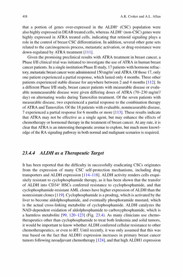
418 A.K. Croker and A.L. Allan
that a portion of genes over-expressed in the ALDH+ (CSC) population were also highly expressed in DEAB treated cells, whereas ALDH− (non-CSC) genes were highly expressed in ATRA treated cells, indicating that retinoid signaling plays a role in the control of breast CSC differentiation. In addition, several other gene sets related to the carcinogenesis process, metastatic activation, or drug-resistance were down-regulated by ATRA treatment [111].
Given the promising preclinical results with ATRA treatment in breast cancer, a Phase I/II clinical trial was initiated to investigate the use of ATRA in human breast cancer patients. In a single institution Phase II study, 17 patients with hormone refrac-tory, metastatic breast cancer were administered 150 mg/m2 oral ATRA. Of those 17, only one patient experienced a partial response, which lasted only 4 months. Three other patients experienced stable disease for anywhere between 2 and 4 months [112]. In a different Phase I/II study, breast cancer patients with measurable disease or evalu-able nonmeasurable disease were given differing doses of ATRA (70–230 mg/m2/day) on alternating weeks during Tamoxifen treatment. Of the seven patients with measurable disease, two experienced a partial response to the combination therapy of ATRA and Tamoxifen. Of the 18 patients with evaluable, nonmeasurable disease, 7 experienced a partial response for 6 months or more [113]. These results indicate that ATRA may not be effective as a single agent, but may enhance the effects of chemotherapy or hormonal therapy in the treatment of breast cancer. At any rate, it is clear that ATRA is an interesting therapeutic avenue to explore, but much more knowl-edge of the RA signaling pathway in both normal and malignant scenarios is required.
23.4.4 ALDH as a Therapeutic Target
It has been reported that the difficulty in successfully eradicating CSCs originates from the expression of many CSC self-protection mechanisms, including drug transporters and ALDH expression [114–118]. ALDH activity renders cells exqui-sitely resistant to cyclophosphamide therapy, as it has been shown that the transfer of ALDH into CD34+ HSCs conferred resistance to cyclophosphamide, and that cyclophosphamide-resistant AML clones have higher expression of ALDH than the nonresistant clones [119]. Cyclophosphamide is a prodrug, which is activated by the liver to become aldolphosphamide, and eventually phosphoramide mustard, which is the actual cross-linking metabolite of cyclophosphamide. ALDH catalyzes the NAD-dependent oxidation of aldolphosphamide to carboxyphosphamide, which is a harmless metabolite [99, 120–123] (Fig. 23.4). As many clinicians use chemo-therapeutics other than cyclophosphamide to treat both leukemia and solid tumors, it would be important to know whether ALDH conferred cellular resistance to other chemotherapeutics, or even to RT. Until recently, it was only assumed that this was true based on the fact that ALDH1 expression increases in primary breast cancer tumors following neoadjuvant chemotherapy [124], and that high ALDH1 expression
41923 Future Directions: Cancer Stem Cells as Therapeutic Targets
correlates with poor patient outcome [125]. Recently, however, studies performed in our lab [126] have shown that when resistant ALDHhiCD44+ breast cancer cells are pretreated with DEAB (to block ALDH activity) or ATRA (to indirectly down-regulate ALDH), a significant initial sensitization to doxorubicin, paclitaxel, and RT was observed, in many cases to a level equivalent to that of the nonresistant ALDHlowCD44− cells. However, only DEAB pretreatment was able to also reduce the long-term regrowth/colony-forming ability of chemotherapy- or radiation-treated ALDHhiCD44+ cells, whereas cells pretreated with ATRA were able to regrow just as well as non-ATRA treated cells. These results indicate that specifi-cally blocking ALDH is key for sensitizing resistant ALDHhiCD44+ cells to therapy (Fig. 23.4). As discussed earlier, ATRA can down-regulate ALDH through an indi-rect route, but in our breast cancer cells, we saw that this ALDH down-regulation was short-lived as cells began re-expressing ALDH as early as 48 h following treat-ment. Therefore, a more direct targeted approach, such as DEAB inhibition of ALDH or ALDH-specific siRNA, may be necessary [126].
Fig. 23.4 Inhibition of ALDH as a strategy for overcoming treatment resistance. When cells are treated with ATRA, this increases the levels of exogenous RA, which in turn causes ALDH down-regulation by decreasing the amount of the RARa and C/EBPb transcription factor complex. (a) This decrease in ALDH causes a direct sensitization of cells to cyclophosphamide. Cyclophosphamide is a pro-drug, which is activated by the liver to become aldolphosphamide, and eventually phosphoramide mustard, which is the actual cross-linking metabolite of cyclophosph-amide. ALDH catalyzes the NAD-dependent oxidation of aldolphosphamide to carboxy-phosphamide, which is a harmless metabolite. (b and c) A decrease in ALDH causes a sensitization of breast cancer cells to both taxanes and anthracyclines (b) and radiation (c) by an as yet unknown mechanism [97, 98, 126]
420 A.K. Croker and A.L. Allan
23.5 Concluding Remarks
It is true that there have been giant leaps forward in designing and testing successful novel cancer therapeutics that target the disease more successfully, and even (in some cases) with fewer side effects. It is clear that the more we learn about the biology of both solid tumors and leukemia, the better and more targeted our therapies will become. Half the battle in combating cancer is known which cell(s) to target, since there is usually great heterogeneity within cancers. More and more evidence is accu-mulating to support the idea that CSCs play a major role in not only initiating and sustaining primary tumors, but also in facilitating metastasis to distant organs [125, 127–137]. In this way, it is logical to assume that these CSCs might make excellent therapeutic targets since, if we can get rid of the CSC population, then we could theo-retically be able to get rid of the tumor. However, targeting the CSC population using conventional therapies has proven extremely challenging since it has been shown that CSCs preferentially survive both chemotherapy and/or RT compared to their non-CSC counterparts [20, 21, 24, 35, 36, 38, 126, 138]. Interestingly, normal SCs are also highly capable of protecting themselves from cellular insult, much more so than their differentiated progeny, via a number of different mechanisms including up-regulating multidrug resistance pumps, DNA protection mechanisms, and even just via their quiescent nature [21]. In this way, perhaps by amalgamating what we know of normal SC biology and applying it to the cancer field, we will be better poised to tackle the problem of therapy resistance.
This has led to work investigating whether blocking either normal SC properties or pathways could lead to CSC sensitization to therapy, and this has been met with moder-ate success. Drugs have been developed to target different stages of multiple SC path-ways including Notch [55–58], Hh [46, 52, 54], Wnt/b-catenin [62–64], and c-kit/SCF/Snail/Slug signaling [14, 66]. Drugs that target SC self-protection mechanisms have also been used with some interest, including drugs that target MDR-1, BCRP, and ALDH to try and sensitize tumors to conventional therapy [20, 23, 24, 47, 50, 51, 53, 115]. Finally, the idea of “differentiation therapy” has been used to successfully treat APL patients by differentiating the LSC population, thereby rendering the disease more sensitive to conventional chemotherapy (cytarabine) [48, 86, 96]. Further work is needed to determine whether differentiation therapy will be as successful in solid tumors.
Although focusing in on the CSC population is a huge leap forward in identifying the cells that need to be targeted in cancer, it is important not to forget that the CSC population itself may, in fact, be a heterogeneous population. Therefore, it will be cru-cial to better study and understand the true nature of the CSC population. By doing this, we will be able to better answer important questions about whether there is a true CSC hierarchy within cancers, with some of the most primitive cancer cells, quiescent and elusive, maintaining the cancer and surviving the cancer therapies. Furthermore, are all CSCs inherently resistant to cancer therapy, and are all CSCs equally able to metasta-size? Interesting work done by Hermann et al. found that only a small subpopulation of pancreatic CSCs (with a CD133+CXCR4+ phenotype) was able to successfully metastasize, and that if CXCR4 was blocked, the cells were no longer able to metastasize [131]. This kind of research has enormously implications for cancer therapy.
42123 Future Directions: Cancer Stem Cells as Therapeutic Targets
For pathologists and clinicians, it would be very interesting if, along with normal staging and tumor analysis, the CSC population could also be analyzed to identify the best possible therapeutic course of action. If it is true that tumors are driven by a small population of CSCs, then treating the tumor based on the characteristics of the CSC population, rather than the characteristics of the bulk of the tumor, would be more helpful in successfully eradicating the disease. By analyzing CSCs, it would also be easier to identify which SC signaling pathways (if any) are activated, which may also help guide therapeutic decisions.
We live in a very exciting time where identifying novel cancer therapeutic targets and drugs is encouraged and supported [15]. With this freedom and support, how-ever, it is essential that impartiality be maintained in order for scientists to conduct good scientific research. Millions to billions of dollars are invested in each new drug that makes its way through clinical trials. Without carefully designed preclinical and Phase I/II trials, the likelihood of a drug being successful in huge, expensive Phase III clinical trials is not very high. Currently, there are more Phase III trials that fail than succeed, and it seems that many compounds are being recycled instead of new compounds developed. Although it is difficult to let go of a favorite compound, if it has failed to show benefit in a clinical trial, it is imperative to move on. If not, targets that are not going to be effective will continue to be pushed down the pipeline, and the results will inevitably be disappointing. This current approach negatively affects not only just the bottom line, but also the patients who are impatiently waiting on a therapy that will successfully treat their cancer.
Over the short term, agents that target/inhibit SC signaling pathways combined with conventional chemotherapy and RT will probably be our most effective means of battling cancer. Blocking the SC properties of the CSC population would, in theory, make the cells more sensitive to conventional therapy. As we learn more about both normal SCs and CSCs, and how they avoid or resist our therapies, we will be better suited to identify more useful therapeutic targets. In this way, we are heading toward a time when we have therapies that will make cancer a chronic disease, rather than an acute, life-threatening disease.
Acknowledgments We thank members of our laboratory and our collaborators for their research work and helpful discussions. The authors’ research on CSCs is supported by research grants from the Ontario Institute for Cancer Research (#08NOV230), and the Canada Foundation for Innovation (#13199) (to ALA). AKC is the recipient of a scholarship from the Canadian Institute of Health Research (CIHR). ALA is supported by a CIHR New Investigator Award and an Early Researcher Award from the Ontario Ministry of Research and Innovation.
References
1. Jemal A, Siegel R, Xu J, Ward E (2010) Cancer Statistics, 2010. CA: a cancer journal for clinicians 60:277–300
2. Clarke M, Collins R, Darby S, Davies C, Elphinstone P, Evans E, Godwin J, Gray R, Hicks C, James S, MacKinnon E, McGale P, McHugh T, Peto R, Taylor C, Wang Y (2005) Effects of radiotherapy and of differences in the extent of surgery for early breast cancer on local recur-rence and 15-year survival: an overview of the randomised trials. Lancet 366:2087–2106
422 A.K. Croker and A.L. Allan
3. Cole BF, Gelber RD, Gelber S, Coates AS, Goldhirsch A (2001) Polychemotherapy for early breast cancer: an overview of the randomised clinical trials with quality-adjusted survival analysis. Lancet 358:277–286
4. Slamon DJ, Leyland-Jones B, Shak S, Fuchs H, Paton V, Bajamonde A, Fleming T, Eiermann W, Wolter J, Pegram M, Baselga J, Norton L (2001) Use of chemotherapy plus a monoclonal antibody against HER2 for metastatic breast cancer that overexpresses HER2. The New England journal of medicine 344:783–792
5. Hurwitz H, Fehrenbacher L, Novotny W, Cartwright T, Hainsworth J, Heim W, Berlin J, Baron A, Griffing S, Holmgren E, Ferrara N, Fyfe G, Rogers B, Ross R, Kabbinavar F (2004) Bevacizumab plus irinotecan, fluorouracil, and leucovorin for metastatic colorectal cancer. The New England journal of medicine 350:2335–2342
6. Sandler A, Gray R, Perry MC, Brahmer J, Schiller JH, Dowlati A, Lilenbaum R, Johnson DH (2006) Paclitaxel-carboplatin alone or with bevacizumab for non-small-cell lung cancer. The New England journal of medicine 355:2542–2550
7. Thatcher N, Chang A, Parikh P, {Rodrigues Pereira} J, Ciuleanu T, von Pawel J, Thongprasert S, Tan EH, Pemberton K, Archer V, Carroll K (2005) Gefitinib plus best supportive care in previously treated patients with refractory advanced non-small-cell lung cancer: results from a randomised, placebo-controlled, multicentre study (Iressa Survival Evaluation in Lung Cancer). Lancet 366:1527–1537
8. Cameron D, Casey M, Press M, Lindquist D, Pienkowski T, Romieu CG, Chan S, Jagiello-Gruszfeld A, Kaufman B, Crown J, Chan A, Campone M, Viens P, Davidson N, Gorbounova V, Raats JI, Skarlos D, Newstat B, Roychowdhury D, Paoletti P, Oliva C, Rubin S, Stein S, Geyer CE (2008) A phase III randomized comparison of lapatinib plus capecitabine versus capecit-abine alone in women with advanced breast cancer that has progressed on trastuzumab: updated efficacy and biomarker analyses. Breast cancer research and treatment 112:533–543
9. Xia W, Mullin RJ, Keith BR, Liu L-H, Ma H, Rusnak DW, Owens G, Alligood KJ, Spector NL (2002) Anti-tumor activity of GW572016: a dual tyrosine kinase inhibitor blocks EGF activa-tion of EGFR/erbB2 and downstream Erk1/2 and AKT pathways. Oncogene 21:6255–6263
10. Druker BJ TS, Buchdunger E, Ohno S, Segal GM, Fanning S, Zimmermann J, Lydon NB. (1996) Effects of a selective inhibitor of the Abl tyrosine kinase on the growth of Bcr-Abl posi-tive cells. Nature medicine 2 (5):561–566
11. Sawyers CL HA, Feldman E, Goldman JM, Miller CB, Ottmann OG, Schiffer CA, Talpaz M, Guilhot F, Deininger MW, Fischer T, O’Brien SG, Stone RM, Gambacorti-Passerini CB, Russell NH, Reiffers JJ, Shea TC, Chapuis B, Coutre S, Tura S, Morra E, Larson RA, Saven A, Peschel C, Gratwohl A, Mandelli F, Ben-Am M, Gathmann I, Capdeville R, Paquette RL, Druker BJ. (2002) Imatinib induces hematologic and cytogenetic responses in patients with chronic myelogenous leukemia in myeloid blast crisis: results of a phase II study. Blood 99 (10):3530–3539
12. Holtz MS SM, Zhang F, Sawyers CL, Forman SJ, Bhatia R (2002) Imatinib mesylate (STI571) inhibits growth of primitive malignant progenitors in chronic myelogenous leukemia through reversal of abnormally increased proliferation. Blood 99 (10):3792–3800
13. Pusztai L, Hortobagyi GN (1998) High-dose chemotherapy: how resistant is breast cancer? Drug resistance updates: reviews and commentaries in antimicrobial and anticancer chemo-therapy 1:62–72
14. Catalano A, Rodilossi S, Rippo MR, Caprari P, Procopio A (2004) Induction of stem cell factor/c-Kit/slug signal transduction in multidrug-resistant malignant mesothelioma cells. The Journal of biological chemistry 279:46706–46714
15. Chabner BA, Jr TGR (2005) Chemotherapy and the war on cancer. Nature Reviews Cancer 5:65–72
16. Gerlinger M, Swanton C (2010) How Darwinian models inform therapeutic failure initiated by clonal heterogeneity in cancer medicine. British journal of cancer 103:1139–1143
17. Fornari F, Randolph J, Yalowich J, Ritke M, Gewirtz D (1994) Interference by doxorubicin with DNA unwinding in MCF-7 breast tumor cells. Molecular Pharmacology 45 (4):649–656
42323 Future Directions: Cancer Stem Cells as Therapeutic Targets
18. Eyler CE, Rich JN (2008) Survival of the Fittest: Cancer Stem Cells in Therapeutic Resistance and Angiogenesis. Journal of clinical oncology: official journal of the American Society of Clinical Oncology 26 (17):2839–2845
19. Croker AK, Allan AL (2007) Cancer stem cells: implications for the progression and treatment of metastatic disease. Journal of cellular and molecular medicine 12 (2):374–390
20. Lou H, Dean M (2007) Targeted therapy for cancer stem cells: the patched pathway and ABC transporters. Oncogene 26:1357–1360
21. Kvinlaug BT, Huntly BJ (2007) Targeting Cancer Stem Cells. Expert opinion on therapeutic targets 11 (7):915–927
22. Hong SP, Wen J, Bang S, Park S, Song SY (2009) CD44-positive cells are responsible for gemcitabine resistance in pancreatic cancer cells. International journal of cancer Journal inter-national du cancer 125:2323–2331
23. Liu G, Yuan X, Zeng Z, Tunici P, Ng H, Abdulkadir IR, Lu L, Irvin D, Black KL, Yu JS (2006) Analysis of gene expression and chemoresistance of CD133+ cancer stem cells in glioblastoma. Molecular cancer 5:67
24. Nakai E, Park K, Yawata T, Chihara T, Kumazawa A, Nakabayashi H, Shimizu K (2009) Enhanced MDR1 expression and chemoresistance of cancer stem cells derived from glioblas-toma. Cancer investigation 27:901–908
25. Miletti-González KE, Chen S, Muthukumaran N, Saglimbeni GN, Wu X, Yang J, Apolito K, Shih WJ, Hait WN, Rodríguez-Rodríguez L (2005) The CD44 receptor interacts with P-glycoprotein to promote cell migration and invasion in cancer. Cancer research 65:6660–6667
26. Liu S, Dontu G, Mantle ID, Patel S, Ahn N-s, Jackson KW, Suri P, Wicha MS (2006) Hedgehog signaling and Bmi-1 regulate self-renewal of normal and malignant human mammary stem cells. Cancer research 66:6063–6071
27. Park I-K, Morrison SJ, Clarke MF (2004) Bmi1, stem cells, and senescence regulation. The Journal of clinical investigation 113:175–179
28. Berman DM, Karhadkar SS, Maitra A, {Montes De Oca} R, Gerstenblith MR, Briggs K, Parker AR, Shimada Y, Eshleman JR, Watkins DN, Beachy PA (2003) Widespread requirement for Hedgehog ligand stimulation in growth of digestive tract tumours. Nature 425:846–851
29. Thayer SP, di Magliano MP, Heiser PW, Nielsen CM, Roberts DJ, Lauwers GY, Qi YP, Gysin S, {Fernández-del Castillo} C, Yajnik V, Antoniu B, McMahon M, Warshaw AL, Hebrok M (2003) Hedgehog is an early and late mediator of pancreatic cancer tumorigenesis. Nature 425:851–856
30. Karhadkar SS, Bova GS, Abdallah N, Dhara S, Gardner D, Maitra A, Isaacs JT, Berman DM, Beachy PA (2004) Hedgehog signalling in prostate regeneration, neoplasia and metastasis. Nature 431:707–712
31. Rubin JB, Kung AL, Klein RS, Chan JA, Sun Y, Schmidt K, Kieran MW, Luster AD, Segal RA (2003) A small-molecule antagonist of CXCR4 inhibits intracranial growth of primary brain tumors. Proceedings of the National Academy of Sciences of the United States of America 100:13513–13518
32. Jin L, Hope KJ, Zhai Q, Smadja-Joffe F, Dick JE (2006) Targeting of CD44 eradicates human acute myeloid leukemic stem cells. Nature medicine 12:1167–1174
33. Draffin JE, McFarlane S, Hill A, Johnston PG, Waugh DJJ (2004) CD44 potentiates the adherence of metastatic prostate and breast cancer cells to bone marrow endothelial cells. Cancer research 64:5702–5711
34. Diehn M, Clarke MF (2006) Cancer stem cells and radiotherapy: new insights into tumor radioresistance. Journal of the National Cancer Institute 98:1755–1757
35. Phillips TM, McBride WH, Pajonk F (2006) The response of CD24(−/low)/CD44+ breast cancer-initiating cells to radiation. Journal of the National Cancer Institute 98:1777–1785
36. Bao S, Wu Q, McLendon RE, Hao Y, Shi Q, Hjelmeland AB, Dewhirst MW, Bigner DD, Rich JN (2006) Glioma stem cells promote radioresistance by preferential activation of the DNA damage response. Nature 444:756–760
37. Politi K, Feirt N, Kitajewski J (2004) Notch in mammary gland development and breast cancer. Seminars in cancer biology 14:341–347
424 A.K. Croker and A.L. Allan
38. Diehn M, Cho RW, Lobo NA, Kalisky T, Dorie MJ, Kulp AN, Qian D, Lam JS, Ailles LE, Wong M, Joshua B, Kaplan MJ, Wapnir I, Dirbas FM, Somlo G, Garberoglio C, Paz B, Shen J, Lau SK, Quake SR, Brown JM, Weissman IL, Clarke MF (2009) Association of reactive oxygen species levels and radioresistance in cancer stem cells. Nature 458:780–783
39. Glasow A, Barrett A, Petrie K, Gupta R, Boix-Chornet M, Zhou D-C, Grimwade D, Gallagher R, von Lindern M, Waxman S, Enver T, Hildebrandt G, Zelent A (2008) DNA methylation-independent loss of RARA gene expression in acute myeloid leukemia. Blood 111:2374–2377
40. Puccetti E, Ruthardt M (2004) Acute promyelocytic leukemia: PML/RARalpha and the leukemic stem cell. Leukemia: official journal of the Leukemia Society of America, Leukemia Research Fund, UK 18:1169–1175
41. Spiekermann K, Pau M, Schwab R, Schmieja K, Franzrahe S, Hiddemann W (2002) Constitutive activation of STAT3 and STAT5 is induced by leukemic fusion proteins with protein tyrosine kinase activity and is sufficient for transformation of hematopoietic precursor cells. Experimental hematology 30:262–271
42. Vianello F, Villanova F, Tisato V, Lymperi S, Ho K-K, Gomes AR, Marin D, Bonnet D, Apperley J, Lam EW-F, Dazzi F (2010) Bone marrow mesenchymal stromal cells non-selec-tively protect chronic myeloid leukemia cells from imatinib-induced apoptosis via the CXCR4/CXCL12 axis. Haematologica 95:1081–1089
43. Bonnet D (2005) Normal and leukaemic stem cells. British journal of haematology 130:469–479 44. Campbell CJV, Lee JB, Levadoux-Martin M, Wynder T, Xenocostas A, Leber B, Bhatia M
(2010) The human stem cell hierarchy is defined by a functional dependence on Mcl-1 for self-renewal capacity. Blood 116(9):1433–42
45. Snow GE, Kasper AC, Busch AM, Schwarz E, Ewings KE, Bee T, Spinella MJ, Dmitrovsky E, Freemantle SJ (2009) Wnt pathway reprogramming during human embryonal carcinoma differentiation and potential for therapeutic targeting. BMC cancer 9:383
46. O’Toole SA, Swarbrick A, Sutherland RL (2009) The Hedgehog signalling pathway as a therapeu-tic target in early breast cancer development. Expert opinion on therapeutic targets 13:1095–1103
47. Chen Y-J, Sims-Mourtada J, Izzo J, Chao KSC (2007) Targeting the hedgehog pathway to mitigate treatment resistance. Cell cycle (Georgetown, Tex) 6:1826–1830
48. Kobune M, Takimoto R, Murase K, Iyama S, Sato T, Kikuchi S, Kawano Y, Miyanishi K, Sato Y, Niitsu Y, Kato J (2009) Drug resistance is dramatically restored by hedgehog inhibitors in CD34+ leukemic cells. Cancer science 100:948–955
49. Foulkes WD, Smith IE, Reis-Filho JS (2010) Triple-negative breast cancer. The New England journal of medicine 363 (20):1938–1948
50. Dierks C, Beigi R, Guo G-R, Zirlik K, Stegert MR, Manley P, Trussell C, Schmitt-Graeff A, Landwerlin K, Veelken H, Warmuth M (2008) Expansion of Bcr-Abl-positive leukemic stem cells is dependent on Hedgehog pathway activation. Cancer cell 14:238–249
51. Zhao C, Chen A, Jamieson CH, Fereshteh M, Abrahamsson A, Blum J, Kwon HY, Kim J, Chute JP, Rizzieri D, Munchhof M, VanArsdale T, Beachy PA, Reya T (2009) Hedgehog signalling is essential for maintenance of cancer stem cells in myeloid leukaemia. Nature 458:776–779
52. Bar EE, Chaudhry A, Farah MH, Eberhart CG (2007) Hedgehog signaling promotes medullo-blastoma survival via Bc/II. The American journal of pathology 170:347–355
53. Sims-Mourtada J, Izzo JG, Ajani J, Chao KSC (2007) Sonic Hedgehog promotes multiple drug resistance by regulation of drug transport. Oncogene 26:5674–5679
54. Feldmann G, Dhara S, Fendrich V, Bedja D, Beaty R, Mullendore M, Karikari C, Alvarez H, Iacobuzio-Donahue C, Jimeno A, Gabrielson KL, Matsui W, Maitra A (2007) Blockade of hedgehog signaling inhibits pancreatic cancer invasion and metastases: a new paradigm for combination therapy in solid cancers. Cancer research 67:2187–2196
55. Hovinga KE, Shimizu F, Wang R, Panagiotakos G, {Van Der Heijden} M, Moayedpardazi H, Correia AS, Soulet D, Major T, Menon J, Tabar V (2010) Inhibition of notch signaling in glio-blastoma targets cancer stem cells via an endothelial cell intermediate. Stem cells (Dayton, Ohio) 28:1019–1029
56. Wang J, Wakeman TP, Lathia JD, Hjelmeland AB, Wang X-F, White RR, Rich JN, Sullenger BA (2010) Notch promotes radioresistance of glioma stem cells. Stem cells (Dayton, Ohio) 28:17–28
42523 Future Directions: Cancer Stem Cells as Therapeutic Targets
57. Rizzo P, Osipo C, Foreman K, Golde T, Osborne B, Miele L (2008) Rational targeting of Notch signaling in cancer. Oncogene 27:5124–5131
58. Pannuti A, Foreman K, Rizzo P, Osipo C, Golde T, Osborne B, Miele L (2010) Targeting Notch to Target Cancer Stem Cells. Clinical cancer research: an official journal of the American Association for Cancer Research 16(12):3141–52
59. Korkaya H, Wicha MS (2009) HER-2, notch, and breast cancer stem cells: targeting an axis of evil. Clinical cancer research: an official journal of the American Association for Cancer Research 15:1845–1847
60. Magnifico A, Albano L, Campaner S, Delia D, Castiglioni F, Gasparini P, Sozzi G, Fontanella E, Menard S, Tagliabue E (2009) Tumor-initiating cells of HER2-positive carcinoma cell lines express the highest oncoprotein levels and are sensitive to trastuzumab. Clinical cancer research: an official journal of the American Association for Cancer Research 15:2010–2021
61. Korkaya H, Paulson A, Iovino F, Wicha MS (2008) HER2 regulates the mammary stem/pro-genitor cell population driving tumorigenesis and invasion. Oncogene 27:6120–6130
62. Naka K, Hoshii T, Hirao A (2010) Novel therapeutic approach to eradicate tyrosine kinase inhibitor resistant chronic myeloid leukemia stem cells. Cancer science 101:1577–1581
63. Teng Y, Wang X, Wang Y, Ma D (2010) Wnt/beta-catenin signaling regulates cancer stem cells in lung cancer A549 cells. Biochemical and biophysical research communications 392:373–379
64. Woodward Wa, Chen MS, Behbod F, Alfaro MP, Buchholz Ta, Rosen JM (2007) WNT/beta-catenin mediates radiation resistance of mouse mammary progenitor cells. Proceedings of the National Academy of Sciences of the United States of America 104:618–623
65. Whittaker S, Marais R, Zhu A (2010) The role of signaling pathways in the development and treatment of hepatocellular carcinoma. Oncogene 29:4989–5005
66. Kurrey NK, Jalgaonkar SP, Joglekar AV, Ghanate AD, Chaskar PD, Doiphode RY, Bapat SA (2009) Snail and slug mediate radioresistance and chemoresistance by antagonizing p53-medi-ated apoptosis and acquiring a stem-like phenotype in ovarian cancer cells. Stem cells (Dayton, Ohio) 27:2059–2068
67. Pérez-Losada J, Sánchez-Martín M, Pérez-Caro M, Pérez-Mancera PA, Sánchez-García I (2003) The radioresistance biological function of the SCF/kit signaling pathway is mediated by the zinc-finger transcription factor Slug. Oncogene 22:4205–4211
68. Jensen B, Akin C, Gilfillan A (2008) Pharmacological targeting of the KIT growth factor receptor: a therapeutic consideration for mast cell disorders. British Journal of Pharmacology 154:1572–1582
69. Leszczyniecka M, Roberts T, Dent P, Grant S, Fisher PB (2001) Differentiation therapy of human cancer: basic science and clinical applications. Pharmacology & therapeutics 90:105–156
70. Sell S (2006) Cancer stem cells and differentiation therapy. Tumour biology: the journal of the International Society for Oncodevelopmental Biology and Medicine 27:59–70
71. Bonde J, Hess Da, Nolta Ja (2004) Recent advances in hematopoietic stem cell biology. Current opinion in hematology 11:392–398
72. Delaney C, Heimfeld S, Brashem-Stein C, Voorhies H, Manger RL, Bernstein ID (2010) Notch-mediated expansion of human cord blood progenitor cells capable of rapid myeloid reconstitution. Nature medicine 16:232–236
73. Himburg HA, Muramoto GG, Daher P, Meadows SK, Russell JL, Doan P, Chi J-T, Salter AB, Lento WE, Reya T, Chao NJ, Chute JP (2010) Pleiotrophin regulates the expansion and regen-eration of hematopoietic stem cells. Nature medicine 16:475–482
74. Evans T (2005) Regulation of hematopoiesis by retinoid signaling. Experimental hematology 33:1055–1061
75. Lu J, Tan L, Li P, Gao H, Fang B, Ye S, Geng Z, Zheng P, Song H (2009) All-trans retinoic acid promotes neural lineage entry by pluripotent embryonic stem cells via multiple pathways. BMC cell biology 10:57
76. Luo P, Wang A, Payne KJ, Peng H, Wang J-g, Parrish YK, Rogerio JW, Triche TJ, He Q, Wu L (2007) Intrinsic retinoic acid receptor alpha-cyclin-dependent kinase-activating kinase sig-naling involves coordination of the restricted proliferation and granulocytic differentiation of human hematopoietic stem cells. Stem cells (Dayton, Ohio) 25:2628–2637
426 A.K. Croker and A.L. Allan
77. Mark M, Ghyselinck NB, Chambon P (2009) Function of retinoic acid receptors during embryonic development. Nuclear receptor signaling 7:1–15
78. Marlétaz F, Holland LZ, Laudet V, Schubert M (2006) Retinoic acid signaling and the evolu-tion of chordates. International journal of biological sciences 2:38–47
79. Stavridis MP, Collins BJ, Storey KG (2010) Retinoic acid orchestrates fibroblast growth factor signalling to drive embryonic stem cell differentiation. Development (Cambridge, England) 137:881–890
80. Yu C, Liu Y, Miao Z, Yin M, Lu W, Lv Y, Ding M, Deng H (2010) Retinoic acid enhances the generation of hematopoietic progenitors from human embryonic stem cell-derived hemato-vascular precursors. Blood 116(23):4786–94
81. Le XF, Groner Y, Kornblau SM, Gu Y, Hittelman WN, Levanon D, Mehta K, Arlinghaus RB, Chang KS (1999) Regulation of AML2/CBFA3 in hematopoietic cells through the retinoic acid receptor alpha-dependent signaling pathway. The Journal of biological chemistry 274: 21651–21658
82. Sacchi S, Russo D, Avvisati G, Dastoli G, Lazzarino M, Pelicci PG, Bonora MR, Visani G, Grassi C, Iacona I, Luzzi L, Vanzanelli P (1997) All-trans retinoic acid in hematological malig-nancies, an update. GER (Gruppo Ematologico Retinoidi). Haematologica 82:106–121
83. Chute JP, Muramoto GG, Whitesides J, Colvin M, Safi R, Chao NJ, McDonnell DP (2006) Inhibition of aldehyde dehydrogenase and retinoid signaling induces the expansion of human hematopoietic stem cells. Proceedings of the National Academy of Sciences of the United States of America 103:11707–11712
84. Muramoto GG, Russell JL, Safi R, Salter AB, Himburg HA, Daher P, Meadows SK, Doan P, Storms RW, Chao NJ, McDonnell DP, Chute JP (2010) Inhibition of aldehyde dehydrogenase expands hematopoietic stem cells with radioprotective capacity. Stem cells (Dayton, Ohio) 28:523–534
85. Safi R, Muramoto GG, Salter AB, Meadows S, Himburg H, Russell L, Daher P, Doan P, Leibowitz MD, Chao NJ, McDonnell DP, Chute JP (2009) Pharmacological manipulation of the RAR/RXR signaling pathway maintains the repopulating capacity of hematopoietic stem cells in culture. Molecular endocrinology (Baltimore, Md) 23:188–201
86. Petrie K, Zelent A, Waxman S (2009) Differentiation therapy of acute myeloid leukemia: past, present and future. Current opinion in hematology 16:84–91
87. Alcalay M, Meani N, Gelmetti V, Fantozzi A, Fagioli M, Orleth A, Riganelli D, Sebastiani C, Cappelli E, Casciari C, Sciurpi MT, Mariano AR, Minardi SP, Luzi L, Muller H, {Di Fiore} PP, Frosina G, Pelicci PG (2003) Acute myeloid leukemia fusion proteins deregulate genes involved in stem cell maintenance and DNA repair. The Journal of clinical investigation 112:1751–1761
88. Lutterbach B, Westendorf JJ, Linggi B, Patten a, Moniwa M, Davie JR, Huynh KD, Bardwell VJ, Lavinsky RM, Rosenfeld MG, Glass C, Seto E, Hiebert SW (1998) ETO, a target of t(8;21) in acute leukemia, interacts with the N-CoR and mSin3 corepressors. Molecular and cellular biology 18:7176–7184
89. Mikesch J-H, Steffen B, Berdel WE, Serve H, Müller-Tidow C (2007) The emerging role of Wnt signaling in the pathogenesis of acute myeloid leukemia. Leukemia: official journal of the Leukemia Society of America, Leukemia Research Fund, UK 21:1638–1647
90. Zheng X, Beissert T, Kukoc-Zivojnov N, Puccetti E, Altschmied J, Strolz C, Boehrer S, Gul H, Schneider O, Ottmann OG, Hoelzer D, Henschler R, Ruthardt M (2004) Gamma-catenin con-tributes to leukemogenesis induced by AML-associated translocation products by increasing the self-renewal of very primitive progenitor cells. Blood 103:3535–3543
91. Jing X, Infante J, Nachtman RG, Jurecic R (2008) E3 ligase FLRF (Rnf41) regulates differen-tiation of hematopoietic progenitors by governing steady-state levels of cytokine and retinoic acid receptors. Experimental hematology 36:1110–1120
92. Kastner P, Chan S (2001) Function of RARalpha during the maturation of neutrophils. Oncogene 20:7178–7185
93. Passeri D, Marcucci A, Rizzo G, Billi M, Panigada M, Leonardi L, Tirone F, Grignani F (2006) Btg2 enhances retinoic acid-induced differentiation by modulating histone H4 methylation and acetylation. Molecular and cellular biology 26:5023–5032
42723 Future Directions: Cancer Stem Cells as Therapeutic Targets
94. Zhu J, Heyworth CM, Glasow A, Huang QH, Petrie K, Lanotte M, Benoit G, Gallagher R, Waxman S, Enver T, Zelent A (2001) Lineage restriction of the RARalpha gene expression in myeloid differentiation. Blood 98:2563–2567
95. Fenaux P, Castaigne S, Dombret H, Archimbaud E, Duarte M, Morel P, Lamy T, Tilly H, Guerci A, Maloisel F (1992) All-transretinoic acid followed by intensive chemotherapy gives a high complete remission rate and may prolong remissions in newly diagnosed acute promy-elocytic leukemia: a pilot study on 26 cases. Blood 80:2176–2181
96. Lin TL, Vala MS, Barber JP, Karp JE, Smith BD, Matsui W, Jones RJ (2007) Induction of acute lymphocytic leukemia differentiation by maintenance therapy. Leukemia: official jour-nal of the Leukemia Society of America, Leukemia Research Fund, UK 21:1915–1920
97. Elizondo G, Corchero J, Sterneck E, Gonzalez FJ (2000) Feedback inhibition of the retinalde-hyde dehydrogenase gene ALDH1 by retinoic acid through retinoic acid receptor alpha and CCAAT/enhancer-binding protein beta. The Journal of biological chemistry 275: 39747–39753
98. Moreb JS, Gabr A, Vartikar GR, Gowda S, Zucali JR, Mohuczy D (2005) Retinoic acid down-regulates aldehyde dehydrogenase and increases cytotoxicity of 4-hydroperoxycyclophosphamide and acetaldehyde. The Journal of pharmacology and experimental therapeutics 312: 339–345
99. Giorgianni F, Bridson PK, Sorrentino BP, Pohl J, Blakley RL (2000) Inactivation of aldophosph-amide by human aldehyde dehydrogenase isozyme 3. Biochemical pharmacology 60:325–338
100. Luo P, Yang X, Ying M, Chaudhry P, Wang a, Shimada H, May Wa, Adams GB, Mock D, Triche TJ, He Q, Wu L (2010) Retinoid-suppressed phosphorylation of RARalpha mediates the differentiation pathway of osteosarcoma cells. Oncogene 29:2772–2783
101. Montesinos P, González JD, González J, Rayón C, de Lisa E, Amigo ML, Ossenkoppele GJ, Peñarrubia MJ, Pérez-Encinas M, Bergua J, Debén G, Sayas MJ, {de La Serna} J, Ribera JM, Bueno J, Milone G, Rivas C, Brunet S, Löwenberg B, Sanz M (2010) Therapy-Related Myeloid Neoplasms in Patients With Acute Promyelocytic Leukemia Treated With All-Trans-Retinoic Acid and Anthracycline-Based Chemotherapy. Journal of clinical oncology: official journal of the American Society of Clinical Oncology 28(24):3872–9
102. Sanz MA, Martín G, González M, León A, Rayón C, Rivas C, Colomer D, Amutio E, Capote FJ, Milone GA, {De La Serna} J, Román J, Barragán E, Bergua J, Escoda L, Parody R, Negri S, Calasanz MJ, Bolufer P (2004) Risk-adapted treatment of acute promyelocytic leukemia with all-trans-retinoic acid and anthracycline monochemotherapy: a multicenter study by the PETHEMA group. Blood 103:1237–1243
103. Schinke C, Goel S, Bhagat TD, Zhou L, Mo Y, Gallagher R, Kabalka GW, Platanias LC, Verma A, Das B (2010) Design and synthesis of novel derivatives of all-trans retinoic acid demonstrate the combined importance of acid moiety and conjugated double bonds in its binding to PML-RAR-alpha oncogene in acute promyelocytic leukemia. Leukemia & lym-phoma 51:1108–1114
104. Cassinat B, Guillemot I, Moluçon-Chabrot C, Zassadowski F, Fenaux P, Tournilhac O, Chomienne C (2006) Favourable outcome in an APL patient with PLZF/RARalpha fusion gene: quantitative real-time RT-PCR confirms molecular response. Haematologica 91:ECR58
105. Mistry AR, Pedersen EW, Solomon E, Grimwade D (2003) The molecular pathogenesis of acute promyelocytic leukaemia: implications for the clinical management of the disease. Blood reviews 17:71–97
106. Okazuka K, Masuko M, Seki Y, Hama H, Honma N, Furukawa T, Toba K, Kishi K, Aizawa Y (2007) Successful all-trans retinoic acid treatment of acute promyelocytic leukemia in a patient with NPM/RAR fusion. International journal of hematology 86:246–249
107. Rego EM, Ruggero D, Tribioli C, Cattoretti G, Kogan S, Redner RL, Pandolfi PP (2006) Leukemia with distinct phenotypes in transgenic mice expressing PML/RAR alpha, PLZF/RAR alpha or NPM/RAR alpha. Oncogene 25:1974–1979
108. Morosetti R, Grignani F, Liberatore C, Pelicci PG, Schiller GJ, Kizaki M, Bartram CR, Miller CW, Koeffler HP (1996) Infrequent alterations of the RAR alpha gene in acute myelogenous leukemias, retinoic acid-resistant acute promyelocytic leukemias, myelodysplastic syndromes, and cell lines. Blood 87:4399–4403
428 A.K. Croker and A.L. Allan
109. Yan T-D, Wu H, Zhang H-P, Lu N, Ye P, Yu F-H, Zhou H, Li W-G, Cao X, Lin Y-Y, He J-Y, Gao W-W, Zhao Y, Xie L, Chen J-B, Zhang X-K, Zeng J-Z (2010) Oncogenic potential of retinoic acid receptor-gamma in hepatocellular carcinoma. Cancer research 70:2285–2295
110. Mangiarotti R, Danova M, Alberici R, Pellicciari C (1998) All-trans retinoic acid (ATRA)-induced apoptosis is preceded by G1 arrest in human MCF-7 breast cancer cells. British journal of cancer 77:186–191
111. Ginestier C, Wicinski J, Cervera N, Monville F, Finetti P, Bertucci F, Wicha MS, Birnbaum D, Charafe-Jauffret E (2009) Retinoid signaling regulates breast cancer stem cell differentia-tion. Cell cycle (Georgetown, Tex) 8:3297–3302
112. Sutton LM, Warmuth Ma, Petros WP, Winer EP (1997) Pharmacokinetics and clinical impact of all-trans retinoic acid in metastatic breast cancer: a phase II trial. Cancer chemotherapy and pharmacology 40:335–341
113. Budd GT, Adamson PC, Gupta M, Homayoun P, Sandstrom SK, Murphy RF, McLain D, Tuason L, Peereboom D, Bukowski RM, Ganapathi R (1998) Phase I/II trial of all-trans retinoic acid and tamoxifen in patients with advanced breast cancer. Clinical cancer research: an official journal of the American Association for Cancer Research 4:635–642
114. Honoki K, Fujii H, Kubo A, Kido A, Mori T, Tanaka Y, Tsujiuchi T (2010) Possible involve-ment of stem-like populations with elevated ALDH1 in sarcomas for chemotherapeutic drug resistance. Oncology reports 24:501–505
115. Li R, Wu Ra, Zhao L, Wu M, Yang L, Zou H (2010) P-glycoprotein antibody functionalized carbon nanotube overcomes the multidrug resistance of human leukemia cells. ACS nano 4:1399–1408
116. Marques DS, Sandrini JZ, Boyle RT, Marins LF, Trindade GS (2010) Relationships between multidrug resistance (MDR) and stem cell markers in human chronic myeloid leukemia cell lines. Leukemia research 34:757–762
117. Saito Y, Kitamura H, Hijikata A, Tomizawa-Murasawa M, Tanaka S, Takagi S, Uchida N, Suzuki N, Sone A, Najima Y, Ozawa H, Wake A, Taniguchi S, Shultz LD, Ohara O, Ishikawa F (2010) Identification of therapeutic targets for quiescent, chemotherapy-resistant human leukemia stem cells. Science translational medicine 2:17ra19
118. Tiwari AK, An X, Chen Z-S (2010) The role of stem cell markers in multidrug resistance mediated by ABC transporters. Leukemia research 34:696–697
119. Takebe N, Zhao SC, Adhikari D, Mineishi S, Sadelain M, Hilton J, Colvin M, Banerjee D, Bertino JR (2001) Generation of dual resistance to 4-hydroperoxycyclophosphamide and methotrexate by retroviral transfer of the human aldehyde dehydrogenase class 1 gene and a mutated dihydrofolate reductase gene. Molecular therapy: the journal of the American Society of Gene Therapy 3:88–96
120. Andersson BS, Mroue M, Britten Ra, Farquhar D, Murray D (1995) Mechanisms of cyclo-phosphamide resistance in a human myeloid leukemia cell line. Acta oncologica (Stockholm, Sweden) 34:247–251
121. Andersson BS, Mroue M, Britten Ra, Murray D (1994) The role of DNA damage in the resis-tance of human chronic myeloid leukemia cells to cyclophosphamide analogues. Cancer research 54:5394–5400
122. de la Lande IS, Stepien JM, Philpott AC, Hughes PA, Stafford I, Horowitz JD (2004) Aldehyde dehydrogenase, nitric oxide synthase and superoxide in ex vivo nitrate tolerance in rat aorta. European journal of pharmacology 496:141–149
123. Magni M, Shammah S, Schiró R, Mellado W, Dalla-Favera R, Gianni AM (1996) Induction of cyclophosphamide-resistance by aldehyde-dehydrogenase gene transfer. Blood 87:1097–1103
124. Tanei T, Morimoto K, Shimazu K, Kim SJ, Tanji Y, Taguchi T, Tamaki Y, Noguchi S (2009) Association of breast cancer stem cells identified by aldehyde dehydrogenase 1 expression with resistance to sequential Paclitaxel and epirubicin-based chemotherapy for breast can-cers. Clinical cancer research: an official journal of the American Association for Cancer Research 15:4234–4241
125. Ginestier C, Hur MH, Charafe-Jauffret E, Monville F, Dutcher J, Brown M, Jacquemier J, Viens P, Kleer CG, Liu S, Schott A, Hayes D, Birnbaum D, Wicha MS, Dontu G (2007)
42923 Future Directions: Cancer Stem Cells as Therapeutic Targets
ALDH1 is a marker of normal and malignant human mammary stem cells and a predictor of poor clinical outcome. Cell stem cell 1:555–567
126. Croker AK, Allan AL (2010) Aldehyde dehydrogenase (ALDH) mediates chemotherapy and radiation resistance of stem-like ALDHhiCD44+ breast cancer cells 22nd EORTC-NCIAACR 2010 Symposium on Molecular Targets and Cancer Therapeutics, Berlin, Germany, November 16–19, 2010
127. Al-Hajj M, Wicha MS, Benito-Hernandez A, Morrison SJ, Clarke MF (2003) Prospective identification of tumorigenic breast cancer cells. Proceedings of the National Academy of Sciences of the United States of America 100:3983–3988
128. Charafe-Jauffret E, Ginestier C, Iovino F, Tarpin C, Diebel M, Esterni B, Houvenaeghel G, Extra J-M, Bertucci F, Jacquemier J, Xerri L, Dontu G, Stassi G, Xiao Y, Barsky SH, Birnbaum D, Viens P, Wicha MS (2010) Aldehyde dehydrogenase 1-positive cancer stem cells mediate metastasis and poor clinical outcome in inflammatory breast cancer. Clinical cancer research: an official journal of the American Association for Cancer Research 16:45–55
129. Collins AT, Berry PA, Hyde C, Stower MJ, Maitland NJ (2005) Prospective identification of tumorigenic prostate cancer stem cells. Cancer research 65:10946–10951
130. Croker AK, Goodale D, Chu J, Postenka C, Hedley BD, Hess DA, Allan AL (2009) High aldehyde dehydrogenase and expression of cancer stem cell markers selects for breast cancer cells with enhanced malignant and metastatic ability. Journal of cellular and molecular medi-cine 13:2236–2252
131. Hermann PC, Huber SL, Herrler T, Aicher A, Ellwart JW, Guba M, Bruns CJ, Heeschen C (2007) Distinct populations of cancer stem cells determine tumor growth and metastatic activity in human pancreatic cancer. Cell stem cell 1:313–323
132. Hope KJ, Jin L, Dick JE (2004) Acute myeloid leukemia originates from a hierarchy of leukemic stem cell classes that differ in self-renewal capacity. Nature immunology 5:738–743
133. Lapidot T, Sirard C, Vormoor J, Murdoch B, Hoang T, Caceres-Cortes J, Minden M, Paterson B, Caligiuri MA, Dick JE (1994) A cell initiating human acute myeloid leukaemia after trans-plantation into SCID mice. Nature 367:645–648
134. Li C, Heidt DG, Dalerba P, Burant CF, Zhang L, Adsay V, Wicha M, Clarke MF, Simeone DM (2007) Identification of pancreatic cancer stem cells. Cancer research 67:1030–1037
135. O’Brien Ca, Pollett A, Gallinger S, Dick JE (2007) A human colon cancer cell capable of initiating tumour growth in immunodeficient mice. Nature 445:106–110
136. Patrawala L, Calhoun T, Schneider-Broussard R, Li H, Bhatia B, Tang S, Reilly JG, Chandra D, Zhou J, Claypool K, Coghlan L, Tang DG (2006) Highly purified CD44+ prostate cancer cells from xenograft human tumors are enriched in tumorigenic and metastatic progenitor cells. Oncogene 25:1696–1708
137. Singh SK, Hawkins C, Clarke ID, Squire JA, Bayani J, Hide T, Henkelman RM, Cusimano MD, Dirks PB (2004) Identification of human brain tumour initiating cells. Nature 432:396–401
138. Fillmore CM, Kuperwasser C (2008) Human breast cancer cell lines contain stem-like cells that self-renew, give rise to phenotypically diverse progeny and survive chemotherapy. Breast cancer research: BCR 10:R25

Part VIFinal Thoughts
433A.L. Allan (ed.), Cancer Stem Cells in Solid Tumors, Stem Cell Biology and Regenerative Medicine, DOI 10.1007/978-1-61779-246-5_24, © Springer Science+Business Media, LLC 2011
Abstract Many patients die of cancers that are metastatic at presentation, or relapse after treatment with curative intent. Cancers are known to contain heterogeneous populations of cells. The cancer stem cell (CSC) hypothesis posits the intriguing possi-bility that cancer cells are hierarchically organized, such that an identifiable subgroup of these cells may cause metastatic spread, treatment failure, and relapse. These “CSCs” should then become the focus of our research and treatment efforts. Although there is increasing evidence to support this hypothesis, it remains controversial due to increasing complexities in the data reported. We will discuss these maturing data under the framework of the scientific method itself; how we formulate and conceptu-alize the hypothesis, how we experimentally test the hypothesis, and how we analyze our experimental data. Whether tumor heterogeneity is ultimately determined to be hierarchical or stochastic, interrogating the CSC hypothesis will lead to novel mech-anistic insights and improved outcomes for patients with cancer.
Abbreviations
ALDH Aldehyde dehydrogenaseAML Acute myeloid leukemiaBLAST Basic local alignment search toolBRCA Breast cancer susceptibility geneCD Cluster of differentiationCSC Cancer stem cell
L. Ailles (*) Ontario Cancer Institute, Campbell Family Institute of Cancer Research; and Departments of Medical Biophysics, University of Toronto, Toronto, ON, Canadae-mail: [email protected]
Chapter 24Final Thoughts: Complexity and Controversy Surrounding the “Cancer Stem Cell” Paradigm
Craig Gedye, Richard P. Hill and Laurie Ailles
434 C. Gedye et al.
EHS Engelbreth-Holm-SwarmEMT Epithelial-to-mesenchymal transitionEpCAM Epithelial cell adhesion moleculeESA Epithelial specific antigenGM-CSF Granulocyte macrophage colony-stimulating factorHGF Hepatocyte growth factorIGH Immunoglobulin heavy chainIL InterleukiniPS Induced pluripotent stem cellsLIC Leukemia-initiating cellNCBI National Center for Biotechnology InformationNGFR Nerve growth factor receptorNK Natural killerNOD/SCID Nonobese diabetic/severe combined immune deficiencyNSG NOD/SCID/IL2Rg−/−PDGFR Platelet-derived growth factor receptorPECAM1 Platelet endothelial cell adhesion molecule 1SCF Stem cell factorShh Sonic hedgehogTGF-b Transforming growth factor betaTIC Tumor-initiating cells
24.1 The Cancer Stem Cell Hypothesis: What It Is and What It Isn’t
The cancer stem cell (CSC) hypothesis postulates that a hierarchy exists within cancers such that only some cancer cells have the ability to self-renew, extensively proliferate, and recapitulate the phenotype of the original tumor. This has the obvi-ous clinical implication that perhaps only a subset of cells are the most relevant target for treating cancer patients. The hypothesis has grown from many observa-tions; Paget’s recognition that cancers seed into organs that provide a fertile soil; [1] large numbers of cells needed to transplant spontaneously arising murine tumors into syngeneic mice; [2] Hamburger and Salmon’s demonstration that only a frac-tion of cells from freshly excised human cancers are clonogenic [3], and from frankly unethical experiments which demonstrated that millions of human cancer cells were required to form tumors when injected into palliative cancer patients [4]. Although the increasingly complex evidence supporting the CSC hypothesis has been discussed in detail throughout this book, the concept of a “CSC” in solid tumors remains controversial [5, 6]. There are many interdependent concepts that inform this controversy, and we will discuss these under the framework of the sci-entific method; how the hypothesis is posed, how the experiments to test the hypoth-esis are performed, and how the experiments are analysed.
43524 Final Thoughts: Complexity and Controversy Surrounding…
24.1.1 What It Is
CSCs are defined based upon their functional properties. CSCs have a selective ability to initiate tumors in immunocompromised mice (with the implication that the remaining non-CSC cells cannot initiate tumors in mice); they have the ability to recapitulate the heterogeneity of the primary tumor (i.e., to give rise to both more CSCs and to the non-CSC cancer cells within the tumor); and they can be prospectively isolated based on a variety of biomarkers. Since these properties are typically assayed by performing tumorigenicity assays in immunocompromised mice, many in the field now prefer to use terms such as “cancer-initiating cell,” or “tumor-initiating cell” (TIC) to more accurately describe the functional assay used to define them. The obvious clinical implications of the CSC hypothesis are that we may not have to eliminate every cell within a tumor to eliminate the cancer. This attractive concept had early support from radiation therapy studies in syngeneic transplantable sponta-neous rodent tumors, where the dose of radiation required to cure early generation transplants was inversely proportional to the number of cells required to transplant the tumor [7]. These studies suggested that the proportion of CSCs within these tumors was very low (£1%) but more importantly implied that not every cell within a tumor needs to be eliminated in order to achieve a cure [7].
24.1.2 What It Isn’t
The CSC hypothesis does not state that CSCs necessarily arise from normal somatic stem cells. In tissues that undergo rapid regeneration throughout the lifetime of an organism (such as the myeloid component of the blood or the epithelial lining of the gastro-intestinal tract or the skin), the somatic stem cell is the most logical candi-date for the accumulation of a sufficient number of mutations to cause malignant transformation due to its long lifespan relative to its downstream progeny. However, this does not preclude the accumulation of “premalignant events” in the somatic stem cell, with the final transforming event occurring in a downstream, short-lived progenitor, or even terminally differentiated cell. Alternatively, although perhaps less likely, a rare event may occur within a short-lived progenitor or terminally dif-ferentiated cell that confers self-renewal upon that cell, thus lengthening its lifespan to allow for the accumulation of the required additional events; indeed in some cases, a single event may be sufficient to cause transformation (e.g., MLL fusion genes in myeloid progenitors [8, 9]). In both of these scenarios, the cell of origin would then be a nonstem cell that has acquired the characteristics of CSCs described earlier. In other tissues which do not have rapid turnover of terminally differentiated cells (such as the brain), there is no reason to assume that terminally differentiated, post-mitotic cells cannot acquire mutations that could lead to their “de-differentiation” back to a proliferative, less differentiated state, culminating in cancer. Indeed, the ability to transform terminally differentiated cells into induced pluripotent stem
436 C. Gedye et al.
(iPS) cells [10] supports the concept that such events may be possible. Although this can occur through artificial manipulation of cells in a culture dish, it is unclear how likely is it for the perfect combination of genetic and epigenetic events to converge within a single differentiated cell within the lifetime of a human. Overall it seems likely that tumors of a particular type may arise from different cells of origin in dif-ferent patients, including somatic stem cells, progenitors, and terminally differenti-ated cells, and that these events may correlate with other tumor characteristics such as tumor grade, aggressiveness, response to therapy, and TIC frequency.
The CSC hypothesis does not imply that a fully normal differentiation program is intact within cancers, such that the non-CSCs within the tumor resemble the normal differentiated cell phenotype. Although this appears to be the case in some tumors (i.e., well-differentiated squamous cell carcinomas contain cells with a terminally differentiated squamous cell phenotype, based on histological resemblance to kerati-nocytes as well as expression of normal differentiation markers such as involucrin [11]), in other tumor types the non-CSC compartment in no way resembles the normal differentiated cells (i.e., blasts in acute myeloid leukemia [AML]).
24.2 The Hypothesis
Although we strive for rationality and objectivity, it is human nature to remain vulner-able to perceptual or psychological pitfalls that can lead us to sheepishly say in hindsight “if I hadn’t believed it, I never would have seen it” [12]. These cognitive biases face us in every walk of life, including the field of scientific research.
In the first instance “pareidolia,” or the tendency to perceive a random stimulus as organized or significant (Fig. 24.1) may impact our interpretation of tumor heterogeneity by priming us to accept the more organized hierarchical CSC model, rather than a more stochastic, context-dependent model of epigenetic heterogene-ity in cancer. The “survivorship bias” [13] may also confound our perception, in that because only a small number of cells seem to be required to cause relapse, metastasis or treatment failure, we assume that these surviving cells must a priori have “special properties” that render them resistant. “Inattentional blindness” [14] is a cognitive bias made famous by counting basketball passes that may also lead us to over-interpret the ability of the CSC hypothesis to account for tumor hetero-geneity. For example, while we focus on the contribution of epigenetic mecha-nisms to tumor heterogeneity, we may not be paying attention to genetic clonal evolution that is likely to be occurring in parallel, or to heterogeneity within the nonmalignant cell compartments within a tumor (see below). Finally, the “anchor-ing” or confirmation bias, where we rely too heavily on past models or informa-tion when faced with a novel situation [15], may influence our assumption that “CSCs” represent an aberrant or flawed version of the normal somatic stem cell hierarchy. Anchoring may be a particular challenge surrounding the CSC hypoth-esis since it is a very polarizing idea with important implications for how much of cancer research and new drug development studies are currently performed; one seems to either intuitively accept or reject the idea. Interestingly, it has been
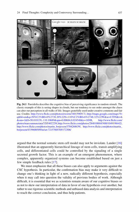
43724 Final Thoughts: Complexity and Controversy Surrounding…
argued that the normal somatic stem cell model may not be inviolate. Lander [16] illustrated that an apparently hierarchical lineage of stem cells, transit-amplifying cells, and differentiated cells could be controlled by the signaling of a single secreted growth factor. This is an example of an emergent phenomenon, where complex, apparently organized systems can become established based on just a few simple feedback rules [17].
We must emphasize that all these biases can also apply to arguments against the CSC hypothesis. In particular, the confirmation bias may make it very difficult to change one’s thinking in light of a new, radically different hypothesis, especially when it may call into question the validity of previous bodies of work. Although difficult, it is essential that we as scientists remain aware of our cognitive biases so as not to skew our interpretation of data in favor of one hypothesis over another, but rather to use rigorous scientific methods and unbiased data analysis and interpretation to reach the correct conclusion, and thus help patients.
Fig. 24.1 Pareidolia describes the cognitive bias of perceiving significance in random stimuli. The classic example of this is seeing shapes in clouds, but our tendency to see order amongst the chaos can alter our perception in all walks of life. Images gratefully used under creative commons and fair use. Credits: http://www.flickr.com/photos/eworm/3401596917/, http://maps.google.com/maps?f= q&hl=en&q=50%C2%B0+0%2738.20%22N+110%C2%B0+6%2748.32%22W&ie=UTF8&t=h&om=1&ll=50.010139,-110.10689&spn=0.00684,0.024548&z=16NB, http://www.flickr.com/ photos/marcoannunziata/3205402229/, http://www.flickr.com/photos/28481088@N00/1049198442/, http://www.flickr.com/photos/martin_borjesson/3704268639/, http://www.flickr.com/photos/martin_borjesson/4339608509/in/set-72157605305172306/
438 C. Gedye et al.
Finally, we should recognize that the CSC hypothesis is based upon inductive reasoning, and as Popper proposed, inductive statements are by definition difficult to verify and must be tested by falsifiability [18]. Thus, as the CSC field matures, and we try to account for the increasing number of conflicting observations and interpretations being reported, future experiments may be best informed by attempts to disprove the CSC hypothesis, and by constantly reflecting on the relevance of these data with regard to cancer patients’ clinical and pathological outcomes.
24.3 The Experiment
Having visited some of the perceptual biases that may distort our conception of tumoral heterogeneity, let us consider some of the technical and methodological challenges. As the CSC hypothesis is grounded upon the “gold standard” of prospec-tive isolation and serial xenotransplantation assays [19], we will review these con-cerns in the context of the steps required to perform these experiments. The serial xenotransplantation assay (Fig. 24.2a) has been employed to demonstrate serial self-renewal in subpopulations of cancer cells, such that if a marker is able to fractionate cells with tumorigenic potential from cells with little or no ability to form tumors, then these marker-positive cells are functionally defined as TICs. In addition, the xenografts formed should continue to express the putative CSC marker, and marker-positive cells from these xenografts should continue to proliferate extensively and retain all the tumor-initiating ability in subsequent mice. Finally, the resulting xeno-grafts should recapitulate the phenotypic heterogeneity of the patient tumor.
24.3.1 The Seeds: Do We Use the Wrong Cancer Cells, Processed in the Wrong Way?
24.3.1.1 Practical Limitations to Isolating Cell Subsets from Solid Tumors
The first technical problem that confounds attempts to quantify TICs in cancers relates to the source of the cancer cells used, and their preparation and handling. For example, in solid tumors we collect tumor tissues from patients undergoing surgery who have kindly consented to share their tissue for research. As the tumor must also undergo formal histological examination (particularly of the tumor margins) to con-firm the diagnosis and provide prognostic information, we can most often only receive a small sample of the tumor that typically comes from deep within the lesion. Thus, we may unavoidably be introducing a sampling bias, as cancer cells from dif-ferent parts of the same tumor may have different properties [20]. For example, there is evidence that self-renewal and CSC marker expression are lower and prolif-erative potential higher in glioma cells at the “presumed” periphery of a tumor com-pared with tumor cells at the “presumed” center of the tumor [21]. Having taken a
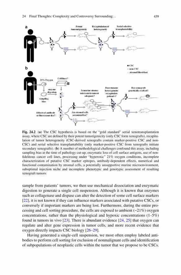
43924 Final Thoughts: Complexity and Controversy Surrounding…
sample from patients’ tumors, we then use mechanical dissociation and enzymatic digestion to generate a single cell suspension. Although it is known that enzymes such as collagenase and dispase can alter the detection of some cell surface markers [22], it is not known if they can influence markers associated with putative CSCs, or conversely if important markers are being lost. Furthermore, during the entire pro-cessing and cell sorting procedure, the cells are exposed to ambient (~21%) oxygen concentrations, rather than the physiological and hypoxic concentrations (1–5%) found in tumors in vivo [23]. There is abundant evidence [24, 25] that oxygen can regulate and alter gene expression in tumor cells, and more recent evidence that oxygen directly impacts CSC biology [26–29].
Having generated a single-cell suspension, we most often employ labeled anti-bodies to perform cell sorting for exclusion of nonmalignant cells and identification of subpopulations of neoplastic cells within the tumor that we propose to be CSCs.
Fig. 24.2 (a) The CSC hypothesis is based on the “gold standard” serial xenotransplantation assay, where CSC are defined by their potent tumorigenicity (only CSC form xenografts), recapitu-lation of tumor heterogeneity (CSC-derived xenografts contain marker-positive CSC and non-CSC) and serial selective transplantability (only marker-positive CSC from xenografts initiate secondary xenografts). (b) A number of methodological challenges confound this assay, including sampling bias at the time of pathology cut-up, enzymatic loss of cell surface antigens, use of non-fidelitous cancer cell lines, processing under “hyperoxic” 21% oxygen conditions, incomplete characterization of putative CSC marker epitopes, antibody-dependent effects, numerical and functional contamination by stromal cells, a potentially unsupportive murine microenvironment, suboptimal injection niche and incomplete phenotypic and genotypic assessment of resulting xenograft tumors
440 C. Gedye et al.
But what are we actually separating? Some of the CSC markers that are commonly employed are complex, with multiple isoforms (e.g., CD44 [30]) that may have different biological associations i.e., CD44 variants and metastasis [31]) or multiple glycosylation states with uncharacterised epitopes (e.g., AC133 in CD133 [32]). The antibodies used to separate cells may themselves influence the underlying biology. For example, a recent report suggests that CD38 monoclonal antibodies, but not CD38 antibody Fab fragments can deplete leukemia-initiating cells, presumably because of antibody-dependent cell killing [33]. Putative CSC markers themselves may not be expressed in all patients’ cancers [34, 35], although this inter-patient heterogeneity is increasingly being recognized and dissected [36]. The use of intra-cellular markers of “stemness” such as the side-population of Hoechst stained cells and Aldefluor staining for aldehyde dehydrogenase (ALDH) activity are also employed in the search for CSCs. These too must be examined critically as Hoechst dyes may be toxic to cells [37], and although promising, ALDH activity may not segregate stem-like cells in every tumor type [38].
Finally, though many publications in the CSC field are based on primary patient cancer samples or passaged xenografts, there are also many studies that use com-mercial cancer cell lines, which may have acquired different properties associated with the ability to grow in culture. Human cancer cells cultured for extended periods of time in animal serum rapidly acquire in vitro mutations, which irreversibly corrupt their genotype and phenotype compared with the patient’s tumor [39, 40], and an increasing body of evidence suggests that commercially available serum-grown cell lines are poor models for tumor heterogeneity [41, 42]. Studies involving the use of these types of cell lines may yield interesting preclinical results, but these should be validated in human cancer samples to ensure clinical relevance.
24.3.1.2 Do We Know Which Cells Are Actually Being Assayed?
In addition to cancer cells, solid tumors also contain nonmalignant stromal fibro-blasts, infiltrating immune cells and blood vessels. Many authors have accounted for these stromal components in CSC studies by depleting the lineage-positive cells, i.e., labeling and removing these cells during the cell sorting process. Markers such as CD31 (PECAM1) and CD45 have been used to account for endothelial cells and haematopoietic cells, respectively [11, 43]. Some groups have attempted to account for the presence of stromal fibroblasts (e.g., staining for CD140b/PDGFR-b as a fibroblast marker in breast cancer [43]), but fibroblasts have an ambiguous and incompletely defined cell surface phenotype, and no single marker can as yet capture their diversity across all human tissues and tumors [44]. In fact, although CD140b/PDGFR-b has not been studied extensively in breast cancer, it does not appear to be ubiquitously expressed by breast-cancer associated fibroblasts [45], and may be expressed by tumor cells in some breast cancers [46, 47]. Accounting for stromal fibroblasts may be particularly important in cancers dominated by a dense desmo-plastic reaction, where fibroblasts may numerically outnumber tumor cells, for exam-ple in colorectal and pancreatic cancers (Fig. 24.3a, b). Being unable to account for
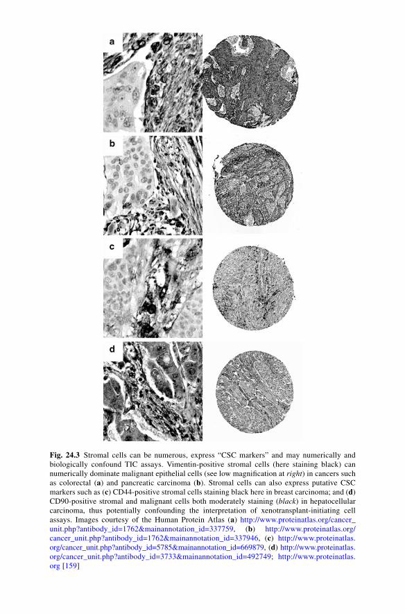
Fig. 24.3 Stromal cells can be numerous, express “CSC markers” and may numerically and biologically confound TIC assays. Vimentin-positive stromal cells (here staining black) can numerically dominate malignant epithelial cells (see low magnification at right) in cancers such as colorectal (a) and pancreatic carcinoma (b). Stromal cells can also express putative CSC markers such as (c) CD44-positive stromal cells staining black here in breast carcinoma; and (d) CD90-positive stromal and malignant cells both moderately staining (black) in hepatocellular carcinoma, thus potentially confounding the interpretation of xenotransplant-initiating cell assays. Images courtesy of the Human Protein Atlas (a) http://www.proteinatlas.org/cancer_unit.php?antibody_id=1762&mainannotation_id=337759, (b) http://www.proteinatlas.org/ cancer_unit.php?antibody_id=1762&mainannotation_id=337946, (c) http://www.proteinatlas.org/cancer_unit.php?antibody_id=5785&mainannotation_id=669879, (d) http://www.proteinatlas.org/cancer_unit.php?antibody_id=3733&mainannotation_id=492749; http://www.proteinatlas.org [159]
442 C. Gedye et al.
fibroblasts may numerically under-estimate the TIC frequency of a cancer, conceivably by up to tenfold. In addition, stromal cells may express markers that are shared with the putative CSC themselves. For example, CD44 as a marker of CSC in breast cancer is confounded by the presence of CD44+ stromal cells in many breast cancer samples (Fig. 24.3c). This is not confined to the CD44 marker; for example, the interpretation of CD90+ hepatocellular carcinoma CSCs [48, 49] may be confounded by CD90+ fibroblasts (Fig. 24.3d). If stromal cells are contaminating one or both of the CSC and non-CSC fractions, we may instead be measuring the tumorigenic potential of cancer cells and stromal cells vs. cancer cells alone. This contamination may be particularly confounding if cosorted tumor cells and stromal cells express the same marker (i.e., CD44 or CD90). A method to overcome this problem might be to include markers specific for malignant epithelial cells. In their early studies, Al-Hajj et al. employed ESA/EpCAM to identify tumor cells from which to frac-tionate CD44+/CD24−/lineage− CSC in breast carcinoma [43]. However, subsequent studies have demonstrated that ESA/EpCAM has variable expression and may not stain all cancer cells in epithelial cancers; for example, approximately two-thirds of breast cancers had low intensity and infrequent epithelial cell adhesion molecule (EpCAM) staining in a study of 205 breast cancer patient samples [50]. Accounting for stromal cells such as fibroblasts remains a challenging practical concern while studying human tumor heterogeneity.
24.3.2 The Soil: Stroma and Microenvironment
24.3.2.1 Which Cells Are Absent?
Once a single cell suspension has been generated and candidate CSC and non-CSC populations have been purified, they are assayed for tumor-initiating ability in immunocompromised mice. Paradoxically, efforts to efficiently deplete all of the stromal components prior to assaying a cell population for TIC activity may also confound results by excluding cell populations that may contribute to the growth of the cancer cells in vivo (i.e., fibroblasts [51] and inflammatory cells [52, 53]), and thus also lead to an underestimation of true TIC frequency [54]. Early work from the Bissell lab documented that virus-initiated tumors in avian models could form only at a site of wound inflammation and healing [55]. Compelling evidence for the influence of stromal cells in tumorigenicity also comes from careful studies in spon-taneously arising transplantable murine tumor models. First demonstrated by Révész [56] and confirmed by other workers in a wider panel of murine tumors [57], it was shown that the presence of lethally irradiated tumor cells can have a profound effect on absolute and relative tumorigenicity, increasing the TIC frequency by several orders of magnitude. In another report, the number of viable tumor cells in tumor fragments was estimated, and either enzymatically digested single cell suspensions or undigested tumor fragments were transplanted into syngeneic or allogeneic recipients. In three different tumor models, the number of cells in suspension required to form tumors vastly outnumbered the calculated number in the tumor
44324 Final Thoughts: Complexity and Controversy Surrounding…
fragments [58], suggesting that extracellular matrices and/or other 3-D architectural elements are important for tumor initiation.
There are recent reports that stromal cells may influence the behavior of populations functionally defined as CSC in human cancers. These include CD133+ pancreatic cancer cells [59], prostate CSCs in a conditional Pten deletion mouse model [60], the influence of the vascular niche on glioblastoma multiforme CSC [61], and the influence of mes-enchymal stem cells on mammosphere formation and stem-like cells in breast cancer [62]. Thus, the ideal assay would be a situation in which the putative CSC population is efficiently purified, and then recombined with the “microenvironmental components” to assess their true ability to initiate murine xenografts.
The ability to form xenografts is dependent on the grade and type [63] of tumor, and also on the anatomical niche of implantation [64]. Orthotopic implantation of human tumor xenografts would seem to be the ideal, but apart from the obvious niche for breast [43] and brain cancers [65], most other CSC publications have utilized the heterotopic subcutaneous injection site. Whether this can influence TIC frequency per se has not yet been reported, but injection of melanoma cell lines into the subder-mal space rather than subcutaneously is associated with a more physiological model of melanoma with a higher propensity for metastasis [66]. Similarly injection of renal carcinoma cell lines underneath the renal capsule is associated with a higher rate of metastasis compared with subcutaneous injection [67, 68]. This suggests the possibility that some cancers may be more independent of cell–cell signaling and are more “niche-permissive” (e.g., melanoma), while other cancers may be more dependent on stromal cell paracrine and endocrine signaling. In these latter cancers (e.g., leukemia, medulloblastoma), larger doses of closely apposed human cells may be required to provide sufficient paracrine signals. Alternatively, the propensity of human tumors to engraft into a xenogenic environment may simply parallel the rela-tive “niche-fastidiousness” of their putative cells-of-origin. For example, while hae-matopoietic stem cells require a very specific niche in bone marrow [69], melanocyte precursors spread much more widely from the neural crest and are found throughout the body [70] where they retain self-renewal capacity in adult life [71]. One obser-vation supporting this hypothesis is the clinical presentation of different cancers; for example, a myeloid sarcoma or chloroma (a solid mass of leukemic blasts outside the spleen or marrow) is a rare finding in patients with AML [72], but melanoma can metastasize to almost any organ in the body including the small intestine [73].
24.3.2.2 Mouse Models Matter
In addition to considering the location, purity, and identity of the population(s) of cells that are injected, let us consider the host animal itself. One of the most compelling challenges to the relevance of the CSC hypothesis has recently come from a study of tumorigenic cells in melanoma: Quintana et al. [74] injected melanoma cells with high concentration Matrigel (a heterogeneous mixture of basement membrane proteins secreted by Engelbreth-Holm-Swarm (EHS) mouse sarcoma cells) into severely immunocompromised mice; the NOD/SCID/IL2Rg−/− (NSG) strain which entirely lacks natural killer (NK) cell activity (compared with the previous “gold standard”
444 C. Gedye et al.
nonobese diabetic/severe combined immune deficiency (NOD/SCID) strain in which low NK cell activity is present). Matrigel [75, 76] and tissue components such as brain extract [77] are well known to enhance tumor transplantability. Whether this is due to tumor cell aggregation due to the formation of a gel that maintains the injected cells in proximity, or due to growth factors embedded in the matrix remains unclear, but it does not appear to be due to collagen or laminin components of the matrix alone [78]. The rationale was that these conditions would be more permissive for human primary xenograft formation. Under these conditions, the authors found that tumorigenic cells in melanoma were relatively common, such that around 1 in 4 melanoma cells implanted subcutaneously could form a xeno-graft. This contrasted with much lower tumorigenic frequencies from melanoma implanted in parallel in NOD/SCID mice (~1/111,000), and in a previous report (~1/1,000,000) [79]. Subsequent data demonstrated that when marker positive and negative sorted cell populations (using several markers, including CD133, p75/NGFR, and others) were implanted in NSG mice, both populations gave rise to a mixed xenograft, expressing both marker-positive and marker-negative cells; [80] i.e., no selection (and hence no hierarchy) could be demonstrated. A criticism of this work was that it used cells from passaged xenografts and from advanced stage III and IV melanoma specimens; however, this more recent data show that single mela-noma cells are highly tumorigenic (TIC frequency 29%) even when directly isolated from early stage patient samples [80]. Thus with sufficient optimization of the xeno-transplant model, tumorigenic cells in melanoma were found to be common, rather than rare as had been previously proposed.
The CSC hypothesis does not assume that CSCs must be rare. The hypothesis initially arose in part from the observation that large numbers of cells are required to initiate tumors, suggesting that only a rare subset has this ability. However, even in cases where cancers have high TIC frequencies such as melanoma, this may simply represent a more “shallow” hierarchy, i.e., where few malignant cells exhibit a termi-nally differentiated phenotype (Fig. 24.4b). One could imagine that the depth of the hierarchy will depend on many parameters, including the depth of the hierarchy in the normal tissue from which the cancer arose, where the cell from which the cancer arose is situated in the hierarchy, and the nature of the underlying mutations (e.g., mutations that block differentiation may lead to a higher frequency of TIC than mutations that increase self-renewal, or inhibit cell death). We and others [81] recently reported limiting dilution tumorigenicity experiments performed side-by-side in NOD/SCID and NSG mice, showing that the TIC frequency remains rare in non-small cell lung carcinoma, pancreatic carcinoma, and head and neck squamous cell carcinoma, using the same methods as Quintana et al. This suggests that the phenomenon observed in melanoma does not necessarily apply to other solid tumors, but does not rule out the possibility that the heavily immunocompromised NSG mouse model is still lacking other essential microenvironmental components that, if present, would radically alter the readout of these assays in other tumor types.
While some have taken this work from the Morrison lab as a harbinger of doom for the CSC hypothesis, it may be that cancers can follow two models of heteroge-neity. In some cancers, a hierarchy may not exist and tumor heterogeneity is best
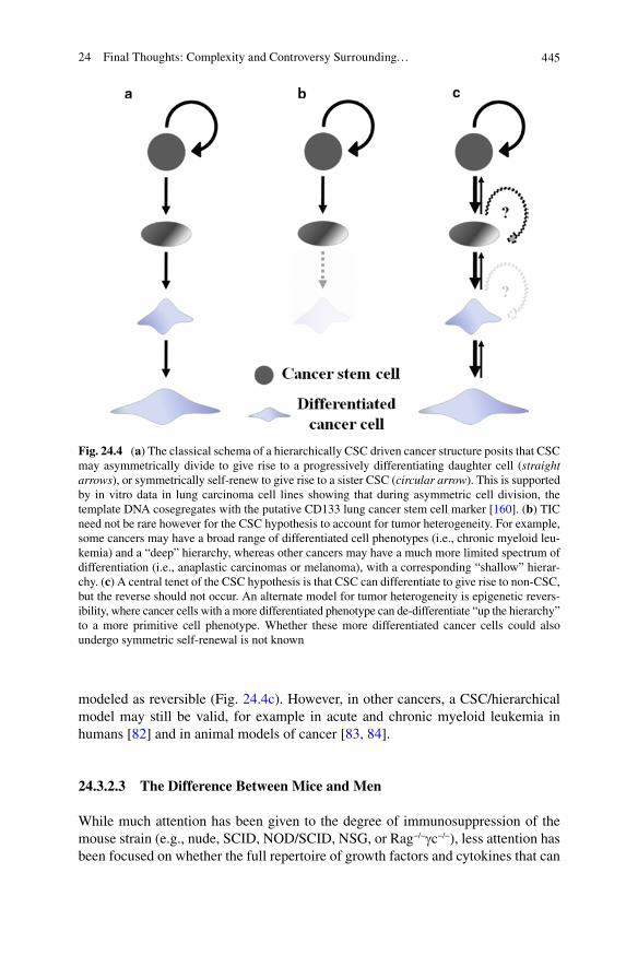
44524 Final Thoughts: Complexity and Controversy Surrounding…
Fig. 24.4 (a) The classical schema of a hierarchically CSC driven cancer structure posits that CSC may asymmetrically divide to give rise to a progressively differentiating daughter cell (straight arrows), or symmetrically self-renew to give rise to a sister CSC (circular arrow). This is supported by in vitro data in lung carcinoma cell lines showing that during asymmetric cell division, the template DNA cosegregates with the putative CD133 lung cancer stem cell marker [160]. (b) TIC need not be rare however for the CSC hypothesis to account for tumor heterogeneity. For example, some cancers may have a broad range of differentiated cell phenotypes (i.e., chronic myeloid leu-kemia) and a “deep” hierarchy, whereas other cancers may have a much more limited spectrum of differentiation (i.e., anaplastic carcinomas or melanoma), with a corresponding “shallow” hierar-chy. (c) A central tenet of the CSC hypothesis is that CSC can differentiate to give rise to non-CSC, but the reverse should not occur. An alternate model for tumor heterogeneity is epigenetic revers-ibility, where cancer cells with a more differentiated phenotype can de-differentiate “up the hierarchy” to a more primitive cell phenotype. Whether these more differentiated cancer cells could also undergo symmetric self-renewal is not known
modeled as reversible (Fig. 24.4c). However, in other cancers, a CSC/hierarchical model may still be valid, for example in acute and chronic myeloid leukemia in humans [82] and in animal models of cancer [83, 84].
24.3.2.3 The Difference Between Mice and Men
While much attention has been given to the degree of immunosuppression of the mouse strain (e.g., nude, SCID, NOD/SCID, NSG, or Rag−/−gc−/−), less attention has been focused on whether the full repertoire of growth factors and cytokines that can
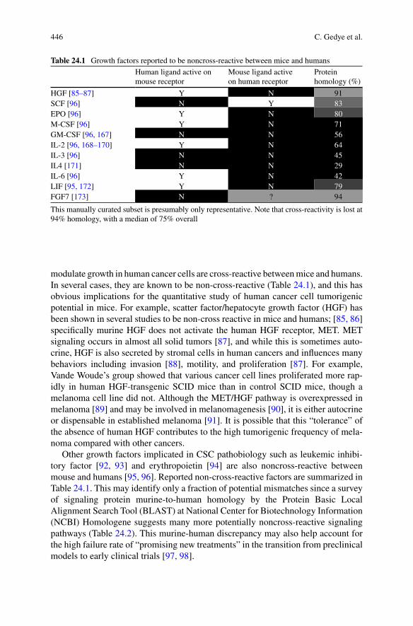
446 C. Gedye et al.
modulate growth in human cancer cells are cross-reactive between mice and humans. In several cases, they are known to be non-cross-reactive (Table 24.1), and this has obvious implications for the quantitative study of human cancer cell tumorigenic potential in mice. For example, scatter factor/hepatocyte growth factor (HGF) has been shown in several studies to be non-cross reactive in mice and humans; [85, 86] specifically murine HGF does not activate the human HGF receptor, MET. MET signaling occurs in almost all solid tumors [87], and while this is sometimes auto-crine, HGF is also secreted by stromal cells in human cancers and influences many behaviors including invasion [88], motility, and proliferation [87]. For example, Vande Woude’s group showed that various cancer cell lines proliferated more rap-idly in human HGF-transgenic SCID mice than in control SCID mice, though a melanoma cell line did not. Although the MET/HGF pathway is overexpressed in melanoma [89] and may be involved in melanomagenesis [90], it is either autocrine or dispensable in established melanoma [91]. It is possible that this “tolerance” of the absence of human HGF contributes to the high tumorigenic frequency of mela-noma compared with other cancers.
Other growth factors implicated in CSC pathobiology such as leukemic inhibi-tory factor [92, 93] and erythropoietin [94] are also noncross-reactive between mouse and humans [95, 96]. Reported non-cross-reactive factors are summarized in Table 24.1. This may identify only a fraction of potential mismatches since a survey of signaling protein murine-to-human homology by the Protein Basic Local Alignment Search Tool (BLAST) at National Center for Biotechnology Information (NCBI) Homologene suggests many more potentially noncross-reactive signaling pathways (Table 24.2). This murine-human discrepancy may also help account for the high failure rate of “promising new treatments” in the transition from preclinical models to early clinical trials [97, 98].
Table 24.1 Growth factors reported to be noncross-reactive between mice and humans
Human ligand active on mouse receptor
Mouse ligand active on human receptor
Protein homology (%)
HGF [85–87] Y N 91SCF [96] N Y 83EPO [96] Y N 80M-CSF [96] Y N 71GM-CSF [96, 167] N N 56IL-2 [96, 168–170] Y N 64IL-3 [96] N N 45IL4 [171] N N 29IL-6 [96] Y N 42LIF [95, 172] Y N 79FGF7 [173] N ? 94
This manually curated subset is presumably only representative. Note that cross-reactivity is lost at 94% homology, with a median of 75% overall
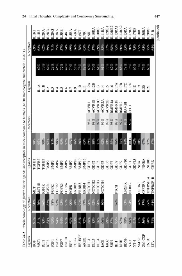
44724 Final Thoughts: Complexity and Controversy Surrounding…
Tabl
e 24
.2
Prot
ein
hom
olog
y of
gro
wth
fac
tor
ligan
ds a
nd r
ecep
tors
in m
ice
com
pare
d to
hum
ans
(NC
BI
hom
olog
ene
and
prot
ein
BL
AST
)
Lig
ands
Rec
epto
rsL
igan
dsR
ecep
tors
Lig
ands
Rec
epto
rs
HG
F91
%90
%M
ET
TG
FB1
90%
69%
IL1R
1M
ST1
81%
76%
MST
1RT
GFB
295
%IL
1A62
%62
%IL
1R2
IGF1
83%
96%
IGF1
RT
GFB
398
%IL
1B70
%62
%IL
2RA
IGF2
84%
82%
IGF2
RB
MP2
92%
IL2
64%
59%
IL2R
BFG
F195
%98
%FG
FR1
BM
P381
%IL
329
%71
%IL
2RG
FGF2
94%
97%
FGFR
2B
MP4
98%
IL4
45%
35%
IL3R
AFG
F794
%93
%FG
FR3
BM
P593
%IL
572
%57
%IL
3RB
FGF1
091
%91
%FG
FR4
BM
P692
%IL
642
%54
%IL
4RE
GF
68%
90%
EG
FRB
MP7
98%
IL7
70%
69%
IL5R
AT
GF-
a93
%88
%E
RB
B2
BM
P8a
86%
IL9
57%
56%
IL6R
AH
B-E
GF
81%
91%
ER
BB
3B
MP1
086
%IL
1073
%78
%IL
6ST
AR
EG
72%
97%
ER
BB
4B
MP1
565
%IL
1188
%65
%IL
7RD
LL
188
%90
%N
OT
CH
1G
DF1
69%
98%
AC
VR
1IL
12A
60%
62%
IL9R
DL
L3
83%
92%
NO
TC
H2
GD
F280
%98
%A
CV
R1B
IL12
B70
%57
%IL
10R
AD
LL
486
%90
%N
OT
CH
3G
DF3
71%
97%
AC
VR
1CIL
1361
%69
%IL
10R
BJA
G1
96%
80%
NO
TC
H4
GD
F592
%99
%A
CV
R2A
IL14
91%
83%
IL11
RA
JAG
289
%G
DF6
89%
99%
AC
VR
2BIL
1573
%57
%IL
12R
B1
SHH
87%
GD
F785
%98
%B
MPR
1aIL
1682
%68
%IL
12R
B2
IHH
95%
96%
PTC
HG
DF8
96%
98%
BM
PR1b
IL17
A63
%75
%IL
13R
A1
DH
H97
%G
DF9
74%
97%
BM
PR2
IL17
B88
%60
%IL
13R
A2
BD
NF
98%
93%
LN
GFR
GD
F10
84%
93%
BA
MB
IIL
17C
78%
55%
IL15
RA
NT-
396
%94
%N
TR
K2
GD
F11
100%
53%
CFC
1IL
17D
78%
70%
IL17
RA
NT-
491
%G
DF1
562
%IL
1866
%75
%IL
17R
BM
-CSF
71%
76%
CSF
1RIN
HA
80%
IL19
70%
65%
IL18
R1
GM
-CSF
56%
37%
CSF
2RA
INH
BA
97%
IL20
76%
65%
IL20
RA
TN
FA80
%66
%T
NFR
SF1A
INH
BB
97%
IL21
63%
75%
IL20
RB
LTA
73%
64%
TN
FRSF
1BC
HR
D87
%65
%IL
21R
(con
tinue
d)
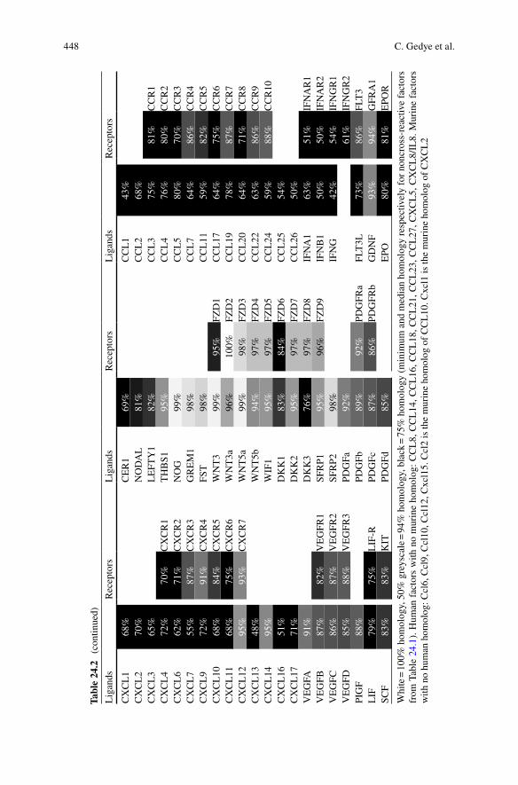
448 C. Gedye et al.
Tabl
e 24
.2
(con
tinue
d)
Lig
ands
Rec
epto
rsL
igan
dsR
ecep
tors
Lig
ands
Rec
epto
rs
CX
CL
168
%C
ER
169
%C
CL
143
%C
XC
L2
70%
NO
DA
L81
%C
CL
268
%C
XC
L3
65%
LE
FTY
182
%C
CL
375
%81
%C
CR
1C
XC
L4
72%
70%
CX
CR
1T
HB
S195
%C
CL
476
%80
%C
CR
2C
XC
L6
62%
71%
CX
CR
2N
OG
99%
CC
L5
80%
70%
CC
R3
CX
CL
755
%87
%C
XC
R3
GR
EM
198
%C
CL
764
%86
%C
CR
4C
XC
L9
72%
91%
CX
CR
4FS
T98
%C
CL
1159
%82
%C
CR
5C
XC
L10
68%
84%
CX
CR
5W
NT
399
%95
%FZ
D1
CC
L17
64%
75%
CC
R6
CX
CL
1168
%75
%C
XC
R6
WN
T3a
96%
100%
FZD
2C
CL
1978
%87
%C
CR
7C
XC
L12
95%
93%
CX
CR
7W
NT
5a99
%98
%FZ
D3
CC
L20
64%
71%
CC
R8
CX
CL
1348
%W
NT
5b94
%97
%FZ
D4
CC
L22
63%
86%
CC
R9
CX
CL
1495
%W
IF1
95%
97%
FZD
5C
CL
2459
%88
%C
CR
10C
XC
L16
51%
DK
K1
83%
84%
FZD
6C
CL
2554
%C
XC
L17
71%
DK
K2
95%
97%
FZD
7C
CL
2650
%V
EG
FA91
%D
KK
376
%97
%FZ
D8
IFN
A1
63%
51%
IFN
AR
1V
EG
FB87
%82
%V
EG
FR1
SFR
P195
%96
%FZ
D9
IFN
B1
50%
50%
IFN
AR
2V
EG
FC86
%87
%V
EG
FR2
SFR
P298
%IF
NG
42%
54%
IFN
GR
1V
EG
FD85
%88
%V
EG
FR3
PDG
Fa92
%61
%IF
NG
R2
PIG
F88
%PD
GFb
89%
92%
PDG
FRa
FLT
3L73
%86
%FL
T3
LIF
79%
75%
LIF
-RPD
GFc
87%
86%
PDG
FRb
GD
NF
93%
94%
GFR
A1
SCF
83%
83%
KIT
PDG
Fd85
%E
PO80
%81
%E
POR
Whi
te =
100
% h
omol
ogy,
50%
gre
ysca
le =
94%
hom
olog
y, b
lack
= 7
5% h
omol
ogy
(min
imum
and
med
ian
hom
olog
y re
spec
tivel
y fo
r no
ncro
ss-r
eact
ive
fact
ors
from
Tab
le 2
4.1)
. Hum
an f
acto
rs w
ith n
o m
urin
e ho
mol
og:
CC
L8,
CC
L14
, CC
L16
, CC
L18
, CC
L21
, CC
L23
, CC
L27
, CX
CL
5, C
XC
L8/
IL8.
Mur
ine
fact
ors
with
no
hum
an h
omol
og: C
cl6,
Ccl
9, C
cl10
, Ccl
12, C
xcl1
5. C
cl2
is th
e m
urin
e ho
mol
og o
f C
CL
10. C
xcl1
is th
e m
urin
e ho
mol
og o
f C
XC
L2
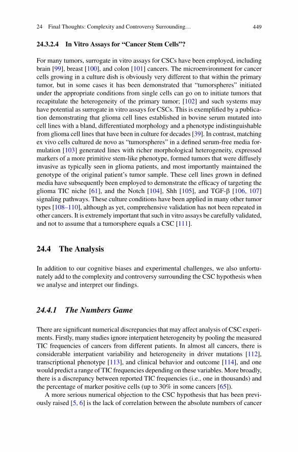
44924 Final Thoughts: Complexity and Controversy Surrounding…
24.3.2.4 In Vitro Assays for “Cancer Stem Cells”?
For many tumors, surrogate in vitro assays for CSCs have been employed, including brain [99], breast [100], and colon [101] cancers. The microenvironment for cancer cells growing in a culture dish is obviously very different to that within the primary tumor, but in some cases it has been demonstrated that “tumorspheres” initiated under the appropriate conditions from single cells can go on to initiate tumors that recapitulate the heterogeneity of the primary tumor; [102] and such systems may have potential as surrogate in vitro assays for CSCs. This is exemplified by a publica-tion demonstrating that glioma cell lines established in bovine serum mutated into cell lines with a bland, differentiated morphology and a phenotype indistinguishable from glioma cell lines that have been in culture for decades [39]. In contrast, matching ex vivo cells cultured de novo as “tumorspheres” in a defined serum-free media for-mulation [103] generated lines with richer morphological heterogeneity, expressed markers of a more primitive stem-like phenotype, formed tumors that were diffusely invasive as typically seen in glioma patients, and most importantly maintained the genotype of the original patient’s tumor sample. These cell lines grown in defined media have subsequently been employed to demonstrate the efficacy of targeting the glioma TIC niche [61], and the Notch [104], Shh [105], and TGF-b [106, 107] signaling pathways. These culture conditions have been applied in many other tumor types [108–110], although as yet, comprehensive validation has not been repeated in other cancers. It is extremely important that such in vitro assays be carefully validated, and not to assume that a tumorsphere equals a CSC [111].
24.4 The Analysis
In addition to our cognitive biases and experimental challenges, we also unfortu-nately add to the complexity and controversy surrounding the CSC hypothesis when we analyse and interpret our findings.
24.4.1 The Numbers Game
There are significant numerical discrepancies that may affect analysis of CSC experi-ments. Firstly, many studies ignore interpatient heterogeneity by pooling the measured TIC frequencies of cancers from different patients. In almost all cancers, there is considerable interpatient variability and heterogeneity in driver mutations [112], transcriptional phenotype [113], and clinical behavior and outcome [114], and one would predict a range of TIC frequencies depending on these variables. More broadly, there is a discrepancy between reported TIC frequencies (i.e., one in thousands) and the percentage of marker positive cells (up to 30% in some cancers [65]).
A more serious numerical objection to the CSC hypothesis that has been previ-ously raised [5, 6] is the lack of correlation between the absolute numbers of cancer

450 C. Gedye et al.
cells with tumorigenic potential in unsorted tumor cell suspensions and the absolute number of cells with tumorigenic potential in marker-positive and marker-negative sorted cell fractions. For example, if the tumor-initiating frequency in unsorted cells indicates that there are n cells capable of tumor-initiation within a tumor derived single cell suspension, and x of those n cells are found in the marker-positive population, then n−x = y of the TIC ought to be found in the marker-negative population. However, this is generally not the case, and it has been argued that these analyses are most consistent with the presence of an inhibitor [115], i.e., either the presence of marker-negative cells inhibits the tumor initiating capacity of marker-positive cells, or that tumorigenic potential is context-dependent rather than an intrinsic property of a specific tumor cell subset. Indeed, if the appropriate calculations are done, in some cases the absolute number of TIC is actually higher in the “non-CSC” fraction than in the “CSC-enriched” fraction [116].
Another unresolved numerical issue within the CSC field is the use of the posthoc rationalization that xenograft tumors formed after injection of non-CSC (i.e., marker-negative cells) must be due to contamination of the non-CSC cells by (marker-positive) CSC. By performing flow cytometry purity checks on sorted pop-ulations and appropriate statistical calculations, it should be possible to calculate the probability that contamination of non-CSC with CSC has in fact occurred. This has not been published in a CSC study to date. Finally, we may be using entirely the wrong mathematical models to discuss heterogeneity in cancer. For example, dynamic models [117] and statistical approaches employed in evolutionary and population biology may be more appropriate [118].
24.4.2 Measuring Xenograft Heterogeneity
One of the tenets of the CSC hypothesis is that tumors initiated by the CSC subset must recapitulate the heterogeneity of the primary tumor. Most CSC publications show his-tological images to demonstrate morphological similarity, and flow cytometry and/or immunohistochemistry to demonstrate regeneration of the marker-positive and marker-negative populations, but extensive phenotypic characterization has not commonly been presented. This is of considerable importance as it would seem likely that different genomic subgroups of cancers may have different hierarchical phenotypes [36] with different “CSC” populations, for example as has recently been demonstrated in mouse lung cancer models [119]. Furthermore, genomic heterogeneity is likely to remain in active flux during the serial xenotransplantation process [120], as cancer genomes are known to be unstable. The relationship between stemness and differentiation may therefore change over time. Studies performed in transplantable murine tumors [121, 122] showed that dynamic changes are possible in cancer cell genomes or epigenomes, at relatively high rates (~10−5 per cell per generation). If such frequencies apply to human cancers, there may be 1,000 of such events per day in clinically apparent tumors, especially in cancers where DNA repair defects are a feature of their particular geno-type (i.e., mismatch repair genes and BRCA1/2 mutations).
45124 Final Thoughts: Complexity and Controversy Surrounding…
Considering the genotype of xenograft tumors raises other questions regarding tumor heterogeneity and the CSC hypothesis. For example, does the xenografting process itself maintain the genomic heterogeneity of the parent tumor or does the “most flexible” clone outcompete it’s less xeno-capable peers? The comprehensive genomic sequencing of a basal breast carcinoma, a subsequent metastases and a xenograft established from the primary tumor [123], suggested that a small popula-tion of cells within the primary tumor gave rise to the metastases and the xenograft.
Genetic heterogeneity alone may not fully account for the functional and anti-genic heterogeneity present in all human malignancies. For example, ultradeep sequencing of the immunoglobulin heavy chain (IGH) locus in patients with chronic lymphocytic leukemia has revealed phylogenetic structures indicative of clonal evolution within the malignant cell compartment. Intriguingly, patients with early stage disease had more complex clonal structures whereas patients with advanced disease seemed to have undergone selection to a dominant clone [124]. Clonal dominance was also suggested by copy-number variation analysis in end-stage metastatic prostate cancer [125], while methylation analysis of topologically distinct biopsies from primary colorectal cancers also suggests that heterogeneity of clinically apparent cancers can “almost always” be accounted for by a single clonal expansion [126].
There are conflicting data, however. For example, distinct genetic clones within an individual patient’s cancer can be associated with morphologically and functionally distinct phenotypes, as elegantly demonstrated in a recent study of metaplastic breast cancers [127], where lobular and squamous histological subtypes could be found in adjacent parts of a primary breast cancer, each with unique focal genomic amplifications and immunohistochemical staining patterns. Sampling from one part of the tumor may not allow us to accurately encompass the heterogeneity seen throughout such a patient’s cancer, and genetic selection occurring during the xeno-graft process would complicate this further [123].
Parallel assessment of genetic and epigenetic heterogeneity is now, however, becoming technically possible and early data are being presented [128]. Such paral-lel analysis of two likely mechanisms of heterogeneity within cancers will provide powerful information to improve patient outcomes. Indeed it is highly likely that both genetic and epigenetic heterogeneity can evolve simultaneously, as has recently been eloquently argued [120]. With increasingly detailed sequence data becoming available for different tumor types [129], it is possible to compare genomic com-plexity with tumorigenic frequency in various cancers (Fig. 24.5). Though this data-set is obviously limited, it will be intriguing to see if there truly is a correlation between the number of coding mutations and the frequency of tumorigenic cells as more genome and TIC frequency data becomes available.
Finally, though the CSC hierarchical model must involve epigenetic regulation, little attention to date has been paid to known epigenetic mechanisms such as DNA methylation, histone acetylation, microRNA, and noncoding RNA. As both the tumor heterogeneity and epigenetics fields mature, we expect new evidence to inform our understanding of the validity of the CSC hypothesis.
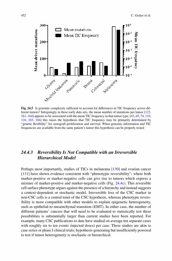
452 C. Gedye et al.
24.4.3 Reversibility Is Not Compatible with an Irreversible Hierarchical Model
Perhaps most importantly, studies of TICs in melanoma [130] and ovarian cancer [131] have shown evidence consistent with “phenotypic reversibility”; where both marker-positive or marker-negative cells can give rise to tumors which express a mixture of marker-positive and marker-negative cells (Fig. 24.4c). This reversible cell surface phenotype argues against the presence of a hierarchy and instead suggests a context-dependent or stochastic model. Irreversible loss of the CSC marker in non-CSC cells is a central tenet of the CSC hypothesis, whereas phenotypic revers-ibility is more compatible with other models to explain epigenetic heterogeneity, such as epithelial-to-mesenchymal transition (EMT). In either case, the number of different patients’ cancers that will need to be evaluated to statistically test these possibilities is substantially larger than current studies have been reported. For example, many CSC publications to date have studied on average ten separate cases with roughly six to ten events (injected doses) per case. These studies are akin to case series or phase I clinical trials; hypothesis-generating but insufficiently powered to test if tumor heterogeneity is stochastic or hierarchical.
Fig. 24.5 Is genomic complexity sufficient to account for differences in TIC frequency across dif-ferent tumors? Intriguingly in these early data sets, the mean number of mutations per tumor [123, 161–164] appears to be associated with the mean TIC frequency in that tumor type; [43, 65, 74, 110, 116, 165, 166] this raises the hypothesis that TIC frequency may be primarily determined by “genetic flexibility” for xenograft proliferation and survival. When genomic information and TIC frequencies are available from the same patient’s tumor this hypothesis can be properly tested
45324 Final Thoughts: Complexity and Controversy Surrounding…
24.5 The Next Iteration of the CSC Hypothesis
Marston Bates said that “research is the process of going up alleys to see if they are blind.” Despite the complexities and controversies outlined above, we believe it will be of great value to continue to interrogate the CSC hypothesis. Although hypotheses can never be conclusively proven to be true in a Popperian world-view, comprehen-sively disproving the CSC hypothesis in a given cancer would require extremely fastidious and detailed knowledge of the intratumoral heterogeneity in individual patients’ cancers. Even if the CSC hypothesis is false, that is if epigenetic heteroge-neity is not organized in an irreversible hierarchy, it would seem likely that the molecular pathways and signaling mechanisms being identified in CSC studies (e.g., Notch [104], Shh [105], and TGF-b [106, 107]) will continue to be relevant when studying epigenetic heterogeneity in human cancers. Perhaps the best example of this potential duality is in breast cancer heterogeneity. One of the earliest reports of CSCs in solid tumors described the CD44+/CD24− subpopulation in breast cancer as being highly enriched for breast CSCs [43]. This work generated considerable discussion and controversy, and led to work by other groups that sought to refute the concept [132, 133], instead postulating the heterogeneity in breast cancer was reversibly regulated. A number of subsequent publications have strengthened the connection between CD44+/CD24− breast cancer cells and EMT [134–136], inva-sion [137], and mechanisms of treatment resistance such as immunoevasion [138–141], chemo-resistance [142, 143], and radiation resistance [144, 145]. Our attempts to interrogate complex phenomena with incomplete models by indirect observation may leave us like the Blind Men and the Elephant; [146] using limited sensory data to get partial knowledge, but all approaching the same truth from a multiplicity of perspectives. Finally, as we have outlined above, the act of attempting to investigate the state and behavior of cell fractions may influence the state and behavior of those cells, such that it is impossible to accurately predict their future behavior; the biologi-cal equivalent of Heisenberg’s uncertainty principle. This has been eloquently dis-cussed at the level of the genome of a single cell [147], and by extension we imagine it would be no less intractable in populations of cells within a tissue or cancer.
A number of methodological improvements are also likely to assist in supporting or refuting the CSC hypothesis. For example, tracking of each individual cancer cell within a xenograft population would help us to define if human solid tumors are organized hierarchically or stochastically. Perhaps the most compelling evidence for the CSC hypothesis in human cancer is the application of this concept with clonal analysis of leukemic-initiating cells (LIC) by lentiviral insertion site tracking, which demonstrated a hierarchy of short-term and long-term LIC in AML [82].
Optimizing mouse strains to better model the human microenvironment in xeno-grafts would also help us to more accurately address the question of whether TICs are rare or common in human cancer. Examples of this endeavor include the study of TIC in myeloma engrafted in mice implanted with human fetal bone [148] and the finding that AML xenograft efficiency in NSG mice can be substantially improved by constitutive expression of human stem cell factor (SCF), granulocyte macrophage colony-stimulating factor (GM-CSF), and IL-3 [149].
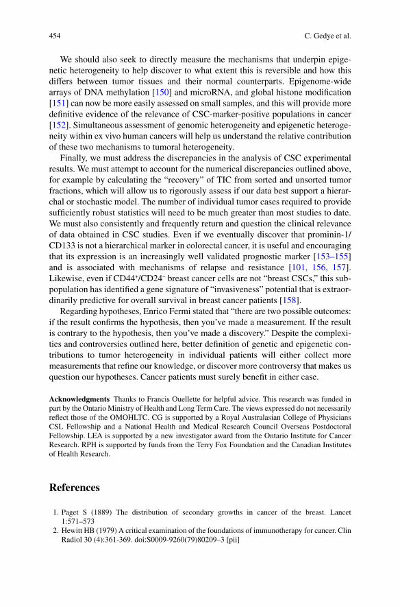
454 C. Gedye et al.
We should also seek to directly measure the mechanisms that underpin epige-netic heterogeneity to help discover to what extent this is reversible and how this differs between tumor tissues and their normal counterparts. Epigenome-wide arrays of DNA methylation [150] and microRNA, and global histone modification [151] can now be more easily assessed on small samples, and this will provide more definitive evidence of the relevance of CSC-marker-positive populations in cancer [152]. Simultaneous assessment of genomic heterogeneity and epigenetic heteroge-neity within ex vivo human cancers will help us understand the relative contribution of these two mechanisms to tumoral heterogeneity.
Finally, we must address the discrepancies in the analysis of CSC experimental results. We must attempt to account for the numerical discrepancies outlined above, for example by calculating the “recovery” of TIC from sorted and unsorted tumor fractions, which will allow us to rigorously assess if our data best support a hierar-chal or stochastic model. The number of individual tumor cases required to provide sufficiently robust statistics will need to be much greater than most studies to date. We must also consistently and frequently return and question the clinical relevance of data obtained in CSC studies. Even if we eventually discover that prominin-1/CD133 is not a hierarchical marker in colorectal cancer, it is useful and encouraging that its expression is an increasingly well validated prognostic marker [153–155] and is associated with mechanisms of relapse and resistance [101, 156, 157]. Likewise, even if CD44+/CD24− breast cancer cells are not “breast CSCs,” this sub-population has identified a gene signature of “invasiveness” potential that is extraor-dinarily predictive for overall survival in breast cancer patients [158].
Regarding hypotheses, Enrico Fermi stated that “there are two possible outcomes: if the result confirms the hypothesis, then you’ve made a measurement. If the result is contrary to the hypothesis, then you’ve made a discovery.” Despite the complexi-ties and controversies outlined here, better definition of genetic and epigenetic con-tributions to tumor heterogeneity in individual patients will either collect more measurements that refine our knowledge, or discover more controversy that makes us question our hypotheses. Cancer patients must surely benefit in either case.
Acknowledgments Thanks to Francis Ouellette for helpful advice. This research was funded in part by the Ontario Ministry of Health and Long Term Care. The views expressed do not necessarily reflect those of the OMOHLTC. CG is supported by a Royal Australasian College of Physicians CSL Fellowship and a National Health and Medical Research Council Overseas Postdoctoral Fellowship. LEA is supported by a new investigator award from the Ontario Institute for Cancer Research. RPH is supported by funds from the Terry Fox Foundation and the Canadian Institutes of Health Research.
References
1. Paget S (1889) The distribution of secondary growths in cancer of the breast. Lancet 1:571–573
2. Hewitt HB (1979) A critical examination of the foundations of immunotherapy for cancer. Clin Radiol 30 (4):361-369. doi:S0009-9260(79)80209–3 [pii]
45524 Final Thoughts: Complexity and Controversy Surrounding…
3. Hamburger AW, Salmon SE (1977) Primary bioassay of human tumor stem cells. Science 197 (4302):461–463
4. Brunschwig A, Southam CM, Levin AG (1965) Host resistance to cancer. Clinical experiments by homotransplants, autotransplants and admixture of autologous leucocytes. Ann Surg 162 (3):416–425
5. Hill RP (2006) Identifying cancer stem cells in solid tumors: Case not proven. Cancer Res 66 (4):1891–1895; discussion 1890
6. Hill RP, Perris R (2007) “Destemming” Cancer stem cells. J Natl Cancer Inst 99 (19):1435–1440. doi:10.1093/jnci/djm136
7. Hill RP, Milas L (1989) The proportion of stem cells in murine tumors. International journal of radiation oncology, biology, physics 16 (2):513–518
8. Cozzio A, Passegue E, Ayton PM, Karsunky H, Cleary ML, Weissman IL (2003) Similar mll-associated leukemias arising from self-renewing stem cells and short-lived myeloid pro-genitors. Genes Dev 17 (24):3029–3035
9. Krivtsov AV, Twomey D, Feng Z, Stubbs MC, Wang Y, Faber J, Levine JE, Wang J, Hahn WC, Gilliland DG, Golub TR, Armstrong SA (2006) Transformation from committed progenitor to leukaemia stem cell initiated by mll-af9. Nature 442 (7104):818–822. doi:nature04980 [pii] 10.1038/nature04980
10. Zhou H, Wu S, Joo JY, Zhu S, Han DW, Lin T, Trauger S, Bien G, Yao S, Zhu Y, Siuzdak G, Schöler HR, Duan L, Ding S (2009) Generation of induced pluripotent stem cells using recom-binant proteins. Cell Stem Cell 4 (5):381–384
11. Prince ME, Sivanandan R, Kaczorowski A, Wolf GT, Kaplan MJ, Dalerba P, Weissman IL, Clarke MF, Ailles LE (2007) Identification of a subpopulation of cells with cancer stem cell properties in head and neck squamous cell carcinoma. Proc Natl Acad Sci USA 104 (3):973–978
12. Committee on Science E, and Public Policy, National Academy of Sciences, National Academy of Engineering, and Institute of Medicine (2009) On being a scientist: A guide to responsible conduct in research. 3rd edn. National Academies Press, Washington, D.C.
13. Taleb NN (2001) Fooled by randomness: The hidden role of chance in life and in the markets. Texere LLC, London
14. Simons DJ, Chabris CF (1999) Gorillas in our midst: Sustained inattentional blindness for dynamic events. Perception 28 (9):1059–1074
15. Fugelsang JA, Stein CB, Green AE, Dunbar KN (2004) Theory and data interactions of the scientific mind: Evidence from the molecular and the cognitive laboratory. Can J Exp Psychol 58 (2):86–95
16. Lander A (2009) The ‘stem cell’ concept: Is it holding us back? Journal of Biology 8 (8):70 17. Reynolds CW (1987) Flocks, herds and schools: A distributed behavioral model. Paper pre-
sented at the Proceedings of the 14th annual conference on Computer graphics and interactive techniques
18. Popper K (1959) The logic of scientific discovery. Hutchinson, London 19. Clarke MF (2005) A self-renewal assay for cancer stem cells. Cancer Chemother Pharmacol
56 Suppl 7:64–68 20. Piccirillo SGM, Combi R, Cajola L, Patrizi A, Redaelli S, Bentivegna A, Baronchelli S, Maira
G, Pollo B, Mangiola A, DiMeco F, Dalpra L, Vescovi AL (2009) Distinct pools of cancer stem-like cells coexist within human glioblastomas and display different tumorigenicity and independent genomic evolution. Oncogene 28 (15):1807–1811. doi:10.1038/onc.2009.27
21. Glas M, Rath BH, Simon M, Reinartz R, Schramme A, Trageser D, Eisenreich R, Leinhaas A, Keller M, Schildhaus HU, Garbe S, Steinfarz B, Pietsch T, Steindler DA, Schramm J, Herrlinger U, Brüstle O, Scheffler B (2010) Residual tumor cells are unique cellular targets in glioblas-toma. Annals of Neurology 68:264–269. doi:10.1002/ana.22036
22. Abuzakouk M, Feighery C, O’Farrelly C (1996) Collagenase and dispase enzymes disrupt lympho-cyte surface molecules. J Immunol Methods 194 (2):211–216. doi:0022-1759(96)00038–5 [pii]
23. Koumenis C, Wouters BG (2006) “Translating” Tumor hypoxia: Unfolded protein response (upr)-dependent and upr-independent pathways. Mol Cancer Res 4 (7):423–436. doi:10.1158/ 1541-7786.mcr-06-0150
456 C. Gedye et al.
24. Young SD, Hill RP (1990) Effects of reoxygenation on cells from hypoxic regions of solid tumors: Anticancer drug sensitivity and metastatic potential. J Natl Cancer Inst 82 (5):371–380
25. Zhang L, Hill RP (2004) Hypoxia enhances metastatic efficiency by up-regulating mdm2 in kht cells and increasing resistance to apoptosis. Cancer Res 64 (12):4180–4189. doi:10.1158/0008-5472.CAN-03-3038 64/12/4180 [pii]
26. Diehn M, Cho RW, Lobo NA, Kalisky T, Dorie MJ, Kulp AN, Qian D, Lam JS, Ailles LE, Wong M, Joshua B, Kaplan MJ, Wapnir I, Dirbas FM, Somlo G, Garberoglio C, Paz B, Shen J, Lau SK, Quake SR, Brown JM, Weissman IL, Clarke MF (2009) Association of reactive oxygen species levels and radioresistance in cancer stem cells. Nature 458 (7239):780–783
27. Heddleston JM, Li Z, McLendon RE, Hjelmeland AB, Rich JN (2009) The hypoxic microenvironment maintains glioblastoma stem cells and promotes reprogramming towards a cancer stem cell phenotype. Cell Cycle 8 (20):3274–3284. doi:9701 [pii]
28. Kim Y, Lin Q, Zelterman D, Yun Z (2009) Hypoxia-regulated delta-like 1 homologue enhances cancer cell stemness and tumorigenicity. Cancer Res:0008-5472.CAN-0009-1605. doi:10.1158/0008-5472.can-09–1605
29. Mohyeldin A, Garzón-Muvdi T, Quiñones-Hinojosa A (2010) Oxygen in stem cell biology: A critical component of the stem cell niche. Cell Stem Cell 7 (2):150–161
30. Ponta H, Sherman L, Herrlich PA (2003) Cd44: From adhesion molecules to signalling regu-lators. Nat Rev Mol Cell Biol 4 (1):33–45
31. Jothy S (2003) Cd44 and its partners in metastasis. Clin Exp Metastasis 20 (3):195–201 32. Fargeas CA, Fonseca A-V, Huttner WB, Corbeil D (2006) Prominin-1 (cd133): From pro-
genitor cells to human diseases. Future Lipidology 1 (2):213–225 33. Taussig DC, Miraki-Moud F, Anjos-Afonso F, Pearce DJ, Allen K, Ridler C, Lillington D,
Oakervee H, Cavenagh J, Agrawal SG, Lister TA, Gribben JG, Bonnet D (2008) Anti-cd38 antibody-mediated clearance of human repopulating cells masks the heterogeneity of leuke-mia-initiating cells. Blood 112 (3):568–575. doi:10.1182/blood-2007-10-118331
34. Ogden AT, Waziri AE, Lochhead RA, Fusco D, Lopez K, Ellis JA, Kang J, Assanah M, McKhann GM, Sisti MB, McCormick PC, Canoll P, Bruce JN (2008) Identification of a2b5+cd133- tumor-initiating cells in adult human gliomas. Neurosurgery 62 (2):505–514; discussion 514–505
35. Beier D, Hau P, Proescholdt M, Lohmeier A, Wischhusen J, Oefner PJ, Aigner L, Brawanski A, Bogdahn U, Beier CP (2007) cd133(+) and cd133(−) glioblastoma-derived cancer stem cells show differential growth characteristics and molecular profiles. Cancer Res 67 (9):4010–4015
36. Chan KS, Espinosa I, Chao M, Wong D, Ailles L, Diehn M, Gill H, Presti J, Jr., Chang HY, van de Rijn M, Shortliffe L, Weissman IL (2009) Identification, molecular characterization, clinical prognosis, and therapeutic targeting of human bladder tumor-initiating cells. Proc Natl Acad Sci USA 106 (33):14016–14021. doi:0906549106 [pii] 10.1073/pnas.0906549106
37. Smalley MJ, Clarke RB (2005) The mammary gland “Side population”: A putative stem/progenitor cell marker? J Mammary Gland Biol Neoplasia 10 (1):37–47. doi:10.1007/s10911-005-2539-0
38. Prasmickaite L, Engesaeter BO, Skrbo N, Hellenes T, Kristian A, Oliver NK, Suo Z, Maelandsmo GM (2010) Aldehyde dehydrogenase (aldh) activity does not select for cells with enhanced aggressive properties in malignant melanoma. PLoS ONE 5 (5):e10731. doi:10.1371/journal.pone.0010731
39. Lee J, Kotliarova S, Kotliarov Y, Li A, Su Q, Donin NM, Pastorino S, Purow BW, Christopher N, Zhang W, Park JK, Fine HA (2006) Tumor stem cells derived from glioblastomas cultured in bfgf and egf more closely mirror the phenotype and genotype of primary tumors than do serum-cultured cell lines. Cancer Cell 9 (5):391–403
40. De Witt Hamer PC, Van Tilborg AA, Eijk PP, Sminia P, Troost D, Van Noorden CJ, Ylstra B, Leenstra S (2008) The genomic profile of human malignant glioma is altered early in primary cell culture and preserved in spheroids. Oncogene 27 (14):2091–2096
41. van Staveren WC, Solis DY, Hebrant A, Detours V, Dumont JE, Maenhaut C (2009) Human cancer cell lines: Experimental models for cancer cells in situ? For cancer stem cells? Biochimica et biophysica acta 1795 (2):92–103
45724 Final Thoughts: Complexity and Controversy Surrounding…
42. Daniel VC, Marchionni L, Hierman JS, Rhodes JT, Devereux WL, Rudin CM, Yung R, Parmigiani G, Dorsch M, Peacock CD, Watkins DN (2009) A primary xenograft model of small-cell lung cancer reveals irreversible changes in gene expression imposed by culture in vitro. Cancer Res 69 (8):3364–3373. doi:0008-5472.CAN-08-4210 [pii] 10.1158/0008-5472.CAN-08-4210
43. Al-Hajj M, Wicha MS, Benito-Hernandez A, Morrison SJ, Clarke MF (2003) Prospective iden-tification of tumorigenic breast cancer cells. Proc Natl Acad Sci USA 100 (7):3983–3988
44. Sorrell JM, Caplan AI (2009) Chapter 4 fibroblasts – a diverse population at the center of it all. In: Kwang WJ (ed) International review of cell and molecular biology, vol Volume 276. Academic, pp 161–214
45. Paulsson J, Sjoblom T, Micke P, Ponten F, Landberg G, Heldin C-H, Bergh J, Brennan DJ, Jirstrom K, Ostman A (2009) Prognostic significance of stromal platelet-derived growth fac-tor {beta}-receptor expression in human breast cancer. Am J Pathol 175 (1):334–341. doi:10.2353/ajpath.2009.081030
46. Raica M, Cimpean AM (2010) Platelet-derived growth factor (pdgf)/pdgf receptors (pdgfr) axis as target for antitumor and antiangiogenic therapy. Pharmaceuticals 3 (3):572–599
47. Coltrera MD, Wang J, Porter PL, Gown AM (1995) Expression of platelet-derived growth fac-tor b-chain and the platelet-derived growth factor receptor b subunit in human breast tissue and breast carcinoma. Cancer Research 55 (12):2703–2708
48. Yang ZF, Ho DW, Ng MN, Lau CK, Yu WC, Ngai P, Chu PWK, Lam CT, Poon RTP, Fan ST (2008) Significance of cd90+ cancer stem cells in human liver cancer. Cancer Cell 13 (2):153–166
49. Yang ZF, Ngai P, Ho DW, Yu WC, Ng MN, Lau CK, Li ML, Tam KH, Lam CT, Poon RT, Fan ST (2008) Identification of local and circulating cancer stem cells in human liver cancer. Hepatology 47 (3):919–928. doi:10.1002/hep.22082
50. Gastl G, Spizzo G, Obrist P, Dunser M, Mikuz G (2000) Ep-cam overexpression in breast cancer as a predictor of survival. Lancet 356 (9246):1981–1982. doi:S0140-6736(00)03312-2 [pii] 10.1016/S0140-6736(00)03312-2
51. Picard O, Rolland Y, Poupon MF (1986) Fibroblast-dependent tumorigenicity of cells in nude mice: Implication for implantation of metastases. Cancer Research 46 (7):3290–3294
52. Salter RD, Shurin MR, Kukreja A (2009) Protumorigenic function of dendritic cells. In: Dendritic cells in cancer. Springer New York, pp 1–14. doi:10.1007/978-0-387-88611-4_17
53. Mantovani A, Allavena P, Sica A, Balkwill F (2008) Cancer-related inflammation. Nature 454 (7203):436–444
54. Bissell MJ, Labarge MA (2005) Context, tissue plasticity, and cancer: Are tumor stem cells also regulated by the microenvironment? Cancer Cell 7 (1):17-23. doi:S1535610804003757 [pii] 10.1016/j.ccr.2004.12.013
55. Martins-Green M, Boudreau N, Bissell MJ (1994) Inflammation is responsible for the devel-opment of wound-induced tumors in chickens infected with rous sarcoma virus. Cancer Res 54 (16):4334–4341
56. Révész L (1956) Genetic studies of the relationship of tumor-host cells: Effect of tumor cells killed by x-rays upon the growth of admixed viable cells. Nature 178 (4547):1391–1392
57. Hewitt HB, Blake E, Proter EH (1973) The effect of lethally irradiated cells on the transplant-ability of murine tumors. Br J Cancer 28 (2):123–135
58. Singh S, Ross SR, Acena M, Rowley DA, Schreiber H (1992) Stroma is critical for preventing or permitting immunological destruction of antigenic cancer cells. J Exp Med 175 (1):139–146
59. Moriyama T, Ohuchida K, Mizumoto K, Cui L, Ikenaga N, Sato N, Tanaka M (2010) Enhanced cell migration and invasion of cd133+ pancreatic cancer cells cocultured with pan-creatic stromal cells. Cancer 116 (14):3357–3368. doi:10.1002/cncr.25121
60. Liao CP, Adisetiyo H, Liang M, Roy-Burman P (2010) Cancer-associated fibroblasts enhance the gland-forming capability of prostate cancer stem cells. Cancer Res 70 (18):7294–7303. doi:0008-5472.CAN-09-3982 [pii] 10.1158/0008-5472.CAN-09-3982
61. Calabrese C, Poppleton H, Kocak M, Hogg TL, Fuller C, Hamner B, Oh EY, Gaber MW, Finklestein D, Allen M, Frank A, Bayazitov IT, Zakharenko SS, Gajjar A, Davidoff A, Gilbertson RJ (2007) A perivascular niche for brain tumor stem cells. Cancer Cell 11 (1):69–82
458 C. Gedye et al.
62. Klopp AH, Lacerda L, Gupta A, Debeb BG, Solley T, Li L, Spaeth E, Xu W, Zhang X, Lewis MT, Reuben JM, Krishnamurthy S, Ferrari M, Gaspar Rr, Buchholz TA, Cristofanilli M, Marini F, Andreeff M, Woodward WA (2010) Mesenchymal stem cells promote mammosphere forma-tion and decrease e-cadherin in normal and malignant breast cells. PLoS ONE 5 (8):e12180
63. Morton CL, Houghton PJ (2007) Establishment of human tumor xenografts in immunodefi-cient mice. Nature protocols 2 (2):247–250
64. Wang Y, Revelo MP, Sudilovsky D, Cao M, Chen WG, Goetz L, Xue H, Sadar M, Shappell SB, Cunha GR, Hayward SW (2005) Development and characterization of efficient xenograft models for benign and malignant human prostate tissue. Prostate 64 (2):149–159. doi:10.1002/pros.20225
65. Singh S, Hawkins C, Clarke I, Squire J, Bayani J, Hide T, Henkelman R, Cusimano M, Dirks P (2004) Identification of human brain tumor initiating cells. Nature 432 (7015):396–401
66. Cornil I, Man S, Fernandez B, Kerbel RS (1989) Enhanced tumorigenicity, melanogenesis, and metastases of a human malignant melanoma after subdermal implantation in nude mice. J Natl Cancer Inst 81 (12):938–944. doi:10.1093/jnci/81.12.938
67. Grossi FS, Zhao X, Romijn JC, Kate FJW, Schröder FH (1992) Metastatic potential of human renal cell carcinoma: Experimental model using subrenal capsule implantation in athymic nude mice. Urological Research 20 (4):303–306
68. Naito S, von Eschenbach AC, Giavazzi R, Fidler IJ (1986) Growth and metastasis of tumor cells isolated from a human renal cell carcinoma implanted into different organs of nude mice. Cancer Res 46 (8):4109–4115
69. Butler JM, Nolan DJ, Vertes EL, Varnum-Finney B, Kobayashi H, Hooper AT, Seandel M, Shido K, White IA, Kobayashi M, Witte L, May C, Shawber C, Kimura Y, Kitajewski J, Rosenwaks Z, Bernstein ID, Rafii S (2010) Endothelial cells are essential for the self-renewal and repopulation of notch-dependent hematopoietic stem cells. Cell Stem Cell 6 (3):251–264. doi:S1934-5909(10)00045-7 [pii] 10.1016/j.stem.2010.02.001
70. Dupin E, Le Douarin NM (2003) Development of melanocyte precursors from the vertebrate neural crest. Oncogene 22 (20):3016–3023
71. Trentin A, Glavieux-Pardanaud C, Le Douarin NM, Dupin E (2004) Self-renewal capacity is a widespread property of various types of neural crest precursor cells. Proc Natl Acad Sci USA 101 (13):4495–4500
72. Guermazi A, Feger C, Rousselot P, Merad M, Benchaib N, Bourrier P, Mariette X, Frija J, Kerviler Ed (2002) Granulocytic sarcoma (chloroma): Imaging findings in adults and chil-dren. Am J Roentgenol 178 (2):319–325
73. Lens M, Bataille V, Krivokapic Z (2009) Melanoma of the small intestine. The Lancet Oncology 10 (5):516–521. doi:10.1016/s1470-2045(09)70036-1
74. Quintana E, Shackleton M, Sabel MS, Fullen DR, Johnson TM, Morrison SJ (2008) Efficient tumor formation by single human melanoma cells. Nature 456 (7222):593–598
75. Fridman R, Kibbey MC, Royce LS, Zain M, Sweeney TM, Jicha DL, Yannelli JR, Martin GR, Kleinman HK (1991) Enhanced tumor growth of both primary and established human and murine tumor cells in athymic mice after coinjection with matrigel. J Natl Cancer Inst 83 (11):769–774. doi:10.1093/jnci/83.11.769
76. Noel A, De Pauw-Gillet MC, Purnell G, Nusgens B, Lapiere CM, Foidart JM (1993) Enhancement of tumorigenicity of human breast adenocarcinoma cells in nude mice by matri-gel and fibroblasts. Br J Cancer 68 (5):909–915
77. Peters LJ, Hewitt HB (1974) The influence of fibrin formation on the transplantability of murine tumor cells: Implications for the mechanism of the revesz effect. Br J Cancer 29 (4):279–291
78. Topley P, Jenkins DC, Jessup EA, Stables JN (1993) Effect of reconstituted basement mem-brane components on the growth of a panel of human tumor cell lines in nude mice. Br J Cancer 67 (5):953–958
79. Schatton T, Murphy GF, Frank NY, Yamaura K, Waaga-Gasser AM, Gasser M, Zhan Q, Jordan S, Duncan LM, Weishaupt C, Fuhlbrigge RC, Kupper TS, Sayegh MH, Frank MH (2008) Identification of cells initiating human melanomas. Nature 451 (7176):345–349
45924 Final Thoughts: Complexity and Controversy Surrounding…
80. Quintana E, Shackleton M, Foster HR, Fullen DR, Sabel MS, Johnson TM, Morrison SJ (2010) Phenotypic heterogeneity among tumorigenic melanoma cells from patients that is reversible and not hierarchically organized. Cancer Cell 18 (5):510–523
81. Ishizawa K, Rasheed ZA, Karisch R, Wang Q, Kowalski J, Susky E, Pereira K, Karamboulas C, Moghal N, Rajeshkumar NV, Hidalgo M, Tsao M, Ailles L, Waddell TK, Maitra A, Neel BG, Matsui W (2010) Tumor-initiating cells are rare in many human tumors. Cell Stem Cell 7 (3):279–282. doi:S1934-5909(10)00401-7 [pii] 10.1016/j.stem.2010.08.009
82. Hope KJ, Jin L, Dick JE (2004) Acute myeloid leukemia originates from a hierarchy of leu-kemic stem cell classes that differ in self-renewal capacity. Nat Immunol 5 (7):738–743
83. Yilmaz OH, Valdez R, Theisen BK, Guo W, Ferguson DO, Wu H, Morrison SJ (2006) Pten dependence distinguishes haematopoietic stem cells from leukaemia-initiating cells. Nature 441 (7092):475–482
84. Oravecz-Wilson KI, Philips ST, Yilmaz ÖH, Ames HM, Li L, Crawford BD, Gauvin AM, Lucas PC, Sitwala K, Downing JR, Morrison SJ, Ross TS (2009) Persistence of leukemia-initiating cells in a conditional knockin model of an imatinib-responsive myeloproliferative disorder. Cancer Cell 16 (2):137–148
85. Zhang YW, Su Y, Lanning N, Gustafson M, Shinomiya N, Zhao P, Cao B, Tsarfaty G, Wang LM, Hay R, Vande Woude GF (2005) Enhanced growth of human met-expressing xenografts in a new strain of immunocompromised mice transgenic for human hepatocyte growth factor/scatter factor. Oncogene 24 (1):101–106. doi:1208181 [pii] 10.1038/sj.onc.1208181
86. Brodeur J, Anthony Monti, Sriram Kollipara, Kelly Connolly, Andrea Boudrow, Hamid Tissire, Tong Zi, Riyun Huang, Joerg Heyer, Kristan Meetze, and William M. Rideout III Knock-in of human hgf into the mouse genome maintains endogenous hgf regulation and supports the growth of hgf-dependent human cancer cell lines. In: Proceedings of the 100th Annual Meeting of the American Association for Cancer Research, Colorado Convention Center, Denver, CO, April 18–22 2009. American Association for Cancer Research,
87. Knudsen BS, Vande Woude G (2008) Showering c-met-dependent cancers with drugs. Current Opinion in Genetics & Development 18 (1):87–96
88. Grugan KD, Miller CG, Yao Y, Michaylira CZ, Ohashi S, Klein-Szanto AJ, Diehl JA, Herlyn M, Han M, Nakagawa H, Rustgi AK (2010) Fibroblast-secreted hepatocyte growth factor plays a functional role in esophageal squamous cell carcinoma invasion. Proceedings of the National Academy of Sciences 107 (24):11026–11031. doi:10.1073/pnas.0914295107
89. Mascarenhas JB, Littlejohn EL, Wolsky RJ, Young KP, Nelson M, Salgia R, Lang D (2010) Pax3 and sox10 activate met receptor expression in melanoma. Pigment cell & melanoma research 23 (2):225–237. doi:PCR667 [pii] 10.1111/j.1755-148X.2010.00667.x
90. Halaban R, Rubin JS, Funasaka Y, Cobb M, Boulton T, Faletto D, Rosen E, Chan A, Yoko K, White W, et al. (1992) Met and hepatocyte growth factor/scatter factor signal transduction in normal melanocytes and melanoma cells. Oncogene 7 (11):2195–2206
91. Halaban R, Rubin JS, White W (1993) Met and hgf-sf in normal melanocytes and melanoma cells. EXS 65:329–339
92. Inda M-d-M, Bonavia R, Mukasa A, Narita Y, Sah DWY, Vandenberg S, Brennan C, Johns TG, Bachoo R, Hadwiger P, Tan P, DePinho RA, Cavenee W, Furnari F (2010) Tumor hetero-geneity is an active process maintained by a mutant egfr-induced cytokine circuit in glioblas-toma. Genes Dev 24 (16):1731–1745. doi:10.1101/gad.1890510
93. Adhikari AS, Agarwal N, Wood BM, Porretta C, Ruiz B, Pochampally RR, Iwakuma T (2010) Cd117 and stro-1 identify osteosarcoma tumor-initiating cells associated with metas-tasis and drug resistance. Cancer Res 70 (11):4602–4612. doi:0008-5472.CAN-09-3463 [pii] 10.1158/0008-5472.CAN-09-3463
94. Cao Y, Lathia JD, Eyler CE, Wu Q, Li Z, Wang H, McLendon RE, Hjelmeland AB, Rich JN (2010) Erythropoietin receptor signaling through stat3 is required for glioma stem cell main-tenance. Genes & Cancer 1 (1):50–61. doi:10.1177/1947601909356352
95. Owczarek CM, Zhang Y, Layton MJ, Metcalf D, Roberts B, Nicola NA (1997) The unusual species cross-reactivity of the leukemia inhibitory factor receptor alpha-chain is determined
460 C. Gedye et al.
primarily by the immunoglobulin-like domain. Journal of Biological Chemistry 272 (38):23976–23985. doi:10.1074/jbc.272.38.23976
96. Manz MG (2007) Human-hemato-lymphoid-system mice: Opportunities and challenges. Immunity 26 (5):537–541. doi:S1074-7613(07)00251-8 [pii] 10.1016/j.immuni.2007.05.001
97. Damia G, D’Incalci M (2009) Contemporary pre-clinical development of anticancer agents – what are the optimal preclinical models? Eur J Cancer 45 (16):2768–2781. doi:S0959-8049(09)00587-5 [pii] 10.1016/j.ejca.2009.08.008
98. Lowenstein PR, Castro MG (2009) Uncertainty in the translation of preclinical experiments to clinical trials. Why do most phase iii clinical trials fail? Curr Gene Ther 9 (5):368–374
99. Pollard SM, Yoshikawa K, Clarke ID, Danovi D, Stricker S, Russell R, Bayani J, Head R, Lee M, Bernstein M, Squire JA, Smith A, Dirks P (2009) Glioma stem cell lines expanded in adher-ent culture have tumor-specific phenotypes and are suitable for chemical and genetic screens. Cell Stem Cell 4 (6):568–580. doi:S1934-5909(09)00149-0 [pii] 10.1016/j.stem. 2009.03.014
100. Ginestier C, Hur MH, Charafe-Jauffret E, Monville F, Dutcher J, Brown M, Jacquemier J, Viens P, Kleer CG, Liu S, Schott A, Hayes D, Birnbaum D, Wicha MS, Dontu G (2007) Aldh1 is a marker of normal and malignant human mammary stem cells and a predictor of poor clinical outcome. Cell Stem Cell 1 (5):555–567
101. Todaro M, Perez Alea M, Scopelliti A, Medema JP, Stassi G (2008) Il-4-mediated drug resis-tance in colon cancer stem cells. Cell Cycle 7 (3):309–313
102. Todaro M, Alea MP, Di Stefano AB, Cammareri P, Vermeulen L, Iovino F, Tripodo C, Russo A, Gulotta G, Medema JP, Stassi G (2007) Colon cancer stem cells dictate tumor growth and resist cell death by production of interleukin-4. Cell Stem Cell 1 (4):389–402
103. Brewer GJ, Torricelli JR, Evege EK, Price PJ (1993) Optimized survival of hippocampal neurons in b27-supplemented neurobasal, a new serum-free medium combination. J Neurosci Res 35 (5):567–576
104. Fan X, Khaki L, Zhu TS, Soules ME, Talsma CE, Gul N, Koh C, Zhang J, Li YM, Maciaczyk J, Nikkhah G, Dimeco F, Piccirillo S, Vescovi AL, Eberhart CG (2010) Notch pathway blockade depletes cd133-positive glioblastoma cells and inhibits growth of tumor neurospheres and xenografts. Stem Cells 28 (1):5–16. doi:10.1002/stem.254
105. Clement V, Sanchez P, de Tribolet N, Radovanovic I, Ruiz i Altaba A (2007) Hedgehog-gli1 signaling regulates human glioma growth, cancer stem cell self-renewal, and tumorigenicity. Curr Biol 17 (2):165–172
106. Peñuelas S, Anido J, Prieto-Sánchez RM, Folch G, Barba I, Cuartas I, García-Dorado D, Poca MA, Sahuquillo J, Baselga J, Seoane J (2009) Tgf-beta increases glioma-initiating cell self-renewal through the induction of lif in human glioblastoma. Cancer Cell 15 (4):315–327
107. Piccirillo SG, Reynolds BA, Zanetti N, Lamorte G, Binda E, Broggi G, Brem H, Olivi A, Dimeco F, Vescovi AL (2006) Bone morphogenetic proteins inhibit the tumorigenic potential of human brain tumor-initiating cells. Nature 444 (7120):761–765
108. Ricci-Vitiani L, Lombardi DG, Pilozzi E, Biffoni M, Todaro M, Peschle C, De Maria R (2007) Identification and expansion of human colon-cancer-initiating cells. Nature 445 (7123):111–115
109. Charafe-Jauffret E, Ginestier C, Iovino F, Tarpin C, Diebel M, Esterni B, Houvenaeghel G, Extra JM, Bertucci F, Jacquemier J, Xerri L, Dontu G, Stassi G, Xiao Y, Barsky SH, Birnbaum D, Viens P, Wicha MS (2009) Aldehyde dehydrogenase 1-positive cancer stem cells mediate metastasis and poor clinical outcome in inflammatory breast cancer. Clin Cancer Res 16 (1):45–55. doi:1078-0432.CCR-09-1630 [pii] 10.1158/1078-0432.CCR-09-1630
110. Hermann PC, Huber SL, Herrler T, Aicher A, Ellwart JW, Guba M, Bruns CJ, Heeschen C (2007) Distinct populations of cancer stem cells determine tumor growth and metastatic activity in human pancreatic cancer. Cell Stem Cell 1 (3):313–323
111. Coles-Takabe BLK, Brain I, Purpura KA, Karpowicz P, Zandstra PW, Morshead CM, van der Kooy D (2008) Don’t look: Growing clonal versus non-clonal neural stem cell colonies. Stem Cells:2008–0558. doi:10.1634/stemcells.2008-0558
112. Wood LD, Parsons DW, Jones S, Lin J, Sjoblom T, Leary RJ, Shen D, Boca SM, Barber T, Ptak J, Silliman N, Szabo S, Dezso Z, Ustyanksky V, Nikolskaya T, Nikolsky Y, Karchin R, Wilson PA,
46124 Final Thoughts: Complexity and Controversy Surrounding…
Kaminker JS, Zhang Z, Croshaw R, Willis J, Dawson D, Shipitsin M, Willson JK, Sukumar S, Polyak K, Park BH, Pethiyagoda CL, Pant PV, Ballinger DG, Sparks AB, Hartigan J, Smith DR, Suh E, Papadopoulos N, Buckhaults P, Markowitz SD, Parmigiani G, Kinzler KW, Velculescu VE, Vogelstein B (2007) The genomic landscapes of human breast and colorectal cancers. Science 318 (5853):1108–1113. doi:1145720 [pii] 10.1126/science.1145720
113. Tothill RW, Tinker AV, George J, Brown R, Fox SB, Lade S, Johnson DS, Trivett MK, Etemadmoghadam D, Locandro B, Traficante N, Fereday S, Hung JA, Chiew Y-E, Haviv I, Australian Ovarian Cancer Study G, Gertig D, deFazio A, Bowtell DDL (2008) Novel molecular subtypes of serous and endometrioid ovarian cancer linked to clinical outcome. Clin Cancer Res 14 (16):5198–5208. doi:10.1158/1078-0432.ccr-08-0196
114. John T, Black MA, Toro TT, Leader D, Gedye CA, Davis ID, Guilford PJ, Cebon JS (2008) Predicting clinical outcome through molecular profiling in stage iii melanoma. Clin Cancer Res 14 (16):5173–5180. doi:10.1158/1078-0432.ccr-07-4170
115. Kern SE, Shibata D (2007) The fuzzy math of solid tumor stem cells: A perspective. Cancer Res 67 (19):8985-8988
116. Li C, Heidt DG, Dalerba P, Burant CF, Zhang L, Adsay V, Wicha M, Clarke MF, Simeone DM (2007) Identification of pancreatic cancer stem cells. Cancer Res 67 (3):1030–1037
117. Michor F, Hughes TP, Iwasa Y, Branford S, Shah NP, Sawyers CL, Nowak MA (2005) Dynamics of chronic myeloid leukaemia. Nature 435 (7046):1267–1270
118. Gatenby RA, Silva AS, Gillies RJ, Frieden BR (2009) Adaptive therapy. Cancer Res 69 (11):4894–4903. doi:69/11/4894 [pii] 10.1158/0008-5472.CAN-08-3658
119. Curtis SJ, Sinkevicius KW, Li D, Lau AN, Roach RR, Zamponi R, Woolfenden AE, Kirsch DG, Wong K-K, Kim CF (2010) Primary tumor genotype is an important determinant in identification of lung cancer propagating cells. Cell Stem Cell 7 (1):127–133
120. Greaves M (2010) Cancer stem cells: Back to darwin? Semin Cancer Biol 20 (2):65–70. doi:S1044-579X(10)00010-6 [pii] 10.1016/j.semcancer.2010.03.002
121. Hill RP, Chambers AF, Ling V, Harris JF (1984) Dynamic heterogeneity: Rapid generation of metastatic variants in mouse b16 melanoma cells. Science 224 (4652):998–1001
122. Ling V, Chambers AF, Harris JF, Hill RP (1984) Dynamic heterogeneity and metastasis. J Cell Physiol Suppl 3:99–103
123. Ding L, Ellis MJ, Li S, Larson DE, Chen K, Wallis JW, Harris CC, McLellan MD, Fulton RS, Fulton LL, Abbott RM, Hoog J, Dooling DJ, Koboldt DC, Schmidt H, Kalicki J, Zhang Q, Chen L, Lin L, Wendl MC, McMichael JF, Magrini VJ, Cook L, McGrath SD, Vickery TL, Appelbaum E, DeSchryver K, Davies S, Guintoli T, Lin L, Crowder R, Tao Y, Snider JE, Smith SM, Dukes AF, Sanderson GE, Pohl CS, Delehaunty KD, Fronick CC, Pape KA, Reed JS, Robinson JS, Hodges JS, Schierding W, Dees ND, Shen D, Locke DP, Wiechert ME, Eldred JM, Peck JB, Oberkfell BJ, Lolofie JT, Du F, Hawkins AE, O’Laughlin MD, Bernard KE, Cunningham M, Elliott G, Mason MD, Thompson Jr DM, Ivanovich JL, Goodfellow PJ, Perou CM, Weinstock GM, Aft R, Watson M, Ley TJ, Wilson RK, Mardis ER (2010) Genome remod-elling in a basal-like breast cancer metastasis and xenograft. Nature 464 (7291):999–1005
124. Campbell PJ, Pleasance ED, Stephens PJ, Dicks E, Rance R, Goodhead I, Follows GA, Green AR, Futreal PA, Stratton MR (2008) Subclonal phylogenetic structures in cancer revealed by ultra-deep sequencing. Proceedings of the National Academy of Sciences 105 (35): 13081–13086. doi:10.1073/pnas.0801523105
125. Liu W, Laitinen S, Khan S, Vihinen M, Kowalski J, Yu G, Chen L, Ewing CM, Eisenberger MA, Carducci MA, Nelson WG, Yegnasubramanian S, Luo J, Wang Y, Xu J, Isaacs WB, Visakorpi T, Bova GS (2009) Copy number analysis indicates monoclonal origin of lethal metastatic prostate cancer. Nat Med 15 (5):559–565. doi:nm.1944 [pii] 10.1038/nm.1944
126. Hong YJ, Marjoram P, Shibata D, Siegmund KD (2010) Using DNA methylation patterns to infer tumor ancestry. PLoS ONE 5 (8):e12002
127. Geyer FC, Weigelt B, Natrajan R, Lambros MB, de Biase D, Vatcheva R, Savage K, Mackay A, Ashworth A, Reis-Filho JS (2010) Molecular analysis reveals a genetic basis for the phe-notypic diversity of metaplastic breast carcinomas. J Pathol 220 (5):562–573. doi:10.1002/path.2675
462 C. Gedye et al.
128. Morrison SJ, Elsa Quintana, Mark Shackleton. Some cancers follow a stem cell model, while other cancers have common tumorigenic cells with little hierarchical organization In: Proceedings of the 101st Annual Meeting of the American Association for Cancer Research, Washington, Apr 17–21 2010. American Association for Cancer Research
129. Vogelstein B The sequence of all 185,000 coding exons in each of 100 human tumors: What has it taught us? In: Proceedings of the 101st Annual Meeting of the American Association for Cancer Research, Washington, Apr 17–21 2010. American Association for Cancer Research.
130. Shackleton M, Quintana E, Fearon ER, Morrison SJ (2009) Heterogeneity in cancer: Cancer stem cells versus clonal evolution. Cell 138 (5):822–829
131. Curley MD, Therrien VA, Cummings CL, Sergent PA, Koulouris CR, Friel AM, Roberts DJ, Seiden MV, Scadden DT, Rueda BR, Foster R (2009) cd133 expression defines a tumor initi-ating cell population in primary human ovarian cancer. Stem Cells 27 (12):2875–2883. doi:10.1002/stem.236
132. Park SY, Lee HE, Li H, Shipitsin M, Gelman R, Polyak K (2010) Heterogeneity for stem cell related markers according to tumor subtype and histologic stage in breast cancer. Clinical Cancer Research 16 (3):876–887. doi:10.1158/1078-0432.ccr-09-1532
133. Shipitsin M, Campbell LL, Argani P, Weremowicz S, Bloushtain-Qimron N, Yao J, Nikolskaya T, Serebryiskaya T, Beroukhim R, Hu M, Halushka MK, Sukumar S, Parker LM, Anderson KS, Harris LN, Garber JE, Richardson AL, Schnitt SJ, Nikolsky Y, Gelman RS, Polyak K (2007) Molecular definition of breast tumor heterogeneity. Cancer Cell 11 (3):259–273
134. Mani SA, Guo W, Liao M-J, Eaton EN, Ayyanan A, Zhou AY, Brooks M, Reinhard F, Zhang CC, Shipitsin M, Campbell LL, Polyak K, Brisken C, Yang J, Weinberg RA (2008) The epithelial-mesenchymal transition generates cells with properties of stem cells. Cell 133 (4):704–715
135. Bhat-Nakshatri P, Appaiah H, Ballas C, Pick-Franke P, Goulet R, Jr., Badve S, Srour EF, Nakshatri H (2010) Slug/snai2 and tumor necrosis factor generate breast cells with cd44+/cd24- phenotype. BMC Cancer 10:411. doi:1471–2407-10-411 [pii] 10.1186/1471-2407-10-411
136. Blick T, Hugo H, Widodo E, Waltham M, Pinto C, Mani SA, Weinberg RA, Neve RM, Lenburg ME, Thompson EW (2010) Epithelial mesenchymal transition traits in human breast cancer cell lines parallel the cd44(hi/)cd24 (lo/-) stem cell phenotype in human breast cancer. J Mammary Gland Biol Neoplasia 15 (2):235–252. doi:10.1007/s10911-010-9175-z
137. Sheridan C, Kishimoto H, Fuchs RK, Mehrotra S, Bhat-Nakshatri P, Turner CH, Goulet R, Jr., Badve S, Nakshatri H (2006) Cd44+/cd24- breast cancer cells exhibit enhanced invasive properties: An early step necessary for metastasis. Breast Cancer Res 8 (5):R59. doi:bcr1610 [pii] 10.1186/bcr1610
138. Reim F, Dombrowski Y, Ritter C, Buttmann M, Hausler S, Ossadnik M, Krockenberger M, Beier D, Beier CP, Dietl J, Becker JC, Honig A, Wischhusen J (2009) Immunoselection of breast and ovarian cancer cells with trastuzumab and natural killer cells: Selective escape of cd44high/cd24low/her2low breast cancer stem cells. Cancer Res 69 (20):8058–8066. doi:0008-5472.CAN-09-0834 [pii] 10.1158/0008-5472.CAN-09-0834
139. Oliveras-Ferraros C, Vazquez-Martin A, Martin-Castillo B, Cufi S, Del Barco S, Lopez-Bonet E, Brunet J, Menendez JA (2010) Dynamic emergence of the mesenchymal cd44(pos)cd24(neg/low) phenotype in her2-gene amplified breast cancer cells with de novo resistance to trastuzumab (herceptin). Biochem Biophys Res Commun 397 (1):27–33. doi:S0006-291X(10)00932-0 [pii] 10.1016/j.bbrc.2010.05.041
140. Reiman JM, Knutson KL, Radisky DC (2010) Immune promotion of epithelial-mesenchymal transition and generation of breast cancer stem cells. Cancer Res 70 (8):3005–3008. doi:70/8/3005 [pii] 10.1158/0008-5472.CAN-09-4041
141. Santisteban M, Reiman JM, Asiedu MK, Behrens MD, Nassar A, Kalli KR, Haluska P, Ingle JN, Hartmann LC, Manjili MH, Radisky DC, Ferrone S, Knutson KL (2009) Immune-induced epithelial to mesenchymal transition in vivo generates breast cancer stem cells. Cancer Res 69 (7):2887–2895. doi:0008-5472.CAN-08-3343 [pii] 10.1158/0008-5472.CAN-08-3343
142. Fillmore C, Kuperwasser C (2008) Human breast cancer cell lines contain stem-like cells that self-renew, give rise to phenotypically diverse progeny and survive chemotherapy. Breast Cancer Research 10 (2):R25
46324 Final Thoughts: Complexity and Controversy Surrounding…
143. Li X, Lewis MT, Huang J, Gutierrez C, Osborne CK, Wu M-F, Hilsenbeck SG, Pavlick A, Zhang X, Chamness GC, Wong H, Rosen J, Chang JC (2008) Intrinsic resistance of tumori-genic breast cancer cells to chemotherapy. J Natl Cancer Inst 100 (9):672–679. doi:10.1093/jnci/djn123
144. Phillips TM, McBride WH, Pajonk F (2006) The response of cd24(−/low)/cd44+ breast can-cer-initiating cells to radiation. J Natl Cancer Inst 98 (24):1777–1785
145. Lagadec C, Vlashi E, Della Donna L, Meng Y, Dekmezian C, Kim K, Pajonk F (2010) Survival and self-renewing capacity of breast cancer initiating cells during fractionated radia-tion treatment. Breast Cancer Res 12 (1):R13. doi:bcr2479 [pii] 10.1186/bcr2479
146. Wikipedia (2010) Blind men and an elephant. http://en.wikipedia.org/wiki/Blind_men_ and_an_elephant.
147. Strippoli P, Canaider S, Noferini F, D’Addabbo P, Vitale L, Facchin F, Lenzi L, Casadei R, Carinci P, Zannotti M, Frabetti F (2005) Uncertainty principle of genetic information in a living cell. Theor Biol Med Model 2:40. doi:1742-4682-2-40 [pii] 10.1186/1742-4682-2-40
148. Kim D, Irving L. Weissman. Enrichment of xenotransplantable clonal cells in cd38high/cd138+ cells of multiple myeloma patients In: Proceedings of the 101st Annual Meeting of the American Association for Cancer Research, Washington, Apr 17–21, 2010. American Association for Cancer Research
149. Wunderlich M, Chou FS, Link KA, Mizukawa B, Perry RL, Carroll M, Mulloy JC (2010) Aml xenograft efficiency is significantly improved in nod/scid-il2rg mice constitutively expressing human scf, gm-csf and il-3. Leukemia 24 (10):1785–8. doi:leu2010158 [pii] 10.1038/leu.2010.158
150. Bock C, Tomazou EM, Brinkman AB, Muller F, Simmer F, Gu H, Jager N, Gnirke A, Stunnenberg HG, Meissner A (2010) Quantitative comparison of genome-wide DNA methy-lation mapping technologies. Nat Biotech 28 (10):1106–1114
151. Norris KL, Lee J-Y, Yao T-P (2009) Acetylation goes global: The emergence of acetylation biology. Sci Signal 2 (97):pe76. doi:10.1126/scisignal.297pe76
152. Rueda BR, Anne M. Friel, Ling Zhang, Michael D. Curley, Gayatry Mohapatra, Petra A. Sergent, Vanessa A. Therrien, Rosemary Foster. Human endometrial cancer cell cd133+ cell fractions are regulated by methylation. In: Proceedings of the 101st Annual Meeting of the American Association for Cancer Research, Washington, Apr 17–21 2010. American Association for Cancer Research
153. Horst D, Kriegl L, Engel J, Kirchner T, Jung A (2009) Prognostic significance of the cancer stem cell markers cd133, cd44, and cd166 in colorectal cancer. Cancer Invest 27 (8):844–850
154. Li C-Y, Li B-X, Liang Y, Peng R-Q, Ding Y, Xu D-Z, Zhang X, Pan Z-Z, Wan D-S, Zeng Y-X, Zhu X-F, Zhang X-S (2009) Higher percentage of cd133+ cells is associated with poor prog-nosis in colon carcinoma patients with stage iiib. Journal of Translational Medicine 7 (1):56
155. Nakamura K, Iinuma H, Aoyagi Y, Shibuya H, Watanabe T (2010) Predictive value of cancer stem-like cells and cancer-associated genetic markers for peritoneal recurrence of colorectal cancer in patients after curative surgery. Oncology 78 (5–6):309–315
156. Dallas NA, Xia L, Fan F, Gray MJ, Gaur P, van Buren G, 2nd, Samuel S, Kim MP, Lim SJ, Ellis LM (2009) Chemoresistant colorectal cancer cells, the cancer stem cell phenotype, and increased sensitivity to insulin-like growth factor-i receptor inhibition. Cancer Res 69 (5):1951–1957
157. Elsaba TMA, Martinez-Pomares L, Robins AR, Crook S, Seth R, Jackson D, McCart A, Silver AR, Tomlinson IPM, Ilyas M (2010) The stem cell marker cd133 associates with enhanced colony formation and cell motility in colorectal cancer. PLoS ONE 5 (5):e10714
158. Liu R, Wang X, Chen GY, Dalerba P, Gurney A, Hoey T, Sherlock G, Lewicki J, Shedden K, Clarke MF (2007) The prognostic role of a gene signature from tumorigenic breast-cancer cells. N Engl J Med 356 (3):217–226
159. Uhlen M, Bjorling E, Agaton C, Szigyarto CA, Amini B, Andersen E, Andersson AC, Angelidou P, Asplund A, Asplund C, Berglund L, Bergstrom K, Brumer H, Cerjan D, Ekstrom M, Elobeid A, Eriksson C, Fagerberg L, Falk R, Fall J, Forsberg M, Bjorklund MG, Gumbel K, Halimi A, Hallin I, Hamsten C, Hansson M, Hedhammar M, Hercules G, Kampf C, Larsson K, Lindskog M, Lodewyckx W, Lund J, Lundeberg J, Magnusson K, Malm E, Nilsson P, Odling J, Oksvold P, Olsson I, Oster E, Ottosson J, Paavilainen L, Persson A, Rimini R, Rockberg J, Runeson M,
464 C. Gedye et al.
Sivertsson A, Skollermo A, Steen J, Stenvall M, Sterky F, Stromberg S, Sundberg M, Tegel H, Tourle S, Wahlund E, Walden A, Wan J, Wernerus H, Westberg J, Wester K, Wrethagen U, Xu LL, Hober S, Ponten F (2005) A human protein atlas for normal and cancer tissues based on antibody proteomics. Mol Cell Proteomics 4 (12):1920–1932. doi:M50029-MCP200[pii] 10.1074/mcp.M500279-MCP200
160. Pine SR, Ryan BM, Varticovski L, Robles AI, Harris CC (2010) Microenvironmental modulation of asymmetric cell division in human lung cancer cells. Proceedings of the National Academy of Sciences 107 (5):2195–2200. doi:10.1073/pnas.0909390107
161. Northcott PA, Nakahara Y, Wu X, Feuk L, Ellison DW, Croul S, Mack S, Kongkham PN, Peacock J, Dubuc A, Ra Y-S, Zilberberg K, McLeod J, Scherer SW, Sunil Rao J, Eberhart CG, Grajkowska W, Gillespie Y, Lach B, Grundy R, Pollack IF, Hamilton RL, Van Meter T, Carlotti CG, Boop F, Bigner D, Gilbertson RJ, Rutka JT, Taylor MD (2009) Multiple recur-rent genetic events converge on control of histone lysine methylation in medulloblastoma. Nat Genet 41 (4):465–472
162. Shah SP, Morin RD, Khattra J, Prentice L, Pugh T, Burleigh A, Delaney A, Gelmon K, Guliany R, Senz J, Steidl C, Holt RA, Jones S, Sun M, Leung G, Moore R, Severson T, Taylor GA, Teschendorff AE, Tse K, Turashvili G, Varhol R, Warren RL, Watson P, Zhao Y, Caldas C, Huntsman D, Hirst M, Marra MA, Aparicio S (2009) Mutational evolution in a lobular breast tumor profiled at single nucleotide resolution. Nature 461 (7265):809–813
163. Pleasance ED, Cheetham RK, Stephens PJ, McBride DJ, Humphray SJ, Greenman CD, Varela I, Lin ML, Ordonez GR, Bignell GR, Ye K, Alipaz J, Bauer MJ, Beare D, Butler A, Carter RJ, Chen L, Cox AJ, Edkins S, Kokko-Gonzales PI, Gormley NA, Grocock RJ, Haudenschild CD, Hims MM, James T, Jia M, Kingsbury Z, Leroy C, Marshall J, Menzies A, Mudie LJ, Ning Z, Royce T, Schulz-Trieglaff OB, Spiridou A, Stebbings LA, Szajkowski L, Teague J, Williamson D, Chin L, Ross MT, Campbell PJ, Bentley DR, Futreal PA, Stratton MR (2010) A comprehensive catalogue of somatic mutations from a human cancer genome. Nature 463 (7278):191–196. doi:nature08658 [pii] 10.1038/nature08658
164. Fox EJ, Salk JJ, Loeb LA (2009) Cancer genome sequencing – an interim analysis. Cancer Res 69 (12):4948–4950. doi:0008-5472.CAN-09-1231 [pii] 0.1158/0008-5472.CAN-09-1231
165. Bonnet D, Dick JE (1997) Human acute myeloid leukemia is organized as a hierarchy that originates from a primitive hematopoietic cell. Nat Med 3 (7):730–737
166. O’Brien CA, Pollett A, Gallinger S, Dick JE (2007) A human colon cancer cell capable of initiating tumor growth in immunodeficient mice. Nature 445 (7123):106–110
167. Maliszewski CR, Schoenborn MA, Cerretti DP, Wignall JM, Picha KS, Cosman D, Tushinski RJ, Gillis S, Baker PE (1988) Bovine gm-csf: Molecular cloning and biological activity of the recombinant protein. Mol Immunol 25 (9):843–850
168. Ruscetti FW, Gallo RC (1981) Human t-lymphocyte growth factor: Regulation of growth and function of t lymphocytes. Blood 57 (3):379–394
169. English LS, Latta H, Whitehurst M (1985) Initial characterization of sheep t-cell growth fac-tor and its species-restricted activity on human, rat, and mouse cells. Cell Immunol 90 (2):314–321
170. Redelman D, Bussett E (1983) In vitro studies of the rabbit immune system. Viii. The produc-tion of rabbit t cell growth factor (tcgf) and its relationship to mouse and human tcgf. J Immunol Methods 56 (3):359–370
171. Gascan H, Moreau JF, Jacques Y, Soulillou JP (1989) Response of murine il3-sensitive cell lines to cytokines of human and murine origin. Lymphokine Res 8 (1):79–84
172. Gough NM, Gearing DP, King JA, Willson TA, Hilton DJ, Nicola NA, Metcalf D (1988) Molecular cloning and expression of the human homologue of the murine gene encoding myeloid leukemia-inhibitory factor. Proc Natl Acad Sci USA 85 (8):2623–2627
173. Emoto H, Tagashira S, Mattei M-Gv, Yamasaki M, Hashimoto G, Katsumata T, Negoro T, Nakatsuka M, Birnbaum D, Coulier Fo, Itoh N (1997) Structure and expression of human fibroblast growth factor-10. Journal of Biological Chemistry 272 (37):23191–23194. doi:10.1074/jbc.272.37.23191
465A.L. Allan (ed.), Cancer Stem Cells in Solid Tumors, Stem Cell Biology and Regenerative Medicine, DOI 10.1007/978-1-61779-246-5, © Springer Science+Business Media, LLC 2011
Index
AABC transporters. See ATP-binding cassette
(ABC) transportersActivin signaling, regulatory pathways,
214–215Acute myeloid leukemia (AML)
CSC, 5mouse models, 319prostate cancer, 104
Acute promyelocytic leukemia (APL), 415–417
Aldehyde dehydrogenase (ALDH)breast cancer, 25–26, 384–385CRC, 70CSC, 7HCC, 190HNSCC, 201, 202lung cancer, 142, 143OCSCs, 159pancreatic cancer, 87radioresistance, 347stem cell assays, 266therapeutic targets, 418–419
All-trans retinoic acid (ATRA)OCSCs, 163therapeutic targets, 417–419
AML. See Acute myeloid leukemia (AML)APL. See Acute promyelocytic
leukemia (APL)ATP-binding cassette (ABC) transporters
antineoplastic drugs, 366–367ED
50, 367
HNSCC, 202–203implications, 373MDR inhibitors, 368
MDR transporters, discovery, 363–364innate vs. acquired drug resistance,
371–372measurement, 364–366NTSC, 364
melanoma, 123multipotent, therapy resistant, clonogenic
cell, 368–370physiologic roles, 370–371SP, 365SP1, 371tumor grade and therapeutic index,
372–373ATRA. See All-trans retinoic acid (ATRA)
BBasic Local Alignment Search Tool (BLAST),
446–448b-catenin-Smad2 complexes, 250Benign liver disease, 179–180Benign prostatic hyperplasia (BPH), 100, 103Beta–2-microglobullin (b2M), 316–317Bioluminescence (BLI), optical imaging, 300Bone morphogenetic protein (BMP)
breast cancer, 26CRC, 62
BPH. See Benign prostatic hyperplasia (BPH)Brain cancer, 37–49Brain tumor initiating cells (BTIC)
BTSC, 42–43CD133, 43–44conventional chemotherapy, 49culture, 42discovery of, 42–43
466 Index
Brain tumor initiating cells (BTIC) (cont.)EGF, 39GBM, 45–46HSC, 39identification and propagation
caveats, cell surface markers, 48divergence, culture methods, 47–48
implications for therapy, 48–49leukemia, 38–39markers, 43–45medulloblastoma, 46–47molecular genetics, 45–47NSC, 39–40oligoastrocytomas, 47oligodendrogliomas, 47radiotherapy, 49SSEA1, 44–45stem cell self-renewal
molecular mechanisms, 40–41traditional hypothesis, 41
Brain tumor stem cell (BTSC), 42–43Breast cancer
Aldefluor® assay, 18, 19, 25BMP, 26cell-cell interactions, 20, 21cell-ECM interactions, 20, 21CSC
identification, 19–20vs. MaSCs, 26
CXCR4, 27DCIS, 17endocrine therapy
agent differentiation, 393–394ALDH1, 384–385cellular hierarchy, 385–386cellular hierarchy, ER expression,
389–390CSCs, 383–385EGFR pathway, 387–388EMT, 390endocrine treatment, 393–394epigenetic regulation, 391–392HDAC, 393, 394lymph node, 392mesenchymal phenotype, 390–391notch pathway, 388–389stem cell niche, influence, 392–393steroid hormones, 385–386
HSC, 25initiation and disease progression, 17LCIS, 17markers
ALDH, 25–26CD24, 23–24
CD44, 20–23cell surface, 24–25lineage, 24
MaSCs, 17–19metastasis, 26–27statistics, 16stem cell assays, 261–262therapy resistance, 27–28T-IC, 17
BTIC. See Brain tumor initiating cells (BTIC)
CCancer stem cell paradigm, complexity and
controversy inanalysis
hierarchical model, phenotypic reversibility and, 452
numerical issue, 449–450xenograft heterogeneity, 450–452
experimentcell subsets isolation, practical
limitations, 438–440stroma and microenvironment, 442–449stromal cells, 440–442xenotransplantation assay, 438, 439
hypothesis, 434–436, 453–454anchoring/confirmation bias, 436, 437biases, 436, 437CD44+/CD24-, 453epigenetic heterogeneity, 454inattentional blindness, 436pareidolia, 436survivorship bias, 436
Cancer stem cells (CSCs)ALDH, 7–8definition, 4hematologic malignancies, 4immunophenotyping, 7leukemia stem cells, 5 (see also
Leukemic stem cells (LSCs))teratocarcinomas, 4
CD24breast cancer
implications and potential role, 23–24
normal tissue functions, 23OCSCs, 158
CD44breast cancer
description, 20first implications and potential role,
22–23normal tissue function, 21
467Index
HNSCCBMI1, 199CSC phenotype, 199, 200culture method, 200–201isoforms, 199
OCSCs, 157CD133
BTICdescription, 43–44limitations, 44
OCSCs, 157–158positive cells, 184–185
CD117, OCSCs, 155–157CD90-positive cells, 186Cell surface markers
BRCA mutated mouse model, 265breast cancer, 24–25CD44+CD24-/low phenotype, 263CD133 expression, 265CSCs identification, 263, 264DNER and DLL1, 265ESA antibody, 265–266fluorescence activated cell sorting, 263leukemia, 263multiparametric cell sorting, 265tumor heterogeneity, 263
Cellular plasticitycell-derived factors, 230–231HIF proteins, 233hypoxia, 232intratumoral oxygen tensions, 231–232Notch, 233oxygen availability, 231
Chronic myeloid leukemia (CML), 5, 8, 333, 336
Circulating melanoma cell (CMC), 125CK20. See Cytokeratin 20 (CK20)Colon tumorigenesis, 65–66Colorectal cancer (CRC)
adenoma–carcinoma sequence, 65ALDH1, 70APC, 60, 66BMP, 62CK20, 68, 69colon tumorigenesis, 65–66COX, 63CRC-SC model, therapeutic implications,
71–73CSC
CD133, 70–71in vitro selection, 68, 69proposed stem cell markers, 67, 68SC markers, 70–71tumor formation, 67
description, 57HNPCC, 65–66intestinal epithelium
crypts, 58enterocyte lineage, 59–60morphological unit, 59mucosa, 58stem cells, 62–65villi, 58, 59
intestine development and homeostasisFAP, 60gastrointestinal tract, 60JPS, 62Notch pathway, 61PI3K, 61Wnt signaling pathway, 60–61
NOD/SCID, 68, 70OAT, 62PI3K, 61PTEN, 61
Conventional chemotherapy, BTIC, 49Copper 64 (Cu–64), imaging, 302Cytokeratin 20 (CK20), 68, 69
DDelta/notch-like EGF repeat containing
protein (DNER), 265Diseases prognosis, CSC
biomarkersfunctional quantification, 334–335IGS, 335IHC, 334
clinical response, 333–334EMT, 332identification strategies and
complexity, 331progression, 332relapse, 331targeting therapies
CML, 336HER2/neu signaling, 335–336tumor response rate, 336–337
tumor cells, 330DNA methyltransferases (DNMT), 165Drug-tolerant expanded persisters
(DTEP), 160Drug-tolerant persisters (DTP), 160
EEffective Dose
50 (ED
50), 367
EGFR pathway. See Epidermal growth factor receptor (EGFR) pathway
468 Index
Embryonal rhabdomyosarcoma (ERMS), zebrafish, 290
Embryonic microenvironment, tumor progression
cellular mediatorsNodal, 226–228Notch, 228–230
cellular plasticity, 230–233culture, 225definition, 224, 225HIF, 232–233NICD, 228T-ALL, 229tumor plasticity, 234–235
Embryonic stem (ES) cellsepigenetics, 211Nanog, 211Oct4, 210–211
EMT. See Epithelial-to-mesenchymal transition (EMT)
EMT promoting Smad complexes (EPSC), 249–250
EpCAM. See Epithelial cell adhesion molecule (EpCAM)
Eph-B receptors, 64–65Epidermal growth factor receptor (EGFR)
pathway, 386Epithelial cell adhesion molecule (EpCAM)
breast cancer, 442pancreatic cancer, 87, 88positive cells, 185–186
Epithelial specific antigen (ESA), 442Epithelial-to-mesenchymal transition (EMT)
b-Cat, 245, 246, 248b-catenin-Smad2 complexes, 250breast cancer, 390consequences of
cell-cell adhesion loss, 244–245mesenchymal genes activation,
245–246CSC, 246, 332description, 243EPSC, 249–250GSK–3b, 248MMP, 245niches, 250–251pancreatic cancer, 83, 89RTK, 248signaling pathways
Hedgehog signaling, 248Ras signaling, 248Smad signaling, 247TGF-b signaling, 247Wnt Signaling, 247–248
Sp1, 246stem cell pathways, transcriptional
crosstalkSmad activator complexes, 250Smad repressor complexes, 249–250transcription factors, 248–249
tumor metastasis, 244–246EPSC. See EMT promoting Smad complexes
(EPSC)ER a. See Estrogen receptor a??ER a)ERK. See Extracellular signal-regulated kinase
(ERK)ERMS. See Embryonal rhabdomyosarcoma
(ERMS), zebrafishES cells. See Embryonic stem (ES) cellsEstrogen receptor a??ER a), 246, 384, 395Extracellular matrix (ECM), 128Extracellular signal-regulated kinase (ERK),
120
FFamilial adenomatous polyposis (FAP), 60FGF signaling, 213–214Fin regeneration, 288–289Fluorescence imaging (FI), 300
GGlioblastoma multiforme (GBM)
BTIC, 45–46therapeutic targets, 407, 408
Green fluorescence protein (GFP), 300, 301, 303
HHCA. See Hepatocellular adenoma (HCA)HCC. See Hepatocellular cancer (HCC)HCSC. See Hepatocellular cancer stem cell
(HCSC)HDAC. See Histone deacetylase (HDAC)Head and neck squamous cell carcinoma
(HNSCC)clinical implications, 203–204CSC
hypothesis, 198properties of, 203
CXCR1, 203description, 197markers
ALDH, 201, 202CD44, 199–201SP, 202–203
469Index
Heart regeneration, 288Hedgehog (Hh) signaling pathways
therapeutic targets, 145, 410–413EMT, 248HCSC, 189
Hematopoietic malignancies, 319Hematopoietic stem cells (HSCs)
breast cancer, 25BTIC, 39lung cancer, 142melanoma, 127therapeutic targets, 415zebrafish
hematopoietic and endothelial lineages, 285
mutants, 285–286Hepatocellular adenoma (HCA), 180Hepatocellular cancer (HCC)
ALDH, 190conventional chemotherapy, 189–190definition, 177, 178HCSC (see Hepatocellular cancer stem cell
(HCSC))Hepatocellular cancer stem cell (HCSC)
concept of, 183genomics and signaling pathways
Hedgehog signaling, 189MYC, 189Notch signaling, 188TGF-b signaling, 187–188Wnt signaling, 187
isolation approaches, 183liver progenitors, 179–180markers
CD90-positive cells, 186CD133-positive cells, 184–185EpCAM-positive cells, 185–186
oval cells, 179progenitor cells, 179properties of, 178–179
Hepatocyte growth factor (HGF), 446Hereditary non-polyposis colon cancer
(HNPCC), 65–66HER2/neu signaling, 335–336hESCs. See Human embryonic stem cells
(hESCs)HIF. See Hypoxia-inducible factor (HIF)Histone deacetylase (HDAC)
breast cancer, 393, 394OCSCs, 165–166
HNPCC. See Hereditary non-polyposis colon cancer (HNPCC)
HNSCC. See Head and neck squamous cell carcinoma (HNSCC)
Holoclone formation, 271–272HSCs. See Hematopoietic stem cells (HSCs)Human embryonic stem cells (hESCs)
embryonic microenvironment, 227regulatory pathways, 210, 211, 213–214
Human stem cells (HSC)NOD/SCID, 315–316
b2M, 316–317IL–2Rg, 317–318MPSVII, 318
SCID, 313–315xenotransplantation, 314
Hypoxia-inducible factor (HIF)embryonic microenvironment, 232–233radioresistance, 353
IIFN-a. See Interferon-alpha (IFN-a)IGF. See Insulin-like growth factor (IGF)IGS. See Invasiveness gene signature (IGS)IL-6. See Interleukin–6 (IL-6)IL–2Rg. See Interleukin–2 receptor common
gamma chain (IL–2Rg)Imaging, CSC
Cu–64, 302detection CSC, 298GFP, 300, 301, 303implications, 305in vivo imaging techniques, 298, 299MRI, 302–304multimodality, 304nuclear imaging techniques, 301–302optical, 300–301PET, 301–302PET/SPECT, 301–302radionuclide techniques, 305SPECT, 301–302
Immunophenotyping, 7Induced pluripotent stem cells (iPSCs),
215–216Insulin-like growth factor (IGF), 128Interferon-alpha (IFN-a), 154–155Interleukin–6 (IL-6), 106Interleukin–2 receptor common gamma chain
(IL–2Rg), 317–318Intestinal epithelial stem cells
definition, 62Eph-B receptors, 64–65musashi–1 (Msi–1), 64olfactomedin–4 (OLFM4), 65+4 position model, 63stem cell zone model, 63unitarian hypothesis, 62, 63
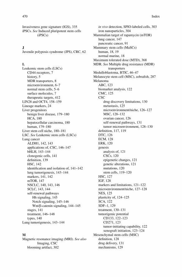
470 Index
Invasiveness gene signature (IGS), 335iPSCs. See Induced pluripotent stem cells
(iPSCs)
JJuvenile polyposis syndrome (JPS), CRC, 62
LLeukemic stem cells (LSCs)
CD44 receptors, 7history, 5MDR transporters, 8microenvironment, 6–7normal stem cells, 5–6surface molecules, 7therapeutic targets, 412
LIN28 and OCT4, 158–159Lineage markers, 24Liver progenitors
benign liver disease, 179–180HCA, 180hepatocellular carcinoma, 180human, 179–180
Liver stem cell niche, 180–181LSC. See Leukemic stem cells (LSCs)Lung cancer
ALDH1, 142, 143applications of, CSC, 146–147bHLH, 143–144clonogenic cells, 141definition, 139HSC, 142identification and isolation of, 141–142lung tumorigenesis, 143–144markers, 141, 142mTOR, 147NSCLC, 140, 143, 146SCLC, 143, 144self-renewal pathways
Hh signaling, 145Notch signaling, 145–146Wnt/b-catenin signaling, 144–145
stages, 141treatment, 146–148types, 140
Lung tumorigenesis, 143–144
MMagnetic resonance imaging (MRI). See also
Imaging, CSCblooming artifact, 302
in vivo detection, SPIO-labeled cells, 303iron nanoparticles, 304
Mammalian target of rapamycin (mTOR)lung cancer, 147pancreatic cancer, 91
Mammary stem cells (MaSCs)human, 18, 19normal murine, 18
Maximum tolerated dose (MTD), 368MDR. See Multiple drug resistance (MDR)
transportersMedulloblastoma, BTIC, 46–47Melanocyte stem cell (MSC), zebrafish, 287Melanoma
ABC, 123biomarker analysis, 122CMC, 125CSC
drug discovery limitations, 130metastasis, 125microenvironment/niche, 126–127MSC, 128–132ovarian cancer, 126self-renewal pathways, 131tumor microenvironment, 128–130
definition, 117, 119DTC, 126ECM, 128ERK, 120genesis
analysis of, 121CSCs, 120epigenetic changes, 121genetic alterations, 121mutations, 120stem cells, 119–120
HSC, 127IGF, 128markers and limitations, 121–122microenvironment/niche, 127–128NES, 125plasticity of, 124–125SCA, 122SDF–1, 129treatment, 130–131tumorigenic potential
CD133, 122–123CD271, 123tumor-initiating capability, 122xenograft initiation, 123–124
Mesenchymal stem cells (MSC)definition, 128drug delivery, 131mechanisms, 129
471Index
research on, 132tumor cells, 129tumor sites migration, 129–130
MMTV. See Murine mammary tumor virus (MMTV)
Mouse models, CSC studyadvancement of, 320hematopoietic malignancies, 319normal human stem cells
NOD/SCID, 315–316NOD/SCID b2M, 316–317NOD/SCID IL–2Rg, 317–318NOD/SCID MPSVII, 318SCID, 313–315xenotransplantation, 314
solid tumors, 319–320MTD. See Maximum tolerated dose (MTD)mTOR. See Mammalian target of rapamycin
(mTOR)Multimodality imaging, 304Multiple drug resistance (MDR) transporters
CSC, 8discovery, 363–364innate vs. acquired drug resistance,
371–372measurement, 364–366NTSC, 364
Murine mammary tumor virus (MMTV), 351–353
Musashi–1 (Msi–1), CRC, 64MYC, HCSC, 189Myeloid differentiation factor 88
(MyD88), 158
NNanog transcription, 211National Center for Biotechnology
Information (NCBI), 446–448Nestin (NES), 125Neural stem cells (NSCs)
BTIC, 39–40zebrafish, 286–287
Neurogenin 3 (Ngn3), 83Neurosphere (NS) cells, 383–384NICD. See Notch intracellular domain (NICD)Nodal signaling
embryonic development, 226–227expression, 227hESCs, 227pro-metastatic role, 228regulatory pathways, 214–215signaling, 227TGF-b, 226
NOD/SCID. See Nonobese diabetic plus severe combined immunodeficient (NOD/SCID)
Nonadherent sphere culture, 270–271Nonobese diabetic plus severe combined
immunodeficient (NOD/SCID)beta2 migroglobulin (b 2M), 316–317breast cancer, 383, 384, 392IL–2 receptor g (IL–2R g), 317–318mucopolysaccharidosis type VII, 318
Non small-cell lung cancer (NSCLC), 140, 143, 146
Nontumorigenic cell (NTC), 347Notch intracellular domain (NICD)
embryonic microenvironment, 228OCSCs, 162
Notch signaling pathwayscellular plasticity, 233complexity, 230CRC, 61HCSC, 188lung cancer, 145–146mammalian, 228OCSCs, 162oncogenic, 229–230pancreatic cancer, 91T-ALL, 229therapeutic targets, 413
NSCLC. See Non small-cell lung cancer (NSCLC)
NSCs. See Neural stem cells (NSCs)NTC. See Nontumorigenic cell (NTC)
OOCSCs. See Ovarian cancer
stem cell (OCSCs)Oct4, 210–211Oligoastrocytomas, 47Oligodendrogliomas, 47Optical imaging, CSC
BLI, 300limitations, 300ZsGreen, 301
OSE. See Ovarian surface epithelium (OSE)
Ovarian cancerbiology and pathology, 152–153definition, 151EOC, 152–153OCSCs
differentiation therapies, 163–164elimination therapies, 161–163epigenetic therapies, 164–166
472 Index
Ovarian cancer (cont.)isolation and characterization of,
154–159niche, 164origin of, 159–161
Ovarian cancer stem cell (OCSCs)ATRA, 163differentiation therapies, 163–164DNMT, 165DTEP, 160DTP, 160elimination therapies
Notch pathway, 162PI3K/Akt signaling, 161–162SHH, 162Wnt Signaling, 162–163
epigenetic therapies, 164–166FTE, 160gene expression analyses, 160HDAC, 165–166HOX, 160IFN-a, 154–155isolation and characterization of
ALDH1, 159ascites, 155CD24, 158CD44, 157CD117, 155–157CD133, 157–158LIN28 and OCT4, 158–159MyD88, 158putative, 154–155surface markers, 155, 156
NICD, 162niche, 164origin of, 159–161OSE, 153, 156, 159–160SCF, 156tumor chemotherapy, 165
Ovarian surface epithelium (OSE), 153, 156, 159–160
Ovarian tumor chemotherapy, 165
PPaired box gene 4 (Pax4), 82, 83Pancreas-specific transcription factor 1
(Ptf1), 82Pancreatic adenocarcinoma.
See Pancreatic cancerPancreatic and duodenal homeobox 1
(Pdx1), 81–83
Pancreatic cancerABC, 90ALDH1, 87Arx, 83CAC, 85cancer progression model, 91–92chronic pancreatitis role, 86clinical records, 80, 81CSC
CD133, 87CXCR4 receptor, 89EMT, 89hypothesis, 86–87markers, 87stem cell properties, 87–88tumor-initiating cells, 87, 88
CXCR4, 88, 89description, 80EMT, 83, 89EpCAM, 87, 88mTOR, 91Ngn3, 83Notch pathway, 91origin of cells
cell types, 84ductal morphology, 85early stages, 85Kras activation, 84, 85mouse models, 84tumor formation, 83
PanIN, 84Pax4, 82, 83Pdx1, 81–83Ptf1, 82SDF–1, 88SP cells, 90stem cells
a-and b-cell fate, 83exocrine fate, 83transcription factors, 81, 82
telomeres, 90therapeutic implications, 89–91
Pancreatic intraepithelial neoplasia (PanIN), 84
Pareidolia, 436, 437Pax4. See Paired box gene 4 (Pax4)Pdx1. See Pancreatic and duodenal
homeobox 1 (Pdx1)PET. See Positron emission
tomography (PET)Phosphatase and tensin homolog
(PTEN), 61, 355
473Index
Phosphati-dylinositol 3-kinase (PI3K), 61PI3K/Akt signaling, 161–162Plasticity
cellular, 230–233Nodal signaling
embryonic development, 226–227expression, 227hESCs, 227pro-metastatic role, 228signaling, 227TGF-b, 226
Notch signalingcomplexity, 230mammalian, 228oncogenic, 229–230T-ALL, 229
tumor, 233–235Positron emission tomography (PET),
301–302Prostate cancer
AML, 104anatomy and development, 100–101AR, 101, 104BER, 108BPH, 100, 103BRCA, 108cell types, 101CSC, 104–105description, 99, 103–104DSB, 108epithelial stem cells
basal and luminal cells, 101–102identification and characterization, 103murine hematopoietic stem cell
marker, 102murine prostate, 102
gene expression, 105GSTP1, 107identification and isolation, 109IL-6, 106markers, 104–105MMR, 108molecular mechanisms
epigenetic deregulation, 106–107JAK-STAT signaling, 106Wnt/b-catenin signaling, 105–106
normal prostate tissue model, 101PSA, 103therapy resistance, 107–109TMPRSS2, 105
Protein homology, of growth factor ligands and receptors, 447–448
PTEN. See Phosphatase and tensin homolog (PTEN)
Ptf1. See Pancreas-specific transcription factor 1 (Ptf1)
RRadiation therapy (RT), 408–409Radionuclide imaging, 305Radioresistance
ALDH, 347CSC
enhanced DNA repair, 351–352hypoxic microenvironment, 353induced autophagy, 354low ROS levels, 352notch pathway activation, 354overcoming, 354–356theory of, 346–347wnt signaling pathway, activation,
352–353DNA, 348, 350–352MMTV, 351–353redistribution of cells, 348–349reoxygenation, 349repopulation of cells, 349
Radiotherapy, 49RAR. See Retinoic acid receptor (RAR)Ras signaling, EMT, 248Reactive oxygen species (ROS), 350, 352Receptor tyrosine kinase (RTK), 248Regulatory pathways
Activin signaling, 214–215ES cells
epigenetics, 211Nanog, 211Oct4, 210–211Sox2, 211
FGF signaling, 213–214hESC, 210, 211, 213–214iPSCs, 215–216miRNA, 211Nodal signaling, 214–215TGFb signaling, 214–215Wnt signaling, 212–213
Retinoic acid receptor (RAR), 415RT. See Radiation therapy (RT)RTK. See Receptor tyrosine kinase (RTK)
SSCA. See Sphere cell formation assay (SCA)SCF. See Stem cell factor (SCF)SCID. See Severe combined immunodeficient
(SCID)SCID leukemia-initiating cells (SL-IC), 319
474 Index
SCLC. See Small-cell lung cancer (SCLC)SDF–1. See Stromal derived factor–1
(SDF–1)Severe combined immunodeficient (SCID),
313, 315Shh. See Sonic hedgehog (Shh)Side population (SP)
ABC transporters, 365HNSCC, 202–203human HCC, 181–182
self-renewal and tumor initiation capacity, 182
therapeutic resistance, 182–183normal livers, 181stem cell assays, 262
Single photon emission computed tomography (SPECT), 301–302
SL-IC. See SCID leukemia-initiating cells (SL-IC)
Smad signaling, 247Small-cell lung cancer (SCLC), 143, 144Snail/slug signaling, 414Sonic hedgehog (Shh)
OCSCs, 162therapeutic targets, 410, 412
Sox2, 211SP. See Side population (SP)Specificity protein (Sp1), EMT, 246SPECT. See Single photon emission
computed tomography (SPECT)Sphere cell formation assay (SCA), 122Sphingosine–1-phosphate (SP1), 371Stem cell assays
breast cancer, 261–262in vitro methods
ALDEFLUOR assay, 266–268cell surface markers, 263–266holoclone formation, 271–272immunohistochemical staining,
268–269nonadherent sphere culture, 270–271PKH26 Labeling, 269–270side population, 262
in vivo modelsextra cellular matrix component
laminin, 274human hematopoietic cell, 274humanizing techniques, 273xenograft model, 273, 275xenotransplantation method, 273
methodologies, 261Stem cell factor (SCF)
OCSCs, 156therapeutic targets, 414
Stem cell pathways, 409–414hedgehog signaling, 410–413notch signaling, 413snail/slug signaling, 414Wnt/b-catenin signaling, 414
Stroma and microenvironmentcells in, 442–443growth factors, 446in vitro assays for, 449mice and humans, difference between,
445–446mouse models, 443–445protein homology, of growth factor ligands
and receptors, 447–448Stromal derived factor–1 (SDF–1)
melanoma, 129pancreatic cancer, 88
TT-acute lymphoblastic leukemia (T-ALL)
embryonic microenvironment, 229zebrafish, 290
Telomeres, 90TGF-b signaling. See Transforming growth
factor beta (TGFb) signalingTherapeutic targets, CSC
acquired resistance stochastic model, 405–406
ATRA, 417–419differentiation therapy, 414–419
ALDH, 417–418APL, 415–417retinoid signaling, 415solid tumors, 417–418
GBM, 407, 408HSC, 415implications, 407–409
cytotoxic therapy, 407–408radiation therapy, 408–409
innate resistance, 406RAR, 415stem cell pathways, 409–414
hedgehog signaling, 410–413notch signaling, 413snail/slug signaling, 414Wnt/b-catenin signaling, 414
TIC. See Tumor-initiating cells (TIC)Transforming growth factor beta (TGFb)
signalingembryonic microenvironment, 226EMT, 247HCSC, 187–188regulatory pathways, 214–215
475Index
Tumor-initiating cells (TIC), 442, 444, 449, 452Tumor plasticity
anti-cancer strategies, 234embryonal carcinoma cells, 235in vitro 3D model, 234–235zebrafish models, 234
VVentricular zone (VZ), zebrafish, 286
WWingless (Wnt) signaling pathway,
60–61, 352–353EMT, 247–248HCSC, 187OCSCs, 162–163regulatory pathways, 212–213
Wnt/b-catenin signaling pathways, 144–145, 414
XXenograft heterogeneity, 450–452Xenotransplantation, 314
ZZebrafish, CSC study
cancer modelsCre/lox technology, 290gene identification, 289pancreatic neuroendocrine
tumors, 291Rag2 promoter, 290
and CSC, 292–293development
HSC, 284–286MSC, 287NSC, 286–287
ERMS, 290hematopoiesis, 284T-ALL, 290tissue regeneration
fin, 288–289heart, 288physiological and pathological
aging, 289transgenic, 293tumor plasticity, 234VZ, 286
Related Documents