Statin-triggered cell death in primary human lung mesenchymal cells involves p53-PUMA and release of Smac and Omi but not cytochrome c Saeid Ghavami a,b,c , Mark M. Mutawe a,c , Kristin Hauff d , Gerald L. Stelmack a,c , Dedmer Schaafsma a,c , Pawan Sharma a,b,c , Karol D. McNeill a,c , Tyler S. Hynes a,b,c , Sam K. Kung e , Helmut Unruh f , Thomas Klonisch g , Grant M. Hatch d , Marek Los h , Andrew J. Halayko a,b,c,f, ⁎ a Department of Physiology, University of Manitoba, Winnipeg, MB, Canada b National Training Program in Allergy and Asthma, University of Manitoba, Winnipeg, MB, Canada c Biology of Breathing Group, Manitoba Institute of Child Health, Winnipeg, MB, Canada d Department of Pharmacology, University of Manitoba, Winnipeg, MB, Canada e Department of Immunology, University of Manitoba, Winnipeg, MB, Canada f Department of Internal Medicine, University of Manitoba, Winnipeg, MB, Canada g Department of Human Anatomy and Cell Science, University of Manitoba, Winnipeg, MB, Canada h Interfaculty Institute of Biochemistry, Univ. Tuebingen, Germany abstract article info Article history: Received 5 October 2009 Received in revised form 16 December 2009 Accepted 16 December 2009 Available online 4 January 2010 Keywords: Apoptosis Statin Caspase Airway smooth muscle Fibroblast Mitochondria Statins inhibit 3-hydroxy-3-methyl-glutarylcoenzyme CoA (HMG-CoA) reductase, the proximal enzyme for cholesterol biosynthesis. They exhibit pleiotropic effects and are linked to health benefits for diseases including cancer and lung disease. Understanding their mechanism of action could point to new therapies, thus we investigated the response of primary cultured human airway mesenchymal cells, which play an effector role in asthma and chronic obstructive lung disease (COPD), to simvastatin exposure. Simvastatin induced apoptosis involving caspase-9, -3 and -7, but not caspase-8 in airway smooth muscle cells and fibroblasts. HMG-CoA inhibition did not alter cellular cholesterol content but did abrogate de novo cholesterol synthesis. Pro-apoptotic effects were prevented by exogenous mevalonate, geranylgeranyl pyrophosphate and farnesyl pyrophosphate, downstream products of HMG-CoA. Simvastatin increased expression of Bax, oligomerization of Bax and Bak, and expression of BH3-only p53-dependent genes, PUMA and NOXA. Inhibition of p53 and silencing of p53 unregulated modulator of apoptosis (PUMA) expression partly counteracted simvastatin-induced cell death, suggesting a role for p53-independent mechanisms. Simvastatin did not induce mitochondrial release of cytochrome c, but did promote release of inhibitor of apoptosis (IAP) proteins, Smac and Omi. Simvastatin also inhibited mitochondrial fission with the loss of mitochondrial Drp1, an essential component of mitochondrial fission machinery. Thus, simvastatin activates novel apoptosis pathways in lung mesenchymal cells involving p53, IAP inhibitor release, and disruption of mitochondrial fission. © 2009 Elsevier B.V. All rights reserved. 1. Introduction Due to structural similarity with 3-hydroxy-3-methyl-glutaryl-CoA (HMG-CoA), statins such as simvastatin, lovastatin and atorvastatin, inhibit HMG-CoA reductase, the proximal rate-limiting enzyme in cholesterol biosynthesis [1]. They effectively lower serum cholesterol but overall benefits exceed that predicted by this outcome alone, suggesting additional cholesterol-independent effects [2]. Thus, the impact of statins on disease states including cancer, neurological disorders, diabetes, kidney disease, lung infection, and chronic inflammatory lung disease, is being assessed in clinical trials [2,3]. HMG-CoA reductase catalyzes the conversion of HMG-CoA to the fatty acid mevalonate [2], which undergoes stepwise conversion into isoprenoids, farnesyl pyrophosphate (FPP) and geranylgeranyl pyro- phosphate (GGPP). Isoprenoids can be covalently linked to signaling Biochimica et Biophysica Acta 1803 (2010) 452–467 Abbreviations: AIF, apoptosis-inducing factor; DISC, death-induced signaling complex; Drp1, dynamin-related protein 1; Endo G, Endo nuclease G; FADD, Fas-Associated Death Domain; FPP, farnesyl pyrophosphate; GGPP, geranylgeranyl pyrophosphate; GAPDH, Glyceraldehyde 3-phosphate dehydrogenase; HASM, Human airway smooth muscle; HAF, Human airway fibroblast; HMG-CoA, 3-hydroxy-3-methyl-glutaryl-CoA; HtrA2, high- temperature requirement A2; IAP, inhibitor of apoptosis protein; IBM, IAP binding motif; JC-1, 5,5′,6,6′-tetrachloro-1,1′,3,3′-tetraethylbenzimidazolylcarbocyanine iodide; ΔΨ m , mito- chondrial trans-membrane potential; MTT, 3-(4,5-dimethyl-2-thiazolyl)-2,5-diphenyl-2H- tetrazolium bromide; NAC, N-acetyl-L-cysteine; PCD, programmed cell death; PUMA, p53 unregulated modulator of apoptosis; ROS, reactive oxygen species; shRNAi, small hairpin inhibitory RNA; Smac, second mitochondria-derived activator of caspases; XIAP, X-linked inhibitor of apoptosis protein ⁎ Corresponding author. Departments of Physiology and Internal Medicine Univer- sity of Manitoba, Winnipeg, MB, Canada. Tel.: +1 204 787 2062. E-mail address: [email protected] (A.J. Halayko). 0167-4889/$ – see front matter © 2009 Elsevier B.V. All rights reserved. doi:10.1016/j.bbamcr.2009.12.005 Contents lists available at ScienceDirect Biochimica et Biophysica Acta journal homepage: www.elsevier.com/locate/bbamcr

Welcome message from author
This document is posted to help you gain knowledge. Please leave a comment to let me know what you think about it! Share it to your friends and learn new things together.
Transcript

Biochimica et Biophysica Acta 1803 (2010) 452–467
Contents lists available at ScienceDirect
Biochimica et Biophysica Acta
j ourna l homepage: www.e lsev ie r.com/ locate /bbamcr
Statin-triggered cell death in primary human lung mesenchymal cells involvesp53-PUMA and release of Smac and Omi but not cytochrome c
Saeid Ghavami a,b,c, Mark M. Mutawe a,c, Kristin Hauff d, Gerald L. Stelmack a,c, Dedmer Schaafsma a,c,Pawan Sharma a,b,c, Karol D. McNeill a,c, Tyler S. Hynes a,b,c, Sam K. Kung e, Helmut Unruh f,Thomas Klonisch g, Grant M. Hatch d, Marek Los h, Andrew J. Halayko a,b,c,f,⁎a Department of Physiology, University of Manitoba, Winnipeg, MB, Canadab National Training Program in Allergy and Asthma, University of Manitoba, Winnipeg, MB, Canadac Biology of Breathing Group, Manitoba Institute of Child Health, Winnipeg, MB, Canadad Department of Pharmacology, University of Manitoba, Winnipeg, MB, Canadae Department of Immunology, University of Manitoba, Winnipeg, MB, Canadaf Department of Internal Medicine, University of Manitoba, Winnipeg, MB, Canadag Department of Human Anatomy and Cell Science, University of Manitoba, Winnipeg, MB, Canadah Interfaculty Institute of Biochemistry, Univ. Tuebingen, Germany
Abbreviations: AIF, apoptosis-inducing factor; DISC, deDrp1, dynamin-related protein 1; Endo G, Endo nucleaseDomain; FPP, farnesyl pyrophosphate; GGPP, geranylgeGlyceraldehyde 3-phosphate dehydrogenase; HASM, HumHuman airway fibroblast; HMG-CoA, 3-hydroxy-3-mettemperature requirement A2; IAP, inhibitor of apoptosis pJC-1, 5,5′,6,6′-tetrachloro-1,1′,3,3′-tetraethylbenzimidazolylchondrial trans-membrane potential; MTT, 3-(4,5-dimethtetrazolium bromide; NAC, N-acetyl-L-cysteine; PCD, progunregulated modulator of apoptosis; ROS, reactive oxygeninhibitory RNA; Smac, second mitochondria-derived activinhibitor of apoptosis protein⁎ Corresponding author. Departments of Physiology
sity of Manitoba, Winnipeg, MB, Canada. Tel.: +1 204 7E-mail address: [email protected] (A.J. Halay
0167-4889/$ – see front matter © 2009 Elsevier B.V. Adoi:10.1016/j.bbamcr.2009.12.005
a b s t r a c t
a r t i c l e i n f oArticle history:Received 5 October 2009Received in revised form 16 December 2009Accepted 16 December 2009Available online 4 January 2010
Keywords:ApoptosisStatinCaspaseAirway smooth muscleFibroblastMitochondria
Statins inhibit 3-hydroxy-3-methyl-glutarylcoenzyme CoA (HMG-CoA) reductase, the proximal enzyme forcholesterol biosynthesis. They exhibit pleiotropic effects and are linked to health benefits for diseasesincluding cancer and lung disease. Understanding their mechanism of action could point to new therapies,thus we investigated the response of primary cultured human airway mesenchymal cells, which play aneffector role in asthma and chronic obstructive lung disease (COPD), to simvastatin exposure. Simvastatininduced apoptosis involving caspase-9, -3 and -7, but not caspase-8 in airway smooth muscle cells andfibroblasts. HMG-CoA inhibition did not alter cellular cholesterol content but did abrogate de novocholesterol synthesis. Pro-apoptotic effects were prevented by exogenous mevalonate, geranylgeranylpyrophosphate and farnesyl pyrophosphate, downstream products of HMG-CoA. Simvastatin increasedexpression of Bax, oligomerization of Bax and Bak, and expression of BH3-only p53-dependent genes, PUMAand NOXA. Inhibition of p53 and silencing of p53 unregulated modulator of apoptosis (PUMA) expressionpartly counteracted simvastatin-induced cell death, suggesting a role for p53-independent mechanisms.Simvastatin did not induce mitochondrial release of cytochrome c, but did promote release of inhibitor ofapoptosis (IAP) proteins, Smac and Omi. Simvastatin also inhibited mitochondrial fission with the loss ofmitochondrial Drp1, an essential component of mitochondrial fission machinery. Thus, simvastatin activatesnovel apoptosis pathways in lung mesenchymal cells involving p53, IAP inhibitor release, and disruption ofmitochondrial fission.
ath-induced signaling complex;G; FADD, Fas-Associated Deathranyl pyrophosphate; GAPDH,an airway smooth muscle; HAF,hyl-glutaryl-CoA; HtrA2, high-rotein; IBM, IAP binding motif;carbocyanine iodide;ΔΨm, mito-yl-2-thiazolyl)-2,5-diphenyl-2H-rammed cell death; PUMA, p53species; shRNAi, small hairpin
ator of caspases; XIAP, X-linked
and Internal Medicine Univer-87 2062.ko).
ll rights reserved.
© 2009 Elsevier B.V. All rights reserved.
1. Introduction
Due to structural similaritywith 3-hydroxy-3-methyl-glutaryl-CoA(HMG-CoA), statins such as simvastatin, lovastatin and atorvastatin,inhibit HMG-CoA reductase, the proximal rate-limiting enzyme incholesterol biosynthesis [1]. They effectively lower serum cholesterolbut overall benefits exceed that predicted by this outcome alone,suggesting additional cholesterol-independent effects [2]. Thus, theimpact of statins on disease states including cancer, neurologicaldisorders, diabetes, kidney disease, lung infection, and chronicinflammatory lung disease, is being assessed in clinical trials [2,3].HMG-CoA reductase catalyzes the conversion of HMG-CoA to the fattyacid mevalonate [2], which undergoes stepwise conversion intoisoprenoids, farnesyl pyrophosphate (FPP) and geranylgeranyl pyro-phosphate (GGPP). Isoprenoids can be covalently linked to signaling

453S. Ghavami et al. / Biochimica et Biophysica Acta 1803 (2010) 452–467
proteins, in particular to members of the Ras superfamily, enablingattachment to lipid membranes required for activation and subse-quent regulation of critical cell functions [4]. Suppression of proteinprenylation is a key component of the broad effects of statins, but is itunclear if there are other cholesterol-independent mechanisms thatunderpin functional effects in different cell types. Understanding thesemechanisms offers potential to identify new therapies to prevent orreverse human disease.
Statins can induce apoptosis in some cancer cell lines, primarycultured vascular smoothmuscle, mesangial cells, and fibroblasts [5–7].Consistent with a role for FPP and GGPP in this response, isoprenoidreplacement overcomes pro-apoptotic effects of statins, the selectiveinhibition of farnesyl- and geranylgeranyl transferases can mimic pro-apoptotic effects, and prenylation-dependent membrane-associatedGTPase signaling pathways may mediate the response [5,8,9]. None-theless, more downstream apoptotic signaling induced by statinsremains incompletely understood. Apoptosis is under complex controlby a receptor-mediated extrinsic pathway, and an intrinsic pathwaytriggered by a wide range of cellular stressors [10]. The intrinsicpathway is chiefly regulated by Bcl-2 family proteins via the disruptionofmitochondrial trans-membranepotential (ΔΨm)and the formationofmembrane permeability pores through which mitochondrial proteinsare released to promote activation of caspase-9 [11]. For example, pro-apoptotic Bax and Bak proteins associate and form mitochondrialpermeability pores, a process that can be inhibited by anti-apoptoticproteins such as Bcl-XL [12]. Pro-apoptotic proteins also include BH3-only proteins, such as p53-upregulatedmodulator of apoptosis (PUMA)and NOXA [10,13], which can inhibit the anti-apoptotic action of Bcl-XLto promote p53 assisted mitochondrial membrane translocation of Baxand Bak [12,14]. Proteins released from mitochondria include cyto-chrome c, which together with caspase-9, (d)ATP, and Apaf-1 formapoptosomes. Like its extrinsic pathway counterpart caspase-8, cas-pase-9 cleaves and activates caspase-3 and -7 to drive the executionphase of apoptosis [15]. Mitochondria also release Smac/DIABLO andOmi/HtrA2 that bind to, and suppress inhibitor of apoptosis proteins(IAPs) that otherwise prevent caspase activity [16,17].
Almost all data on statin-induced apoptosis have been obtained oncancer cell lines and there remains some controversy. Statins inducechromatin condensation andDNA laddering [18,19], and in a few casesactivate caspase-8 as well as an upstream receptor, FAS/CD95 [20].Notably, in human T-, B-, and myeloma tumor cells statin exposureactivates both caspase-8 and -9, disrupts ΔΨtm, and promotes releaseof Smac frommitochondria [20]. However, inhibition of caspase-8 hasno effect on apoptosis induction by lovastatin in mammary carcinoma[18]. In contrast, inhibitors of caspase-9 or executioner-caspasessuppress statin-induced apoptosis [18]. Lovastatin-induced apoptosishas been shown to dependon the loss of the anti-apoptotic proteinBcl-2 in acute mylogeneous leukemia cells. In p53-deficient culturedbreast cancer cells statins can induce mitochondrial translocation ofBax with a concomitant loss of mitochondrial membrane potential(ΔΨm) [8,18]. Thus, so far published data do not clearly depict thedownstream mechanisms involved in statin-triggered apoptosis incancer cells. Furthermore, despite extensivemedical use the potential,and mechanisms of apoptosis induction by statins in primary cellshave not been systematically investigated.
In this study we investigated apoptosis mechanisms invoked byHMG-CoA reductase inhibition in human lung mesenchymal cells. Wefound that simvastatin induced apoptosis via a novel intrinsicpathway. Studies were performed on primary cultured human airwaysmooth muscle (HASM) cells and airway fibroblasts (HAF) as thesecells underpin acute and chronic wound healing and inflammationassociated with prevalent lung diseases such as asthma, chronicobstructive lung disease (COPD), and cystic fibrosis [21,22]. Indeed,there is new evidence suggesting statins have beneficial effects onthese conditions [23–25]. Moreover, many therapies under develop-ment for these pathologies aim to reduce airway mesenchymal cell
number. Thus, unraveling mechanisms of statin-mediated cell deathwithin HAF and HASM may reveal new therapeutic targets.
2. Methods
2.1. Reagents
Cell culture plasticware was obtained from Corning Costar Co.(Canada). Cell culture media, propidium iodide (PI), simvastatin,NS3694, mevalonate, farnesyl pyrophosphate (FPP), geranylgeranylpyrophosphate (GGPP), cyclic Pifithrin-α, and 3-(4,5-dimethyl-2-thiazolyl)-2,5-diphenyl-2H-tetrazolium bromide) (MTT), and N-ace-tyl-L-cysteine (NAC) were obtained from Sigma (Sigma-Aldrich,Oakville, CA). [14C]acetate and [14C]mevalonate were purchasedfrom Perkin Elmer (Vaudreuil-Dorion, Quebec PQ). Rabbit anti-humancleaved caspase-6, -7, -8, poly ADP-ribose polymerase (PARP), rabbitanti-human Bak, Bax, PUMA, Bcl-2, Bid, phospho-p53 (Ser 15) and(Ser 37), Rac1/2/3, p21 and cytochrome c were purchased from CellSignaling (Canada). Rabbit anti-Drp1, rabbit anti-glyceraldehyde-3-phosphate dehydrogenase (GAPDH), rabbit anti-Smac/DIABLO, rabbitanti-Omi/HtrA2, mouse anti-cytochrome c, and goat anti-endonucle-ase G (Endo G) were obtained from Santa Cruz Biotechnologies (USA).Mouse anti-p53 and anti-NOXA were obtained from Abcam (USA).5,5′,6,6′-tetrachloro-1,1′,3,3′-tetraethylbenzimidazolylcarbocyanineiodide (JC-1), Mitotracker Red, and Mitosox were obtained fromInvitrogen Molecular Probes (Canada). Caspase-Glo®-3/7, Caspase-Glo®-8 and Caspase-Glo®-9 assay were purchased from Promega(USA). N7-Smac peptide (cell permeable) was purchased fromCalbiochem (Canada).
2.2. Primary HASM and HAF cell culture preparation
For all experiments we used primary cultured human airwaysmooth muscle (HASM) cells and airway fibroblasts (HAF) that wereprepared from 2–4th generation bronchi in macroscopically healthysegments of resected lung specimens. After microdissection toseparate the lamina reticularis and submucosal compartment fromencircling airway smooth muscle bundle, HAF and HASM, respective-ly, were isolated by enzymatic dissociation as we have described[26,27]. All procedures were approved by the Human Research EthicsBoard (University of Manitoba) and all donors gave informed consent.Unless otherwise stated cells were cultured in Dulbecco's modifiedEagle's medium (DMEM) supplemented with 10% fetal bovine serum(FBS) and medium was changed every 48 h. For all experiments,passage 3–7 of HASM and HAF were used.
2.3. Cell viability-, cell death-, and related assays
Cytotoxicity of simvastatin towards the HASM and HAF wasdetermined by MTT-assays as previously described [16]. Apoptosiswas measured using the Nicoletti method as we have describedpreviously [16]. Luminometric assays Caspase-Glo®-8, -9 and -3/7(Promega, Canada, Nepean, ON) were used to measure the proteolyticactivity of caspases-3/7 (DEVD-ase), -8 (IETD-ase), and -9 (LEHD-ase)as we have previously done [28]. Mitochondrial membrane potentialwasmeasuredemploying themitochondria-specific cationic ratiometricdye JC-1 that undergoes ΔΨm-dependent aggregation in the mitochon-dria: JC-1 exists as a green fluorescent (540 nm, excitation 490 nm)monomer at ΔΨmb140 mV, but when ΔΨmN140 mV JC-1 aggregatesand emit red fluorescence (590 nm, excitation 540 nm) [16,29].
2.4. Quantitative analysis of nuclear DNA fluorescence usinglaser scanning cytometry
Human bronchial smooth muscle cells were plated in 12 well cellculture clusters and grown to 80% confluence, maintained for 48 h in
454 S. Ghavami et al. / Biochimica et Biophysica Acta 1803 (2010) 452–467
DMEM/0.5% FBS, prior to addition of simvastatin (10 µM) or vehiclefor 96 h. Fluorescent staining of live cell nuclear DNA was carried outby incubation in HBSS containing 5 µg/mL H33342 dye (15 min,37 °C). Quantification of nuclear DNA fluorescence was carried out atroom temperature using an iCYS laser scanning cytometer (Compu-cyte Corp., Westwood, MA, USA). Signals for blue channel fluores-cence (Ex: 405 nm; Em: 463/39 nm) from contoured cell nuclei andlight scatter data (488 nm argon laser line) for shaded relief imageswere captured. The DNA content data, collected from a circular scanarea 0.12 cm2 with individual scan field dimensions of 400×384 µm,included total integrated fluorescence and maximum pixel fluores-cence, which provides a measure of nuclear condensation, within theboundary of each contoured cell nucleus. As photomultiplier (PMT)detectors have a bit depth of 14, individual pixel elements may havegrey level values between 0–16,384.
2.5. Analysis of cellular morphology
Toassess cell viability based on gross cellular appearance (chromatincondensation and cell shrinkage) HASM andHAF cells grown on 12wellplates were assessed by phase contrast microscopy (Olympus CK40)using a Olympus DP10 CCD digital camera to capture images.
2.6. Total cellular cholesterol assay
HASM and HAF cells (48 h in 0.5% FBS) were treated with sim-vastatin (10 µM) of vehicle (DMSO) for 48 or 96 h, then cells wereharvested, washed with cold PBS, and centrifuged (800×g, 5 min).Cells were mixed with a 1:1 water–methanol solution and then a 1:5water–chloroform solution before centrifuged in desktop microfuge(800×g, 10 min). The organic phase was dried and then isopropylalcohol and Triton X-100 (0.1%) was added to cells to extractcholesterol, which was subsequently quantified using the AmexInvitrogen Cholesterol Assay according to the manufacturer's proto-col. The assay uses enzyme-coupled reactions that hydrolyzedcholesteryl esters by cholesterol esterase into cholesterol, and thenall cholesterol into H2O2 and a ketone by cholesterol oxidase. H2O2 isdetected using 10-acetyl-3,7-dihydroxyphenoxazine, which in thepresence of horseradish peroxidase (HRP) emits fluorescence in 1:1stoichiometry with H2O2 [30,31].
2.7. Cholesterol de novo synthesis assay
HASM and HAF cells (48 h, 0.5% FBS) were treated with simvastatin(10 µM) or vehicle control (DMSO) for up to 96 h and cholesterolsynthesis was determined using amodification of theMokashi protocol[32]. [14C]Acetate (Perkin Elmer, 60 mCi/mmol) was added (1 μCi/well), cells were incubated at 37 °C overnight. Mevinolin (1 µM, SigmaChemical) was included in incubations with [14C]mevalonate to blockHMG-CoA reductase, and lipid-replete serum was used in all studies.Medium was removed, cells washed twice with 1% PBS (pH 7.4),harvested by scraping, re-suspended in 20 mM Tris–EDTA (pH 7.4)containing 0.1% Triton X-100, and then lysed by sonication at mediumsetting three times for 5 s with a Sonic Dismembrator (Fisher Scientific,Canada). Lipids were extracted without saponification (to allowquantitation of cholesterol-esters) with a mixture of chloroform/methanol (2:1), subjected to centrifugal evaporation, re-suspended in50 µL of same and then resolved by silica thin-layer chromatography(Whatman) in petroleum ether/ethyl ether/acetic acid (60:40:1).Radiolabeled cholesterol and cholesterol-esters were visualized andquantified by electronic autoradiography (Packard Instant Imager) andauthenticated by comparison to standards visualized by iodine-vaporstain. Cholesterolwas also authenticated by gas-chromatographic/massspectrometry of eluted samples.
2.8. Membrane anchoring of Rho GTPases
For determination of membrane anchoring of prenylated Rho andRac GTPases, HASM cells were cultured in DMEM/0.5% FBS in thepresence or absence of simvastatin (10 µM), and after washing cellswere scraped in ice cold buffer (10 mM Tris–HCl, pH 7.5, 0.1 mMEDTA, 0.1 mM EGTA, 1 mM dithiothreitol, and protease inhibitorcocktail), sonicated on ice 3 times for 5 s, and then the homogenatewas separated into cytoplasmic and membrane fractions by ultra-centrifugation (100,000×g for 35 min) [33]. The membrane fractionswere solubilized in dissociation buffer (50 mM Tris–HCl, pH 7.5,0.15 M NaCl, 1 mM dithiothreitol, 1% SDS, 1 mM EDTA, 1 mM EGTA,protease inhibitor cocktail), and subsequently size fractioned by SDS-PAGE for immunoblot analysis using anti-Rac1/2/3 and anti-RhoAprimary antibodies (Cell Signaling).
2.9. MitoSox reactive oxygen species assay
As mitochondria can be a source of ROS, we used cell permeantMitoSOX™ Red, which can be oxidized selectively by mitochondrialsuperoxide, a reaction is prevented by superoxide dismutase in livecells, but that is not subject to effects of other ROS− or reactivenitrogen species (RNS). Once in the mitochondria, dye oxidized bysuperoxide exhibits red fluorescence. HASM and HAF sub-cultured in12 well plate (35,000 cells/well) DMEM/0.5% FBS were treated withsimvastatin (10 μM) or vehicle for up to 72 h, then 5 μM MitoSOX™reagent was added for 10 min (37 °C) in the dark. After washing cellDNA was stained with Hoechst 33256 (5 μg/mL, 15 min), fixed (4%parafomaldehyde, 120 mM sucrose) and cell staining quantified usinga iCys Laser Scanning Cytometry (LSC) (CompuCyte Corporation,Cambridge, MA).
2.10. Immunoblotting
We usedWestern blotting to detect cleaved caspase-8, -3, -9, -6, -7PARP, Blc2, Bid, PUMA, NOXA, Bax, DRP1, hFIS1, p53, phosphor-p53(Ser 15 and Ser37), Smac/DIABLO, Omi/HrA2, cytochrome c, Mn-SOD2, and GAPDH. Briefly, cells were washed and protein extractsprepared in lysis buffer (20 mM Tris–HCl (pH 7.5), 0.5% Nonidet P-40,0.5 mM PMSF, 100 µM β-glycerol 3-phosphate and 0.5% proteaseinhibitor cocktail). After a high-speed spin (13,000g×10 min) super-natant protein content was determined by Lowry protein assay, thenproteins were size fractionated by SDS-PAGE and transferred on tonylon membranes under reducing conditions, except for assessmentof Bak dimerization, with proteins separated under non-reducingconditions. After blocking membranes with non-fat dried milk andTween 20, blots were incubated overnight with the primary anti-bodies at 4 °C. HRP-conjugated secondary antibody incubationwas for1 h at room temperature, then blots were developed by enhancedchemiluminescence (ECL) detection (Amersham-Pharmacia Biotech).In experiments detecting Bak oligomerization non-reducing immu-noblotting was used.
2.11. Immunocytochemistry, confocal imaging and electron microscopy
For immunocytochemistry, HASM and HAF cells were grownovernight on coverslips and then treated with simvastatin (10 µM) orvehicle for 72 h prior to fixation (4% paraformaldehyde/120 mMsucrose) and permeabilization (0.1% Triton X-100). Cells wereincubated with rabbit anti-DIABLO IgG (1:150), goat anti-Endo GIgG (1:75), or mouse anti-AIF IgG (1:75), then with correspondingfluorochrome-conjugated secondary antibodies. Thereafter, mito-chondria were stained with Mitotracker Red CMXRos (MolecularProbes; 200 nM). The fluorescent images were then observed andanalyzed using an Olympus FluoView multi-laser confocal micro-scope. For transmission electron microscopy (TEM), cells were fixed

455S. Ghavami et al. / Biochimica et Biophysica Acta 1803 (2010) 452–467
(2.5% glutaraldehyde in PBS (pH 7.4) for 1 h at 4 °C) and post-fixed(1% osmium tetroxide) before embedding in Epon. TEM was per-formed with a Philips CM10, at 80 kV, on ultra-thin sections (100 nmon 200 mesh grids) stained with uranyl acetate and counterstainedwith lead citrate.
2.12. Subcellular fractionation
Cytosolic and mitochondrial fractions were generated using adigitonin-based subcellular fractionation technique at 4 °C [34]. Cellswere scraped, pelletedby centrifugation (800×g), thenwashed (PBSpH7.2) and re-centrifuged. Pellets were permeabilized for 5 min on ice:3×10⁷ cells/mL of cytosolic extraction buffer (250 mM sucrose, 70 mMKCl, 137 mM NaCl, 4.3 mM Na2HPO4, 1.4 mM KH2PO4 pH 7.2, 100 µMPMSF, 10 µg/mL leupeptin, 2 µg/mL aprotinin, containing 200 µg/mLdigitonin). Plasma membrane permeabilization was confirmed bystaining with 0.2% trypan blue solution then cells were pelleted(1000×g, 5 min). The supernatant (cytosolic fractions) was removed,then pellets were solubilized in the same volume of mitochondrial lysisbuffer (50 mMTris pH7.4, 150 mMNaCl, 2 mMEDTA, 2 mMEGTA, 0.2%Triton X-100, 0.3% NP-40, 100 µM PMSF, 10 µg/mL leupeptin, 2 µg/mLaprotinin), followed by pelleting at 10,000×g for 10 min at 4 °C.Supernatant was collected as mitochondrial fraction. For the detectionof specific protein by immunoblotting proteins, an equal amount ofcytosolic and pellet fraction protein were supplemented with 5×SDS-PAGE loading buffer, subjected to standard 12% or 15% SDS-PAGE andtransferred to nitrocellulose membranes.
2.13. Stable gene silencing: lentiviral delivery of shRNA
All short hairpin (sh)RNA containing constructs were from OpenBiosystems distributed by the Biomedical Functionality Resource, atUniversity of Manitoba. The PUMA shRNA construct (accession #NM_014417, cat# RHS3979-9601020), distributed in a bacterialculture of E. coli (DH5α), included a “stem” of 21 sense (GAGGGTCCTG-TACAATCTCAT) and antisense base pairs and a 6 base pair loop, clonedinto the lentiviral vector pLKO1. Individual colonies were grown up inLB broth with ampicillin (100 μg/mL), purified using a Qiagen Maxi-prep Kit (cat# 12663, Mississauga, ON). A vesicular stomatitis virus G(VSVG) pseudo-typed lentiviral vector wasmade using HEK 293T cellsby calcium phosphate transfection of purified PUMA shRNA plasmid,virus packaging vector (8.2Δvpr), and a VSVG plasmid as describedpreviously [35]. After 3 days the supernatant was collected andconcentrated by ultra-centrifugation (17,000 rpm for 90 min). Prima-ry HASM cells were grown to 70% confluence and transduced at a MOIof 6, in the presence of 8 μg/mL polybrene (final concentration), for2 h. Excess viral vectors were removed, and the transduced cells werecultured in fresh medium for 2 days before puromycin selection. Cellsexhibiting stable expression of the shRNAwere selected by growing inculture media containing puromycin (4 μg/mL) for at least 3 weeks.For control cells, in tandem with preparation of PUMA shRNAilentivirus a pLKO1 vector harboring “scrambled” non-coding shRNAwas also prepared and used to generate lentivirus for transduction ofthe same primary HASM cell lines that were used to generate PUMA-deficient stable cultures.
2.14. Quantitative PCR for PUMA and NOXA mRNA
Total cellular RNAwas isolatedusing theRNeasy PlusMiniKit (Qiagen,Mississauga, ON) then 1 µg was reverse transcribed using the QuantiTectReverse TranscriptionKit (Qiagen,Mississauga, ON). Abundance ofmRNAfor PUMA and NOXA was determined with an Applied Biosystems 7500Real-Time PCR System instrument using the Power SYBR Green PCRMaster Mix (Applied Biosystems) [36]. Oligonucleotide primers were asfollows: for NOXA, 5′-ATTACCGCTGGCCTACTGTG-3′ (forward) and 5′-GTGCTGAGTTGGCACTGAAA-3′ (reverse); for PUMA, 5′-CTGTGAATC-
CTGTGCTCTGC-3′ (forward) and 5′-AATGAATGCCAGTGGTCACA-3′ (re-verse). A dissociation curvewas generated at the end of each PCR reactiontoverify that a singleproductwas amplified. The relative expression levelsof genes normalized to the endogenous reference gene (18 S rRNA:primers=5′-CGCCGCTAGAGGTGAAATTC-3′ (forward) and 5′-TTGGCAA-ATGCTTTCGCTC-3′ (reverse)) and relative to vehicle treated controls wascalculated by the equation 2(−ΔΔCT). The ΔCT value was determined bysubtracting the average 18 s rRNA CT value from the average CT value ofthe corresponding target transcript. The calculation of ΔΔCT valuesinvolves subtraction of the ΔCT calibrator value (vehicle treated). For thevehicle treated samples ΔΔCT=0 and 20 equals 1. For the simvastatin-treated samples, 2−ΔΔCT indicates the fold change in gene expressionrelative to time-matched controls.
2.15. Statistical analysis
The results were expressed as means±SDE and statistical differ-ences were evaluated by one-way or two-way ANOVA followed byTukey's or Bonferroni's post hoc test, using Graph Pad Prism 4.0. Pb0.05was considered significant. For all experiments data was collected intriplicate from at least three cell lines unless otherwise indicated.
3. Results
3.1. Simvastatin induces apoptosis in human lung mesenchymal cells
We initially tested the concentration- and temporal effects ofsimvastatin on primary cultured HASM and HAF viability using MTTassay (Fig. 1A, B). Viability in both cell types was significantlycompromised after 48 h by maximum simvastatin concentrations(20 μM), whilst the lowest concentration of simvastatin used (5 μM)was significantly toxic (∼25%) only after 96 h; the maximum effectcaused by 20 μM simvastatin at this time point exceeded 50%. As weused multiple primary human airway mesenchymal cells of relativelylow passage number we did observe some degree of biologicalvariation in the magnitude of the simvastatin effect, however all celllines exhibited the same trend in response. Concomitant with reducedviability, loss of spindle-shaped mesenchymal cell morphology, andthe appearance of features of apoptosis (e.g. cell rounding, shrinkage,partial detachment) were observed (Fig. 1C). Apoptotic cell deathwas confirmed by flow cytometry detection of hypodiploid nuclei(Fig. 1D, F, G)) in HASM and HAF. Laser scanning cytometry was usedfor further confirmation of simvastatin-induced apoptosis based onanalysis of nuclear DNA content, condensation and nuclear mor-phology (Fig. 1E). These data demonstrate simvastatin induces apo-ptosis in primary cultured human lung mesenchymal cells.
3.2. Inhibition of the mevalonate cascade but not cellcholesterol depletion drives apoptosis
In cancer cells statin-induced apoptosis is due to the loss of cellmembrane cholesterol and/or depletion of polyisoprene cholesterolprecursors, FPP andGGPP, essential lipid anchors for active smallGTPaseproteins [37,38]. Thus, we investigated the effect of: (i) simvastatintreatment on de novo synthesis and total cellular cholesterol content,(ii) replacement of cholesterol intermediates on apoptosis induced byHMG-CoA reductase inhibition, and (iii) simvastatin exposure onmembrane anchoring of small GTPases. Simvastatin markedly dimin-ished de novo cholesterol synthesis in HASM and HAF (Fig. 2A) but nottotal cellular cholesterol (Fig. 2B) over 96 h of treatment. In subsequentexperimentswemeasured the effect of addingmevalonate, FPP or GGPPon simvastatin-induced apoptosis. For these and similar studiesdescribed hereafter, we completed preliminary experiments (notshown) to assess concentration-response effects of mevalonate (0–10mM), FPP (0–30 µM), and GGPP (0–30 µM) in the absence ofsimvastatin to identify the highest concentration that could be used

456 S. Ghavami et al. / Biochimica et Biophysica Acta 1803 (2010) 452–467
without affecting cell viability; in doing so we determined that themaximum concentration of each compound we could use whilstmaintaining normal cell viability was 2 mM for mevalonate, and7.5 µM for FPP and GGPP. Using these conditions we found that whilethere was no effect of mevalonate, FPP and GGPP on cell viability, these
compoundswere sufficient to prevent any statistically significant loss ofcell viability when simvastatin was added concomitantly (Fig. 2C–E)(pN0.05). These data suggest that depletion of mevalonate cascadeintermediates, but not cholesterol itself, is linked to simvastatin-inducedapoptosis. As Ras small GTPases require geranylgeranylation or
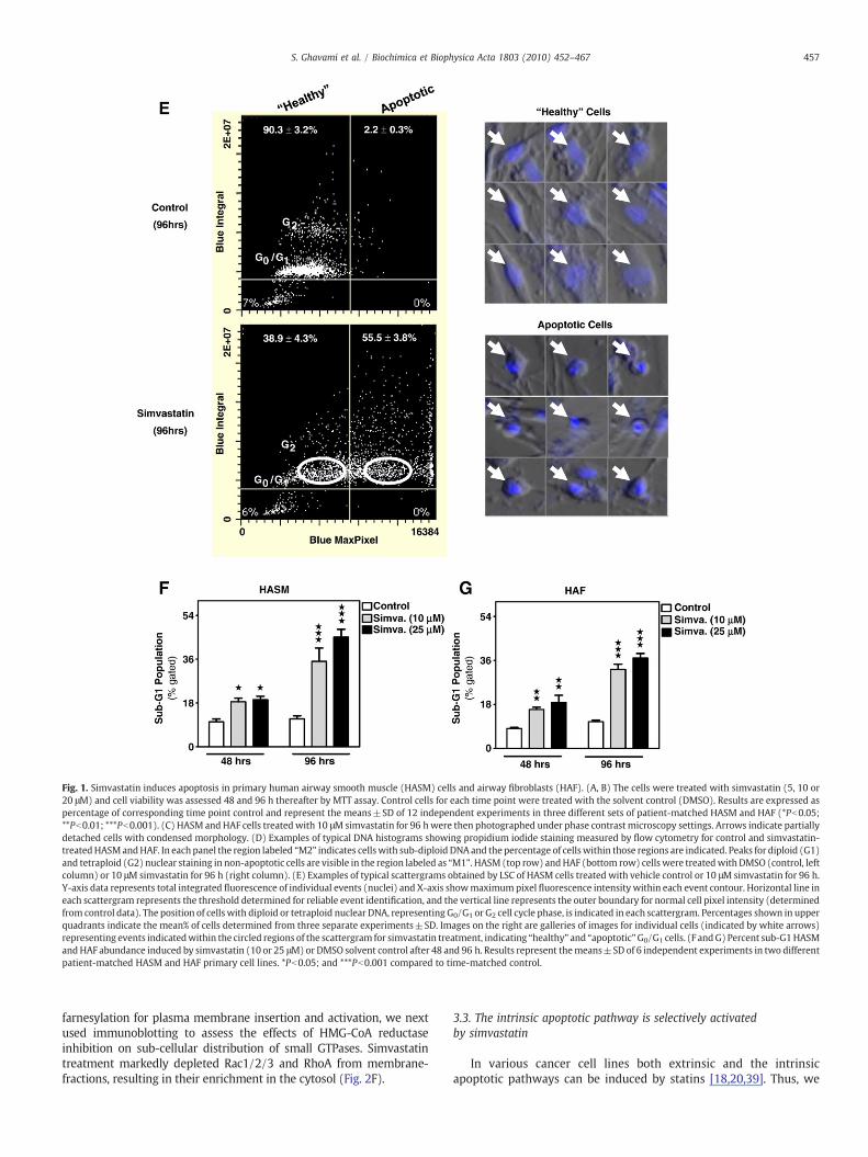
Fig. 1. Simvastatin induces apoptosis in primary human airway smooth muscle (HASM) cells and airway fibroblasts (HAF). (A, B) The cells were treated with simvastatin (5, 10 or20 μM) and cell viability was assessed 48 and 96 h thereafter by MTT assay. Control cells for each time point were treated with the solvent control (DMSO). Results are expressed aspercentage of corresponding time point control and represent the means±SD of 12 independent experiments in three different sets of patient-matched HASM and HAF (*Pb0.05;**Pb0.01; ***Pb0.001). (C) HASM and HAF cells treated with 10 μMsimvastatin for 96 hwere then photographed under phase contrast microscopy settings. Arrows indicate partiallydetached cells with condensed morphology. (D) Examples of typical DNA histograms showing propidium iodide staining measured by flow cytometry for control and simvastatin-treatedHASMandHAF. In each panel the region labeled “M2” indicates cellswith sub-diploid DNAand the percentage of cellswithin those regions are indicated. Peaks for diploid (G1)and tetraploid (G2) nuclear staining in non-apoptotic cells are visible in the region labeled as “M1”. HASM (top row) andHAF (bottom row) cells were treatedwithDMSO (control, leftcolumn) or 10 μM simvastatin for 96 h (right column). (E) Examples of typical scattergrams obtained by LSC of HASM cells treated with vehicle control or 10 μM simvastatin for 96 h.Y-axis data represents total integrated fluorescence of individual events (nuclei) and X-axis showmaximumpixel fluorescence intensity within each event contour. Horizontal line ineach scattergram represents the threshold determined for reliable event identification, and the vertical line represents the outer boundary for normal cell pixel intensity (determinedfrom control data). The position of cells with diploid or tetraploid nuclear DNA, representing G0/G1 or G2 cell cycle phase, is indicated in each scattergram. Percentages shown in upperquadrants indicate the mean% of cells determined from three separate experiments±SD. Images on the right are galleries of images for individual cells (indicated by white arrows)representing events indicatedwithin the circled regions of the scattergram for simvastatin treatment, indicating “healthy” and “apoptotic”G0/G1 cells. (F andG) Percent sub-G1HASMandHAF abundance induced by simvastatin (10 or 25 μM) or DMSO solvent control after 48 and 96 h. Results represent themeans±SD of 6 independent experiments in two differentpatient-matched HASM and HAF primary cell lines. *Pb0.05; and ***Pb0.001 compared to time-matched control.
457S. Ghavami et al. / Biochimica et Biophysica Acta 1803 (2010) 452–467
farnesylation for plasma membrane insertion and activation, we nextused immunoblotting to assess the effects of HMG-CoA reductaseinhibition on sub-cellular distribution of small GTPases. Simvastatintreatment markedly depleted Rac1/2/3 and RhoA from membrane-fractions, resulting in their enrichment in the cytosol (Fig. 2F).
3.3. The intrinsic apoptotic pathway is selectively activatedby simvastatin
In various cancer cell lines both extrinsic and the intrinsicapoptotic pathways can be induced by statins [18,20,39]. Thus, we
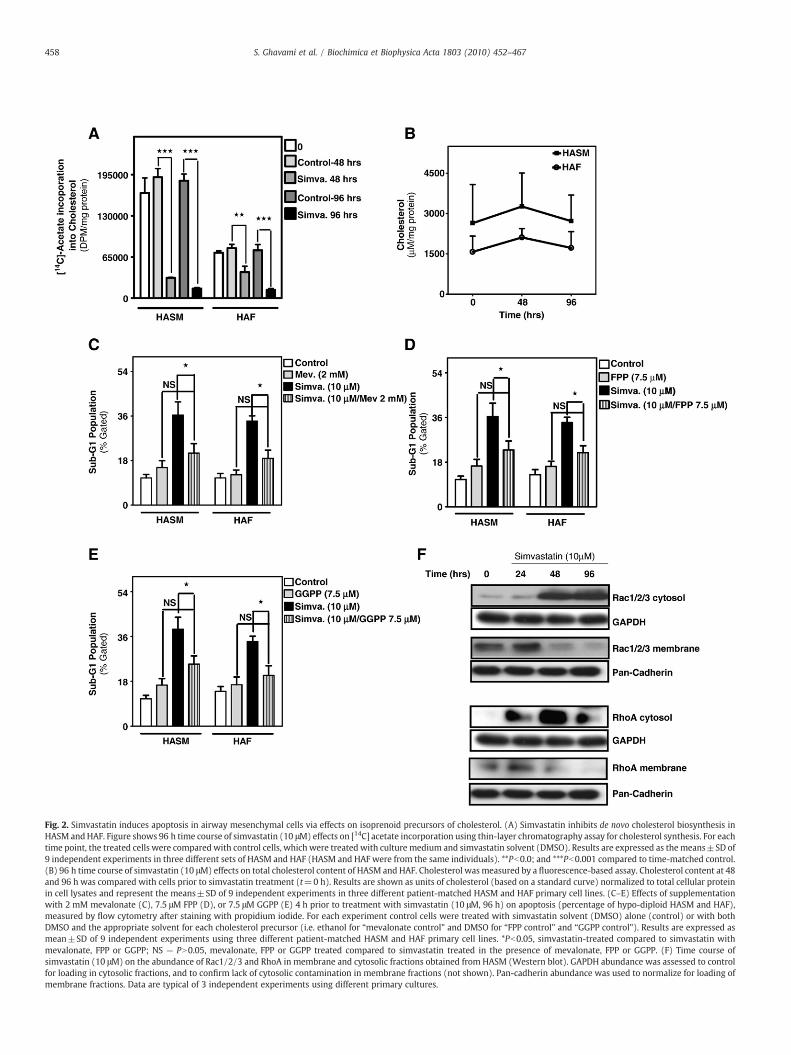
Fig. 2. Simvastatin induces apoptosis in airway mesenchymal cells via effects on isoprenoid precursors of cholesterol. (A) Simvastatin inhibits de novo cholesterol biosynthesis inHASM andHAF. Figure shows 96 h time course of simvastatin (10 μM) effects on [14C] acetate incorporation using thin-layer chromatography assay for cholesterol synthesis. For eachtime point, the treated cells were compared with control cells, which were treated with culture medium and simvastatin solvent (DMSO). Results are expressed as themeans±SD of9 independent experiments in three different sets of HASM and HAF (HASM and HAF were from the same individuals). **Pb0.0; and ***Pb0.001 compared to time-matched control.(B) 96 h time course of simvastatin (10 μM) effects on total cholesterol content of HASM and HAF. Cholesterol wasmeasured by a fluorescence-based assay. Cholesterol content at 48and 96 h was compared with cells prior to simvastatin treatment (t=0 h). Results are shown as units of cholesterol (based on a standard curve) normalized to total cellular proteinin cell lysates and represent the means±SD of 9 independent experiments in three different patient-matched HASM and HAF primary cell lines. (C–E) Effects of supplementationwith 2 mM mevalonate (C), 7.5 μM FPP (D), or 7.5 μM GGPP (E) 4 h prior to treatment with simvastatin (10 μM, 96 h) on apoptosis (percentage of hypo-diploid HASM and HAF),measured by flow cytometry after staining with propidium iodide. For each experiment control cells were treated with simvastatin solvent (DMSO) alone (control) or with bothDMSO and the appropriate solvent for each cholesterol precursor (i.e. ethanol for “mevalonate control” and DMSO for “FPP control” and “GGPP control”). Results are expressed asmean±SD of 9 independent experiments using three different patient-matched HASM and HAF primary cell lines. *Pb0.05, simvastatin-treated compared to simvastatin withmevalonate, FPP or GGPP; NS — PN0.05, mevalonate, FPP or GGPP treated compared to simvastatin treated in the presence of mevalonate, FPP or GGPP. (F) Time course ofsimvastatin (10 μM) on the abundance of Rac1/2/3 and RhoA in membrane and cytosolic fractions obtained from HASM (Western blot). GAPDH abundance was assessed to controlfor loading in cytosolic fractions, and to confirm lack of cytosolic contamination in membrane fractions (not shown). Pan-cadherin abundance was used to normalize for loading ofmembrane fractions. Data are typical of 3 independent experiments using different primary cultures.
458 S. Ghavami et al. / Biochimica et Biophysica Acta 1803 (2010) 452–467
459S. Ghavami et al. / Biochimica et Biophysica Acta 1803 (2010) 452–467
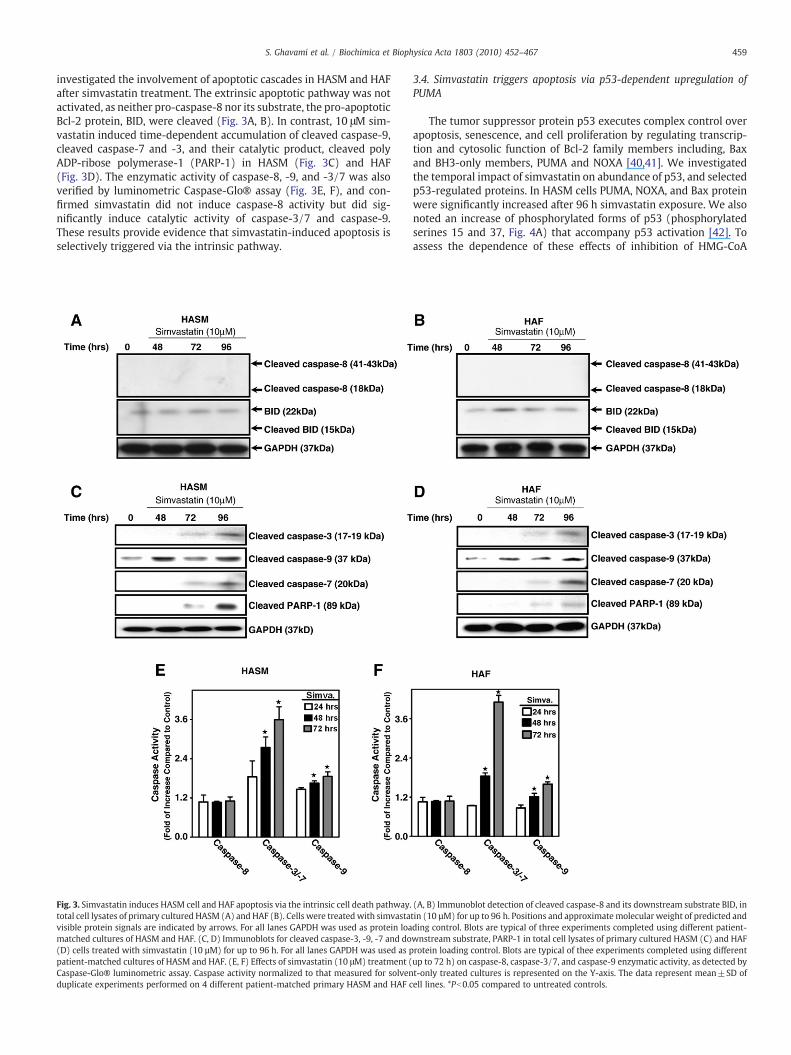
investigated the involvement of apoptotic cascades in HASM and HAFafter simvastatin treatment. The extrinsic apoptotic pathway was notactivated, as neither pro-caspase-8 nor its substrate, the pro-apoptoticBcl-2 protein, BID, were cleaved (Fig. 3A, B). In contrast, 10 μM sim-vastatin induced time-dependent accumulation of cleaved caspase-9,cleaved caspase-7 and -3, and their catalytic product, cleaved polyADP-ribose polymerase-1 (PARP-1) in HASM (Fig. 3C) and HAF(Fig. 3D). The enzymatic activity of caspase-8, -9, and -3/7 was alsoverified by luminometric Caspase-Glo® assay (Fig. 3E, F), and con-firmed simvastatin did not induce caspase-8 activity but did sig-nificantly induce catalytic activity of caspase-3/7 and caspase-9.These results provide evidence that simvastatin-induced apoptosis isselectively triggered via the intrinsic pathway.
Fig. 3. Simvastatin induces HASM cell and HAF apoptosis via the intrinsic cell death pathway.total cell lysates of primary cultured HASM (A) and HAF (B). Cells were treated with simvastavisible protein signals are indicated by arrows. For all lanes GAPDH was used as protein loamatched cultures of HASM and HAF. (C, D) Immunoblots for cleaved caspase-3, -9, -7 and do(D) cells treated with simvastatin (10 µM) for up to 96 h. For all lanes GAPDH was used as ppatient-matched cultures of HASM and HAF. (E, F) Effects of simvastatin (10 μM) treatment (Caspase-Glo® luminometric assay. Caspase activity normalized to that measured for solvenduplicate experiments performed on 4 different patient-matched primary HASM and HAF c
3.4. Simvastatin triggers apoptosis via p53-dependent upregulation ofPUMA
The tumor suppressor protein p53 executes complex control overapoptosis, senescence, and cell proliferation by regulating transcrip-tion and cytosolic function of Bcl-2 family members including, Baxand BH3-only members, PUMA and NOXA [40,41]. We investigatedthe temporal impact of simvastatin on abundance of p53, and selectedp53-regulated proteins. In HASM cells PUMA, NOXA, and Bax proteinwere significantly increased after 96 h simvastatin exposure. We alsonoted an increase of phosphorylated forms of p53 (phosphorylatedserines 15 and 37, Fig. 4A) that accompany p53 activation [42]. Toassess the dependence of these effects of inhibition of HMG-CoA
(A, B) Immunoblot detection of cleaved caspase-8 and its downstream substrate BID, intin (10 µM) for up to 96 h. Positions and approximatemolecular weight of predicted andding control. Blots are typical of three experiments completed using different patient-wnstream substrate, PARP-1 in total cell lysates of primary cultured HASM (C) and HAFrotein loading control. Blots are typical of thee experiments completed using differentup to 72 h) on caspase-8, caspase-3/7, and caspase-9 enzymatic activity, as detected byt-only treated cultures is represented on the Y-axis. The data represent mean±SD ofell lines. *Pb0.05 compared to untreated controls.
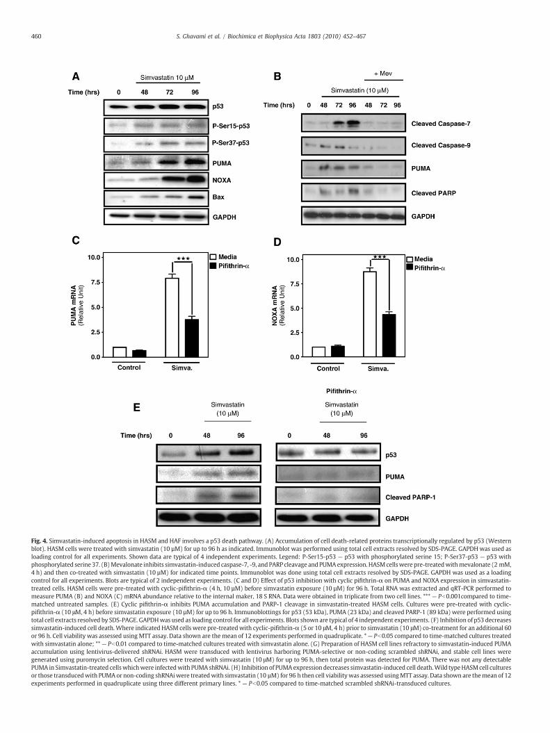
Fig. 4. Simvastatin-induced apoptosis in HASM and HAF involves a p53 death pathway. (A) Accumulation of cell death-related proteins transcriptionally regulated by p53 (Westernblot). HASM cells were treated with simvastatin (10 µM) for up to 96 h as indicated. Immunoblot was performed using total cell extracts resolved by SDS-PAGE. GAPDHwas used asloading control for all experiments. Shown data are typical of 4 independent experiments. Legend: P-Ser15-p53 — p53 with phosphorylated serine 15; P-Ser37-p53 — p53 withphosphorylated serine 37. (B)Mevalonate inhibits simvastatin-induced caspase-7, -9, and PARP cleavage andPUMAexpression. HASMcellswere pre-treatedwithmevalonate (2 mM,4 h) and then co-treated with simvastatin (10 µM) for indicated time points. Immunoblot was done using total cell extracts resolved by SDS-PAGE. GAPDH was used as a loadingcontrol for all experiments. Blots are typical of 2 independent experiments. (C and D) Effect of p53 inhibition with cyclic pifithrin-α on PUMA and NOXA expression in simvastatin-treated cells. HASM cells were pre-treated with cyclic-pifithrin-α (4 h, 10 µM) before simvastatin exposure (10 µM) for 96 h. Total RNA was extracted and qRT-PCR performed tomeasure PUMA (B) and NOXA (C) mRNA abundance relative to the internal maker, 18 S RNA. Data were obtained in triplicate from two cell lines. *** — Pb0.001compared to time-matched untreated samples. (E) Cyclic pifithrin-α inhibits PUMA accumulation and PARP-1 cleavage in simvastatin-treated HASM cells. Cultures were pre-treated with cyclic-pifithrin-α (10 μM, 4 h) before simvastatin exposure (10 µM) for up to 96 h. Immunoblottings for p53 (53 kDa), PUMA (23 kDa) and cleaved PARP-1 (89 kDa) were performed usingtotal cell extracts resolved by SDS-PAGE. GAPDHwas used as loading control for all experiments. Blots shown are typical of 4 independent experiments. (F) Inhibition of p53 decreasessimvastatin-induced cell death.Where indicated HASM cells were pre-treatedwith cyclic-pifithrin-α (5 or 10 μM, 4 h) prior to simvastatin (10 μM) co-treatment for an additional 60or 96 h. Cell viability was assessed using MTT assay. Data shown are the mean of 12 experiments performed in quadruplicate. *— Pb0.05 compared to time-matched cultures treatedwith simvastatin alone; **— Pb0.01 compared to time-matched cultures treated with simvastatin alone. (G) Preparation of HASM cell lines refractory to simvastatin-induced PUMAaccumulation using lentivirus-delivered shRNAi. HASM were transduced with lentivirus harboring PUMA-selective or non-coding scrambled shRNAi, and stable cell lines weregenerated using puromycin selection. Cell cultures were treated with simvastatin (10 μM) for up to 96 h, then total protein was detected for PUMA. There was not any detectablePUMA in Simvastatin-treated cellswhichwere infectedwith PUMAshRNAi. (H) Inhibition of PUMAexpression decreases simvastatin-induced cell death.Wild typeHASMcell culturesor those transducedwith PUMAor non-coding shRNAiwere treatedwith simvastatin (10 µM) for 96 h then cell viabilitywas assessed usingMTT assay. Data shown are themean of 12experiments performed in quadruplicate using three different primary lines. * — Pb0.05 compared to time-matched scrambled shRNAi-transduced cultures.
460 S. Ghavami et al. / Biochimica et Biophysica Acta 1803 (2010) 452–467

Fig. 4 (continued).
461S. Ghavami et al. / Biochimica et Biophysica Acta 1803 (2010) 452–467
reductase, we co-treated cells with simvastatin and mevalonate.Addition of mevalonate was sufficient to block caspase-9 and -7cleavage, PUMA expression and PARP cleavage (Fig. 4B). To confirmthe involvement of p53 in simvastatin-induced accumulation of BH3-only proteins, we examined the effects of the p53-transcriptionalinhibitor, cyclic-pifithrin-α [43], on simvastatin-induced accumula-tion of mRNA for PUMA and NOXA. Simvastatin treatment induced a7–8-fold increase in PUMA and NOXA after 96 h (Fig. 4C, D), whereasp53 inhibition suppressed this response by nearly 60% (Fig. 4C, D). Weobserved a similar suppression of simvastatin-induced accumulationof PUMA and cleaved PARP-1 (Fig. 4E). Importantly, p53 inhibitionwith cyclic-pifithrin-α also significantly inhibited, but did not fullyprevent, simvastatin-induced cell death in HASM cells (Fig. 4F).
The best understood pathway for p53-induced apoptosis involvesPUMA, which is transcriptionally induced by p53 and once translated,binds Bcl-XL to permit cytoplasmic p53-facilitated formation ofmitochondrial permeability pores by Bax [13,40]. Therefore we testedwhether PUMA is required for simvastatin driven lung mesenchymalcell apoptosis. We developed a lentiviral vector with puromycin
selection cassette that encodes shRNA for human PUMA andtransduced HASM cell to establish stable silencing of PUMA. Ourinitial experiments confirmed that the cell cultures generated wererefractory to simvastatin-induced PUMA expression, whereas controlcultures expressing a non-coding scrambled shRNA exhibited PUMAprotein accumulation upon simvastatin exposure (Fig. 4G). Wemeasured cell viability of PUMA-null mesenchymal cells in responseto simvastatin and found that whereas wild type primary HASM andcultures transducedwith scrambled shRNA exhibited a 45–50% loss ofviability after 96 h, cell cultures deficient in PUMA were significantlyless prone, but not fully protected from simvastatin-induced cyto-toxicity (Fig. 4H).
3.5. Simvastatin induces mitochondrial membrane permeability and ROSformation
To determine the biologic significance of p53-dependent PUMAand NOXA upregulation by simvastatin, we next tested if simvastatinexposure affected mitochondrial function in HASM cells and HAF. We
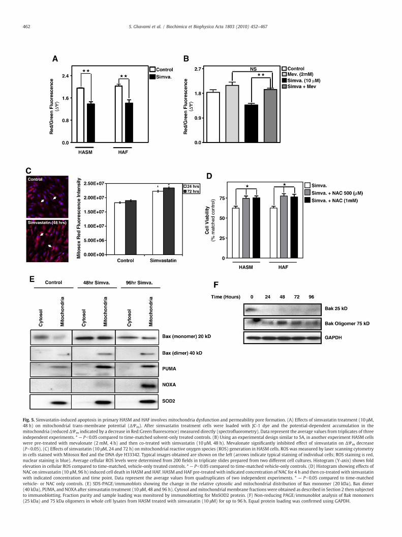
Fig. 5. Simvastatin-induced apoptosis in primary HASM and HAF involves mitochondria dysfunction and permeability pore formation. (A) Effects of simvastatin treatment (10 μM,48 h) on mitochondrial trans-membrane potential (ΔΨm). After simvastatin treatment cells were loaded with JC-1 dye and the potential-dependent accumulation in themitochondria (reduced ΔΨm indicated by a decrease in Red:Green fluorescence) measured directly (spectrofluorometry). Data represent the average values from triplicates of threeindependent experiments. * — Pb0.05 compared to time-matched solvent-only treated controls. (B) Using an experimental design similar to 5A, in another experiment HASM cellswere pre-treated with mevalonate (2 mM, 4 h) and then co-treated with simvastatin (10 µM, 48 h). Mevalonate significantly inhibited effect of simvastatin on ΔΨm decrease(Pb0.05). (C) Effects of simvastatin (10 μM, 24 and 72 h) on mitochondrial reactive oxygen species (ROS) generation in HASM cells. ROS was measured by laser scanning cytometryin cells stained with Mitosox Red and the DNA dye H33342. Typical images obtained are shown on the left (arrows indicate typical staining of individual cells; ROS staining is red,nuclear staining is blue). Average cellular ROS levels were determined from 200 fields in triplicate slides prepared from two different cell cultures. Histogram (Y-axis) shows foldelevation in cellular ROS compared to time-matched, vehicle-only treated controls. * — Pb0.05 compared to time-matched vehicle-only controls. (D) Histogram showing effects ofNAC on simvastatin (10 µM, 96 h) induced cell death in HASM and HAF. HASM and HAF pre-treated with indicated concentration of NAC for 4 h and then co-treated with simvastatinwith indicated concentration and time point. Data represent the average values from quadruplicates of two independent experiments. * — Pb0.05 compared to time-matchedvehicle- or NAC only controls. (E) SDS-PAGE/immunoblots showing the change in the relative cytosolic and mitochondrial distribution of Bax monomer (20 kDa), Bax dimer(40 kDa), PUMA, and NOXA after simvastatin treatment (10 μM, 48 and 96 h). Cytosol andmitochondrial membrane fractions were obtained as described in Section 2 then subjectedto immunoblotting. Fraction purity and sample loading was monitored by immunoblotting for MnSOD2 protein. (F) Non-reducing PAGE/immunoblot analysis of Bak monomers(25 kDa) and 75 kDa oligomers in whole cell lysates from HASM treated with simvastatin (10 μM) for up to 96 h. Equal protein loading was confirmed using GAPDH.
462 S. Ghavami et al. / Biochimica et Biophysica Acta 1803 (2010) 452–467
463S. Ghavami et al. / Biochimica et Biophysica Acta 1803 (2010) 452–467
measured ΔΨm upon simvastatin treatment using the ratiometricfluorescent indicator, JC-1 and found a marked decrease in ΔΨm
(Fig. 5A). Notably, as we observed for effects of simvastatin on cleav-age of caspase-7, -9 and PARP, and PUMA expression, the addition ofexogenous mevalonate significantly inhibited simvastatin-inducedsuppression of ΔΨm, confirming that depletion of mevalonate cascadeintermediates downstream from HMG-CoA reductase activity under-pins disruption in mitochondrial function (Fig 5B). Using the ROS-sensitive dye MitoSox Red, we also observed that there is a markedincrease in mitochondria-derived ROS generation compared tocorresponding controls (Pb0.05) after 24 and 72 h of simvastatinexposure (Fig. 5C). These data implicate involvement of mitochondriain simvastatin-induced lung mesenchymal cell death, an observationstrengthened by the fact that N-acetyl-L-cysteine (NAC), a broadrange ROS scavenger [28], partially inhibited simvastatin-induced celldeath (Fig. 5D). To test whether the loss of ΔΨm was caused bysimvastatin-induced pore formation in mitochondrial membrane wenext tested the association of pro-apoptotic Bcl-2 family memberswith mitochondria. Simvastatin caused marked enrichment ofmitochondrial PUMA, NOXA, and both monomeric and dimerizedBax (Fig. 5E). Also within 24 h of treatment, simvastatin inducedoligomerization of Bak that was sustained through 96 h of treatment(Fig. 5F). Collectively these observations confirm that simvastatincauses significant disruption of mitochondrial function, including theformation of membrane permeability pores, thus creating the poten-tial for the release of proteins that promote apoptosis.
3.6. Simvastatin induces selective release of mitochondrial IAP inhibitorproteins
To further examine the effect of simvastatin on mitochondria, wemonitored the release of various factors associated with the mito-chondrial death pathway. We first examined the mitochondrialapoptosis initiating factor (AIF), a FAD-dependent oxidoreductase[44], and the mitochondrial nuclease, endonuclease G (Endo G) [34].When released these proteins translocate to the nucleus to initiateDNA damage. Using confocal imaging we observed AIF and Endo G inmitochondria, but saw no evidence for AIF or EndoG in cell nuclei aftertreatingHASMandHAFwith simvastatin (10 μM,72 h) (Fig. 6A, B). In aparallel experiment in which we employed biochemical fractionationof mitochondria we did observe a marked cytosolic accumulation oftwo mitochondrial proteins, Smac/DIABLO and Omi/HtrA2, whichblock IAP proteins (Fig. 6C). In striking contrast, in the sameexperiment we were unable to detect release of cytochrome c frommitochondria. This suggests that the activation of caspase-9 bysimvastatin treatment (Fig. 3) occurs independently of apoptosomeformation. To more directly assess whether simvastatin-inducedcaspase-9 activation occurs in the absence of apoptosome formationwe compared simvastatin-induced caspase-9 activity in the presenceand absence of the apoptosome formation inhibitor NS3694. Apopto-some inhibition had no effect on the effects of simvastatin, as caspase-9 activity increased approximately 1.8-fold, similar to the effect inFig. 3E and F, in HASM cells and in HAF (not shown) (Fig. 6D), thus,confirming that mitochondrial release of cytochrome c is not a prin-cipal mechanism in simvastatin-induced cell death.
As our findings implicate a significant role for Smac and Omi instatin provoked cell death, we next tested whether Smac can po-tentiate simvastatin-induced apoptosis and caspase-9 activation bymeasuring the impact of an exogenous N7-Smac mimetic peptide thatcontains the 7 terminal amino acid–IAP-binding motif [45]. Notably,we observed that though treatment with the N7-Smac peptide aloneshowed little effect on caspase-9 activity, it did strongly potentiatesimvastatin-induced caspase-9 activation and cell death (Fig. 6E, F).These observations indicate that mitochondrial release of Smac con-tributes to cell death-triggered by simvastatin treatment.
3.7. Simvastatin treatment disrupts mitochondrial fission
Electron microscopy of simvastatin-treated lung mesenchymalcells revealed profound effects on mitochondrial morphology.Mitochondria in simvastatin-treated HASM cells and HAF exhibitedan atypical stretched forkhead morphology that in other systems hasbeen identified as a feature linked to the inhibition of mitochondrialfission (Fig. 7A) [16]. As mitochondria are dynamic organelles thatmove, fuse and divide, disruption of these activities has beenassociated with cell dysfunction and death [46,47]. Moreover, weand others have reported that the inhibition of mitochondrial fissionpromotes release of Smac, but not cytochrome c, and leads to Bax/Bak-dependent apoptosis [16,47,48]. Thus, to better determinewhether mitochondrial fission is compromised by simvastatin, weisolated mitochondria and used immunoblotting to examine the sub-cellular distribution of dynamin-related protein 1 (Drp1), an essentialcomponent of the mitochondria-associated protein complex that un-derpins mitochondrial division [49]. Our findings show that thoughsimvastatin increased Drp1 expression in HASM and HAF it alsopromoted a virtually complete loss of mitochondria-associated Drp1with concomitant accumulation of inactive protein in the cytosol(Fig. 7B, C). These observations suggest that the selective release ofSmac and Omi induced by simvastatin (Fig. 6C) may be related to thesuppression of mitochondrial fission.
4. Discussion
As statins are widely used and have broad positive health benefitsbeyond their cholesterol-lowering capacity [50], a full understandingof their mechanism of action is needed. Consistent with previousstudies, we show that statin-induced cell death in normal somaticcells is mediated by depletion of isoprenoids [5,6,51,52]. Our studyalso has several unique features and significantly extends currentunderstanding of downstream mechanisms for statin-induced apo-ptosis in otherwise healthy cells. New data conclusively point tomitochondrial mechanisms and intrinsic apoptosis signaling in statin-induced mesenchymal cell death. We reveal the involvement of p53and its induction of BH3-only protein expression to promotemitochondrial permeability pore formation involving Bax and Bak.Moreover, we show for the first time that mitochondria-derived ROSare induced by statins and contribute to mesenchymal cell death. Aparticularly novel aspect of this study hinges on evidence formitochondrial release of IAP inhibitors, Smac and Omi, in the absenceof cytochrome c release to trigger caspase activation leading to celldeath. Furthermore, we show new evidence that statin exposure isassociated with disrupted mitochondrial fission; consistent withstudies using different compounds and cell types [16,47,48], this islikely a critical determinant of selective IAP inhibitor release. Our useof primary cultured human lungmesenchymal cells is also unique andimportant, both because to date cancer cell lines have chiefly beenused to investigate statin-triggered cell death pathways, and theaccumulation of HASM cells and HAFs is a significant underpinning ofobstructive lung diseases, which appear to respond well to statintherapy [23–25].
We show that simvastatin inhibitsde novo synthesis of cholesterol inHASM and HAF, which is consistent with studies showing HMG-CoAreductase inhibition suppresses the incorporation of [14C]-acetate intonewly synthesized sterols [53]. Our observation that cellular cholesterolcontent was unaffected despite suppression of de novo synthesis forseveral days is somewhat paradoxical, but it is important tonote thatweincluded 0.5% FBS in culturemedia and that cells of mesenchymal originhave a high intrinsic capacity to take-up sterols [54]. Thus, HASM cellsand HAF likely incorporate sufficient cholesterol from FBS in culturemedia to compensate for suppression of intracellular cholesterolbiosynthesis. Inhibition of HMG-CoA reductase does lead to depletionof mevalonate and downstream metabolites, GGPP and FPP, which are
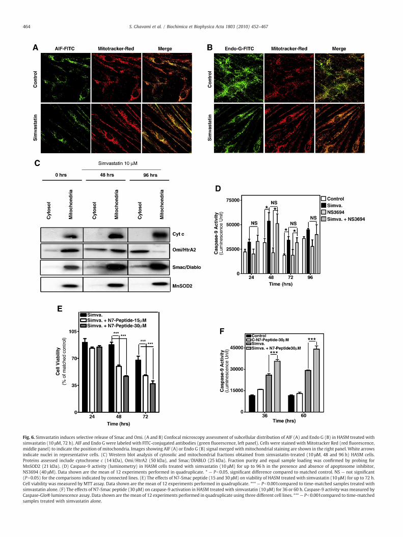
Fig. 6. Simvastatin induces selective release of Smac and Omi. (A and B) Confocal microscopy assessment of subcellular distribution of AIF (A) and Endo G (B) in HASM treated withsimvastatin (10 μM, 72 h). AIF and Endo G were labeled with FITC-conjugated antibodies (green fluorescence, left panel). Cells were stained with Mitotracker Red (red fluorescence,middle panel) to indicate the position of mitochondria. Images showing AIF (A) or Endo G (B) signal merged with mitochondrial staining are shown in the right panel. White arrowsindicate nuclei in representative cells. (C) Western blot analysis of cytosolic and mitochondrial fractions obtained from simvastatin-treated (10 μM, 48 and 96 h) HASM cells.Proteins assessed include cytochrome c (14 kDa), Omi/HtrA2 (50 kDa), and Smac/DIABLO (25 kDa). Fraction purity and equal sample loading was confirmed by probing forMnSOD2 (21 kDa). (D) Caspase-9 activity (luminometry) in HASM cells treated with simvastatin (10 μM) for up to 96 h in the presence and absence of apoptosome inhibitor,NS3694 (40 μM). Data shown are the mean of 12 experiments performed in quadruplicate. * — Pb0.05, significant difference compared to matched control. NS — not significant(PN0.05) for the comparisons indicated by connected lines. (E) The effects of N7-Smac peptide (15 and 30 μM) on viability of HASM treated with simvastatin (10 μM) for up to 72 h.Cell viability was measured by MTT assay. Data shown are the mean of 12 experiments performed in quadruplicate. *** — Pb0.001compared to time-matched samples treated withsimvastatin alone. (F) The effects of N7-Smac peptide (30 μM) on caspase-9 activation in HASM treated with simvastatin (10 μM) for 36 or 60 h. Caspase-9 activity was measured byCaspase-Glo® luminescence assay. Data shown are the mean of 12 experiments performed in quadruplicate using three different cell lines. ***— Pb0.001compared to time-matchedsamples treated with simvastatin alone.
464 S. Ghavami et al. / Biochimica et Biophysica Acta 1803 (2010) 452–467

Fig. 7. Simvastatin shows hallmark features of mitochondrial fission inhibition. (A) TEMmicrographs of HASM cells (top row) and HAF (bottom row) before (left panel) andafter (right panel) simvastatin treatment (10 μM, 72 h). Representative mitochondriaare indicated by black arrows. Images are typical of multiple micrographs obtained in atleast two different cell cultures. Magnification: HASM=34×103; HAF=2.65×10³. (B)Western blot detection of Drp1 (80 kDa) in whole cell lysates of HASM treated withsimvastatin (10 μM) for up to 96 h. GAPDH (37 kDa) was used to confirm equal proteinloading. Blots shown are typical of those obtained in duplicate from at least threeindependent cultures. (C) Immunoblots for Drp1 (80 kDa) in cytosol and mitochondrialfractions isolated from HASM treated with simvastatin (10 μM) for 48 or 96 h. Fractionpurity and equal sample loading was confirmed by probing for MnSOD2 (21 kDa). Blotsshown are typical of those obtained in duplicate from at least three independentcultures.
465S. Ghavami et al. / Biochimica et Biophysica Acta 1803 (2010) 452–467
needed for membrane anchoring of small GTPases including Ras, RhoA,and Rac that have been implicated in death signaling induced by statins[6,8,20]. Our results are consistent with such a mechanism assimvastatin caused a marked loss of membrane-associated Rac1/2/3andRhoA, and in reconstitution experimentsmevalonate, FPP andGGPPall diminished the apoptotic effects of simvastatin, a result that parallelsstudies with rat vascular smooth muscle cells [5]. In contrast to ourfindings, depletion of cellular cholesterol has been linked to statin-induced apoptosis in some cancer cells [38], supporting the need forcareful dissection of apoptosis mechanisms in healthy cells.
Caspases serve as initiators and executioners of extrinsic andintrinsic apoptotic pathways [10]. In HASM and HAF simvastatin failed
to activate caspase-8 or induce Bid cleavage indicating no role for anextrinsic pathway. This contrasts some studies with cancer cells,human umbilical endothelial cells and skeletal myoblasts that showstatins can activate caspase-8 [18,19,55], though this is not requiredfor apoptosis in all cell types [18]. This suggests cell-type dependentstatin-triggered caspase-8 activation. Our data support a central rolefor intrinsic apoptosis in primary lung mesenchymal cells, assimvastatin promotes activation of caspase-9 and its downstreamexecutioner cysteine proteases, caspase-7 and -3, as well as cleavageof the distal substrate, PARP-1. This is consistent with studies usingcancer cell lines and other primary cell types [18,19,56]. We alsouncovered mitochondria-associated mechanisms including increasedouter membrane permeability upon statin exposure, which correlatesthe loss in ΔΨm in some cancer lines [20]. Notably, we showsimvastatin induces intracellular ROS generation, which to ourknowledge is a unique observation. The mitochondrial-selective dyewe used is sensitive to an oxidation reaction catalyzed by mitochon-drial superoxide that can be counteracted by cellular superoxidedismutase. Thus, simvastatin promotes ROS generation via aerobicrespiration that overcomes intrinsic antioxidant defenses. This is offunctional importance, as we observed that the clinically used anti-oxidant, NAC, partially suppresses simvastatin-induced HASM andHAF cell death, indicating mitochondrial ROS contribute to the death-triggering effects of statins.
Oxidative stress is a principal signal for genotoxic stress that in-duces expression and activation of the tumor suppressor gene, p53[57]. The p53 protein can bind to nearly 300 different promoterelements in the human genome [58], among them PUMA, Bax andNOXA [42], and it broadly alters patterns of gene expression that cansupport mitochondrial apoptotic pathways [59]. A link between p53and statin-induced apoptosis has not been clearly establishedheretofore. Indeed, a previous report using p53 mutant breast cancercells indicated that lovastatin was able to induce pro-apoptoticmitochondrial mechanisms, including cytochrome c release [18]. Thiscontrasts our studies that reveal a central role for p53 in adult HASMcells and HAF; simvastatin activated p53 both by increasing itsexpression and by fostering phosphorylation of Ser-15 and -37, andthis effect was partially required for cell death as the p53-transcriptional inhibitor, cyclic-pifithrin-α [43], significantly reducedmesenchymal cell apoptosis and PARP-1 cleavage. Simvastatin alsoinduced p53-dependent expression of PUMA, NOXA and Bax, ascyclic-pifithrin-α suppressed accumulation of NOXA and PUMAmRNA and protein. Moreover, silencing of the p53-dependent accu-mulation of PUMA with shRNAi was sufficient to partially preventapoptosis. These observations underscore an initiator role for p53 inmitochondrial mechanisms as we observed Bax/Bak oligomerization,and marked accumulation of PUMA and NOXA in mitochondriaconcomitant with the loss ofΔΨm. PUMA and NOXA interact with BH3domains of cytosol-localized anti-apoptotic proteins such as Bcl-XLthat sequester Bax [12,14]. Apoptotic signals can promote Bax trans-location to mitochondria [60] and induce allosteric conformationalchange in mitochondrial Bak [61], thereby inducing formation ofmitochondrial permeability pores that reduce ΔΨm and create aconduit for the release of pro-apoptotic proteins [62,63]. This mech-anism is initiated by p53 in simvastatin-treated adult lung mesen-chymal cells, however, our data suggest that it is not sufficient toaccount fully for the apoptotic response, and unlike cancer cells [18]cytochrome c release does not appear to be involved.
Several molecules are released from the mitochondrial inter-mem-brane space in response to apoptotic stimuli. Some well-characterizedproteins include cytochrome c, Smac, Omi, AIF and Endo G [64]. Therelease of cytochrome c, Smac and Omi leads to the proteolytic (auto)activation of caspase-9, which is further sustained by positive feedbackinvolvingactivatedcaspase-3 [10].WeobservedBax/Bakoligomerization,Bax, PUMA, and NOXA mitochondrial translocation, and the release ofSmac and Omi but not cytochrome c, AIF or Endo G from mitochondria

466 S. Ghavami et al. / Biochimica et Biophysica Acta 1803 (2010) 452–467
upon simvastatin treatment. Smac, Omi and cytochrome c are more-or-less soluble in the inter-membrane space whereas AIF is anchored in theinner membrane space [65] and Endo G is localized in the matrix [66],perhaps explaining why the latter are not released by simvastatinexposure. As cytochrome c, Smac and Omi release is typically a con-comitant event with similar kinetic profiles [28,67,68], our observationsindicating the uncoupling of this relationship are unique and provocative.Moreover, we and others have reported that the inhibition of mitochon-drial fission promotes release of Smac, but not cytochrome c, and leads toBax/Bak-dependent apoptosis [16,47,48]. We confirmed this observationin HASM and HAF with independent experiments that also assessed thefunctional relevance of our findings; simvastatin-induced caspase-9activation and apoptosis was refractory to the apoptosome-selectiveinhibitor, NS3694, thus confirming no significant role for mitochondrialcytochrome c release. These findings suggest cytochrome c, a componentof mitochondrial respiratory chain, may be loosely attached to the innermitochondrial membrane via protein–protein interactions, making it lessprone to release than Smac and Omi under some conditions. Of particularinterest are reports that indicate Smac andOmi aremore loosely bound tothemitochondria than cytochrome c in Drp1-depletedmitochondria [69].Indeed, differential release of cytochrome c, Smac, Omi and other inter-membrane proteins has been observed during Bax/Bak-dependent apo-ptosis in Drp1-deficient cells [16,47]. These observations correlate wellwith our data showing almost all mitochondrial Drp1 was lost whilstcytosolic levels increased markedly upon simvastatin exposure. Drp1 is alarge GTPase that associates with mitochondria where it couples GTPhydrolysiswithmitochondrialmembrane constriction and fission [70]. Ofnote, we observed that simvastatin promoted formation of large forkheadmitochondria that are a hallmark of suppressed mitochondrial fission,strongly suggesting simvastatin disrupted mitochondrial dynamicsconcomitant with the loss of Drp1 from the organelles. Interestingly,Drp1 is a GTPase and its translocation from mitochondrial membrane tocytosol might be explained by simvastatin inhibition of protein prenyla-tion, however this mechanism needs to be clarified.
The mitochondrial pro-apoptotic protein Smac interferes with IAP-mediated caspase inhibition in several cell death models [48,71]. Thus,Smac released during simvastatin treatment could promote cell death byrelieving IAP inhibition of caspase-9, -3 and -7 [72]. In our experimentalsystem Smac N7 peptide further compromised cell viability supportingthe concept that increased release of Smac can promote simvastatin-induced apoptosis in adult human airway mesenchymal cells. Smac N7does not contain the Smac dimerization domain needed to inhibit XIAPbut it does contain an IBM domain that binds the XIAP initiator caspasebinding site, therefore the peptide directly mitigates mechanisms thatsuppress caspase-9 (auto)activation [73]. Overall, our observations pro-vide support for a simvastatin-induced pro-apoptotic pathway that relieson activation of caspases-9, -7 and -3 by Smac, which is released frommitochondria due both to Bax/Bak-dependent mitochondrial permeabil-ity driven by p53-PUMA, and the disruption of mitochondrial fissionresulting froma loss ofmitochondrial Drp1. Our data also suggest that thispathwaymay be linked to reduced prenylation of signaling proteins, thusourgroup is currently focusedon identifying thepathways thatmightplaya role in simvastatin-induced cell death in human airway mesenchymalcells.
Acknowledgements
The authors would like to thank Ms. Karen A. Detillieux for hereditorial contribution to this manuscript. This work was supported bygrants from the Canadian Institutes of Health Research (CIHR),GlaxoSmithKline Collaborative Innovation Research Fund, ManitobaInstitute of Child Health, and Canada Foundation for Innovation. SG issupported by a CIHR/Canadian Lung Association/GlaxoSmithKlinePostdoctoral fellowship, and a personnel award from the NationalTraining Program in Allergy and Asthma (NTPAA). KH was supportedby a Manitoba Health research Council (MHRC) studentship. DS is
supported by a MHRC/Manitoba Institute of Child Health (MICH)Postdoctoral Fellowship, and holds a CIHR IMPACT Strategic TrainingPostdoctoral fellowship. PS and TH are supported by personnelawards from the NTPAA, MHRC and MICH. PS is supported by aUniversity of Manitoba Graduate Fellowship. SKK is a Basic ScienceCareer Development Research Awardee of the Manitoba MedicalService Foundation supported with funds provided by the ManitobaBlue Cross, and the Biomedical Functionality Resource he directs wasestablished under the support of Dean Strategic Research Fund,Faculty of Medicine at University of Manitoba. GMH is a CanadaResearch Chair in Molecular Cardiolipin Metabolism. TK acknowl-edges the support by the Natural Sciences and Engineering ResearchCouncil of Canada (NSERC). ML thankfully acknowledges the supportfrom DFG (SFB 773, GRK 1302) and Deutsche Krebshilfe. AJH holds aCanada Research Chair in Airway Cell and Molecular Biology.
References
[1] J.L. Goldstein, M.S. Brown, Regulation of the mevalonate pathway, Nature 343(1990) 425–430.
[2] J.K. Liao, U. Laufs, Pleiotropic effects of statins, Annu. Rev. Pharmacol. Toxicol. 45(2005) 89–118.
[3] M.F. Demierre, P.D. Higgins, S.B. Gruber, E. Hawk, S.M. Lippman, Statins and cancerprevention, Nat. Rev. Cancer 5 (2005) 930–942.
[4] W.W. Wong, J. Dimitroulakos, M.D. Minden, L.Z. Penn, HMG-CoA reductaseinhibitors and the malignant cell: the statin family of drugs as triggers of tumor-specific apoptosis, Leukemia 16 (2002) 508–519.
[5] C. Guijarro, L.M. Blanco-Colio, M. Ortego, C. Alonso, A. Ortiz, J.J. Plaza, C. Diaz, G.Hernandez, J. Egido, 3-Hydroxy-3-methylglutaryl coenzyme a reductase andisoprenylation inhibitors induce apoptosis of vascular smooth muscle cells inculture, Circ. Res. 83 (1998) 490–500.
[6] J. Heusinger-Ribeiro, B. Fischer, M. Goppelt-Struebe, Differential effects ofsimvastatin on mesangial cells, Kidney Int. 66 (2004) 187–195.
[7] K. Yokota, F. Miyoshi, T. Miyazaki, K. Sato, Y. Yoshida, Y. Asanuma, Y. Akiyama, T.Mimura, High concentration simvastatin induces apoptosis in fibroblast-likesynoviocytes from patients with rheumatoid arthritis, J. Rheumatol. 35 (2008)193–200.
[8] J.Wu,W.W.Wong, F. Khosravi, M.D. Minden, L.Z. Penn, Blocking the Raf/MEK/ERKpathway sensitizes acute myelogenous leukemia cells to lovastatin-inducedapoptosis, Cancer Res. 64 (2004) 6461–6468.
[9] J.K. Liao, Isoprenoids as mediators of the biological effects of statins, J. Clin. Invest.110 (2002) 285–288.
[10] S. Ghavami, M. Hashemi, S.R. Ande, B. Yeganeh, W. Xiao, M. Eshraghi, C.J. Bus, K.Kadkhoda, E. Wiechec, A.J. Halayko, M. Los, Apoptosis and cancer: mutationswithin caspase genes, J. Med. Genet. 46 (2009) 497–510.
[11] S. Cory, J.M. Adams, The Bcl2 family: regulators of the cellular life-or-death switch,Nat. Rev. Cancer 2 (2002) 647–656.
[12] S.N.Willis, L. Chen, G. Dewson, A. Wei, E. Naik, J.I. Fletcher, J.M. Adams, D.C. Huang,Proapoptotic Bak is sequestered by Mcl-1 and Bcl-xL, but not Bcl-2, until displacedby BH3-only proteins, Genes Dev. 19 (2005) 1294–1305.
[13] J.E. Chipuk, L. Bouchier-Hayes, T. Kuwana, D.D. Newmeyer, D.R. Green, PUMAcouples the nuclear and cytoplasmic proapoptotic function of p53, Science 309(2005) 1732–1735.
[14] W.X. Zong, T. Lindsten, A.J. Ross, G.R. MacGregor, C.B. Thompson, BH3-onlyproteins that bind pro-survival Bcl-2 family members fail to induce apoptosis inthe absence of Bax and Bak, Genes Dev. 15 (2001) 1481–1486.
[15] N.A. Thornberry, Y. Lazebnik, Caspases: enemies within, Science 281 (1998)1312–1316.
[16] S. Ghavami, C. Kerkhoff, W.J. Chazin, K. Kadkhoda,W. Xiao, A. Zuse, M. Hashemi, M.Eshraghi, K. Schulze-Osthoff, T. Klonisch, M. Los, S100A8/9 induces cell death via anovel, RAGE-independent pathway that involves selective release of Smac/DIABLO and Omi/HtrA2, Biochim. Biophys. Acta 1783 (2008) 297–311.
[17] H. Zou, Y. Li, X. Liu, X. Wang, An APAF-1.cytochrome c multimeric complex is afunctional apoptosome that activates procaspase-9, J. Biol. Chem. 274 (1999)11549–11556.
[18] M.A. Shibata, Y. Ito, J. Morimoto, Y. Otsuki, Lovastatin inhibits tumor growth andlung metastasis in mouse mammary carcinoma model: a p53-independentmitochondrial-mediated apoptotic mechanism, Carcinogenesis 25 (2004)1887–1898.
[19] M. Marcelli, G.R. Cunningham, S.J. Haidacher, S.J. Padayatty, L. Sturgis, C. Kagan, L.Denner, Caspase-7 is activated during lovastatin-induced apoptosis of the prostatecancer cell line LNCaP, Cancer Res. 58 (1998) 76–83.
[20] P. Cafforio, F. Dammacco, A. Gernone, F. Silvestris, Statins activate themitochondrial pathway of apoptosis in human lymphoblasts and myeloma cells,Carcinogenesis 26 (2005) 883–891.
[21] J.A. Elias, Z. Zhu, G. Chupp, R.J. Homer, Airway remodeling in asthma, J. Clin. Invest.104 (1999) 1001–1006.
[22] N. Regamey, M. Ochs, T.N. Hilliard, C. Muhlfeld, N. Cornish, L. Fleming, S. Saglani, E.W. Alton, A. Bush, P.K. Jeffery, J.C. Davies, Increased airway smooth muscle mass inchildren with asthma, cystic fibrosis, and non-cystic fibrosis bronchiectasis, Am. J.Respir. Crit. Care Med. 177 (2008) 837–843.

467S. Ghavami et al. / Biochimica et Biophysica Acta 1803 (2010) 452–467
[23] E. Hothersall, C. McSharry, N.C. Thomson, Potential therapeutic role for statins inrespiratory disease, Thorax 61 (2006) 729–734.
[24] F. Ratjen, New pulmonary therapies for cystic fibrosis, Curr. Opin. Pulm. Med. 13(2007) 541–546.
[25] A.A. Zeki, L. Franzi, J. Last, N.J. Kenyon, Simvastatin inhibits airway hyperreactivity:implications for the mevalonate pathway and beyond, Am. J. Respir. Crit. CareMed. 180 (2009) 731–740.
[26] R. Gosens, G.L. Stelmack, G. Dueck, K.D. McNeill, A. Yamasaki, W.T. Gerthoffer, H.Unruh, A.S. Gounni, J. Zaagsma, A.J. Halayko, Role of caveolin-1 in p42/p44 MAPkinase activation and proliferation of human airway smooth muscle, Am. J.Physiol. Lung Cell. Mol. Physiol. 291 (2006) L523–L534.
[27] E.T. Naureckas, I.M. Ndukwu, A.J. Halayko, C. Maxwell, M.B. Hershenson, J. Solway,Bronchoalveolar lavage fluid from asthmatic subjects is mitogenic for humanairway smooth muscle, Am. J. Respir. Crit. Care Med. 160 (1999) 2062–2066.
[28] S. Ghavami, M. Eshraghi, K. Kadkhoda, M.M. Mutawe, S. Maddika, G.H. Bay, S.Wesselborg, A.J. Halayko, T. Klonisch, M. Los, Role of BNIP3 in TNF-induced celldeath—TNF upregulates BNIP3 expression, Biochim. Biophys. Acta 1793 (2009)546–560.
[29] S. Ghavami, A. Asoodeh, T. Klonisch, A. Halayko, K. Kadkhoda, K.T., S.B. Gibson, E.P.Booy, H. Naderi-Manesh, M. Los, Brevinin-2R semi-selectively kills cancer cells bya distinct mechanism, which involves the lysosomal-mitochondrial deathpathway, J. Cell. Mol. Med. (2008) 1–18.
[30] M. Zhou, Z. Diwu, N. Panchuk-Voloshina, R.P. Haugland, A stable nonfluorescentderivative of resorufin for the fluorometric determination of trace hydrogenperoxide: applications in detecting the activity of phagocyte NADPH oxidase andother oxidases, Anal. Biochem. 253 (1997) 162–168.
[31] J.G. Mohanty, J.S. Jaffe, E.S. Schulman, D.G. Raible, A highly sensitive fluorescentmicro-assay of H2O2 release from activated human leukocytes using adihydroxyphenoxazine derivative, J. Immunol. Methods 202 (1997) 133–141.
[32] V.Mokashi, D.K. Singh, T.D. Porter, Supernatant protein factor stimulatesHMG-CoAreductase in cell culture and in vitro, Arch. Biochem. Biophys. 433 (2005) 474–480.
[33] D. Tang, H.J. Park, S.P. Georgescu, S.M. Sebti, A.D. Hamilton, J.B. Galper, Simvastatinpotentiates tumor necrosis factor alpha-mediated apoptosis of human vascularendothelial cells via the inhibition of the geranylgeranylation of RhoA, Life Sci. 79(2006) 1484–1492.
[34] C. Adrain, E.M. Creagh, S.J. Martin, Apoptosis-associated release of Smac/DIABLOfrom mitochondria requires active caspases and is blocked by Bcl-2, EMBO J. 20(2001) 6627–6636.
[35] J. Tran, S.K. Kung, Lentiviral vectors mediate stable and efficient gene delivery intoprimary murine natural killer cells, Mol. Ther. 15 (2007) 1331–1339.
[36] P. Sharma, T. Tran, G.L. Stelmack, K. McNeill, R. Gosens, M.M. Mutawe, H. Unruh,W.T. Gerthoffer, A.J. Halayko, Expression of the dystrophin–glycoprotein complexis a marker for human airway smooth muscle phenotype maturation, Am. J.Physiol. Lung Cell. Mol. Physiol. 294 (2008) L57–L68.
[37] N.W. van de Donk, M.M. Kamphuis, B. van Kessel, H.M. Lokhorst, A.C. Bloem,Inhibition of protein geranylgeranylation induces apoptosis in myeloma plasmacells by reducing Mcl-1 protein levels, Blood 102 (2003) 3354–3362.
[38] L. Zhuang, J. Kim, R.M. Adam, K.R. Solomon, M.R. Freeman, Cholesterol targetingalters lipid raft composition and cell survival in prostate cancer cells andxenografts, J. Clin. Invest. 115 (2005) 959–968.
[39] R. Gniadecki, Depletion of membrane cholesterol causes ligand-independent activationof Fas and apoptosis, Biochem. Biophys. Res. Commun. 320 (2004) 165–169.
[40] J.E. Chipuk, T. Kuwana, L. Bouchier-Hayes, N.M. Droin, D.D. Newmeyer, M. Schuler,D.R. Green, Direct activation of Bax by p53 mediates mitochondrial membranepermeabilization and apoptosis, Science 303 (2004) 1010–1014.
[41] S. Maddika, S.R. Ande, S. Panigrahi, T. Paranjothy, K. Weglarczyk, A. Zuse, M.Eshraghi, K.D. Manda, E. Wiechec, M. Los, Cell survival, cell death and cell cyclepathways are interconnected: implications for cancer therapy, Drug Resist. Updat.10 (2007) 13–29.
[42] K.H. Vousden, X. Lu, Live or let die: the cell's response to p53, Nat. Rev. Cancer 2(2002) 594–604.
[43] P.G. Komarov, E.A. Komarova, R.V. Kondratov, K. Christov-Tselkov, J.S. Coon, M.V.Chernov, A.V. Gudkov, A chemical inhibitor of p53 that protects mice from the sideeffects of cancer therapy, Science 285 (1999) 1733–1737.
[44] Z. Abdullaev, M.E. Bodrova, B.V. Chernyak, D.A. Dolgikh, R.M. Kluck, M.O.Pereverzev, A.S. Arseniev, R.G. Efremov, M.P. Kirpichnikov, E.N. Mokhova, D.D.Newmeyer, H. Roder, V.P. Skulachev, A cytochrome c mutant with high electrontransfer and antioxidant activities but devoid of apoptogenic effect, Biochem J. 362(2002) 749–754.
[45] V. Bilim, K. Yuuki, T. Itoi, A. Muto, T. Kato, A. Nagaoka, T. Motoyama, Y. Tomita,Double inhibition of XIAP and Bcl-2 axis is beneficial for retrieving sensitivity ofrenal cell cancer to apoptosis, Br. J. Cancer 98 (2008) 941–949.
[46] A. Tanaka, R.J. Youle, A chemical inhibitor of DRP1 uncouples mitochondrial fissionand apoptosis, Mol. Cell 29 (2008) 409–410.
[47] P.A. Parone, J.C. Martinou, Mitochondrial fission and apoptosis: an ongoing trial,Biochim. Biophys. Acta 1763 (2006) 522–530.
[48] P.A. Parone, D.I. James, S. Da Cruz, Y. Mattenberger, O. Donze, F. Barja, J.C.Martinou, Inhibiting the mitochondrial fission machinery does not prevent Bax/Bak-dependent apoptosis, Mol. Cell. Biol. 26 (2006) 7397–7408.
[49] D.I. James, P.A. Parone, Y. Mattenberger, J.C. Martinou, hFis1, a novel component ofthe mammalian mitochondrial fission machinery, J. Biol. Chem. 278 (2003)36373–36379.
[50] C.J. Vaughan, M.B. Murphy, B.M. Buckley, Statins do more than just lowercholesterol, Lancet 348 (1996) 1079–1082.
[51] S. Demyanets, C. Kaun, S. Pfaffenberger, P.J. Hohensinner, G. Rega, J. Pammer, G.Maurer, K. Huber, J. Wojta, Hydroxymethylglutaryl-coenzyme A reductaseinhibitors induce apoptosis in human cardiac myocytes in vitro, Biochem.Pharmacol. 71 (2006) 1324–1330.
[52] A.M. Connor, S. Berger, A. Narendran, E.C. Keystone, Inhibition of proteingeranylgeranylation induces apoptosis in synovial fibroblasts, Arthritis Res.Ther. 8 (2006) R94.
[53] P. Negre-Aminou, A.K. van Vliet, M. van Erck, G.C. van Thiel, R.E. van Leeuwen, L.H.Cohen, Inhibition of proliferation of human smooth muscle cells by various HMG-CoA reductase inhibitors; comparison with other human cell types, Biochim.Biophys. Acta 1345 (1997) 259–268.
[54] O. Stein, J. Vanderhoek, G. Friedman, Y. Stein, Deposition and mobilization ofcholesterol ester in cultured human skin fibroblasts, Biochim. Biophys. Acta 450(1976) 367–378.
[55] M. Diamant, M.E. Tushuizen, M.N. Abid-Hussein, C.M. Hau, A.N. Boing, A. Sturk, R.Nieuwland, Simvastatin-induced endothelial cell detachment and microparticlerelease are prenylation dependent, Thromb. Haemost. 100 (2008) 489–497.
[56] L.M. Blanco-Colio, A. Villa, M. Ortego, M.A. Hernandez-Presa, A. Pascual, J.J. Plaza, J.Egido, 3-Hydroxy-3-methyl-glutaryl coenzyme A reductase inhibitors, atorvasta-tin and simvastatin, induce apoptosis of vascular smooth muscle cells bydownregulation of Bcl-2 expression and Rho A prenylation, Atherosclerosis 161(2002) 17–26.
[57] J.L. Martindale, N.J. Holbrook, Cellular response to oxidative stress: signaling forsuicide and survival, J. Cell. Physiol. 192 (2002) 1–15.
[58] C.L. Wei, Q. Wu, V.B. Vega, K.P. Chiu, P. Ng, T. Zhang, A. Shahab, H.C. Yong, Y. Fu, Z.Weng, J. Liu, X.D. Zhao, J.L. Chew, Y.L. Lee, V.A. Kuznetsov, W.K. Sung, L.D. Miller, B.Lim, E.T. Liu, Q. Yu, H.H. Ng, Y. Ruan, A global map of p53 transcription-factorbinding sites in the human genome, Cell 124 (2006) 207–219.
[59] M. Schuler, U. Maurer, J.C. Goldstein, F. Breitenbucher, S. Hoffarth, N.J. Waterhouse,D.R. Green, p53 triggers apoptosis in oncogene-expressing fibroblasts by theinduction of Noxa and mitochondrial Bax translocation, Cell Death Differ. 10(2003) 451–460.
[60] T. Mashima, T. Tsuruo, Defects of the apoptotic pathway as therapeutic targetagainst cancer, Drug Resist. Updat. 8 (2005) 339–343.
[61] A. Gross, J.M. McDonnell, S.J. Korsmeyer, BCL-2 family members and themitochondria in apoptosis, Genes Dev. 13 (1999) 1899–1911.
[62] J. Yu, L. Zhang, P.M. Hwang, K.W. Kinzler, B. Vogelstein, PUMA induces the rapidapoptosis of colorectal cancer cells, Mol. Cell 7 (2001) 673–682.
[63] T. Shibue, K. Takeda, E. Oda, H. Tanaka, H. Murasawa, A. Takaoka, Y. Morishita, S.Akira, T. Taniguchi, N. Tanaka, Integral role of Noxa in p53-mediated apoptoticresponse, Genes Dev. 17 (2003) 2233–2238.
[64] S.J. Riedl, Y. Shi, Molecular mechanisms of caspase regulation during apoptosis,Nat. Rev. Mol. Cell. Biol. 5 (2004) 897–907.
[65] H. Otera, S. Ohsakaya, Z. Nagaura, N. Ishihara, K. Mihara, Export of mitochondrialAIF in response to proapoptotic stimuli depends on processing at the intermem-brane space, EMBO J. 24 (2005) 1375–1386.
[66] D. Arnoult, B. Gaume, M. Karbowski, J.C. Sharpe, F. Cecconi, R.J. Youle,Mitochondrial release of AIF and EndoG requires caspase activation downstreamof Bax/Bak-mediated permeabilization, EMBO J. 22 (2003) 4385–4399.
[67] G. van Loo, M. van Gurp, B. Depuydt, S.M. Srinivasula, I. Rodriguez, E.S. Alnemri, K.Gevaert, J. Vandekerckhove, W. Declercq, P. Vandenabeele, The serine proteaseOmi/HtrA2 is released from mitochondria during apoptosis. Omi interacts withcaspase-inhibitor XIAP and induces enhanced caspase activity, Cell Death Differ. 9(2002) 20–26.
[68] T.M. Bocan, Pleiotropic effects of HMG-CoA reductase inhibitors, Curr. Opin.Investig. Drugs 3 (2002) 1312–1317.
[69] A. Laurent, C. Nicco, C. Chereau, C. Goulvestre, J. Alexandre, A. Alves, E. Levy, F.Goldwasser, Y. Panis, O. Soubrane, B. Weill, F. Batteux, Controlling tumor growthby modulating endogenous production of reactive oxygen species, Cancer Res. 65(2005) 948–956.
[70] E. Smirnova, D.L. Shurland, S.N. Ryazantsev, A.M. van der Bliek, A human dynamin-related protein controls the distribution of mitochondria, J. Cell. Biol. 143 (1998)351–358.
[71] N. Ishihara, M. Nomura, A. Jofuku, H. Kato, S.O. Suzuki, K. Masuda, H. Otera, Y.Nakanishi, I. Nonaka, Y. Goto, N. Taguchi, H. Morinaga, M. Maeda, R. Takayanagi, S.Yokota, K. Mihara, Mitochondrial fission factor Drp1 is essential for embryonicdevelopment and synapse formation in mice, Nat. Cell. Biol. 11 (2009) 958–966.
[72] P. Maycotte, S. Blancas, J. Moran, Role of inhibitor of apoptosis proteins and Smac/DIABLO in staurosporine-induced cerebellar granule neurons death, Neurochem.Res. 33 (2008) 1534–1540.
[73] S.M. Srinivasula, R. Hegde, A. Saleh, P. Datta, E. Shiozaki, J. Chai, R.A. Lee, P.D.Robbins, T. Fernandes-Alnemri, Y. Shi, E.S. Alnemri, A conserved XIAP-interactionmotif in caspase-9 and Smac/DIABLO regulates caspase activity and apoptosis,Nature 410 (2001) 112–116.
Related Documents