university of copenhagen Porphyromonas gingivalis in saliva associates with chronic and aggressive periodontitis Damgaard, Christian; Danielsen, Anne Katrine; Enevold, Christian; Massarenti, Laura; Nielsen, Claus Henrik; Holmstrup, Palle; Belstrøm, Daniel Published in: Journal of Oral Microbiology DOI: 10.1080/20002297.2019.1653123 Publication date: 2019 Document version Publisher's PDF, also known as Version of record Document license: CC BY-NC Citation for published version (APA): Damgaard, C., Danielsen, A. K., Enevold, C., Massarenti, L., Nielsen, C. H., Holmstrup, P., & Belstrøm, D. (2019). Porphyromonas gingivalis in saliva associates with chronic and aggressive periodontitis. Journal of Oral Microbiology, 11(1), 1-9. [1653123]. https://doi.org/10.1080/20002297.2019.1653123 Download date: 21. maj. 2020

Welcome message from author
This document is posted to help you gain knowledge. Please leave a comment to let me know what you think about it! Share it to your friends and learn new things together.
Transcript
u n i ve r s i t y o f co pe n h ag e n
Porphyromonas gingivalis in saliva associates with chronic and aggressiveperiodontitis
Damgaard, Christian; Danielsen, Anne Katrine; Enevold, Christian; Massarenti, Laura;Nielsen, Claus Henrik; Holmstrup, Palle; Belstrøm, Daniel
Published in:Journal of Oral Microbiology
DOI:10.1080/20002297.2019.1653123
Publication date:2019
Document versionPublisher's PDF, also known as Version of record
Document license:CC BY-NC
Citation for published version (APA):Damgaard, C., Danielsen, A. K., Enevold, C., Massarenti, L., Nielsen, C. H., Holmstrup, P., & Belstrøm, D.(2019). Porphyromonas gingivalis in saliva associates with chronic and aggressive periodontitis. Journal of OralMicrobiology, 11(1), 1-9. [1653123]. https://doi.org/10.1080/20002297.2019.1653123
Download date: 21. maj. 2020
Full Terms & Conditions of access and use can be found athttps://www.tandfonline.com/action/journalInformation?journalCode=zjom20
Journal of Oral Microbiology
ISSN: (Print) 2000-2297 (Online) Journal homepage: https://www.tandfonline.com/loi/zjom20
Porphyromonas gingivalis in saliva associates withchronic and aggressive periodontitis
Christian Damgaard, Anne Katrine Danielsen, Christian Enevold, LauraMassarenti, Claus Henrik Nielsen, Palle Holmstrup & Daniel Belstrøm
To cite this article: Christian Damgaard, Anne Katrine Danielsen, Christian Enevold, LauraMassarenti, Claus Henrik Nielsen, Palle Holmstrup & Daniel Belstrøm (2019) Porphyromonasgingivalis in saliva associates with chronic and aggressive periodontitis, Journal of OralMicrobiology, 11:1, 1653123, DOI: 10.1080/20002297.2019.1653123
To link to this article: https://doi.org/10.1080/20002297.2019.1653123
© 2019 The Author(s). Published by InformaUK Limited, trading as Taylor & FrancisGroup.
Published online: 09 Aug 2019.
Submit your article to this journal
Article views: 35
View related articles
View Crossmark data
Porphyromonas gingivalis in saliva associates with chronic and aggressiveperiodontitisChristian Damgaard a,b, Anne Katrine Danielsena,b, Christian Enevoldb, Laura Massarentib,Claus Henrik Nielsena,b, Palle Holmstrupa and Daniel Belstrøma
aSection for Periodontology and Microbiology, Department of Odontology, Faculty of Health and Medical Sciences, University ofCopenhagen, Copenhagen, Denmark; bInstitute for Inflammation Research, Center for Rheumatology and Spine Diseases, Rigshospitalet,Copenhagen University Hospital, Copenhagen, Denmark
ABSTRACTObjective: To characterize the salivary microbiota of patients with aggressive periodontitis,patients with chronica periodontitis and orally healthy individuals.Methods: A total of 81 unstimulated saliva samples from aggressive periodontitis patients (n = 31),chronic periodontitis patients (n=25), andorally healthy controls (n =25)were examined. TheV1-V3region of the 16S rDNA gene was sequenced with Illumina® MiSeqTM, and sequences were anno-tated to the expanded Human Oral Microbiome Database (eHOMD).Results: A mean percentage of 97.6 (range: 89.8–99.7) of sequences could be identified atspecies level. Seven bacterial species, including Porphyromonas gingivalis, were identified withsignificantly higher relative abundance in saliva from aggressive periodontitis patients than insaliva from orally healthy controls. Salivary abundance of P. gingivalis could discriminateaggressive (AUC: 0.80, p = 0.0001) and chronic periodontitis (AUC: 0.72, p = 0.006) fromhealthy controls. Likewise, salivary presence of P. gingivalis was significantly associated withaggressive (p < 0.0001, RR: 8.1 (95% CI 2.1–31.2)) and chronic periodontitis (p = 0.002, RR: 6.5(95% CI: 1.6–25.9)).Conclusion: Salivary presence and relative abundance of P. gingivalis associate with aggres-sive and chronic periodontitis, but do not discriminate between aggressive and chronicperiodontitis.
ARTICLE HISTORYReceived 2 June 2019Revised 25 July 2019Accepted 2 August 2019
KEYWORDSPeriodontal disease;periodontitis;Porphyromonas gingivalis;microbiota; HOMINGS; saliva
Introduction
When entering the oral cavity saliva is sterile [1].However, when sampled from the oral cavity, onemilliliter of saliva contains hundred millions of bac-teria [2]. Thus, the salivary microbiota is believed tobe a compilation of bacteria shed from oral surfaces[3,4], which is why salivary microbiota may poten-tially be used as a biomarker of oral health status. Inoral health, the salivary microbiota is closely relatedto microbiotas found on the tongue and the oralmucosa [5]. However, salivary carriage of specificputative periodontal bacterial species, includingPorphyromonas gingivalis, has previously beenreported to associate with periodontitis [6,7].
Periodontitis is a prevalent, multifactorial inflamma-tory disease, induced by a biofilm on tooth surfaces, andthe associated breakdown of tooth-supporting tissues isdriven by a chronic inflammation within the period-ontal tissues [8]. Periodontitis is diagnosed clinicallyand may present in either an aggressive form withearly onset and rapid progression, affecting only 5%,or a chronic form characterized by slower breakdown oftooth-supporting tissues and onset later in life, affecting
approximately 40% of the adult population [9,10]. A 40-year follow-up study in male plantation workers fromSri Lanka, without access to dental care, reported dif-ferent trajectories of periodontal breakdown [11].Consequently, the immune responses towards the per-iodontal microbiota are believed to distinguish aggres-sive from chronic forms of periodontitis [8].
By means of various molecular techniques, cross-sectional analyses have reported that the salivary micro-biota of patients with periodontitis differs from that oforally healthy controls [12–14]. Furthermore, severalstudies have demonstrated correlation of subgingivaland salivary levels of specific bacteria proposed to bepresent in the periodontium of patients with period-ontitis [15–18]. Finally, two longitudinal studies haveshown that non-surgical periodontal treatment impactssalivary levels of proposed periodontal bacteria [19,20].Thus, salivary carriage of bacteria associated with per-iodontitis seems to reflect the periodontal status.However, to the best of our knowledge, the character-ization of the salivary microbiota in aggressive period-ontitis patients remains to be performed.
The purpose of the present study was to character-ize the salivary microbiota of patients with aggressive
CONTACT Christian Damgaard [email protected] Section for Periodontology, Department of Odontology, Faculty of Health and MedicalSciences, University of Copenhagen, Nørre Allé 202200, Copenhagen, Denmark
JOURNAL OF ORAL MICROBIOLOGY2019, VOL. 11, 1653123https://doi.org/10.1080/20002297.2019.1653123
© 2019 The Author(s). Published by Informa UK Limited, trading as Taylor & Francis Group.This is an Open Access article distributed under the terms of the Creative Commons Attribution-NonCommercial License (http://creativecommons.org/licenses/by-nc/4.0/),which permits unrestricted non-commercial use, distribution, and reproduction in any medium, provided the original work is properly cited.
periodontitis and compare it with that of chronicperiodontitis and of orally healthy controls. Thehypothesis to be tested was that composition of thesalivary microbiota in patients with aggressive peri-odontitis is distinct from those of patients withchronic periodontitis and orally healthy controls.Such salivary characteristics might hold potential foruse as biomarkers for detection of periodontitispatients with rapidly progressing periodontitis.
Methods
Sample size
Sample sizes were calculated using one-way ANOVAwith estimated levels of relevant biomarkers (controlsµ1 = 10,000, and periodontitis patients µ2 = 15,000)with a predicted standard deviation of 6,250, α = 0.05and a power of 0.80. Based on the sample size calcu-lation each group should consist of a minimum of 25participants. The sample size was adjusted to allow asmuch as 20% of the participants to withdraw theirconsent, leaving 30 participants in each group.Because of the novelty of the methods applied forsequencing, we accounted for unforeseen technicaldifficulties during analyses of the samples, by inclu-sion of an additional aggressive periodontitis patient,as these were the primary population of interest.
Study population
In the period from 8 February 2017 to 12 October 2018,a total of 90 individuals were recruited at the Departmentof Odontology, University of Copenhagen. All partici-pants were medically healthy and assigned in accordancewith the periodontal disease classification system from1999 American Academy of Periodontology [9].Participants were divided into three groups: Aggressiveperiodontitis, chronic periodontitis and periodontallyhealthy controls on this basis. The inclusion criteria were:
Aggressive periodontitis patients:
● 19–40 years of age.● Interproximal attachment loss at minimum 3
teeth that were neither first molars nor incisors.● Clinical attachment loss at minimum 10 sites,
also characterized by bleeding and appearanceof pus upon probing.
● Radiographic bone loss.
Chronic periodontitis patients:
● 50–60 years of age.● Interproximal attachment loss at minimum
three teeth besides molars and incisors.● Clinical attachment loss at minimum 10 sites,
also characterized by bleeding on probing.● Radiographic bone loss.
Orally healthy controls:
● 19–61 years of age.● No interproximal attachment loss.● No clinical attachment loss at sites with bleeding
on probing.● No radiographic bone loss.● No sign of other inflammatory lesions in the
oral mucosa.
General exclusion criteria included pregnant andbreastfeeding women, systemic antibiotic treatmentwithin 6 months, systemic diseases, and hematologicanomalies or syndromes. All participants signed aninformed consent, and the study was approved by theregional ethical committee (The Capital Region ofDenmark, protocol: H-1602473) and registered bythe Danish Data Authorization (approval number:P-2019–18), and registered at clinicaltrials.gov:NCT03225950.
Clinical registrations
Background information of the study populationincluding gender, age, ethnicity, and smoking statuswere recorded (Table 1). Clinical registrations wererecorded at six sites per tooth (3rd molars excluded),including pocket depth (PD), clinical attachment loss(CAL), bleeding on probing (BOP), plaque index(PI), number of teeth excluding 3rd molars, and num-ber of decayed, missing, filled teeth (DMFT). Fordetails see Table 2.
Saliva sample collection
Two ml of unstimulated saliva samples were collectedin Oragene DNA tubes (Cat.#OG-500, DNA GenotekInc., Ontario, Canada) as previously described [21]and stored immediately at −80°C until further analy-sis. Clinical registrations on healthy controls andsample collection were performed by the same exam-iner (AKD). All clinical registrations on patients withperiodontitis were confirmed by a clinical instructorat the Section of Periodontology, Department ofOdontology, University of Copenhagen.
DNA extraction
One hundred µl of a 25 mg/ml lysozyme (Cat.#90082,ThermoFisher, Roskilde, Denmark) were mixed with400 µl of Oragene saliva sample, incubated for 2 h at37°C, and the entire sample was subsequently purifiedusing the Maxwell 16 Cell DNA Purification Kit (Cat.#AS1020, Promega, Wisconsin). Purified DNAwas mea-sured using the Qubit dsDNA high-sensitivity kit (Cat.#Q32854, ThermoFisher, Roskilde, Denmark) anddiluted to 20 ng/ul.
2 C. DAMGAARD ET AL.
Sequencing and data analysis
Bacterial 16S rRNA gene targeted amplicon sequen-cing was performed using a custom dual-indexprotocol [22]. Custom 16S primers amplified theV1-V3 region of the 16S rRNA gene and weredesigned to provide the best coverage of the 16SrRNA gene while maintaining high sensitivity. Thesample libraries were prepared using a 22 cyclePCR reaction to reduce chimera formation (unlessotherwise noted). The final PCR products werepurified using Ampure XP beads, pooled in equalamounts, and gel purified using the QIAGENMinElute Gel Extraction Kit (Qiagen, Hilden,Germany). Purified, pooled libraries were quanti-fied using the NEBNext Library Quant Kit forIllumina. Final libraries were sequenced onIllumina® MiSeq™ with a v2 reagent kit (500 cycles).The sequencing was performed at a 10 pM loadingconcentration with >20% PhiX spike-in. TheDADA2 R package [23] was used to identify andquantify amplicon sequencing reads on the FastQCfiles obtained after demultiplexing with theIllumina MiSeq software. Briefly, reads weretrimmed and filtered to remove sequences withlow quality. Quality of the trimmed and filteredreads was assessed using FastQC. Results ofFastQC were compiled using MultiQC [24]. Thetrimmed and filtered reads were then processedthrough the denoising, concatenating read1 andread2 with a 10 N spacer, and chimera removal
steps of DADA2 to identify and quantify trueamplicon sequence variants (ASV) present in thesample. Taxonomy of the identified ASVs wasassigned using the RDP classifier algorithm [25]implemented in the DADA2 package witha training dataset developed at The ForsythInstitute, Cambridge, MA and based on theExtended Human Oral Microbe Database(eHOMD).
Statistics
All parameters tested were checked for normality.Metadata of the study population, clinical data andsequence metadata, which followed a Gaussian dis-tribution were compared using t-test, chi-squaretest and ANOVA, whereas non-parametric datawere compared by Mann-Whitney U test,Friedman test and Kruskall-Wallis H test. Thecore salivary microbiota was defined as bacterialgenera and species present with a mean relativeabundance >1% across all samples. The salivarymicrobiota were characterized and compared bymeans of predominant genera and species, relativeabundance, Shannon index, principal componentanalysis and correspondence analysis. Data on rela-tive abundance were corrected for multiple depen-dent associations using Benjamini-Hochbergscorrection [26]. All statistics were computed with
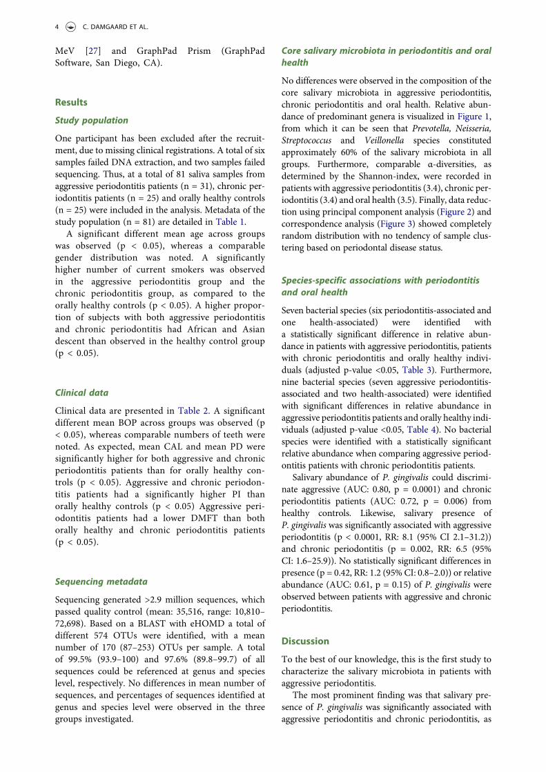
Table 2. Clinical data of the study population. Pocket depth (PD), clinical attachment loss (CAL), bleeding onprobing (BOP), plaque index (PI), number of teeth excluding 3rd molars, number of decayed, missing, filled teeth(DMFT). All parameters tested were checked for normality. Metadata of the clinical data of the study population,which followed a Gaussian distribution were compared using t-test, chi-square test and ANOVA, whereas non-parametric data were compared by Mann-Whitney U test, Friedman test and Kruskall-Wallis H test. All statisticswere computed with GraphPad Prism (Graphpad Software, San Diego, CA).
Aggressive periodontitis patients(n = 31)
Chronic periodontitis patients(n = 26)
Orally healthy controls(n = 25)
Mean PD in mm (range) 3.6 (2.2; 5.3) 3.5 (1.8; 6.6) 2.3 (1.4; 2.7)Mean CAL in mm (range) 3.5 (1.3; 5.9) 4.0 (1.9; 6.8) 1.1 (0.3; 2.7)Mean BOP in % (range) 56.4 (5.0; 98.8) 39.4 (2.3; 99.2) 4.0 (0; 76.2)Mean PI in % (range) 65.2 (1.2; 99.4) 55.9 (0.6; 100) 6.22 (0; 66.1)Number of teeth (range) 27.3 (25; 28) 25.9 (21;28) 27.5 (25; 28)DMFT (range) 3.9 (0; 14) 10.4 (1; 20) 6.17 (0; 21)
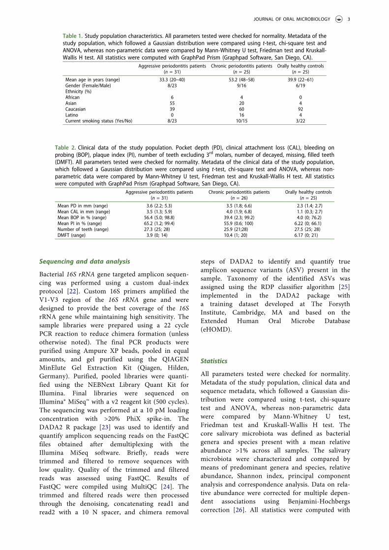
Table 1. Study population characteristics. All parameters tested were checked for normality. Metadata of thestudy population, which followed a Gaussian distribution were compared using t-test, chi-square test andANOVA, whereas non-parametric data were compared by Mann-Whitney U test, Friedman test and Kruskall-Wallis H test. All statistics were computed with GraphPad Prism (Graphpad Software, San Diego, CA).
Aggressive periodontitis patients(n = 31)
Chronic periodontitis patients(n = 25)
Orally healthy controls(n = 25)
Mean age in years (range) 33.3 (20–40) 53.2 (48–58) 39.9 (22–61)Gender (Female/Male) 8/23 9/16 6/19Ethnicity (%)African 6 4 0Asian 55 20 4Caucasian 39 60 92Latino 0 16 4Current smoking status (Yes/No) 8/23 10/15 3/22
JOURNAL OF ORAL MICROBIOLOGY 3
MeV [27] and GraphPad Prism (GraphPadSoftware, San Diego, CA).
Results
Study population
One participant has been excluded after the recruit-ment, due to missing clinical registrations. A total of sixsamples failed DNA extraction, and two samples failedsequencing. Thus, at a total of 81 saliva samples fromaggressive periodontitis patients (n = 31), chronic per-iodontitis patients (n = 25) and orally healthy controls(n = 25) were included in the analysis. Metadata of thestudy population (n = 81) are detailed in Table 1.
A significant different mean age across groupswas observed (p < 0.05), whereas a comparablegender distribution was noted. A significantlyhigher number of current smokers was observedin the aggressive periodontitis group and thechronic periodontitis group, as compared to theorally healthy controls (p < 0.05). A higher propor-tion of subjects with both aggressive periodontitisand chronic periodontitis had African and Asiandescent than observed in the healthy control group(p < 0.05).
Clinical data
Clinical data are presented in Table 2. A significantdifferent mean BOP across groups was observed (p< 0.05), whereas comparable numbers of teeth werenoted. As expected, mean CAL and mean PD weresignificantly higher for both aggressive and chronicperiodontitis patients than for orally healthy con-trols (p < 0.05). Aggressive and chronic periodon-titis patients had a significantly higher PI thanorally healthy controls (p < 0.05) Aggressive peri-odontitis patients had a lower DMFT than bothorally healthy and chronic periodontitis patients(p < 0.05).
Sequencing metadata
Sequencing generated >2.9 million sequences, whichpassed quality control (mean: 35,516, range: 10,810–72,698). Based on a BLAST with eHOMD a total ofdifferent 574 OTUs were identified, with a meannumber of 170 (87–253) OTUs per sample. A totalof 99.5% (93.9–100) and 97.6% (89.8–99.7) of allsequences could be referenced at genus and specieslevel, respectively. No differences in mean number ofsequences, and percentages of sequences identified atgenus and species level were observed in the threegroups investigated.
Core salivary microbiota in periodontitis and oralhealth
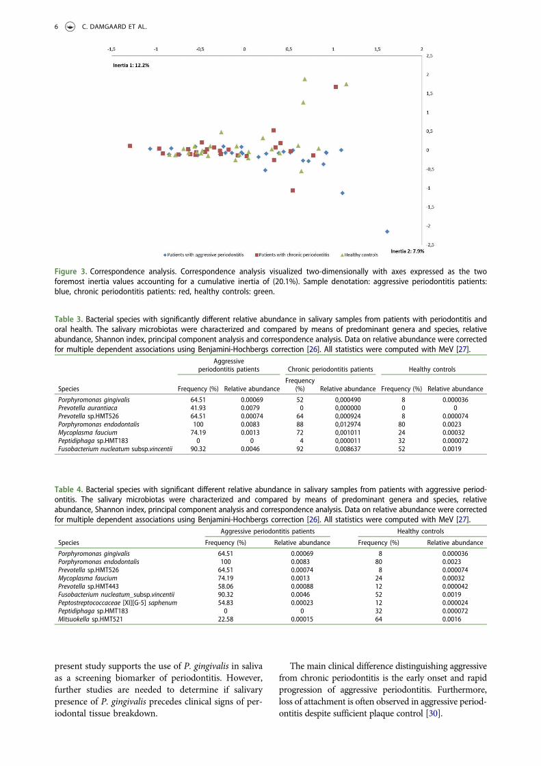
No differences were observed in the composition of thecore salivary microbiota in aggressive periodontitis,chronic periodontitis and oral health. Relative abun-dance of predominant genera is visualized in Figure 1,from which it can be seen that Prevotella, Neisseria,Streptococcus and Veillonella species constitutedapproximately 60% of the salivary microbiota in allgroups. Furthermore, comparable α-diversities, asdetermined by the Shannon-index, were recorded inpatients with aggressive periodontitis (3.4), chronic per-iodontitis (3.4) and oral health (3.5). Finally, data reduc-tion using principal component analysis (Figure 2) andcorrespondence analysis (Figure 3) showed completelyrandom distribution with no tendency of sample clus-tering based on periodontal disease status.
Species-specific associations with periodontitisand oral health
Seven bacterial species (six periodontitis-associated andone health-associated) were identified witha statistically significant difference in relative abun-dance in patients with aggressive periodontitis, patientswith chronic periodontitis and orally healthy indivi-duals (adjusted p-value <0.05, Table 3). Furthermore,nine bacterial species (seven aggressive periodontitis-associated and two health-associated) were identifiedwith significant differences in relative abundance inaggressive periodontitis patients and orally healthy indi-viduals (adjusted p-value <0.05, Table 4). No bacterialspecies were identified with a statistically significantrelative abundance when comparing aggressive period-ontitis patients with chronic periodontitis patients.
Salivary abundance of P. gingivalis could discrimi-nate aggressive (AUC: 0.80, p = 0.0001) and chronicperiodontitis patients (AUC: 0.72, p = 0.006) fromhealthy controls. Likewise, salivary presence ofP. gingivalis was significantly associated with aggressiveperiodontitis (p < 0.0001, RR: 8.1 (95% CI 2.1–31.2))and chronic periodontitis (p = 0.002, RR: 6.5 (95%CI: 1.6–25.9)). No statistically significant differences inpresence (p = 0.42, RR: 1.2 (95% CI: 0.8–2.0)) or relativeabundance (AUC: 0.61, p = 0.15) of P. gingivalis wereobserved between patients with aggressive and chronicperiodontitis.
Discussion
To the best of our knowledge, this is the first study tocharacterize the salivary microbiota in patients withaggressive periodontitis.
The most prominent finding was that salivary pre-sence of P. gingivalis was significantly associated withaggressive periodontitis and chronic periodontitis, as
4 C. DAMGAARD ET AL.

compared to orally healthy controls (Table 3).Specifically, salivary abundance of P. gingivalis coulddiscriminate aggressive (AUC: 0.80, p = 0.0001) andchronic periodontitis (AUC: 0.72, p = 0.006) fromhealthy controls. Likewise, salivary presence ofP. gingivalis was significantly associated with aggres-sive (p < 0.0001, RR: 8.1 (95% CI 2.1–31.2))and chronic periodontitis (p = 0.002, RR: 6.5
(95% CI: 1.6–25.9)). Subgingival colonization byP. gingivalis is considered a risk factor periodontitis[28], which is why subgingival presence of P. gingivalishas been used as a screening biomarker of period-ontitis [6,29]. Interestingly, several cross-sectional stu-dies based on different molecular methods havereported a strong correlation in subgingival and sali-vary levels of P. gingivalis [15–18]. Thus, data from the
Figure 1. Predominant bacterial genera. Mean relative abundance of predominant bacterial genera in patients with aggressiveperiodontitis, chronic periodontitis and orally healthy controls. The core salivary microbiota was defined as species/genus witha mean relative abundance >1% across sample. The predominant species/genera were compared using Mann-Whitney U testand Kruskall-Wallis test with Benjamini-Hochberg correction.
Figure 2. Principal component analysis. Principal component analysis visualized two-dimensionally with axes expressed as twoprincipal components values accounting for a cumulative value of (38.0%). Sample denotation: aggressive periodontitis patients:blue, chronic periodontitis patients: red, healthy controls: green.
JOURNAL OF ORAL MICROBIOLOGY 5
present study supports the use of P. gingivalis in salivaas a screening biomarker of periodontitis. However,further studies are needed to determine if salivarypresence of P. gingivalis precedes clinical signs of per-iodontal tissue breakdown.
The main clinical difference distinguishing aggressivefrom chronic periodontitis is the early onset and rapidprogression of aggressive periodontitis. Furthermore,loss of attachment is often observed in aggressive period-ontitis despite sufficient plaque control [30].
Figure 3. Correspondence analysis. Correspondence analysis visualized two-dimensionally with axes expressed as the twoforemost inertia values accounting for a cumulative inertia of (20.1%). Sample denotation: aggressive periodontitis patients:blue, chronic periodontitis patients: red, healthy controls: green.
Table 3. Bacterial species with significantly different relative abundance in salivary samples from patients with periodontitis andoral health. The salivary microbiotas were characterized and compared by means of predominant genera and species, relativeabundance, Shannon index, principal component analysis and correspondence analysis. Data on relative abundance were correctedfor multiple dependent associations using Benjamini-Hochbergs correction [26]. All statistics were computed with MeV [27].
Aggressiveperiodontitis patients Chronic periodontitis patients Healthy controls
Species Frequency (%) Relative abundanceFrequency
(%) Relative abundance Frequency (%) Relative abundance
Porphyromonas gingivalis 64.51 0.00069 52 0,000490 8 0.000036Prevotella aurantiaca 41.93 0.0079 0 0,000000 0 0Prevotella sp.HMT526 64.51 0.00074 64 0,000924 8 0.000074Porphyromonas endodontalis 100 0.0083 88 0,012974 80 0.0023Mycoplasma faucium 74.19 0.0013 72 0,001011 24 0.00032Peptidiphaga sp.HMT183 0 0 4 0,000011 32 0.000072Fusobacterium nucleatum subsp.vincentii 90.32 0.0046 92 0,008637 52 0.0019
Table 4. Bacterial species with significant different relative abundance in salivary samples from patients with aggressive period-ontitis. The salivary microbiotas were characterized and compared by means of predominant genera and species, relativeabundance, Shannon index, principal component analysis and correspondence analysis. Data on relative abundance were correctedfor multiple dependent associations using Benjamini-Hochbergs correction [26]. All statistics were computed with MeV [27].
Aggressive periodontitis patients Healthy controls
Species Frequency (%) Relative abundance Frequency (%) Relative abundance
Porphyromonas gingivalis 64.51 0.00069 8 0.000036Porphyromonas endodontalis 100 0.0083 80 0.0023Prevotella sp.HMT526 64.51 0.00074 8 0.000074Mycoplasma faucium 74.19 0.0013 24 0.00032Prevotella sp.HMT443 58.06 0.00088 12 0.000042Fusobacterium nucleatum_subsp.vincentii 90.32 0.0046 52 0.0019Peptostreptococcaceae [XI][G-5] saphenum 54.83 0.00023 12 0.000024Peptidiphaga sp.HMT183 0 0 32 0.000072Mitsuokella sp.HMT521 22.58 0.00015 64 0.0016
6 C. DAMGAARD ET AL.
In the present study, patients with aggressive per-iodontitis had a notably high PI. All participants wereincluded prior to periodontal treatment; thus, therapidity of disease progression was estimated by relat-ing the attachment loss to the age of the participant.The patients with aggressive periodontitis were allaged under 40 years and presented with absence ofmedical diseases, but with severe attachment loss(mean 3.5 mm) at the time of inclusion.
Subgingival colonization of virulent periodontal bac-teria, such as P. gingivalis may drive the destructiveimmune responses towards the periodontal microbiota.In line herewith, abundance of P. gingivalis have beensuggested as a possible explanation to the different clin-ical phenotypes observed in aggressive and chronic per-iodontitis [31,32]. The anaerobic environment found ininflamed periodontal pockets offers nutritional require-ments, which favors growth of especially P. gingivalis.Interestingly, we found no differences between the sali-vary microbiota of patients with aggressive periodontitispatients and that of patients with chronic periodontitis(Figure 1–3). Accordingly, this may be explained by thesimilar clinical characteristics observed in the aggressiveperiodontitis and chronic periodontitis group in the pre-sent study (Table 2). However, virulence mechanismsmay varywithin a species, andP. gingivalis can be dividedaccording to fimA genotype.WhileP. gingivalis fimA typeII is the most prevalent in periodontitis and in particularin patients with aggressive periodontitis [33,34],P. gingivalis fimA type I is exclusively found in period-ontally healthy subjects [35]. HOMINGS cannot discri-minate P. gingivalis according to fimA genotypes, whichmay explain the present comparable findings ofP. gingivalis in aggressive and chronic periodontitis.Nevertheless, the finding of comparable salivary micro-biotas in patients with aggressive and chronic period-ontitis may also support the assumption of the immuneresponse towards the periodontal microbiota as the deci-sive factor behind the clinical presentations of period-ontitis [8].
Another notable finding was that salivary presenceand relative abundance of Prevotella aurantiaca wasassociated with aggressive periodontitis, see Table 3.P. aurantiaca, which is a member of the Prevotellaintermedia-group [36], was isolated from the oralcavity in 2010 [37]. To the best of our knowledge,this is the first study that associates P. aurantiacawith aggressive periodontitis, which is, why furtherstudies are needed to evaluate this particular finding.
In the present study comparable core salivarymicrobiotas were observed in patients with period-ontitis and orally healthy controls (Figures 1–3).Accordingly, the salivary microbiota is believed tobe a conglomerate of bacteria shed from oral surfaces,with the tongue, throat and tonsils as the primarydonor sites [5]. Therefore, salivary presence andabundance of P. gingivalis recorded in the present
study could potentially originate from either the ton-gue or the subgingival pocket. Previous studies havereported that periodontal bacteria associated withperiodontitis may be found on the tongue of orallyhealthy individuals [38]. It is therefore interestingthat P. gingivalis was observed in saliva from 64%and 52% of patients with aggressive and chronicperiodontitis, respectively, as compared to only 8%of orally healthy controls (Table 3). Therefore,P. gingivalis identified in saliva from patients withaggressive and chronic periodontitis in the presentstudy were probably spill-over from concomitant per-iodontal lesions in these patients rather than rem-nants from the tongue.
Some limitations apply to the present study, includingthat six samples failed DNA extraction and two samplesfailed sequencing, which is why only data from 81 parti-cipants were included in downstream analyses.Furthermore, saliva samples were collected throughoutthe day, which means that the data presented couldpotentially be influenced by diurnal variation. However,previous studies have reported a minimal impact ofcollection method and sampling time on the composi-tion of the salivarymicrobiota [21,39]. Finally, as the newclassification of periodontal diseases from 2018 [40],were introduced during the study period it was notpossible to use this classification in the present study.However, it appears that the aggressive periodontitispatients included comprising periodontitis, grade C, gen-eralized, leaving only the staging undescribed. The mainstrength of the present study was the capability of thenew HOMINGS protocol, which targets the V1-V3region of the 16S rRNA gene to identify 97.6% of allsequences at species level. To the best of our knowledge,this is the first study to use next-generation sequencing toachieve >97% coverage at species level.
In conclusion, salivary presence and relative abun-dance of P. gingivalis associate with aggressive andchronic periodontitis compared to oral health, but donot discriminate between aggressive and chronic per-iodontitis. Based on the findings of the present study,P. gingivalis is relevant as a proxy for deep period-ontal pockets and thus periodontitis in clinical trials.However, future investigations would benefit fromdata discriminating P. gingivalis according to fimAgenotypes, to allow differentiation according to thevirulence of the strains.
Acknowledgments
Sequencing was performed at the Forsyth Institute,Cambridge, MA by the Sequencing and Bioinformatics Core.
Disclosure statement
No potential conflict of interest was reported by the authors.
JOURNAL OF ORAL MICROBIOLOGY 7
Data availability
Access to all data, including raw sequence data, will begranted upon request ([email protected]).
Funding
The study was financially supported by the DanishFoundation for Mutual Efforts in Dental Care and theDanish Dental Association.
ORCID
Christian Damgaard http://orcid.org/0000-0003-2394-746X
References
[1] Schroder SA, Bardow A, Eickhardt-Dalboge S, et al. Isparotid saliva sterile on entry to the oral cavity? ActaOtolaryngol. 2017;137(7):762–764.
[2] Curtis MA, Zenobia C, Darveau RP. The relationshipof the oral microbiotia to periodontal health anddisease. Cell Host Microbe. 2011;10(4):302–306.
[3] Kilian M, Chapple IL, Hannig M, et al. The oralmicrobiome – an update for oral healthcare profes-sionals. Br Dent J. 2016;221(10):657–666.
[4] Krishnan K, Chen T, Paster BJ. A practical guide tothe oral microbiome and its relation to health anddisease. Oral Dis. 2017;23(3):276–286.
[5] Segata N, Haake SK, Mannon P, et al. Composition ofthe adult digestive tract bacterial microbiome basedon seven mouth surfaces, tonsils, throat and stoolsamples. Genome Biol. 2012;13(6):R42.
[6] Liljestrand JM, Gursoy UK, Hyvarinen K, et al.Combining salivary pathogen and serum antibodylevels improves their diagnostic ability in detectionof periodontitis. J Periodontol. 2014;85(1):123–131.
[7] Paju S, Pussinen PJ, Suominen-Taipale L, et al.Detection of multiple pathogenic species in saliva isassociated with periodontal infection in adults. J ClinMicrobiol. 2009;47(1):235–238.
[8] Bartold PM, Van Dyke TE. Periodontitis: ahost-mediated disruption of microbial homeostasis.Unlearning learned concepts. Periodontol 2000.2013;62(1):203–217.
[9] Armitage GC. Development of a classification systemfor periodontal diseases and conditions. AnnPeriodontol. 1999;4(1):1–6.
[10] Eke PI, Dye BA, Wei L, et al. Prevalence of period-ontitis in adults in the USA: 2009 and 2010. J DentRes. 2012;91(10):914–920.
[11] Ramseier CA, Anerud A, Dulac M, et al. Naturalhistory of periodontitis: disease progression andtooth loss over 40 years. J Clin Periodontol. 2017;44(12):1182–1191.
[12] Belstrom D, Constancias F, Liu Y, et al. Metagenomicand metatranscriptomic analysis of saliva revealsdisease-associated microbiota in patients with period-ontitis and dental caries. NPJ Biofilms Microbiomes.2017;3:23.
[13] Belstrom D, Jersie-Christensen RR, Lyon D, et al.Metaproteomics of saliva identifies human proteinmarkers specific for individuals with periodontitis
and dental caries compared to orally healthycontrols. PeerJ. 2016;4:e2433.
[14] Belstrom D, Paster BJ, Fiehn NE, et al. Salivary bac-terial fingerprints of established oral disease revealedby the Human Oral Microbe Identification using NextGeneration Sequencing (HOMINGS) technique.J Oral Microbiol. 2016;8:30170.
[15] Boutaga K, Savelkoul PH, Winkel EG, et al.Comparison of subgingival bacterial sampling withoral lavage for detection and quantification of period-ontal pathogens by real-time polymerase chainreaction. J Periodontol. 2007;78(1):79–86.
[16] He J, Huang W, Pan Z, et al. Quantitative analysis ofmicrobiota in saliva, supragingival, and subgingivalplaque of Chinese adults with chronic periodontitis.Clin Oral Investig. 2012;16(6):1579–1588.
[17] Nickles K, Scharf S, Rollke L, et al. Comparison of twodifferent sampling methods for subgingival plaque:subgingival paper points or mouthrinse sample?J Periodontol. 2017;88(4):399–406.
[18] Haririan H, Andrukhov O, Bertl K, et al. Microbialanalysis of subgingival plaque samples compared tothat of whole saliva in patients with periodontitis.J Periodontol. 2014;85(6):819–828.
[19] Kageyama S, Takeshita T, Asakawa M, et al. Relativeabundance of total subgingival plaque-specific bacteriain salivary microbiota reflects the overall periodontalcondition in patients with periodontitis. PLoS One.2017;12(4):e0174782.
[20] Belstrom D, Grande MA, Sembler-Moller ML, et al.Influence of periodontal treatment on subgingivaland salivary microbiotas. J Periodontol. 2018;89(5):531–539.
[21] Belstrom D, Holmstrup P, Bardow A, et al.Comparative analysis of bacterial profiles in unstimu-lated and stimulated saliva samples. J Oral Microbiol.2016;8:30112.
[22] Kozich JJ, Westcott SL, Baxter NT, et al. Developmentof a dual-index sequencing strategy and curation pipe-line for analyzing amplicon sequence data on theMiSeq Illumina sequencing platform. Appl EnvironMicrobiol. 2013;79(17):5112–5120.
[23] Callahan BJ, McMurdie PJ, Rosen MJ, et al. DADA2:high-resolution sample inference from Illuminaamplicon data. Nat Methods. 2016;13(7):581–583.
[24] Ewels P, Magnusson M, Lundin S, et al. MultiQC:summarize analysis results for multiple tools and sam-ples in a single report. Bioinformatics. 2016;32(19):3047–3048.
[25] Wang Q, Garrity GM, Tiedje JM, et al. Naive Bayesianclassifier for rapid assignment of rRNA sequences intothe new bacterial taxonomy. Appl Environ Microbiol.2007;73(16):5261–5267.
[26] Hochberg Y, Benjamini Y. More powerful proceduresfor multiple significance testing. Stat Med. 1990;9(7):811–818.
[27] Saeed AI, Bhagabati NK, Braisted JC, et al. TM4microarray software suite. Methods Enzymol.2006;411:134–193.
[28] Socransky SS, Haffajee AD, Cugini MA, et al.Microbial complexes in subgingival plaque. J ClinPeriodontol. 1998;25(2):134–144.
[29] Kinney JS, Morelli T, Braun T, et al. Saliva/pathogenbiomarker signatures and periodontal diseaseprogression. J Dent Res. 2011;90(6):752–758.
[30] Albandar JM. Aggressive and acute periodontaldiseases. Periodontol 2000. 2014;65(1):7–12.
8 C. DAMGAARD ET AL.
[31] Kononen E, Muller HP. Microbiology of aggressiveperiodontitis. Periodontol 2000. 2014;65(1):46–78.
[32] Kulkarni C, Kinane DF. Host response in aggres-sive periodontitis. Periodontol 2000. 2014;65(1):79–91.
[33] Amano A, Kuboniwa M, Nakagawa I, et al. Prevalence ofspecific genotypes of Porphyromonas gingivalis fimA andperiodontal health status. J Dent Res. 2000;79(9):1664–1668.
[34] Zhao L, Wu YF, Meng S, et al. Prevalence of fimAgenotypes of Porphyromonas gingivalis and periodon-tal health status in Chinese adults. J Periodontal Res.2007;42(6):511–517.
[35] Miura M, Hamachi T, Fujise O, et al. The preva-lence and pathogenic differences of Porphyromonasgingivalis fimA genotypes in patients with aggres-sive periodontitis. J Periodontal Res. 2005;40(2):147–152.
[36] Fteita D, Könönen E, Gürsoy M, et al. Quorum sen-sing molecules regulate epithelial cytokine responseand biofilm-related virulence of three Prevotellaspecies. Anaerobe. 2018;54:128–135.
[37] Sakamoto M, Suzuki N, Okamoto M. Prevotellaaurantiaca sp. nov., isolated from the human oralcavity. Int J Syst Evol Microbiol. 2010;60:500–503.
[38] Tanner AC, Paster BJ, Lu SC, et al. Subgingival andtongue microbiota during early periodontitis. J DentRes. 2006;85(4):318–323.
[39] Belstrom D, Holmstrup P, Bardow A, et al. Temporalstability of the salivary microbiota in oral health. PLoSOne. 2016;11(1):e0147472.
[40] Papapanou PN, Sanz M, Buduneli N, et al.Periodontitis: consensus report of workgroup 2 ofthe 2017 world workshop on the classification of per-iodontal and peri-implant diseases and conditions.J Clin Periodontol. 2018;45(Suppl 20):S162–S170.
JOURNAL OF ORAL MICROBIOLOGY 9
Related Documents


![Dentistry tripolyphosphate against Porphyromonas species ... · were Porphyromonas species including Porphyromonas gulae, Porphyromonas cangingivalis, and Porphyromonas cansulci [11,12].](https://static.cupdf.com/doc/110x72/5f02cfa57e708231d4062096/dentistry-tripolyphosphate-against-porphyromonas-species-were-porphyromonas.jpg)

