Sleep, 18(6):478-500 © 1995 American Sleep Disorders Association and Sleep Research Society State of the Art Review Magnocellular Nuclei of the Basal Forebrain: Substrates of Sleep and Arousal Regulation Ronald Szymusiak Sleep Physiology Research (l51A3), Dept. Veterans Affairs Medical Center, Sepulveda, CA 91343 and Dept. of Medicine, School of Medicine, University of California at Los Angeles, Los Angeles, CA 90024 Summary: Magnocellular regions of the basal forebrain (BF) are recognized as important sites of sleep-wake regulation. Evidence is reviewed for the coexistence within the BF of mechanisms that regulate neocortical and limbic system arousal along with mechanisms that promote sleep. Arousal-related functions are mediated by a system of magnocellular cholinergic neurons. BF cholinergic neurons project monosynaptically to the entire neo- cortex and limbic telencephalon, exert excitatory effects on target cells and participate in the regulation of activated EEG patterns characteristic of waking and REM sleep. Evidence suggests that, within the waking state, the BF cholinergic system modulates processing of sensory information in the neocortex and is involved in cognitive processes. One or more noncholinergic cell types are responsible for the sleep-promoting functions of the BF. Neurons that display elevated discharge rates during transitions from waking to sleep and during nonREM sleep have been recorded in BF sites where electrical stimulation evokes sleep and experimental lesions cause insomnia. BF neurons function to promote sleep, in part, via descending inhibition of caudal hypothalamic and brainstem activating systems. GABAergic neurons located within magnocellular regions of the BF are hypothesized to mediate sleep-promoting actions. Afferents to the BF from hypothalamic and brainstem regions are functionally important for sleep-wake regulation. Thermosensitive inputs from the anterior hypothalamus modulate the activity of BF sleep- and arousal-related cell types. Excitatory effects of brainstem inputs to BF arousal-related cells have been documented. Additional evidence supports a critical role for GABAergic-cholinergic interactions, both within the magnocellular BF and at cortical and diencephalic sites, in the regulation of behavioral state. Key Words: Basal forebrain- Nucleus basalis- Preoptic area- Anterior hypothalamus- Arousal- NonREM sleep- EEG-Sleep-wake regulation - Acetylcholine - GABA. The heterogeneous region ofthe mammalian ventral telencephalon known as the basal forebrain (BF) has been critically implicated in the regulation of sleep and wakefulness. Early studies of neuropathology in man, and lesion and stimulation studies in experimental an- imals, provided evidence that portions of the BF ad- jacent to the rostral hypothalamus function as a sleep- promoting area, capable of modulating somato- and viscero-motor activity and the EEG. More recently, a collection of acetylcholine (ACh)- containing neurons has been localized to the BF, and these neurons were shown to project monosynaptically to the entire limbic telencephalon and neocortex. The BF cholinergic system appears to regulate important Accepted for publication April 1995. Address correspondence and reprint requests to Ronald Szymu- siak, Ph.D., Sleep Physiology Research (15IA3), Dept. Veterans Affairs Medical Center, Sepulveda, CA 91343, U.S.A. aspects of limbic system and neocortical arousal, to modulate sensory processing in the neocortex, and to participate in attentional processes. Degeneration of BF cholinergic neurons in Alzheimer's disease, and lesions of cholinergic portions of the BF in experi- mental animals are associated with slowing of the wak- ing cortical EEG and cognitive deficits. A goal of this review will be to reconcile the seem- ingly conflicting postulated roles for the BF in behav- ioral state regulation; that is, that of a sleep-regulating system and of one that enhances cortical activation. Evidence will be reviewed that suggests that sleep-pro- moting and arousal-related mechanisms coexist within the BF, and that these are neurochemically and, at least partially, anatomically distinct. First, the anatomy of the BF as it relates to these putative behavioral state- regulating functions will be reviewed. Second, I will review what is known about the behavioral neuro- physiology of the BF cholinergic system. Third, the 478 Downloaded from https://academic.oup.com/sleep/article/18/6/478/2749729 by guest on 05 July 2022

Welcome message from author
This document is posted to help you gain knowledge. Please leave a comment to let me know what you think about it! Share it to your friends and learn new things together.
Transcript
Sleep, 18(6):478-500 © 1995 American Sleep Disorders Association and Sleep Research Society
State of the Art Review
Magnocellular Nuclei of the Basal Forebrain: Substrates of Sleep and Arousal Regulation
Ronald Szymusiak
Sleep Physiology Research (l51A3), Dept. Veterans Affairs Medical Center, Sepulveda, CA 91343 and Dept. of Medicine, School of Medicine,
University of California at Los Angeles, Los Angeles, CA 90024
Summary: Magnocellular regions of the basal forebrain (BF) are recognized as important sites of sleep-wake regulation. Evidence is reviewed for the coexistence within the BF of mechanisms that regulate neocortical and limbic system arousal along with mechanisms that promote sleep. Arousal-related functions are mediated by a system of magnocellular cholinergic neurons. BF cholinergic neurons project monosynaptically to the entire neocortex and limbic telencephalon, exert excitatory effects on target cells and participate in the regulation of activated EEG patterns characteristic of waking and REM sleep. Evidence suggests that, within the waking state, the BF cholinergic system modulates processing of sensory information in the neocortex and is involved in cognitive processes. One or more noncholinergic cell types are responsible for the sleep-promoting functions of the BF. Neurons that display elevated discharge rates during transitions from waking to sleep and during nonREM sleep have been recorded in BF sites where electrical stimulation evokes sleep and experimental lesions cause insomnia. BF neurons function to promote sleep, in part, via descending inhibition of caudal hypothalamic and brainstem activating systems. GABAergic neurons located within magnocellular regions of the BF are hypothesized to mediate sleep-promoting actions. Afferents to the BF from hypothalamic and brainstem regions are functionally important for sleep-wake regulation. Thermosensitive inputs from the anterior hypothalamus modulate the activity of BF sleep- and arousal-related cell types. Excitatory effects of brainstem inputs to BF arousal-related cells have been documented. Additional evidence supports a critical role for GABAergic-cholinergic interactions, both within the magnocellular BF and at cortical and diencephalic sites, in the regulation of behavioral state. Key Words: Basal forebrain- Nucleus basalis-Preoptic area-Anterior hypothalamus-Arousal-NonREM sleep-EEG-Sleep-wake regulation - Acetylcholine - GABA.
The heterogeneous region ofthe mammalian ventral telencephalon known as the basal forebrain (BF) has been critically implicated in the regulation of sleep and wakefulness. Early studies of neuropathology in man, and lesion and stimulation studies in experimental animals, provided evidence that portions of the BF adjacent to the rostral hypothalamus function as a sleeppromoting area, capable of modulating somato- and viscero-motor activity and the EEG.
More recently, a collection of acetylcholine (ACh)containing neurons has been localized to the BF, and these neurons were shown to project monosynaptically to the entire limbic telencephalon and neocortex. The BF cholinergic system appears to regulate important
Accepted for publication April 1995. Address correspondence and reprint requests to Ronald Szymu
siak, Ph.D., Sleep Physiology Research (15IA3), Dept. Veterans Affairs Medical Center, Sepulveda, CA 91343, U.S.A.
aspects of limbic system and neocortical arousal, to modulate sensory processing in the neocortex, and to participate in attentional processes. Degeneration of BF cholinergic neurons in Alzheimer's disease, and lesions of cholinergic portions of the BF in experimental animals are associated with slowing of the waking cortical EEG and cognitive deficits.
A goal of this review will be to reconcile the seemingly conflicting postulated roles for the BF in behavioral state regulation; that is, that of a sleep-regulating system and of one that enhances cortical activation. Evidence will be reviewed that suggests that sleep-promoting and arousal-related mechanisms coexist within the BF, and that these are neurochemically and, at least partially, anatomically distinct. First, the anatomy of the BF as it relates to these putative behavioral stateregulating functions will be reviewed. Second, I will review what is known about the behavioral neurophysiology of the BF cholinergic system. Third, the
478
Dow
nloaded from https://academ
ic.oup.com/sleep/article/18/6/478/2749729 by guest on 05 July 2022
I .~
I
'.'
MAGNOCELLULAR NUCLEI OF THE BASAL FOREBRAIN 479
experimental evidence for a hypnogenic mechanism within the BF will be considered. Finally, I will summarize what is and is not known about interactions among BF cholinergic and noncholinergic cell types in the regulation of arousal and sleep.
ANATOMICAL CONSIDERATIONS
The review of BF anatomy here will be brief, with a focus on features relevant to behavioral state control. The reader is referred to recent comprehensive reviews of BF anatomy (1-5).
The BF comprises a heterogeneous set of structures on the ventralmost aspects of the telencephalon, adjacent to preoptic, supraoptic and rostral infundibular levels of the hypothalamus. Since the discovery of a system of ACh-containing magnocellular neurons that are the primary source of cholinergic innervation to the limbic system and neocortex, the term "basal forebrain" has become nearly synonymous with the magnocellular cholinergic system. Although this is an oversimplification, the distribution of cholinergic neurons within the BF constitutes a suitable reference point for considering the functional anatomy of this region as it relates to behavioral state control (Fig. 1).
The medial preoptic area and anterior hypothalamic areas are also frequently considered to be part of the ventral forebrain hypnogenic system. The anatomical and functional relationships of these areas to the magnocellular BF will be considered in a subsequent section.
The reader should be aware that important regions within the BF contain few magnocellular neurons, and have anatomical and functional relationships that are at least partially independent of the cholinergic cell groups and of sleep-wake regulating mechanisms. For example, the nucleus accumbens and portions of the substantial innominata are closely related to both the basal ganglia and the limbic system including the amygdala (5). These structures participate in motor control and limbic system-motor integration (6). In focusing on the role of the magnocellular systems in regulating EEG-defined sleep-waking states, these aspects of BF function will, for the most part, not be considered. However, a complete understanding of forebrain state-regulating functions will eventually have to incorporate the ways in which sleep-waking mechanisms interact with the striatopallidal and limbic system components of the BF.
Cholinergic neurons
The distribution of cholinergic neurons within the rat BF is shown in Fig. 1. Neurons that are immunohistochemically positive for the ACh-synthesizing en-
zyme choline-acetyl transferase (ChAT) occur as a continuous series of cell clusters that are contained within several structures that comprise the BF. From rostral to caudal, they are found in the medial septum (MS; Fig. lA and B), the vertical (Fig. lA) and horizontal (Fig. 1 B-D) limbs of the diagonal bands of Broca (VDB and HDB), magnocellular preoptic area (MCP; Fig. 1 C and D), subpallidal substantia innominata (SI; Fig. ID and E) and the peripalIidal nucleus basalis ofMeynert (NB; Fig. IE and F). ChAT-positive neurons are large and multipolar. Several fiber systems traverse these regions, including the medial forebrain bundle, anterior commissure, ansa lenticularis, ansa peduncularis and inferior thalamic peduncle. At many sites within the BF, magnocellular ACh neurons occur as clusters embedded among one or more of these fiber systems.
Cholinergic neurons of the BF are widely hypothesized to modulate the level of arousal or activation in a variety of limbic system and cortical sites (7-10). Consistent with this hypothesized function is the documented monosynaptic projection of BF cholinergic neurons to the entire limbic telencephalon and neocortex. Different subpopulations ofBF cholinergic neurons have different projection targets. Neurons in the MS and VDB innervate the hippocampus, those within the HDB and MCP target primarily the olfactory bulb, entorhinal cortex and piriform cortex, and SI and NB cholinergic neurons project to the neocortex (11-17). There is a topographical organization to the neocortical projection with a rostral to caudal progression of SII NB neurons reaching a medial to lateral sequence of cortical sites (14, 17-19). This topography is not strictly somatotopic. For example, in rat sensorimotor cortex, adjacent subregions receive projections from distributed portions of the SIINB (20). The projection should not be considered to be diffuse, however, as few individual cholinergic neurons « 5%) project to multiple cortical sites (11,14,20).
These basic features of the BF-neocortical cholinergic projection are consistent across species, although some specific differences are noteworthy. In primates, the NB forms a fairly compact nucleus of cholinergic neurons closely associated with the globus pallidus (19). Nearly all of the cortically projecting neurons (>95%) in the primate NB are cholinergic (19). In cats and rats, the cholinergic neurons of the NB are more diffusely organized, found within and adjacent to the globus pallidus, internal capsule and nucleus of the ansa lenticularis. The precise borders ofSI and NB cholinergic neurons are not as distinct in these species as in primates. Significant numbers of noncholinergic projection neurons have been described in the NB of cats (16,21). In rat, 80-90% ofNB projection neurons are cholinergic, but less than 50% ofSI projection neurons are immunoreactive for ChAT (7,22).
Sleep. Vol. 18. No.6. 1995
Dow
nloaded from https://academ
ic.oup.com/sleep/article/18/6/478/2749729 by guest on 05 July 2022

480 R. SZYMUSIAK
LSi , ";.~Sv~·
.\
A
c
F
FIG. I. Distribution of ChAT-immunoreactive neurons (shown as small black dots) in the rat basal forebrain. Abbreviations: ac, anterior commissure; Acb, accumbens nucleus; AH, anterior hypothalamus; BL, basolateral amygdaloid nucleus; BSt, bed nucleus of the stria terminalis; CA, central amygdaloid nucleus; CP, caudate putamen; DM, dorsomedial hypothalamic nucleus; f, fornix; GP, globus pallidus; HDB, horizontal limb diagonal band nucleus; Hi, hippocampus; ic, internal capsule; LSd, LSi and LSv, lateral septal nucleus, dorsal, intermediate and ventral; LV, lateral ventricle; MCP, magnocellular preoptic nucleus; MD, mediodorsal thalamic nucleus; MP, medial
Sleep, Vol. 18, No.6, 1995
Dow
nloaded from https://academ
ic.oup.com/sleep/article/18/6/478/2749729 by guest on 05 July 2022
MAGNOCELLULAR NUCLEI OF THE BASAL FOREBRAIN 481
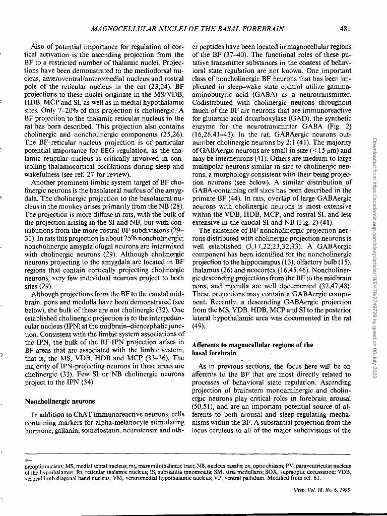
Also of potential importance for regulation of cortical activation is the ascending projection from the BF to a restricted number of thalamic nuclei. Projections have been demonstrated to the mediodorsal nucleus, anteroventrallanteromedial nucleus and rostral pole of the reticular nucleus in the cat (23,24). BF projections to these nuclei originate in the MS/VDB, HDB, MCP and SI, as well as in medial hypothalamic sites. Only 7-20% of this projection is cholinergic. A BF projection to the thalamic reticular nucleus in the rat has been described. This projection also contains cholinergic and noncholinergic components (25,26). The BF-reticular nucleus projection is of particular potential importance for EEG regulation, as the thalamic reticular nucleus is critically involved in controlling thalamocortical oscillations during sleep and wakefulness (see ref. 27 for review).
Another prominent limbic system target ofBF cholinergic neurons is the basolateral nucleus of the amygdala. The cholinergic projection to the basolateral nucleus in the monkey arises primarily from the NB (28). The projection is more diffuse in rats, with the bulk of the projection arising in the SI and NB, but with contributions from the more rostral BF subdivisions (29-31). In rats this projection is about 25% noncholinergic; noncholinergic amygdalofugal neurons are intermixed with cholinergic neurons (29). Although cholinergic neurons projecting to the amygdala are located in BF regions that contain cortically projecting cholinergic neurons, very few individual neurons project to both sites (29).
Although projections from the BF to the caudal midbrain, pons and medulla have been demonstrated (see below), the bulk of these are not cholinergic (32). One established cholinergic projection is to the interpeduncular nucleus (IPN) at the midbrain-<iiencephalic junction. Consistent with the limbic system associations of the lPN, the bulk of the BF-IPN projection arises in BF areas that are associated with the limbic system, that is, the MS, VDB, HDB and MCP (33-36). The majority of lPN-projecting neurons in these areas are cholinergic (33). Few SI or NB cholinergic neurons project to the IPN (34).
Noncholinergic neurons
In addition to ChAT immunoreactive neurons, cells containing markers for alpha-melanocyte stimulating hormone, gallanin, somatostatin, neurotensin and oth-
t--'
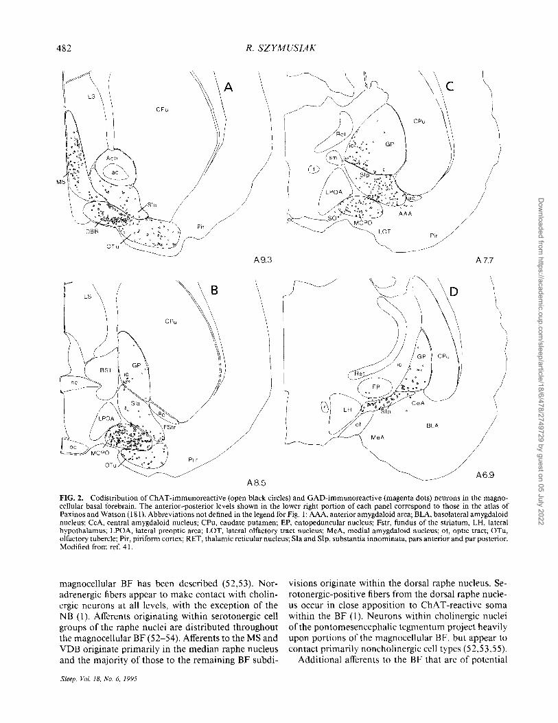
er peptides have been located in magnocellular regions of the BF (37-40). The functional roles of these putative transmitter substances in the context of behavioral state regulation are not known. One important class of noncholinergic BF neurons that has been implicated in sleep-wake state control utilize gammaaminobutyric acid (GABA) as a neurotransmitter. Codistributed with cholinergic neurons throughout much of the BF are neurons that are immunoreactive for glutamic acid decarboxylase (GAD), the synthetic enzyme for the neurotransmitter GABA (Fig. 2) (16,26,41-43). In the rat, GABAergic neurons outnumber cholinergic neurons by 2:1 (41). The majority of GABAergic neurons are small in size ( < 15 ,urn) and may be intemeurons (41). Others are medium to large multipolar neurons similar in size to cholinergic neurons, a morphology consistent with their being projection neurons (see below). A similar distribution of GABA-containing cell sizes ha~ 'been described in the primate BF (44). In rats, overlap of large GABAergic neurons with cholinergic neurons is most extensive within the VDB, HDB, MCP, and rostral SI, and less extensive in the caudal SI and NB (Fig. 2) (41).
The existence of BF noncholinergic projection neurons distributed with cholinergic projection neurons is well established (3,17,22,23,32,33). A GABAergic component has been identified for the noncholinergic projection to the hippocampus (13), olfactory bulb (15), thalamus (26) and neocortex (16,45,46). Noncholinergic descending projections from the BF to the midbrain pons, and medulla are well documented (32,47,48). These projections may contain a GABAergic component. Recently, a descending GABAergic' projection from the MS, VDB, HDB, MCP and SI to the posterior lateral hypothalamic area was documented in the rat (49).
Afferents to magnocellular regions of the basal forebrain
As in previous sections, the focus here will be on afferents to the BF that are most directly related to processes of behavioral state regulation. Ascending projection of brainstem monoaminergic and cholinergic neurons play critical roles in forebrain arousal (50,51), and are an important potential source of afferents to both arousal and sleep-regulating mechanisms within the BF. A substantial projection from the locus ceruleus to all of the major subdivisions of the
preoptic nucleus; MS, medial septal nucleus; mt, mammilothalamic tract; NB, nucleus basalis; ox, optic chiasm; PV, paraventricular nucleus of the hypothalamus; Rt, reticular thalamic nucleus; IS, substantia innominata; SM, stria medullaris; SOX, supraoptic decussation; VDB, vertical limb diagonal band nucleus; VM, ventromedial hypothalamic nucleus; VP, ventral pallidum. Modified from ref. 61.
Sleep. Vol. 18. No.6, 1995
Dow
nloaded from https://academ
ic.oup.com/sleep/article/18/6/478/2749729 by guest on 05 July 2022
482 R. SZYMUSIAK
A 7.7
LS
P, r
A6.9 A85
FIG. 2. Codistribution of ChAT-immunoreactive (open black circles) and GAD-immunoreactive (magenta dots) neurons in the magnocellular basal forebrain. The anterior-posterior levels shown in the lower right portion of each panel correspond to those in the atlas of Paxinos and Watson (181). Abbreviations not defined in the legend for Fig. I: AAA, anterior amygdaloid area; BLA, basolateral amygdaloid nucleus; CeA, central amygdaloid nucleus; CPu, caudate putamen; EP, entopeduncular nucleus; Fstr, fundus of the striatum, LH, lateral hypothalamus; LPOA, lateral preoptic area; LOT, lateral olfactory tract nucleus; MeA, medial amygdaloid nucleus; ot, optic tract; OTu, olfactory tubercle; Pir, piriform cortex; RET, thalamic reticular nucleus; SIa and SIp, substantia innominata, pars anterior and par posterior. Modified from ref. 41.
magnocellular BF has been described (52,53). Noradrenergic fibers appear to make contact with cholinergic neurons at all levels, with the exception of the NB (1). Afferents originating within serotonergic cell groups of the raphe nuclei are distributed throughout the magnocellular BF (52-54). Afferents to the MS and VDB originate primarily in the median raphe nucleus and the majority of those to the remaining BF subdi-
Sleep. Vol. 18. No.6, 1995
visions originate within the dorsal raphe nucleus. Serotonergic-positive fibers from the dorsal raphe nucleus occur in close apposition to ChAT -reactive soma within the BF (1). Neurons within cholinergic nuclei of the pontomesencephalic tegmentum project heavily upon portions of the magnocellular BF, but appear to contact primarily noncholinergic cell types (52,53,55).
Additional afferents to the BF that are of potential
Dow
nloaded from https://academ
ic.oup.com/sleep/article/18/6/478/2749729 by guest on 05 July 2022
MAGNOCELLULAR NUCLEI OF THE BASAL FOREBRAIN 483
functional importance for sleep-wake regulation arise from throughout the brainstem reticular core (1,52-54). The projection from the midbrain preferentially targets the subpaUidal SI and NB, whereas the MCP, HDB, VDB and MS are contacted predominantly by pontine reticular formation neurons. The EEG activating effects of the brainstem reticular formation are well known, and these BF afferents are of potential functional importance for both sleep- and waking-regulating mechanisms. Local BF injections of excitatory amino acid receptor antagonists block increased ACh release in cortex evoked by brainstem stimulation (56), indicating that a component of the brainstem projection that exerts excitatory effects on corticopetal BF cholinergic neurons is glutamatergic.
Portions of the posterior lateral hypothalamus, at levels extending from the mesencephalic-diencephalic junction to the perifornical area, participate in the regulation of arousal and EEG activation. These regions are the site of origin of a direct neocortical projection system (57). Lesions of these hypothalamic sites in experimental animals yield a syndrome of behavioral somnolence and enhanced cortical EEG synchrony (58-60). Cells within the posterior lateral hypothalamus also project to the magnocellular BF. Far lateral hypothalamic sites target the more dorsal peri- and subpallidal regions, and more medial portions project to the MCP and HDB (61). Fibers of this projection are found in close association with both cholinergic and noncholinergic BF neurons.
The medial preoptic area and anterior hypothalamus (MPI AH; Fig. 1) have important sleep-regulating functions, and projections from these midline structures to the adjacent magnocellular BF have been identified (61). Symmetric synaptic contacts from MP/AH neurons have been observed on BF cholinergic cells, suggesting an inhibitory influence. Neurophysiological evidence, reviewed later, indicates an inhibitory action on arousal-related, putative BF cholinergic neurons from temperature-sensitive neurons within the MPI AH (62).
Cholinergic regions of the magnocellular BF also receive afferents from the neocortex. These are also of potential interest in terms of the state-dependent neurophysiology of the BF. In the thalamo-cortical projection system, reciprocal cortical thalamic afferents are important in modulating synchronous discharge patterns of thalamic relay neurons during nonREM sleep (63,64). However, unlike the thalamo-cortical system where there is extensive reciprocity between the site of termination of thalamic afferents and the origin of the cortico-thalamic projection, the cholinergic BF receives projections from a comparatively restricted number of cortical sites. There are few projections from primary sensory or motor areas. The
majority arise from allocortical regions, including orbital cortical areas, insular cortex and piriform cortex (1). The projection to the magnocellular BF from the prefrontal cortex is substantial, and innervates the HDB and portions of the MCP and SI most densely (65,66). Direct cortical projections to the NB are sparse. Cortical influences on cells in these areas may be mediated via multisynaptic pathways involving the striatum and nucleus accumbens (1).
FUNCTIONAL ASPECTS OF THE BASAL FOREBRAIN CHOLINERGIC SYSTEM
There has been intense interest in the behavioral neurobiology of the BF cholinergic system, in part because of its involvement in the neuropathology of Alzheimer's disease. Two significant neurological deficits in this disorder are slowing of the dominant cortical EEG frequencies and dementia. Accordingly, research efforts have focused on the role of BF cholinergic cell groups in regulating EEG activation and in cognitive functions.
Cholinergic regulation of neocortical arousal and activated EEG patterns
Substantial evidence points to an involvement ofBF neurons, as the source of cortical ACh, in regulating neocortical activation. ACh-mediated activation of the cortex is important for expression of de synchronized EEG patterns (low voltage, mixed high frequencies) characteristic of waking and REM sleep. Spontaneous release of ACh measured at the cortical surface is elevated during waking and REM sleep, compared to nonREM sleep (67,68). Electrical stimulation of the SI/NB enhances cortical ACh release in the anesthetized rat (69,70). Stimulation at these sites also increases neocortical blood flow, an effect blocked by muscarinic antagonists, suggesting a vasodilatory role for the cholinergic innervation of cortical blood vessels (see ref. 71 for review). Regional cortical metabolic activation evoked by sensory stimulation is diminished following BF lesions in cats (72), a response that could reflect diminished regional blood flow as well as diminished neuronal activation. Within the waking state, rates of ACh release can vary with ongoing behavior, being elevated during locomotion compared to quiet waking (73,74). Ascending excitatory inputs to BF cholinergic neurons from the brainstem can influence cortical ACh release. Stimulation at the mesopontine tegmentum evokes EEG desynchrony and increased cortical ACh release (56). These effects are blocked by infusing glutamatergic agonists directly into the BF coincident with brainstem stimulation (56).
Reduced cortical ACh levels produced by excitotoxic
Sleep, Vol. 18, No.6, 1995
Dow
nloaded from https://academ
ic.oup.com/sleep/article/18/6/478/2749729 by guest on 05 July 2022
484
A
B 5
I ". set .s
• • • • • • • •
o~----~----~------
R. SZYMUSIAK
...-30 u Q) en
"'- 25 en Q) ~
'0. 20 en
w ~ 15 a:::: w c..? 10 0::: « I u 5 (f)
o
c
AW OW S1 S2 RT RA RQ SLEEP-WAKING STATE
D • J1l n • L .. .J. ,1'2 J. I. WAKI~G EEG.-.·., 11' ... ,,,., ,,"'.,..k -. • .,. ..... 1(*.4 • ~ ~ "l ..... (Y.",.;,~ .... A-~t" ....... '14,. 0" ".(rrt-Y __ ,..,.." ... *
EEG-"~"".~ .................... .,..~ ... ~-~. t/ ..... t"" ... ~to!.~,.,. ",+ ... ~ ... ~~.....,..,..,...
EOG~~~~~ EMG ~"I E 1.1il: I 11 Ull5Icn
~'~-V------"--~'~;J-J/
.j,lll ta4 J
UNITi •• , ....... Jl1b Ini, 111,_ ...... £ : ._.' •• iU'._'~'''"'' ... ".1'.,.,,, 3 \ _, .... INT ~--v...vr~~ /.-v'~I(IIYk.-'.-v'~."",/~""/, ............... ~/(~~III/"'rv"~/'
.0. REM SLEEP ....... .......,....,. tlf II" ... T ~."", ....... OI_.""'; .... ·_ .. ~ ... i'II,...~~"""" ..... J.~,..",.t __ · ........ Ij ... "!I~ ..... L...,"'~~'1"""'.1_ •• ~4'''''fa,",,-.... !IIoIIiIIIi\.''''''''''''''!''''' -""~'-"''''''''''''''''_F ~.~
Jlr.....'!I/,..w"'"""'''' .... ,W/(I;(/; ~...v~Mr~~~j .....'N..J~"..d"'.N".·,I>// 10 SEC
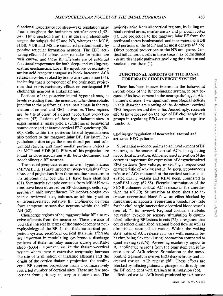
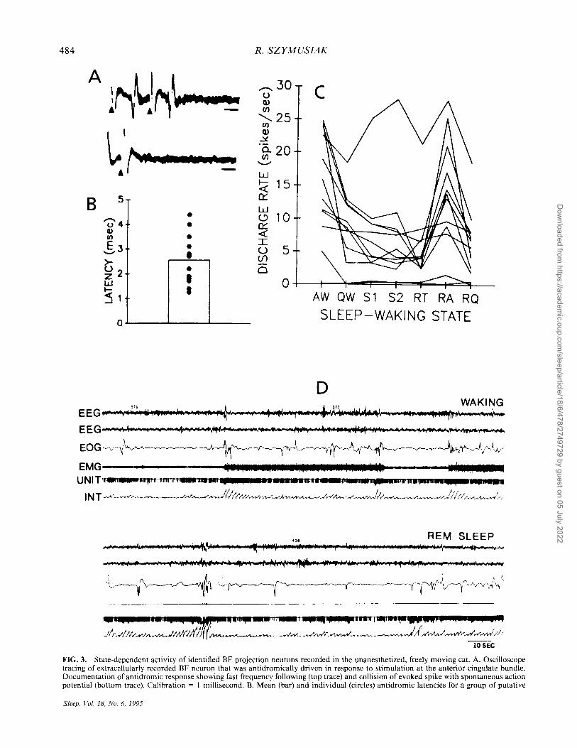
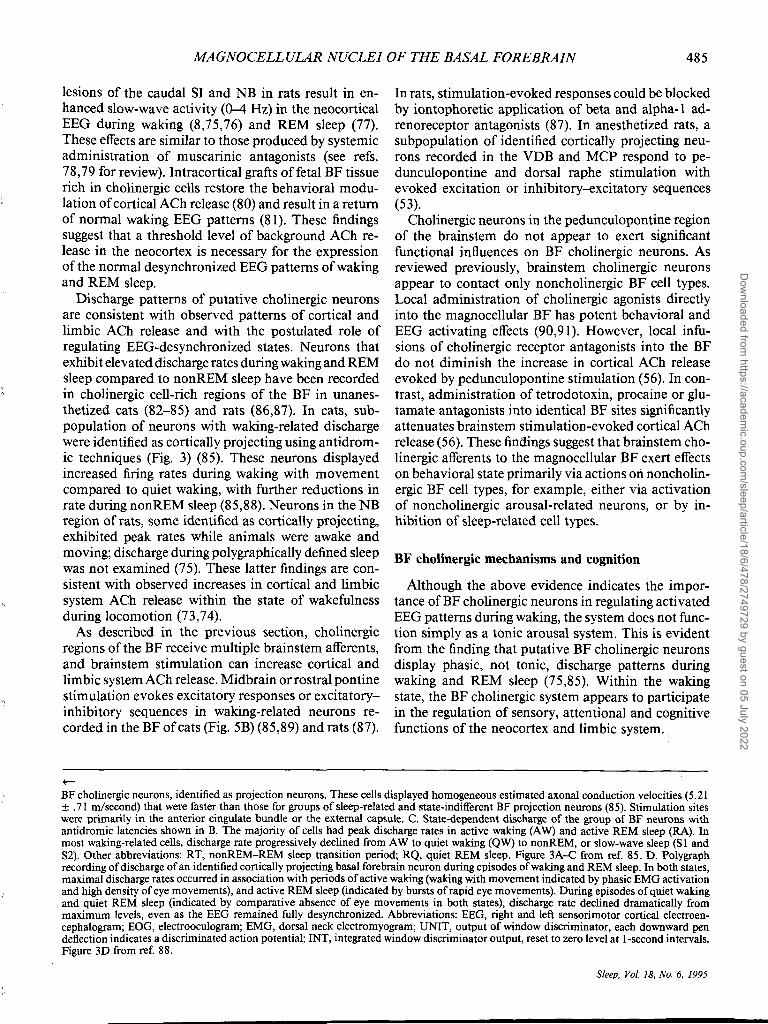
FIG. 3. State-dependent activity of identified BF projection neurons recorded in the unanesthetized, freely moving cat. A. Oscilloscope tracing of extracellularly recorded BF neuron that was antidromically driven in response to stimulation at the anterior cingulate bundle. Documentation of antidromic response showing fast frequency following (top trace) and collision of evoked spike with spontaneous action potential (bottom trace). Calibration = I millisecond. B. Mean (bar) and individual (circles) antidromic latencies for a group of putative
Sleep, Vol. 18, No.6, 1995
Dow
nloaded from https://academ
ic.oup.com/sleep/article/18/6/478/2749729 by guest on 05 July 2022
MAGNOCELLULAR NUCLEI OF THE BASAL FOREBRAIN 485
lesions of the caudal SI and NB in rats result in enhanced slow-wave activity (0-4 Hz) in the neocortical EEG during waking (8,75,76) and REM sleep (77). These effects are similar to those produced by systemic administration of muscarinic antagonists (see refs. 78,79 for review). Intracortical grafts offetal BF tissue rich in cholinergic cells restore the behavioral modulation of cortical ACh release (80) and result in a return of normal waking EEG patterns (81). These findings suggest that a threshold level of background ACh release in the neocortex is necessary for the expression of the normal de synchronized EEG patterns of waking and REM sleep.
Discharge patterns of putative cholinergic neurons are consistent with observed patterns of cortical and limbic ACh release and with the postulated role of regulating EEG-desynchronized states. Neurons that exhibit elevated discharge rates during waking and REM sleep compared to non REM sleep have been recorded in cholinergic cell-rich regions of the BF in unanesthetized cats (82-85) and rats (86,87). In cats, subpopulation of neurons with waking-related discharge were identified as cortically projecting using antidromic techniques (Fig. 3) (85). These neurons displayed increased firing rates during waking with movement compared to quiet waking, with further reductions in rate during nonREM sleep (85,88). Neurons in the NB region of rats, some identified as cortically projecting, exhibited peak rates while animals were awake and moving; discharge during polygraphically defined sleep was not examined (75). These latter findings are consistent with observed increases in cortical and limbic system ACh release within the state of wakefulness during locomotion (73,74).
As described in the previous section, cholinergic regions of the BF receive multiple brainstem afferents, and brainstem stimulation can increase cortical and lim bic system ACh release. Midbrain or rostral pontine stimulation evokes excitatory responses or excitatoryinhibitory sequences in waking-related neurons recorded in the BF of cats (Fig. 5B) (85,89) and rats (87).
~
In rats, stimulation-evoked responses could be blocked by iontophoretic application of beta and alpha-l adreno receptor antagonists (87). In anesthetized rats, a subpopulation of identified cortically projecting neurons recorded in the VDB and MCP respond to pedunculopontine and dorsal raphe stimulation with evoked excitation or inhibitory--excitatory sequences (53).
Cholinergic neurons in the pedunculopontine region of the brainstem do not appear to exert significant functional influences on BF cholinergic neurons. As reviewed previously, brainstem cholinergic neurons appear to contact only noncholinergic BF cell types. Local administration of cholinergic agonists directly into the magnocellular BF has potent behavioral and EEG activating effects (90,91). However, local infusions of cholinergic receptor antagonists into the BF do not diminish the increase in cortical ACh release evoked by pedunculopontine stimulation (56). In contrast, administration of tetrodotoxin, procaine or glutamate antagonists into identical BF sites significantly attenuates brainstem stimulation-evoked cortical ACh release (56). These findings suggest that brainstem cholinergic afferents to the magnocellular BF exert effects on behavioral state primarily via actions on noncholinergic BF cell types, for example, either via activation of noncholinergic arousal-related neurons, or by inhibition of sleep-related cell types.
BF cholinergic mechanisms and cognition
Although the above evidence indicates the importance ofBF cholinergic neurons in regulating activated EEG patterns during waking, the system does not function simply as a tonic arousal system. This is evident from the finding that putative BF cholinergic neurons display phasic, not tonic, discharge patterns during waking and REM sleep (75,85). Within the waking state, the BF cholinergic system appears to participate in the regulation of sensory, attentional and cognitive functions of the neocortex and limbic system.
BF cholinergic neurons, identified as projection neurons. These cells displayed homogeneous estimated axonal conduction velocities (5.21 ± .71 m/second) that were faster than those for groups of sleep-related and state-indifferent BF projection neurons (85). Stimulation sites were primarily in the anterior cingulate bundle or the external capsule. C. State-dependent discharge of the group of BF neurons with antidromic latencies shown in B. The majority of cells had peak discharge rates in active waking (A W) and active REM sleep (RA). In most waking-related cells, discharge rate progressively declined from A W to quiet waking (QW) to nonREM, or slow-wave sleep (S I and S2). Other abbreviations: RT, nonREM-REM sleep transition period; RQ, quiet REM sleep. Figure 3A-C from ref. 85. D. Polygraph recording of discharge of an identified cortically projecting basal forebrain neuron during episodes of waking and REM sleep. In both states, maximal discharge rates occurred in association with periods of active waking (waking with movement indicated by phasic EMG activation and high density of eye movements), and active REM sleep (indicated by bursts of rapid eye movements). During episodes of quiet waking and quiet REM sleep (indicated by comparative absence of eye movements in both states), discharge rate declined dramatically from maximum levels, even as the EEG remained fully desynchronized. Abbreviations: EEG, right and left sensorimotor cortical electroencephalogram; EOG, electrooculogram; EMG, dorsal neck electromyogram; UNIT, output of window discriminator, each downward pen deflection indicates a discriminated action potential; INT, integrated window discriminator output, reset to zero level at I-second intervals. Figure 3D from ref. 88.
Sleep, Vol. 18, No.6, 1995
Dow
nloaded from https://academ
ic.oup.com/sleep/article/18/6/478/2749729 by guest on 05 July 2022
486 R. SZYMUSIAK
Loss of BF cholinergic neurons is a critical feature of the neuropathology of Alzheimer's disease, and is believed to contribute to cognitive deficits in this disorder (see ref. 92 for review). The cognitive effects of excitotoxin-induced lesions of cholinergic regions of the BF in experimental animals (primarily rodents) have been intensely investigated. Such lesions impair performance in a variety of learning tasks including t-maze, radial-arm maze, Morris water maze, delayed matching to sample and passive and active avoidance (see ref. 93 for review). A global impairment in cognitive function following such lesions implicates the BF cholinergic system as critical for normal acquisition and retention of newly learned responses.
Although BF lesions can produce profound memory disturbances in experimental animals, the extent to which selective loss of cholinergic neurons is responsible for these deficits is controversial (see refs. 94,95 for review). As reviewed in a previous section, there is extensive intermingling of cholinergic and noncholinergic neurons throughout much of the BF. Excitotoxins, such as ibotenic acid, can be expected to damage noncholinergic BF neurons as well as cholinergic ones. BF lesions produced by quisqualic acid are more effective in reducing cortical ACh levels than are ibotenic acid-induced lesions, but the former treatment can result in less severe learning and memory deficits than the latter one (95). Thus, global memory impairment may require destruction of both BF cholinergic and noncholinergic cell types.
Behavioral and neurophysiological studies suggest that rather than mediating memory processes directly, BF cholinergic neurons regulate attentional processes. Recent studies in the rat (96) and monkey (97) find evidence for impaired attentional focusing and not primary impairment of memory following excitotoxic BF lesions.
Neurophysiological evidence indicates that activation of the BF cholinergic neurons may produce regional enhancement of sensory processing within the cortex. Evidence from in vitro cortical slice preparations demonstrates that the effects of acetylcholine on cortical pyramidal neurons include a suppression of two rectifying potassium currents, 1m (muscarinic sensitive) and the calcium-activated IAHP (after hyperpolarization) (98). Suppression of these currents reduces postexcitatory after hyperpolarizations (AHPs) and decreases spike frequency adaptation, thereby increasing pyramidal cell excitability. These actions have a net depolarizing effect, promote tonic single-spike firing characteristic of pyramidal cell discharge during desynchronized EEG states and render cortical cells more responsive to excitatory synaptic input (98).
Local cortical application of muscarinic agonists in anesthetized animals enhances the sensory-evoked re-
Sleep, Vol. 18, No.6, 1995
sponses of neurons in somatosensory (99), auditory (100) and visual cortex (10 I) recorded in vivo. Electrical stimulation of the NB combined with somatosensory (102,103) or auditory (104,105) stimulation augments evoked excitatory responses in cortical neurons. These effects are blocked by prior treatment with muscarinic receptor antagonists. Repeated pairings of NB stimulation with somatosensory or auditory stimulation enhances sensory stimulation-evoked responses in somatosensory (103) and auditory cortex (106) for several minutes. These long-lasting effects are present only when NB and sensory stimulation are repeatedly paired; repetitive stimulation of the NB alone is not effective.
This type of modality- or contingency-specific coactivation of BF cholinergic neurons could contribute to attentional focusing by enhancing the representation of selected sensory information in the cortex. One way in which such coactivation could be achieved is via collateral inputs to the BF cholinergic system from the primary sensory pathway. Such a mechanism may function in the auditory system, where a projection from auditory thalamus to cholinergic regions of the BF has been described (107).
Another mechanism by which BF activation could become associated with particular sensory stimuli is through conditioning processes. Within the primate NB, single unit recordings demonstrate that few putative cholinergic neurons respond to specific sensory, motor or mnemonic aspects of learning and memory tasks. Rather, a subset ofNB neurons displays elevated discharge in response to delivery of reinforcement or to sensory stimuli that are associated with reward (108,109). In rats (110) and rabbits (111) a significant proportion of neurons recorded throughout the BF respond to the CS+, but not the CS- stimuli in classical conditioning paradigms. These findings suggest a physiological substrate of attentional focusing, in which stimuli that have come to be associated with delivery or availability of reinforcement activate BF cholinergic neurons, which, in turn, enhance processing of those stimuli in the appropriate cortical regIOn (99,104,108,109).
The concept of cholinergic BF neurons as a source of cortical activation can be easily reconciled with a more specific role in modulating cortical excitability in response to particular kinds of stimuli or contingencies. As indicated by the EEG effects ofNB lesions, a minimal level of ACh release in the cortex may be necessary to maintain the activated EEG patterns normally associated with wakefulness. In addition, BF neurons could respond to particular stimuli occurring within a specific context leading to further increases in ACh release in restricted cortical regions.
To summarize, neurons can be recorded within cho-
Dow
nloaded from https://academ
ic.oup.com/sleep/article/18/6/478/2749729 by guest on 05 July 2022
MAGNOCELLULAR NUCLEI OF THE BASAL FOREBRAIN 487
linergic regions of the BF that display firing rate profiles consistent with a role in regulating de synchronized EEG patterns. Reinforcement-related neuronal discharge can also be recorded within these regions, particularly in the primate NB. Because the state-dependent and cognitive correlates of BF neuronal activity have been studied by separate laboratories, the extent to which the spontaneous discharge of reinforcement-related neurons is modulated by sleep-waking state is not known. Nevertheless, both cell types can be considered to be putatively cholinergic because of 1) location, 2) the known behavioral state correlates of cortical ACh release and 3) identified cortically projecting BF neurons have been shown to be waking-related (75,85) or reinforcement-related (108,109).
However, because of the anatomical configuration of the BF cholinergic system, cells recorded with extracellular techniques cannot be identified as cholinergic with any certainty, even when shown to be cortically projecting. Although the majority of cortically projecting neurons in the NB of the rat and cat are cholinergic, 10-20% are not. Within the MCP and SI, as many as 50% of cortically projecting cells are not cholinergic. This is reflected in the heterogeneity of axon conduction velocities and behavioral state correlates described for BF neurons in the rat and cat (85,112,113). Within the primate NB, the percentage of corticopetal cells that are also cholinergic approaches 100% (19). However, although some reinforcementrelated neurons have been identified as cortically projecting, most recorded neurons cannot be identified as such. Due to the compact organization of the cholinergic cells within the primate NB, and with careful anatomical and physiological characterization of recorded cells (e.g. see ref. 109), cholinergic neurons can be most reliably and consistently recorded within the primate NB. In other species, and for neurons located within the BF but outside the NB in primates, confidently classifying recorded BF neurons as cholinergic remains problematic. To date, no easily implemented and reliable identifying feature of the extracellularly recorded activity of BF cholinergic neurons has been demonstrated.
Influence of intrinsic properties of BF neurons on state-dependent discharge patterns
Intracellular recordings ofBF neurons using in vitro brain slice or dissociated cell preparations have revealed properties of cholinergic neurons that may assist identification in extracellular in vivo recordings, as well as provide potentially valuable information about their state-dependent physiology. An important advantage of intracellular recording in these preparations is the ability to determine the neurochemical pheno-
type of recorded cells. This is accomplished through intracellular injection of a dye combined with immunohistochemistry for specific neurotransmitter markers.
Using this approach, Khateb et al. (114) demonstrated that ChAT -positive neurons in brain slices containing the MCP/SI region of guinea pig BF possess T -type calcium channels. T -type calcium channels have also been identified in isolated putative cholinergic neurons obtained from the guinea pig MS (115). These channels are responsible for the voltage-dependent calcium current It. It is present in a variety of cell types including hippocampal neurons (116), thalamic relay neurons (117) and cholinergic neurons in the pontomesencephalic tegmentum (118). It has several interesting properties, but of direct relevance here is that its presence enables neurons to fire high-frequency bursts of action potentials in a voltage-dependent manner. It is inactivated when membrane voltage (V m) is more depolarized than about -60 mY, but becomes deinactivated at more hyperpolarized levels of V m (117). When cells that possess It are depolarized from this hyperpolarized state, they respond with a broad depolarizing spike that is calcium-mediated. This lowthreshold spike can be crowned by a high-frequency burst (typically> 200 Hz) offast action potentials (117). The burst is usually followed by an AHP that is due, in part, to the voltage-dependent inactivation of I" although other currents can make important contributions to the AHP (117,119). When V m is at levels >60 mY, depolarizing inputs evoke trains of single action potentials, as It is inactivated at these levels of V m •
Thus, cells that possess T -type calcium channels have the ability to display two modes of firing, single-spike and high-frequency burst firing. A dramatic example of how this determines the state-dependent firing patterns of cells is observed in thalamic relay neurons. These cells preferentially display single-spike firing during wakefulness and REM sleep, and burst firing during non REM sleep (see ref. 120 for review). Extracellular recordings of thalamic relay neurons during nonREM sleep are characterized by high-frequency bursts, followed by long periods during which no action potentials are recorded. These silent periods reflect AHPs that arise due to a combination of intrinsic properties and synaptic input (see refs. 27,120 for review). Burst firing in thalamic relay neurons during nonREM sleep is associated with impaired sensory transmission through the thalamus to the cortex. This occurs because relay neurons are unresponsive to afferent input during periods of AHP, and there is little correspondence between incoming volleys from primary sensory inputs and the output of the relay neurons to the cortex (27,120,121).
Sleep, Vol. 18, No.6, 1995
(
Dow
nloaded from https://academ
ic.oup.com/sleep/article/18/6/478/2749729 by guest on 05 July 2022

488 R. SZYMUSIAK
The demonstration of It in BF cholinergic neurons in vitro raises the question of the role of tonic versus burst firing in the normal in vivo physiology of these cells (114). For example, do the two firing modes display the same state correlates observed in thalamic relay neurons (i.e. single-spike firing in waking and REM sleep and burst firing in nonREM sleep)? Burst firing in BF cholinergic neurons during sleep could diminish the flow of activating influences from the brainstem and from ascending sensory pathways to the cortex (7). In the primate NB, putative cholinergic neurons are characterized by relatively tonic single-spike firing during wakefulness (109). Indeed, the absence of burst firing in waking is used to distinguish NB neurons from those in globus pallidus (109,122). In naturally sleeping cats, 43% of the neurons recorded in the ventral and peripallidal area displayed burst firing in nonREM sleep (83). In a population of waking-related neurons recorded in the MCP and rostral SI of unanesthetized cats, 16% displayed a pattern of burst firing exclusively during nonREM sleep that was identical to the pattern observed in the majority of neurons recorded in a thalamic relay nucleus (123). If more extensive mapping studies of identified cholinergic and noncholinergic neurons confirm initial findings from in vitro experiments that cholinergic neurons are the only cell type in the magnocellular BF to possess It, then the presence of extracellularly recorded burst firing could prove to be a simple marker for BF cholinergic neurons in vivo.
SLEEP-PROMOTING FUNCTIONS OF THE MAGNOCELLULAR BASAL
FOREBRAIN AND MEDIAL PREOPTIC/ANTERIOR HYPOTHALAMUS
Prior to the current focus on the neurobiology of the BF cholinergic system and its relationship to cortical activation and cognition, this portion of the ventral telencephalon was implicated as the site of a sleeppromoting mechanism. Based on clinical correlations and the location of encephalitis-related neuropathology, von Economo noted a strong association of damage between the region of the rostral hypothalamus and preoptic area and symptoms of insomnia (124). Nauta examined the effects oftransections at different levels of the hypothalamus on behavior and sleepwaking in the rat, and found that transections at supraoptic levels resulted in severe insomnia (125).
Subsequent experimental work has focused on the sleep-promoting functions of ventral telencephalic regions, including portions of the magnocellular BF that overlap with the locations of cholinergic neurons, as well as the nonmagnocellular medial preoptic/an-
Sleep, Vol. 18, No.6, 1995
terior hypothalamus (MP/AH; see Fig. 1). Published reports describing the effects oflesions, unit recording or chemical stimulation of the BF on sleep actually describe manipulations of the MP/ AH, not the more laterally located magnocellular regions (e.g. 126,127). Although both areas are critically involved in sleepwake regulation, they are anatomically and functionally very different. In addition, the term "basal forebrain" has evolved to refer principally to the magnocellular cholinergic system in much of the contemporary neuroscience literature. Including these regions and the MP/ AH under a single "basal forebrain" rubric can be confusing to readers not familiar with the history of sleep research in this area. In the following section, the evidence for a hypnogenic mechanism within the magnocellular BF and the MP/ AH will be considered separately.
Evidence for a sleep-promoting function in the magnocellular BF
Based on the work of von Economo (124) and Nauta (125), and the findings of Hess (128) and Kaada (129) demonstrating motor suppression and parasympathetic activation in response to electrical stimulation of the ventral telencephalon in experimental animals, Sterman and Clemente examined the effects of stimulation throughout the BF on EEG patterns and behavioral sleep in cats. They found that BF stimulation evoked cortical EEG synchronization in cats treated with Flaxedil (130) and EEG synchronization with behavioral sleep in unanesthetized, chronically prepared cats (131). The most effective sites were within the HDB, the MCP and the rostral SI. Stimulation in the region of the NB and caudal SI did not induce sleep. Although effective stimulation sites were in regions now known to contain cholinergic neurons, they are also the sites where overlap of cholinergic and GABAergic neurons is most extensive (41). At the more caudal BF sites where stimulation was not sleep-inducing, cholinergic neurons with cortical projection targets are the predominate magnocellular neuronal type. Subsequent studies demonstrated that discrete electrolytic lesions centered in the HDB of cats resulted in persistent insomnia (132,133). Neurotoxin lesions in the region of the HDB, MCP and rostral SI also caused chronic sleep loss in adult cats (134).
Single-unit recordings in unanesthetized, naturally sleeping animals have identified patterns of neuronal discharge within the magnocellular BF that are consistent with sleep-regulating functions. In work initiallY performed in freely moving adult cats using a chronic micro wire technique, neurons recorded within the HDB, MCP and SI displayed peak discharge rates during nonREM sleep, and comparatively reduced rates
Dow
nloaded from https://academ
ic.oup.com/sleep/article/18/6/478/2749729 by guest on 05 July 2022
MAGNOCELLULAR NUCLEI OF THE BASAL FOREBRAIN 489
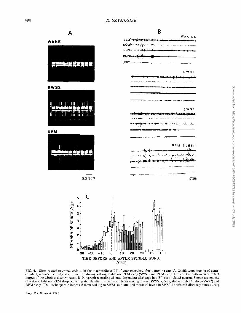
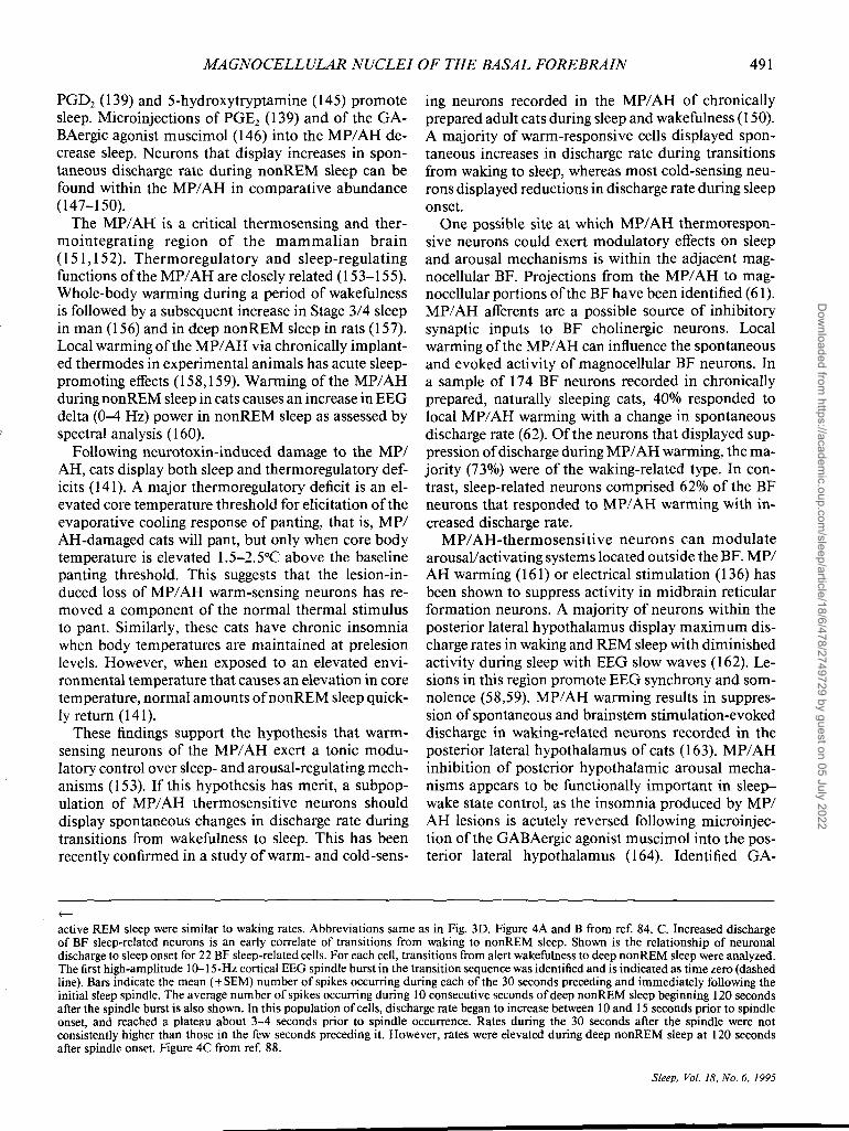
during waking and REM sleep (Fig. 4A and B) (84). Sleep-related neurons constituted about 20% of the total cell sample. During transitions from periods of sustained wakefulness to sustained sleep, discharge of sleep-related cells initially increased several seconds prior to the first signs of EEG synchronization (Fig. 4C) (85,88). Sleep-related neurons were electrophysiologically identified as sources of projections to the neocortex and the brainstem (85). Estimated axonal conduction velocities of sleep-related projection neurons were significantly slower than those of wakingrelated, presumably cholinergic, projection neurons. Stimulation of brains tern arousal-related regions of the midbrain and pontomesencephalic tegmentum evoked orthodromic suppression of spontaneous activity in sleep-related cell types, in contrast with the orthodromic excitatory effects observed in waking-related BF neurons (Fig. 5A and B) (85).
The responses of neurons in the midbrain reticular formation (MRF) to BF stimulation were examined in a subsequent study to assess the functional significance of the descending projections ofBF sleep-related neurons. In agreement with a previous study (135), stimulation at sites within the HDB and rostral SI in cats during wakefulness evoked prolonged periods of discharge suppression (average duration of 87 milliseconds), that was sometimes preceded by a brief period of excitation (Fig. 5C-F) (136). This finding suggests that BF sleep-related neurons participate in behavioral state control, in part, via a descending modulation of brainstem arousal systems. This is consistent with the ability of BF stimulation to antagonize behavioral activating effects of MRF stimulation in cats traversing a runway for a food reward (137). GABAergic neurons with descending projections are abundant in effective stimulation sites, and are candidates for mediating these inhibitory effects (49).
The presence of sleep-related neuronal discharge within the magnocellular BF, as originally described in cats, has been confirmed in rats. In urethane-anesthetized rats, a subgroup of BF neurons displayed elevated discharge rates during periods of EEG synchrony (138). These could not be identified as projection neurons and were hypothesized to be interneurons (138). Neurons displaying increased discharge rates during nonREM sleep compared to waking have recently been described within the HDB and SI of unanesthetized rats (86,87). Prostaglandin D2 (PGD2) has sleep-enhancing effects when administered centrally to rats (139), and about one-third ofBF sleep-related neurons in rats were excited by iontophoretic application of PGD2 (86). As in the cat, some BF sleep-related neurons could be confirmed to be a source of brain stem projections (87). Electrical stimulation of the pontine and medullary reticular formation caused suppression
of discharge in about one-half of the sleep-related neurons examined (87).
Evidence for a sleep-promoting function of the medial anterior hypothalamus/preoptic area
As noted above, the stimulation sites of Sterman and Clemente (130,131), and the insomnia-producing lesions reviewed above (132,134), involved magnocellular portions of the BF, principally the HDB, MCP and rostral SL The hypothalamic region located adjacent and medial to these rostral magnocellular BF sites, the MP / AH, is also important in sleep-wake regulation, and has primarily sleep-promoting actions. Although MP/ AH behavioral state-regulating mechanisms appear to interact with both cholinergic and noncholinergic cell types within the magnocellular BF, there are substantial anatomical and functional differences between these two regions.
The MP/ AH is an area of considerable functional complexity. Cells in this region play pivotal roles in the regulation of body temperature, body fluid balance, cardiovascular regulation, neuroendocrine control, reproductive physiology and behavior. Although some of these functional aspects interact with sleep-wake control mechanisms, behavioral state control is only one of many important regulatory functions that neurons of the MPI AH participate in.
Anatomically, the MPI AH and magnocellular BF are different. As with other peri ventricular portions of the hypothalamus, the cells in this area are medium to small in size and are densely packed. An extensive list of neuropeptides, hormone releasing factors and neurotransmitters have been localized to MPI AH neurons (se ref. 140 for review). No cholinergic neurons are found within these nuclei, but GABA-containing cells are numerous. GABAergic neurons with identified projections to the posterior lateral hypothalamic area form a lateral to medial continuum from magnocellular portions of the BF to the MP/AH (49). Similarly, a projection to the thalamic reticular nucleus and the anteromedialldorsomedial thalamic nuclei has both magnocellular BF and MPI AH components (23,24). As reviewed previously, these pathways are of potential importance for sleep-wake regulation.
The evidence for a role of the MPI AH in the regulation of sleep and wakefulness is as extensive and as compelling as that for any region of the brain. It is likely that damage to the MPI AH was responsible for the insomnia observed by Nauta (125) after rostral hypothalamic transections in rats. Electrolytic or neurotoxin-induced damage to this area has been repeatedly shown to produce profound and long-lasting reductions in sleep (126,127,141,142). Microinjections of benzodiazepines (143), adenosine agonists (144),
Sleep, Vol. 18, No.6, 1995
Dow
nloaded from https://academ
ic.oup.com/sleep/article/18/6/478/2749729 by guest on 05 July 2022
490
A
WAKE
SWS2
REM
0.2 SEC
7 c 6
5
4
3
2
1
R. SZYMUSIAK
8 WAKING
EEG1 ~ '~I'" 1'1' • ,
EOG V'-·-""--h-V·---~~~ -. ~-.-.-.- -~ ~~-LGN , II \1 "\\ •• r • • • , hr4
EMGI ~ ~ ~I
UNIT --,- rr-----
5 W 51
.... ,. '., .... til" I be .. r'l' ~I' .••. !_.!~
II' P • f ."
SWS 2 '. '" -jl.'''j~~,.,. ,oj '41.i ..... ~' ,.',. iIIIof~ ... n.
REM SLEEP
" ~ I"", ,41.1
~"""""T~----
10 SEC
o~~~~~~~~~~~~~~~
-30 -20 -10 0 10 20 30 120 130
TIME BEFORE AND AFTER SPINDLE BURST (SEC)
FIG. 4. Sleep-related neuronal activity in the magnocellular BF of unanesthetized, freely moving cats. A. Oscilloscope tracing of extracellularly recorded activity of a BF neuron during waking, stable nonREM sleep (SWS2) and REM sleep. Dots on the bottom trace reflect output of the window discriminator. B. Polygraph recording of state-dependent discharge in a BF sleep-related neuron. Shown are epochs of waking, light nonREM sleep occurring shortly after the transition from waking to sleep (SWSI), deep, stable nonREM sleep (SWS2) and REM sleep. The discharge rate increased from waking to SWS I. and attained maximal levels in SWS2. In this cell discharge rates during
Sleep, Vol. 18, No.6, 1995
Dow
nloaded from https://academ
ic.oup.com/sleep/article/18/6/478/2749729 by guest on 05 July 2022
MAGNOCELLULAR NUCLEI OF THE BASAL FOREBRAIN 491
PGD2 (139) and 5-hydroxytryptamine (145) promote sleep. Microinjections of PGE2 (139) and of the GABAergic agonist muscimol (146) into the MP/AH decrease sleep. Neurons that display increases in spontaneous discharge rate during nonREM sleep can be found within the MPI AH in comparative abundance (147-150).
The MPI AH is a critical thermosensing and thermointegrating region of the mammalian brain (151,152). Thermoregulatory and sleep-regulating functions of the MPI AH are closely related (153-155). Whole-body warming during a period of wakefulness is followed by a subsequent increase in Stage 3/4 sleep in man (156) and in deep nonREM sleep in rats (157). Local warming of the MPI AH via chronically implanted thermodes in experimental animals has acute sleeppromoting effects (158,159). Warming of the MP/AH during nonREM sleep in cats causes an increase in EEG delta (0-4 Hz) power in non REM sleep as assessed by spectral analysis (160).
Following neurotoxin-induced damage to the MPI AH, cats display both sleep and thermoregulatory deficits (141). A major thermoregulatory deficit is an elevated core temperature threshold for elicitation of the evaporative cooling response of panting, that is, MPI AH-damaged cats will pant, but only when core body temperature is elevated 1.5-2.5°C above the baseline panting threshold. This suggests that the lesion-induced loss of MPI AH warm-sensing neurons has removed a component of the normal thermal stimulus to pant. Similarly, these cats have chronic insomnia when body temperatures are maintained at prelesion levels. However, when exposed to an elevated environmental temperature that causes an elevation in core temperature, normal amounts of non REM sleep quickly return (141).
These findings support the hypothesis that warmsensing neurons of the MPI AH exert a tonic modulatory control over sleep- and arousal-regulating mechanisms (153). If this hypothesis has merit, a subpopulation of MPI AH thermo sensitive neurons should display spontaneous changes in discharge rate during transitions from wakefulness to sleep. This has been recently confirmed in a study of warm- and cold-sens-
f-
ing neurons recorded in the MP 1 AH of chronically prepared adult cats during sleep and wakefulness (150). A majority of warm-responsive cells displayed spontaneous increases in discharge rate during transitions from waking to sleep, whereas most cold-sensing neurons displayed reductions in discharge rate during sleep onset.
One possible site at which MPI AH thermoresponsive neurons could exert modulatory effects on sleep and arousal mechanisms is within the adjacent magno cellular BF. Projections from the MPI AH to magnocellular portions of the BF have been identified (61). MPI AH afferents are a possible source of inhibitory synaptic inputs to BF cholinergic neurons. Local warming of the MPI AH can influence the spontaneous and evoked activity of magnocellular BF neurons. In a sample of 174 BF neurons recorded in chronically prepared, naturally sleeping cats, 40% responded to local MP/AH warming with a change in spontaneous discharge rate (62). Of the neurons that displayed suppression of discharge during MPI AH warming, the majority (73%) were of the waking-related type. In contrast, sleep-related neurons comprised 62% of the BF neurons that responded to MPI AH warming with increased discharge rate.
MPI AH-thermosensitive neurons can modulate arousal/activating systems located outside the BF. MPI AH warming (161) or electrical stimulation (136) has been shown to suppress activity in midbrain reticular formation neurons. A majority of neurons within the posterior lateral hypothalamus display maximum discharge rates in waking and REM sleep with diminished activity during sleep with EEG slow waves (162). Lesions in this region promote EEG synchrony and somnolence (58,59). MPI AH warming results in suppression of spontaneous and brainstem stimulation-evoked discharge in waking-related neurons recorded in the posterior lateral hypothalamus of cats (163). MP/AH inhibition of posterior hypothalamic arousal mechanisms appears to be functionally important in sleep-wake state control, as the insomnia produced by MPI AH lesions is acutely reversed following microinjection of the GABAergic agonist muscimol into the posterior lateral hypothalamus (164). Identified GA-
active REM sleep were similar to waking rates. Abbreviations same as in Fig. 3D. Figure 4A and B from ref. 84. C. Increased discharge of BF sleep-related neurons is an early correlate of transitions from waking to nonREM sleep. Shown is the relationship of neuronal discharge to sleep onset for 22 BF sleep-related cells. For each cell, transitions from alert wakefulness to deep nonREM sleep were analyzed. The first high-amplitude 1O-15-Hz cortical EEG spindle burst in the transition sequence was identified and is indicated as time zero (dashed line). Bars indicate the mean (± SEM) number of spikes occurring during each of the 30 seconds preceding and immediately following the initial sleep spindle. The average number of spikes occurring during 10 consecutive seconds of deep nonREM sleep beginning 120 seconds after the spindle burst is also shown. In this population of cells, discharge rate began to increase between 10 and 15 seconds prior to spindle onset, and reached a plateau about 3-4 seconds prior to spindle occurrence. Rates during the 30 seconds after the spindle were not consistently higher than those in the few seconds preceding it. However, rates were elevated during deep nonREM sleep at 120 seconds after spindle onset. Figure 4C from ref. 88.
Sleep, Vol. 18, No.6, 1995
Dow
nloaded from https://academ
ic.oup.com/sleep/article/18/6/478/2749729 by guest on 05 July 2022
492 R. SZYMUSIAK
10 A 50 B
8 40
(/) W ~ 6 30 a.. (/)
u.. 4 20 0 ~
2 10
o ~ ........ _ ..... IIIIII! -100-50 0 50 100 150 200 250 300
a -10D-SO 0 50 100 150 200 250 300
TIME (MSEC)
c 10 l F
I I
81
D if) I
~ ~ 6 ....... 0... if)
C:.t.. 4 0 ==1:t:
E 2
a -100 -50 a 50 100 150 200 250 300
TI~fE (msec)
FIG. 5. A. B. BF sleep-related and waking-related cells exhibit qualitatively different responses following stimulation of the paramedian midbrain reticular formation (MRF). A. Low-intensity, single-pulse MRF stimulation (100 pulses ofO.2-millisecond duration delivered at a rate of I Hz) during non REM sleep evoked suppression of discharge in a sleep-related neuron that lasted for> 150 milliseconds. Stimulation at the same site evoked short-latency excitation of the waking discharge in an identified cortically projecting, waking-related BF neuron. B. From ref. 85. C-F. Example of suppression of waking-related discharge in an MRF neuron in response to stimulation of the BF. C-E. This MRF cell was identified as projecting to the BF by antidromic activation. The evoked spike followed a high-frequency paired pulse (C) and collided with a spontaneous spike at short collision intervals (D), but not when the collision interval was longer than the antidromic latency (E). Calibration = 2 milliseconds. F. Peri stimulus histogram of evoked orthodromic response in the same MRF cell, based on 100 BF stimulation pulses delivered during waking at a rate of I Hz. BF stimulation evoked suppression of waking discharge that lasted for approximately 70 seconds. The polarity of BF stimulation was reversed from that in C-E such that no antidromic response was elicited. Figure 5C-F from ref. 136.
Sleep. Vol. 18. No.6, 1995
Dow
nloaded from https://academ
ic.oup.com/sleep/article/18/6/478/2749729 by guest on 05 July 2022
MAGNOCELLULAR NUCLEI OF THE BASAL FOREBRAIN 493
BAergic monosynaptic projections from the MPI AH to the posterior lateral hypothalamus could mediate these inhibitory effects (49). The extent to which MPI AH GABAergic neurons are also thermo sensitive is not known.
CHOLINERGIC-GABAERGIC INTERACTIONS IN THE BASAL
FOREBRAIN AND THE REGULATION OF BEHAVIORAL STATE
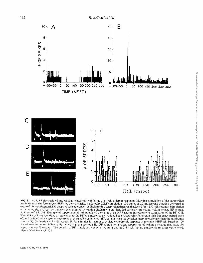
This review of the behavioral state-regulating functions of the BF has summarized evidence for the coexistence of arousal- and sleep-regulating functions within magnocellular BF regions. The critically involved cell groups and what is known and/or hypothesized about the functional connections among them are summarized in Fig. 6.
The role of the magnocellular cholinergic system in arousal and activation is firmly established. One or more unidentified noncholinergic cell types are responsible for the sleep-promoting functions of the magnocellular BF, with GABAergic projection neurons being possible candidates for mediating descending inhibitory effects on hypothalamic and brainstem arousal systems. The functional roles ofthe ascending GABAergic projections to the thalamus and cortex have not been characterized, but sleep-promoting functions of the BF can be hypothesized to involve local circuit and projection GABAergic neurons. Magnocellular neurons subserving sleep and arousal functions overlap partially at rostral-ventral BF levels, particularly within portions of the HDB and MCP. Further caudally, in sub- and peri pallidal areas, cortically projecting cholinergic neurons are more completely segregated from other magnocellular cell types. Thus, lesions centered in the HDB and MCP can cause insomnia (132,134), whereas lesions centered in the NB result in diminished EEG activation during wakefulness and REM sleep (8,75,77).
Cholinergic components of several mechanisms of cortical and limbic system activation have been identified, although noncholinergic cell types may participate in arousal-related processes as well. Activation of BF cholinergic neurons can be expected, via suppression of potassium channels, to diminish AHPs, promote tonic firing and render cortical neurons more responsive to afferent input (98). These features ofpyramidal cell activation would function to generate the de synchronized EEG patterns characteristic of activated behavioral states. Indeed, results of neurotoxic lesions of the NB indicate that a tonic level of cortical ACh release appears to be necessary for maintenance of normal desynchronized EEG patterns of waking and REM sleep (8,75-77).
FIG. 6. Schematic representation of input -output relationships of BF state-regulating cell types. Schematic is shown within a horizontal hemisection of the rat brain to show approximate positions of cell types in the neuraxis. Rostral is to the left of the figure and medial is at the top. Cell groups located within the magnocellular BF are indicated by the shaded boxes. BF cholinergic neurons with wakingrelated discharge are shown as projecting to the thalamus and the neocortex, where they exert depolarizing effects on target neurons and promote arousal and activated EEG patterns. These BF cholinergic cells receive excitatory afferents from glutamatergic (GLU) neurons of the pontine and midbrain reticular formation (PRF and MRF), serotonergic (5HT) neurons of the dorsal raphe nucleus (DRN), noradrenergic (NE) cells of the locus ceruleus (LC) and posterior lateral hypothalamic (PLH) neurons. Inhibitory afferents to BF cholinergic, waking-related cells are hypothesized to arise from warmsensing neurons of the MP/ AH, BF sleep-related projection neurons and/or BF sleep-related interneurons. All three of these cell types are hypothesized to be GABAergic. Disfacilitation ofBF cholinergic neurons could also arise from inhibitory effects of MP/AH and BF sleep-related neurons on the PRF, MRF and PLH. Not shown in this figure are excitatory afferents from brainstem cholinergic cell groups to noncholinergic, waking-related cells within the magnocellular BF. BF sleep-related cells are shown as sources of descending inhibitory influences on neurons in the PRF, MRF and PLH. They are also shown as hypothesized sources of inhibitory input to thalamus and neocortex that would act to promote synchronized EEG patterns in these structures during nonREM sleep. BF sleep-related cell types are further shown as receiving inhibitory inputs from brainstem arousal systems (those from the PRF and MRF are omitted in the figure for clarity of other connections), and excitatory inputs from MP/AH thermosensing neurons.
The activity of BF cholinergic neurons and the rate of cortical ACh release are modulated within the waking state (73-75,85,108,109), consistent with hypothesized roles in striatopallidal-related aspects of motor control and in cognitive functioning. The BF cholinergic input to the cortex appears to participate in attentional processes by selectively augmenting cortical processing of particular stimuli. Both direct cortical ACh administration and NB electrical stimulation enhance sensory stimulation-evoked responses in the corresponding cortical region (99-105). In addition to these neuronal responses, ACh-mediated vasodilation can augment regional cortical perfusion to meet enhanced metabolic demands (71). Sensory modalityspecific or contingency-specific regional cortical activation can be achieved through excitation of subpopulations of BF cholinergic neurons via sensory collat-
Sleep, Vol. 18, No.6, 1995
Dow
nloaded from https://academ
ic.oup.com/sleep/article/18/6/478/2749729 by guest on 05 July 2022
494 R. SZYMUSIAK
erals (107), and/or conditioning-dependent activation of these neurons by stimuli or contingencies associated with reward (108,109).
The synaptic mechanisms that underlie the learned contingency-dependent aspects of BF cholinergic activation remain to be determined, but ascending inputs from brainstem-activating systems are likely to regulate tonic aspects of BF cholinergic excitability. These would include direct serotonergic, noradrenergic and glutamatergic afferents. Brainstem cholinergic afferents to the BF appear to influence primarily noncholinergic arousal-regulating mechanisms. Because these brainstem cell groups all undergo spontaneous decreases in neuronal discharge during transitions from wakefulness to sleep (165), withdrawal of brain stem-activating inputs could be an important determinant of statedependent excitability changes in BF waking-related neurons. This would be analogous to the situation in thalamocortical relay neurons where these same brainstem systems exert tonic activating influences (51,166). The demonstration of It in identified BF cholinergic neurons studied in vitro (114,115), and the evidence for tonic firing in waking and nonREM sleep-related burst firing in putative BF cholinergic neurons in vivo (83,123), suggests further parallels with the thalamocortical system.
This review has emphasized cholinergic influences on the neocortex, but similar mechanisms could function in other targets ofthe BF cholinergic system. Cholinergic input to the hippocampus originates in the MS and VDB. The generation of activated EEG patterns within the hippocampus (hippocampal theta) is dependent upon this cholinergic input, and the hippocampal EEG is modulated by stimulation of brain stem afferents to the MS and VDB (54).
The cholinergic component of the BF projection to the thalamus is also of potential functional importance. Identified neurons in the pathway have not, to date, been studied across sleep-waking cycles. However, if discharge patterns of BF-thalamic projection neurons are similar to those of cortically projecting BF wakingrelated neurons, they could function to promote tonic firing and facilitate sensory transmission through the thalamus to the cortex during wakefulness.
Although the evidence for a sleep-promoting mechanism within the magnocellular BF is equally compelling, less is known about the cell types involved. The functional anatomy of ventral telencephalic sleeppromoting systems is further complicated by the important involvement of the MP/AH. Within the magnocellular BF, stimulation, lesion and unit-recording studies point to the HDB, MCP and rostro-ventral SI as sites where the sleep-promoting mechanisms can be most reliably manipulated. Among other cell types, these areas contain high concentrations of GABAergic
Sleep, Vol. 18, No.6, 1995
neurons (41). Although many of these GABA-containing cells are interneurons, others have been identified as the source of ascending and descending projections (13,15,16,26,45,49). The descending GABAergic projection is of particular interest, as descending inhibitory effects appear to be a functioI1ally important aspect of the BF hypnogenic system (135-137,163). A GABAergic projection to the posterior lateral hypothalamus has been identified (49). The extent to which noncholinergic projections from the magnocellular BF to mesencephalic and pontine arousal-related areas are GABAergic remains to be determined.
GABAergic neurons within the MP/ AH can be hypothesized to mediate the sleep-promoting functions of this area as well. As previously indicated, identified GABAergic neurons that project to the posterior hypothalamus can be localized to the MP/ AH (49). Local activation of MP/ AH warm-responsive neurons results in short latency suppression of waking discharge in posterior lateral hypothalamic neurons (163). In cats with insomnia secondary to neurotoxin-induced MP/ AH cell loss, microinjection of GABAergic agonists into the posterior hypothalamus restores sleep (164). Extracellular concentration of GAB A within the posterior lateral hypothalamus of adult cats, sampled with in vivo microdialysis techniques, is significantly elevated during nonREM sleep compared to both waking and REM sleep (167). These findings suggest that descending GABAergic projections constitutes a mechanism via which the rostro-ventral magnocellular BF and MP/ AH inhibit posterior hypothalamic arousal mechanisms and exert sleep-promoting actions (49,163).
The evidence for a descending action of the BF hypnogenic mechanism is most extensive, but ascending projections may also playa role. A restricted number of thalamic nuclei receive a projection from the magnocellular BF and MP/ AH (23,24). This projection contains cholinergic and noncholinergic neurons, and has a GABAergic component (26). The proportion of this projection that is GABAergic is not known, nor has the state-dependent discharge pattern of cells in this projection been characterized. Nevertheless, such a projection could represent an additional source of sleep-related inhibition on thalamocortical neurons within the anteromedial/dorsomedial nuclei, and to the thalamic reticular nucleus. Depolarization of neurons in the thalamic reticular nucleus by microinjection of glutamate has been recently shown to suppress sleeplike rhythmic oscillatory discharge patterns in both the reticular nucleus and in the corresponding relay nucleus (168). Electrical stimulation of subpallidal portions of the BF evoked primarily inhibitory postsynaptic potentials in the thalamic reticular nucleus of anesthetized rats (169). GABA-mediated hyperpolar-
Dow
nloaded from https://academ
ic.oup.com/sleep/article/18/6/478/2749729 by guest on 05 July 2022
MAGNOCELLULAR NUCLEI OF THE BASAL FOREBRAIN 495
izations ongmating from BF and MPI AH neurons might then be expected to promote oscillatory burst firing in these nuclei during transitions from waking to sleep.
A subset of GABAergic neurons within the magnocellular BF project directly to the neocortex (16,45,46). A subset of sleep-related neurons recorded in the BF of cats has been identified as cortically projecting (85), although identification of such cells as GABAergic is not possible in the context of extracellular recording studies in intact animals. The functional role ofthe BF--cortical GABAergic projection is not well characterized. Anatomical studies indicate that terminals of BF GABAergic cells contact primarily GABAergic interneurons within the neocortex (46). This suggests that activation of this projection would produce cortical disinhibition, an effect that might be expected to yield cortical activation. However, in cortical slice preparations studied in vitro, drugs that moderately suppress GABA-mediated inhibition, presumably arising solely from interneurons in this preparation, produce enhanced synchronous burst firing in local cortical circuits (170). The extent to which such a mechanism could contribute to cortical synchronization during natural sleep in the intact brain remains to be determined.
In addition to interactions between BF cholinergic and noncholinergic projection neurons at brainstem, diencephalic, limbic system and neocortical targets, local GABAergic--cholinergic interactions within the BF are of potential functional importance for behavioral state control. As in thalamic relay neurons, diminished excitability and expression of high-frequency burst firing patterns during natural sleep in BF cholinergic neurons would depend on membrane hyperpolarization (27,120). This could be achieved via reductions in tonic brainstem depolarizing inputs, and through increased GABA-mediated inhibition. Both mechanisms are important in the thalamus.
Anatomical and physiological evidence supports hypothesized inhibitory GABAergic influences on BF cholinergic neurons. Cortically projecting BF cholinergic neurons are contacted by significant numbers of GABA-containing nerve terminals (171,172). Sources of GABAergic input to BF cholinergic neurons include local interneurons, axon collaterals of magnocellular GABAergic projection neurons, the MP/AH and the nucleus accumbens (1). Cortical ACh release in rats (173,174), cortical ACh turnover in guinea pigs (175) and high-affinity choline uptake in rat cortical tissue (176) are reduced following microinjections of GAB A or GABA-A receptor agonists into SIINB regions of the BF. In rats, unilateral infusion of GABA into the NB acutely reduced ipsilateral turning in a radial arm maze (177). Bilateral microinjections of the GABA-A
receptor antagonist bicuculline into the SI of rats enhanced motor activity, and injections of the GABA-A receptor agonist muscimol into the same sites suppressed locomotion in rats (178). Muscimol microinjections into the SI attenuated increased locomotor activity evoked by local infusion of a quisqualic acid receptor agonist (179). In rats, SI microinjection of muscimol caused a dose-dependent impairment in performance of a visual discrimination task (180). Effects of muscimol on performance were partially antagonized by administration of physostigmine.
Due to the difficulty in determining the neurochemical identity of recorded cells during in vivo neurophysiological recordings, the state-dependent activity profiles of the GABAergic innervation of BF cholinergic cell groups are not known. As reviewed above, neurons with sleep-related discharge profiles are found in comparative abundance both within the magnocellular BF and the MPI AH. In addition, inhibitory effects on putative BF cholinergic neurons are seen in response to activation ofMPI AH warm-sensing neurons by local warming (62). MP/AH thermoresponsive cell types display spontaneous changes in activity during normal wake-to-sleep transitions that are identical to those evoked by warming (150). Therefore, it seems reasonable to hypothesize that GABA-mediated inhibition from one or more of these ventral telencephalic sources participate in modulating the state-dependent excitability of the BF cholinergic projection system. This could constitute an additional mechanism through which GABAergic neurons in the magnocellular BF and MPI AH exert control over somatomotor and cortical activation, and function to promote sleep onset and non REM sleep.
CONCLUDING REMARKS
The explosion of research on the neurobiology of the BF within the past decade has been fueled by the discovery of the cholinergic system and the recognition of its vulnerability in human neurodegenerative disorders such as Alzheimer's disease. As a result, considerable detail about the anatomy and cellular biology of this brain region is now known. This progress has been of substantial benefit to the field of sleep research. A critical forebrain arousal system has been identified and partially characterized. Possible cellular mechanisms responsible for the BF hypnogenic actions are better defined.
Future progress will depend, in part, on the ability to reliably identify BF cells recorded during natural sleep-waking states by transmitter phenotype, sources of afferent control and efferent targets. In this regard, in vitro studies of BF tissue are likely to be of critical importance. Only such studies can specify intrinsic
Sleep, Vol. 18, No.6, 1995
Dow
nloaded from https://academ
ic.oup.com/sleep/article/18/6/478/2749729 by guest on 05 July 2022
496 R. SZYMUSIAK
properties and detail the neuropharmacology of identified cell types. This information can then be extrapolated to the characterization of cells recorded in the intact brain. A syncrgism among in vitro and in vivo approaches has been crucial to the recent advances in understanding the state-dependent neurobiology of thalamocortical systems (see 27 for review). This approach appears to hold equal promise for characterizing the ventral forebrain regulation of normal and disordered sleep.
Acknowledgements: The author thanks Drs. Dennis McGinty and Joel Benington for helpful comments on a previous version of this manuscript. This work was supported by the Medical Research Service of the Department of Veterans Affairs.
REFERENCES
1. Zaborszky L, Cullinan WE, Braun A. Afferents to basal forebrain cholinergic projection neurons: an update. In: Napier TC, Kalivas PW. Hanin I. eds. The basal forebrain: anatomy to function. New York: Plenum Press, 1991 :43-100.
2. Alheid GF, Heimer L. New perspectives in basal forebrain organization of special relevance for neuropsychiatric disorders: the striatopallidal, amygdaloid and corticopetal components of the substantia innominata. Neuroscience 1988;27: 1-39.
3. WoolfNJ. Cholinergic systems in mammalian brain and spinal cord. Prog l'v'eurobiol 1991;37:475-524.
4. Butcher LL, Semba K. Reassessing the cholinergic basal forebrain: nomenclature schemata and concepts. Trends Neurosci 1989; 12:483-5.
5. Heimer L, Alheid GF. Piecing together the puzzle of basal forebrain anatomy. In: Napier TC, Kalivas PW, Hanin I, eds. The basalforebrain: anatomy tofimction. New York: Plenum Press, 1991: 1-42.
6. Mogenson GJ. Yang CR. The contribution of basal forebrain to limbic-motor integration and the mediation of motivation to action. In: Napier TC, Kalivas PW, Hanin I, eds. Basal JiJrebrain: anatomy to/unction. New York: Plenum Press, 1991: 267-90.
7. Jones BE. The organization of central cholinergic systems and their functional importance in sleep-waking states. Prog Brain Res 1993;98:61-71.
8. Stewart DJ, Macfabe DF, Vanderwolf CH. Cholinergic activation of the electrocorticogram: role of the substantia innominata and effects of atropine and quinuclidinyl benzilate. Brain Res 1984;322:219-32.
9. Semba K. The cholinergic basal forebrain: a critical role in cortical arousal. In: Napier TC, Kalivas PW, Hanin I, eds. The basa/forebrain: anatom.\' to(imction. New York: Plenum Press, 1991:197-218.
10. Buzsaki G, Gage FH. The cholinergic nucleus basalis: a key structure in neocortical arousal. In: Frotscher M, Misgeld U, eds. Central cholinergic synaptic transmission. Boston: Birkhauser Verlag, 1989:159-71.
11. WoolfNJ, Eckenstein F, Butcher LL. Cholinergic projections from the basal forebrain to the frontal cortex: a combined fluorescent tracer and immunohistochemical analysis in the rat. Neurosci Lett 1983;40:93-8.
12. WoolfNJ, Eckenstein F, Butcher LL. Cholinergic systems in the rat brain: I. Projections to the limbic telencephalon. Brain Res Bull 1984;13:751-84.
13. Wainer BH, Levy AI, Rye DB, Mesulam MM, Mufson EJ. Cholinergic and non-cholinergic septohippocampal pathways. Neurosci Lett 1985;54:45-52.
Sleep. Vol. 18. No.6. 1995
14. Saper CB. Organization of cerebral cortical afferent systems in the rat: I. Magnocellular basal nucleus. J Comp lVeurol 1984; 222:313-42.
15. Zaborszky L. Carlsen J, Brashear HR, Heimer L. Cholinergic and GABAergic afferents to the olfactory bulb in rat with special emphasis on the projection neurons in the nucleus of the horizontal limb of the diagonal band. J Comp Neurol 1986; 243:488-509.
16. Fisher RS. Buchwald NA, Hull CD. Levine MS. GABAergic basal forebrain neurons project to neocortex: the localization of glutamic acid decarboxylase and choline acetyltransferase in feline corticopetal neurons. J Comp 1'1'eurol 1988;272:489-502.
17. Rye DB, Wainer BH, Mesulam MM, Mufson EJ. Saper CB. Cortical projections arising from the basal forebrain: a study of cholinergic and noncholinergic components employing combined retrograde tracing and immunohistochemical localization of choline-acetyltransferase. 1Veuroscience 1984; 13:627-43.
18. Bigl V, Woolf NJ, Butcher LL. Cholinergic projections from the basal forebrain to frontal, parietal. temporal. occipital and cingulate cortices: a combined fluorescent tracer and acetylcholine-esterase analysis. Brain Res Bull 1982;8:727-49.
19. Mesulam MM. Mufson EJ. Levy AI. Wainer BH. Cholinergic innervation of cortex by the basal forebrain: cytochemistry and cortical connections of the septal area. diagonal band nucleus basalis (substantia innominata) and hypothalamus in the rhesus monkey. J Comp Neurol 1983;214: 170-97.
20. Baskerville KA, Chang HT, Herron P. Topography of cholinergic afferents from the nucleus basalis of Meynert to representational areas of sensorimotor corticies in rats. J Camp Neurol 1993;335:552-62.
21. Adams CE, Cepeda C, Boylan MK, et al. Basal forebrain neurons have axon collaterals that project to widely divergent cortical areas in the cat. Brain Res 1986;397:365-71.
22. WoolfNJ, Hemit MC, Butcher LL. Cholinergic and non-cholinergic projections from the rat basal forebrain revealed by combined choline acetyltransferase and phaseolus vulgaris leucoagglutinin immunohistochemistry. ]\'eurosci Lett 1986;66: 281-6.
23. Parent A, Pare D, Smith Y, Steriade M. Basal forebrain cholinergic and noncholinergic projections to the thalamus and brainstem in cats and monkeys. J Comp l'l'euI'0/1988;277:281-301.
24. Steriade M, Parent A, Pare D. Smith Y. Cholinergic and noncholinergic neurons of cat basal forebrain project to reticular and mediodorsal thalamic nuclei. Brain Res 1987;408:372-6.
25. Levey AI, Hallanger AE. Wainer BH. Cholinergic nucleus basalis neurons may influence the cortex via the thalamus. Neurosci Lett 1987;74:7-13.
26. Asanuma C, Porter LL. Light and electron microscopic evidence for a GABAergic projection from the caudal basal forebrain to the thalamic reticular nucleus in rats. J Camp Neurol 1990;302: 159-72.
27. Steriade M, McCormick DA, Sejnowski TJ. Thalamocortical oscillations in the sleeping and aroused brain. Science 1993; 262:679-85.
28. Aggleton JP, Friedman DP. Mishkin M. A comparison between the connections of the amygdala and hippocampus with the basal forebrain in the macaque. Exp Brain Res 1987;67: 556-68.
29. Carlsen J, Zaborszky L, Heimer L. Cholinergic projections from the basal forebrain to the basolateral amygdaloid complex: a combined retrograde fluorescent and immunohistochemical study. J Camp Neul'o/1985;234: 155-67.
30. Woolf NJ, Butcher LL. Cholinergic projections to the basolateral amygdala: a combined Evans blue and acetylcholinesterase analysis. Brain Res Bull 1982;8:751-63.
31. Nagai T, Kimura H, Maeda T, McGeer PL, Peng F, McGeer EG. Cholinergic projections from the basal forebrain of rat to the amygdala. J Neurosci 1982;2:513-20.
32. Semba K, Reiner PB, McGeer EG, Fibiger HC, Brainstem projecting neurons in the rat basal forebrain: neurochemica:,
Dow
nloaded from https://academ
ic.oup.com/sleep/article/18/6/478/2749729 by guest on 05 July 2022
MAGNOCELLULAR NUCLEI OF THE BASAL FOREBRAIN 497
topographical and physiological distinctions from cortically projecting cholinergic neurons. Brain Res Bu1l1989;22:50 1-9.
33. Albanese A, Castagna M. Altavista Mc' Cholinergic and noncholinergic projections to the interpeduncular nucleus. Brain Res 1985:329:334-9.
34. Woolf NJ, Butchcr LL. Cholinergic systems in the rat brain. II. Projections to the interpeduncular nucleus. Brain Res Bull 1985; 14:63-83.
35. Tomimoto H, Kamo H, Kameyama M, McGeer PL, Kimura H. Descending projections of the basal forebrain in the rat demonstrated by the anterograde neural tracer Phascolus vulgaris leucoagglutinin (PHA-L). Brain Res 1987:425:248-55.
36. Groenewegen HJ. Wouterlood FG. Basal forebrain inputs to the interpeduncular nucleus in the rat studied with Phaseolus I'ulgaris-Ieucoagglutinin tracing method. Brain Res Bull 1988; 21:643-9.
37. Kohler C, Swanson L W. Acetylcholine esterase-containing cells in the lateral hypothalamic area are immunoreactive for alpha melanocyte stimulating hormone and have cortical projections in the rat. Neurosci Lett 1984:49:39-43.
38. Melander T, Staines W A, Hokfelt T. et al. Galanin-like immunoreactivity in cholinergic neurons of the septum-basal forebrain complex projecting to the hippocampus of the rat. Brain Res 1985:360: 130-8.
39. Vincent SR, McIntosh CHS, Buchan AMJ. Brown Jc, Central somatostatin systems revealed with monoclonal antibodies. J Comp Seurol 1985:238: 169-86.
40. Zahm OS. Heimer L. Ventral striatopallidal parts of the basal ganglia in the rat. I. Neuronal compartmentation as reflected by the distributions of neurotensin and substance P immunoreactivity. J Comp l\'eural 1988;272:516-35.
41. Gritti I, Mainville L, Jones BE. Codistribution of GAB A- with acetylcholine-synthesizing neurons in the basal forebrain of the rat. J Comp Neurol 1993:329:438-57.
42. Brashear HR, Zaborszky L, Heimer L. Distribution of GABAergic and cholinergic neurons in the rat diagonal band. Neuroscience 1986: 17:439-51.
43. Brauer K, Schober A, Wolff JR, et al. Morphology of neurons in the rat basal forebrain nuclei: comparison between NADPHdiaphorase histochemistry and immunohistochemistry of glutamic acid decarboxylase, choline acetyl transferase, somatostatin, and parvalbumin. J Hirnforsch 1991 :32: 1-17.
44. Walker LC, Price DL, Young WS. GABAergic neurons in the primate basal forebrain magnocellular complex. Brain Res 1989; 499: 188-92.
45. Freund TF, Meskenaite V. Gamma-aminobutyric acid-containing basal forebrain neurons innervate inhibitory interneurons in the neocortex. Proc Natl Acad Sci ['SA 1992;89:738-42.
46. Freund TF, Gulyas AI. GABAergic interneurons containing calbindin D28k or somatostatin are major targets of GABAergic basal forebrain cholinergic afferents in the rat neocortex. J Comp Neurol 1991 :314: 187-99.
47. Dinopoulos A, Papadopoulos GC, Paranvelas JG, Antonopoulos J, Karmanlidis AN. Basal forebrain projections to the lower brainstem in the rat. Exp Neurol 1989; I 05:316-9.
48. Swanson LW, Mogenson GJ, Gerfen CR, Robinson P. Evidence for a projection from the lateral preoptic area and substantia innominata to the 'mesencephalic locomotor region' in the rat. Brain Res 1984;295:161-78.
49. Gritti L Mainville L. Jones BE. Projections ofGABAergic and cholinergic basal forebrain and GABAergic peroptic-anterior hypothalamic neurons to the posterior lateral hypothalamus of the rat. J Comp Neurol 1994;339:251-68.
50. Jacobs BL, Jones BE. The role ofmonoamines and acetylcholine systems in sleep-wakefulness state: mediation or modulation. In: Butcher LL, ed. Cholinergic-monoaminergic interactions in the brain. New York: Academic Press, 1978:271-90.
51. Steriade M. Datta S. Pare 0, Oakson G, Dossi RC, Neuronal activities in brain-stem cholinergic nuclei related to tonic activation processes in thalamocortical systems. J Neurosci 1990; 10:2527-45.
52. Jones BE, Cuello AC, Afferents to the basal forebrain cholinergic cell area from ponto mesencephalic-catecholamine, serotonin, and acetylcholine-neurons. i\'euroscience 1989:31:37-61.
53. Semba K, Reiner PB, McGeer EG, Fibiger HC, Brainstem afferents to the magnocellular basal forebrain studied by axonal transport, immunohistochemistry. and electrophysiology in the rat. J Comp Neurol 1988:267:433-53.
54. Vertes RP. Brainstem afferents to the basal forebrain in the rat. ll/euroscience 1988:24:907-35.
55. Woolf NJ, Butcher LL. Cholinergic systems in the rat brain: III. Projections from the pontomesencephalic tegmentum to the thalamus, tectum, basal ganglia. and basal forebrain. Brain Res Bull 1986:16:603-37.
56. Rasmusson DO, Clow K. Szerb Jc, Modification of neocortical acetylcholine release and electroencephalogram desynchronization due to brainstem stimulation by drugs applied to the basal forebrain. Neuroscience 1994;60:665-77.
57. Saper CB. Organization of cerebral cortical afferent systems in the rat. II. Hypothalamocortical projections. J Comp Neurol 1985;237:21-46.
58. McGinty OJ. Somnolence, recovery. and hyposomnia following ventromedia diencephalic lesions in the rat. Electroencephalogr Gin Neurophysiol 1969:26:70-9.
59. Shoham S, Teitelbaum P. Subcortical waking and sleep during lateral hypothalamic "somnolence" in rats. Physiol Behar 1982; 28:323-34.
60. Swett CP, Hobson JA. The effects of posterior hypothalamic lesions on behavioral and electrographic manifestations of sleep and waking in cats. Arch Ital Bioi 1968; I 06:270-82.
61. Cullinan WE, Zaborszky L. Organization of ascending hypothalamic projections to the rostral forebrain with special reference to the innervation of cholinergic projection neurons. J Comp Neurol 1991:306:631-67.
62. Alam MN, Szymusiak R, McGinty D. The influence of medial preoptic area warm ing on the neuronal discharge of sleep-wake related neurons in the basal forebrain. Sleep Res 1994;23:2.
63. Steriade M, Curro-Dossi R. Nunez A. Network modulation of a slow intrinsic oscillation of cat thalamocortical neurons implicated in sleep delta waves: cortically induced synchronization and brainstem cholinergic suppression. J i\'eurosci 1991; 11 :3200-17.
64. Steriade M, Contreras D. Curro-Dossi R, Nunez A. The slow « I Hz) oscillation in reticular thalamic and thalamocortical neurons: scenario of sleep rhythm generation in interacting thalamic and neocortical networks. J l\'eurosci 1993; 13:3284-99.
65. Gaykema RPA, Van Wecghel R. Hersh LB. Luiten PGM. Prefrontal cortical projections to the cholinergic neurons in the basal forebrain. J Comp Neurol 1991 ;303:563-83.
66. Zaborszky L, Cullinan WE, Gaykema RPA. Kiss J. Termination of prefrontal axons in the basal forebrain. Soc Neurosci Abstr 1993;19:287.
67. Jasper HH, Tessier J. Acetylcholine liberation from cerebral cortex during paradoxical (REM) sleep. Science 1971; I 72:60 1-2.
68. Phillis JW. Acetylcholine release from the cerebral cortex; its role in cortical arousal. Brain Res 1968;7:378-9.
69. Rassmusson DO, Clow K,Szerb Jc, Frequency-dependent increase in cortical acetylcholine release evoked by stimulation of the nucleus basalic magnocellularis in the rat. Brain Res 1992;594: 150-4.
70. Kurosawa M, Sato A, Sato Y. Stimulation of the nucleus basalis ofMeynert increases acetylcholine release in the cerebral cortex in rats. Neurosci Lett 1989:98:45-50.
71. Sato A, Sato Y. Regulation of regional cerebral blood flow by cholinergic fibers originating in the basal forebrain. Neurosci Res 1992:14:242-72.
72. Wu M, Hohmann CF, Coyle JT, Juliano SL. Lesions of the basal forebrain alter stimulus-evoked metabolic activity in mouse somatosensory cortex. J Comp Seurol 1989;288:414-27.
73. Day J, Damsma G, Fibiger HC, Cholinergic activity in the rat hippocampus, cortex, and striatum correlates with locomotor
Sleep, Vol. 18, No.6, 1995
Dow
nloaded from https://academ
ic.oup.com/sleep/article/18/6/478/2749729 by guest on 05 July 2022
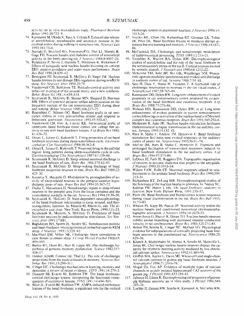
498 R. SZYMUSIAK
activity: an in vivo microdialysis study. Pharmacol Biochem Behar 1991:38:723-9.
74. Kurosawa M. Okada K. Sato A. Uchida S. Extracellular release of acetylcholine. noradrenalin and serotonin increase in the cerebral cortex during walking in conscious rats. Neurosci Lett 1993;161:73-6.
75. Buzsaki G, Bickford RG, PonomareffG, ThaI LJ. Mandel R, Gage FH. Nucleus basalis and thalamic control of neocortical activity in the freely moving rat. J J\'eurosci 1988;8:4007-26.
76. Reikkinen P, Sirvio J, Hannila T, Miettinen R, Riekkinen P. Effects of quisqualic acid nucleus basalis lesioning on cortical EEG, passive avoidance and water maze performance. Brain Res Bull 1990:24:839-42.
77. Benington IH. Szymusiak R. McGinty D. Siegel 1M. Nucleus basalis lesions do not disrupt EEG regulation during nonREM sleep. Soc Neurosci Ahstr 1994;20:79.
78. Vanderwolf CH, Robinson TE. Reticulo-cortical activity and behavior: a critique of the arousal theory and a new synthesis. Bchm' Brain Sci 1981 :4:459-514.
79. Szymusiak R, McGinty 0, Shouse MN. Shepard D. Sterman MB. Effects of systemic atropine sulfate administration on the frequency content of the cat sensorimotor EEG during sleep and waking. Behar iVeurosci 1990; 1 04:217-25.
80. Rosenblad C Nilsson OG. Basal forebrain grafts in rat neocortex restore in vivo acetylcholine release and respond to behavioral activation. Neuroscience 1993:55:353-62.
81. Vanderwolf CH. Fine A. Cooley RK. Intracortical grafts of embryonic basal forebrain tissue restore low voltage fast activity in rats with basal forebrain lesions. Exp Brain Res 1990; 81 :426-32.
82. Detari L, luhasz G. Kukorelli T. Firing properties of cat basal forebrain neurones during sleep-wakefulness cycle. Electroencephalogr Clin i"ellroph1'5ioI1984:58:362-8.
83. Detari L. Juhasz G, Kukorelli T. Neuronal firing in the pallidal region: firing patterns during sleep-wakefulness cycle in cats. Electroencephalogr Clin Neurophysiol 1987;67: 159-66.
84. Szymusiak R. McGinty D. Sleep-related neuronal discharge in the basal forebrain of cats. Brain Res 1986:370:82-92.
85. Szymusiak R. McGinty D. Sleep-waking discharge of basal forebrain projection neurons in cats. Brain Res Bull 1989;22: 423-30.
86. Koyama Y, Hayaishi O. Modulation by prostaglandins of activity of sleep-related neurons in the preoptic/anterior hypothalamic areas of rats. Brain Res Bull 1994;33:367-72.
87. Osaka T. Matsumura H. Noradrenergic inputs to sleep related neurons in the preoptic area from the locus coeruleus and the ventrolateral medulla in the rat. Neurosci Res 1994; 19:39-50.
88. Szymusiak R. McGinty D. State-dependent neurophysiology of the basal forebrain: relationship to sleep, arousal, and thermoregulatory function. In: Mancia M, Marini G, eds. The diencephalon and sleep. New York: Raven Press, 1990: 111-23.
89. Szymusiak R, Morairty S, McGinty D. Excitation of basal forebrain neurons by pedunculopontine stimulation. Soc Neurosci Ahstr 1991:17:881.
90. Baghdoyan HA, Spotts JL, Snyder SG. Simultaneous pontine and basal forebrain microinjections of carbachol supress REM sleep. J Neurosci 1993: 13:229-42.
91. MacPhail EM, Miller NE. Cholinergic brain stimulation in cats: failure to obtain sleep. J Comp Physiol Psychol 1968;65: 499-503.
92. Bartus RT, Dean RL, Beer B, Lippa AS. The cholinergic hypothesis of geriatric memory dysfunction. Science 1982;217: 408-17.
93. Dekker AIAM, Connor 01, ThaI LJ. The role of cholinergic projections from the nucleus basalis in memory. Neurosci BiohehavRev 1991;15:299-317.
94. Fibiger HC Cholinergic mechanisms in learning, memory and dementia: a review of recent evidence. TI1\'S 1991: 14:220-3.
95. Dunnett SB, Everitt BJ. Robbins TW. The basal forebraincortical cholinergic system: interpreting the functional consequences of excitotoxic lesions. TINS 1991; 14:494-50 I.
96. Muir lL, Everitt Bl, Robbins TW. AMPA-induced excitotoxic lesions of the basal forebrain: a significant role for the cortical
Sleep. Vol. 18. No.6. 1995
cholinergic system in attentional function. J l,'eurosci 1994: 14: 2313-26.
97. Voytko ML, Olton OS, Richardson RT, Gorman LK. Tobin JR, Price DL. Basal forebrain lesions in monkeys disrupt attention but not learning and memory. J Neurosci 1994; 14: 167-86.
98. McCormick DA. Cholinergic and noradrenergic modulation of thalamocortical processing. TIXS 1989: 12:215-21.
99. Tremblay N, Warren RA. Dykes RW. Electrophysiological studies of acetylcholine and the role of the basal forebrain in the somatosensory cortex of the cat II. Cortical neurons excited by somatic stimuli. J IVeurophl'siol 1990:64: 1212-22.
100. McKenna TM, Ashe JH. Hui GK. Weinberger NM. Muscarinic agonists modulate spontaneous and evoked unit discharge in auditory cortex of cat. Synapse 1988:2:54-68.
101. Sato H, Hata Y, Masui H, Tsumoto T. A functional role of cholinergic innervation to neurons in the cat visual cortex. J NeurophysioI1987:58:765-80.
102. Rasmusson DO, Dykes RW. Long-term enhancement of evoked potentials in cat somatosensory cortex produced by co-activation of the basal forebrain and cutaneous receptors. Exp Brain Res 1988:70:276-86.
103. Webster HH, Rasmusson DO, Dykes RW, et al. Long term enhancement of evoked potentials in racoon somatosensory cortex following co-activation of the nucleus basalis ofMeynert complex and cutaneous receptors. Brain Res 1991 :545:292-6.
104. Metherate R, Ashe IH. Nucleus basalis stimulation facilitates thalamocortical synaptic transmission in the rat auditory cortex. Synapse 1993;14:132-43.
105. Hars B, Maho e, Edeline JM, Hennevin E. Basal forebrain stimulation facilitates tone-evoked responses in the auditory cortex of awake rat. Neuroscience 1993:56:61-74.
106. Edeline JM, Hars B, Maho C Hennevin E. Transient and prolonged facilitation of tone-evoked responses induced by basal forebrain stimulation in the rat auditory cortex. Exp Brain Res 1994:97:373-86.
107. LeDoux JE, Farb JE, Ruggiero DA. Topographic organization of neurons in acoustic thalamus that project to the amygdala. J Neurosci 1990: 1 0: 1043-54.
108. Wilson FA W. Rolls ET. Neuronal responses related to reinforcement in the primate basal forebrain. Brain Res 1990:509: 213-31.
109. Richardson RT, DeLong MR. Electrophysiological studies of the functions of the nucleus basalis in primates. In: Napier TC Kalivas PW, Hanin L eds. The hasalforehrain: anatomy to (unction. New York: Plenum Press, 1991 :233-52.
110. 'Pirch JH. Basal forebrain and frontal cortex neuron responses during visual discrimination in the rat. Brain Res Bull 1993; 31:73-83.
111. Whalen PJ, Kapp BS, Pascoe JP. Neuronal activity within the nucleus basalis and conditioned neocortical electroencephalographic activation. J IVeurosci 1994: 14: 1623-33.
112. Aston-Jones G, Shaver R. Dinan TG. Nucleus basalis neurons exhibit axonal branching with decreased impulse conduction velocity in rat cerebrocortex. Brain Res 1985;325:271-85.
113. Reiner PB, Semba K, Fibiger He, McGeer EG. Physiological evidence for subpopulations of cortically projecting basal forebrain neurons in the anesthetized rat. Xeuroscience 1986;20: 629-36.
114. Khateb A, Muhlethaler M. Alonso A, Serafin M, Mainville L. Jones BE. Cholinergic nucleus basalis neurons display the capacity for rhythmic bursting activity mediated by low-threshold calcium spikes. Neuroscience 1992:51 :489-94.
115. Griffith WH, Taylor L, Davis MJ. Whole-cell and single-channel calcium currents in guinea pig basal forebrain neurons. J NeurophysioI1994;71:2359-76.
116. Mogul OJ, Fox AP. Evidence of multiple types of calcium channels in acutely isolated hippocampal C A3 neurons of the guinea pig. J Physiol 1991 ;433:259-81.
117. lahnsen H, Llinas R. Electrophysiological properties of guineapig thalamic neurons: an in vitro study. J Physiol 1984:349: 205-26.
118. LuebkeJI, Greene RW. Semba K. Kamondi A. McCarley RW,
Dow
nloaded from https://academ
ic.oup.com/sleep/article/18/6/478/2749729 by guest on 05 July 2022
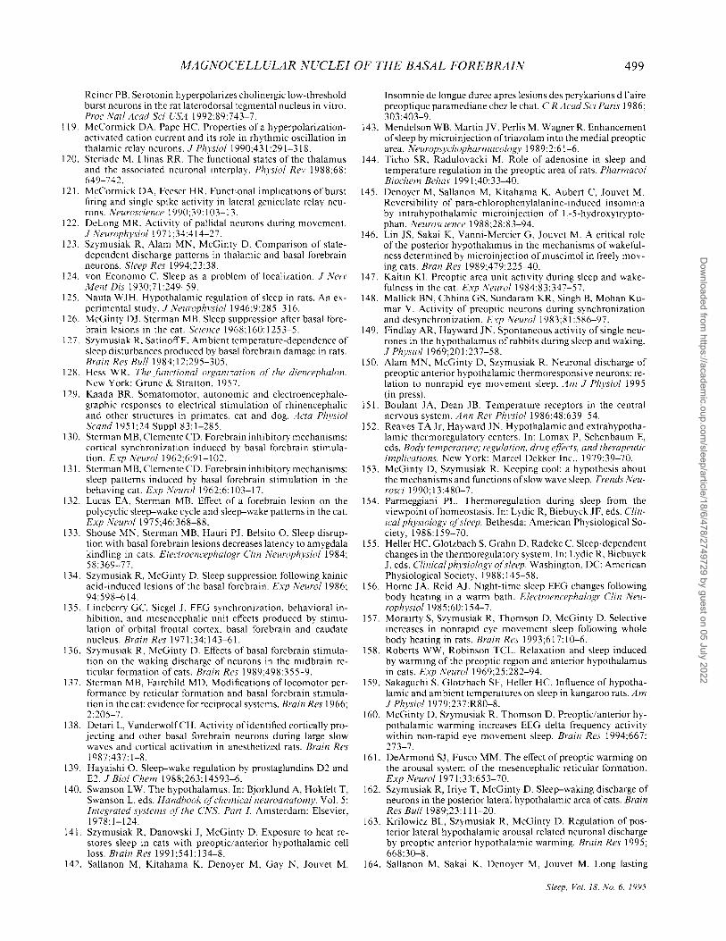
MAGNOCELLULAR NUCLEI OF THE BASAL FOREBRAIN 499
Reiner PB. Serotonin hyperpolarizes cholinergic low-threshold burst neurons in the rat laterodorsal tegmental nucleus in vitro. Proc Natl Acad Sci ['SA 1992;89:743-7.
119. McCormick DA, Pape He. Properties of a hyperpolarizationactivated cation current and its role in rhythmic oscillation in thalamic relay neurons. J Physiol 1990;431 :291-318.
120. Steriade M, Llinas RR. The functional states of the thalamus and the associated neuronal interplay. Physiol Rev 1988;68: 649-742.
121. McCormick DA, Feeser HR. Functional implications of burst firing and single spike activity in lateral geniculate relay neurons. Neuroscience 1990;39: 103-13.
122. DeLong MR. Activity of pallidal neurons during movement. J Neurophysiol 1971 ;34:414-27.
123. Szymusiak R, Alam MN, McGinty D. Comparison of statedependent discharge patterns in thalamic and basal forebrain neurons. Sleep Res 1994;23:38.
124. von Economo e. Sleep as a problem of localization. J Nerv J1ent Dis 1930;71:249-59.
125. Nauta WJH. Hypothalamic regulation of sleep in rats. An experimental study. J Neurophysiol 1946;9:285-316.
126. McGinty OJ, Sterman MB. Sleep suppression after basal forebrain lesions in the cat. Science 1968; 160: 1253-5.
127. Szymusiak R. SatinoffE. Ambient temperature-dependence of sleep disturbances produced by basal forebrain damage in rats. Brain Res Bull 1984; 12:295-305.
128. Hess WR. The functional organi::ation of the diencephalon. New York: Grune & Stratton, 1957.
129. Kaada BR. Somatomotor, autonomic and electroencephalographic responses to electrical stimulation of rhinencephalic and other structures in primates, cat and dog. Acta Physiol Scand 1951;24 Suppl 83:1-285.
130. Sterman MB, Clemente CD. Forebrain inhibitory mechanisms: cortical synchronization induced by basal forebrain stimulation. Exp lVeuro/1962;6:91-102.
131. Sterman MB, Clemente CD. Forebrain inhibitory mechanisms: sleep patterns induced by basal forebrain stimulation in the behaving cat. Exp ,'\'eurol 1962;6: 103-17.
132. Lucas EA, Sterman MB. Effect of a forebrain lesion on the polycyclic sleep-wake cycle and sleep-wake patterns in the cat. Exp NeuroI1975:46:368-88.
133. Shouse MN, Sterman MB, Hauri PJ, Belsito O. Sleep disruption with basal forebrain lesions decreases latency to amygdala kindling in cats. Electroencephalogr Clin Neurophysiol 1984; 58:369-77.
134. Szymusiak R, McGinty D. Sleep suppression following kainic acid-induced lesions of the basal forebrain. Exp Neurol 1986; 94:598-614.
135. Lineberry Ge. Siegel J. EEG synchronization, behavioral inhibition, and mesencephalic unit effects produced by stimulation of orbital frontal cortex, basal forebrain and caudate nucleus. Brain Res 1971 ;34: 143-61.
136. Szymusiak R, McGinty D. Effects of basal forebrain stimulation on the waking discharge of neurons in the midbrain reticular formation of cats. Brain Res 1989;498:355-9.
137. Sterman MB, Fairchild MD. Modifications of locomotor performance by reticular formation and basal forebrain stimulation in the cat: evidence for reciprocal systems. Brain Res 1966; 2:205-7.
138. Detari L, VanderwolfCH. Activity of identified cortically projecting and other basal forebrain neurons during large slow waves and cortical activation in anesthetized rats. Brain Res 1987;437: 1-8.
139. Hayaishi O. Sleep-wake regulation by prostaglandins D2 and E2. J Bioi Chem 1988;263: 14593-6.
140. Swanson LW. The hypothalamus. In: Bjorklund A, Hokfelt T, Swanson L, eds. Handhook of chemical neuroanatomy. Vol. 5: Integrated systems of the CNS, Part I. Amsterdam: Elsevier, 1978:1-124.
141. Szymusiak R, Danowski J, McGinty D. Exposure to heat restores sleep in cats with preoptic/anterior hypothalamic cell loss. Brain Res 1991 ;541: 134-8.
142. Sallanon M, Kitahama K, Denoyer M, Gay N, Jouvet M.
Insomnie de longue duree apres lesions des perykarions d I'aire preoptique paramediane chez Ie chat. C R Acad Sci Paris 1986; 303:403-9.
143. Mendelson WB, Martin JV, Perlis M, Wagner R. Enhancement of sleep by microinjection oftriazolam into the medial preoptic area. Neuropsychopharmacology 1989;2:61-6.
144. Ticho SR, Radulovacki M. Role of adenosine in sleep and temperature regulation in the preoptic area of rats. Pharmacol Biochem Behav 1991 ;40:33-40.
145. Denoyer M, Sallanon M, Kitahama K, Aubert e, Jouvet M. Reversibility of para-chlorophenylalanine-induced insomnia by intrahypothalamic microinjection of L-5-hydroxytryptophan. Neuroscience 1988;28:83-94.
146. Lin JS, Sakai K, Vanni-Mercier G, Jouvet M. A critical role of the posterior hypothalamus in the mechanisms of wakefulness determined by microinjection ofmuscimol in freely moving cats. Bran Res 1989:479:225-40.
147. Kaitin KI. Preoptic area unit activity during sleep and wakefulness in the cat. Exp Neuro/1984;83:347-57.
148. Mallick BN, Chhina GS, Sundaram KR, Singh B, Mohan Kumar V. Activity of preoptic neurons during synchronization and desynchronization. Exp Neurol 1983;81 :586-97.
149. Findlay AR, Hayward IN. Spontaneous activity of single neurones in the hypothalamus of rabbits during sleep and waking, J Physiol 1969;20 I :237-58.
150. Alam MN, McGinty 0, Szymusiak R. Neuronal discharge of preoptic anterior hypothalamic thermoresponsive neurons: relation to nonrapid eye movement sleep. Am J Physiol 1995 (in press),
151. Boulant JA, Dean JB. Temperature receptors in the central nervous system. Ann ReI' Physiol 1986;48:639-54.
152. Reaves TA Jr, Hayward IN. Hypothalamic and extrahypothalamic thermoregulatory centers. In: Lomax P, Schenbaum E, eds. Body temperature; regulation, drug e{!'ects, and therapeutic implications. New York: Marcel Dekker Inc., 1979:39-70.
153. McGinty 0, Szymusiak R. Keeping cool: a hypothesis about the mechanisms and functions of slow wave sleep. Trends Neurosci 1990; 13 :480-7.
154. Parmeggiani PL. Thermoregulation during sleep from the viewpoint of homeostasis. In: Lydic R, Biebuyck JF, eds. Clinical physiology of sleep. Bethesda: American Physiological Society, 1988: 159-70.
155. Heller He, Glotzbach S, Grahn 0, Radeke e. Sleep-dependent changes in the thermoregulatory system. In: Lydic R, Biebuyck J, eds. Clinical physiology o,fsleep. Washington. DC: American Physiological Society, 1988: 145-58.
156. Home JA, Reid AJ. Night-time sleep EEG changes following body heating in a warm bath. Electroencephalogr Clin Neuroph),siol 1985;60: 154-7.
157. Morairty S, Szymusiak R, Thomson 0, McGinty D. Selective increases in nonrapid eye movement sleep following whole body heating in rats. Brain Res 1993;617: 10-6.
158. Roberts WW, Robinson TCL. Relaxation and sleep induced by warming of the preoptic region and anterior hypothalamus in cats. Exp NeuroI1969;25:282-94.
159. Sakaguchi S, Glotzbach SF, Heller He. Influence of hypothalamic and ambient temperatures on sleep in kangaroo rats. Am J Physiol 1979;237:R80-8.
160. McGinty 0, Szymusiak R, Thomson D. Preoptic/anterior hypothalamic warming increases EEG delta frequency activity within non-rapid eye movement sleep. Brain Res 1994;667: 273-7.
161. DeArmond SJ, Fusco MM. The effect of preoptic warming on the arousal system of the mesencephalic reticular formation. Exp Neurol1971 ;33:653-70.
162. Szymusiak R, Iriye T, McGinty D. Sleep-waking discharge of neurons in the posterior lateral hypothalamic area of cats. Brain Res Buff 1989;23: 111-20.
163. Krilowicz BL, Szymusiak R, McGinty D. Regulation of posterior lateral hypothalamic arousal related neuronal discharge by preoptic anterior hypothalamic warming. Brain Res 1995; 668:30-8.
164. Sallanon M, Sakai K, Denoyer M, Jouvet M. Long lasting
Sleep, Vol. 18, No.6. 1995
Dow
nloaded from https://academ
ic.oup.com/sleep/article/18/6/478/2749729 by guest on 05 July 2022
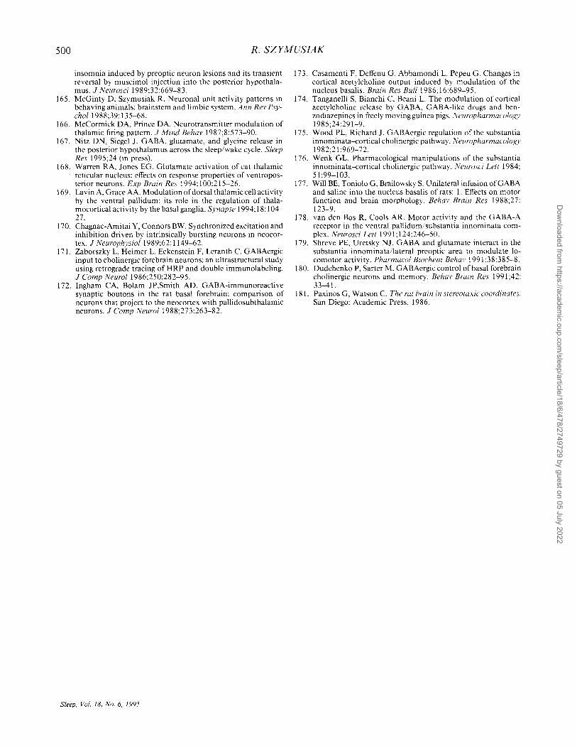
500 R. SZYMUSIAK
insomnia induced by preoptic neuron lesions and its transient reversal by muscimol injection into the posterior hypothalamus. J Neurosci 1989;32:669-83.
165. McGinty D, Szymusiak R. Neuronal unit activity patterns in behaving animals: brainstem and limbic system. Ann Rev PsychoI1988;39: 135-68.
166. McCormick DA, Prince DA. Neurotransmitter modulation of thalamic firing pattern. J Mind Behar 1987;8:573-90.
167. Nitz DN, Siegel J. GABA, glutamate, and glycine release in the posterior hypothalamus across the sleep/wake cycle. Sleep Res 1995;24 (in press).
168. Warren RA, Jones EG. Glutamate activation of cat thalamic reticular nucleus: effects on response properties of ventroposterior neurons. Exp Brain Res 1994; I 00:215-26.
169. Lavin A, Grace AA. Modulation of dorsal thalamic cell activity by the ventral pallidum: its role in the regulation of thalamocortical activity by the basal ganglia. Synapse 1994; 18: 104-27.
170. Chagnac-Amitai Y, Connors BW. Synchronized excitation and inhibition driven by intrinsically bursting neurons in neocortex. J Neurophysiol 1989;62: 1149-62.
171. Zaborszky L, Heimer L, Eckenstein F, Leranth C. GABAergic input to cholinergic forebrain neurons: an ultrastructural study using retrograde tracing ofHRP and double immunolabeling. J Comp Neurol 1986;250:282-95.
172. Ingham CA, Bolam JP,Smith AD. GABA-immunoreactive synaptic boutons in the rat basal forebrain: comparison of neurons that project to the neocortex with pallidosubthalamic neurons. J Comp NeuroI1988;273:263-82.
Sleep. Vol. 18. No.6. 1995
173. Casamenti F, Deffenu G, Abbamondi L Pepeu G. Changes in cortical acetylcholine output induced by modulation of the nucleus basalis. Brain Res Bull 1986; 16:689-95.
174. Tanganelli S, Bianchi C, Beani L The modulation of cortical acetylcholine relcase by GABA, GABA-like drugs and benzodiazepines in freely moving guinea pigs. l\'europharmacology 1985;24:291-9.
175. Wood PL, Richard J. GABAergic regulation of the substantia innominata--cortical cholinergic pathway. l\'europharmacology 1982;21 :969-72.
176. Wenk GL Pharmacological manipulations of the substantia innominata--cortical cholinergic pathway. ]\'eurosci Lett 1984; 51:99-103.
177. Will BE, Toniolo G, Brailowsky S. Unilateral infusion of GAB A and saline into the nucleus basalis of rats: I. Effects on motor function and brain morphology. Behar Brain Res 1988;27: 123-9.
178. van den Bos R, Cools AR. Motor activity and the GABA-A receptor in the ventral pallidum/substantia innominata complex. Neurosci Lett 1991; 124:246-50.
179. Shreve PE, Uretsky NJ. GAB A and glutamate interact in the substantia innominata/lateral preoptic area to modulate locomotor activity. Pharmacol Biochem Behar 1991 ;38:385-8.
180. Dudchenko P, Sarter M. GABAergic control of basal forebrain cholinergic neurons and memory. Behar Brain Res 1991 ;42: 33-41.
181. Paxinos G, Watson C. The rat brain in stereotaxic coordinates. San Diego: Academic Press, 1986.
Dow
nloaded from https://academ
ic.oup.com/sleep/article/18/6/478/2749729 by guest on 05 July 2022
Related Documents