STANDARDISED TRAINING AND ASSESSMENT IN RADIATION SAFETY FOR DIAGNOSTIC RADIOGRAPHERS by BELINDA VAN DER MERWE Thesis submitted in fulfilment of the requirements for the degree Philosophiae Doctor in Health Professions Education Ph.D. HPE in the DIVISION HEALTH SCIENCES EDUCATION FACULTY OF HEALTH SCIENCES UNIVERSITY OF THE FREE STATE BLOEMFONTEIN NOVEMBER 2014 PROMOTER: DR S.B. KRUGER CO-PROMOTER: PROF. DR M.M. NEL

Welcome message from author
This document is posted to help you gain knowledge. Please leave a comment to let me know what you think about it! Share it to your friends and learn new things together.
Transcript
STANDARDISED TRAINING AND ASSESSMENT IN
RADIATION SAFETY FOR DIAGNOSTIC RADIOGRAPHERS
by
BELINDA VAN DER MERWE
Thesis submitted in fulfilment of the requirements for the degree Philosophiae Doctor in Health Professions Education
Ph.D. HPE
in the
DIVISION HEALTH SCIENCES EDUCATION
FACULTY OF HEALTH SCIENCES
UNIVERSITY OF THE FREE STATE
BLOEMFONTEIN
NOVEMBER 2014
PROMOTER: DR S.B. KRUGER
CO-PROMOTER: PROF. DR M.M. NEL
ii
DECLARATION
I hereby declare that the work submitted here is the result of my own independent
investigation. Where help was sought, it was acknowledged. I further declare that this
work is submitted for the first time at this university/faculty towards a Philosophiae
Doctor degree in Health Professions Education and that it has never been submitted to
any other university/faculty for the purpose of obtaining a degree.
……………………………………. …………………………
Ms B. van der Merwe Date
I hereby cede copyright of this product in favour of the University of the Free State.
…………………………………. …………………………
Ms B. van der Merwe Date

iii
DEDICATION
I dedicate this thesis to radiographers; a crowd I am proud to belong to. I am
convinced that we embrace all the accoutrements to implement radiation safety.

iv
ACKNOWLEDGEMENTS
I wish to express my sincere thanks and appreciation to the following:
• My promoter, Dr Sonet Kruger, Division of Health Sciences Education, Faculty of
Health Sciences, University of the Free State, for her constant encouragement,
timely feedback and careful guidance.
• My co-promoter, Prof. Marietjie Nel, Head: Division of Health Sciences Education,
Faculty of Health Sciences, University of the Free State, for her sound advice,
expert supervision, and graceful wisdom.
• Delphi panellists for their paramount patience, prompt responses, constructive
comments and valuable time spent on the extensive questionnaire.
• Pilot participants: Lecturers, radiographers and students for their willingness to
scrutinise and test the Delphi questionnaires and student questionnaires.
• Quality Control Officers at Universitas Hospital, Anita Erasmus and Henra Muller,
for their assistance and guidance regarding the quality control tests.
• Dr Hannamarie Bezuidenhout for her language editing, referencing and meticulous
attention to detail in order to round off the thesis.
• Ms Elmarié Robberts for her professional editing, formatting and approachable
assistance during the boot camps.
• The former Dean of the Faculty of Health and Environmental Sciences, Central
University of Technology, Prof. Linda de Jager, for granting me permission to
undertake the research.
• The Head of the Department of Clinical Sciences, Central University of Technology,
Prof. Hesta Friedrich-Nel for her constant support of my studies and motivation in
my academic career.
• My CUT colleagues, Jeanette, Louisa, Renè and Bea for your inputs, enthusiasm
and guidance during the design and implementation of the Bachelor in
Radiography.
• The first- and third-year students of 2014 in the radiography programme of the
Central University of Technology, who participated in this study, for their input,
time and kind cooperation. I wish I could list the names of the 85 students who
made this study possible.
• Instructional designer, Nico Baird, not only for his support with the student
questionnaires and video recording, but also as fellow-student for his camaraderie.

v
• My parents, Johan and Aniki van der Merwe, for their continuous prayers and
admirable example to change the world for the better.
• My two daughters, Zindri and Sherike, for enthusiastically cheering me on to
believe that nothing is impossible.
• My husband, Handrè, for his utmost patience, loving empathy and unselfish
encouragement to fulfil this goal.
• My heavenly Father, for granting me the grace to endeavour to make a difference.
I declare that I am nothing without Him.
vi
TABLE OF CONTENTS
Page
CHAPTER 1: ORIENTATION TO THE STUDY
1.1 INTRODUCTION ....................................................................... 1
1.2 BACKGROUND TO THE RESEARCH PROBLEM 2
1.2.1 Licence holder education guidelines ........................................ 3
1.2.2 Issuing of dosimeters to students ............................................ 3
1.2.3 Issuing of dosimeters to supporting staff ................................ 4
1.2.4 The radiation protection training need ..................................... 5
1.3 PROBLEM STATEMENT AND RESEARCH QUESTIONS ............... 5
1.4 OVERALL GOAL, AIM AND OBJECTIVES OF THE STUDY ........... 7
1.4.1 Overall goal of the study ......................................................... 7
1.4.2 Aim of the study ...................................................................... 7
1.4.3 Objectives of the study ............................................................ 7
1.5 DEMARCATION OF THE FIELD AND SCOPE OF THE STUDY ...... 8
1.6 THE VALUE AND SIGNIFICANCE OF THE STUDY ...................... 9
1.7 RESEARCH DESIGN OF THE STUDY AND METHODS OF
INVESTIGATION ......................................................................
10
1.7.1 Design of the study ................................................................. 10
1.7.2 Methods of investigation ......................................................... 11
1.8 IMPLEMENTATION OF THE FINDINGS ..................................... 13
1.9 ARRANGEMENT OF THE REPORT ............................................. 13
1.10 CONCLUSION ........................................................................... 14
CHAPTER 2: MASTERY OF RADIATION SAFETY REQUIREMENTS FOR
DIAGNOSTIC RADIOGRAPHERS
2.1 INTRODUCTION ...................................................................... 15
2.2 LEGISLATURY DOCUMENTS ..................................................... 19
2.2.1 Hazardous Substances Act 15 (RSA DoH 1973) ....................... 20
2.2.2 Regulations concerning the control of electronic products
(RSA DoH 1973a) .....................................................................
22
vii
2.3 CODE OF PRACTICE FOR USERS OF MEDICAL X-RAY
EQUIPMENT ) ...........................................................................
28
2.3.1 Licencing and the responsible person ...................................... 28
2.3.2 The radiographer as radiation workers ................................... 31
2.3.3 Monitoring radiation workers .................................................. 32
2.3.4 Monitoring the patient ............................................................. 33
2.3.5 Premises requirements ............................................................ 36
2.3.6 Guideline and reference documents......................................... 36
2.4 REQUIREMENTS FOR LICENCE HOLDERS IWTH RESPECT TO
QUALITY CONTROL TESTS FOR DIAGNOSTIC X-RAY IMAGING
SYSTEMS (DRC 2012) ..............................................................
37
2.5 STANDARDISED TRAINING FOR RADIATION SAFETY ............. 41
2.5.1 Diploma in Radiography transformed to bachelor status ......... 43
2.5.1.1 The South African Qualifications Authority (SAQA) ................. 44
2.5.1.2 Exit-level outcomes pertaining to radiation safety .................. 45
2.5.1.3 Critical cross field outcomes pertaining to radiography ........... 47
2.5.1.4 Formulation of objectives ........................................................ 49
2.5.2 Teaching and learning strategies ............................................. 51
2.5.2.1 Problem-based learning ........................................................... 52
2.5.2.2 Active learning ......................................................................... 52
2.5.2.3 Distance education .................................................................. 53
2.5.2.4 Project-based learning ............................................................. 53
2.5.2.5 Lectures ................................................................................... 54
2.5.2.6 Simulations .............................................................................. 55
2.5.2.7 E-learning ................................................................................ 55
2.5.2.8 Blended learning ...................................................................... 56
2.5.2.9 Portfolios.................................................................................. 56
2.5.2.10 M-learning ................................................................................ 57
2.5.3 Assessment strategies ............................................................. 57
2.5.3.1 Assessment principles .............................................................. 58
2.5.3.2 Associated Assessment Criteria ............................................... 60
2.5.3.3 Integrated assessment ............................................................ 62
2.5.3.4 Theories of assessment ............................................................ 63
2.5.3.5 Assessment types..................................................................... 64
viii
2.6 ALIGNMENT OF CURRICULUM OBJECTIVES, TEACHING AND
LEARNING ACTIVITIES AND ASSESSMENT ..............................
64
2.7 PROOF OF MASTERY OF RADIATION SAFETY REQUIREMENTS 69
2.7.1 Retrieval / Recalling of information......................................... 71
2.7.2 Standard setting ...................................................................... 72
2.7.3 Bechmark of the radiation safety training and assessment ..... 73
2.8 CONCLUSION ........................................................................... 75
CHAPTER 3: RESEARCH DESIGN AND METHODOLOGY
3.1 INTRODUCTION .................................................................... 76
3.2 THEORETICAL PERSPECTIVES ON THE RESEARH
METHODOLOGY .....................................................................
76
3.3 RESEARCH METHODS ............................................................ 77
3.3.1 Literature review ................................................................... 77
3.3.2 The Delphi technique .............................................................. 78
3.3.2.1 Theoretical aspects ................................................................ 78
3.3.2.2 The Delphi process in this study ............................................ 80
3.3.2.3 Sample selection ................................................................... 81
3.3.3 The questionnaire survey ....................................................... 86
3.3.3.1 Theoretical aspects ............................................................... 86
3.3.3.2 The questionnaire survey in this study ................................. 87
3.3.3.3 Sample selection ................................................................... 89
3.4 ENSURING THE QUALITY, RELIABILITY, VALIDITY AND
TRUSTWORTHINESS OF THE STUDY .....................................
91
3.4.1 Reliability .............................................................................. 91
3.4.2 Validity .................................................................................. 92
3.4.3 Trustworthiness .................................................................... 92
3.5 ETHICAL CONSIDERATIONS .................................................. 93
3.5.1 Approval ................................................................................ 93
3.5.2 Informed consent .................................................................. 93
3.5.3 Right to privacy and confidentiality ...................................... 94
3.5.4 Minimising potential misinterpretation of results .................. 94
3.6 CONCLUSION ......................................................................... 95
ix
CHAPTER 4: DESCRIPTION AND DISCUSSION OF THE FINDINGS OF THE
DELPHI SURVEY
4.1 INTRODUCTION .................................................................... 96
4.2 DESCRIPTION AND DISCUSSION OF THE DELPHI SURVEY ... 96
4.2.1 Round One of the Delphi survey ............................................ 96
4.2.1.1 The measuring instrument ..................................................... 96
4.2.1.2 Analysis of the Round One responses .................................... 99
4.2.1.3 The findings of Round One of the Delphi survey .................... 99
4.2.2 Round Two of the Delphi survey ............................................ 99
4.2.2.1 The measuring instrument ..................................................... 99
4.2.2.2 Analysis of the Round Two responses .................................... 100
4.2.3 Round Three of the Delphi survey ......................................... 110
4.2.3.1 The measuring instrument ..................................................... 110
4.2.3.2 Analysis of the round Three responses .................................. 111
4.2.4 Round Four of the Delphi survey ........................................... 112
4.2.4.1 The measuring instrument ..................................................... 112
4.2.4.2 Analysis of the round four responses ..................................... 113
4.2.4.3 The findings of Round Four of the Delphi survey ................... 113
4.2.5 Summative discussion of the outcomes of the Delphi survey 128
4.2.6 Findings on consensus statements ........................................ 128
4.2.7 Findings regarding statements on which consensus was not
reached ..................................................................................
130
4.3 CONCLUSION ......................................................................... 130
CHAPTER 5: STANDARDISED RADIATION SAFETY TRAINING AND
ASSESSMENT FOR DIAGNOSTIC RADIOGRAPHERS
5.1 INTRODUCTION ..................................................................... 132
5.2 DESCRIPTION AND DISCUSSION OF THE CONTENT OF THE
RADIATION SAFETY REQUIREMENT TRAINING ......................
134
5.2.1 Consequence of the Delphi survey .......................................... 136
5.2.1.1 The training outcomes for radiation safety requirements ....... 137
5.2.1.2 The intended learning outcomes ............................................. 138
5.3 ASSESSMENT FOR RADIATION SAFETY REQUIREMENTS ........ 141
x
5.3.1 The measuring instrument ...................................................... 141
5.3.1.1 The first-year students’ orientation Objective structured
Clinical Examination (OSCE) ....................................................
142
5.3.1.2 The first-year students’ evidence of clinical observation ........ 143
5.3.1.3 The third-year students’ portfolio of evidence for quality
control tests ............................................................................
145
5.4 TEACHING AND LEARNING ACTIVITIES .................................. 147
5.5 INTEGRATING RAIDATION SAFETY REQUIREMENTS IN THE
CURRENT CURRICULUM ..........................................................
149
5.6 CONCLUSION ........................................................................... 150
CHAPTER 6: PRE-AND POST-TEST QUESTIONNAIRE FOR RADIATION
SAFETY
6.1 INTRODUCTION .................................................................... 151
6.2 POPULATION AND SCHEDULE OF THE TEST
QUESTIONNAIRES……………….…………………………………..
152
6.3 RESULTS OF THE BASIC AND ADVANCED TEST
QUESTIONNAIRES .................................................................
153
6.3.1 Descriptions of data from the basic test questionnaire ......... 153
6.3.1.1 Summative description of the students’ know ledge of the
general definitions and licencing conditions, responsibilit ies
of licence holders/ responsible persons .................................
157
6.3.1.2 Summative description of the students’ know ledge of the
operators of X-ray equipment and radiation workers ...........
161
6.3.1.3 Summative description of the student’s know ledge of the
radiation protection of radiation workers .............................
163
6.3.1.4 Summative description of the students’ know ledge of the
radiation protection of patients.............................................
168
6.3.1.5 Summative description of the students’ know ledge of the
quality control tests ..............................................................
173
6.3.2 Descriptions of data from the advanced test questionnaire .. 174
6.3.2.1 Summative description of the students’ know ledge of the
general definitions and licencing conditions, responsibilit ies
of licence holders/ responsible persons .................................
177
xi
6.3.2.2 Summative description of the students’ know ledge of the
operators of X-ray equipment and radiation workers ...........
183
6.3.2.3 Summative description of the students’ know ledge of the
radiation protection of radiation workers .............................
185
6.3.2.4 Summative description of the students’ know ledge of the
radiation protection of patients.............................................
188
6.3.2.5 Summative description of the students’ know lege of the
quality control tests ..............................................................
193
6.4 CONCLUSION ......................................................................... 198
CHAPTER 7: FINAL DISCUSSION ON THE FINDINGS OF THE RESEARCH
7.1 INTRODUCTION ................................................................... 199
7.2 DESCRIPTION OF THE IMPLICATION OF THE FINDINGS
FROM THE DELPHI SURVEY ...................................................
199
7.2.1 Delphi questionnaire comments from the panellists ............. 223
7.3 DESCRIPTION OF THE IMPLICATION OF THE FINDINGS
FROM THE PRE-AND POST-TEST QUESTIONNAIRE ...............
225
7.3.1 The purpose of assessment is to certify that a specific level
of performance has been achieved ........................................
225
7.3.2 Assessment can identify areas for remedial teaching and
learning activities or where adjustment in BASIC question
statements should be made ..................................................
226
7.3.2.1 Summative discussion on the students’ know ledge of the
general definitions and licencing conditions,
responsibilit ies of licence hodlers/ responsible persons ........
227
7.3.2.2 Summative discussion on the students’ know ledge of the
operators of X-ray equipment and radiation workers ...........
229
7.3.2.3 Summative discussion of the students’ know ledge of the
protection of radiation workers gainst radiation ...................
230
7.3.2.4 Summative discussion of the students’ know ledge of
patient’ protection against radiation .....................................
232
7.3.2.5 Summative discussion on the students’ know ledge of the
quality control tests ..............................................................
234
xii
7.33 Assessment can identify areas for remedial teaching and
learning activities or where adjustment in ADVANCED
question statements can be made .........................................
234
7.3.3.1 Summative discussion of the students’ know ledge of the
general definitions and licencing conditions, responsbilities
of licence holders/ responsible persons .................................
234
7.3.3.2 Summative discussion of the students’ know ledge of the
operators of X-ray equipment and radiation workers ...........
237
7.3.3.3 Summative discussion of the students’ know ledge of the
radiation protection of radiation workers .............................
238
7.3.3.4 Summative discussion on the students’ know ledge of the
protection of patients against radiation ................................
239
7.3.3.5 Summative discussion on the students’ know ledge of the
quality control tests ..............................................................
241
7.3.4 The role of feedback in assessment....................................... 243
7.3.4.1 Description of the results of the unintended third basic and
advanced tests ......................................................................
243
7.4 IMPLICATIONS OF THE FINDINGS OF THE TEACHING,
LEARNING AND ASSESSMENT ACTIVITIES ...........................
247
7.4.1 Portfolio of evidence for quality control tests ....................... 247
7.4.2 Additional adjustment in teaching and learning activities .... 248
7.5 CONCLUSION ......................................................................... 248
CHAPTER 8: CONCLUSIONS, RECOMMENDATIONS AND LIMITATIONS OF
THE STUDY
8.1 INTRODUCTION .................................................................... 249
8.2 OVERVIEW OF THE STUDY .................................................... 250
8.2.1 Research question 1 .............................................................. 250
8.2.2 Research question 2 .............................................................. 252
8.2.3 Research question 3 .............................................................. 253
8.2.4 Research question 4 .............................................................. 253
8.3 CONCLUSION ......................................................................... 254
8.4 LIMITATIONS OF THE STUDY ................................................ 256
8.5 CONTRIBUTION OF THE RESEARCH ...................................... 257
xiii
8.6 RECOMMENDATIONS ............................................................ 258
8.7 CONCLUSIVE REMARK ........................................................... 259
REFERENCES ......................................................................... 260
APPENDIX A APPENDIX A1: LETTER OF REQUEST TO PARTICIPATE IN THE DELPHI
QUESTIONNAIRE APPENDIX A2: CONSENT FORM: DELPHI SURVEY PARTICIPATION
APPENDIX B APPENDIX B1: DELPHI QUESTIONNAIRE FOR ROUND 1 APPENDIX B2: ACCOMPANYING LETTER FOR ROUND 1
APPENDIX C APPENDIX C1: FEEDBACK ON ROUND 1 APPENDIX C1B: FEEDBACK TO DELPHI PANEISTS AFTER ROUND 1 APPENDIX C2: INFORMATION LETTER TO DELPHI PANEL ROUND 2 APPENDIX C3: DELPHI QUESTIONNAIRE FOR ROUND 2
APPENDIX D APPENDIX D1: FEEDBACK ON ROUND 2 APPENDIX D1B: DELPHI FEEDBACK TO PANELISTS AFTER ROUND 2 APPENDIX D2: INFORMATION LETTER TO DELPHI PANEL ROUND 3 APPENDIX D3: DELPHI QUESTIONNAIRE FOR ROUND 3 APPENDIX D4: FEEDBACK ON ROUND 3 APPENDIX D4B: QUESTIONNAIRE FEEDBACK ON ROUND 3 APPENDIX D4C: DELPHI QUESTIONNAIRE FOR ROUND 4 APPENDIX D5: INFORMATION LETTER TO DELPHI PANEL ROUND 4 APPENDIX D6: LETTER TO DELPHI PANEL AFTER ROUND 4 APPENDIX D6B: SUMMARY OF THE DELPHI OUTCOME CONSENSUS AND
STABILITY APPENDIX D7: DELPHI PANEL FEEDBACK REGARDING THE FINAL TESTS
APPENDIX E APPENDIX E1: INFORMATION DOCUMENT: STUDENT INVITATION APPENDIX E2: CONSENT FORM STUDENT: PRE- AND POST-QUESTIONNAIRES APPENDIX E3: BASIC ASSESSMENT FOR RADIATION SAFETY (RESPONDUS) APPENDIX E4: ADVANCED ASSESSMENT FOR RADIATION SAFETY (RESPONDUS) APPENDIX E5: BENCHMARK OF RADIATION SAFETY TRAINING ISRRT APPENDIX E6: EUROPEAN DIRECTIVE BENCHMARK APPENDIX E7: INFORMATION TO PILOT QUESTIONNAIRE PARTICIPANTS
APPENDIX F APPENDIX F1: REQUEST TO THE DEAN OF THE FACULTY OF HEALTH SCIENCES,
SCHOOL OF MEDICINE, UFS APPENDIX F2: PERMISSION FROM THE DEAN OF THE FACULTY OF HEALTH AND
ENVIRONMENTAL SCIENCES, CUT APPENDIX F3: PERMISSION FROM THE HOD, DEPARTMENT OF CLINICAL
SCIENCES, CUT
xiv
APPENDIX F4A: REQUEST TO INCLUDE THE TRAININGG AND ASSESSMENT IN THE BACHELOR OF RADIOGRAPHY
APPENDIX F4B: AGREEMENT TO INCLUDE THE TRAININGG AND ASSESSMENT IN THE BACHELOR OF RADIOGRAPHY
APPENDIX G * Available on the included DVD APPENDIX G1: * POWERPOINT: RADIATION SAFETY FOR DIAGNOSTIC
RADIOGRAPHERS (ON DVD) APPENDIX G2: * STUDENT GUIDE -RADIATION SAFETY REQUIREMENTS (ON DVD) APPENDIX G3: STUDENT ACTIVITY- BIOLOGICAL EFFECTS APPENDIX G4: STUDENT ACTIVITY-LICENCE HOLDER APPENDIX G5: STUDENT ACTIVITY-PROTECTIVE SHIELDING APPENDIX G6: PORTFOLIO OF EVIDENCE OF QUALITY CONTROL TESTS APPENDIX G7: OSCE FOR FIRST YEAR STUDENTS APPENDIX G8: EVIDENCE OF FIRST YEAR CLINICAL OBSERVATION APPENDIX G9: COMPETITION ACTIVITY/OPEN BOOK TEST FOR THIRD YEARS APPENDIX G10: * LECTURER GUIDE FOR RADIATION SAFETY REQUIREMENTS (ON
DVD) APPENDIX G11: ROLE PLAY FOR RADIATION SAFETY APPENDIX G12: * C-ARM ORIENTATION IN THEATRE (VIDEO ON DVD) APPENDIX G13: PROPOSED LEARNING MODULE FOR RADIATION SAFETY
APPENDIX H
APPENDIX H1: BASIC POST TEST RESULTS
APPENDIX H2: ADVANCED POST TEST RESULTS
APPENDIX R * All requirement (R) appendices available on the included DVD
APPENDIX R1: HAZARDOUS AND SUBSTANCES ACT APPENDIX R2: REGULATIONS CONCERNING THE CONTROL OF ELECTRONIC
PRODUCTS APPENDIX R3: CODE OF PRACTICE FOR USERS OF MEDICAL X-RAY EQUIPMENT APPENDIX R4: REQUIREMENTS FOR LICENCE HOLDERS WITH RESPECT TO
QUALITY CONTROL TESTS FOR DIAGNOSTIC X-RAY IMAGING SYSTEMS
APPENDIX R5: RADIATION MONITORING REQUIREMENTS AND RADIATION OCCURRENCES (11/2011) APPENDIX R6: PERSONAL MONITORING WHEN A LEAD RUBBER APRON IS
WORN – MEDICAL AND VETERINARY USE OF X-RAY EQUIPMENT APPENDIX R7: PROTECTIVE CLOTHING APPENDIX R8: MANAGEMENT OF PREGNANT RADIOGRAPHERS AND OTHER
STAFF MEMBERS APPENDIX R9: MEDICAL EXAMINATIONS FOR RADIATION WORKERS (10/2009) APPENDIX R10: RADIATION PROTECTION OF PERSONNEL IN THEATRE APPENDIX R11: MONITORING OF RADIATION WORKERS IN A THEATRE
(11/2011) APPENDIX R12: HOLDING OF PATIENTS DURING X-RAY PROCEDURES (10/2009) APPENDIX R13: REQUEST FOR MEDICAL X-RAY EXAMINATIONS (10/2009) APPENDIX R14: FDA PUBLIC HEALTH NOTIFICATION: REDUCING RADIATION
RISK FROM COMPUTED TOMOGRAPHY FOR PEDIATRIC AND SMALL ADULT PATIENTS – (10/ 2009)
APPENDIX R15: PATIENT DOSE MEASUREMENTS IN DIAGNOSTIC RADIOLOGY (10/2009)
APPENDIX R16: DESIGN OF X-RAY ROOMS APPENDIX R17: DISPLAY AND FORMAT OF RADIATION WARNING SIGNS AT
ENTRANCES TO ROOMS CONTAINING X-RAY UNITS
xv
APPENDIX R18: TUBE LEAKAGE PROCEDURES AND MEASUREMENTS (NCRP REPORT 2004)
APPENDIX R19: MINIMUM REQUIREMENTS FOR FIXED DIAGNOSTIC X-RAY INSTALLATIONS
APPENDIX R20: RADIOGRAPHIC GRID RATIO (10/2009) APPENDIX R21: BONE DENSITOMETER – SHIELDING, MONITORING AND
POSITIONING OF OPERATORS (10/2009) APPENDIX R22: TEST PROCEDURES FOR FILM PROCESSING AND INTENSIFYING
SCREENS (6/2010) APPENDIX R23: SENSITOMETRIC TECHNIQUE FOR EVALUATION OF PROCESSING
(STEP) APPENDIX R24: SEALING / NOT SEALING AND UNSEALING OF X-RAY UNITS /
FILM PROCESSORS APPENDIX R25: DISPLAY CONSIDERATIONS FOR HOSPITAL-WIDE VIEWING OF
SOFT COPY IMAGES (BRITISH JOURNAL OF RADIOLOGY 2007) APPENDIX R26: DEFINITIONS: SUPERVISION APPENDIX R27: REGULATORY CONTROL OF X-RAY EQUIPMENT USED IN THE
MINING INDUSTRY IN SOUTH AFRICA TO SCREEN WORKERS FOR SECURITY PURPOSES (30/11/2011)
APPENDIX R28: REQUIREMENT OF CONVEYER BELT/CABINET TYPE LUGGAGE X-RAY EQUIPMENT (2/2/98)
APPENDIX R29: DENTAL RADIOGRAPHY ADDITIONAL APPENDICES INCLUDED IN THE STUDENT GUIDE (Available on DVD) APPENDIX R30: TEST PROCEDURES FOR FILM PROCESSING AND INTENSIFYING
SCREENS APPENDIX R31: RC008 REGISTRATION AS RADIATION WORKER-SABS APPENDIX R31A: RC009 MEDICAL REPORT ON RADIATION WORKER APPENDIX R31B: RC010 NOTIFICATION OF RADIATION OCCURRANCE APPENDIX R32: POLICY MINI C ARM SHIELDING AND OPERATORS APPENDIX R33: RC001 APPLICATION LICENCE X RAY EQUIPMENT ACCORDING
TO HAZARDOUS ACT APPENDIX R34A: SABS GENERAL INFORMATION USE AND TYPES APPENDIX R34B: RPS SABS MONITORING APPLICATION ADMIN DOCUMENTS APPENDIX R34C: RPS SABS GENERAL INFORMATION TLD METERS APPENDIX R35: PORTFOLIO OF EVIDENCE FOR MASTERY OF QC TESTS APPENDIX R36: EVIDENCE OF FIRST YEAR CLINICAL OBSERVATION

xvi
LIST OF FIGURES Page
FIGURE 1.1: A SCHEMATIC OVERVIEW OF THE STUDY ...................... 12
FIGURE 2.1: A DIAGRAMMATIC OVERVIEW OF THE DIFFERENT
ASPECTS DISCUSSED IN CHAPTER 2 .............................
18
FIGURE 5.1: CLINICAL OBSERVATION FEEDBACK FORM ................... 143
FIGURE 5.2: PORTFOLIO OF EVIDENCE FOR QC TESTS (EXTRACT) .... 145
xvii
LIST OF TABLES
Page
TABLE 2.1: ACT, REGULATIONS AND LICENSING CONDITION
DOCUMENTS ....................................................................
19
TABLE 2.2: ACCEPTANCE AND ROUTINE QUALITY CONTROL
TESTS EXTRACT FROM THE REQUIREMENTS FOR
LICENCE HOLDERS WITH RESPECT TO THE
REQUIREMENTS FOR QUALITY CONTROL TESTS
DOCUMENT: TABLE 2 (III.1) ...........................................
39
TABLE 4.1: CONSENSUS STATEMENTS AFTER ROUND 2 ................... 101
TABLE 4.2: CONSENSUS STATEMENTS AFTER ROUND 4 ................... 114
TABLE 4.3 : STABILITY STATEMENTS ROUND FOUR (FINAL
ROUND
122
TABLE 6.1: COMPARISON OF THE BASIC TEST PRE- AND POST-
QUESTIONNAIRE DISCRIMINATION ...............................
154
TABLE 6.2: COMPARISON OF THE BASIC TEST DIFFICULTY PRE-
AND POST-QUESTIONNAIRE ...........................................
154
TABLE 6.3: COMPARISON OF THE BASIC TEST PRE- AND POST-
QUESTIONNAIRE: STUDENT SCORE AND PERCENTAGE
154
TABLE 6.4: COMPARISON OF THE BASIC TEST PRE- AND POST-
QUESTIONNAIRE: GRADE DISTRIBUTION
156
TABLE 6.5: SECTION 1: BASIC TEST QUESTIONS GENERAL
DEFINITIONS AND LICENCING CONDITIONS:
RESPONSIBILITIES OF LICENCE HOLDERS /
RESPONSIBLE PERSONS .................................................
157
TABLE 6.6: SECTION 2: BASIC TEST QUESTIONS: OPERATORS OF
X-RAY EQUIPMENT AND RADIATION WORKERS ............
161
TABLE 6.7: SECTION 3: RADIATION PROTECTION OF RADIATION
WORKERS ........................................................................
163
TABLE 6.8: SECTION 4: BASIC TEST QUESTIONS: RADIATION
PROTECTION OF PATIENTS .............................................
168
TABLE 6.9: SECTION 5: BASIC TEST QUESTIONS: QUALITY
CONTROL TESTS ..............................................................
173
xviii
TABLE 6.10: COMPARISON OF THE ADVANCED TEST PRE - AND
POST QUESTIONNAIRE DISCRIMINATION………………
174
TABLE 6.11: COMPARISON OF THE BASIC TEST PRE- AND POST-
QUESTIONNAIRE DIFFICULTY ........................................
175
TABLE 6.12: COMPARISON OF THE ADVANCED TEST PRE- AND
POST-QUESTIONNAIRES STUDENT SCORE AND
PERCENTAGE ...................................................................
175
TABLE 6.13: COMPARISON OF THE ADVANCED TEST PRE- AND
POST-QUESTIONNAIRE GRADE DISTRIBUTION .............
176
TABLE 6.14: SECTION 1: ADVANCED TEST QUESTIONS: GENERAL
DEFINITIONS AND LICENCING CONDITIONS,
RESPONSIBILITIES OF LICENCE HOLDERS /
RESPONSIBLE PERSONS .................................................
177
TABLE 6.15: SECTION 2: ADVANCED TEST QUESTIONS:
OPERATORS OF X-RAY EQUIPMENT AND RADIATION
WORKERS ........................................................................
183
TABLE 6.16: SECTION 3: ADVANCED TEST QUESTIONS:
RADIATION PROTECTION OF RADIATION WORKERS .....
185
TABLE 6.17: SECTION 4: ADVANCED TEST QUESTIONS:
RADIATION PROTECTION OF PATIENTS .........................
189
TABLE 6.18: SECTION 5: ADVANCED TEST QUESTIONS: QUALITY
CONTROL TESTS ..............................................................
193
TABLE 7.1: SUMMARY OF DELPHI QUESTIONNAIRE ROUNDS
COLOUR CODED AND COMMENTS ...................................
200
TABLE 7.2: SECTION 1: BASIC TEST QUESTIONS: CORRECT
STUDENT RESPONSES BELOW 75% ................................
227
TABLE 7.3: SECTION 2: BASIC TEST QUESTIONS: CORRECT
STUDENT RESPONSES BELOW 75% ................................
229
TABLE 7.4: SECTION 3: BASIC TEST QUESTIONS: CORRECT
STUDENT RESPONSES BELOW 75% .............................
230
TABLE 7.5: SECTION 4: BASIC TEST QUESTIONS: CORRECT
STUDENT RESPONSES BELOW 75% ................................
232
xix
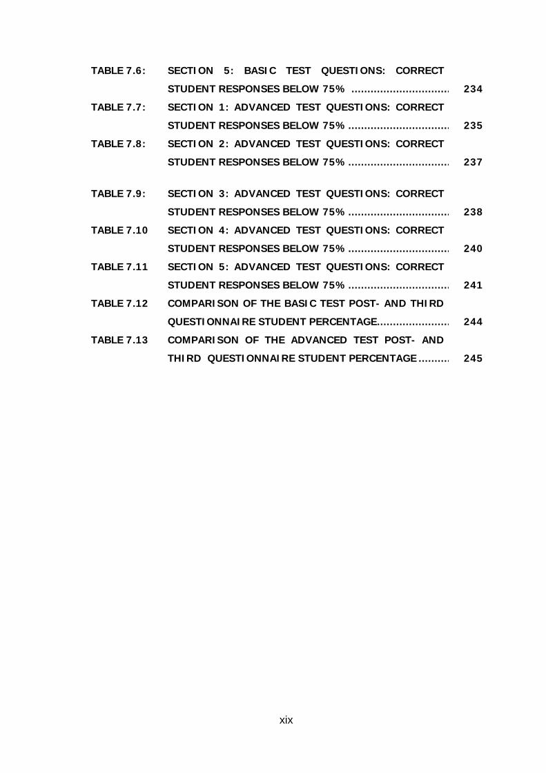
TABLE 7.6: SECTION 5: BASIC TEST QUESTIONS: CORRECT
STUDENT RESPONSES BELOW 75% ...............................
234
TABLE 7.7: SECTION 1: ADVANCED TEST QUESTIONS: CORRECT
STUDENT RESPONSES BELOW 75% ................................
235
TABLE 7.8: SECTION 2: ADVANCED TEST QUESTIONS: CORRECT
STUDENT RESPONSES BELOW 75% ................................
237
TABLE 7.9: SECTION 3: ADVANCED TEST QUESTIONS: CORRECT
STUDENT RESPONSES BELOW 75% ................................
238
TABLE 7.10 SECTION 4: ADVANCED TEST QUESTIONS: CORRECT
STUDENT RESPONSES BELOW 75% ................................
240
TABLE 7.11 SECTION 5: ADVANCED TEST QUESTIONS: CORRECT
STUDENT RESPONSES BELOW 75% ................................
241
TABLE 7.12 COMPARISON OF THE BASIC TEST POST- AND THIRD
QUESTIONNAIRE STUDENT PERCENTAGE .......................
244
TABLE 7.13 COMPARISON OF THE ADVANCED TEST POST- AND
THIRD QUESTIONNAIRE STUDENT PERCENTAGE ..........
245
xx
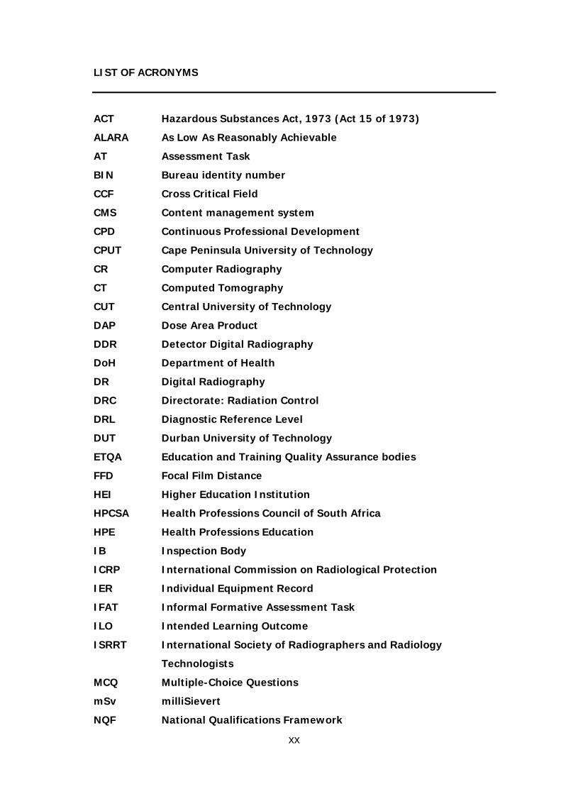
LIST OF ACRONYMS
ACT Hazardous Substances Act, 1973 (Act 15 of 1973)
ALARA As Low As Reasonably Achievable
AT Assessment Task
BIN Bureau identity number
CCF Cross Critical Field
CMS Content management system
CPD Continuous Professional Development
CPUT Cape Peninsula University of Technology
CR Computer Radiography
CT Computed Tomography
CUT Central University of Technology
DAP Dose Area Product
DDR Detector Digital Radiography
DoH Department of Health
DR Digital Radiography
DRC Directorate: Radiation Control
DRL Diagnostic Reference Level
DUT Durban University of Technology
ETQA Education and Training Quality Assurance bodies
FFD Focal Film Distance
HEI Higher Education Institution
HPCSA Health Professions Council of South Africa
HPE Health Professions Education
IB Inspection Body
ICRP International Commission on Radiological Protection
IER Individual Equipment Record
IFAT Informal Formative Assessment Task
ILO Intended Learning Outcome
ISRRT International Society of Radiographers and Radiology
Technologists
MCQ Multiple-Choice Questions
mSv milliSievert
NQF National Qualifications Framework
xxi
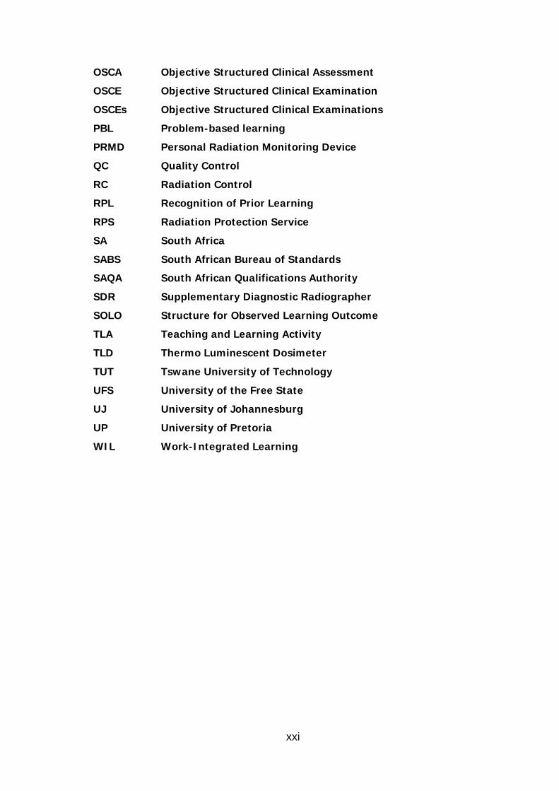
OSCA Objective Structured Clinical Assessment
OSCE Objective Structured Clinical Examination
OSCEs Objective Structured Clinical Examinations
PBL Problem-based learning
PRMD Personal Radiation Monitoring Device
QC Quality Control
RC Radiation Control
RPL Recognition of Prior Learning
RPS Radiation Protection Service
SA South Africa
SABS South African Bureau of Standards
SAQA South African Qualifications Authority
SDR Supplementary Diagnostic Radiographer
SOLO Structure for Observed Learning Outcome
TLA Teaching and Learning Activity
TLD Thermo Luminescent Dosimeter
TUT Tswane University of Technology
UFS University of the Free State
UJ University of Johannesburg
UP University of Pretoria
WIL Work-Integrated Learning
xxii
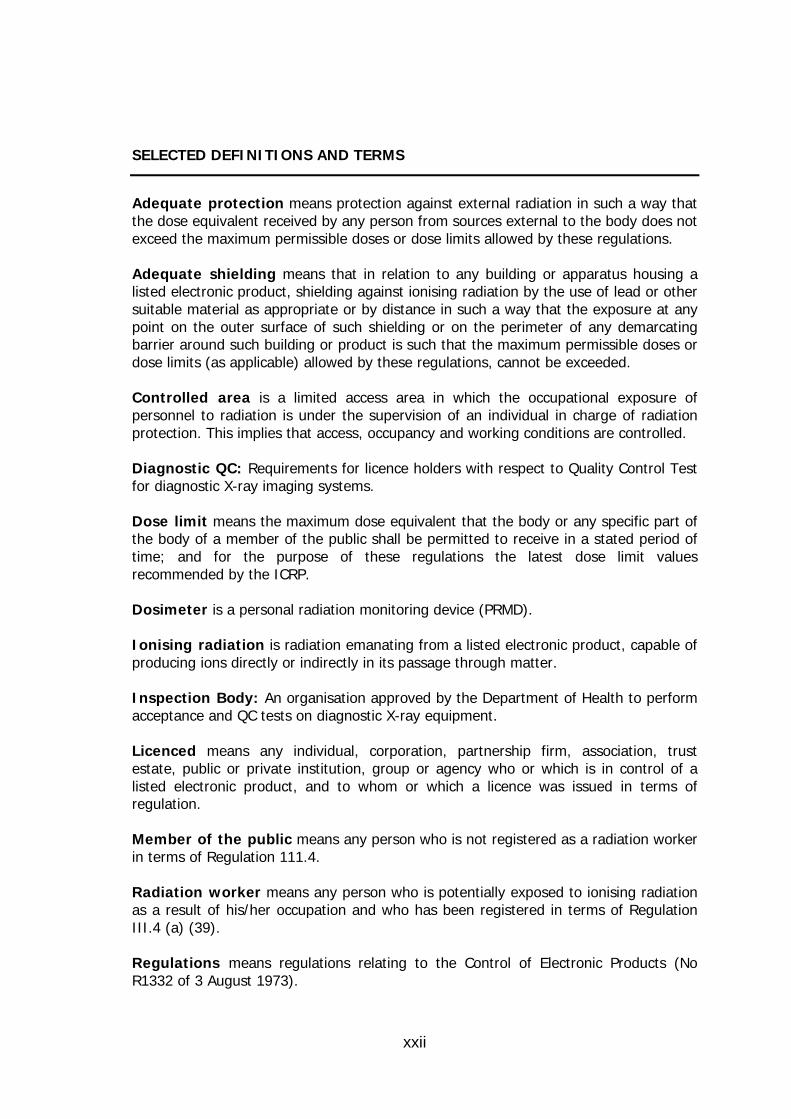
SELECTED DEFINITIONS AND TERMS
Adequate protection means protection against external radiation in such a way that the dose equivalent received by any person from sources external to the body does not exceed the maximum permissible doses or dose limits allowed by these regulations. Adequate shielding means that in relation to any building or apparatus housing a listed electronic product, shielding against ionising radiation by the use of lead or other suitable material as appropriate or by distance in such a way that the exposure at any point on the outer surface of such shielding or on the perimeter of any demarcating barrier around such building or product is such that the maximum permissible doses or dose limits (as applicable) allowed by these regulations, cannot be exceeded. Controlled area is a limited access area in which the occupational exposure of personnel to radiation is under the supervision of an individual in charge of radiation protection. This implies that access, occupancy and working conditions are controlled. Diagnostic QC: Requirements for licence holders with respect to Quality Control Test for diagnostic X-ray imaging systems. Dose limit means the maximum dose equivalent that the body or any specific part of the body of a member of the public shall be permitted to receive in a stated period of time; and for the purpose of these regulations the latest dose limit values recommended by the ICRP. Dosimeter is a personal radiation monitoring device (PRMD). Ionising radiation is radiation emanating from a listed electronic product, capable of producing ions directly or indirectly in its passage through matter. Inspection Body: An organisation approved by the Department of Health to perform acceptance and QC tests on diagnostic X-ray equipment. Licenced means any individual, corporation, partnership firm, association, trust estate, public or private institution, group or agency who or which is in control of a listed electronic product, and to whom or which a licence was issued in terms of regulation. Member of the public means any person who is not registered as a radiation worker in terms of Regulation 111.4. Radiation worker means any person who is potentially exposed to ionising radiation as a result of his/her occupation and who has been registered in terms of Regulation III.4 (a) (39). Regulations means regulations relating to the Control of Electronic Products (No R1332 of 3 August 1973).
xxiii
Responsible person refers to the person nominated by the holder pursuant to Regulation III-3 (e): (46). Supervision: The supervisor accepts and shares with the supervisee responsibility for ensuring that the supervisee’s work is professional and ethical, operating within whatever legal requirements and organisational norms apply. Supervision (direct): The supervisor provides on-site and in-view observation and guidance of a supervisee who performs an assigned activity. The proximity of this supervision is such that immediate intervention is possible if problems occur. X-ray unit means an electronic product which is designed, manufactured or assembled with the primary purpose of producing X-rays or which utilises X-rays to accomplish its primary purpose and from which such emissions are intended.
xxiv
SUMMARY Key terms: radiation worker, licence holder, standardised training and
assessment, radiation safety requirements, quality control requirements
Radiographers are occupationally exposed to ionising radiation and therefore
considered radiation workers. First-year radiography students are placed in clinical
practice within weeks of enrolment without proof of knowledge of radiation safety
requirements. The qualified radiographer may apply to be the licence holder of X-ray
equipment or is often appointed as a responsible person for X-ray equipment. The
third-year radiography student on the brink of graduation is thus a potential licence
holder of medical X-ray equipment. The Department of Health mandates the
responsibilities of radiation workers and licence holders in the Hazardous Substances
Act, Regulations 1332, and guideline documents, namely the Code of practice for users
of medical X-ray equipment and the Requirements for licence holders with respect to
quality control tests for diagnostic X-ray imaging systems. The purpose of the
regulations is to ensure the safe use of X-ray equipment so that the ionising radiation
dose to the staff and the patient is kept as low as reasonably achievable.
The research problem is that the regulations depict that licence holders of X-ray
equipment must educate radiation workers and implement quality control tests, but
nationally no standardised monitoring of radiation safety and quality control
requirements education is currently in place.
The purpose of this study was to develop standardised radiation safety and quality
control requirement training and assessment for diagnostic radiography to address
radiation safety.
The methods that were utilised were a literature review that provided background in
order to contextualise the research problem and to develop the criteria for the training
and assessment; a Delphi survey involving a panel of experts to establish a set of
criteria suitable for a basic or advanced component of the training and assessment;
questionnaires for radiography students to determine the knowledge of the radiation
worker before the training, and questionnaires determining the effect of training on
the knowledge by means of a post-test.
xxv
Results of the Delphi survey identified the content of the radiation safety and quality
control requirements for training and assessment by means of the contribution of a
panel of experts. The development and execution of the training and assessment
statements formed part of action research that contributed to fill the gap pertaining to
the education and training in the requirements for radiation safety and quality control
for radiation workers and medical X-ray equipment licence holders in the higher
education environment.
The contribution of the research was to develop standardised training and assessment
content and methods for diagnostic radiographers regarding the radiation safety and
quality control requirements for radiation workers and medical X-ray equipment licence
holders to be implemented in the diagnostic radiography healthcare environment. The
recommendation is that this study may serve as a directive for higher education
institutions, the Directorate: Radiation Control, as well as licence holders, that will
benefit if evidence can be confirmed of the educational and training attainment of
radiographers regarding the requirements for radiation safety and quality control.
Standardised training and assessment in radiation safety and quality control
requirements have the potential to enhance the safety of the first-year radiography
students as beginner radiation workers, the compliance of the third-year students as
potential licence holders of medical X-ray equipment, and, as a result, the safety of
patients.
xxvi
OPSOMMING
Sleutelterme: Bestralingswerker; lisensiehouer; gestandaardiseerde
opleiding en assessering; veiligheidsvereistes rakende bestraling;
gehaltebeheervereistes
Radiografiste is uit die aard van hul beroep blootgestel aan ioniseringsbestraling, en
word dus beskou as bestralingswerkers. Eerstejaar-radiografiestudente word binne
weke na registrasie in kliniese praktyk geplaas sonder enige bewyse van kennis van die
vereistes vir bestralingsveiligheid. Die gekwalifiseerde radiografis mag aansoek doen as
lisensiehouer van x-straaltoerusting, of word dikwels aangestel as die persoon
verantwoordelik vir die x-straaltoerusting. Die derdejaar-radiografiestudente wat op
die punt staan om te gradueer is dus potensiële lisensiehouers van mediese x-
straaltoerusting. Die Departement van Gesondheid dra die verantwoordelikheid vir die
uitvoering van die Wet op Gevaarhoudende Stowwe, Regulasie 1332 en
riglyndokumente, naamlik die Praktykkode vir die gebruikers van mediese x-
straaltoerusting en die Vereistes vir lisensiehouers ten opsigte van gehaltebeheertoetse
van diagnostiese x-straal-beeldingstelsels aan lisensiehouers en bestralingswerkers op.
Die doel van die regulasies is om die veilige gebruik van x-straaltoerusting te verseker
sodat die ioniseringsbestralingsdosis van die personeel en die pasiënte so laag as wat
redelik moontlik is, gehou word.
Die navorsingsprobleem is dat die regulasies bepaal dat lisensiehouers van x-
straaltoerusting bestralingspersoneel moet onderrig en gehaltebeheertoetse moet
implementeer, maar nasionaal is daar tans geen gestandaardiseerde onderwys vir die
monitering van bestralingsveiligheid en gehaltebeheervereistes in plek nie.
Die doel van die studie was om gestandaardiseerde bestralingsveiligheids- en
gehaltebeheeropleiding en -assessering vir diagnostiese radiografie te ontwikkel om
aandag te skenk aan bestralingsveiligheid.
Die metodes wat aangewend is, was ʼn literatuurstudie wat agtergrond verskaf het vir
die kontekstualisering van die navorsingsprobleem en ook om kriteria daar te stel vir
die opleiding en assessering; ʼn Delphi-ondersoek waarby ‘n deskundigheidspaneel
betrek is om ʼn stel kriteria daar te stel wat vir die basiese of gevorderde komponent
xxvii
van die opleiding en assessering geskik sou wees; vraelyste vir radiografiestudente om
die kennis van bestralingswerkers voor opleiding te bepaal, asook vraelyste om die
uitwerking van die opleiding op hul kennis te bepaal deur middel van ‘n na-toets.
Deur die resultate van die Delphi-opname is die inhoudelike vir die opleiding en
assessering in bestralingsveiligheids- en gehaltebeheervereistes geïdentifiseer deur die
bydraes van die paneel van deskundiges. Die ontwikkeling en implementering van die
opleiding- en assesseringstellings het deel gevorm van die aksienavorsing wat bygedra
het om die gaping wat in die hoëronderwysomgewing bestaan ten opsigte van die
onderwys en opleiding in die vereistes vir bestralingsveiligheid en gehaltebeheer vir
bestralingswerkers en die houers van lisensies vir mediese x-straaltoerusting, te vul.
Die bydrae van die navorsing is daarop gerig om gestandaardiseerde opleidings- en
assesseringsinhoudelike en –metodes te ontwikkel vir diagnostiese radiografiste
rakende die bestralingsveiligheid- en gehaltebeheervereistes vir bestralingswerkers en
die lisensiehouers van mediese x-straaltoerusting om in die gesondheidsorgomgewing
van die diagnostiese radiografie geïmplementeer te word. Dit word aanbeveel dat die
studie as aanwyser vir hoëronderwysinstellings, die Direktoraat: Stralingsbeheer, asook
lisensiehouers gebruik word, aangesien hulle daarby baat sal vind as die bewyse
bevestig kan word van wat bereik is deur die onderwys en opleiding van radiografiste
ten opsigte van die vereistes vir bestralingsveiligheid en -gehaltebeheer.
Gestandaardiseerde opleiding en assessering in bestralingsveiligheid- en
-gehaltebeheervereistes het die potensiaal om die veiligheid van eerstejaar-
radiografiestudente as nuwelingbestralingswerkers te bevorder, asook om te verseker
dat derdejaarstudente, as potensiële lisensiehouers van mediese x-straaltoerusting,
aan die vereistes voldoen, en daardeur hul eie en gevolglik ook die veiligheidsituasie
van pasiënte, uit te bou.
STANDARDISED TRAINING AND ASSESSMENT IN RADIATION SAFETY FOR
DIAGNOSTIC RADIOGRAPHERS
CHAPTER 1
ORIENTATION TO THE STUDY
1.1 INTRODUCTION
In this research, an in-depth study was conducted by the researcher with a view to
developing standardised training and assessment in radiation safety for diagnostic
radiographers.
Radiographers are occupationally exposed to radiation and therefore considered radiation
workers. The qualified radiographer may apply to be the licence holder of X-ray
equipment or is often appointed as a responsible person for X-ray equipment. The
Department of Health mandates the responsibilities of radiation workers in two
documents, namely the Code of practice for users of medical X-ray equipment and the
Requirements for licence holders with respect to quality control tests for diagnostic X-ray
imaging systems. The purpose of the regulations is to ensure the safe use of X-ray
equipment so that the ionising radiation dose to the staff and the patient is kept as low as
reasonably achievable.
The regulations depict that licence holders of X-ray equipment must identify radiation
workers, monitor the ionising radiation received by these workers and issue personal
monitoring devices. Furthermore, radiation workers must be educated in the safety and
risks of ionising radiation. The education pertaining to ionising radiation safety of staff
members is the responsibility of each license holder, but nationally no standardised
monitoring of the required education is currently in place.
Curricula at different tertiary institutions include academic exposure to the aspects
pertaining to the code of practice and quality control tests, but standardisation concerning
the teaching of radiography students by higher education institutions is not certain, and
the extent to which these aspects are covered everywhere can therefore not be attested.
The authentic interpretation of the qualification exit-level outcomes of every tertiary
2
institution in South Africa, therefore, may have the potential to result in differences in
subject content and assessment.
This study may serve as a directive for higher education institutions, the Directorate
Radiation Control, as well as licence holders that will benefit if evidence of fulfilment of
the education of radiographers as radiation workers in the hazards and risks of ionising
radiation can be confirmed.
The aim of the first chapter is to orientate the reader to the study. It provides the
background to the research problem – including the research questions, the overall goal,
aim, and objectives of the study. These are followed by the demarcation of the study and
highlights regarding the significance and value of the research. Subsequently, a brief
overview of the research design and methods of investigation are presented. The chapter
is concluded by a lay-out of the succeeding chapters and a summative conclusion.
1.2 BACKGROUND TO THE RESEARCH PROBLEM
The Department of Health (DoH), Directorate Radiation Control (DRC), lists the
responsibilities of license holders of medical X-ray equipment in the Code of Practice for
users of medical X-ray equipment. The licence holder and responsible person, apart from
equipment requirements, must ensure that persons occupationally exposed to ionising
radiation (radiation workers) are identified and issued with personal radiation monitoring
devices (PRMDs). The code further mandates that every radiation worker receive
education regarding the risks and safety rules of ionizing radiation (DRC 2011:8).
The PRMDs are commonly referred to as dosimeters, and can be ordered from the
Radiation Protection Service (RPS) of the South African Bureau of Standards (SABS). The
only requirement from the Directorate Radiation Control before issuing the dosimeter is
that a new radiation worker must undergo a medical examination to determine fitness for
work (DRC 2011:11). This implies that a licence holder may order dosimeters without
submitting proof of education of radiation workers regarding the ionising radiation safety.
Diagnostic radiographers employed in X-ray departments are potentially exposed to
ionising radiation and therefore are radiation workers (RSA DoH 1973:5). Entry-level
radiation workers, for example, first-year radiography students, are legally required to be
monitored and issued with dosimeters as soon as they are placed in clinical practice.
3
Specialist physicians and supporting nursing staff similarly are required to wear
dosimeters if they are chronically exposed to radiation (DRC 2011:8). The education
pertaining to ionising radiation safety of these staff members is the responsibility of each
licence holder, but nationally no standardised monitoring of the required education is
currently in place.
1.2.1 Licence holder education guidelines
The licence holder of medical X-ray equipment is responsible for the following education
of radiation workers, as indicated by the Code of Practice document (DRC 2011:8):
• “Radiation workers (occupationally exposed persons) are identified and issued with
personal radiation monitoring devices (PRMDs);
• The appropriate protective clothing, devices and equipment are provided to personnel
and properly used;
• Radiation safety rules are communicated to and followed by all personnel;
• Operational procedures are established and maintained to ensure that the radiation
exposure to workers, patients and public is kept as low as reasonably achievable
(ALARA) without compromising the diagnostic efficiency of the result,
• Workers are educated in the hazards and risks of ionising radiation”.
The code obligation is that every licence holder must ensure that radiation workers are
educated in the above-mentioned aspects. This study supported the code by determining
the specific outcomes to satisfy the guidelines in order to develop a training course with
aligned assessment. The lack of the proper implementation of the quality tests in clinical
practice was addressed in the study and training course. The inconsistency in the training
of radiography students as dosimeter holders (therefore radiation workers) and potential
licence holders will be discussed in the following section.
1.2.2 Issuing of dosimeters to students
Radiography training institutions have different policies regarding the issuing of
dosimeters to first-year radiography students. One university may issue the dosimeters
and incorporate the radiation safety assessment combined with a radiation protection
test, while another issues the dosimeters within the first week of clinical practice
(Swindon 2012:electronic mail), only to cover the academic aspects of dosimeters and
4
radiation risks over the course of a year (Kekana 2012: electronic mail; Hudson
2012:electronic mail). The situation exists, as explained in electronic communication by
Van Dyk (2012), where the training institution places the radiation safety responsibility
solely on the hospital or practice where the student is placed for work-integrated-learning
(WIL). The Central University of Technology (CUT), where the researcher is currently a
lecturer, issues the dosimeters after an hour contact session by a physicist and a 20-
question test that the student must complete successfully with a pass mark of 80%.
As the WIL coordinator for the radiography programme at the CUT, the researcher
observed (at eight different hospitals) that student radiographers as well as qualified
radiographers do not adhere to the ALARA principle at all times. This ignorance may be
due to a lack of knowledge regarding the aspects of ionising radiation safety or merely
because the level of knowledge is not conveyed and assessed summatively and uniformly.
Health care workers outside the scope of radiography, who are also considered radiation
workers, may even be more ignorant. The planned training course has the potential to
benefit a wide scope of healthcare professionals.
1.2.3 Issuing of dosimeters to supporting staff
Nursing staff and physicians in theatre where fluoroscopy is required during operations
are chronically exposed to ionising radiation. These staff members therefore also are
regarded as radiation workers and issued with dosimeters. The researcher observed
ignorance regarding the wearing of dosimeters (Van der Merwe 2008:2). The education of
the above-mentioned staff regarding radiation is not formally monitored in most hospitals.
Reluctance to wear lead protection against radiation and to leave dosimeters in lockers or
in drawers may reflect negatively on the licence holder. Dosimeters not utilised for the
purpose of measuring the radiation dose are ineffective and expensive. Ignorance
towards radiation aspects may be due to a lack of formal training. In addition, it may be
ascribed to the human element - people do not necessarily do what is expected, but what
is inspected. Adherence to guidelines can be monitored by means of a formal training
course with standardised assessment expectations. The training course that was
developed and is reported in this thesis focused, foremost, on radiography students with
the aim of expanding the training to other disciplines in future.
5
1.2.4 The radiation protection training need
Knowledge and education have a direct effect on the implementation of protection
measures (Mojiri & Moghimbeigi 2011:5) and thus need meticulous focus. Radiographers
in South Africa attend continuing professional development (CPD) events that may
reinforce the tertiary exposure to radiation protection principles. The concern, however, is
that radiographers are often deficient in the application of foundational principles. A study
in Sweden recently reiterated the importance of ensuring professional standards by
means of continuous education and to assess radiographers’ clinical competencies
(Andersson, Jacobsson & Brostrom 2012:635). In radiography, the current situation
regarding the training of radiation safety can be improved by providing proof that
foundational knowledge regarding the existing guidelines is mastered and implemented.
Vano (2010:200) reminds the reader that the international organisation, by name the
International Commission on Radiological Protection (ICRP) acknowledges the importance
of education and training in reducing patient doses while maintaining image quality.
Training must be considered at different levels - not only for entry-level users, but also for
retraining and certification. In this article the European perspective is discussed within the
framework of the Directive on Medical Exposures. The directive assures that the Member
States of the European Union shall establish curricula to certify competence in radiation
protection (Vano 2010:201). This urgent trend to accredit radiation protection curricula
confirms that this training course developed during this study is relevant for South Africa
(SA).
Motorists in SA are required to provide proof of mastering of knowledge by successfully
completing standardised assessment. User-friendly visual guides are available to prepare
entry-level candidates for these assessments. This successful method to study for a
learner driver’s licence may provide a solution to address the mammoth task to
standardise the training of radiation workers issued with dosimeters, not only in
diagnostic radiography, but also for radiation workers in the healthcare environment
spectrum.
1.3 PROBLEM STATEMENT AND RESEARCH QUESTIONS
The problem that was addressed is the development of a training course and assessment
for diagnostic radiography students. The ALARA (as low as reasonably achievable)
6
principle is not honoured due to the absence of standardised education and assessment
for diagnostic radiography. Officially assessed and standardised education for
radiographers has the potential to ensure that radiation safety rules and quality tests are
applied in diagnostic radiography.
No recent studies concerning a national standardised training course for diagnostic
radiographers in South Africa could be traced. Searches done on the National Research
Fund (NRF) website and Nexus database system did not produce information on relevant
dissertations/theses on training for radiation safety in diagnostic radiography. However, a
number of dissertations were found concerning radiation protection, medical radiation
dosimetry, and radiation safety. Examples of scholarly work on studies abroad include
Radiographers’ professional Competence (Andersson, Jacobsson & Brostrom 2012),
Medical imaging physics teaching to radiologic technologists in Kuwait (Ballini & Sukkar
2005), Mandatory Radiation Safety training for interventionalists: The European
perspective (Vano 2010), and Awareness and attitude of radiographers towards radiation
protection (Mojiri & Moghimbeigi 2011).
The researcher also searched the Ebscohost database, Medline database, EMBASE, SACat,
Academic Search Premier, Science Direct and Best Evidence medical database to identify
relevant articles. Some sections in the dissertations/theses and articles were helpful and
are acknowledged and referenced as such.
In conclusion, there seemed to be no recent scientific standardised training and
assessment for diagnostic radiographers with regard to radiation safety, and consequently
the implementation of quality tests of X–ray equipment.
In order to address the problem stated, the following research questions were addressed:
1. How can the radiation safety and quality control requirements for radiography
radiation workers and medical X-ray equipment licence holders be contextualised and
conceptualised to compile a theoretical framework for the study?
2. What are the outcomes for the radiation safety and quality control requirements
training: basic for first-year radiography students (representing the entry level
radiation worker issued with a dosimeter) and advanced for third-year radiography
students (representing the licence holder, responsible person, and the qualified
radiographer)?
7
3. What are effective teaching and learning activities and assessment strategies for the
radiography radiation safety and quality control requirement training?
4. Will the training and assessment enhance the knowledge of the radiography students
regarding the requirements for radiation safety and the requirements for quality
control tests?
The research was carried out and completed based on these four research questions. The
findings of the research will serve as the foundation for the training and assessment of
radiographers in radiation safety.
1.4 OVERALL GOAL, AIM AND OBJECTIVES OF THE STUDY
The formulation of the research goal, aim and objectives provides a useful means for
effectively delimiting the focus of the study.
1.4.1 Overall goal of the study
The overall goal of the study was to enhance the safety of radiographers as radiation
workers, the compliance of licence holders of medical X-ray equipment and as a result,
the safety of patients.
1.4.2 Aim of the study
The purpose of this study was to develop standardised training and assessment for
diagnostic radiography to address radiation safety.
1.4.3 Objectives of the study
To achieve the aim, the following objectives were pursued:
1. To contextualise and conceptualise the radiation safety and quality control
requirements for radiography radiation workers and medical X-ray equipment licence
holders by means of a literature study, in order to create a theoretical framework for
the study.
This objective addressed research question 1.
8
2. To determine appropriate outcomes for the radiation safety and quality control
requirements training by using a Delphi questionnaire: basic for first-year radiography
students (representing the entry level radiation worker issued with a dosimeter) and
advanced for third- year radiography students (representing the licence holder,
responsible person and the qualified radiographer).
This objective addressed research question 2.
3. To develop the effective teaching and learning activities and assessment strategies
for the radiography radiation safety and quality control requirements training to be
presented at the Central University of Technology (CUT) based on the findings of the
Delphi survey.
This objective addressed research question 3.
4. To assess (by employing pre- and post-training questionnaires) the entry-level
participants’ knowledge regarding radiation safety requirements for radiation workers
and for the advanced level their knowledge regarding the quality control
requirements.
This objective addressed research question 4.
1.5 DEMARCATION OF THE FIELD AND SCOPE OF THE STUDY
It is hoped that the findings of the study will be implemented in the radiography
programme of the Department of Clinical Sciences at the Central University of the Free
State. The findings of the study also will be utilised to propose a standardised training
course for radiography students at the higher education institutions (HEIs) in South
Africa. The study fits the field of Health Professions Education (HPE) and lies in the
domain of academic programme development.
The participants in the Delphi questionnaire were experts in the field of diagnostic
imaging and included lecturers at higher education institutions involved in radiography
training, medical physicists involved in quality tests in diagnostic departments, diagnostic
radiography managers of X-ray departments and the Directorate: Radiation Control. This
study is interdisciplinary as it reaches across Health Professions Education and Medicine.
The participants in the questionnaire survey included the diagnostic radiography students
in the first and third year, enrolled at the Central University of Technology for 2014.
9
In a personal context, the researcher in this study is a qualified radiographer, registered
with the Health Professions Council of South Africa (HPCSA). During the period after the
completion of her honours degree, she had the opportunity to lecture radiography
subjects on a part-time basis concurrently with clinical practice as a senior radiographer in
a coronary catheterisation unit. The researcher registered a radiography practice and
gained experience in the operating theatre where she observed ignorance towards
radiation protection. This observation sparked an interest in ionising radiation distribution
in theatre that was articulated in a Magister Technologiae in Radiography. The researcher
is currently a full-time radiography lecturer with the main responsibility as the work-
integrated-learning coordinator of the 150 radiography students placed for clinical practice
at eight hospitals in the Free State. The researcher gained experience in developing a
training module for radiographers in theatre, conducted in-service training at various
hospitals and presented papers at several national and international congresses with
regard to radiation protection. In clinical practice the researcher observed the
indisputable need for standardised radiation and quality test training - not only for
students, but also for qualified radiographers.
As far as the timeframe is concerned, the study was conducted between 2012 and 2014,
with the empirical research phase from July 2013 to December 2013 (Delphi rounds) and
January 2014 to June 2014 (student questionnaires).
1.6 THE VALUE AND SIGNIFICANCE OF THE STUDY
The value of this study entails that the result may contribute to ensuring that radiography
students enrolled at the CUT will be equipped with sufficient knowledge to implement
regulations and requirements of the Department of Health to make sure that the radiation
exposure of radiation workers and patients is kept ALARA.
The study will contribute significantly to the radiography environment, because the
successful completion of the training course will endow licence holders with proof of
mastery of regulations pertaining to ionising radiation best practice.
Nursing staff and physicians that are chronically exposed to radiation are considered
radiation workers and wear dosimeters devoid of proof of mastery of regulations
regarding radiation protection concepts. The training model includes basic concepts that
will benefit radiation workers outside the radiography curriculum.
10
1.7 RESEARCH DESIGN OF THE STUDY AND METHODS OF INVESTIGATION
The purpose of describing the research design and methods of investigation is to inform
the reader of the processes followed to come to the conclusion and in order for the
reader to develop confidence in the methods followed and thus in the findings (cf.
Strydom 2002:255).
1.7.1 Design of the study
A quantitative design was appropriate for this study. A quantitative research design was
used to gain more insight into the research problem, as well as to enhance the
interpretability of the research findings.
The function of research design is to guide the strategy of the researcher and to define
the techniques that will be utilised to acquire the data (Trafford & Leshem 2008:93). The
research design is the plan of the study. The data collection method entailed the Delphi
process that was mainly quantitative in nature with the option to add comments or
suggestions. Quantitative studies are defined as a process that is systematic and in which
numerical data are used to provide information (Babbie and Mouton 2001:49), and the
findings can be expressed by means of statistical data. The qualitative findings were
reported by incorporating them in the follow-up rounds of the Delphi process.
The Delphi survey involved a panel of experts in the field to agree on the outcomes of the
training course. In order to avoid the time-consuming and unreliable postal system, the
Delphi questionnaire was distributed electronically. The questionnaires were provided in
print format when a panellist preferred a hard copy.
The quantitative design was also followed to determine the extent of the students’
knowledge before and after training. This was done by means of questionnaires –
therefore a pre-test-post-test design. The quantitatively designed questionnaires were
accessible on the content management system (CMS) available at the CUT. The reason
for employing the electronic questionnaires was with future implementation of the
questionnaire at different institutions in mind; to involve a broad audience (cf. Hofstee
2009:114).
11
The research was aimed at improving the current practice of radiation safety training of
radiographers and was therefore considered action research (cf. Denscombe 2007:122).
The findings generated from the Delphi survey were applied in designing the training
course and compiling the questionnaires for the student survey. The processes of action
and research were integrated because after the Delphi survey the teaching activities and
assessment were developed aligned with the criteria accepted through the Delphi process
(cf. Denscombe 2007:124). The research design followed in this study is described in
more detail in Chapter 3.
1.7.2 Methods of investigation
The methods that were used and which formed the basis of the study comprised a
literature review, and, as empirical study, a questionnaire survey and a Delphi process.
The purpose of the literature review was to provide a background in order to
contextualise the research problem and to develop the questionnaires. The literature
review increased the knowledge of the researcher (cf. Singleton and Straits 199:544).
Electronic and paper media were consulted for this purpose to provide the background for
the Delphi survey and to determine the content of the training course. The DoH
requirements for licence holders of medical X-ray equipment, contained in the Code of
Practice and Quality Test documents (DRC 2011,2012), guided the criteria included in the
questionnaire.
The Delphi process was used to establish a set of criteria required for the design and
development, and implementation of a training course for diagnostic radiography
students. The Delphi process was regarded an appropriate method to determine the
objectives for the dosimeter training course. The Delphi participants were selected by the
researcher based upon the value these individuals would add to the study (cf. Denscombe
2007:17).
The responses of the panellists guided the outcomes for the training course. The training
course with appropriate teaching and learning activities was developed by the researcher
to align the learning activities with assessment, all with the purpose of testing whether a
student met the outcomes. The assessments were delivered in the form of pre- and post-
training questionnaires. Two questionnaires were compiled, basic, for the first-year
radiography students (representing the other support staff, namely nursing staff) and
12
advanced for third-year radiography students (representing the licence holder, responsible
person, the qualified radiographer). The target population included all undergraduate
students involved in diagnostic radiography during 2014 in the Department of Clinical
Sciences that granted consent to complete the questionnaire. The detailed description of
the population, sampling methods, data collection techniques, data analysis and reporting
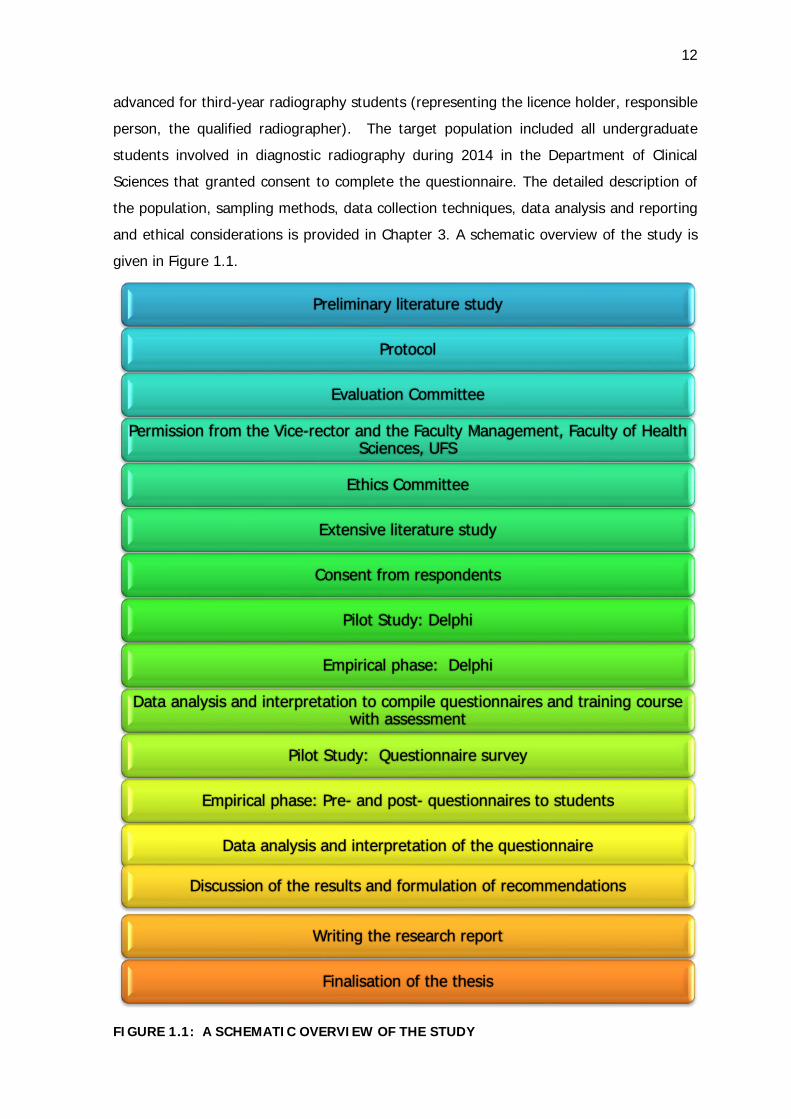
and ethical considerations is provided in Chapter 3. A schematic overview of the study is
given in Figure 1.1.
FIGURE 1.1: A SCHEMATIC OVERVIEW OF THE STUDY
Preliminary literature study
Protocol
Evaluation Committee
Permission from the Vice-rector and the Faculty Management, Faculty of Health Sciences, UFS
Ethics Committee
Extensive literature study
Consent from respondents
Pilot Study: Delphi
Empirical phase: Delphi
Data analysis and interpretation to compile questionnaires and training course with assessment
Pilot Study: Questionnaire survey
Empirical phase: Pre- and post- questionnaires to students
Data analysis and interpretation of the questionnaire
Discussion of the results and formulation of recommendations
Writing the research report
Finalisation of the thesis
13
1.8 IMPLEMENTATION OF THE FINDINGS
The report containing the findings of the research will be brought to the attention of the
Dean of the Faculty of Health and Environmental Sciences and the Head of the
Department of Clinical Sciences to be implemented in the radiography programme of the
Central University of Technology. It will be recommended that the training and
assessment be applied in the radiography programmes of other HEIs offering radiography
qualifications. The training course will be submitted to the Department of Radiation
Control for recommendation.
The research findings will be submitted to academic journals with a view to publication, as
the researcher aims to contribute significantly to the radiography environment by
equipping radiographers to implement radiation safety best practice.
1.9 ARRANGEMENT OF THE REPORT
To provide more insight into the topic, the methods used to find solutions and the final
outcome of the study will be reported as follows:
In this chapter, Chapter 1, Orientation to the study, the background to the study was
provided and the problem together with the research questions was stated. The overall
goal, aim and objectives were stated and the research design and methods utilised were
discussed concisely to provide the reader with a report overview. The chapter demarcated
the field of study and the role of the study was pointed out pertaining to radiation safety
for radiographers.
In Chapter 2, Mastery of radiation safety requirements for diagnostic
radiographers is deliberated. The purpose of the literature review was to provide a
background in order to contextualise the research problem and to develop the
questionnaires. Attention was given to clarify radiation workers, DoH regulations, training
for radiography, dosimeter training, radiation safety training, and online training models.
The themes that were researched focused on the DoH regulations pertaining to radiation
workers and radiation safety requirements as set out in the Code of Practice for users of
medical X-ray equipment and Requirements for licence holders with respect to quality
control tests for diagnostic X-ray imaging systems. Teaching and learning and assessment
14
strategies are discussed, bearing the exit-level outcomes of the radiography qualification
in mind.
In Chapter 3, Research design and methodology, the research design and the
methods applied will be conversed in detail. The data collecting methods and data
analysis will be discussed as applied for the Delphi process as well as the student
questionnaires.
Chapter 4, Description and discussion of the findings of the Delphi survey, will
provide the reader with the report of the responses of the Delphi panellists that guided
the outcomes for the training course.
Chapter 5, Standardised radiation safety training and assessment for diagnostic
radiographers, converses in full the development of the training course with appropriate
teaching and learning activities aligned with assessment.
Chapter 6, Pre- and post-test questionnaires for radiation safety, reports on the
assessments that were delivered in the form of pre- and post-training questionnaires.
Chapter 7, Final discussion on the findings of the research, reports on the analysis
of the Delphi survey results, assessments that were delivered in the form of pre- and
post-training questionnaires and the teaching and learning activities.
Chapter 8, Conclusion, recommendations and limitations of the study, will give a
summative conclusion of the study and recommendations as to the value of the results.
The application and limitations of the study are outlined.
1.10 CONCLUSION
Chapter 1 provided the background and introduction to the research undertaken
regarding the standardised training and assessment in radiation safety for radiographers.
The next chapter, Chapter 2, entitled Mastery of radiation safety requirements for
diagnostic radiographers, will provide an overview on the relevant literature.
CHAPTER 2
MASTERY OF RADIATION SAFETY REQUIREMENTS FOR DIAGNOSTIC
RADIOGRAPHERS
2.1 INTRODUCTION
This study was aimed at ensuring that radiography students who are enrolled at the CUT
are instructed in implementing regulations and requirements of the Department of Health
to ensure that the radiation exposure of radiation workers and patients is kept ALARA.
The study was embarked upon to contribute to the radiography environment because the
successful completion of training endows licence holders of medical X-ray equipment with
proof that radiography graduates are knowledgeable about the regulations pertaining to
ionising radiation best practice. Nursing staff and physicians that are chronically exposed
to radiation are considered radiation workers and wear dosimeters without proof that they
have mastered the regulations regarding radiation protection concepts. The training
model that has been developed includes basic concepts that have the potential to benefit
radiation workers outside the radiography curriculum.
The number of diagnostic and interventional medical procedures using ionising radiations
is rising and procedures resulting in higher patient and staff doses are being performed
more frequently. The need for education and training of medical staff and other
healthcare professionals in the principles of radiation protection, therefore, currently is
even more convincing (ICRP 2009).
The International Commission on Radiological Protection (ICRP) is the primary body for
protection against ionising radiation. ICRP is a non-governmental organisation created by
the 1928 International Congress of Radiology to promote radiological protection as public
interest. The ICRP quarterly publishes recommendations on and guidance in protection
against the risks associated with ionising radiation as the journal Annals of the ICRP. Each
issue provides in-depth coverage of a specific subject area (ICRP 2009). The Commission
has made basic recommendations for education and training of medical staff in ICRP
Publications 103 and 105 (ICRP 2007). Publication 113 provides guidance regarding the
necessary radiological protection education and training for use by:
16
• regulators, health authorities, medical institutions, and professional bodies with
responsibility for radiological protection in medicine;
• industry that produces the equipment used in these procedures; and
• universities and academic institutions educating professionals involved in the use of
ionising radiation in health care (ICRP 2009).
In the context of the mentioned ICRP publication,
the term ‘education’ refers to imparting knowledge and understanding on the
topics of radiation health effects, radiation quantities and units, principles of
radiological protection, radiological protection legislation, and the factors in
practice that affect patient and staff doses. Such education should be part of the
curriculum in pursuit of medical, dental, radiography and other health care
degrees and for specialists such as radiologists, nuclear medicine specialists and
medical physicists as part of the curriculum of postgraduate degrees. The term
‘training’ refers to providing instruction with regard to radiological protection for
the justified application of the specific ionising radiation modalities (e.g. computed
tomography, fluoroscopy) that a medical practitioner or other healthcare or
support professional will utilise in that individual’s role during medical practice
(ICRP 2009).
In South Africa, the Department of Health (DoH) accepted the recommendations of the
ICRP and regulates radiation protection in the framework of the Hazardous and
Substances Act No 15 of 1973 (RSA DoH 1973). This act stipulates that the Minister of
Health and, specifically, the Director-General, may issue licences to manufacturers,
importers and users of electro-medical (X-ray) equipment. X-ray equipment is referred to
as electronic products (X-rays) and therefore considered a Group III hazardous
substance. Group III hazardous substances are regulated by Regulation 1332, titled:
Regulations concerning the control of electronic products (RSA DoH 1973a).
The DoH in South Africa applies the international standards as legal requirements and
guidelines through the Directorate Radiation Control (DRC). The DRC issues a licence if
the product and usage comply with the legislative and international requirements for
safety and performance (RSA DoH 1973). The two documents that are supplied when a
licence is issued are:
17
1. Code of Practice for Users of Medical X-ray equipment [hereinafter Code of Practice]
(DRC 2011)
2. Requirements for licence holders with respect to quality control tests for Diagnostic X-
ray imaging systems [hereinafter Requirements for quality control tests] (DRC 2012).
These above-mentioned two documents are listed on the DRC website with further
reference to other guideline documents. The two above-mentioned requirement
documents as well as the guideline documents were consulted to develop the radiation
safety training for the radiography students at the Central University of Technology
(CUT). The researcher organised the documents according to a checklist in different
themes to make future referencing of the documents less confusing for the student
(cf. Table 2.1). The contents of the DRC documents guided the criteria for the Delphi
questionnaire (cf. Appendix B1) with the purpose of defining objectives and outcomes for
the training in radiation safety.
The researcher recognises the current debate and criticism regarding the administration
of the Hazardous and Substances Act and uncertainty of licensing conditions as published
in the South African Journal of Radiology. Herbst and Fick (2012:50,53) argue that the
Substances Act provides sufficient legislation and regulations but that human error caused
80% of over-exposure in radiology. Users of equipment should be adequately equipped to
have the skills and knowledge to operate with the minimal risk to patient and staff. The
researcher did not focus on the specific article’s recommendation of a future synergy
between the different regulatory bodies within a National Advisory Committee on
Radiation Equipment, but on a practical, authentic purpose to address the human
incompetency of regular users of X-ray equipment, specifically the radiography students.
The obligation of the Department of Clinical Sciences, Radiography, at the Central
University of Technology, therefore, in this thesis, is the education and training of the
radiography students in radiological protection.
In this chapter the radiation protection elements of the documents that regulate radiation
safety in South Africa, namely the Hazardous and Substances Act and Regulations
concerning the control of electronic products are discussed. The interpretation of the Act
and Regulations by the DRC are described in the Code of Practice, Requirements for
quality control tests and guideline documents from the DRC (cf. Appendix R). Since the
two DRC documents are user-friendly extractions of the Act and regulations these
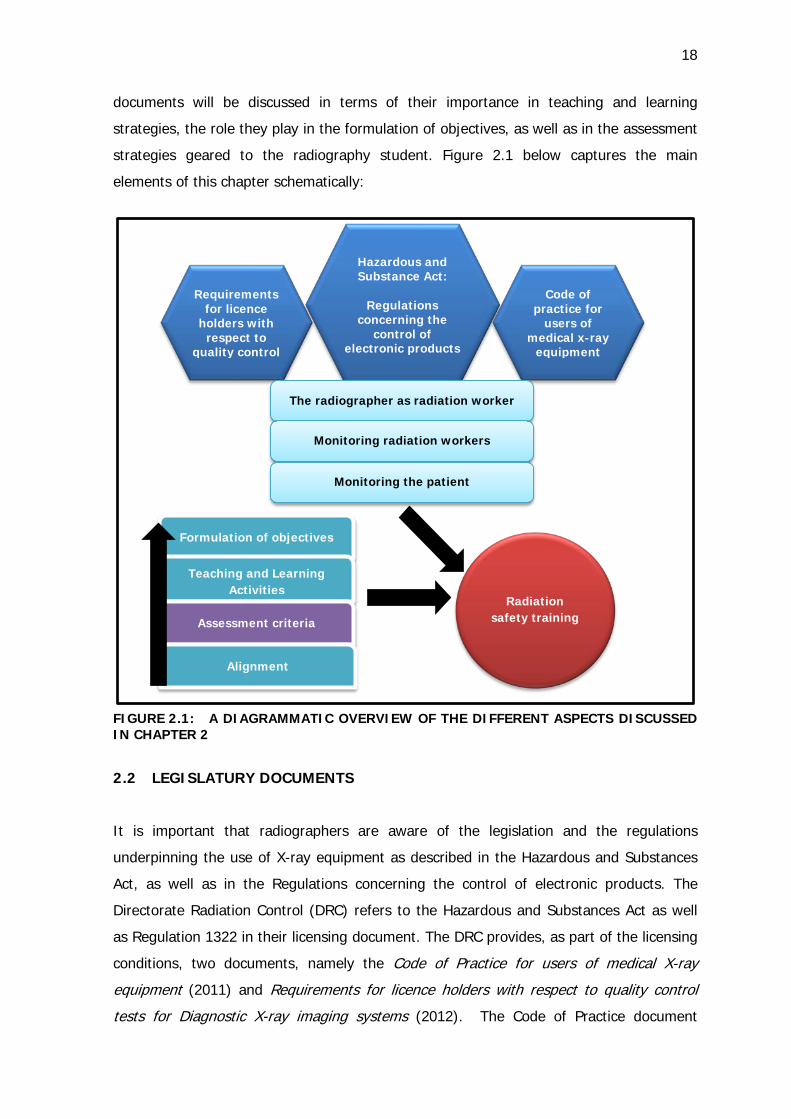
18
documents will be discussed in terms of their importance in teaching and learning
strategies, the role they play in the formulation of objectives, as well as in the assessment
strategies geared to the radiography student. Figure 2.1 below captures the main
elements of this chapter schematically:
FIGURE 2.1: A DIAGRAMMATIC OVERVIEW OF THE DIFFERENT ASPECTS DISCUSSED IN CHAPTER 2
2.2 LEGISLATURY DOCUMENTS
It is important that radiographers are aware of the legislation and the regulations
underpinning the use of X-ray equipment as described in the Hazardous and Substances
Act, as well as in the Regulations concerning the control of electronic products. The
Directorate Radiation Control (DRC) refers to the Hazardous and Substances Act as well
as Regulation 1322 in their licensing document. The DRC provides, as part of the licensing
conditions, two documents, namely the Code of Practice for users of medical X-ray
equipment (2011) and Requirements for licence holders with respect to quality control
tests for Diagnostic X-ray imaging systems (2012). The Code of Practice document
Requirements for licence
holders with respect to
quality control
Hazardous and Substance Act:
Regulations
concerning the control of
electronic products
Code of practice for
users of medical x-ray
equipment
The radiographer as radiation worker
Monitoring radiation workers
Monitoring the patient
Formulation of objectives
Teaching and Learning Activities
Assessment criteria
Alignment
Radiation safety training
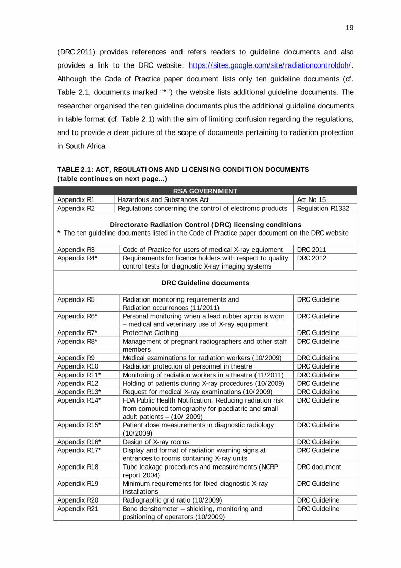
19
(DRC 2011) provides references and refers readers to guideline documents and also
provides a link to the DRC website: https://sites.google.com/site/radiationcontroldoh/.
Although the Code of Practice paper document lists only ten guideline documents (cf.
Table 2.1, documents marked “*”) the website lists additional guideline documents. The
researcher organised the ten guideline documents plus the additional guideline documents
in table format (cf. Table 2.1) with the aim of limiting confusion regarding the regulations,
and to provide a clear picture of the scope of documents pertaining to radiation protection
in South Africa.
TABLE 2.1: ACT, REGULATIONS AND LICENSING CONDITION DOCUMENTS (table continues on next page...)
RSA GOVERNMENT Appendix R1 Hazardous and Substances Act Act No 15 Appendix R2 Regulations concerning the control of electronic products Regulation R1332
Directorate Radiation Control (DRC) licensing conditions * The ten guideline documents listed in the Code of Practice paper document on the DRC website Appendix R3 Code of Practice for users of medical X-ray equipment DRC 2011 Appendix R4* Requirements for licence holders with respect to quality
control tests for diagnostic X-ray imaging systems DRC 2012
DRC Guideline documents
Appendix R5 Radiation monitoring requirements and
Radiation occurrences (11/2011) DRC Guideline
Appendix R6* Personal monitoring when a lead rubber apron is worn – medical and veterinary use of X-ray equipment
DRC Guideline
Appendix R7* Protective Clothing DRC Guideline Appendix R8* Management of pregnant radiographers and other staff
members DRC Guideline
Appendix R9 Medical examinations for radiation workers (10/2009) DRC Guideline Appendix R10 Radiation protection of personnel in theatre DRC Guideline Appendix R11* Monitoring of radiation workers in a theatre (11/2011) DRC Guideline Appendix R12 Holding of patients during X-ray procedures (10/2009) DRC Guideline Appendix R13* Request for medical X-ray examinations (10/2009) DRC Guideline Appendix R14* FDA Public Health Notification: Reducing radiation risk
from computed tomography for paediatric and small adult patients – (10/ 2009)
DRC Guideline
Appendix R15* Patient dose measurements in diagnostic radiology (10/2009)
DRC Guideline
Appendix R16* Design of X-ray rooms DRC Guideline Appendix R17* Display and format of radiation warning signs at
entrances to rooms containing X-ray units DRC Guideline
Appendix R18 Tube leakage procedures and measurements (NCRP report 2004)
DRC document
Appendix R19 Minimum requirements for fixed diagnostic X-ray installations
DRC Guideline
Appendix R20 Radiographic grid ratio (10/2009) DRC Guideline Appendix R21 Bone densitometer – shielding, monitoring and
positioning of operators (10/2009) DRC Guideline
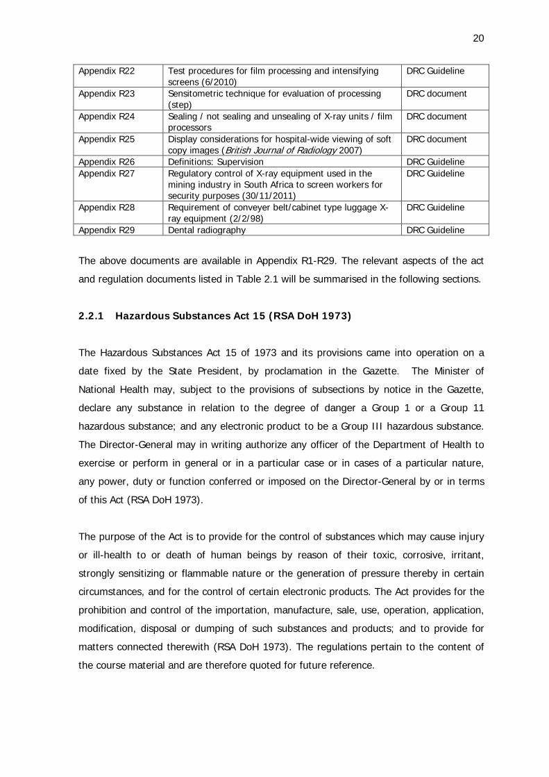
20
Appendix R22 Test procedures for film processing and intensifying screens (6/2010)
DRC Guideline
Appendix R23 Sensitometric technique for evaluation of processing (step)
DRC document
Appendix R24 Sealing / not sealing and unsealing of X-ray units / film processors
DRC document
Appendix R25 Display considerations for hospital-wide viewing of soft copy images (British Journal of Radiology 2007)
DRC document
Appendix R26 Definitions: Supervision DRC Guideline Appendix R27 Regulatory control of X-ray equipment used in the
mining industry in South Africa to screen workers for security purposes (30/11/2011)
DRC Guideline
Appendix R28 Requirement of conveyer belt/cabinet type luggage X-ray equipment (2/2/98)
DRC Guideline
Appendix R29 Dental radiography DRC Guideline
The above documents are available in Appendix R1-R29. The relevant aspects of the act
and regulation documents listed in Table 2.1 will be summarised in the following sections.
2.2.1 Hazardous Substances Act 15 (RSA DoH 1973)
The Hazardous Substances Act 15 of 1973 and its provisions came into operation on a
date fixed by the State President, by proclamation in the Gazette. The Minister of
National Health may, subject to the provisions of subsections by notice in the Gazette,
declare any substance in relation to the degree of danger a Group 1 or a Group 11
hazardous substance; and any electronic product to be a Group III hazardous substance.
The Director-General may in writing authorize any officer of the Department of Health to
exercise or perform in general or in a particular case or in cases of a particular nature,
any power, duty or function conferred or imposed on the Director-General by or in terms
of this Act (RSA DoH 1973).
The purpose of the Act is to provide for the control of substances which may cause injury
or ill-health to or death of human beings by reason of their toxic, corrosive, irritant,
strongly sensitizing or flammable nature or the generation of pressure thereby in certain
circumstances, and for the control of certain electronic products. The Act provides for the
prohibition and control of the importation, manufacture, sale, use, operation, application,
modification, disposal or dumping of such substances and products; and to provide for
matters connected therewith (RSA DoH 1973). The regulations pertain to the content of
the course material and are therefore quoted for future reference.
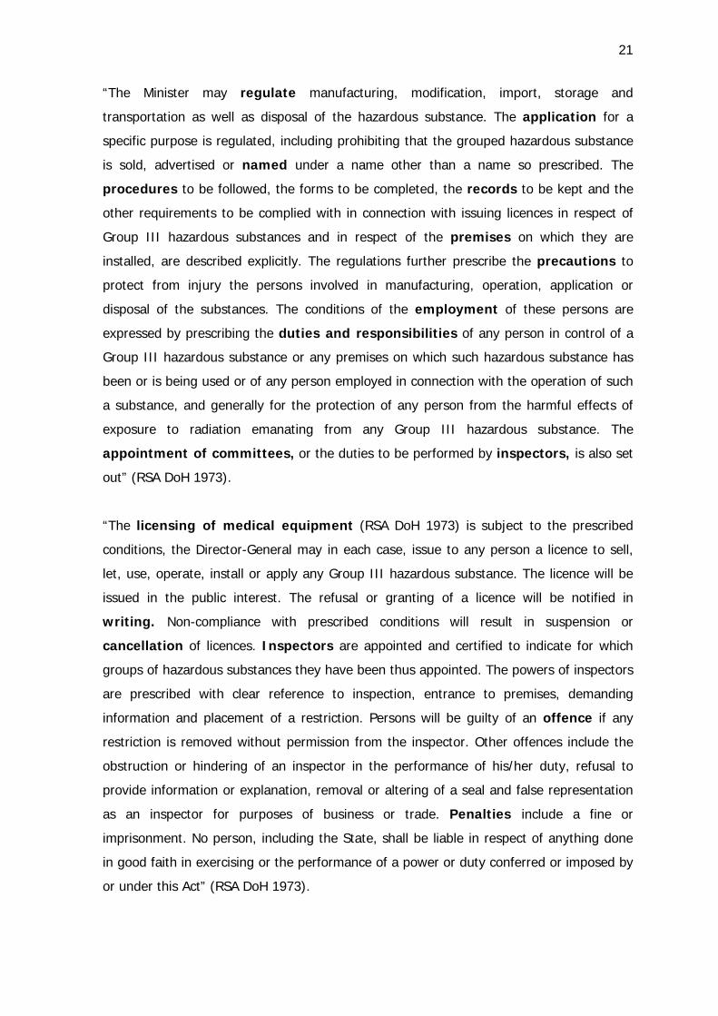
21
“The Minister may regulate manufacturing, modification, import, storage and
transportation as well as disposal of the hazardous substance. The application for a
specific purpose is regulated, including prohibiting that the grouped hazardous substance
is sold, advertised or named under a name other than a name so prescribed. The
procedures to be followed, the forms to be completed, the records to be kept and the
other requirements to be complied with in connection with issuing licences in respect of
Group III hazardous substances and in respect of the premises on which they are
installed, are described explicitly. The regulations further prescribe the precautions to
protect from injury the persons involved in manufacturing, operation, application or
disposal of the substances. The conditions of the employment of these persons are
expressed by prescribing the duties and responsibilities of any person in control of a
Group III hazardous substance or any premises on which such hazardous substance has
been or is being used or of any person employed in connection with the operation of such
a substance, and generally for the protection of any person from the harmful effects of
exposure to radiation emanating from any Group III hazardous substance. The
appointment of committees, or the duties to be performed by inspectors, is also set
out” (RSA DoH 1973).
“The licensing of medical equipment (RSA DoH 1973) is subject to the prescribed
conditions, the Director-General may in each case, issue to any person a licence to sell,
let, use, operate, install or apply any Group III hazardous substance. The licence will be
issued in the public interest. The refusal or granting of a licence will be notified in
writing. Non-compliance with prescribed conditions will result in suspension or
cancellation of licences. Inspectors are appointed and certified to indicate for which
groups of hazardous substances they have been thus appointed. The powers of inspectors
are prescribed with clear reference to inspection, entrance to premises, demanding
information and placement of a restriction. Persons will be guilty of an offence if any
restriction is removed without permission from the inspector. Other offences include the
obstruction or hindering of an inspector in the performance of his/her duty, refusal to
provide information or explanation, removal or altering of a seal and false representation
as an inspector for purposes of business or trade. Penalties include a fine or
imprisonment. No person, including the State, shall be liable in respect of anything done
in good faith in exercising or the performance of a power or duty conferred or imposed by
or under this Act” (RSA DoH 1973).
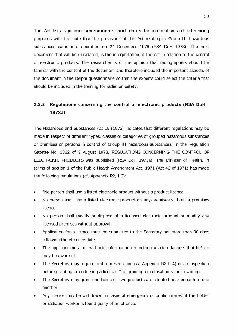
22
The Act lists significant amendments and dates for information and referencing
purposes with the note that the provisions of this Act relating to Group III hazardous
substances came into operation on 24 December 1976 (RSA DoH 1973). The next
document that will be elucidated, is the interpretation of the Act in relation to the control
of electronic products. The researcher is of the opinion that radiographers should be
familiar with the content of the document and therefore included the important aspects of
the document in the Delphi questionnaire so that the experts could select the criteria that
should be included in the training for radiation safety.
The Hazardous and Substances Act 15 (1973) indicates that different regulations may be
made in respect of different types, classes or categories of grouped hazardous substances
or premises or persons in control of Group III hazardous substances. In the Regulation
Gazette No. 1822 of 3 August 1973, REGULATIONS CONCERNING THE CONTROL OF
ELECTRONIC PRODUCTS was published (RSA DoH 1973a). The Minister of Health, in
terms of section 1 of the Public Health Amendment Act, 1971 (Act 42 of 1971) has made
the following regulations (cf. Appendix R2,II.2):
• “No person shall use a listed electronic product without a product licence.
• No person shall use a listed electronic product on any-premises without a premises
licence.
• No person shall modify or dispose of a licensed electronic product or modify any
licensed premises without approval.
• Application for a licence must be submitted to the Secretary not more than 90 days
following the effective date.
• The applicant must not withhold information regarding radiation dangers that he/she
may be aware of.
• The Secretary may require oral representation (cf. Appendix R2,II.4) or an inspection
before granting or endorsing a licence. The granting or refusal must be in writing.
• The Secretary may grant one licence if two products are situated near enough to one
another.
• Any licence may be withdrawn in cases of emergency or public interest if the holder
or radiation worker is found guilty of an offence.
2.2.2 Regulations concerning the control of electronic products (RSA DoH
1973a)
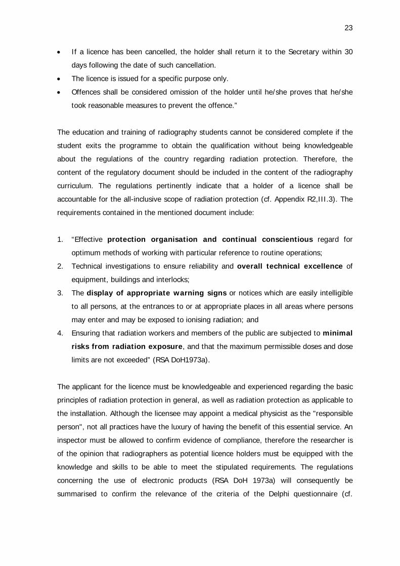
23
• If a licence has been cancelled, the holder shall return it to the Secretary within 30
days following the date of such cancellation.
• The licence is issued for a specific purpose only.
• Offences shall be considered omission of the holder until he/she proves that he/she
took reasonable measures to prevent the offence.”
The education and training of radiography students cannot be considered complete if the
student exits the programme to obtain the qualification without being knowledgeable
about the regulations of the country regarding radiation protection. Therefore, the
content of the regulatory document should be included in the content of the radiography
curriculum. The regulations pertinently indicate that a holder of a licence shall be
accountable for the all-inclusive scope of radiation protection (cf. Appendix R2,III.3). The
requirements contained in the mentioned document include:
1. “Effective protection organisation and continual conscientious regard for
optimum methods of working with particular reference to routine operations;
2. Technical investigations to ensure reliability and overall technical excellence of
equipment, buildings and interlocks;
3. The display of appropriate warning signs or notices which are easily intelligible
to all persons, at the entrances to or at appropriate places in all areas where persons
may enter and may be exposed to ionising radiation; and
4. Ensuring that radiation workers and members of the public are subjected to minimal
risks from radiation exposure, and that the maximum permissible doses and dose
limits are not exceeded” (RSA DoH1973a).
The applicant for the licence must be knowledgeable and experienced regarding the basic
principles of radiation protection in general, as well as radiation protection as applicable to
the installation. Although the licensee may appoint a medical physicist as the "responsible
person", not all practices have the luxury of having the benefit of this essential service. An
inspector must be allowed to confirm evidence of compliance, therefore the researcher is
of the opinion that radiographers as potential licence holders must be equipped with the
knowledge and skills to be able to meet the stipulated requirements. The regulations
concerning the use of electronic products (RSA DoH 1973a) will consequently be
summarised to confirm the relevance of the criteria of the Delphi questionnaire (cf.
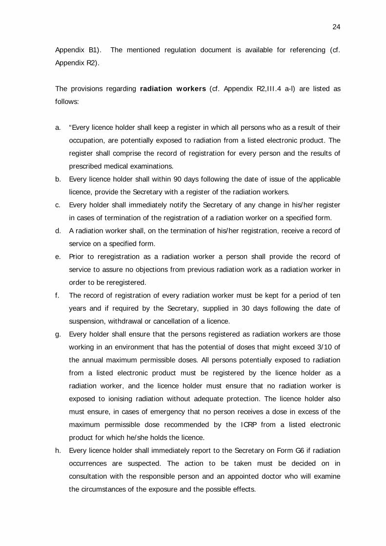
24
Appendix B1). The mentioned regulation document is available for referencing (cf.
Appendix R2).
The provisions regarding radiation workers (cf. Appendix R2,III.4 a-l) are listed as
follows:
a. “Every licence holder shall keep a register in which all persons who as a result of their
occupation, are potentially exposed to radiation from a listed electronic product. The
register shall comprise the record of registration for every person and the results of
prescribed medical examinations.
b. Every licence holder shall within 90 days following the date of issue of the applicable
licence, provide the Secretary with a register of the radiation workers.
c. Every holder shall immediately notify the Secretary of any change in his/her register
in cases of termination of the registration of a radiation worker on a specified form.
d. A radiation worker shall, on the termination of his/her registration, receive a record of
service on a specified form.
e. Prior to reregistration as a radiation worker a person shall provide the record of
service to assure no objections from previous radiation work as a radiation worker in
order to be reregistered.
f. The record of registration of every radiation worker must be kept for a period of ten
years and if required by the Secretary, supplied in 30 days following the date of
suspension, withdrawal or cancellation of a licence.
g. Every holder shall ensure that the persons registered as radiation workers are those
working in an environment that has the potential of doses that might exceed 3/10 of
the annual maximum permissible doses. All persons potentially exposed to radiation
from a listed electronic product must be registered by the licence holder as a
radiation worker, and the licence holder must ensure that no radiation worker is
exposed to ionising radiation without adequate protection. The licence holder also
must ensure, in cases of emergency that no person receives a dose in excess of the
maximum permissible dose recommended by the ICRP from a listed electronic
product for which he/she holds the licence.
h. Every licence holder shall immediately report to the Secretary on Form G6 if radiation
occurrences are suspected. The action to be taken must be decided on in
consultation with the responsible person and an appointed doctor who will examine
the circumstances of the exposure and the possible effects.
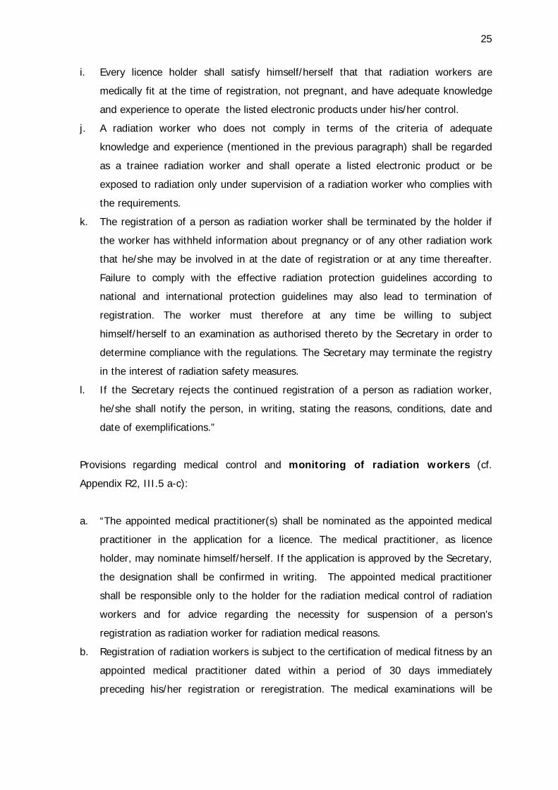
25
i. Every licence holder shall satisfy himself/herself that that radiation workers are
medically fit at the time of registration, not pregnant, and have adequate knowledge
and experience to operate the listed electronic products under his/her control.
j. A radiation worker who does not comply in terms of the criteria of adequate
knowledge and experience (mentioned in the previous paragraph) shall be regarded
as a trainee radiation worker and shall operate a listed electronic product or be
exposed to radiation only under supervision of a radiation worker who complies with
the requirements.
k. The registration of a person as radiation worker shall be terminated by the holder if
the worker has withheld information about pregnancy or of any other radiation work
that he/she may be involved in at the date of registration or at any time thereafter.
Failure to comply with the effective radiation protection guidelines according to
national and international protection guidelines may also lead to termination of
registration. The worker must therefore at any time be willing to subject
himself/herself to an examination as authorised thereto by the Secretary in order to
determine compliance with the regulations. The Secretary may terminate the registry
in the interest of radiation safety measures.
l. If the Secretary rejects the continued registration of a person as radiation worker,
he/she shall notify the person, in writing, stating the reasons, conditions, date and
date of exemplifications.”
Provisions regarding medical control and monitoring of radiation workers (cf.
Appendix R2, III.5 a-c):
a. “The appointed medical practitioner(s) shall be nominated as the appointed medical
practitioner in the application for a licence. The medical practitioner, as licence
holder, may nominate himself/herself. If the application is approved by the Secretary,
the designation shall be confirmed in writing. The appointed medical practitioner
shall be responsible only to the holder for the radiation medical control of radiation
workers and for advice regarding the necessity for suspension of a person's
registration as radiation worker for radiation medical reasons.
b. Registration of radiation workers is subject to the certification of medical fitness by an
appointed medical practitioner dated within a period of 30 days immediately
preceding his/her registration or reregistration. The medical examinations will be
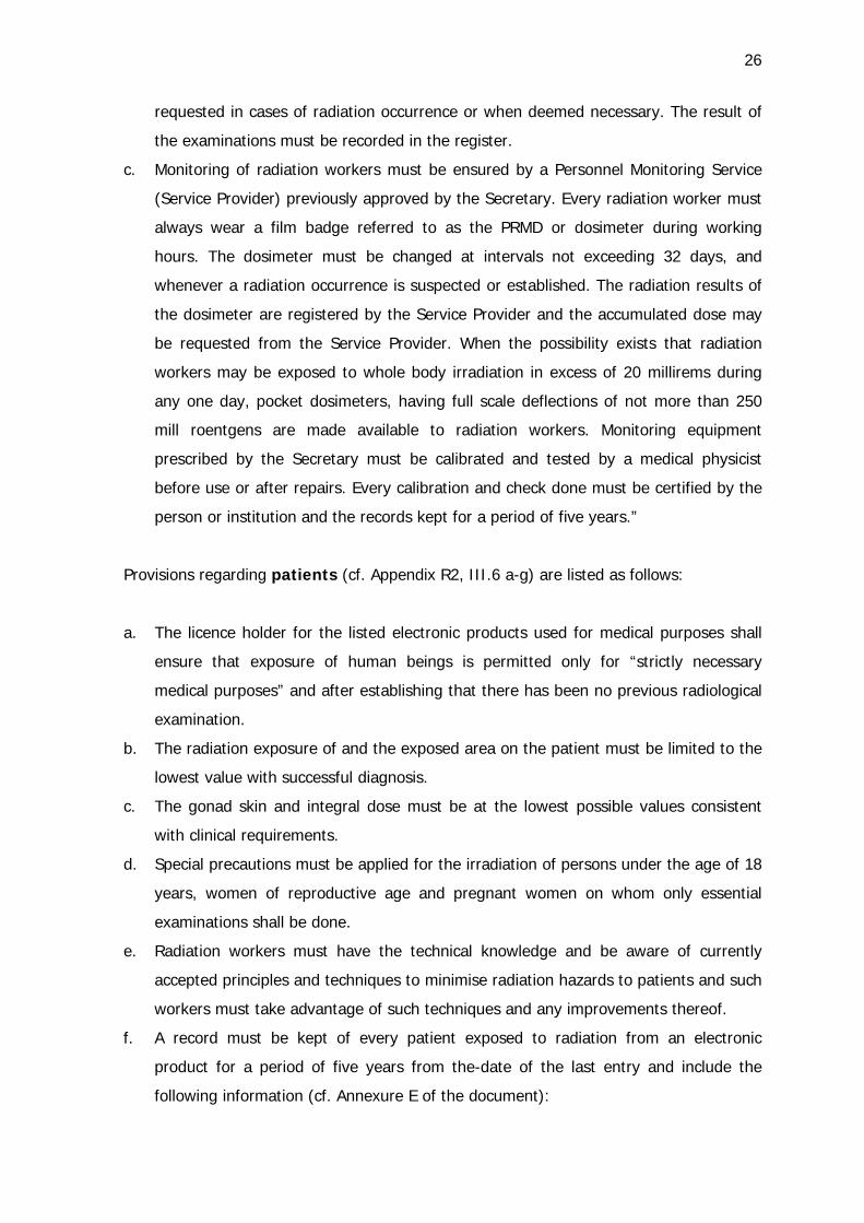
26
requested in cases of radiation occurrence or when deemed necessary. The result of
the examinations must be recorded in the register.
c. Monitoring of radiation workers must be ensured by a Personnel Monitoring Service
(Service Provider) previously approved by the Secretary. Every radiation worker must
always wear a film badge referred to as the PRMD or dosimeter during working
hours. The dosimeter must be changed at intervals not exceeding 32 days, and
whenever a radiation occurrence is suspected or established. The radiation results of
the dosimeter are registered by the Service Provider and the accumulated dose may
be requested from the Service Provider. When the possibility exists that radiation
workers may be exposed to whole body irradiation in excess of 20 millirems during
any one day, pocket dosimeters, having full scale deflections of not more than 250
mill roentgens are made available to radiation workers. Monitoring equipment
prescribed by the Secretary must be calibrated and tested by a medical physicist
before use or after repairs. Every calibration and check done must be certified by the
person or institution and the records kept for a period of five years.”
Provisions regarding patients (cf. Appendix R2, III.6 a-g) are listed as follows:
a. The licence holder for the listed electronic products used for medical purposes shall
ensure that exposure of human beings is permitted only for “strictly necessary
medical purposes” and after establishing that there has been no previous radiological
examination.
b. The radiation exposure of and the exposed area on the patient must be limited to the
lowest value with successful diagnosis.
c. The gonad skin and integral dose must be at the lowest possible values consistent
with clinical requirements.
d. Special precautions must be applied for the irradiation of persons under the age of 18
years, women of reproductive age and pregnant women on whom only essential
examinations shall be done.
e. Radiation workers must have the technical knowledge and be aware of currently
accepted principles and techniques to minimise radiation hazards to patients and such
workers must take advantage of such techniques and any improvements thereof.
f. A record must be kept of every patient exposed to radiation from an electronic
product for a period of five years from the-date of the last entry and include the
following information (cf. Annexure E of the document):
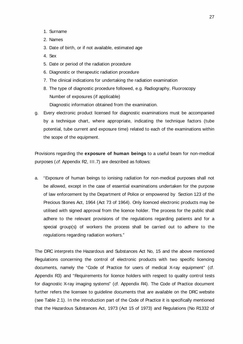
27
1. Surname
2. Names
3. Date of birth, or if not available, estimated age
4. Sex
5. Date or period of the radiation procedure
6. Diagnostic or therapeutic radiation procedure
7. The clinical indications for undertaking the radiation examination
8. The type of diagnostic procedure followed, e.g. Radiography, Fluoroscopy
Number of exposures (if applicable)
Diagnostic information obtained from the examination.
g. Every electronic product licensed for diagnostic examinations must be accompanied
by a technique chart, where appropriate, indicating the technique factors (tube
potential, tube current and exposure time) related to each of the examinations within
the scope of the equipment.
Provisions regarding the exposure of human beings to a useful beam for non-medical
purposes (cf. Appendix R2, III.7) are described as follows:
a. “Exposure of human beings to ionising radiation for non-medical purposes shall not
be allowed, except in the case of essential examinations undertaken for the purpose
of law enforcement by the Department of Police or empowered by Section 123 of the
Precious Stones Act, 1964 (Act 73 of 1964). Only licenced electronic products may be
utilised with signed approval from the licence holder. The process for the public shall
adhere to the relevant provisions of the regulations regarding patients and for a
special group(s) of workers the process shall be carried out to adhere to the
regulations regarding radiation workers.”
The DRC interprets the Hazardous and Substances Act No, 15 and the above mentioned
Regulations concerning the control of electronic products with two specific licencing
documents, namely the “Code of Practice for users of medical X-ray equipment” (cf.
Appendix R3) and “Requirements for licence holders with respect to quality control tests
for diagnostic X-ray imaging systems” (cf. Appendix R4). The Code of Practice document
further refers the licensee to guideline documents that are available on the DRC website
(see Table 2.1). In the introduction part of the Code of Practice it is specifically mentioned
that the Hazardous Substances Act, 1973 (Act 15 of 1973) and Regulations (No R1332 of
28
3 August 1973) govern the safe use of medical X-ray equipment in South Africa, excluding
dental and veterinary use. The Requirements from the Act and Regulations are
incorporated in the Code of Practice (cf. Appendix R3). The two DRC documents will be
described in the following section, starting with the Code of Practice.
2.3 CODE OF PRACTICE FOR USERS OF MEDICAL X-RAY EQUIPMENT (DRC 2011)
The researcher includes the Code of Practice document as part of this thesis for
information purposes in order for the reader to be acquainted with the current
interpretation of DRC regarding the Hazardous and Substances Act as well as Regulation
1332. The reader is reminded that this thesis excludes the current debate (cf. 2.1)
regarding the validity of the documents (Herbst and Fick 2012:50,53). The researcher
however, acknowledges three notes presented in the Code of Practice that indicate
certain stipulations of Regulation III.4 (b) and (c) are no longer required. According to the
notes of the Code of Practice, licence holders are no longer required to submit form
RC008 to the DoH (cf. 2.3.3 Paragraph 6.5), or required to inform the DoH of any change
in the register (cf. 2.3.3 Paragraph 6.8), or to submit radiation workers to annual medical
examinations (cf. 2.3.3 Paragraph 6.6).
The Code of Practice document will be consequently presented in the following sequence:
licencing and responsible person, monitoring the radiation worker, the radiographer as
radiation worker, monitoring of the patient, premise requirements and guideline
documents. The content of the Code of Practice, Act and regulations were incorporated in
the Delphi criteria list (cf. Appendix B1) to ascertain from a panel of experts the learning
outcomes of training in radiation safety for radiographers.
Whenever compliance with a requirement in the Code of Practice is commanded the word
must / shall is used. The word should indicate a practice that is commended, but not
obligatory (cf. Appendix R3).
2.3.1 Licencing and the responsible person
The licencing of X-ray units and responsibilities of the licence holder and responsible
person (DRC 2011:3-8) are described as follows:
29
Paragraph 3.1: “A joint product and premises licence must be obtained for X-ray
equipment before it may be installed and commissioned. Licences are not transferable
and are issued to a specific person or institution - for specific equipment and its
application, and for a specific premise. The licences are issued subject to the Regulations
concerning the Control of Electronic Products and the application of specific conditions.
Licence holders must verify the accuracy of the information displayed on the licence
issued and communicate any inaccuracies to the DOH.”
Paragraph 3.2: “It is the responsibility of the prospective user of an X-ray unit to
be in possession of a licence from DoH prior to installation of the unit. The document,
namely the RC-DEALER form, must be completed by both the supplier and the end user of
a new X-ray unit. The prospective user must obtain the RC-DEALER form from the
supplier of the X-ray unit. The section applicable to the user must be completed and the
form returned to the supplier. The supplier is responsible to submit the completed form to
DoH for processing. Form RC001 must be completed for pre-owned units and it is the
responsibility of the prospective user to submit the completed application form RC001 to
the DoH. The user must allow 30 days for the processing of applications.”
Paragraph 3.3: “The installation of an X-ray unit may only start after a licence to install
the unit has been issued.”
Paragraph 3.4: “Only Inspection Bodies (IP) approved by the DoH may perform
acceptance tests on X-ray equipment. A list of approved Inspection Bodies and the
scope of the licences are available on the DoH website.”
Paragraph 3.5: “When a new unit is installed, acceptance tests must be performed by
the supplier of the X-ray unit and the results recorded on the prescribed form and filed in
the Individual IER of the unit.”
Paragraph 3.6: “The prospective user of a pre-owned unit must ensure that acceptance
tests are performed. Granting of a licence to use a unit is subject to submission of the
results of the tests to DOH. Even when an existing licenced unit is moved to a new
building or room, prior to use, acceptance tests must be performed on the unit and the
results submitted to the DoH.”
30
According to Paragraph 3.7 the licence holder must apply for and obtain permission from
the DoH by submitting a completed form RC002 indicating particulars prior to
cancellation, modification, disposal and/or sale of X-ray equipment before cancellation
of the licence will be effected.
Paragraph 3.8: “The licence holder must apply for and obtain permission prior to
modification of any licensed premises or layout of equipment on such premises. “
Paragraph 3:9: “The licence holder must appoint a responsible person that has
adequate knowledge and experience in the field of radiation protection in general. The
appointed person is responsible to the licence holder for the safe use of the X-ray
equipment.”
Paragraph 3.10: “The responsible person appointed must be qualified in either of the
following categories and registered with the HPCSA: Radiography; Radiology; Medical
Physics, or Chiropractic.”
Paragraph 3.11: “The responsible person must be appointed in writing indicating the
scope of the actions delegated by the licence holder.”
Paragraph 3.11: “The licence holder must notify the DoH of a change in responsible
person by submitting a completed form RC002.”
Paragraph 4.1: “The licence holder of a diagnostic X-ray facility is ultimately responsible
for the entire scope of radiation safety, for the equipment and premises for which
he/she holds a licence; fulfilment of all related statutory requirements, and compliance to
conditions specified in the licence.”
31
Paragraph 4.1: The licence holder / responsible person must adhere to the following
responsibilities:
• “Ensure that the equipment and the facilities, in which such equipment is installed
and used, meet all applicable radiation safety standards;
• Ensure the equipment is maintained and functions properly;
• Ensure the equipment is used and maintained only by competent and appropriately
trained persons / personnel;
• Ensure that the applicable Quality Control (QC) tests are performed at the prescribed
frequencies as stipulated in the Requirements for quality control tests document;
• The required QC equipment is provided;
• Ensure that radiation surveys to monitor safe performance of equipment and to
monitor radiation levels in work areas are undertaken;
• Identify radiation workers (occupationally exposed persons) and issue with PRMDs;
• Provide the appropriate protective clothing, devices and equipment to personnel and
ensure these are properly used;
• Communicate radiation safety rules to all personnel and make sure they are followed;
• Establish and maintain operational procedures to ensure that the radiation exposure
to workers, patients and the public is kept ALARA without compromising the
diagnostic efficiency of the result;
• Educate workers in the hazards and risks of ionising radiation.”
2.3.2 The radiographer as radiation worker
The radiographer as radiation worker (DRC 2011:10-11) is described as follows:
Paragraph 5.1: “Only persons, who are appropriately trained and registered with the
HPCSA in Radiography and/or Radiology and/or Chiropractor may operate X-ray
equipment and perform examinations within their appropriate scope of practice.
Supplementary Diagnostic Radiographers (SDRs) may only work in a Government hospital
or an institution operated or subsidised by government or the provincial authority or by
the South African Chamber of Mines. SDRs must be supervised, at least once a week,
by a qualified registered radiographer. “
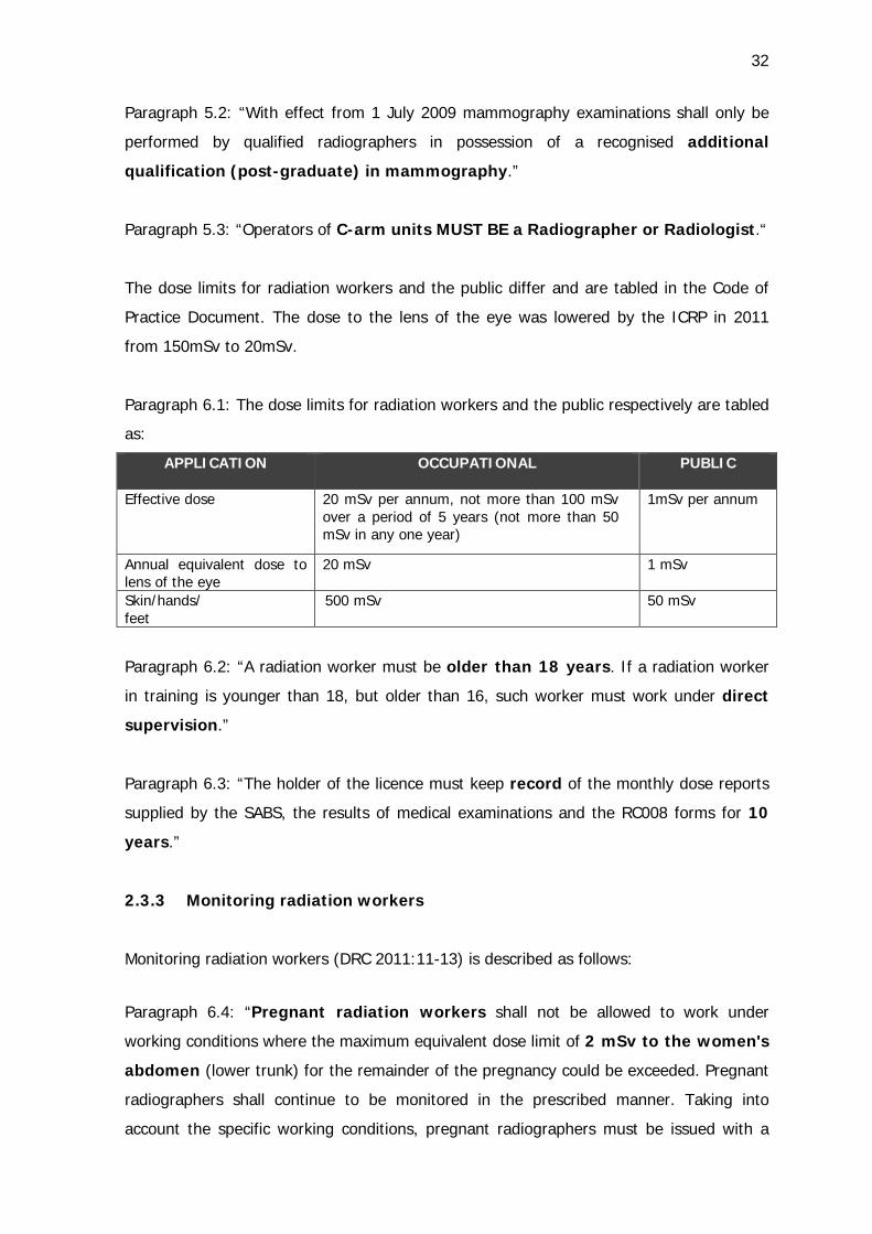
32
Paragraph 5.2: “With effect from 1 July 2009 mammography examinations shall only be
performed by qualified radiographers in possession of a recognised additional
qualification (post-graduate) in mammography.”
Paragraph 5.3: “Operators of C-arm units MUST BE a Radiographer or Radiologist.“
The dose limits for radiation workers and the public differ and are tabled in the Code of
Practice Document. The dose to the lens of the eye was lowered by the ICRP in 2011
from 150mSv to 20mSv.
Paragraph 6.1: The dose limits for radiation workers and the public respectively are tabled
as:
APPLICATION OCCUPATIONAL PUBLIC
Effective dose 20 mSv per annum, not more than 100 mSv over a period of 5 years (not more than 50 mSv in any one year)
1mSv per annum
Annual equivalent dose to lens of the eye
20 mSv 1 mSv
Skin/hands/ feet
500 mSv 50 mSv
Paragraph 6.2: “A radiation worker must be older than 18 years. If a radiation worker
in training is younger than 18, but older than 16, such worker must work under direct
supervision.”
Paragraph 6.3: “The holder of the licence must keep record of the monthly dose reports
supplied by the SABS, the results of medical examinations and the RC008 forms for 10
years.”
2.3.3 Monitoring radiation workers
Monitoring radiation workers (DRC 2011:11-13) is described as follows:
Paragraph 6.4: “Pregnant radiation workers shall not be allowed to work under
working conditions where the maximum equivalent dose limit of 2 mSv to the women's
abdomen (lower trunk) for the remainder of the pregnancy could be exceeded. Pregnant
radiographers shall continue to be monitored in the prescribed manner. Taking into
account the specific working conditions, pregnant radiographers must be issued with a
33
direct reading pocket alarm dosimeter, in so doing preventing that such women are
unwittingly exposed to radiation. The employer should provide continuous education as to
the risks to the foetus and actual dose levels in the various working environments.”
Paragraph 6.5: “The appointment of radiation workers is implemented by completing and
filing form RC008 in the licence holder’s register. Note: Licence holders are no longer
required to submit form RC008 or inform the DoH of any change in the register as
stipulated in Regulation III.4 (b) & (c).”
Paragraph 6.6: “Before any person is appointed as a radiation worker, he/she must
undergo a general occupational medical examination for determining fitness for
work. When a radiation occurrence / incident resulting in an abnormally high dose is
suspected to have taken place or has been confirmed, a medical examination may be
required as well as in cases when a practitioner or regulatory authority of the licence
holder considers it necessary. The radiation worker may also undergo a medical
examination when the worker suspects that his/her health has been, or will be adversely
affected by occupational factors. Note: Annual medical examinations are no longer
required by the DoH, but it remains the prerogative of the licence holder, should he/she
deems it necessary.”
Paragraph 6.7: “The licence holder must ensure that all radiation workers are issued with
a PRMD (Personal Radiation Monitoring Device). The contact details of the service
provider, the SABS (South African Bureau of Standards) are provided in the document.
The service provider will provide the radiation dose records to the licence holder on a
monthly basis or after a radiation occurrence. The dose records must be kept for
10 years. The licence holder must ensure that the service provider replaces PRMDs at
regular intervals not exceeding 32 days.”
Paragraph 6.8: “Termination of employment as a radiation worker requires that the
licence holder must ensure that the form RC008 is completed in duplicate, a copy given to
the radiation worker. The records must be preserved for a period of 10 years. Note:
Licence holders are no longer required to inform the DoH of any change in the register as
stipulated in Regulation III.4 (c).”
Paragraph 6.9: “On appointment of radiation workers by a new employer, the licence
holder must obtain the form RC008, completed by the previous employer, from the
radiation worker.”
34
Paragraph 6.10: “Radiation occurrences must be instantly reported to the Director:
Radiation Control on form RC010.”
2.3.4 Monitoring the patient
Monitoring the patient (DRC 2011:13-17) is described as follows:
Paragraph 7: “Basic radiation protection principles are based on the justification of the
practice (benefit outweighs the associated risk), optimisation of protection (radiation
doses from medical exposures must be kept ALARA), and limitation of individual dose
and risk (radiation doses must not exceed the specified dose limits).”
Paragraph 7.1: All medical exposures should be subject to the principles of justification
and optimisation.
• “X-ray examinations shall not be performed unless there are valid clinical
indications.
• Examinations on children shall require a higher justification, since such patients
may be more sensitive to radiation.
• Obtain previous X-ray images to minimise the taking of repeat films.
• CT Screening programmes of asymptomatic persons shall not be instituted unless
approved by the DoH.
• Screening programmes prescribed by other Governmental Acts are
acceptable.
• Licence holders should be aware of the approximate patient radiation doses.
Diagnostic reference levels (DRL) should be introduced for applications in
diagnostic X-ray examinations as performed in their facilities and not exceeded. The
specific purpose of the DRL is to provide a benchmark for comparison, not to define a
maximum or minimum exposure limit.
• When appropriate, consider other modalities, such as MRI or ultrasound which do
not use ionising radiation.
• Examinations with potential high patient doses such as CT examinations should only
be carried out after a proper clinical justification by the radiologist.
• For each projection, select the highest kilovoltage (KV) and fastest film-screen
combination compatible with the image quality requirements of the examination.
• The primary beam shall be collimated at all times.
35
• Means to permanently transfer patient identification, prior to processing of the
images, must be provided.
• Radiation examinations may only be requested by a medical practitioner or any
appropriately trained and registered health professional. Reference is made to the
guideline document A.4.
• Pulsed fluoroscopy should be routinely used. During fluoroscopy, radiation time is
minimised by pulsing the beam. At low-pulsed frequencies, major dose savings are
made while ensuring diagnostic images.
• A film screen speed of not less than 300 is recommended for general
radiography.”
Paragraph 7.2: “Examinations of pregnant patients must be justified and only essential
views performed. Alternative imaging modalities, especially ultrasound for obstetric
procedures, shall be used where appropriate. An X-ray examination shall not be
performed to assess foetal development where ultrasound facilities are available. X-ray
pelvimetry shall not be performed on a routine basis. For examinations where the
primary beam unavoidably irradiates the foetus, the methods of minimising dose shall be
used, as appropriate and particular attention shall be given to: minimising the number of
views; beam collimation; higher kVp settings; fast image recording media and using PA
projections in preference to AP projections.”
Paragraph 7.3: “The protection of women of reproductive capacity shall be compulsory
unless there are strong clinical indications for the examination. Notices must be
posted at several places within the radiology facility to minimise the possibility of
unintentional exposure to the embryo / foetus.”
Paragraph 7.4: “The longer life expectancy of children results in greater potential for
the manifestation of possible harmful effects of radiation. For a given procedure each
view shall be examined, where practical, before deciding whether to take a further view;
fluoroscopy shall be used only when general radiography will not provide the information
required and there shall be strong justification for X-ray procedures involving high doses
such as CT.”
Paragraph 7.5: “Members of the public are not allowed to enter controlled areas
unsupervised or remain in the X-ray room during any X-ray procedure unless they are
36
required to be in attendance. The occasional use of non–radiation personnel to give
assistance, particularly in ward or theatre radiography, is acceptable but shall involve the
full use of protective clothing, devices and techniques to minimise personnel dose. Care
shall be taken to ensure that the same non-radiation personnel are not always involved.
Women who are pregnant shall not be used in this role.”
Paragraph 7.6: “No person shall hold a patient, X-ray film cassette, or other imaging
equipment or X-ray tube head in position during exposures unless it is otherwise
impossible to obtain a diagnostically useful image and not merely because it is a matter of
convenience. Holding of patients or X-ray film cassettes during exposure shall be done by
persons accompanying the patient in preference to non-radiation personnel; and by
non-radiation personnel in preference to radiation workers. Non-radiation personnel
should be chosen on the basis of a roster, i.e. it shall not be the same person who does
the holding. No pregnant women or young persons (under the age of 18) shall do any
holding. Any persons holding patients or film cassettes in position during an X-ray
examination shall wear a lead rubber apron and wherever practicable, lead rubber gloves.
No part of the holder’s body shall be in the primary beam, even if covered with
protective clothing.”
Paragraph 7.7: “Any person who cannot remain in the protected area during X-ray
examinations shall wear a protective apron of at least 0,25mm lead equivalence.
Any person standing within 1 metre of the X-ray tube or patient when the X-ray machine
is operated at tube voltages above 100KV should wear a protective apron of at least
0.35mm lead equivalence. Protective gloves should be at least 0.35mm lead equivalence.
Gonad shields shall be at least 0.5mm lead equivalence.”
2.3.5 Premises requirements
The requirements for premises (DRC 2011:17) are described as follows:
Paragraph 8: “General Guidelines with regard to the design of X-ray rooms (see
Annexure A.6 in this Code).”
Paragraph 9.1:”Radiation warning signs and notices must be displayed. Fixed rooms:
“X-RAYS - NO UNAUTHORISED ENTRY” must be displayed at all entrances leading to the
rooms where X-ray units are installed. Mobile units: the sign must be displayed on the
control panel of the X-ray units. A red warning light, which is only activated when the
37
beam is on and when fluoroscopy is in progress, must be mounted in an obvious place
outside the entrance to the X-ray rooms.”
2.3.6 Guideline and reference documents
The reference to the guideline documents (DRC 2011:18) is provided in the Code of
Practice by means of a website link:
https://sites.goggle.com/site/radiationcontroldoh/
The guideline documents are listed as follows:
• Diagnostic QC – Requirements for licence holders with respect to Quality Control tests
for diagnostic X-ray imaging systems.
• Management of pregnant radiographers and other staff members.
• Personal monitoring when a lead rubber apron is worn – medical and veterinary use
of X-ray equipment.
• Request for medical examinations.
• FDA Public Health Notification: Reducing radiation risk from computed tomography
for paediatric and small adult patients – 2 November 2001.
• General guidelines with regard to the design of X-ray rooms.
• Display and format of radiation warning signs at entrances to rooms containing X-ray
units.
• Guidelines – Protective Clothing.
• Patient dose measurements in Diagnostic Radiography.
• Radiation protection of personnel in theatre.
The above list with the heading “10 ANNEXURE A” differs from the documents provided
on the website (mentioned above) of the DRC. The researcher however, included not
only the above ten documents but all the documents on the website (cf. Table 2.1) as
criteria in the Delphi questionnaire in order to create comprehensive training for the
radiography students in radiation safety. It is also reiterated that paragraph 6.5, 6.7 and
6.8 in the Code of Practice contain notes to indicate that Regulation III.4 are no longer
applicable. The second document as published by the DRC, focusing on quality tests, will
be described next.
2.4 REQUIREMENTS FOR LICENCE HOLDERS WITH RESPECT TO QUALITY
CONTROL TESTS FOR DIAGNOSTIC X-RAY IMAGING SYSTEMS
(DRC 2012)
38
The DRC published a second document that is provided as part of the diagnostic licence
conditions with respect to quality control (QC) tests in order to outline the requirements
for the acceptance- and quality control tests of diagnostic X-ray equipment. As from
31 March 2009 an Inspection Body (IB), approved by the Department of Health (DoH) or
an appropriately trained professional registered with the HPCSA as a medical physicist,
must be used to perform all the acceptance tests as well as the routine tests listed in
section III.2 of Table 2 in the Requirements for Quality Control Tests Document (cf. Table
2.2). If a medical physicist is used to perform the tests, an IB approved by the DoH must
formally contract such person(s). Formally contracted means that the medical physicist is
contracted by the IB to perform the tests.
Acceptance tests are the initial tests performed directly after installation and before any
X-ray equipment is put into clinical service or substantially upgraded. Acceptance tests
have three purposes, namely to:
1. ensure that the unit meets stated specifications;
2. establish baseline parameters for the future quality control programme,
3. familiarise the customer with operation of the unit.
The entire Requirements for Quality Control Tests Document (27 pages) will not be
included in this thesis, but is available for reference as Appendix R4. The significant
aspects, however, will be described in order to reiterate the importance of radiographers
being not only familiar with the requirements, but also equipped with the knowledge and
skills to perform the routine tests. It is important that the radiographer will be able to
interpret the tests and adjust necessary parameters, so that the ALARA principle can be
applied and maintained on a daily basis.
The significant aspects of these Requirements for Quality Control Tests Document include
the following:
The licence holder must acquire the relevant quality control manuals or compile in-house
written protocols, which describe each test step by step to ensure that QC tests listed
in the Requirements for Quality Control Tests Document, marked as Section III.1, are
correctly performed. The licence holder must ensure that persons that perform routine
tests in Section III.1 are competent to execute the tests. The quality control tests are
influenced by the age, stability, make, model, etc., of the equipment, but must be
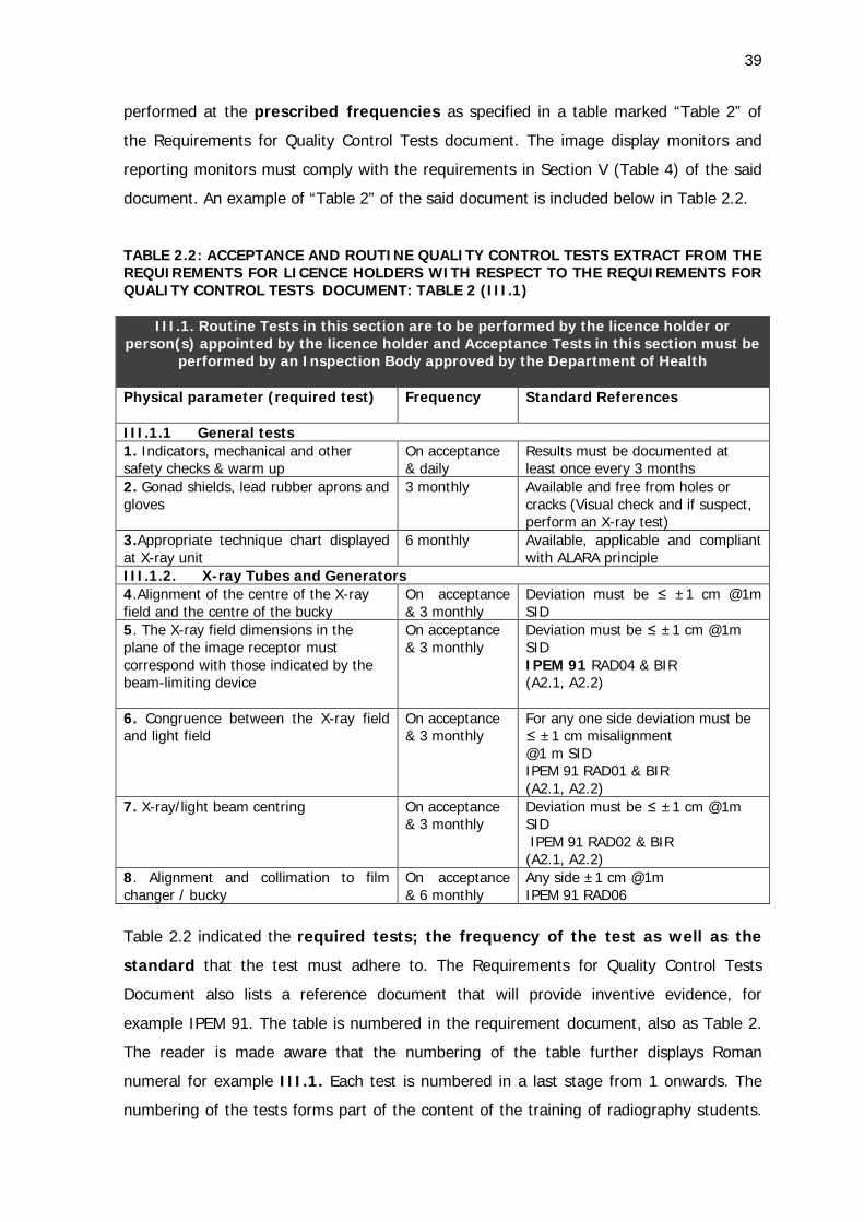
39
performed at the prescribed frequencies as specified in a table marked “Table 2” of
the Requirements for Quality Control Tests document. The image display monitors and
reporting monitors must comply with the requirements in Section V (Table 4) of the said
document. An example of “Table 2” of the said document is included below in Table 2.2.
TABLE 2.2: ACCEPTANCE AND ROUTINE QUALITY CONTROL TESTS EXTRACT FROM THE REQUIREMENTS FOR LICENCE HOLDERS WITH RESPECT TO THE REQUIREMENTS FOR QUALITY CONTROL TESTS DOCUMENT: TABLE 2 (III.1)
III.1. Routine Tests in this section are to be performed by the licence holder or person(s) appointed by the licence holder and Acceptance Tests in this section must be
performed by an Inspection Body approved by the Department of Health Physical parameter (required test) Frequency Standard References
III.1.1 General tests 1. Indicators, mechanical and other safety checks & warm up
On acceptance & daily
Results must be documented at least once every 3 months
2. Gonad shields, lead rubber aprons and gloves
3 monthly Available and free from holes or cracks (Visual check and if suspect, perform an X-ray test)
3.Appropriate technique chart displayed at X-ray unit
6 monthly
Available, applicable and compliant with ALARA principle
III.1.2. X-ray Tubes and Generators 4.Alignment of the centre of the X-ray field and the centre of the bucky
On acceptance & 3 monthly
Deviation must be ≤ ±1 cm @1m SID
5. The X-ray field dimensions in the plane of the image receptor must correspond with those indicated by the beam-limiting device
On acceptance & 3 monthly
Deviation must be ≤ ±1 cm @1m SID IPEM 91 RAD04 & BIR (A2.1, A2.2)
6. Congruence between the X-ray field and light field
On acceptance & 3 monthly
For any one side deviation must be ≤ ±1 cm misalignment @1 m SID IPEM 91 RAD01 & BIR (A2.1, A2.2)
7. X-ray/light beam centring On acceptance & 3 monthly
Deviation must be ≤ ±1 cm @1m SID IPEM 91 RAD02 & BIR (A2.1, A2.2)
8. Alignment and collimation to film changer / bucky
On acceptance & 6 monthly
Any side ±1 cm @1m IPEM 91 RAD06
Table 2.2 indicated the required tests; the frequency of the test as well as the
standard that the test must adhere to. The Requirements for Quality Control Tests
Document also lists a reference document that will provide inventive evidence, for
example IPEM 91. The table is numbered in the requirement document, also as Table 2.
The reader is made aware that the numbering of the table further displays Roman
numeral for example III.1. Each test is numbered in a last stage from 1 onwards. The
numbering of the tests forms part of the content of the training of radiography students.
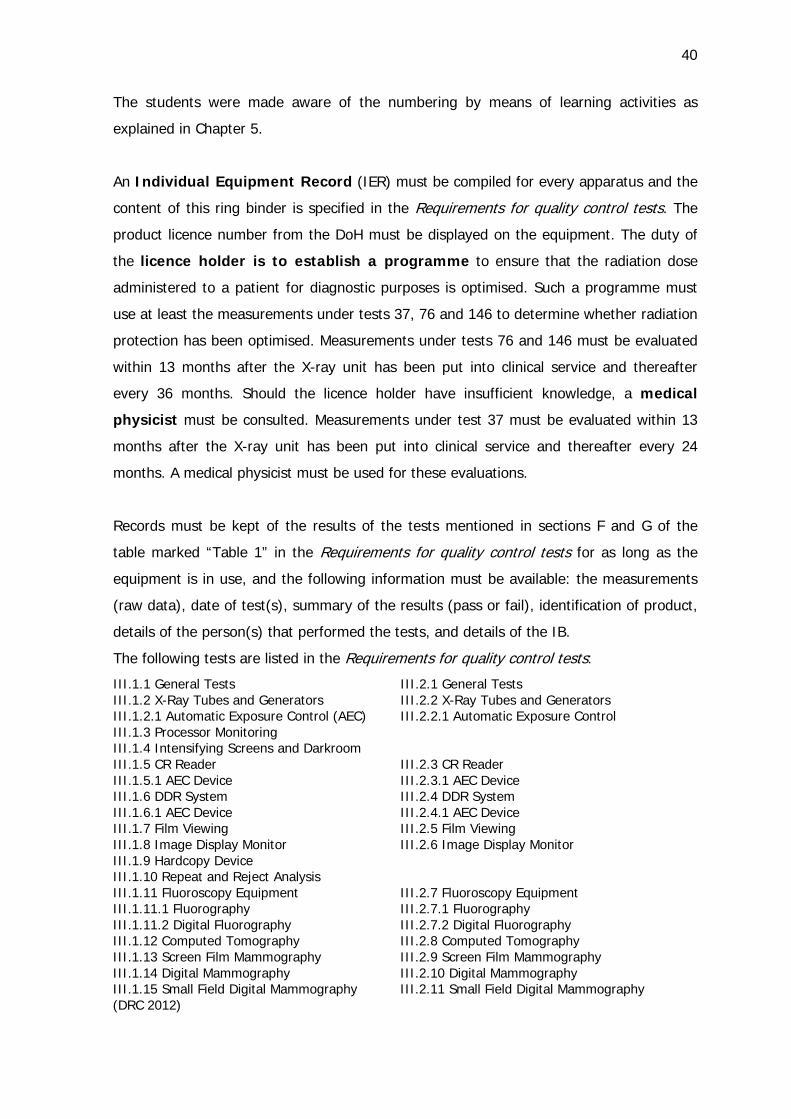
40
The students were made aware of the numbering by means of learning activities as
explained in Chapter 5.
An Individual Equipment Record (IER) must be compiled for every apparatus and the
content of this ring binder is specified in the Requirements for quality control tests. The
product licence number from the DoH must be displayed on the equipment. The duty of
the licence holder is to establish a programme to ensure that the radiation dose
administered to a patient for diagnostic purposes is optimised. Such a programme must
use at least the measurements under tests 37, 76 and 146 to determine whether radiation
protection has been optimised. Measurements under tests 76 and 146 must be evaluated
within 13 months after the X-ray unit has been put into clinical service and thereafter
every 36 months. Should the licence holder have insufficient knowledge, a medical
physicist must be consulted. Measurements under test 37 must be evaluated within 13
months after the X-ray unit has been put into clinical service and thereafter every 24
months. A medical physicist must be used for these evaluations.
Records must be kept of the results of the tests mentioned in sections F and G of the
table marked “Table 1” in the Requirements for quality control tests for as long as the
equipment is in use, and the following information must be available: the measurements
(raw data), date of test(s), summary of the results (pass or fail), identification of product,
details of the person(s) that performed the tests, and details of the IB.
The following tests are listed in the Requirements for quality control tests:
III.1.1 General Tests III.2.1 General Tests III.1.2 X-Ray Tubes and Generators III.2.2 X-Ray Tubes and Generators III.1.2.1 Automatic Exposure Control (AEC) III.2.2.1 Automatic Exposure Control III.1.3 Processor Monitoring III.1.4 Intensifying Screens and Darkroom III.1.5 CR Reader III.2.3 CR Reader III.1.5.1 AEC Device III.2.3.1 AEC Device III.1.6 DDR System III.2.4 DDR System III.1.6.1 AEC Device III.2.4.1 AEC Device III.1.7 Film Viewing III.2.5 Film Viewing III.1.8 Image Display Monitor III.2.6 Image Display Monitor III.1.9 Hardcopy Device III.1.10 Repeat and Reject Analysis III.1.11 Fluoroscopy Equipment III.2.7 Fluoroscopy Equipment III.1.11.1 Fluorography III.2.7.1 Fluorography III.1.11.2 Digital Fluorography III.2.7.2 Digital Fluorography III.1.12 Computed Tomography III.2.8 Computed Tomography III.1.13 Screen Film Mammography III.2.9 Screen Film Mammography III.1.14 Digital Mammography III.2.10 Digital Mammography III.1.15 Small Field Digital Mammography III.2.11 Small Field Digital Mammography (DRC 2012)
41
The radiography student is the potential licence holder of X-ray equipment and must be
familiar with the content of the regulations for quality testing. Radiographers are
responsible for the routine quality tests of X-ray equipment on a daily basis and should be
equipped with the skills to execute the tests. It should, however, not only be a knowledge
exercise, but the radiographer must be armed with contextual competencies to interpret
the test results. The radiographer must be familiar with the regulations in order to
delegate responsibilities by consulting physicists in the quality control of X-ray equipment.
Education and training in regulations and safe practices are therefore imperative.
2.5 STANDARDISED TRAINING FOR RADIATION SAFETY
The ICRP acknowledges the importance of education and training in reducing patient
doses while maintaining image quality. Training must be considered at different levels;
not only for entry users but also for retraining and certification. The European perspective
mentioned earlier, operates within the framework of the Directive on Medical Exposures.
The directive assures that the Member States of the European Union shall establish
curricula to certify competence in radiation protection (Vano 2010:201). This urgent trend
to accredit radiation protection curricula confirms that the training developed in this thesis
is current and relevant for South Africa.
The importance of justification of radiological procedures should be highlighted for every
procedure involving ionising radiation, and the use of imaging modalities that are non-
ionising should always receive priority. The basic aim of optimisation of radiological
protection is to obtain diagnostic images with the lowest possible radiation dose. This can
be obtained by fine-tuning imaging parameters and the establishing protective measures.
New imaging equipment should be purchased with utmost care, for instance, in terms of
paediatric use. Fluoroscopic techniques such as patient positioning, field size and
adequate collimation can lower the dose and a second, specific level of training in
radiological protection is desirable, in some countries, this is mandatory (ICRP 2013). The
radiographer must be informed about all the measures as set forth by the ICRP and not
only as deemed important by individual lecturers or preferred by different training
institutions. The safety training for radiography students at the CUT has the potential to
address the mastery of the implementation and application of the above mentioned
radiation protection principles.
42
The licence holder of medical X-ray equipment is responsible for the following education
of radiation workers as indicated by the Code of Practice (DRC 2011:8):
• “Radiation workers (occupationally exposed persons) are identified and issued with
personal radiation monitoring devices (PRMDs);
• The appropriate protective clothing, devices and equipment is provided to personnel
and properly used;
• Radiation safety rules are communicated to and followed by all personnel;
• Operational procedures are established and maintained to ensure that the radiation
exposure to workers, patients and public is kept as low as reasonably achievable
(ALARA) without compromising the diagnostic efficiency of the result;
• Workers are educated in the hazards and risks of ionising radiation.”
The Code of Practice obligation is that every licence holder must ensure that the radiation
workers are educated in the above-mentioned aspects. The radiography graduate as
licence holder must therefore be equipped with knowledge as well as the ability to apply
the principles of radiation safety. This study supported the Code of Practice by
determining the specific outcomes to satisfy the guidelines in order to develop a training
module with aligned assessment. The lack of the proper implementation of the quality
tests in most clinical practices was addressed by the research and training.
The Code of Practice was compiled according the outlines published in the Government
Gazette of 1973 as regulations concerning the control of electronic products. The
regulations are focused on the licence holder of medical equipment. The responsibility of
the licence holder of medical equipment is to be held liable for offences committed under
this act unless he/she can prove that it was not committed with his/her authority, or
planned. The licence holder must prove that he/she took measures to implement the
regulations and that any omission “whether legal or illegal, of the nature in question did
not under any conditions or in any circumstances fall within the course of the work or the
scope of the authority of the person concerned” (RSA DoH Regulation 1332: III.3).
The licence holder of medical X-ray equipment shall be liable for the all-inclusive aspects
of radiation protection on the premises under his licence. The specific aspects include:
1. “effective protection, organisation and continual conscientious regard for optimum
methods of working with particular reference to routine operations;
43
2. technical investigations to ensure reliability and overall technical excellence of
equipment, buildings and interlocks;
3. the display of appropriate warning signs or notices which are easily intelligible to all
persons, at the entrances to or at appropriate places in all areas where persons may
enter and may be exposed to ionising radiation;
4. ensuring that radiation workers and members of the public are subjected to minimal
risks from radiation exposure and that the maximum permissible doses and dose
limits are not exceeded” (RSA DoH Regulation 1332: III.3).
The licence applicant has knowledge and or experience regarding the basic principles of
radiation protection in general; as well as specific aspects of radiation protection as
related to the equipment under his licence conditions. If a person is unable to fulfil the
requirements in terms of knowledge and experience, the licensee may appoint a
responsible person. This person may be a medical physicist or any other person to fulfil
this role. The responsible person must be appointed by the licensee in writing (RSA DoH
Regulation 1332:c). Currently it is assumed that qualified radiographers are
knowledgeable regarding radiation safety and will be able to act as licence holders or
responsible persons and to implement quality testing. However, a deficiency in the
application of radiation safety in clinical practice was observed, with the excuse of
radiographers that claimed that they had not learned about the QC tests during their
training. The quality control tests currently are assigned to only a few individuals in most
practices where CUT students are placed. The radiography students who graduate and
have to do community service for a year are not necessarily ready to manage a
department as a person responsible for radiation safety independently, although in many
instances this is the case. This research aimed to empower the radiography graduate with
the knowledge pertaining to radiation safety regulations, not only a limited awareness.
This establishment of knowledge is subjective, but the training module with proof of
successful completion of the tests may assist to provide proof of radiation protection
knowledge for radiographers.
2.5.1 Diploma in Radiography transformed to bachelor status
Radiography is offered at various HEIs in South-Africa as a three-year diploma course,
namely the Central University of Technology (CUT), Cape Peninsula University of
Technology (CPUT), Durban University of technology (DUT), University of Johannesburg
44
(UJ) and University of Pretoria (UP). The radiography curriculum currently includes
principles of radiation protection and some of the quality control tests are dealt with over
the three years training in different learning areas. The student is presumably exposed to
most of the regulations. The observance in clinical practice of the lack of implementation
and application of the ALARA principles plus the inability of radiography graduates to
perform all the prescribed quality tests, pressed the researcher to believe the contrary. In
2014 the CUT and CPUT were the first two institutions to receive accreditation and
permission to offer the 4-year bachelor degree in Radiography (SAQA 2013). The
lecturers at the CUT had the opportunity to design the curriculum with the benefit of
hindsight to address gaps experienced in the diploma content. It was with the
researcher’s opinion - that radiation safety must be directed and assessed to leave no
room for assumption of knowledge gained - in mind that a radiation safety module was
proposed. As a result, radiation safety was incorporated as learning units in 2014 at the
CUT in the module “Radiographic Procedures”.
The implementation of radiation safety training and assessment required the content and
assessment to be designed and developed. In order to exclude the possibility of bias of
the researcher, the opinion of a panel of experts were consulted through a Delphi
process. The development and accreditation of a curriculum further are guided by
national standards that will be addressed now.
2.5.1.1 The South African Qualifications Authority (SAQA)
The South African Qualifications Authority (SAQA) stipulates that the functions of this
qualifications authority are to oversee the development of the National Qualifications
Framework (NQF) and to formulate and publish policies and criteria for registration of
bodies that are responsible for establishing standards and qualifications. SAQA thus
oversees the implementation of the NQF by registering qualifications. The Diploma in
Radiography was designed at NQF Level 6 and the Bachelor of Diagnostic Radiography
was structured as a 480-credit qualification with an exit at level 8 on the NQF. The 480-
credit bachelor’s degree at NQF level 8 has both a higher volume of learning and a
greater cognitive demand than the 360-credit degree at Level 7 (RSA DHET 2012).
SAQA accredits bodies responsible for the “monitoring and auditing of the quality of the
teaching and learning provision for the achievement of registered standards and
45
qualifications”, namely Education and Training Quality Assurance bodies (ETQAs). The
NQF is an outcomes-based education and training framework for education and training
standards and qualifications. The assessment practices and procedures for the NQF have
to be aligned with those of an outcomes-based education and training system (SAQA
2001). The specific assessment principles will be discussed in 2.5.1.6.
The purpose of the bachelor’s qualification as stipulated by the SAQA document reiterates
that the HEIs are obliged to produce graduates that will be able to function
independently. The end product (graduate) is beautifully described in the document as to
develop a competent learner who has a thorough grounding in the knowledge and
skills required for the Diagnostic Radiography profession and who has gained
experience in applying such knowledge and skills in the appropriate workplace
context. This qualification enables the learner to competently apply an integration
of theory, principles, proven techniques, practical experience and appropriate skills
to the solution of well-defined and abstract problems in the selected field of
Diagnostic Radiography. The learner should become a reflective practitioner and a
life-long learner in his or her profession, thereby benefiting the community and
society. Skills in management and research will also be demonstrated allowing the
holder of this qualification to work independently and in a supervisory capacity
within the health care team. Successful completion of this qualification will enable
the learner to register with the relevant Professional Council as a Specialist
Radiographer (SAQA 2013).
Diagnostic Radiography is identified as a scarce skill in South Africa, indispensable in both
the public and the private sectors as part of a multidisciplinary team providing a holistic
health care service. The qualification is recognised by the relevant Professional Health
Council as a requirement for registration to practise in the field of Diagnostic Radiography.
The exit-level outcomes for this qualification describe the foundational, practical and
reflexive competencies required for Diagnostic Radiography service at this level (SAQA
2013). The content of radiation safety training must accordingly adhere to the exit-level
outcomes for the qualification.
2.5.1.2 Exit-level outcomes pertaining to radiation safety
Lecturers and module designers are required to develop structured and accredited means
for the assessment of individual learners against exit-level outcomes of the qualification.
46
The Diagnostic Radiography exit-level outcomes 1 to 7 establishes the fundamental and
core components of the qualification, and together total 480 credits (SAQA 2013). The
exit-level outcomes are listed for referencing and to ensure that the importance of
radiation safety is reflected in the content of the qualification:
1. “Perform routine and specialized radiographic procedures to produce images of
diagnostic quality.
2. Access, organize and present information applicable to the radiography context in
order to record, retrieve and communicate patient data.
3. Evaluate the quality of routine and specialised radiographic images and perform
image interpretation to identify normal and abnormal appearances.
4. Plan, develop and apply total quality management appropriate to the diagnostic
radiography context.
5. Perform safe and effective patient care in accordance with the patient's needs and
departmental protocol to provide a quality service and to maintain the welfare of the
patient.
6. Apply the principles of human rights, ethics and relevant medical law which ensure
the well-being of the patient.
7. Apply the principles, specific knowledge, skills and values related to one of the
chosen electives as listed.
8. Conduct research” (SAQA 2013).
It is appropriate to highlight the exit -level outcomes pertaining to radiation safety:
Exit Level 1 requires of the learner to produce images of diagnostic quality. QC tests to
manage the optimum performance of equipment plus the knowledge of measures such as
collimation, scatter and distance to the IR are appropriately addressed in the designed
radiation safety training.
Exit Level 2 is addressed in radiation safety training since to retrieve, record and
communicate patient data, the imaging department adheres to requirements, such as the
requirements for the reporting monitors of radiologists.
Exit Level 3 requires that the quality of routine and specialised radiographic images be
optimum so that abnormal pathology can be detected. The aim of QC testing is to prove
adherence to the requirements for image quality.
47
Exit Level 4 requires that learners are guided to plan total quality management -
radiation safety and QC tests play a huge role in the management process.
Exit Level 5 demands that the learners be equipped to perform safe and effective
patient care in accordance with the patient's needs. Implementation of the ALARA
principle is foundational in radiation safety training and is aimed at maintaining the
welfare of the patient. Justification, optimisation and awareness of radiation form core
components of the training with the aim to protecting not only the staff, but also the
patient.
Exit Level 6 is honoured because the learner should be familiar with the documents and
requirements in order to apply the relevant medical law that ensures, through ALARA, the
well-being of the patient.
The critical cross-field education and training outcomes, commonly known as the critical
outcomes, are an additional mechanism through which the NQF identifies the
development in students. These outcomes will be discussed next.
2.5.1.3 Critical cross-field outcomes pertaining to radiography
Critical cross-field education and training outcomes describe the outcomes within the
education and training system, regardless of the specific learning area, that are critical for
the development of the aptitude for life-long learning. It is mandatory that qualifications
must ensure that all critical cross-field outcomes have been addressed appropriately at
the specific level within the qualification (SAQA 2000). The critical cross-field outcomes
adopted by SAQA are listed for referencing and testing of the radiation safety importance
in the content of the qualification:
• “Identify and solve problems in which responses display that responsible decisions
using critical and creative thinking have been made.
• Work effectively with others as a member of a team, group, organisation, community.
• Organise and manage oneself and one’s activities responsibly and effectively.
• Collect, analyse, organise and critically evaluate information.
• Communicate effectively using visual, mathematical and/or language skills in the
modes of oral and/or written presentation.
• Use science and technology effectively and critically, showing responsibility towards
the environment and health of others.
48
• Demonstrate an understanding of the world as a set of related systems by
recognising that problem-solving contexts do not exist in isolation.
• In order to contribute to the full personal development of each learner and the social
and economic development of the society at large, it must be the intention underlying
any programme of learning to make an individual aware of the importance of:
o Reflecting on and exploring a variety of strategies to learn more effectively;
o Participating as responsible citizens in the life of local, national and global
communities;
o Being culturally and aesthetically sensitive across a range of social contexts;
o Exploring education and career opportunities, and
o Developing entrepreneurial opportunities” (SAQA 2000).
The Bachelor of Diagnostic Radiography outlines the critical cross-field outcomes in the
following manner:
• “Identify health problems in the context of diagnostic radiography and suggest and
implement a solution or plan of action in order to solve the problem professionally -
will be promoted through effective and safe patient care practices in accordance with
the patient's needs by taking into consideration ethical principles as well as human
rights and medical law requirements.
• Perform professional duties with confidence in collaboration with other health care
professionals and where appropriate assume leadership in tasks or projects in order
to promote efficient and effective service delivery and total quality management in
the radiography profession as well as the healthcare service in general.
• Keep up with the current trends and changing needs of Diagnostic Radiography
service on a regional, national and international level by undertaking research or
fostering a research climate within the radiography profession.
• Contribute towards and facilitate continuing professional development of Diagnostic
Radiography staff by either engaging in research or fostering the research
environment and encouraging teamwork among radiographers and other healthcare
professionals.
• Communicate effectively in the learning and health care environment by
demonstrating competency and skills necessary for use of technology and associated
accessories necessary for transfer or sharing of information among healthcare
49
workers and other stakeholders so as to deliver quality patient care and facilitate
management processes.
• Use science and technology in order to improve Diagnostic Radiography practice
through efficient organizational and management skills for both patient's information
and any other information necessary for efficient healthcare service delivery.
• Demonstrate an understanding of Diagnostic Radiography principles in order to solve
practical problems within radiography will be promoted by the competent
performance of routine and specialised radiographic procedures” (SAQA 2013).
The radiation safety training contributes substantially to attaining the critical cross- field
outcomes by promoting effective and safe patient care practices to fulfil the patient's
needs by taking into consideration medical law requirements. The training further
promotes total quality management in the radiography profession by planning the
frequency and executing the QC tests. The training is in pace with international trends -
the QC testing was only implemented in 2008 (DRC 2012). The content speaks to the
absence of formal standardised radiation safety training as a result of the recent
requirement for QC tests. The radiation worker uses science (physics) and technology (QC
test equipment and software) in order to improve diagnostic radiography practice for both
patient's information and any other information. In South Africa equipment for QC tests
are not readily available, especially in government hospitals. QC tests improvisation, for
instance, forms part of the training material. The training, in conclusion, is geared to
equipping the learner to understand diagnostic radiography principles in order to solve
practical problems within radiography.
The NQF philosophy indicates that decisions in respect of learning programme design,
development, delivery and assessment need to consider the learning outcomes that
learners need to demonstrate constantly. A qualification further means that the learner
has demonstrated applied competence in specific skills and content areas (SAQA 2000).
The formulation of objectives in outcomes-based education will receive attention in the
following section.
2.5.1.4 Formulation of objectives
Lecturers must not focus on what to teach but rather on what the students learn.
Outcome-based education implies the “what” and describes how students can apply what
they know (Smith 2009:162). The role of the lecturer is not only to formulate WHAT they
50
should learn but also what to DO with it and HOW to learn it. The intended learning
outcomes (ILOs) are formulated to give meaning to teaching and learning activities
(TLAs) as well as assessment tasks (ATs). All three factors are steered by a specific verb
with the ultimate goal to change the perspective of the student. This will result in a deep
approach to learning (Biggs & Tang 2007:54).
From the Delphi questionnaire criteria surfaced what a student needs in practice to
master radiation safety; those criteria were defined in terms of intended learning
outcomes in order to achieve mastery. The outcomes of the basic and advanced training
assisted the lecturer and student to focus on the task (Biggs & Tang 2007:21). The
specific taxonomy was incorporated to address the level of understanding. The structure
for observed learning outcome (SOLO) taxonomy was chosen because of its purpose to
formulate ILOs for understanding at university level (Brahbrand & Dahl 2009:534).
Understanding, for instance, is a non-specific term that demands the need for action
verbs in outcomes that are classified to direct the actions of the lecturer as well as the
student. The understanding is classified in five levels from a pre-structural to rational
level. For deep learning to take place, the student preferably must act at higher levels of
understanding or at all the levels. “Name” is on a lower level than “describe”, which is
followed by verbs such as “relate” or “analyse”. The highest level of understanding is
formulated by verbs such as “judge”, “reflect” or “predict” (Brabrand & Dahl 2009:536).
Objectives should be worth measuring and measurable. The objective should be
observable, measurable and carried out by the students. It should also be determined if
the outcomes have knowledge (remember and understand), skill (psychomotor) or
attitude (personal quality) components (McAleer 2009:318,319).
During the Delphi process the criteria were established according to policy documents
providing information about which requirements students must comply with to
demonstrate mastery. Evidence of achievement must be measurable in terms of the
achievement of the ILOs. As example, the outcomes formulated for the quality control
tests should be evaluated by means of objective structured clinical examinations (OSCE),
because the proof of mastery of quality control tests within the clinical environment alone
will be impractical (Smith 2009:164). The educational approach of the radiation safety
training is interested in the achievement of the set outcomes. The focus moved away
from teaching and instruction with the aim to engage the student (cf. Custers &
Boshuizen 2004:177) with effective learning and teaching activities.
51
2.5.2 Teaching and learning strategies
In literature various curriculum models are described, including outcomes-based
education, problem-based learning, task-based learning, integrated systems approach and
a spiral curriculum (Dent & Harden 2009:3). There is no single best curriculum, because
students have different learning approaches and methods, and therefore we must provide
different opportunities to accommodate all (Dent & Harden 2009:5). The curriculum for
the proposed training in radiation safety consists of educational environment, learning
outcomes, the content, educational strategies, learning opportunities, and assessment.
This is moving away from a process model where teaching and learning experiences
matter, to “a product model where what matters are the learning outcomes and the
product and where there is increasingly clarity of focus for learning” (Dent & Harden
2009:11).
A spiral curriculum is helpful in arranging the content of the training by revisiting topics
(basic and advanced) throughout the course, at numerous levels of difficulty. New
learning is related to previous learning and the competencies of the learners may increase
with each visit. The SPICES model advocates medical education that is student-centred
instead of teacher-centred, problem-based instead of information-gathering-based,
integrated instead of discipline-based, community-based instead of hospital-based, and
elective/uniform and systematic instead of apprenticeship-based. Elements of the SPICES
model of educational strategies are used in this radiation safety training course because it
is:
• student centred, not teacher centred,
• problem based, not only information oriented,
• integrated, not subject specific,
• interprofessional, not discipline based, and systematic, where learning experience
and content are planned to meet the learning outcomes (Dent & Harden 2009:13).
Instructional design means to develop teaching and learning that will ensure that learning
takes place. Deep learning should be encouraged to enable students to be able to apply
the knowledge. Tasks that students perform should involve knowledge, skills and
attitudes in combination, referred to as competencies. A competency is more than a set of
knowledge, skills and attitudes but includes cognitive, affective and other abilities, and the
52
ability to use these in a professional task (Stewart 2009:206). Strategies, including
problem-based learning, active learning, distance education, project-based learning,
lectures, e-learning and portfolios were considered for the radiation safety training. The
teaching and learning activities implemented in the radiation safety course are reported
on in Chapter 5.
2.5.2.1 Problem-based learning
Problem solving as an educational outcome will mean that the job must be completed in
disordered situations. Obstacles must be created because being a good problem solver is
the essence of a professional (Smith 2009:167). Problem-based learning (PBL) (Sefton
2009:174) stimulates interest, critical thinking and the student is engaged in the process.
The lecturer is not the source of information, but only the facilitator of learning to create
an environment that is tactful and constructive. According to Sefton (2009:178) the
student may consult other sources, and a problem may be presented in paper, on
computer or a video clip. The group must then discuss the key issues, directed by a scribe
with the goal of organising the solutions to reach a conclusion. The radiography students
were involved after the development of the program in role–play to open the discussion
for problem-based scenarios.
2.5.2.2 Active learning
It is assumed that active students can monitor their own learning. Learning strategies for
active learning include memorization, mnemonic strategies, building on meaning, self-
directed learning skills, and context-bound learning. Mnemonic strategies are useful if
information cannot be learned in a meaningful sense and memorization must be utilised
to master the concepts of a discipline (Custers & Boshuizen 2004:177,180). Context-
bound learning refers to the importance of the context in which the knowledge is learned;
when skills and knowledge are learned in the functional context, that is, in an authentic
situation through observation, coaching and practices, learning becomes active and
meaningful. The first-year students were required after the development of the training to
reflect on the first observation in practice as to how the theory acquired in class might be
applied in clinical practice.
53
2.5.2.3 Distance education
Distance education may be utilised to deliver the radiation safety training in the future
and is therefore included as part of the learning strategies. The teacher and student are
separated and a relationship is built by relying on computers. Relationships can still be
established. Relationships are the key, since it remains the heart of the learning. Second
to lacking relationships, dropout for distance education is mostly influenced by the quality
of the course, although other factors like time constraints and family responsibilities may
also play a role. The lecturer should focus on strategies to keep the student from feeling
disengaged. Deliberate strategies must connect the student to the lecturer and the
institution. It is of utmost importance that that the training is linked to practice, therefore
Hodges (2004:581-593) reiterates that activities must be created to maximize the
contextual relevance of the learning.
Distance learning should be a blend of planned experiences for students, who are
evaluated for quality and flexibility (Grant & Zachariah 2009:132-137). The text should be
structured in a certain way – clear objectives with time frames should be set. The style in
which it is presented should create an atmosphere of a lecture. The sessions should be
short and no session may be skipped. The page layout must be clear and the options not
confusing, the activities must be clear and time limits will assist the student to focus and
to complete the task. Feedback must indicate progress. The learning module might be
developed to be self-contained to provide all the information, or it may be utilized as
material to complement the learning material. Feedback is important in terms of in-text
activities, tutorials, on-line support and assessments.
The feedback from the Delphi panellists indicated that although theory can be completed
out of the classroom, contact sessions and practical experience in clinical practice will be
imperative to ensure successful completion of the quality control test objectives.
2.5.2.4 Project-based learning
Project-based learning means that the student must be active by drawing on current
expertise and experience to develop new skills to apply and to solve problems.
Not only must the student master knowledge, but he/she also needs to practise
competencies to get personal insights - the issue must influence the student directly. The

54
project may be used as opportunities for dialogue (Hodges 2004: 604). The third-year
students were required to complete a project based on knowledge to compile a protocol
for quality control tests for the departments in which they were placed for clinical
practice.
2.5.2.5 Lectures
The main purposes of lectures are to cover the content – this in itself can also be
considered as a weakness of lecturing (Brown & Edmunds 2009:73). Lectures in radiation
safety training may increase understanding by providing background, helping students to
make connections with previously mastered knowledge, and to motivate students to
learn. The following suggestions should be kept in mind when lecturing is selected as
teaching strategy:
• If passive, lectures will be boring.
• Provide a brief outline of content rather than only a list of outcomes.
• Good preparation and eye contact with students will assist to facilitate engagement.
• Too many gestures and movement in class may be distractive.
• Using educational media, such as videos and sound will enhance the lecture.
• Observe the students’ reactions.
• Take cognisance of the limited capacity of the working memory – do not to speak too
fast or overload students with information.
• When listening attention declines after 20 minutes.
• Make frequent use of summaries and concept maps.
• Provide personal examples.
• Narrating is a powerful skill to keep attention, if it is done effectively.
• Compare and contrast facts (Brown & Edmunds 2009:75).
Activities during lectures must be relevant and with one goal in mind, to engage the
student. An example of engaging students during lectures is an interactive hand-out with
skeleton diagrams that the students can complete during the lecture to keep them
focused.
55
2.5.2.6 Simulations
Simulations can be incorporated to teach skills in order for a student to rehearse and
practise certain skills, for example, the execution of the routine quality control tests. Non-
technical skills may also be successfully simulated if a student participates in role play.
Non-technical skills can be reinforced during role play, which is especially valuable to
practise how to approach patient and staff protection in theatre and in the department
during X-ray procedures (Ker 2009:91). The educational theories that underpin learning
through simulation are behaviourism, cognitive constructivism and reflective learning:
• Behaviourism: skills such as drills - quality testing of equipment;
• Cognitive constructivism: builds on previous experiences and knowledge; and
• Reflective learning: the tool to reinforce knowledge and to provide preparation.
The first-year students were required to attend different simulation sessions in order to
familiarise themselves with the outlay and design of the X-ray room. The simulation
included protective clothing, distance of the X-ray source to the image receptor and the
setting of exposure factors. An example of simulation may be when students are
requested to participate in a situation when the outlay and design of the X-ray room are
inspected, and they have to consider aspects such as protective clothing, distance of the
X-ray source to the image receptor and the setting of exposure factors.
2.5.2.7 E-learning
E-learning refers to the support of learning through using technology. It is important here
to distinguish between what the teacher does (e-teaching) and what the learner does (e-
learning). In terms of e-learning it is important that the student be prepared with skills to
use a computer. Content must be focused on information that must be imparted, and a
lecturer can prepare and plan the content, but also involve instructional designers to help
prepare the content for technology use. The content may range from images, webpages,
documents, multi-page content, or online text books (Ellaway 2009:227).
E-learning content must include more visual content than text, no duplications, and the
content must be essential for the educational purpose. The cognitive load should be
manageable, and of course, the content must be aligned with the objectives. PowerPoint
56
is a basic form of e-learning, but students may prefer recorded lectures (Ellaway
2009:232). Recorded lectures may be useful to encourage students to prepare for contact
time, and thus ensure that contact time is used more efficiently.
E-teaching activities may result in acquiring knowledge, but it still remains essential to be
able to apply the knowledge. Learning management systems (LMSs) should be able to
track progress. For e-teaching to be effective, lecturers must bear in mind that electronic
communication is as important as person-to person contact, and the learning environment
must be well structured so that students can support themselves (Ellaway 2009:233).
Alignment is essential between e-teaching and training course outcomes. During this
study, outcomes and assessment criteria of the training course were aligned by means of
the Delphi process (cf. 2.5.3.5). The students’ progress was tracked by means of the
results of the electronic assessment of the radiation safety tests.
2.5.2.8 Blended learning
The idea of blended learning entails combining traditional and new techniques. Blended e-
learning involves a reasonable amount of teaching. The disadvantages may be that it is
more expensive, students need electronic connection and computers, and the institution
must have a reliable information technology (IT) infrastructure. Less contact time with
students may mean lower quality of learning, and staff as subject experts may feel
inferior if unfamiliar with technologies (Ellaway 2009:244). The radiation safety training
incorporated blended learning.
2.5.2.9 Portfolios
Portfolios facilitate students and the lecturer to keep track of the students’ learning record
and to build upon their experiences. Portfolios will show gaps the learning, and to decide
on future strategies to improve performance, as well as to attain outcomes. One of the
most important advantages of portfolio learning is the creation of reflection
opportunities(Ellaway 2009:244). In the radiography programme lecturers and students
have used portfolios for summative assessment and it has been identified to be part of
the graduation requirements. The advanced radiation safety training requires from the
students to submit a portfolio of evidence of quality control test mastery.
57
2.5.2.10 M-learning
M-learning refers to using mobile devices with computing power for teaching and learning
on the move (Sandars 2009:246). The main application of a mobile device is the
availability of functions for the workplace or remote areas. Immediacy can help to engage
the learner. Data can be uploaded on the device and video recordings can be sent from a
student for the e-portfolio.
With an internet connection the student can have access to web-based resources. It is a
solid tool for communication and the tutor/lecturer can send reminders or revision notes.
The regular broadcasting of an audio or video file by RSS (Really Simple Syndication) is
called podcasting (Sandars 2009:247). Podcasting refers to audio or video files in the
educational environment. The large file sizes nowadays are compressed to make
uploading easier. The MPEG (moving picture experts group) file is referred to as MP3 file.
The large MP3 file is called a podcast when compressed. A lecturer can create a podcast
and send it to learners via email, or on CD ROM, or to a website or the LMS. The student
can gain access to it via Windows Media Player or on the mobile device MP3 player
(Sandars 2009:248). The current young student generation is referred to as the iPod
generation. If lecturers do not want to create their own podcasts, many resources are
available from medical and scientific journals that will keep students updated with the
latest research. It is important to note, however, that the attention span for M-learning is
only fifteen minutes. Problems that may be experienced with the use of m-learning are
short battery life, insufficient device memory, and small-screen devices. As mobile devices
are always switched on, m-learning offers exciting opportunities for teaching and learning.
The researcher plans to make videos to convey information of practical aspects that
students do not readily observe in clinical practice. The reason for this is that the first-
year student group is not exposed to clinical practice on a regular basis. The reader is
also reminded that the CUT is in the process to acquire mobile software for learning and
teaching activities in 2015.
2.5.3 Assessment strategies
In order for assessment to be effective, each lecturer has to embrace certain well-
founded assessment principles and theories. Amongst these are: assessment must be
58
planned to be aligned with the learning outcomes; the associated assessment criteria
therefore must be linked to the exit-level outcomes. The student must have the
opportunity to be exposed to different assessment types in order for the assessment to be
fair, valid and reliable. These principles and premises of assessment will be described in
the following section.
2.5.3.1 Assessment principles
Assessment influences the future of students and should therefore be applied responsibly
with ethics always in mind. The principles of effective assessment can be tested against
the following criteria:
• The assessment task must be linked to learning outcomes and linked to simple
criteria to render it meaningful for the student, because he/she will know and
understand what is required in order to prove that learning has taken place.
• Assessment must be continuous to monitor progress of learning - not only
summative, that is, assessment at the end of a learning experience.
• Assessment must be practicable considering financial resources, equipment, facilities
and time available (SAQA, 2001:19).
Credibility of assessment is achieved when the principles of fairness, validity, reliability
and practicability are honoured. This boils down to the assessment being transparent and
it will provide an equal playing field for all involved. The question that must be answered
next is the purpose of assessment.
The assessment instrument must have validity (McAleer 2009:320) in terms of
measuring what it is supposed to measure. Validity is not measured straightforward, but
can be divided in to five types: content validity, concurrent validity, predictive validity,
construct validity, and face validity.
Content validity establishes the outcomes expected and whether cognitive/behavioural or
attitudinal processes are involved. The number of items should then be determined and
should be in proportion to the time spent on teaching and learning. Predictive validity
indicates if a test can predict future performance – important for selection. Face validity
refers to whether the students feel that the assessment is fair, which implies that they will
59
be more motivated to perform well. This can be established through feedback from
participants or peer reviewers. The assessor and/or moderator should check for construct
validity by making sure that there are no vague or misleading instructions, or complicated
wording. Insufficient time, inappropriate for the outcomes, or items which are too easy or
too difficult should be identified and corrected (McAleer 2009:321).
Reliability is when one can trust the instrument. It must consistently measure what it is
supposed to measure. One must have confidence that the outcome will be the same if the
assessment is taken again (McAleer 2009:322).
An equivalent form of reliability refers to consistency of scores across two different
formats of the same test. These tests are identical in terms of number of questions,
structure and level of difficulty. The same objectives are to be met, but the wording in the
assessment instrument differs. The main use for this reliability test is in studies that
requires a pre- and post-test. The major difficulty is to construct a test that renders the
two versions essentially equivalent.
Factors that influence reliability are:
• Test length - more meaningful increases reliability
• Objectivity in scoring - more structured and defined - more reliable
• Environmental errors - better in the early morning than at the end of the day
• Processing error - mistakes with calculation of scores
• Bias errors – assessor may be prejudiced towards certain students
• Some markers may be less strict than others (McAleer 2009:322).
Feasibility of the instrument is related to resources and time. How long will it take to
construct the instrument? How long will marking take? Is it practical in terms of
organization? Can quality feedback result from the instrument? Will the instrument
indicate to students the important elements in the course and will the instrument
influence good study habits and positive career aspirations? If a full range of instruments
are used, there will be more confidence that the results obtained are a true reflection of
the students’ performance (McAleer 2009:323).
60
Insufficient time to engage the student with the content may result in a surface approach
to learning that involves low-level cognitive activities (Biggs & Tang 2003:24).The role of
the lecturer as assessor, however, is to create the ideal student-centred environment
where the student is actively involved by using higher level cognitive activities (Biggs &
Tang 2003:21). The paradigm shift in higher education refers to altering the focus from
teaching to focusing on learning (Rust 2002:146). A deep approach to learning will be
achieved if the lecturer formulates assessment tasks that are aligned with the aims of the
curriculum (Biggs &Tang 2003:23). Assessments thus are used to determine “what
students know, understand and can do” (CHE HEQC 2004:121). Assessment will
determine if student learning takes place, and must not be seen as an add-on experience
but should support deep learning.
All assessments are lacking to some extent in measuring either knowledge, skills, and
performance. The important aspect is not the assessment imperfections, but rather the
information provided about students’ capabilities. Evidence to support reliable analyses of
the capabilities should defend the assessment (Howley 2004:287).
2.5.3.2 Associated Assessment Criteria
SAQA has made a direct statement about integrated assessment that permits the learner
to demonstrate applied competence, namely that a variety of formative and summative
assessment methods should be incorporated in a course to ensure quality assessments
that complies with the requirements of the principles of assessment (SAQA 2000). The
opinion of the Delphi panellists guided the assessment decisions taken for the programme
designed during this study (cf. Chapter 6). However, the diversity of assessment methods
was indicated in the Radiographic Practice module containing the radiation safety training
(cf. Appendix G13). Integrated assessment as well as theories of assessment, assessment
types, tools and assessment principles will be discussed under this section. Above all the
associated assessment criteria for the Bachelor in Diagnostic Radiography must be
deliberated.
Associated assessment criteria, linked to the exit-level-outcomes as mentioned in 2.5.1.1.
pertaining to radiation safety training will be mentioned. The full description of all the
criteria is available in the SAQA document describing the Bachelor of Diagnostic
Radiography (SAQA 2013).
61
The following assessment criteria are incorporated in the radiation safety training and
assessment for diagnostic radiographers:
Associated Assessment Criteria for Exit-Level Outcome 1:
1.2 Accessory equipment and imaging systems are selected and appropriately utilized to
specific positioning techniques to ensure optimal exposure factors are selected and
adapted to produce images of diagnostic quality.
1.3 Radiation protection and safety measures are effectively applied to each radiographic
technique and procedure.
Associated Assessment Criteria for Exit-Level Outcome 2:
2.1 Information technology skills are demonstrated to record, retrieve and communicate
patient data.
2.2 Data is compiled and information is scientifically presented.
2.3 Information technology is effectively communicated within the radiographic context.
2.4 Relevant information is selected and critically evaluated.
Associated Assessment Criteria for Exit-Level Outcome 3:
3.1 Radiographic images are evaluated for diagnostic quality according to relevant
evaluation criteria and also to ensure that the images conform to the medico-legal
requirements.
3.5 Independent judgment and discretion in the performance of additional radiographic
views are exercised where justified.
Associated Assessment Criteria for Exit-Level Outcome 4:
4.1 Principles of quality assurance and quality control are demonstrated in the
development of departmental protocols.
4.2 Equipment and accessories are selected, ordered and maintained within an available
budget to provide an efficient and cost-effective service.
4.3 Communication and co-operation among all role players are effectively developed and
maintained in order to provide an optimal service.
62
4.4 Performance management strategies are developed in a manner which shows an
understanding of management principles and relevant current legislation.
4.5 Departmental records and statistics are accurately maintained in accordance with the
departmental and professional council requirements.
4.6 Integrated knowledge of management, technology and legislation is demonstrated in
the ability to design and equip a new, or alter an existing, diagnostic facility.
Associated Assessment Criteria for Exit-Level Outcome 5:
5.1 Patient is assessed relevant to presenting clinical condition and appropriate action is
taken.
5.4 Optimal patient care is applied before, during and after the procedure.
5.5 Effective written, verbal and non-verbal communication skills during interaction with
patients and health care team members are demonstrated.
Associated Assessment Criteria for Exit-Level Outcome 6:
6.2 Rights of the patient, as entrenched in the Bill of Rights, the Patients’ Charter and
relevant medical law are protected and confidentiality is maintained (SAQA 2013).
In order for the learner to prove applied competencies, assessment must be integrated.
2.5.3.3 Integrated assessment
Integrated assessment takes the form of an appropriate variety of assessment methods,
for example: Written and oral examinations, problem-solving assignments, projects,
presentations, case studies, portfolios, log books, clinical reports, assessment of clinical
competence through simulated and clinical assessment in situ, Objective Structured
Clinical Examinations (OSCEs), and the successful completion of a mini-dissertation. The
qualification will be awarded to a learner who has provided evidence to the satisfaction of
the assessors that the stated competence of the qualification, as detailed in the stated
outcomes, has been achieved, either through education and training in a single provider's
learning programme or though experience that complies with the stated specific
outcomes, i.e. recognition of prior learning (RPL) is recognised. However, the integrated
assessment should have the following characteristics:
63
• It should assess the extent to which learners can practise competently, effectively
and safely in any clinical context, nationally and internationally.
• It should measure the extent to which learners have integrated the professional roles,
knowledge and skills delivered through the different outcomes reflected in the
relevant programme.
• It should provide opportunities for reflection-in-action and reflection-on-action to
develop reflexive competence.
2.5.3.4 Theories of assessment
The lecturer must base teaching and assessment on a specific theory. Biggs and Tang
(2003:20, 21) describe two basic theories, namely, phenomenography and constructivism.
The standpoint of phenomenography is that the learning of the student is dependent on
the perspective of the student and that teaching has the potential to change that
perspective. Constructivism claims that the student builds new knowledge on previous
knowledge. Both theories reiterate that true education results in change.
Change will not take place if the lecturer only provides information without clear
outcomes to motivate the student to deepen understanding. Traditional educators aim to
transmit knowledge so that students may acquire knowledge and skills that will be
reproduced in final exams - the curriculum drives the assessment (Killen 2007:337). The
current need in radiography, however, is that the lecturer must reflect constantly to build
on prior knowledge so that students will be motivated to desire to know. This inevitably
will assist the student to find meaning and application of knowledge (Biggs & Tang
2003:24). What the assessors want to assess, must be formulated in learning experiences
- assessment thus should drive the curriculum. Authentic assessments (Killen 2007:338)
will prepare the student for real-world scenarios by creating a context of reality, and by
describing the performance of the student against predetermined criteria.
Examinations have a significant effect on student learning (Dent & Harden 2009:14).
Questions that come to mind is what should be assessed or what are efficient methods of
assessment, but the main question should be how one can determine that students have
reached the level of competence set out in the outcomes. The aim in radiation safety
training is to provide proof that the students are competent in radiation safety. Evidence
of achievement is the key (Smith 2009:163).
64
2.5.3.5 Assessment types
Formative assessments guide the learning process, since formative means that
assessment will intervene with the intent to improve. The summative assessment will
make decisions to provide proof of success or, in other cases, proof of failure (Friedman
Ben-David 2000:303, 304). Assessment is important to assure that predetermined
outcomes are met, to identify students that can be promoted or need to repeat, allowing
students to monitor their own progress, or generating performance profiles of student
strengths and weaknesses. For promotion, summative assessment is unsurpassed.
Students prefer the formative approach because it provides opportunity for growth and
learning. The formative information in itself becomes a teachable moment.
E-assessment can encompass formative and summative assessment (Ellaway 2009:234).
Formative assessment will guide the student to understand and to realise weaknesses and
gaps. It relates to variations of multiple-choice and knowledge-based tests. Ellaway
(2009:234) suggests a number of other opportunities in the form of virtual patients, or
educational games to test skills and judgment. Feedback, however, is the most important
component of assessment. E-assessment tools, such as the so-called clickers or response
systems, can also be used in class to acquire a simultaneous sample of student
responses. Summative e-assessment may be utilised at OSCE stations, during individual
and group work, for and MCQs and other types of questions. The challenges of e-
assessment are listed as security, invigilation, ensuring student identity and that the
educational objective is tested and not the students’ technological competence (Ellaway
2009:235). The suggestion of different assessment opportunities and different ideas
regarding e-assessment was incorporated in the safety training course.
2.6 ALIGNMENT OF CURRICULUM OBJECTIVES, TEACHING AND LEARNING
ACTIVITIES AND ASSESSMENT
Lecturers, who are well prepared in terms of the mastering of content, will be able to
provide students with the bigger picture. This will earn the respect of the students.
Lecturers further should create a safe learning environment in order to invite students to
interact in class. Highlighting the application of knowledge in practice is essential to
entice the students’ interest. This theory of the teaching of the lecturer combined with an
65
effort to prepare, to create a safe environment and to highlight application of knowledge,
however, does not guarantee that learning will take place.
It is rewarding and does not take too much effort to lecture to students who grasp the
content immediately and who have the ability to assimilate the concepts by giving model
textbook answers during assessment. Most students, however, are not in this ’league’, but
the slower learners have the ability to eventually reach a similar level than the ’bright’
students. Engagement is the factor that might provide students with an equal
opportunity.
An effective lecturer plus a diligent student might not necessarily result in effective
learning. The lecturer must therefore constantly define learning outcomes that are
intended to reach the purpose of the training so that the student can become what
he/she “must be” after completing the training (Biggs 2003:95).
Theories of teaching determine the lecturer’s approach to learning and will be discussed
consequently. The first theory of teaching is the “one-way delivery method” characterised
by the lecturer covering content in a traditional classroom setting. If learning does not
happen, the blame is put on the student. The main reason given for not reaching
expectations is that students are not academically oriented (Biggs & Tang 2007:17).
The second theory involves the effort of the lecturer to guarantee learning (Biggs & Tang
2007:19). During lecturing the lecturer should utilise various techniques to involve the
students, for example, by asking questions, incorporating videos, fine-tuning PowerPoint
presentations, or organising the content. Even the combination of the effective lecturer
and the bright student does not necessarily add up to the final answer for a learning
problem.
Advanced theories include ’phenomenography’ built on the idea that the student’s
perspective influences learning more than what the lecturer does. Constructivism falls in
the same category and postulates that the learner must be actively involved with the
content in order to construct new knowledge based on what he/she already understands
(Biggs & Tang 2007:21). Being engaged with the content stands out as the bastion of the
solution to the problem of students’ inability to construct meaning, and thus to learn
effectively.
66
Good teaching is to make sure that all students are making use of activities that will
involve deep learning (Biggs and Tang 2007:12). The lecturer can assist students to adopt
a deeper approach to learning by providing patterns and connections in the content
(Entwistle 2007:37). The student must see the bigger picture and find meaning (Biggs
and Tang 2007:21). The memorisation of terminology can provide a sound foundation for
future higher cognitive involvement in terms of application and problem solving, but the
student must be aware that the process fits the purpose of providing foundations.
A deep approach to learning, according to Biggs and Tang (2007:24-33), may be
encouraged by the following:
• Create a need–to-know climate: Radiographers need to have a sound knowledge of
ways to protect the staff against radiation in order to earn respect as an essential
part of the professional medical team.
• Balance structure: Outline what is going to come next, but do not over-structure.
Over-structuring can be a trap for students to revert to memorising. The balance
comes with reflective experience (Biggs 2003:75-77).
• Build on what the student knows: After a week in practice and exposure to radiation
protection, the importance of correct placement of the dosimeter or collimation of the
X-ray beam tends to motivate learning.
• Create a positive atmosphere in class: Lecturers should encourage students to come
forth with answers, even if mistakes are made, and then correct them in a positive
way (Biggs 2003:78).
The above-mentioned ideas will only remain ideas and will not make any difference in the
learning process if they are not implemented in a structural way. Constructive alignment
is based on the theory that the activity of learners will construct their knowledge (Biggs
and Tang 2007: 50). In order to achieve the intended learning outcome (ILOs), proven by
an assessment task (AT), a certain teaching and learning activity (TLA) must be activated
by the verb in the outcome (Biggs & Tang 2007:52).
ILO > TLA > AT = constructive alignment.
Both the lecturer and the student play an important role in the process. The role of the
teacher is not only to formulate WHAT they should learn but also what to DO with it and
67
HOW to learn it. The intended learning outcomes (ILOs) are formulated to give meaning
to teaching and learning activities as well as assessment tasks (ATs). All three factors are
steered by a specific verb (action word) with the ultimate goal to change the perspective
of the student. This will result in a deep approach to learning (Biggs and Tang 2007:54).
ILO > TLA > AT >constructive alignment >change in perspective = deep learning
Intended learning outcomes
The radiography lecturer must decide what it is what a student needs in practice and
define ILOs to achieve it. The outcomes will assist the teacher and learner to focus on the
task (Biggs and Tang 2007:21). We want the students to understand the work but
understanding is a broad term that may have different meanings for different people. A
specific taxonomy is needed to address the level of understanding. The ILO will engage
the student through TLAs.
Teaching and learning activity
Students must get involved because they will remember 80% of what they do and 95% of
what they teach somebody else (Biggs 2003:80). The learning activity must be
meaningful; the student must construct knowledge through their own activities, for
example to demonstrate positioning of the patient. Students must be engaged starting
with their own understanding (Biggs and Tang 2007:21).
TLAs can take three directions: lecturer-directed activities in the form of lectures or
interactive software; peer-directed that involves feedback; and report back or self-
directed through study activities (Biggs 2003: 81). The TLA should be aligned to verbs in
the ILO but also adhere to other criteria:
Achievable Build on the knowledge base
Chance to reflect Involve activity from the student
Motivation will lie in the ’why’ (Biggs and Tang 2007:91).
68
Ideas of TLAs include concept maps that can indicate how concepts relate – this will
result in getting the overall picture (Biggs 2003:85). Problem solving and case studies can
provide integration and application of concepts.
During demonstrations given by the lecturer in the X-ray lab, some students tend to lose
focus. The activity was changed to get all the students involved in groups. The
assignment was formulated to include research, synergy among students from different
hospitals and to be peer assessed. The learning is now enhanced. Peer-managed tasks
have benefits because students are exposed to things they have not thought of, not to
mention the development of communication and social skills (Biggs 2003:89). The degree
to which the learning activity has been achieved and whether the ILO has been attained
will be judged by means of AT.
Assessment tasks
Assessment tasks (ATs) are sources of evidence of the achievement of the ILO (Biggs and
Tang 2007: 215). Rubrics must be compiled to judge performance. Declarative knowledge
may be addressed in a formal exam, but the most effective way of assessing is by using
evidence from more than one task by using rubrics for each ILO. The implementation of
the ILO, TLA and AT in radiographic practice will be addressed in the next section.
The alignment between the ILO, TLA and AT for this subject is formulated as follows:
ILO
1. Describe the regulations and guidelines pertaining to radiation protection.
2. Analyse principles to apply in clinical practice to lower the dose to patient and staff.
3. Work and communicate effectively with others (patient, colleagues and seniors) in
the health care environment.
4. Evaluate radiation protection and imaging principles in every unique situation.
5. Judge professional ethics and patient care.
TLA
1. Interactive lectures (PowerPoint slides, work along in handbook).
2. Demonstrations by lecturer and in groups with peer review.
3. Case studies and problem-based learning via computer activities.
69
4. Integrated assignment and projects pertaining to quality control tests.
5. Reflections of practice experience (orally).
6. Simulations.
7. Videos and podcasts.
AT
1. Pre and post-test (summative MCQ).
2. Portfolio of evidence (rubric).
3. Formative assessments: written, groups, clickers.
4. Objective Simulated Clinical Evaluation (OSCE weekly).
Alignment between the ILO, TLA and AT is of utmost importance to enhance the deep
learning approach of students. TLAs have the power to exercise the meta cognitive
muscles of the student from BIG (beyond instruction given) to WIG (without instruction
given) (Biggs 2003:94). The anticipated result is the life-long learner. Retrieval practice is
a powerful way to enhance student learning. However, students are unaware of the direct
benefits of retrieval; on the other hand, the testing is not only an assessment of learning
but also a way to increase learning (cf. Roediger & Karpicke, 2006:245).
2.7 PROOF OF MASTERY OF RADIATION SAFETY REQUIREMENTS
The radiation safety training for the radiography students is outcomes based and the
outcomes must be measurable against formulated criteria. Mastery of the outcomes is
important and therefore proof of evidence must be collected in order to judge a student’s
level of competence.
Competence can be classified as practical, foundational and reflexive (Geyser 2004:104).
Practical competence is demonstrated in the skills and ability of the students and is
grounded in foundational knowledge. Action that is built on foundational competence can
be explained by the following example: The basics regarding radiation imaging principles
and radiation protection must be understood before the student will be able to apply it
effectively in practice. Practical and functional competence is integrated to make decisions
with understanding of the consequences. The student will learn to execute the routine
quality tests but what happens if the tests outcome indicates that there is a deviation in
the results compared to previous tests? What proof is there that the student will be able

70
to apply knowledge and has the skill to change parameters in order to comply with the
quality test requirements? The evidence must be aligned to the outcomes and criteria,
since practical competency cannot be judged with only a theory test.
Types of evidence that will be applied in the training in radiation safety include:
• Direct evidence: written or oral answers.
• Indirect evidence from third party sources - observers in clinical practice.
• Historical evidence of what the learner did in the past in the form of portfolios or
testimonial reports.
• Extended evidence - not historical but may take place in one day or over several
months (Geyser 2004:103-104).
• Integrated evidence - assesses more than one outcome and assessment criteria
together (Geyser 2004:102).
• Authentic assessments involve functional knowledge that is not fragmented -
problem-solving case studies (Geyser 2004:102).
• Norm-referenced assessment compares students; places them in ranked order. The
problem is reliability and fairness of the procedures that result in the marks (Killen
2007:341-342). Norm-referenced assessment gives a score that represents an index
of learning on a scale that is either wrong or correct.
• Criteria-referenced assessment judges the student performance against pre-
determined criteria and the students are not compared with one another.
Assessment activities are intended to collect confirmation of mastery of the set outcomes
being assessed (SAQA 2000:18). For successful assessment, the assessor will need to
apply a variety of methods of assessment depending on what is being assessed, e.g., on-
the job tests, observation, simulations, written examinations, continuous assessment,
integrated assessment, portfolios, performance appraisals, self- and peer assessment, etc.
The assessment methods must be appropriate for assessing the candidate’s knowledge
and skills in relation to the benchmarks provided in the qualifications and unit standards.
The task of the assessor is to select the most appropriate form of assessment, and design
assessment activities capable of gathering evidence relative to the outcomes.
71
2.7.1 Retrieval/ Recalling of information
In a study by Karpicke (2012:158), the effectiveness of a computer program that guides
retrieval practice was evaluated in terms of long-term learning. Generally there are
different options that students exploit to study for an assessment after they have read
through the material once. They may choose to restudy either all of the material or parts
of it, or try to recall the material without restudying, or to do something else. Most
students in the mentioned study indicated that they would reread the material again. The
decision to read it repeatedly is logical if we identify learning with processes of encoding
and constructing knowledge and consider retrieval to be only a way to assess prior
learning. It makes sense that more studying should produce more learning, whereas
recall should measure learning, but not produce it. The question is if students will be
more successful by repeatedly reading rather than by engaging in retrieval.
The method of the retrieval experiment involved the students to read educational texts
and recall them under one of three conditions. One group of students spent time
repeatedly studying a text in four study periods. A second group read a text in three
study periods and then recalled it in one retrieval period in which the students wrote
down as many ideas from the text as they could recall. A third group read the text during
one study period and then practised recalling it during three consecutively repeated
retrieval periods. The last group of students did not reread the text or receive any
feedback after any of the recall periods; they only actively practised retrieving material
(Karpicke 2012:159).
At the end of the learning phase, the students made a judgment of learning: a prediction
of how well they would remember the material in the future. Then, one week later,
students recalled the material again to see how much they actually retained in the longer
term. “The more times students repeatedly read the material, the better they believed
they had learned it. However, more times students practised actively retrieving the
material; the better they retained it in the long term” (Karpicke 2012:159).
This study resulted in the idea to give students the opportunity to undertake the
assessments for the basic and advanced radiation safety training repeatedly until the
evidence of mastery could be recorded. The researcher is of the opinion that this is similar
72
to the testing of learner drivers. Proof of mastery of radiation safety will subsequently
receive attention.
2.7.2 Standard setting
Standard setting is the process of determining the minimum level of knowledge and skill
required and to identify a score that a student must attain in order to be successful.
Standards on assessment are the expression of judgment in the context of that specific
assessment and its purpose. The standard must be credible and defensible. Types of
standards include relative and absolute. Relative means that if students are tested for
selection purposes, the upper percentage will be selected. Absolute standards mean that
the performance is measured against the test material. A passing score may be
considered as enough knowledge has been mastered. The characteristics of standard
setters are the determining factor in credibility. The standard setters will be credible if
they understand the purpose of the test and are familiar with the content. A number of
standard setters must be included in the group, for instance a mix of educators and
practitioners. The standard setting must be supported by a body of research and must be
implementable (Norcini & McKinley 2009:309).
Standard setting in the true sense of the definition was considered outside the scope of
this thesis. The Delphi panel in this research was selected on expertise and therefore
assumed to provide reliable judgment in terms of the acceptable pass mark to indicate
successful completion of the assessment for radiation safety.
In designing assessment, the compiler of the test tasks expects from the candidates to
respond correctly to the test criteria and to score the maximum points. The cut-off point
to determine non-mastery of the criteria is needed and a traditional cut-off point of 70%
does not necessarily provide valid evidence (Friedman Ben-David 2000:120). In medical
education a concern is that students must master core knowledge. Essay-type questions
can be time consuming but Multiple-Choice questions (MCQs) provide a good method of
measuring core knowledge – valuable for cueing and problem-based training. True/false
items eliminate cueing and are an alternative to measure progress. Mastery testing
(Friedman Ben David 2000:305) means that if successful, the student has mastered 100%
of the content; a mark of 65% means mastery is adequate, and if the test contains only
core items, it is reasonable to set it at 85% (Friedman Ben David 2000:306). Students
73
may repeat until mastery is attained. The Delphi panellists were probed for their opinion
regarding an appropriate pass mark. The outcome of the assessment is discussed in
Chapter 7 (cf. point 7.3).
2.7.3 Benchmark of the radiation safety training and assessment
The content of the radiation safety course was selected by referring to the regulatory
documents from the Department of Health in South Africa. The assessment statements
were aligned with the content. The researcher completed on-line training courses
available from the International Society of Radiographers and Radiological Technologists
(ISRRT) in order to benchmark the content and assessment for the radiation safety
training. The on-line learning activities successfully completed by the researcher
(cf. Appendix E5) included Radiation Safety in the Operating Room (Phillips, n.d),
Radiation Safety for Diagnostic Radiographic Imaging: Plain and Simple (Giordano, n.d),
Basics of Radiation Protection (Haynes, n.d) and X-ray Radiation Safety Course (Kane &
Deters, n.d).
The requirements for quality control tests were implemented in South Africa by the DoH
only on 31 March 2009 (DRC 2012). Training and assessment therefore are not yet readily
available in South Africa. There were sporadic training sessions scheduled by individual
radiographers, one in particular, Wendy Bower (2010), in order to address this need.
Training material of a private company that offered training for limited intervals in Cape
Town was consulted. Permission was fortunately and generously granted by the compiler
of the material and the facilitator of the training to be incorporated in the advanced
radiation safety content applicable for this study (Adams, 2011).
The Medical Radiation Protection Education and Training (MEDRAPET) guidance document
was published by the European Commission in the Radiation Protection Series No. 175
(European Commission, 2014). The most recent revision of European legislation for
radiation protection (Council Directive) maintains the education and training requirements
of the previous legislation and provides a further basis for integrating the protection of
medical staff and patients. In 2000 the European Commission published Radiation
Protection 116: Guidelines on education and training in radiation protection for medical
exposures. However, the technological development of the past decade and the constant
growth of ionising radiation use in medicine have necessitated an update of this
74
document. Furthermore, Radiation Protection Report 116 does not provide learning
outcomes compatible with the European qualifications framework. The revised Euratom
Basic Safety Standards Directive 2013/59 states that Member States shall ensure that
appropriate curricula are established and shall recognise the corresponding diplomas,
certificates or formal qualifications. These guidelines have been divided into sections
according to the roles of the healthcare professionals in question, and the learning
outcomes for diagnostic radiographers are provided (cf. Appendix E6). There are
similarities between the radiation safety training proposed in this study and the mentioned
learning outcomes. The radiation safety training and assessment proposed in this thesis,
however, focus on the knowledge of the student regarding the legislative and requirement
documents in South Africa, not on a specific qualification.
The researcher attended the 18th World Congress of the International Society of
Radiographers and Radiological Technologists (ISRRT), in Helsinki, Finland in June 2014.
The importance of radiation safety training was reiterated by the World Health
Organisation (WHO). The resolutions of the World Health Assembly (WHA) resulted in the
2007 WHA resolution (60.29) urging the WHO to “draw up guidelines to ensure the
quality safety and efficacy of medical devices”. Millions of X-ray examinations are annually
executed worldwide. The benefit risk/balance of every examination must therefore always
be considered. The education of radiographers has the potential to change behaviour in
order to implement a culture of safe patient care. The important factor is that procedures
and requirements must be easily understood by health professionals (Perez 2014). The
aim of the training course for and assessment of radiation safety for this thesis translates
into developing content and assessment that will make it possible for radiography
students to comprehend the requirements contained in the various legislative documents.
The researcher recognises the recent recommendations by the International Atomic
Energy Agency (IAEA) to promote a safety culture by motivating commitment to
protection and safety at all levels. The said document indicates that radiation safety
participation must be encouraged, accountability must be ensured, means to carry out
tasks safely should be provided and a learning attitude should be promoted (IAEA
2014:28). The development of the training and assessment presented in this thesis
answers the international call to not be complacent about the current situation in terms of
radiation protection and safety.
75
2.8 CONCLUSION
It can thus be concluded that the literature shows that the number of diagnostic and
interventional medical procedures using ionising radiations is rising and procedures
resulting in higher patient and staff doses are being performed more frequently. The need
for education and training of medical staff and other healthcare professionals in the
principles of radiation protection recently has been confirmed by international bodies and
directives.
The current criticism surrounding the implementation of radiation protection in South
Africa in the light of human error reiterates the need for standardised training of
radiography students as radiation workers, but also potential licence holders. The
consultation of experts to establish comprehensive objectives for a radiation safety course
ensured that reliable and valid training can be provided to radiography students at the
CUT. The assessment, with evidence of mastery of regulations and principles depicted by
the DoH, puts the academic institution in a position to share the responsibility for
radiation protection.
The different learning strategies, the diverse arsenal of instruments for assessment and
alignment with the training objectives are directed at deep learning to develop the
radiography learner and to transform him/her into a citizen that is equipped to impact the
health care environment positively.
In Chapter 3, Research design and methodology, the research design and the
methods applied will be conversed in detail. The student questionnaires, as well as the
data collecting methods and data analysis as employed for the Delphi process will be
discussed.
CHAPTER 3
RESEARCH DESIGN AND METHODOLOGY
3.1 INTRODUCTION
This chapter deals with the research design and research methodology employed in the
study. Theoretical perspectives on the research are followed by a detailed explanation of
the process of each technique. The methods that will be described include the Delphi
questionnaire and the pre- and post-questionnaires. The pilot studies, sample selection
and the data analysis will receive particular emphasis.
The research design provided a strategy to explore the training of radiographers in
radiation safety. The method was chosen to determine the content of the training course
and to assess if the outcomes of the course were the most appropriate to answer the
research questions (Marshall & Rossman 2011:94). The Delphi process comprised
qualitative criteria that were drawn from the regulations that depict radiation safety
practices. The opinion of the panellists resulted in consensus so that a list of criteria could
guide the inclusion of the outcomes in the student questionnaires. The questionnaires
thus were aligned with the outcomes of the Delphi process. The qualitative pre- and post-
questionnaires were the ideal vehicle to test the knowledge of the students before and
after the training course. The Delphi process provided an opportunity for the panellists to
add comments or suggestions that were included in the follow-up rounds. The two types
of methodology complemented each other to set the stage for the development and
implementation of the radiation safety training course for radiography students.
3.2 THEORETICAL PERSPECTIVES ON THE RESEARH METHODOLOGY
The function of research design is to guide the strategy of the research and to define the
techniques that will be utilised to acquire the data (Trafford and Leshem 2008:93). The
research design was that of the Delphi method and was mainly quantitative in nature with
the option of adding comments or suggestions.
Quantitative studies are defined as a process that is systematic and in which numerical
data are used to provide information, and the findings can be expressed by means of
77
statistical data (Babbie & Mouton 2001:49). The Delphi survey involved a panel of experts
in the field to agree on the outcomes of the proposed training course and allowed
opportunities to add comments and suggestions. These qualitative expressions of the
panellists were incorporated in the follow-up rounds. In order to avoid the time-
consuming and unreliable postal system, the Delphi survey questionnaires were
distributed electronically. The questionnaires were available in print too, but the panellist
preferred the electronic copy.
The extent and quality of the students’ knowledge before and after the training were
assessed through questionnaires. The quantitatively designed questionnaires were
accessible on the content management system (CMS) available at the CUT. The reason
behind employing the electronic questionnaires was to involve a broad audience, and with
a view to future application of the assessment (cf. Hofstee 2009:114).
The research was aimed at improving the prevailing practice of radiation training of
radiographers and was therefore considered action research (Denscombe 2007:122). The
information generated from the Delphi survey was incorporated in the training course and
questionnaires. The processes of action and research were integrated, because after the
Delphi survey the teaching activities and assessment were developed and aligned with the
criteria accepted during the Delphi process (Denscombe 2007:124).
3.3 RESEARCH METHODS
The methods that were used and that formed the basis of the study comprised a
literature review, a Delphi process for the empirical study, and a questionnaire survey. In
this section, information on the sampling, data collection, data analysis, ethical
consideration, and validity, trustworthiness, and reliability will be addressed.
3.3.1 Literature review
The purpose of the literature review was to provide a background in order to
contextualise the research problem and to develop the questionnaires. According to
Singleton and Straits (1999:544), a literature review increases the knowledge of the
researcher. Electronic and paper media were consulted for this purpose to provide the
background for the Delphi survey and to determine the content of the training course.
78
For electronic searching the following keywords were used: radiation workers, DoH
regulations, training for radiography, dosimeter training, radiation safety training, and on-
line training models. Search engines used included PubMed, Medline and Google Scholar.
References were sourced from radiography websites, specifically focusing on radiography-
related training.
The themes that were researched focused on the following aspects:
• Department of Health regulations pertaining to radiation workers;
• Radiation safety requirements as set in the Code of Practice for users of medical -ray
equipment;
• Requirements for licence holders with respect to quality control tests for diagnostic X-
ray imaging systems;
• Training and assessment in radiography ;
• Radiation protection training and certification;
• Questionnaires and related assessment, i.e. standard setting;
• Delphi survey; and
• On-line radiation safety training.
The literature supporting this study is discussed comprehensively in Chapter 2.
3.3.2 The Delphi technique
The Delphi process involves group communication and decision-making to agree on a
specific issue.
3.3.2.1 Theoretical aspects
The Delphi process in this study was used to reach consensus on the outcomes of the
dosimeter training course (cf. Du Plessis & Human 2007:14). The Delphi technique differs
from other methods of gathering data from a group of people, because it involves a
research team, who are involved collectively with the goal of enhancing the quality and
utilisation of the research (Du Plessis & Human 2007:17).
The Delphi method is a decision-making process that has been used for planning and
collective decision-making, not only in the fields of technology, but also in healthcare and
79
education (Loo 2002:762). In the decision to use the Delphi technique, two inferences are
taken cognisance of, namely that decisions are more valid if the judgment of a group of
people is involved, and the possibility exists that the group members may be influenced
by one another if decision-making occurs in the presence of the group (Murray &
Hammons 1995:426).
The Delphi process was used in this study to establish a set of criteria needed for the
development and implementation of a training course for diagnostic radiography students.
The process involved a quantitative approach that was appropriate for determining the
objectives for the radiation safety training course. On the Delphi questionnaire the
participants had to respond by making choices between various statements, and they
were granted opportunities to add comments or suggestions. The latter gave the
panellists an opportunity for inductive reasoning and to make unique contributions (cf.
Cottrell & McKenzie 2011:228). The Delphi questionnaire encouraged expression of the
expert opinion of the panellists by indicating in the information document that the
responses would be incorporated in follow-up rounds. The controlled anonymous
feedback is a positive characteristic of the process, rendering it suitable to get feedback
from individuals who are physically separated (Nel 2007:100).
The disadvantages and advantages of the Delphi process were scrutinised to make an
informed decision on the appropriateness of the method. The nominal group technique
(NGT) and the interacting group technique (IGT) were not suitable for this study due to
the logistics of the targeted panellists and the possible partiality if panellists had the
opportunity to interact. The advantage of the repetition of the rounds to confirm
consensus and stability is excluded in the latter methods (Loo 2002:764). The reader is
reminded that it is probable for individuals to consider advantages or disadvantages
differently (Kennedy 2004:505). The researcher considers the anonymity of the process
as a benefit, but Goodman (1987:732) reiterates that this may result in a watering down
of answerability. Furthermore, opponents of the process dismiss Delphi as an ‘opinion-
based’ rather than an ‘evidence-based’ process (Powel 2003:381).
The validity of the research is reliant on the representativeness of the experts – the
criteria to select the individual members will be described under the description of the
sample.
80
The aim of the Delphi process is for the panel of experts to reach consensus on each
criterion statement. Instances of similar responses were recorded to determine
consensus. Consensus was reached in this study when 80% of the panellists made an
identical choice. The criteria on which consensus was not reached, were circulated again.
The participants had the option to choose to respond differently in the second round
(cf. Hsu & Sandford 2007:3). The process continued for a third round to determine
whether the responses had attained stability.
A study by Skulmosk, Hartman and Krahn (2007:12) indicates that because of the
flexibility of the Delphi process, the method may be adapted creatively to fit most studies.
This mentioned study provides proof of numerous three-round studies with successful
effect. The current study however, required four rounds. In the fourth round of the
current study, the panellists were informed that if they wished to they could change their
opinion. Stability in this study was declared when participants did not change the
selection in more than one round (cf. Linstone & Turoff 1979:227).
The Delphi questionnaires were distributed to the panellists in electronic format. Email is a
relatively fast and cost-effective method of gathering expert opinions from a wide
diversity of experts from various backgrounds involved in the radiography environment.
Participants from different geographical areas have equal opportunity to contribute to the
process without pressure of time limitations. Whitelaw and Barry (2006:1302) investigated
the web-based Delphi survey process and reached the conclusion that it is a practical way
to gather information and participants preferred the electronic over the paper method.
The hundred per cent response rate in all four rounds confirmed that the panellists were
comfortable with the electronic distribution of the Delphi questionnaire.
The validity and justification of the Delphi process are important considerations. In this
study, a group of experts were involved in the decision-making process to establish the
radiation safety training course objectives; the decisions were not based merely on the
interpretation of the researcher.
3.3.2.2 The Delphi process in this study
The Delphi process was selected for the reason of being a quick and cost-effective way to
collect the opinions of experts that participated from different geographical areas. The
process anticipated to render as result a set of criteria that would be combined decision of
81
individuals with various credentials. The panellists could indicate preferences in their own
time without pressure from a group which was considered appropriate as a research
method. The Delphi questionnaire was a product of the literature cited with the focus on
the DoH documents pertaining to the code of practice and requirements for quality tests
for users of medical X-ray equipment. The Delphi questionnaire contained a large number
of criteria that could be distributed conveniently for panellists to select responses in their
own time (cf. Murray and Hammons 1995:426).
3.3.2.3 Sample selection
The selection of the appropriate panel is the foundation of the validity of the Delphi
process (Goodman 1987:731). The researcher, in consultation with the promoters, took
care in the selection criteria of the panellists. The criteria evaluated the qualities of the
individuals to attest representation of the expertise (cf. Powell 2003:378).
Target population
The target population was selected by the researcher based upon the value that these
individuals could add to the study (cf. Denscombe 2007:17). The target population for
the Delphi consisted of a panel of experts in the field of diagnostic imaging including
lecturers at higher education institutions involved in radiography training, medical
physicists involved in quality tests in diagnostic departments, diagnostic radiography
managers of X-ray departments and the Directorate Radiation Control.
Survey population
The survey population included lecturers in radiography at higher education institutions,
medical physicists involved in diagnostic imaging, managers of diagnostic imaging
departments and inspectors of the Directorate Radiation Control who consented to
participate in the Delphi process.
The survey population was representative of the radiographic community in the sense
that it comprised policy makers, qualified radiographers involved in day-to-day clinical
practice, lecturers and supervisors of students. The physicists managing the quality
testing in the privileged imaging departments completed the inclusiveness of the sample.
82
Exclusion criteria (after the individuals had qualified for the list of criteria) were lack of
availability, not consenting to participate, or ruled out on the grounds of the possibility to
act as examiner of the Ph.D. thesis. Examiners became known to the promoters after
having been selected on the same criteria as the Delphi panellists. The names of possible
examiners were removed from the Delphi list of experts so that the examination process
could transpire in an unprejudiced way.
Sample size
The sample consisted of the individuals that gave consent to be involved in the Delphi
rounds. The total number of individuals who participated in the Delphi survey is ten
(n=10) and the panel consisted of:
• Lecturers/researchers in diagnostic radiography - from the Central University of
Technology (CUT), University of Johannesburg (UJ), Cape Peninsula (CPUT) and the
University of Pretoria (UP).
• Diagnostic radiography managers at a hospital in the Free State where the CUT
students are placed for work-integrated-learning (WIL). These managers are
responsible for quality control and implementing the Code of Practice for users of
medical X-ray equipment.
• Physicists at one of the hospitals where the CUT students are placed for WIL. The
physicists are directly involved in the quality control tests of diagnostic medical
systems and dosimeter control of staff.
• Qualified diagnostic radiographers at one of the hospitals where the CUT students are
placed for WIL. These radiographers are directly involved in daily quality control tests
and the clinical supervision of students.
• Representatives from the Directorate Radiation Control. The representatives are
accustomed to the current legislation regarding radiation monitoring and safety, as
well as quality control of medical imaging systems.
Description of the sample
The experts in a Delphi survey vary between four and 141 (Skulmosk, Hartman and Krahn
2007:5). The sample of this study consisted of ten individuals (who met the
abovementioned criteria) from several institutions that consented to participate in the
Delphi process. This included male and female participants considered knowledgeable in
83
the Code of Practice for users of medical X-ray equipment and the DoH requirements for
licence holders of diagnostic imaging systems. The lecturers were involved in the modules
pertaining to radiation protection and the radiography managers were involved in quality
control of medical imaging systems.
Knowledgeable and involvement criteria comprised the following:
• Qualifications in diagnostic radiography or medical physics,
• more than 5 year experience in the occupation,
• lecturing radiation safety modules,
• managing or executing quality control tests,
• monitoring radiation workers,
• student training/lecturing/supervision,
• national standing or congress presentation or publications, and
• involved in policy making.
Pilot study
A pilot study was conducted to pre-test the questionnaire for the Delphi process. This
consultation process ensured the reliability, validity and trustworthiness of the study. The
pilot sample consisted of:
• Two radiography lecturers from the Radiography programme at the CUT.
The pilot panel members scrutinised the Delphi questionnaire for clarity and confirmation
of the extensiveness of the content for the training course. The comments and
suggestions of the pilot members were incorporated in the final Delphi questionnaire.
Data gathering
The survey for the Delphi process was developed based on literature. The DoH
requirements for licence holders of medical X-ray equipment, contained in the Code of
Practice and Requirements for quality control tests documents (DoH 2011; 2012), guided
the criteria included in the questionnaire. The criteria were ranked by the panellists
according to a scale, and the opportunity to add new criteria was encouraged in order not
to limit the criteria to the opinion of the researcher alone. The second round
84
accommodated these qualitative responses from the first round and the questionnaire was
updated with the new information – the update was communicated without disclosing
identity to the participants. The members once again ranked the list of criteria. Collective
agreement was indicated before the follow-up round. In the third round the participants
were requested to reconsider items in the light of providing opinion on the criteria to be
included in the basic course. The four rounds of the process had one aim - to smooth the
progress of consensus of opinion of the group (cf. Du Plessis & Human 2007:20).
Prior to commencing the research, letters of invitation were distributed to the targeted
panel members to request participation in the research (cf. Appendix A1). Participants
that confirmed agreement to be involved received the consent to participate in the study
that was distributed via personal and electronic communication (cf. Appendix A2). All the
panellists involved in the Delphi process gave signed consented to participate.
Data for the first round were gathered by means of the Delphi questionnaire for round 1
(cf. Appendix B1) that was sent via email together with an accompanying letter for
round 1 (cf. Appendix B2) to each participating panel member. The panel members were
contacted telephonically prior to the email to enhance the response rate (cf. Denscombe
2007:9).
The second round of the Delphi commenced with an information letter informing the
panellists about the outcome of round 1 (cf. Appendix C1) accompanied by the
questionnaire feedback of the round (cf. Appendix C1 B). A second letter with information
and instructions on the procedure for round 2 (cf. Appendix C2) was distributed to the
panellists (cf. Appendix C3) with a specific return date - as a rule with a two-week
response time - to receive the reply. The researcher made contact telephonically to
confirm that the panellist had received the questionnaire to keep response time as short
as possible.
Feedback of the second round (cf. Appendix D1/D1 B) was completed and accompanied
an information letter to inform them of the procedure for the third round (cf. Appendix
D2). The Delphi questionnaire for round 3 (cf. Appendix D3) was completed by all the
panellists and the feedback was indicated in the questionnaire and the accompanying
letter (cf. D4/D4 B). The Delphi questionnaire for the fourth round (D4 C) was
accompanied by a letter with information on the final round (cf. D5) in order to round off
the process in terms of stability and consensus. The researcher analysed the responses
85
under guidance of the promoters and distributed the final outcome of round 4 with
appreciation to all the participating experts (cf. Appendix D6).
Data analysis of the Delphi survey
The Delphi questionnaire criteria were listed by hand with reference to literature
combined with consultation during the pilot stage of the process.
The analysis of the various rounds of the Delphi process was prepared manually by the
researcher. All quantitative responses were entered by the researcher in Microsoft Excel
for calculation of consensus and stability and the development of the questions for the
next round. The qualitative data were categorised into themes to make an identifying
summary. These common themes were added in the next round as additional criterion
items. The new items were incorporated in the following round and communicated as
such to the panellists. Every round served to refine the results of the previous rounds
(cf. Du Plessis & Human 2007:20).
Delphi is an effective way to gain group consensus in healthcare if an analytical process is
followed. A study by physiotherapists reiterates that consensus was presented by
“increase in agreement percentages, merging of range with standard deviations of
importance ratings, and a decrease in the number of comments made by panellists.
Stability was demonstrated by a trend of increasing Kappa values” (Holey, Feeley, Dixon
& Whittaker 2007:online). Consensus in the current study was reached when 80% of the
panellists agreed on a certain criterion (cf. Larson & Wissman 2000:45). Stability was
determined upon completion of the fourth round. Stability is described by Linstone and
Turoff (1979:277) as the tendency of opinions of experts to merge, when there is stability
in the movement of the group’s responses.
The responses of the panellists guided the outcomes (cf. Chapter 5) for the training
course. The training course with appropriate teaching and learning activities was
developed by the researcher to align with assessment, all with the purpose of testing if a
student has attained the outcomes. The analysis of the various rounds of the Delphi
process was done manually by the researcher and is discussed in Chapter 4.
The assessments were delivered in the form of pre- and post-training questionnaires that
will be discussed in the following section.
86
3.3.3 The questionnaire survey
The theoretical aspects and methodological foundations for the use of the questionnaire
are discussed in this section.
3.3.3.1 Theoretical aspects
Questionnaires
Mastering testing is to indicate if an individual has mastered a given subject and requires
that 100% of the items are marked correctly (Dent & Harden 2009:305).
The use of technology to support learning is referred to as e-learning. The convenience of
access to information as well as the ability to track the input from the student changed
the practice and experience of both the student and the lecturer. It is important that
students will be confident by mastering competencies in order to be able to focus on the
task at hand. The complexity of e-assessment has been discussed in Chapter 2. The
importance of formative e-assessment is to diagnose weakness in knowledge and skills
and relates well to multiple-choice questions (MCQs) and other knowledge-based tests.
Dent and Harden (2009:229-235) reiterate that it is a usable tool to create sets of
interactive tests, but that the most essential tool is to give feedback. Summative
assessment may include the MCQs as knowledge tests but video and audio may be
added. The convenience of marking and availability of the content are some of the
advantages that may benefit the learning process.
The disadvantages of e-assessment are found mainly in the unavailability of computers as
well as unreliable and/or unavailable IT infrastructure to deliver the questionnaires.
Reduced contact time between the lecturer and the student, and students who are
unfamiliar with the technologies are also listed as possible hindrances. The advantages,
however, are that e-assessment can be integrated into other teaching approaches to
maximise the benefits that these bring. Although educators may feel insecure in
implementing the latest technologies, they need to be able to teach and learn in on-line
environments (Dent & Harden 2009:236).
87
3.3.3.2 The questionnaire survey in this study
The purpose of the study was to compile a training course for radiographers at basic and
advanced levels. The first-year student will be registered as a registration worker and will
receive a dosimeter within the first weeks of working in the hospital. The student must be
knowledgeable about basic radiation aspects in order to protect him-/herself, other health
care workers and the patient. It is important that the training institution has proof that
the student will be able to preserve a safe working environment. The third-year student
on the brink of graduation will be required to complete community service and to be a
professional radiographer. The qualified radiographer is also a potential licence holder of
X-ray equipment. The radiographer must comply with the requirements of the code of
practice for users of X-ray equipment.
Prior to the pre-questionnaire, permission and consent had to be obtained from the
student participants. A letter of information and an invitation to participate in the training
course were handed to the first- and third-year radiography students (cf. Appendix E1).
The participants in the study had to sign a written consent form (cf. Appendix E2). The
students were informed of the purpose of the study while involved in the project and after
the results were available. The pertinent information included the following:
• Publication and presentation of the results;
• No remuneration to students;
• Contact sessions incorporated in the routine periods scheduled for Clinical
Radiography Practice and Radiographic Procedures;
• Confidentiality of the results; and
• Marks will not count towards the subject final marks.
The questionnaires were compiled only after the results of the Delphi process were
available. The purpose of the Delphi process was to determine radiation safety criteria
that were translated into the outcomes of the training course. The questionnaires thus
were aligned with the outcomes of the Delphi process. The purpose of the questionnaires
was to determine the knowledge of the radiographer student as radiation worker
regarding the decided outcomes (cf. Appendix E) before the teaching and learning
activities and to conclude the effect of the planned teaching and learning activities on the
accomplishment of the outcomes by means of a post-questionnaire. Two questionnaires
were compiled: basic, for the first-year radiography students to represent the other
88
support staff, namely nursing staff (cf. Appendix E3) and advanced, for third-year
radiography students to represent the licence holder, responsible person, or the qualified
radiographer (cf. Appendix E4). The questionnaires were compiled in the official academic
language of the CUT, English.
The questionnaires were compiled in alignment with the outcomes selected by the Delphi
panellists. The questionnaires were compiled with the Respondus software available as
part of the CMS of the CUT. Various rating scales were used in the questionnaires: for
example, multiple choice, true and false, selection of two options and, for the advanced
course, some open-ended questions. In compiling the questionnaires, use was made of
the survey instruments of the International Society of Radiographers and Radiological
Technologists (ISRRT) to benchmark the planned training and assessment (cf. Appendix
E5). The assessment was furthermore benchmarked with the on-line course available
from the European Directive (cf. Appendix E6).
The assessment questionnaires were mostly quantitative and divided into the sections
used for the Delphi questionnaire in order to deliver the test in different sittings. The
sections were:
• General definitions and licensing conditions;
• Responsibilities of license holders / responsible persons;
• Operators of equipment and radiation workers;
• Radiation protection of patients;
• Radiation protection for the radiation worker; and
• Quality control tests for diagnostic medical systems.
The challenge of low return rate of questionnaires was met by scheduling an assessment
venue and time where the researcher acted as invigilator. The second challenge of a
questionnaire is to decide on the participants. In this study it was obvious to select the
first-year students as entry-level radiation workers and the third-year graduates to
represent the potential licence holders.
89
3.3.3.3 Sample selection
Target population
A target population includes individuals who share a certain kind of characteristic (De Vos
1998:14). This study included all the diagnostic radiography students in their first year of
study during 2014 and third-year students of 2014 enrolled at the Central University of
Technology. The second year students did not take part in the study. This implies that the
first and third year undergraduate students involved in diagnostic radiography in the
Department of Clinical Sciences who granted consent completed the questionnaires.
Survey population
The survey population included the first- and third-year undergraduate students from the
diagnostic radiography programme at the CUT who voluntarily agreed to complete the
questionnaire.
Sample size
This study included the first- and third-year radiography students who were enrolled in
the radiography programme in the Department of Clinical Sciences during 2014 and who
completed the anonymous questionnaire. The estimated number at the beginning of the
study was ninety-one but the total number in the sample was eighty-five (n=85) and
consisted of:
• 46 first-year students from the radiography programme
• 39 third-year students from the radiography programme
The possibility was taken into consideration that some students might prefer not to
participate in the study.
Description of the sample
The sample size consisted of the active, registered undergraduate students in the first and
third year of study in the radiography programme of the Department of Clinical Sciences.
90
Male as well as female students formed part of the sample. The total number of students
included in the sample was 85 students.
The pilot study
A pilot study of a questionnaire is of importance in determining the time that is needed to
complete the questionnaire as well as to point out the questions that the pilot participants
considered confusing or difficult. The feedback assists the compiler in deciding if
questions need rewording (Clarke-Farr 2005:221-222). The pilot process on the CMS
would surface technical errors and technical matters such as whether students are
compelled to complete the test in one session and whether submission is compulsory (cf.
Appendix E7).
The pilot study included three second-year students from the CUT (for the basic
questionnaire), three (3) qualified radiographers from three different hospitals (for the
advanced questionnaire), as well as two lecturers in the radiography programme at the
CUT to scrutinise the questionnaire in terms of clarity and technical errors. The three
students that participated in the pilot study were in the second study year, because they
were exposed to radiation-related content in the previous year of study. The students
were selected on the grounds of previous test marks in the subject “Radiographic
Practice” in order to be representative of the above-average, average and under-average
student. The three qualified radiographers were selected on the grounds of interest in
the content and involvement in a diagnostic imaging department. The researcher and
promoters decided with discretion not to incorporate the comments and suggestions of
the pilot members in the final questionnaire; only technical and grammatical errors were
rectified. The comments from the participants are discussed in Chapter 7.
Data gathering
Student questionnaires were completed utilising the Blackboard platform before the
learning activities and directly after an academic contact session, provided that they had
completed the teaching and learning activities. The teaching and learning activities were
conducted during periods scheduled for the “Radiographic Procedures” session for the
first years and the “Clinical Radiographic Practice” learning area on the timetable of the
third-year students in the radiography programme. The basic assessment consisted of 178
questions with a value of 259 marks. The advanced test consisted of 194 questions with a
91
total of 344 marks. The researcher invigilated during completion of the electronic
assessment and the students submitted the completed questionnaires electronically in one
seating before they were dismissed. The students completed the test within less than two
hours. The results were generated by the CMS system, generally referred to as E-Thuto.
Data analysis of the pre-and post-questionnaire
The statistical analysis of the quantitative data was done and interpreted by the
researcher under the guidance of a biostatistician in the Department of Biostatistics at the
University of the Free State. The collected data are integrated and summarised in the
research report. Comparison of the results of the pre- and post-test was explored by a
statistical difference test. Tests of significance assisted the researcher to determine
whether the teaching and learning activities had influenced the knowledge of the students
to meet the outcomes (cf. Cottrell & McKenzie 2011:257). The differences between the
results of the pre-and post-test will be calculated by the average score for all the graded
attempts, the average completion time for all submitted attempts and discrimination of
the questions. The analysis of the questionnaires is reported in Chapter 6.
3.4 ENSURING THE QUALITY, RELIABILITY, VALIDITY AND
TRUSTWORTHINESS OF THE STUDY
3.4.1 Reliability
Reliability addresses the question of the possibility to repeat the research and to get the
same results (Silverman 2011:360) The Delphi survey was reliable because the panel that
was chosen represented academics and radiographers that are knowledgeable in the field
of radiation protection and requirements prescribed for licence holders of X-ray
equipment. The criteria included in the Delphi survey were extracted from DoH
documents pertaining to the requirements associated with the safe use of X-ray
equipment. The panel of experts contributed to the findings by indicating that the criteria
had covered the requirements (cf. Hofstee 2009:116). The findings will be repeatable
because of the professional and academic background of the panellists.
The student questionnaires were web-based, consistent and repeatable because the
students had to selected responses from a range of options and just had to submit
answers by making an electronic choice. The data can be converted electronically into an
92
Excel spread sheet that will enhance accuracy (cf. Denscombe 2007:181). The responses
to the questionnaire were an indication of radiation-protection-related knowledge before
and after the compiled teaching and learning activities.
3.4.2 Validity
Validity is defined as the extent to which the instrument measures what it purports to
measure or how truthful the results are (Hofstee 2009:116). The Delphi survey proved to
be a valid tool to determine the outcomes of the training course through the collective
opinions of experts. Critics of the process dismiss Delphi as ‘opinion-based’ rather than
‘evidence-based’ process. To the contrary, individual clinical expertise can be described as
the know-how attained through clinical experience and clinical practice. This is well-suited
for the Delphi process, as expert opinion can be considered evidence-based (Powell
2003:381). The criteria selected as a result of this collective effort signposted the content
that will be included in the training course. For this reason, the quality and
representativeness of the contributions of the panellists were determined after a process
of criteria attested, as explained in section 3.3.2.3. The academic and ethical integrity of
the panellists were considered being beyond question. The validity, furthermore, was
supported by the expertise of the promoters and the biostatistician.
The researcher compiled the assessment questionnaires after having done research and
consulting on how to design the appropriate type of questions. These pre- and post-test
questionnaires were aligned with the mentioned outcomes and aimed at assessing the
knowledge of radiation workers. The curriculum examples of international training courses
(cf. Vano 2010:202) were consulted to benchmark the criteria of the Delphi survey and
thus confirmed the authentication of the assessment.
3.4.3 Trustworthiness
The results of the Delphi process are believable because the confidentiality created the
advantage that the panel members who did not meet face to face had the opportunity to
express opinions without group partiality. The panel members completed the survey
individually (cf. Du Plessis & Human 2007:17).
The student questionnaires offered confidentiality and were evaluated quantitatively (cf.
Hofstee 2009:133). The questionnaires were consistent and precise because all the
93
students read an identical set of questions (cf. Descombe 2007:154). The training course
was trustworthy because it was aligned with the criteria of the Delphi survey and the
assessment questionnaire. A student was only considered knowledgeable after successful
completion of the questionnaire. Although the minimum score was established following a
literature survey and consultation with assessors and the Delphi members, the outcome of
the marks were lower than expected. It may be debated that pre-coded answers have the
potential not to be trustworthy, because students can guess answers. In this instance,
however, the fact that the students were aware that a minimum score had to be
achieved, added to the seriousness of invigilation; the tests had the potential to be a true
reflection of current knowledge of the student.
3.5 ETHICAL CONSIDERATIONS
Ethics in research refers to the set of obligations and rules every researcher should
adhere to in order to ensure the correct conduct with regard to subjects and respondents
in a study.
3.5.1 Approval
Ethical approval to execute the research project was obtained from the Ethics Committee
of the Faculty of Health Sciences, UFS (Ethics Committee approval number ECUFS
74/2013). Approval was obtained from the Dean of the Faculty of Health Sciences of UFS
as well as the Vice-Rector: Academic at the UFS (cf. Appendix F1).
Approval to distribute the questionnaires to the student population and lecturers at the
CUT was obtained from the Dean: Health and Environmental Sciences (cf. Appendix F2)
at the CUT. Permission was granted by the Head: Department of Clinical Sciences (cf.
Appendix F3) to conduct the training of the first- and third-year students in the
radiography programme as well as to deliver the questionnaires to the students on the
CMS of the CUT.
As no patients were involved in the study, approval from the provincial executive was not
required.
3.5.2 Informed consent
94
The Delphi procedure was commenced with an invitation letter with information about the
purpose of the study, the process and the duration of the study; the participants gave
written consent upon receipt of the invitation letter. The consent form could either be
downloaded by the participant, signed and faxed back, or the option was given to add an
electronic signature that was considered consent to participate in the study (cf. Descombe
2007:148). The consent was therefore informed and voluntary.
Informed consent was required from all student participants, although the questionnaire
results were kept confidential. A short overview of the study and its purpose was provided
to the student participants with an explanation of what was required. The students had
the opportunity to sign the content form prior to completing the electronic questionnaire.
The students had the choice to participate in the pre- and post-questionnaire as well as
the training course. The consent was informed and voluntary.
3.5.3 Right to privacy and confidentiality
Participants in the Delphi process were assured that all information would remain
confidential and anonymous. The researcher had to know the identity of the panellists,
since the process requires that experts be selected on the grounds of certain criteria. The
respondents were not known to each other. The panellists received the contact details of
the researcher and promoters in the invitation letters. The panellists were advised not to
discuss the questionnaire with any person. The panellists were informed about the results
of each round without disclosing identities and the participants will have access to the
published results of the study.
The questionnaires to students had to be handled in a confidential manner and no name
or personal information was or will be made known. The nature and quality of the
participants’ responses were dealt with confidentially.
3.5.4 Minimising potential misinterpretation of results
To ensure correct analysis of the quantitative data, a statistician was consulted to conduct
the analysis of the student questionnaires before and after the training intervention. The
researcher is convinced that all possible measures were taken to ensure that the study
complied with high ethical standards in terms of cross- referencing and scientific
referencing.
95
The results of the study will be disseminated by publication in scientific literature and at
radiography-related seminars and conferences. The name and contact details of the
researcher will accompany all publications resulting from this research project.
3.6 CONCLUSION
Chapter 3 provided an overview of the research methodology employed in the study and
the procedures that were followed.
Chapter 4, entitled Description and discussion of the findings of the Delphi
survey, will provide the reader with the report of the responses of the Delphi panellists
that determined the outcomes for the training course.
CHAPTER 4
DESCRIPTION AND DISCUSSION OF THE FINDINGS OF THE DELPHI SURVEY
4.1 INTRODUCTION
The previous chapter described the research methodology, the research design, the data
collecting methods and data analysis for the Delphi process. The results and key findings
of the Delphi process are reported in this chapter. The Delphi technique was described in
Chapter 3 (cf. 3.3.2). The results are described according to the different rounds of the
Delphi process.
4.2 DESCRIPTION AND DISCUSSION OF THE DELPHI SURVEY
The results and findings of the Delphi survey are provided for each round with a
summative discussion of the outcome of the Delphi survey at the end of the chapter.
4.2.1 Round One of the Delphi survey
This section provides an overview of the process followed and includes explanations with
regard to the different sections, subsections, number of statements in each and the
measuring scales used. A summary of the results and the analysis of the responses in
terms of consensus and stability are also reported.
4.2.1.1 The measuring instrument
During the first round, a letter (cf. Appendix B2) accompanied the Delphi questionnaire
(cf. Appendix B1). In the letter to the Delphi participants, the researcher explained the
process that was to be followed, how the questionnaire was structured and provided
instructions on how to complete the questionnaire.
It was explained that each statement had to be evaluated as a criterion that had to be
included in a basic training course before dosimeters should be issued to the beginner
radiation worker or the advanced training course for the potential licence holder of X-ray
equipment. The options were stated on a four-point Likert scale. These points were
defined as follows:
97
1= Both courses [The criteria must definitely be included in BOTH the basic and
advanced course]
2= Basic only [The criteria must be included in the BASIC COURSE ONLY]
3= Advanced [The criteria must be included in the ADVANCED COURSE ONLY]
4= None [The criteria can be EXCLUDED (unnecessary) for both courses]
The layout of the questionnaire will now be discussed per section, and will be discussed
only this once because the basic structure of the questionnaire remained the same
throughout all the rounds.
Section 1 of the Delphi questionnaire was entitled GENERAL DEFINITIONS AND
LICENCING CONDITIONS (cf. Appendix B1) and dealt with the requirements and
recommendation documents for radiation safety associated with the use of medical
diagnostic X-ray equipment. It also dealt with the licencing conditions for medical X-ray
equipment with specific reference to the requirements of the apparatus for diagnostic use.
The adherence to specific conditions for premises of X-ray equipment was stated in detail.
This section was divided into three subsections - each subsection containing various
statements (n=84). Space was provided for comments for each specific statement as well
as at the end of the section for additional comments deemed necessary by the panellists.
Section 2 of the Delphi questionnaire was entitled RESPONSIBILITIES OF LICENSE
HOLDERS / RESPONSIBLE PERSON (cf. Appendix B1) and dealt with the
responsibilities of licence holders or appointed responsible persons. This section contained
various statements (n=18). Space was provided for comments for each specific statement
as well as at the end of the section for additional comments deemed necessary by the
panellists.
Section 3 of the Delphi questionnaire was entitled OPERATORS OF EQUIPMENT AND
RADIATION WORKERS (cf. Appendix B1) and dealt with the operators of diagnostic X-
ray equipment with specifics on the application and monitoring aspects of radiation
workers. The issuing of the personal monitoring device with the detailed threshold dose
limits for radiation workers received attention in this section. This section was divided into
two subsections - each subsection containing various statements (n=38). Space was
98
provided for comments for each specific statement as well as at the end of the section for
additional comments deemed necessary by the panellists.
Section 4 of the Delphi questionnaire was entitled RADIATION PROTECTION OF
PATIENTS (cf. Appendix B1) and dealt with the basic radiation principles for the public.
The importance of justification, optimisation and limitation in managing ionising radiation
was stated in order to adhere to the ALARA principle. This section was divided into four
subsections - each subsection containing various statements (n=81). The statements
referred to general radiography as well as fluoroscopy and computed tomography (CT).
Space was provided for comments for each specific statement as well as at the end of the
section for additional comments deemed necessary by the panellists.
Section 5 of the Delphi questionnaire was entitled RADIATION PROTECTION FOR THE
RADIATION WORKER (cf. Appendix B1) and dealt with the basic radiation principles
and personal monitoring devices for the radiation worker. The statements dealt with the
identification and application of principles and techniques to lower the radiation dose to
staff in the healthcare environment. The care of the monitoring device with regard to
optimal usage was also specifically stated. This section was divided into two subsections -
each subsection containing various statements (n=77). Space was provided for comments
for each specific statement as well as at the end of the section for additional comments
deemed necessary by the panellists.
Section 6 of the Delphi questionnaire was entitled QUALITY CONTROL TESTS FOR
DIAGNOSTIC MEDICAL SYSTEMS (cf. Appendix B1) and dealt with the requirements
for licence holders with respect to quality control tests for diagnostic imaging systems.
The recording, interpretation and management of the results of the tests received
meticulous focus. The frequencies of the tests were listed for diagnostic, CT and
mammography equipment. This section was divided into four subsections - each
subsection containing various statements (n=94). Space was provided for comments for
each specific statement as well as at the end of the section for additional comments
deemed necessary by the panellists.
Section 7 of the Delphi questionnaire was entitled THE TRAINING COURSE
(cf. Appendix B1) and dealt with the training course presentation and assessment. The
statements dealt with the learning and teaching activities for the basic and the advanced
99
course in terms of the presentation, either online or in a classroom setting. This section
contained various statements (n=26). The percentage of participants making a specific
choice in this section on the Likert scale is indicated as selecting either ’Strongly agree’ or
’Agree’ or ’Disagree’ or ’Strongly disagree’, for example, 1=80%, 2=0%, 3=20%, 4=0%,
with 1, 2, 3 and 4 referring to the respective terms on the scale in the order mentioned
above. Space was provided for comments on each specific statement as well as at the
end of the section for additional comments, deemed necessary by the panellists.
4.2.1.2 Analysis of the Round One responses
The responses obtained from the Delphi panellists were analysed manually by calculating
the frequency of responses for each statement on the Likert scale. Consensus was
considered to be achieved if 80% of the 10 participants chose similar options. The
panellists answered all the statements. All the statements on which consensus was
reached in Round One were excluded from Round Two.
4.2.1.3 The findings of Round One of the Delphi survey
All the panellists completed and returned the Round One questionnaire of the Delphi
survey (100% response rate). In Round One consensus was reached on 130 of the 418
statements in the questionnaire, giving a 31,1% overall consensus.
Following the analysis of the Round One questionnaire, a feedback letter was sent to all
panellists (cf. Appendix C1). The letter was accompanied by a copy of the Delphi
questionnaire, shading the statements on which consensus had been reached and further
including all the comments and questions from the participants (Appendix C1 B).
4.2.2 Round Two of the Delphi survey
The measuring instrument used during Round Two of the Delphi survey will be discussed.
The method of analysis and findings are also provided.
4.2.2.1 The measuring instrument
A few days after sending out the feedback on Round One of the Delphi survey, the Round
Two questionnaires were sent to the panellists by e-mail (cf. Appendix C3). The
questionnaire was accompanied by an information letter (cf. Appendix C2). The
100
communication stated that the criteria were structured in such a way to determine their
opinion on criteria on which consensus had not been reached in the first round. The
statements were numbered exactly the same as in Round 1 and provided opportunities
for reconsideration, additional comments and suggestions. Panellists’ individual choices on
the Likert scale were not indicated, so that participants could not be influenced by the
choices of others. It was explicitly indicated that panellists could change their opinions if
they wished to afford other levels of importance to any of the statements.
4.2.2.2 Analysis of the Round Two Responses
The responses from the Delphi participants were analysed manually by calculating the
frequency of responses for each statement on the Likert scale, as had been done with the
responses obtained in Round One.
4.2.2.2 The findings of Round Two of the Delphi survey
All the panellists completed and returned the Round Two questionnaires of the Delphi
survey (100% response rate). After Round Two, consensus was reached on 154 of the
418 statements in the questionnaire, giving a 37% overall consensus.
Following the analysis of the Round Two questionnaires, a feedback letter was sent to all
panellists (cf. Appendix D1). The letter was accompanied by a copy of the Delphi
questionnaire, shading the statements on which consensus had been reached and further
including all the comments and questions from the participants (Appendix D1 B). As had
been done after Round One, the results of the second round were e-mailed to all the
panellists. Table 4.1 provides the details regarding the statements on which consensus
was reached in Round One (R1) and Round Two (R2). The percentage of the participants
selecting a particular number on the Likert scale is indicated choosing either ’Both
courses’ or ‘Basic course only’ or ’Advanced course only’ or ’None’, for example, 1=80%,
2=0%, 3=20%, 4=0%, with 1, 2, 3 and 4 referring to the respective descriptions of the
numbers on the scale in the order mentioned above.
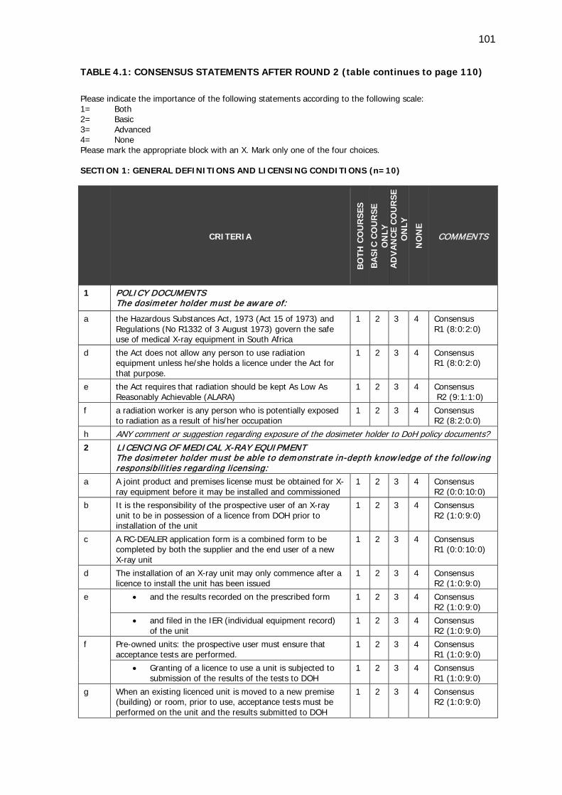
101
TABLE 4.1: CONSENSUS STATEMENTS AFTER ROUND 2 (table continues to page 110)
Please indicate the importance of the following statements according to the following scale: 1= Both 2= Basic 3= Advanced 4= None Please mark the appropriate block with an X. Mark only one of the four choices. SECTION 1: GENERAL DEFINITIONS AND LICENSING CONDITIONS (n=10)
CRITERIA
BO
TH C
OU
RSE
S
BA
SIC
CO
UR
SE
ON
LY
AD
VA
NC
E C
OU
RSE
O
NLY
NO
NE
COMMENTS
1 POLICY DOCUMENTS The dosimeter holder must be aware of:
a the Hazardous Substances Act, 1973 (Act 15 of 1973) and Regulations (No R1332 of 3 August 1973) govern the safe use of medical X-ray equipment in South Africa
1
2
3
4
Consensus R1 (8:0:2:0)
d the Act does not allow any person to use radiation equipment unless he/she holds a licence under the Act for that purpose.
1 2 3 4 Consensus R1 (8:0:2:0)
e the Act requires that radiation should be kept As Low As Reasonably Achievable (ALARA)
1 2 3 4 Consensus R2 (9:1:1:0)
f a radiation worker is any person who is potentially exposed to radiation as a result of his/her occupation
1 2 3 4 Consensus R2 (8:2:0:0)
h ANY comment or suggestion regarding exposure of the dosimeter holder to DoH policy documents? 2
LICENCING OF MEDICAL X-RAY EQUIPMENT The dosimeter holder must be able to demonstrate in-depth know ledge of the follow ing responsibil it ies regarding licensing:
a A joint product and premises license must be obtained for X-ray equipment before it may be installed and commissioned
1 2 3 4 Consensus R2 (0:0:10:0)
b It is the responsibility of the prospective user of an X-ray unit to be in possession of a licence from DOH prior to installation of the unit
1 2 3 4 Consensus R2 (1:0:9:0)
c A RC-DEALER application form is a combined form to be completed by both the supplier and the end user of a new X-ray unit
1 2 3 4 Consensus R1 (0:0:10:0)
d The installation of an X-ray unit may only commence after a licence to install the unit has been issued
1 2 3 4 Consensus R2 (1:0:9:0)
e • and the results recorded on the prescribed form 1 2 3 4 Consensus R2 (1:0:9:0)
• and filed in the IER (individual equipment record) of the unit
1 2 3 4 Consensus R2 (1:0:9:0)
f Pre-owned units: the prospective user must ensure that acceptance tests are performed.
1 2 3 4 Consensus R1 (1:0:9:0)
• Granting of a licence to use a unit is subjected to submission of the results of the tests to DOH
1 2 3 4 Consensus R1 (1:0:9:0)
g When an existing licenced unit is moved to a new premise (building) or room, prior to use, acceptance tests must be performed on the unit and the results submitted to DOH
1 2 3 4 Consensus R2 (1:0:9:0)
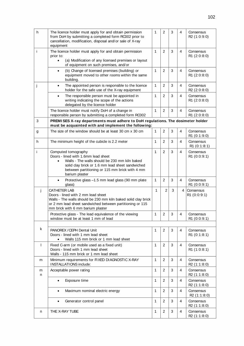
102
h The licence holder must apply for and obtain permission from DoH by submitting a completed form RC002 prior to cancellation, modification, disposal and/or sale of X-ray equipment
1 2 3 4 Consensus R2 (1:0:9:0)
i The licence holder must apply for and obtain permission prior to:
• (a) Modification of any licensed premises or layout of equipment on such premises, and/or
1 2 3 4 Consensus R1 (2:0:8:0)
• (b) Change of licensed premises (building) or equipment moved to other rooms within the same building.
1 2 3 4 Consensus R1 (2:0:8:0)
j • The appointed person is responsible to the licence holder for the safe use of the X-ray equipment
1 2 3 4 Consensus R2 (2:0:8:0)
• The responsible person must be appointed in writing indicating the scope of the actions delegated by the licence holder
1 2 3 4 Consensus R1 (2:0:8:0)
The licence holder must notify DoH of a change in responsible person by submitting a completed form RC002
1 2 3 4 Consensus R1 (2:0:8:0)
3 PREMISES X-ray departments must adhere to DoH regulations. The dosimeter holder must be acquainted with and implement the following:
g The size of the window should be at least 30 cm x 30 cm 1 2 3 4 Consensus R1 (0:1:9:0)
h The minimum height of the cubicle is 2.2 meter 1 2 3 4 Consensus R1 (0:1:8:1)
i Computed tomography Doors - lined with 1.6mm lead sheet
• Walls - The walls should be 230 mm kiln baked solid clay brick or 1.6 mm lead sheet sandwiched between partitioning or 115 mm brick with 4 mm barium plaster
1 2 3 4 Consensus R1 (0:0:9:1)
• Protective glass –1.5 mm lead glass (90 mm plate glass)
1 2 3 4 Consensus R1 (0:0:9:1)
j CATHETER LAB Doors - lined with 2 mm lead sheet Walls - The walls should be 230 mm kiln baked solid clay brick or 2 mm lead sheet sandwiched between partitioning or 115 mm brick with 6 mm barium plaster
1 2 3 4 Consensus R1 (0:0:9:1)
Protective glass - The lead equivalence of the viewing window must be at least 1 mm of lead
1 2 3 4 Consensus R1 (0:0:9:1)
k
PANOREX /CEPH Dental Unit Doors - lined with 1 mm lead sheet
• Walls 115 mm brick or 1 mm lead sheet
1 2 3 4 Consensus R1 (0:1:8:1)
l Fixed C-arm (or mobile used as a fixed unit) Doors - lined with 1 mm lead sheet Walls - 115 mm brick or 1 mm lead sheet
1 2 3 4 Consensus R1 (1:0:8:1)
m Minimum requirements for FIXED DIAGNOSTIC X-RAY INSTALLATIONS include:
1 2 3 4 Consensus R2 (1:1:8:0)
m n
Acceptable power rating 1 2 3 4 Consensus R2 (1:1:8:0)
• Exposure time 1 2 3 4 Consensus R2 (1:1:8:0)
• Maximum nominal electric energy 1 2 3 4 Consensus R2 (1:1:8:0)
• Generator control panel 1 2 3 4 Consensus R2 (1:1:8:0)
n THE X-RAY TUBE 1 2 3 4 Consensus R2 (1:1:8:0)
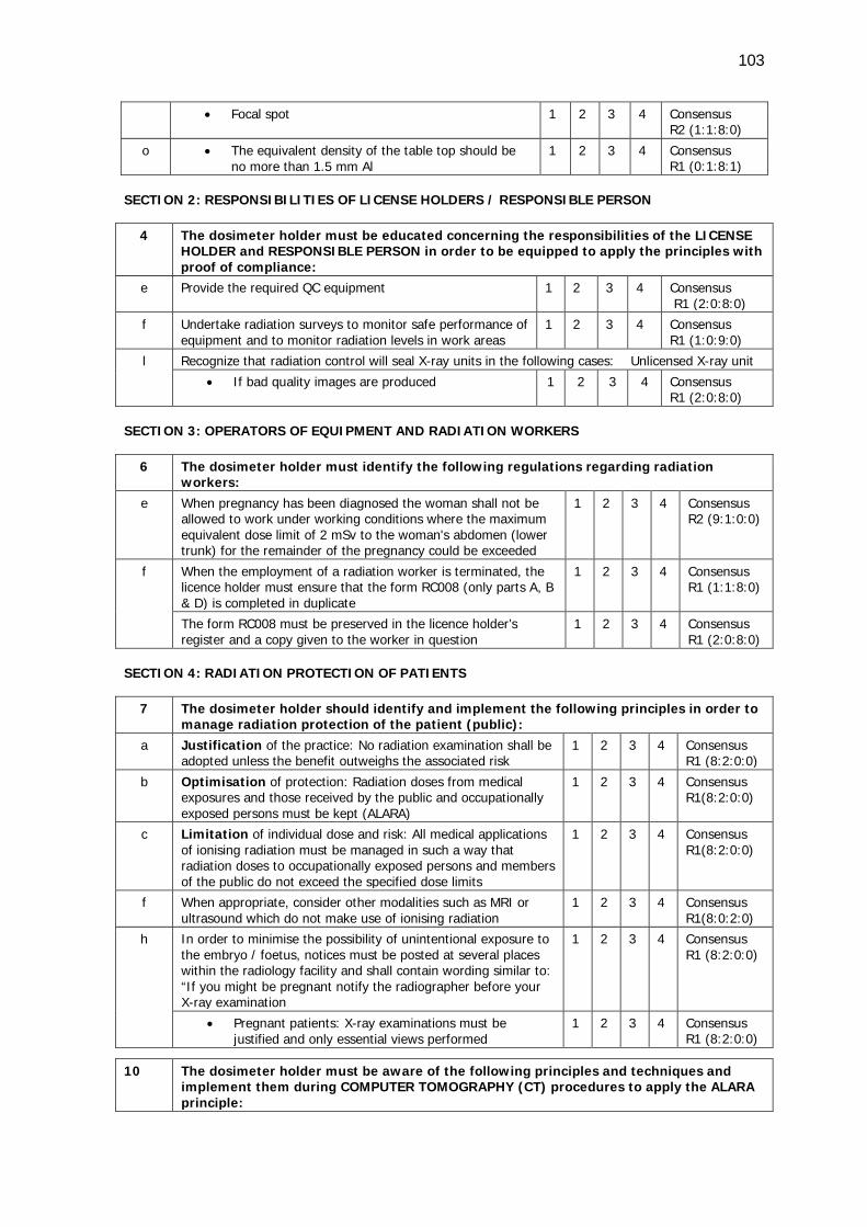
103
• Focal spot 1 2 3 4 Consensus R2 (1:1:8:0)
o • The equivalent density of the table top should be no more than 1.5 mm Al
1 2 3 4 Consensus R1 (0:1:8:1)
SECTION 2: RESPONSIBILITIES OF LICENSE HOLDERS / RESPONSIBLE PERSON
4 The dosimeter holder must be educated concerning the responsibilities of the LICENSE HOLDER and RESPONSIBLE PERSON in order to be equipped to apply the principles with proof of compliance:
e Provide the required QC equipment 1 2 3 4 Consensus R1 (2:0:8:0)
f Undertake radiation surveys to monitor safe performance of equipment and to monitor radiation levels in work areas
1 2 3 4 Consensus R1 (1:0:9:0)
l Recognize that radiation control will seal X-ray units in the following cases: Unlicensed X-ray unit
• If bad quality images are produced 1 2 3 4 Consensus R1 (2:0:8:0)
SECTION 3: OPERATORS OF EQUIPMENT AND RADIATION WORKERS
6 The dosimeter holder must identify the following regulations regarding radiation workers:
e When pregnancy has been diagnosed the woman shall not be allowed to work under working conditions where the maximum equivalent dose limit of 2 mSv to the woman's abdomen (lower trunk) for the remainder of the pregnancy could be exceeded
1 2 3 4 Consensus R2 (9:1:0:0)
f When the employment of a radiation worker is terminated, the licence holder must ensure that the form RC008 (only parts A, B & D) is completed in duplicate
1 2 3 4 Consensus R1 (1:1:8:0)
The form RC008 must be preserved in the licence holder’s register and a copy given to the worker in question
1 2 3 4 Consensus R1 (2:0:8:0)
SECTION 4: RADIATION PROTECTION OF PATIENTS
7 The dosimeter holder should identify and implement the following principles in order to manage radiation protection of the patient (public):
a Justification of the practice: No radiation examination shall be adopted unless the benefit outweighs the associated risk
1 2 3 4 Consensus R1 (8:2:0:0)
b Optimisation of protection: Radiation doses from medical exposures and those received by the public and occupationally exposed persons must be kept (ALARA)
1 2 3 4 Consensus R1(8:2:0:0)
c Limitation of individual dose and risk: All medical applications of ionising radiation must be managed in such a way that radiation doses to occupationally exposed persons and members of the public do not exceed the specified dose limits
1 2 3 4 Consensus R1(8:2:0:0)
f When appropriate, consider other modalities such as MRI or ultrasound which do not make use of ionising radiation
1 2 3 4 Consensus R1(8:0:2:0)
h In order to minimise the possibility of unintentional exposure to the embryo / foetus, notices must be posted at several places within the radiology facility and shall contain wording similar to: “If you might be pregnant notify the radiographer before your X-ray examination
1 2 3 4 Consensus R1 (8:2:0:0)
• Pregnant patients: X-ray examinations must be justified and only essential views performed
1 2 3 4 Consensus R1 (8:2:0:0)
10 The dosimeter holder must be aware of the following principles and techniques and implement them during COMPUTER TOMOGRAPHY (CT) procedures to apply the ALARA principle:
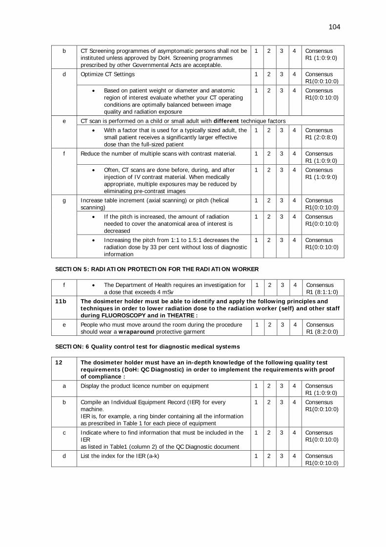
104
b CT Screening programmes of asymptomatic persons shall not be instituted unless approved by DoH. Screening programmes prescribed by other Governmental Acts are acceptable.
1 2 3 4 Consensus R1 (1:0:9:0)
d Optimize CT Settings 1 2 3 4 Consensus R1(0:0:10:0)
• Based on patient weight or diameter and anatomic region of interest evaluate whether your CT operating conditions are optimally balanced between image quality and radiation exposure
1 2 3 4 Consensus R1(0:0:10:0)
e CT scan is performed on a child or small adult with different technique factors • With a factor that is used for a typically sized adult, the
small patient receives a significantly larger effective dose than the full-sized patient
1 2 3 4 Consensus R1 (2:0:8:0)
f Reduce the number of multiple scans with contrast material. 1 2 3 4 Consensus R1 (1:0:9:0)
• Often, CT scans are done before, during, and after injection of IV contrast material. When medically appropriate, multiple exposures may be reduced by eliminating pre-contrast images
1 2 3 4 Consensus R1 (1:0:9:0)
g Increase table increment (axial scanning) or pitch (helical scanning)
1 2 3 4 Consensus R1(0:0:10:0)
• If the pitch is increased, the amount of radiation needed to cover the anatomical area of interest is decreased
1 2 3 4 Consensus R1(0:0:10:0)
• Increasing the pitch from 1:1 to 1.5:1 decreases the radiation dose by 33 per cent without loss of diagnostic information
1 2 3 4 Consensus R1(0:0:10:0)
SECTION 5: RADIATION PROTECTION FOR THE RADIATION WORKER
f • The Department of Health requires an investigation for a dose that exceeds 4 mSv
1 2 3 4 Consensus R1 (8:1:1:0)
11b The dosimeter holder must be able to identify and apply the following principles and techniques in order to lower radiation dose to the radiation worker (self) and other staff during FLUOROSCOPY and in THEATRE :
e People who must move around the room during the procedure should wear a wraparound protective garment
1 2 3 4 Consensus R1 (8:2:0:0)
SECTION: 6 Quality control test for diagnostic medical systems
12 The dosimeter holder must have an in-depth knowledge of the following quality test
requirements (DoH: QC Diagnostic) in order to implement the requirements with proof of compliance :
a Display the product licence number on equipment 1 2 3 4 Consensus R1 (1:0:9:0)
b Compile an Individual Equipment Record (IER) for every machine. IER is, for example, a ring binder containing all the information as prescribed in Table 1 for each piece of equipment
1 2 3 4 Consensus R1(0:0:10:0)
c Indicate where to find information that must be included in the IER as listed in Table1 (column 2) of the QC Diagnostic document
1 2 3 4 Consensus R1(0:0:10:0)
d List the index for the IER (a-k) 1 2 3 4 Consensus R1(0:0:10:0)

105
a) Unit - make, model and system ID b) Generator – make, model and serial number c) Product Licence number, date of the latest licence & reference to where a copy of the licence is kept d) Date of installation e) Operator’s manual – (Indication that the operator’s manual is available and reference to where it is kept) f) Results of acceptance tests g) Results of routine quality control tests h) Date(s) of tube replacement(s) i) Details of repairs/maintenance and/or modification(s) k) Details of the IB and person(s) that performed the test(s)and system ID
1 2 3 4 Consensus R1(0:0:10:0)
e The licence holder must ensure that all the applicable test(s) are performed that could be affected by the afore-mentioned
1 2 3 4 Consensus R1 (1:0:9:0)
• Should any of the tests in Table 2 indicate non-compliance or should any problems be detected (indicated), the licence holder must implement corrective maintenance (repairs followed by re-testing)
1 2 3 4 Consensus R1 (1:0:9:0)
• New equipment acceptance tests are the responsibility of the company that installed the equipment
1 2 3 4 Consensus R1 (1:0:9:0)
f An Inspection Body is an Organisation that is accredited by SANAS (www.sanas.co.za) and approved by the Department of Health
1 2 3 4 Consensus R1 (2:0:8:0)
g As from 31 March 2009 an Inspection Body (IB) approved by the Department of Health (DoH) OR an appropriately trained professional registered with the HPCSA as a medical physicist must be used to perform all the acceptance tests as well as the routine tests listed in section III.2 of table 2
1 2 3 4 Consensus R1 (2:0:8:0)
• If a medical physicist is used to perform the tests, an Inspection Body approved by the Department of Health (DoH) must formally contract such person(s). Formally contracted means that the medical physicist is contracted by the IB (ISO/IEC 17020 and TR78-01 par 14.2a) to perform the tests
1 2 3 4 Consensus R1 (1:0:9:0)
h Acquire the relevant quality control manuals or compile in-house written protocols, which describe each test step by step to ensure that QC tests listed in section III.1 of Table 2 are correctly performed
1 2 3 4 Consensus R1 (1:0:9:0)
• Ensure that persons that perform routine tests in section III.1 of Table 2 are competent to execute the tests
1 2 3 4 Consensus R1 (1:0:9:0)
i Ensure that the required acceptance tests are performed before the diagnostic X-ray equipment listed in Table 2 is put into clinical service when: Acquired or Substantially upgraded
1 2 3 4 Consensus R1 (1:0:9:0)
• Acceptance tests are the initial tests performed directly after installation and before the equipment is being put into clinical service
1 2 3 4 Consensus R1 (2:0:8:0)
• Acceptance tests have three purposes: To ensure that the unit meets stated specifications; To establish baseline parameters for the future quality control programme, and to familiarize the customer with operation of the unit.
1 2 3 4 Consensus R1 (2:0:8:0)
j Ensure that all the quality control tests are performed at the prescribed frequencies as specified in Table 2
1 2 3 4 Consensus R2 (1:1:8:0)
Gonad shields, lead rubber aprons and gloves: 3 monthly
1 2 3 4 Consensus R1 (1:0:9:0)
Appropriate technique chart displayed at X-ray unit 6 monthly
1 2 3 4 Consensus R1 (2:0:8:0)
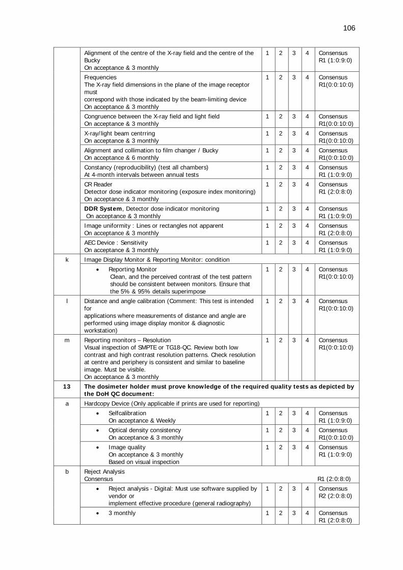
106
Alignment of the centre of the X-ray field and the centre of the Bucky On acceptance & 3 monthly
1 2 3 4 Consensus R1 (1:0:9:0)
Frequencies The X-ray field dimensions in the plane of the image receptor must correspond with those indicated by the beam-limiting device On acceptance & 3 monthly
1 2 3 4 Consensus R1(0:0:10:0)
Congruence between the X-ray field and light field On acceptance & 3 monthly
1 2 3 4 Consensus R1(0:0:10:0)
X-ray/light beam centrring On acceptance & 3 monthly
1 2 3 4 Consensus R1(0:0:10:0)
Alignment and collimation to film changer / Bucky On acceptance & 6 monthly
1 2 3 4 Consensus R1(0:0:10:0)
Constancy (reproducibility) (test all chambers) At 4-month intervals between annual tests
1 2 3 4 Consensus R1 (1:0:9:0)
CR Reader Detector dose indicator monitoring (exposure index monitoring) On acceptance & 3 monthly
1 2 3 4 Consensus R1 (2:0:8:0)
DDR System, Detector dose indicator monitoring On acceptance & 3 monthly
1 2 3 4 Consensus R1 (1:0:9:0)
Image uniformity : Lines or rectangles not apparent On acceptance & 3 monthly
1 2 3 4 Consensus R1 (2:0:8:0)
AEC Device : Sensitivity On acceptance & 3 monthly
1 2 3 4 Consensus R1 (1:0:9:0)
k Image Display Monitor & Reporting Monitor: condition • Reporting Monitor
Clean, and the perceived contrast of the test pattern should be consistent between monitors. Ensure that the 5% & 95% details superimpose
1 2 3 4 Consensus R1(0:0:10:0)
l Distance and angle calibration (Comment: This test is intended for applications where measurements of distance and angle are performed using image display monitor & diagnostic workstation)
1 2 3 4 Consensus R1(0:0:10:0)
m Reporting monitors – Resolution Visual inspection of SMPTE or TG18-QC. Review both low contrast and high contrast resolution patterns. Check resolution at centre and periphery is consistent and similar to baseline image. Must be visible. On acceptance & 3 monthly
1 2 3 4 Consensus R1(0:0:10:0)
13 The dosimeter holder must prove knowledge of the required quality tests as depicted by the DoH QC document:
a Hardcopy Device (Only applicable if prints are used for reporting) • Selfcalibration
On acceptance & Weekly 1 2 3 4 Consensus
R1 (1:0:9:0)
• Optical density consistency On acceptance & 3 monthly
1 2 3 4 Consensus R1(0:0:10:0)
• Image quality On acceptance & 3 monthly Based on visual inspection
1 2 3 4 Consensus R1 (1:0:9:0)
b Reject Analysis Consensus R1 (2:0:8:0)
• Reject analysis - Digital: Must use software supplied by vendor or implement effective procedure (general radiography)
1 2 3 4 Consensus R2 (2:0:8:0)
• 3 monthly 1 2 3 4 Consensus R1 (2:0:8:0)
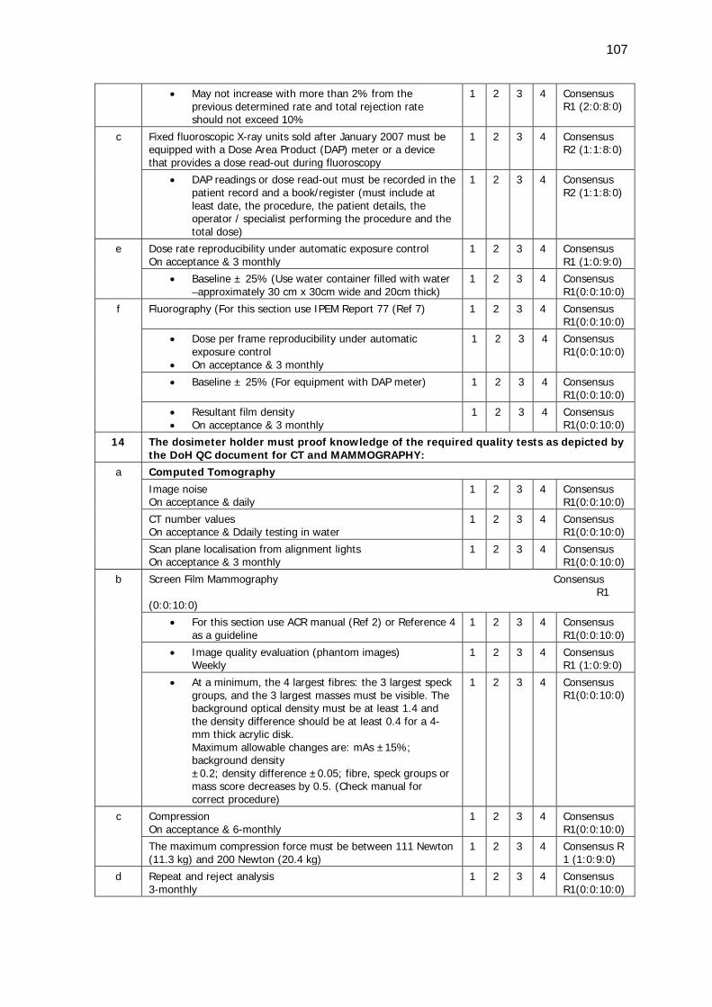
107
• May not increase with more than 2% from the previous determined rate and total rejection rate should not exceed 10%
1 2 3 4 Consensus R1 (2:0:8:0)
c Fixed fluoroscopic X-ray units sold after January 2007 must be equipped with a Dose Area Product (DAP) meter or a device that provides a dose read-out during fluoroscopy
1 2 3 4 Consensus R2 (1:1:8:0)
• DAP readings or dose read-out must be recorded in the patient record and a book/register (must include at least date, the procedure, the patient details, the operator / specialist performing the procedure and the total dose)
1 2 3 4 Consensus R2 (1:1:8:0)
e Dose rate reproducibility under automatic exposure control On acceptance & 3 monthly
1 2 3 4 Consensus R1 (1:0:9:0)
• Baseline ± 25% (Use water container filled with water –approximately 30 cm x 30cm wide and 20cm thick)
1 2 3 4 Consensus R1(0:0:10:0)
f Fluorography (For this section use IPEM Report 77 (Ref 7) 1 2 3 4 Consensus R1(0:0:10:0)
• Dose per frame reproducibility under automatic exposure control
• On acceptance & 3 monthly
1 2 3 4 Consensus R1(0:0:10:0)
• Baseline ± 25% (For equipment with DAP meter) 1 2 3 4 Consensus R1(0:0:10:0)
• Resultant film density • On acceptance & 3 monthly
1 2 3 4 Consensus R1(0:0:10:0)
14 The dosimeter holder must proof knowledge of the required quality tests as depicted by the DoH QC document for CT and MAMMOGRAPHY:
a Computed Tomography Image noise On acceptance & daily
1 2 3 4 Consensus R1(0:0:10:0)
CT number values On acceptance & Ddaily testing in water
1 2 3 4 Consensus R1(0:0:10:0)
Scan plane localisation from alignment lights On acceptance & 3 monthly
1 2 3 4 Consensus R1(0:0:10:0)
b Screen Film Mammography Consensus R1 (0:0:10:0)
• For this section use ACR manual (Ref 2) or Reference 4 as a guideline
1 2 3 4 Consensus R1(0:0:10:0)
• Image quality evaluation (phantom images) Weekly
1 2 3 4 Consensus R1 (1:0:9:0)
• At a minimum, the 4 largest fibres: the 3 largest speck groups, and the 3 largest masses must be visible. The background optical density must be at least 1.4 and the density difference should be at least 0.4 for a 4-mm thick acrylic disk. Maximum allowable changes are: mAs ±15%; background density ±0.2; density difference ±0.05; fibre, speck groups or mass score decreases by 0.5. (Check manual for correct procedure)
1 2 3 4 Consensus R1(0:0:10:0)
c Compression On acceptance & 6-monthly
1 2 3 4 Consensus R1(0:0:10:0)
The maximum compression force must be between 111 Newton (11.3 kg) and 200 Newton (20.4 kg)
1 2 3 4 Consensus R 1 (1:0:9:0)
d Repeat and reject analysis 3-monthly
1 2 3 4 Consensus R1(0:0:10:0)
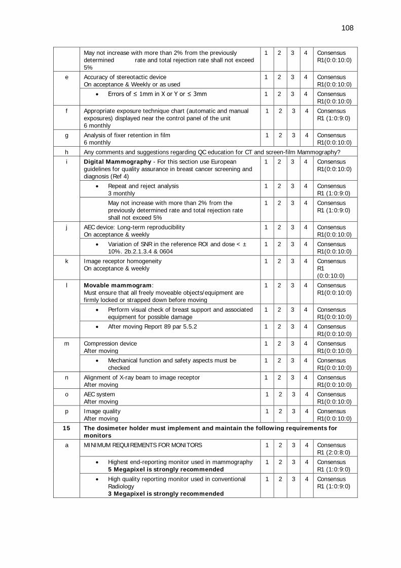
108
May not increase with more than 2% from the previously determined rate and total rejection rate shall not exceed 5%
1 2 3 4 Consensus R1(0:0:10:0)
e Accuracy of stereotactic device On acceptance & Weekly or as used
1 2 3 4 Consensus R1(0:0:10:0)
• Errors of ≤ 1mm in X or Y or ≤ 3mm 1 2 3 4 Consensus R1(0:0:10:0)
f Appropriate exposure technique chart (automatic and manual exposures) displayed near the control panel of the unit 6 monthly
1 2 3 4 Consensus R1 (1:0:9:0)
g Analysis of fixer retention in film 6 monthly
1 2 3 4 Consensus R1(0:0:10:0)
h Any comments and suggestions regarding QC education for CT and screen-film Mammography? i Digital Mammography - For this section use European
guidelines for quality assurance in breast cancer screening and diagnosis (Ref 4)
1 2 3 4 Consensus R1(0:0:10:0)
• Repeat and reject analysis 3 monthly
1 2 3 4 Consensus R1 (1:0:9:0)
May not increase with more than 2% from the previously determined rate and total rejection rate shall not exceed 5%
1 2 3 4 Consensus R1 (1:0:9:0)
j AEC device: Long-term reproducibility On acceptance & weekly
1 2 3 4 Consensus R1(0:0:10:0)
• Variation of SNR in the reference ROI and dose < ± 10%. 2b.2.1.3.4 & 0604
1 2 3 4 Consensus R1(0:0:10:0)
k Image receptor homogeneity On acceptance & weekly
1 2 3 4 Consensus R1 (0:0:10:0)
l Movable mammogram: Must ensure that all freely moveable objects/equipment are firmly locked or strapped down before moving
1 2 3 4 Consensus R1(0:0:10:0)
• Perform visual check of breast support and associated equipment for possible damage
1 2 3 4 Consensus R1(0:0:10:0)
• After moving Report 89 par 5.5.2 1 2 3 4 Consensus R1(0:0:10:0)
m Compression device After moving
1 2 3 4 Consensus R1(0:0:10:0)
• Mechanical function and safety aspects must be checked
1 2 3 4 Consensus R1(0:0:10:0)
n Alignment of X-ray beam to image receptor After moving
1 2 3 4 Consensus R1(0:0:10:0)
o AEC system After moving
1 2 3 4 Consensus R1(0:0:10:0)
p Image quality After moving
1 2 3 4 Consensus R1(0:0:10:0)
15 The dosimeter holder must implement and maintain the following requirements for monitors
a MINIMUM REQUIREMENTS FOR MONITORS 1 2 3 4 Consensus R1 (2:0:8:0)
• Highest end-reporting monitor used in mammography 5 Megapixel is strongly recommended
1 2 3 4 Consensus R1 (1:0:9:0)
• High quality reporting monitor used in conventional Radiology 3 Megapixel is strongly recommended
1 2 3 4 Consensus R1 (1:0:9:0)
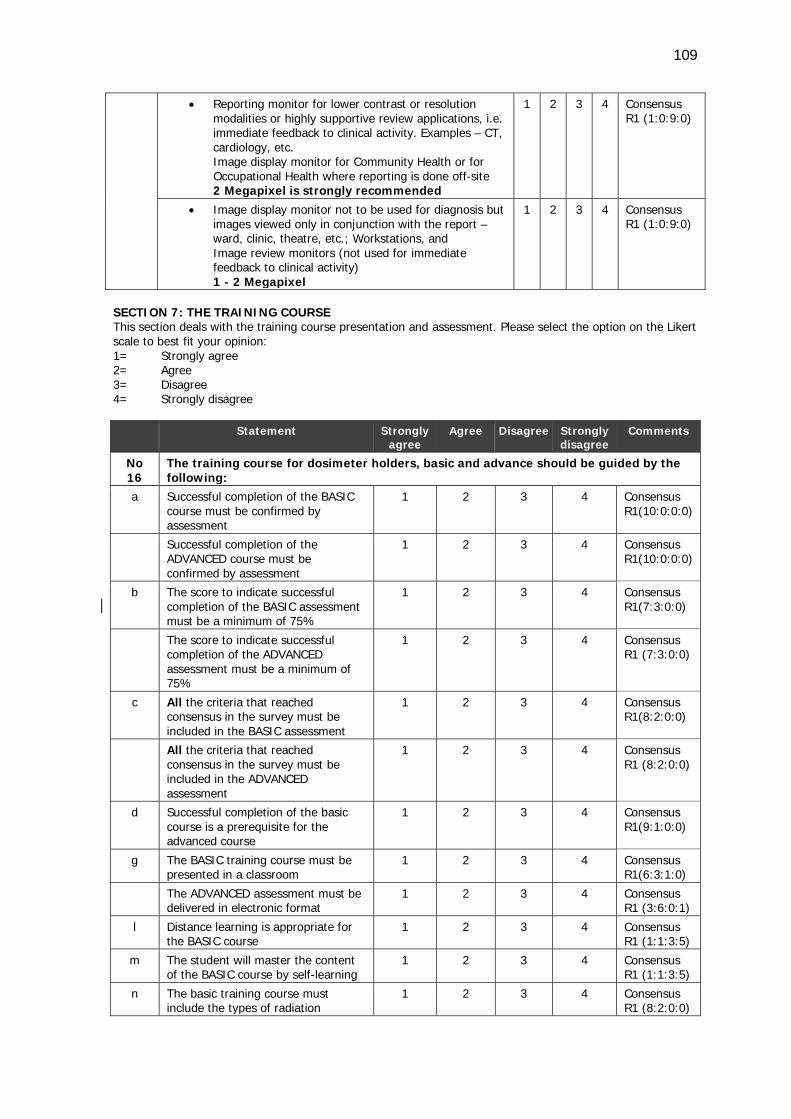
109
• Reporting monitor for lower contrast or resolution modalities or highly supportive review applications, i.e. immediate feedback to clinical activity. Examples – CT, cardiology, etc. Image display monitor for Community Health or for Occupational Health where reporting is done off-site 2 Megapixel is strongly recommended
1 2 3 4 Consensus R1 (1:0:9:0)
• Image display monitor not to be used for diagnosis but images viewed only in conjunction with the report – ward, clinic, theatre, etc.; Workstations, and Image review monitors (not used for immediate feedback to clinical activity) 1 - 2 Megapixel
1 2 3 4 Consensus R1 (1:0:9:0)
SECTION 7: THE TRAINING COURSE This section deals with the training course presentation and assessment. Please select the option on the Likert scale to best fit your opinion: 1= Strongly agree 2= Agree 3= Disagree 4= Strongly disagree
Statement Strongly agree
Agree Disagree Strongly disagree
Comments
No 16
The training course for dosimeter holders, basic and advance should be guided by the following:
a Successful completion of the BASIC course must be confirmed by assessment
1 2 3 4 Consensus R1(10:0:0:0)
Successful completion of the ADVANCED course must be confirmed by assessment
1 2 3 4 Consensus R1(10:0:0:0)
b The score to indicate successful completion of the BASIC assessment must be a minimum of 75%
1 2 3 4 Consensus R1(7:3:0:0)
The score to indicate successful completion of the ADVANCED assessment must be a minimum of 75%
1 2 3 4 Consensus R1 (7:3:0:0)
c All the criteria that reached consensus in the survey must be included in the BASIC assessment
1 2 3 4 Consensus R1(8:2:0:0)
All the criteria that reached consensus in the survey must be included in the ADVANCED assessment
1 2 3 4 Consensus R1 (8:2:0:0)
d Successful completion of the basic course is a prerequisite for the advanced course
1 2 3 4 Consensus R1(9:1:0:0)
g The BASIC training course must be presented in a classroom
1 2 3 4 Consensus R1(6:3:1:0)
The ADVANCED assessment must be delivered in electronic format
1 2 3 4 Consensus R1 (3:6:0:1)
l Distance learning is appropriate for the BASIC course
1 2 3 4 Consensus R1 (1:1:3:5)
m The student will master the content of the BASIC course by self-learning
1 2 3 4 Consensus R1 (1:1:3:5)
n The basic training course must include the types of radiation
1 2 3 4 Consensus R1 (8:2:0:0)
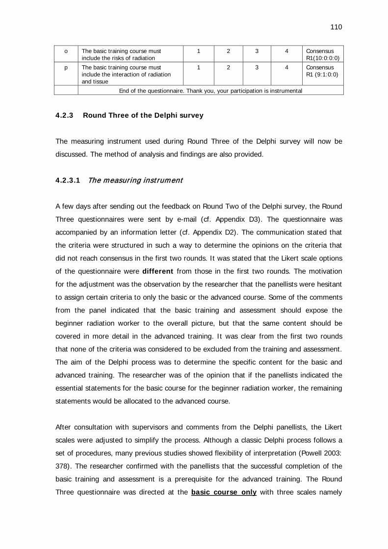
110
o The basic training course must include the risks of radiation
1 2 3 4 Consensus R1(10:0:0:0)
p The basic training course must include the interaction of radiation and tissue
1 2 3 4 Consensus R1 (9:1:0:0)
End of the questionnaire. Thank you, your participation is instrumental
4.2.3 Round Three of the Delphi survey
The measuring instrument used during Round Three of the Delphi survey will now be
discussed. The method of analysis and findings are also provided.
4.2.3.1 The measuring instrument
A few days after sending out the feedback on Round Two of the Delphi survey, the Round
Three questionnaires were sent by e-mail (cf. Appendix D3). The questionnaire was
accompanied by an information letter (cf. Appendix D2). The communication stated that
the criteria were structured in such a way to determine the opinions on the criteria that
did not reach consensus in the first two rounds. It was stated that the Likert scale options
of the questionnaire were different from those in the first two rounds. The motivation
for the adjustment was the observation by the researcher that the panellists were hesitant
to assign certain criteria to only the basic or the advanced course. Some of the comments
from the panel indicated that the basic training and assessment should expose the
beginner radiation worker to the overall picture, but that the same content should be
covered in more detail in the advanced training. It was clear from the first two rounds
that none of the criteria was considered to be excluded from the training and assessment.
The aim of the Delphi process was to determine the specific content for the basic and
advanced training. The researcher was of the opinion that if the panellists indicated the
essential statements for the basic course for the beginner radiation worker, the remaining
statements would be allocated to the advanced course.
After consultation with supervisors and comments from the Delphi panellists, the Likert
scales were adjusted to simplify the process. Although a classic Delphi process follows a
set of procedures, many previous studies showed flexibility of interpretation (Powell 2003:
378). The researcher confirmed with the panellists that the successful completion of the
basic training and assessment is a prerequisite for the advanced training. The Round
Three questionnaire was directed at the basic course only with three scales namely
111
1=’Definitely’, 2=’Undecided’ and 3=’Not at all’ as the options. The options were defined
as follows:
1= Definitely [The criterion must definitely be included in the basic course]
2= Undecided [You are undecided whether to select either ’definitely’ or ’not at all’]
3= Not at all [The criterion must be excluded from the basic course]
Section 7 also included three scales, namely 1=’Agree’, 2=’Undecided’ and 3=’Disagree’
as the options. The layout of the questionnaire remained the same in all the rounds. The
statements were numbered exactly as in the first two rounds and provided opportunities
for reconsideration, additional comments and suggestions.
4.2.3.2 Analysis of the Round Three Responses
The responses from the Delphi participants were analysed manually by calculating the
frequency of responses for each statement on the Likert scale similarly to how it was
done for Round Two.
4.2.3.3 The findings of Round Three of the Delphi survey
All the panellists completed and returned the Round Three questionnaires of the Delphi
survey (100% response rate). After Round Three, consensus was reached on 278 of the
418 statements in the questionnaire, giving a 67% overall consensus.
Following the analysis of the Round Three questionnaires, a feedback letter was sent to
all panellists (cf. Appendix D4). The letter was accompanied by a copy of the Delphi
questionnaire, shading the statements on which consensus had been reached and further
including all the comments and questions from the participants (cf. Appendix D4 B). As
after Round Two, the results of the third round were e-mailed to all the panellists.
4.2.4 Round Four of the Delphi survey
The fourth and final round of the Delphi survey was managed in the same way as the
previous rounds, with a feedback letter containing the results of the preceding Round
Three (cf. Appendix D4) and the questionnaire with the consensus statements shaded
(cf. Appendix D4-B). Similar to the previous rounds the comments from panellists or the
112
explanation from the researcher was included. The researcher acknowledged the
perseverance of the panellists.
After the completion of Round Four, a feedback letter was sent to all panellists with the
results of Round Four (cf. Appendix D6).
The measuring instrument used during Round Four of the Delphi survey will be discussed
next. The method of analysis and the findings are also elucidated.
4.2.4.1 The measuring instrument
A few days after sending out the feedback on Round Three of the Delphi survey, the
Round Four questionnaires were sent to the panellists by e-mail (cf. Appendix D4-C). The
Round Four questionnaire was accompanied by an information letter (cf. Appendix D5).
The communication stated that the criteria were structured in such a way to determine
the opinion on criteria on which consensus had not been reached in the first three rounds,
or if failing to do so, to declare stability. Consensus was pre-defined as 80% of the
panellists agreeing on a specific criterion. Stability could be declared when movement of
opinion of the group as a whole reached stability.
It was stated that the Likert-scale options of the questionnaire were similar to the options
in the previous round (Round Three). The percentage of the participants selecting a
particular number on the Likert scale is indicated choosing either ’Definitely in basic’ or
’Undecided’ or ’Not at all in basic’, for example, 1=50%, 2=10%, 3=40% with 1, 2 and 3
referring to the respective terms on the scale in the order mentioned above. The
statements were numbered exactly the same as in the previous rounds and provided
opportunities for reconsideration, additional comments, and suggestions.
4.2.4.2 Analysis of the Round Four Responses
The responses from the Delphi participants were analysed manually by calculating the
frequency of responses for each statement on the Likert scale similarly to what was done
in Round Three.
4.2.4.3 The findings of Round Four of the Delphi survey
113
All the panellists completed and returned the Round Four questionnaire of the Delphi
survey (100% response rate). After Round Four, consensus was reached on 309 of the
418 statements in the questionnaire, giving 74% of the statements reached consensus.
Stability was reached on 109 statements in Round Four giving 26% of the statements
stability.
Following the analysis of the Round Four questionnaire, a feedback letter was sent to all
panellists (cf. Appendix D6). It should be noted that 90% of the Delphi panellists
indicated that they would not change their opinions on choices made in Round Four.
Table 4.2 provides the details regarding the statements on which consensus was reached
in Round Three (R3) and Round Four (R4). The consensus is reported separately from
those of Round One and Round Two due to the adjustment in the Likert-scale options (cf.
Table 4.1). The percentage of the participants selecting a particular number on the Likert
scale that indicated either ’Definitely in basic’, or ’Undecided’, or ’Not at all in basic’, for
example, 1=50%, 2=10%, 3=40%, with 1, 2 and 3 referring to the respective terms on
the scale in the order given above.
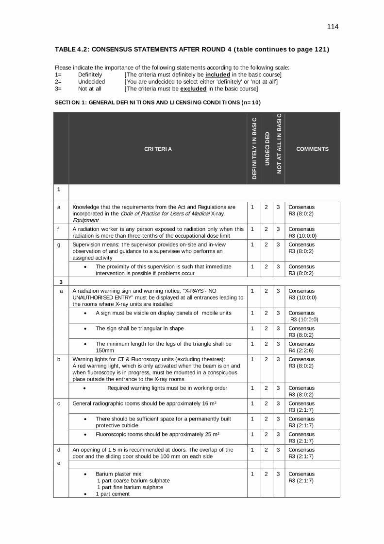
114
TABLE 4.2: CONSENSUS STATEMENTS AFTER ROUND 4 (table continues to page 121)
Please indicate the importance of the following statements according to the following scale: 1= Definitely [The criteria must definitely be included in the basic course] 2= Undecided [You are undecided to select either ’definitely’ or ’not at all’] 3= Not at all [The criteria must be excluded in the basic course] SECTION 1: GENERAL DEFINITIONS AND LICENSING CONDITIONS (n=10)
CRITERIA
DEF
INIT
ELY
IN
BA
SIC
UN
DEC
IDED
NO
T A
T A
LL I
N B
ASI
C
COMMENTS
1
a Knowledge that the requirements from the Act and Regulations are incorporated in the Code of Practice for Users of Medical X-ray Equipment
1 2 3 Consensus R3 (8:0:2)
f A radiation worker is any person exposed to radiation only when this radiation is more than three-tenths of the occupational dose limit
1 2 3 Consensus R3 (10:0:0)
g Supervision means: the supervisor provides on-site and in-view observation of and guidance to a supervisee who performs an assigned activity
1 2 3 Consensus R3 (8:0:2)
• The proximity of this supervision is such that immediate intervention is possible if problems occur
1 2 3 Consensus R3 (8:0:2)
3 a
A radiation warning sign and warning notice, “X-RAYS - NO UNAUTHORISED ENTRY” must be displayed at all entrances leading to the rooms where X-ray units are installed
1 2 3 Consensus R3 (10:0:0)
• A sign must be visible on display panels of mobile units 1 2 3 Consensus R3 (10:0:0)
• The sign shall be triangular in shape 1 2 3 Consensus R3 (8:0:2)
• The minimum length for the legs of the triangle shall be 150mm
1 2 3 Consensus R4 (2:2:6)
b Warning lights for CT & Fluoroscopy units (excluding theatres): A red warning light, which is only activated when the beam is on and when fluoroscopy is in progress, must be mounted in a conspicuous place outside the entrance to the X-ray rooms
1 2 3 Consensus R3 (8:0:2)
• Required warning lights must be in working order 1 2 3 Consensus R3 (8:0:2)
c General radiographic rooms should be approximately 16 m² 1 2 3 Consensus R3 (2:1:7)
• There should be sufficient space for a permanently built protective cubicle
1 2 3 Consensus R3 (2:1:7)
• Fluoroscopic rooms should be approximately 25 m² 1 2 3 Consensus R3 (2:1:7)
d e
An opening of 1.5 m is recommended at doors. The overlap of the door and the sliding door should be 100 mm on each side
1 2 3 Consensus R3 (2:1:7)
• Barium plaster mix: 1 part coarse barium sulphate 1 part fine barium sulphate
• 1 part cement
1 2 3 Consensus R3 (2:1:7)
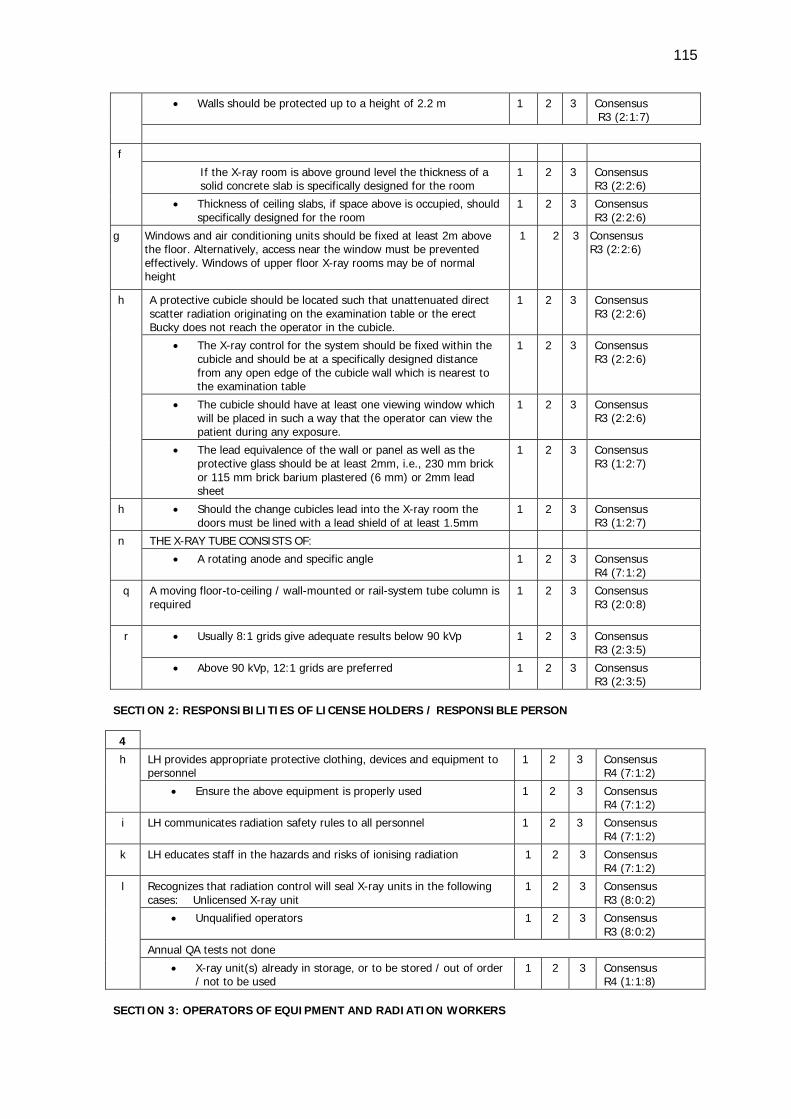
115
• Walls should be protected up to a height of 2.2 m 1 2 3 Consensus R3 (2:1:7)
f
If the X-ray room is above ground level the thickness of a solid concrete slab is specifically designed for the room
1 2 3 Consensus R3 (2:2:6)
• Thickness of ceiling slabs, if space above is occupied, should specifically designed for the room
1 2 3 Consensus R3 (2:2:6)
g Windows and air conditioning units should be fixed at least 2m above the floor. Alternatively, access near the window must be prevented effectively. Windows of upper floor X-ray rooms may be of normal height
1 2 3 Consensus R3 (2:2:6)
h
A protective cubicle should be located such that unattenuated direct scatter radiation originating on the examination table or the erect Bucky does not reach the operator in the cubicle.
1 2 3 Consensus R3 (2:2:6)
• The X-ray control for the system should be fixed within the cubicle and should be at a specifically designed distance from any open edge of the cubicle wall which is nearest to the examination table
1 2 3 Consensus R3 (2:2:6)
• The cubicle should have at least one viewing window which will be placed in such a way that the operator can view the patient during any exposure.
1 2 3 Consensus R3 (2:2:6)
• The lead equivalence of the wall or panel as well as the protective glass should be at least 2mm, i.e., 230 mm brick or 115 mm brick barium plastered (6 mm) or 2mm lead sheet
1 2 3 Consensus R3 (1:2:7)
h
• Should the change cubicles lead into the X-ray room the doors must be lined with a lead shield of at least 1.5mm
1 2 3 Consensus R3 (1:2:7)
n
THE X-RAY TUBE CONSISTS OF:
• A rotating anode and specific angle 1 2 3 Consensus R4 (7:1:2)
q
A moving floor-to-ceiling / wall-mounted or rail-system tube column is required
1 2 3 Consensus R3 (2:0:8)
r • Usually 8:1 grids give adequate results below 90 kVp 1 2 3 Consensus R3 (2:3:5)
• Above 90 kVp, 12:1 grids are preferred 1 2 3 Consensus R3 (2:3:5)
SECTION 2: RESPONSIBILITIES OF LICENSE HOLDERS / RESPONSIBLE PERSON 4
h LH provides appropriate protective clothing, devices and equipment to personnel
1 2 3 Consensus R4 (7:1:2)
• Ensure the above equipment is properly used 1 2 3 Consensus R4 (7:1:2)
i LH communicates radiation safety rules to all personnel 1 2 3 Consensus R4 (7:1:2)
k LH educates staff in the hazards and risks of ionising radiation 1 2 3 Consensus R4 (7:1:2)
l Recognizes that radiation control will seal X-ray units in the following cases: Unlicensed X-ray unit
1 2 3 Consensus R3 (8:0:2)
• Unqualified operators 1 2 3 Consensus R3 (8:0:2)
Annual QA tests not done
• X-ray unit(s) already in storage, or to be stored / out of order / not to be used
1 2 3 Consensus R4 (1:1:8)
SECTION 3: OPERATORS OF EQUIPMENT AND RADIATION WORKERS
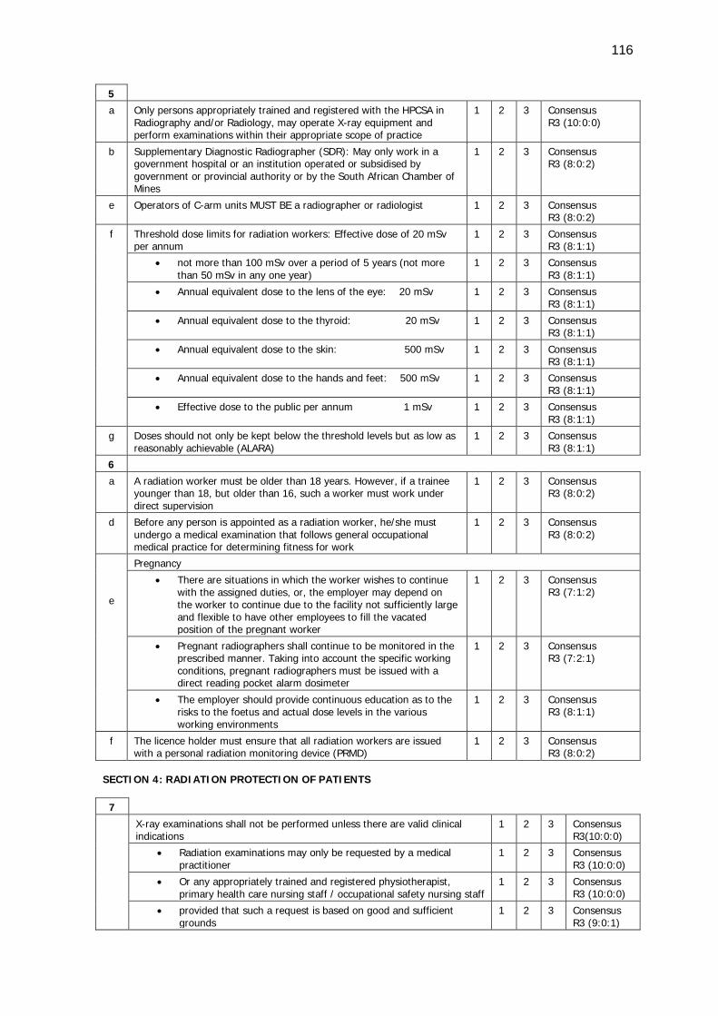
116
5 a Only persons appropriately trained and registered with the HPCSA in
Radiography and/or Radiology, may operate X-ray equipment and perform examinations within their appropriate scope of practice
1 2 3 Consensus R3 (10:0:0)
b Supplementary Diagnostic Radiographer (SDR): May only work in a government hospital or an institution operated or subsidised by government or provincial authority or by the South African Chamber of Mines
1 2 3 Consensus R3 (8:0:2)
e Operators of C-arm units MUST BE a radiographer or radiologist 1 2 3 Consensus R3 (8:0:2)
f Threshold dose limits for radiation workers: Effective dose of 20 mSv per annum
1 2 3 Consensus R3 (8:1:1)
• not more than 100 mSv over a period of 5 years (not more than 50 mSv in any one year)
1 2 3 Consensus R3 (8:1:1)
• Annual equivalent dose to the lens of the eye: 20 mSv 1 2 3 Consensus R3 (8:1:1)
• Annual equivalent dose to the thyroid: 20 mSv 1 2 3 Consensus R3 (8:1:1)
• Annual equivalent dose to the skin: 500 mSv 1 2 3 Consensus R3 (8:1:1)
• Annual equivalent dose to the hands and feet: 500 mSv 1 2 3 Consensus R3 (8:1:1)
• Effective dose to the public per annum 1 mSv 1 2 3 Consensus R3 (8:1:1)
g Doses should not only be kept below the threshold levels but as low as reasonably achievable (ALARA)
1 2 3 Consensus R3 (8:1:1)
6
a A radiation worker must be older than 18 years. However, if a trainee younger than 18, but older than 16, such a worker must work under direct supervision
1 2 3 Consensus R3 (8:0:2)
d Before any person is appointed as a radiation worker, he/she must undergo a medical examination that follows general occupational medical practice for determining fitness for work
1 2 3 Consensus R3 (8:0:2)
e
Pregnancy
• There are situations in which the worker wishes to continue with the assigned duties, or, the employer may depend on the worker to continue due to the facility not sufficiently large and flexible to have other employees to fill the vacated position of the pregnant worker
1 2 3 Consensus R3 (7:1:2)
• Pregnant radiographers shall continue to be monitored in the prescribed manner. Taking into account the specific working conditions, pregnant radiographers must be issued with a direct reading pocket alarm dosimeter
1 2 3 Consensus R3 (7:2:1)
• The employer should provide continuous education as to the risks to the foetus and actual dose levels in the various working environments
1 2 3 Consensus R3 (8:1:1)
f The licence holder must ensure that all radiation workers are issued with a personal radiation monitoring device (PRMD)
1 2 3 Consensus R3 (8:0:2)
SECTION 4: RADIATION PROTECTION OF PATIENTS
7
X-ray examinations shall not be performed unless there are valid clinical indications
1 2 3 Consensus R3(10:0:0)
• Radiation examinations may only be requested by a medical practitioner
1 2 3 Consensus R3 (10:0:0)
• Or any appropriately trained and registered physiotherapist, primary health care nursing staff / occupational safety nursing staff
1 2 3 Consensus R3 (10:0:0)
• provided that such a request is based on good and sufficient grounds
1 2 3 Consensus R3 (9:0:1)
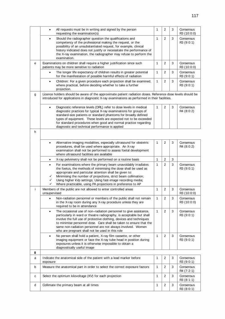
117
• All requests must be in writing and signed by the person requesting the examination(s)
1 2 3 Consensus R3 (10:0:0)
• Should the radiographer question the qualifications and competency of the professional making the request, or the possibility of an unsubstantiated request, for example, clinical history indicated does not justify or necessitate the performance of the X-ray examination, the radiographer may refuse to perform the examination.
1 2 3 Consensus R3 (9:0:1)
e Examinations on children shall require a higher justification since such patients may be more sensitive to radiation
1 2 3 Consensus R3 (10:0:0)
• The longer life expectancy of children results in greater potential for the manifestation of possible harmful effects of radiation
1 2 3 Consensus R3 (9:0:1)
• Children: For a given procedure each projection shall be examined, where practical, before deciding whether to take a further projection.
1 2 3 Consensus R3 (9:0:1)
g Licence holders should be aware of the approximate patient radiation doses. Reference dose levels should be introduced for applications in diagnostic X-ray examinations as performed in their facilities.
• Diagnostic reference levels (DRL) refer to dose levels in medical diagnostic practices for typical X-ray examinations for groups of standard-size patients or standard phantoms for broadly defined types of equipment. These levels are expected not to be exceeded for standard procedures when good and normal practice regarding diagnostic and technical performance is applied
1 2 3 Consensus R4 (8:0:2)
h
• Alternative imaging modalities, especially ultrasound for obstetric procedures, shall be used where appropriate. An X-ray examination shall not be performed to assess foetal development where ultrasound facilities are available
1 2 3 Consensus R4 (8:0:2)
• X-ray pelvimetry shall not be performed on a routine basis 1 2 3
• For examinations where the primary beam unavoidably irradiates the foetus, the methods of minimising the dose shall be used as appropriate and particular attention shall be given to:
Minimising the number of projections, strict beam collimation; Using higher kVp settings; Using fast-image recording media; Where practicable, using PA projections in preference to AP
1 2 3 Consensus R3 (9:0:1)
i Members of the public are not allowed to enter controlled areas unsupervised
1 2 3 Consensus R3 (10:0:0)
• Non-radiation personnel or members of the public shall not remain in the X-ray room during any X-ray procedure unless they are required to be in attendance
1 2 3 Consensus R3 (10:0:0)
• The occasional use of non–radiation personnel to give assistance, particularly in ward or theatre radiography, is acceptable but shall involve the full use of protective clothing, devices and techniques to minimise personnel dose. Care shall be taken to ensure that the same non-radiation personnel are not always involved. Women who are pregnant shall not be used in this role
1 2 3 Consensus R3 (9:0:1)
• No person shall hold a patient, X-ray film cassette, or other imaging equipment or face the X-ray tube head in position during exposures unless it is otherwise impossible to obtain a diagnostically useful image
1 2 3 Consensus R3 (9:0:1)
8
a Indicate the anatomical side of the patient with a lead marker before exposure
1 2 3 Consensus R3 (9:0:1)
b Measure the anatomical part in order to select the correct exposure factors 1 2 3 Consensus R4 (7:2:1)
c Select the optimum kilovoltage (KV) for each projection 1 2 3 Consensus R3 (8:1:1)
d Collimate the primary beam at all times 1 2 3 Consensus R3 (8:0:1)
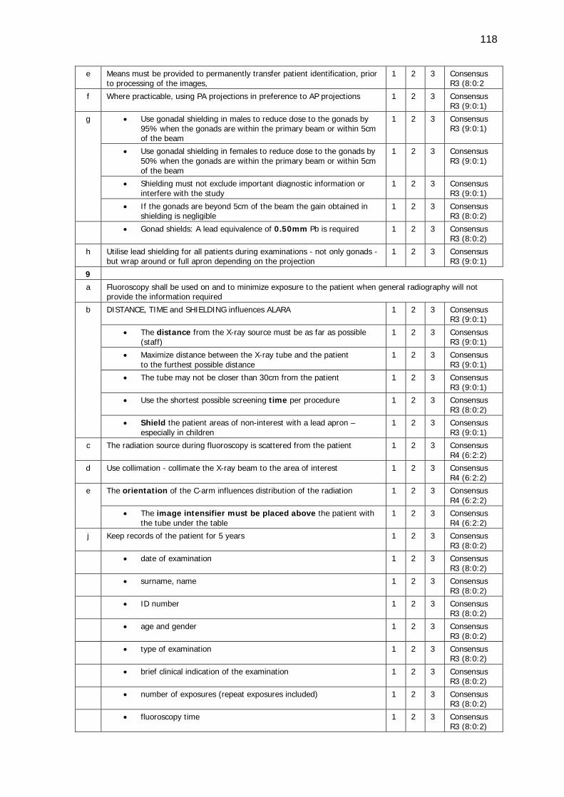
118
e Means must be provided to permanently transfer patient identification, prior to processing of the images,
1 2 3 Consensus R3 (8:0:2
f Where practicable, using PA projections in preference to AP projections 1 2 3 Consensus R3 (9:0:1)
g • Use gonadal shielding in males to reduce dose to the gonads by 95% when the gonads are within the primary beam or within 5cm of the beam
1 2 3 Consensus R3 (9:0:1)
• Use gonadal shielding in females to reduce dose to the gonads by 50% when the gonads are within the primary beam or within 5cm of the beam
1 2 3 Consensus R3 (9:0:1)
• Shielding must not exclude important diagnostic information or interfere with the study
1 2 3 Consensus R3 (9:0:1)
• If the gonads are beyond 5cm of the beam the gain obtained in shielding is negligible
1 2 3 Consensus R3 (8:0:2)
• Gonad shields: A lead equivalence of 0.50mm Pb is required 1 2 3 Consensus R3 (8:0:2)
h Utilise lead shielding for all patients during examinations - not only gonads - but wrap around or full apron depending on the projection
1 2 3 Consensus R3 (9:0:1)
9 a Fluoroscopy shall be used on and to minimize exposure to the patient when general radiography will not
provide the information required
b DISTANCE, TIME and SHIELDING influences ALARA 1 2 3 Consensus R3 (9:0:1)
• The distance from the X-ray source must be as far as possible (staff)
1 2 3 Consensus R3 (9:0:1)
• Maximize distance between the X-ray tube and the patient to the furthest possible distance
1 2 3 Consensus R3 (9:0:1)
• The tube may not be closer than 30cm from the patient 1 2 3 Consensus R3 (9:0:1)
• Use the shortest possible screening time per procedure 1 2 3 Consensus R3 (8:0:2)
• Shield the patient areas of non-interest with a lead apron – especially in children
1 2 3 Consensus R3 (9:0:1)
c The radiation source during fluoroscopy is scattered from the patient 1 2 3 Consensus R4 (6:2:2)
d Use collimation - collimate the X-ray beam to the area of interest
1 2 3 Consensus R4 (6:2:2)
e The orientation of the C-arm influences distribution of the radiation 1 2 3 Consensus R4 (6:2:2)
• The image intensifier must be placed above the patient with the tube under the table
1 2 3 Consensus R4 (6:2:2)
j Keep records of the patient for 5 years 1 2 3 Consensus R3 (8:0:2)
• date of examination 1 2 3 Consensus R3 (8:0:2)
• surname, name 1 2 3 Consensus R3 (8:0:2)
• ID number 1 2 3 Consensus R3 (8:0:2)
• age and gender 1 2 3 Consensus R3 (8:0:2)
• type of examination 1 2 3 Consensus R3 (8:0:2)
• brief clinical indication of the examination 1 2 3 Consensus R3 (8:0:2)
• number of exposures (repeat exposures included) 1 2 3 Consensus R3 (8:0:2)
• fluoroscopy time 1 2 3 Consensus R3 (8:0:2)
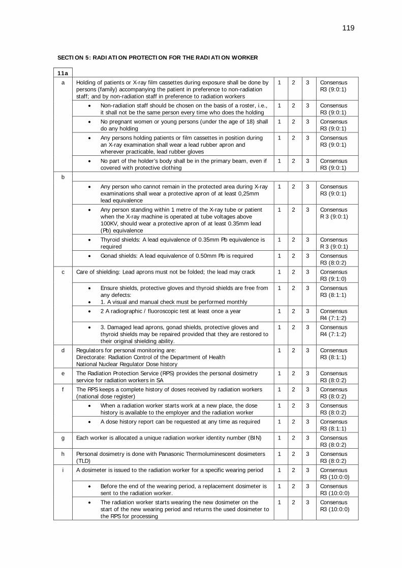
119
SECTION 5: RADIATION PROTECTION FOR THE RADIATION WORKER 11a
a Holding of patients or X-ray film cassettes during exposure shall be done by persons (family) accompanying the patient in preference to non-radiation staff; and by non-radiation staff in preference to radiation workers
1 2 3 Consensus R3 (9:0:1)
• Non-radiation staff should be chosen on the basis of a roster, i.e., it shall not be the same person every time who does the holding
1 2 3 Consensus R3 (9:0:1)
• No pregnant women or young persons (under the age of 18) shall do any holding
1 2 3 Consensus R3 (9:0:1)
• Any persons holding patients or film cassettes in position during an X-ray examination shall wear a lead rubber apron and wherever practicable, lead rubber gloves
1 2 3 Consensus R3 (9:0:1)
• No part of the holder’s body shall be in the primary beam, even if covered with protective clothing
1 2 3 Consensus R3 (9:0:1)
b
• Any person who cannot remain in the protected area during X-ray examinations shall wear a protective apron of at least 0,25mm lead equivalence
1 2 3 Consensus R3 (9:0:1)
• Any person standing within 1 metre of the X-ray tube or patient when the X-ray machine is operated at tube voltages above 100KV, should wear a protective apron of at least 0.35mm lead (Pb) equivalence
1 2 3 Consensus R 3 (9:0:1)
• Thyroid shields: A lead equivalence of 0.35mm Pb equivalence is required
1 2 3 Consensus R 3 (9:0:1)
• Gonad shields: A lead equivalence of 0.50mm Pb is required 1 2 3 Consensus R3 (8:0:2)
c Care of shielding: Lead aprons must not be folded; the lead may crack 1 2 3 Consensus R3 (9:1:0)
• Ensure shields, protective gloves and thyroid shields are free from any defects:
• 1. A visual and manual check must be performed monthly
1 2 3 Consensus R3 (8:1:1)
• 2 A radiographic / fluoroscopic test at least once a year 1 2 3 Consensus R4 (7:1:2)
• 3. Damaged lead aprons, gonad shields, protective gloves and thyroid shields may be repaired provided that they are restored to their original shielding ability.
1 2 3 Consensus R4 (7:1:2)
d Regulators for personal monitoring are: Directorate: Radiation Control of the Department of Health National Nuclear Regulator Dose history
1 2 3 Consensus R3 (8:1:1)
e The Radiation Protection Service (RPS) provides the personal dosimetry service for radiation workers in SA
1 2 3 Consensus R3 (8:0:2)
f The RPS keeps a complete history of doses received by radiation workers (national dose register)
1 2 3 Consensus R3 (8:0:2)
• When a radiation worker starts work at a new place, the dose history is available to the employer and the radiation worker
1 2 3 Consensus R3 (8:0:2)
• A dose history report can be requested at any time as required 1 2 3 Consensus R3 (8:1:1)
g Each worker is allocated a unique radiation worker identity number (BIN) 1 2 3 Consensus R3 (8:0:2)
h Personal dosimetry is done with Panasonic Thermoluminescent dosimeters (TLD)
1 2 3 Consensus R3 (8:0:2)
i A dosimeter is issued to the radiation worker for a specific wearing period 1 2 3 Consensus R3 (10:0:0)
• Before the end of the wearing period, a replacement dosimeter is sent to the radiation worker.
1 2 3 Consensus R3 (10:0:0)
• The radiation worker starts wearing the new dosimeter on the start of the new wearing period and returns the used dosimeter to the RPS for processing
1 2 3 Consensus R3 (10:0:0)
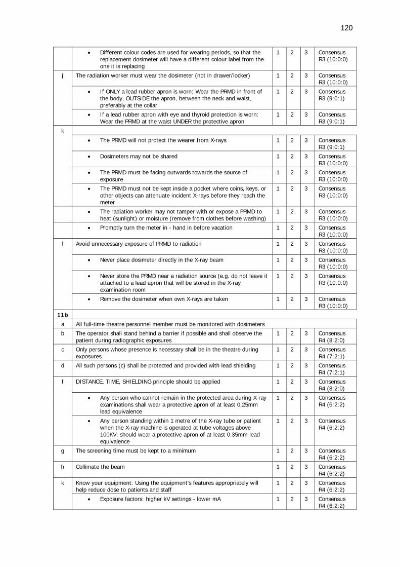
120
• Different colour codes are used for wearing periods, so that the replacement dosimeter will have a different colour label from the one it is replacing
1 2 3 Consensus R3 (10:0:0)
j The radiation worker must wear the dosimeter (not in drawer/locker) 1 2 3 Consensus R3 (10:0:0)
• If ONLY a lead rubber apron is worn: Wear the PRMD in front of the body, OUTSIDE the apron, between the neck and waist, preferably at the collar
1 2 3 Consensus R3 (9:0:1)
• If a lead rubber apron with eye and thyroid protection is worn: Wear the PRMD at the waist UNDER the protective apron
1 2 3 Consensus R3 (9:0:1)
k
• The PRMD will not protect the wearer from X-rays 1 2 3 Consensus R3 (9:0:1)
• Dosimeters may not be shared 1 2 3 Consensus R3 (10:0:0)
• The PRMD must be facing outwards towards the source of exposure
1 2 3 Consensus R3 (10:0:0)
• The PRMD must not be kept inside a pocket where coins, keys, or other objects can attenuate incident X-rays before they reach the meter
1 2 3 Consensus R3 (10:0:0)
• The radiation worker may not tamper with or expose a PRMD to heat (sunlight) or moisture (remove from clothes before washing)
1 2 3 Consensus R3 (10:0:0)
• Promptly turn the meter in - hand in before vacation 1 2 3 Consensus R3 (10:0:0)
l Avoid unnecessary exposure of PRMD to radiation 1 2 3 Consensus R3 (10:0:0)
• Never place dosimeter directly in the X-ray beam 1 2 3 Consensus R3 (10:0:0)
• Never store the PRMD near a radiation source (e.g. do not leave it attached to a lead apron that will be stored in the X-ray examination room
1 2 3 Consensus R3 (10:0:0)
• Remove the dosimeter when own X-rays are taken 1 2 3 Consensus R3 (10:0:0)
11b
a All full-time theatre personnel member must be monitored with dosimeters b The operator shall stand behind a barrier if possible and shall observe the
patient during radiographic exposures 1 2 3 Consensus
R4 (8:2:0)
c Only persons whose presence is necessary shall be in the theatre during exposures
1 2 3 Consensus R4 (7:2:1)
d All such persons (c) shall be protected and provided with lead shielding 1 2 3 Consensus R4 (7:2:1)
f DISTANCE, TIME, SHIELDING principle should be applied 1 2 3 Consensus R4 (8:2:0)
• Any person who cannot remain in the protected area during X-ray examinations shall wear a protective apron of at least 0,25mm lead equivalence
1 2 3 Consensus R4 (6:2:2)
• Any person standing within 1 metre of the X-ray tube or patient when the X-ray machine is operated at tube voltages above 100KV, should wear a protective apron of at least 0.35mm lead equivalence
1 2 3 Consensus R4 (6:2:2)
g The screening time must be kept to a minimum 1 2 3 Consensus R4 (6:2:2)
h Collimate the beam 1 2 3 Consensus R4 (6:2:2)
k Know your equipment: Using the equipment’s features appropriately will help reduce dose to patients and staff
1 2 3 Consensus R4 (6:2:2)
• Exposure factors: higher kV settings - lower mA 1 2 3 Consensus R4 (6:2:2)
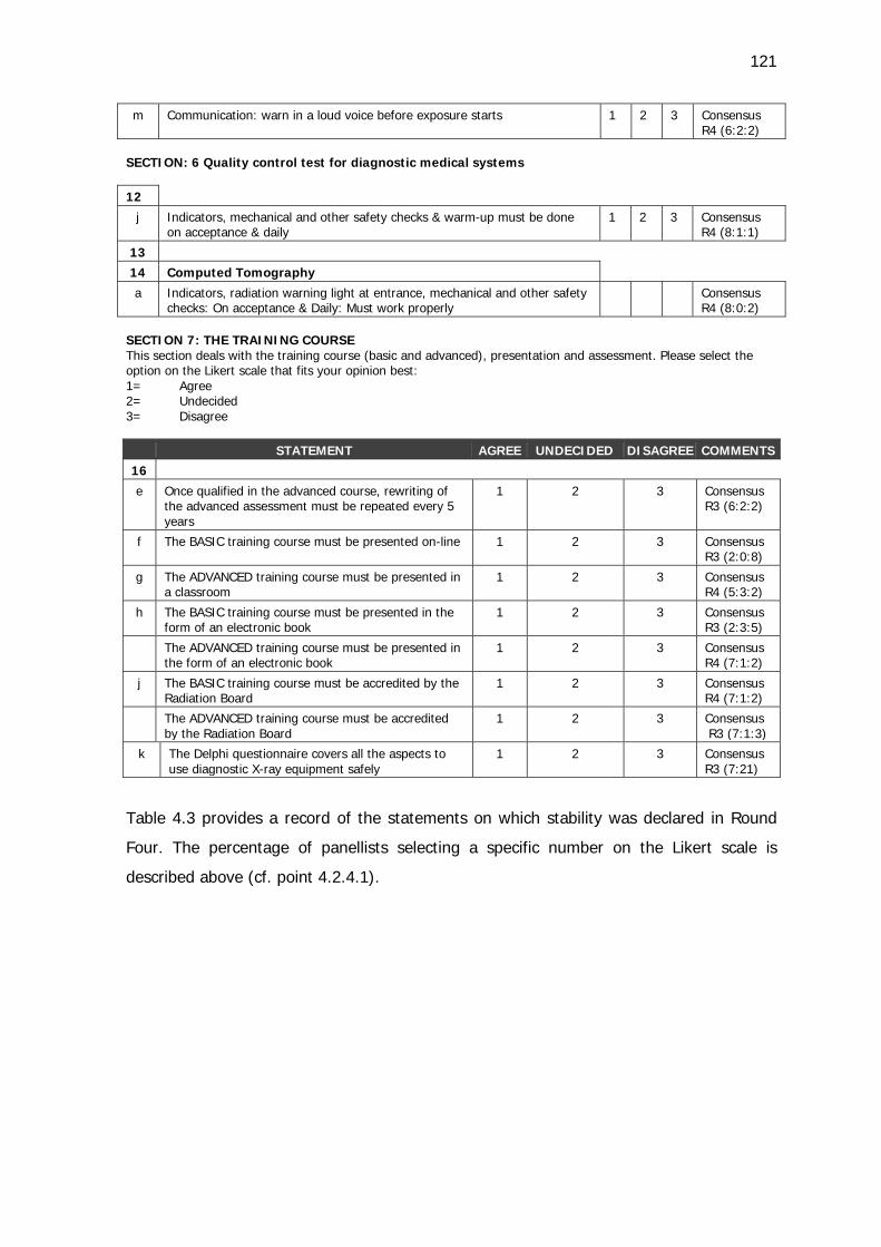
121
m Communication: warn in a loud voice before exposure starts 1 2 3 Consensus R4 (6:2:2)
SECTION: 6 Quality control test for diagnostic medical systems 12
j Indicators, mechanical and other safety checks & warm-up must be done on acceptance & daily
1 2 3 Consensus R4 (8:1:1)
13 14 Computed Tomography
a Indicators, radiation warning light at entrance, mechanical and other safety checks: On acceptance & Daily: Must work properly
Consensus R4 (8:0:2)
SECTION 7: THE TRAINING COURSE This section deals with the training course (basic and advanced), presentation and assessment. Please select the option on the Likert scale that fits your opinion best: 1= Agree 2= Undecided 3= Disagree
STATEMENT AGREE UNDECIDED DISAGREE COMMENTS 16
e Once qualified in the advanced course, rewriting of the advanced assessment must be repeated every 5 years
1 2 3 Consensus R3 (6:2:2)
f The BASIC training course must be presented on-line 1 2 3 Consensus R3 (2:0:8)
g The ADVANCED training course must be presented in a classroom
1 2 3 Consensus R4 (5:3:2)
h The BASIC training course must be presented in the form of an electronic book
1 2 3 Consensus R3 (2:3:5)
The ADVANCED training course must be presented in the form of an electronic book
1 2 3 Consensus R4 (7:1:2)
j The BASIC training course must be accredited by the Radiation Board
1 2 3 Consensus R4 (7:1:2)
The ADVANCED training course must be accredited by the Radiation Board
1 2 3 Consensus R3 (7:1:3)
k The Delphi questionnaire covers all the aspects to use diagnostic X-ray equipment safely
1 2 3 Consensus R3 (7:21)
Table 4.3 provides a record of the statements on which stability was declared in Round
Four. The percentage of panellists selecting a specific number on the Likert scale is
described above (cf. point 4.2.4.1).
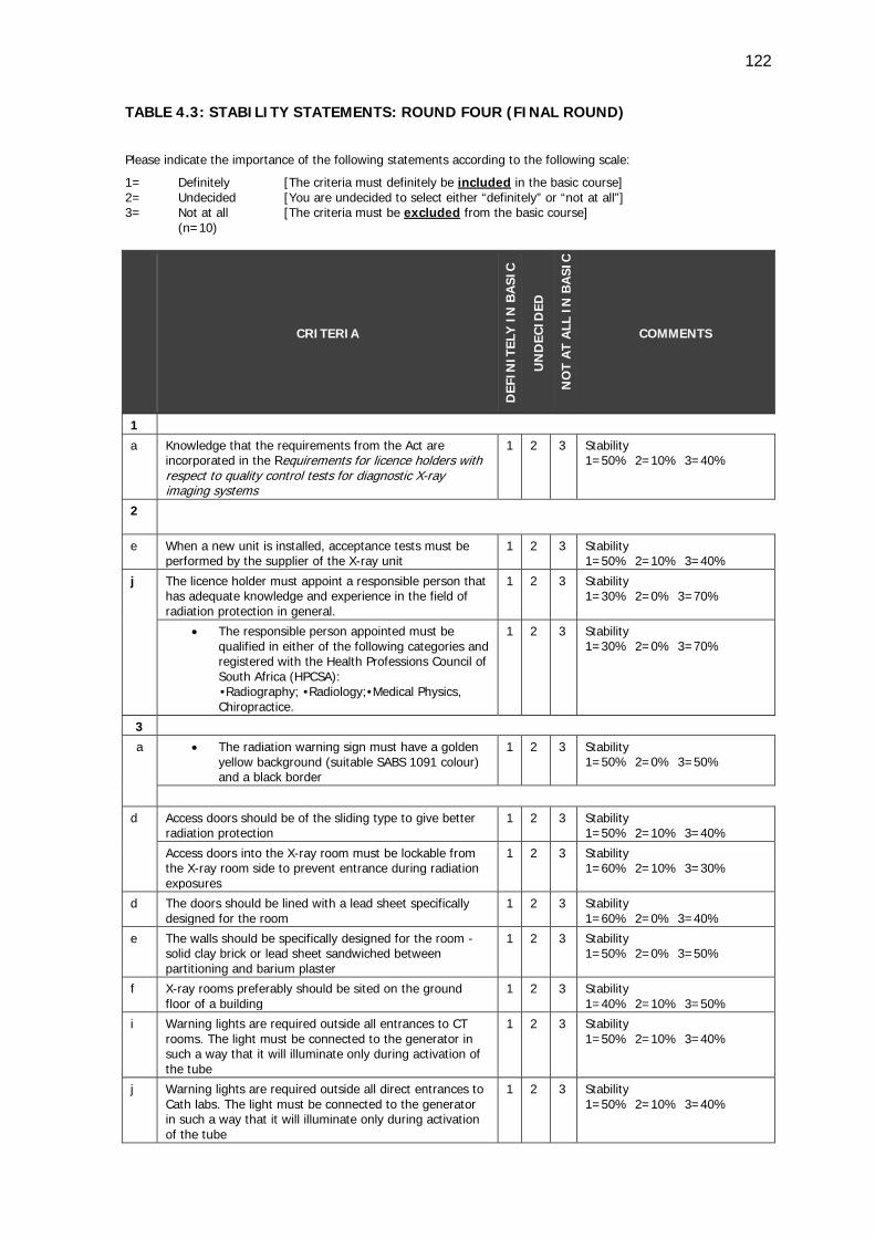
122
TABLE 4.3: STABILITY STATEMENTS: ROUND FOUR (FINAL ROUND)
Please indicate the importance of the following statements according to the following scale:
1= Definitely [The criteria must definitely be included in the basic course] 2= Undecided [You are undecided to select either “definitely” or “not at all”] 3= Not at all [The criteria must be excluded from the basic course] (n=10)
CRITERIA
DEF
INIT
ELY
IN
BA
SIC
UN
DEC
IDED
NO
T A
T A
LL I
N B
ASI
C
COMMENTS
1
a Knowledge that the requirements from the Act are incorporated in the Requirements for licence holders with respect to quality control tests for diagnostic X-ray imaging systems
1 2 3 Stability 1=50% 2=10% 3=40%
2
e When a new unit is installed, acceptance tests must be performed by the supplier of the X-ray unit
1 2 3 Stability 1=50% 2=10% 3=40%
j The licence holder must appoint a responsible person that has adequate knowledge and experience in the field of radiation protection in general.
1 2 3 Stability 1=30% 2=0% 3=70%
• The responsible person appointed must be qualified in either of the following categories and registered with the Health Professions Council of South Africa (HPCSA):
•Radiography; •Radiology;•Medical Physics, Chiropractice.
1 2 3 Stability 1=30% 2=0% 3=70%
3
a
• The radiation warning sign must have a golden yellow background (suitable SABS 1091 colour) and a black border
1 2 3 Stability 1=50% 2=0% 3=50%
d Access doors should be of the sliding type to give better radiation protection
1 2 3 Stability 1=50% 2=10% 3=40%
Access doors into the X-ray room must be lockable from the X-ray room side to prevent entrance during radiation exposures
1 2 3 Stability 1=60% 2=10% 3=30%
d The doors should be lined with a lead sheet specifically designed for the room
1 2 3 Stability 1=60% 2=0% 3=40%
e The walls should be specifically designed for the room - solid clay brick or lead sheet sandwiched between partitioning and barium plaster
1 2 3 Stability 1=50% 2=0% 3=50%
f X-ray rooms preferably should be sited on the ground floor of a building
1 2 3 Stability 1=40% 2=10% 3=50%
i Warning lights are required outside all entrances to CT rooms. The light must be connected to the generator in such a way that it will illuminate only during activation of the tube
1 2 3 Stability 1=50% 2=10% 3=40%
j Warning lights are required outside all direct entrances to Cath labs. The light must be connected to the generator in such a way that it will illuminate only during activation of the tube
1 2 3 Stability 1=50% 2=10% 3=40%
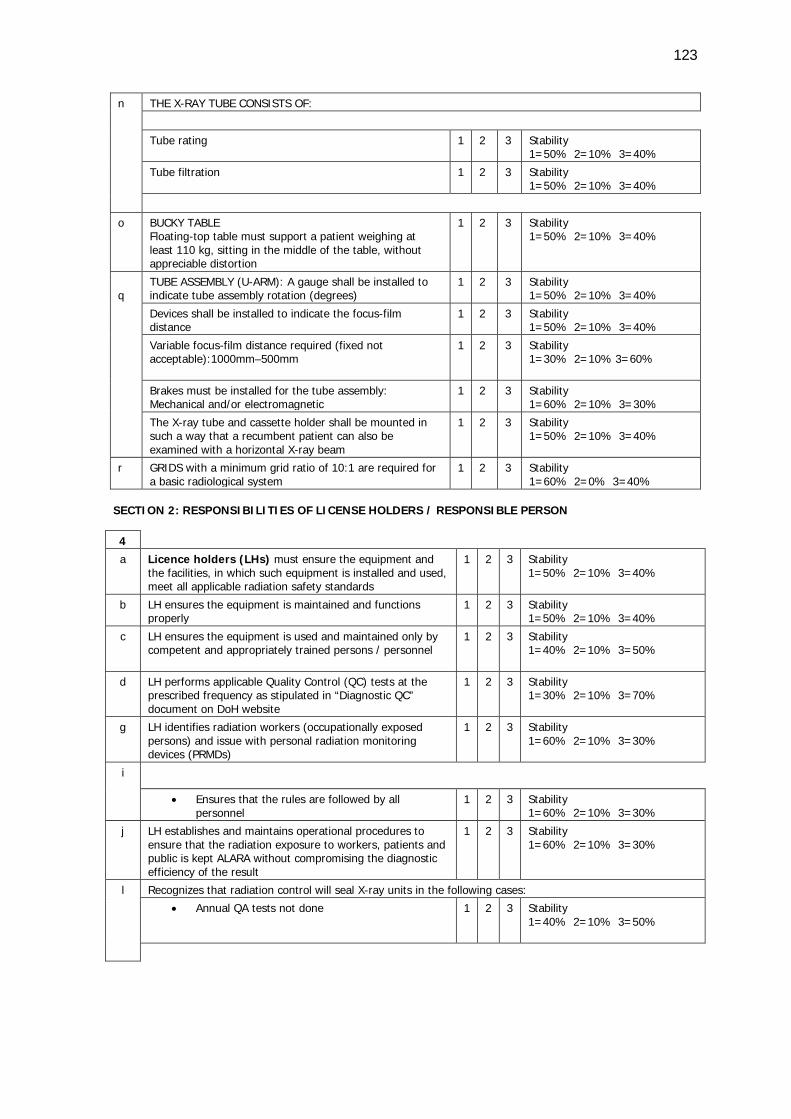
123
n THE X-RAY TUBE CONSISTS OF:
Tube rating 1 2 3 Stability 1=50% 2=10% 3=40%
Tube filtration 1 2 3 Stability 1=50% 2=10% 3=40%
o BUCKY TABLE Floating-top table must support a patient weighing at least 110 kg, sitting in the middle of the table, without appreciable distortion
1 2 3 Stability 1=50% 2=10% 3=40%
q
TUBE ASSEMBLY (U-ARM): A gauge shall be installed to indicate tube assembly rotation (degrees)
1 2 3 Stability 1=50% 2=10% 3=40%
Devices shall be installed to indicate the focus-film distance
1 2 3 Stability 1=50% 2=10% 3=40%
Variable focus-film distance required (fixed not acceptable):1000mm–500mm
1 2 3 Stability 1=30% 2=10% 3=60%
Brakes must be installed for the tube assembly: Mechanical and/or electromagnetic
1 2 3 Stability 1=60% 2=10% 3=30%
The X-ray tube and cassette holder shall be mounted in such a way that a recumbent patient can also be examined with a horizontal X-ray beam
1 2 3 Stability 1=50% 2=10% 3=40%
r GRIDS with a minimum grid ratio of 10:1 are required for a basic radiological system
1 2 3 Stability 1=60% 2=0% 3=40%
SECTION 2: RESPONSIBILITIES OF LICENSE HOLDERS / RESPONSIBLE PERSON 4 a Licence holders (LHs) must ensure the equipment and
the facilities, in which such equipment is installed and used, meet all applicable radiation safety standards
1 2 3 Stability 1=50% 2=10% 3=40%
b LH ensures the equipment is maintained and functions properly
1 2 3 Stability 1=50% 2=10% 3=40%
c LH ensures the equipment is used and maintained only by competent and appropriately trained persons / personnel
1 2 3 Stability 1=40% 2=10% 3=50%
d LH performs applicable Quality Control (QC) tests at the prescribed frequency as stipulated in “Diagnostic QC” document on DoH website
1 2 3 Stability 1=30% 2=10% 3=70%
g LH identifies radiation workers (occupationally exposed persons) and issue with personal radiation monitoring devices (PRMDs)
1 2 3 Stability 1=60% 2=10% 3=30%
i
• Ensures that the rules are followed by all personnel
1 2 3 Stability 1=60% 2=10% 3=30%
j LH establishes and maintains operational procedures to ensure that the radiation exposure to workers, patients and public is kept ALARA without compromising the diagnostic efficiency of the result
1 2 3 Stability 1=60% 2=10% 3=30%
l Recognizes that radiation control will seal X-ray units in the following cases:
• Annual QA tests not done 1 2 3 Stability 1=40% 2=10% 3=50%

124
SECTION 3: OPERATORS OF EQUIPMENT AND RADIATION WORKERS 5 c Supplementary diagnostic radiographers must be
supervised, at least once a week, by a qualified registered radiographer
1 2 3 Stability 1=40% 2=20% 3=40%
• The supervisor is not required to be present at all times. However, the supervisor shall meet with the supervisee on a continuous and regular basis and review the assigned duties as appropriate to the tasks to be performed
1 2 3 Stability 1=40% 2=20% 3=40%
d With effect from 1 July 2009 mammography examinations shall only be performed by qualified radiographers in possession of a recognised additional qualification (post-graduate) in mammography
1 2 3 Stability 1=60% 2=0% 3=40%
6
b Form RC008 (parts A,C,D) must be completed for each radiation worker
1 2 3 Stability 1=50% 2=10% 3=40%
• The completed form RC008 must be kept in the licence holder’s register
1 2 3 Stability 1=40% 2=10% 3=50%
• Licence holders are no longer required to submit form RC008 or inform DoH of any change in the register
1 2 3 Stability 1=30% 2=10% 3=60%
c The holder of the licence must keep record of the following for a period of 10 years for each radiation worker
1 2 3 Stability 1=40% 2=10% 3=50%
• The monthly dose reports furnished by the SABS 1 2 3 Stability 1=60% 2=10% 3=30%
• Results of medical examinations 1 2 3 Stability 1=60% 2=10% 3=30%
d • Annual medical examinations are no longer required by DoH but it remains the prerogative of the licence holder, should he/she deem it necessary
1 2 3 Stability 1=40% 2=10% 3=50%
• Each radiation worker will be required to undergo a medical examination in the event of the following:
• When a radiation occurrence / incident resulting in an abnormally high dose is suspected to have taken place or has been confirmed
1 2 3 Stability 1=50% 2=20% 3=30%
When a medical practitioner deems it necessary 1 2 3 Stability 1=40% 2=20% 3=40%
• When such an examination is considered necessary either by the regulatory authority or the holder of the licence
1 2 3 Stability 1=40% 2=20% 3=40%
• When the radiation worker suspects that his/her health has been, or will be adversely affected by occupational factors
1 2 3 Stability 1=40% 2=20% 3=40%
f • Application forms for a PRMD can be obtained directly from the following current monitoring service provider: SABS Holdings (Pty) Ltd
1 2 3 Stability 1=30% 2=20% 3=50%
The licence holder must ensure that the service provider replaces PRMDs at regular intervals not exceeding 32 days
1 2 3 Stability 1=50% 2=10% 3=40%
• The records must be preserved for a period of 10 years from the date of the last entry
1 2 3 Stability 1=30% 2=20% 3=50%
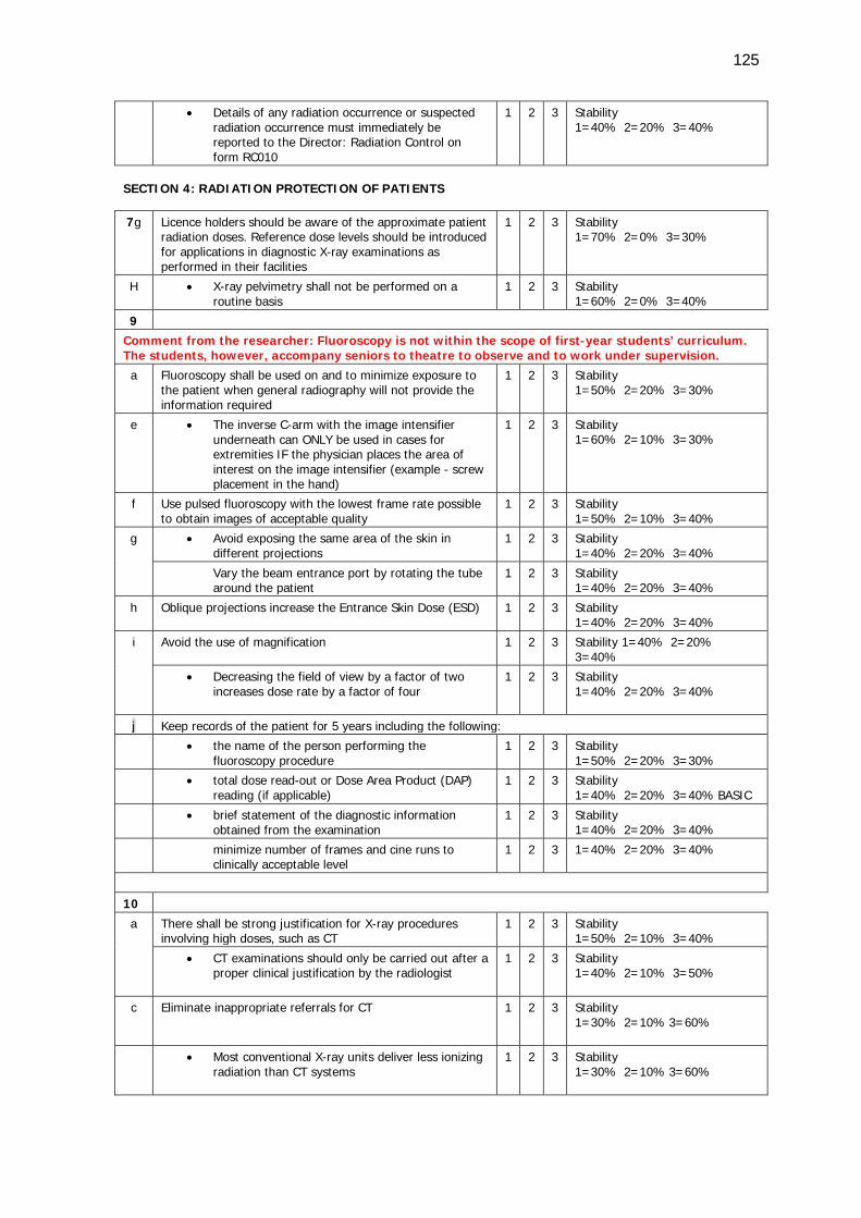
125
• Details of any radiation occurrence or suspected radiation occurrence must immediately be reported to the Director: Radiation Control on form RC010
1 2 3 Stability 1=40% 2=20% 3=40%
SECTION 4: RADIATION PROTECTION OF PATIENTS 7g Licence holders should be aware of the approximate patient
radiation doses. Reference dose levels should be introduced for applications in diagnostic X-ray examinations as performed in their facilities
1 2 3 Stability 1=70% 2=0% 3=30%
H • X-ray pelvimetry shall not be performed on a routine basis
1 2 3 Stability 1=60% 2=0% 3=40%
9 Comment from the researcher: Fluoroscopy is not within the scope of first-year students’ curriculum. The students, however, accompany seniors to theatre to observe and to work under supervision. a Fluoroscopy shall be used on and to minimize exposure to
the patient when general radiography will not provide the information required
1 2 3 Stability 1=50% 2=20% 3=30%
e • The inverse C-arm with the image intensifier underneath can ONLY be used in cases for extremities IF the physician places the area of interest on the image intensifier (example - screw placement in the hand)
1 2 3 Stability 1=60% 2=10% 3=30%
f Use pulsed fluoroscopy with the lowest frame rate possible to obtain images of acceptable quality
1 2 3 Stability 1=50% 2=10% 3=40%
g • Avoid exposing the same area of the skin in different projections
1 2 3 Stability 1=40% 2=20% 3=40%
Vary the beam entrance port by rotating the tube around the patient
1 2 3 Stability 1=40% 2=20% 3=40%
h Oblique projections increase the Entrance Skin Dose (ESD) 1 2 3 Stability 1=40% 2=20% 3=40%
i Avoid the use of magnification 1 2 3 Stability 1=40% 2=20% 3=40%
• Decreasing the field of view by a factor of two increases dose rate by a factor of four
1 2 3 Stability 1=40% 2=20% 3=40%
j Keep records of the patient for 5 years including the following: • the name of the person performing the
fluoroscopy procedure 1 2 3 Stability
1=50% 2=20% 3=30%
• total dose read-out or Dose Area Product (DAP) reading (if applicable)
1 2 3 Stability 1=40% 2=20% 3=40% BASIC
• brief statement of the diagnostic information obtained from the examination
1 2 3 Stability 1=40% 2=20% 3=40%
minimize number of frames and cine runs to clinically acceptable level
1 2 3 1=40% 2=20% 3=40%
10
a There shall be strong justification for X-ray procedures involving high doses, such as CT
1 2 3 Stability 1=50% 2=10% 3=40%
• CT examinations should only be carried out after a proper clinical justification by the radiologist
1 2 3 Stability 1=40% 2=10% 3=50%
c Eliminate inappropriate referrals for CT 1 2 3 Stability 1=30% 2=10% 3=60%
• Most conventional X-ray units deliver less ionizing radiation than CT systems
1 2 3 Stability 1=30% 2=10% 3=60%
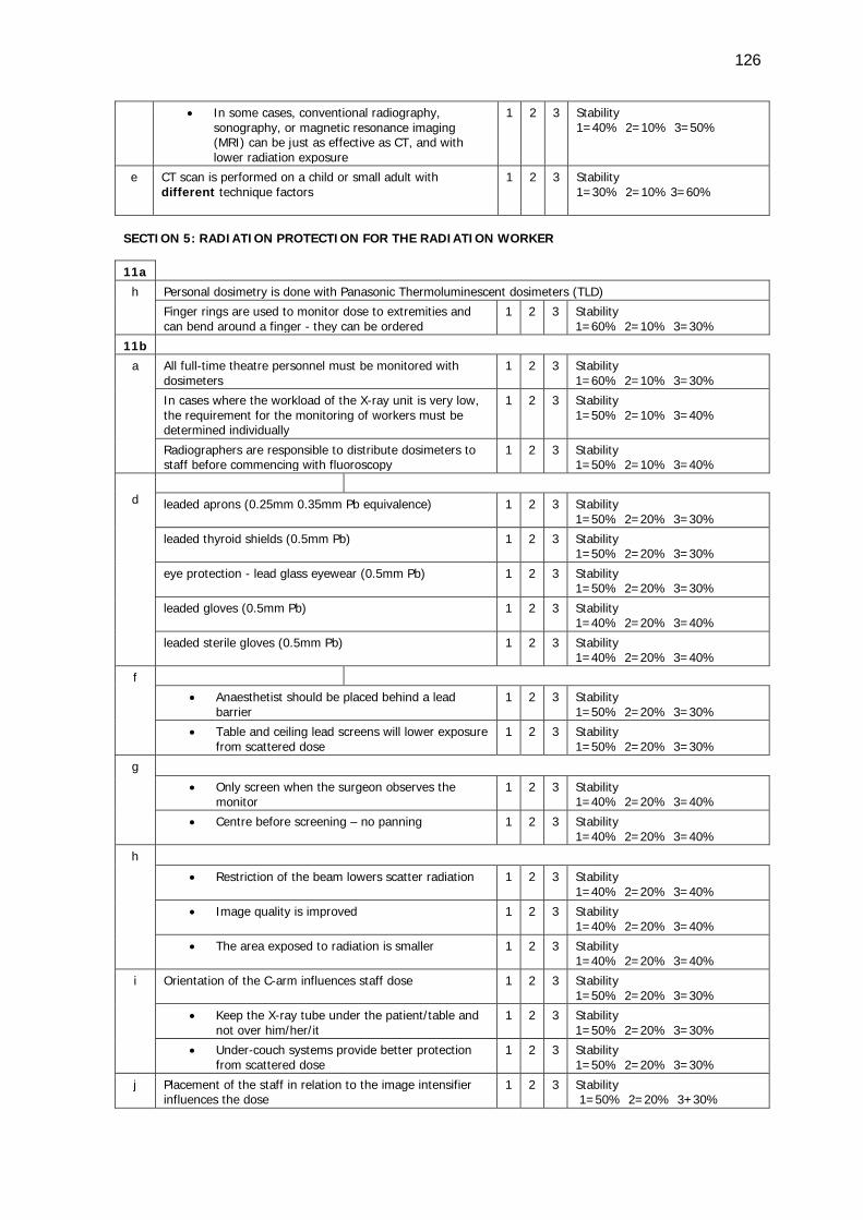
126
• In some cases, conventional radiography, sonography, or magnetic resonance imaging (MRI) can be just as effective as CT, and with lower radiation exposure
1 2 3 Stability 1=40% 2=10% 3=50%
e CT scan is performed on a child or small adult with different technique factors
1 2 3 Stability 1=30% 2=10% 3=60%
SECTION 5: RADIATION PROTECTION FOR THE RADIATION WORKER 11a
h Personal dosimetry is done with Panasonic Thermoluminescent dosimeters (TLD) Finger rings are used to monitor dose to extremities and can bend around a finger - they can be ordered
1 2 3 Stability 1=60% 2=10% 3=30%
11b a All full-time theatre personnel must be monitored with
dosimeters 1 2 3 Stability
1=60% 2=10% 3=30%
In cases where the workload of the X-ray unit is very low, the requirement for the monitoring of workers must be determined individually
1 2 3 Stability 1=50% 2=10% 3=40%
Radiographers are responsible to distribute dosimeters to staff before commencing with fluoroscopy
1 2 3 Stability 1=50% 2=10% 3=40%
d
leaded aprons (0.25mm 0.35mm Pb equivalence) 1 2 3 Stability 1=50% 2=20% 3=30%
leaded thyroid shields (0.5mm Pb) 1 2 3 Stability 1=50% 2=20% 3=30%
eye protection - lead glass eyewear (0.5mm Pb) 1 2 3 Stability 1=50% 2=20% 3=30%
leaded gloves (0.5mm Pb) 1 2 3 Stability 1=40% 2=20% 3=40%
leaded sterile gloves (0.5mm Pb) 1 2 3 Stability 1=40% 2=20% 3=40%
f
• Anaesthetist should be placed behind a lead barrier
1 2 3 Stability 1=50% 2=20% 3=30%
• Table and ceiling lead screens will lower exposure from scattered dose
1 2 3 Stability 1=50% 2=20% 3=30%
g
• Only screen when the surgeon observes the monitor
1 2 3 Stability 1=40% 2=20% 3=40%
• Centre before screening – no panning 1 2 3 Stability 1=40% 2=20% 3=40%
h
• Restriction of the beam lowers scatter radiation 1 2 3 Stability 1=40% 2=20% 3=40%
• Image quality is improved 1 2 3 Stability 1=40% 2=20% 3=40%
• The area exposed to radiation is smaller 1 2 3 Stability 1=40% 2=20% 3=40%
i Orientation of the C-arm influences staff dose 1 2 3 Stability 1=50% 2=20% 3=30%
• Keep the X-ray tube under the patient/table and not over him/her/it
1 2 3 Stability 1=50% 2=20% 3=30%
• Under-couch systems provide better protection from scattered dose
1 2 3 Stability 1=50% 2=20% 3=30%
j Placement of the staff in relation to the image intensifier influences the dose
1 2 3 Stability 1=50% 2=20% 3+30%
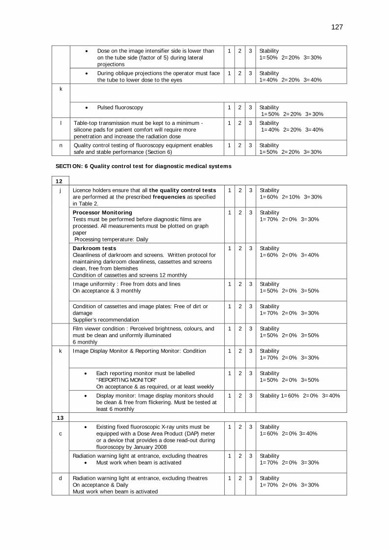
127
• Dose on the image intensifier side is lower than on the tube side (factor of 5) during lateral projections
1 2 3 Stability 1=50% 2=20% 3=30%
• During oblique projections the operator must face the tube to lower dose to the eyes
1 2 3 Stability 1=40% 2=20% 3=40%
k
• Pulsed fluoroscopy 1 2 3 Stability 1=50% 2=20% 3+30%
l Table-top transmission must be kept to a minimum - silicone pads for patient comfort will require more penetration and increase the radiation dose
1 2 3 Stability 1=40% 2=20% 3=40%
n Quality control testing of fluoroscopy equipment enables safe and stable performance (Section 6)
1 2 3 Stability 1=50% 2=20% 3=30%
SECTION: 6 Quality control test for diagnostic medical systems
12
j Licence holders ensure that all the quality control tests are performed at the prescribed frequencies as specified in Table 2.
1 2 3 Stability 1=60% 2=10% 3=30%
Processor Monitoring Tests must be performed before diagnostic films are processed. All measurements must be plotted on graph paper Processing temperature: Daily
1 2 3 Stability 1=70% 2=0% 3=30%
Darkroom tests Cleanliness of darkroom and screens. Written protocol for maintaining darkroom cleanliness, cassettes and screens clean, free from blemishes Condition of cassettes and screens 12 monthly
1 2 3 Stability 1=60% 2=0% 3=40%
Image uniformity : Free from dots and lines On acceptance & 3 monthly
1 2 3 Stability 1=50% 2=0% 3=50%
Condition of cassettes and image plates: Free of dirt or damage Supplier’s recommendation
1 2 3 Stability 1=70% 2=0% 3=30%
Film viewer condition : Perceived brightness, colours, and must be clean and uniformly illuminated 6 monthly
1 2 3 Stability 1=50% 2=0% 3=50%
k Image Display Monitor & Reporting Monitor: Condition 1 2 3 Stability 1=70% 2=0% 3=30%
• Each reporting monitor must be labelled “REPORTING MONITOR” On acceptance & as required, or at least weekly
1 2 3 Stability 1=50% 2=0% 3=50%
• Display monitor: Image display monitors should be clean & free from flickering. Must be tested at least 6 monthly
1 2 3 Stability 1=60% 2=0% 3=40%
13
c
• Existing fixed fluoroscopic X-ray units must be equipped with a Dose Area Product (DAP) meter or a device that provides a dose read-out during fluoroscopy by January 2008
1 2 3 Stability 1=60% 2=0% 3=40%
Radiation warning light at entrance, excluding theatres • Must work when beam is activated
1 2 3 Stability 1=70% 2=0% 3=30%
d Radiation warning light at entrance, excluding theatres On acceptance & Daily Must work when beam is activated
1 2 3 Stability 1=70% 2=0% 3=30%
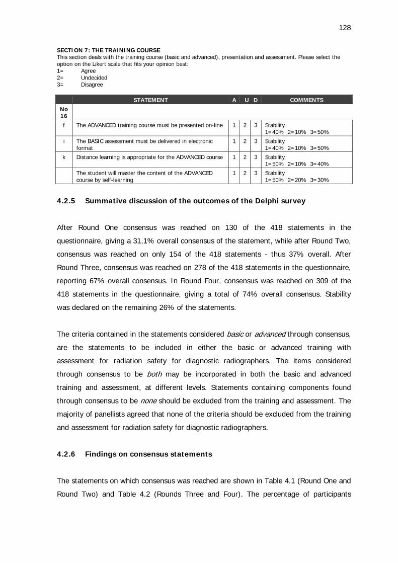
128
SECTION 7: THE TRAINING COURSE This section deals with the training course (basic and advanced), presentation and assessment. Please select the option on the Likert scale that fits your opinion best: 1= Agree 2= Undecided 3= Disagree
STATEMENT A U D COMMENTS
No 16
f The ADVANCED training course must be presented on-line 1 2 3 Stability 1=40% 2=10% 3=50%
i The BASIC assessment must be delivered in electronic format
1 2 3 Stability 1=40% 2=10% 3=50%
k Distance learning is appropriate for the ADVANCED course 1 2 3 Stability 1=50% 2=10% 3=40%
The student will master the content of the ADVANCED course by self-learning
1 2 3 Stability 1=50% 2=20% 3=30%
4.2.5 Summative discussion of the outcomes of the Delphi survey
After Round One consensus was reached on 130 of the 418 statements in the
questionnaire, giving a 31,1% overall consensus of the statement, while after Round Two,
consensus was reached on only 154 of the 418 statements - thus 37% overall. After
Round Three, consensus was reached on 278 of the 418 statements in the questionnaire,
reporting 67% overall consensus. In Round Four, consensus was reached on 309 of the
418 statements in the questionnaire, giving a total of 74% overall consensus. Stability
was declared on the remaining 26% of the statements.
The criteria contained in the statements considered basic or advanced through consensus,
are the statements to be included in either the basic or advanced training with
assessment for radiation safety for diagnostic radiographers. The items considered
through consensus to be both may be incorporated in both the basic and advanced
training and assessment, at different levels. Statements containing components found
through consensus to be none should be excluded from the training and assessment. The
majority of panellists agreed that none of the criteria should be excluded from the training
and assessment for radiation safety for diagnostic radiographers.
4.2.6 Findings on consensus statements
The statements on which consensus was reached are shown in Table 4.1 (Round One and
Round Two) and Table 4.2 (Rounds Three and Four). The percentage of participants
129
choosing a particular point on the Likert scale is indicated in brackets in the far right
column of the tables.
In Table 4.1 the answer columns were indicated as follows:
1 = percentage of participants indicating the criteria to be included in both the basic and
advance training and assessment
2 = percentage of participants indicating the criteria to be included in the basic training
and assessment
3 = percentage of participants indicating the criteria to be included in the advance
training and assessment
4 = percentage of participants indicating the criteria to be excluded (none) from the
training and assessment
In Table 4.2 the answer columns were indicated as follows:
1 = percentage of participants indicating the criteria to be included (definitely in basic) in
the basic training and assessment
2 = percentage of participants indicating to be undecided about the criteria to be included
in the basic training and assessment
3 = percentage of participants indicating the criteria to be excluded (not at all in basic)
from the basic training and assessment.
Considering the combination of the four rounds, the deduction is valid that consensus was
reached on 309 of the 418 statements in the questionnaire, giving a total of 74% of
statements that reached consensus. From these 418 statements the following results
were obtained:
1. Consensus was reached on 13 selections for the option ’both’ on the Likert scale, in
other words 4.2%.
2. Consensus was reached in 131 selections for the option ’basic’ on the Likert scale, in
other words 42.4%.
3. Consensus was reached on 137 selections for the option ’advanced’ on the Likert
scale, in other words 44.3%.
4. No statements were excluded from the training and assessment.
130
5. Consensus was reached on 28 statements for the training course presentation and
assessment, thus 9.1%.
4.2.7 Findings regarding statements on which consensus was not reached
The statements on which consensus was not reached at the end of Round Four, include a
total of 109 out of the total of 418 statements, thus a percentage of 26%. Stability could
be declared on all the remaining statements, thus 100% on all the non-consensus
statements. The panellists all indicated that they did not desire to reconsider opinions
after Round Four.
For the 109 stability statements, it was decided by the researcher and supervisors that all
the statements, for which 50% or more of the Delphi panellists had indicated a specific
point on the Likert scale, would be included in that specific course. For example (cf. Table
4.3, Section1:1c); five panellists made a choice for the basic training, one panellist was
undecided and four indicated that the statement fitted in with the advanced training. In
this example, more panellists indicated preference towards the basic training.
The proof of the engagement of the panellists in the Delphi rounds is supported by the
general comments and opinions. In Round One, 130 comments were made by seven
Delphi panellists, and in Round Two, 48 comments. Nine panellists made 66 comments in
Round Three and five raised 52 opinions in the final round, Round Four. The 28
comments on the non-consensus statements supported the reaching of stability in Round
Four. The specific comments as well as the general comments are indicated in Appendix D
6B and discussed in Chapter 7 (cf. point 7.2) as the implication of the findings of the
Delphi survey. The comments from the panellists are elaborated on in Chapter 7 (cf. point
7.2.1). The mentioned Appendix are summarised in Table 7.1 and it also provides a
summary of the consensus and stability statements with specific reference to the inclusion
of the criteria for the basic or advanced training and assessment. Chapter 5 will be
devoted to the content of the training and assessment for radiation safety with reference
to the comments and recommendation from the Delphi panellists.
4.3 CONCLUSION
This chapter comprises the results of the Delphi survey and explanations of the findings
from the data. A response rate of 100% was obtained for all four rounds.
131
Consensus was pre-defined as when 80% of the panellists agreed on specific criteria.
Consensus was reached on 309 of the 418 statements in the questionnaire, giving a 74%
consensus. From these 418 statements consensus was reached on 13 selections for both
basic and advanced training and assessment, 131 selections for basic training and
assessment and 137 selections for advanced training and assessment, with no exclusion
of any statements from the training and assessment.
Stability may be declared when movement of opinion of the group as a whole has
reached stability. This was reached on the remaining 26% of statements.
The relatively high degree of consensus and stability, combined with no statements
excluded from the training and assessment by a diverse group of panellists, supports the
appropriateness of the conclusions drawn from these data.
As indicated by the title of the following chapter, A standardised training course for
diagnostic radiographers, this chapter is devoted to the content and learning activities
of the training and assessment. This is accomplished by means of a synthesis of the
findings from the literature review and the results from the Delphi survey.
CHAPTER 5
STANDARDISED RADIATION SAFETY TRAINING AND ASSESSMENT FOR
DIAGNOSTIC RADIOGRAPHERS
5.1 INTRODUCTION
The Department of Health (DoH), Directorate Radiation Control (DRC), lists the
responsibilities of license holders of medical X-ray equipment in the Code of Practice for
users of medical X-ray equipment. The licence holder and responsible person, apart from
complying with equipment requirements, must ensure that radiation workers are
identified and issued with personal radiation monitoring devices (PRMDs). The code
further requires every radiation worker to receive education regarding the risks and safety
rules of ionizing radiation (DRC 2011:8). The education pertaining to ionising radiation
safety of these staff members is the responsibility of each license holder, but nationally no
standardised monitoring of the required education currently is in place.
The DRC further lists the responsibilities of the licence holder in the Requirements for
licence holders with respect to quality control tests for diagnostic X-ray imaging systems
(DRC 2012:4). The licence holder must acquire the relevant quality control manuals or
compile in-house written protocols, which describe each test step by step to ensure that
quality control (QC) tests are performed correctly. The licence holder thus must ensure
that persons who perform routine tests are competent to execute the tests.
It therefore is important that the radiographer will be able to interpret the tests, and
adjust specific parameters so that the ALARA principle can be applied and maintained on
a daily basis. The training course proposed in this study is aimed at providing instruction
with regard to radiation protection so that the radiographer be equipped to apply the
recommendations and principles in clinical practice. The requirements regarding radiation
safety, promulgated in the mentioned requirements documents, oblige every licence
holder to ensure that radiation workers are educated in radiation safety aspects.
This study supported the code of practice and the QC test requirements by determining
the specific outcomes students need to attain to comply with the guidelines, in order to
develop a training course with aligned assessment. The study attempts to address the
inconsistency in the training of radiography students as radiation workers (dosimeter
133
holders) and the lack of the skilful implementation of the quality control tests in clinical
practice.
The student preparation before students are issued with dosimeters in practice
necessitates closer inspection. No standardised orientation currently exists for radiography
students before the practices issue dosimeters. The researcher is of the opinion that
neither the training institutions nor the workplace necessarily takes responsibility for the
orientation of the radiation worker before issuing a monitoring device (cf. point 1.2.2).
The institution where the researcher currently is lecturing, arranges a one-hour contact
session by a physicist, followed by a test to the value of 20 marks to that effect. The
requirement is that the student must successfully complete the test with a pass mark of
80%. The current research study makes a formal attempt to address the lack of proof at
the CUT of instruction for radiography students pertaining to requirements of ionising
radiation best practice (cf. point 2.1). It is expected that the implementation of the
training and assessment regarding ionising radiation best practice as learning units in the
curriculum of the Bachelor of Radiography at CUT will contribute to imparting knowledge
of the principles of radiation protection, radiological protection legislation and the factors
that affect the radiation dose of staff and patients.
The methodology employed in developing this training course included a literature study
(cf. Chapter 2), a Delphi survey (cf. Chapter 4), pre- and post-tests (cf. Chapter 6), and,
through action research, the implementation of training for radiographers as part of the
current curriculum. The aim of the research was to provide, in a structured and
systematically researched manner, the outcomes, content and assessment of radiation
safety training.
The DoH in South Africa applies the international standards as legal requirements and
guidelines through the DRC. The DRC issues a licence if the product and usage comply
with the legislative and international requirements for safety and performance (RSA DoH
1973). Two documents, namely Code of Practice (2011) and Requirements for quality
control tests (2012) were consulted to develop the radiation safety training for the
radiography students at CUT. These documents refer to even more documents with more
requirements. The researcher organised the mentioned documents according to a
checklist in different themes to make future referencing to the documents for the student
less confusing (cf. Chapter 2, Table 2.1). The contents of the DRC documents guided the

134
criteria for the Delphi questionnaire with the purpose to define objectives for the training
in radiation safety.
Chapter 1 gave an introduction and orientation to the study. A brief background to the
study was offered, the research problem presented and the foundational research
questions put forward. The overall goals, aim of the study and objectives were then
defined. Subsequently the scope of the study was outlined with reference to its potential
value in diagnostic radiography training. Finally the research design and methods were
introduced.
In Chapter 2, the need for standardised training for radiography students as radiation
workers but also potential licence holders was explored. The chapter deliberated on
different learning strategies, instruments for assessment and the alignment with the
training objectives.
Chapter 3 mapped theoretical perspectives on the research methodology before
describing the research design and methods. The development and utilisation of the
Delphi questionnaire and the pre- and post-assessment as data collection tools were
presented in detail.
Chapter 4 described the measurement instrument for the Delphi survey as developed
form the literature and requirements obtained from the DoH documents. The survey
obtained the opinion of the panellists on what to include in a basic or advanced training
courses. The results were presented and discussed.
This chapter is devoted to a discussion of the development of the teaching and learning
activities for the envisaged training. The elements of the training course will be addressed
in detail and contextualised in the light of the findings of the literature study, Delphi
survey and the assessment.
5.2 DESCRIPTION AND DISCUSSION OF THE CONTENT OF THE RADIATION
SAFETY REQUIREMENT TRAINING
The literature survey resulted in the compilation of a Delphi questionnaire that contained
the requirements and legislation as described in the Code of Practice and Requirements
135
for Quality Control Tests (DRC 2011, 2012). The specific requirements were stated as
criteria statements in the Delphi questionnaire. The criteria statements thus represented
the radiation safety requirements as provided in the relevant documents. The purpose of
the questionnaire was to obtain the opinion of the panellists regarding the inclusion of the
criteria in either the basic or advanced training, or which criteria should be excluded from
the training. The selections of the panellists were considered to compile the content of
both the basic and advanced training.
The Delphi questionnaire was divided in different sections namely Section 1, entitled
GENERAL DEFINITIONS AND LICENCING CONDITIONS (cf. Appendix B1) that
dealt with the requirements and recommendations documents for radiation safety
associated with the use of medical diagnostic X-ray equipment. It also dealt with the
licencing conditions for medical X-ray equipment with specific reference to the
requirements for the apparatus for diagnostic use.
Section 2 of the Delphi questionnaire was entitled RESPONSIBILITIES OF LICENSE
HOLDERS / RESPONSIBLE PERSON (cf. Appendix B1) and dealt with the
responsibilities of licence holders or appointed responsible persons.
Section 3 of the Delphi questionnaire was entitled OPERATORS OF EQUIPMENT AND
RADIATION WORKERS (cf. Appendix B1) and dealt with the operators of diagnostic X-
ray equipment with specifics of the application and monitoring aspects of radiation
workers. The issuing of the personal monitoring device with the detailed threshold dose
limits for radiation workers received attention in this section.
Section 4 of the Delphi questionnaire was entitled RADIATION PROTECTION OF
PATIENTS (cf. Appendix B1) and dealt with the basic radiation principles for the public.
The importance of justification, optimisation and limitation to manage ionising radiation
were stated in order to adhere to the ALARA principle.
Section 5 of the Delphi questionnaire was entitled RADIATION PROTECTION FOR THE
RADIATION WORKER (cf. Appendix B1) and dealt with the basic radiation principles
and personal monitoring devices for the radiation worker. The statements dealt with the
identification and application of principles and techniques to lower the radiation dose to
136
staff in the healthcare environment. The care of the monitoring device with regard to
optimal usage was also specifically stated.
Section 6 of the Delphi questionnaire was entitled QUALITY CONTROL TEST FOR
DIAGNOSTIC MEDICAL SYSTEMS (cf. Appendix B1) and dealt with the requirements
for licence holders with respect to quality control tests for diagnostic imaging systems.
The recording, interpretation and management of the results of the tests received
meticulous focus.
Section 7 of the Delphi questionnaire was entitled THE TRAINING COURSE
(cf. Appendix B1) and dealt with the training course presentation and assessment. The
statements dealt with the learning and teaching activities for the basic and the advanced
course in terms of whether the presentation should be done on-line or in a classroom
setting.
The sections of the Delphi questionnaire were used for the division of the content for the
training and assessment.
5.2.1 Consequence of the Delphi survey
The consensus of the Delphi survey was that no criteria should be excluded from the
training and assessment. The consensus and stability as result of the survey process
either indicated the criteria that should be built in the basic training or advanced training
or both. There were comments from panellists that the basic course should introduce the
learner to the concept, purpose and reasoning behind the concept, but that the advanced
course should include the detailed information (cf. Appendix D6 B). Therefore statements
1a, 1d and 1e, for example (cf. Appendix D6 B), were included in both courses. The
comments from the panellists reiterated that a spiral curriculum (cf. point 2.5.2) would
work best if topics were revisited at different levels of difficulty. Training should be
offered at different levels; not only for entry-users but also for retraining and certification
(cf. point 2.5). The successful completion of the basic training and assessment, for that
reason, will be a prerequisite for the advanced training.
The development of a training course for radiation safety comprises objectives, learning
outcomes, the content, educational strategies, learning opportunities and assessment.
137
What matters is to align the end product with the learning outcomes; therefore, the
Delphi questionnaire criteria statements were defined as intended learning outcomes that
will be discussed in the following section.
5.2.1.1 The training outcomes for radiation safety requirements
The critical cross-field (CCF) education and training outcomes that are mandatory for all
qualifications were described in Chapter 2. The CCF outcomes for the Bachelor of
Diagnostic Radiography (SAQA 2013) are also outlined in Chapter 2 (cf. point 2.5.1.3).
The radiation safety training contributes to the attainment of critical cross-field outcomes
by promoting effective and safe patient care practices to fulfil the patient's needs by
taking into consideration medical law requirements. The training further promotes total
quality management in the radiography profession through the planning of the
frequencies and execution of the QC tests. The training is on par with international
trends, because the QC testing was only implemented in South Africa since 2008 (DRC
2012). The content addresses the absence of formal standardised radiation safety training
as a result of the recent requirement for QC tests.
The National Qualifications Framework (NQF) philosophy indicates that decisions in
respect of learning programme design, development, delivery and assessment need to
consider the learning outcomes that learners need to demonstrate constantly. A
qualification means that the learner has demonstrated applied competence in specific
skills and content areas (SAQA 2000a). In the formulation of objectives with regard to the
outcomes-based training in radiation safety the critical cross-field outcomes, the
competence and the relatively recent requirements for the QC tests were taken into
consideration.
The role of the lecturer is not only to formulate WHAT students should learn, but also
what to DO with it and HOW to learn it (Biggs & Tang 2007:54). The intended learning
outcomes (ILOs) are formulated to give meaning to teaching and learning activities
(TLAs) as well as assessment tasks (ATs). All three factors are steered by a specific verb
with the ultimate goal of changing the perspective of the student. The Delphi
questionnaire brought to light indicators (the criteria) of what a student needs in practice
to master radiation safety - those criteria were reformulated in intended learning
outcomes in order to achieve mastery.
138
The following outcomes were constructed for the basic and advanced training and
assessment:
Upon completion of the basic training the radiographer will be able to recall the
regulations and recommendations as described in the Code of Practice for users of
medical X-ray equipment pertaining to:
1. General definitions and licensing conditions;
2. Responsibilities of license holders / responsible person;
3. Operators of equipment and radiation workers;
4. Radiation protection of patients;
5. Radiation protection for the radiation worker; and
6. Existing quality control testing for diagnostic medical systems.
After completing the advanced training, the radiographer should be able to reflect,
compare, contrast, apply and evaluate the regulations and recommendations pertaining to
the DRC documents (Code of Practice for users of medical X-ray equipment (DRC 2011)
and Requirements for licence holders with respect to quality control tests for Diagnostic X-
ray imaging systems (DRC2012) in order to apply current radiation protection standards
and to optimise procedures.
The specific learning outcomes for each section now will receive attention.
5.2.1.2 The intended learning outcomes
The content of the training was divided into the same sections as the Delphi
questionnaire. Intended learning outcomes were formulated to give meaning to the
teaching and learning activities (cf. 2.5.1.1) and the assessment tasks. The learning
outcomes for each section will be described next.
Section 1 entitled GENERAL DEFINITIONS AND LICENCING included the following
intended learning outcomes:
1. Indicate the Act that governs the safe use of medical X-ray equipment
2. Define the ALARA principle
3. List the documents dealing with radiation safety of the Department of Health (DoH)
139
4. Recall licencing conditions in terms of the awareness of the licence holder and
responsible person
5. Outline basic requirements regarding the premises
6. State the identification of radiation workers.
Section 2 entitled RESPONSIBILITIES OF LICENSE HOLDERS / RESPONSIBLE
PERSON included the following intended learning outcomes:
1. Identify radiation workers
2. Monitor radiation workers
3. Provide protective clothing and devices
4. Maintain equipment to function properly.
Section 3, entitled OPERATORS OF EQUIPMENT AND RADIATION, included the
following intended learning outcomes:
1. Describe the HPCSA registration of radiation workers
2. List the legal operators of X-ray equipment
3. Define the term radiation worker
4. Recognise and name the relevant Application form
5. Describe the conditions and process for the termination of radiation workers
6. Recall the threshold dose limits
7. Distinguish monitoring requirements of radiation workers.
Section 4, entitled RADIATION PROTECTION OF PATIENTS, included the following
intended learning outcomes:
1. Define the terms justification, optimisation and limitation.
2. Identify persons that may request X-rays
3. Name the basic radiographic techniques to limit dose to the patient
4. Describe the meaning of distance, time and shielding in terms of patient protection
5. Categorise the protective devices
6. List the requirements in terms of patient record keeping
7. Express awareness to protect members of the public against ionising radiation
8. Explain techniques for patient protection during fluoroscopy
9. Recall the source of radiation during fluoroscopy.
140
Section 5, entitled RADIATION PROTECTION FOR THE RADIATION WORKER,
included the following intended learning outcomes:
1. Categorise protective clothing
2. Explain the holding of patients / cassettes
3. Discuss distance, time and shielding in terms of staff protection
4. Identify the regulatory body and the South-African Radiation Protection Service
5. Describe the care of the PRMD/TLD/Dosimeter
6. Briefly explain the biological effects of radiation (stochastic and non-stochastic)
7. Briefly describe the measures to limit radiation dose to staff during fluoroscopy
a. C-arm orientation, b. Distance c. Time d. Scatter
Section 6, entitled QUALITY CONTROL TEST FOR DIAGNOSTIC MEDICAL
SYSTEMS, included the following intended learning outcomes for the advanced training
for potential licence holder of medical X-ray equipment:
1. Indicate the Act that governs the safe use of medical X-ray equipment
2. Define the ALARA principle
3. List the requirement documents from the Department of Health (DoH)
4. Explain licencing conditions in terms of the licence holder and responsible person
5. Name the forms for licencing and responsible persons
6. Describe adherence to licencing requirements for premises and products
7. Discuss basic premises requirements
8. Describe how to apply the requirements for radiation workers identification and
monitoring
9. Evaluate the implementation of the ALARA principle
10. Apply the requirements listed the relevant documents from the Department of Health
(DoH) in given scenarios
11. Describe how to implement licencing conditions in terms of the licence holder and
responsible person
12. Specify the forms for licencing and responsible persons
13. Discuss how to adhere to licencing requirements for premises and products
14. Discuss basic premises requirements
15. Discuss the requirements for the identification and monitoring of radiation workers
16. Demonstrate execution of the 3-monthly QC control tests
141
17. Compile a protocol with step by step description of each test
18. Create a portfolio of evidence as proof of a usable working document.
Section 7 was entitled THE TRAINING COURSE. No learning outcomes were formulated
for this section as it pertains to the presentation of the training. The results of Section 7
of the Delphi questionnaire are discussed in Chapter 7 (cf. point 7.4).
The assessment was aligned with the intended learning outcomes and will receive
attention in the following section.
5.3 ASSESSMENT FOR RADIATION SAFETY REQUIREMENTS
The deep approach to learning reiterated by Biggs and Tang (2007:54) entails that the
ILOs are formulated to give meaning to TLAs as well as ATs. The training material
provided to the student in the form of a student guide (cf. Appendix G2) therefore
includes intended learning outcomes, content, learning activities and assessment. The
assessment strategies that were explained in Chapter 2 (cf. point 2.5.3) were taken into
consideration to plan the radiation safety assessment of this research. The publication by
Karpicke (2012) (cf. point 2.6.1) raised the awareness of the researcher to give students
the opportunity to undertake the assessments for the basic and advanced radiation safety
training repeatedly to investigate levels of evidence of mastery. The proof of mastery in
terms of standard setting (cf. point 2.7.2) is a matter of further research but, for this
research, the results of the student questionnaire were discussed in Chapter 6 and the
implication of the findings from the pre-and post-test questionnaires in Chapter 7
(cf. point 7.3). The purpose of the assessment, namely to certify that a specific level of
performance was achieved (cf. point 7.3.1) influenced the teaching and learning activities
(cf. point 7.3.2). The measuring instruments utilised in this research will subsequently
receive attention.
5.3.1 The measuring instrument
The level the student’s knowledge before and after the training was determined by means
of student questionnaires. The content of the student questionnaires had been
determined before the time when the statements (items) for the Delphi questionnaire
were identified. The quantitatively designed questionnaires were accessible on the
142
content management system (CMS) available at the CUT named ’eThuto’. The reason for
employing electronic questionnaires was that in future they would have to be suitable for
assessment involving a broad audience. Other assessment instruments (cf. point 2.5.2)
that were utilised during the presentation of the radiation safety training to the
radiography students concerned were portfolios (cf. point 7.4.1) and OSCEs (cf. point
7.4.2). The results of the portfolio and OSCE were recorded but not reflected in the
results of this research study - this will be addressed in Chapter 7 because it was a
consequence of the action research approach (cf. point 7.4.1 and 7.4.2). An open book
test (cf. point 7.3.4.1) was proposed for inclusion and consideration for the third-year
students’ QC assessment in order to facilitate engagement with the Requirements for
quality control document. The open book test example (cf. Appendix G9) is included in
the student guide (cf. Appendix G2).
The criteria used in the Delphi questionnaires were aligned with the intended learning
outcomes (cf. point 5.2.1.2), and 177 assessment items were formulated for the basic
assessment (cf. point Appendix H1) and 194 items for the advanced assessment (cf. point
Appendix H2). The quantitatively designed questionnaires were compiled with Respondus
Version 4, accessible on the content management system (CMS) available at the CUT. The
analysis of the results of the basic- and advanced student questionnaires are discussed in
Chapter 6.
The student questionnaires for the basic assessment (cf. Appendix H) and the advanced
assessment (cf. Appendix H2) are available as appendices because of the amount of
information. The OSCE, proof of clinical observation and portfolio of evidence will be
explained next.
5.3.1.1 The first-year students’ orientation Objective structured Clinical
Examination (OSCE)
The bachelor first-year students are not placed in clinical practice as was the custom with
the diploma students. It was essential for the researcher to introduce the terminology,
principles and requirements regarding radiation safety in a tangible and visual manner. A
PowerPoint presentation (cf. Appendix G1) formed the basis of the lecture contact
sessions. The students were exposed to the radiography lab to observe components of a
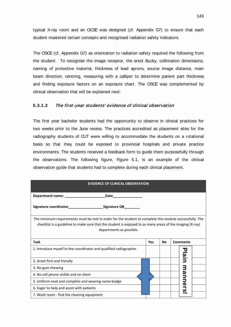
143
typical X-ray room and an OCSE was designed (cf. Appendix G7) to ensure that each
student mastered certain concepts and recognised radiation safety indicators.
The OSCE (cf. Appendix G7) as orientation to radiation safety required the following from
the student: To recognise the image receptor, the erect Bucky, collimation dimensions,
naming of protective material, thickness of lead aprons, source image distance, main
beam direction, centring, measuring with a calliper to determine patient part thickness
and finding exposure factors on an exposure chart. The OSCE was complemented by
clinical observation that will be explained next.
5.3.1.2 The first-year students’ evidence of clinical observation
The first year bachelor students had the opportunity to observe in clinical practices for
two weeks prior to the June recess. The practices accredited as placement sites for the
radiography students of CUT were willing to accommodate the students on a rotational
basis so that they could be exposed to provincial hospitals and private practice
environments. The students received a feedback form to guide them purposefully through
the observations. The following figure, Figure 5.1, is an example of the clinical
observation guide that students had to complete during each clinical placement.
EVIDENCE OF CLINICAL OBSERVATION
Department name: _____________________Date_______________
Signature coordinator__________________Signature QR________
The minimum requirements must be met in order for the student to complete this module successfully. The checklist is a guideline to make sure that the student is exposed to as many areas of the imaging (X-ray)
departments as possible.
Task Yes No Comments
1. Introduce myself to the coordinator and qualified radiographer
2. Greet first and friendly
3. No gum chewing
4. No cell phone visible and on silent
5. Uniform neat and complete and wearing name badge
6. Eager to help and assist with patients
7. Wash room - find the cleaning equipment
Plain m
anners!
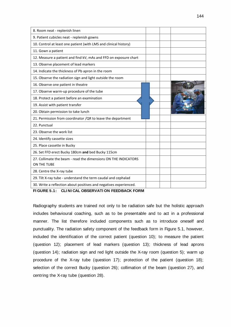
144
8. Room neat - replenish linen
9. Patient cubicles neat - replenish gowns
10. Control at least one patient (with LMS and clinical history)
11. Gown a patient
12. Measure a patient and find kV, mAs and FFD on exposure chart
13. Observe placement of lead markers
14. Indicate the thickness of Pb apron in the room
15. Observe the radiation sign and light outside the room
16. Observe one patient in theatre
17. Observe warm-up procedure of the tube
18. Protect a patient before an examination
19. Assist with patient transfer
20. Obtain permission to take lunch
21. Permission from coordinator /QR to leave the department
22. Punctual
23. Observe the work list
24. Identify cassette sizes
25. Place cassette in Bucky
26. Set FFD erect Bucky 180cm and bed Bucky 115cm
27. Collimate the beam - read the dimensions ON THE INDICATORS ON THE TUBE
28. Centre the X-ray tube
29. Tilt X-ray tube - understand the term caudal and cephalad
30. Write a reflection about positives and negatives experienced. FIGURE 5.1: CLINICAL OBSERVATION FEEDBACK FORM
Radiography students are trained not only to be radiation safe but the holistic approach
includes behavioural coaching, such as to be presentable and to act in a professional
manner. The list therefore included components such as to introduce oneself and
punctuality. The radiation safety component of the feedback form in Figure 5.1, however,
included the identification of the correct patient (question 10); to measure the patient
(question 12); placement of lead markers (question 13); thickness of lead aprons
(question 14); radiation sign and red light outside the X-ray room (question 5); warm up
procedure of the X-ray tube (question 17); protection of the patient (question 18);
selection of the correct Bucky (question 26); collimation of the beam (question 27), and
centring the X-ray tube (question 28).
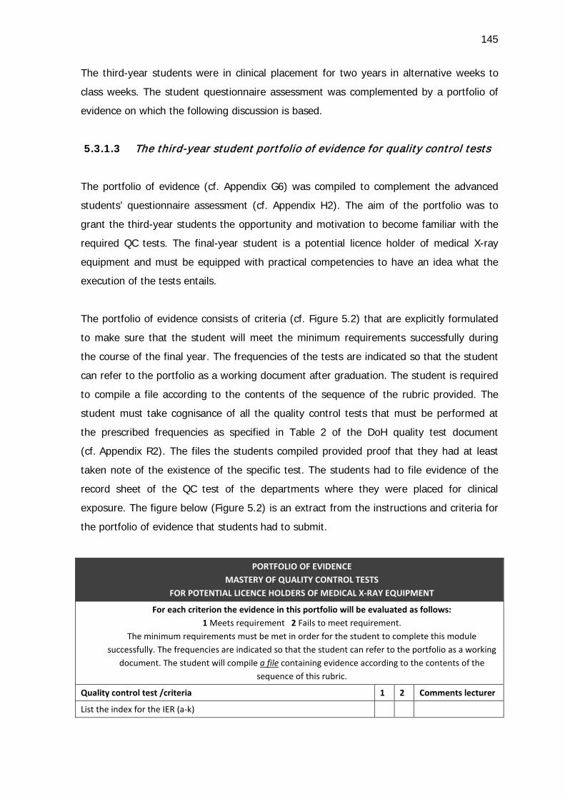
145
The third-year students were in clinical placement for two years in alternative weeks to
class weeks. The student questionnaire assessment was complemented by a portfolio of
evidence on which the following discussion is based.
5.3.1.3 The third-year student portfolio of evidence for quality control tests
The portfolio of evidence (cf. Appendix G6) was compiled to complement the advanced
students’ questionnaire assessment (cf. Appendix H2). The aim of the portfolio was to
grant the third-year students the opportunity and motivation to become familiar with the
required QC tests. The final-year student is a potential licence holder of medical X-ray
equipment and must be equipped with practical competencies to have an idea what the
execution of the tests entails.
The portfolio of evidence consists of criteria (cf. Figure 5.2) that are explicitly formulated
to make sure that the student will meet the minimum requirements successfully during
the course of the final year. The frequencies of the tests are indicated so that the student
can refer to the portfolio as a working document after graduation. The student is required
to compile a file according to the contents of the sequence of the rubric provided. The
student must take cognisance of all the quality control tests that must be performed at
the prescribed frequencies as specified in Table 2 of the DoH quality test document
(cf. Appendix R2). The files the students compiled provided proof that they had at least
taken note of the existence of the specific test. The students had to file evidence of the
record sheet of the QC test of the departments where they were placed for clinical
exposure. The figure below (Figure 5.2) is an extract from the instructions and criteria for
the portfolio of evidence that students had to submit.
PORTFOLIO OF EVIDENCE MASTERY OF QUALITY CONTROL TESTS
FOR POTENTIAL LICENCE HOLDERS OF MEDICAL X-RAY EQUIPMENT
For each criterion the evidence in this portfolio will be evaluated as follows: 1 Meets requirement 2 Fails to meet requirement.
The minimum requirements must be met in order for the student to complete this module successfully. The frequencies are indicated so that the student can refer to the portfolio as a working
document. The student will compile a file containing evidence according to the contents of the sequence of this rubric.
Quality control test /criteria 1 2 Comments lecturer
List the index for the IER (a-k)
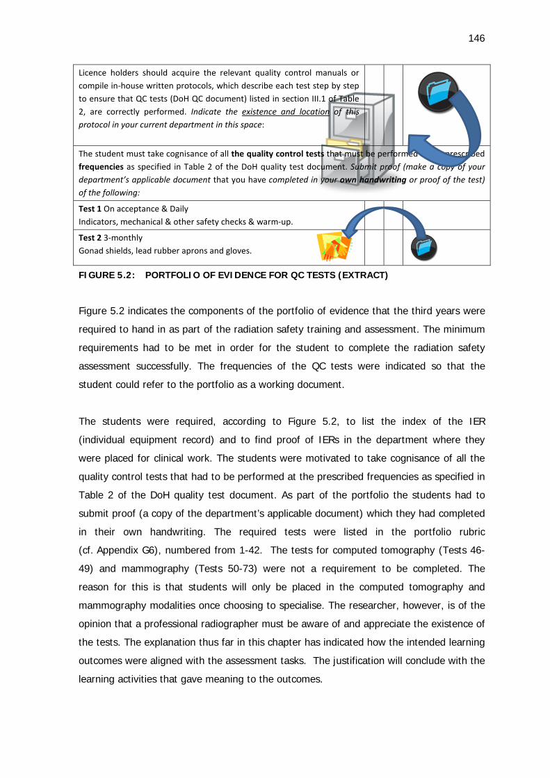
146
Licence holders should acquire the relevant quality control manuals or compile in-house written protocols, which describe each test step by step to ensure that QC tests (DoH QC document) listed in section III.1 of Table 2, are correctly performed. Indicate the existence and location of this protocol in your current department in this space:
The student must take cognisance of all the quality control tests that must be performed at the prescribed frequencies as specified in Table 2 of the DoH quality test document. Submit proof (make a copy of your department’s applicable document that you have completed in your own handwriting or proof of the test) of the following:
Test 1 On acceptance & Daily Indicators, mechanical & other safety checks & warm-up.
Test 2 3-monthly Gonad shields, lead rubber aprons and gloves.
FIGURE 5.2: PORTFOLIO OF EVIDENCE FOR QC TESTS (EXTRACT)
Figure 5.2 indicates the components of the portfolio of evidence that the third years were
required to hand in as part of the radiation safety training and assessment. The minimum
requirements had to be met in order for the student to complete the radiation safety
assessment successfully. The frequencies of the QC tests were indicated so that the
student could refer to the portfolio as a working document.
The students were required, according to Figure 5.2, to list the index of the IER
(individual equipment record) and to find proof of IERs in the department where they
were placed for clinical work. The students were motivated to take cognisance of all the
quality control tests that had to be performed at the prescribed frequencies as specified in
Table 2 of the DoH quality test document. As part of the portfolio the students had to
submit proof (a copy of the department’s applicable document) which they had completed
in their own handwriting. The required tests were listed in the portfolio rubric
(cf. Appendix G6), numbered from 1-42. The tests for computed tomography (Tests 46-
49) and mammography (Tests 50-73) were not a requirement to be completed. The
reason for this is that students will only be placed in the computed tomography and
mammography modalities once choosing to specialise. The researcher, however, is of the
opinion that a professional radiographer must be aware of and appreciate the existence of
the tests. The explanation thus far in this chapter has indicated how the intended learning
outcomes were aligned with the assessment tasks. The justification will conclude with the
learning activities that gave meaning to the outcomes.
147
5.4 TEACHING AND LEARNING ACTIVITIES
Curriculum models include outcomes-based education, problem-based learning, task-
based learning, integrated systems approach and a spiral curriculum. According to Dent
and Harden (2009:3-11), there is no single best curriculum because students have
different ways of learning and therefore the lecturer must provide different opportunities
to accommodate all. A spiral curriculum (cf. point 2.5.2) approach was the preferred
choice in this case, because the content was arranged by revisiting topics throughout the
basic and advanced training at increasing levels of difficulty. Problem solving was
employed as an educational outcome by presenting problems to the students regarding
the content of the course. For instance, the students had to discuss, during basic training,
how the situation of a pregnant woman referred for X-rays after a car accident could be
justified in terms of radiation exposure. Final year students discussed justification in terms
of risk and how to communicate the situation with a referring physician. Students were
encouraged to be active during the training with proof of memorisation and building on
meaning due to the usefulness of the content in everyday clinical practice.
Lectures in radiation safety training enhanced understanding by providing background,
creating connections in student minds, and motivating students to learn. PowerPoint
slides were utilised to summarise the content, and to include graphics for clearer
understanding. The slides were printed for each student as reference (cf. Appendix G1).
The bachelor degree first-year students were not placed in clinical practice as was the
custom with the diploma students. It therefore was essential to introduce the
terminology, principles and requirements regarding radiation safety in a tangible and
visual manner. The visual display made possible with the PowerPoint presentation thus
formed the basis of the lecture contact sessions.
Simulation was incorporated to teach skills in order for a student to rehearse and practise
certain skills, for example the execution of the routine quality control tests (cf. point
5.3.1.3) as part of the portfolio of evidence. Non-technical skills were reinforced by means
of role play, which is especially valuable in preparing for practice. The role play scenarios
(cf. Appendix G11) were created to provide real-life challenges to the students, for
example: A physically disabled patient is referred for X-ray examination of the long bones.
It is impossible for the patient to keep still on his own. He is accompanied by a young
female family member. The aim of the role play in this example was to reiterate the
148
possibility of requesting a family member to assist with the holding of a patient in
preference to a radiation worker. The other role play scenarios are listed in Appendix G11.
Student activities (cf. Appendices G 3-5) were compiled after the student questionnaires
results had been made known in order to address the identified challenging areas. The
analysis of the responses of the students that showed a gap in student achievement is
discussed in Chapter 7. The idea of this learner activity was to engage the students in
order to promote deep learning. The learner activities included a work sheet on which
the student had to match the biological effects of radiation (cf. Appendix G3); fill out the
responsibilities of licence holders (cf. Appendix G4), match the lead (Pb) thickness of the
different protective garments, and indicate the threshold doses for diagnostic radiography
(cf. Appendix G5).
The researcher with the assistance of the Instructional Designer at CUT, recorded a video
of the theatre and of a mobile unit to indicate certain aspects to the students regarding
radiation safety (cf. Appendix G12). The first-year students witnessed the protective dress
of the radiographer and other staff, as well as the c-arm orientations that may be a
challenging abstract concept. The third-year students were reminded through the video of
the higher entrance skin dose during oblique positioning of the c-arm, the distance from
the image intensifier of the c-arm from the patient, and the protection of other theatre
staff.
The result of the action research (Delphi questionnaire, student questionnaire and
lecturing) resulted in the compilation of the student guide for radiation safety. The
student guide (cf. Appendix G2) was compiled to include the following:
1. PowerPoint notes
2. Student activities
3. OSCE
4. Proof of clinical observation
5. Portfolio of evidence for QC tests
6. Role plays
7. DoH requirement documents (including RC forms)
8. Directorate Radiation Control guideline documents
9. 10 pearls of radiation protection.
149
The researcher compiled a lecturer guide for radiation safety (cf. Appendix G10) to
include additional lecturer notes as guide for role-plays, the student competition and
open-book tests, as well as the memorandums of the student activities. The Respondus
assessments for the basic and advanced students were included as hard copies as well as
in electronic format on DVD in the lecturer guide. The video component was included to
round off the support to the lecturer.
The integration of the radiation safety training and assessment in the current curriculum
also warrants some elucidation.
5.5 INTEGRATING RADIATION SAFETY REQUIREMENTS IN THE CURRENT
CURRICULUM
The researcher motivated for the inclusion of the radiation safety training and assessment
as compiled in this research study in the newly developed curriculum of the bachelor
qualification for radiography students by means of a request letter to the Department of
Clinical Sciences at CUT (cf. Appendix F4). The department was requested to consider the
training and assessment to be incorporated in the learning area Radiographic Procedures
and Image Recording of the degree Bachelor of Radiography. The draft student guide
(cf. Appendix G2) as well as the lecturer guide (cf. Appendix G10) were submitted as
evidence of the completed project with the 2014 first and third year students.
The lecturer guide included a total of 346 PowerPoint slides, electronic assessment for the
first year students (177 items), advanced assessment for the third year students (194
items), video on fluoroscopy, lesson plan and a module proposal to integrate the training
and assessment with other learning areas. The learning material and assessment were
made available for future use by the department and radiography lecturers. The proposal
for a complete radiation safety module is provided in Appendix G13. The CUT curriculum
for the Bachelor of Radiography in the past has not included a module dedicated to
radiation safety. The researcher therefore proposed that the learning units for radiation
safety were incorporated in current modules, namely Radiographic Procedures and Image
Recording. The learning units proposed were listed as follows:
1. General definitions and licencing conditions
2. Responsibilities of license holders / responsible person
3. Operators of equipment and radiation workers
4. Radiation protection of patients
150
5. Radiation protection for the radiation worker
6. Quality control test for diagnostic medical systems (second-year tests 1-20)
7. Quality control test for diagnostic medical systems (intermediate third-year tests 23-
36)
8. Quality control test for diagnostic medical systems (advanced fourth-year tests 37-
73).
Learning units one (1) to five (5) are suitable for the first year, but must also be
incorporated to elucidate the detailed aspects of radiation safety in the second, third and
fourth years of study. The quality control test listed as learning units six (6) to eight (8)
can be divided and distributed over the second, third and fourth year to give the students
time to assemble the required proof for the portfolio of evidence. The basic student
questionnaire can be written in the first year and the advanced test may be divided to
spiral to the third and final years. The pre-training tests were executed for research
purposes and will not be used in future. The students will acquire additional information in
the other learning areas that will benefit them by equipping them to confront the
integrated assessment associated with the radiation safety learning units.
5.6 CONCLUSION
This chapter combined the results of the literature research and results of the Delphi
survey to develop assessment and teaching and learning strategies for the radiation
safety training of diagnostic radiographers. The intended learning outcomes and teaching
activities, as well as the assessment methods and procedures were discussed. The
training institution for radiography students is in the process of training radiation workers
and to produce potential licence holders of X-ray equipment. The radiation safety training
and assessment thus are positioned to support the code of practice and the quality
control test requirements by determining the specific outcomes to satisfy the guidelines.
The student guide is aimed at providing standardised content, learning activities and
assessment opportunities. The contextualisation of the training and assessment
presented combined with the action research in this study, if successfully implemented,
might equip the radiographer to apply radiation safety principles and to honour the ALARA
principle more successfully.
In the next chapter, Chapter 6, the results of the pre- and post-assessment will be
reported in detail.
CHAPTER 6
PRE-AND POST-TEST QUESTIONNAIRE FOR RADIATION SAFETY
6.1 INTRODUCTION
This study involved different phases. The literature search established the Delphi
questionnaire statements pertaining to the requirements as described in the DoH
documents, namely Code of Practice (DRC 2011) and Requirements for quality control
tests (DRC 2012). The Delphi panellists indicated the allocation of the statements that
should be included in the basic or advanced section of the training respectively. The
researcher compiled questions for student assessment related to the mentioned Delphi
statements. The main purpose of the student assessment questions were to assess (by
means of pre- and post-training questionnaires) the entry-level participants’ knowledge
regarding safety requirements for radiation workers and for the advanced level student,
their knowledge regarding the Code of Practice for users of medical X-ray equipment and
the Requirements for quality control tests for users of diagnostic X-ray imaging systems.
Chapter 1 gave an introduction by describing a brief background to the study and set the
scene in terms of the research questions. The research design and methods were
announced to guide the sequence of the process.
Chapter 2 expressed the need for standardised training for radiography students as
radiation workers as well as potential licence holders. The learning outcomes and exit
level outcomes of the qualification were discussed in detail.
Chapter 3 mapped the utilisation of the Delphi questionnaire and the pre- and post-
assessment as data collection tools.
Chapter 4 described and analysed the Delphi survey with the final results of the opinion of
the panellists on what to include in a basic or advanced training course.
Chapter 5 discussed the development of the teaching and learning activities guided by the
exit level outcomes for radiography as depicted by the South African Qualifications
Authority.
152
This chapter is devoted to the results and data analysis of the pre-and post-training
student questionnaires. The study population and scheduling of the questionnaires will be
addressed first, followed by the description of the results of the basic and advanced
questionnaires respectively. The student questionnaire results are presented to correlate
with the sections of the Delphi questionnaire.
6.2 POPULATION AND SCHEDULE OF THE TEST QUESTIONNAIRES
A total of 80 radiography students participated in the pre- and post-test questionnaire
survey (n=80). The total number of students was 44 first-year students and 36 third-year
students. The reasons for the difference in the number of participants as described in
Chapter 3 (n=85) will be provided in each section below.
The first-year students wrote the basic test and the third-year students the advanced test.
The first-year students completed the basic pre-test questionnaire during the first class
week (27 January 2014) before being exposed to any form of terminology or principles
pertaining to ionising radiation. The third-year students completed the advanced pre-test
questionnaire before the radiation safety training material had been distributed, thus
relying on knowledge accumulated over the previous years of study in the radiography
programme.
The first-year students wrote the basic post-test questionnaire after eight hours of
lecturer contact time. The contact time comprised lectures with the use of a PowerPoint
presentation and corresponding notes (cf. Appendix G). The first-year students also
participated in practical sessions in the X-ray facility of the institution in order to
familiarise the students with general X-ray equipment, protective tools and exposure
controls. The goal of the contact time was to introduce the students to radiography
terminology, terms and basic radiation protection principles.
The third-year students wrote the advanced post-test questionnaire after six hours’
contact time. During the contact time, each student received the relevant documents of
the DoH, namely the Code of Practice and Requirements for quality control tests. The
PowerPoint notes were made available on the student information system. During the
contact time the third-year students revised the Quality Control (QC) tests that they had
completed during their previous year of study, contained in the learning area referred to
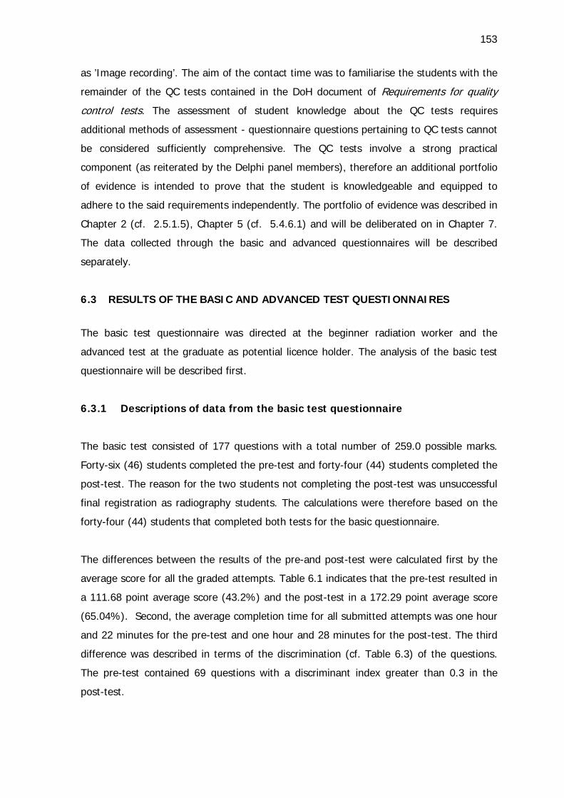
153
as ’Image recording’. The aim of the contact time was to familiarise the students with the
remainder of the QC tests contained in the DoH document of Requirements for quality
control tests. The assessment of student knowledge about the QC tests requires
additional methods of assessment - questionnaire questions pertaining to QC tests cannot
be considered sufficiently comprehensive. The QC tests involve a strong practical
component (as reiterated by the Delphi panel members), therefore an additional portfolio
of evidence is intended to prove that the student is knowledgeable and equipped to
adhere to the said requirements independently. The portfolio of evidence was described in
Chapter 2 (cf. 2.5.1.5), Chapter 5 (cf. 5.4.6.1) and will be deliberated on in Chapter 7.
The data collected through the basic and advanced questionnaires will be described
separately.
6.3 RESULTS OF THE BASIC AND ADVANCED TEST QUESTIONNAIRES
The basic test questionnaire was directed at the beginner radiation worker and the
advanced test at the graduate as potential licence holder. The analysis of the basic test
questionnaire will be described first.
6.3.1 Descriptions of data from the basic test questionnaire
The basic test consisted of 177 questions with a total number of 259.0 possible marks.
Forty-six (46) students completed the pre-test and forty-four (44) students completed the
post-test. The reason for the two students not completing the post-test was unsuccessful
final registration as radiography students. The calculations were therefore based on the
forty-four (44) students that completed both tests for the basic questionnaire.
The differences between the results of the pre-and post-test were calculated first by the
average score for all the graded attempts. Table 6.1 indicates that the pre-test resulted in
a 111.68 point average score (43.2%) and the post-test in a 172.29 point average score
(65.04%). Second, the average completion time for all submitted attempts was one hour
and 22 minutes for the pre-test and one hour and 28 minutes for the post-test. The third
difference was described in terms of the discrimination (cf. Table 6.3) of the questions.
The pre-test contained 69 questions with a discriminant index greater than 0.3 in the
post-test.
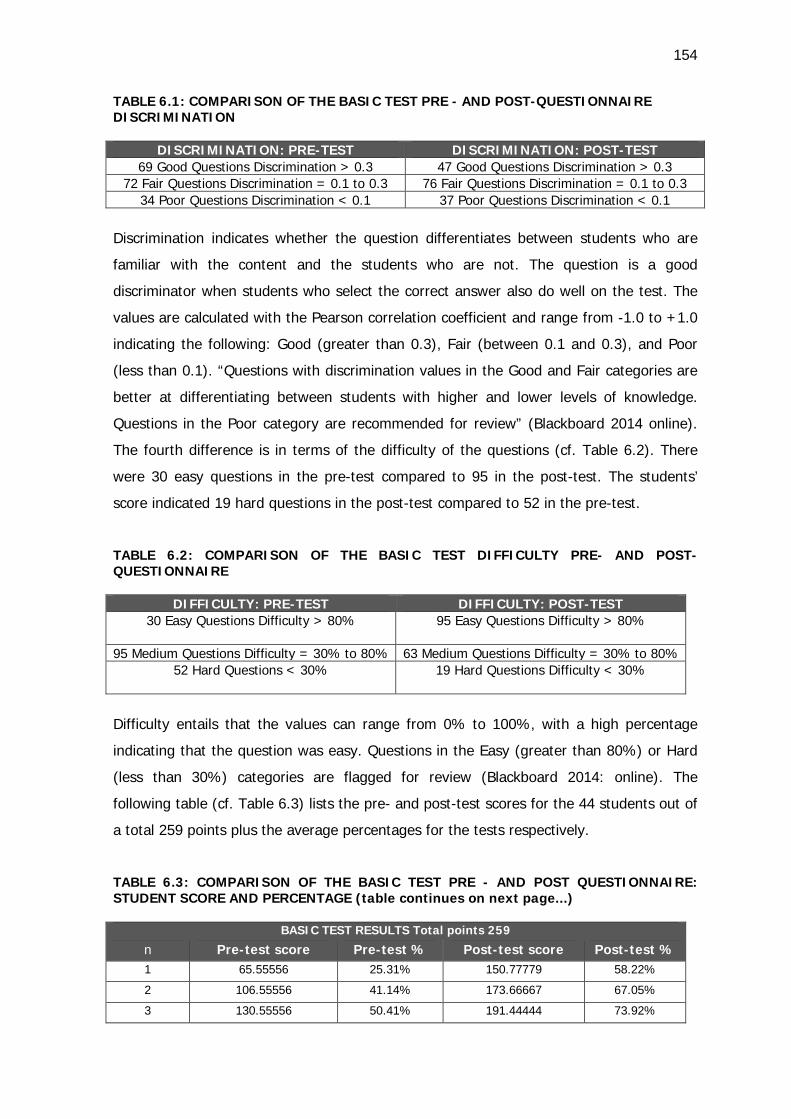
154
TABLE 6.1: COMPARISON OF THE BASIC TEST PRE - AND POST-QUESTIONNAIRE DISCRIMINATION
DISCRIMINATION: PRE-TEST DISCRIMINATION: POST-TEST 69 Good Questions Discrimination > 0.3 47 Good Questions Discrimination > 0.3
72 Fair Questions Discrimination = 0.1 to 0.3 76 Fair Questions Discrimination = 0.1 to 0.3 34 Poor Questions Discrimination < 0.1 37 Poor Questions Discrimination < 0.1
Discrimination indicates whether the question differentiates between students who are
familiar with the content and the students who are not. The question is a good
discriminator when students who select the correct answer also do well on the test. The
values are calculated with the Pearson correlation coefficient and range from -1.0 to +1.0
indicating the following: Good (greater than 0.3), Fair (between 0.1 and 0.3), and Poor
(less than 0.1). “Questions with discrimination values in the Good and Fair categories are
better at differentiating between students with higher and lower levels of knowledge.
Questions in the Poor category are recommended for review” (Blackboard 2014 online).
The fourth difference is in terms of the difficulty of the questions (cf. Table 6.2). There
were 30 easy questions in the pre-test compared to 95 in the post-test. The students’
score indicated 19 hard questions in the post-test compared to 52 in the pre-test.
TABLE 6.2: COMPARISON OF THE BASIC TEST DIFFICULTY PRE- AND POST-QUESTIONNAIRE
DIFFICULTY: PRE-TEST DIFFICULTY: POST-TEST 30 Easy Questions Difficulty > 80%
95 Easy Questions Difficulty > 80%
95 Medium Questions Difficulty = 30% to 80% 63 Medium Questions Difficulty = 30% to 80%
52 Hard Questions < 30%
19 Hard Questions Difficulty < 30%
Difficulty entails that the values can range from 0% to 100%, with a high percentage
indicating that the question was easy. Questions in the Easy (greater than 80%) or Hard
(less than 30%) categories are flagged for review (Blackboard 2014: online). The
following table (cf. Table 6.3) lists the pre- and post-test scores for the 44 students out of
a total 259 points plus the average percentages for the tests respectively.
TABLE 6.3: COMPARISON OF THE BASIC TEST PRE - AND POST QUESTIONNAIRE: STUDENT SCORE AND PERCENTAGE (table continues on next page...)
BASIC TEST RESULTS Total points 259
n Pre-test score Pre-test % Post-test score Post-test % 1 65.55556 25.31% 150.77779 58.22%
2 106.55556 41.14% 173.66667 67.05%
3 130.55556 50.41% 191.44444 73.92%
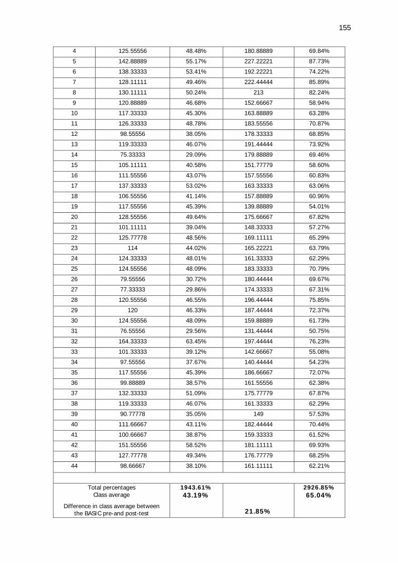
155
4 125.55556 48.48% 180.88889 69.84%
5 142.88889 55.17% 227.22221 87.73%
6 138.33333 53.41% 192.22221 74.22%
7 128.11111 49.46% 222.44444 85.89%
8 130.11111 50.24% 213 82.24%
9 120.88889 46.68% 152.66667 58.94%
10 117.33333 45.30% 163.88889 63.28%
11 126.33333 48.78% 183.55556 70.87%
12 98.55556 38.05% 178.33333 68.85%
13 119.33333 46.07% 191.44444 73.92%
14 75.33333 29.09% 179.88889 69.46%
15 105.11111 40.58% 151.77779 58.60%
16 111.55556 43.07% 157.55556 60.83%
17 137.33333 53.02% 163.33333 63.06%
18 106.55556 41.14% 157.88889 60.96%
19 117.55556 45.39% 139.88889 54.01%
20 128.55556 49.64% 175.66667 67.82%
21 101.11111 39.04% 148.33333 57.27%
22 125.77778 48.56% 169.11111 65.29%
23 114 44.02% 165.22221 63.79%
24 124.33333 48.01% 161.33333 62.29%
25 124.55556 48.09% 183.33333 70.79%
26 79.55556 30.72% 180.44444 69.67%
27 77.33333 29.86% 174.33333 67.31%
28 120.55556 46.55% 196.44444 75.85%
29 120 46.33% 187.44444 72.37%
30 124.55556 48.09% 159.88889 61.73%
31 76.55556 29.56% 131.44444 50.75%
32 164.33333 63.45% 197.44444 76.23%
33 101.33333 39.12% 142.66667 55.08%
34 97.55556 37.67% 140.44444 54.23%
35 117.55556 45.39% 186.66667 72.07%
36 99.88889 38.57% 161.55556 62.38%
37 132.33333 51.09% 175.77779 67.87%
38 119.33333 46.07% 161.33333 62.29%
39 90.77778 35.05% 149 57.53%
40 111.66667 43.11% 182.44444 70.44%
41 100.66667 38.87% 159.33333 61.52%
42 151.55556 58.52% 181.11111 69.93%
43 127.77778 49.34% 176.77779 68.25%
44 98.66667 38.10% 161.11111 62.21%
Total percentages Class average
Difference in class average between the BASIC pre-and post-test
1943.61% 43.19%
21.85%
2926.85% 65.04%
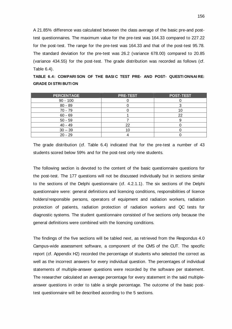
156
A 21.85% difference was calculated between the class average of the basic pre-and post-
test questionnaires. The maximum value for the pre-test was 164.33 compared to 227.22
for the post-test. The range for the pre-test was 164.33 and that of the post-test 95.78.
The standard deviation for the pre-test was 26.2 (variance 678.00) compared to 20.85
(variance 434.55) for the post-test. The grade distribution was recorded as follows (cf.
Table 6.4).
TABLE 6.4: COMPARISON OF THE BASIC TEST PRE- AND POST- QUESTIONNAIRE:
GRADE DISTRIBUTION
PERCENTAGE PRE-TEST POST-TEST
90 - 100 0 0 80 - 89 0 3 70 - 79 0 10 60 - 69 1 22 50 - 59 7 9 40 - 49 22 0 30 – 39 10 0 20 - 29 4 0
The grade distribution (cf. Table 6.4) indicated that for the pre-test a number of 43
students scored below 59% and for the post-test only nine students.
The following section is devoted to the content of the basic questionnaire questions for
the post-test. The 177 questions will not be discussed individually but in sections similar
to the sections of the Delphi questionnaire (cf. 4.2.1.1). The six sections of the Delphi
questionnaire were: general definitions and licencing conditions, responsibilities of licence
holders/responsible persons, operators of equipment and radiation workers, radiation
protection of patients, radiation protection of radiation workers and QC tests for
diagnostic systems. The student questionnaire consisted of five sections only because the
general definitions were combined with the licencing conditions.
The findings of the five sections will be tabled next, as retrieved from the Respondus 4.0
Campus-wide assessment software, a component of the CMS of the CUT. The specific
report (cf. Appendix H2) recorded the percentage of students who selected the correct as
well as the incorrect answers for every individual question. The percentages of individual
statements of multiple-answer questions were recorded by the software per statement.
The researcher calculated an average percentage for every statement in the said multiple-
answer questions in order to table a single percentage. The outcome of the basic post-
test questionnaire will be described according to the 5 sections.
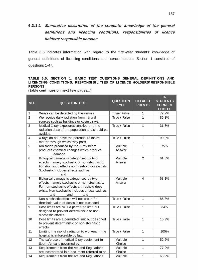
157
6.3.1.1 Summative description of the students’ know ledge of the general
definitions and licencing conditions, responsibilit ies of licence
holders/ responsible persons
Table 6.5 indicates information with regard to the first-year students’ knowledge of
general definitions of licencing conditions and licence holders. Section 1 consisted of
questions 1-47.
TABLE 6.5: SECTION 1: BASIC TEST QUESTIONS GENERAL DEFINITIONS AND LICENCING CONDITIONS: RESPONSIBILITIES OF LICENCE HOLDERS/RESPONSIBLE PERSONS (table continues on next few pages...)
NO. QUESTION TEXT
QUESTION TYPE
DEFAULT POINTS
% STUDENTS CORRECT CHOICE
1 X-rays can be detected by the senses. True/ False 1 72.7% 2 We receive daily radiation from natural
sources such as buildings or cosmic rays. True / False 1 86.3%
3 Medical X-ray exposures contribute to the radiation dose of the population and should be avoided.
True / False 1 31.8%
4 X-rays do not have the potential to ionise matter through which they pass.
True / False 1 90.9%
5 Ionisation produced by the X-ray beam produces chemical changes which produce ________damage.
Multiple Answer
1 75%
6 Biological damage is categorised by two effects, namely stochastic or non-stochastic. For stochastic effects no threshold dose exists. Stochastic includes effects such as ____________and ____________.
Multiple Answer
2 61.3%
7 Biological damage is categorised by two effects, namely stochastic or non-stochastic. For non-stochastic effects a threshold dose exists. Non-stochastic includes effects such as ______and ______and _____and _________.
Multiple Answer
4 68.1%
8 Non-stochastic effects will not occur if a threshold value of doses is not exceeded.
True / False 1 86.3%
9 Dose limits are NOT a permitted limit but designed to prevent deterministic or non- stochastic effects.
True / False 1 34%
10 Dose limits are a permitted limit but designed to prevent deterministic or non-stochastic effects.
True / False 1 15.9%
11 Limiting the risk of radiation to workers in the hospital is enforceable by law.
True / False 1 100%
12 The safe use of medical X-ray equipment in South Africa is governed by
Multiple Choice
1 52.2%
13 Requirements from the Act and Regulations are incorporated in a document referred to as
Multiple Choice
1 77.2%
14 Requirements from the Act and Regulations Multiple 1 65.9%
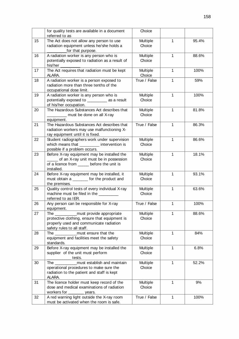
158
for quality tests are available in a document referred to as
Choice
15 The Act does not allow any person to use radiation equipment unless he/she holds a _________for that purpose.
Multiple Choice
1 95.4%
16 A radiation worker is any person who is potentially exposed to radiation as a result of his/her _________
Multiple Choice
1 88.6%
17 The Act requires that radiation must be kept ALARA.
Multiple Choice
1 100%
18 A radiation worker is a person exposed to radiation more than three tenths of the occupational dose limit.
True / False 1 59%
19 A radiation worker is any person who is potentially exposed to _________ as a result of his/her occupation.
Multiple Choice
1 100%
20 The Hazardous Substances Act describes that _________ must be done on all X-ray equipment.
Multiple Choice
1 81.8%
21 The Hazardous Substances Act describes that radiation workers may use malfunctioning X-ray equipment until it is fixed.
True / False 1 86.3%
22 Student radiographers work under supervision which means that _________ intervention is possible if a problem occurs.
Multiple Choice
1 86.6%
23 Before X-ray equipment may be installed the _____ of an X-ray unit must be in possession of a licence from _____ before the unit is installed.
Multiple Choice
1 18.1%
24 Before X-ray equipment may be installed, it must obtain a _______ for the product and the premises.
Multiple Choice
1 93.1%
25 Quality control tests of every individual X-ray machine must be filed in the _________ referred to as IER.
Multiple Choice
1 63.6%
26 Any person can be responsible for X-ray equipment.
True / False 1 100%
27 The __________must provide appropriate protective clothing, ensure that equipment is properly used and communicate radiation safety rules to all staff.
Multiple Choice
1 88.6%
28 The __________must ensure that the equipment and facilities meet the safety standards.
Multiple Choice
1 84%
29 Before X-ray equipment may be installed the supplier of the unit must perform ___________ tests.
Multiple Choice
1 6.8%
30 The __________must establish and maintain operational procedures to make sure the radiation to the patient and staff is kept ALARA.
Multiple Choice
1 52.2%
31 The licence holder must keep record of the dose and medical examinations of radiation workers for _______ years.
Multiple Choice
1 9%
32 A red warning light outside the X-ray room must be activated when the room is safe.
True / False 1 100%
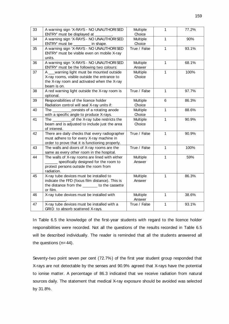
159
33 A warning sign ’X-RAYS - NO UNAUTHORISED ENTRY’ must be displayed at ________.
Multiple Choice
1 77.2%
34 A warning sign ‘X-RAYS - NO UNAUTHORISED ENTRY’ must be ________ in shape.
Multiple Choice
1 90%
35 A warning sign ’X-RAYS - NO UNAUTHORISED ENTRY’ must be visible even on mobile X-ray units.
True / False 1 93.1%
36 A warning sign ’X-RAYS - NO UNAUTHORISED ENTRY’ must be the following two colours:
Multiple Answer
1 68.1%
37 A ___warning light must be mounted outside X-ray rooms, visible outside the entrance to the X-ray room and activated when the X-ray beam is on.
Multiple Choice
1 100%
38 A red warning light outside the X-ray room is optional.
True / False 1 97.7%
39 Responsibilities of the licence holder Radiation control will seal X-ray units if:
Multiple Choice
6 86.3%
40 The _________consists of a rotating anode with a specific angle to produce X-rays.
Multiple Choice
1 88.6%
41 The _________of the X-ray tube restricts the beam and is adjusted to include just the area of interest.
Multiple Choice
1 90.9%
42 There are daily checks that every radiographer must adhere to for every X-ray machine in order to prove that it is functioning properly.
True / False 1 90.9%
43 The walls and doors of X-ray rooms are the same as every other room in the hospital.
True / False 1 100%
44 The walls of X-ray rooms are lined with either ______ specifically designed for the room to protect persons outside the room from radiation.
Multiple Answer
1 59%
45 X-ray tube devices must be installed to indicate the FFD (focus film distance). This is the distance from the _______ to the cassette or film.
Multiple Answer
1 86.3%
46 X-ray tube devices must be installed with __________.
Multiple Answer
1 38.6%
47 X-ray tube devices must be installed with a GRID to absorb scattered X-rays.
True / False 1 93.1%
In Table 6.5 the knowledge of the first-year students with regard to the licence holder
responsibilities were recorded. Not all the questions of the results recorded in Table 6.5
will be described individually. The reader is reminded that all the students answered all
the questions (n=44).
Seventy-two point seven per cent (72.7%) of the first year student group responded that
X-rays are not detectable by the senses and 90.9% agreed that X-rays have the potential
to ionise matter. A percentage of 86.3 indicated that we receive radiation from natural
sources daily. The statement that medical X-ray exposure should be avoided was selected
by 31.8%.
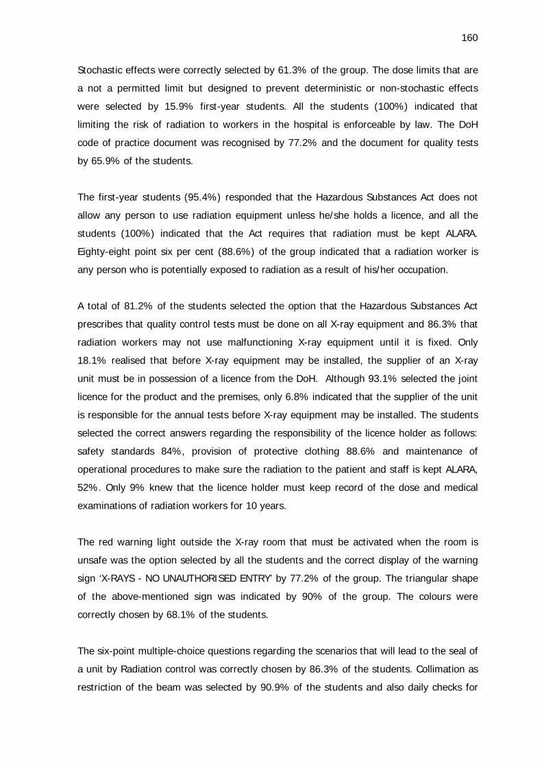
160
Stochastic effects were correctly selected by 61.3% of the group. The dose limits that are
a not a permitted limit but designed to prevent deterministic or non-stochastic effects
were selected by 15.9% first-year students. All the students (100%) indicated that
limiting the risk of radiation to workers in the hospital is enforceable by law. The DoH
code of practice document was recognised by 77.2% and the document for quality tests
by 65.9% of the students.
The first-year students (95.4%) responded that the Hazardous Substances Act does not
allow any person to use radiation equipment unless he/she holds a licence, and all the
students (100%) indicated that the Act requires that radiation must be kept ALARA.
Eighty-eight point six per cent (88.6%) of the group indicated that a radiation worker is
any person who is potentially exposed to radiation as a result of his/her occupation.
A total of 81.2% of the students selected the option that the Hazardous Substances Act
prescribes that quality control tests must be done on all X-ray equipment and 86.3% that
radiation workers may not use malfunctioning X-ray equipment until it is fixed. Only
18.1% realised that before X-ray equipment may be installed, the supplier of an X-ray
unit must be in possession of a licence from the DoH. Although 93.1% selected the joint
licence for the product and the premises, only 6.8% indicated that the supplier of the unit
is responsible for the annual tests before X-ray equipment may be installed. The students
selected the correct answers regarding the responsibility of the licence holder as follows:
safety standards 84%, provision of protective clothing 88.6% and maintenance of
operational procedures to make sure the radiation to the patient and staff is kept ALARA,
52%. Only 9% knew that the licence holder must keep record of the dose and medical
examinations of radiation workers for 10 years.
The red warning light outside the X-ray room that must be activated when the room is
unsafe was the option selected by all the students and the correct display of the warning
sign ‘X-RAYS - NO UNAUTHORISED ENTRY’ by 77.2% of the group. The triangular shape
of the above-mentioned sign was indicated by 90% of the group. The colours were
correctly chosen by 68.1% of the students.
The six-point multiple-choice questions regarding the scenarios that will lead to the seal of
a unit by Radiation control was correctly chosen by 86.3% of the students. Collimation as
restriction of the beam was selected by 90.9% of the students and also daily checks for
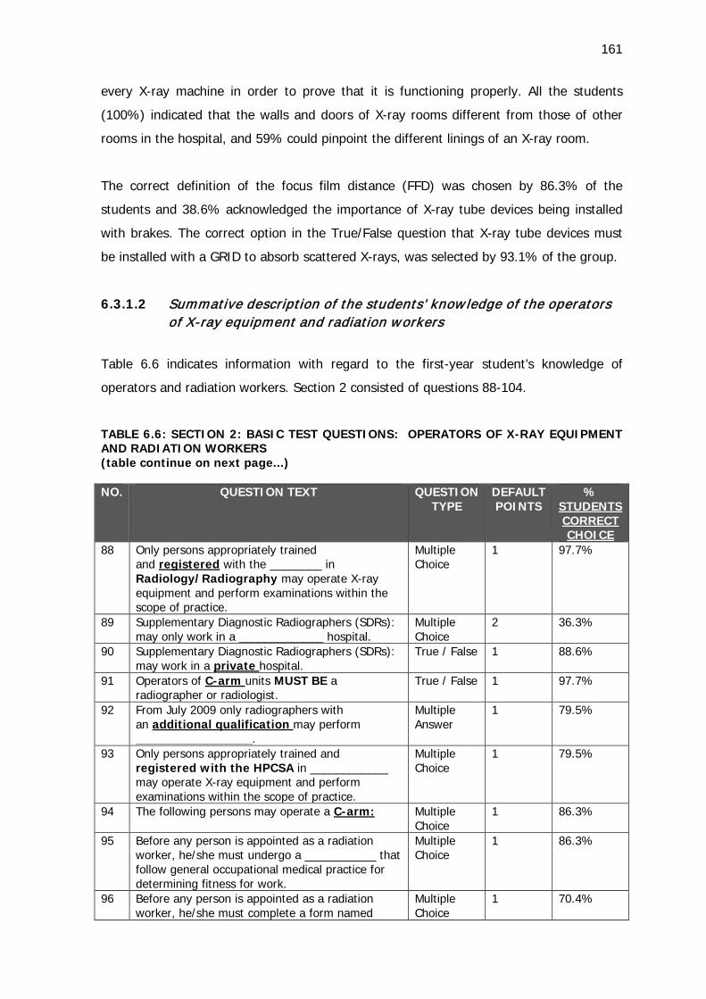
161
every X-ray machine in order to prove that it is functioning properly. All the students
(100%) indicated that the walls and doors of X-ray rooms different from those of other
rooms in the hospital, and 59% could pinpoint the different linings of an X-ray room.
The correct definition of the focus film distance (FFD) was chosen by 86.3% of the
students and 38.6% acknowledged the importance of X-ray tube devices being installed
with brakes. The correct option in the True/False question that X-ray tube devices must
be installed with a GRID to absorb scattered X-rays, was selected by 93.1% of the group.
6.3.1.2 Summative description of the students’ know ledge of the operators of X-ray equipment and radiation workers
Table 6.6 indicates information with regard to the first-year student’s knowledge of
operators and radiation workers. Section 2 consisted of questions 88-104.
TABLE 6.6: SECTION 2: BASIC TEST QUESTIONS: OPERATORS OF X-RAY EQUIPMENT AND RADIATION WORKERS (table continue on next page...) NO. QUESTION TEXT QUESTION
TYPE
DEFAULT POINTS
% STUDENTS CORRECT CHOICE
88 Only persons appropriately trained and registered with the ________ in Radiology/Radiography may operate X-ray equipment and perform examinations within the scope of practice.
Multiple Choice
1 97.7%
89 Supplementary Diagnostic Radiographers (SDRs): may only work in a _____________ hospital.
Multiple Choice
2 36.3%
90 Supplementary Diagnostic Radiographers (SDRs): may work in a private hospital.
True / False 1 88.6%
91 Operators of C-arm units MUST BE a radiographer or radiologist.
True / False 1 97.7%
92 From July 2009 only radiographers with an additional qualification may perform __________________.
Multiple Answer
1 79.5%
93 Only persons appropriately trained and registered with the HPCSA in ____________ may operate X-ray equipment and perform examinations within the scope of practice.
Multiple Choice
1 79.5%
94 The following persons may operate a C-arm: Multiple Choice
1 86.3%
95 Before any person is appointed as a radiation worker, he/she must undergo a ___________ that follow general occupational medical practice for determining fitness for work.
Multiple Choice
1 86.3%
96 Before any person is appointed as a radiation worker, he/she must complete a form named
Multiple Choice
1 70.4%
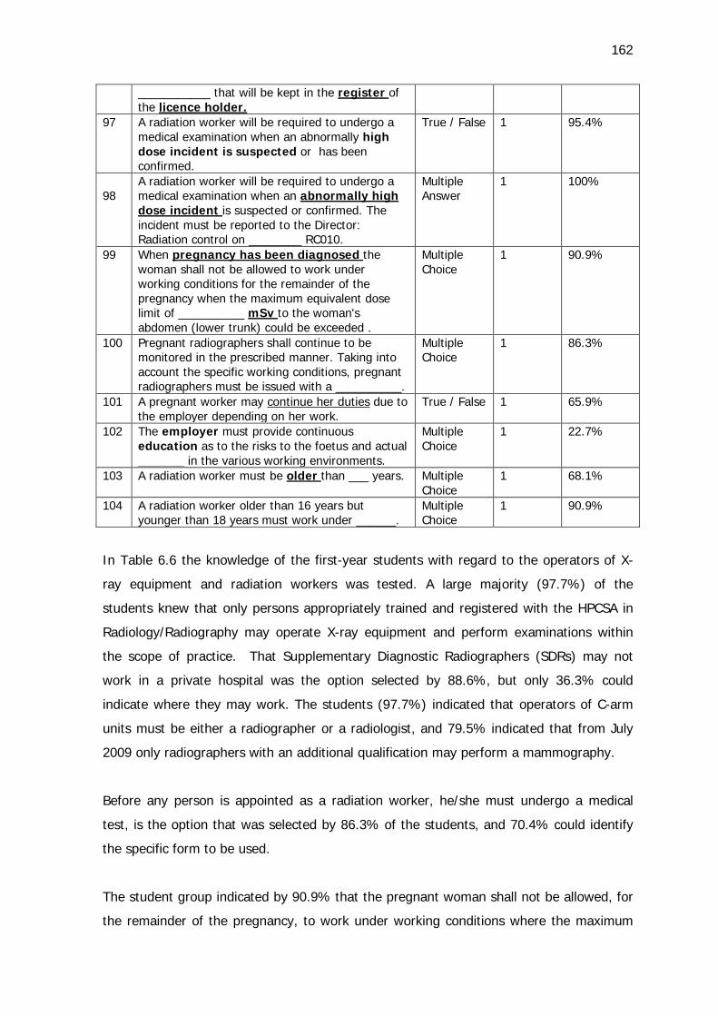
162
___________ that will be kept in the register of the licence holder.
97 A radiation worker will be required to undergo a medical examination when an abnormally high dose incident is suspected or has been confirmed.
True / False 1 95.4%
98
A radiation worker will be required to undergo a medical examination when an abnormally high dose incident is suspected or confirmed. The incident must be reported to the Director: Radiation control on ________ RC010.
Multiple Answer
1 100%
99 When pregnancy has been diagnosed the woman shall not be allowed to work under working conditions for the remainder of the pregnancy when the maximum equivalent dose limit of __________ mSv to the woman's abdomen (lower trunk) could be exceeded .
Multiple Choice
1 90.9%
100 Pregnant radiographers shall continue to be monitored in the prescribed manner. Taking into account the specific working conditions, pregnant radiographers must be issued with a __________.
Multiple Choice
1 86.3%
101 A pregnant worker may continue her duties due to the employer depending on her work.
True / False 1 65.9%
102 The employer must provide continuous education as to the risks to the foetus and actual _______ in the various working environments.
Multiple Choice
1 22.7%
103 A radiation worker must be older than ___ years. Multiple Choice
1 68.1%
104 A radiation worker older than 16 years but younger than 18 years must work under ______.
Multiple Choice
1 90.9%
In Table 6.6 the knowledge of the first-year students with regard to the operators of X-
ray equipment and radiation workers was tested. A large majority (97.7%) of the
students knew that only persons appropriately trained and registered with the HPCSA in
Radiology/Radiography may operate X-ray equipment and perform examinations within
the scope of practice. That Supplementary Diagnostic Radiographers (SDRs) may not
work in a private hospital was the option selected by 88.6%, but only 36.3% could
indicate where they may work. The students (97.7%) indicated that operators of C-arm
units must be either a radiographer or a radiologist, and 79.5% indicated that from July
2009 only radiographers with an additional qualification may perform a mammography.
Before any person is appointed as a radiation worker, he/she must undergo a medical
test, is the option that was selected by 86.3% of the students, and 70.4% could identify
the specific form to be used.
The student group indicated by 90.9% that the pregnant woman shall not be allowed, for
the remainder of the pregnancy, to work under working conditions where the maximum
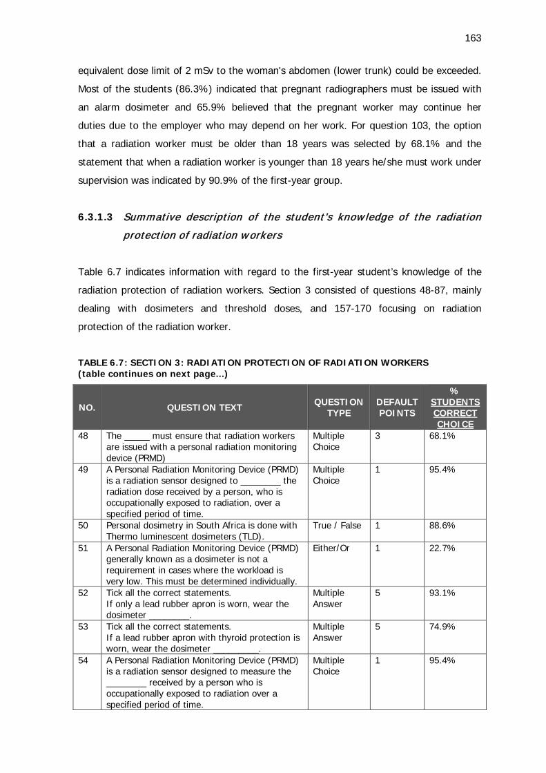
163
equivalent dose limit of 2 mSv to the woman's abdomen (lower trunk) could be exceeded.
Most of the students (86.3%) indicated that pregnant radiographers must be issued with
an alarm dosimeter and 65.9% believed that the pregnant worker may continue her
duties due to the employer who may depend on her work. For question 103, the option
that a radiation worker must be older than 18 years was selected by 68.1% and the
statement that when a radiation worker is younger than 18 years he/she must work under
supervision was indicated by 90.9% of the first-year group.
6.3.1.3 Summative description of the student’s know ledge of the radiation
protection of radiation workers
Table 6.7 indicates information with regard to the first-year student’s knowledge of the
radiation protection of radiation workers. Section 3 consisted of questions 48-87, mainly
dealing with dosimeters and threshold doses, and 157-170 focusing on radiation
protection of the radiation worker.
TABLE 6.7: SECTION 3: RADIATION PROTECTION OF RADIATION WORKERS (table continues on next page...)
NO. QUESTION TEXT
QUESTION TYPE
DEFAULT POINTS
% STUDENTS CORRECT CHOICE
48 The _____ must ensure that radiation workers are issued with a personal radiation monitoring device (PRMD)
Multiple Choice
3 68.1%
49 A Personal Radiation Monitoring Device (PRMD) is a radiation sensor designed to ________ the radiation dose received by a person, who is occupationally exposed to radiation, over a specified period of time.
Multiple Choice
1 95.4%
50 Personal dosimetry in South Africa is done with Thermo luminescent dosimeters (TLD).
True / False 1 88.6%
51 A Personal Radiation Monitoring Device (PRMD) generally known as a dosimeter is not a requirement in cases where the workload is very low. This must be determined individually.
Either/Or 1 22.7%
52 Tick all the correct statements. If only a lead rubber apron is worn, wear the dosimeter ________.
Multiple Answer
5 93.1%
53 Tick all the correct statements. If a lead rubber apron with thyroid protection is worn, wear the dosimeter _________.
Multiple Answer
5 74.9%
54 A Personal Radiation Monitoring Device (PRMD) is a radiation sensor designed to measure the ________ received by a person who is occupationally exposed to radiation over a specified period of time.
Multiple Choice
1 95.4%
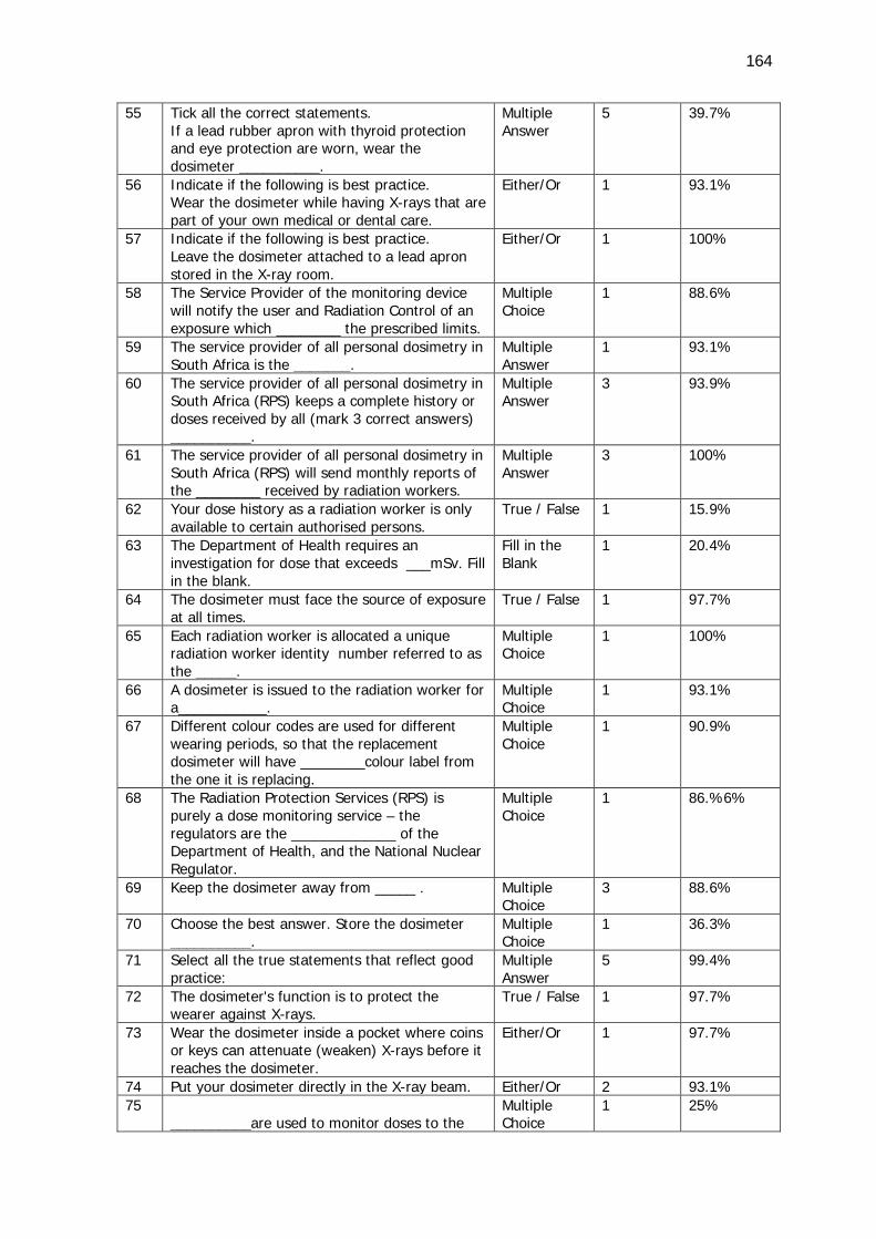
164
55 Tick all the correct statements. If a lead rubber apron with thyroid protection and eye protection are worn, wear the dosimeter __________.
Multiple Answer
5 39.7%
56 Indicate if the following is best practice. Wear the dosimeter while having X-rays that are part of your own medical or dental care.
Either/Or 1 93.1%
57 Indicate if the following is best practice. Leave the dosimeter attached to a lead apron stored in the X-ray room.
Either/Or 1 100%
58 The Service Provider of the monitoring device will notify the user and Radiation Control of an exposure which ________ the prescribed limits.
Multiple Choice
1 88.6%
59 The service provider of all personal dosimetry in South Africa is the _______.
Multiple Answer
1 93.1%
60 The service provider of all personal dosimetry in South Africa (RPS) keeps a complete history or doses received by all (mark 3 correct answers) __________.
Multiple Answer
3 93.9%
61 The service provider of all personal dosimetry in South Africa (RPS) will send monthly reports of the ________ received by radiation workers.
Multiple Answer
3 100%
62 Your dose history as a radiation worker is only available to certain authorised persons.
True / False 1 15.9%
63 The Department of Health requires an investigation for dose that exceeds ___mSv. Fill in the blank.
Fill in the Blank
1 20.4%
64 The dosimeter must face the source of exposure at all times.
True / False 1 97.7%
65 Each radiation worker is allocated a unique radiation worker identity number referred to as the _____.
Multiple Choice
1 100%
66 A dosimeter is issued to the radiation worker for a___________.
Multiple Choice
1 93.1%
67 Different colour codes are used for different wearing periods, so that the replacement dosimeter will have ________colour label from the one it is replacing.
Multiple Choice
1 90.9%
68 The Radiation Protection Services (RPS) is purely a dose monitoring service – the regulators are the _____________ of the Department of Health, and the National Nuclear Regulator.
Multiple Choice
1 86.%6%
69 Keep the dosimeter away from _____ . Multiple Choice
3 88.6%
70 Choose the best answer. Store the dosimeter __________.
Multiple Choice
1 36.3%
71 Select all the true statements that reflect good practice:
Multiple Answer
5 99.4%
72 The dosimeter's function is to protect the wearer against X-rays.
True / False 1 97.7%
73 Wear the dosimeter inside a pocket where coins or keys can attenuate (weaken) X-rays before it reaches the dosimeter.
Either/Or 1 97.7%
74 Put your dosimeter directly in the X-ray beam. Either/Or 2 93.1% 75
__________are used to monitor doses to the Multiple Choice
1 25%
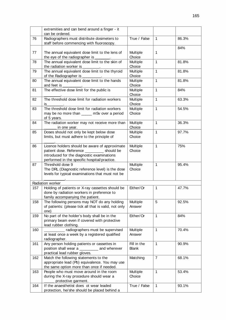
165
extremities and can bend around a finger - it can be ordered.
76 Radiographers must distribute dosimeters to staff before commencing with fluoroscopy.
True / False 1 86.3%
77
The annual equivalent dose limit to the lens of the eye of the radiographer is _______.
Multiple Choice
1
84%
78 The annual equivalent dose limit to the skin of the radiation worker is ________.
Multiple Choice
1 81.8%
79 The annual equivalent dose limit to the thyroid of the Radiographer is _________.
Multiple Choice
1 81.8%
80 The annual equivalent dose limit to the hands and feet is _________.
Multiple Choice
1 81.8%
81 The effective dose limit for the public is ________.
Multiple Choice
1 84%
82 The threshold dose limit for radiation workers is________.
Multiple Choice
1 63.3%
83 The threshold dose limit for radiation workers may be no more than _____ mSv over a period of 5 years.
Multiple Choice
1 54.5%
84 The radiation worker may not receive more than ______ in one year.
Multiple Choice
1 36.3%
85 Doses should not only be kept below dose limits, but must adhere to the principle of _____.
Multiple Choice
1 97.7%
86 Licence holders should be aware of approximate patient dose. Reference _________ should be introduced for the diagnostic examinations performed in the specific hospital/practice.
Multiple Choice
1 75%
87 Threshold dose 9 The DRL (Diagnostic reference level) is the dose levels for typical examinations that must not be __________.
Multiple Choice
1 95.4%
Radiation worker 157 Holding of patients or X-ray cassettes should be
done by radiation workers in preference to family accompanying the patient.
Either/Or 1 47.7%
158 The following persons may NOT do any holding of patients: (please tick all that is valid, not only one)
Multiple Answer
5 92.5%
159 No part of the holder’s body shall be in the primary beam even if covered with protective lead rubber clothing.
Either/Or 1 84%
160 __________ radiographers must be supervised at least once a week by a registered qualified radiographer.
Multiple Answer
1 70.4%
161 Any person holding patients or cassettes in position shall wear a _________ and wherever practical lead rubber gloves.
Fill in the Blank
1 90.9%
162 Match the following statements to the appropriate lead (Pb) equivalence. You may use the same option more than once if needed.
Matching 7 68.1%
163 People who must move around in the room during the X-ray procedure should wear a _____ protective garment.
Multiple Choice
1 53.4%
164 If the anaesthetist does ot wear leaded protection, he/she should be placed behind a
True / False 1 93.1%
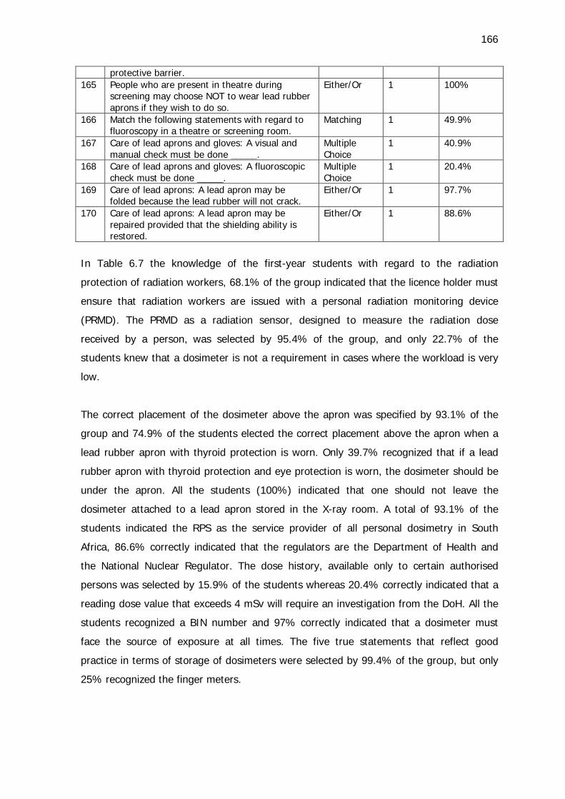
166
protective barrier. 165 People who are present in theatre during
screening may choose NOT to wear lead rubber aprons if they wish to do so.
Either/Or 1 100%
166 Match the following statements with regard to fluoroscopy in a theatre or screening room.
Matching 1 49.9%
167 Care of lead aprons and gloves: A visual and manual check must be done _____.
Multiple Choice
1 40.9%
168 Care of lead aprons and gloves: A fluoroscopic check must be done _____.
Multiple Choice
1 20.4%
169 Care of lead aprons: A lead apron may be folded because the lead rubber will not crack.
Either/Or 1 97.7%
170 Care of lead aprons: A lead apron may be repaired provided that the shielding ability is restored.
Either/Or 1 88.6%
In Table 6.7 the knowledge of the first-year students with regard to the radiation
protection of radiation workers, 68.1% of the group indicated that the licence holder must
ensure that radiation workers are issued with a personal radiation monitoring device
(PRMD). The PRMD as a radiation sensor, designed to measure the radiation dose
received by a person, was selected by 95.4% of the group, and only 22.7% of the
students knew that a dosimeter is not a requirement in cases where the workload is very
low.
The correct placement of the dosimeter above the apron was specified by 93.1% of the
group and 74.9% of the students elected the correct placement above the apron when a
lead rubber apron with thyroid protection is worn. Only 39.7% recognized that if a lead
rubber apron with thyroid protection and eye protection is worn, the dosimeter should be
under the apron. All the students (100%) indicated that one should not leave the
dosimeter attached to a lead apron stored in the X-ray room. A total of 93.1% of the
students indicated the RPS as the service provider of all personal dosimetry in South
Africa, 86.6% correctly indicated that the regulators are the Department of Health and
the National Nuclear Regulator. The dose history, available only to certain authorised
persons was selected by 15.9% of the students whereas 20.4% correctly indicated that a
reading dose value that exceeds 4 mSv will require an investigation from the DoH. All the
students recognized a BIN number and 97% correctly indicated that a dosimeter must
face the source of exposure at all times. The five true statements that reflect good
practice in terms of storage of dosimeters were selected by 99.4% of the group, but only
25% recognized the finger meters.
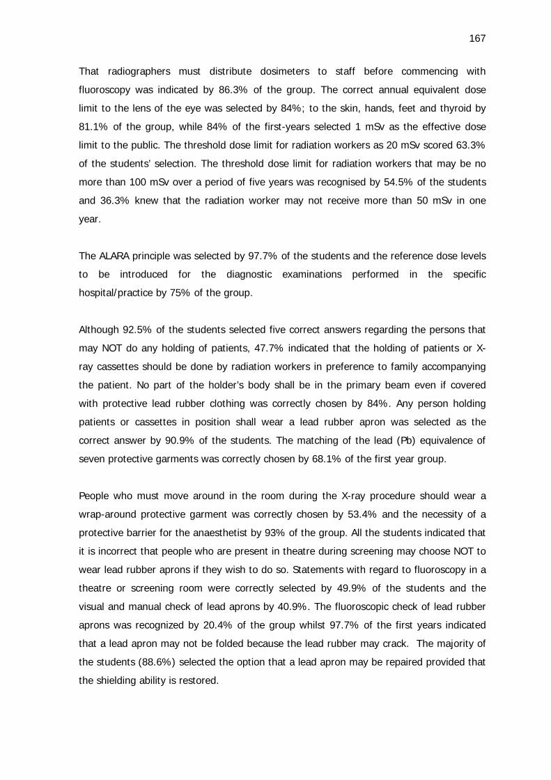
167
That radiographers must distribute dosimeters to staff before commencing with
fluoroscopy was indicated by 86.3% of the group. The correct annual equivalent dose
limit to the lens of the eye was selected by 84%; to the skin, hands, feet and thyroid by
81.1% of the group, while 84% of the first-years selected 1 mSv as the effective dose
limit to the public. The threshold dose limit for radiation workers as 20 mSv scored 63.3%
of the students’ selection. The threshold dose limit for radiation workers that may be no
more than 100 mSv over a period of five years was recognised by 54.5% of the students
and 36.3% knew that the radiation worker may not receive more than 50 mSv in one
year.
The ALARA principle was selected by 97.7% of the students and the reference dose levels
to be introduced for the diagnostic examinations performed in the specific
hospital/practice by 75% of the group.
Although 92.5% of the students selected five correct answers regarding the persons that
may NOT do any holding of patients, 47.7% indicated that the holding of patients or X-
ray cassettes should be done by radiation workers in preference to family accompanying
the patient. No part of the holder’s body shall be in the primary beam even if covered
with protective lead rubber clothing was correctly chosen by 84%. Any person holding
patients or cassettes in position shall wear a lead rubber apron was selected as the
correct answer by 90.9% of the students. The matching of the lead (Pb) equivalence of
seven protective garments was correctly chosen by 68.1% of the first year group.
People who must move around in the room during the X-ray procedure should wear a
wrap-around protective garment was correctly chosen by 53.4% and the necessity of a
protective barrier for the anaesthetist by 93% of the group. All the students indicated that
it is incorrect that people who are present in theatre during screening may choose NOT to
wear lead rubber aprons if they wish to do so. Statements with regard to fluoroscopy in a
theatre or screening room were correctly selected by 49.9% of the students and the
visual and manual check of lead aprons by 40.9%. The fluoroscopic check of lead rubber
aprons was recognized by 20.4% of the group whilst 97.7% of the first years indicated
that a lead apron may not be folded because the lead rubber may crack. The majority of
the students (88.6%) selected the option that a lead apron may be repaired provided that
the shielding ability is restored.
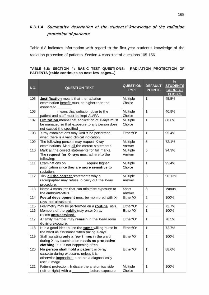
168
6.3.1.4 Summative description of the students’ know ledge of the radiation
protection of patients
Table 6.8 indicates information with regard to the first-year student’s knowledge of the
radiation protection of patients. Section 4 consisted of questions 105-156.
TABLE 6.8: SECTION 4: BASIC TEST QUESTIONS: RADIATION PROTECTION OF PATIENTS (table continues on next few pages...)
NO. QUESTION TEXT
QUESTION TYPE
DEFAULT POINTS
% STUDENTS CORRECT CHOICE
105 Justification means that the radiation examination benefit must be higher than the associated _______.
Multiple Choice
1 45.5%
106 _________means that radiation dose to the patient and staff must be kept ALARA.
Multiple Choice
1 40.9%
107 Limitation means that application of X-rays must be managed so that exposure to any person does not exceed the specified ________.
Multiple Choice
1 88.6%
108 X-ray examinations may ONLY be performed when there is a valid clinical indication.
Either/Or 1 95.4%
109 The following persons may request X-ray examinations: Mark all the correct statements
Multiple Answer
5 72.1%
110 Mark all the correct statements for full marks. The request for X-rays must adhere to the following:
Multiple Answer
5 94.3%
111 Examinations on __________ require higher justification since they are more sensitive to radiation.
Multiple Choice
1 95.4%
112 Tick all the correct statements why a radiographer may refuse o carry out the X-ray procedure.
Multiple Answer
3 90.13%
113 Name 4 measures that can minimise exposure to the embryo/foetus
Short Answer
8 Manual
114 Foetal development must be monitored with X-rays, not ultrasound.
Either/Or 2 100%
115 Pelvimetry may be performed on a routine asis. Either/Or 2 72.7% 116 Members of the public may enter X-ray
rooms unsupervised. Either/Or 1 100%
117 A family member may remain in the X-ray room during exposure.
Either/Or 1 70.5%
118 It is a good idea to use the same willing nurse in the ward as assistance when taking X-rays.
Either/Or 1 72.7%
119 Staff assisting only a few times in the ward during X-ray examination needs no protective clothing if it is not happening often.
Either/Or 1 100%
120 No person shall hold a patient or X-ray cassette during exposure, unless it is otherwise impossible to obtain a diagnostically useful image.
Either/Or 1 88.6%
121 Patient protection: Indicate the anatomical side (left or right) with a _________ before exposure.
Multiple Choice
1 100%
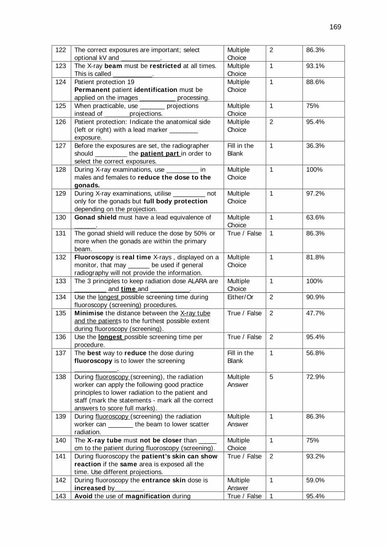
169
122 The correct exposures are important; select optional kV and ___________.
Multiple Choice
2 86.3%
123 The X-ray beam must be restricted at all times. This is called ___________.
Multiple Choice
1 93.1%
124 Patient protection 19 Permanent patient identification must be applied on the images __________ processing.
Multiple Choice
1 88.6%
125 When practicable, use _______ projections instead of _______projections.
Multiple Choice
1 75%
126 Patient protection: Indicate the anatomical side (left or right) with a lead marker ________ exposure.
Multiple Choice
2 95.4%
127 Before the exposures are set, the radiographer should _________ the patient part in order to select the correct exposures.
Fill in the Blank
1 36.3%
128 During X-ray examinations, use _________ in males and females to reduce the dose to the gonads.
Multiple Choice
1 100%
129 During X-ray examinations, utilise _________ not only for the gonads but full body protection depending on the projection.
Multiple Choice
1 97.2%
130 Gonad shield must have a lead equivalence of ______.
Multiple Choice
1 63.6%
131 The gonad shield will reduce the dose by 50% or more when the gonads are within the primary beam.
True / False 1 86.3%
132 Fluoroscopy is real time X-rays , displayed on a monitor, that may ______ be used if general radiography will not provide the information.
Multiple Choice
1 81.8%
133 The 3 principles to keep radiation dose ALARA are _________ and time and ___________.
Multiple Choice
1 100%
134 Use the longest possible screening time during fluoroscopy (screening) procedures.
Either/Or 2 90.9%
135 Minimise the distance between the X-ray tube and the patients to the furthest possible extent during fluoroscopy (screening).
True / False 2 47.7%
136 Use the longest possible screening time per procedure.
True / False 2 95.4%
137 The best way to reduce the dose during fluoroscopy is to lower the screening ____________.
Fill in the Blank
1 56.8%
138 During fluoroscopy (screening), the radiation worker can apply the following good practice principles to lower radiation to the patient and staff (mark the statements - mark all the correct answers to score full marks).
Multiple Answer
5 72.9%
139 During fluoroscopy (screening) the radiation worker can _______ the beam to lower scatter radiation.
Multiple Answer
1 86.3%
140 The X-ray tube must not be closer than _____ cm to the patient during fluoroscopy (screening).
Multiple Choice
1 75%
141 During fluoroscopy the patient’s skin can show reaction if the same area is exposed all the time. Use different projections.
True / False 2 93.2%
142 During fluoroscopy the entrance skin dose is increased by________.
Multiple Answer
1 59.0%
143 Avoid the use of magnification during True / False 1 95.4%

170
fluoroscopy. 144 The X-ray tube of the C-arm in theatre or during
other screening (fluoroscopy) examinations must be _____________ the table.
Multiple Choice
1 79.5%
145 The image intensifier of the C-arm in theatre or during other screening (fluoroscopy) examination must be _____________ the table.
Multiple Choice
1 79.5%
146 The inverse C-arm (intensifier below) may only be used for extremities IF the physician places the anatomical part directly on the __________ (example: Screw placement in the hand)
Multiple Choice
1 59%
147 The radiation source during fluoroscopy (screening) is _____ from the patient.
Multiple Choice
1 59%
148 The radiation source during fluoroscopy (screening) is _______ from the patient.
Fill in the Blank
1 27.2%
149 Dose on the (1) _____ side is lower than the dose on the (2) __________side.
Multiple Answer
1 75%
150 Dose on the tube ide is _________ than on the image intensifier side.
Multiple Answer
1 81.8%
151 ______________ of C-arms enable safe and stable performance.
Multiple Answer
1 65.9%
152 Fixed fluoroscopic units must be equipped with a ____________ that provides a dose readout during fluoroscopy.
Multiple Answer
1 59%
153 The X-ray examination information of all patients must be kept for ______ years.
Multiple Choice
1 84%
154 The X-ray examination records of all patients for fluoroscopy in theatre must include the following: ________ (mark all 8 items that MUST be recorded).
Multiple Answer
8 84%
155 Silicon pads on the tables that provide comfort to the patient will ______ radiation dose because it will require more penetration.
Multiple Answer
1 25%
156 Fixed fluoroscopic units must be equipped with a DAP meter that provides a __________ during fluoroscopy.
Multiple Answer
1 86.3%
In Table 6.8 the knowledge of the first-year students with regard to the radiation
protection of patients showed that 45.5% selected the option that justification means that
the radiation examination benefits must be higher than the associated risk. The meaning
of optimisation as radiation dose to the patient and staff as being kept ALARA was
selected by 40.9%, and limitation as managing of the application of X-rays was indicated
by 88.6% of the student group. The fact that X-ray examinations may ONLY be
performed when there is a valid clinical indication was recognised by 95.4% and the
persons who may request X-ray examinations were correctly marked correctly by 72.1%
of the group. A percentage of 94.3% of the first years knew the statements to which a
request for X-rays has to adhere, whilst 90.13% selected all the correct reasons for a
radiographer to refuse to carry out an X-ray procedure. Examinations on children require

171
more profound justification, since children are more sensitive to radiation. This option
was the choice of 95.4% of the student group.
The first-year students were instructed to name the four measures that can minimise
exposure to the embryo/foetus and the researcher manually allocated marks to the
students. A full record of the responses of the students is listed under question 113 in
Appendix H2. Some of the answers included the following:
• “… restrict collimation to just the area of interest
• … use a fast image recorder
• Limit time for screening
• … use shielding
• Use the PA (post-anterior) position (projection) instead of AP (Anterior-post)
(projection) where possible
• Maximize the kilovoltage (using the maximum kilovoltage)
• … use alternative modalities such as ultrasound, if possible (where ionising radiation
is not used)
• … having the mSv not exceed 2 mSv
• … by giving a pregnant radiographer an alarm dosimeter that will ring when the
person is exposed to radiation,
• … giving the mother the dosimeter,
• Not performing pelvimetry on a pregnant women on daily basis
• pregnant women must not work long hours in the X-ray room”.
All the students indicated that foetal development must be monitored with ultrasound, not
X-rays, and 72.7% that pelvimetry may be not be performed on a routine basis. All the
students indicated that members of the public may not enter X-ray rooms unsupervised,
and 70.5% indicated that a family member may remain in the X-ray room during
exposure.
All the students indicated that staff assisting only a few times in the ward during X-ray
examination needs no protective clothing if this does not happen often, but 72.7% opted
for the choice that it is a good idea to use the same willing nurse in the ward for
assistance when taking X-rays. The group indicated, with a percentage of 88.6, that no
person shall hold a patient or X-ray cassette during exposure, unless it is otherwise
172
impossible to obtain a diagnostically useful image. Indication of the anatomical side (left
or right) with a lead marker before exposure was recognized as patient protection by all
the students.
For the setting of correct exposures, specifically mAs, the correct option was chosen by
86.3%, and 93.1% of the group indicated that collimation means restriction of the beam.
That patient identification on the images is important was the selected option of 88.6%,
whilst 75% agreed that when it is practicable, PA projections instead of AP projections
should be chosen. The practice to measure the patient before the exposures are set was
the selected option of 36.3%. All the students opted for gonad shields to reduce the dose
to the gonads, and 63.6% of the student group could indicate the lead equivalence of the
gonad shield. The three principles of radiation protection, namely distance, time and
shielding were selected correctly by all the students.
The questions pertaining to fluoroscopy scored as follows: 90.9% of the students
indicated that the shortest possible screening time during fluoroscopy (screening)
procedures is ideal, and 47.7% selected the option to maximize the distance between the
X-ray tube and the patient to the furthest possible extent during fluoroscopy. The fill-in
question where students had to write down ’time’ as the best way to reduce the dose
during fluoroscopy, was correctly answered by 56.8%.
The good practice principles that the radiation worker can apply to lower radiation to the
patient and staff during fluoroscopy were correctly marked by 72.9% of the group.
Collimation of the beam as a good practice was indicated by 86.3% of the students. That
the X-ray tube must not be closer than 30 cm to the patient was the option selected by
75% of students, and to avoid magnification as far as possible by 95.4% of the first-year
student group.
The correct routine orientation of the image intensifier (II) of the C-arm in theatre above
the table was selected by 79.5% of the group. The inverse C-arm (intensifier below), that
may only be used for extremities IF the physician places the anatomical part directly on
the II, received acknowledgement by 59% of the group.
The scatter from the patient as radiation source during fluoroscopy (screening) was
selected by 59% and 27% filled in “scatter” from the patient as the radiation source
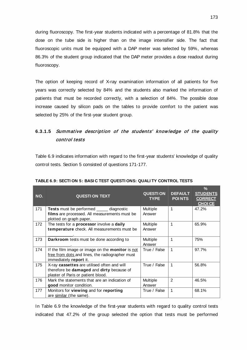
173
during fluoroscopy. The first-year students indicated with a percentage of 81.8% that the
dose on the tube side is higher than on the image intensifier side. The fact that
fluoroscopic units must be equipped with a DAP meter was selected by 59%, whereas
86.3% of the student group indicated that the DAP meter provides a dose readout during
fluoroscopy.
The option of keeping record of X-ray examination information of all patients for five
years was correctly selected by 84% and the students also marked the information of
patients that must be recorded correctly, with a selection of 84%. The possible dose
increase caused by silicon pads on the tables to provide comfort to the patient was
selected by 25% of the first-year student group.
6.3.1.5 Summative description of the students’ know ledge of the quality
control tests
Table 6.9 indicates information with regard to the first-year students’ knowledge of quality
control tests. Section 5 consisted of questions 171-177.
TABLE 6.9: SECTION 5: BASIC TEST QUESTIONS: QUALITY CONTROL TESTS
NO. QUESTION TEXT
QUESTION TYPE
DEFAULT POINTS
% STUDENTS CORRECT CHOICE
171 Tests must be performed _____ diagnostic films are processed. All measurements must be plotted on graph paper.
Multiple Answer
1 47.2%
172 The tests for a processor involve a daily temperature check. All measurements must be ___________.
Multiple Answer
1 65.9%
173 Darkroom tests must be done according to __________.
Multiple Answer
1 75%
174 If the film image or image on the monitor is not free from dots and lines, the radiographer must immediately report it.
True / False 1 97.7%
175 X-ray cassettes are utilised often and will therefore be damaged and dirty because of plaster of Paris or patient blood.
True / False 1 56.8%
176 Mark the statements that are an indication of good monitor condition.
Multiple Answer
2 46.5%
177 Monitors for viewing and for reporting are similar (the same).
True / False 1 68.1%
In Table 6.9 the knowledge of the first-year students with regard to quality control tests
indicated that 47.2% of the group selected the option that tests must be performed
174
before diagnostic films are processed, and 65.9% that the measurements must be plotted
on graph paper. Darkroom tests must be done according to a written protocol was the
choice of 75% of the group. Most students (97.7%) selected that if the film image or
image on the monitor is not free from dots and lines, the radiographer must immediately
report it.
When given a true/false option, 56.8% of the group indicated that it is true that X-ray
cassettes are utilised often and will therefore be damaged and dirty because of plaster of
Paris or patient blood. The two statements regarding good monitor condition were
correctly chosen by 46.5%, while 68.1% discerned the requirements difference between
monitors for viewing and for reporting.
The results of the advanced test will be discussed next.
6.3.2 Descriptions of data from the advanced test questionnaire
The advanced test consisted of 194 questions with a total number of 344 possible points.
Thirty-nine (39) students completed the pre-test and thirty-six (36) completed the post-
test. The three students who did not complete the post-test, were absent because of
health challenges. The calculations were therefore based on the thirty-six (36) students
that completed both tests for the advanced questionnaire.
The first difference between the results of the pre-and post-test was the average score
for all graded attempts. The pre-test resulted in a 168.1 points average score (48.7%)
and the post-test in a 193.38 points average score (56.2%). Second, the average
completion time for all submitted attempts was one hour and 34 minutes for the pre-test
and one hour and 40 minutes for the post-test. The third difference was in terms of the
discrimination value of the so-called 37 good questions, with a 0.3 discrimination in the
pre-test compared to the 70 good questions in the post-test. Table 6.10 presents a
comparison of the discrimination values of the advanced pre- and post-questionnaire
tests.
TABLE 6.10: COMPARISON OF THE ADVANCED TEST PRE- AND POST QUESTIONNAIRE DISCRIMINATION
PRE-TEST POST-TEST 37 Good Questions Discrimination > 0.3 70 Good Questions Discrimination > 0.3 84 Fair Questions Discrimination = 0.1 to 0.3 57 Fair Questions Discrimination = 0.1 to 0.3 61 Poor Questions Discrimination < 0.1 55 Poor Questions Discrimination < 0.1
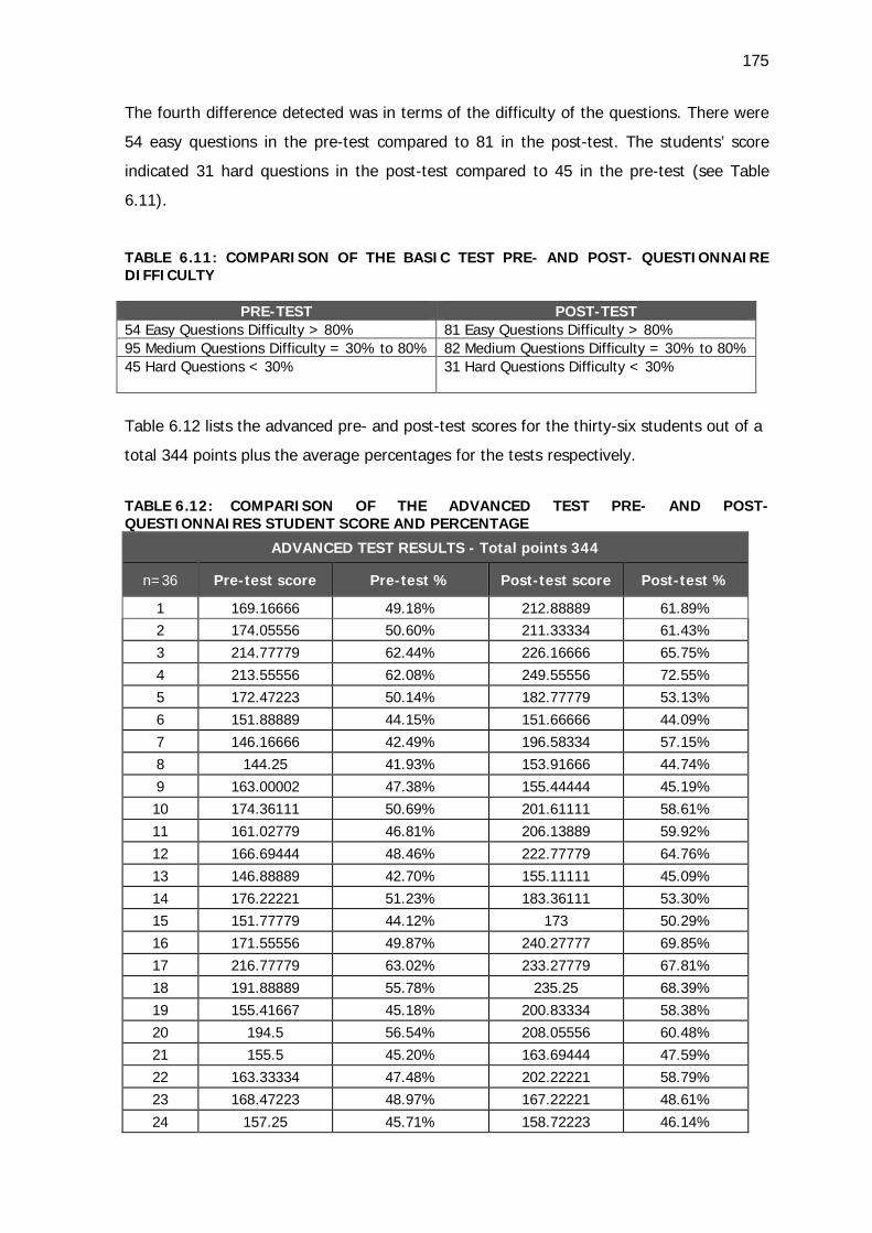
175
The fourth difference detected was in terms of the difficulty of the questions. There were
54 easy questions in the pre-test compared to 81 in the post-test. The students’ score
indicated 31 hard questions in the post-test compared to 45 in the pre-test (see Table
6.11).
TABLE 6.11: COMPARISON OF THE BASIC TEST PRE- AND POST- QUESTIONNAIRE DIFFICULTY
PRE-TEST POST-TEST 54 Easy Questions Difficulty > 80% 81 Easy Questions Difficulty > 80% 95 Medium Questions Difficulty = 30% to 80% 82 Medium Questions Difficulty = 30% to 80% 45 Hard Questions < 30%
31 Hard Questions Difficulty < 30%
Table 6.12 lists the advanced pre- and post-test scores for the thirty-six students out of a
total 344 points plus the average percentages for the tests respectively.
TABLE 6.12: COMPARISON OF THE ADVANCED TEST PRE- AND POST-QUESTIONNAIRES STUDENT SCORE AND PERCENTAGE
ADVANCED TEST RESULTS - Total points 344
n=36 Pre-test score Pre-test % Post-test score Post-test %
1 169.16666 49.18% 212.88889 61.89% 2 174.05556 50.60% 211.33334 61.43% 3 214.77779 62.44% 226.16666 65.75% 4 213.55556 62.08% 249.55556 72.55% 5 172.47223 50.14% 182.77779 53.13% 6 151.88889 44.15% 151.66666 44.09% 7 146.16666 42.49% 196.58334 57.15% 8 144.25 41.93% 153.91666 44.74% 9 163.00002 47.38% 155.44444 45.19% 10 174.36111 50.69% 201.61111 58.61% 11 161.02779 46.81% 206.13889 59.92% 12 166.69444 48.46% 222.77779 64.76% 13 146.88889 42.70% 155.11111 45.09% 14 176.22221 51.23% 183.36111 53.30% 15 151.77779 44.12% 173 50.29% 16 171.55556 49.87% 240.27777 69.85% 17 216.77779 63.02% 233.27779 67.81% 18 191.88889 55.78% 235.25 68.39% 19 155.41667 45.18% 200.83334 58.38% 20 194.5 56.54% 208.05556 60.48% 21 155.5 45.20% 163.69444 47.59% 22 163.33334 47.48% 202.22221 58.79% 23 168.47223 48.97% 167.22221 48.61% 24 157.25 45.71% 158.72223 46.14%
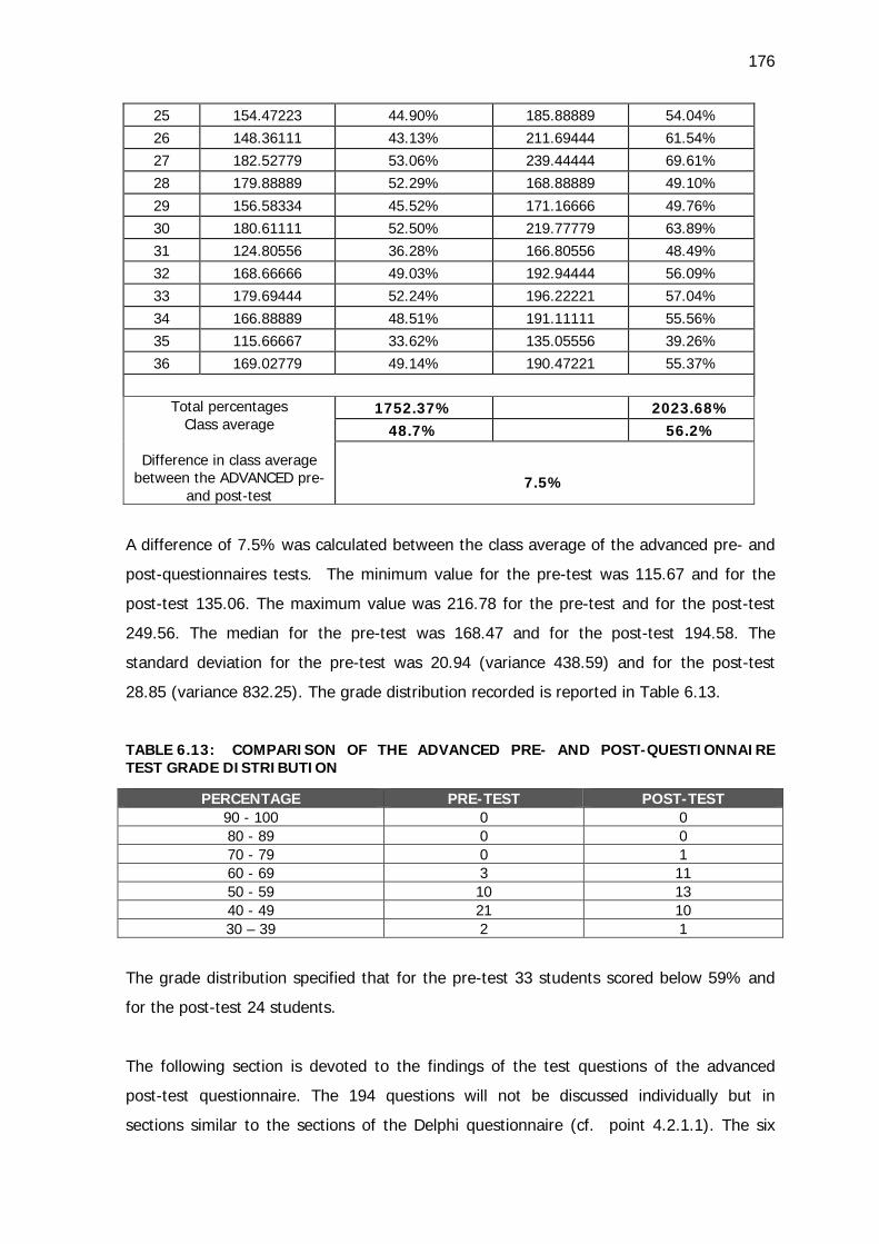
176
25 154.47223 44.90% 185.88889 54.04% 26 148.36111 43.13% 211.69444 61.54% 27 182.52779 53.06% 239.44444 69.61% 28 179.88889 52.29% 168.88889 49.10% 29 156.58334 45.52% 171.16666 49.76% 30 180.61111 52.50% 219.77779 63.89% 31 124.80556 36.28% 166.80556 48.49% 32 168.66666 49.03% 192.94444 56.09% 33 179.69444 52.24% 196.22221 57.04% 34 166.88889 48.51% 191.11111 55.56% 35 115.66667 33.62% 135.05556 39.26% 36 169.02779 49.14% 190.47221 55.37%
Total percentages
Class average
Difference in class average between the ADVANCED pre-
and post-test
1752.37% 2023.68% 48.7% 56.2%
7.5%
A difference of 7.5% was calculated between the class average of the advanced pre- and
post-questionnaires tests. The minimum value for the pre-test was 115.67 and for the
post-test 135.06. The maximum value was 216.78 for the pre-test and for the post-test
249.56. The median for the pre-test was 168.47 and for the post-test 194.58. The
standard deviation for the pre-test was 20.94 (variance 438.59) and for the post-test
28.85 (variance 832.25). The grade distribution recorded is reported in Table 6.13.
TABLE 6.13: COMPARISON OF THE ADVANCED PRE- AND POST-QUESTIONNAIRE TEST GRADE DISTRIBUTION
PERCENTAGE PRE-TEST POST-TEST 90 - 100 0 0 80 - 89 0 0 70 - 79 0 1 60 - 69 3 11 50 - 59 10 13 40 - 49 21 10 30 – 39 2 1
The grade distribution specified that for the pre-test 33 students scored below 59% and
for the post-test 24 students.
The following section is devoted to the findings of the test questions of the advanced
post-test questionnaire. The 194 questions will not be discussed individually but in
sections similar to the sections of the Delphi questionnaire (cf. point 4.2.1.1). The six
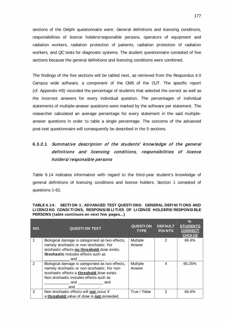
177
sections of the Delphi questionnaire were: General definitions and licencing conditions,
responsibilities of licence holders/responsible persons, operators of equipment and
radiation workers, radiation protection of patients, radiation protection of radiation
workers, and QC tests for diagnostic systems. The student questionnaire consisted of five
sections because the general definitions and licencing conditions were combined.
The findings of the five sections will be tabled next, as retrieved from the Respondus 4.0
Campus wide software, a component of the CMS of the CUT. The specific report
(cf. Appendix H3) recorded the percentage of students that selected the correct as well as
the incorrect answers for every individual question. The percentages of individual
statements of multiple-answer questions were marked by the software per statement. The
researcher calculated an average percentage for every statement in the said multiple-
answer questions in order to table a single percentage. The outcome of the advanced
post-test questionnaire will consequently be described in the 5 sections.
6.3.2.1 Summative description of the students’ know ledge of the general
definitions and licencing conditions, responsibilit ies of licence
holders/ responsible persons
Table 6.14 indicates information with regard to the third-year student’s knowledge of
general definitions of licencing conditions and licence holders. Section 1 consisted of
questions 1-62.
TABLE 6.14: SECTION 1: ADVANCED TEST QUESTIONS: GENERAL DEFINITIONS AND LICENCING CONDITIONS, RESPONSIBILITIES OF LICENCE HOLDERS/RESPONSIBLE PERSONS (table continues on next few pages...)
NO. QUESTION TEXT
QUESTION TYPE
DEFAULT POINTS
% STUDENTS CORRECT CHOICE
1 Biological damage is categorised as two effects, namely stochastic or non-stochastic. For stochastic effects no threshold dose exists. Stochastic includes effects such as ____________ and ____________.
Multiple Answer
2 69.4%
2 Biological damage is categorised as two effects, namely stochastic or non-stochastic. For non-stochastic effects a threshold dose exists. Non-stochastic includes effects such as ____________ and ____________ and ___________ and ___________.
Multiple Answer
4 65.25%
3 Non-stochastic effects will not occur if a threshold value of dose is not exceeded.
True / False 2 66.6%
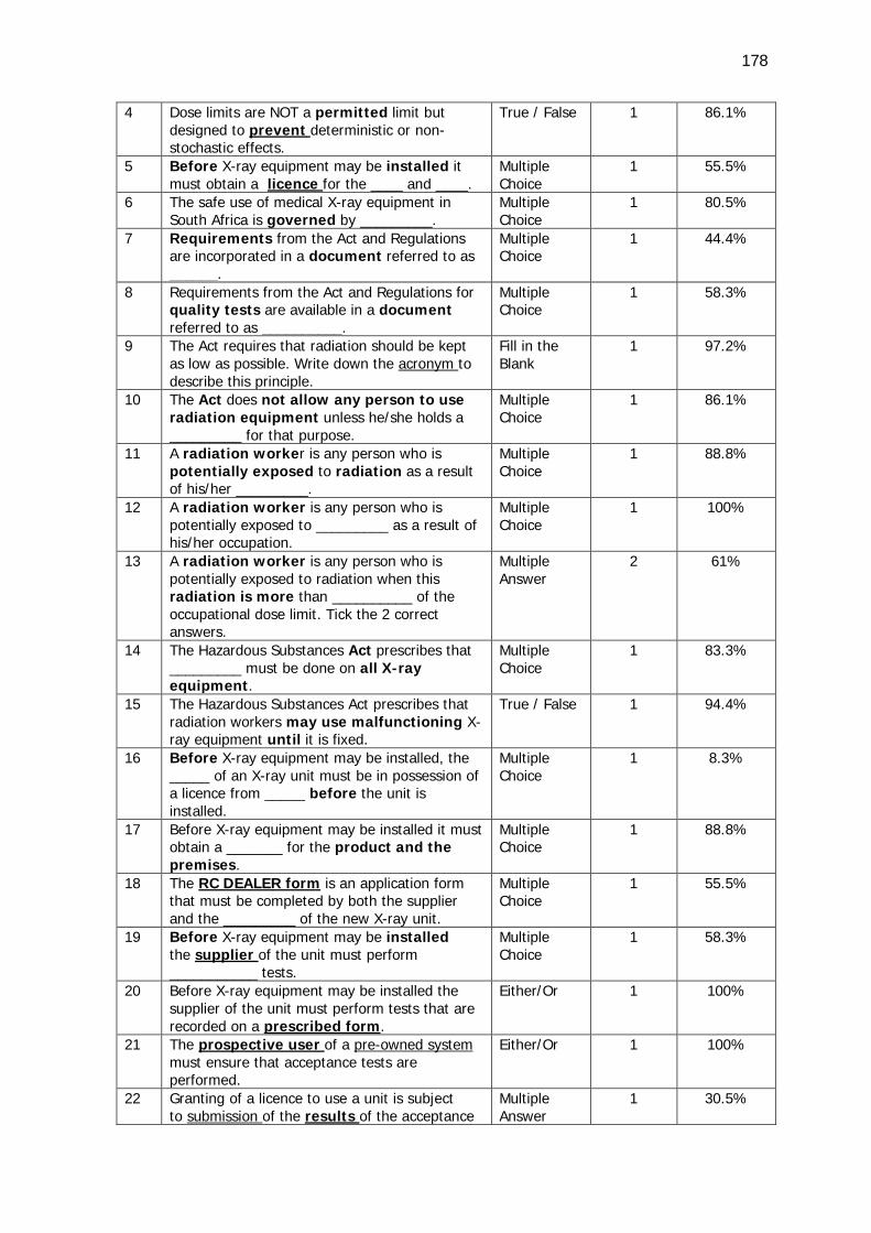
178
4 Dose limits are NOT a permitted limit but designed to prevent deterministic or non-stochastic effects.
True / False 1 86.1%
5 Before X-ray equipment may be installed it must obtain a licence for the ____ and ____.
Multiple Choice
1 55.5%
6 The safe use of medical X-ray equipment in South Africa is governed by _________.
Multiple Choice
1 80.5%
7 Requirements from the Act and Regulations are incorporated in a document referred to as ______.
Multiple Choice
1 44.4%
8 Requirements from the Act and Regulations for quality tests are available in a document referred to as __________.
Multiple Choice
1 58.3%
9 The Act requires that radiation should be kept as low as possible. Write down the acronym to describe this principle.
Fill in the Blank
1 97.2%
10 The Act does not allow any person to use radiation equipment unless he/she holds a _________ for that purpose.
Multiple Choice
1 86.1%
11 A radiation worker is any person who is potentially exposed to radiation as a result of his/her _________.
Multiple Choice
1 88.8%
12 A radiation worker is any person who is potentially exposed to _________ as a result of his/her occupation.
Multiple Choice
1 100%
13 A radiation worker is any person who is potentially exposed to radiation when this radiation is more than __________ of the occupational dose limit. Tick the 2 correct answers.
Multiple Answer
2 61%
14 The Hazardous Substances Act prescribes that _________ must be done on all X-ray equipment.
Multiple Choice
1 83.3%
15 The Hazardous Substances Act prescribes that radiation workers may use malfunctioning X-ray equipment until it is fixed.
True / False 1 94.4%
16 Before X-ray equipment may be installed, the _____ of an X-ray unit must be in possession of a licence from _____ before the unit is installed.
Multiple Choice
1 8.3%
17 Before X-ray equipment may be installed it must obtain a _______ for the product and the premises.
Multiple Choice
1 88.8%
18 The RC DEALER form is an application form that must be completed by both the supplier and the _________ of the new X-ray unit.
Multiple Choice
1 55.5%
19 Before X-ray equipment may be installed the supplier of the unit must perform ___________ tests.
Multiple Choice
1 58.3%
20 Before X-ray equipment may be installed the supplier of the unit must perform tests that are recorded on a prescribed form.
Either/Or 1 100%
21 The prospective user of a pre-owned system must ensure that acceptance tests are performed.
Either/Or 1 100%
22 Granting of a licence to use a unit is subject to submission of the results of the acceptance
Multiple Answer
1 30.5%
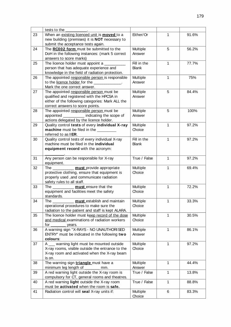
179
tests to the __________. 23 When an existing licenced unit is moved to a
new building (premises) it is NOT necessary to submit the acceptance tests again.
Either/Or 1 91.6%
24 The RC002 form must be submitted to the DoH in the following instances: (mark 5 correct answers to score marks)
Multiple Answer
5 56.2%
25 The licence holder must appoint a __________ person that has adequate experience and knowledge in the field of radiation protection.
Fill in the Blank
1 77.7%
26 The appointed responsible person is responsible to the licence holder for the _____________. Mark the one correct answer.
Multiple Answer
1 75%
27 The appointed responsible person must be qualified and registered with the HPCSA in either of the following categories: Mark ALL the correct answers to score points.
Multiple Answer
5 84.4%
28 The appointed responsible person must be appointed __________ indicating the scope of actions delegated by the licence holder.
Multiple Answer
5 100%
29 Quality control tests of every individual X-ray machine must be filed in the _________ referred to as IER.
Multiple Choice
1 97.2%
30 Quality control tests of every individual X-ray machine must be filed in the individual equipment record with the acronym: _________.
Fill in the Blank
1 97.2%
31 Any person can be responsible for X-ray equipment.
True / False 1 97.2%
32 The __________ must provide appropriate protective clothing, ensure that equipment is properly used ,and communicate radiation safety rules to all staff.
Multiple Choice
1 69.4%
33 The __________ must ensure that the equipment and facilities meet the safety standards.
Multiple Choice
1 72.2%
34 The __________ must establish and maintain operational procedures to make sure the radiation to the patient and staff is kept ALARA.
Multiple Choice
1 33.3%
35 The licence holder must keep record of the dose and medical examinations of radiation workers for _______ years.
Multiple Choice
1 30.5%
36 A warning sign "X-RAYS - NO UNAUTHORISED ENTRY" must be indicated in the following two colours:
Multiple Answer
1 86.1%
37 A ___ warning light must be mounted outside X-ray rooms, visible outside the entrance to the X-ray room and activated when the X-ray beam is on.
Multiple Choice
1 97.2%
38 The warning sign triangle must have a minimum leg length of _______ mm.
Multiple Answer
1 44.4%
39 A red warning light outside the X-ray room is compulsory for CT, general rooms and theatres.
True / False 1 13.8%
40 A red warning light outside the X-ray room must be activated when the room is safe.
True / False 1 88.8%
41 Radiation control will seal X-ray units if: Multiple Choice
6 83.3%
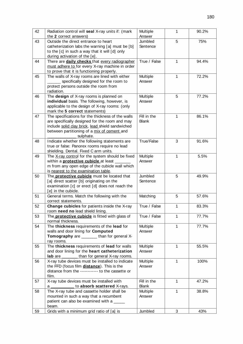
180
42 Radiation control will seal X-ray units if: (mark the 2 correct answers)
Multiple Answer
1 90.2%
43 Outside the direct entrance to heart catheterization labs the warning [a] must be [b] to the [c] in such a way that it will [d] only during activation of the [e].
Jumbled Sentence
5 75%
44 There are daily checks that every radiographer must adhere to for every X-ray machine in order to prove that it is functioning properly.
True / False 1 94.4%
45 The walls of X-ray rooms are lined with either ______ specifically designed for the room to protect persons outside the room from radiation.
Multiple Answer
1 72.2%
46 The design of X-ray rooms is planned on individual basis. The following, however, is applicable to the design of X-ray rooms: (only mark the 5 correct statements)
Multiple Answer
5 77.2%
47 The specifications for the thickness of the walls are specifically designed for the room and may include solid clay brick, lead shield sandwiched between partitioning of a mix of cement and _____________ sulphate.
Fill in the Blank
1 86.1%
48 Indicate whether the following statements are true or false: Panorex rooms require no lead shielding. Dental. Fixed C arm units.
True/False 3 91.6%
49 The X-ray control for the system should be fixed within a protective cubicle at least _______ m from any open edge of the cubicle wall which is nearest to the examination table.
Multiple Answer
1 5.5%
50 The protective cubicle must be located that [a] direct scatter [b] originating on the examination [c] or erect [d] does not reach the [e] in the cubicle.
Jumbled Sentence
5 49.9%
51 General terms. Match the following with the correct statements.
Matching 5 57.6%
52 Change cubicles for patients inside the X-ray room need no lead shield lining.
True / False 1 83.3%
53 The protective cubicle is fitted with glass of normal thickness.
True / False 1 77.7%
54 The thickness requirements of the lead for walls and door lining for Computed Tomography are _______ than for general X-ray rooms.
Multiple Answer
1 77.7%
55 The thickness requirements of lead for walls and door lining for the heart catheterization lab are _______ than for general X-ray rooms.
Multiple Answer
1 55.5%
56 X-ray tube devices must be installed to indicate the FFD (focus film distance). This is the distance from the ------------ to the cassette or film.
Multiple Answer
1 100%
57 X-ray tube devices must be installed with a _________ to absorb scattered X-rays.
Fill in the Blank
1 47.2%
58 The X-ray tube and cassette holder shall be mounted in such a way that a recumbent patient can also be examined with a _____ beam.
Multiple Answer
1 38.8%
59 Grids with a minimum grid ratio of [a] is Jumbled 3 43%
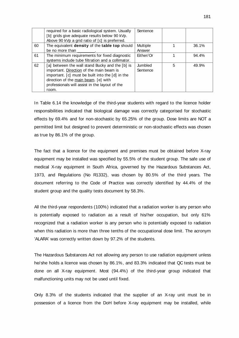
181
required for a basic radiological system. Usually [b] grids give adequate results below 90 kVp. Above 90 kVp a grid ratio of [c] is preferred.
Sentence
60 The equivalent density of the table top should be no more than ________.
Multiple Answer
1 36.1%
61 The minimum requirements for fixed diagnostic systems include tube filtration and a collimator.
Either/Or 1 94.4%
62 [a] between the wall stand Bucky and the [b] is important. Direction of the main beam is important. [c] must be built into the [d] in the direction of the main beam. [e] with professionals will assist in the layout of the room.
Jumbled Sentence
5 49.9%
In Table 6.14 the knowledge of the third-year students with regard to the licence holder
responsibilities indicated that biological damage was correctly categorised for stochastic
effects by 69.4% and for non-stochastic by 65.25% of the group. Dose limits are NOT a
permitted limit but designed to prevent deterministic or non-stochastic effects was chosen
as true by 86.1% of the group.
The fact that a licence for the equipment and premises must be obtained before X-ray
equipment may be installed was specified by 55.5% of the student group. The safe use of
medical X-ray equipment in South Africa, governed by the Hazardous Substances Act,
1973, and Regulations (No R1332), was chosen by 80.5% of the third years. The
document referring to the Code of Practice was correctly identified by 44.4% of the
student group and the quality tests document by 58.3%.
All the third-year respondents (100%) indicated that a radiation worker is any person who
is potentially exposed to radiation as a result of his/her occupation, but only 61%
recognized that a radiation worker is any person who is potentially exposed to radiation
when this radiation is more than three tenths of the occupational dose limit. The acronym
’ALARA’ was correctly written down by 97.2% of the students.
The Hazardous Substances Act not allowing any person to use radiation equipment unless
he/she holds a licence was chosen by 86.1%, and 83.3% indicated that QC tests must be
done on all X-ray equipment. Most (94.4%) of the third-year group indicated that
malfunctioning units may not be used until fixed.
Only 8.3% of the students indicated that the supplier of an X-ray unit must be in
possession of a licence from the DoH before X-ray equipment may be installed, while
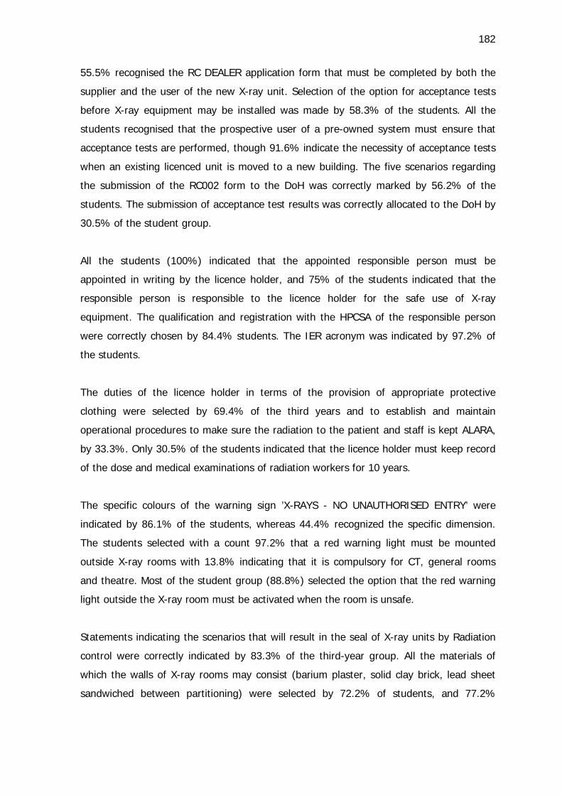
182
55.5% recognised the RC DEALER application form that must be completed by both the
supplier and the user of the new X-ray unit. Selection of the option for acceptance tests
before X-ray equipment may be installed was made by 58.3% of the students. All the
students recognised that the prospective user of a pre-owned system must ensure that
acceptance tests are performed, though 91.6% indicate the necessity of acceptance tests
when an existing licenced unit is moved to a new building. The five scenarios regarding
the submission of the RC002 form to the DoH was correctly marked by 56.2% of the
students. The submission of acceptance test results was correctly allocated to the DoH by
30.5% of the student group.
All the students (100%) indicated that the appointed responsible person must be
appointed in writing by the licence holder, and 75% of the students indicated that the
responsible person is responsible to the licence holder for the safe use of X-ray
equipment. The qualification and registration with the HPCSA of the responsible person
were correctly chosen by 84.4% students. The IER acronym was indicated by 97.2% of
the students.
The duties of the licence holder in terms of the provision of appropriate protective
clothing were selected by 69.4% of the third years and to establish and maintain
operational procedures to make sure the radiation to the patient and staff is kept ALARA,
by 33.3%. Only 30.5% of the students indicated that the licence holder must keep record
of the dose and medical examinations of radiation workers for 10 years.
The specific colours of the warning sign ’X-RAYS - NO UNAUTHORISED ENTRY’ were
indicated by 86.1% of the students, whereas 44.4% recognized the specific dimension.
The students selected with a count 97.2% that a red warning light must be mounted
outside X-ray rooms with 13.8% indicating that it is compulsory for CT, general rooms
and theatre. Most of the student group (88.8%) selected the option that the red warning
light outside the X-ray room must be activated when the room is unsafe.
Statements indicating the scenarios that will result in the seal of X-ray units by Radiation
control were correctly indicated by 83.3% of the third-year group. All the materials of
which the walls of X-ray rooms may consist (barium plaster, solid clay brick, lead sheet
sandwiched between partitioning) were selected by 72.2% of students, and 77.2%
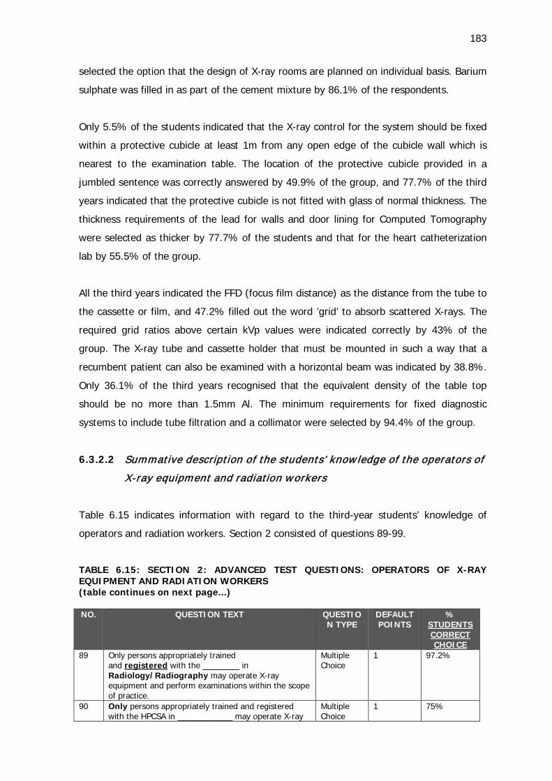
183
selected the option that the design of X-ray rooms are planned on individual basis. Barium
sulphate was filled in as part of the cement mixture by 86.1% of the respondents.
Only 5.5% of the students indicated that the X-ray control for the system should be fixed
within a protective cubicle at least 1m from any open edge of the cubicle wall which is
nearest to the examination table. The location of the protective cubicle provided in a
jumbled sentence was correctly answered by 49.9% of the group, and 77.7% of the third
years indicated that the protective cubicle is not fitted with glass of normal thickness. The
thickness requirements of the lead for walls and door lining for Computed Tomography
were selected as thicker by 77.7% of the students and that for the heart catheterization
lab by 55.5% of the group.
All the third years indicated the FFD (focus film distance) as the distance from the tube to
the cassette or film, and 47.2% filled out the word ’grid’ to absorb scattered X-rays. The
required grid ratios above certain kVp values were indicated correctly by 43% of the
group. The X-ray tube and cassette holder that must be mounted in such a way that a
recumbent patient can also be examined with a horizontal beam was indicated by 38.8%.
Only 36.1% of the third years recognised that the equivalent density of the table top
should be no more than 1.5mm Al. The minimum requirements for fixed diagnostic
systems to include tube filtration and a collimator were selected by 94.4% of the group.
6.3.2.2 Summative description of the students’ know ledge of the operators of
X-ray equipment and radiation workers
Table 6.15 indicates information with regard to the third-year students’ knowledge of
operators and radiation workers. Section 2 consisted of questions 89-99.
TABLE 6.15: SECTION 2: ADVANCED TEST QUESTIONS: OPERATORS OF X-RAY EQUIPMENT AND RADIATION WORKERS (table continues on next page...) NO. QUESTION TEXT QUESTIO
N TYPE
DEFAULT POINTS
% STUDENTS CORRECT CHOICE
89 Only persons appropriately trained and registered with the ________ in Radiology/Radiography may operate X-ray equipment and perform examinations within the scope of practice.
Multiple Choice
1 97.2%
90 Only persons appropriately trained and registered with the HPCSA in ____________ may operate X-ray
Multiple Choice
1 75%
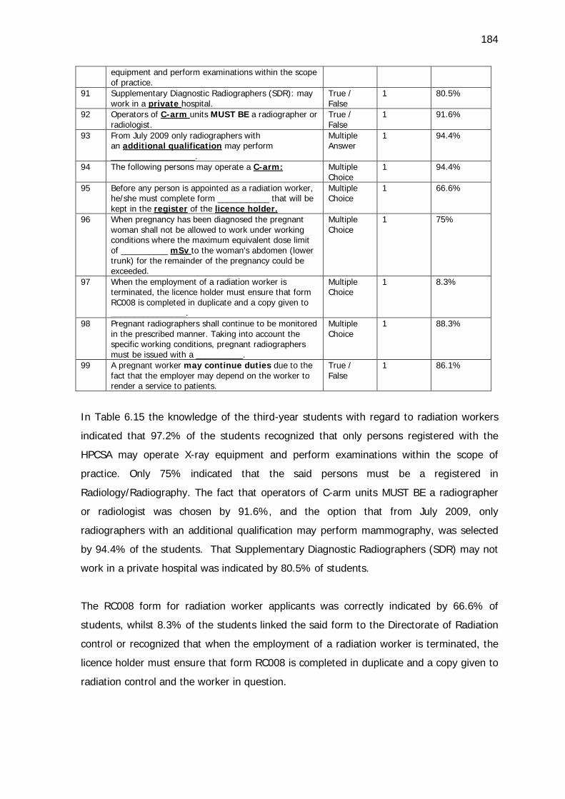
184
equipment and perform examinations within the scope of practice.
91 Supplementary Diagnostic Radiographers (SDR): may work in a private hospital.
True / False
1 80.5%
92 Operators of C-arm units MUST BE a radiographer or radiologist.
True / False
1 91.6%
93 From July 2009 only radiographers with an additional qualification may perform __________________.
Multiple Answer
1 94.4%
94 The following persons may operate a C-arm: Multiple Choice
1 94.4%
95 Before any person is appointed as a radiation worker, he/she must complete form ___________ that will be kept in the register of the licence holder.
Multiple Choice
1 66.6%
96 When pregnancy has been diagnosed the pregnant woman shall not be allowed to work under working conditions where the maximum equivalent dose limit of __________ mSv to the woman's abdomen (lower trunk) for the remainder of the pregnancy could be exceeded.
Multiple Choice
1 75%
97 When the employment of a radiation worker is terminated, the licence holder must ensure that form RC008 is completed in duplicate and a copy given to ________________.
Multiple Choice
1 8.3%
98 Pregnant radiographers shall continue to be monitored in the prescribed manner. Taking into account the specific working conditions, pregnant radiographers must be issued with a __________.
Multiple Choice
1 88.3%
99 A pregnant worker may continue duties due to the fact that the employer may depend on the worker to render a service to patients.
True / False
1 86.1%
In Table 6.15 the knowledge of the third-year students with regard to radiation workers
indicated that 97.2% of the students recognized that only persons registered with the
HPCSA may operate X-ray equipment and perform examinations within the scope of
practice. Only 75% indicated that the said persons must be a registered in
Radiology/Radiography. The fact that operators of C-arm units MUST BE a radiographer
or radiologist was chosen by 91.6%, and the option that from July 2009, only
radiographers with an additional qualification may perform mammography, was selected
by 94.4% of the students. That Supplementary Diagnostic Radiographers (SDR) may not
work in a private hospital was indicated by 80.5% of students.
The RC008 form for radiation worker applicants was correctly indicated by 66.6% of
students, whilst 8.3% of the students linked the said form to the Directorate of Radiation
control or recognized that when the employment of a radiation worker is terminated, the
licence holder must ensure that form RC008 is completed in duplicate and a copy given to
radiation control and the worker in question.
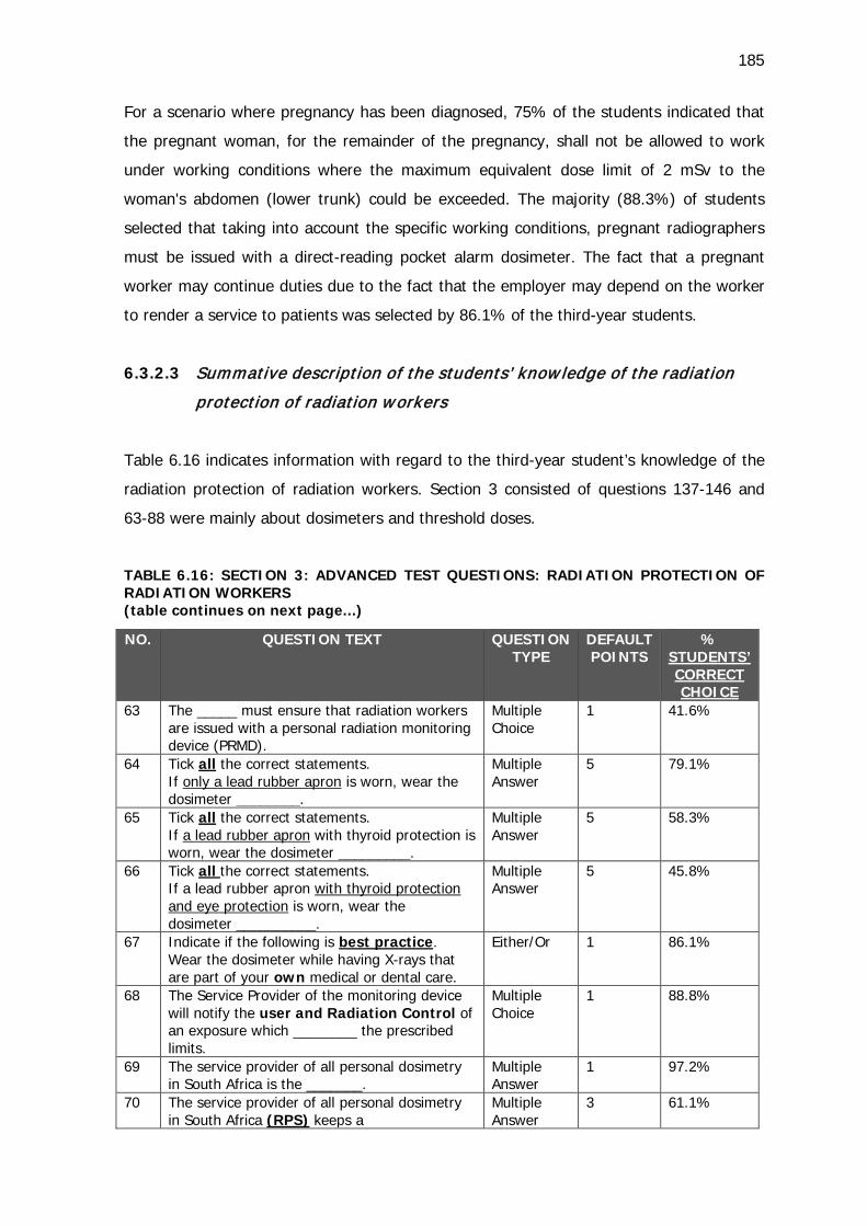
185
For a scenario where pregnancy has been diagnosed, 75% of the students indicated that
the pregnant woman, for the remainder of the pregnancy, shall not be allowed to work
under working conditions where the maximum equivalent dose limit of 2 mSv to the
woman's abdomen (lower trunk) could be exceeded. The majority (88.3%) of students
selected that taking into account the specific working conditions, pregnant radiographers
must be issued with a direct-reading pocket alarm dosimeter. The fact that a pregnant
worker may continue duties due to the fact that the employer may depend on the worker
to render a service to patients was selected by 86.1% of the third-year students.
6.3.2.3 Summative description of the students’ know ledge of the radiation
protection of radiation workers
Table 6.16 indicates information with regard to the third-year student’s knowledge of the
radiation protection of radiation workers. Section 3 consisted of questions 137-146 and
63-88 were mainly about dosimeters and threshold doses.
TABLE 6.16: SECTION 3: ADVANCED TEST QUESTIONS: RADIATION PROTECTION OF RADIATION WORKERS (table continues on next page...)
NO. QUESTION TEXT QUESTION TYPE
DEFAULT POINTS
% STUDENTS’ CORRECT CHOICE
63 The _____ must ensure that radiation workers are issued with a personal radiation monitoring device (PRMD).
Multiple Choice
1 41.6%
64 Tick all the correct statements. If only a lead rubber apron is worn, wear the dosimeter ________.
Multiple Answer
5 79.1%
65 Tick all the correct statements. If a lead rubber apron with thyroid protection is worn, wear the dosimeter _________.
Multiple Answer
5 58.3%
66 Tick all the correct statements. If a lead rubber apron with thyroid protection and eye protection is worn, wear the dosimeter __________.
Multiple Answer
5 45.8%
67 Indicate if the following is best practice. Wear the dosimeter while having X-rays that are part of your own medical or dental care.
Either/Or 1 86.1%
68 The Service Provider of the monitoring device will notify the user and Radiation Control of an exposure which ________ the prescribed limits.
Multiple Choice
1 88.8%
69 The service provider of all personal dosimetry in South Africa is the _______.
Multiple Answer
1 97.2%
70 The service provider of all personal dosimetry in South Africa (RPS) keeps a
Multiple Answer
3 61.1%
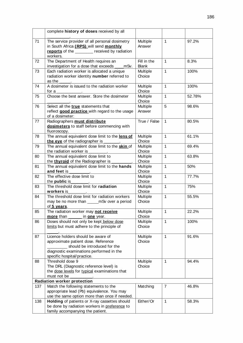
186
complete history of doses received by all __________.
71 The service provider of all personal dosimetry in South Africa (RPS) will send monthly reports of the ________ received by radiation workers.
Multiple Answer
1 97.2%
72 The Department of Health requires an investigation for a dose that exceeds ____mSv.
Fill in the Blank
1 8.3%
73 Each radiation worker is allocated a unique radiation worker identity number referred to as the _____.
Multiple Choice
1 100%
74 A dosimeter is issued to the radiation worker for a ___________.
Multiple Choice
1 100%
75 Choose the best answer. Store the dosimeter __________.
Multiple Choice
1 52.78%
76 Select all the true statements that reflect good practice with regard to the usage of a dosimeter:
Multiple Answer
5 98.6%
77 Radiographers must distribute dosimeters to staff before commencing with fluoroscopy.
True / False 1 80.5%
78 The annual equivalent dose limit to the lens of the eye of the radiographer is _______.
Multiple Choice
1 61.1%
79 The annual equivalent dose limit to the skin of the radiation worker is ________.
Multiple Choice
1 69.4%
80 The annual equivalent dose limit to the thyroid of the Radiographer is _________.
Multiple Choice
1 63.8%
81 The annual equivalent dose limit to the hands and feet is _________.
Multiple Choice
1 50%
82 The effective dose limit to the public is________.
Multiple Choice
1 77.7%
83 The threshold dose limit for radiation workers is________.
Multiple Choice
1 75%
84 The threshold dose limit for radiation workers may be no more than _____mSv over a period of 5 years.
Multiple Choice
1 55.5%
85 The radiation worker may not receive more than ______ in one year.
Multiple Choice
1 22.2%
86 Doses should not only be kept below dose limits but must adhere to the principle of _____.
Multiple Choice
1 100%
87 Licence holders should be aware of approximate patient dose. Reference _________ should be introduced for the diagnostic examinations performed in the specific hospital/practice.
Multiple Choice
1 91.6%
88 Threshold dose 9 The DRL (Diagnostic reference level) is the dose levels for typical examinations that must not be __________.
Multiple Choice
1 94.4%
Radiation worker protection 137 Match the following statements to the
appropriate lead (Pb) equivalence. You may use the same option more than once if needed.
Matching 7 46.8%
138 Holding of patients or X-ray cassettes should be done by radiation workers in preference to family accompanying the patient.
Either/Or 1 58.3%
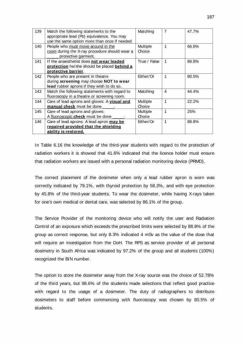
187
139 Match the following statements to the appropriate lead (Pb) equivalence. You may use the same option more than once if needed.
Matching 7 47.7%
140 People who must move around in the room during the X-ray procedure should wear a _____ protective garment.
Multiple Choice
1 66.6%
141 If the anaesthetist does not wear leaded protection he/she should be placed behind a protective barrier.
True / False 1 88.8%
142 People who are present in theatre during screening may choose NOT to wear lead rubber aprons if they wish to do so.
Either/Or 1 80.5%
143 Match the following statements with regard to fluoroscopy in a theatre or screening room.
Matching 4 44.4%
144 Care of lead aprons and gloves: A visual and manual check must be done _____.
Multiple Choice
1 22.2%
145 Care of lead aprons and gloves: A fluoroscopic check must be done _____.
Multiple Choice
1 25%
146 Care of lead aprons: A lead apron may be repaired provided that the shielding ability is restored.
Either/Or 1 88.8%
In Table 6.16 the knowledge of the third-year students with regard to the protection of
radiation workers it is showed that 41.6% indicated that the licence holder must ensure
that radiation workers are issued with a personal radiation monitoring device (PRMD).
The correct placement of the dosimeter when only a lead rubber apron is worn was
correctly indicated by 79.1%, with thyroid protection by 58.3%, and with eye protection
by 45.8% of the third-year students. To wear the dosimeter, while having X-rays taken
for one’s own medical or dental care, was selected by 86.1% of the group.
The Service Provider of the monitoring device who will notify the user and Radiation
Control of an exposure which exceeds the prescribed limits were selected by 88.8% of the
group as correct response, but only 8.3% indicated 4 mSv as the value of the dose that
will require an investigation from the DoH. The RPS as service provider of all personal
dosimetry in South Africa was indicated by 97.2% of the group and all students (100%)
recognized the BIN number.
The option to store the dosimeter away from the X-ray source was the choice of 52.78%
of the third years, but 98.6% of the students made selections that reflect good practice
with regard to the usage of a dosimeter. The duty of radiographers to distribute
dosimeters to staff before commencing with fluoroscopy was chosen by 80.5% of
students.
188
The annual equivalent dose limit to the lens of the eye of the radiographer as 20 mSv was
selected by 61.1% of the group; 500 mSv to the skin by 69.4% of the group; 20 mSv to
the thyroid by 63.8%; 500 mSv to the hands and feet by 50%, and 1 mSv to the public
by 77.7% of the third-year students.
The threshold dose limit for radiation workers as 20 mSv was chosen by 75%, and only
55.5% selected that the threshold dose may not be more than 100 mSv over a period of
5 years. A percentage of 22.2% students indicated that the radiation worker may not
receive more than 50 mSv in one year.
The DRL (Diagnostic reference level) for typical examinations that must not be exceeded
was chosen by 94.4% of the group. The lead equivalence for seven protective attires
could be correctly matched by 46.8% of the group.
Holding of patients or X-ray cassettes by radiation workers, in preference to family
accompanying the patient, was selected by 58.3% of the group. The wrap-around
protective garment for people who must move around in the room during the X-ray
procedures was the choice of 66.6% of students. If the anaesthetist does not wear leaded
protection he/she should be placed behind a protective barrier – that was the choice of
88.8% of students, whereas 80.5% of students indicated that people who are present in
theatre during screening may choose NOT to wear lead rubber aprons if they wish to do
so.
Statements with regard to fluoroscopy in a theatre or screening room, including panning,
collimation or only screening when the surgeon observes the monitor, was indicated by
44.4% of the group. The monthly care of lead aprons and gloves by means of a visual
and manual check and the yearly fluoroscopic check was indicated by 22.2% and 25% of
students respectively. The lead apron that may be repaired provided that the shielding
ability is restored was the choice of 88.8% of the students.
6.3.2.4 Summative description of the students’ know ledge of the radiation
protection of patients
Table 6.17 indicates information with regard to the third-year students’ knowledge of
radiation protection of patients. Section 4 consisted of questions 100-136.
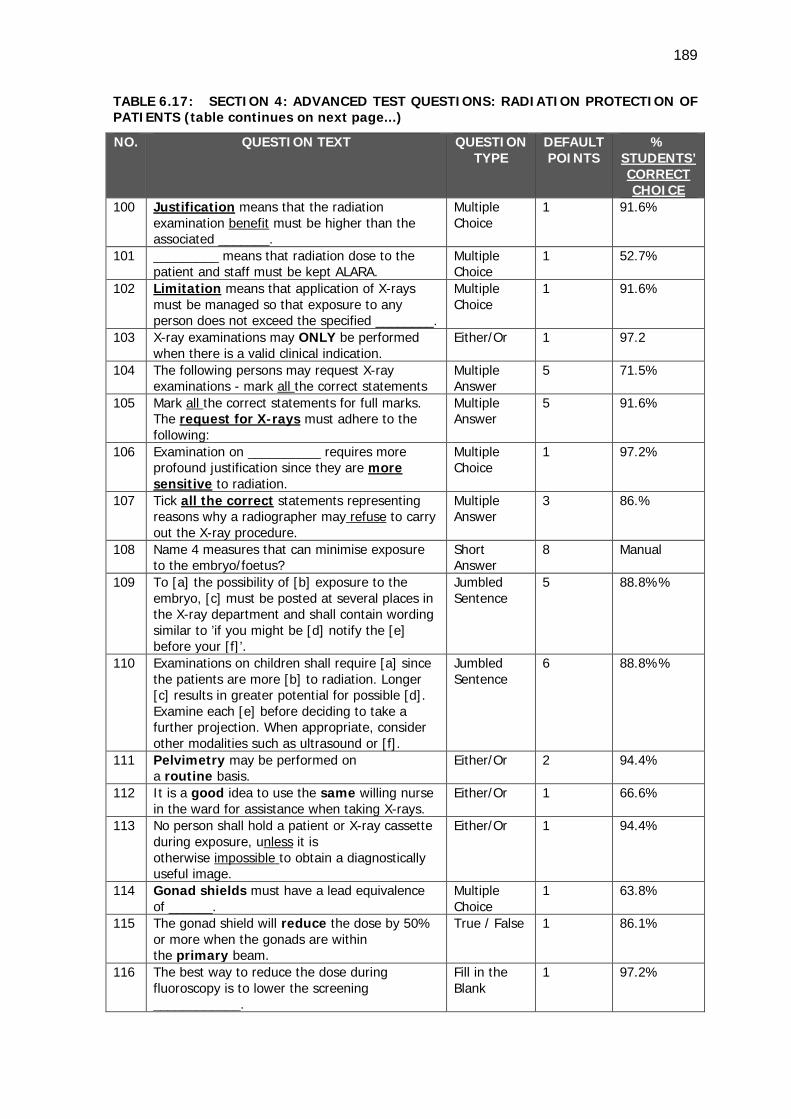
189
TABLE 6.17: SECTION 4: ADVANCED TEST QUESTIONS: RADIATION PROTECTION OF PATIENTS (table continues on next page...)
NO. QUESTION TEXT QUESTION TYPE
DEFAULT POINTS
% STUDENTS’ CORRECT CHOICE
100 Justification means that the radiation examination benefit must be higher than the associated _______.
Multiple Choice
1 91.6%
101 _________ means that radiation dose to the patient and staff must be kept ALARA.
Multiple Choice
1 52.7%
102 Limitation means that application of X-rays must be managed so that exposure to any person does not exceed the specified ________.
Multiple Choice
1 91.6%
103 X-ray examinations may ONLY be performed when there is a valid clinical indication.
Either/Or 1 97.2
104 The following persons may request X-ray examinations - mark all the correct statements
Multiple Answer
5 71.5%
105 Mark all the correct statements for full marks. The request for X-rays must adhere to the following:
Multiple Answer
5 91.6%
106 Examination on __________ requires more profound justification since they are more sensitive to radiation.
Multiple Choice
1 97.2%
107 Tick all the correct statements representing reasons why a radiographer may refuse to carry out the X-ray procedure.
Multiple Answer
3 86.%
108 Name 4 measures that can minimise exposure to the embryo/foetus?
Short Answer
8 Manual
109 To [a] the possibility of [b] exposure to the embryo, [c] must be posted at several places in the X-ray department and shall contain wording similar to ’if you might be [d] notify the [e] before your [f]’.
Jumbled Sentence
5 88.8%%
110 Examinations on children shall require [a] since the patients are more [b] to radiation. Longer [c] results in greater potential for possible [d]. Examine each [e] before deciding to take a further projection. When appropriate, consider other modalities such as ultrasound or [f].
Jumbled Sentence
6 88.8%%
111 Pelvimetry may be performed on a routine basis.
Either/Or 2 94.4%
112 It is a good idea to use the same willing nurse in the ward for assistance when taking X-rays.
Either/Or 1 66.6%
113 No person shall hold a patient or X-ray cassette during exposure, unless it is otherwise impossible to obtain a diagnostically useful image.
Either/Or 1 94.4%
114 Gonad shields must have a lead equivalence of ______.
Multiple Choice
1 63.8%
115 The gonad shield will reduce the dose by 50% or more when the gonads are within the primary beam.
True / False 1 86.1%
116 The best way to reduce the dose during fluoroscopy is to lower the screening ____________.
Fill in the Blank
1 97.2%
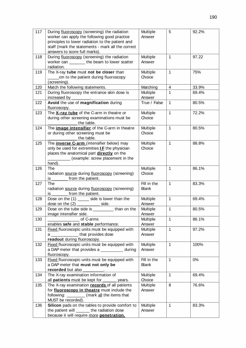
190
117 During fluoroscopy (screening) the radiation worker can apply the following good practice principles to lower radiation to the patient and staff (mark the statements - mark all the correct answers to score full marks).
Multiple Answer
5 92.2%
118 During fluoroscopy (screening) the radiation worker can _______ the beam to lower scatter radiation.
Multiple Answer
1 97.22
119 The X-ray tube must not be closer than _____cm to the patient during fluoroscopy (screening).
Multiple Choice
1 75%
120 Match the following statements. Matching 4 33.9% 121 During fluoroscopy the entrance skin dose is
increased by ________. Multiple Answer
1 69.4%
122 Avoid the use of magnification during fluoroscopy.
True / False 1 80.5%
123 The X-ray tube of the C-arm in theatre or during other screening examinations must be _____________ the table.
Multiple Choice
1 72.2%
124 The image intensifier of the C-arm in theatre or during other screening must be _____________ the table.
Multiple Choice
1 80.5%
125 The inverse C-arm (intensifier below) may only be used for extremities IF the physician places the anatomical part directly on the __________ (example: screw placement in the hand).
Multiple Choice
1 88.8%
126 The radiation source during fluoroscopy (screening) is _______ from the patient.
Multiple Choice
1 86.1%
127 The radiation source during fluoroscopy (screening) is _______ from the patient.
Fill in the Blank
1 83.3%
128 Dose on the (1) _____ side is lower than the dose on the (2) __________ side.
Multiple Answer
1 69.4%
129 Dose on the tube side is _________ than on the image intensifier side.
Multiple Answer
1 80.5%
130 ______________ of C-arms enables safe and stable performance.
Multiple Answer
1 86.1%
131 Fixed fluoroscopic units must be equipped with a ____________ that provides dose readout during fluoroscopy.
Multiple Answer
1 97.2%
132 Fixed fluoroscopic units must be equipped with a DAP meter that provides a __________ during fluoroscopy.
Multiple Answer
1 100%
133 Fixed fluoroscopic units must be equipped with a DAP meter that must not only be recorded but also _________.
Fill in the Blank
1 0%
134 The X-ray examination information of all patients must be kept for ______ years.
Multiple Choice
1 69.4%
135 The X-ray examination records of all patients for fluoroscopy in theatre must include the following: ________ (mark all the items that MUST be recorded).
Multiple Answer
8 76.6%
136 Silicon pads on the tables to provide comfort to the patient will ______ the radiation dose because it will require more penetration.
Multiple Answer
1 83.3%
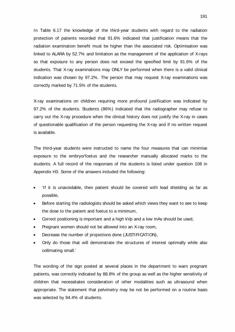
191
In Table 6.17 the knowledge of the third-year students with regard to the radiation
protection of patients recorded that 91.6% indicated that justification means that the
radiation examination benefit must be higher than the associated risk. Optimisation was
linked to ALARA by 52.7% and limitation as the management of the application of X-rays
so that exposure to any person does not exceed the specified limit by 91.6% of the
students. That X-ray examinations may ONLY be performed when there is a valid clinical
indication was chosen by 97.2%. The person that may request X-ray examinations was
correctly marked by 71.5% of the students.
X-ray examinations on children requiring more profound justification was indicated by
97.2% of the students. Students (86%) indicated that the radiographer may refuse to
carry out the X-ray procedure when the clinical history does not justify the X-ray in cases
of questionable qualification of the person requesting the X-ray and if no written request
is available.
The third-year students were instructed to name the four measures that can minimise
exposure to the embryo/foetus and the researcher manually allocated marks to the
students. A full record of the responses of the students is listed under question 108 in
Appendix H3. Some of the answers included the following:
• ‘If it is unavoidable, then patient should be covered with lead shielding as far as
possible,
• Before starting the radiologists should be asked which views they want to see to keep
the dose to the patient and foetus to a minimum,
• Correct positioning is important and a high kVp and a low mAs should be used,
• Pregnant women should not be allowed into an X-ray room,
• Decrease the number of projections done (JUSTIFICATION),
• Only do those that will demonstrate the structures of interest optimally while also
collimating small.’
The wording of the sign posted at several places in the department to warn pregnant
patients, was correctly indicated by 88.8% of the group as well as the higher sensitivity of
children that necessitates consideration of other modalities such as ultrasound when
appropriate. The statement that pelvimetry may be not be performed on a routine basis
was selected by 94.4% of students.
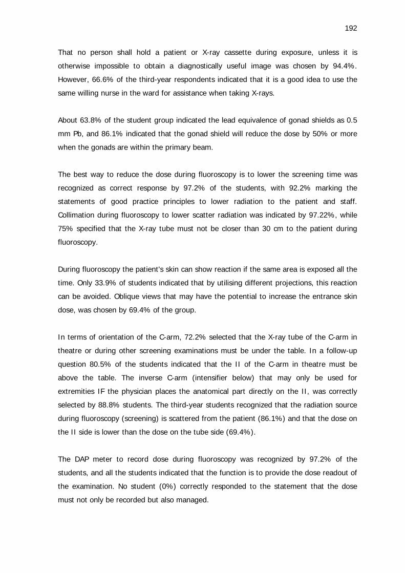
192
That no person shall hold a patient or X-ray cassette during exposure, unless it is
otherwise impossible to obtain a diagnostically useful image was chosen by 94.4%.
However, 66.6% of the third-year respondents indicated that it is a good idea to use the
same willing nurse in the ward for assistance when taking X-rays.
About 63.8% of the student group indicated the lead equivalence of gonad shields as 0.5
mm Pb, and 86.1% indicated that the gonad shield will reduce the dose by 50% or more
when the gonads are within the primary beam.
The best way to reduce the dose during fluoroscopy is to lower the screening time was
recognized as correct response by 97.2% of the students, with 92.2% marking the
statements of good practice principles to lower radiation to the patient and staff.
Collimation during fluoroscopy to lower scatter radiation was indicated by 97.22%, while
75% specified that the X-ray tube must not be closer than 30 cm to the patient during
fluoroscopy.
During fluoroscopy the patient's skin can show reaction if the same area is exposed all the
time. Only 33.9% of students indicated that by utilising different projections, this reaction
can be avoided. Oblique views that may have the potential to increase the entrance skin
dose, was chosen by 69.4% of the group.
In terms of orientation of the C-arm, 72.2% selected that the X-ray tube of the C-arm in
theatre or during other screening examinations must be under the table. In a follow-up
question 80.5% of the students indicated that the II of the C-arm in theatre must be
above the table. The inverse C-arm (intensifier below) that may only be used for
extremities IF the physician places the anatomical part directly on the II, was correctly
selected by 88.8% students. The third-year students recognized that the radiation source
during fluoroscopy (screening) is scattered from the patient (86.1%) and that the dose on
the II side is lower than the dose on the tube side (69.4%).
The DAP meter to record dose during fluoroscopy was recognized by 97.2% of the
students, and all the students indicated that the function is to provide the dose readout of
the examination. No student (0%) correctly responded to the statement that the dose
must not only be recorded but also managed.
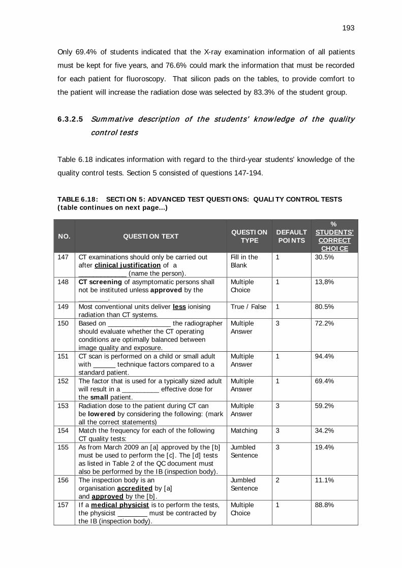
193
Only 69.4% of students indicated that the X-ray examination information of all patients
must be kept for five years, and 76.6% could mark the information that must be recorded
for each patient for fluoroscopy. That silicon pads on the tables, to provide comfort to
the patient will increase the radiation dose was selected by 83.3% of the student group.
6.3.2.5 Summative description of the students’ know ledge of the quality
control tests
Table 6.18 indicates information with regard to the third-year students’ knowledge of the
quality control tests. Section 5 consisted of questions 147-194.
TABLE 6.18: SECTION 5: ADVANCED TEST QUESTIONS: QUALITY CONTROL TESTS (table continues on next page...)
NO. QUESTION TEXT
QUESTION TYPE
DEFAULT POINTS
% STUDENTS’ CORRECT CHOICE
147 CT examinations should only be carried out after clinical justification of a _____________ (name the person).
Fill in the Blank
1 30.5%
148 CT screening of asymptomatic persons shall not be instituted unless approved by the ________.
Multiple Choice
1 13,8%
149 Most conventional units deliver less ionising radiation than CT systems.
True / False 1 80.5%
150 Based on _________________ the radiographer should evaluate whether the CT operating conditions are optimally balanced between image quality and exposure.
Multiple Answer
3 72.2%
151 CT scan is performed on a child or small adult with ______ technique factors compared to a standard patient.
Multiple Answer
1 94.4%
152 The factor that is used for a typically sized adult will result in a __________ effective dose for the small patient.
Multiple Answer
1 69.4%
153 Radiation dose to the patient during CT can be lowered by considering the following: (mark all the correct statements)
Multiple Answer
3 59.2%
154 Match the frequency for each of the following CT quality tests:
Matching 3 34.2%
155 As from March 2009 an [a] approved by the [b] must be used to perform the [c]. The [d] tests as listed in Table 2 of the QC document must also be performed by the IB (inspection body).
Jumbled Sentence
3 19.4%
156 The inspection body is an organisation accredited by [a] and approved by the [b].
Jumbled Sentence
2 11.1%
157 If a medical physicist is to perform the tests, the physicist ________ must be contracted by the IB (inspection body).
Multiple Choice
1 88.8%
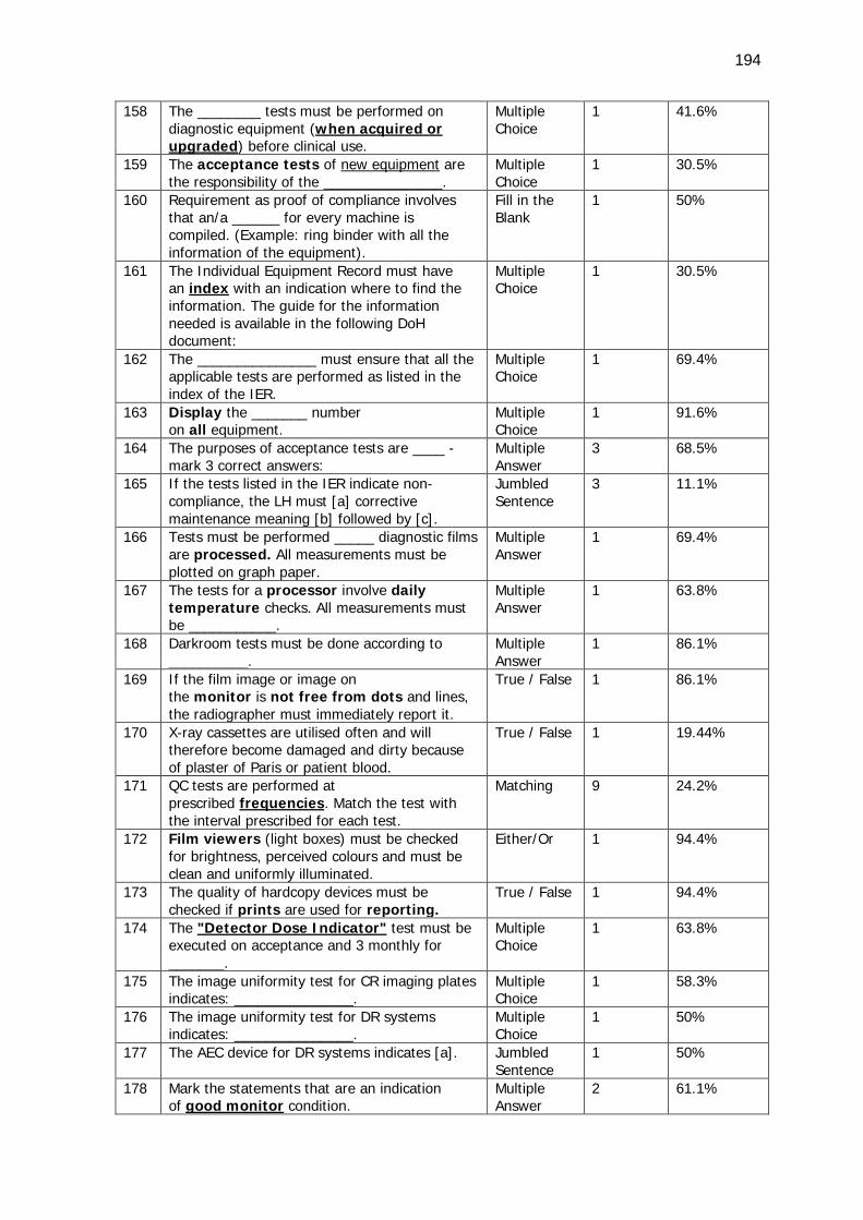
194
158 The ________ tests must be performed on diagnostic equipment (when acquired or upgraded) before clinical use.
Multiple Choice
1 41.6%
159 The acceptance tests of new equipment are the responsibility of the _______________.
Multiple Choice
1 30.5%
160 Requirement as proof of compliance involves that an/a ______ for every machine is compiled. (Example: ring binder with all the information of the equipment).
Fill in the Blank
1 50%
161 The Individual Equipment Record must have an index with an indication where to find the information. The guide for the information needed is available in the following DoH document:
Multiple Choice
1 30.5%
162 The _______________ must ensure that all the applicable tests are performed as listed in the index of the IER.
Multiple Choice
1 69.4%
163 Display the _______ number on all equipment.
Multiple Choice
1 91.6%
164 The purposes of acceptance tests are ____ - mark 3 correct answers:
Multiple Answer
3 68.5%
165 If the tests listed in the IER indicate non-compliance, the LH must [a] corrective maintenance meaning [b] followed by [c].
Jumbled Sentence
3 11.1%
166 Tests must be performed _____ diagnostic films are processed. All measurements must be plotted on graph paper.
Multiple Answer
1 69.4%
167 The tests for a processor involve daily temperature checks. All measurements must be ___________.
Multiple Answer
1 63.8%
168 Darkroom tests must be done according to __________.
Multiple Answer
1 86.1%
169 If the film image or image on the monitor is not free from dots and lines, the radiographer must immediately report it.
True / False 1 86.1%
170 X-ray cassettes are utilised often and will therefore become damaged and dirty because of plaster of Paris or patient blood.
True / False 1 19.44%
171 QC tests are performed at prescribed frequencies. Match the test with the interval prescribed for each test.
Matching 9 24.2%
172 Film viewers (light boxes) must be checked for brightness, perceived colours and must be clean and uniformly illuminated.
Either/Or 1 94.4%
173 The quality of hardcopy devices must be checked if prints are used for reporting.
True / False 1 94.4%
174 The "Detector Dose Indicator" test must be executed on acceptance and 3 monthly for _______.
Multiple Choice
1 63.8%
175 The image uniformity test for CR imaging plates indicates: _______________.
Multiple Choice
1 58.3%
176 The image uniformity test for DR systems indicates: _______________.
Multiple Choice
1 50%
177 The AEC device for DR systems indicates [a]. Jumbled Sentence
1 50%
178 Mark the statements that are an indication of good monitor condition.
Multiple Answer
2 61.1%
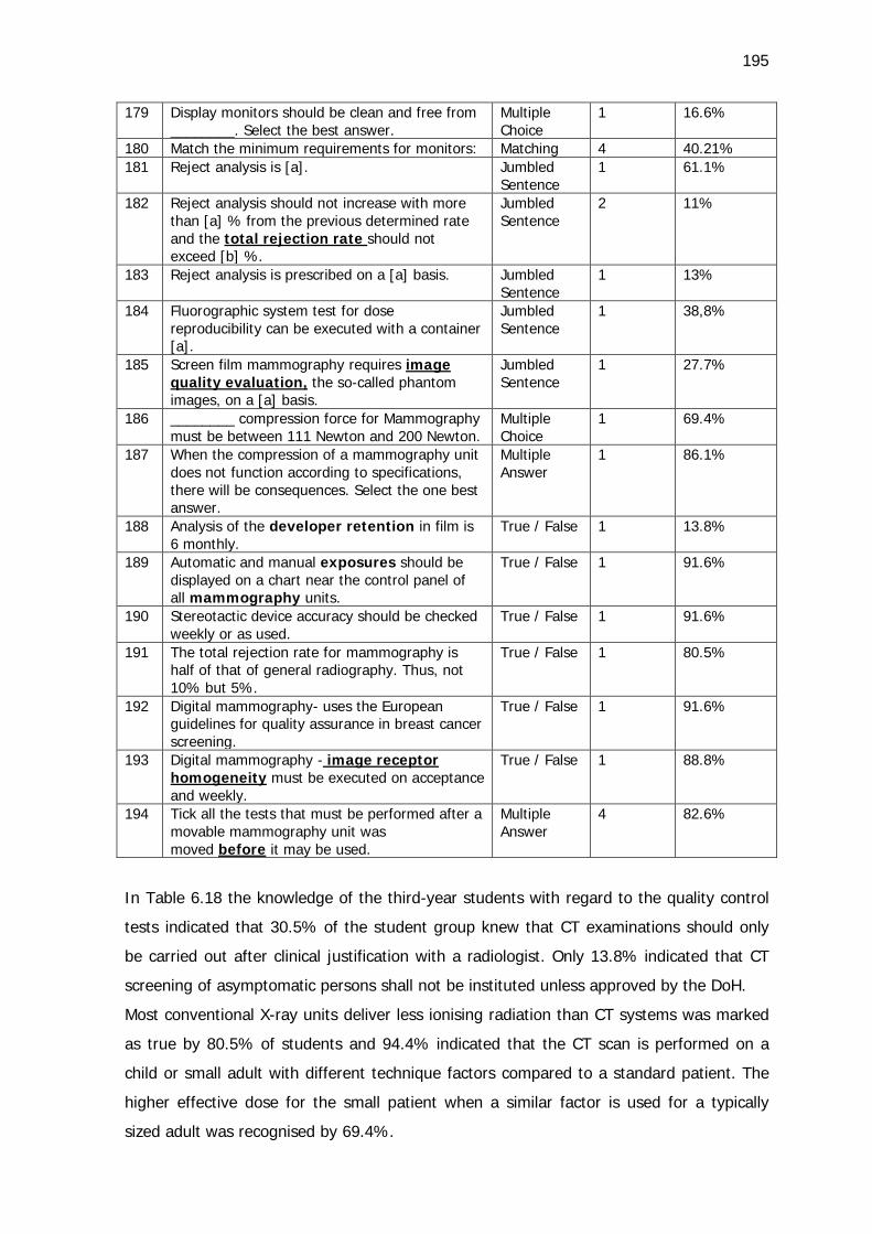
195
179 Display monitors should be clean and free from ________. Select the best answer.
Multiple Choice
1 16.6%
180 Match the minimum requirements for monitors: Matching 4 40.21% 181 Reject analysis is [a]. Jumbled
Sentence 1 61.1%
182 Reject analysis should not increase with more than [a] % from the previous determined rate and the total rejection rate should not exceed [b] %.
Jumbled Sentence
2 11%
183 Reject analysis is prescribed on a [a] basis. Jumbled Sentence
1 13%
184 Fluorographic system test for dose reproducibility can be executed with a container [a].
Jumbled Sentence
1 38,8%
185 Screen film mammography requires image quality evaluation, the so-called phantom images, on a [a] basis.
Jumbled Sentence
1 27.7%
186 ________ compression force for Mammography must be between 111 Newton and 200 Newton.
Multiple Choice
1 69.4%
187 When the compression of a mammography unit does not function according to specifications, there will be consequences. Select the one best answer.
Multiple Answer
1 86.1%
188 Analysis of the developer retention in film is 6 monthly.
True / False 1 13.8%
189 Automatic and manual exposures should be displayed on a chart near the control panel of all mammography units.
True / False 1 91.6%
190 Stereotactic device accuracy should be checked weekly or as used.
True / False 1 91.6%
191 The total rejection rate for mammography is half of that of general radiography. Thus, not 10% but 5%.
True / False 1 80.5%
192 Digital mammography- uses the European guidelines for quality assurance in breast cancer screening.
True / False 1 91.6%
193 Digital mammography - image receptor homogeneity must be executed on acceptance and weekly.
True / False 1 88.8%
194 Tick all the tests that must be performed after a movable mammography unit was moved before it may be used.
Multiple Answer
4 82.6%
In Table 6.18 the knowledge of the third-year students with regard to the quality control
tests indicated that 30.5% of the student group knew that CT examinations should only
be carried out after clinical justification with a radiologist. Only 13.8% indicated that CT
screening of asymptomatic persons shall not be instituted unless approved by the DoH.
Most conventional X-ray units deliver less ionising radiation than CT systems was marked
as true by 80.5% of students and 94.4% indicated that the CT scan is performed on a
child or small adult with different technique factors compared to a standard patient. The
higher effective dose for the small patient when a similar factor is used for a typically
sized adult was recognised by 69.4%.
196
To lower the radiation dose to the patient during CT, 59.2% of students opted for the
responses to increase the pitch, to increase the table increment and to eliminate pre-
contrast images. The frequency for the CT quality tests, namely in cases of image noise
and on the basis of CT number values (on acceptance and daily), as well as the plane
localisation from alignment lights (on acceptance and 3-monthly) was correctly selected
by 34.2% of the group.
Only 19.4% could successfully indicate that, as from March 2009, an IB, approved by the
DoH, must be used to perform the acceptance tests, and that the QC tests as listed in
Table 2 of the QC document must also be performed by the IB. Few of the students
(11.1%) could select SANAS as an accreditation body. Most of the students (88.8%)
indicated that if a medical physicist is to perform the tests, the physicist must be
contracted by the IB.
The acceptance tests that must be performed on diagnostic equipment (when acquired or
upgraded) before clinical use were selected by 41.6% of the students, and 30.5%
indicated that the acceptance tests of new equipment are the responsibility of the
company that installs the equipment.
The ring binder with all the information of the equipment, namely IER, was correctly filled
in by 50% of the students, whereas 30.5% could indicate the document named Quality
Control Tests of diagnostic equipment as the document that lists the index of the IER.
The licence holder, who must ensure that all the applicable tests are performed as listed
in the index of the IER, was the choice of 69.4% of the third years. The purposes of
acceptance tests to ensure the unit meets the stated specifications, to establish baseline
parameters for future quality control, and to familiarise the customer with operation of
the unit were marked by 68.5% of the students. Most students (91.6%) indicated that the
licence number must be displayed on all X-ray equipment.
The statement that if the tests listed in the IER indicate non-compliance, the LH must
implement corrective maintenance, meaning repairs followed by re-testing, was correctly
selected by 11.1% of students.
Tests that must be performed before diagnostic films are processed, was indicated by
69.4% of students and 63.8% students indicated that the measurements must be plotted
197
on graph paper. The option, darkroom tests to be done according to a written protocol,
was the choice of 86.1% of the group. Most students (86.1%) selected that if the film
image or image on the monitor is not free from dots and lines, the radiographer must
immediately report it.
The third-year group indicated with a percentage of 19.4%, that it is false that X-ray
cassettes are utilised often and will therefore be damaged and dirty because of plaster of
Paris or patient blood. The two statements regarding good monitor condition were
correctly chosen by 61.1% of the third years. The fact that display monitors should be
clean and free from flicker was indicated by 16.6% of the students and 40.21% correctly
matched the minimum requirements for the different monitors for reporting and display.
QC test frequencies were matched correctly by 24.2% of the third-year group. That film
viewers (light boxes) must be checked for brightness, perceived colours and must be
clean and uniformly illuminated were recognized by 94.4% of the students; 63.8% of
students indicated that the ’Detector Dose Indicator’ test must be executed on acceptance
and three monthly for DDR and CR systems. Half of the students (50%) specified that the
image uniformity test for CR imaging plates indicates lines or rectangles otherwise not
apparent.
Only 11% of the third years specified that reject analysis should not increase with more
than 2% from the previous determined rate and the total rejection rate should not exceed
10%. The three-monthly basis for reject analysis was indicated by only 13% of the
students.
Execution of the dose reproducibility with fluorographic systems by means of a container
filled with water was selected by 38.8% students.
The responses to questions regarding mammography was recorded as follows: The
weekly evaluation for screen film mammography phantom images (27%), maximum
compression force for mammography between 111 Newton and 200 Newton (69.4%),
and the fact that pathology can be missed when the compression of a mammography unit
does not function according to specifications (86.1%). Analysis of the developer retention
in film every six months was recognized by 13.8% of the students, but 91.6% indicated
that automatic and manual exposures should be displayed on a chart near the control
198
panel of all mammography units. The statement that the total rejection rate for
mammography is half of that of general radiography was chosen by 80.5% of the third-
year students. Digital mammography that uses the European guidelines for quality
assurance in breast cancer screening was marked as true by 91.6% of the students and
88.8% considered it the truth that for digital mammography-image receptor homogeneity
must be executed on acceptance and weekly. The tests that must be performed after a
movable mammography unit has been moved before it may be used, was correctly
chosen by 82.6% of the student group.
These results complete the results from the basic and advanced student questionnaires.
6.4 CONCLUSION
The results of the third phase of the research have been presented in this chapter and the
data findings for the pre-and post-test student questionnaires have been described.
Tables were used to summarise the findings of the relevant sections of the student
questionnaires.
The aim of the student questionnaires was to determine mastery of the content as
selected by the Delphi panellists. The class average for the basic post-test was 65.04%
and for the advanced post-test 56.2%. Taking the mastery level of 75% into
consideration that was proposed in the Delphi survey (cf. Table 4.1) the questions where
less than 75% of the student group made a correct choice, were revisited. The questions
with a lower score than 75% will be discussed in the following chapter in order to
determine if different teaching and learning activities would guide the students to increase
mastery levels. The results of the current chapter determined change and inclusion of the
questions in the final student assessment questionnaire.
In the following chapter, Chapter 7, the final discussion on the findings of the research
will be presented with specific discussion of the implications of the outcome of the Delphi
rounds and the student pre-and post-test questionnaires.
CHAPTER 7 FINAL DISCUSSION ON THE FINDINGS OF THE RESEARCH
7.1 INTRODUCTION
The quantitative results of the research involved the findings of the Delphi questionnaire
as well as of the student questionnaires. The consensus and stability outcome of the
Delphi questionnaires were described in Chapter 4. The outcome of the Delphi
questionnaire determined the content of the learning outcomes, the teaching and learning
activities and the assessments of the radiation safety training as presented in Chapter 5.
The data findings for the pre- and post-test student questionnaires were described in the
Chapter 6. The current chapter will discuss the implications of the findings of the Delphi
rounds and the student questionnaires on the content, teaching and learning activities
and the assessment of radiation safety.
The Delphi questionnaire will be dealt with first with specific reference to the comments
from the Delphi panellists. The student questionnaires will be addressed with reference to
the role of feedback to identify areas where adjustment in teaching and learning can be
made before the final assessment opportunity. The chapter concludes with a description
of the adjustment of the teaching and learning activities in order to improve mastery of
the content for the beginner radiation worker, as well as the advanced student, as
potential future licence holder of medical X-ray equipment.
7.2 DESCRIPTION OF THE IMPLICATIONS OF THE FINDINGS FROM THE
DELPHI SURVEY
The Delphi panellists were encouraged to not only make choices for the inclusion of each
statement in the basic or advanced training, but also to provide comments if they felt
necessary. The questionnaire additionally made provision for the respondents to add
comments after each section in the questionnaire. In Round One, 130 comments were
made by seven Delphi panellists and in Round Two, 48 comments. Nine panellists made
66 comments in Round Three and five panellists raised 52 opinions in the final Round
Four. The 28 comments on the non-consensus statements supported the reaching of
stability in Round Four. The specific comments as well as the general comments were
included in Appendix D6 B and are listed in Table 7.1
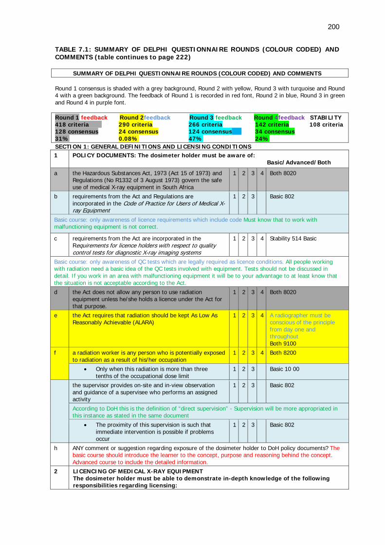
200
TABLE 7.1: SUMMARY OF DELPHI QUESTIONNAIRE ROUNDS (COLOUR CODED) AND COMMENTS (table continues to page 222)
SUMMARY OF DELPHI QUESTIONNAIRE ROUNDS (COLOUR CODED) AND COMMENTS Round 1 consensus is shaded with a grey background, Round 2 with yellow, Round 3 with turquoise and Round 4 with a green background. The feedback of Round 1 is recorded in red font, Round 2 in blue, Round 3 in green and Round 4 in purple font. Round 1 feedback Round 2feedback Round 3 feedback Round 4feedback STABILITY 418 criteria 290 criteria 266 criteria 142 criteria 108 criteria 128 consensus 24 consensus 124 consensus 34 consensus 31% 0.08% 47% 24% SECTION 1: GENERAL DEFINITIONS AND LICENSING CONDITIONS 1 POLICY DOCUMENTS: The dosimeter holder must be aware of:
Basic/Advanced/Both
a the Hazardous Substances Act, 1973 (Act 15 of 1973) and Regulations (No R1332 of 3 August 1973) govern the safe use of medical X-ray equipment in South Africa
1 2 3 4 Both 8020
b requirements from the Act and Regulations are incorporated in the Code of Practice for Users of Medical X-ray Equipment
1 2 3 Basic 802
Basic course: only awareness of licence requirements which include code Must know that to work with malfunctioning equipment is not correct.
c requirements from the Act are incorporated in the Requirements for licence holders with respect to quality control tests for diagnostic X-ray imaging systems
1 2 3 4 Stability 514 Basic
Basic course: only awareness of QC tests which are legally required as licence conditions. All people working with radiation need a basic idea of the QC tests involved with equipment. Tests should not be discussed in detail. If you work in an area with malfunctioning equipment it will be to your advantage to at least know that the situation is not acceptable according to the Act.
d the Act does not allow any person to use radiation equipment unless he/she holds a licence under the Act for that purpose.
1 2 3 4 Both 8020
e the Act requires that radiation should be kept As Low As Reasonably Achievable (ALARA)
1 2 3 4 A radiographer must be conscious of the principle from day one and throughout Both 9100
f a radiation worker is any person who is potentially exposed to radiation as a result of his/her occupation
1 2 3 4 Both 8200
• Only when this radiation is more than three tenths of the occupational dose limit
1 2 3 Basic 10 00
the supervisor provides on-site and in-view observation and guidance of a supervisee who performs an assigned activity
1 2 3 Basic 802
According to DoH this is the definition of “direct supervision” - Supervision will be more appropriated in this instance as stated in the same document
• The proximity of this supervision is such that immediate intervention is possible if problems occur
1 2 3 Basic 802
h ANY comment or suggestion regarding exposure of the dosimeter holder to DoH policy documents? The basic course should introduce the learner to the concept, purpose and reasoning behind the concept. Advanced course to include the detailed information.
2
LICENCING OF MEDICAL X-RAY EQUIPMENT The dosimeter holder must be able to demonstrate in-depth knowledge of the following responsibilities regarding licensing:
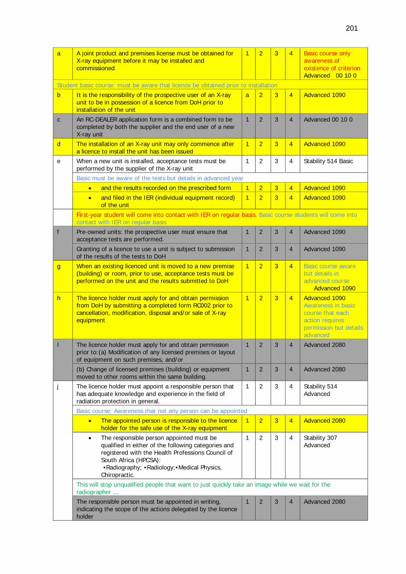
201
a A joint product and premises license must be obtained for X-ray equipment before it may be installed and commissioned
1 2 3 4 Basic course only awareness of existence of criterion Advanced 00 10 0
Student basic course: must be aware that licence be obtained prior to installation
b It is the responsibility of the prospective user of an X-ray unit to be in possession of a licence from DoH prior to installation of the unit
a 2 3 4 Advanced 1090
c An RC-DEALER application form is a combined form to be completed by both the supplier and the end user of a new X-ray unit
1 2 3 4 Advanced 00 10 0
d The installation of an X-ray unit may only commence after a licence to install the unit has been issued
1 2 3 4 Advanced 1090
e When a new unit is installed, acceptance tests must be performed by the supplier of the X-ray unit
1 2 3 4 Stability 514 Basic
Basic must be aware of the tests but details in advanced year
• and the results recorded on the prescribed form 1 2 3 4 Advanced 1090
• and filed in the IER (individual equipment record) of the unit
1 2 3 4 Advanced 1090
First-year student will come into contact with IER on regular basis. Basic course students will come into contact with IER on regular basis
f Pre-owned units: the prospective user must ensure that acceptance tests are performed.
1 2 3 4 Advanced 1090
Granting of a licence to use a unit is subject to submission of the results of the tests to DoH
1 2 3 4 Advanced 1090
g When an existing licenced unit is moved to a new premise (building) or room, prior to use, acceptance tests must be performed on the unit and the results submitted to DoH
1 2 3 4 Basic course aware but details in advanced course Advanced 1090
h The licence holder must apply for and obtain permission from DoH by submitting a completed form RC002 prior to cancellation, modification, disposal and/or sale of X-ray equipment
1 2 3 4 Advanced 1090 Awareness in basic course that each action requires permission but details advanced
I The licence holder must apply for and obtain permission prior to:(a) Modification of any licensed premises or layout of equipment on such premises, and/or
1 2 3 4 Advanced 2080
(b) Change of licensed premises (building) or equipment moved to other rooms within the same building.
1 2 3 4 Advanced 2080
j The licence holder must appoint a responsible person that has adequate knowledge and experience in the field of radiation protection in general.
1 2 3 4 Stability 514 Advanced
Basic course: Awareness that not any person can be appointed
• The appointed person is responsible to the licence holder for the safe use of the X-ray equipment
1 2 3 4 Advanced 2080
• The responsible person appointed must be qualified in either of the following categories and registered with the Health Professions Council of South Africa (HPCSA):
•Radiography; •Radiology;•Medical Physics, Chiropractic.
1 2 3 4 Stability 307 Advanced
This will stop unqualified people that want to just quickly take an image while we wait for the radiographer …
The responsible person must be appointed in writing, indicating the scope of the actions delegated by the licence holder
1 2 3 4 Advanced 2080
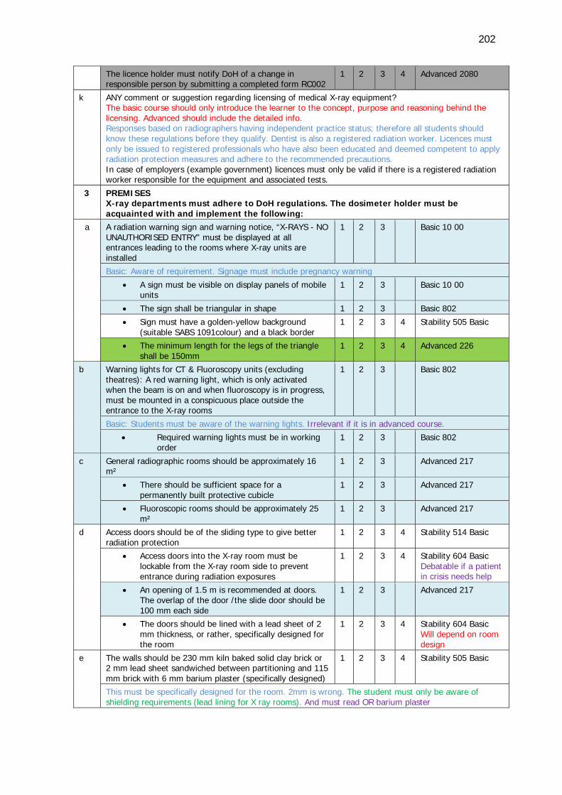
202
The licence holder must notify DoH of a change in responsible person by submitting a completed form RC002
1 2 3 4 Advanced 2080
k ANY comment or suggestion regarding licensing of medical X-ray equipment? The basic course should only introduce the learner to the concept, purpose and reasoning behind the licensing. Advanced should include the detailed info. Responses based on radiographers having independent practice status; therefore all students should know these regulations before they qualify. Dentist is also a registered radiation worker. Licences must only be issued to registered professionals who have also been educated and deemed competent to apply radiation protection measures and adhere to the recommended precautions. In case of employers (example government) licences must only be valid if there is a registered radiation worker responsible for the equipment and associated tests.
3 PREMISES X-ray departments must adhere to DoH regulations. The dosimeter holder must be acquainted with and implement the following:
a
A radiation warning sign and warning notice, “X-RAYS - NO UNAUTHORISED ENTRY” must be displayed at all entrances leading to the rooms where X-ray units are installed
1 2 3 Basic 10 00
Basic: Aware of requirement. Signage must include pregnancy warning
• A sign must be visible on display panels of mobile units
1 2 3 Basic 10 00
• The sign shall be triangular in shape 1 2 3 Basic 802
• Sign must have a golden-yellow background (suitable SABS 1091colour) and a black border
1 2 3 4 Stability 505 Basic
• The minimum length for the legs of the triangle shall be 150mm
1 2 3 4 Advanced 226
b Warning lights for CT & Fluoroscopy units (excluding theatres): A red warning light, which is only activated when the beam is on and when fluoroscopy is in progress, must be mounted in a conspicuous place outside the entrance to the X-ray rooms
1 2 3 Basic 802
Basic: Students must be aware of the warning lights. Irrelevant if it is in advanced course.
• Required warning lights must be in working order
1 2 3 Basic 802
c General radiographic rooms should be approximately 16 m²
1 2 3 Advanced 217
• There should be sufficient space for a permanently built protective cubicle
1 2 3 Advanced 217
• Fluoroscopic rooms should be approximately 25 m²
1 2 3 Advanced 217
d Access doors should be of the sliding type to give better radiation protection
1 2 3 4 Stability 514 Basic
• Access doors into the X-ray room must be lockable from the X-ray room side to prevent entrance during radiation exposures
1 2 3 4 Stability 604 Basic Debatable if a patient in crisis needs help
• An opening of 1.5 m is recommended at doors. The overlap of the door /the slide door should be 100 mm each side
1 2 3 Advanced 217
• The doors should be lined with a lead sheet of 2 mm thickness, or rather, specifically designed for the room
1 2 3 4 Stability 604 Basic Will depend on room design
e The walls should be 230 mm kiln baked solid clay brick or 2 mm lead sheet sandwiched between partitioning and 115 mm brick with 6 mm barium plaster (specifically designed)
1 2 3 4 Stability 505 Basic
This must be specifically designed for the room. 2mm is wrong. The student must only be aware of shielding requirements (lead lining for X ray rooms). And must read OR barium plaster
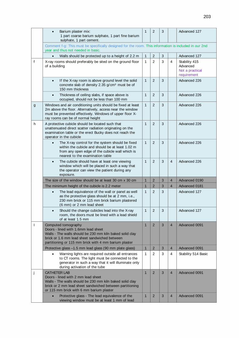
203
• Barium plaster mix: 1 part coarse barium sulphate, 1 part fine barium sulphate, 1 part cement.
1 2 3 Advanced 127
Comment f-g: This must be specifically designed for the room. This information is included in our 2nd year and thus not needed in basic.
• Walls should be protected up to a height of 2.2 m 1 2 3 Advanced 127
f X-ray rooms should preferably be sited on the ground floor of a building
1 2 3 4 Stability 415 Advanced Not a practical requirement
• If the X-ray room is above ground level the solid concrete slab of density 2.35 g/cm³ must be of 150 mm thickness
1 2 3 Advanced 226
• Thickness of ceiling slabs, if space above is occupied, should not be less than 100 mm
1 2 3 Advanced 226
g Windows and air conditioning units should be fixed at least 2m above the floor. Alternatively, access near the window must be prevented effectively. Windows of upper floor X-ray rooms can be of normal height
1 2 3 Advanced 226
h A protective cubicle should be located such that unattenuated direct scatter radiation originating on the examination table or the erect Bucky does not reach the operator in the cubicle
1 2 3 Advanced 226
• The X-ray control for the system should be fixed within the cubicle and should be at least 1.02 m from any open edge of the cubicle wall which is nearest to the examination table
1 2 3 Advanced 226
• The cubicle should have at least one viewing window which will be placed in such a way that the operator can view the patient during any exposure.
1 2 3 4 Advanced 226
The size of the window should be at least 30 cm x 30 cm 1 2 3 4 Advanced 0190
The minimum height of the cubicle is 2.2 meter 1 2 3 4 Advanced 0181
• The lead equivalence of the wall or panel as well as the protective glass should be at 2 mm, i.e., 230 mm brick or 115 mm brick barium plastered (6 mm) or 2 mm lead sheet
1 2 3 Advanced 127
• Should the change cubicles lead into the X-ray room, the doors must be lined with a lead shield of at least 1.5 mm
1 2 3 Advanced 127
I Computed tomography Doors - lined with 1.6mm lead sheet Walls - The walls should be 230 mm kiln baked solid clay brick or 1.6 mm lead sheet sandwiched between partitioning or 115 mm brick with 4 mm barium plaster
1 2 3 4 Advanced 0091
Protective glass –1.5 mm lead glass (90 mm plate glass) 1 2 3 4 Advanced 0091
• Warning lights are required outside all entrances to CT rooms. The light must be connected to the generator in such a way that it will illuminate only during activation of the tube
1 2 3 4 Stability 514 Basic
j CATHETER LAB Doors - lined with 2 mm lead sheet Walls - The walls should be 230 mm kiln baked solid clay brick or 2 mm lead sheet sandwiched between partitioning or 115 mm brick with 6 mm barium plaster
1 2 3 4 Advanced 0091
• Protective glass - The lead equivalence of the viewing window must be at least 1 mm of lead
1 2 3 4 Advanced 0091
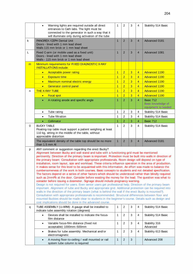
204
• Warning lights are required outside all direct entrances to Cath labs. The light must be connected to the generator in such a way that it will illuminate only during activation of the tube
1 2 3 4 Stability 514 Basic
k PANOREX /CEPH Dental Unit Doors - lined with 1 mm lead sheet Walls 115 mm brick or 1 mm lead sheet
1 2 3 4 Advanced 0181
L Fixed C-arm (or mobile used as a fixed unit) Doors - lined with 1 mm lead sheet Walls - 115 mm brick or 1 mm lead sheet
1 2 3 4 Advanced 1081
m Minimum requirements for FIXED DIAGNOSTIC X-RAY INSTALLATIONS include:
• Acceptable power rating 1 2 3 4 Advanced 1190
• Exposure time 1 2 3 4 Advanced 1190
• Maximum nominal electric energy 1 2 3 4 Advanced 1190
• Generator control panel 1 2 3 4 Advanced 1190
n THE X-RAY TUBE 1 2 3 4 Advanced 1190
• Focal spot 1 2 3 4 Advanced 1190
• A rotating anode and specific angle 1 2 3 4 Basic 712 Basic knowledge of equipment is needed
• Tube rating 1 2 3 4 Stability 514 Basic
• Tube filtration 1 2 3 4 Stability 514 Basic
• Collimator 1 2 3 4 Basic 712
0 BUCKY TABLE Floating-top table must support a patient weighing at least 110 kg, sitting in the middle of the table, without appreciable distortion
1 2 3 4 Stability 514 Basic
The equivalent density of the table top should be no more than 1.5 mm Al
1 2 3 4 Advanced 0181
p ANY comment or suggestion regarding the erect Bucky? Alignment between Bucky and wall stand and tube with a functioning grid must be mentioned pertinently. Direction of the primary beam is important. Protection must be built into walls in direction of the primary beam: Consultation with appropriate professionals. Room design will depend on type of installation, room layout, size and workload. These criteria influence operation in the area of production. It makes sense for this level to be acquainted with this information. An effort was made to balance the cumbersomeness of the work in both courses. Basic concepts to students and not detailed specification. The factors depend on a series of other factors which should be understood rather than blindly required such as 2mmPb at the door. Consider before wasting the money for the lead. The question was what to consider before issuing a dosimeter. Signage should include pregnancy warning. Design is not required for users. Even senior users get professional help. Direction of the primary beam important. Alignment of tube and Bucky and appropriate grid. Additional protection can be required into walls in the direction of the primary beam (what is behind the wall if the erect Bucky is mounted?). Consultation with appropriate professionals is recommended. Structural differences between floor and mounted Buckies should be made clear to students in the beginner’s course. Details such as design and cost implications should be done in the advanced course.
q TUBE ASSEMBLY (U-ARM): A gauge shall be installed to indicate tube assembly rotation (degrees)
1 2 3 4 Stability 514 Basic
• Devices shall be installed to indicate the focus-film distance
1 2 3 4 Stability 514 Basic
• Variable focus-film distance (fixed not acceptable):1000mm–500mm
1 2 3 4 Stability 316 Advanced
• Brakes for tube assembly: Mechanical and/or electromagnetic
1 2 3 4 Stability 613 Basic
• A moving floor-to-ceiling / wall mounted or rail system tube column is required
1 2 3 Advanced 208
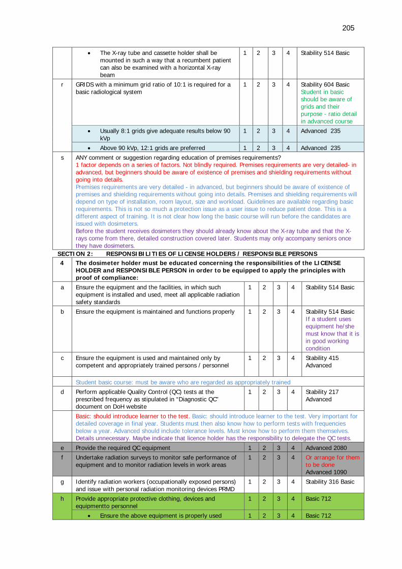
205
• The X-ray tube and cassette holder shall be mounted in such a way that a recumbent patient can also be examined with a horizontal X-ray beam
1 2 3 4 Stability 514 Basic
r GRIDS with a minimum grid ratio of 10:1 is required for a basic radiological system
1 2 3 4 Stability 604 Basic Student in basic should be aware of grids and their purpose - ratio detail in advanced course
• Usually 8:1 grids give adequate results below 90 kVp
1 2 3 4 Advanced 235
• Above 90 kVp, 12:1 grids are preferred 1 2 3 4 Advanced 235
s ANY comment or suggestion regarding education of premises requirements? 1 factor depends on a series of factors. Not blindly required. Premises requirements are very detailed- in advanced, but beginners should be aware of existence of premises and shielding requirements without going into details. Premises requirements are very detailed - in advanced, but beginners should be aware of existence of premises and shielding requirements without going into details. Premises and shielding requirements will depend on type of installation, room layout, size and workload. Guidelines are available regarding basic requirements. This is not so much a protection issue as a user issue to reduce patient dose. This is a different aspect of training. It is not clear how long the basic course will run before the candidates are issued with dosimeters. Before the student receives dosimeters they should already know about the X-ray tube and that the X-rays come from there, detailed construction covered later. Students may only accompany seniors once they have dosimeters.
SECTION 2: RESPONSIBILITIES OF LICENSE HOLDERS / RESPONSIBLE PERSONS 4 The dosimeter holder must be educated concerning the responsibilities of the LICENSE
HOLDER and RESPONSIBLE PERSON in order to be equipped to apply the principles with proof of compliance:
a Ensure the equipment and the facilities, in which such equipment is installed and used, meet all applicable radiation safety standards
1 2 3 4 Stability 514 Basic
b Ensure the equipment is maintained and functions properly 1 2 3 4 Stability 514 Basic If a student uses equipment he/she must know that it is in good working condition
c Ensure the equipment is used and maintained only by competent and appropriately trained persons / personnel
1 2 3 4 Stability 415 Advanced
Student basic course: must be aware who are regarded as appropriately trained
d Perform applicable Quality Control (QC) tests at the prescribed frequency as stipulated in “Diagnostic QC” document on DoH website
1 2 3 4 Stability 217 Advanced
Basic: should introduce learner to the test. Basic: should introduce learner to the test. Very important for detailed coverage in final year. Students must then also know how to perform tests with frequencies below a year. Advanced should include tolerance levels. Must know how to perform them themselves. Details unnecessary. Maybe indicate that licence holder has the responsibility to delegate the QC tests.
e Provide the required QC equipment 1 2 3 4 Advanced 2080
f Undertake radiation surveys to monitor safe performance of equipment and to monitor radiation levels in work areas
1 2 3 4 Or arrange for them to be done Advanced 1090
g Identify radiation workers (occupationally exposed persons) and issue with personal radiation monitoring devices PRMD
1 2 3 4 Stability 316 Basic
h Provide appropriate protective clothing, devices and equipmentto personnel
1 2 3 4 Basic 712
• Ensure the above equipment is properly used 1 2 3 4 Basic 712
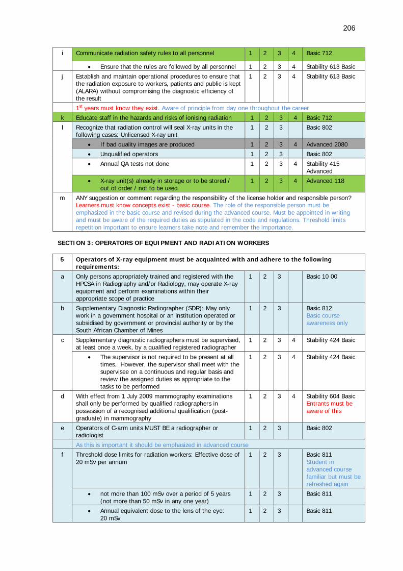
206
i Communicate radiation safety rules to all personnel 1 2 3 4 Basic 712
• Ensure that the rules are followed by all personnel 1 2 3 4 Stability 613 Basic
j Establish and maintain operational procedures to ensure that the radiation exposure to workers, patients and public is kept (ALARA) without compromising the diagnostic efficiency of the result
1 2 3 4 Stability 613 Basic
1st years must know they exist. Aware of principle from day one throughout the career
k Educate staff in the hazards and risks of ionising radiation 1 2 3 4 Basic 712
l Recognize that radiation control will seal X-ray units in the following cases: Unlicensed X-ray unit
1 2 3 Basic 802
• If bad quality images are produced 1 2 3 4 Advanced 2080
• Unqualified operators 1 2 3 Basic 802
• Annual QA tests not done 1 2 3 4 Stability 415 Advanced
• X-ray unit(s) already in storage or to be stored / out of order / not to be used
1 2 3 4 Advanced 118
m ANY suggestion or comment regarding the responsibility of the license holder and responsible person? Learners must know concepts exist - basic course. The role of the responsible person must be emphasized in the basic course and revised during the advanced course. Must be appointed in writing and must be aware of the required duties as stipulated in the code and regulations. Threshold limits repetition important to ensure learners take note and remember the importance.
SECTION 3: OPERATORS OF EQUIPMENT AND RADIATION WORKERS 5 Operators of X-ray equipment must be acquainted with and adhere to the following
requirements: a Only persons appropriately trained and registered with the
HPCSA in Radiography and/or Radiology, may operate X-ray equipment and perform examinations within their appropriate scope of practice
1 2 3 Basic 10 00
b Supplementary Diagnostic Radiographer (SDR): May only work in a government hospital or an institution operated or subsidised by government or provincial authority or by the South African Chamber of Mines
1 2 3 Basic 812 Basic course awareness only
c Supplementary diagnostic radiographers must be supervised, at least once a week, by a qualified registered radiographer
1 2 3 4 Stability 424 Basic
• The supervisor is not required to be present at all times. However, the supervisor shall meet with the supervisee on a continuous and regular basis and review the assigned duties as appropriate to the tasks to be performed
1 2 3 4 Stability 424 Basic
d With effect from 1 July 2009 mammography examinations shall only be performed by qualified radiographers in possession of a recognised additional qualification (post-graduate) in mammography
1 2 3 4 Stability 604 Basic Entrants must be aware of this
e Operators of C-arm units MUST BE a radiographer or radiologist
1 2 3 Basic 802
As this is important it should be emphasized in advanced course
f Threshold dose limits for radiation workers: Effective dose of 20 mSv per annum
1 2 3 Basic 811 Student in advanced course familiar but must be refreshed again
• not more than 100 mSv over a period of 5 years (not more than 50 mSv in any one year)
1 2 3 Basic 811
• Annual equivalent dose to the lens of the eye: 20 mSv
1 2 3 Basic 811
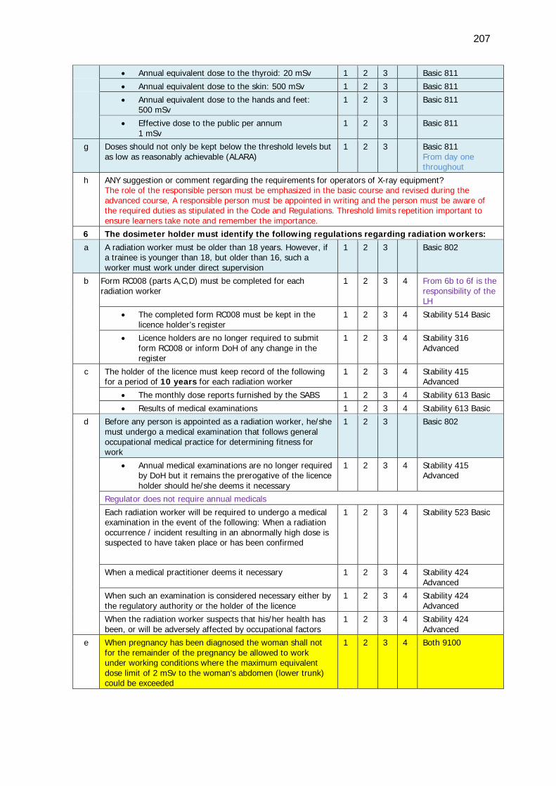
207
• Annual equivalent dose to the thyroid: 20 mSv 1 2 3 Basic 811 • Annual equivalent dose to the skin: 500 mSv 1 2 3 Basic 811 • Annual equivalent dose to the hands and feet:
500 mSv 1 2 3 Basic 811
• Effective dose to the public per annum 1 mSv
1 2 3 Basic 811
g Doses should not only be kept below the threshold levels but as low as reasonably achievable (ALARA)
1 2 3 Basic 811 From day one throughout
h ANY suggestion or comment regarding the requirements for operators of X-ray equipment? The role of the responsible person must be emphasized in the basic course and revised during the advanced course, A responsible person must be appointed in writing and the person must be aware of the required duties as stipulated in the Code and Regulations. Threshold limits repetition important to ensure learners take note and remember the importance.
6 The dosimeter holder must identify the following regulations regarding radiation workers: a A radiation worker must be older than 18 years. However, if
a trainee is younger than 18, but older than 16, such a worker must work under direct supervision
1 2 3 Basic 802
b Form RC008 (parts A,C,D) must be completed for each radiation worker
1 2 3 4 From 6b to 6f is the responsibility of the LH
• The completed form RC008 must be kept in the licence holder’s register
1 2 3 4 Stability 514 Basic
• Licence holders are no longer required to submit form RC008 or inform DoH of any change in the register
1 2 3 4 Stability 316 Advanced
c The holder of the licence must keep record of the following for a period of 10 years for each radiation worker
1 2 3 4 Stability 415 Advanced
• The monthly dose reports furnished by the SABS 1 2 3 4 Stability 613 Basic
• Results of medical examinations 1 2 3 4 Stability 613 Basic
d Before any person is appointed as a radiation worker, he/she must undergo a medical examination that follows general occupational medical practice for determining fitness for work
1 2 3 Basic 802
• Annual medical examinations are no longer required by DoH but it remains the prerogative of the licence holder should he/she deems it necessary
1 2 3 4 Stability 415 Advanced
Regulator does not require annual medicals
Each radiation worker will be required to undergo a medical examination in the event of the following: When a radiation occurrence / incident resulting in an abnormally high dose is suspected to have taken place or has been confirmed
1 2 3 4 Stability 523 Basic
When a medical practitioner deems it necessary 1 2 3 4 Stability 424 Advanced
When such an examination is considered necessary either by the regulatory authority or the holder of the licence
1 2 3 4 Stability 424 Advanced
When the radiation worker suspects that his/her health has been, or will be adversely affected by occupational factors
1 2 3 4 Stability 424 Advanced
e When pregnancy has been diagnosed the woman shall not for the remainder of the pregnancy be allowed to work under working conditions where the maximum equivalent dose limit of 2 mSv to the woman's abdomen (lower trunk) could be exceeded
1 2 3 4 Both 9100
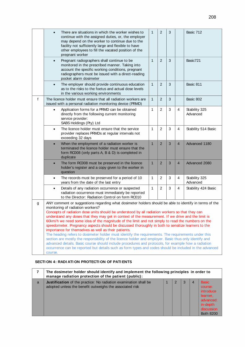
208
• There are situations in which the worker wishes to continue with the assigned duties, or, the employer may depend on the worker to continue due to the facility not sufficiently large and flexible to have other employees to fill the vacated position of the pregnant worker
1 2 3 Basic 712
• Pregnant radiographers shall continue to be monitored in the prescribed manner. Taking into account the specific working conditions, pregnant radiographers must be issued with a direct-reading pocket alarm dosimeter
1 2 3 Basic721
• The employer should provide continuous education as to the risks to the foetus and actual dose levels in the various working environments
1 2 3 Basic 811
f The licence holder must ensure that all radiation workers are issued with a personal radiation monitoring device (PRMD)
1 2 3 Basic 802
• Application forms for a PRMD can be obtained directly from the following current monitoring service provider: SABS Holdings (Pty) Ltd
1 2 3 4 Stability 325 Advanced
• The licence holder must ensure that the service provider replaces PRMDs at regular intervals not exceeding 32 days
1 2 3 4 Stability 514 Basic
• When the employment of a radiation worker is terminated the licence holder must ensure that the form RC008 (only parts A, B & D) is completed in duplicate
1 2 3 4 Advanced 1180
• The form RC008 must be preserved in the licence holder’s register and a copy given to the worker in question
1 2 3 4 Advanced 2080
• The records must be preserved for a period of 10 years from the date of the last entry
1 2 3 4 Stability 325 Advanced
• Details of any radiation occurrence or suspected radiation occurrence must immediately be reported to the Director: Radiation Control on form RC010
1 2 3 4 Stability 424 Basic
g ANY comment or suggestions regarding what dosimeter holders should be able to identify in terms of the monitoring of radiation workers? Concepts of radiation dose units should be understood by all radiation workers so that they can understand any doses that they may get in context of the measurement. If we drive and the limit is 60km/h we need some idea of the magnitude of the limit and not simply to read the numbers on the speedometer. Pregnancy aspects should be discussed thoroughly in both to sensitize learners to the importance for themselves as well as their patients. The heading refers to dosimeter holder must identify the requirements. The requirements under this section are mostly the responsibility of the licence holder and employer. Basic thus only identify and advanced details. Basic course should include procedures and protocols, for example how a radiation occurrence can be reported but details such as form types and codes should be included in the advanced course.
SECTION 4: RADIATION PROTECTION OF PATIENTS 7 The dosimeter holder should identify and implement the following principles in order to
manage radiation protection of the patient (public): a Justification of the practice: No radiation examination shall be
adopted unless the benefit outweighs the associated risk 1 2 3 4 Basic
course: introduce learner, advanced: in-depth discussion Both 8200
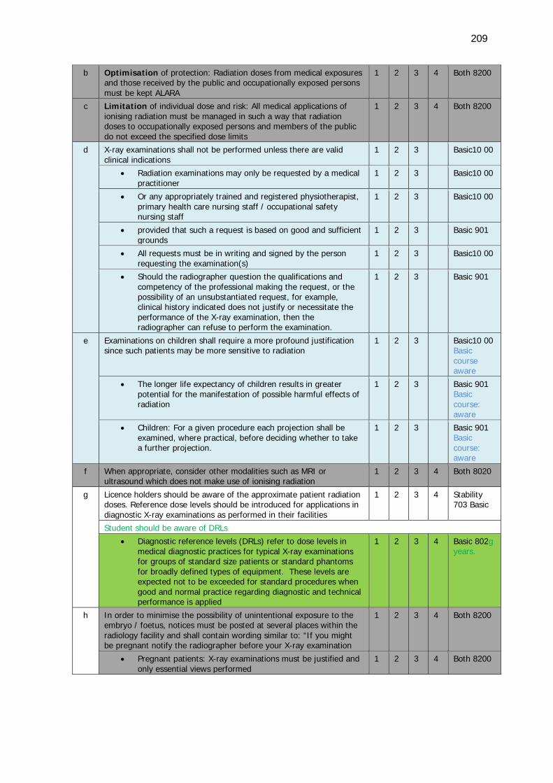
209
b Optimisation of protection: Radiation doses from medical exposures and those received by the public and occupationally exposed persons must be kept ALARA
1 2 3 4 Both 8200
c Limitation of individual dose and risk: All medical applications of ionising radiation must be managed in such a way that radiation doses to occupationally exposed persons and members of the public do not exceed the specified dose limits
1 2 3 4 Both 8200
d X-ray examinations shall not be performed unless there are valid clinical indications
1 2 3 Basic10 00
• Radiation examinations may only be requested by a medical practitioner
1 2 3 Basic10 00
• Or any appropriately trained and registered physiotherapist, primary health care nursing staff / occupational safety nursing staff
1 2 3 Basic10 00
• provided that such a request is based on good and sufficient grounds
1 2 3 Basic 901
• All requests must be in writing and signed by the person requesting the examination(s)
1 2 3 Basic10 00
• Should the radiographer question the qualifications and competency of the professional making the request, or the possibility of an unsubstantiated request, for example, clinical history indicated does not justify or necessitate the performance of the X-ray examination, then the radiographer can refuse to perform the examination.
1 2 3 Basic 901
e Examinations on children shall require a more profound justification since such patients may be more sensitive to radiation
1 2 3 Basic10 00 Basic course aware
• The longer life expectancy of children results in greater potential for the manifestation of possible harmful effects of radiation
1 2 3 Basic 901 Basic course: aware
• Children: For a given procedure each projection shall be examined, where practical, before deciding whether to take a further projection.
1 2 3 Basic 901 Basic course: aware
f When appropriate, consider other modalities such as MRI or ultrasound which does not make use of ionising radiation
1 2 3 4 Both 8020
g Licence holders should be aware of the approximate patient radiation doses. Reference dose levels should be introduced for applications in diagnostic X-ray examinations as performed in their facilities
1 2 3 4 Stability 703 Basic
Student should be aware of DRLs
• Diagnostic reference levels (DRLs) refer to dose levels in medical diagnostic practices for typical X-ray examinations for groups of standard size patients or standard phantoms for broadly defined types of equipment. These levels are expected not to be exceeded for standard procedures when good and normal practice regarding diagnostic and technical performance is applied
1 2 3 4 Basic 802g years.
h In order to minimise the possibility of unintentional exposure to the embryo / foetus, notices must be posted at several places within the radiology facility and shall contain wording similar to: “If you might be pregnant notify the radiographer before your X-ray examination
1 2 3 4 Both 8200
• Pregnant patients: X-ray examinations must be justified and only essential views performed
1 2 3 4 Both 8200
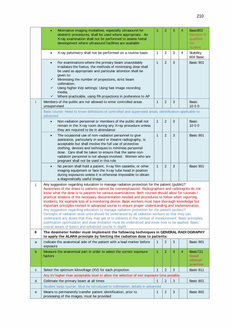
210
• Alternative imaging modalities, especially ultrasound for obstetric procedures, shall be used where appropriate. An X-ray examination shall not be performed to assess foetal development where ultrasound facilities are available
1 2 3 4 Basic802 Decision of qualified not student
• X-ray pelvimetry shall not be performed on a routine basis 1 2 3 4 Stability 604 Basic
• For examinations where the primary beam unavoidably irradiates the foetus, the methods of minimising dose shall be used as appropriate and particular attention shall be given to:
Minimising the number of projections, strict beam collimation;
Using higher kVp settings; Using fast image recording media;
Where practicable, using PA projections in preference to AP
1 2 3 Basic 901
i Members of the public are not allowed to enter controlled areas unsupervised
1 2 3 Basic 10 0 0.
Basic course: Need to know definitions of controlled and supervised areas, identification applicable to advanced
• Non-radiation personnel or members of the public shall not remain in the X-ray room during any X-ray procedure unless they are required to be in attendance
1 2 3 Basic 10 0 0
• The occasional use of non–radiation personnel to give assistance, particularly in ward or theatre radiography, is acceptable but shall involve the full use of protective clothing, devices and techniques to minimise personnel dose. Care shall be taken to ensure that the same non-radiation personnel is not always involved. Women who are pregnant shall not be used in this role
1 2 3 Basic 901
• No person shall hold a patient, X-ray film cassette, or other imaging equipment or face the X-ray tube head in position during exposures unless it is otherwise impossible to obtain a diagnostically useful image
1 2 3 Basic 901
j Any suggestion regarding education to manage radiation protection for the patient (public)? Awareness of the doses to patients cannot be overemphasized. Radiographers and radiologists do not know what the dose is to patients for various examinations. Both courses should allow for tutorials / practical sessions of the necessary documentation needed and procedures to follow when reporting incidents, for example loss of a monitoring device. Basic workers must have thorough knowledge but important principles revised in advanced course to ensure proper understanding and implementation. Any suggestion regarding education to manage radiation protection for the patient (public)? Concepts of radiation dose units should be understood by all radiation workers so that they can understand any doses that they may get or to patients in the context of measurement. Basic principles, justification optimization and dose limitation must be understood and know how to be applied. Basic course aware of basics and advanced course in depth.
8 The dosimeter holder must implement the following techniques in GENERAL RADIOGRAPHY to apply the ALARA principle by limiting the radiation dose to patients:
a Indicate the anatomical side of the patient with a lead marker before exposure
1 2 3 Basic 901
b Measure the anatomical part in order to select the correct exposure factors
1 2 3 4 Basic721 Good clinical practice
c Select the optimum kilovoltage (KV) for each projection 1 2 3 Basic 811 Any kV higher than acceptable level to allow the selection of min exposure time possible
d Collimate the primary beam at all times 1 2 3 Basic 801 Student basic course: Must be introduced to collimation, details in advanced
e Means to permanently transfer patient identification, prior to processing of the images, must be provided
1 2 3 Basic 802
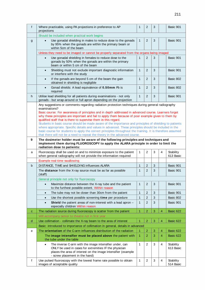
211
f Where practicable, using PA projections in preference to AP projections
1 2 3 Basic 901
Should be included when practical work begins
g • Use gonadal shielding in males to reduce dose to the gonads by 95% when the gonads are within the primary beam or within 5cm of the beam
1 2 3 Basic 901
Unless they need to be imaged or cannot be properly separated from the organs being imaged
• Use gonadal shielding in females to reduce dose to the gonads by 50% when the gonads are within the primary beam or within 5 cm of the beam
1 2 3 Basic 901
• Shielding must not exclude important diagnostic information or interfere with the study
1 2 3 Basic 901
• If the gonads are beyond 5 cm of the beam the gain obtained in shielding is negligible
1 2 3 Basic 802
• Gonad shields: A lead equivalence of 0.50mm Pb is required
1 2 3 Basic 802
h Utilise lead shielding for all patients during examinations - not only gonads - but wrap-around or full apron depending on the projection
1 2 3 Basic 901
Any suggestions or comments regarding radiation protection techniques during general radiography examinations? Basic course: For awareness of principles and in depth addressed in advanced course. Learners forget why these principles are important and fail to apply them because of poor example given to them by qualified staff that is there to supervise them in this regard. Students in basic course should be made aware of the importance and principles of shielding to patients where appropriate. Specific details and values in advanced. These principles should be included in the basic course for students to apply the correct principles throughout the training. It is therefore assumed that there will not be a need to repeat the theory in the advanced course.
9 The dosimeter holder must be aware of the following principles and techniques and implement them during FLUOROSCOPY to apply the ALARA principle in order to limit the radiation dose to patients:
a Fluoroscopy shall be used on and to minimize exposure to the patient when general radiography will not provide the information required
1 2 3 4 Stability 613 Basic
Example real-time swallowing
b DISTANCE, TIME and SHIELDING influences ALARA 1 2 3 Basic 901 The distance from the X-ray source must be as far as possible (staff)
1 2 3 Basic 901
General principle not only for fluoroscopy
• Maximize distance between the X-ray tube and the patient to the furthest possible extent. Within reason
1 2 3 Basic 901
• The tube may not be closer than 30cm from the patient 1 2 3 Basic 901
• Use the shortest possible screening time per procedure 1 2 3 Basic 802
• Shield the patient areas of non-interest with a lead apron – especially children Within reason
1 2 3 Basic 901
c The radiation source during fluoroscopy is scatter from the patient 1 2 3 4 Basic 622 Will accompany senior to theatre not in first year
d Use collimation - collimate the X-ray beam to the area of interest 1 2 3 4 Basic 622 Basic: introduced to importance of collimation in general, details in advanced
e The orientation of the C-arm influences distribution of the radiation 1 2 3 4 Basic 622 The image intensifier must be placed above the patient with the tube under the table
1 2 3 4 Basic 622
• The inverse C-arm with the image intensifier under, can ONLY be used in cases for extremities IF the physician places the area of interest on the image intensifier (example - screw placement in the hand)
1 2 3 4 Stability 613 Basic
f Use pulsed fluoroscopy with the lowest frame rate possible to obtain images of acceptable quality
1 2 3 4 Stability 514 Basic
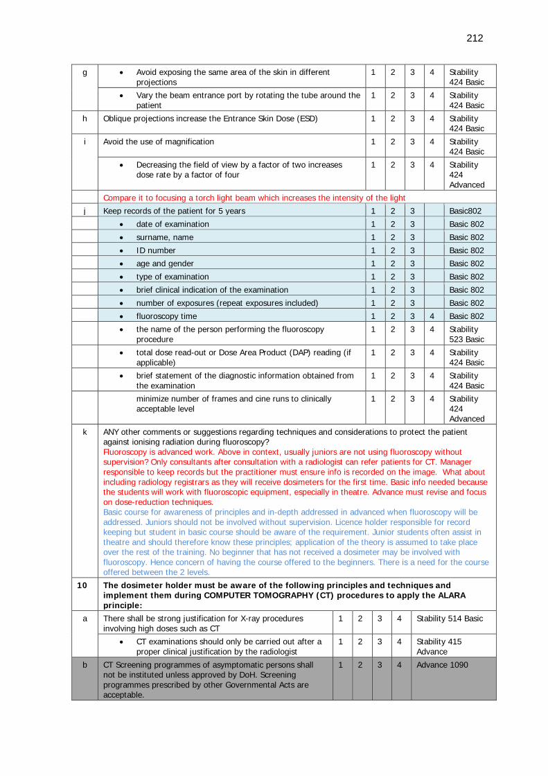
212
g • Avoid exposing the same area of the skin in different projections
1 2 3 4 Stability 424 Basic
• Vary the beam entrance port by rotating the tube around the patient
1 2 3 4 Stability 424 Basic
h Oblique projections increase the Entrance Skin Dose (ESD) 1 2 3 4 Stability 424 Basic
i Avoid the use of magnification 1 2 3 4 Stability 424 Basic
• Decreasing the field of view by a factor of two increases dose rate by a factor of four
1 2 3 4 Stability 424 Advanced
Compare it to focusing a torch light beam which increases the intensity of the light
j Keep records of the patient for 5 years 1 2 3 Basic802
• date of examination 1 2 3 Basic 802
• surname, name 1 2 3 Basic 802
• ID number 1 2 3 Basic 802
• age and gender 1 2 3 Basic 802
• type of examination 1 2 3 Basic 802
• brief clinical indication of the examination 1 2 3 Basic 802
• number of exposures (repeat exposures included) 1 2 3 Basic 802
• fluoroscopy time 1 2 3 4 Basic 802
• the name of the person performing the fluoroscopy procedure
1 2 3 4 Stability 523 Basic
• total dose read-out or Dose Area Product (DAP) reading (if applicable)
1 2 3 4 Stability 424 Basic
• brief statement of the diagnostic information obtained from the examination
1 2 3 4 Stability 424 Basic
minimize number of frames and cine runs to clinically acceptable level
1 2 3 4 Stability 424 Advanced
k ANY other comments or suggestions regarding techniques and considerations to protect the patient against ionising radiation during fluoroscopy? Fluoroscopy is advanced work. Above in context, usually juniors are not using fluoroscopy without supervision? Only consultants after consultation with a radiologist can refer patients for CT. Manager responsible to keep records but the practitioner must ensure info is recorded on the image. What about including radiology registrars as they will receive dosimeters for the first time. Basic info needed because the students will work with fluoroscopic equipment, especially in theatre. Advance must revise and focus on dose-reduction techniques. Basic course for awareness of principles and in-depth addressed in advanced when fluoroscopy will be addressed. Juniors should not be involved without supervision. Licence holder responsible for record keeping but student in basic course should be aware of the requirement. Junior students often assist in theatre and should therefore know these principles; application of the theory is assumed to take place over the rest of the training. No beginner that has not received a dosimeter may be involved with fluoroscopy. Hence concern of having the course offered to the beginners. There is a need for the course offered between the 2 levels.
10 The dosimeter holder must be aware of the following principles and techniques and implement them during COMPUTER TOMOGRAPHY (CT) procedures to apply the ALARA principle:
a There shall be strong justification for X-ray procedures involving high doses such as CT
1 2 3 4 Stability 514 Basic
• CT examinations should only be carried out after a proper clinical justification by the radiologist
1 2 3 4 Stability 415 Advance
b CT Screening programmes of asymptomatic persons shall not be instituted unless approved by DoH. Screening programmes prescribed by other Governmental Acts are acceptable.
1 2 3 4 Advance 1090
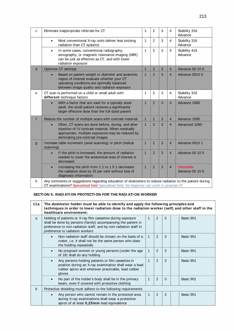
213
c Eliminate inappropriate referrals for CT 1 2 3 4 Stability 316 Advance
• Most conventional X-ray units deliver less ionizing radiation than CT systems
1 2 3 4 Stability 316 Advance
• In some cases, conventional radiography, sonography, or magnetic resonance imaging (MRI) can be just as effective as CT, and with lower radiation exposure
1 2 3 4 Stability 415 Advance
d Optimize CT settings 1 2 3 4 Advance 00 10 0
• Based on patient weight or diameter and anatomic region of interest evaluate whether your CT operating conditions are optimally balanced between image quality and radiation exposure
1 2 3 4 Advance 0010 0
e CT scan is performed on a child or small adult with different technique factors
1 2 3 4 Stability 316 Advance
• With a factor that are used for a typically sized adult, the small patient receives a significantly larger effective dose than the full-sized patient
1 2 3 4 Advance 2080
f Reduce the number of multiple scans with contrast material. 1 2 3 4 Advance 1090
• Often, CT scans are done before, during, and after injection of IV contrast material. When medically appropriate, multiple exposures may be reduced by eliminating pre-contrast images
1 2 3 4 Advanced 1090
g Increase table increment (axial scanning) or pitch (helical scanning)
1 2 3 4 Advance 0010 1
• If the pitch is increased, the amount of radiation needed to cover the anatomical area of interest is decreased
1 2 3 4 Advance 00 10 0
• Increasing the pitch from 1:1 to 1.5:1 decreases the radiation dose by 33 per cent without loss of diagnostic information
1 2 3 4 Debatable Advance 00 10 0
h Any comments or suggestions regarding education of dosimeters to reduce radiation to the patient during CT examinations? Specialised field Specialised field. No beginner can work or practise CT
SECTION 5: RADIATION PROTECTION FOR THE RADIATION WORKER 11a The dosimeter holder must be able to identify and apply the following principles and
techniques in order to lower radiation dose to the radiation worker (self) and other staff in the healthcare environment:
a Holding of patients or X-ray film cassettes during exposure shall be done by persons (family) accompanying the patient in preference to non-radiation staff; and by non-radiation staff in preference to radiation workers
1 2 3 Basic 901
• Non-radiation staff should be chosen on the basis of a roster, i.e. it shall not be the same person who does the holding repeatedly
1 2 3 Basic 901
• No pregnant women or young persons (under the age of 18) shall do any holding
1 2 3 Basic 901
• Any persons holding patients or film cassettes in position during an X-ray examination shall wear a lead rubber apron and wherever practicable, lead rubber gloves
1 2 3 Basic 901
• No part of the holder’s body shall be in the primary beam, even if covered with protective clothing
1 2 3 Basic 901
b Protective shielding must adhere to the following requirements
• Any person who cannot remain in the protected area during X-ray examinations shall wear a protective apron of at least 0,25mm lead equivalence
1 2 3 Basic 901
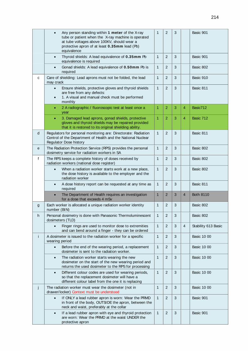
214
• Any person standing within 1 meter of the X-ray tube or patient when the X-ray machine is operated at tube voltages above 100KV, should wear a protective apron of at least 0.35mm lead (Pb) equivalence
1 2 3 Basic 901
• Thyroid shields: A lead equivalence of 0.35mm Pb equivalence is required
1 2 3 Basic 901
• Gonad shields: A lead equivalence of 0.50mm Pb is required
1 2 3 Basic 802
c Care of shielding: Lead aprons must not be folded, the lead may crack
1 2 3 Basic 910
• Ensure shields, protective gloves and thyroid shields are free from any defects:
• 1. A visual and manual check must be performed monthly
1 2 3 Basic 811
• 2 A radiographic / fluoroscopic test at least once a year
1 2 3 4 Basic712
• 3. Damaged lead aprons, gonad shields, protective gloves and thyroid shields may be repaired provided that it is restored to its original shielding ability.
1 2 3 4 Basic 712
d Regulators for personal monitoring are: Directorate: Radiation Control of the Department of Health and the National Nuclear Regulator Dose history
1 2 3 Basic 811
e The Radiation Protection Service (RPS) provides the personal dosimetry service for radiation workers in SA
1 2 3 Basic 802
f The RPS keeps a complete history of doses received by radiation workers (national dose register)
1 2 3 Basic 802
• When a radiation worker starts work at a new place, the dose history is available to the employer and the radiation worker
1 2 3 Basic 802
• A dose history report can be requested at any time as required
1 2 3 Basic 811
• The Department of Health requires an investigation for a dose that exceeds 4 mSv
1 2 3 4 Both 8110
g Each worker is allocated a unique radiation worker identity number (BIN)
1 2 3 Basic 802
h Personal dosimetry is done with Panasonic Thermoluminescent dosimeters (TLD)
1 2 3 Basic 802
• Finger rings are used to monitor dose to extremities and can bend around a finger - they can be ordered
1 2 3 4 Stability 613 Basic
i A dosimeter is issued to the radiation worker for a specific wearing period
1 2 3 Basic 10 00
• Before the end of the wearing period, a replacement dosimeter is sent to the radiation worker.
1 2 3 Basic 10 00
• The radiation worker starts wearing the new dosimeter on the start of the new wearing period and returns the used dosimeter to the RPS for processing
1 2 3 Basic 10 00
• Different colour codes are used for wearing periods, so that the replacement dosimeter will have a different colour label from the one it is replacing
1 2 3 Basic 10 00
j The radiation worker must wear the dosimeter (not in drawer/locker) Context must be understood
1 2 3 Basic 10 00
• If ONLY a lead rubber apron is worn: Wear the PRMD in front of the body, OUTSIDE the apron, between the neck and waist, preferably at the collar
1 2 3 Basic 901
• If a lead rubber apron with eye and thyroid protection are worn: Wear the PRMD at the waist UNDER the protective apron
1 2 3 Basic 901
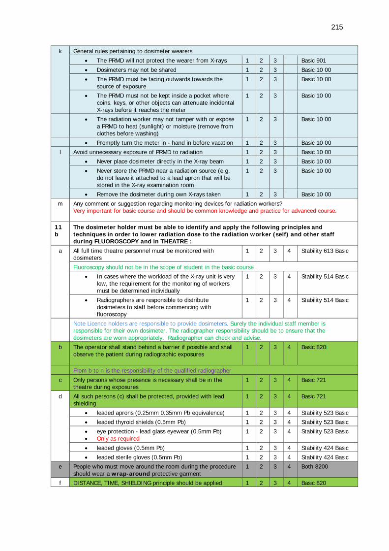
215
k General rules pertaining to dosimeter wearers
• The PRMD will not protect the wearer from X-rays 1 2 3 Basic 901
• Dosimeters may not be shared 1 2 3 Basic 10 00
• The PRMD must be facing outwards towards the source of exposure
1 2 3 Basic 10 00
• The PRMD must not be kept inside a pocket where coins, keys, or other objects can attenuate incidental X-rays before it reaches the meter
1 2 3 Basic 10 00
• The radiation worker may not tamper with or expose a PRMD to heat (sunlight) or moisture (remove from clothes before washing)
1 2 3 Basic 10 00
• Promptly turn the meter in - hand in before vacation 1 2 3 Basic 10 00 l Avoid unnecessary exposure of PRMD to radiation 1 2 3 Basic 10 00
• Never place dosimeter directly in the X-ray beam 1 2 3 Basic 10 00 • Never store the PRMD near a radiation source (e.g.
do not leave it attached to a lead apron that will be stored in the X-ray examination room
1 2 3 Basic 10 00
• Remove the dosimeter during own X-rays taken 1 2 3 Basic 10 00 m Any comment or suggestion regarding monitoring devices for radiation workers?
Very important for basic course and should be common knowledge and practice for advanced course.
11b
The dosimeter holder must be able to identify and apply the following principles and techniques in order to lower radiation dose to the radiation worker (self) and other staff during FLUOROSCOPY and in THEATRE :
a All full time theatre personnel must be monitored with dosimeters
1 2 3 4 Stability 613 Basic
Fluoroscopy should not be in the scope of student in the basic course
• In cases where the workload of the X-ray unit is very low, the requirement for the monitoring of workers must be determined individually
1 2 3 4 Stability 514 Basic
• Radiographers are responsible to distribute dosimeters to staff before commencing with fluoroscopy
1 2 3 4 Stability 514 Basic
Note Licence holders are responsible to provide dosimeters. Surely the individual staff member is responsible for their own dosimeter. The radiographer responsibility should be to ensure that the dosimeters are worn appropriately. Radiographer can check and advise.
b The operator shall stand behind a barrier if possible and shall observe the patient during radiographic exposures
1 2 3 4 Basic 820i
From b to n is the responsibility of the qualified radiographer
c Only persons whose presence is necessary shall be in the theatre during exposures
1 2 3 4 Basic 721
d All such persons (c) shall be protected, provided with lead shielding
1 2 3 4 Basic 721
• leaded aprons (0.25mm 0.35mm Pb equivalence) 1 2 3 4 Stability 523 Basic • leaded thyroid shields (0.5mm Pb) 1 2 3 4 Stability 523 Basic • eye protection - lead glass eyewear (0.5mm Pb) • Only as required
1 2 3 4 Stability 523 Basic
• leaded gloves (0.5mm Pb) 1 2 3 4 Stability 424 Basic • leaded sterile gloves (0.5mm Pb) 1 2 3 4 Stability 424 Basic
e People who must move around the room during the procedure should wear a wrap-around protective garment
1 2 3 4 Both 8200
f DISTANCE, TIME, SHIELDING principle should be applied 1 2 3 4 Basic 820
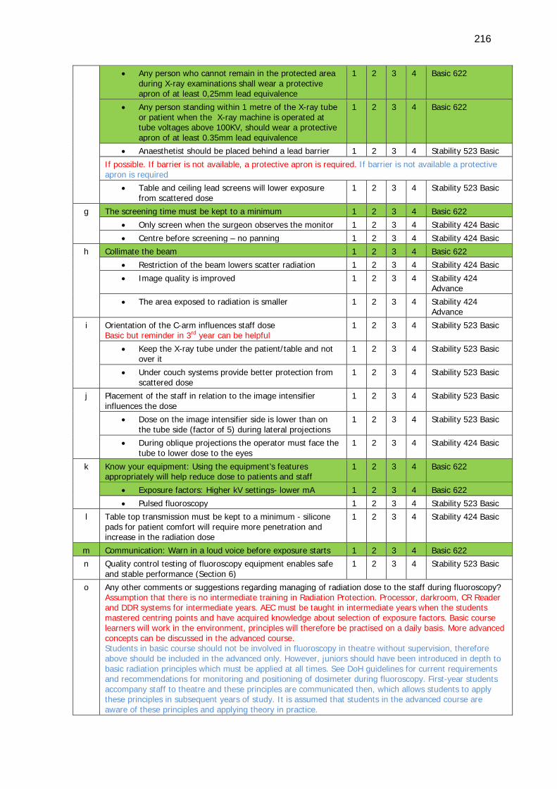
216
• Any person who cannot remain in the protected area during X-ray examinations shall wear a protective apron of at least 0,25mm lead equivalence
1 2 3 4 Basic 622
• Any person standing within 1 metre of the X-ray tube or patient when the X-ray machine is operated at tube voltages above 100KV, should wear a protective apron of at least 0.35mm lead equivalence
1 2 3 4 Basic 622
• Anaesthetist should be placed behind a lead barrier 1 2 3 4 Stability 523 Basic If possible. If barrier is not available, a protective apron is required. If barrier is not available a protective apron is required
• Table and ceiling lead screens will lower exposure from scattered dose
1 2 3 4 Stability 523 Basic
g The screening time must be kept to a minimum 1 2 3 4 Basic 622 • Only screen when the surgeon observes the monitor 1 2 3 4 Stability 424 Basic • Centre before screening – no panning 1 2 3 4 Stability 424 Basic
h Collimate the beam 1 2 3 4 Basic 622 • Restriction of the beam lowers scatter radiation 1 2 3 4 Stability 424 Basic • Image quality is improved 1 2 3 4 Stability 424
Advance • The area exposed to radiation is smaller 1 2 3 4 Stability 424
Advance i Orientation of the C-arm influences staff dose
Basic but reminder in 3rd year can be helpful 1 2 3 4 Stability 523 Basic
• Keep the X-ray tube under the patient/table and not over it
1 2 3 4 Stability 523 Basic
• Under couch systems provide better protection from scattered dose
1 2 3 4 Stability 523 Basic
j Placement of the staff in relation to the image intensifier influences the dose
1 2 3 4 Stability 523 Basic
• Dose on the image intensifier side is lower than on the tube side (factor of 5) during lateral projections
1 2 3 4 Stability 523 Basic
• During oblique projections the operator must face the tube to lower dose to the eyes
1 2 3 4 Stability 424 Basic
k Know your equipment: Using the equipment’s features appropriately will help reduce dose to patients and staff
1 2 3 4 Basic 622
• Exposure factors: Higher kV settings- lower mA 1 2 3 4 Basic 622
• Pulsed fluoroscopy 1 2 3 4 Stability 523 Basic
l Table top transmission must be kept to a minimum - silicone pads for patient comfort will require more penetration and increase in the radiation dose
1 2 3 4 Stability 424 Basic
m Communication: Warn in a loud voice before exposure starts 1 2 3 4 Basic 622
n Quality control testing of fluoroscopy equipment enables safe and stable performance (Section 6)
1 2 3 4 Stability 523 Basic
o Any other comments or suggestions regarding managing of radiation dose to the staff during fluoroscopy? Assumption that there is no intermediate training in Radiation Protection. Processor, darkroom, CR Reader and DDR systems for intermediate years. AEC must be taught in intermediate years when the students mastered centring points and have acquired knowledge about selection of exposure factors. Basic course learners will work in the environment, principles will therefore be practised on a daily basis. More advanced concepts can be discussed in the advanced course. Students in basic course should not be involved in fluoroscopy in theatre without supervision, therefore above should be included in the advanced only. However, juniors should have been introduced in depth to basic radiation principles which must be applied at all times. See DoH guidelines for current requirements and recommendations for monitoring and positioning of dosimeter during fluoroscopy. First-year students accompany staff to theatre and these principles are communicated then, which allows students to apply these principles in subsequent years of study. It is assumed that students in the advanced course are aware of these principles and applying theory in practice.
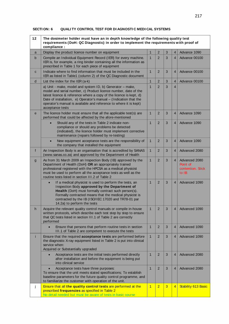
217
SECTION: 6 QUALITY CONTROL TEST FOR DIAGNOSTIC MEDICAL SYSTEMS 12 The dosimeter holder must have an in depth knowledge of the following quality test
requirements (DoH: QC Diagnostic) in order to implement the requirements with proof of compliance :
a Display the product licence number on equipment 1 2 3 4 Advance 1090
b Compile an Individual Equipment Record (IER) for every machine. IER is, for example, a ring binder containing all the information as prescribed in Table 1 for each piece of equipment
1 2 3 4 Advance 00100
c Indicate where to find information that must be included in the IER as listed in Table1 (column 2) of the QC Diagnostic document
1 2 3 4 Advance 00100
d List the index for the IER (a-k) 1 2 3 4 Advance 00100
a) Unit - make, model and system ID, b) Generator – make, model and serial number, c) Product licence number, date of the latest licence & reference where a copy of the licence is kept, d) Date of installation, e) Operator’s manual – (Indication that the operator’s manual is available and reference to where it is kept) acceptance tests
1 2 3 4
e The licence holder must ensure that all the applicable test(s) are performed that could be affected by the afore-mentioned
1 2 3 4 Advance 1090
• Should any of the tests in Table 2 indicate non-compliance or should any problems be detected (indicated), the licence holder must implement corrective maintenance (repairs followed by re-testing)
1 2 3 4 Advance 1090
• New equipment acceptance tests are the responsibility of the company that installed the equipment
1 2 3 4 Advance 1090
f An Inspection Body is an organisation that is accredited by SANAS (www.sanas.co.za) and approved by the Department of Health
1 2 3 4 Advanced 2080
g As from 31 March 2009 an Inspection Body (IB) approved by the Department of Health (DoH) OR an appropriately trained professional registered with the HPCSA as a medical physicist must be used to perform all the acceptance tests as well as the routine tests listed in section III.2 of Table 2
1 2 3 4 Advanced 2080 Point of contention. Stick to IB
• If a medical physicist is used to perform the tests, an Inspection Body approved by the Department of Health (DoH) must formally contract such person(s). Formally contracted means that the medical physicist is contracted by the IB (ISO/IEC 17020 and TR78-01 par 14.2a) to perform the tests
1 2 3 4 Advanced 1090
h Acquire the relevant quality control manuals or compile in-house written protocols, which describe each test step by step to ensure that QC tests listed in section III.1 of Table 2 are correctly performed
1 2 3 4 Advanced 1090
• Ensure that persons that perform routine tests in section III.1 of Table 2 are competent to execute the tests
1 2 3 4 Advanced 1090
i Ensure that the required acceptance tests are performed before the diagnostic X-ray equipment listed in Table 2 is put into clinical service when: Acquired or Substantially upgraded
1 2 3 4 Advanced 1090
• Acceptance tests are the initial tests performed directly after installation and before the equipment is being put into clinical service
1 2 3 4 Advanced 2080
• Acceptance tests have three purposes: To ensure that the unit meets stated specifications; To establish baseline parameters for the future quality control programme, and to familiarize the customer with operation of the unit.
1 2 3 4 Advanced 2080
j Ensure that all the quality control tests are performed at the prescribed frequencies as specified in Table 2 No detail needed but must be aware of tests in basic course
1 2 3 4 Stability 613 Basic
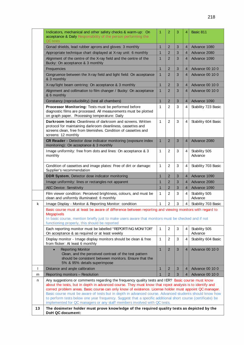
218
Indicators, mechanical and other safety checks & warm-up: On acceptance & Daily Responsibility of the person performing the QC tests
1 2 3 4 Basic 811
Gonad shields, lead rubber aprons and gloves: 3 monthly 1 2 3 4 Advance 1080
Appropriate technique chart displayed at X-ray unit: 6 monthly 1 2 3 4 Advance 2080
Alignment of the centre of the X-ray field and the centre of the Bucky: On acceptance & 3 monthly
1 2 3 4 Advance 1090
Frequencies 1 2 3 4 Advance 00 10 0
Congruence between the X-ray field and light field: On acceptance & 3 monthly
1 2 3 4 Advance 00 10 0
X-ray/light beam centring: On acceptance & 3 monthly 1 2 3 4 Advance 00 10 0
Alignment and collimation to film changer / Bucky: On acceptance & 6 monthly
1 2 3 4 Advance 00 10 0
Constancy (reproducibility) (test all chambers) 1 2 3 4 Advance 1090
Processor Monitoring: Tests must be performed before diagnostic films are processed. All measurements must be plotted on graph paper. Processing temperature: Daily
1 2 3 4 Stability 723 Basic
Darkroom tests: Cleanliness of darkroom and screens. Written protocol for maintaining darkroom cleanliness, cassettes and screens clean, free from blemishes. Condition of cassettes and screens: 12 monthly
1 2 3 4 Stability 604 Basic
CR Reader - Detector dose indicator monitoring (exposure index monitoring): On acceptance & 3 monthly
1 2 3 4 Advance 2080
Image uniformity: free from dots and lines: On acceptance & 3 monthly
1 2 3 4 Stability 505 Advance
Condition of cassettes and image plates: Free of dirt or damage: Supplier’s recommendation
1 2 3 4 Stability 703 Basic
DDR System, Detector dose indicator monitoring 1 2 3 4 Advance 1090
Image uniformity: lines or rectangles not apparent 1 2 3 4 Advance 2080
AEC Device: Sensitivity 1 2 3 4 Advance 1090
Film viewer condition: Perceived brightness, colours, and must be clean and uniformly illuminated: 6 monthly
1 2 3 4 Stability 505 Advance
k Image Display - Monitor & Reporting Monitor: condition 1 2 3 4 Stability 703 Basic Basic course must at least be aware of difference between reporting and viewing monitors with regard to Megapixels In basic course, mention briefly just to make users aware that monitors must be checked and if not functioning properly, this should be reported
Each reporting monitor must be labelled “REPORTING MONITOR” On acceptance & as required or at least weekly
1 2 3 4 Stability 505 Advance
Display monitor - Image display monitors should be clean & free from flicker: At least 6 monthly
1 2 3 4 Stability 604 Basic
• Reporting Monitor Clean, and the perceived contrast of the test pattern should be consistent between monitors. Ensure that the 5% & 95% details superimpose
1 2 3 4 Advance 00 10 0
l Distance and angle calibration 1 2 3 4 Advance 00 10 0
m Reporting monitors – Resolution 1 2 3 4 Advance 00 10 0
n Any suggestions or comments regarding the frequency quality tests and IER? Basic course must know about the tests, but in depth in advanced course. They must know that reject analysis is to identify and correct problem areas. Basic course can only know of existence. License holder must appoint QC manager. Basic course must be aware of tests but in depth in advanced course. Advanced students should know how to perform tests below one year frequency. Suggest that a specific additional short course (certificate) be implemented for QC managers or any staff members involved with QC tests.
13 The dosimeter holder must prove knowledge of the required quality tests as depicted by the DoH QC document:
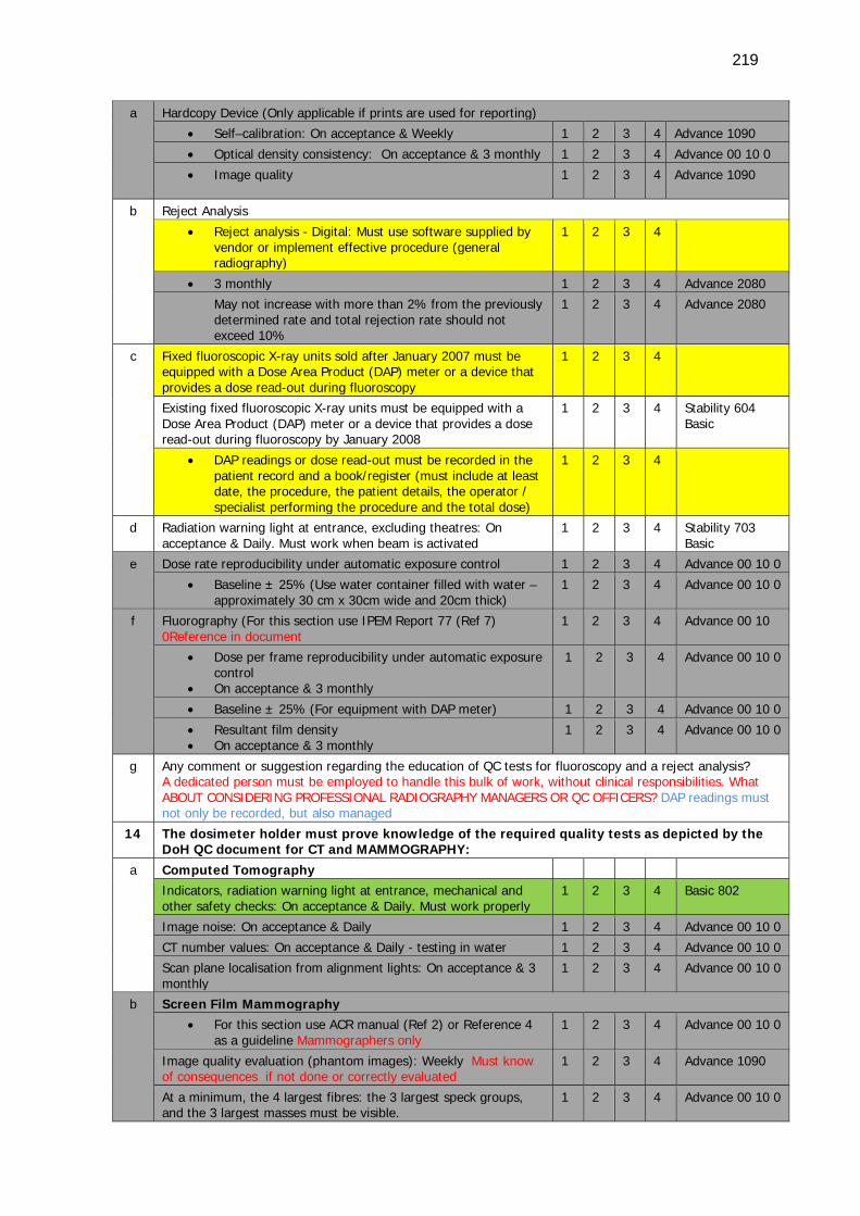
219
a Hardcopy Device (Only applicable if prints are used for reporting) • Self–calibration: On acceptance & Weekly 1 2 3 4 Advance 1090
• Optical density consistency: On acceptance & 3 monthly 1 2 3 4 Advance 00 10 0
• Image quality
1 2 3 4 Advance 1090
b Reject Analysis
• Reject analysis - Digital: Must use software supplied by vendor or implement effective procedure (general radiography)
1 2 3 4
• 3 monthly 1 2 3 4 Advance 2080
May not increase with more than 2% from the previously determined rate and total rejection rate should not exceed 10%
1 2 3 4 Advance 2080
c Fixed fluoroscopic X-ray units sold after January 2007 must be equipped with a Dose Area Product (DAP) meter or a device that provides a dose read-out during fluoroscopy
1 2 3 4
Existing fixed fluoroscopic X-ray units must be equipped with a Dose Area Product (DAP) meter or a device that provides a dose read-out during fluoroscopy by January 2008
1 2 3 4 Stability 604 Basic
• DAP readings or dose read-out must be recorded in the patient record and a book/register (must include at least date, the procedure, the patient details, the operator / specialist performing the procedure and the total dose)
1 2 3 4
d Radiation warning light at entrance, excluding theatres: On acceptance & Daily. Must work when beam is activated
1 2 3 4 Stability 703 Basic
e Dose rate reproducibility under automatic exposure control 1 2 3 4 Advance 00 10 0
• Baseline ± 25% (Use water container filled with water –approximately 30 cm x 30cm wide and 20cm thick)
1 2 3 4 Advance 00 10 0
f Fluorography (For this section use IPEM Report 77 (Ref 7) 0Reference in document
1 2 3 4 Advance 00 10
• Dose per frame reproducibility under automatic exposure control
• On acceptance & 3 monthly
1 2 3 4 Advance 00 10 0
• Baseline ± 25% (For equipment with DAP meter) 1 2 3 4 Advance 00 10 0
• Resultant film density • On acceptance & 3 monthly
1 2 3 4 Advance 00 10 0
g Any comment or suggestion regarding the education of QC tests for fluoroscopy and a reject analysis? A dedicated person must be employed to handle this bulk of work, without clinical responsibilities. What ABOUT CONSIDERING PROFESSIONAL RADIOGRAPHY MANAGERS OR QC OFFICERS? DAP readings must not only be recorded, but also managed
14 The dosimeter holder must prove knowledge of the required quality tests as depicted by the DoH QC document for CT and MAMMOGRAPHY:
a Computed Tomography
Indicators, radiation warning light at entrance, mechanical and other safety checks: On acceptance & Daily. Must work properly
1 2 3 4 Basic 802
Image noise: On acceptance & Daily 1 2 3 4 Advance 00 10 0
CT number values: On acceptance & Daily - testing in water 1 2 3 4 Advance 00 10 0
Scan plane localisation from alignment lights: On acceptance & 3 monthly
1 2 3 4 Advance 00 10 0
b Screen Film Mammography • For this section use ACR manual (Ref 2) or Reference 4
as a guideline Mammographers only 1 2 3 4 Advance 00 10 0
Image quality evaluation (phantom images): Weekly Must know of consequences if not done or correctly evaluated
1 2 3 4 Advance 1090
At a minimum, the 4 largest fibres: the 3 largest speck groups, and the 3 largest masses must be visible.
1 2 3 4 Advance 00 10 0
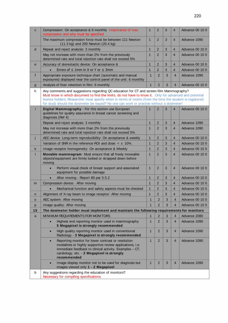
220
c Compression: On acceptance & 6 monthly Importance of max compression and why must be specified
1 2 3 4 Advance 00 10 0
The maximum compression force must be between 111 Newton (11.3 kg) and 200 Newton (20.4 kg)
1 2 3 4 Advance 1090
d Repeat and reject analysis: 3 monthly 1 2 3 4 Advance 00 10 0
May not increase with more than 2% from the previously determined rate and total rejection rate shall not exceed 5%
1 2 3 4 Advance 00 10 0
e Accuracy of stereotactic device: On acceptance & 1 2 3 4 Advance 00 10 0
• Errors of ≤ 1mm in X or Y or ≤ 3mm 1 2 3 4 Advance 00 10 0
f Appropriate exposure technique chart (automatic and manual exposures) displayed near the control panel of the unit: 6 monthly
1 2 3 4 Advance 1090
g Analysis of fixer retention in film: 6 monthly 1 2 3 4 Advance 00 10 0
h Any comments and suggestions regarding QC education for CT and screen-film Mammography? Must know in which document to find the limits, do not have to know it. Only for advanced and potential licence holders. Researcher must specify when in terms of moths (from the time the student is registered for stud) should the dosimeter be issued? No one can work or practise without a dosimeter!
i Digital Mammography - For this section use European guidelines for quality assurance in breast cancer screening and diagnosis (Ref 4)
1 2 3 4 Advance 00 10 0
Repeat and reject analysis: 3 monthly 1 2 3 4 Advance 1090
May not increase with more than 2% from the previously determined rate and total rejection rate shall not exceed 5%
1 2 3 4 Advance 1090
j AEC device: Long-term reproducibility: On acceptance & weekly 1 2 3 4 Advance 00 10 0
Variation of SNR in the reference ROI and dose: < ± 10%. 1 2 3 4 Advance 00 10 0
k Image receptor homogeneity: On acceptance & Weekly 1 2 3 4 Advance 00 10 0
l Movable mammogram: Must ensure that all freely moveable objects/equipment are firmly locked or strapped down before moving
1 2 3 4 Advance 00 10 0
• Perform visual check of breast support and associated equipment for possible damage
1 2 3 4 Advance 00 10 0
• After moving - Report 89 par 5.5.2 1 2 3 4 Advance 00 10 0
m Compression device: After moving 1 2 3 4 Advance 00 10 0
• Mechanical function and safety aspects must be checked 1 2 3 4 Advance 00 10 0
n Alignment of X-ray beam to image receptor: After moving 1 2 3 4 Advance 00 10 0
o AEC system: After moving 1 2 3 4 Advance 00 10 0
p Image quality: After moving 1 2 3 4 Advance 00 10 0
15 The dosimeter holder must implement and maintain the following requirements for monitors a MINIMUM REQUIREMENTS FOR MONITORS 1 2 3 4 Advance 2080
• Highest end reporting monitor used in mammography 5 Megapixel is strongly recommended
1 2 3 4 Advance 1090
• High quality reporting monitor used in conventional Radiology - 3 Megapixel is strongly recommended
1 2 3 4 Advance 1090
• Reporting monitor for lower contrast or resolution modalities or highly supportive review applications, i.e. immediate feedback to clinical activity. Examples – CT, cardiology, etc. - 2 Megapixel is strongly recommended
1 2 3 4 Advance 1090
• Image display monitor not to be used for diagnosis but images viewed only 1 - 2 Megapixel
1 2 3 4 Advance 1090
b Any suggestions regarding the education of monitors? Necessary for compiling specifications.
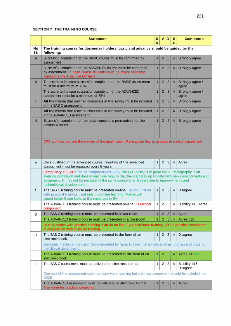
221
SECTION 7: THE TRAINING COURSE
Statement S A
A D SD
Comments
No 16
The training course for dosimeter holders, basic and advance should be guided by the following:
a Successful completion of the BASIC course must be confirmed by assessment
1 2 3 4 Strongly agree
Successful completion of the ADVANCED course must be confirmed by assessment In basic course students must be aware of license conditions which include QC tests
1 2 3 4 Strongly agree
b The score to indicate successful completion of the BASIC assessment must be a minimum of 75%
1 2 3 4 Strongly agree / agree
The score to indicate successful completion of the ADVANCED assessment must be a minimum of 75%
1 2 3 4 Strongly agree / agree
c All the criteria that reached consensus in the survey must be included in the BASIC assessment
1 2 3 4 Strongly agree
All the criteria that reached consensus in the survey must be included in the ADVANCED assessment
1 2 3 4 Strongly agree
d Successful completion of the basic course is a prerequisite for the advanced course
1 2 3 4 Strongly agree
CPD suffices, but not the rewrite of the qualification Prerequisite first to practise in clinical department
e Once qualified in the advanced course, rewriting of the advanced assessment must be repeated every 5 years
1 2 3 4 Agree
Compulsory 10 CDP? Can be compulsory for CPD, The CPD policy is of great value. Radiography is an evolving profession and does in any case require that the staff stay up to date with new developments and equipment. It may not be necessarily the same course after 5 years due to improvements and technological developments
f The BASIC training course must be presented on-line . In conjunction with practical training, . not only by on-line learning. Maybe will sound better if one looks at the resources in SA
1 2 3 4 Disagree
The ADVANCED training course must be presented on-line ./ Practical component
1 2 3 4 Stability 415 Agree
g The BASIC training course must be presented in a classroom 1 2 3 4 Agree
The ADVANCED training course must be presented in a classroom 1 2 3 4 Agree 532
In conjunction with practical training. Can do on-line if you had basic training, with a practical component in conjunction with in-house training.
h The BASIC training course must be presented in the form of an electronic book
1 2 3 4 Disagree
Electronic media can be used. Complemented by faced to face interactions such as tutorials and visits to the clinical department.
The ADVANCED training course must be presented in the form of an electronic book
1 2 3 4 Agree 712Aof
i The BASIC assessment must be delivered in electronic format 1 2 3 4 Stability 415 Disagree
One part of the assessment could be done via e-learning but a clinical component should be included, i.e. OSCE
The ADVANCED assessment must be delivered in electronic format Also room for practical component
1 2 3 4 Agree
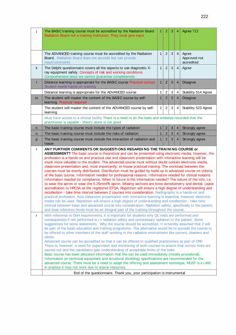
222
j The BASIC training course must be accredited by the Radiation Board Radiation Board not a training institution. They must give input
1 2 3 4 Agree 712
The ADVANCED training course must be accredited by the Radiation Board. Radiation Board does not accredit but can provide input/comments
1 2 3 4 Agree Approved not accredited
k The Delphi questionnaire covers all the aspects to use diagnostic X-ray equipment safely. Concepts of risk and working conditions Comprehensive since we cannot guarantee completeness
1 2 3 4 Agree
l Distance learning is appropriate for the BASIC course Practical contact Student needs hands on training
1 2 3 4 Disagree
Distance learning is appropriate for the ADVANCED course 1 2 3 4 Stability 514 Agree
m The student will master the content of the BASIC course by self-learning. Practical required
1 2 3 4 Disagree
The student will master the content of the ADVANCED course by self-learning
1 2 3 4 Stability 523 Agree
Must have access to a clinical facility There is a need to do the tests and evidence recorded that the practitioner is capable - theory alone is not good
n The basic training course must include the types of radiation 1 2 3 4 Strongly agree
o The basic training course must include the risks of radiation 1 2 3 4 Strongly agree
p The basic training course must include the interaction of radiation and tissue
1 2 3 4 Strongly agree
r ANY FURTHER COMMENTS OR SUGGESTIONS REGARDING THE TRAINING COURSE or ASSESSMENT? The basic course is theoretical and can be presented using electronic media; however, the profession is a hands-on and practical one and classroom presentation with interactive learning will be much more valuable to the student. The advanced course must without doubt contain electronic media, classroom presentation and, most importantly, in-house practical training. The workload between the courses must be evenly distributed. Distribution must be guided by build up in advanced course on criteria of the basic course. Information needed for professional reasons. Information needed for clinical reasons. Information needed for compliance. When in future is the information needed? The nature of the info, i.e. to wear the apron or wear the 0.25mmPb apron. Missing sections are bone densitometry and dental. Leave accreditation to HPCSA as the registered ETQA. Repetition will ensure a high degree of understanding and recollection - take time interval between 2 courses into consideration. Radiography is a hands-on and practical profession, thus classroom presentation with interactive learning is essential, however electronic media can be used. Repetition will ensure a high degree of understanding and recollection - take time interval between basic and advanced course into consideration. Radiation safety, specifically to the patient, and dose reference levels must be an integral part of the training throughout the course.
r With reference to DoH requirements, it is important for students why QC tests are performed and consequences if not performed w.r.t radiation safety and unnecessary radiation to the patient. Some suggestions for some statements. Why the course should be accredited. It is hereby assumed that it will be part of the basic education and training programme. The alternative would be to accredit the course to be offered to other members of the staff working in the radiation environment like porters, cleaners and clerks. Advanced course can be accredited so that it can be offered to qualified practitioners as part of CPD There is, however, a need for supervision and monitoring of both courses to ensure that correct tests are carried out and the candidates gain understanding of acceptable limits of the tests. Basic course has been allocated information that the can be used immediately (mostly procedural). Information on technical equipment and structural (building) specifications are recommended for the advanced course. There must be a need to adapt the offering and assessment technique, MUST is a LAW, in practice it may not work due to scarce resources.
End of the questionnaire. Thank you, your participation is instrumental
223
7.2.1 Delphi questionnaire comments of the panellists
All the above comments provided in Table 7.1 will not be discussed individually, because
the comments of the panellists that involved alteration to the statements were attended
to by adjusting the statements to be suitable for the next round. For example, in Round
One a panellist commented on Section One statement 3e (cf. Appendix D6 B) about the
door Pb lining of an X-ray room. The comment was: “2 mm is wrong. This must be
specifically designed for the room.” The researcher therefore adjusted the statement by
adding the “specifically designed for the room” part in a follow-up Delphi round
questionnaire (cf. Appendix D3). Some of the panellists repeated comments and the
researcher combined similar comments. Many of the comments only confirmed the
statement or motivated the choice of the respondent, for example, Section One c (cf.
Appendix D6 B) where the specific panel member selected the basic course and hence
commented: “All people working with radiation need a basic idea of the QC test involved
with equipment. Tests should not be discussed in detail. If you work in an area with
malfunctioning equipment it will be to your advantage to at least know that the situation
is not acceptable according to the Act”.
The comments regarding the content of radiation safety and assessment provided insight
that guided the researcher to consider important aspects, such as that the basic course
must address the awareness of principles and the advanced course to engage the student
in more depth. The importance of the training and assessment was reiterated with
comments for instance, “Learners forget why these principles are important and fail to
apply them because of poor example given to them by qualified staff that is there to
supervise them in this regard” (cf. Appendix D6 B Section 4 9i).
Section seven of the Delphi questionnaire dealt with the presentation of the training and
the panellist’s opinion regarding the assessment of the radiation safety. The respondents
were required to make four selections namely, ’Strongly agree’, ‘Agree’, ’Disagree’ and
’Strongly disagree’. The panellists strongly agreed that all the criteria on which consensus
was reached in the survey had to be included in the basic and advanced assessment. The
panellists strongly agreed that successful completion of the basic and advanced courses
had to be confirmed by assessment. The panel agreed that the score to indicate
successful completion of both assessments had to be a minimum of 75%.
224
The opinion of the panel was diverse with regard to rewriting the advanced assessment
every five years. The suggestion was that continuous professional development (CPD)
would meet the requirement to stay abreast of new developments in the evolving
profession. The panellists disagreed in that the basic course had to be presented on-line
because of the importance of practical training and the resources in South Africa that
might add to the challenge of availability. An electronic book for the basic course was
disagreed on although a comment in favour of electronic media was provided. The panel
however agreed that part of the advanced course could be presented on-line, provided
that the practical training and in-house training formed part of the parcel. An electronic
book for the advanced training received an ’Agree’ from the respondents.
The panel could not reach consensus on the delivery of the basic assessment in electronic
format. A comment was that one part of the assessment could be done via e-learning, but
a clinical component should be included, i.e. an OSCE. The panel agreed that the
advanced assessment should be delivered in electronic format. Most members of the
panel agreed that the basic and advanced assessment had to be approved (not
accredited) by the Directorate: Radiation Control by means of providing input and
comments.
The panellists agreed that the Delphi questionnaire covered all the aspects required to
use diagnostic X-ray equipment safely, with the comment that it was comprehensive
without the guarantee of completeness. The panel disagreed that distance learning was
appropriate for the basic training, because students need hands-on training. The panel
did not reach consensus on the appropriateness of distance learning for the advanced
training. The panel disagreed on the statement that the student would master the content
of the training by self-learning and added specifically that there was a need to execute
the tests and that evidence should be recorded for the advanced students. The panellists
strongly agreed that risks of radiation and interaction of radiation and tissue had to be
included in the basic training.
Further comments from the panellists included that insight in the workload should be
evenly distributed between the courses, and that the advanced course should build on
criteria for the basic course. The information contained in the course was regarded
necessary for different reasons, including professional, clinical, or for compliance reasons.
Repetition of the content, according to the panellists, would ensure a high degree of
225
understanding and recollection. The basic course was allocated information that the
students could use immediately, but information on technical equipment and structural
specifications was recommended for the advanced course. The concluding comments
addressed the need for supervision and monitoring for both courses to ensure that correct
tests were carried out and that candidates gained understanding of acceptable limits of
the tests. Flexibility was reiterated in terms of the offering and assessment, due to the
reality of scarce resources in South Africa.
The comments from the panellists informed the final list of criteria to be included in either
the basic or advanced training plus assessment. The comments were incorporated in the
teaching and learning activities as discussed later (cf. point 7.4) as well as in the student
pre-and post-test questionnaires (cf. Chapter 6). The findings of the student
questionnaires will be discussed consequently.
7.3 DESCRIPTION OF THE IMPLICATION OF THE FINDINGS FROM THE PRE-
AND POST-TEST QUESTIONNAIRE
The aim of the student questionnaires was to assess (via pre- and post-training
questionnaires) the entry-level participants’ knowledge regarding safety requirements for
radiation workers, and for the advanced level students, their knowledge regarding the
Code of Practice for users of medical X-ray equipment and the Quality Control (QC)
requirements. The results of the tests (cf. 6.3) were described in the previous chapter in
terms of individual points scored in the test, median, class average and grade distribution.
The Delphi panellists reached consensus (cf. Table 4.1:16) that the minimum score for a
student to successfully complete the radiation safety test should be 75%. Only five
students achieved a score of 75% and above for the basic post-test and no students for
the advanced post-test. The correctness of a cut-off percentage in terms of standard
setting is, with the benefit of hindsight, debatable (cf. point 2.5.1.7), but the results of
the tests were scrutinised with future implementation and recommendations in mind. The
aim of the training and assessment is to prepare and equip the student with knowledge in
order to meet radiation safety requirements, therefore is it important to make sure that
the highest mastery level possible is achieved by each student.
7.3.1 The purpose of assessment is to certify that a specific level of
performance has been achieved
226
The researcher analysed the Respondus reports (cf. Appendix H1 and H2) in order to
establish the percentage of students that made a correct choice for each individual
question. The aim was to reconsider the questions in order to determine possible reasons
for the incorrect responses. Although the questionnaires had been piloted (cf. point
3.3.3.3) by students, lecturers and qualified radiographers, the questions were revisited in
terms of clarity, marks allocated and statement choices. The questions to which 75% and
more of the students selected the correct response were considered reasonable, but the
questions with lower than 75% correct responses by the students were considered
challenging questions. The researcher selected a percentage of 75% after consultation
with the supervisors as an indication that most of the class did not understand the
question, or the question was not clearly stated, or it indicated the need for remedial
learning activities. The challenging questions identified areas where adjustments in
teaching and learning activity were needed. The remedial adjustments of the learning
activities will receive attention in this chapter (cf. point 7.3.5). The adjustments and
revision of the basic and advanced questions will be described next.
7.3.2 Assessment can identify areas for remedial teaching and learning
activities or where adjustment in BASIC question statements should be
made
The following section is devoted to the analysis of the challenging test questions for the
basic post-test. The reason for analysing the post-test only was to establish if changes in
the teaching and learning strategies were necessary in order to improve student success
in mastery of the content for radiation safety. The questions will not be discussed
individually but in sections similar to the sections of the Delphi questionnaire. The six
sections of the Delphi questionnaire were: General definitions and licencing conditions,
responsibilities of licence holders/responsible persons, operators of equipment and
radiation workers, radiation protection of patients, radiation protection of radiation
workers, and QC tests for diagnostic systems. The student questionnaire consisted of only
five sections, because the general definitions and licencing conditions were combined.
The basic post-test results were available in the report section of the Respondus 4.0
Campus-wide software. The specific report (cf. Appendix H1) recorded the percentage of
correct as well as incorrect answers for every individual question. The questions that
recorded a score of lower than 75% will be discussed individually. The researcher
reflected on the wording of the question, the significance of the content as well as the
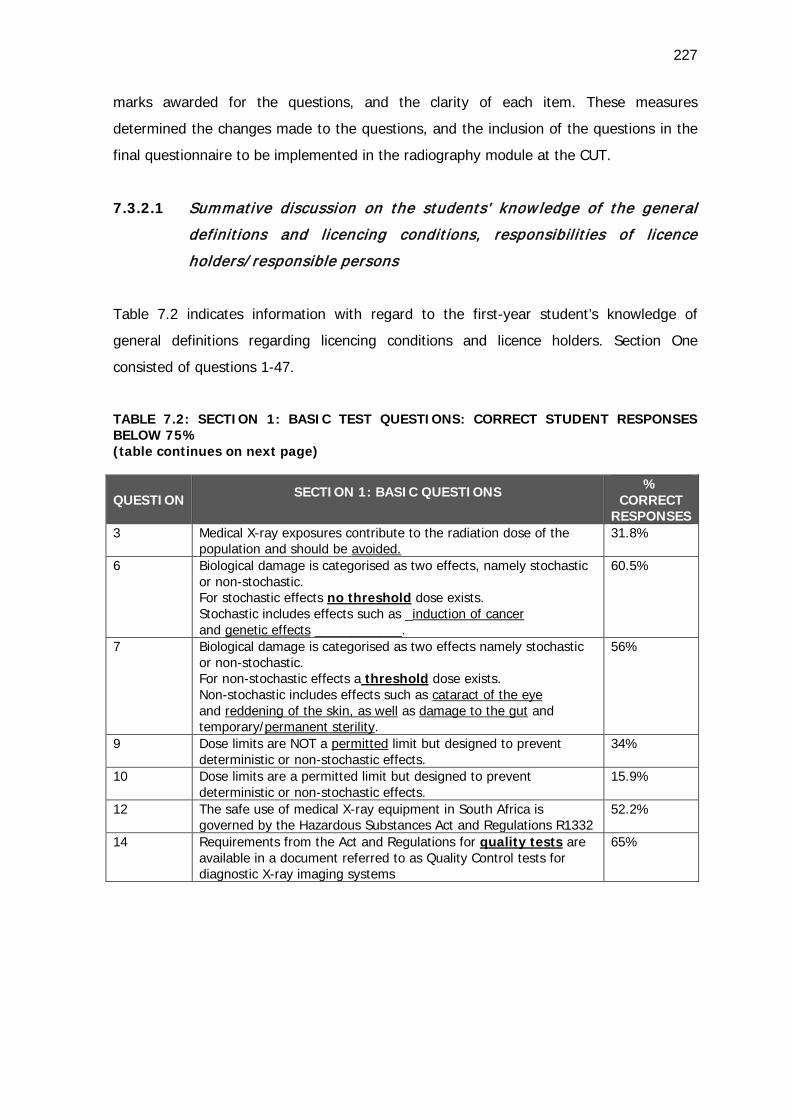
227
marks awarded for the questions, and the clarity of each item. These measures
determined the changes made to the questions, and the inclusion of the questions in the
final questionnaire to be implemented in the radiography module at the CUT.
7.3.2.1 Summative discussion on the students’ know ledge of the general
definitions and licencing conditions, responsibilit ies of licence
holders/ responsible persons
Table 7.2 indicates information with regard to the first-year student’s knowledge of
general definitions regarding licencing conditions and licence holders. Section One
consisted of questions 1-47.
TABLE 7.2: SECTION 1: BASIC TEST QUESTIONS: CORRECT STUDENT RESPONSES BELOW 75% (table continues on next page)
QUESTION SECTION 1: BASIC QUESTIONS
% CORRECT
RESPONSES 3 Medical X-ray exposures contribute to the radiation dose of the
population and should be avoided. 31.8%
6 Biological damage is categorised as two effects, namely stochastic or non-stochastic. For stochastic effects no threshold dose exists. Stochastic includes effects such as _induction of cancer and genetic effects ____________.
60.5%
7 Biological damage is categorised as two effects namely stochastic or non-stochastic. For non-stochastic effects a threshold dose exists. Non-stochastic includes effects such as cataract of the eye and reddening of the skin, as well as damage to the gut and temporary/permanent sterility.
56%
9 Dose limits are NOT a permitted limit but designed to prevent deterministic or non-stochastic effects.
34%
10 Dose limits are a permitted limit but designed to prevent deterministic or non-stochastic effects.
15.9%
12 The safe use of medical X-ray equipment in South Africa is governed by the Hazardous Substances Act and Regulations R1332
52.2%
14 Requirements from the Act and Regulations for quality tests are available in a document referred to as Quality Control tests for diagnostic X-ray imaging systems
65%
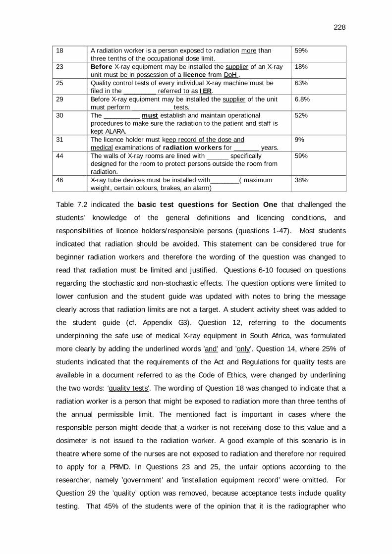
228
18 A radiation worker is a person exposed to radiation more than three tenths of the occupational dose limit.
59%
23 Before X-ray equipment may be installed the supplier of an X-ray unit must be in possession of a licence from DoH .
18%
25 Quality control tests of every individual X-ray machine must be filed in the _________ referred to as IER.
63%
29 Before X-ray equipment may be installed the supplier of the unit must perform ___________ tests.
6.8%
30 The __________ must establish and maintain operational procedures to make sure the radiation to the patient and staff is kept ALARA.
52%
31 The licence holder must keep record of the dose and medical examinations of radiation workers for _______ years.
9%
44 The walls of X-ray rooms are lined with ______ specifically designed for the room to protect persons outside the room from radiation.
59%
46 X-ray tube devices must be installed with________( maximum weight, certain colours, brakes, an alarm)
38%
Table 7.2 indicated the basic test questions for Section One that challenged the
students’ knowledge of the general definitions and licencing conditions, and
responsibilities of licence holders/responsible persons (questions 1-47). Most students
indicated that radiation should be avoided. This statement can be considered true for
beginner radiation workers and therefore the wording of the question was changed to
read that radiation must be limited and justified. Questions 6-10 focused on questions
regarding the stochastic and non-stochastic effects. The question options were limited to
lower confusion and the student guide was updated with notes to bring the message
clearly across that radiation limits are not a target. A student activity sheet was added to
the student guide (cf. Appendix G3). Question 12, referring to the documents
underpinning the safe use of medical X-ray equipment in South Africa, was formulated
more clearly by adding the underlined words ’and’ and ’only’. Question 14, where 25% of
students indicated that the requirements of the Act and Regulations for quality tests are
available in a document referred to as the Code of Ethics, were changed by underlining
the two words: ‘quality tests’. The wording of Question 18 was changed to indicate that a
radiation worker is a person that might be exposed to radiation more than three tenths of
the annual permissible limit. The mentioned fact is important in cases where the
responsible person might decide that a worker is not receiving close to this value and a
dosimeter is not issued to the radiation worker. A good example of this scenario is in
theatre where some of the nurses are not exposed to radiation and therefore nor required
to apply for a PRMD. In Questions 23 and 25, the unfair options according to the
researcher, namely ’government’ and ’installation equipment record’ were omitted. For
Question 29 the ’quality’ option was removed, because acceptance tests include quality
testing. That 45% of the students were of the opinion that it is the radiographer who
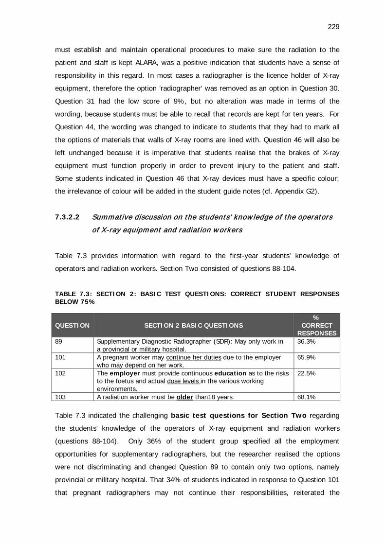
229
must establish and maintain operational procedures to make sure the radiation to the
patient and staff is kept ALARA, was a positive indication that students have a sense of
responsibility in this regard. In most cases a radiographer is the licence holder of X-ray
equipment, therefore the option ’radiographer’ was removed as an option in Question 30.
Question 31 had the low score of 9%, but no alteration was made in terms of the
wording, because students must be able to recall that records are kept for ten years. For
Question 44, the wording was changed to indicate to students that they had to mark all
the options of materials that walls of X-ray rooms are lined with. Question 46 will also be
left unchanged because it is imperative that students realise that the brakes of X-ray
equipment must function properly in order to prevent injury to the patient and staff.
Some students indicated in Question 46 that X-ray devices must have a specific colour;
the irrelevance of colour will be added in the student guide notes (cf. Appendix G2).
7.3.2.2 Summative discussion on the students’ know ledge of the operators
of X-ray equipment and radiation workers
Table 7.3 provides information with regard to the first-year students’ knowledge of
operators and radiation workers. Section Two consisted of questions 88-104.
TABLE 7.3: SECTION 2: BASIC TEST QUESTIONS: CORRECT STUDENT RESPONSES BELOW 75%
QUESTION SECTION 2 BASIC QUESTIONS %
CORRECT RESPONSES
89 Supplementary Diagnostic Radiographer (SDR): May only work in a provincial or military hospital.
36.3%
101 A pregnant worker may continue her duties due to the employer who may depend on her work.
65.9%
102 The employer must provide continuous education as to the risks to the foetus and actual dose levels in the various working environments.
22.5%
103 A radiation worker must be older than18 years. 68.1% Table 7.3 indicated the challenging basic test questions for Section Two regarding
the students’ knowledge of the operators of X-ray equipment and radiation workers
(questions 88-104). Only 36% of the student group specified all the employment
opportunities for supplementary radiographers, but the researcher realised the options
were not discriminating and changed Question 89 to contain only two options, namely
provincial or military hospital. That 34% of students indicated in response to Question 101
that pregnant radiographers may not continue their responsibilities, reiterated the
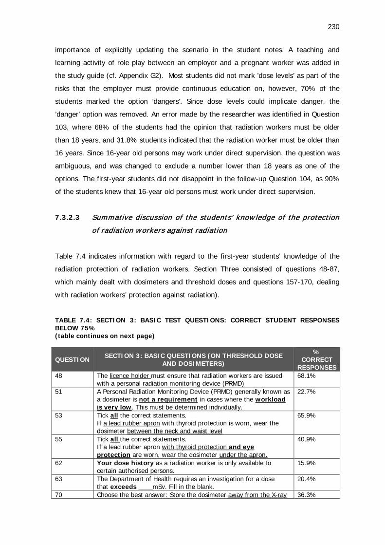
230
importance of explicitly updating the scenario in the student notes. A teaching and
learning activity of role play between an employer and a pregnant worker was added in
the study guide (cf. Appendix G2). Most students did not mark ’dose levels’ as part of the
risks that the employer must provide continuous education on, however, 70% of the
students marked the option ’dangers’. Since dose levels could implicate danger, the
’danger’ option was removed. An error made by the researcher was identified in Question
103, where 68% of the students had the opinion that radiation workers must be older
than 18 years, and 31.8% students indicated that the radiation worker must be older than
16 years. Since 16-year old persons may work under direct supervision, the question was
ambiguous, and was changed to exclude a number lower than 18 years as one of the
options. The first-year students did not disappoint in the follow-up Question 104, as 90%
of the students knew that 16-year old persons must work under direct supervision.
7.3.2.3 Summative discussion of the students’ know ledge of the protection
of radiation workers against radiation
Table 7.4 indicates information with regard to the first-year students’ knowledge of the
radiation protection of radiation workers. Section Three consisted of questions 48-87,
which mainly dealt with dosimeters and threshold doses and questions 157-170, dealing
with radiation workers’ protection against radiation).
TABLE 7.4: SECTION 3: BASIC TEST QUESTIONS: CORRECT STUDENT RESPONSES BELOW 75% (table continues on next page)
QUESTION SECTION 3: BASIC QUESTIONS (ON THRESHOLD DOSE AND DOSIMETERS)
% CORRECT
RESPONSES 48 The licence holder must ensure that radiation workers are issued
with a personal radiation monitoring device (PRMD) 68.1%
51 A Personal Radiation Monitoring Device (PRMD) generally known as a dosimeter is not a requirement in cases where the workload is very low. This must be determined individually.
22.7%
53 Tick all the correct statements. If a lead rubber apron with thyroid protection is worn, wear the dosimeter between the neck and waist level
65.9%
55 Tick all the correct statements. If a lead rubber apron with thyroid protection and eye protection are worn, wear the dosimeter under the apron.
40.9%
62 Your dose history as a radiation worker is only available to certain authorised persons.
15.9%
63 The Department of Health requires an investigation for a dose that exceeds ____mSv. Fill in the blank.
20.4%
70 Choose the best answer: Store the dosimeter away from the X-ray 36.3%
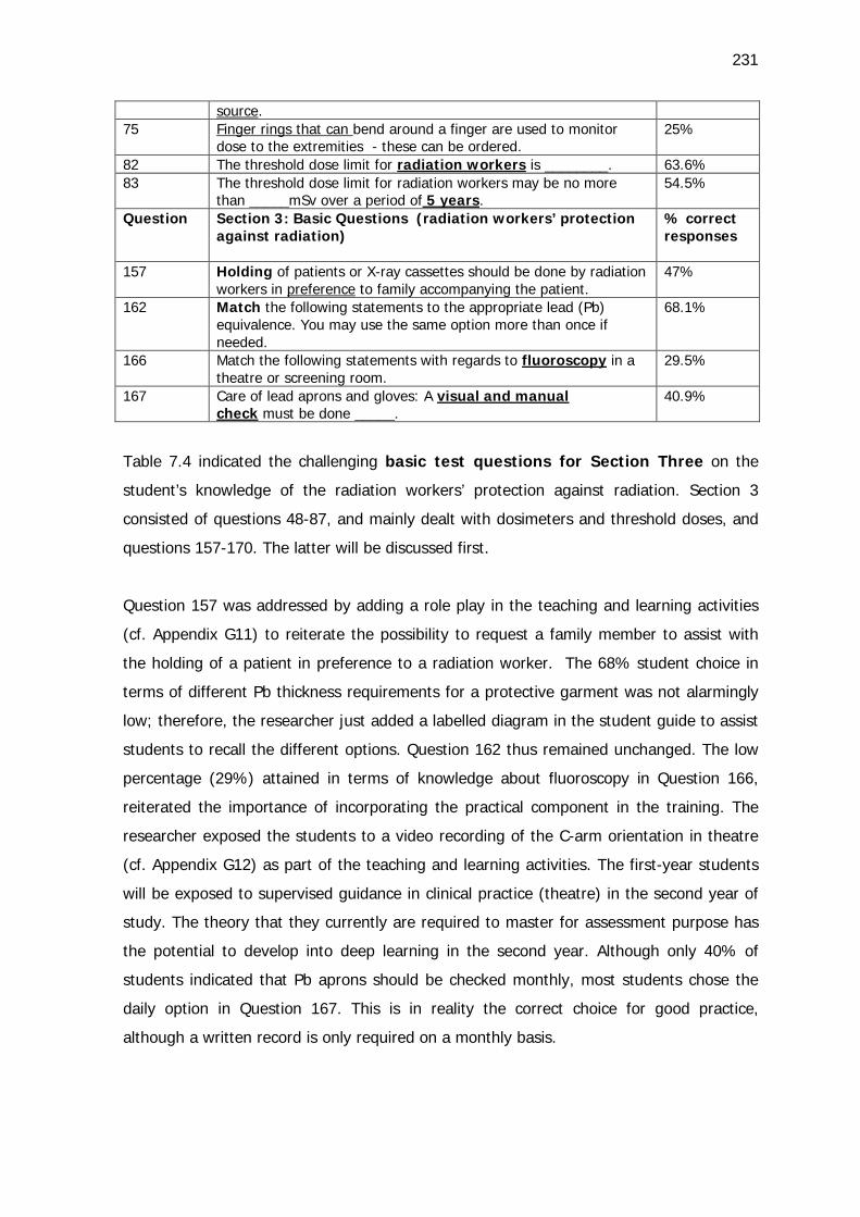
231
source. 75 Finger rings that can bend around a finger are used to monitor
dose to the extremities - these can be ordered. 25%
82 The threshold dose limit for radiation workers is ________. 63.6% 83 The threshold dose limit for radiation workers may be no more
than _____mSv over a period of 5 years. 54.5%
Question Section 3: Basic Questions (radiation workers’ protection against radiation)
% correct responses
157 Holding of patients or X-ray cassettes should be done by radiation workers in preference to family accompanying the patient.
47%
162 Match the following statements to the appropriate lead (Pb) equivalence. You may use the same option more than once if needed.
68.1%
166 Match the following statements with regards to fluoroscopy in a theatre or screening room.
29.5%
167 Care of lead aprons and gloves: A visual and manual check must be done _____.
40.9%
Table 7.4 indicated the challenging basic test questions for Section Three on the
student’s knowledge of the radiation workers’ protection against radiation. Section 3
consisted of questions 48-87, and mainly dealt with dosimeters and threshold doses, and
questions 157-170. The latter will be discussed first.
Question 157 was addressed by adding a role play in the teaching and learning activities
(cf. Appendix G11) to reiterate the possibility to request a family member to assist with
the holding of a patient in preference to a radiation worker. The 68% student choice in
terms of different Pb thickness requirements for a protective garment was not alarmingly
low; therefore, the researcher just added a labelled diagram in the student guide to assist
students to recall the different options. Question 162 thus remained unchanged. The low
percentage (29%) attained in terms of knowledge about fluoroscopy in Question 166,
reiterated the importance of incorporating the practical component in the training. The
researcher exposed the students to a video recording of the C-arm orientation in theatre
(cf. Appendix G12) as part of the teaching and learning activities. The first-year students
will be exposed to supervised guidance in clinical practice (theatre) in the second year of
study. The theory that they currently are required to master for assessment purpose has
the potential to develop into deep learning in the second year. Although only 40% of
students indicated that Pb aprons should be checked monthly, most students chose the
daily option in Question 167. This is in reality the correct choice for good practice,
although a written record is only required on a monthly basis.
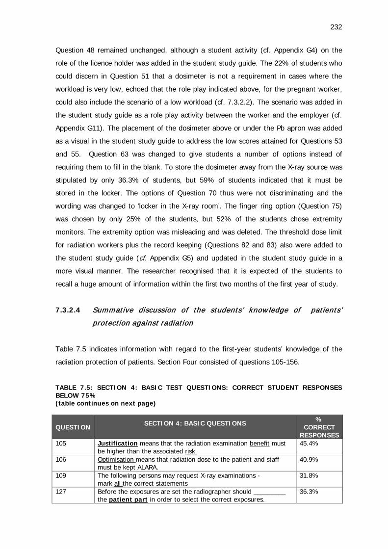
232
Question 48 remained unchanged, although a student activity (cf. Appendix G4) on the
role of the licence holder was added in the student study guide. The 22% of students who
could discern in Question 51 that a dosimeter is not a requirement in cases where the
workload is very low, echoed that the role play indicated above, for the pregnant worker,
could also include the scenario of a low workload (cf. 7.3.2.2). The scenario was added in
the student study guide as a role play activity between the worker and the employer (cf.
Appendix G11). The placement of the dosimeter above or under the Pb apron was added
as a visual in the student study guide to address the low scores attained for Questions 53
and 55. Question 63 was changed to give students a number of options instead of
requiring them to fill in the blank. To store the dosimeter away from the X-ray source was
stipulated by only 36.3% of students, but 59% of students indicated that it must be
stored in the locker. The options of Question 70 thus were not discriminating and the
wording was changed to ’locker in the X-ray room’. The finger ring option (Question 75)
was chosen by only 25% of the students, but 52% of the students chose extremity
monitors. The extremity option was misleading and was deleted. The threshold dose limit
for radiation workers plus the record keeping (Questions 82 and 83) also were added to
the student study guide (cf. Appendix G5) and updated in the student study guide in a
more visual manner. The researcher recognised that it is expected of the students to
recall a huge amount of information within the first two months of the first year of study.
7.3.2.4 Summative discussion of the students’ know ledge of patients’
protection against radiation
Table 7.5 indicates information with regard to the first-year students’ knowledge of the
radiation protection of patients. Section Four consisted of questions 105-156.
TABLE 7.5: SECTION 4: BASIC TEST QUESTIONS: CORRECT STUDENT RESPONSES BELOW 75% (table continues on next page)
QUESTION SECTION 4: BASIC QUESTIONS
% CORRECT
RESPONSES 105 Justification means that the radiation examination benefit must
be higher than the associated risk. 45.4%
106 Optimisation means that radiation dose to the patient and staff must be kept ALARA.
40.9%
109 The following persons may request X-ray examinations - mark all the correct statements
31.8%
127 Before the exposures are set the radiographer should _________ the patient part in order to select the correct exposures.
36.3%

233
130 Gonad shield must have a lead equivalence of ______. 63.6% 135 Minimise the distance between the X-ray tube and the patient to
the furthest possible extent during fluoroscopy (screening). 47.7%
137 The best way to reduce the dose during fluoroscopy is to lower the screening ____________.
56.8%
138 During fluoroscopy (screening) the radiation worker can apply the following good practice principles to lower radiation to the patient and staff (mark the statements - mark all the correct answers to score full marks). High kV and low mAs
61.3%
142 During fluoroscopy the entrance skin dose is increased by ________.
59.09%
146 The inverse C-arm (intensifier below) may only be used for extremities IF the physician places the anatomical part directly on the __________. (example: screw placement in the hand)
59.09%
147 The radiation source during fluoroscopy (screening) is _______ from the patient.
27.7%
151 Quality control tests of C-arms enable safe and stable performance.
65.9%
152 Fixed fluoroscopic units must be equipped with a DAP meter that provides a dose readout during fluoroscopy.
59.09%
155 Silicon pads on the tables that provide comfort to the patient will increase radiation dose because it will require more penetration.
25%
Table 7.5 indicated the challenging basic test questions for Section Four on the
students’ knowledge with regard to the radiation protection of patients (questions 105-
156). Although only 45.4% of students indicated that justification means that the
radiation examination benefit must be higher than the associated risk, 29% of students
chose exposure (question 105). The word ’exposure’ was confusing and thus removed
from the list of options. The options of Question 106 seemed confusing and therefore the
options list was reduced to two choices. Question 109, regarding health professionals that
may request X-rays, was changed by excluding nursing staff as part of the options list.
The nursing staff options were added as part of an additional but separate question.
Measuring of a patient and the link with an exposure chart was included in theOSCA
(Objective Structured Clinical Assessment) for the module to address the low score of
Question 127. Questions 135 to 151 addressed patient protection during fluoroscopy.
Although first-year students are not exposed to theatre placement yet – the practical
implication will be addressed in clinical placement only in the senior years, the students
are exposed to a practical demonstration with a C-arm and theatre videos. In order to
make it fair towards the beginner radiation worker, the fil- in-the-blank questions (137,
147) were replaced with multiple-choice questions. For example, in Question 137 the
student will not be expected to write the word, but to have ’Time’ as an option. Question
146 was changed to contain only two options to make sure the student understands that
the inverse C-arm means that the tube is above the patient part. The researcher changed
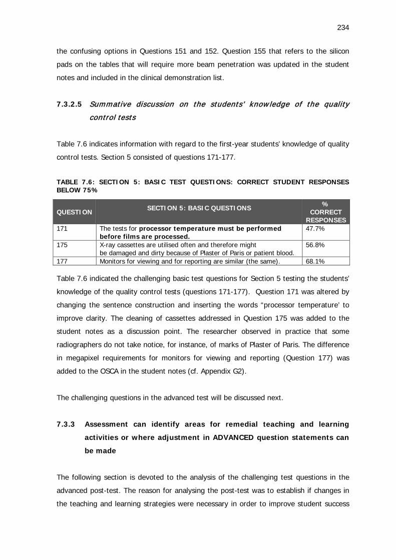
234
the confusing options in Questions 151 and 152. Question 155 that refers to the silicon
pads on the tables that will require more beam penetration was updated in the student
notes and included in the clinical demonstration list.
7.3.2.5 Summative discussion on the students’ know ledge of the quality
control tests
Table 7.6 indicates information with regard to the first-year students’ knowledge of quality
control tests. Section 5 consisted of questions 171-177.
TABLE 7.6: SECTION 5: BASIC TEST QUESTIONS: CORRECT STUDENT RESPONSES BELOW 75%
QUESTION SECTION 5: BASIC QUESTIONS
% CORRECT
RESPONSES 171 The tests for processor temperature must be performed
before films are processed. 47.7%
175 X-ray cassettes are utilised often and therefore might be damaged and dirty because of Plaster of Paris or patient blood.
56.8%
177 Monitors for viewing and for reporting are similar (the same). 68.1%
Table 7.6 indicated the challenging basic test questions for Section 5 testing the students’
knowledge of the quality control tests (questions 171-177). Question 171 was altered by
changing the sentence construction and inserting the words “processor temperature’ to
improve clarity. The cleaning of cassettes addressed in Question 175 was added to the
student notes as a discussion point. The researcher observed in practice that some
radiographers do not take notice, for instance, of marks of Plaster of Paris. The difference
in megapixel requirements for monitors for viewing and reporting (Question 177) was
added to the OSCA in the student notes (cf. Appendix G2).
The challenging questions in the advanced test will be discussed next.
7.3.3 Assessment can identify areas for remedial teaching and learning
activities or where adjustment in ADVANCED question statements can
be made
The following section is devoted to the analysis of the challenging test questions in the
advanced post-test. The reason for analysing the post-test was to establish if changes in
the teaching and learning strategies were necessary in order to improve student success
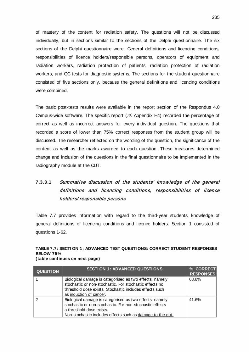
235
of mastery of the content for radiation safety. The questions will not be discussed
individually, but in sections similar to the sections of the Delphi questionnaire. The six
sections of the Delphi questionnaire were: General definitions and licencing conditions,
responsibilities of licence holders/responsible persons, operators of equipment and
radiation workers, radiation protection of patients, radiation protection of radiation
workers, and QC tests for diagnostic systems. The sections for the student questionnaire
consisted of five sections only, because the general definitions and licencing conditions
were combined.
The basic post-tests results were available in the report section of the Respondus 4.0
Campus-wide software. The specific report (cf. Appendix H4) recorded the percentage of
correct as well as incorrect answers for every individual question. The questions that
recorded a score of lower than 75% correct responses from the student group will be
discussed. The researcher reflected on the wording of the question, the significance of the
content as well as the marks awarded to each question. These measures determined
change and inclusion of the questions in the final questionnaire to be implemented in the
radiography module at the CUT.
7.3.3.1 Summative discussion of the students’ know ledge of the general
definitions and licencing conditions, responsibilit ies of licence
holders/ responsible persons
Table 7.7 provides information with regard to the third-year students’ knowledge of
general definitions of licencing conditions and licence holders. Section 1 consisted of
questions 1-62.
TABLE 7.7: SECTION 1: ADVANCED TEST QUESTIONS: CORRECT STUDENT RESPONSES BELOW 75% (table continues on next page)
QUESTION SECTION 1: ADVANCED QUESTIONS
% CORRECT RESPONSES
1 Biological damage is categorised as two effects, namely stochastic or non-stochastic. For stochastic effects no threshold dose exists. Stochastic includes effects such as induction of cancer.
63.8%
2 Biological damage is categorised as two effects, namely stochastic or non-stochastic. For non-stochastic effects a threshold dose exists. Non-stochastic includes effects such as damage to the gut.
41.6%
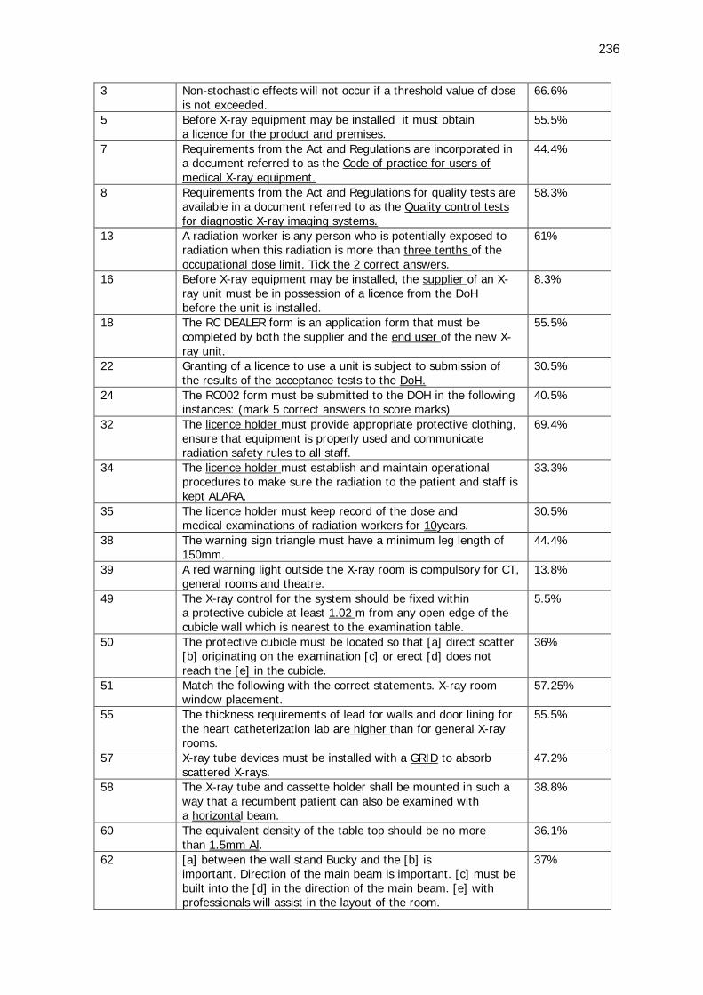
236
3 Non-stochastic effects will not occur if a threshold value of dose is not exceeded.
66.6%
5 Before X-ray equipment may be installed it must obtain a licence for the product and premises.
55.5%
7 Requirements from the Act and Regulations are incorporated in a document referred to as the Code of practice for users of medical X-ray equipment.
44.4%
8 Requirements from the Act and Regulations for quality tests are available in a document referred to as the Quality control tests for diagnostic X-ray imaging systems.
58.3%
13 A radiation worker is any person who is potentially exposed to radiation when this radiation is more than three tenths of the occupational dose limit. Tick the 2 correct answers.
61%
16 Before X-ray equipment may be installed, the supplier of an X-ray unit must be in possession of a licence from the DoH before the unit is installed.
8.3%
18 The RC DEALER form is an application form that must be completed by both the supplier and the end user of the new X-ray unit.
55.5%
22 Granting of a licence to use a unit is subject to submission of the results of the acceptance tests to the DoH.
30.5%
24 The RC002 form must be submitted to the DOH in the following instances: (mark 5 correct answers to score marks)
40.5%
32 The licence holder must provide appropriate protective clothing, ensure that equipment is properly used and communicate radiation safety rules to all staff.
69.4%
34 The licence holder must establish and maintain operational procedures to make sure the radiation to the patient and staff is kept ALARA.
33.3%
35 The licence holder must keep record of the dose and medical examinations of radiation workers for 10years.
30.5%
38 The warning sign triangle must have a minimum leg length of 150mm.
44.4%
39 A red warning light outside the X-ray room is compulsory for CT, general rooms and theatre.
13.8%
49 The X-ray control for the system should be fixed within a protective cubicle at least 1.02 m from any open edge of the cubicle wall which is nearest to the examination table.
5.5%
50 The protective cubicle must be located so that [a] direct scatter [b] originating on the examination [c] or erect [d] does not reach the [e] in the cubicle.
36%
51 Match the following with the correct statements. X-ray room window placement.
57.25%
55 The thickness requirements of lead for walls and door lining for the heart catheterization lab are higher than for general X-ray rooms.
55.5%
57 X-ray tube devices must be installed with a GRID to absorb scattered X-rays.
47.2%
58 The X-ray tube and cassette holder shall be mounted in such a way that a recumbent patient can also be examined with a horizontal beam.
38.8%
60 The equivalent density of the table top should be no more than 1.5mm Al.
36.1%
62 [a] between the wall stand Bucky and the [b] is important. Direction of the main beam is important. [c] must be built into the [d] in the direction of the main beam. [e] with professionals will assist in the layout of the room.
37%
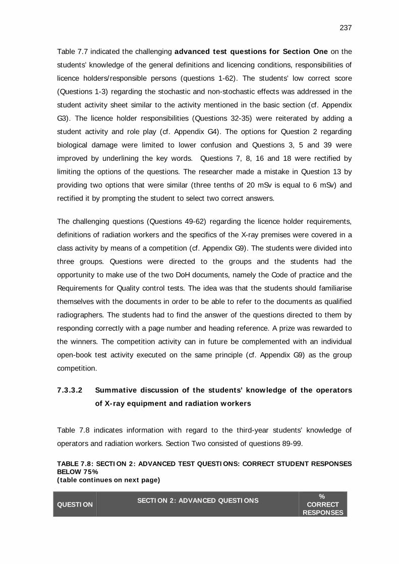
237
Table 7.7 indicated the challenging advanced test questions for Section One on the
students’ knowledge of the general definitions and licencing conditions, responsibilities of
licence holders/responsible persons (questions 1-62). The students’ low correct score
(Questions 1-3) regarding the stochastic and non-stochastic effects was addressed in the
student activity sheet similar to the activity mentioned in the basic section (cf. Appendix
G3). The licence holder responsibilities (Questions 32-35) were reiterated by adding a
student activity and role play (cf. Appendix G4). The options for Question 2 regarding
biological damage were limited to lower confusion and Questions 3, 5 and 39 were
improved by underlining the key words. Questions 7, 8, 16 and 18 were rectified by
limiting the options of the questions. The researcher made a mistake in Question 13 by
providing two options that were similar (three tenths of 20 mSv is equal to 6 mSv) and
rectified it by prompting the student to select two correct answers.
The challenging questions (Questions 49-62) regarding the licence holder requirements,
definitions of radiation workers and the specifics of the X-ray premises were covered in a
class activity by means of a competition (cf. Appendix G9). The students were divided into
three groups. Questions were directed to the groups and the students had the
opportunity to make use of the two DoH documents, namely the Code of practice and the
Requirements for Quality control tests. The idea was that the students should familiarise
themselves with the documents in order to be able to refer to the documents as qualified
radiographers. The students had to find the answer of the questions directed to them by
responding correctly with a page number and heading reference. A prize was rewarded to
the winners. The competition activity can in future be complemented with an individual
open-book test activity executed on the same principle (cf. Appendix G9) as the group
competition.
7.3.3.2 Summative discussion of the students’ knowledge of the operators
of X-ray equipment and radiation workers
Table 7.8 indicates information with regard to the third-year students’ knowledge of
operators and radiation workers. Section Two consisted of questions 89-99.
TABLE 7.8: SECTION 2: ADVANCED TEST QUESTIONS: CORRECT STUDENT RESPONSES BELOW 75% (table continues on next page)
QUESTION SECTION 2: ADVANCED QUESTIONS
% CORRECT
RESPONSES
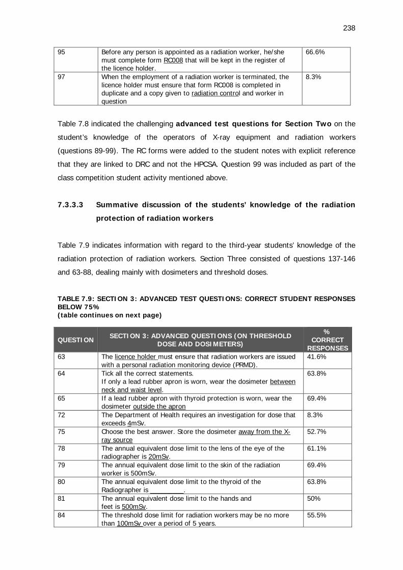
238
95 Before any person is appointed as a radiation worker, he/she must complete form RC008 that will be kept in the register of the licence holder.
66.6%
97 When the employment of a radiation worker is terminated, the licence holder must ensure that form RC008 is completed in duplicate and a copy given to radiation control and worker in question
8.3%
Table 7.8 indicated the challenging advanced test questions for Section Two on the
student’s knowledge of the operators of X-ray equipment and radiation workers
(questions 89-99). The RC forms were added to the student notes with explicit reference
that they are linked to DRC and not the HPCSA. Question 99 was included as part of the
class competition student activity mentioned above.
7.3.3.3 Summative discussion of the students’ knowledge of the radiation
protection of radiation workers
Table 7.9 indicates information with regard to the third-year students’ knowledge of the
radiation protection of radiation workers. Section Three consisted of questions 137-146
and 63-88, dealing mainly with dosimeters and threshold doses.
TABLE 7.9: SECTION 3: ADVANCED TEST QUESTIONS: CORRECT STUDENT RESPONSES BELOW 75% (table continues on next page)
QUESTION SECTION 3: ADVANCED QUESTIONS (ON THRESHOLD DOSE AND DOSIMETERS)
% CORRECT
RESPONSES 63 The licence holder must ensure that radiation workers are issued
with a personal radiation monitoring device (PRMD). 41.6%
64 Tick all the correct statements. If only a lead rubber apron is worn, wear the dosimeter between neck and waist level.
63.8%
65 If a lead rubber apron with thyroid protection is worn, wear the dosimeter outside the apron
69.4%
72 The Department of Health requires an investigation for dose that exceeds 4mSv.
8.3%
75 Choose the best answer. Store the dosimeter away from the X-ray source
52.7%
78 The annual equivalent dose limit to the lens of the eye of the radiographer is 20mSv.
61.1%
79 The annual equivalent dose limit to the skin of the radiation worker is 500mSv.
69.4%
80 The annual equivalent dose limit to the thyroid of the Radiographer is _________.
63.8%
81 The annual equivalent dose limit to the hands and feet is 500mSv.
50%
84 The threshold dose limit for radiation workers may be no more than 100mSv over a period of 5 years.
55.5%
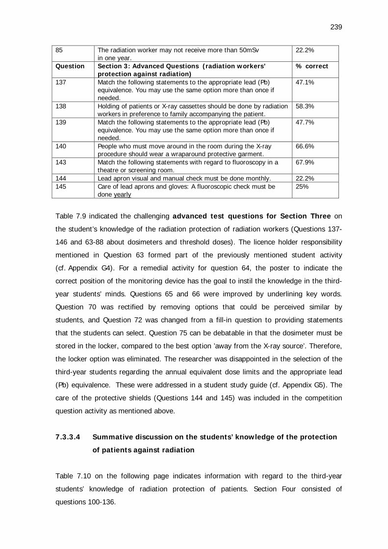
239
85 The radiation worker may not receive more than 50mSv in one year.
22.2%
Question Section 3: Advanced Questions (radiation workers’ protection against radiation)
% correct
137 Match the following statements to the appropriate lead (Pb) equivalence. You may use the same option more than once if needed.
47.1%
138 Holding of patients or X-ray cassettes should be done by radiation workers in preference to family accompanying the patient.
58.3%
139 Match the following statements to the appropriate lead (Pb) equivalence. You may use the same option more than once if needed.
47.7%
140 People who must move around in the room during the X-ray procedure should wear a wraparound protective garment.
66.6%
143 Match the following statements with regard to fluoroscopy in a theatre or screening room.
67.9%
144 Lead apron visual and manual check must be done monthly. 22.2% 145 Care of lead aprons and gloves: A fluoroscopic check must be
done yearly 25%
Table 7.9 indicated the challenging advanced test questions for Section Three on
the student’s knowledge of the radiation protection of radiation workers (Questions 137-
146 and 63-88 about dosimeters and threshold doses). The licence holder responsibility
mentioned in Question 63 formed part of the previously mentioned student activity
(cf. Appendix G4). For a remedial activity for question 64, the poster to indicate the
correct position of the monitoring device has the goal to instil the knowledge in the third-
year students’ minds. Questions 65 and 66 were improved by underlining key words.
Question 70 was rectified by removing options that could be perceived similar by
students, and Question 72 was changed from a fill-in question to providing statements
that the students can select. Question 75 can be debatable in that the dosimeter must be
stored in the locker, compared to the best option ’away from the X-ray source’. Therefore,
the locker option was eliminated. The researcher was disappointed in the selection of the
third-year students regarding the annual equivalent dose limits and the appropriate lead
(Pb) equivalence. These were addressed in a student study guide (cf. Appendix G5). The
care of the protective shields (Questions 144 and 145) was included in the competition
question activity as mentioned above.
7.3.3.4 Summative discussion on the students’ knowledge of the protection
of patients against radiation
Table 7.10 on the following page indicates information with regard to the third-year
students’ knowledge of radiation protection of patients. Section Four consisted of
questions 100-136.
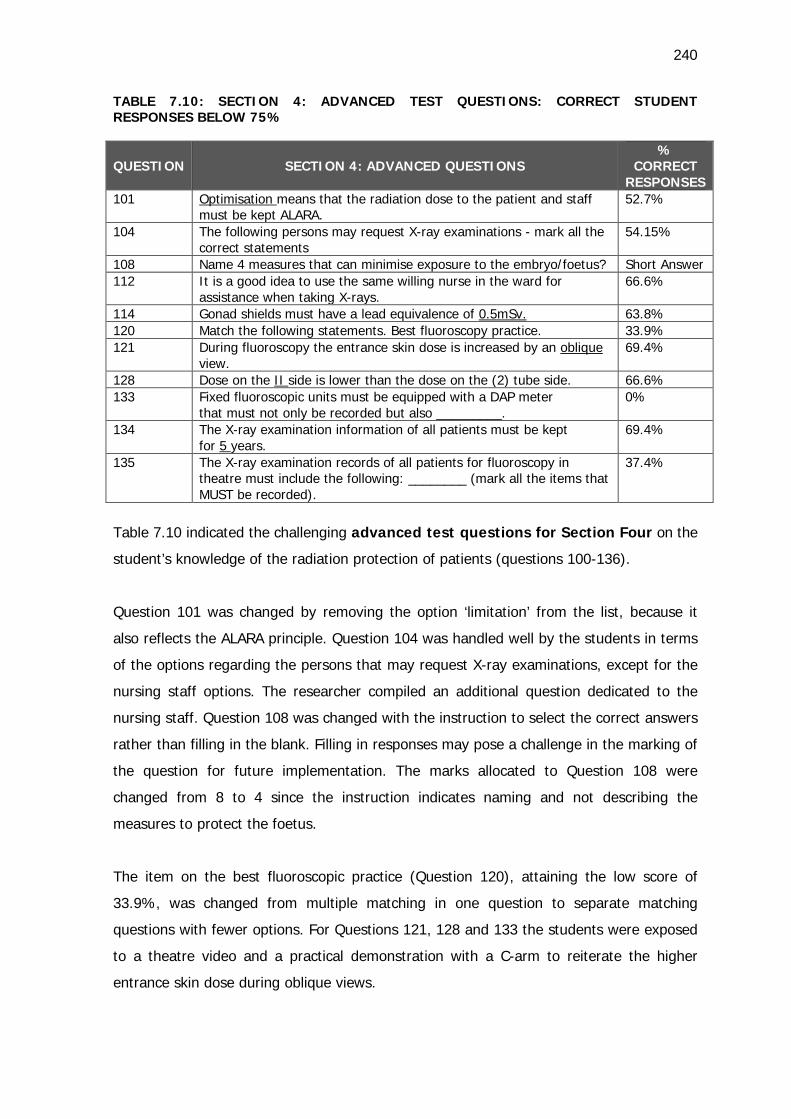
240
TABLE 7.10: SECTION 4: ADVANCED TEST QUESTIONS: CORRECT STUDENT RESPONSES BELOW 75%
QUESTION SECTION 4: ADVANCED QUESTIONS %
CORRECT RESPONSES
101 Optimisation means that the radiation dose to the patient and staff must be kept ALARA.
52.7%
104 The following persons may request X-ray examinations - mark all the correct statements
54.15%
108 Name 4 measures that can minimise exposure to the embryo/foetus? Short Answer 112 It is a good idea to use the same willing nurse in the ward for
assistance when taking X-rays. 66.6%
114 Gonad shields must have a lead equivalence of 0.5mSv. 63.8% 120 Match the following statements. Best fluoroscopy practice. 33.9% 121 During fluoroscopy the entrance skin dose is increased by an oblique
view. 69.4%
128 Dose on the II side is lower than the dose on the (2) tube side. 66.6% 133 Fixed fluoroscopic units must be equipped with a DAP meter
that must not only be recorded but also _________. 0%
134 The X-ray examination information of all patients must be kept for 5 years.
69.4%
135 The X-ray examination records of all patients for fluoroscopy in theatre must include the following: ________ (mark all the items that MUST be recorded).
37.4%
Table 7.10 indicated the challenging advanced test questions for Section Four on the
student’s knowledge of the radiation protection of patients (questions 100-136).
Question 101 was changed by removing the option ‘limitation’ from the list, because it
also reflects the ALARA principle. Question 104 was handled well by the students in terms
of the options regarding the persons that may request X-ray examinations, except for the
nursing staff options. The researcher compiled an additional question dedicated to the
nursing staff. Question 108 was changed with the instruction to select the correct answers
rather than filling in the blank. Filling in responses may pose a challenge in the marking of
the question for future implementation. The marks allocated to Question 108 were
changed from 8 to 4 since the instruction indicates naming and not describing the
measures to protect the foetus.
The item on the best fluoroscopic practice (Question 120), attaining the low score of
33.9%, was changed from multiple matching in one question to separate matching
questions with fewer options. For Questions 121, 128 and 133 the students were exposed
to a theatre video and a practical demonstration with a C-arm to reiterate the higher
entrance skin dose during oblique views.
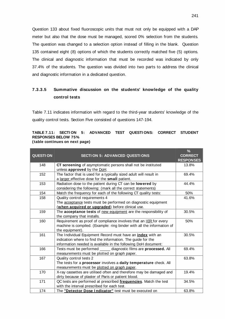
241
Question 133 about fixed fluoroscopic units that must not only be equipped with a DAP
meter but also that the dose must be managed, scored 0% selection from the students.
The question was changed to a selection option instead of filling in the blank. Question
135 contained eight (8) options of which the students correctly matched five (5) options.
The clinical and diagnostic information that must be recorded was indicated by only
37.4% of the students. The question was divided into two parts to address the clinical
and diagnostic information in a dedicated question.
7.3.3.5 Summative discussion on the students’ knowledge of the quality
control tests
Table 7.11 indicates information with regard to the third-year students’ knowledge of the
quality control tests. Section Five consisted of questions 147-194.
TABLE 7.11: SECTION 5: ADVANCED TEST QUESTIONS: CORRECT STUDENT RESPONSES BELOW 75% (table continues on next page)
QUESTION SECTION 5: ADVANCED QUESTIONS %
CORRECT RESPONSES
148 CT screening of asymptomatic persons shall not be instituted unless approved by the DoH.
13.8%
152 The factor that is used for a typically sized adult will result in a larger effective dose for the small patient.
69.4%
153 Radiation dose to the patient during CT can be lowered by considering the following: (mark all the correct statements)
44.4%
154 Match the frequency for each of the following CT quality tests: 50% 158 Quality control requirements 4
The acceptance tests must be performed on diagnostic equipment (when acquired or upgraded) before clinical use.
41.6%
159 The acceptance tests of new equipment are the responsibility of the company that installs.
30.5%
160 Requirement as proof of compliance involves that an IER for every machine is compiled. (Example: ring binder with all the information of the equipment).
50%
161 The Individual Equipment Record must have an index with an indication where to find the information. The guide for the information needed is available in the following DoH document:
30.5%
166 Tests must be performed _____ diagnostic films are processed. All measurements must be plotted on graph paper.
69.4%
167 Quality control tests 2 The tests for a processor involves a daily temperature check. All measurements must be plotted on graph paper.
63.8%
170 X-ray cassettes are utilised often and therefore may be damaged and dirty because of plaster of Paris or patient blood.
19.4%
171 QC tests are performed at prescribed frequencies. Match the test with the interval prescribed for each test.
34.5%
174 The "Detector Dose Indicator" test must be executed on 63.8%
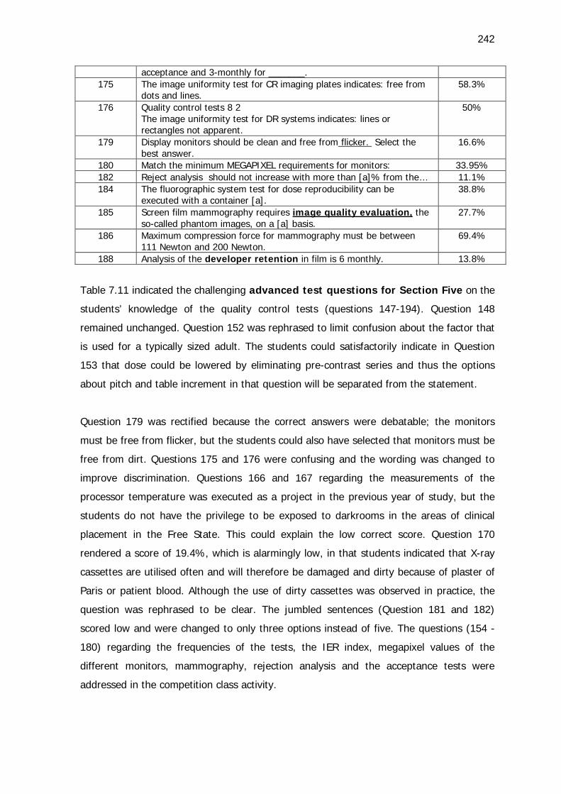
242
acceptance and 3-monthly for _______. 175 The image uniformity test for CR imaging plates indicates: free from
dots and lines. 58.3%
176 Quality control tests 8 2 The image uniformity test for DR systems indicates: lines or rectangles not apparent.
50%
179 Display monitors should be clean and free from flicker. Select the best answer.
16.6%
180 Match the minimum MEGAPIXEL requirements for monitors: 33.95% 182 Reject analysis should not increase with more than [a]% from the… 11.1% 184 The fluorographic system test for dose reproducibility can be
executed with a container [a]. 38.8%
185 Screen film mammography requires image quality evaluation, the so-called phantom images, on a [a] basis.
27.7%
186 Maximum compression force for mammography must be between 111 Newton and 200 Newton.
69.4%
188 Analysis of the developer retention in film is 6 monthly. 13.8%
Table 7.11 indicated the challenging advanced test questions for Section Five on the
students’ knowledge of the quality control tests (questions 147-194). Question 148
remained unchanged. Question 152 was rephrased to limit confusion about the factor that
is used for a typically sized adult. The students could satisfactorily indicate in Question
153 that dose could be lowered by eliminating pre-contrast series and thus the options
about pitch and table increment in that question will be separated from the statement.
Question 179 was rectified because the correct answers were debatable; the monitors
must be free from flicker, but the students could also have selected that monitors must be
free from dirt. Questions 175 and 176 were confusing and the wording was changed to
improve discrimination. Questions 166 and 167 regarding the measurements of the
processor temperature was executed as a project in the previous year of study, but the
students do not have the privilege to be exposed to darkrooms in the areas of clinical
placement in the Free State. This could explain the low correct score. Question 170
rendered a score of 19.4%, which is alarmingly low, in that students indicated that X-ray
cassettes are utilised often and will therefore be damaged and dirty because of plaster of
Paris or patient blood. Although the use of dirty cassettes was observed in practice, the
question was rephrased to be clear. The jumbled sentences (Question 181 and 182)
scored low and were changed to only three options instead of five. The questions (154 -
180) regarding the frequencies of the tests, the IER index, megapixel values of the
different monitors, mammography, rejection analysis and the acceptance tests were
addressed in the competition class activity.
243
This concludes the analysis of the challenging test questions for the basic and advanced
post-tests.
7.3.4 The role of feedback in assessment
Assessment should provide feedback to students on their progress and add value in the
workplace. Assessment should thus not only focus on the end product but must form part
of teaching and learning. Assessment will foster accountability and should be transparent
guides to students (CHE HEQC 2004:124). Assessment hence is a two-edged sword
specifically to put effort into learning and to improve teaching and learning activities
(Killen 2007:321).
A study to evaluate the Immediate Feedback Assessment Technique (IF AT), in which
students received immediate feedback and were allowed to respond until the answer was
correct, indicated that the participating students achieved higher scores when retested
(Epstein, Lazarus, Calvano, Matthews, Hendel, Epstein and Brosvic 2002:187). The
advantages of immediate feedback are mainly that misconceptions are immediately
replaced with knowledge, that students are guided to correct answers and that retention
is improved. The principle to teach while assessing (Epstein et al. 2002:200) motivated
the researcher to schedule the students for a third testing session.
The third tests were scheduled during the assessment period of the CUT. The students
were granted a day study leave prior to the scheduled assessment period. The question
statements were modified as indicated in the analysis of the challenging questions above.
The question statements were improved by adding feedback statements. The students
could see the feedback on the answers they had selected as soon as they submitted their
completed exam - or at the time that the settings were changed to allow feedback to be
viewed.
7.3.4.1 Description of the results of the unintended third basic and advanced tests
The third test was not part of the initial objectives and is not described in the
methodology. The researcher executed the third test as add-on with future
implementation of the test in mind. The third tests were scheduled to be taken after the
students had completed the remedial activities as indicated previously in this chapter.
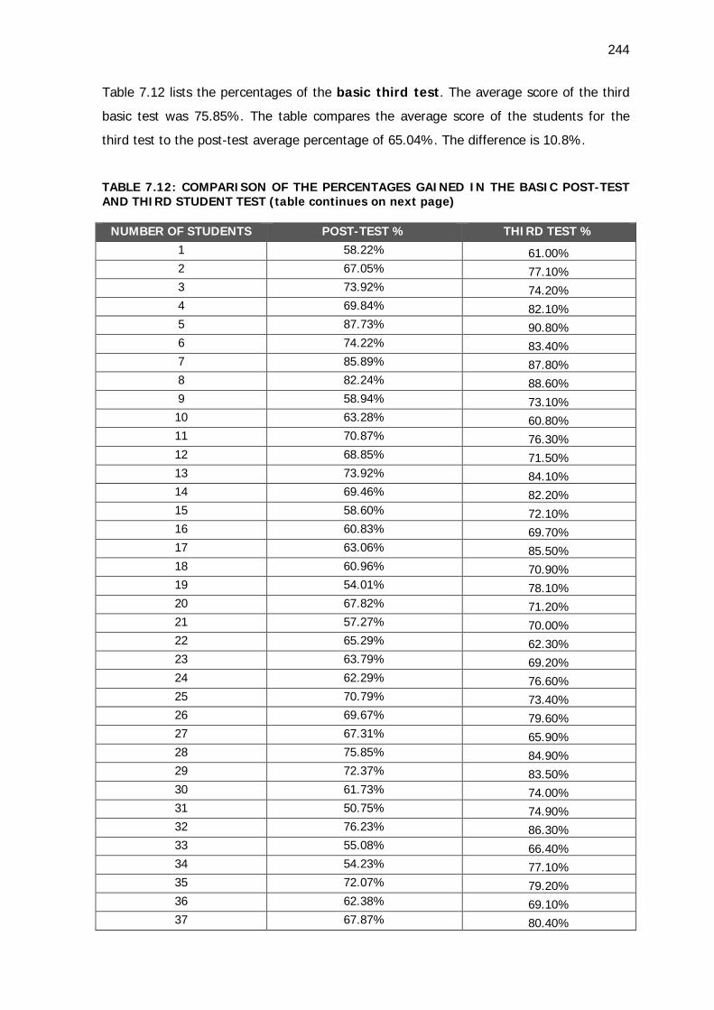
244
Table 7.12 lists the percentages of the basic third test. The average score of the third
basic test was 75.85%. The table compares the average score of the students for the
third test to the post-test average percentage of 65.04%. The difference is 10.8%.
TABLE 7.12: COMPARISON OF THE PERCENTAGES GAINED IN THE BASIC POST-TEST AND THIRD STUDENT TEST (table continues on next page)
NUMBER OF STUDENTS POST-TEST % THIRD TEST % 1 58.22% 61.00% 2 67.05% 77.10% 3 73.92% 74.20% 4 69.84% 82.10% 5 87.73% 90.80% 6 74.22% 83.40% 7 85.89% 87.80% 8 82.24% 88.60% 9 58.94% 73.10% 10 63.28% 60.80% 11 70.87% 76.30% 12 68.85% 71.50% 13 73.92% 84.10% 14 69.46% 82.20% 15 58.60% 72.10% 16 60.83% 69.70% 17 63.06% 85.50% 18 60.96% 70.90% 19 54.01% 78.10% 20 67.82% 71.20% 21 57.27% 70.00% 22 65.29% 62.30% 23 63.79% 69.20% 24 62.29% 76.60% 25 70.79% 73.40% 26 69.67% 79.60% 27 67.31% 65.90% 28 75.85% 84.90% 29 72.37% 83.50% 30 61.73% 74.00% 31 50.75% 74.90% 32 76.23% 86.30% 33 55.08% 66.40% 34 54.23% 77.10% 35 72.07% 79.20% 36 62.38% 69.10% 37 67.87% 80.40%
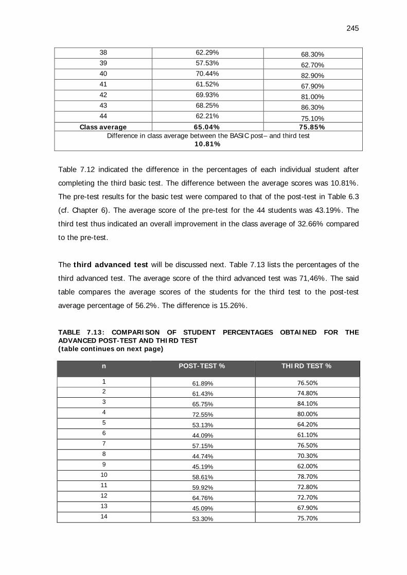
245
38 62.29% 68.30% 39 57.53% 62.70% 40 70.44% 82.90% 41 61.52% 67.90% 42 69.93% 81.00% 43 68.25% 86.30% 44 62.21% 75.10%
Class average 65.04% 75.85% Difference in class average between the BASIC post– and third test
10.81%
Table 7.12 indicated the difference in the percentages of each individual student after
completing the third basic test. The difference between the average scores was 10.81%.
The pre-test results for the basic test were compared to that of the post-test in Table 6.3
(cf. Chapter 6). The average score of the pre-test for the 44 students was 43.19%. The
third test thus indicated an overall improvement in the class average of 32.66% compared
to the pre-test.
The third advanced test will be discussed next. Table 7.13 lists the percentages of the
third advanced test. The average score of the third advanced test was 71,46%. The said
table compares the average scores of the students for the third test to the post-test
average percentage of 56.2%. The difference is 15.26%.
TABLE 7.13: COMPARISON OF STUDENT PERCENTAGES OBTAINED FOR THE ADVANCED POST-TEST AND THIRD TEST (table continues on next page)
n POST-TEST % THIRD TEST %
1 61.89% 76.50% 2 61.43% 74.80% 3 65.75% 84.10% 4 72.55% 80.00% 5 53.13% 64.20% 6 44.09% 61.10% 7 57.15% 76.50% 8 44.74% 70.30% 9 45.19% 62.00% 10 58.61% 78.70% 11 59.92% 72.80% 12 64.76% 72.70% 13 45.09% 67.90% 14 53.30% 75.70%
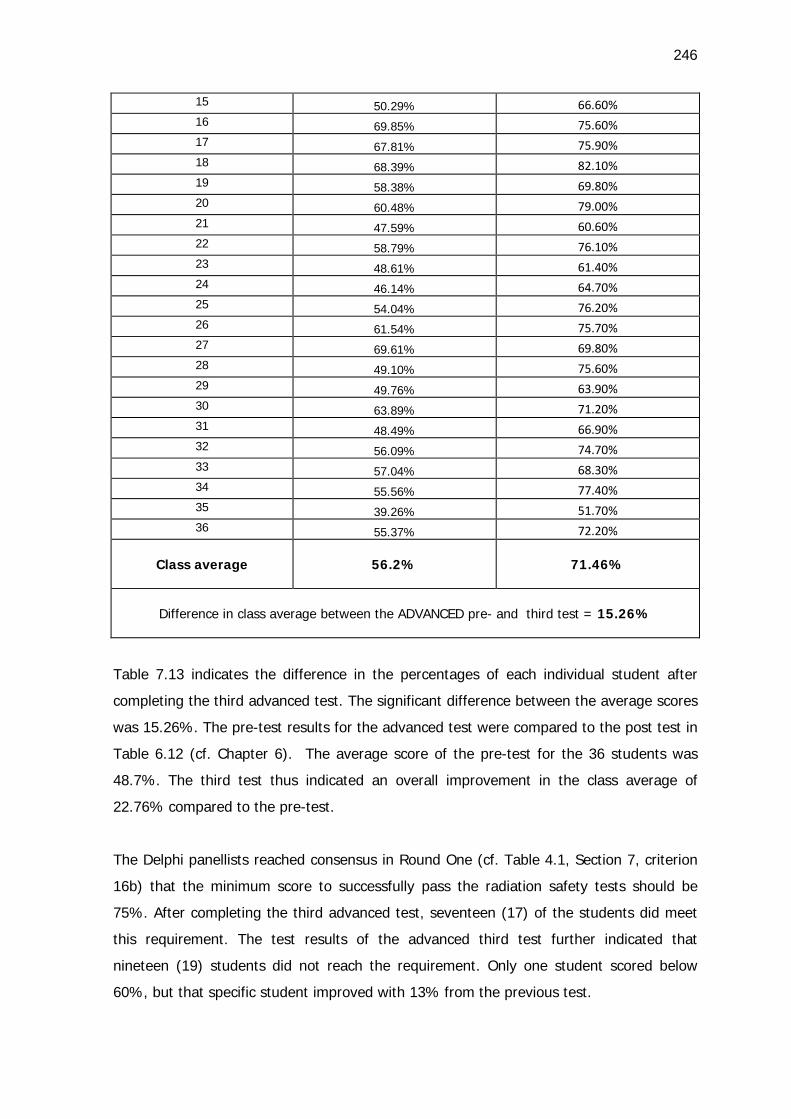
246
15 50.29% 66.60% 16 69.85% 75.60% 17 67.81% 75.90% 18 68.39% 82.10% 19 58.38% 69.80% 20 60.48% 79.00% 21 47.59% 60.60% 22 58.79% 76.10% 23 48.61% 61.40% 24 46.14% 64.70% 25 54.04% 76.20% 26 61.54% 75.70% 27 69.61% 69.80% 28 49.10% 75.60% 29 49.76% 63.90% 30 63.89% 71.20% 31 48.49% 66.90% 32 56.09% 74.70% 33 57.04% 68.30% 34 55.56% 77.40% 35 39.26% 51.70% 36 55.37% 72.20%
Class average
56.2% 71.46%
Difference in class average between the ADVANCED pre- and third test = 15.26%
Table 7.13 indicates the difference in the percentages of each individual student after
completing the third advanced test. The significant difference between the average scores
was 15.26%. The pre-test results for the advanced test were compared to the post test in
Table 6.12 (cf. Chapter 6). The average score of the pre-test for the 36 students was
48.7%. The third test thus indicated an overall improvement in the class average of
22.76% compared to the pre-test.
The Delphi panellists reached consensus in Round One (cf. Table 4.1, Section 7, criterion
16b) that the minimum score to successfully pass the radiation safety tests should be
75%. After completing the third advanced test, seventeen (17) of the students did meet
this requirement. The test results of the advanced third test further indicated that
nineteen (19) students did not reach the requirement. Only one student scored below
60%, but that specific student improved with 13% from the previous test.
247
After completing the basic test, only twenty (20) out of the 44 first-year students did not
achieve the required 75%. No student scored below 60%. With the benefit of hindsight
the researcher is of the opinion that the minimum requirement of 75% must be further
researched. The technical data that the students are exposed to, increase the cognitive
load, but mastery will improve when the content is distributed over a period of four years
in the new curriculum.
The researcher established the average test percentages for some of the learning areas of
the radiography students for the purpose of comparison and perspective. During the first
semester of the first year the class average scored for two other learning areas was 64%
and 53% respectively. The third-year class average for two learning areas were 75% and
70% respectively. The outcome of the radiation safety tests are higher for the first years,
but in a similar range as that of the third years.
7.4 IMPLICATIONS OF THE FINDINGS OF THE TEACHING, LEARNING AND
ASSESSMENT ACTIVITIES
The testing of student knowledge of the QC tests requires additional methods of
assessment because questions pertaining to the QC tests may not be considered
comprehensive. The QC tests involve a practical component – a matter reiterated by the
comments from the Delphi panel members (cf. point 7.2), therefore the use of a portfolio
of evidence intended to prove that the student is knowledgeable and equipped to adhere
to the said requirements independently is recommended. With reference to the DoH
requirements, it is important for students to understand why QC tests are performed.
Consequences if tests are not performed may for instance result in unnecessary radiation
to the patient.
7.4.1 Portfolio of evidence for quality control tests
The portfolio of evidence was compiled to complement the advanced student
questionnaire assessment (cf. Appendix G6). The aim of the portfolio was to grant the
third-year student the opportunity and motivation to become familiar with the required
QC tests. The final-year student is a potential licence holder of medical X-ray equipment
and must be equipped with practical competencies to be informed about what the
execution of the tests entails.
248
The portfolio of evidence must comply with criteria that are explicitly formulated to make
sure that the student will meet the minimum requirements successfully during the course
of the final year. The frequencies of the tests are indicated so that the student can refer
to the portfolio as a working document after graduation. The student is required to
compile a file according to the contents of the sequence of the rubric provided. The
student must take cognisance of all the quality control tests that must be performed at
the prescribed frequencies as specified in Table 2 of the DoH quality test document
(cf. Appendix R4). The completed file will provide proof that the students have at least
witnessed the execution of the specific test. The student must file proof of a record sheet
of the QC tests of the department to which the student has been allocated for clinical
exposure. The portfolio contains the frequencies at which all the tests must be performed
and the student will be able to use the file as a working document.
7.4.2 Additional adjustment in teaching and learning activities
Apart from the student activities that were mentioned previously (cf. Appendix G3-5), the
researcher improved the student study guide (cf. Appendix G2) by adding the RC forms
from the Directorate Radiation Control so that students may feel confident to utilise the
forms in future.
Lecturer notes were added as guide in terms of the role-plays (cf. Appendix G11), group
activities (cf. Appendix G9), and a video C-arm demonstration in theatre (cf. Appendix
G12). An open-book test was added as an individual alternative to the competition activity
in order for students to become familiar with the licence holder responsibilities, premises
requirements and QC test frequencies.
7.5 CONCLUSION
A description of the implications of the findings from the Delphi survey and post-test
student questionnaires was given in this chapter. The varied backgrounds of the Delphi
panellists proved to add value to the research by the comments that provided valuable
insight. The student responses indicated the need for remedial activities, as well as the
errors made by the compiler of the tests. The results of the current chapter determined
change and inclusion of the questions in the final questionnaire to be implemented in the
radiography programme at the CUT. The modification of the learning activities reiterated
249
the significance of lecturers to reflect constantly on outcomes, assessment, and learning
activities to promote deep learning.
In the following chapter, Chapter 8, the thesis will be concluded by providing conclusions,
limitations and the recommendations of the study.
CHAPTER 8 CONCLUSIONS, RECOMMENDATIONS AND LIMITATIONS OF THE STUDY
8.1 INTRODUCTION
An in-depth study was conducted with a view to developing standardised training and
assessment for diagnostic radiographers to address radiation safety.
Radiographers must be cognisant of the legislation and the regulations underpinning the
use of X-ray equipment as described in the Hazardous and Substances Act, as well as in
the Regulations concerning the control of electronic products. The radiographer must also
be acquainted with the responsibilities of radiation workers depicted in the Code of
practice for users of medical X-ray equipment (DRC 2011)and the Requirements for
licence holders with respect to quality control tests for diagnostic X-ray imaging systems
(DRC 2012). The purpose of the regulations is to ensure the safe use of X-ray equipment
so that the ionising radiation dose to the staff and the patient is kept as low as reasonably
achievable. The regulations decree that licence holders of X-ray equipment must identify
radiation workers, monitor the ionising radiation received by these workers, and issue
personal monitoring devices. Furthermore, radiation workers must be educated in the
safety and risks of ionising radiation. The education pertaining to ionising radiation safety
of staff members is the responsibility of each license holder, but nationally no
standardised monitoring of the required education currently (2014) is in place. The first-
year radiography student is considered a radiation worker from day one, just by entering
into the profession. The final-year radiography student is a potential licence holder of X-
ray equipment. In this study, the development of standardised training and assessment
for the radiography students in radiation safety was investigated and evaluated in order
to equip the student to adhere to safety regulations.
The aim of this chapter is to provide a brief overview of the study and to present
comments and some concluding thoughts on the final findings. The chapter commences
with an overview of the study, followed by recommendations for the implementation of
radiation safety training and assessment for diagnostic radiographers, conclusions drawn,
a short discussion on the limitations of the study, the contribution to knowledge in the
field, and the significance of the study; recommendations on the way forward, and
conclusive remarks.
250
8.2 OVERVIEW OF THE STUDY
The research was carried out and completed based on four research questions. The
findings of the research served as foundation for standardised training and assessment in
radiation safety for diagnostic radiographers.
In Chapter 1 (cf. point 1.3) an outline of the various research questions was presented.
The research questions guided the study and shaped the final outcome. From point 7.2.1
onwards, the four research questions are reviewed together with the main findings and
final outcome of each research question.
8.2.1 Research question 1
The research question was stated as:
How can the radiation safety and quality control requirements for radiography radiation
workers and medical X-ray equipment licence holders be contextualised and
conceptualised to compile a theoretical framework for the study?
The following objective was pursued:
To contextualise and conceptualise the radiation safety and quality control requirements
for radiography radiation workers and medical X-ray equipment licence holders by means
of a literature study, in order to create a theoretical framework for the study. This
objective addressed research question 1.
The researched question was aimed at providing the background of the study and at
identifying the content of training and assessment in radiography. In Chapter 2, radiation
safety training and assessment were contextualised from the perspective of legislation
and regulations by describing the International Commission on Radiological Protection
(ICRP) as the primary body in protection against ionising radiation (cf. point 2.1). In
South Africa, the Department of Health (DoH) accepted the recommendations of the
ICRP and regulates radiation protection in the framework of the Hazardous and
Substances Act No 15 of 1973 (cf. point 2.2.1). This act regulates that the Minister of
Health and specifically the Director-General, issues X-ray equipment licences to users of
251
electro-medical (X-ray) equipment. This equipment is regulated by Regulation 1332,
entitled: ‘Regulations concerning the control of electronic products’ (cf. point 2.2.2). The
DoH in South Africa applies international standards as legal requirements and guidelines
through the Directorate: Radiation Control (DRC) that issues a licence if the product
and usage comply with the legislative and international requirements for safety and
performance. The two documents that are supplied when a licence is issued, namely the
Code of practice for users of medical X-ray equipment (cf. point 2.3), and the
Requirements for licence holders w ith respect to quality control tests for
diagnostic X-ray imaging systems (cf. point 2.4) were scrutinised in order to be able
to provide a clear view of the requirements to radiation workers (cf. point 2.3.1) and
licence holders (cf. point 2.2.2). The requirements and guidelines were emphasised in
terms of the monitoring of radiation workers (cf. point 2.3.2), monitoring of patients
(cf. 2.3.3), premises requirements (cf. point 2.3.4), and the guideline documents (cf.
point 2.3.5).
To contextualise the importance of the preparation of the third-year students as potential
licence holders, the quality control tests (cf. point 2.4.1) with the required frequencies
(cf. point 2.4.2), as depicted in the documents, were emphasised.
The standardised training and assessment were viewed from the recently accredited
Bachelor of Radiography as starting point (cf. point 2.5.1), progressing to exit-level
outcomes (cf. point 2.5.1.2) and critical cross-field outcomes, in order to formulate
objectives (cf. point 5.1.4). Relevant teaching and learning strategies (cf. point
2.5.2) were described prior to elaborating on assessment principles (cf. point 2.5.3)
and the associated assessment criteria (cf. point 2.5.3.1) for the six exit-level
outcomes. Effecting deep learning was contextualised in terms of alignment (cf. point
2.6) of the curriculum objectives, teaching and learning activities, and assessment.
To contextualise the mastery of radiation safety requirements (cf. point 2.7), the
retrieval of information (cf. point 2.7.1), standard setting (cf. point 2.7.2),
assessment instruments (cf. point 2.7.3), and transformative learning were
clarified. The training and assessment were benchmarked with a few available on-line
training courses (cf. point 2.8).
252
An outline of Chapter 2 has been provided in Figure 1.2 in the form of a diagrammatic
overview.
8.2.2 Research question 2
The research question was stated as:
What are the outcomes for the radiation safety and quality control requirements training:
basic for first-year radiography students (representing the entry level radiation worker
issued with a dosimeter) and advanced for third-year radiography students (representing
the licence holder, responsible person, and the qualified radiographer)?
The following objective was pursued:
To determine appropriate outcomes for the radiation safety and quality control
requirements training by using a Delphi questionnaire: basic for first-year radiography
students (representing the entry level radiation worker issued with a dosimeter) and
advanced for third- year radiography students (representing the licence holder,
responsible person and the qualified radiographer). This objective addressed research
question 2.
The conceptualisation of the content of the training and assessment was achieved with
the aid of the literature review described in Chapter 2, and this resulted in the compilation
of the Delphi questionnaire, which was compiled comprising seven sections
(cf. point 5.2). The conceptualisation of the Delphi survey was achieved by describing
the theoretical perspectives on the methodology (cf. point 3.2). The measuring
instrument, analysis, and findings of the four rounds of the Delphi survey were
described in Chapter 4 (cf. point 4.2.1 to 4.2.4), concluding with the findings regarding
consensus statements (cf. point 4.2.6), and stability (cf. point 4.2.7). A description of
the implications of the findings (cf. point 7.2) from the Delphi survey, and
comments (cf. point 7.2.1) from the Delphi panellists were provided in Chapter 7. The
intended learning outcomes (cf. point 5.2.1.2) of the radiation safety training were
compiled according to the sections of the Delphi questionnaire. The consensus and
stability outcome of the Delphi survey resulted in the allocation of the content, and thus
outcomes, into basic or advanced criteria statements (cf. point Table 4.2).
253
8.2.3 Research question 3
The research question was stated as:
What are effective teaching and learning activities and assessment strategies for the
radiography radiation safety and quality control requirements training?
The following objective was pursued:
To develop the effective teaching and learning activities and assessment strategies for the
radiography radiation safety and quality control requirements training to be presented at
the Central University of Technology (CUT) based on the findings of the Delphi survey.
This objective addressed research question 3.
This research question was aimed at developing teaching and learning activities (cf.
point 5.4), contextualised in Chapter 2 (cf. point 2.5.2), aligned with the intended
learning outcomes (cf. 5.2.1.2) of the radiation safety training and the assessment
criteria statements as confirmed by the Delphi results (cf. Table 4.2). The assessment
criteria statements (cf. Table 4.2) of the Delphi questionnaire were adapted and taken up
in basic (cf. Appendix E3) and advanced (cf. Appendix E4) student questionnaires.
In Chapter 5 references are provided to the student study guide (cf. Appendix G2?) that
contains copies of the DoH requirement documents, intended learning outcomes and
student activities. The implications of the findings regarding the teaching and
learning activities were described in Chapter 7. A portfolio was discussed (cf. point
7.3.4.1), which final-year students will have to compile, containing evidence of quality
control tests in order the address the practical component of the training. The
adjustments (cf. point 7.3.4.1) made after reflection on the responses of students in the
pre- and post-tests were described to round off the development of the training course.
The comments made by the Delphi panellists (cf. point 7.2.1) regarding section seven, the
training course, were taken into consideration to plan the future implementation of the
course. The possibility to incorporate the radiation safety learning units in the current
modules of the radiography curriculum was proposed as conclusion in Chapter 7.
8.2.4 Research question 4
The research question was:

254
Will the training and assessment enhance the knowledge of the radiography students
regarding the requirements for radiation safety and the requirements for quality control
tests?
The following objective was pursued:
To assess (by employing pre- and post-training questionnaires) the entry-level
participants’ knowledge regarding radiation safety requirements for radiation workers and
for the advanced level their knowledge regarding the quality control requirements. This
objective addressed research question 4.
This research question was aimed at determining the knowledge regarding safety
requirements of the first- and third-year students. Conceptualisation of the student
questionnaire (cf. point 3.3.3.1) was achieved by describing the theoretical aspects of
the methodology. The results (cf. point 6.3) of the basic and advanced test questions
were compared to the pre- and post-test results in terms of individual marks, grade
distribution, class average and difficulty of questions. A summative description of the
results was described according to five sections of the Delphi survey for the basic test
(cf. point 6.3.1.1 to 6.3.1.5), as well as the advanced test (cf. point 6.3.2.1 to 6.3.2.5).
The results were tabled to present a percentage of the students in the class who selected
the correct answers (cf. Table 6.1.1). A description of the implications of the findings
of the pre- and post-test questionnaires (cf. point 7.3) determined the means by
which assessment can identify remedial areas for teaching and learning activities. The
incorrect responses from a determined percentage of the students of each group (cf.
point 7.3.2.1 to 7.3.3.5) were individually analysed for language errors, clarity and
discrimination in order to determine inclusion in the basic and advance student
assessment. The findings from the unintended third round of student
questionnaires (cf. point 7.3.4.1) were discussed to reflect on the future
implementation of the basic and advanced student questionnaire.
8.3 CONCLUSION
The study originated from the Hazardous Substances Act 15 (RSA DoH 1973); Regulation
R1332 (RSA DoH 1973a); Regulation R690 (RSA DoH 1989) and the scholarly work of
255
Biggs (2003); Dent and Harden (2009); Cohen-Schotanus and Van Der Vleuten (2010);
Herbst and Fick (2012) and Karpicke (2012).
The study was based on the recognition and acknowledgement of a gap that
existed in the knowledge of:
i. First-year radiography students as beginner radiation workers regarding the
requirements for radiation workers and the monitoring of patient radiation safety.
ii. Third-year radiography students as potential licence holders of X-ray
equipment, regarding the requirements for radiation workers, equipment
requirements and quality control tests of X-ray units.
There seemed to be no standardised training and assessment for radiation safety
implemented at the tertiary training institutions offering the radiography qualification in
South-Africa. To bridge the gap, standardised radiation safety training and assessment
were researched, developed and executed in the radiography programme at the Central
University of Technology in the Free State.
A combination of methods was used to generate data and the findings were
interpreted to form the basis of the compilation of the content of the teaching and
learning activities and the assessment criteria statements. A detailed description of the
factual aspects regarding the Delphi survey was given in Chapter 4, and in Chapter 6
regarding the student assessment. The 100% response rate in four rounds in the Delphi
survey contributed to the reliability and validity of the results. In Chapter 7 the
interpretations, as scientific evidence, converged to compile the content of standardised
training for and assessment of radiation safety in radiography. It was clear that most of
the Delphi panellists were of the opinion that all the criterion statements had to be
included in the training and assessment of the radiography student, provided that the
criteria were presented at different levels, namely basic or advanced. The Central
University of Technology was one of the first training institutions for Radiography to
implement the four year qualification in Radiography in 2014. The improvement of the
curriculum provided opportunity for action research in order to implement training and
assessment by taking possible curriculum overload into account. It was evident that
student responses identified the need for remedial activities and constant reflection on
256
the teaching and learning processes on the part of the lecturer in order to improve
mastery of the radiation safety content.
The theoretical perspectives based on a literature study of the legislature documents
from the Department of Health, documents pertaining to requirements of the Directorate:
Radiation Control, and linked to scholarly contributions of various authors, facilitated to
build a conceptual framework as basis for the research. On the conceptual level it
became clear that it is required from the radiographer as radiation worker and potential
licence holder of X-ray equipment to be responsible for the application of radiation
safety in the health care environment. To equip the student radiographer with
standardised knowledge and to expect from the student to provide standardised
evidence of mastery of radiation safety principles and requirements, the first
step is to apply the ALARA principle in practice. The role of the academic institution in
providing the education and training to radiographers as radiation workers in
the hazards and risks of ionising radiation will be evidenced in their accountability to
plan, develop, implement, monitor, and effect change in radiation safety that will make a
significant contribution to the health care environment.
8.4 LIMITATIONS OF THE STUDY
The researcher recognises the following limitations in the study.
First, the literature review does not represent a comprehensive study of the topics at
hand, but this has been compensated for by a directed approach to assist in the study.
Second, although the study was demarcated, it became a comprehensive study,
generating a large amount of data. The data were presented in full, but not extensively
discussed. The unattended data will be discussed in journal publications. The selection of
data was directed at aspects pertinent to the development and execution of the radiation
safety training and assessment. The third limitation was the lack of enthusiasm of a
minority of the third-year radiography students to prepare for the post-test. The fact that
the student consent form indicated that the marks would not contribute to the official
course mark, was identified as a contributing, but unavoidable factor. In the fourth
instance it has to be admitted that an assessment expert had not been involved in
compiling the initial student questionnaires, which ensued in some assessment principles
not being taken cognisance of. This oversight, however, was corrected in the subsequent
257
questionnaires. For example, an error made by the researcher was identified in question
103 (multiple choice), where 68% of the students indicated that radiation workers must
be older than 18 years, and 31.8% students indicated that the radiation worker must be
older than 16 years. Since 16-year old persons may work under direct supervision, the
question was ambiguous, and was changed to exclude a number lower than 18 years as
one of the options. In the fifth instance, question 59 (cf. Table 6.7), appears to lead the
student to the answer in question 60. The researcher categorised questions with similar
themes for discussion and data interpretation purposes. The software program
Respondus, can be set to randomly generate questions. This random questioning should
avoid questions with different angles on the same theme, to follow in sequence.
The researcher recognises an updated version in June 2014 of the Code of Practice for
users of medical X-ray equipment. No major changes were noted. The content list was
updated with the record and report of radiation injuries. The note in paragraph 6.6
regarding the medical tests, additionally indicates that no medical tests are required for
deregistration of radiation workers. The student and lecturer guide will be updated with
the latest version (DRC 2014).
8.5 CONTRIBUTION OF THE RESEARCH
It is believed that the research that is reported here has made a contribution to the
existing body of knowledge in the radiography environment. By determining the content
of the radiation safety training and the criteria and methods for assessment with the
contribution of a panel of experts, the gap has been bridged. The development
and execution of the training and assessment statements contributed to motivate the
implementation of the training and assessment in the modules of the Bachelor of
Radiography at the CUT (cf. Appendix F4). The training and assessment proposals were
accepted by the Department of Clinical Sciences to be incorporated in the current (2014)
modules of the Bachelor of Radiography (cf. Appendix F4B) at the CUT. The research
approach and methodology ensured the quality, validity and reliability of the research.
This study is an example of action research that can be a guide to determine content
and assessment criteria and methods for other learning areas. The completed research
can form a basis for a further research agenda.
258
The overall goal of the study was to enhance the safety of radiographers as radiation
workers, the compliance of licence holders of medical X-ray equipment, and as a result,
the safety of patients. The value of this study will be recognised once radiography
students, who are enrolled at the CUT, are equipped with sufficient knowledge to
implement regulations and requirements of the Department of Health pertaining to
radiation safety. The third-year students were equipped with knowledge and skills, and a
portfolio of evidence as a working document to refer to once established as a
qualified radiographer. The radiation safety training and assessment includes basic
concepts that have the potential to be of value to radiation workers in other disciplines
in the healthcare environment. This study may serve as a directive for higher education
institutions, and the Directorate: Radiation Control, as well as licence holders of X-ray
equipment; and will be to their benefit if evidence of educational and training attainment
of radiographers as radiation workers in the hazards and risks of ionising radiation can be
confirmed.
The research will make a significant contribution to the preparation of the first-year
students with regard to radiation safety prior to placement in clinical practice. Previously
the only preparation they had, was a radiation safety talk scheduled for an hour and
concluded with a twenty-mark test. The third-year student previously was not equipped
with sufficient information and competence for the radiation safety aspects of
his/her profession to take responsibility as a possible licence holder of X-ray equipment.
The data of the student questionnaires represents a significant improvement in the
radiation safety knowledge of the students participating in the study. The training is
further in pace with international trends, because the QC testing has been
implemented in South Africa only since 2008 (DRC 2012). The HEI has the
obligation to introduce the student radiographer to the requirements regarding the QC
tests. The content speaks to the absence of formal standardised radiation safety training
as a result of the recent requirement for QC tests. This study supports the code of
practice and the quality control (QC) test requirements by having determined the specific
outcomes to satisfy the guidelines and developing a training course with aligned
assessment.

259
8.6 RECOMMENDATIONS
In order for this study to yield significant and valuable results, the researcher takes the
liberty to recommend and to undertake to pursue the following:
• To implement the findings of this study in 2015 in the Department of Clinical
Sciences, Central University of Technology, as learning units in the proposed modules
for the Bachelor of Radiography.
• To submit manuscripts for articles on the research results for publication in accredited
journals.
• To present the research results at national and international conferences.
• To develop complementary learning material to be included in the student guide.
• To publish the student guide with comprehensive illustrations (similar to the design of
the K53 learner licence guide).
• To design an electronic book (student guide) with interactive components.
• To develop a complementary website for radiation safety training and assessment.
• To disseminate the student guides and electronic tests to HEIs offering a radiography
qualification.
• To propose to the Directorate: Radiation Control to consider listing the requirement
documents (cf. Table 2.1) on the DRC website to make searching of documents more
accessible.
• To do further studies of standard setting in basic and advanced assessment of
radiation safety.
• To investigate the possibility of simulating the quality control tests to limit the burden
on the responsible radiographers in imaging departments.
• To conduct research on radiation safety training for radiation workers of other
disciplines.
• To continue investigating the effect of the standardised training on the application in
clinical practice.
• To investigate the recognition of radiation safety training in South-Africa.
• To continue studying the spiralling of the radiation safety training in the current
curriculum.
• To promote further research on implementing radiation safety training as a
standardised continuous professional development (CPD) component in radiography
practice, including the submission of a portfolio of practical evidence.
260
8.7 CONCLUSIVE REMARK
The study acknowledges the guidance from the International Commission on Radiological
Protection to include radiation safety education in the curriculum of the radiography
students and to instruct students to justify the application of ionising radiation. The
professional radiographer is obliged to constantly apply the ALARA principle. Standardised
training and evidence of mastery will contribute to empower and equip radiation workers
to meet the radiation safety requirements for themselves, other healthcare professionals
and, ultimately, the patient.
260
REFERENCES
Adams, E. 2011. Quality Control workshop. Student manual. Cape Town. 21-25
November.
Andersson, B.T., Jacobsson, U. and Brostrom, A. 2012. Radiographers’ self-assessed level
and use of competencies: A national survey. Insights Imaging3(6):635-645.
Babbie, E. and Mouton, J. 2001. The practice of social research. Cape Town: Oxford
University Press.
Ballini, N.S. and Sukkar, I. 2005. Medical imaging physics teaching to radiologic
technologists in Kuwait. The Society and College of Radiographers 11:67-70. Elsevier.
Bejar, I.I. 2008. Standard Setting: What Is It? Why Is It Important? R&D Connections
(7). ETS Research & Development Educational Testing Service. Retrieved on 25 May 2014
from http://www.ets.org/research/contact.html.
Barman, A. 2008. Standard Setting in Student Assessment: Is a Defensible Method Yet to
Come? Annals Academy of Medicine.37:957-963
Biggs J. 2003. Teaching for Quality Learning at University: What the student does (2nd
ed.). Society for Research into Higher Education. Berkshire, UK: Open University Press.
Biggs J and Tang C. 2007. Teaching for Quality Learning at University (3rd ed.). Society
for Research into Higher Education. Berkshire, UK: Open University Press.
Blackboard Learn. n.d.
https://help.blackboard.com/en-us/Learn/9.1_SP_10_and_SP_11/Instructor/
070_Tests_Surveys_ Pools/112_Running_Item_Analysis_on_a_Test
Retrieved 27 May 2014.
Bower, W. 2010. Quality control Test manual. Groote Schuur Hospital. March.
Brabrand C and Dahl B. 2009. Using SOLO Taxonomy to Analyse Competence Progression
of University Science Curricula. Denmark: Springer Science and Business Media.
261
Brown, G. 2001. Assessment: A Guide for Lecturers. UK: Learning and Teaching Support
Network (LTSN) (http://www.ltsn.ac.uk). Retrieved November 2013
Brown, G. and Edmunds, S. 2009. Lectures. in Dent J.A. & Harden, R.M. A practical guide
for medical teachers. Churchill Livingstone Elsevier.
Clarke-Farr, P. 2005. The development of a post-graduate education and training
programme for health care workers for the prevention and management of ocular
complications in diabetic patients. UFS. (Unpublished)
Cohen-Schotanus, J and Van Der Vleuten, C.P.M . 2010. A standard setting method with
the best performing students as point of reference: Practical and affordable. Medical
Teacher 32: 154–160. Informa Healthcare Ltd. Doi: 10.3109/01421590903196979.
Custers, E.J.F.M. and Boshuizen, H.P. 2004. The psychology of learning: International
Handbook of research in medical education Part One.Kluwer Academic Publishers p 163-
201
Cottrell, R.R. and McKenzie, J.F. 2011. Health promotion and education research
methods. Sudbury: Jones and Bartlett Publishers.
CHE HEQC 2004. ITL Resource no 5. The assessment of student learning. Higher
Education Quality Committee, Council on Higher Education. Unpublished
Denscombe, M. 2007. The good research guide (3rd ed.). England: Open University Press.
Dent J.A. and Harden, R.M. 2009. A practical guide for medical teachers. Churchill
Livingstone: Elsevier.
De Vos, A.S. 1998. Research at grass roots: A primer for caring professions. Hatfield
Pretoria: Van Schaik.
DRC (Directorate Radiation Control). 2012. Requirements for licence holders with respect
to quality control tests for Diagnostic x-ray imaging systems. Code: Diagnostic QC.
Version 7. Pretoria: Department of Health.
http://www.doh.gov.za/department/radiation/01.html.
Retrieved 20 February 2013.
262
DRC (Directorate Radiation Control). 2011. Code of Practice for users of medical x-ray
equipment. Code: Diagnostic use. Pretoria: Department of Health.
http://www.doh.gov.za/department/radiation/01.html. Retrieved 11 February 2013.
DRC (Directorate Radiation Control). Guideline documents. n.d
https://sites.google.com/site/radiationcontroldoh/. Retrieved 11 February 2013.
Du Plessis, E. and Human, S. 2007. The art of the Delphi technique: Highlighting its
scientific merit. Health SA Gesondheid. 12(4):13-24.
Ellaway, R.H. 2009. E-learning and e-teaching. In Dent J.A. & Harden, R.M. A practical
guide for medical teachers. Churchill Livingstone: Elsevier.
Entwistle N. 2009. Teaching for Understanding at University: Deep approaches and
Distinctive ways of thinking. Palgrave, Macmillan.
Epstein, M., Lazarus, A.D., Calvano, T.D., Matthews, K.A. Hendel, R.A., Epstein, B.B and
Brosvic, G.M. 2002. Immediate Feedback Assessment Technique Promotes Learning and
Corrects Inaccurate First Responses. The Psychological Record 52:187-201.
http://opensiuc.lib.siu.edu/cgi/viewcontent.cgi?article=1212&context=tpr
Retrieved 26 May 2014.
European Commission. 2014. Radiation Protection No. 175. Guidelines on Radiation
Protection Education and Training of Medical Professionals in The European Union.
Luxembourg: Publications Office of the European Union. Retrieved on 10 August 2014
from http://ec.europa.eu/energy/nuclear/radiation_protection
Friedman Ben-David, M. 2000. AMEE Guide No. 18: Standard setting in student
assessment. Medical Teacher Vol. 22, No. 2. Carfax Publishing.
Geyser, H. 2004. Teaching and learning in higher education. Pretoria: Van Schaik.
Giordano, T.A. n.d. Radiation Safety for Diagnostic Radiographic Imaging Plain and Simple
7140 Online Learning Activity 7/5/2013 12/11/2013 100.0 Instructional Hours: 1.50
1235191 Philips Healthcare Learning Center International Society of Radiographers and
Radiological Technologists ASRT-CTZ0161039R.
Available from http://theonlinelearningcenter.com/study.aspx. Retrieved May 2013.
263
Goodman, C.M. 1987. The Delphi technique: A critique. Journal of Advanced Nursing
12:729-734.
Grant, J. and Zachariah, A. 2009. Distance Education. In Dent J.A. & Harden, R.M. A
practical guide for medical teachers. Churchill Livingstone Elsevier.
Grimaldi, P. J. and Karpicke, J. D. (in press). Guided retrieval practice of educational
materials using automated scoring. Journal of Educational Psychology.
http://learninglab.psych.purdue.edu/publications/
Retrieved 6 July 2013.
Haynes K. n.d. Basics of Radiation Protection. CE Approver or Sponsor: American Society
of Radiologic Technologists. Available from
http://theonlinelearningcenter.com/study.aspx.
Retrieved June 2013.
Herbst, C.P. and Fick, G.H. 2012. Radiation protection and the safe use of X-ray
equipment: Laws, regulations and responsibilities. South African Journal of Radiology
16(2):50-54.
Hodges, B. 2004. Distance Education and the Health Professions. International Handbook
of research in medical education: Part one. Dordrecht: Kluwer Academic Publishers.
Hofstee, E. 2009. Constructing a good dissertation. Sandton: EPE.
Holey, E.A., Feeley, J.L., Dixon, J. and Whittaker, J.L. 2007. An exploration of the use of
simple statistics to measure consensus and stability in Delphi studies. BMC Medical
Research Methodology. 7:52.
http://www.ncbi.nlm.nih.gov/pmc/articles/PMC2216026/pdf/1471-2288-7-52.pdf.
Retrieved 11 March 2013.
Howley, L.D. 2004. Performance Assessment in Medical Education: Where We’ve Been
and Where We’re Going. Evaluation & The Health Professions 27 (3): 285-303. Sage
Publications DOI: 10.1177/0163278704267044.
264
Hsu, C. and Sandford, B. 2007. The Delphi Technique: Making sense of consensus.
Practical Assessment, Research & Evaluation 12 (10):6.
IAEA (International Atomic Energy Agency). 2014. Radiation protection and safety of
radiation sources : International basic safety standards. IAEA Safety Standards Series
GSR Part 3. Vienna.
http://www-pub.iaea.org/books/IAEABooks/8930/Radiation-Protection-and-Safety-of-
Radiation-Sources-International-Basic-Safety-Standards.
Retrieved 24 August 2014.
ICRP. 2007. PUBLICATION 103:. The 2007 Recommendations of the International
Commission on Radiological Protection. Ann. ICRP 37 (2-4).
ICRP. 2009. PUBLICATION 113: Education and Training in Radiological Protection for
Diagnostic and Interventional Procedures. Ann. ICRP 39 (5).
ICRP. 2010. PUBLICATION 117: Radiological Protection in Fluoroscopically Guided
Procedures Performed Outside the Imaging Department. Ann ICRP.???? 40 (6) December
(1–102).
ICRP. 2013. PUBLICATION 121: Radiological protection in paediatric diagnostic and
interventional radiology. Ann. ICRP 42(2) April (1–63).
Johnson, L.L., Adams, S.S. and Cummins, M.M. 2012. Technology Outlook for Australian
Tertiary Education 2012-2017: An NMC Horizon Report Regional Analysis. Griffith
University: New Media.
http://ehis.ebscohost.com/eds/detail?vid=9&sid=2cf5df94-a2da-4fc3-8892-
a91ac36eb333%40sessionmgr11&hid=107&bdata=JnNpdGU9ZWRzLWxpdmU%3d#db=e
ric&AN=ED532405
Retrieved 11 February 2013.
Karpicke, J.D. 2012. Retrieval-based learning: Active retrieval promotes meaningful
learning. Current Directions in Psychological Science 21: 157-163.
265
Kaufman, D.M., Mann, E.K.V., Muijtjens, A.M.M., van der Vleuten, C.P.M. 2000. A
Comparison of Standard-setting Procedures for an OSCE in Undergraduate Medical
Education. Academic Medicine 75(3):267–271.
Kane, J. and Deters, R. n.d. X-ray Radiation Safety Course. Office of Radiological Control
545. Southern University of Illinois School of Medicine. Available at
http://www.siumed.edu/adrfa/orc/X-raySafetyTrainingModule%20.pdf.
Retrieved on June 2013.
Kennedy, H.P. 2004. Enhancing Delphi research: Methods and results. Journal of
Advanced Nursing 45(5):504-511.
Ker, J.S. 2009. Clinical skills center teaching. In Dent J.A. & Harden, R.M. A practical
guide for medical teachers. Churchill Livingstone: Elsevier.
Killen, R. 2007. Teaching strategies for Outcomes-based Education (2nd ed.). Cape
Town: JUTA.,. Landman, W.A. 1988. Basic concept in research methodology. Pretoria:
Serva.
Larson, E. and Wissman, J. 2000. Critical academic skills for Kansas Community College
graduates: A Delphi study. Community College Review 28(2):43-56.
Lategan, L.O.K., Hay, D., Holtzhausen, S., Truscott, M. & Vermeulen, W. 2003. Research
made easy. Bloemfontein: Tekskor.
Linstone, H.A. and Turoff, M. 1979. The Delphi Method: Technique and Application.
London: Addison-Wesley.
Loo R. 2002. The Delphi method: A powerful tool for strategic management. An
International Journal of Policy Strategies & Management 25 (4): 762-769.
Marshall, C. and Rossman, G.B. 2011. Designing qualitative research. London: Sage
Publications.
McAleer, S. 2009. Choosing assessment instruments. In Dent J.A. & Harden, R.M. A
practical guide for medical teachers. Churchill Livingstone: Elsevier.
266
Murray, J.W. Jr. and Hammons, J.O. 1995. Delphi: A versatile methodology for
conducting qualitative research. The Review of Higher Education 18(4):423-436.
Mojiri, M. and Moghimbeigi, A. 2011. Awareness and attitude of radiographers towards
radiation protection. Journal of Paramedical Sciences 2(4):2-5.
McGowen, J.J. and Berner, E.S. 2004. Computers in Medical Education: International
Handbook of research in medical education. Part one. Dordrecht: Kluwer Academic
Publishers.
Nel, C.P.G. 2007. A framework for achieving excellence as a clinical educator in the School
of Medicine, University of the Free State. Unpublished M study. Bloemfontein, University
of the Free State.
Norcini, J and McKinley, D.W. 2009. Standard setting. In Dent J.A. & Harden, R.M. A
practical guide for medical teachers. Churchill Livingstone: Elsevier.
Pell, G. and Roberts, T.E. 2006. Setting standards for student Assessment. International
Journal of Research & Method in Education 29(1):91–103.
Perez M. 2014. Enhanching radiation safety and quality in healthcare. (Paper presented
at the 18th ISRRT World Congress, held at the Convention Centre in Helsinki on 12-15
June). World Health Organisation (WHO).
Phillips, A.J. n.d. Radiation Safety in the Operating Room. Phillips 3001 Online Learning
Activity 7/5/2013 12/9/2013 87.0 Instructional Hours: 0.50 1234237 Philips Healthcare
Learning Center International Society of Radiographers and Radiological Technologists
ASRT-CTZ0169017. Available from
http://theonlinelearningcenter.com/study.aspx.
Retrieved on May 2013.
Powell, C. 2003. Methodological issues in nursing research: The Delphi technique: myths
and realities. Journal of Advanced Nursing 41(4):376–382.
Roediger, H.L. and Karpicke, J.D. 2006. Test-Enhanced Learning: Taking Memory Tests
Improves Long-Term Retention. Psychological Science 17(3):249-255.
267
RSA DoH (Republic of South Africa. Department of Health). 1973. Government. Act 15.
Hazardous Substances Act. GN 550 in Government Gazette 3834 of 4 April 19 73.
Pretoria: Government Printers.
RSA DoH (Republic of South Africa. Department of Health). 1973a. Regulation R1332:
Regulations concerning the control of electronic products. (GN R1332) in Government
Gazette 1822 of 3 August 1973.
http://www.doh.gov.za/docs/regulations/1973/reg1332.htm 2005/03/22.
RSA DoH (Republic of South Africa. Department of Health). 1989. Regulation R690.
Regulations relating to Group III Hazardous Substances. (GN R690) in Government
Gazette 11823 of 14 April 1989.
Rust, C. 2002. The impact of assessment on student learning. London: The Institute for
Learning and Teaching in Higher Education and SAGE publications.
Sandars, J.E. 2009. M learning. In Dent J.A. & Harden, R.M. A practical guide for
medical teachers. Churchill Livingstone: Elsevier.
Schmitt-Hannig, A. 2012. European Workshop on Education and Training in Medical
Radiation Protection, 21 – 23 April 2012, Athens.
Sefton, A.E. 2009. Problem-based learning. In Dent J.A. & Harden, R.M. A practical
guide for medical teachers. Churchill Livingstone: Elsevier.
Shagoury, R. & Power, B. 2012. Living the Questions: A Guide For Teacher-Researchers
(2nd ed.).
http://search.ebscohost.com/login.aspx?direct=true&db=eric&AN=ED531142&site=eds-
live
Retrieved on 11 February 2012.
Silverman, D. 2011. Interpreting qualitative data (4th ed.). London: SAGE Publication.
Singleton, R.A. and Straits, B.C. 1999. Approaches to social research (3rd ed.). Oxford:
Oxford University Press.
268
Skulmosk, G.J., Hartman, F.T. and Krahn, J. 2007. The Delphi Method for Graduate
Research. Journal of Information Technology Education (6):1.
Smith, S.R. 2009. Outcome-based curriculum. In Dent J.A. & Harden, R.M. A practical
guide for medical teachers. Churchill Livingstone: Elsevier.
SAQA (South African Qualifications Authority). 2000. The National Qualifications
Framework and Standards Setting. Pretoria: SAQA (South African Qualifications
Authority). http://www.saqa.org.za/structure/nqf/docs/curriculum_dev.pdf
Retrieved on 24 May 2014.
SAQA (South African Qualifications Authority). 2000a. The National Qualifications
Framework and Curriculum Development. Pretoria: SAQA (South African Qualifications
Authority).
http://www.saqa.org.za/structure/nqf/docs/curriculum_dev.pdf.
Retrieved on 12 March 2014.
SAQA (South African Qualifications Authority). 2001. Criteria and guidelines for
assessment of NQF registered unit standards and qualifications. Pretoria: SAQA. (South
African Qualifications Authority).
http://www.saqa.org.za/docs/critguide/assessment/assessment.pdf.
Retrieved on 12 February 2007.
SAQA (South African Qualifications Authority). 2013. Registered Qualification: Bachelor
of Diagnostic Radiography. SAQA Qual ID 66949. Qualification Title Bachelor of
Diagnostic Radiography. Originating Provider Task Team – Radiography.
http://www.hpcsa.co.za/downloads/radiography/reg_qual_bach_diag_rad_20_02_2013.pdf
Retrieved on 3 March 2014.
Stewart, A. 2009. Instructional design. In Dent, J.A. & Harden, R.M. A practical guide
for medical teachers. Churchill Livingstone: Elsevier.
Strydom H. 2002. Writing the research report. In A.S. de Vos (Ed.). Research at grass
roots: For the social sciences and human service professions. Hatfield, Pretoria: Van
Schaik.
269
Tobias, G., Johannes, W., Heiko von der, G. and Inga-Lena, D. n.d. Validating an
innovative real-time Delphi approach: A methodological comparison between real-time
and conventional Delphi studies.
http://ehis.ebscohost.com/eds/detail?vid=7&sid=2cf5df94-a2da-4fc3-8892-
a91ac36eb333%40sessionmgr11&hid=6&bdata=JnNpdGU9ZWRzLWxpdmU%3d#db=eds
elp&AN=S0040162511000813.
Retrieved 11 February 2013.
Trafford, V. and Leshem, S. 2008. Stepping stones to achieving your doctorate. New
York: Open University Press.
Trochim, W.M. 2006. The Research Methods Knowledge Base. (2nd
ed.). http://www.socialresearchmethods.net/kb/. Retrieved on 16 January 2013.
Van der Merwe, B. 2008. Radiation distribution in a private neurological theatre during
invasive back pain management procedures. (Unpublished Magister thesis). Bloemfontein:
Central University of the Free State.
Vano, E. 2010. Mandatory radiation safety training for interventionalists: The European
perspective. Techniques in Vascular and Interventional Radiology 13: 200-203. Elsevier.
Whitelaw, P.A. and Barry, O.G. 2006: To the City and Beyond. Victoria University. School
of Hospitality, Tourism and Marketing, 2006: 1302-1315.
Electronic mail
Kekana, M. [email protected]. PRMD-Dosimeters. 30 July 2012.
Swindon, L. [email protected]. PRMD-Dosimeters. 31 July 2012.
Van Dyk, B. [email protected]. PRMD-Dosimeters. 31 July 2012.
Hudson, L. [email protected]. PRMD-Dosimeters. 31 July 2012.
Personal communication
Nel, S. 2012. Director: Directorate: Radiation Control, Department of Health. Telephonic
interview conducted on 17 October.
Related Documents