STANDARDISATION INITIATIVES BY THE FICCI HEALTH INSURANCE GROUP - A REPORT, July 2009

Welcome message from author
This document is posted to help you gain knowledge. Please leave a comment to let me know what you think about it! Share it to your friends and learn new things together.
Transcript
STANDARDISATION INITIATIVES BY THE
FICCI HEALTH INSURANCE GROUP- A REPORT, July 2009

ForewordChairman-IRDA
Forew
ord
Health insurance continues to be one of the most dynamic and fast evolving sectors in the Indian Insurance Industry. During 2008-09, the general insurance industry has earned a health premium of Rs 6625 crores, which is a 30% improvement over the previous year, and
more than twice the level seen just 2 years ago. However, the growth in numbers is also fraught with numerous challenges of ensuring accessibility, affordability and efficiency in the health insurance system of the country, which requires sustained and focused efforts on the part of all stakeholders.
Recognizing the need for engagement with multiple stakeholders in finding solutions to these challenges, IRDA has been associated with FICCI and other industry chambers in several such working groups comprising of representatives from insurers, TPA, hospitals and other stakeholders, as also through Committees constituted by IRDA, on various current issues pertinent to the development of the health insurance industry. In my view, each of these working groups addresses a critical piece of the overall approach required to ensure the orderly and steady development of the health insurance sector in the country. IRDA is also the common thread across these working groups in ensuring smooth co-ordination among the activities of the groups and ensuring that there is no duplication of efforts across the industry's various initiatives. A testimony to the sustained and dedicated efforts of these working groups is this document on Standard Treatment Guidelines, Standard Definitions of Critical Illnesses and Listing of Standard Non-Medical Expenses for the Indian Insurance Industry, which certainly reflects the resolve of the industry to arrive at solutions for the challenges facing us.
I am sure that this creation of Standard Treatment Guidelines for 20 common causes of hospitalization by the FICCI working group on health insurance will spearhead many more efforts in this direction, so that we have comprehensive Indian standards of care for most health conditions very soon. Similarly, the standard definitions of critical illnesses will not only enhance the customer's understanding of these terms but also ensure easier comparison of the product offerings in the market. The standard list of non-medical expenses will also smoothen the interaction between the patients, hospitals, TPAs and insurers by minimizing the ambiguities on what is payable under health insurance policies. The document, of course, should now be available for comments and feedback by all stakeholders in the health insurance eco-system, and will certainly stand enriched in its content and acceptability through such wider dissemination and consultation.
On our part, IRDA stands committed to undertake developmental initiatives for the health insurance sector of the country, and it is indeed heartening to see the fructification of our joint efforts undertaken with FICCI over the last 18 months in the form of this document being released at the time of the FICCI Health Insurance Conference, 2009. I compliment FICCI and all the contributors to this document for an excellent task achieved.
STANDARDISATION INITIATIVES BY THE FICCI HEALTH INSURANCE GROUP- A REPORT
Foreword Chairman, FICCI Health Services Committee
Dear All,
strong healthcare delivery system providing access to quality healthcare to a vast majority of the population requires a healthy and vibrant healthcare insurance market. Less than 15% of population in India today has any kind of
healthcare cover be it community insurance, employers' expenditure, social insurance (ESIS) etc. Lack of proper understanding between the health care providers and health insurance companies, the two significant stakeholders of health insurance business, is considered to be a prime reason for slow spread of health insurance.
To resolve this issue, FICCI took the initiative of constituting a Joint Health Insurance Group comprising of senior representatives of the healthcare providers and the health insurance companies to help identify the key issues concerning the two key stakeholders. The group engages itself in creating appropriate level of consumer awareness in order to build consumer capacity to make informed choice.
This initiative is meant to help drive deeper penetration of health insurance by encouraging greater innovation in product design, incentives for consumers to invest in health insurance products and enhancing quality deliverance for both healthcare providers and insurers.
According to FICCI Group, the critical area that needs immediate attention in order to bring about effective change is seamless management between both stakeholders to enable quality & hassle free success.
The Health insurance market is becoming significant for the Indian insurance sector as it already contributes a sizeable chunk of the premium generated. The high claim ratio however makes the health Insurance business unviable for insurers. Hence, there is a need to develop products which create a win win situation for insurance companies, healthcare providers and consumers.
The key challenge, however, is to create products that can reach the bottom half of the population which enables greater access to quality healthcare. Putting money and access in the hands of those who cannot afford will create an inclusive health system in the country.
Taking the issue to much larger audience for discussion and debate, FICCI's Group on health insurance has identified this critical area amongst others that need urgent attention. I am sure post the deliberations in The Health Insurance Conference, we will be able to come out with concrete recommendations that will bring about a more inclusive health system.
Shivinder Mohan Singh Managing Director
Fortis Healthcare Limited
A
Forew
ord
Fore
wo
rd
Foreword Chairman, FICCI Committee on Insurance
You might find it hard to digest that the average lifespan in India at Independence was
37. In less than sixty years this has increased to 63. Yet the average lifespan in India is
much below developed countries' average of 78-80. India also lags behind
considerably in other healthcare parameters.
If you are wondering whether the quadrupling of per capita over the next few decades will
automatically solve the problem, you're asking the right questions. As you will see, we don't have to wait that long. A key contributor to lifespan and quality of living for any population is the quality of healthcare. But financing of the healthcare is as critical an element in the
chain. Globally, sustainable financing of healthcare has to come from health insurance; not
plain financing in its traditional sense. Right now, most people in India are either not
insured, or are underinsured; so financing the healthcare is a real issue.
The cause of the problem is easy to describe. But the cure is more elusive. A deeper dive
shows that insurance companies do not yet have a stable ecosystem. How can such an
ecosystem be created? Basically the need is for a set of standards that is agreed upon by all
participants in the ecosystem. When customers insure themselves, they need to know what the standard definitions of an ailment are, and what the standard exclusions are. A
hospital or a doctor wouldn't want a dispute with an insurance company on what they
believe was an appropriate treatment, and hence billing for an ailment. The need is for having standard definitions for ailments, investigations, treatment practices and disallowances. Just like GAAP, generally accepted accounting practices, there needs to be Generally Accepted Norms (GANs) in Healthcare, which are broadly agreed upon by all participants of the ecosystem, namely customers, insurers and healthcare providers.
FICCI has done pioneering work in creating the standards for the key areas in the health
insurance ecosystem. FICCI is now putting out three significant reports:
a) Standardisation of acceptable treatment guidelines for common hospitalizations
b) Standardization of definitions of Critical Illnesses for the health insurance industry
c) Standardization of “Exclusions” in Hospital Indemnity plans for non medical items.
The process of creating such standards was by consensus and included a wide participation from various stakeholders in the ecosystem. The report provides valuable inputs which will
help create a sustainable health insurance model for India. This will help India have a productive workforce and take us closer to global standards in longevity and
quality of life.
I would like to thank the entire team which has contributed to report.
V Vaidyanathan, Chairman FICCI Committee on Insurance, and
MD & CEO, ICICI Prudential Life Insurance Co Ltd.
STANDARDISATION INITIATIVES BY THE FICCI HEALTH INSURANCE GROUP- A REPORT
STANDARDISATION INITIATIVES BY THE FICCI HEALTH INSURANCE GROUP- A REPORT
Preface Secretary General, FICCI
Preface
H
n
n
n
ealth Insurance is of great importance to make quality healthcare affordable to masses at large. However, health insurance industry in India is at a nascent stage as compared to developed countries like USA, UK, France, Germany etc. Around
70% of India's healthcare expenditure is financed out-of-pocket with only 15% of Indian population covered by health related insurance schemes. This limits the capacity of Indians to spend on healthcare particularly in lower and middle income groups which comprises around 95% of the population.
In the FICCI Health Insurance Conference held in November 2007, Chairman IRDA emphasized the significance of collaborative effort of Health Services & Insurance Committees of FICCI towards development of Health Insurance in India to help increase affordable quality healthcare to the common masses. Accordingly FICCI's Committee's on Health Services and Insurance came together under the leadership of Mr Shivinder Mohan Singh, Managing Director, Fortis Healthcare Limited, New Delhi and Ms Shikha Sharma, former Managing Director & CEO, ICICI Prudential Life Insurance Co Ltd, Mumbai in their capacity as Chairperson of the respective Committee's, to identify the core issues and arriving at solutions to remove the bottlenecks without hindering the growth of Health Insurance market in India. Mr V Vaidyanathan, Managing Director & CEO, ICICI Prudential Life Insurance Co Ltd, Mumbai carried forward the good work initiated by Ms Shikha Sharma on behalf of FICCI Insurance Committee.
The Joint Health Insurance Group created a short-term action plan to address the immediate operational issues and build trust between the healthcare providers, insurers and the consumers. The long-term objective of the Group is to find ways to encourage greater innovation in developing insurance products catering to all segments of the society and enhance quality deliverance of healthcare and insurance that will ultimately help in deepening the health insurance market.
With this mandate, three Working Groups were created:
Standard Treatment Guidelines (STGs) for common reasons for hospitalization -21 STGs developed and peer reviewed
Standard Definitions of Critical Illnesses for Indian Insurance Industry – Definition of 11 Critical Illnesses standardized
Standardization of List of Excluded (“Non-Medical”)Expenses in Hospital Indemnity Policy – 203 items categorized under Non-Medical Expenses
The terms of reference and members of each of the Working Groups were identified in consultation with Insurance Regulatory and Development Authority (IRDA). This document presents the work carried out so far by the respective Working Groups and includes the feedback received from leading Hospitals, Medical institutions, Insurance companies/TPA's, Reinsurers etc. The aim of the conference is to share the findings, disseminate the work done by the FICCI's Group on Health Insurance to a larger audience and seek their response.
Dr Amit MitraSecretary General
FICCI
Acknowledgements
It gives us immense pleasure to bring out the “ Standardisation Initiatives by
the FICCI Health Insurance Committee - A Report ” during the Health Insurance
Conference on 10th July 2009 on the theme “ Health Insurance : Social and
Economic Imperative”.
We sincerely appreciate and acknowledge the direction and content provided by the
key drivers of this FICCI activities; IRDA, Fortis Healthcare Limited and ICICI Prudential
Life Insurance Co Ltd. in enabling us accomplish this task successfully.
We take this opportunity to convey our sincere appreciation to all renowned clinical
experts involved in framing the guidelines, numerous hospitals and healthcare
organisations involved in the exercise, General Insurance Council, Life Insurance
Council, Insurance Companies, TPAs, Re-Insurance Companies to make this initiative
meaningful and useful for the industry.
Our special thanks to Milliman India which is an international provider of evidence
based clinical content for providing technical assistance to the FICCI Health Insurance
Committee in editing and formatting the content of the standard treatment guidelines.
Our special thanks to Mr. Shivinder Mohan Singh, Chairman, FICCI Heath Services
Committee & Managing Director, Fortis Healthcare Limited, Ms. Shikha Sharma, Former
Managing Director and CEO, ICICI Prudential Life Insurance Co Ltd., Mr. V Vaidyanathan,
Chairman FICCI Committee on Insurance & MD & CEO, ICICI Prudential Life Insurance
Co Ltd., Dr Narrotam Puri, President- Medical Strategy & Quality, Fortis Healthcare Ltd,
New Delhi , Mr. S.L. Mohan, Secretary General, General Insurance Council, Mr. S.B.
Mathur, Secretary General, Life Insurance Council, Dr Somil Nagpal, Special Officer-
Health Insurance, IRDA, who have been an integral part of these groups and have
continuously guided & supported us in this endeavor.
Organisers
Ack
no
wle
dge
men
ts
STANDARDISATION INITIATIVES BY THE FICCI HEALTH INSURANCE GROUP- A REPORT
STANDARDISATION INITIATIVES BY THE FICCI HEALTH INSURANCE GROUP- A REPORT
TABLE OF CONTENTTa
ble
of
Co
nte
nt
FICCI WORKING GROUP REPORTS
Section I: STANDARD TREATMENT GUIDELINES FOR . . . . . . . . . . . . . . . . . . 1-172 COMMON REASONS OF HOSPITALISATION (STGs)
Introduction, Background, Methodology . . . . . . . . . . . . . . . . . . . . . . . . 3
Standard Treatment Guidelines. . . . . . . . . . . . . . . . . . . . . . . . . . . . . . . . 5
Annexure- Restricted Antibiotics List . . . . . . . . . . . . . . . . . . . . . . . . . 166
Annexure- Template for Development of STGs . . . . . . . . . . . . . . . . . . 169
List of Participants in meetings of the working group. . . . . . . . . . . . . 170
Section II: STANDARD DEFINITIONS OF . . . . . . . . . . . . . . . . . . . . . . . . . . 173-180CRITICAL ILLNESS FOR INDIAN INSURANCE INDUSTRY
Introduction, Background, Methodology . . . . . . . . . . . . . . . . . . . . . . 175
Standard Critical Illness Definitions. . . . . . . . . . . . . . . . . . . . . . . . . . . 176
List of Members of the working group . . . . . . . . . . . . . . . . . . . . . . . . 180
Section III: STANDARD LIST OF EXPENSES GENERALLY EXCLUDED . . . . . 181-196(“ NON-MEDICAL EXPENSES”) IN HOSPITALISATION INDEMNITY POLICIES
Introduction, Background, Methodology . . . . . . . . . . . . . . . . . . . . . . 183
Standard List of Excluded Items . . . . . . . . . . . . . . . . . . . . . . . . . . . . . 185
List of Members of the working group . . . . . . . . . . . . . . . . . . . . . . . . 194
Section IV: FICCI HEALTH INSURANCE GROUP . . . . . . . . . . . . . . . . . . . . . 197-201
List of members of the Health Insurance Group . . . . . . . . . . . . . . . . . 198
Technical Board . . . . . . . . . . . . . . . . . . . . . . . . . . . . . . . . . . . . . . . . . . 199
Key Support Persons . . . . . . . . . . . . . . . . . . . . . . . . . . . . . . . . . . . . . . 200
About FICCI . . . . . . . . . . . . . . . . . . . . . . . . . . . . . . . . . . . . . . . . . . . . . . . . . . . . 202
FICCI Coordinators . . . . . . . . . . . . . . . . . . . . . . . . . . . . . . . . . . . . . . . . . . . . . . 202
STANDARDISATION INITIATIVES BY THE FICCI HEALTH INSURANCE GROUP- A REPORT
STANDARDISATION INITIATIVES BY THE FICCI HEALTH INSURANCE GROUP- A REPORT
TABLE OF CONTENT
Tab
le o
f C
on
ten
t
FICCI WORKING GROUP REPORTS
Section I: STANDARD TREATMENT GUIDELINES FOR . . . . . . . . . . . . . . . . . . 1-172 COMMON REASONS OF HOSPITALISATION (STGs)
Introduction, Background, Methodology . . . . . . . . . . . . . . . . . . . . . . . . 3
Standard Treatment Guidelines. . . . . . . . . . . . . . . . . . . . . . . . . . . . . . . . 5
Annexure- Restricted Antibiotics List . . . . . . . . . . . . . . . . . . . . . . . . . 166
Annexure- Template for Development of STGs . . . . . . . . . . . . . . . . . . 169
List of Participants in meetings of the working group. . . . . . . . . . . . . 170
Section II: STANDARD DEFINITIONS OF . . . . . . . . . . . . . . . . . . . . . . . . . . 173-180CRITICAL ILLNESS FOR INDIAN INSURANCE INDUSTRY
Introduction, Background, Methodology . . . . . . . . . . . . . . . . . . . . . . 175
Standard Critical Illness Definitions. . . . . . . . . . . . . . . . . . . . . . . . . . . 176
List of Members of the working group . . . . . . . . . . . . . . . . . . . . . . . . 180
Section III: STANDARD LIST OF EXPENSES GENERALLY EXCLUDED . . . . . 181-196(“ NON-MEDICAL EXPENSES”) IN HOSPITALISATION INDEMNITY POLICIES
Introduction, Background, Methodology . . . . . . . . . . . . . . . . . . . . . . 183
Standard List of Excluded Items . . . . . . . . . . . . . . . . . . . . . . . . . . . . . 185
List of Members of the working group . . . . . . . . . . . . . . . . . . . . . . . . 194
Section IV: FICCI HEALTH INSURANCE GROUP . . . . . . . . . . . . . . . . . . . . . 197-201
List of members of the Health Insurance Group . . . . . . . . . . . . . . . . . 198
Technical Board . . . . . . . . . . . . . . . . . . . . . . . . . . . . . . . . . . . . . . . . . . 199
Key Support Persons . . . . . . . . . . . . . . . . . . . . . . . . . . . . . . . . . . . . . . 200
About FICCI . . . . . . . . . . . . . . . . . . . . . . . . . . . . . . . . . . . . . . . . . . . . . . . . . . . . 202
FICCI Coordinators . . . . . . . . . . . . . . . . . . . . . . . . . . . . . . . . . . . . . . . . . . . . . . 202
STANDARDISATION INITIATIVES BY THE FICCI HEALTH INSURANCE GROUP- A REPORT
STANDARDISATION INITIATIVES BY THE FICCI HEALTH INSURANCE GROUP- A REPORT

1STANDARDISATION INITIATIVES BY THE FICCI HEALTH INSURANCE GROUP- A REPORT
STANDARD TREATMENT GUIDELINES
1STANDARDISATION INITIATIVES BY THE FICCI HEALTH INSURANCE GROUP- A REPORT
STANDARD TREATMENT GUIDELINES

3
Standard Treatment Guidelines for Common Reasons of Hospitalisation
BACKGROUND
INTRODUCTION
n
n
n
n
The Standard Treatment Guidelines for common causes of hospitalization are expected
to be a useful reference tool for the insurance industry when settling claims pertaining
to these conditions. Also, by following a rigorous, consensus and peer-review based
approach, the STGs help in providing essential standards to both hospitals and
insurance companies that can further help in bringing understanding of the insurance
products and transparency in the health eco-system. At the time of claim settlement
also, there would be standard parameters available which can be used for cross
checking the claims and thus reducing disputes at the time of settlement. STGs can also
enable better assessment of the insurance sub-limits to be incorporated in policies and
also provide a framework for mutual negotiation on package costs between the payors
and the providers.
FICCI created a Working Group under its Health Insurance Group to identify Standard
Treatment Guidelines For Common Reasons of Hospitalization, which would be
acceptable to both the healthcare providers and the insurers, and will also promote
the concept of quality standards at reasonable costs. The group has been working
under the Chairmanship of Dr. Narottam Puri, President-Medical Strategy & Quality,
Fortis Health Care Ltd. & Escorts Heart Institute & Research Centre Ltd and with
members of the group being leading clinical experts in their respective fields, as also
representatives of the insurance industry- life and non-life, and the General Insurance
Council.
It is only after this intensive endeavor of the clinical experts, insurers, representatives
from IRDA and FICCI secretariat to make this initiative meaningful and useful for the
industry.
The aim of these treatment guidelines are to
Reduce claim disputes substantially by providing a reference framework for payors
to process medical claims for these conditions and thus reducing the needs for
queries moving back and forth between payors and providers
Enable increased automation of claims handling resulting in faster claim processing
and reduction of TATs(turn around time) for a significant proportion of claims
Help in setting appropriate grades/levels of payout for different types of surgeries in
fixed benefit plans and setting scientific and reasonable sub-limits for different
procedures in reimbursement plans
Provide a framework for development of appropriate price range for these
conditions in different situations
FICC
I Wo
rking G
rou
p R
epo
rt
STANDARDISATION INITIATIVES BY THE FICCI HEALTH INSURANCE GROUP- A REPORT
4
The guidelines also provide the essential investigations which need to be carried out in
case of a particular condition, as also any specific additional ones, which may be opted
for in case of specified circumstances. The guidelines also include a detailed discussion
on implants or other surgical consumables, including specific recommendations which
meet quality expectations at a reasonable cost to the system.
The commonest causes of Hospitalization based on insurance claim data were
selected under the broad categories of surgical conditions and medical conditions
requiring hospitalization, and across various specialties, to develop the standards.
In the present phase, STGs for over 20 conditions have been developed by the
group, and more conditions are expected to be taken up in due course based on the
industry’s feedback to the same.
The presentations on the recommended treatment guidelines were developed by
identified Clinical Experts based on a standard protocol (Annexure).
The group analyzed and undertook detailed discussions on each of the
presentations and their feedback was included by the lead content developer in the
revised presentation which was again presented and discussed in the group.
The finalized guidelines developed by the lead content developer were then edited
by a professional team for uniform and consistent style of presenting these
standards and the documents of STGs were created.
Peer review of the guidelines created by the clinical experts was carried out by a
cross section of other experts from the same domain, across hospitals and medical
colleges located in various parts of the country, in order to secure a professional
consensus on the guidelines and wider acceptance.
The peer review comments were incorporated in the STGs by the lead content
developer, and this document along with peer reviews received thereupon was also
vetted by an independent Technical Board constituted by FICCI.
METHODOLOGY
n
n
n
n
n
n
5
FICC
I Wo
rking G
rou
p R
epo
rt
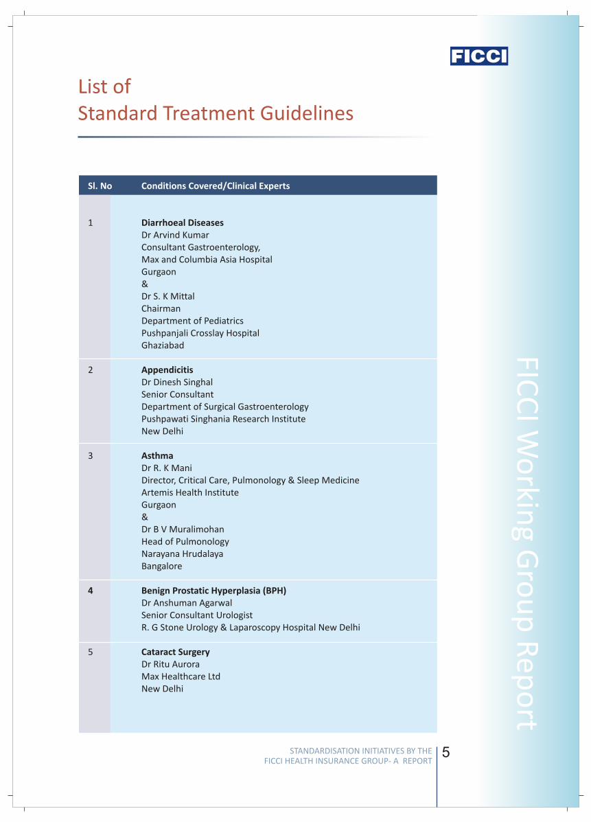
List of Standard Treatment Guidelines
Sl. No Conditions Covered/Clinical Experts
1 Diarrhoeal Diseases Dr Arvind KumarConsultant Gastroenterology,Max and Columbia Asia HospitalGurgaon& Dr S. K Mittal ChairmanDepartment of PediatricsPushpanjali Crosslay HospitalGhaziabad
2 AppendicitisDr Dinesh SinghalSenior ConsultantDepartment of Surgical GastroenterologyPushpawati Singhania Research InstituteNew Delhi
3 AsthmaDr R. K ManiDirector, Critical Care, Pulmonology & Sleep MedicineArtemis Health InstituteGurgaon & Dr B V MuralimohanHead of PulmonologyNarayana HrudalayaBangalore
4 Benign Prostatic Hyperplasia (BPH)Dr Anshuman AgarwalSenior Consultant UrologistR. G Stone Urology & Laparoscopy Hospital New Delhi
5 Cataract SurgeryDr Ritu AuroraMax Healthcare LtdNew Delhi
STANDARDISATION INITIATIVES BY THE FICCI HEALTH INSURANCE GROUP- A REPORT
STANDARDISATION INITIATIVES BY THE FICCI HEALTH INSURANCE GROUP- A REPORT
Stan
dar
d T
reat
men
t G
uid
elin
es
4
The guidelines also provide the essential investigations which need to be carried out in
case of a particular condition, as also any specific additional ones, which may be opted
for in case of specified circumstances. The guidelines also include a detailed discussion
on implants or other surgical consumables, including specific recommendations which
meet quality expectations at a reasonable cost to the system.
The commonest causes of Hospitalization based on insurance claim data were
selected under the broad categories of surgical conditions and medical conditions
requiring hospitalization, and across various specialties, to develop the standards.
In the present phase, STGs for over 20 conditions have been developed by the
group, and more conditions are expected to be taken up in due course based on the
industry’s feedback to the same.
The presentations on the recommended treatment guidelines were developed by
identified Clinical Experts based on a standard protocol (Annexure).
The group analyzed and undertook detailed discussions on each of the
presentations and their feedback was included by the lead content developer in the
revised presentation which was again presented and discussed in the group.
The finalized guidelines developed by the lead content developer were then edited
by a professional team for uniform and consistent style of presenting these
standards and the documents of STGs were created.
Peer review of the guidelines created by the clinical experts was carried out by a
cross section of other experts from the same domain, across hospitals and medical
colleges located in various parts of the country, in order to secure a professional
consensus on the guidelines and wider acceptance.
The peer review comments were incorporated in the STGs by the lead content
developer, and this document along with peer reviews received thereupon was also
vetted by an independent Technical Board constituted by FICCI.
METHODOLOGY
n
n
n
n
n
n
5
FICC
I Wo
rking G
rou
p R
epo
rtList of Standard Treatment Guidelines
Sl. No Conditions Covered/Clinical Experts
1 Diarrhoeal Diseases Dr Arvind KumarConsultant Gastroenterology,Max and Columbia Asia HospitalGurgaon& Dr S. K Mittal ChairmanDepartment of PediatricsPushpanjali Crosslay HospitalGhaziabad
2 AppendicitisDr Dinesh SinghalSenior ConsultantDepartment of Surgical GastroenterologyPushpawati Singhania Research InstituteNew Delhi
3 AsthmaDr R. K ManiDirector, Critical Care, Pulmonology & Sleep MedicineArtemis Health InstituteGurgaon & Dr B V MuralimohanHead of PulmonologyNarayana HrudalayaBangalore
4 Benign Prostatic Hyperplasia (BPH)Dr Anshuman AgarwalSenior Consultant UrologistR. G Stone Urology & Laparoscopy Hospital New Delhi
5 Cataract SurgeryDr Ritu AuroraMax Healthcare LtdNew Delhi
STANDARDISATION INITIATIVES BY THE FICCI HEALTH INSURANCE GROUP- A REPORT
STANDARDISATION INITIATIVES BY THE FICCI HEALTH INSURANCE GROUP- A REPORT
Stan
dar
d T
reat
men
t G
uid
elin
es
6
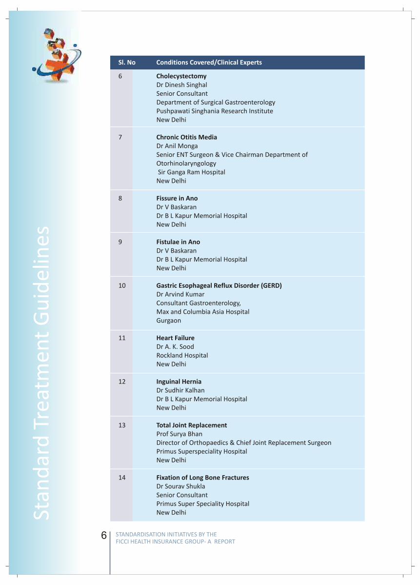
6 CholecystectomyDr Dinesh SinghalSenior ConsultantDepartment of Surgical GastroenterologyPushpawati Singhania Research InstituteNew Delhi
7 Chronic Otitis MediaDr Anil MongaSenior ENT Surgeon & Vice Chairman Department of Otorhinolaryngology Sir Ganga Ram HospitalNew Delhi
8 Fissure in AnoDr V BaskaranDr B L Kapur Memorial Hospital
New Delhi
9 Fistulae in AnoDr V BaskaranDr B L Kapur Memorial Hospital
New Delhi
10 Gastric Esophageal Reflux Disorder (GERD)Dr Arvind KumarConsultant Gastroenterology,Max and Columbia Asia HospitalGurgaon
11 Heart Failure Dr A. K. Sood Rockland Hospital New Delhi
12 Inguinal HerniaDr Sudhir KalhanDr B L Kapur Memorial Hospital New Delhi
13 Total Joint ReplacementProf Surya Bhan Director of Orthopaedics & Chief Joint Replacement SurgeonPrimus Superspeciality HospitalNew Delhi
14 Fixation of Long Bone FracturesDr Sourav ShuklaSenior ConsultantPrimus Super Speciality Hospital New Delhi
Sl. No Conditions Covered/Clinical Experts
7
FICC
I Wo
rking G
rou
p R
epo
rt
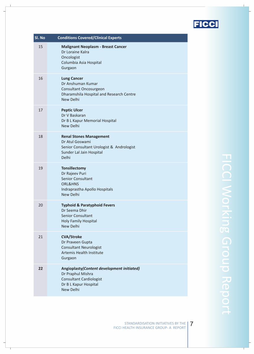
15 Malignant Neoplasm - Breast CancerDr Loraine KalraOncologistColumbia Asia HospitalGurgaon
16 Lung CancerDr Anshuman KumarConsultant OncosurgeonDharamshila Hospital and Research Centre
New Delhi
17 Peptic UlcerDr V BaskaranDr B L Kapur Memorial Hospital
New Delhi
18 Renal Stones ManagementDr Atul GoswamiSenior Consultant Urologist & Andrologist Sunder Lal Jain HospitalDelhi
19 TonsillectomyDr Rajeev PuriSenior ConsultantORL&HNS
Indraprastha Apollo Hospitals New Delhi
20 Typhoid & Paratyphoid FeversDr Seema DhirSenior Consultant Holy Family HospitalNew Delhi
21 CVA/StrokeDr Praveen GuptaConsultant NeurologistArtemis Health InstituteGurgaon
22 Angioplasty(Content development initiated)Dr Praphul MishraConsultant CardiologistDr B L Kapur HospitalNew Delhi
Sl. No Conditions Covered/Clinical Experts
STANDARDISATION INITIATIVES BY THE FICCI HEALTH INSURANCE GROUP- A REPORT
STANDARDISATION INITIATIVES BY THE FICCI HEALTH INSURANCE GROUP- A REPORT
Stan
dar
d T
reat
men
t G
uid
elin
es
6
6 CholecystectomyDr Dinesh SinghalSenior ConsultantDepartment of Surgical GastroenterologyPushpawati Singhania Research InstituteNew Delhi
7 Chronic Otitis MediaDr Anil MongaSenior ENT Surgeon & Vice Chairman Department of Otorhinolaryngology Sir Ganga Ram HospitalNew Delhi
8 Fissure in AnoDr V BaskaranDr B L Kapur Memorial Hospital
New Delhi
9 Fistulae in AnoDr V BaskaranDr B L Kapur Memorial Hospital
New Delhi
10 Gastric Esophageal Reflux Disorder (GERD)Dr Arvind KumarConsultant Gastroenterology,Max and Columbia Asia HospitalGurgaon
11 Heart Failure Dr A. K. Sood Rockland Hospital New Delhi
12 Inguinal HerniaDr Sudhir KalhanDr B L Kapur Memorial Hospital New Delhi
13 Total Joint ReplacementProf Surya Bhan Director of Orthopaedics & Chief Joint Replacement SurgeonPrimus Superspeciality HospitalNew Delhi
14 Fixation of Long Bone FracturesDr Sourav ShuklaSenior ConsultantPrimus Super Speciality Hospital New Delhi
Sl. No Conditions Covered/Clinical Experts
7
FICC
I Wo
rking G
rou
p R
epo
rt15 Malignant Neoplasm - Breast Cancer
Dr Loraine KalraOncologistColumbia Asia HospitalGurgaon
16 Lung CancerDr Anshuman KumarConsultant OncosurgeonDharamshila Hospital and Research Centre
New Delhi
17 Peptic UlcerDr V BaskaranDr B L Kapur Memorial Hospital
New Delhi
18 Renal Stones ManagementDr Atul GoswamiSenior Consultant Urologist & Andrologist Sunder Lal Jain HospitalDelhi
19 TonsillectomyDr Rajeev PuriSenior ConsultantORL&HNS
Indraprastha Apollo Hospitals New Delhi
20 Typhoid & Paratyphoid FeversDr Seema DhirSenior Consultant Holy Family HospitalNew Delhi
21 CVA/StrokeDr Praveen GuptaConsultant NeurologistArtemis Health InstituteGurgaon
22 Angioplasty(Content development initiated)Dr Praphul MishraConsultant CardiologistDr B L Kapur HospitalNew Delhi
Sl. No Conditions Covered/Clinical Experts
STANDARDISATION INITIATIVES BY THE FICCI HEALTH INSURANCE GROUP- A REPORT
STANDARDISATION INITIATIVES BY THE FICCI HEALTH INSURANCE GROUP- A REPORT
Stan
dar
d T
reat
men
t G
uid
elin
es
8
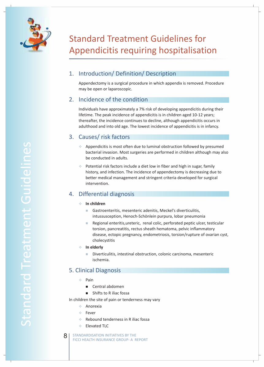
Standard Treatment Guidelines for Appendicitis requiring hospitalisation
1. Introduction/ Definition/ Description
2. Incidence of the condition
3. Causes/ risk factors
4. Differential diagnosis
5. Clinical Diagnosis
Appendectomy is a surgical procedure in which appendix is removed. Procedure
may be open or laparoscopic.
Individuals have approximately a 7% risk of developing appendicitis during their
lifetime. The peak incidence of appendicitis is in children aged 10-12 years;
thereafter, the incidence continues to decline, although appendicitis occurs in
adulthood and into old age. The lowest incidence of appendicitis is in infancy.
Appendicitis is most often due to luminal obstruction followed by presumed
bacterial invasion. Most surgeries are performed in children although may also
be conducted in adults.
Potential risk factors include a diet low in fiber and high in sugar, family
history, and infection. The incidence of appendectomy is decreasing due to
better medical management and stringent criteria developed for surgical
intervention.
Gastroenteritis, mesenteric adenitis, Meckel's diverticulitis,
intussusception, Henoch-Schönlein purpura, lobar pneumonia
Regional enteritis,ureteric, renal colic, perforated peptic ulcer, testicular
torsion, pancreatitis, rectus sheath hematoma, pelvic inflammatory
disease, ectopic pregnancy, endometriosis, torsion/rupture of ovarian cyst,
cholecystitis
In elderly
Diverticulitis, intestinal obstruction, colonic carcinoma, mesenteric
ischemia.
Pain
nCentral abdomen
nShifts to R iliac fossa
In children the site of pain or tenderness may vary
Anorexia
Fever
Rebound tenderness in R iliac fossa
Elevated TLC
v
v
v
n
n
v
n
v
v
v
v
v
In children
9
FICC
I Wo
rking G
rou
p R
epo
rt
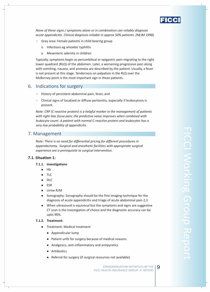
None of these signs / symptoms alone or in combination can reliably diagnose
acute appendicitis. Clinical diagnosis reliable in approx 50% patients. (NEJM 1998)
Grey area: Female patients in child bearing group
o Infections eg amoebic typhlitis
o Mesenteric adenitis in children
Typically, symptoms begin as periumbilical or epigastric pain migrating to the right
lower quadrant (RLQ) of the abdomen. Later, a worsening progressive pain along
with vomiting, nausea, and anorexia are described by the patient. Usually, a fever
is not present at this stage. Tenderness on palpation in the RLQ over the
McBurney point is the most important sign in these patients.
History of persistent abdominal pain, fever, and
Clinical signs of localized or diffuse peritonitis, especially if leukocytosis is
present.
Note: CRP (C-reactive protein) is a helpful marker in the management of patients
with right iliac fossa pain; the predictive value improves when combined with
leukocyte count. A patient with normal C-reactive protein and leukocytes has a
very low probability of appendicitis
Note: There is no need for differential pricing for different procedures in
appendectomy. Surgical and anesthetic facilities with appropriate surgical
experience are a prerequisite to surgical intervention.
7.1. Situation 1:
7.1.1. Investigations
lHb
lTLC
lDLC
lESR
lUrine-R/M
lSonography: Sonography should be the first imaging technique for the
diagnosis of acute appendicitis and triage of acute abdominal pain 2,3
lWhen ultrasound is equivocal but the symptoms and signs are suggestive
CT scan is the investigation of choice and the diagnostic accuracy can be
upto 90%.
7.1.2. Treatment:
lTreatment: Medical treatment
lAppendicular lump
lPatient unfit for surgery because of medical reasons.
lAnalgesics, anti-inflammatory and antipyretics
lAntibiotics
lReferral for surgery (if surgical resources not available)
v
v
v
6. Indications for surgery
7. Management
STANDARDISATION INITIATIVES BY THE FICCI HEALTH INSURANCE GROUP- A REPORT
STANDARDISATION INITIATIVES BY THE FICCI HEALTH INSURANCE GROUP- A REPORT
Stan
dar
d T
reat
men
t G
uid
elin
es
8
Standard Treatment Guidelines for Appendicitis requiring hospitalisation
1. Introduction/ Definition/ Description
2. Incidence of the condition
3. Causes/ risk factors
4. Differential diagnosis
5. Clinical Diagnosis
Appendectomy is a surgical procedure in which appendix is removed. Procedure
may be open or laparoscopic.
Individuals have approximately a 7% risk of developing appendicitis during their
lifetime. The peak incidence of appendicitis is in children aged 10-12 years;
thereafter, the incidence continues to decline, although appendicitis occurs in
adulthood and into old age. The lowest incidence of appendicitis is in infancy.
Appendicitis is most often due to luminal obstruction followed by presumed
bacterial invasion. Most surgeries are performed in children although may also
be conducted in adults.
Potential risk factors include a diet low in fiber and high in sugar, family
history, and infection. The incidence of appendectomy is decreasing due to
better medical management and stringent criteria developed for surgical
intervention.
Gastroenteritis, mesenteric adenitis, Meckel's diverticulitis,
intussusception, Henoch-Schönlein purpura, lobar pneumonia
Regional enteritis,ureteric, renal colic, perforated peptic ulcer, testicular
torsion, pancreatitis, rectus sheath hematoma, pelvic inflammatory
disease, ectopic pregnancy, endometriosis, torsion/rupture of ovarian cyst,
cholecystitis
In elderly
Diverticulitis, intestinal obstruction, colonic carcinoma, mesenteric
ischemia.
Pain
nCentral abdomen
nShifts to R iliac fossa
In children the site of pain or tenderness may vary
Anorexia
Fever
Rebound tenderness in R iliac fossa
Elevated TLC
v
v
v
n
n
v
n
v
v
v
v
v
In children
9
FICC
I Wo
rking G
rou
p R
epo
rtNone of these signs / symptoms alone or in combination can reliably diagnose
acute appendicitis. Clinical diagnosis reliable in approx 50% patients. (NEJM 1998)
Grey area: Female patients in child bearing group
o Infections eg amoebic typhlitis
o Mesenteric adenitis in children
Typically, symptoms begin as periumbilical or epigastric pain migrating to the right
lower quadrant (RLQ) of the abdomen. Later, a worsening progressive pain along
with vomiting, nausea, and anorexia are described by the patient. Usually, a fever
is not present at this stage. Tenderness on palpation in the RLQ over the
McBurney point is the most important sign in these patients.
History of persistent abdominal pain, fever, and
Clinical signs of localized or diffuse peritonitis, especially if leukocytosis is
present.
Note: CRP (C-reactive protein) is a helpful marker in the management of patients
with right iliac fossa pain; the predictive value improves when combined with
leukocyte count. A patient with normal C-reactive protein and leukocytes has a
very low probability of appendicitis
Note: There is no need for differential pricing for different procedures in
appendectomy. Surgical and anesthetic facilities with appropriate surgical
experience are a prerequisite to surgical intervention.
7.1. Situation 1:
7.1.1. Investigations
lHb
lTLC
lDLC
lESR
lUrine-R/M
lSonography: Sonography should be the first imaging technique for the
diagnosis of acute appendicitis and triage of acute abdominal pain 2,3
lWhen ultrasound is equivocal but the symptoms and signs are suggestive
CT scan is the investigation of choice and the diagnostic accuracy can be
upto 90%.
7.1.2. Treatment:
lTreatment: Medical treatment
lAppendicular lump
lPatient unfit for surgery because of medical reasons.
lAnalgesics, anti-inflammatory and antipyretics
lAntibiotics
lReferral for surgery (if surgical resources not available)
v
v
v
6. Indications for surgery
7. Management
STANDARDISATION INITIATIVES BY THE FICCI HEALTH INSURANCE GROUP- A REPORT
STANDARDISATION INITIATIVES BY THE FICCI HEALTH INSURANCE GROUP- A REPORT
Stan
dar
d T
reat
men
t G
uid
elin
es
10
Surgery is the main stay in the treatment of acute appendicitis. A diagnosed
case of acute appendicitis requires surgery as soon as possible.
7.1.3. Referral criteria to a specialist centre for immediate appendectomy:
lA rising pulse rate
lVomiting or increase in gastric aspiration
lIncrease in abdominal pain
lIncrease in the size of lump
7.2. Situation 2
7.2.1. Investigations:
lMinimum
o Hemogram
o Coagulation profile
o Urine- Routine (incl alb & sugar) + Microscopic
o USG – abdomen + pelvis (for all)
o Others – CxR, ECG
o CRP 1
lAcceptable for select patients
o KFT, ECG, CT scan abdomen (if any associated co-morbidity)
lIPre anesthetic checks
7.2.2. Additional investigations (with specific indications)
lICT/ MRI (in pregnancy and complicated cases and If the diagnosis is
equivocal) 4
(USG –10% in 1997 to 60% in 2007, CT scan – 0% in 1997 to 35% in 2007)
7.2.3. Treatment:
Surgical Treatment is the removal of appendix.
7.2.3.1. Procedures for Appendectomy:
§Conventional appendectomy: Immediate appendectomy should be
performed to obviate possibility of rupture of appendix and spreading
peritonitis.
§Laparoscopic appendectomy: The advantage of laparoscopic
appendectomy over conventional appendectomy is that it can be used
to confirm the diagnosis before appendectomy. Diagnostic laparoscopy
is useful in evaluating patients with right lower abdominal pain,
especially in those with equivocal signs of acute appendicitis. It also has
the additional benefit of being therapeutic. Premenopausal women
benefit the most from this procedure 5, 6, 7
§Laparoscopic appendectomy has a shorter median Length of Stay (LOS),
a trend toward less postoperative infectious complications, and fewer
clinic visits than Open Appendicectomy, which makes it a safe and
effective procedure for patients with perforated appendicitis 8
11
FICC
I Wo
rking G
rou
p R
epo
rt
§Sample should be taken for Histo Pathological Examination and report
attached with the file- this is to be statistically monitored.
7.2.3. Admission criteria:
lAcute appendicitis
lInterval appendectomy six weeks after treatment of appendicular mass
lRecurrent appendicitis
Pain management, infection control and gradual return to normal activity
Appendicular rupture, Appendicular mass, Appendicular abscess, Suppurative
pylephlebitis
1. Ortega-Deballon P, Ruiz de Adana-Belbel JC, Hernández-Matías A, García-
Septiem J, Moreno-Azcoita M.Usefulness of laboratory data in the
management of right iliac fossa pain in adults. Dis Colon Rectum. 2008
Jul;51(7):1093-9. Epub 2008 May 17.
2. Gaitini D, Beck-Razi N, Mor-Yosef D, Fischer D, Ben Itzhak O, Krausz MM, Engel
A. Diagnosing acute appendicitis in adults: accuracy of color Doppler
sonography and MDCT compared with surgery and clinical follow-up. AJR Am J
Roentgenol. 2008 May; 190(5):1300-6.
3. Mardan MA, Mufti TS, Khattak IU, Chilkunda N, Alshayeb AA, Mohammad AM,
ur Rehman Z. Role of ultrasound in acute appendicitis.J Ayub Med Coll
Abbottabad. 2007 Jul-Sep; 19(3):72-9.
4. Israel GM, Malguria N, McCarthy S, Copel J, Weinreb J. MRI vs. ultrasound for
suspected appendicitis during pregnancy. J Magn Reson Imaging. 2008 Aug;
28(2):428-33.
5. Lim GH, Shabbir A, So JB. Diagnostic laparoscopy in the evaluation of right
lower abdominal pain: a one-year audit. Singapore Med J. 2008 Jun;49(6):451-
3.
6. Ates M, Sevil S, Bulbul M. Routine use of laparoscopy in patients with clinically
doubtful diagnosis of appendicitis. J Laparoendosc Adv Surg Tech A. 2008
Apr;18(2):189-93.
7. Utpal D. Laparoscopic versus open appendectomy in West Bengal, India. Chin J
Dig Dis. 2005; 6(4):165-9.
8. Taqi E, Al Hadher S, Ryckman J, Su W, Aspirot A, Puligandla P, Flageole H,
Laberge JM. Outcome of laparoscopic appendectomy for perforated
appendicitis in children. J Pediatr Surg. 2008 May;43(5):893-5
It was suggested that there could be no single modality for the surgery and it
could either be classic open procedure or laparoscopic depending on
v
8. Post Operative Care
9. Complications
10. References
Important Information on this Procedure
STANDARDISATION INITIATIVES BY THE FICCI HEALTH INSURANCE GROUP- A REPORT
STANDARDISATION INITIATIVES BY THE FICCI HEALTH INSURANCE GROUP- A REPORT
Stan
dar
d T
reat
men
t G
uid
elin
es
10
Surgery is the main stay in the treatment of acute appendicitis. A diagnosed
case of acute appendicitis requires surgery as soon as possible.
7.1.3. Referral criteria to a specialist centre for immediate appendectomy:
lA rising pulse rate
lVomiting or increase in gastric aspiration
lIncrease in abdominal pain
lIncrease in the size of lump
7.2. Situation 2
7.2.1. Investigations:
lMinimum
o Hemogram
o Coagulation profile
o Urine- Routine (incl alb & sugar) + Microscopic
o USG – abdomen + pelvis (for all)
o Others – CxR, ECG
o CRP 1
lAcceptable for select patients
o KFT, ECG, CT scan abdomen (if any associated co-morbidity)
lIPre anesthetic checks
7.2.2. Additional investigations (with specific indications)
lICT/ MRI (in pregnancy and complicated cases and If the diagnosis is
equivocal) 4
(USG –10% in 1997 to 60% in 2007, CT scan – 0% in 1997 to 35% in 2007)
7.2.3. Treatment:
Surgical Treatment is the removal of appendix.
7.2.3.1. Procedures for Appendectomy:
§Conventional appendectomy: Immediate appendectomy should be
performed to obviate possibility of rupture of appendix and spreading
peritonitis.
§Laparoscopic appendectomy: The advantage of laparoscopic
appendectomy over conventional appendectomy is that it can be used
to confirm the diagnosis before appendectomy. Diagnostic laparoscopy
is useful in evaluating patients with right lower abdominal pain,
especially in those with equivocal signs of acute appendicitis. It also has
the additional benefit of being therapeutic. Premenopausal women
benefit the most from this procedure 5, 6, 7
§Laparoscopic appendectomy has a shorter median Length of Stay (LOS),
a trend toward less postoperative infectious complications, and fewer
clinic visits than Open Appendicectomy, which makes it a safe and
effective procedure for patients with perforated appendicitis 8
11
FICC
I Wo
rking G
rou
p R
epo
rt§Sample should be taken for Histo Pathological Examination and report
attached with the file- this is to be statistically monitored.
7.2.3. Admission criteria:
lAcute appendicitis
lInterval appendectomy six weeks after treatment of appendicular mass
lRecurrent appendicitis
Pain management, infection control and gradual return to normal activity
Appendicular rupture, Appendicular mass, Appendicular abscess, Suppurative
pylephlebitis
1. Ortega-Deballon P, Ruiz de Adana-Belbel JC, Hernández-Matías A, García-
Septiem J, Moreno-Azcoita M.Usefulness of laboratory data in the
management of right iliac fossa pain in adults. Dis Colon Rectum. 2008
Jul;51(7):1093-9. Epub 2008 May 17.
2. Gaitini D, Beck-Razi N, Mor-Yosef D, Fischer D, Ben Itzhak O, Krausz MM, Engel
A. Diagnosing acute appendicitis in adults: accuracy of color Doppler
sonography and MDCT compared with surgery and clinical follow-up. AJR Am J
Roentgenol. 2008 May; 190(5):1300-6.
3. Mardan MA, Mufti TS, Khattak IU, Chilkunda N, Alshayeb AA, Mohammad AM,
ur Rehman Z. Role of ultrasound in acute appendicitis.J Ayub Med Coll
Abbottabad. 2007 Jul-Sep; 19(3):72-9.
4. Israel GM, Malguria N, McCarthy S, Copel J, Weinreb J. MRI vs. ultrasound for
suspected appendicitis during pregnancy. J Magn Reson Imaging. 2008 Aug;
28(2):428-33.
5. Lim GH, Shabbir A, So JB. Diagnostic laparoscopy in the evaluation of right
lower abdominal pain: a one-year audit. Singapore Med J. 2008 Jun;49(6):451-
3.
6. Ates M, Sevil S, Bulbul M. Routine use of laparoscopy in patients with clinically
doubtful diagnosis of appendicitis. J Laparoendosc Adv Surg Tech A. 2008
Apr;18(2):189-93.
7. Utpal D. Laparoscopic versus open appendectomy in West Bengal, India. Chin J
Dig Dis. 2005; 6(4):165-9.
8. Taqi E, Al Hadher S, Ryckman J, Su W, Aspirot A, Puligandla P, Flageole H,
Laberge JM. Outcome of laparoscopic appendectomy for perforated
appendicitis in children. J Pediatr Surg. 2008 May;43(5):893-5
It was suggested that there could be no single modality for the surgery and it
could either be classic open procedure or laparoscopic depending on
v
8. Post Operative Care
9. Complications
10. References
Important Information on this Procedure
STANDARDISATION INITIATIVES BY THE FICCI HEALTH INSURANCE GROUP- A REPORT
STANDARDISATION INITIATIVES BY THE FICCI HEALTH INSURANCE GROUP- A REPORT
Stan
dar
d T
reat
men
t G
uid
elin
es
12
surgeon's choice and the circumstances. However, this may have cost
implications for the Insurance industry, as laparoscopic is more expensive but
can be compensated by a swifter discharge. More details on this will be
incorporated by the expert concerned.
High incidence of negative appendicectomies globally resulting in
unnecessary costs and hospital admissions.
Patient care issues
Negative appendicectomy (NA) rate of 20 – 40%
Health care issues
Un-necessary hospital admissions
Costs
Note: 300,000 appendectomies in the US annually. If NA rate is 15%,
45,000 procedures are un- necessary!!
Introduction of cross sectional imaging
USG -10% in 1997 to 60% in 2007
CT scan - 0% in 1997 to 35% in 2007
NEJM 1998 - the landmark study - 100 patients
Avoid 13 NA ( cost saving of $ 47,281)
Avoid un-necessary admissions (saving of $20,250)
Cost of 100 appendiceal CT ($ 22800)
Net saving of $ 447 per patient ($44700)
Negative Appendicectomy (3540 patients, 2006-7)
No imaging 9.8%
US - 8.1%
CT - 6%
Negative Appendecectomy is closely linked to US/ CT accuracy.
Imaging accuracy for Acute Appendecitis is a measure of quality (Ann Surg 2008).
Negative Appendecectomy rate is a measure of quality of health services.
v
v
v
v
v
v
v
v
v
v
13
FICC
I Wo
rking G
rou
p R
epo
rt
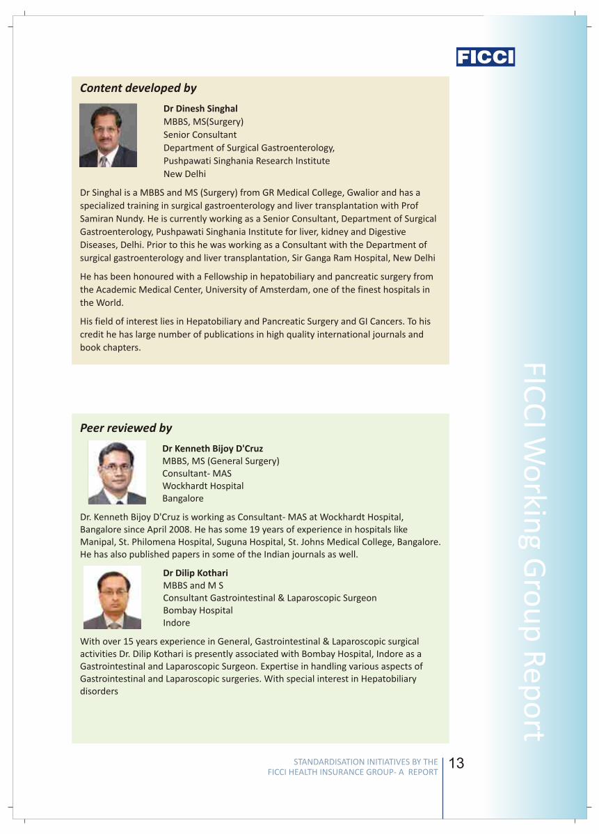
Content developed by
Dr Dinesh Singhal
MBBS, MS(Surgery)
Senior Consultant
Department of Surgical Gastroenterology,
Pushpawati Singhania Research Institute
New Delhi
Dr Singhal is a MBBS and MS (Surgery) from GR Medical College, Gwalior and has a
specialized training in surgical gastroenterology and liver transplantation with Prof
Samiran Nundy. He is currently working as a Senior Consultant, Department of Surgical
Gastroenterology, Pushpawati Singhania Institute for liver, kidney and Digestive
Diseases, Delhi. Prior to this he was working as a Consultant with the Department of
surgical gastroenterology and liver transplantation, Sir Ganga Ram Hospital, New Delhi
He has been honoured with a Fellowship in hepatobiliary and pancreatic surgery from
the Academic Medical Center, University of Amsterdam, one of the finest hospitals in
the World.
His field of interest lies in Hepatobiliary and Pancreatic Surgery and GI Cancers. To his
credit he has large number of publications in high quality international journals and
book chapters.
Peer reviewed by
Dr Kenneth Bijoy D'CruzMBBS, MS (General Surgery)Consultant- MASWockhardt HospitalBangalore
Dr. Kenneth Bijoy D'Cruz is working as Consultant- MAS at Wockhardt Hospital, Bangalore since April 2008. He has some 19 years of experience in hospitals like Manipal, St. Philomena Hospital, Suguna Hospital, St. Johns Medical College, Bangalore. He has also published papers in some of the Indian journals as well.
Dr Dilip KothariMBBS and M SConsultant Gastrointestinal & Laparoscopic SurgeonBombay HospitalIndore
With over 15 years experience in General, Gastrointestinal & Laparoscopic surgical activities Dr. Dilip Kothari is presently associated with Bombay Hospital, Indore as a Gastrointestinal and Laparoscopic Surgeon. Expertise in handling various aspects of Gastrointestinal and Laparoscopic surgeries. With special interest in Hepatobiliary disorders
STANDARDISATION INITIATIVES BY THE FICCI HEALTH INSURANCE GROUP- A REPORT
STANDARDISATION INITIATIVES BY THE FICCI HEALTH INSURANCE GROUP- A REPORT
Stan
dar
d T
reat
men
t G
uid
elin
es
12
surgeon's choice and the circumstances. However, this may have cost
implications for the Insurance industry, as laparoscopic is more expensive but
can be compensated by a swifter discharge. More details on this will be
incorporated by the expert concerned.
High incidence of negative appendicectomies globally resulting in
unnecessary costs and hospital admissions.
Patient care issues
Negative appendicectomy (NA) rate of 20 – 40%
Health care issues
Un-necessary hospital admissions
Costs
Note: 300,000 appendectomies in the US annually. If NA rate is 15%,
45,000 procedures are un- necessary!!
Introduction of cross sectional imaging
USG -10% in 1997 to 60% in 2007
CT scan - 0% in 1997 to 35% in 2007
NEJM 1998 - the landmark study - 100 patients
Avoid 13 NA ( cost saving of $ 47,281)
Avoid un-necessary admissions (saving of $20,250)
Cost of 100 appendiceal CT ($ 22800)
Net saving of $ 447 per patient ($44700)
Negative Appendicectomy (3540 patients, 2006-7)
No imaging 9.8%
US - 8.1%
CT - 6%
Negative Appendecectomy is closely linked to US/ CT accuracy.
Imaging accuracy for Acute Appendecitis is a measure of quality (Ann Surg 2008).
Negative Appendecectomy rate is a measure of quality of health services.
v
v
v
v
v
v
v
v
v
v
13
FICC
I Wo
rking G
rou
p R
epo
rtContent developed by
Dr Dinesh Singhal
MBBS, MS(Surgery)
Senior Consultant
Department of Surgical Gastroenterology,
Pushpawati Singhania Research Institute
New Delhi
Dr Singhal is a MBBS and MS (Surgery) from GR Medical College, Gwalior and has a
specialized training in surgical gastroenterology and liver transplantation with Prof
Samiran Nundy. He is currently working as a Senior Consultant, Department of Surgical
Gastroenterology, Pushpawati Singhania Institute for liver, kidney and Digestive
Diseases, Delhi. Prior to this he was working as a Consultant with the Department of
surgical gastroenterology and liver transplantation, Sir Ganga Ram Hospital, New Delhi
He has been honoured with a Fellowship in hepatobiliary and pancreatic surgery from
the Academic Medical Center, University of Amsterdam, one of the finest hospitals in
the World.
His field of interest lies in Hepatobiliary and Pancreatic Surgery and GI Cancers. To his
credit he has large number of publications in high quality international journals and
book chapters.
Peer reviewed by
Dr Kenneth Bijoy D'CruzMBBS, MS (General Surgery)Consultant- MASWockhardt HospitalBangalore
Dr. Kenneth Bijoy D'Cruz is working as Consultant- MAS at Wockhardt Hospital, Bangalore since April 2008. He has some 19 years of experience in hospitals like Manipal, St. Philomena Hospital, Suguna Hospital, St. Johns Medical College, Bangalore. He has also published papers in some of the Indian journals as well.
Dr Dilip KothariMBBS and M SConsultant Gastrointestinal & Laparoscopic SurgeonBombay HospitalIndore
With over 15 years experience in General, Gastrointestinal & Laparoscopic surgical activities Dr. Dilip Kothari is presently associated with Bombay Hospital, Indore as a Gastrointestinal and Laparoscopic Surgeon. Expertise in handling various aspects of Gastrointestinal and Laparoscopic surgeries. With special interest in Hepatobiliary disorders
STANDARDISATION INITIATIVES BY THE FICCI HEALTH INSURANCE GROUP- A REPORT
STANDARDISATION INITIATIVES BY THE FICCI HEALTH INSURANCE GROUP- A REPORT
Stan
dar
d T
reat
men
t G
uid
elin
es
14 15
FICC
I Wo
rking G
rou
p R
epo
rt
Dr U Vasudeva RaoConsultant General Surgery Manipal HospitalBangalore
Dr Rao has around three decades of experience in general & laparoscopic surgery and his special interest lie in vascular surgery. He has fair amount of administrative experience and was in charge of one of the units of Manipal Health Systems (North side Hospital) for a brief period. He is also a member of various committees in the hospital and functioned as secretary of the Academic Society during the initial period. He has conducted more than 3000 operations during his professional career at Manipal Hospital with good results. To his credit he has published many articles in journals and has delivered quite a number of guest lectures at various places within the country and abroad.
Dr B S S SainadhMBBS, DNB (Surgery)Consultant SurgeonApollo HospitalHyderabad
Dr B S S Sainadh is Consultant Surgeon in Apollo Hospital, Hyderabad. He is also teaching faculty for surgical DNB at the hospital. His area of interest includes Laparoscopic Surgery.
Dr Randeep Wadhawan MS, FIAGES, FMAS, FAISSenior Consultant and Incharge Department of Minimal AccessSurgery and Bariatric SurgeryFortis HospitalNew Delhi
He is an acclaimed surgeon with vast experience in the field of laparoscopic Gastrointestinal surgery. He has several academic achievements including international presentations to his credit. His area of interest is Bariatric (Weight loss) surgery.
Standard Treatment Guidelines forAsthma requiring hospitalisation
1. Introduction/ Definition/ Description
2. Prevalence of the condition
3. Differential Diagnosis
4. Clinical Diagnosis
5. Causes
(3,4)
Asthma is a chronic inflammatory disorder of the airways. In susceptible
individuals, this inflammation causes recurrent episodes of wheezing,
breathlessness, chest tightness and coughing, particularly at night or in the early
morning.
(5)
Asthma prevalence varies from region to region, but worldwide probably is about
8-12%. It is commoner in boys than girls, before the age of 14, after which the
gender gap narrows, and among adults, the prevalence among women may
actually be higher.
(3,4)
chronic obstructive pulmonary disease (COPD) (chronic bronchitis and
emphysema),
congestive heart failure
gastroesophageal reflux disease
mechanical obstruction of the airways
tumor/neoplasm and
vocal cord dysfunction
(3,4,5,6)
A clinical diagnosis of asthma is based mainly on symptoms (recurrent eoisodes of
cough, wheeze, chest tightness and breathlessness, often worse at night and in the
early morning), supported by physical examination (bilateral polyphonic wheezes),
and confirmed by laboratory examination (eosinophilia and reversible airflow
obstruction on spirometry).
(3,6)
Genetic factors, including genes for atopy and bronchial hyperresponsiveness
Environmental factors, including specific allergens (indoor and outdoor), non-
specific irritants including cold air, pollution and tobacco smoke, and
occupational sensitizers/agents. These environmental factors may often act as
triggers of an asthma attack.
Others
o Viral respiratory infections
o Aspirin or nonsteroidal anti-inflammatory drug hypersensitivity
n
n
n
n
n
n
n
n
n
STANDARDISATION INITIATIVES BY THE FICCI HEALTH INSURANCE GROUP- A REPORT
STANDARDISATION INITIATIVES BY THE FICCI HEALTH INSURANCE GROUP- A REPORT
Stan
dar
d T
reat
men
t G
uid
elin
es
14 15
FICC
I Wo
rking G
rou
p R
epo
rtDr U Vasudeva RaoConsultant General Surgery Manipal HospitalBangalore
Dr Rao has around three decades of experience in general & laparoscopic surgery and his special interest lie in vascular surgery. He has fair amount of administrative experience and was in charge of one of the units of Manipal Health Systems (North side Hospital) for a brief period. He is also a member of various committees in the hospital and functioned as secretary of the Academic Society during the initial period. He has conducted more than 3000 operations during his professional career at Manipal Hospital with good results. To his credit he has published many articles in journals and has delivered quite a number of guest lectures at various places within the country and abroad.
Dr B S S SainadhMBBS, DNB (Surgery)Consultant SurgeonApollo HospitalHyderabad
Dr B S S Sainadh is Consultant Surgeon in Apollo Hospital, Hyderabad. He is also teaching faculty for surgical DNB at the hospital. His area of interest includes Laparoscopic Surgery.
Dr Randeep Wadhawan MS, FIAGES, FMAS, FAISSenior Consultant and Incharge Department of Minimal AccessSurgery and Bariatric SurgeryFortis HospitalNew Delhi
He is an acclaimed surgeon with vast experience in the field of laparoscopic Gastrointestinal surgery. He has several academic achievements including international presentations to his credit. His area of interest is Bariatric (Weight loss) surgery.
Standard Treatment Guidelines forAsthma requiring hospitalisation
1. Introduction/ Definition/ Description
2. Prevalence of the condition
3. Differential Diagnosis
4. Clinical Diagnosis
5. Causes
(3,4)
Asthma is a chronic inflammatory disorder of the airways. In susceptible
individuals, this inflammation causes recurrent episodes of wheezing,
breathlessness, chest tightness and coughing, particularly at night or in the early
morning.
(5)
Asthma prevalence varies from region to region, but worldwide probably is about
8-12%. It is commoner in boys than girls, before the age of 14, after which the
gender gap narrows, and among adults, the prevalence among women may
actually be higher.
(3,4)
chronic obstructive pulmonary disease (COPD) (chronic bronchitis and
emphysema),
congestive heart failure
gastroesophageal reflux disease
mechanical obstruction of the airways
tumor/neoplasm and
vocal cord dysfunction
(3,4,5,6)
A clinical diagnosis of asthma is based mainly on symptoms (recurrent eoisodes of
cough, wheeze, chest tightness and breathlessness, often worse at night and in the
early morning), supported by physical examination (bilateral polyphonic wheezes),
and confirmed by laboratory examination (eosinophilia and reversible airflow
obstruction on spirometry).
(3,6)
Genetic factors, including genes for atopy and bronchial hyperresponsiveness
Environmental factors, including specific allergens (indoor and outdoor), non-
specific irritants including cold air, pollution and tobacco smoke, and
occupational sensitizers/agents. These environmental factors may often act as
triggers of an asthma attack.
Others
o Viral respiratory infections
o Aspirin or nonsteroidal anti-inflammatory drug hypersensitivity
n
n
n
n
n
n
n
n
n
STANDARDISATION INITIATIVES BY THE FICCI HEALTH INSURANCE GROUP- A REPORT
STANDARDISATION INITIATIVES BY THE FICCI HEALTH INSURANCE GROUP- A REPORT
Stan
dar
d T
reat
men
t G
uid
elin
es
16 17
FICC
I Wo
rking G
rou
p R
epo
rt
o Use of beta-adrenergic receptor blockers
o Occupational exposure
o Irritants such as household sprays and paint fumes
o Emotional factors or stress
o Obesity
6.1. Situation 1: At secondary hospital/non metro situation
6.1.1. Investigations:
All management must include some mandatory investigations to confirm the
diagnosis and to exclude other conditions that may mimic asthma. These include:
lHb, TLC, DLC
lBlood Sugar, Urea, Creatinine, Electrolytes
lPulse Oximetry
lArterial Blood Gases, If Available
lChest X ray, PFT, Peak flow – PEFR (if PFT is not available)
Additional investigations (with specific indicators)
lECG, Echocardiogram (Indications: when cardiac/hemodynamic instability is
suspected)
lBlood And Sputum Culture (Indications: to identify accompanying Infections)
6.1.2. Treatment
lOxygen Supplementation
lNebulized Bronchodilators ( Salbutamol/ Terbutaline/ Beclomethasone)
lInjectable Corticosteroids (Hydrocortisone/Methylprednisolone)
lTheophylline group (Deriphyllin/Etophylline)
lInjectible Magnesium, Inhaled SABA ipratropium with where indicated
lAntibiotics- To be used to treat respiratory infections.
Only those not on the “restricted antibiotics” list. Note: If drugs on the list are
prescribed, justification required
lCommon antibiotics used: amoxycillin, ofloxacin, azithromycin, augmentin
Referral criteria for a specialist center if:
lMechanical ventilation indicated
lPresence of co-morbid conditions or associated complications where closer
monitoring or greater expertise/ facilities are required
lFor optimal investigations and treatment
6. Management
6.2. Situation 2: At Super Specialty Facility in Metro location where higher end
technology is available
6.2.1. Investigation: All investigations of situation 1 and:
lArterial Blood Gases analysis
lSpirometry: FEV1 or PEF 60-80% or <60% predicted, PEF or FEV1 variability >
30% where patient is able to perform spirometry, and it can be done in the
intensive care unit without delay
6.2.2. Treatment: All treatment of situation 1 and:
lHospitalization
lNon invasive ventilation , if available , else Consider Endotracheal intubation
and mechanical ventilation
6.2.2.1. Indications of Hospitalization:
lMarked increase in intensity of symptoms
lFailure of exacerbations to respond to initial medical management (3
nebulisations at 20 minute intervals)
lFrequent exacerbations, exhaustion or confusion
lPresence of co-morbid conditions known to exacerbate asthama.
6.2.2.2. Indications for Mechanical Ventilation / intubation in Asthma: Very few
admitted patients with asthma require mechanical ventilation (about 2% of
Hospitalizations) as compared with patients with COPD exacerbations.
lSigns of respiratory muscle fatigue
lRising PCO2; paradoxic respiration; sense of exhaustion
lRespiratory rate >40/min
lFall in Ph<7.25
lAltered mental status
lPersistent hypoxia
lPresence of serious comorbid conditions
6.2.3 Standard requirements for mechanical ventilation
Under these circumstances patient may need to be monitored closely. The
standard requirements for mechanical ventilation support management would
include:
Mechanical ventilator
Central venous/pulmonary arterial catheter placement for hemodynamic
monitoring
Arterial line for blood pressure monitoring
Indwelling catheter for urine output monitoring
Echocardiogram for Hemodynamic assessment
IV fluids, vasopressors (in case of shock)
IV sedation (in selective cases only) and paralytic agents
n
n
n
n
n
n
n
STANDARDISATION INITIATIVES BY THE FICCI HEALTH INSURANCE GROUP- A REPORT
STANDARDISATION INITIATIVES BY THE FICCI HEALTH INSURANCE GROUP- A REPORT
Stan
dar
d T
reat
men
t G
uid
elin
es
16 17
FICC
I Wo
rking G
rou
p R
epo
rto Use of beta-adrenergic receptor blockers
o Occupational exposure
o Irritants such as household sprays and paint fumes
o Emotional factors or stress
o Obesity
6.1. Situation 1: At secondary hospital/non metro situation
6.1.1. Investigations:
All management must include some mandatory investigations to confirm the
diagnosis and to exclude other conditions that may mimic asthma. These include:
lHb, TLC, DLC
lBlood Sugar, Urea, Creatinine, Electrolytes
lPulse Oximetry
lArterial Blood Gases, If Available
lChest X ray, PFT, Peak flow – PEFR (if PFT is not available)
Additional investigations (with specific indicators)
lECG, Echocardiogram (Indications: when cardiac/hemodynamic instability is
suspected)
lBlood And Sputum Culture (Indications: to identify accompanying Infections)
6.1.2. Treatment
lOxygen Supplementation
lNebulized Bronchodilators ( Salbutamol/ Terbutaline/ Beclomethasone)
lInjectable Corticosteroids (Hydrocortisone/Methylprednisolone)
lTheophylline group (Deriphyllin/Etophylline)
lInjectible Magnesium, Inhaled SABA ipratropium with where indicated
lAntibiotics- To be used to treat respiratory infections.
Only those not on the “restricted antibiotics” list. Note: If drugs on the list are
prescribed, justification required
lCommon antibiotics used: amoxycillin, ofloxacin, azithromycin, augmentin
Referral criteria for a specialist center if:
lMechanical ventilation indicated
lPresence of co-morbid conditions or associated complications where closer
monitoring or greater expertise/ facilities are required
lFor optimal investigations and treatment
6. Management
6.2. Situation 2: At Super Specialty Facility in Metro location where higher end
technology is available
6.2.1. Investigation: All investigations of situation 1 and:
lArterial Blood Gases analysis
lSpirometry: FEV1 or PEF 60-80% or <60% predicted, PEF or FEV1 variability >
30% where patient is able to perform spirometry, and it can be done in the
intensive care unit without delay
6.2.2. Treatment: All treatment of situation 1 and:
lHospitalization
lNon invasive ventilation , if available , else Consider Endotracheal intubation
and mechanical ventilation
6.2.2.1. Indications of Hospitalization:
lMarked increase in intensity of symptoms
lFailure of exacerbations to respond to initial medical management (3
nebulisations at 20 minute intervals)
lFrequent exacerbations, exhaustion or confusion
lPresence of co-morbid conditions known to exacerbate asthama.
6.2.2.2. Indications for Mechanical Ventilation / intubation in Asthma: Very few
admitted patients with asthma require mechanical ventilation (about 2% of
Hospitalizations) as compared with patients with COPD exacerbations.
lSigns of respiratory muscle fatigue
lRising PCO2; paradoxic respiration; sense of exhaustion
lRespiratory rate >40/min
lFall in Ph<7.25
lAltered mental status
lPersistent hypoxia
lPresence of serious comorbid conditions
6.2.3 Standard requirements for mechanical ventilation
Under these circumstances patient may need to be monitored closely. The
standard requirements for mechanical ventilation support management would
include:
Mechanical ventilator
Central venous/pulmonary arterial catheter placement for hemodynamic
monitoring
Arterial line for blood pressure monitoring
Indwelling catheter for urine output monitoring
Echocardiogram for Hemodynamic assessment
IV fluids, vasopressors (in case of shock)
IV sedation (in selective cases only) and paralytic agents
n
n
n
n
n
n
n
STANDARDISATION INITIATIVES BY THE FICCI HEALTH INSURANCE GROUP- A REPORT
STANDARDISATION INITIATIVES BY THE FICCI HEALTH INSURANCE GROUP- A REPORT
Stan
dar
d T
reat
men
t G
uid
elin
es
18 19
FICC
I Wo
rking G
rou
p R
epo
rt
7. Complications
8. Rehabilitation measures
9. References
(8)
Cor pulmonale
Respiratory failure
Infection and Sepsis
Multi organ Failure
Chest physiotherapy and mobilization (Advice for postural drainage, breathing
exercises and activity maintenance)
Inspiratory muscle training with spirometric devices (where facilities and
resources available)
Pre-discharge assessment:
Checklist:
Has the patient been off nebulised medication and on inhaled medication for at least 24
hours pre-discharge (unless he/she has a home nebuliser)?
Is the peak flow > 75 % predicted/ previous best?
Is the daily variation of peak flow < 25%?
Has an inhaler assessment been carried out to assess inhaler preference and
correctness of use?
Has the patient received a written self management plan?
Does the patient have an written contact number for medical emergencies?
Has the patient been considered for vaccinations (Influenza/ pneumococcal)?
Has an assessment of smoking status been done, and if a smoker, has smoking cessation
advice been given/ arranged?
Has a follow-up date/time been arranged?
1. Oddo M, Feidel F et al. Intensive care medicine 2006, 32:501-510
2. Mcfadden ER Jr (2003) Acute Severe Asthma. Am J Respir Crit Care Med
168:740–759
3. Guidelines for Management of Asthma at Primary and Secondary Levels of Health
Care in India (2005)- A consensus statement developed under the World Health
Organization and Government of India Collaborative Programme (2004-2005).
Indian J Chest Dis Allied Sci 2005; 47: 309-343
4. DG Jain, RK Singal, et al. Understanding and managing acute severe and difficult
Asthma. JIACM 2006; 7(4): 316-27
5. Siddharth N. Shah, M. Paul Anand, editors. API.Textbook of Medicine. 7th ed.
Mumbai, India: The Association of Physicians of India; 2003
n
n
n
n
n
n
n
Pneumothorax
6. Rahnama'i MS, Geilen RP, Singhi S, van den Akker M, Chavannes NH. Which clinical
signs and symptoms predict hypoxemia in acute childhood asthma? Indian J
Pediatr 2006;73:771-5
7. Standard treatment guidelines (Medical Management and Costing of Select
Conditions)Developed by Armed Forces Medical College In collaboration with
Ministry of Health and Family Welfare, Government of India and World Health
Organization, India Office
8. Col SP Rai, Col AP Patil et al. Best treatment guidelines for bronchial asthma.
MJAFI 2007; 63 : 264-268
STANDARDISATION INITIATIVES BY THE FICCI HEALTH INSURANCE GROUP- A REPORT
STANDARDISATION INITIATIVES BY THE FICCI HEALTH INSURANCE GROUP- A REPORT
Stan
dar
d T
reat
men
t G
uid
elin
es
18 19
FICC
I Wo
rking G
rou
p R
epo
rt7. Complications
8. Rehabilitation measures
9. References
(8)
Cor pulmonale
Respiratory failure
Infection and Sepsis
Multi organ Failure
Chest physiotherapy and mobilization (Advice for postural drainage, breathing
exercises and activity maintenance)
Inspiratory muscle training with spirometric devices (where facilities and
resources available)
Pre-discharge assessment:
Checklist:
Has the patient been off nebulised medication and on inhaled medication for at least 24
hours pre-discharge (unless he/she has a home nebuliser)?
Is the peak flow > 75 % predicted/ previous best?
Is the daily variation of peak flow < 25%?
Has an inhaler assessment been carried out to assess inhaler preference and
correctness of use?
Has the patient received a written self management plan?
Does the patient have an written contact number for medical emergencies?
Has the patient been considered for vaccinations (Influenza/ pneumococcal)?
Has an assessment of smoking status been done, and if a smoker, has smoking cessation
advice been given/ arranged?
Has a follow-up date/time been arranged?
1. Oddo M, Feidel F et al. Intensive care medicine 2006, 32:501-510
2. Mcfadden ER Jr (2003) Acute Severe Asthma. Am J Respir Crit Care Med
168:740–759
3. Guidelines for Management of Asthma at Primary and Secondary Levels of Health
Care in India (2005)- A consensus statement developed under the World Health
Organization and Government of India Collaborative Programme (2004-2005).
Indian J Chest Dis Allied Sci 2005; 47: 309-343
4. DG Jain, RK Singal, et al. Understanding and managing acute severe and difficult
Asthma. JIACM 2006; 7(4): 316-27
5. Siddharth N. Shah, M. Paul Anand, editors. API.Textbook of Medicine. 7th ed.
Mumbai, India: The Association of Physicians of India; 2003
n
n
n
n
n
n
n
Pneumothorax
6. Rahnama'i MS, Geilen RP, Singhi S, van den Akker M, Chavannes NH. Which clinical
signs and symptoms predict hypoxemia in acute childhood asthma? Indian J
Pediatr 2006;73:771-5
7. Standard treatment guidelines (Medical Management and Costing of Select
Conditions)Developed by Armed Forces Medical College In collaboration with
Ministry of Health and Family Welfare, Government of India and World Health
Organization, India Office
8. Col SP Rai, Col AP Patil et al. Best treatment guidelines for bronchial asthma.
MJAFI 2007; 63 : 264-268
STANDARDISATION INITIATIVES BY THE FICCI HEALTH INSURANCE GROUP- A REPORT
STANDARDISATION INITIATIVES BY THE FICCI HEALTH INSURANCE GROUP- A REPORT
Stan
dar
d T
reat
men
t G
uid
elin
es
20 21
Content developed by
Dr R K Mani
Director- Critical Care, Pulmonology & Sleep Medicine
Artemis Health Institute
Gurgaon
Dr R. K Mani has more than 24 years of experience in the fields of Medicine,
Pulmonology and Critical Care in the capacity of Director of Pulmonology, Critical Care
and Sleep Medicine, Fortis Group of Hospitals, New Delhi, Delhi Heart & Lung Institute,
Panchkuian Road, New Delhi and at Indraprastha Apollo Hospital, New Delhi he was
Consultant and Head of Department.
He played a leadership role in setting up the Department of Medicine and Critical Care
at Batra Hospital It was one of the first Critical Care Units in the private sector in Delhi.
He has also been involved in the training of ICU staff and nurses.
In the early 90's he pioneered the introduction of noninvasive ventilatory support. His
early experience with NIPPV for acute respiratory failure and COPD was later shared
nationally and internationally.This led to Noninvasive ventilation later being widely
accepted throughout the country. The experience in noninvasive ventilation was also
later extended to include domiciliary ventilatory support of patients with chronic
respiratory failure due to COPD, neuromuscular disorders or chest wall disorders. His
contributions made in this field was recognized widely and led to scores of invitations
for lectures and workshops across the country. In 1996, he was invited to deliver a
lecture on the Indian experience in NIV at the Royal Prince Alfred Hospital in Sydney,
Australia where he did 2-week training in sleep Medicine with the renowned Prof. Colin
E Sullivan the inventor of CPAP.
He has received an Award for distinguished services in the Medical profession, Delhi
Medical Association, Sep. 1996
Dr B V Murali Mohan
Consultant Pulmonologist and Head
Departments of Internal Medicine and Pulmonology
Narayana Hrudayalaya
Bangalore
Dr B V Murali Mohan is currently working as Consultant Pulmonologist and Head of the
departments of Internal Medicine and Pulmonology at Narayana Hrudayalaya,
Bangalore. He was earlier Professor and Head of the Department of Medicine at Dr B R
Ambedkar Medical College, Bangalore.
After completing his MBBS and MD (General Medicine) at Bangalore Medical College,
he took his MRCP (UK) from the Royal College of Physicians of Edinburgh, and post-
MRCP training in Respiratory Medicine at Ninewells and Kings Cross Hospitals, Dundee,
and at the Newcastle General Hospital, Newcastle upon Tyne.
He has over 30 papers presented at various national and international conferences, and
3 papers in international journals. He also heads the Nightingales Lifesaving Services.
FICC
I Wo
rking G
rou
p R
epo
rt
Peer reviewed by
Dr Manish JainMD, DNB, MRCP (UK)ConsultantBombay HospitalIndore
Dr Manish Jain is a Consultant at Bombay Hospital, Indore. His qualification includes MD from Medical College Raipur, DNB from National Board, MRCP from Royal College of Physicians of London, U.K
Dr Issac MathewMBBS, MD (Medicine), MD (Respiratory Medicine &Tuberculosis)Senior Consultant Respiratory MedicineManipal HospitalBangalore
Dr Issac Mathew is currently working as Senior Consultant Respiratory Medicine, Manipal Hospital Bangalore. His past appointments have been as Director Critical Care and Consultant Respiratory Medicine KLES' Hospital & MRC Belgaum & Professor Medicine AFMC Pune. In the academic sphere he has been an examiner for MBBS, MD (Medicine) & MD (Respiratory Medicine) several Universities in India.
To his credit he has more than 40 publications in national and international journals.
Dr Ravindra M MehtaMBBS, MD (General Medicine)Head- Critical Care MedicineWockhardt HospitalBangalore
Dr Ravindra M Mehta is working as Head- Critical Care Medicine at Wockhardt Hospital, Bangalore since March 2006. Earlier he was Assistant Professor - Medicine with State University of New York, Brookyln from 2002 to 2006. He has been Honoured with Fellowship in Pulmonary Medicine, Critical Care Medicine and Sleep Disorders Medicine. He has more than 28 publications and quite a few presentations to his credit.
Dr Pradyut WaghrayMD (Pulmonology)Senior Consultant Pulmonologist-Critical Care & SleepMedicine SpecalistApollo Hospital, Hyderabad
Dr Pradyut Waghray is Senior Consultant Pulmonologist-Critical Care & Sleep Medicine Specialist with Apollo Hospital, Hyderabad. He is also Fellow of American College of Chest Physicians-USA, DSc –only doctor for India having Doctor of Science in Pulmoary Medicine, Roschnille University- USA.
He is also holding the position of Professor Pulmonory Medicine at SVS Medical College and Managing Director of KIMS Pvt Ltd. Dr Waghray is the recipient of Vijayshree Award and Best Citizen Award of India.
STANDARDISATION INITIATIVES BY THE FICCI HEALTH INSURANCE GROUP- A REPORT
STANDARDISATION INITIATIVES BY THE FICCI HEALTH INSURANCE GROUP- A REPORT
Stan
dar
d T
reat
men
t G
uid
elin
es
20 21
Content developed by
Dr R K Mani
Director- Critical Care, Pulmonology & Sleep Medicine
Artemis Health Institute
Gurgaon
Dr R. K Mani has more than 24 years of experience in the fields of Medicine,
Pulmonology and Critical Care in the capacity of Director of Pulmonology, Critical Care
and Sleep Medicine, Fortis Group of Hospitals, New Delhi, Delhi Heart & Lung Institute,
Panchkuian Road, New Delhi and at Indraprastha Apollo Hospital, New Delhi he was
Consultant and Head of Department.
He played a leadership role in setting up the Department of Medicine and Critical Care
at Batra Hospital It was one of the first Critical Care Units in the private sector in Delhi.
He has also been involved in the training of ICU staff and nurses.
In the early 90's he pioneered the introduction of noninvasive ventilatory support. His
early experience with NIPPV for acute respiratory failure and COPD was later shared
nationally and internationally.This led to Noninvasive ventilation later being widely
accepted throughout the country. The experience in noninvasive ventilation was also
later extended to include domiciliary ventilatory support of patients with chronic
respiratory failure due to COPD, neuromuscular disorders or chest wall disorders. His
contributions made in this field was recognized widely and led to scores of invitations
for lectures and workshops across the country. In 1996, he was invited to deliver a
lecture on the Indian experience in NIV at the Royal Prince Alfred Hospital in Sydney,
Australia where he did 2-week training in sleep Medicine with the renowned Prof. Colin
E Sullivan the inventor of CPAP.
He has received an Award for distinguished services in the Medical profession, Delhi
Medical Association, Sep. 1996
Dr B V Murali Mohan
Consultant Pulmonologist and Head
Departments of Internal Medicine and Pulmonology
Narayana Hrudayalaya
Bangalore
Dr B V Murali Mohan is currently working as Consultant Pulmonologist and Head of the
departments of Internal Medicine and Pulmonology at Narayana Hrudayalaya,
Bangalore. He was earlier Professor and Head of the Department of Medicine at Dr B R
Ambedkar Medical College, Bangalore.
After completing his MBBS and MD (General Medicine) at Bangalore Medical College,
he took his MRCP (UK) from the Royal College of Physicians of Edinburgh, and post-
MRCP training in Respiratory Medicine at Ninewells and Kings Cross Hospitals, Dundee,
and at the Newcastle General Hospital, Newcastle upon Tyne.
He has over 30 papers presented at various national and international conferences, and
3 papers in international journals. He also heads the Nightingales Lifesaving Services.
FICC
I Wo
rking G
rou
p R
epo
rtPeer reviewed by
Dr Manish JainMD, DNB, MRCP (UK)ConsultantBombay HospitalIndore
Dr Manish Jain is a Consultant at Bombay Hospital, Indore. His qualification includes MD from Medical College Raipur, DNB from National Board, MRCP from Royal College of Physicians of London, U.K
Dr Issac MathewMBBS, MD (Medicine), MD (Respiratory Medicine &Tuberculosis)Senior Consultant Respiratory MedicineManipal HospitalBangalore
Dr Issac Mathew is currently working as Senior Consultant Respiratory Medicine, Manipal Hospital Bangalore. His past appointments have been as Director Critical Care and Consultant Respiratory Medicine KLES' Hospital & MRC Belgaum & Professor Medicine AFMC Pune. In the academic sphere he has been an examiner for MBBS, MD (Medicine) & MD (Respiratory Medicine) several Universities in India.
To his credit he has more than 40 publications in national and international journals.
Dr Ravindra M MehtaMBBS, MD (General Medicine)Head- Critical Care MedicineWockhardt HospitalBangalore
Dr Ravindra M Mehta is working as Head- Critical Care Medicine at Wockhardt Hospital, Bangalore since March 2006. Earlier he was Assistant Professor - Medicine with State University of New York, Brookyln from 2002 to 2006. He has been Honoured with Fellowship in Pulmonary Medicine, Critical Care Medicine and Sleep Disorders Medicine. He has more than 28 publications and quite a few presentations to his credit.
Dr Pradyut WaghrayMD (Pulmonology)Senior Consultant Pulmonologist-Critical Care & SleepMedicine SpecalistApollo Hospital, Hyderabad
Dr Pradyut Waghray is Senior Consultant Pulmonologist-Critical Care & Sleep Medicine Specialist with Apollo Hospital, Hyderabad. He is also Fellow of American College of Chest Physicians-USA, DSc –only doctor for India having Doctor of Science in Pulmoary Medicine, Roschnille University- USA.
He is also holding the position of Professor Pulmonory Medicine at SVS Medical College and Managing Director of KIMS Pvt Ltd. Dr Waghray is the recipient of Vijayshree Award and Best Citizen Award of India.
STANDARDISATION INITIATIVES BY THE FICCI HEALTH INSURANCE GROUP- A REPORT
STANDARDISATION INITIATIVES BY THE FICCI HEALTH INSURANCE GROUP- A REPORT
Stan
dar
d T
reat
men
t G
uid
elin
es
22 23
Standard Treatment Guidelines forBenign Prostatic Hyperplasia (BPH) requiring hospitalisation
FICC
I Wo
rking G
rou
p R
epo
rt
1. Introduction
2. Incidence
3. Diagnosis
4. Clinical features/Symptoms
5. Examination
6. Differential Diagnosis
The prostate gland is part of the male reproductive system. It is about the same size and
shape as a walnut and weighs about an ounce. The prostate is located below the
bladder and in front of the rectum. The prostate surrounds a tube called the urethra
that carries urine from the bladder out through the penis. The main function of the
prostate is to produce fluid for semen.
Benign Prostatic hyperplasia (BPH) refers to the increase in size of the prostate in
middle-aged and elderly men. When sufficiently large, the nodules compress the
urethral canal to cause partial, or sometimes complete, obstruction of the urethra
which interrupts the normal flow of urine. It leads to symptoms of urinary hesitancy,
slow stream, frequent urination especially at night, increased risk of urinary tract
infections, blood in urine and urinary retention.
It is found in 60% of men over 60 years of age, and in up to 80% of men over 80 years
of age. At present, BPH cannot be prevented. BPH is not cancer, nor does it lead to
cancer.
Clinically it is diagnosed during routine physical examination and evaluation of
symptoms.
Frequency : Increase in the number of voids
Urgency : inability to hold the desire to urinate
Nocturia : getting up from sleep to pass urine
Poor stream : poor flow
Intermittency : interruption of urinary stream
Straining to void : Sense of incomplete voiding
Examination of external genitalia
Digital rectal examination
Stricture urethra
Bladder weakness
n
n
n
n
n
n
n
n
n
n
n
n
n
n
n
n
n
n
n
n
n
Nocturnal polyuria
Bladder tumor
Bladder Stone
Urinary tract infection
Specific investigations include:
Symptom scoring(IPSS)
Uroflowmetry: Electronic recording of flow rate during micturition, Parameters
include
o Voided Volume
o Peak flow rate ( Qmax)
o Avg. flow rate ( Qavg)
Ultrasound KUB, Trans rectal ultrasound (on selected cases) with Post-void
residual (PVR)
Prostate-specific antigen (PSA)
Kidney function test
Urine Routine / Microscopy & Culture
Cystometry and pressure flow study (selected cases)
Medical
Alfa blockers
5 alpha reductase inhibitors
Lifestyle alteration
Surgical
Indications for surgery
Acute urinary retention, recurrent urinary retention
Persistent or recurrent urinary tract infections
Significant or recurrent Hematuria
Bladder calculi secondary to bladder outlet obstruction
Significant symptoms from bladder outlet obstruction not responsive to
medical management (bothersome symptoms)
Renal insufficiency secondary to chronic bladder outlet obstruction
Surgical options
TURP - Cost effective and so far the gold standard treatment for BPH. Has
Overactive bladder
7. Investigations
8. Treatment options
STANDARDISATION INITIATIVES BY THE FICCI HEALTH INSURANCE GROUP- A REPORT
STANDARDISATION INITIATIVES BY THE FICCI HEALTH INSURANCE GROUP- A REPORT
Stan
dar
d T
reat
men
t G
uid
elin
es
24 25
FICC
I Wo
rking G
rou
p R
epo
rt
limitations in case of large glands i.e. > 100 gms, high risk patients esp. cardiac
risk, patients on pacemakers, anticoagulants, renal failure, obstructive airway
disease.
The most eligible competitor
of TURP. Can be used to treat large prostates of all sizes.It is the endoscopic
equivalent of open prostatectomy and can be safely performed in high risk
patients especially patients on cardiac pacemaker as there is no electrical
interference. As the medium used during the surgery is normal saline there is
no risk of electrolyte imbalance (TUR Syndrome).
Photoselective vaporization (KTP laser)/ Green light laser: Costly, suitable for
smaller glands especially in high risk patients, and patients on anticoagulants.
This laser evaporates the prostatic tissue and thus larger glands are difficult to
deal with. Each use requires a new laser fiber which adds to the cost. In glands
> 50 gms two fibers may be consumed.
Open prostatectomy: still a valid option in India. Especially indicated for very
large glands with large bladder stones where the expertise of HOLEP is not
available. Economical in terms of cost but the hospital stay is longer than
HOLEP/TURP. Not readily accepted in metro cities.
Other lasers: Thullium, Diode laser are new in the Indian market and their long
term results and efficacy are yet to be proven.
Transurethral vapor resection of prostate, Bipolar TURP: are variations in the
standard TURP in order to make it safer for the patient.
Transurethral needle ablation of prostate (TUNA) : minimally invasive
treatment , has limited role after the introduction of lasers.
Prostatic stents: rarely used due to irritative side effects. Can be considered in
patients who are extremely high risk, not suitable for anaesthesia.
Post operative care
Closed catheter irrigation, antibiotics and pain management.
Hospital stay usually of 2-3 days with the endoscopic techniques and 5-7 days
for the open technique.
Complications
Excessive bleeding (Common with TURP esp in large glands)
Urinary infection
TURP Syndrome ( peculiar to TURP done in presence of glycine, distilled water)
Bladder neck contracture
Injury to the Urethra
Urinary incontinence
Retrograde ejaculation (dry orgasm)
Prostate gland re-enlargement ( common with ablative lasers, TURP )
Deep Vein Thrombosis (DVT)
Myocardial Infarction
n
n
n
n
n
n
n
n
n
n
n
n
n
n
n
n
n
n
n
Holep( Holmium laser enucleation of prostate).
Content developed by
Dr Anshuman Agarwal
Senior Consultant Urologist
R. G Stone Urology & Laparoscopy Hospital
New Delhi
Dr Anshuman Agarwal did his graduation and post graduation, in Surgery, from King
Georges Medical College Lucknow and MCh in Genito Urinary Surgery from JIPMER
Pondicherry in 2001.
At RG Stone Dr Agarwal is involved with endourology, laser and laparoscopic surgery. He
is an expert in managing stone disease by minimally invasive techniques.
He is an expert in the management of BPH especially the use of Holmium laser and has
successfully done Holmium laser Enucleation of prostate (HOLEP) in large numbers and
demonstrated in various national and international workshops. Besides this he also
presented the largest series of Holmium laser prostatectomy from RG Stone in USA
during the World Congress.
He is among very few distinguished Urologists who have operated in foreign land and
routinely visits South Korea to operate and train urologists in Holmium laser
prostatectomy.
Peer Reviewed by
Dr Shivaji Basu
FRCS (Edin), FRCS(London), MS (Calcutta University)
Chief Urologist
Wockhardt Hospital & Kidney Institute
Dr Shivaji Basu is currently working as Chief Urologist in Wockhardt Hospital & Kidney
Institute and has around 30 years of world Class experience in Urology. He has
performed around 22,000 Uro surgeries and procedures to date. His past association
has been with Whipps Cross Hospital, London, Lodge Moore Hospital, Sheffield and
Charing Cross Hospital, London.
Honors, Presentations and Publications:
National Conferences
ESWI Monotherapy at 2nd Asian Urology Congress-Bangkok
Dr Rakesh Khera
Consultant Urologist & Kidney Transplant Surgeon
Fortis Healthcare Ltd
New Delhi
Dr Khera is a Consultant Urologist & Kidney transplant surgeon working with Fortis
hospitals, New Delhi. He did his superspeciality in Urology from Grant medical college,
Bombay and thereafter joined Apollo hospitals as a Consultant. He presently is pursuing
his interests in laparoscopic urology and Robotic surgery at his present employment.
n
n
STANDARDISATION INITIATIVES BY THE FICCI HEALTH INSURANCE GROUP- A REPORT
STANDARDISATION INITIATIVES BY THE FICCI HEALTH INSURANCE GROUP- A REPORT
Stan
dar
d T
reat
men
t G
uid
elin
es
24 25
FICC
I Wo
rking G
rou
p R
epo
rtlimitations in case of large glands i.e. > 100 gms, high risk patients esp. cardiac
risk, patients on pacemakers, anticoagulants, renal failure, obstructive airway
disease.
The most eligible competitor
of TURP. Can be used to treat large prostates of all sizes.It is the endoscopic
equivalent of open prostatectomy and can be safely performed in high risk
patients especially patients on cardiac pacemaker as there is no electrical
interference. As the medium used during the surgery is normal saline there is
no risk of electrolyte imbalance (TUR Syndrome).
Photoselective vaporization (KTP laser)/ Green light laser: Costly, suitable for
smaller glands especially in high risk patients, and patients on anticoagulants.
This laser evaporates the prostatic tissue and thus larger glands are difficult to
deal with. Each use requires a new laser fiber which adds to the cost. In glands
> 50 gms two fibers may be consumed.
Open prostatectomy: still a valid option in India. Especially indicated for very
large glands with large bladder stones where the expertise of HOLEP is not
available. Economical in terms of cost but the hospital stay is longer than
HOLEP/TURP. Not readily accepted in metro cities.
Other lasers: Thullium, Diode laser are new in the Indian market and their long
term results and efficacy are yet to be proven.
Transurethral vapor resection of prostate, Bipolar TURP: are variations in the
standard TURP in order to make it safer for the patient.
Transurethral needle ablation of prostate (TUNA) : minimally invasive
treatment , has limited role after the introduction of lasers.
Prostatic stents: rarely used due to irritative side effects. Can be considered in
patients who are extremely high risk, not suitable for anaesthesia.
Post operative care
Closed catheter irrigation, antibiotics and pain management.
Hospital stay usually of 2-3 days with the endoscopic techniques and 5-7 days
for the open technique.
Complications
Excessive bleeding (Common with TURP esp in large glands)
Urinary infection
TURP Syndrome ( peculiar to TURP done in presence of glycine, distilled water)
Bladder neck contracture
Injury to the Urethra
Urinary incontinence
Retrograde ejaculation (dry orgasm)
Prostate gland re-enlargement ( common with ablative lasers, TURP )
Deep Vein Thrombosis (DVT)
Myocardial Infarction
n
n
n
n
n
n
n
n
n
n
n
n
n
n
n
n
n
n
n
Holep( Holmium laser enucleation of prostate).
Content developed by
Dr Anshuman Agarwal
Senior Consultant Urologist
R. G Stone Urology & Laparoscopy Hospital
New Delhi
Dr Anshuman Agarwal did his graduation and post graduation, in Surgery, from King
Georges Medical College Lucknow and MCh in Genito Urinary Surgery from JIPMER
Pondicherry in 2001.
At RG Stone Dr Agarwal is involved with endourology, laser and laparoscopic surgery. He
is an expert in managing stone disease by minimally invasive techniques.
He is an expert in the management of BPH especially the use of Holmium laser and has
successfully done Holmium laser Enucleation of prostate (HOLEP) in large numbers and
demonstrated in various national and international workshops. Besides this he also
presented the largest series of Holmium laser prostatectomy from RG Stone in USA
during the World Congress.
He is among very few distinguished Urologists who have operated in foreign land and
routinely visits South Korea to operate and train urologists in Holmium laser
prostatectomy.
Peer Reviewed by
Dr Shivaji Basu
FRCS (Edin), FRCS(London), MS (Calcutta University)
Chief Urologist
Wockhardt Hospital & Kidney Institute
Dr Shivaji Basu is currently working as Chief Urologist in Wockhardt Hospital & Kidney
Institute and has around 30 years of world Class experience in Urology. He has
performed around 22,000 Uro surgeries and procedures to date. His past association
has been with Whipps Cross Hospital, London, Lodge Moore Hospital, Sheffield and
Charing Cross Hospital, London.
Honors, Presentations and Publications:
National Conferences
ESWI Monotherapy at 2nd Asian Urology Congress-Bangkok
Dr Rakesh Khera
Consultant Urologist & Kidney Transplant Surgeon
Fortis Healthcare Ltd
New Delhi
Dr Khera is a Consultant Urologist & Kidney transplant surgeon working with Fortis
hospitals, New Delhi. He did his superspeciality in Urology from Grant medical college,
Bombay and thereafter joined Apollo hospitals as a Consultant. He presently is pursuing
his interests in laparoscopic urology and Robotic surgery at his present employment.
n
n
STANDARDISATION INITIATIVES BY THE FICCI HEALTH INSURANCE GROUP- A REPORT
STANDARDISATION INITIATIVES BY THE FICCI HEALTH INSURANCE GROUP- A REPORT
Stan
dar
d T
reat
men
t G
uid
elin
es
26
FICC
I Wo
rking G
rou
p R
epo
rt
27
Dr Dilip Kothari
MBBS and M S
Consultant Gastrointestinal & Laparoscopic Surgeon
Bombay Hospital
Indore
With over 15 years experience in General, Gastrointestinal & Laparoscopic surgical
activities Dr. Dilip Kothari is presently associated with Bombay Hospital, Indore as a
Gastrointestinal and Laparoscopic Surgeon. Expertise in handling various aspects of
Gastrointestinal and Laparoscopic surgeries. With special interest in Hepatobiliary
disorders.
Dr V Rajagopal
Senior Consultant Urologist
Apollo Hospital
Hyderabad
Dr V Rajagopal is Senior Consultant Urologist in Apollo Hospital, Hyderabad since 1993.
He is trained in General Surgery and Urology from AIIMS. Dr Rajagopal has worked as
faculty in Osmania and Gandia Hospitals. He has done his training from UK and worked
there for 6 years.
Notes
STANDARDISATION INITIATIVES BY THE FICCI HEALTH INSURANCE GROUP- A REPORT
STANDARDISATION INITIATIVES BY THE FICCI HEALTH INSURANCE GROUP- A REPORT
Stan
dar
d T
reat
men
t G
uid
elin
es
28
Standard Treatment Guidelines forCataract Surgery
Congenital cataracts and childhood cataracts are uncommon and form a separate entity
and managed somewhat differently and hence best discussed separately or to be
avoided in the context of Insurance. The majority of the cataracts are age related
(senile cataracts) and is described below. These treatment guidelines exclude
congenital cataracts and childhood cataracts.
Cataract is defined as any opacity of the lens that may or may not be
associated with visual problems & manifest as an obstruction on red reflex on
fundoscopy
Usual symptoms are blurred vision, glare, and frequent change of glasses.
WHO/NPCB survey – backlog of 22 million blind eyes (12 million blind)
More than a quarter of all Indians aged 65 and older have cataract. It is more
prevalent and appears earlier in those with family history of cataract. Senile
cataract constitutes about 80% of the preventable blindness in India
80.1% blind are due to cataract
Annual incidence is 3.8 million
Presently 1.6-1.9 million cataracts operated annually
Other causes of decreased vision to be ruled out such as retinopathy, refractive
errors, corneal opacity, macular degeneration
(4, 5, 10, 13)
Advancing age – most common
Environmental factors: UV light exposure, radiation
Complicated cataracts (due to ocular condtions)—chronic uveitits, lond
standing retinal detachment. Acute angle closure glaucoma
Previous eye surgery: trabeculactomy, vitrectomy
Systemic condition: diabetes mellitus
Drug: cortioco steroids, phenothiazines, chlorpromazine, nifedipine etc.
Ocular trauma
Clinical diagnosis is made by complete evaluation of affected eye when patient
presents with symptoms of Blurred, distorted, dim, or glare, polyopia
1. Introduction/ Definition/ Description
2. Incidence of the condition
3. Differential Diagnosis
4. Causes/risk factors
5. Clinical diagnosis
v
v
v
v
v
v
v
v
v
v
v
v
v
v
29
6. Indications
7. Management
Note: Presence of cataract alone does not indicate need for surgery. Surgery is
indicated when the cataract reduces visual function to a level that interferes with
everyday activities of the patient
(11) 6.1. General indications for cataract surgery
Significant decrease in vision up to a level that affects activities of daily living
Visual distortions such as glare, monocular diplopia, ghost images or
fluctuating vision in dim or bright illumination that are subjectively disturbing
to the patient
Inability to match the visual acuity to the patient's visual requirements despite
adequate optical or environmental measures
Significant disparity of visual function between the two eyes affecting
binocular vision
Presence of lens-induced diseases (phacomorphic glaucoma, phagolytic
glaucoma, etc.)
Need to visualize the fundus for diagnosis, treatment or monitoring of other
conditions such as diabetic retinopathy
7.1. Situation 1: At secondary hospital/non metro situation:
Optimal Treatment is appropriate diagnosis and rule out of other causes of
visual impairment e.g. Refractive errors, retinopathy, age related macular
degeneration, glaucoma, corneal diseases
Nonsurgical-change in spectacle lens prescription
Surgical intervention if resources and skills available
Referral for surgery if resources / skills not available
7.1.1. Routine Investigations
Ophthalmological examination includes: (8)
lvisual acuity on snellen chart
lslit lamp exam
ltonometry
ldilated fundus exam
Investigations:
lKeratometry
lBiometry
lSyringing where constant watering of the eye or chronic discharge
lBlood sugar
lCBC
n
n
n
n
n
n
n
n
n
n
FICC
I Wo
rking G
rou
p R
epo
rt
STANDARDISATION INITIATIVES BY THE FICCI HEALTH INSURANCE GROUP- A REPORT
STANDARDISATION INITIATIVES BY THE FICCI HEALTH INSURANCE GROUP- A REPORT
Stan
dar
d T
reat
men
t G
uid
elin
es
30
l
Additional Investigations
lFor example ECG in cases of cardio vascular conditions, X-ray, Chest X-ray for
lung conditions.
7.1.2. Treatment
Small incision cataract surgery (SICS) is a safe, cost effective, widely available
procedure. It is the preferred method where requisite facility and skills for suture-
less surgery with Phacoemulsification is not available. The results are comparable
to phacoemulsification. The cost and results are better than the conventional
extra capsular method and as compared to Phacoemulsification, the cost is
significantly cheaper, method widely available and results are comparable. \
lPhacoemulsification, if available
Alternative surgery methodology and specific indications
- Phaco emulsification with foldable IOL implant is the treatment of choice
where trained faculty & equipment is available.
lExtracapsular extraction (ECCE) through a larger incision with sutured closure
of the wound is not a recommended as a routine .Indication for this procedure
would be if a preoperative or intraoperative complication requires a wider
field of exposure and black or a very brown cataract where phaco or SICS is
expected to be complicated
lIntracapsular cataract extraction (ICCE) is not recommended as a planned
surgery. Specific indication may be cases of extensive subluxation or
dislocation of lens.
7.1.3. Referral criteria:
lComplicated cases associated with uveitis, glaucoma, retinal detachment,
subluxated that require greater expertise/ facilities
lFor optimal investigations and treatment
7.2. Situation 2: At Super Specialty Facility in Metro location where
higher end technology is available (14)
7.2.1. Investigations:
Ophthalmological examination
lIndirect ophthalmoscopy
lPotential acuity testing
lPotential acuity testing -Optional
lcontrast glare sensitivity in addition to above-Optional
Additional Investigations where specifically indicated:
lB scan
lFluorescein angiography
lgonioscopy where indicated in addition to the baseline
Urine RE/ME
31
7.2.2. Treatment:
l
where requisite technology and skills are available. Small incision cataract
surgery (SICS) is a safe, cost effective, widely available alternative.
lIOL description: Foldable acrylic IOL are recommended
7.2.3. Referral criteria to a specialist center if:
Retinal disease (eg diabetic retinopathy, ARMD) that needs primary intervention
(laser/surgery)
lGlaucoma needing laser/surgery
lSystemic diseases – uncontrolled diabetes, hypertension, asthma, COPD,
cardiac problem etc
(9, 14)7.2.4 Post operative care
lMedication for pain / increased IOP / nausea, if required.
lPatching of eye until ocular and lid motility is restored.
lantibiotics/ steroid eye drops, optional medication- oral antibiotics,mydriatic
drops, lubricating eye drops, nsaid eye drops
(7, 8)
1. Intra operative complication
Posterior Capsule tear with nucleus drop. This may need additional surgical
intervention, preferably by a vitreo-retinal surgeon.
2. Early Post Op Complications:
Corneal oedema
Would leak and shallow anterior chamber
Toxic anterior segment syndrome(TASS)
Endophthalmitis
Transient glaucoma
3. Late complications
Posterior capsular opacification
Bullous keratopathy
Displaced IOL
Retinal detachment
4. Optical complications
Wound related large astigmatism
Unexpected refractive surprise needing IOL exchange/ LASIK etc
Positive or negative dysphotopsia
n
n
n
n
n
n
n
n
n
n
n
n
n
Phacoemulsification with Foldable IOL implantation is the preferred technique
8. Complications FICC
I Wo
rking G
rou
p R
epo
rt
STANDARDISATION INITIATIVES BY THE FICCI HEALTH INSURANCE GROUP- A REPORT
STANDARDISATION INITIATIVES BY THE FICCI HEALTH INSURANCE GROUP- A REPORT
Stan
dar
d T
reat
men
t G
uid
elin
es
32
9. References
1. Muralikrishnan R, Venkatesh R, Prajna NV, Frick KD. Economic cost of cataract
surgery procedures in an established eye care centre in Southern India.
Ophthalmic Epidemiol. 2004 Dec; 11(5):369-80.
2. Gogate PM, Deshpande M, Wormald RP. Is manual small incision cataract surgery
affordable in the developing countries? A cost comparison with extracapsular
cataract extraction. Br J Ophthalmol. 2003 Jul; 87(7):843-6.
3. Gogate P, Deshpande M, Nirmalan PK. Why do phacoemulsification? Manual
small-incision cataract surgery is almost as effective, but less expensive.
Ophthalmology. 2007 May; 114(5):965-8.
4. Kaid Johar SR, Savalia NK, Vasavada AR, Gupta PD. Epidemiology based etiological
study of pediatric cataracts in Western India. Indian J Med Sci 2004;58:115-21
5. Khandekar R, Sudhan A, Jain BK, Shrivastav K, Sachan R. Pediatric cataract and
surgery outcomes in Central India: A hospital based study. Indian J Med Sci
2007;61:15-22
6. Dholakia SA, Vasavada AR. Intraoperative performance and longterm outcome of
phacoemulsification in age-related cataract. Indian J Ophthalmol 2004;52:311
7. Mathur V, Singh VK. Phacoemulsification: Our experience at a large military
hospital. MJAFI 2004 ;60:11-14
8. Jha KN, Vats DP. Manual small incision cataract surgery: Experience at a military
hospital. MJAFI2006;62:212-215
9. Wilson ME, Pandey SK, Thakur J. Paediatric cataract blindness in the developing
world: surgical techniques and intraocular lenses in the new millennium. Br. J.
Ophthalmol.2003;87;14-19
10. Vijayalakshmi P, Kakkar G, Samprathi A, Banushree R. Ocular manifestations of
congenital rubella syndrome in a developing country. Indian J Ophthalmol
2002;50:307-11
11. Cataract in the adult eye: National Guideline Clearinghouse
12. Basti S, Greenwald MJ. Principles and paradigms of pediatric cataract
management. Indian J Ophthalmol 1995;43:159-76
13. Munjal VP, Dhir SP, Jain IS, Gangwar DN, D'souza M. Topical corticosteroids and
cataract. Indian J Ophthalmol 19;32:478-80
14. Dutta LC, Dutta NK, editors. Modern Ophthalmology. 3rd ed. New Delhi (India):
Jaypee Brothers Medical Publishers; 2005.
33
FICC
I Wo
rking G
rou
p R
epo
rt
Content developed by
Dr Ritu Aurora
Senior Consultant Ophthalmologist with
MMR Eye Institute & Max Healthcare Ltd
(Max Balaji Hospital, New Delhi and
Max Healthcare,Noida)
Dr Ritu Aurora is Senior Consultant Ophthalmologist with MMR Eye Institute and
Maxhealthcare (Max Balaji Hospital and Max Noida). She is a graduate and
postgraduate of Maulana Azad Medical College, New Delhi. After her M.S. in 1995 she
pursued her fellowship at the prestigious Iladevi cataract and IOL Research Institute,
Ahmedabad. Thereafter she was consultant at Icare Hospital, Noida where she
spearheaded the phaco training program. She has a vast experience of over 5000
surgeries including pediatric cataract and squint.
Dr Prashant Bhartiya
MD(Opthalmology), FRCS(UK)
Consultant Opthalmologist
Bombay Hospital
Indore
Dr Prashant Bhartiya is a Consultant Ophthalmologist in Bombay Hospital, Indore. His
qualification includes MD in Ophthalmology from AIIMS, New Delhi and FRCS from
Royal College of Physicians and Surgeons of Glasgow, UK . He did his Senior Residency
program in Cornea, Refractive surgery and Pediatric cataract unit from 2000 to 2003, at
RP Centre, AIIMS, New Delhi and One year Clinical Fellowship in Cornea and Refractive
surgery at The Royal Victorian Eye and Ear Hospital, Melbourne, Australia in 2004-2005.
He is having 11 Indexed Publications and 10 International Presentations to his credit.
He has received Best Resident Award at RP Centre, AIIMS in 2003 and Best Video
Award at the American Academy of Ophthalmology in 2003 and 20
Dr Shikha Fogla
MBBS, MS (Ophthalmology)
Consultant Ophthalmology,
Apollo Hospitals, Hyderabad
Dr Shikha Fogla is working as Consultant Ophthalmology at Apollo Hospitals,
Hyderabad since 2005. Earlier she was working as Consultant in Sankara Nethralaya.
She has also been Honoured with the Fellowship from Sankara Nethralaya. Dr Fogla is
life member of All India Ophthalmology Society, Glaucoma Society of India & North
Zone Opthalmological Society.
Peer reviewed by
STANDARDISATION INITIATIVES BY THE FICCI HEALTH INSURANCE GROUP- A REPORT
STANDARDISATION INITIATIVES BY THE FICCI HEALTH INSURANCE GROUP- A REPORT
Stan
dar
d T
reat
men
t G
uid
elin
es
32
9. References
1. Muralikrishnan R, Venkatesh R, Prajna NV, Frick KD. Economic cost of cataract
surgery procedures in an established eye care centre in Southern India.
Ophthalmic Epidemiol. 2004 Dec; 11(5):369-80.
2. Gogate PM, Deshpande M, Wormald RP. Is manual small incision cataract surgery
affordable in the developing countries? A cost comparison with extracapsular
cataract extraction. Br J Ophthalmol. 2003 Jul; 87(7):843-6.
3. Gogate P, Deshpande M, Nirmalan PK. Why do phacoemulsification? Manual
small-incision cataract surgery is almost as effective, but less expensive.
Ophthalmology. 2007 May; 114(5):965-8.
4. Kaid Johar SR, Savalia NK, Vasavada AR, Gupta PD. Epidemiology based etiological
study of pediatric cataracts in Western India. Indian J Med Sci 2004;58:115-21
5. Khandekar R, Sudhan A, Jain BK, Shrivastav K, Sachan R. Pediatric cataract and
surgery outcomes in Central India: A hospital based study. Indian J Med Sci
2007;61:15-22
6. Dholakia SA, Vasavada AR. Intraoperative performance and longterm outcome of
phacoemulsification in age-related cataract. Indian J Ophthalmol 2004;52:311
7. Mathur V, Singh VK. Phacoemulsification: Our experience at a large military
hospital. MJAFI 2004 ;60:11-14
8. Jha KN, Vats DP. Manual small incision cataract surgery: Experience at a military
hospital. MJAFI2006;62:212-215
9. Wilson ME, Pandey SK, Thakur J. Paediatric cataract blindness in the developing
world: surgical techniques and intraocular lenses in the new millennium. Br. J.
Ophthalmol.2003;87;14-19
10. Vijayalakshmi P, Kakkar G, Samprathi A, Banushree R. Ocular manifestations of
congenital rubella syndrome in a developing country. Indian J Ophthalmol
2002;50:307-11
11. Cataract in the adult eye: National Guideline Clearinghouse
12. Basti S, Greenwald MJ. Principles and paradigms of pediatric cataract
management. Indian J Ophthalmol 1995;43:159-76
13. Munjal VP, Dhir SP, Jain IS, Gangwar DN, D'souza M. Topical corticosteroids and
cataract. Indian J Ophthalmol 19;32:478-80
14. Dutta LC, Dutta NK, editors. Modern Ophthalmology. 3rd ed. New Delhi (India):
Jaypee Brothers Medical Publishers; 2005.
33
FICC
I Wo
rking G
rou
p R
epo
rtContent developed by
Dr Ritu Aurora
Senior Consultant Ophthalmologist with
MMR Eye Institute & Max Healthcare Ltd
(Max Balaji Hospital, New Delhi and
Max Healthcare,Noida)
Dr Ritu Aurora is Senior Consultant Ophthalmologist with MMR Eye Institute and
Maxhealthcare (Max Balaji Hospital and Max Noida). She is a graduate and
postgraduate of Maulana Azad Medical College, New Delhi. After her M.S. in 1995 she
pursued her fellowship at the prestigious Iladevi cataract and IOL Research Institute,
Ahmedabad. Thereafter she was consultant at Icare Hospital, Noida where she
spearheaded the phaco training program. She has a vast experience of over 5000
surgeries including pediatric cataract and squint.
Dr Prashant Bhartiya
MD(Opthalmology), FRCS(UK)
Consultant Opthalmologist
Bombay Hospital
Indore
Dr Prashant Bhartiya is a Consultant Ophthalmologist in Bombay Hospital, Indore. His
qualification includes MD in Ophthalmology from AIIMS, New Delhi and FRCS from
Royal College of Physicians and Surgeons of Glasgow, UK . He did his Senior Residency
program in Cornea, Refractive surgery and Pediatric cataract unit from 2000 to 2003, at
RP Centre, AIIMS, New Delhi and One year Clinical Fellowship in Cornea and Refractive
surgery at The Royal Victorian Eye and Ear Hospital, Melbourne, Australia in 2004-2005.
He is having 11 Indexed Publications and 10 International Presentations to his credit.
He has received Best Resident Award at RP Centre, AIIMS in 2003 and Best Video
Award at the American Academy of Ophthalmology in 2003 and 20
Dr Shikha Fogla
MBBS, MS (Ophthalmology)
Consultant Ophthalmology,
Apollo Hospitals, Hyderabad
Dr Shikha Fogla is working as Consultant Ophthalmology at Apollo Hospitals,
Hyderabad since 2005. Earlier she was working as Consultant in Sankara Nethralaya.
She has also been Honoured with the Fellowship from Sankara Nethralaya. Dr Fogla is
life member of All India Ophthalmology Society, Glaucoma Society of India & North
Zone Opthalmological Society.
Peer reviewed by
STANDARDISATION INITIATIVES BY THE FICCI HEALTH INSURANCE GROUP- A REPORT
STANDARDISATION INITIATIVES BY THE FICCI HEALTH INSURANCE GROUP- A REPORT
Stan
dar
d T
reat
men
t G
uid
elin
es
34 35
FICC
I Wo
rking G
rou
p R
epo
rt
Standard Treatment Guidelines for
Cholecystectomy
1. Introduction/ Definition/ Description
2. Incidence of the condition
3. Causes/ risk factors
4. Differential diagnosis
Cholecystectomy is a surgical procedure in which gallbladder is removed.
Procedure may be open or laparoscopic.
1,2
Gallstones are uncommon in children. At puberty, the concentration of cholesterol
in bile increases. After age 15 years, the prevalence of gallstones in women
increases by about 1% per year; in men, the rate is less, about 0.5% per year.
Incidence in women falls with menopause, but new stone formation in men and
women continues at a rate of about 0.4% per year until late in life.
The prevalence rate of cholelithiasis is higher in women of all age groups.
High-fat diet is associated with the formation of gallstones and symptoms
associated with gallstones.
Estrogen therapy: is associated with higher risk of cholelithiasis.
Genetics have a significant role in development of gallstones.
Dietary considerations: Obesity, high-fat diet, and hypertriglyceridemia are
strongly associated with the formation of gallstones and arising complications.
Additional dietary risk factors include decreased oral intake, rapid weight loss,
and use of parenteral nutrition
1, 2
Appendicitis, Acute
Cholangitis
Hyperosmolar Hyperglycemic Nonketotic Coma
Cholecystitis and Biliary Colic
Inflammatory Bowel Disease
Diabetic Ketoacidosis
Myocardial Infarction
Diverticular Disease
Pancreatitis
Peptic Ulcer Disease
Pneumonia
Gastroenteritis
Hepatitis
v
v
v
v
v
v
v
v
v
v
v
v
v
v
v
v
v
v
Dr P. S Suresh
MBBS, MS (Opthalmology), FRCS (UK)
Consultant – Opthalmologist
Wockhardt Eye Hospital
Mumbai
Dr Suresh is currently working as a Consultant –Opthalmologist with Wockhardt Eye
Hospital, Mumbai. Prior to this he was working as a Senior Registrar with Manchester
Royal Eye Hospital, UK .He has been honoured with fellowships in Cataract and
Refractive Surgery, Moorfields Eye Hospital, London in 2000-2001 and Cornea and
External Diseases, University of Toronto, Canada (1999-2000). To his credit he has 12
International Peer Review Articles.
STANDARDISATION INITIATIVES BY THE FICCI HEALTH INSURANCE GROUP- A REPORT
STANDARDISATION INITIATIVES BY THE FICCI HEALTH INSURANCE GROUP- A REPORT
Stan
dar
d T
reat
men
t G
uid
elin
es
34 35
FICC
I Wo
rking G
rou
p R
epo
rtStandard Treatment Guidelines for
Cholecystectomy
1. Introduction/ Definition/ Description
2. Incidence of the condition
3. Causes/ risk factors
4. Differential diagnosis
Cholecystectomy is a surgical procedure in which gallbladder is removed.
Procedure may be open or laparoscopic.
1,2
Gallstones are uncommon in children. At puberty, the concentration of cholesterol
in bile increases. After age 15 years, the prevalence of gallstones in women
increases by about 1% per year; in men, the rate is less, about 0.5% per year.
Incidence in women falls with menopause, but new stone formation in men and
women continues at a rate of about 0.4% per year until late in life.
The prevalence rate of cholelithiasis is higher in women of all age groups.
High-fat diet is associated with the formation of gallstones and symptoms
associated with gallstones.
Estrogen therapy: is associated with higher risk of cholelithiasis.
Genetics have a significant role in development of gallstones.
Dietary considerations: Obesity, high-fat diet, and hypertriglyceridemia are
strongly associated with the formation of gallstones and arising complications.
Additional dietary risk factors include decreased oral intake, rapid weight loss,
and use of parenteral nutrition
1, 2
Appendicitis, Acute
Cholangitis
Hyperosmolar Hyperglycemic Nonketotic Coma
Cholecystitis and Biliary Colic
Inflammatory Bowel Disease
Diabetic Ketoacidosis
Myocardial Infarction
Diverticular Disease
Pancreatitis
Peptic Ulcer Disease
Pneumonia
Gastroenteritis
Hepatitis
v
v
v
v
v
v
v
v
v
v
v
v
v
v
v
v
v
v
Dr P. S Suresh
MBBS, MS (Opthalmology), FRCS (UK)
Consultant – Opthalmologist
Wockhardt Eye Hospital
Mumbai
Dr Suresh is currently working as a Consultant –Opthalmologist with Wockhardt Eye
Hospital, Mumbai. Prior to this he was working as a Senior Registrar with Manchester
Royal Eye Hospital, UK .He has been honoured with fellowships in Cataract and
Refractive Surgery, Moorfields Eye Hospital, London in 2000-2001 and Cornea and
External Diseases, University of Toronto, Canada (1999-2000). To his credit he has 12
International Peer Review Articles.
STANDARDISATION INITIATIVES BY THE FICCI HEALTH INSURANCE GROUP- A REPORT
STANDARDISATION INITIATIVES BY THE FICCI HEALTH INSURANCE GROUP- A REPORT
Stan
dar
d T
reat
men
t G
uid
elin
es
36
5. Clinical Diagnosis
6. Indications for surgery
7. Management
v
v
v
v
v
v
v
v
v
v
v
v
v
v
v
v
v
v
v
v
abnormal findings on physical examination and confirmed diagnosis is based
on sonographic findings.
During attacks of biliary colic, and especially in acute cholecystitis, patients
may experience tenderness to palpation over the gallbladder.
Patients with acute cholecystitis, ascending cholangitis, or acute pancreatitis,
in addition to abdominal pain, may exhibit fever and may be tachycardia and
hypotensive. In severe cases, bowel sounds are often absent or hypoactive.
The Charcot triad of severe right upper quadrant tenderness with jaundice and
fever is characteristic of ascending cholangitis.
1, 2, 3, 4, 5
In Symptomatic gall bladder diseases:
Biliary colic (steady right upper quadrant or epigastric pain following meals
that may last for 30 minutes to 24 hours)
Acute cholecystitis presenting within 48-72 hours of onset of symptoms
Chronic cholecystitis
Biliary dyskinesia or non- functional gall bladder
Cholelithiasis and/ or Choledocholithiasis after ERCP or PCTH removal of the
CBD stone(s)
Gall stone pancreatitis and cholangitis after initial emergency management
Symptomatic gall bladder polyps or increasing size of GB polyps
Gall bladder carcinoma (confirmed) or suspected or polyps > or = 8 mm in size
Acute and chronic calculus cholecystitis
Mucocele gall bladder
In Asymptomatic gall bladder diseases: All asymptomatic gall bladder diseases do not
warrant surgery but the following conditions require special consideration:
Gallstones with high risk of cancer
Hemolytic diseases with gallstones
Gallbladder polyps
Large gallstone (>2cm) with increased life expectancy (>20 years)
Gall stones with anatomic variations of biliary system
Gall stones with Diabetes Mellitus.
1, 2
7.1. Situation 1:
7.1.1. Investigations
lHemogram
Patients with the lithogenic state or asymptomatic gallstones have no
37
l
lKFT
lLFT
lOthers: CxR, ECG
lImaging – USG upper abdomen
7.1.2. Treatment:
Medical treatment
lAnalgesics, anti inflammatory and antipyretics
lAntibiotics
Referral for surgery (if surgical resources not available)
7.1.3. Referral criteria to a specialist centre for immediate Cholecystectomy:
lVomiting or increase in gastric aspiration
lIncrease in abdominal pain
lIncrease in icterus (jaundice)
7.2. Situation 2:
7.2.1. Investigations: 1, 2, 3, 4, 5
lAbdominal Ultrasonography
lAbdominal Radiography
lFull blood count.
lLFT
lSerum amylase
lSerum electrolytes
lBlood sugar- Fasting and post prandial
lCoagulation profile
lBlood Urea, creatinine and Urine R/M
lECG and Chest X ray
Special Investigation:
lHIDA (hepatoimminodiacetic acid) scan (90-100% sensitive, 80-100% specific)
lshould be considered if ultrasound is negative in the presence of symptoms- if
available.
lAbdominal CT scan - should be considered if either ultrasound scan orHIDA
scan are inconclusive as it confirms acute cholecystitis and its complications.
lIf liver function test or USG is abnormal then MRCP or EUS with or without
ERCP may be required before Lap-chole.
7.2.2. Treatment:
lSurgical removal.
Coagulation profile
FICC
I Wo
rking G
rou
p R
epo
rt
STANDARDISATION INITIATIVES BY THE FICCI HEALTH INSURANCE GROUP- A REPORT
STANDARDISATION INITIATIVES BY THE FICCI HEALTH INSURANCE GROUP- A REPORT
Stan
dar
d T
reat
men
t G
uid
elin
es
38
7.2.2.1. Procedures for Cholecystectomy
l
of gall bladder disease it has a shorter median LOS, a trend toward less
postoperative infectious complications and fewer clinic visits than open
cholecystectomy
lOpen cholecystectomy is considered in presence of co-morbid conditions like
COPD or CHF, history of coagulopathies, preoperative diagnosis of gall bladder
cancer, peritonitis, severe acute pancreatitis, advanced liver cirrhosis and
advanced pregnancy.
lLaparoscopic conversion to open surgery may be required in cases of difficulty
in identifying anatomy.
7.3. Admission criteria
Acute cholelithiasis or if surgical indications met
4, 5
Pain management, infection control and gradual return to normal activity
1, 2, 3, 4
Fever and chills.
Swelling, bleeding, redness or increased drainage from the incision site.
Wound dehiscence, granuloma or infection
Jaundice
Choleperitoneum / Biliary peritonitis.
Cystic duct leak or CBD injury
Subcutaneous emphysema
Hepatic artery injury
Hemorhage from liver bed or cystic artery.
Major bile duct injury
Hemobilia ( due to right hepatic artery aneurysm)
1 Siddharth N. Shah, M. Paul Anand, editors. API Textbook of Medicine. 7th ed.
Mumbai, India: The Association of Physicians of India; 2003.
2 Somen Das. A Concise Textbook of Surgery. 4th ed. Calcutta, India: S. Das; 2006.
3 Puneet Gupta, V.K.Bhartia. Laparoscopic Management of common bile duct
stones: Our experience. Indian Journal of Surgery [April 2005] Volume 67 | Issue 2
4 Kuldip Singh, Ashish Ohri. Difficult laparoscopic cholecystectomy: A large series
from north India. Indian Journal of Surgery [August 2006] Volume 68 | Issue 4
5 S Bal et al. Feasibility and safety of day care laparoscopic cholecystectomy in a
developing country. Postgrad Med J 2003; 79: 284–288
n
v
v
v
v
v
v
v
v
v
v
v
Laparoscopic Cholecystectomy is considered the gold standard for treatment
8. Post Operative Care
9. Complications
10 References
39
FICC
I Wo
rking G
rou
p R
epo
rt
Content developed by
Dr Dinesh Singhal
Senior Consultant
Department of Surgical Gastroenterology,
Pushpawati Singhania Research Institute
New Delhi
Dr Singhal is a MBBS and MS (Surgery) from GR Medical College, Gwalior and has a
specialized training in surgical gastroenterology and liver transplantation with Prof
Samiran Nundy. He is currently working as a Senior Consultant, Department of Surgical
Gastroenterology, Pushpawati Singhania Institute for liver, kidney and Digestive
Diseases, Delhi. Prior to this he was working as a Consultant with the Department of
surgical gastroenterology and liver transplantation, Sir Ganga Ram Hospital, New Delhi
He has been honoured with a Fellowship in hepatobiliary and pancreatic surgery from
the Academic Medical Center, University of Amsterdam, one of the finest hospitals in
the World.
His field of interest lies in Hepatobiliary and Pancreatic Surgery and GI Cancers. To his
credit he has large number of publications in high quality international journals and
book chapters.
Peer reviewed by
Dr Kenneth Bijoy D'Cruz
MBBS, MS (General Surgery)
Consultant- MAS
Wockhardt Hospital
Bangalore
Dr Kenneth Bijoy D'Cruz is working as Consultant- MAS at Wockhardt Hospital,
Bangalore since April 2008. He has some 19 years of experience in hospitals like
Manipal, St. Philomena Hospital, Suguna Hospital, St. Johns Medical College, Bangalore.
He has also published papers in some of the Indian journals as well.
Dr Dilip Kothari
MBBS and M S
Consultant Gastrointestinal & Laparoscopic Surgeon
Bombay Hospital
Indore
With over 15 years experience in General, Gastrointestinal & Laparoscopic surgical
activities Dr. Dilip Kothari is presently associated with Bombay Hospital, Indore as a
Gastrointestinal and Laparoscopic Surgeon. Expertise in handling various aspects of
Gastrointestinal and Laparoscopic surgeries. With special interest in Hepatobiliary
disorders.
STANDARDISATION INITIATIVES BY THE FICCI HEALTH INSURANCE GROUP- A REPORT
STANDARDISATION INITIATIVES BY THE FICCI HEALTH INSURANCE GROUP- A REPORT
Stan
dar
d T
reat
men
t G
uid
elin
es
38
7.2.2.1. Procedures for Cholecystectomy
l
of gall bladder disease it has a shorter median LOS, a trend toward less
postoperative infectious complications and fewer clinic visits than open
cholecystectomy
lOpen cholecystectomy is considered in presence of co-morbid conditions like
COPD or CHF, history of coagulopathies, preoperative diagnosis of gall bladder
cancer, peritonitis, severe acute pancreatitis, advanced liver cirrhosis and
advanced pregnancy.
lLaparoscopic conversion to open surgery may be required in cases of difficulty
in identifying anatomy.
7.3. Admission criteria
Acute cholelithiasis or if surgical indications met
4, 5
Pain management, infection control and gradual return to normal activity
1, 2, 3, 4
Fever and chills.
Swelling, bleeding, redness or increased drainage from the incision site.
Wound dehiscence, granuloma or infection
Jaundice
Choleperitoneum / Biliary peritonitis.
Cystic duct leak or CBD injury
Subcutaneous emphysema
Hepatic artery injury
Hemorhage from liver bed or cystic artery.
Major bile duct injury
Hemobilia ( due to right hepatic artery aneurysm)
1 Siddharth N. Shah, M. Paul Anand, editors. API Textbook of Medicine. 7th ed.
Mumbai, India: The Association of Physicians of India; 2003.
2 Somen Das. A Concise Textbook of Surgery. 4th ed. Calcutta, India: S. Das; 2006.
3 Puneet Gupta, V.K.Bhartia. Laparoscopic Management of common bile duct
stones: Our experience. Indian Journal of Surgery [April 2005] Volume 67 | Issue 2
4 Kuldip Singh, Ashish Ohri. Difficult laparoscopic cholecystectomy: A large series
from north India. Indian Journal of Surgery [August 2006] Volume 68 | Issue 4
5 S Bal et al. Feasibility and safety of day care laparoscopic cholecystectomy in a
developing country. Postgrad Med J 2003; 79: 284–288
n
v
v
v
v
v
v
v
v
v
v
v
Laparoscopic Cholecystectomy is considered the gold standard for treatment
8. Post Operative Care
9. Complications
10 References
39
FICC
I Wo
rking G
rou
p R
epo
rtContent developed by
Dr Dinesh Singhal
Senior Consultant
Department of Surgical Gastroenterology,
Pushpawati Singhania Research Institute
New Delhi
Dr Singhal is a MBBS and MS (Surgery) from GR Medical College, Gwalior and has a
specialized training in surgical gastroenterology and liver transplantation with Prof
Samiran Nundy. He is currently working as a Senior Consultant, Department of Surgical
Gastroenterology, Pushpawati Singhania Institute for liver, kidney and Digestive
Diseases, Delhi. Prior to this he was working as a Consultant with the Department of
surgical gastroenterology and liver transplantation, Sir Ganga Ram Hospital, New Delhi
He has been honoured with a Fellowship in hepatobiliary and pancreatic surgery from
the Academic Medical Center, University of Amsterdam, one of the finest hospitals in
the World.
His field of interest lies in Hepatobiliary and Pancreatic Surgery and GI Cancers. To his
credit he has large number of publications in high quality international journals and
book chapters.
Peer reviewed by
Dr Kenneth Bijoy D'Cruz
MBBS, MS (General Surgery)
Consultant- MAS
Wockhardt Hospital
Bangalore
Dr Kenneth Bijoy D'Cruz is working as Consultant- MAS at Wockhardt Hospital,
Bangalore since April 2008. He has some 19 years of experience in hospitals like
Manipal, St. Philomena Hospital, Suguna Hospital, St. Johns Medical College, Bangalore.
He has also published papers in some of the Indian journals as well.
Dr Dilip Kothari
MBBS and M S
Consultant Gastrointestinal & Laparoscopic Surgeon
Bombay Hospital
Indore
With over 15 years experience in General, Gastrointestinal & Laparoscopic surgical
activities Dr. Dilip Kothari is presently associated with Bombay Hospital, Indore as a
Gastrointestinal and Laparoscopic Surgeon. Expertise in handling various aspects of
Gastrointestinal and Laparoscopic surgeries. With special interest in Hepatobiliary
disorders.
STANDARDISATION INITIATIVES BY THE FICCI HEALTH INSURANCE GROUP- A REPORT
STANDARDISATION INITIATIVES BY THE FICCI HEALTH INSURANCE GROUP- A REPORT
Stan
dar
d T
reat
men
t G
uid
elin
es
40 41
Dr Rajnesh Chander Reddy
Senior Consultant Surgical Gastro
Apollo Hospital
Hyderabad
Dr Rajnesh Chander Reddy is Senior Consultant Surgical Gastro in Apollo Hospital,
Hyderabad since 1992. He is trained in Surgical Gastro and Laparoscopic Surgery. He has
presented many papers and provided training to young surgeons in laparoscopic
surgery.
Dr Sadiq Saleem Sikora
MS, FACS
Head of Department, Surgical Division
Manipal Institute of Liver & Digestive Diseases
Manipal Hospital
Bangalore
Dr Sikora is currently working as Head of Department, Surgical Division of Manipal
Institute of Liver & Digestive Diseases. His field of work is Surgical Oncology and field of
advanced surgical training in Surgical Oncology, Hepato-Pancreatic – Biliary Surgery and
Liver Transplantation. Prior to his current position he has held senior positions in
various hospitals like Lakeshore Hospital and Research Center, Kochi, Sanjay Gandhi
Post Graduate Institute of Medical Sciences, Lucknow and AIIMS, New Delhi. He has
also been honored with the Surgical Oncology Fellowship, University of Pittsburgh
Medical Center, Pittsburgh, PA, USA .To his credit he has received many national and
international awards . He has also published more than 125 articles in International and
National peer reviewed journals with significant contributions in the field of repair of
bile duct injury and in pancreatic diseases.
FICC
I Wo
rking G
rou
p R
epo
rt
Surgical Management of Chronic Otitis Media
1. Introduction/ Definition/ Description
2. Incidence of the condition
3. Differential diagnosis
4. Clinical Diagnosis
5. Causes
It is the chronic inflammation of the middle ear and mastoid cavity, which presents with
recurrent ear discharges or otorrhoea, through a tympanic perforation. The disease
usually begins in childhood as a spontaneous tympanic perforation due to an acute
infection of the middle ear, known as acute otitis media (AOM), or as a sequel of less
severe forms of otitis media (e.g. secretory OM)
1.1 CSOM has traditionally been classified into safe ear disease and unsafe ear
disease
Safe ear disease, sometimes called tubotympanic disease, is characterized as a
central perforation of the pars tensa with the inflammatory process affecting the
mucosa of the middle ear cleft.
Unsafe ear disease, sometimes called atticoantral disease, is typified by a marginal
perforation of the posterosuperior pars tensa or pars flaccida. Cholesteatoma is
frequently present in CSOM with postero superior and attic perforations with foul
smelling discharge.
Tympanoplasty is indicated for chronic inflammation of safe and unsafe ear
disease.
Population suffering from CSOM is more than 8%. 50% need surgery, of which 10%
need urgent surgery for intra /extra cranial complications.
1,3
Foreign body
Wegener's Granulomatosis
TB Otitis media
Malignant Otitis Externa
Malignancy
Clinical Diagnosis of chronic supurative otitis media is made by evaluating
symptoms signs of middle ear effusion, middle ear inflammation and sign of
tympanic membrane perforation.
1
Sequele of acute otitis media
Ascending infections of the Eustachian tubes
Persistent mucoid otorrhoea as a result of allergy
n
n
n
v
v
v
v
v
v
v
v
Standard Treatment Guidelines for
STANDARDISATION INITIATIVES BY THE FICCI HEALTH INSURANCE GROUP- A REPORT
STANDARDISATION INITIATIVES BY THE FICCI HEALTH INSURANCE GROUP- A REPORT
Stan
dar
d T
reat
men
t G
uid
elin
es
40 41
Dr Rajnesh Chander Reddy
Senior Consultant Surgical Gastro
Apollo Hospital
Hyderabad
Dr Rajnesh Chander Reddy is Senior Consultant Surgical Gastro in Apollo Hospital,
Hyderabad since 1992. He is trained in Surgical Gastro and Laparoscopic Surgery. He has
presented many papers and provided training to young surgeons in laparoscopic
surgery.
Dr Sadiq Saleem Sikora
MS, FACS
Head of Department, Surgical Division
Manipal Institute of Liver & Digestive Diseases
Manipal Hospital
Bangalore
Dr Sikora is currently working as Head of Department, Surgical Division of Manipal
Institute of Liver & Digestive Diseases. His field of work is Surgical Oncology and field of
advanced surgical training in Surgical Oncology, Hepato-Pancreatic – Biliary Surgery and
Liver Transplantation. Prior to his current position he has held senior positions in
various hospitals like Lakeshore Hospital and Research Center, Kochi, Sanjay Gandhi
Post Graduate Institute of Medical Sciences, Lucknow and AIIMS, New Delhi. He has
also been honored with the Surgical Oncology Fellowship, University of Pittsburgh
Medical Center, Pittsburgh, PA, USA .To his credit he has received many national and
international awards . He has also published more than 125 articles in International and
National peer reviewed journals with significant contributions in the field of repair of
bile duct injury and in pancreatic diseases.
FICC
I Wo
rking G
rou
p R
epo
rtSurgical Management of Chronic Otitis Media
1. Introduction/ Definition/ Description
2. Incidence of the condition
3. Differential diagnosis
4. Clinical Diagnosis
5. Causes
It is the chronic inflammation of the middle ear and mastoid cavity, which presents with
recurrent ear discharges or otorrhoea, through a tympanic perforation. The disease
usually begins in childhood as a spontaneous tympanic perforation due to an acute
infection of the middle ear, known as acute otitis media (AOM), or as a sequel of less
severe forms of otitis media (e.g. secretory OM)
1.1 CSOM has traditionally been classified into safe ear disease and unsafe ear
disease
Safe ear disease, sometimes called tubotympanic disease, is characterized as a
central perforation of the pars tensa with the inflammatory process affecting the
mucosa of the middle ear cleft.
Unsafe ear disease, sometimes called atticoantral disease, is typified by a marginal
perforation of the posterosuperior pars tensa or pars flaccida. Cholesteatoma is
frequently present in CSOM with postero superior and attic perforations with foul
smelling discharge.
Tympanoplasty is indicated for chronic inflammation of safe and unsafe ear
disease.
Population suffering from CSOM is more than 8%. 50% need surgery, of which 10%
need urgent surgery for intra /extra cranial complications.
1,3
Foreign body
Wegener's Granulomatosis
TB Otitis media
Malignant Otitis Externa
Malignancy
Clinical Diagnosis of chronic supurative otitis media is made by evaluating
symptoms signs of middle ear effusion, middle ear inflammation and sign of
tympanic membrane perforation.
1
Sequele of acute otitis media
Ascending infections of the Eustachian tubes
Persistent mucoid otorrhoea as a result of allergy
n
n
n
v
v
v
v
v
v
v
v
Standard Treatment Guidelines for
STANDARDISATION INITIATIVES BY THE FICCI HEALTH INSURANCE GROUP- A REPORT
STANDARDISATION INITIATIVES BY THE FICCI HEALTH INSURANCE GROUP- A REPORT
Stan
dar
d T
reat
men
t G
uid
elin
es
42 43
FICC
I Wo
rking G
rou
p R
epo
rt
v
v
v
n
n
v
v
n
n
v
v
v
v
v
v
v
v
v
v
v
v
v
Blood borne infection - septicemia
Conservative treatment:
Aural – suction cleaning
Antibiotics – Systemic
Topical treatment
Supportive treatment
Analgesic
Antiallergic
Surgical treatment
Indications for surgery:
Surgery should be considered for failure to respond to a combination of topical
and systemic therapy in 3 wks.
All cases of unsafe ear (operation is a must at any age 1year to 90 years old)
Otorrhea (wet ear) that is persisting for longer than 6 weeks despite antibiotic
use
Cholesteatoma formation
Radiographic evidence of chronic mastoiditis, such as
o coalescent mastoiditis
o radio lucency in a sclerotic mastoid indicating a cholesteatoma
Any perforation (including traumatic perforation that persists beyond 6
weeks); central, small or large & marginal
Discharge: mucoid, thick , purulent & foul smelling
Presence of hearing loss: conductive or mixed type
Persistent conductive deafness with intact TM in an already operated ear.
Any signs of associated complications like recurrent/ persistent headaches,
blood stained discharge, vertigo, facial palsy, mastoid abscess or intracranial
extension, urgent operative management is warranted.
Referral to neurosurgeon for brain abscess.
Referral to physician for associated medical illness like diabetes, hypertension,
renal failure, hepatitis etc.
6.1.1. Investigations
lCBC
lBiochemistry
lUrine R/E - M/E
complication of traumatic perforation
6. Management
Additional Investigations (with specific indications)
lEar pus for C & S in wet ears
lDiabetic profile: If a patient is known case of diabetes or at potential risk
lRenal profile: Indicated in patients with pre existing renal disorder
lLipid profile: Indicated if patient is a known case of CAD
lLFT: Indicated in patients with liver dysfunction
6.1.2. Treatment:
lAural toilet
lAntibiotics (penicillin, amoxicillin, erythromycin)
lSurgery (if indicated and resources/ skills available)
1,3 6.1.3. Referral criteria to a specialist centre if:
lSurgical intervention
lOptimal investigations and treatment
lInvestigations for medical illness, if associated.
6.2 Situation 2: At Super Specialty Facility in Metro location where
higher end technology is available
6.2.1 Investigations: All investigations of situation1 and:
lAudiological : pure tone audiometry
lImpedance- in selected cases
lBERA(optional in suspected sensory neural deafness)
6.2.2 Additional investigations (with specific indications)
lRadiological: X-ray mastoids(not required in every case, only in suspected
cases of acute mastoiditis /cholesteatoma
lCT scan (in suspected cases o f intracranial invasion, vertigo)
lMRI scan (in suspected cases of dura & temporal bone invasion)
lElectrophysiological: EKG , Echo, stress Echo (usually done in patients with
cardiac condition)
Admission criteria:
lMost procedures of Tympanoplasty can be done as day- care admissions and
discharged
lOvernight admission and observation required in the remaining patients
lSome patients may need prolonged admission and treatments for longer
duration if accompanied by other complications. For example: diabetes,
nephropathy, facial- nerve paralysis, intra- cranial complications etc.
6.2.3 Treatment:
Situation 1 + surgical treatment
Indications for safe ear:
lSafe dry ear for 3wks or more
1,2,3
STANDARDISATION INITIATIVES BY THE FICCI HEALTH INSURANCE GROUP- A REPORT
STANDARDISATION INITIATIVES BY THE FICCI HEALTH INSURANCE GROUP- A REPORT
Stan
dar
d T
reat
men
t G
uid
elin
es
42 43
FICC
I Wo
rking G
rou
p R
epo
rtv
v
v
n
n
v
v
n
n
v
v
v
v
v
v
v
v
v
v
v
v
v
Blood borne infection - septicemia
Conservative treatment:
Aural – suction cleaning
Antibiotics – Systemic
Topical treatment
Supportive treatment
Analgesic
Antiallergic
Surgical treatment
Indications for surgery:
Surgery should be considered for failure to respond to a combination of topical
and systemic therapy in 3 wks.
All cases of unsafe ear (operation is a must at any age 1year to 90 years old)
Otorrhea (wet ear) that is persisting for longer than 6 weeks despite antibiotic
use
Cholesteatoma formation
Radiographic evidence of chronic mastoiditis, such as
o coalescent mastoiditis
o radio lucency in a sclerotic mastoid indicating a cholesteatoma
Any perforation (including traumatic perforation that persists beyond 6
weeks); central, small or large & marginal
Discharge: mucoid, thick , purulent & foul smelling
Presence of hearing loss: conductive or mixed type
Persistent conductive deafness with intact TM in an already operated ear.
Any signs of associated complications like recurrent/ persistent headaches,
blood stained discharge, vertigo, facial palsy, mastoid abscess or intracranial
extension, urgent operative management is warranted.
Referral to neurosurgeon for brain abscess.
Referral to physician for associated medical illness like diabetes, hypertension,
renal failure, hepatitis etc.
6.1.1. Investigations
lCBC
lBiochemistry
lUrine R/E - M/E
complication of traumatic perforation
6. Management
Additional Investigations (with specific indications)
lEar pus for C & S in wet ears
lDiabetic profile: If a patient is known case of diabetes or at potential risk
lRenal profile: Indicated in patients with pre existing renal disorder
lLipid profile: Indicated if patient is a known case of CAD
lLFT: Indicated in patients with liver dysfunction
6.1.2. Treatment:
lAural toilet
lAntibiotics (penicillin, amoxicillin, erythromycin)
lSurgery (if indicated and resources/ skills available)
1,3 6.1.3. Referral criteria to a specialist centre if:
lSurgical intervention
lOptimal investigations and treatment
lInvestigations for medical illness, if associated.
6.2 Situation 2: At Super Specialty Facility in Metro location where
higher end technology is available
6.2.1 Investigations: All investigations of situation1 and:
lAudiological : pure tone audiometry
lImpedance- in selected cases
lBERA(optional in suspected sensory neural deafness)
6.2.2 Additional investigations (with specific indications)
lRadiological: X-ray mastoids(not required in every case, only in suspected
cases of acute mastoiditis /cholesteatoma
lCT scan (in suspected cases o f intracranial invasion, vertigo)
lMRI scan (in suspected cases of dura & temporal bone invasion)
lElectrophysiological: EKG , Echo, stress Echo (usually done in patients with
cardiac condition)
Admission criteria:
lMost procedures of Tympanoplasty can be done as day- care admissions and
discharged
lOvernight admission and observation required in the remaining patients
lSome patients may need prolonged admission and treatments for longer
duration if accompanied by other complications. For example: diabetes,
nephropathy, facial- nerve paralysis, intra- cranial complications etc.
6.2.3 Treatment:
Situation 1 + surgical treatment
Indications for safe ear:
lSafe dry ear for 3wks or more
1,2,3
STANDARDISATION INITIATIVES BY THE FICCI HEALTH INSURANCE GROUP- A REPORT
STANDARDISATION INITIATIVES BY THE FICCI HEALTH INSURANCE GROUP- A REPORT
Stan
dar
d T
reat
men
t G
uid
elin
es
44 45
l
lPatient wishes to swim etc
lNote - wet ears should be treated for at least 2-3 weeks and made dry. If it still
stays wet then operate with guarded success (20% lesser chance of success)
Indications for unsafe ear:
lOperation is a must at any age(1yr-90yrs)
lCan be planned & done
lEmergency operation indicated if- any sign of a complication like bleeding
ertigo, facial palsy, mastoid abscess or intracranial extension.
lIf a patient has come from out of station
6.2.3.1 Procedures for CSOM:
lCautery patching usually done as an Outpatient based procedure
lMyringoplasty involves repair of the drum. This can be done under local or
general anesthesia
lTympanoplasty involves the repair of the drum and reconstruction of the
hearing mechanism
lMastoidectomy involves drilling of mastoid bone to clear all disease
lRadical Mastoidectomy is performed in extensive disease and involves wide
disease clearing surgery with exteriorization but not reconstruction.
lModified Radical Mastoidectomy (MRM) involves wide mastoidectomy with
exteriorization and reconstruction of drum
lMRM +Tympanoplasty
lStaged Tympanoplasty – when reconstruction is planned for 2nd stage after 6
9 months
These procedures have specific indicators and reconstruction may require use of
implants. These may have additional costs.
Discharge from hospital-same day in most cases, few may need over night stay
(20%)
Antibiotics 5-10 days (oral amoxycilin / cephalosporins)
Analgesics
Wound healing -10 days
Graft take up by 6-8 wks
Post op audiogram after 3 months
Wound Infection
Hemorrhage
Hearing loss –conductive
7. Post Operative Care
8. Complications
v
v
v
v
v
v
v
v
FICC
I Wo
rking G
rou
p R
epo
rt
Facial Palsy –If Immediate (same day)-needs urgent decompression.
Conservative Treatment if facial palsy has delayed onset.
Brain Abscess-refer for a neuro-surgical consultation
1. PL Dhingra Diseases of ear, nose and throat 4th edition. Elsevier puplisher 2007
2. Ghai OP, Essential pediatrics, 6th edition, CBS Publishers New Delhi 2005
3. Guidelines for Management of CSOM at Primary and Secondary Levels of Health
Care in India (2005)- A consensus statement developed under the World Health
Organization and Government of India Collaborative Programme (2004-2005).
Indian J Chest Dis Allied Sci 2005; 47: 309-343.
v
v
v
Graft Failure <5%-may Need Rev. Surgery After 3months
9. Reference:
STANDARDISATION INITIATIVES BY THE FICCI HEALTH INSURANCE GROUP- A REPORT
STANDARDISATION INITIATIVES BY THE FICCI HEALTH INSURANCE GROUP- A REPORT
Stan
dar
d T
reat
men
t G
uid
elin
es
44 45
l
lPatient wishes to swim etc
lNote - wet ears should be treated for at least 2-3 weeks and made dry. If it still
stays wet then operate with guarded success (20% lesser chance of success)
Indications for unsafe ear:
lOperation is a must at any age(1yr-90yrs)
lCan be planned & done
lEmergency operation indicated if- any sign of a complication like bleeding
ertigo, facial palsy, mastoid abscess or intracranial extension.
lIf a patient has come from out of station
6.2.3.1 Procedures for CSOM:
lCautery patching usually done as an Outpatient based procedure
lMyringoplasty involves repair of the drum. This can be done under local or
general anesthesia
lTympanoplasty involves the repair of the drum and reconstruction of the
hearing mechanism
lMastoidectomy involves drilling of mastoid bone to clear all disease
lRadical Mastoidectomy is performed in extensive disease and involves wide
disease clearing surgery with exteriorization but not reconstruction.
lModified Radical Mastoidectomy (MRM) involves wide mastoidectomy with
exteriorization and reconstruction of drum
lMRM +Tympanoplasty
lStaged Tympanoplasty – when reconstruction is planned for 2nd stage after 6
9 months
These procedures have specific indicators and reconstruction may require use of
implants. These may have additional costs.
Discharge from hospital-same day in most cases, few may need over night stay
(20%)
Antibiotics 5-10 days (oral amoxycilin / cephalosporins)
Analgesics
Wound healing -10 days
Graft take up by 6-8 wks
Post op audiogram after 3 months
Wound Infection
Hemorrhage
Hearing loss –conductive
7. Post Operative Care
8. Complications
v
v
v
v
v
v
v
v
FICC
I Wo
rking G
rou
p R
epo
rtFacial Palsy –If Immediate (same day)-needs urgent decompression.
Conservative Treatment if facial palsy has delayed onset.
Brain Abscess-refer for a neuro-surgical consultation
1. PL Dhingra Diseases of ear, nose and throat 4th edition. Elsevier puplisher 2007
2. Ghai OP, Essential pediatrics, 6th edition, CBS Publishers New Delhi 2005
3. Guidelines for Management of CSOM at Primary and Secondary Levels of Health
Care in India (2005)- A consensus statement developed under the World Health
Organization and Government of India Collaborative Programme (2004-2005).
Indian J Chest Dis Allied Sci 2005; 47: 309-343.
v
v
v
Graft Failure <5%-may Need Rev. Surgery After 3months
9. Reference:
STANDARDISATION INITIATIVES BY THE FICCI HEALTH INSURANCE GROUP- A REPORT
STANDARDISATION INITIATIVES BY THE FICCI HEALTH INSURANCE GROUP- A REPORT
Stan
dar
d T
reat
men
t G
uid
elin
es
46 47
Content developed by
Dr Anil K Monga
MS (ENT) DORL, FCPS
Senior ENT Surgeon & Vice Chairman
Department of Otorhinolaryngology
Sir Ganga Ram Hospital
New Delhi
Dr Anil K Monga is Senior ENT Surgeon & Vice Chairman, Department of
Otorhinolaryngology, Sir Ganga Ram Hospital , New Delhi. He has conducted numerous
workshops in Temporal Bone Dissection & Micro Ear Surgery all over India. Dr Monga is
Teaching PG students for DNB and has Special Interest in Surgery of Deaf & Discharging
Ears, Hearing Reconstruction & Cochlear Implantation.
FICC
I Wo
rking G
rou
p R
epo
rt
Peer reviewed by
Dr Meena Agrawal
MBBS, DLO, DNB(Diplomate National Board),MANMS National
Board
Consultant
Bombay Hospital
Indore
Dr Meena Agrawal is currently working as Consultant, Bombay Hospital, Indore. Apart
from routine ENT work, she is involved in Endoscopic Nasal & Sinus Surgeries,
Microlaryngal Surgeries, Micro Ear Surgeries, all sorts of foreign bodies in ENT and
dealing with all emergencies related to ENT. She has been formally trained in Micro Ear
Surgery at Wurzburg, Germany with Prof. Med J.Helms and specially trained for
Endoscopic Nasal & Sinus Surgery at Ireland with Mr. S.K. Kaluskar. She is a MBBS, DLO
from Devi Ahilya Vishwa Vidyalaya, Indore.
Dr K Rambabu
Senior Consultant
Apollo Hospital
Hyderabad
Dr K Rambabu is Senior Consultant with Apollo Hospital, Hyderabad. He has been an
ENT Surgeon for last 22 years. He is experienced in Cochlear Implants, Endoscopic Sinus
Surgery and Phonosurgery.
Dr Sheelu Srinivas
MBBS, DORL (Diploma in OtoRhinoLaryngology),
MS (Oto-Rhino-Laryngology)
DLO (Diploma in OtoRhinology and Head & Neck Surgery)
Consultant, ENT Surgeon
Wockhardt Hospital
Bangalore
Dr Sheelu Srinivas is working as Consultant- ENT Surgeon in Wockhardt Hospital,
Bangalore. Earlier she has worked with NHS Hospitals, Radcliff Infirmary, Oxford, Kidwai
Memorial Institute of Oncology, Bangalore. She has some well known presentations
and publications to her credit.
STANDARDISATION INITIATIVES BY THE FICCI HEALTH INSURANCE GROUP- A REPORT
STANDARDISATION INITIATIVES BY THE FICCI HEALTH INSURANCE GROUP- A REPORT
Stan
dar
d T
reat
men
t G
uid
elin
es
48 49
FICC
I Wo
rking G
rou
p R
epo
rt
Standard Treatment Guidelines for Diarrhoeal Diseases(Primarily Catering to Adult Diarrhoea)
1. Introduction/ Definition/ Description
2. Incidence of the condition
3. Differential Diagnosis
Diarrhoea is defined as Increase in frequency(>3), fluidity and volume of stools
compared to normal / It is classified as
Acute < 14 days
Persistent- 14 days-1 month
Chronic > 1 month
Burden of Problem
Universal human experience
1.5% of adult hospitalisation in USA
250 cases per year/100 children< 5 years(Relevant to South East Asia)
W.H.O estimates 1.87 million deaths - 19% of deaths in children<5 years age.
Chronic diarrhoea - 5% of population/yr
Usually 90% diarrhea is infection of GI tract and little investigation is required.
Most investigations are usually centered around renal functions and serum
electrolyte assessments and stool tests.
Differential diagnosis should be considered in identifying any acute systemic cause
(suspicion based on clinical assessment) in a sick patient . (Falciparum malaria(5-
38% of cases)
Dengue fever – upto 35% cases
Age < 6 months- Meningitis, Septicaemia, UTI
Other hemorrhagic fevers -Ebola, Hantavirus
Viral hepatitis
Brucellosis- 6-16% cases
Human Plague- 51% cases
Legionella( pneumonic illness)
Toxic shock syndrome
Measles associated diarrhoea
Listeriosis
Ricketsial diseases
Chlymadia (Psittacosis)
v
v
v
v
v
v
v
v
v
v
v
v
v
v
v
v
v
v
v
v
4. Clinical Diagnosis
Diagnosis is based on clinical history and stool tests to identify the causative
organism.
4.1 Clinical features
The Clinical features include:
Increased frequency of loose stools
Blood and mucus in stools
Nausea, Vomiting
Fever
Abdominal pain (usually cramps),
Abdominal distension, tenderness
4.2 Asessment of Dehydration
This is an important task to inform treatment priorities. It is a key indicator for the
need of admssion.
In adults, tachycardia, dry tongue, dry skin with loss of skin turgor, increased
thirst, decreased urine output and hypotension all are markers of dehydration.
More Objective Assessment is defined for children
4.2.1 Mild Dehydration
lLoss of 3-5% of body weight
lDry mucous membrane
lThirst, oliguria
lNormal capillary filling
lNormal BP, pulse rate and heart rate.
4.2.2 Moderate Dehydration
lLoss of 6-9% of body weight
lLoss in tissue turgor and tone.
lDelayed capillary refill
lDry mucus membrane and sunken eyes
lMarked thirst and oliguria (<1 ml/kg/hr)
lOften restlessness and Apathy
lNormal B.P. but pulse volume decreased
lHeart rate increased
4.2.3 Severe Dehydration
lLoss of 10% of body weight or more
lAll features of moderate dehydration and in addition
lPeripheral vasoconstriction, Cyanosis
n
n
n
n
n
n
n
STANDARDISATION INITIATIVES BY THE FICCI HEALTH INSURANCE GROUP- A REPORT
STANDARDISATION INITIATIVES BY THE FICCI HEALTH INSURANCE GROUP- A REPORT
Stan
dar
d T
reat
men
t G
uid
elin
es
48 49
FICC
I Wo
rking G
rou
p R
epo
rtStandard Treatment Guidelines for Diarrhoeal Diseases(Primarily Catering to Adult Diarrhoea)
1. Introduction/ Definition/ Description
2. Incidence of the condition
3. Differential Diagnosis
Diarrhoea is defined as Increase in frequency(>3), fluidity and volume of stools
compared to normal / It is classified as
Acute < 14 days
Persistent- 14 days-1 month
Chronic > 1 month
Burden of Problem
Universal human experience
1.5% of adult hospitalisation in USA
250 cases per year/100 children< 5 years(Relevant to South East Asia)
W.H.O estimates 1.87 million deaths - 19% of deaths in children<5 years age.
Chronic diarrhoea - 5% of population/yr
Usually 90% diarrhea is infection of GI tract and little investigation is required.
Most investigations are usually centered around renal functions and serum
electrolyte assessments and stool tests.
Differential diagnosis should be considered in identifying any acute systemic cause
(suspicion based on clinical assessment) in a sick patient . (Falciparum malaria(5-
38% of cases)
Dengue fever – upto 35% cases
Age < 6 months- Meningitis, Septicaemia, UTI
Other hemorrhagic fevers -Ebola, Hantavirus
Viral hepatitis
Brucellosis- 6-16% cases
Human Plague- 51% cases
Legionella( pneumonic illness)
Toxic shock syndrome
Measles associated diarrhoea
Listeriosis
Ricketsial diseases
Chlymadia (Psittacosis)
v
v
v
v
v
v
v
v
v
v
v
v
v
v
v
v
v
v
v
v
4. Clinical Diagnosis
Diagnosis is based on clinical history and stool tests to identify the causative
organism.
4.1 Clinical features
The Clinical features include:
Increased frequency of loose stools
Blood and mucus in stools
Nausea, Vomiting
Fever
Abdominal pain (usually cramps),
Abdominal distension, tenderness
4.2 Asessment of Dehydration
This is an important task to inform treatment priorities. It is a key indicator for the
need of admssion.
In adults, tachycardia, dry tongue, dry skin with loss of skin turgor, increased
thirst, decreased urine output and hypotension all are markers of dehydration.
More Objective Assessment is defined for children
4.2.1 Mild Dehydration
lLoss of 3-5% of body weight
lDry mucous membrane
lThirst, oliguria
lNormal capillary filling
lNormal BP, pulse rate and heart rate.
4.2.2 Moderate Dehydration
lLoss of 6-9% of body weight
lLoss in tissue turgor and tone.
lDelayed capillary refill
lDry mucus membrane and sunken eyes
lMarked thirst and oliguria (<1 ml/kg/hr)
lOften restlessness and Apathy
lNormal B.P. but pulse volume decreased
lHeart rate increased
4.2.3 Severe Dehydration
lLoss of 10% of body weight or more
lAll features of moderate dehydration and in addition
lPeripheral vasoconstriction, Cyanosis
n
n
n
n
n
n
n
STANDARDISATION INITIATIVES BY THE FICCI HEALTH INSURANCE GROUP- A REPORT
STANDARDISATION INITIATIVES BY THE FICCI HEALTH INSURANCE GROUP- A REPORT
Stan
dar
d T
reat
men
t G
uid
elin
es
50 51
FICC
I Wo
rking G
rou
p R
epo
rt
l
lHyperpyrexia
lExtremely thirsty
lAnuria, acidotic breathing
lReduced conscious level or comatose
4.3 Susceptible to risk of death - High Risk individuals
Very young(<5 years)
Elderly(>60 years)
Already ill (measles, pneumonia, Hemoglobinopathies like sickle cell disease,
valvular heart disease, severe atherosclerosis, cancer, transplant recipients,
vascular grafts, prosthesis, on steroids, other immunocompromised, organ
failures -renal, hepatic)
Malnourished (<60% of expected weight)
5.1 Usual causes
Infective(90% of all causes)
nViral mostly
nBacterial
nProtozoal
Ingested drugs and toxins (food poisoning)
Cl. Difficile diarrhoea
Fecal impaction (pseudo-diarrhoea)
5.2 Rarer Causes
Post Chemotherapy
First presentation of Inflammatory Bowel Disease
Ischemic , vasculitic
Acute Diverticulitis, acute appendicitis.
5.3 Causative pathogens
5.3.1 Common pathogens
lRotavirus (a very common agent in children < 20 months)
Other viruses are Norovirus, calcivirus, adenovirus etc.
lSalmonella
lShigella
lE.coli {STEC (0157:H7),EI,EA,EP,ET}
lE. Histolytica, Giardia
Thready pulse, Hypotension
n
n
n
n
n
n
n
n
n
n
n
n
5. Causes
5.3.2 Uncommon pathogens
l
lCryptosporidium, cyclospora, isospora, microsporidium
lClostridium Difficile
lBacillus cereus, Staph.aureus, Clostridium perfringens- Food poisoning agents.
(Preformed toxins)
Typical management of mild and moderate dehydration is rehydration and
outpatient medication.
Hospitalization may be indicated in these criteria:
Profuse Diarrhoea with moderate to severe dehydration
Grossly bloody stools
High Fever
Severe vomiting - Inability to retain oral feeding even in absence of
dehydration
Severe abdominal pain or tenderness
Diarrhoea in high risk individuals (refer to 4.3)
Duration> 48 hours without improvement (failed OPD treatment)
Age < 6 months- systemic diagnosis suspected
Meningitis, Septicemia, UTI
Previous severe diarrhea, celiac crisis.
6.1. Situation 1
Primary goal of treatment is rehydration and maintain adequate nutrition.
6.1.1. Investigations:
6.1.1.1 Usual investigations for outpatients will include:
Stool Sample
• Stool R/E*
• Stool C/S (yeild < 6%)
• Stool for Occult blood *
*(results may suggest inflammatory diarrhoea)
• Stool for Cl. Difficile toxin
Less frequently the following tests may be prescribed
• Stool lactoferrin assay*
*(suggests inflammatory diarrhoea)
• Stool for Shiga toxin
• EIA (enzyme immunoassay) of stool
Campylobacter, Vibrio,Yersinia
6. Management
v
v
v
v
v
v
v
v
v
v
STANDARDISATION INITIATIVES BY THE FICCI HEALTH INSURANCE GROUP- A REPORT
STANDARDISATION INITIATIVES BY THE FICCI HEALTH INSURANCE GROUP- A REPORT
Stan
dar
d T
reat
men
t G
uid
elin
es
50 51
FICC
I Wo
rking G
rou
p R
epo
rtl
lHyperpyrexia
lExtremely thirsty
lAnuria, acidotic breathing
lReduced conscious level or comatose
4.3 Susceptible to risk of death - High Risk individuals
Very young(<5 years)
Elderly(>60 years)
Already ill (measles, pneumonia, Hemoglobinopathies like sickle cell disease,
valvular heart disease, severe atherosclerosis, cancer, transplant recipients,
vascular grafts, prosthesis, on steroids, other immunocompromised, organ
failures -renal, hepatic)
Malnourished (<60% of expected weight)
5.1 Usual causes
Infective(90% of all causes)
nViral mostly
nBacterial
nProtozoal
Ingested drugs and toxins (food poisoning)
Cl. Difficile diarrhoea
Fecal impaction (pseudo-diarrhoea)
5.2 Rarer Causes
Post Chemotherapy
First presentation of Inflammatory Bowel Disease
Ischemic , vasculitic
Acute Diverticulitis, acute appendicitis.
5.3 Causative pathogens
5.3.1 Common pathogens
lRotavirus (a very common agent in children < 20 months)
Other viruses are Norovirus, calcivirus, adenovirus etc.
lSalmonella
lShigella
lE.coli {STEC (0157:H7),EI,EA,EP,ET}
lE. Histolytica, Giardia
Thready pulse, Hypotension
n
n
n
n
n
n
n
n
n
n
n
n
5. Causes
5.3.2 Uncommon pathogens
l
lCryptosporidium, cyclospora, isospora, microsporidium
lClostridium Difficile
lBacillus cereus, Staph.aureus, Clostridium perfringens- Food poisoning agents.
(Preformed toxins)
Typical management of mild and moderate dehydration is rehydration and
outpatient medication.
Hospitalization may be indicated in these criteria:
Profuse Diarrhoea with moderate to severe dehydration
Grossly bloody stools
High Fever
Severe vomiting - Inability to retain oral feeding even in absence of
dehydration
Severe abdominal pain or tenderness
Diarrhoea in high risk individuals (refer to 4.3)
Duration> 48 hours without improvement (failed OPD treatment)
Age < 6 months- systemic diagnosis suspected
Meningitis, Septicemia, UTI
Previous severe diarrhea, celiac crisis.
6.1. Situation 1
Primary goal of treatment is rehydration and maintain adequate nutrition.
6.1.1. Investigations:
6.1.1.1 Usual investigations for outpatients will include:
Stool Sample
• Stool R/E*
• Stool C/S (yeild < 6%)
• Stool for Occult blood *
*(results may suggest inflammatory diarrhoea)
• Stool for Cl. Difficile toxin
Less frequently the following tests may be prescribed
• Stool lactoferrin assay*
*(suggests inflammatory diarrhoea)
• Stool for Shiga toxin
• EIA (enzyme immunoassay) of stool
Campylobacter, Vibrio,Yersinia
6. Management
v
v
v
v
v
v
v
v
v
v
STANDARDISATION INITIATIVES BY THE FICCI HEALTH INSURANCE GROUP- A REPORT
STANDARDISATION INITIATIVES BY THE FICCI HEALTH INSURANCE GROUP- A REPORT
Stan
dar
d T
reat
men
t G
uid
elin
es
52
Rotavirus
Giardia, cryptosporidium
• Acid fast Staining of stool samples
cyclospora, isospora
6.1.1.2 Other Routine Investigations conducted for an inpatient will usually
include:
• Complete blood count
• Urea, creatinine,
• Na+, K+
• R/E urine
• Sometimes LFT
6.1.1.3 Additional investigations for an inpatient in specific cases may include
• Blood film for Malarial parasite.
• Blood cultures
• Urine culture.
• Lumbar puncture
• Flexible Sigmoidoscopy, Colonoscopy
• UGIE and biopsy
• MAI diarrhoea (HIV +ve),
• X ray chest and abdomen
• CECT Abdomen- diverticulitis, appendicitis
• Triple Phase CECT-ischemic bowel
6.1.2. Inpatient Treatment
lSingle room isolation
lOral Rehydration(ORS), fluids, soups mainstay
lI/V fluids essential in severe dehydration
lMaintainence of nutrition (Banana, rice, khichri,)
lSymptomatic management for vomiting
Ondansetron(0.1-0.2mg/kg/dose) or 2-8 mg TDS.
lProbiotics
lZinc Supplementation x 10-14 days in children reduces severity and
duration of diarrhoea.
10 mg/day below 6 months
20 mg/day> 6 months
lAntisecretory Agents (for watery diarrhoea)
Racecadrotil 1.5 mg/kg/dose every 8th hourly (Children and adults).
53
FICC
I Wo
rking G
rou
p R
epo
rt
Loperamide may be used in adults with watery diarrhoea
lAntispasmodics (dicyclomine, drotaverine ,Hyoscine ) are usually avoided
lAntibiotics - discussed below
6.1.3 Recommendations for Antibiotics in acute diarrhoea
Indications
lReduced gastric acid (eg. Patient on PPI)
lImmunocompromised
lMalnourishment (Grade III, IV)
lSignificant co-morbidity (other illnesses)
lElevated white cell count , Fever
lBloody diarrhoea or fecal wbc>10/HPF
lClostridium difficile diarrhoea
Recommended antibiotics in Acute Diarrhoea
6.1.3.1 Recommendations for Community Acquired Diarrhoea ( Adults)
• Oral Antibiotics
• Ciprofloxacin + Metronidazole/ Tinidazole X 3-5 days
• Parenteral Antibiotics
• Ciprofloxacin(200 mg) OR Ceftriaxzone 1 gm BID
• Metronidazole(500 mg) TID
6.1.3.2 Community Acquired Diarrhoea (Children)-discussed in Annexure
6.1.4 Management of Clostridium Difficile diarrhoea
lStop previous antibiotics
lMetronidazole 250-400 mg orally TID
lVancomycin 125 mg orally QID.
6.1.5 Surgery in Acute Diarrhoea may be required in
lToxic megacolon
lIschemic Bowel
6.1.6. Referral criteria for a specialist center: Rarely Required, may be required in
cases of:
Patient not responding to initial treatment or for further investigations.
Or
Infrastructure facilities limited for testing or management.
6.2. Situation 2:
Most acute cases can be managed in situation 1, Patients at risk of death, high
intensity ICU services or surgical intervention can be referred to situation 2.
STANDARDISATION INITIATIVES BY THE FICCI HEALTH INSURANCE GROUP- A REPORT
STANDARDISATION INITIATIVES BY THE FICCI HEALTH INSURANCE GROUP- A REPORT
Stan
dar
d T
reat
men
t G
uid
elin
es
52
Rotavirus
Giardia, cryptosporidium
• Acid fast Staining of stool samples
cyclospora, isospora
6.1.1.2 Other Routine Investigations conducted for an inpatient will usually
include:
• Complete blood count
• Urea, creatinine,
• Na+, K+
• R/E urine
• Sometimes LFT
6.1.1.3 Additional investigations for an inpatient in specific cases may include
• Blood film for Malarial parasite.
• Blood cultures
• Urine culture.
• Lumbar puncture
• Flexible Sigmoidoscopy, Colonoscopy
• UGIE and biopsy
• MAI diarrhoea (HIV +ve),
• X ray chest and abdomen
• CECT Abdomen- diverticulitis, appendicitis
• Triple Phase CECT-ischemic bowel
6.1.2. Inpatient Treatment
lSingle room isolation
lOral Rehydration(ORS), fluids, soups mainstay
lI/V fluids essential in severe dehydration
lMaintainence of nutrition (Banana, rice, khichri,)
lSymptomatic management for vomiting
Ondansetron(0.1-0.2mg/kg/dose) or 2-8 mg TDS.
lProbiotics
lZinc Supplementation x 10-14 days in children reduces severity and
duration of diarrhoea.
10 mg/day below 6 months
20 mg/day> 6 months
lAntisecretory Agents (for watery diarrhoea)
Racecadrotil 1.5 mg/kg/dose every 8th hourly (Children and adults).
53
FICC
I Wo
rking G
rou
p R
epo
rtLoperamide may be used in adults with watery diarrhoea
lAntispasmodics (dicyclomine, drotaverine ,Hyoscine ) are usually avoided
lAntibiotics - discussed below
6.1.3 Recommendations for Antibiotics in acute diarrhoea
Indications
lReduced gastric acid (eg. Patient on PPI)
lImmunocompromised
lMalnourishment (Grade III, IV)
lSignificant co-morbidity (other illnesses)
lElevated white cell count , Fever
lBloody diarrhoea or fecal wbc>10/HPF
lClostridium difficile diarrhoea
Recommended antibiotics in Acute Diarrhoea
6.1.3.1 Recommendations for Community Acquired Diarrhoea ( Adults)
• Oral Antibiotics
• Ciprofloxacin + Metronidazole/ Tinidazole X 3-5 days
• Parenteral Antibiotics
• Ciprofloxacin(200 mg) OR Ceftriaxzone 1 gm BID
• Metronidazole(500 mg) TID
6.1.3.2 Community Acquired Diarrhoea (Children)-discussed in Annexure
6.1.4 Management of Clostridium Difficile diarrhoea
lStop previous antibiotics
lMetronidazole 250-400 mg orally TID
lVancomycin 125 mg orally QID.
6.1.5 Surgery in Acute Diarrhoea may be required in
lToxic megacolon
lIschemic Bowel
6.1.6. Referral criteria for a specialist center: Rarely Required, may be required in
cases of:
Patient not responding to initial treatment or for further investigations.
Or
Infrastructure facilities limited for testing or management.
6.2. Situation 2:
Most acute cases can be managed in situation 1, Patients at risk of death, high
intensity ICU services or surgical intervention can be referred to situation 2.
STANDARDISATION INITIATIVES BY THE FICCI HEALTH INSURANCE GROUP- A REPORT
STANDARDISATION INITIATIVES BY THE FICCI HEALTH INSURANCE GROUP- A REPORT
Stan
dar
d T
reat
men
t G
uid
elin
es
54 55
FICC
I Wo
rking G
rou
p R
epo
rt
6.2.3. Complications
l
lHypotension, shock.
lRenal failure, acidosis.
lDyselectrolytemia
lSepsis
lMetastatic infections
lAltered sensorium
lGI bleed
lPerforation
lToxic Megacolon
lImmune complications
o Hemolytic Uremic Syndrome
o Reiter's syndrome
o Thyroiditis
o Pericarditis
o Glomerulonephritis
1. Cynthia Boschi-pnto et al.Bulletin of W.H.O.Sept 2008;Vol86(9):657-736
2. Nat Clin Pract Gastroenterol Hepat 2005;2(5)216-222
3. Farthing M et al. The mangement of infective gastroenteritis in adults. A consensus
statement by an expert panel convened by the British society for the study of
infection. J Infect 1996; 33(3):143–52.
4. Nathan M Thielman et al. Acute infectious diarrhoea. NEJM 2004;Vol350(1):38-47
5. Ramon Tormo et al. Acute Infectious diarrhoea in children: new insights in
antisecretory treatment with racecadrotil. Acta Paediatrica 2008;Vol 97(8):1008-
1015
6. Zimbabwe, Bangladesh, South Africa (Zimbasa) Dysentery Study Group.
Multicenter, randomized, double blind clinical trial of short course versus standard
course oral ciprofloxacin for Shigella dysenteriae type 1 dysentery in children.
Pediatr Infect Dis J 2002;21:1136-1141.
7. Bartlett JG. Antibiotic-associated diarrhea. N Engl J Med 2002;346:334-339
Complications
7. References
AnnexureStandard Treatment Guidelines for Diarrhoeal Diseases
(In Infants & Children)
Acute diarrhea is one of the commonest morbidity in childhood particularly in
developing countries like India. It is estimated that on an average a child in India
suffers from 2-3 episodes of diarrhea per year especially in the first five years of
life. Majority of these episodes are benign and self limiting but upto 10% of these
may require hospitalization. Despite advances in management, the disease
continues to be the second most common cause of death among children under 5
years of age.
Acute diarrhea is defined as passage of 3 or more abnormally loose stools per day
Chronic diarrhea due to malabsorption, endocrinopathies, inflammatory bowel
disease
Diarrhea due to Neoplasm
Diarrhea due to food poisoning
Diarrhea due to anatomical defects such as Intussusception, malrotation, intestinal
duplication, Hirschprung disease, short bowel syndromes etc.
Diarrhea in Hemolytic-uremic syndrome
Antibiotic associated diarrhea
Diarrhea due to food allergy / intolerance
Diarrhea in immune deficiency disease, protein losing enteropathy, laxative abuse,
and motility disorders etc.
Note Hemolytic Uremic syndrome is a complication of Diarrheal enteropahogen
(E col i0157 and not a cause of diarrhea as such
The diagnosis is based on clinical examination. It is important to know whether the
child has watery diarrhea or an invasive diarrhea as this would affect the
treatment. Young infants and severely malnourished children would require careful
clinical examination to detect associated systemic infection.
On the other hand watery diarrhea need no further diagnostic workup, as the
management would be the same irrespective of the causative agent.
Similarly those with invasive diarrhea may not need any investigative workup as all
of them would need to be treated on the lines of presumptive infection with
Shigella.
Clinically acute diarrhea episodes can present in 3 distinct ways:
Watery diarrhea - It is the most frequent type of diarrhea, accounting for more
than 90% of episodes. It is characterized by passage of loose frequent watery
1. Introduction/ Definition/ Description
2. Differential Diagnosis
3. Clinical Diagnosis
v
v
v
v
v
v
v
v
v
STANDARDISATION INITIATIVES BY THE FICCI HEALTH INSURANCE GROUP- A REPORT
STANDARDISATION INITIATIVES BY THE FICCI HEALTH INSURANCE GROUP- A REPORT
Stan
dar
d T
reat
men
t G
uid
elin
es
54 55
FICC
I Wo
rking G
rou
p R
epo
rt6.2.3. Complications
l
lHypotension, shock.
lRenal failure, acidosis.
lDyselectrolytemia
lSepsis
lMetastatic infections
lAltered sensorium
lGI bleed
lPerforation
lToxic Megacolon
lImmune complications
o Hemolytic Uremic Syndrome
o Reiter's syndrome
o Thyroiditis
o Pericarditis
o Glomerulonephritis
1. Cynthia Boschi-pnto et al.Bulletin of W.H.O.Sept 2008;Vol86(9):657-736
2. Nat Clin Pract Gastroenterol Hepat 2005;2(5)216-222
3. Farthing M et al. The mangement of infective gastroenteritis in adults. A consensus
statement by an expert panel convened by the British society for the study of
infection. J Infect 1996; 33(3):143–52.
4. Nathan M Thielman et al. Acute infectious diarrhoea. NEJM 2004;Vol350(1):38-47
5. Ramon Tormo et al. Acute Infectious diarrhoea in children: new insights in
antisecretory treatment with racecadrotil. Acta Paediatrica 2008;Vol 97(8):1008-
1015
6. Zimbabwe, Bangladesh, South Africa (Zimbasa) Dysentery Study Group.
Multicenter, randomized, double blind clinical trial of short course versus standard
course oral ciprofloxacin for Shigella dysenteriae type 1 dysentery in children.
Pediatr Infect Dis J 2002;21:1136-1141.
7. Bartlett JG. Antibiotic-associated diarrhea. N Engl J Med 2002;346:334-339
Complications
7. References
AnnexureStandard Treatment Guidelines for Diarrhoeal Diseases
(In Infants & Children)
Acute diarrhea is one of the commonest morbidity in childhood particularly in
developing countries like India. It is estimated that on an average a child in India
suffers from 2-3 episodes of diarrhea per year especially in the first five years of
life. Majority of these episodes are benign and self limiting but upto 10% of these
may require hospitalization. Despite advances in management, the disease
continues to be the second most common cause of death among children under 5
years of age.
Acute diarrhea is defined as passage of 3 or more abnormally loose stools per day
Chronic diarrhea due to malabsorption, endocrinopathies, inflammatory bowel
disease
Diarrhea due to Neoplasm
Diarrhea due to food poisoning
Diarrhea due to anatomical defects such as Intussusception, malrotation, intestinal
duplication, Hirschprung disease, short bowel syndromes etc.
Diarrhea in Hemolytic-uremic syndrome
Antibiotic associated diarrhea
Diarrhea due to food allergy / intolerance
Diarrhea in immune deficiency disease, protein losing enteropathy, laxative abuse,
and motility disorders etc.
Note Hemolytic Uremic syndrome is a complication of Diarrheal enteropahogen
(E col i0157 and not a cause of diarrhea as such
The diagnosis is based on clinical examination. It is important to know whether the
child has watery diarrhea or an invasive diarrhea as this would affect the
treatment. Young infants and severely malnourished children would require careful
clinical examination to detect associated systemic infection.
On the other hand watery diarrhea need no further diagnostic workup, as the
management would be the same irrespective of the causative agent.
Similarly those with invasive diarrhea may not need any investigative workup as all
of them would need to be treated on the lines of presumptive infection with
Shigella.
Clinically acute diarrhea episodes can present in 3 distinct ways:
Watery diarrhea - It is the most frequent type of diarrhea, accounting for more
than 90% of episodes. It is characterized by passage of loose frequent watery
1. Introduction/ Definition/ Description
2. Differential Diagnosis
3. Clinical Diagnosis
v
v
v
v
v
v
v
v
v
STANDARDISATION INITIATIVES BY THE FICCI HEALTH INSURANCE GROUP- A REPORT
STANDARDISATION INITIATIVES BY THE FICCI HEALTH INSURANCE GROUP- A REPORT
Stan
dar
d T
reat
men
t G
uid
elin
es
56 57
FICC
I Wo
rking G
rou
p R
epo
rt
Assessment of Hydration status
lAssessment of Weight loss (Normal weight minus weight after diarrhea)
lWeight loss < 3% indicates no dehydration.
l3 to 8% weight loss indicates some dehydration
l≥ 9% weight loss indicates severe dehydration
5.1.3. Treatment
Most cases can be managed as outpatients. Most important aspect of
management is prevention and treatment of dehydration. It can be achieved by
using low osmolarity ORS (Sodium 75, Glucose 75 and osmolarity 245) given orally
in sufficient amounts. 100ml and 200 ml per loose stool should be replaced for
infants below 1 year and above 1 yr of age respectively.
lMild to moderate dehydration can be corrected by giving 75ml/Kg of the same
over 4 hours under close supervision. Apart from ORS, home available fluids
like coconut water, chach, salty lassi, weak tea, or specially prepared sugar salt
solution (1 tsf sugar and pinch of salt to a glass of water) can also be
used.
lAntiemetics like domperidone, Metochlopropamide or ondansetron may be
occasionally required to stop vomiting and to ensure adequate intake of
ORS.
lApart from fluid replacement it is important to maintain nutrition intake for
which the child may be given whatever food he has already being given in a
somewhat semi liquid preparation. Undiluted Milk feeds should be
continued.
lZinc 10-20mg/day given for 14 days has been shown to be beneficial in
decreasing the diarrheal duration and in preventing further episodes of
diarrhea.
lAntimicrobials are not required in watery diarrhea cases and may be counter
productive. However they must be given for children with invasive diarrhea.
Selection of antimicrobial for invasive diarrhea cases is determined by the
prevailing sensitivity of Shigella in the community. Currently nalidixic acid
(55mg/Kg /day; ofloxacin (10-15mg/Kg/day, cefixime 15mg/kg/day or
trimithoprim sulfa (5-10mg/kg/day of trimethoprim0 are recommended.
l
Assessment of Hydration status
Physical Examination No Dehydration Some Dehydration (2 or more signs) (2 or more signs)
General condition Well, alert Restless, irritable Lethargic orunconscious
Eyes Normal Sunken Sunken
Thirst Drinks normally, Drinks eagerly, Drinks poorly, not thirsty thirsty not able to drink
Skin pinch Goes back quickly Goes back quickly Goes back very slowly(abdomen) (< 1sec) (1 to 2 sec) (>2 sec)
Severe Dehydration
stools with or without mucus. The child may also have fever and vomiting and
develop features of dehydration which include increased thirst, decreased
urine output etc.
Invasive diarrhea (bacillary dysentery) - It is characterized by passage of loose
frequent stools with blood and mucus. Tenesmus is frequent and is a sign of
systemic toxemia.
Acute diarrhea with systemic infection (parenteral diarrhea) - This is largely
seen among young infants or in severly malnourished children. A child usually
passes frequent small green stools with some mucus (Pea soup stools). He may
also have fever and vomiting together with s/s of associated systemic infection
(e.g. acute ear pain /discharging ear in otitis media). However sometimes, the
s/s of associated infections may not be apparent and careful clinical and
laboratory tests may be required to come to a diagnosis of parenteral diarrhea.
Acute diarrhea can be caused by a variety of infective and non infective causes.
The major cause of diarrhea in children is rotavirus. Other causes include virus
(calcivirus, adeno virus etc), bacteria (Esch coli, vibrio cholerae, shigella, salmonella
etc) and protozoas (giardia, entamoeba etc). In young infants and in malnourished
children, systemic infections like otitis media, meningitis, pneumonia, UTI,
septicemia etc can also present as acute diarrhea. Food intolerances such as
lactose intolerance and intolerance to cow's milk protein are more often
responsible for persistent (>14 days) diarrhea.
5.1. Situation 1: At secondary hospital/non metro situation
5.1.1. Investigations:
In most cases investigations are not required. However, the following
investigations may be done in some selected cases:
lRoutine stool exam for pus cells, motile vibrio, ova &cysts, pH and reducing
substances.
lUrine routine and culture (if UTI suspected)
lBlood counts, CRP and blood culture for suspected systemic infections
lSerum electrolytes, BUN, creatinine for children admitted for moderate /
severe dehydration requiring intravenous fluids.
5.1.2. Assessment of Hydration status:
Treatment for diarrhea in children depends on their hydration status which can be
assessed by any one of the following methods:
v
v
4. Causative Factors
5. Management
STANDARDISATION INITIATIVES BY THE FICCI HEALTH INSURANCE GROUP- A REPORT
STANDARDISATION INITIATIVES BY THE FICCI HEALTH INSURANCE GROUP- A REPORT
Stan
dar
d T
reat
men
t G
uid
elin
es
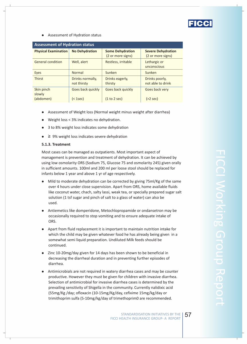
56 57
FICC
I Wo
rking G
rou
p R
epo
rt Assessment of Hydration status
lAssessment of Weight loss (Normal weight minus weight after diarrhea)
lWeight loss < 3% indicates no dehydration.
l3 to 8% weight loss indicates some dehydration
l≥ 9% weight loss indicates severe dehydration
5.1.3. Treatment
Most cases can be managed as outpatients. Most important aspect of
management is prevention and treatment of dehydration. It can be achieved by
using low osmolarity ORS (Sodium 75, Glucose 75 and osmolarity 245) given orally
in sufficient amounts. 100ml and 200 ml per loose stool should be replaced for
infants below 1 year and above 1 yr of age respectively.
lMild to moderate dehydration can be corrected by giving 75ml/Kg of the same
over 4 hours under close supervision. Apart from ORS, home available fluids
like coconut water, chach, salty lassi, weak tea, or specially prepared sugar salt
solution (1 tsf sugar and pinch of salt to a glass of water) can also be
used.
lAntiemetics like domperidone, Metochlopropamide or ondansetron may be
occasionally required to stop vomiting and to ensure adequate intake of
ORS.
lApart from fluid replacement it is important to maintain nutrition intake for
which the child may be given whatever food he has already being given in a
somewhat semi liquid preparation. Undiluted Milk feeds should be
continued.
lZinc 10-20mg/day given for 14 days has been shown to be beneficial in
decreasing the diarrheal duration and in preventing further episodes of
diarrhea.
lAntimicrobials are not required in watery diarrhea cases and may be counter
productive. However they must be given for children with invasive diarrhea.
Selection of antimicrobial for invasive diarrhea cases is determined by the
prevailing sensitivity of Shigella in the community. Currently nalidixic acid
(55mg/Kg /day; ofloxacin (10-15mg/Kg/day, cefixime 15mg/kg/day or
trimithoprim sulfa (5-10mg/kg/day of trimethoprim0 are recommended.
l
Assessment of Hydration status
Physical Examination No Dehydration Some Dehydration (2 or more signs) (2 or more signs)
General condition Well, alert Restless, irritable Lethargic orunconscious
Eyes Normal Sunken Sunken
Thirst Drinks normally, Drinks eagerly, Drinks poorly, not thirsty thirsty not able to drink
Skin pinch Goes back quickly Goes back quickly Goes back very slowly(abdomen) (< 1sec) (1 to 2 sec) (>2 sec)
Severe Dehydration
stools with or without mucus. The child may also have fever and vomiting and
develop features of dehydration which include increased thirst, decreased
urine output etc.
Invasive diarrhea (bacillary dysentery) - It is characterized by passage of loose
frequent stools with blood and mucus. Tenesmus is frequent and is a sign of
systemic toxemia.
Acute diarrhea with systemic infection (parenteral diarrhea) - This is largely
seen among young infants or in severly malnourished children. A child usually
passes frequent small green stools with some mucus (Pea soup stools). He may
also have fever and vomiting together with s/s of associated systemic infection
(e.g. acute ear pain /discharging ear in otitis media). However sometimes, the
s/s of associated infections may not be apparent and careful clinical and
laboratory tests may be required to come to a diagnosis of parenteral diarrhea.
Acute diarrhea can be caused by a variety of infective and non infective causes.
The major cause of diarrhea in children is rotavirus. Other causes include virus
(calcivirus, adeno virus etc), bacteria (Esch coli, vibrio cholerae, shigella, salmonella
etc) and protozoas (giardia, entamoeba etc). In young infants and in malnourished
children, systemic infections like otitis media, meningitis, pneumonia, UTI,
septicemia etc can also present as acute diarrhea. Food intolerances such as
lactose intolerance and intolerance to cow's milk protein are more often
responsible for persistent (>14 days) diarrhea.
5.1. Situation 1: At secondary hospital/non metro situation
5.1.1. Investigations:
In most cases investigations are not required. However, the following
investigations may be done in some selected cases:
lRoutine stool exam for pus cells, motile vibrio, ova &cysts, pH and reducing
substances.
lUrine routine and culture (if UTI suspected)
lBlood counts, CRP and blood culture for suspected systemic infections
lSerum electrolytes, BUN, creatinine for children admitted for moderate /
severe dehydration requiring intravenous fluids.
5.1.2. Assessment of Hydration status:
Treatment for diarrhea in children depends on their hydration status which can be
assessed by any one of the following methods:
v
v
4. Causative Factors
5. Management
STANDARDISATION INITIATIVES BY THE FICCI HEALTH INSURANCE GROUP- A REPORT
STANDARDISATION INITIATIVES BY THE FICCI HEALTH INSURANCE GROUP- A REPORT
Stan
dar
d T
reat
men
t G
uid
elin
es
58
5.1.4 Indications of Hospitalization:
Following are the indications for admitting children with acute diarrhea:
lModerate to severe dehydration present
lPersistent vomiting
lHigh purge rate or failure to maintain hydration despite adequate ORS
lSevere oliguria/ anuria (Failure to pass urine for more than 6-8 hrs)
lAcute diarrhea in a severely malnourished (Body wt less than 60% of
expected)
lPresence of any complication like abdominal distension, renal failure,
convulsions, toxemia, HUS, dyselectrolytemia, etc, Hypovolemic Shock ,
Acidosis, Severe electrolyte imbalance etc
5.2 Situation 2: At Super Specialty Facility in Metro location where
higher end technology is available
5.2.1 Investigation: All investigations of situation 1 and:
lArterial Blood Gases analysis
lBUN, Creatinine, Serum Electrolytes
lBlood counts, CRP, Blood culture if infection suspected
lOther investigations like X-ray Chest, Urine Culture/ CSF examination may be
required occasionally
lRarely CSF examination and culture when meningitis is suspected
Note: The above tests should be obtained before starting intravenous hydration.
5.2.2 Treatment: All treatment of situation 1 and:
lHospitalization
lAs dehydration is the most common indication for admission, the same needs
to be corrected promptly. It is best done by using Ringer's Lactate or Isotonic
saline (100ml / Kg / 8hours) Children with severe dehydration would require to
be given 30-50 ml / kg of IV fluids over fist two hours and the remaining over
the next 6 hours. IV fluids may be required to be repeated if the purge rate
continues to be high and the child's dehydration is not corrected by fluids
given over 8 hrs. After dehydration is corrected age appropriate fluids (One
fifth Isotonic saline for more than one year and one sixth isotonic saline for
infants less than one year) in properly calculated maintenance requirements
should be given. Potassium should be added (20mEq/L) to IV fluids after the
child has started passing urine. Attempt should be made to shift to oral
rehydration at the earliest.
lDrugs - Antiemetics may be required as above. Appropriate antimicrobials may
be required for associated infection (e.g. Cefotaxim and amikacin for
septicemia). Zinc should be given as above.
lNutrition - It is important to maintain adequate nutrition with age appropriate
food intake. Breast feeding must be continued and other milk feed can also be
given undiluted after the dehydration has been corrected.
59
FICC
I Wo
rking G
rou
p R
epo
rt
l
above) must be given orally or intravenously.
Dehydration is the most frequent complication and cause of death in acute
diarrhea.
Oliguria
Acute renal failure
Peripheral circulatory failure
6.1 Management of complications
Acute renal failure may occur occasionally. Mostly it is prerenal and can be
reversed by prompt infusion (20-30ml / Kg over one hour, repeated if required)
of rehydrating fluid (Ringer's Lactate). If the child fails to pass urine despite
correction of dehydration, he may be given an injection of Furesemide (1-
2mg/Kg),. If urine is not passed even after that then the child would need to be
managed on lines of acute parenchymal renal failure with fluid restriction etc.
Peritoneal dialysis may be required occasionally.
Dyselectrolytemias (Hyponatremia, hypokalemia, hypernatremia, metabolic
acidosis are frequent in children with diarrhea and would need appropriate
management
Convulsions can occur because of a variety of reasons but most often are due
to dyselelectrolytemia like hypo or hypernatremia or hypocalcemia etc. They
need to be managed appropriately. Short term anticonvulsants may be
required.
Has the child been able to maintain oral hydration for 6 hours before
discharge?
Has the Frequency of stools reduced and consistency improved?
Has the fever resolved if present earlier?
Has the complication been resolved if present?
Has the caregiver been advised on continued home treatment?
Is the child's immunization complete and caregiver informed about future
immunizations?
Has the growth chart been updated?
Has the caregiver been advised on when to return to the hospital?
Has the caregiver been educated about prevention of diarrhea?
For invasive diarrhea cases apart from fluids appropriate antibiotics (see
6. Complications
7. Pre-discharge assessment Checklist
v
v
v
v
n
n
n
v
v
v
v
v
v
v
v
v
STANDARDISATION INITIATIVES BY THE FICCI HEALTH INSURANCE GROUP- A REPORT
STANDARDISATION INITIATIVES BY THE FICCI HEALTH INSURANCE GROUP- A REPORT
Stan
dar
d T
reat
men
t G
uid
elin
es
58
5.1.4 Indications of Hospitalization:
Following are the indications for admitting children with acute diarrhea:
lModerate to severe dehydration present
lPersistent vomiting
lHigh purge rate or failure to maintain hydration despite adequate ORS
lSevere oliguria/ anuria (Failure to pass urine for more than 6-8 hrs)
lAcute diarrhea in a severely malnourished (Body wt less than 60% of
expected)
lPresence of any complication like abdominal distension, renal failure,
convulsions, toxemia, HUS, dyselectrolytemia, etc, Hypovolemic Shock ,
Acidosis, Severe electrolyte imbalance etc
5.2 Situation 2: At Super Specialty Facility in Metro location where
higher end technology is available
5.2.1 Investigation: All investigations of situation 1 and:
lArterial Blood Gases analysis
lBUN, Creatinine, Serum Electrolytes
lBlood counts, CRP, Blood culture if infection suspected
lOther investigations like X-ray Chest, Urine Culture/ CSF examination may be
required occasionally
lRarely CSF examination and culture when meningitis is suspected
Note: The above tests should be obtained before starting intravenous hydration.
5.2.2 Treatment: All treatment of situation 1 and:
lHospitalization
lAs dehydration is the most common indication for admission, the same needs
to be corrected promptly. It is best done by using Ringer's Lactate or Isotonic
saline (100ml / Kg / 8hours) Children with severe dehydration would require to
be given 30-50 ml / kg of IV fluids over fist two hours and the remaining over
the next 6 hours. IV fluids may be required to be repeated if the purge rate
continues to be high and the child's dehydration is not corrected by fluids
given over 8 hrs. After dehydration is corrected age appropriate fluids (One
fifth Isotonic saline for more than one year and one sixth isotonic saline for
infants less than one year) in properly calculated maintenance requirements
should be given. Potassium should be added (20mEq/L) to IV fluids after the
child has started passing urine. Attempt should be made to shift to oral
rehydration at the earliest.
lDrugs - Antiemetics may be required as above. Appropriate antimicrobials may
be required for associated infection (e.g. Cefotaxim and amikacin for
septicemia). Zinc should be given as above.
lNutrition - It is important to maintain adequate nutrition with age appropriate
food intake. Breast feeding must be continued and other milk feed can also be
given undiluted after the dehydration has been corrected.
59
FICC
I Wo
rking G
rou
p R
epo
rtl
above) must be given orally or intravenously.
Dehydration is the most frequent complication and cause of death in acute
diarrhea.
Oliguria
Acute renal failure
Peripheral circulatory failure
6.1 Management of complications
Acute renal failure may occur occasionally. Mostly it is prerenal and can be
reversed by prompt infusion (20-30ml / Kg over one hour, repeated if required)
of rehydrating fluid (Ringer's Lactate). If the child fails to pass urine despite
correction of dehydration, he may be given an injection of Furesemide (1-
2mg/Kg),. If urine is not passed even after that then the child would need to be
managed on lines of acute parenchymal renal failure with fluid restriction etc.
Peritoneal dialysis may be required occasionally.
Dyselectrolytemias (Hyponatremia, hypokalemia, hypernatremia, metabolic
acidosis are frequent in children with diarrhea and would need appropriate
management
Convulsions can occur because of a variety of reasons but most often are due
to dyselelectrolytemia like hypo or hypernatremia or hypocalcemia etc. They
need to be managed appropriately. Short term anticonvulsants may be
required.
Has the child been able to maintain oral hydration for 6 hours before
discharge?
Has the Frequency of stools reduced and consistency improved?
Has the fever resolved if present earlier?
Has the complication been resolved if present?
Has the caregiver been advised on continued home treatment?
Is the child's immunization complete and caregiver informed about future
immunizations?
Has the growth chart been updated?
Has the caregiver been advised on when to return to the hospital?
Has the caregiver been educated about prevention of diarrhea?
For invasive diarrhea cases apart from fluids appropriate antibiotics (see
6. Complications
7. Pre-discharge assessment Checklist
v
v
v
v
n
n
n
v
v
v
v
v
v
v
v
v
STANDARDISATION INITIATIVES BY THE FICCI HEALTH INSURANCE GROUP- A REPORT
STANDARDISATION INITIATIVES BY THE FICCI HEALTH INSURANCE GROUP- A REPORT
Stan
dar
d T
reat
men
t G
uid
elin
es
60 61
FICC
I Wo
rking G
rou
p R
epo
rt
Content developed by
Dr Arvind Kumar
Senior Consultant
Max Hospital and Columbia Asia
Gurgaon
Dr Kumar is a post doctoral gastroenterology and has been trained in the subject from
P.G.I.M.E.R., Chandigarh and S.G.P.G.I, Lucknow.
He has several national and international publications. He is presently working as a
senior consultant with Max Hospital Group and with Columbia Asia, Gurgaon.
Dr S. K Mittal
MD, FIAP
Pediatrician and Pediatric Gastroenterologist
Chairman, Department of Pediatrics and Adolescent Medicine
Pushpanjali Crosslay Hospital
Ghaziabad &
Visiting Professor, Chacha Nehru Bal Chikitsalya, Delhi
Dr S. K Mittal is currently the Chairman, Department of Pediatrics & Adolescent
Medicine. Prior to this he was Director Professor and Head Department of Pediatrics
and Chief of Pediatric Gastroenterology, Maulana Azad Medical College,New Delhi
Peer reviewed by
Dr L D Bharadwaj
MBBS & MD (Internal Medicine)
Consultant Internist
Bahl Hospital & Medical Research Centre
Rajasthan
Dr Bharadwaj has done his MBBS and MD (Internal Medicine) from S. P Medical College,
Bikaner, Rajasthan. He has topped in his Academic Career and was a Gold Medalist. He
is doing Internal Medicine practice at Sri Ganga Nagar.
Dr S K Sahoo
Senior Consultant in Medicine and Diabetes
Noida Medicare Centre, Ojjus Goodwill Hospital
Noida
A post graduate in general medicine MD(Medicine) from Sambalpur University with a
management degree in health care from FMS Delhi University and is now a senior
consultant in medicine and diabetes attached with NMC hospital and at Goodwill
Hospital Noida. Prior to this assignment Dr Sahoo was chief of Medical services of
GAIL(India) Ltd. During the medical career he has received ICMR Research Studentship
award and felicitated as Best doctor in 50th year of independence by Government of
MP and has presented many national and international papers in various scientific
professional conferences. Now he is associated with every day care operations whose
details can be found from www.everydaycare.co.in.
Dr S C Samal
DM (Gastroenterology)
Senior Consultant Gastroenterologist
Apollo Hospital
Hyderabad
Dr S C Samal is Senior Consultant Gastroenterologist in Apollo Hospital, Hyderabad. He
completed his DM in Gastroenterology from CMC, Vellore in 1995 and worked there as
faculty till 2001. He has more than 15 publications in national and international
journals.
STANDARDISATION INITIATIVES BY THE FICCI HEALTH INSURANCE GROUP- A REPORT
STANDARDISATION INITIATIVES BY THE FICCI HEALTH INSURANCE GROUP- A REPORT
Stan
dar
d T
reat
men
t G
uid
elin
es
60 61
FICC
I Wo
rking G
rou
p R
epo
rtContent developed by
Dr Arvind Kumar
Senior Consultant
Max Hospital and Columbia Asia
Gurgaon
Dr Kumar is a post doctoral gastroenterology and has been trained in the subject from
P.G.I.M.E.R., Chandigarh and S.G.P.G.I, Lucknow.
He has several national and international publications. He is presently working as a
senior consultant with Max Hospital Group and with Columbia Asia, Gurgaon.
Dr S. K Mittal
MD, FIAP
Pediatrician and Pediatric Gastroenterologist
Chairman, Department of Pediatrics and Adolescent Medicine
Pushpanjali Crosslay Hospital
Ghaziabad &
Visiting Professor, Chacha Nehru Bal Chikitsalya, Delhi
Dr S. K Mittal is currently the Chairman, Department of Pediatrics & Adolescent
Medicine. Prior to this he was Director Professor and Head Department of Pediatrics
and Chief of Pediatric Gastroenterology, Maulana Azad Medical College,New Delhi
Peer reviewed by
Dr L D Bharadwaj
MBBS & MD (Internal Medicine)
Consultant Internist
Bahl Hospital & Medical Research Centre
Rajasthan
Dr Bharadwaj has done his MBBS and MD (Internal Medicine) from S. P Medical College,
Bikaner, Rajasthan. He has topped in his Academic Career and was a Gold Medalist. He
is doing Internal Medicine practice at Sri Ganga Nagar.
Dr S K Sahoo
Senior Consultant in Medicine and Diabetes
Noida Medicare Centre, Ojjus Goodwill Hospital
Noida
A post graduate in general medicine MD(Medicine) from Sambalpur University with a
management degree in health care from FMS Delhi University and is now a senior
consultant in medicine and diabetes attached with NMC hospital and at Goodwill
Hospital Noida. Prior to this assignment Dr Sahoo was chief of Medical services of
GAIL(India) Ltd. During the medical career he has received ICMR Research Studentship
award and felicitated as Best doctor in 50th year of independence by Government of
MP and has presented many national and international papers in various scientific
professional conferences. Now he is associated with every day care operations whose
details can be found from www.everydaycare.co.in.
Dr S C Samal
DM (Gastroenterology)
Senior Consultant Gastroenterologist
Apollo Hospital
Hyderabad
Dr S C Samal is Senior Consultant Gastroenterologist in Apollo Hospital, Hyderabad. He
completed his DM in Gastroenterology from CMC, Vellore in 1995 and worked there as
faculty till 2001. He has more than 15 publications in national and international
journals.
STANDARDISATION INITIATIVES BY THE FICCI HEALTH INSURANCE GROUP- A REPORT
STANDARDISATION INITIATIVES BY THE FICCI HEALTH INSURANCE GROUP- A REPORT
Stan
dar
d T
reat
men
t G
uid
elin
es
62 63
Standard Treatment Guidelines for Fissure in Ano (Anal Fissure)
FICC
I Wo
rking G
rou
p R
epo
rt
1. Introduction/ Definition/ Description
2. Incidence of the condition
3. Differential diagnosis:
4. Clinical diagnosis
5. Causes
6. Management
Anal fissure is a linear tear in the muco-cutaneous portion of anal canal, which is
usually in the posterior aspect in men, whereas it can be both posterior and
anterior in women. This is a common perianal condition which presents with pain
in the anal region during and immediately after defecation. Anal fissures can
sometimes cause bleeding per rectum. An anal fissure can be acute, subacute or
chronic.
The exact incidence of this condition is not known in India.
A fissure in ano may be confused with a perianal abscess or sepsis in acute setting.
Or it may be confused with a thrombosed external hemorrhoidal mass when pain
is the only presentation. When bleeding per rectum is significant, it may be
mistaken for internal hemorrhoids.
The diagnosis is made on clinical history and local examination of the perianal
region. Digital rectal examination may or may not be possible, depending upon
the anal sphincteric spasm.
The clinical features include:
Pain in the anal region at and immediately after defecation
At times with bleeding per rectum. The bleeding is usually in the form of a
streak of blood on the stools.
The lesion occurs secondary to passage of hard stools which cause mechanical
injury. Current evidence points to an ischemic etiology as well caused by
sphincteric spasm. In children, anal fissures may occur following diarrhea.
Hospitalization is indicated in the following situations:
For surgery
For severe pain
6.1 Conservative management
The treatment of anal fissure is initially conservative. This may involve:
Stool softeners and bulk purgatives with or without local analgesic gel
v
v
v
v
n
Notes
STANDARDISATION INITIATIVES BY THE FICCI HEALTH INSURANCE GROUP- A REPORT
STANDARDISATION INITIATIVES BY THE FICCI HEALTH INSURANCE GROUP- A REPORT
Stan
dar
d T
reat
men
t G
uid
elin
es
64
Antibiotics may be required if the fissure is infected
Warm water baths (Sitz bath) can provide significant symptomatic relief
Oral anti spasmodic and analgesic drugs are needed for pain relief.
When relief of symptoms does not occur with conservative therapy, local
application of NTG (nitroglycerine) cream or local application / oral calcium
channel blockers are tried.
6.2 Surgical management
Surgery is indicated when conservative therapy fails or there is recurrence or
chronicity with symptoms. The surgery gives immediate relief with low incidence
of fissure recurrence. The surgical options are:
open or internal sphincterotomy
closed internal sphincterotomy
Along with lateral internal sphincterotomy, the patient may be offered excision
of a sentinel tag or pile, or an incision along the fissure distally to avoid a key
hole defect and secondary infection
Anal dilatation is not recommended in view of the risk of uncontrolled tear of anal
sphincter and the attendant incontinence.
Fissurectomy offers no advantage and causes delayed healing and prolonged
hospitalization.
Surgery may also be considered at an early stage to offer early relief rather than
wait through conservative therapy. At the time of surgery, any skin tag (sentinel
tag) or fibroma that coexists with fissure may be excised.
Basic surgical set up, which is available in most surgical centers in non-metro
locations, is adequate to manage anal fissure. The procedure of lateral internal
sphincterotomy can also be performed as day care.
7.1 Investigations:
No investigations are required for diagnosis
Investigations may be required for planning therapy, especially surgery.
No special investigations, except those required for pre anesthetic check up
will be needed.
7.1.1 Admitted with acute perianal pain
lCBC
lUrine RE & Micro
lBlood sugar F &PP
lKidney function tests
7.1.2 Admitted for planned surgery
lCBC
lUrine RE & Micro
n
n
n
n
n
n
n
n
n
7. Situation 1
65
lBlood sugar F &PP
lKidney function tests
lBleeding and coagulation times
lProthrombin time
It is recommended that day care admission may be permitted for patients
undergoing lateral internal sphincterotomy who do not have co morbid conditions
and who undergo this procedure under short GA.
7.2 Treatment
Initial conservative therapy offers approximately 50% response rate. The
treatment involves stool softeners, bulk purgatives, Sitz bath and
administration of local anesthetics.
Local application of NTG cream or local application / oral administration of
calcium channel blockers have been found to offer faster relief of pain and
healing of fissure.
Surgery, if indications met.
7.3 Referral criteria
As anal fissure can be managed in any place where there is a surgeon, there is no
need for referral. However, while evaluating a patient if it is found that the fissure
coexists with a suspected anal canal/ rectal cancer or a stricture, such patients may
be referred to a higher centre.
Situation II: At a superspeciality facility in a metro where higher-end technology
and resources are available
Same as in Situation I.
Spontaneous infection of the anal fissure, especially because of the 'key hole'
deformity (not necessarily due to surgery)
Recurrence of fissure, mainly due to intrinsic pathology, and not a surgical
failure
1. Gupta PJ. Treatment of fissure in ano- revisited. Afr Health Sci. 2004; 4: 58–62.
2. Sajid MS, Rimple J, Cheek E, Baig MK.The efficacy of diltiazem and glyceryltrinitrate
for the medical management of chronic anal fissure: a meta-analysis. Int J
Colorectal Dis. 2008 ;23:1-6.
3. Mousavi SR, Sharifi M, Mehdikhah Z A Comparison Between the Results of
Fissurectomy and Lateral Internal Sphincterotomy in the Surgical Management of
Chronic Anal Fissure. J Gastrointest Surg. 2009
n
n
n
v
8. Complications
9. References
FICC
I Wo
rking G
rou
p R
epo
rt
STANDARDISATION INITIATIVES BY THE FICCI HEALTH INSURANCE GROUP- A REPORT
STANDARDISATION INITIATIVES BY THE FICCI HEALTH INSURANCE GROUP- A REPORT
Stan
dar
d T
reat
men
t G
uid
elin
es
66
Content developed by
Dr V Baskaran
MS, PhD, FACS, FICS,FIAGES
Senior Consultant
Department of Surgical Gastroenterology
Dr BL Kapur Memorial Hospital
New Delhi
A dynamic gastrointestinal and advanced laparoscopic surgeon, with immense body of
surgical skills and academic credentials, including a PhD in gastrointestinal surgery. Has
enormous scientific publications to his credit. Now in practice after 29 years in the
army, this former Professor of Surgery, AFMC, Pune practices evidence based surgery.
Peer reviewed by
Dr Sudhir Kalhan
M. S
Senior Consultant Surgeon, Minimal Access Surgery
B L Kapur Memorial Hospital
New Delhi
Dr Sudhir Kalhan is Practicing General Surgery and Laparoscopic Surgery at B L Kapur
Hospital, New Delhi. He has been actively involved in the training of young Surgeons
and Gynaecologists who want to practice Minimal Access Surgery through training
programmes approved by Indian Academy of Medical Specialties and FOGSI. He is also
Course Cordinator for the operative workshops on' Laparoscopy beyond
Cholecystectomy' held half yearly in association with IMA –AMS. He is Member
Advisory Council, Max Institute of Medical Excellence. He was also the Course Director
for Jointly organizing a training workshop with the “ Chicago Colorectal Society – USA “
on Laparoscopic Colon and Rectal Surgery , Nov2006 at Max Superspeciality Hospital
,Saket, New Delhi.
Earlier he has worked with Max Healthcare, Sita Ram Bhartia Institute and Jeewan Mala
Hospital. He is member of Indian Association of Gastroendoscopic Surgeons,
Endoscopic and Lap. Surgeons of Asia, Gasless International, Tokyo, European
Association of Endoscopic Surgeons, Member – Asia Pacific Hernia Society, Secretary-
Association of Endoscopic Surgeons, New Delhi
Dr Rajnesh Chander Reddy
Senior Consultant Surgical Gastro
Apollo Hospital
Hyderabad
Dr Rajnesh Chander Reddy is Senior Consultant Surgical Gastro in Apollo Hospital,
Hyderabad since 1992. He is trained in Surgical Gastro and Laparoscopic Surgery. He has
presented many papers and provided training to young surgeons in laparoscopic
surgery.
67
Dr Meenakshi Sharma
Consultant in Surgery and Laparoscopic Surgery
Paras Hospitals
Gurgaon
Dr Meenakshi Sharma is currently with Paras hospitals since July 2006 and has been
performing all general, laproscopic & trauma surgeries.Prior to this she was working
with the department of minimal access surgery in Gangaram Hospital as a clinical
research fellow and was doing private practice. Dr Sharma did her post graduation from
Government Medical College, Nagpur and senior residency from Moolchand Hospital
and Batra hospital. Her focus is on Minimal access and Gastrointestinal (GI) surgery and
to her credit has performed many general and laproscopic surgeries independently. She
has attended many conferences in India and abroad.
FICC
I Wo
rking G
rou
p R
epo
rt
STANDARDISATION INITIATIVES BY THE FICCI HEALTH INSURANCE GROUP- A REPORT
STANDARDISATION INITIATIVES BY THE FICCI HEALTH INSURANCE GROUP- A REPORT
Stan
dar
d T
reat
men
t G
uid
elin
es
68
Standard Treatment Guidelines for Fistulae in Ano (Anal Fistula)
69
FICC
I Wo
rking G
rou
p R
epo
rt
v
v
v
v
n
n
n
muco-cutaneous junction, but at times it may be due to infections not in the
anal glands.
Some fistulae are due to serious systemic illnesses such as tuberculosis,
Crohn's disease or due to regional malignancy.
Conservative management for acute pain due to fistula would include
analgesia and antibiotics and drainage of pus.
Surgery is indicated when the patient is troubled by his symptoms. The surgical
options are many and are dictated by the type of fistula. The surgical options
are fistulotomy, fistulectomy, coring out of the fistula, set on suturing and any
combination of these. Fistulectomy, however, offers no advantage over
fistulotomy and causes delayed healing and prolonged hospitalisation.
Anal dilatation as an additional procedure is not recommended in view of the
risk of uncontrolled tear of anal sphincter and the attendant incontinence.
Indications for hospital admission:
For surgery
For drainage of pus when presenting with perianal abscess or for control of
sepsis
7.1 Investigations
7.1.1 Admitted with acute perianal sepsis
lCBC
lUrine RE & Micro
lBlood sugar F &PP
lKidney function tests
lX-ray chest
7.1.2 Admitted for planned surgery
lCBC
lUrine RE & Micro
lBlood sugar F &PP
lKidney function tests
lX-ray chest
lProthrombin time
7.2 Treatment
7.2.1. Conservative management
lWhen admitted with acute pain due to perianal sepsis, the treatment
would include pain killers, antibiotics and drainage of pus under
analgesia/anesthesia.
Usually the fistula is secondary to infection of the anal glands which are at the
6. Management
7. Situation 1
STANDARDISATION INITIATIVES BY THE FICCI HEALTH INSURANCE GROUP- A REPORT
STANDARDISATION INITIATIVES BY THE FICCI HEALTH INSURANCE GROUP- A REPORT
Stan
dar
d T
reat
men
t G
uid
elin
es
1. Introduction/ Definition/ Description
2. Incidence of the condition
3. Differential Diagnosis
4. Clinical Diagnosis
5. Causes
This is a common perianal condition which presents with discharging openings in
the perianal region. A fistula is an abnormal communication between two
epithelial lined surfaces and thus an anal fistula usually means an abnormal tract
between anal canal and the perineal skin. Occasionally the fistula may extend far
away from the anal region and lead to diagnostic confusions. A fistula usually
discharges pus, fecal matter and serosanguinous fluid. However, it may also
discharge blood and flatus.
The exact incidence of this condition is not known in India.
A fistula in ano may present as a perianal abscess or sepsis in acute setting. Or it
may be confused with haemorrhoids when non-purulent discharge is the only
presentation. When bleeding is present, it may be mistaken for internal
haemorrhoids. A fissure may simulate a fistula when there is pus discharge.
The diagnosis is made on clinical history and local examination of the perianal
region.
The presentation may be acute or chronic with periodic exacerbations.
Patients present with pain, anal swelling, redness and fever when there is
acute perianal sepsis. In other instances, they present with skin irritation
around the anus and pus discharging opening/ openings around the anus.
Local examination, digital rectal examination and proctoscopy would guide in
the diagnosis and exclude other anal conditions. Injection of H2O2 or
methylene blue into the tract through the external opening will identify
internal opening in most patients. No investigations are required for diagnosis,
but a fistulogram or MRI fistulogram may be asked for identifying the type of
fistula and the complexity involved to plan surgery.
Investigations required for PA check up may be advised for planning therapy,
especially surgery.
A fistula may be classified in many different ways. The common denominator
determining the nature of surgical intervention is whether the fistula traverses
the entire sphincter complex or the pelvic diaphragm (high or low).
Very often fistula may result from delayed treatment of abscesses in the
Ischiorectal fossae
v
v
v
v
v
68
Standard Treatment Guidelines for Fistulae in Ano (Anal Fistula)
69
FICC
I Wo
rking G
rou
p R
epo
rtv
v
v
v
n
n
n
muco-cutaneous junction, but at times it may be due to infections not in the
anal glands.
Some fistulae are due to serious systemic illnesses such as tuberculosis,
Crohn's disease or due to regional malignancy.
Conservative management for acute pain due to fistula would include
analgesia and antibiotics and drainage of pus.
Surgery is indicated when the patient is troubled by his symptoms. The surgical
options are many and are dictated by the type of fistula. The surgical options
are fistulotomy, fistulectomy, coring out of the fistula, set on suturing and any
combination of these. Fistulectomy, however, offers no advantage over
fistulotomy and causes delayed healing and prolonged hospitalisation.
Anal dilatation as an additional procedure is not recommended in view of the
risk of uncontrolled tear of anal sphincter and the attendant incontinence.
Indications for hospital admission:
For surgery
For drainage of pus when presenting with perianal abscess or for control of
sepsis
7.1 Investigations
7.1.1 Admitted with acute perianal sepsis
lCBC
lUrine RE & Micro
lBlood sugar F &PP
lKidney function tests
lX-ray chest
7.1.2 Admitted for planned surgery
lCBC
lUrine RE & Micro
lBlood sugar F &PP
lKidney function tests
lX-ray chest
lProthrombin time
7.2 Treatment
7.2.1. Conservative management
lWhen admitted with acute pain due to perianal sepsis, the treatment
would include pain killers, antibiotics and drainage of pus under
analgesia/anesthesia.
Usually the fistula is secondary to infection of the anal glands which are at the
6. Management
7. Situation 1
STANDARDISATION INITIATIVES BY THE FICCI HEALTH INSURANCE GROUP- A REPORT
STANDARDISATION INITIATIVES BY THE FICCI HEALTH INSURANCE GROUP- A REPORT
Stan
dar
d T
reat
men
t G
uid
elin
es
1. Introduction/ Definition/ Description
2. Incidence of the condition
3. Differential Diagnosis
4. Clinical Diagnosis
5. Causes
This is a common perianal condition which presents with discharging openings in
the perianal region. A fistula is an abnormal communication between two
epithelial lined surfaces and thus an anal fistula usually means an abnormal tract
between anal canal and the perineal skin. Occasionally the fistula may extend far
away from the anal region and lead to diagnostic confusions. A fistula usually
discharges pus, fecal matter and serosanguinous fluid. However, it may also
discharge blood and flatus.
The exact incidence of this condition is not known in India.
A fistula in ano may present as a perianal abscess or sepsis in acute setting. Or it
may be confused with haemorrhoids when non-purulent discharge is the only
presentation. When bleeding is present, it may be mistaken for internal
haemorrhoids. A fissure may simulate a fistula when there is pus discharge.
The diagnosis is made on clinical history and local examination of the perianal
region.
The presentation may be acute or chronic with periodic exacerbations.
Patients present with pain, anal swelling, redness and fever when there is
acute perianal sepsis. In other instances, they present with skin irritation
around the anus and pus discharging opening/ openings around the anus.
Local examination, digital rectal examination and proctoscopy would guide in
the diagnosis and exclude other anal conditions. Injection of H2O2 or
methylene blue into the tract through the external opening will identify
internal opening in most patients. No investigations are required for diagnosis,
but a fistulogram or MRI fistulogram may be asked for identifying the type of
fistula and the complexity involved to plan surgery.
Investigations required for PA check up may be advised for planning therapy,
especially surgery.
A fistula may be classified in many different ways. The common denominator
determining the nature of surgical intervention is whether the fistula traverses
the entire sphincter complex or the pelvic diaphragm (high or low).
Very often fistula may result from delayed treatment of abscesses in the
Ischiorectal fossae
v
v
v
v
v
70
l
setting.
7.2.2. Surgical management
lThe surgical options are many and are dictated by the type of fistula. The
common surgical options are fistulotomy, coring out of the fistula, set on
suturing and any combination of these.
lThe aim of the operation is to drain the septic focus of the fistula or
remove it with minimal injury to the sphincter complex.
lFistulectomy offers no advantage over fistulotomy and causes delayed
healing and prolonged hospitalization.
lIn very high and complex fistulae, a diverting colostomy may be advised as
a temporary measure to heal the fistula.
lAnal fistula plug insertion is a newer modality in the treatment of anal
fistula with low recurrence rates.
7.3 Referral criteria
As most anal fistulae can be managed in any place where there is a surgeon, only
the complex anal fistulae need to be referred to a higher centre. However, while
evaluating a patient if it is found that the fistula coexists with a suspected anal
canal/ rectal cancer or a stricture or associated with Crohn's disease, such
patients may be referred to a higher centre. Patients needing surgery for a high or
complex fistula often require colostomy and hence may be referred.
7.4 Situation II: At a superspeciality facility in a metro where higher-end
technology and resources are available
Same as in Situation I.
A fistula may recur or the surgery may damage the sphincter in rare cases.
Recurrence of a fistula may at times be due to non identifiable fistula tract or
inability of the surgeon at the initial surgery to identify and excise or lay open the
tract. More often, it may be due to inherent tendency on the part of the patient to
develop a fresh perianal sepsis and go on to develop a new fistula. Delayed wound
healing, anal stenosis and mucosal prolapse are other complications of surgery.
1 Johnson EK, Gaw JU, Armstrong DN. Efficacy of Anal Fistula Plug vs. Fibrin
Glue in Closure of Anorectal Fistulas. Dis Colon Rectum 2006; 49: 371–376.
2. Eric K. Johnson, M.D., Janette U. Gaw, M.D., David N. Armstrong, M.D., F.R.C.S.
Efficacy of Anal Fistula Plug vs. Fibrin Glue in Closure of Anorectal Fistulas. Dis
Colon Rectum 2006; 49: 371–376.
Such patients may need a definitive surgery for fistula later at a second
8. Complications are not common.
9. References
71
Content developed by
Dr V Baskaran
MS, PhD, FACS, FICS,FIAGES
Senior Consultant
Department of Surgical Gastroenterology
Dr BL Kapur Memorial Hospital
New Delhi
A dynamic gastrointestinal and advanced laparoscopic surgeon, with immense body of
surgical skills and academic credentials, including a PhD in gastrointestinal surgery. Has
enormous scientific publications to his credit. Now in practice after 29 years in the
army, this former Professor of Surgery, AFMC, Pune practices evidence based surgery.
FICC
I Wo
rking G
rou
p R
epo
rt
Peer reviewed by
Dr Sudhir Kalhan
M. S
Senior Consultant Surgeon, Minimal Access Surgery
B L Kapur Memorial Hospital
New Delhi
Dr Sudhir Kalhan is Practicing General Surgery and Laparoscopic Surgery at B L Kapur
Hospital, New Delhi. He has been actively involved in the training of young Surgeons
and Gynaecologists who want to practice Minimal Access Surgery through training
programmes approved by Indian Academy of Medical Specialties and FOGSI. He is also
Course Cordinator for the operative workshops on' Laparoscopy beyond
Cholecystectomy' held half yearly in association with IMA –AMS. He is Member
Advisory Council, Max Institute of Medical Excellence. He was also the Course Director
for Jointly organizing a training workshop with the “ Chicago Colorectal Society – USA “
on Laparoscopic Colon and Rectal Surgery , Nov2006 at Max Superspeciality Hospital
,Saket, New Delhi.
Earlier he has worked with Max Healthcare, Sita Ram Bhartia Institute and Jeewan Mala
Hospital. He is member of Indian Association of Gastroendoscopic Surgeons,
Endoscopic and Lap. Surgeons of Asia, Gasless International, Tokyo, European
Association of Endoscopic Surgeons, Member – Asia Pacific Hernia Society, Secretary-
Association of Endoscopic Surgeons, New Delhi
Dr Rajnesh Chander Reddy
Senior Consultant Surgical Gastro
Apollo Hospital
Hyderabad
Dr Rajnesh Chander Reddy is Senior Consultant Surgical Gastro in Apollo Hospital,
Hyderabad since 1992. He is trained in Surgical Gastro and Laparoscopic Surgery. He has
presented many papers and provided training to young surgeons in laparoscopic
surgery.
STANDARDISATION INITIATIVES BY THE FICCI HEALTH INSURANCE GROUP- A REPORT
STANDARDISATION INITIATIVES BY THE FICCI HEALTH INSURANCE GROUP- A REPORT
Stan
dar
d T
reat
men
t G
uid
elin
es
72
Dr Meenakshi Sharma
Consultant in surgery and Laparoscopic surgery
Paras Hospitals
Gurgaon
Dr Meenakshi Sharma is currently with Paras hospitals since July 2006 and has been
performing all general, laproscopic & trauma surgeries.Prior to this she was working
with the department of minimal access surgery in Gangaram Hospital as a clinical
research fellow and was doing private practice. Dr Sharma did her post graduation from
Government Medical College, Nagpur and senior residency from Moolchand Hospital
and Batra hospital. Her focus is on Minimal access and Gastrointestinal (GI) surgery and
to her credit has performed many general and laproscopic surgeries independently. She
has attended many conferences in India and abroad.
73
Standard Treatment Guidelines forGastric Esophageal Reflux Disorder (GERD) requiring hospitalisation
1. Introduction/ Definition/ Description
2. Incidence of the condition
3. Differential Diagnosis
4. Clinical Diagnosis
1
Gastroesophageal reflux disease (GERD) results from reflux of gastric contents into
esophagus. Frequent and persistent occurrence of the reflux can result in
symptoms and esophageal and extra-oesophageal damage.
Fundoplication is the most frequent surgery done in patients with GERD. It
involves wrapping the gastric fundus around the esophagus to restore the
physiology of gastroesophageal junction and provide control of acid and bile
reflux.
GERD is a chronically relapsing problem. In patients with severe esophagitis,
symptoms recur within 1 year in 80% patients; breakthrough esophagitis occurs
while on medication in 10% of patients and 50% of patients may need lifelong
medication.
GER is fairly frequent in infants. Almost 40% infants regurgitate feeds at 4 months
of age. However it is mostly benign and self limiting with symptoms persisting only
in 2% by 18 months of age. Most infants improve spontaneously or with minimum
life style changes like advice on posture, thickening of feeds etc. Some infants
who develop symptoms like failure to thrive, anemia, recurrent respiratory
symptoms or even neurological symptoms may require complete diagnostic
workup and management on the lines given below. Infants and children with
neurological handicaps (like cerebral palsy) tend to have more severe and resistant
GER.
1
Myocardial infarction, Peptic ulcer disease, Cholelithiasis, Infectious esophagitis
2
Typical (esophageal) presentation:
a. Heartburn: Acid regurgitation of into the esophagus
b. Regurgitation of food and gastric acid into pharynx and mouth
c. Dysphagia: reported as a sensation that food is stuck, particularly in the
retrosternal area, which usually suggests stricture, a known and frequent
complication
Atypical (extraesophageal) presentation:
Coughing and/or wheezing or exacerbation of asthma, due to reflux of acid
into the airway
v
FICC
I Wo
rking G
rou
p R
epo
rt
STANDARDISATION INITIATIVES BY THE FICCI HEALTH INSURANCE GROUP- A REPORT
STANDARDISATION INITIATIVES BY THE FICCI HEALTH INSURANCE GROUP- A REPORT
Stan
dar
d T
reat
men
t G
uid
elin
es
74
v
v
v
v
v
v
v
v
v
Hoarseness is often experienced by patients in the morning due to acid
induced injury to larynx
Chest pain resembling a myocardial infarction
1
The basic cause for the reflux is the absence of the one way valve like effect of the
GE junction. This is usually due to a lax sphincter or abnormal position of the GE
junction as it happens in hiatus hernia. At times Connective tissue disorders like
Scleroderma may make the GE junction inelastic and predispose to reflux. Reflux
may be aggravated by certain risk factors like smoking, fatty foods, alcohol, tea/
coffee, obesity, work or hobbies that require stooping or lying down, drugs as
NSAIDs, cardiac medication (e.g. calcium channel blockers, nitrates, beta-blockers),
and certain hormones which cause smooth muscle relaxation(e.g. progesterone).
Management must include some mandatory investigations to confirm the
diagnosis and to exclude other conditions that may mimic GERD. The requisite
investigations in the diagnosis and assessment of GERD include the following:
X-ray chest
ECG
UGI endoscopy
Ba swallow
Radionuclide scintigraphy for assessment of gastro-esophageal reflux and
measurement of gastric emptying
24h ambulatory pHmetry and
At times 24h Holter monitoring and oesophageal pressure monitoring.
The need and the type of therapy are based on the symptoms and complications.
Conservative management with life style modification and medical therapy for
acid suppression is rewarded with relief of symptoms in the vast majority of
patients. However, there exists a subgroup of patients who need medication life
long. Current data suggest that the quality of life for patients with GERD is
significantly lower due to heartburn, regurgitation and the need to have prolonged
medication [4]. It is also well recognized that a number of patients with GERD have
severe symptoms without endoscopic evidence of reflux esophagitis and that
these patients also have a poor quality of life [4].
6.1.1 Life style changes
Reflux is common after heavy meals and fatty meals and hence patients should be
advised not to take very heavy or fatty meals. They benefit with multiple smaller
meals rather than three large meals. Also reflux is common in lying position and
on forward bending. Hence these patients should be advised not to go to sleep or
lie down immediately after food and also should avoid forward bending in the
postprandial period. These patients should sleep with the head end elevated by 15
degrees.
It should be mentioned here that a hiatus hernia without reflux needs no surgical
intervention.
5. Causes
6. Management
75
6.1.2 Medical treatment
lInitial management will include Proton Pump Inhibitor (PPI) therapy for 8
weeks. PPI are the first line of therapy.
lH2 receptor antagonists sucralfate, antacids and prokinetic agents have much
less efficacy as compared to PPIs
lAfter the 8 weeks trial, a step down of PPI therapy should be initiated
l74 – 96% healing in 8 weeks of therapy with PPIs. 50% may need lifelong
medication
lEndoscopy should be considered if symptoms persist after initial therapy.
lRegular endoscopic surveillance including biopsy is indicated for moderate
GERD.
6.1.3 Surgical treatment
When GERD symptoms are uncontrolled by non-surgical methods or when
complications occur due to reflux, surgery is indicated. Indications for GERD
surgery are given in 6.1.3. Antireflux surgery, both open and laparoscopic, has
been demonstrated to be superior to medical therapy in the control of symptoms
of GERD [4-6].
Endoscopic mucosal resection (EMR) is the procedure of choice in patients with
Barrett's esophagus with high grade dysplasia or carcinoma [14].
Photodynamic therapy has been added as an adjunct to EMR to reduce chances of
local recurrence [14].
Comparison of medical and surgical options is shown below:
Choice of surgical repair
Medical Management Surgical Management
o Life long management
o Can be Costly o Relatively Lower total cost
o Controls only acid reflux o Controls alkaline reflux as well
o Risk of progression to dysplasia o Risk of progression to dysplasia & & Ca higher Ca lower
o Long term effects of drugs? o Complications manageable
o No mortality o 1% mortality,15% fail
o One time treatment
Conventional Fundoplication Laparoscopic Fundoplication
o Technically easier o High technical expertise
o Low costs (E 6900) o High costs (E 9100)
o Complications (%) o Complications
nRecurrence 3.7 nRecurrence 2.1
nDysphagia 15 nDysphagia 19
nReoperations 0.6 nReoperations 2.5
o Stay 5 days o Stay 3 days
FICC
I Wo
rking G
rou
p R
epo
rt
STANDARDISATION INITIATIVES BY THE FICCI HEALTH INSURANCE GROUP- A REPORT
STANDARDISATION INITIATIVES BY THE FICCI HEALTH INSURANCE GROUP- A REPORT
Stan
dar
d T
reat
men
t G
uid
elin
es
76
The choice of surgical repair is left to the surgeon, though evidence suggests that
the short and long term results including Quality of Life issues of laparoscopic
fundoplication are better than those of open procedures.
The classical 360 degree floppy fundoplication can be performed through
laparoscopic approach, provided there is an expert surgeon in charge of the
operation
6.1.3 Indications for Fundoplication:
lPatients with symptoms that are not completely controlled by PPI therapy can
be considered for surgery. For example:
• Patients who have respiratory symptoms such as cough or exacerbation of
bronchial asthma due to reflux
• Recurrent spontaneous vomiting or regurgitation into the mouth especially
while in lying postures or when bending forwards
lPresence of Barrett esophagus, particularly patients with persistent
inadequate LES pressure but normal peristaltic contractions in the esophagus
body
lPoor patient compliance to medications or relapses on maintenance therapy
lYoung patients with GERD can be considered for early surgery
lParaesophageal hernia (usually always require surgery)
lExtraoesophageal manifestations such as pharyngo laryngeal reflux, reflux
induced epiglottisitis, erosion of inner aspect of teeth especially the incisors
lWhen there is peptic stricture, surgery is indicated after dilatation
6.2. Situation 1
6.2.1. Investigations:
lLab Studies – for diff diagnosis, H Pylori test,
lImaging Studies: Barium esophagogram not very useful for diagnosis of GERD,
but for complications such as stricture, associated sliding and paraoesophageal
hernias. Nucleotide scans are preferable for diagnosis and semiquantification
of GERD and also for exclusion of gastric stasis
l24 hour ambulatory pHmetry is the investigation of choice, if available.
lEndoscopy and Biopsy
6.2.2. Treatment
6.2.2.1 Medical treatment
lPPI is the mainstay of therapy
lLong term behavior modification through lifestyle changes
lRegular endoscopic surveillance including biopsy is indicated for moderate
GERD.
6.2.2.2 Surgical treatment
Facilities and surgical expertise for laparoscopic fundoplication may not be
available at situation 1, and conventional fundoplication may be done where
surgery is indicated.
77
6.2.3 Referral criteria for a specialist centre:
l
after 6 months of medical treatment or when there is evidence of a
complication such as stricture
6.3 Situation 2:
6.3.1 Investigation
As in situation 1 and additional Investigations
lEsophageal manometry,
lAmbulatory 24-hour pH monitoring
lRadionuclide assessment of gastro-esophageal reflux and measurement of
gastric emptying
6.3.2 Treatment
6.3.2.1 Indications of hospitalization
Hospitalization is indicated usually for surgical intervention. Medical management
rarely requires hospital admission. However it may be required due to:
lAcute chest pain due to GERD for observation and for exclusion of cardiac
cause of pain
lAcute severe esophagitis for diagnosis
lSevere complication e.g. asthma exacerbation, bleeding ulcer
6.3.3 Complications
lEsophagitis
lStrictures
lBarrett's esophagus
lAdenocarcinoma
lIncreased risk of bleeding and perforation
lAsthma exacerbation due to aspiration of acid into bronchial tree is among the
common complications of GERD.
1. Shah SN, Anand MP, editors. API text book of medicine. 7th ed. Mumbai (India):
The association of physicians of India, Mumbai; 2003.
2. Agency for healthcare and quality, Comparative Effectiveness of Management
Strategies For Gastroesophageal Reflux Disease AHRQ Publication No. 06-EHC003-
EF December 2005
3. Balsara KP et al, Laparoscopic surgery for reflux esophagitis and paraesophageal
hernia. Indian Journal of Gastroenterology 2002 Vol 21 May-June.
4. Gurjeet Kaur et al, 2007 Concordance between endoscopic and histological
gastroesophageal reflux disease Indian Journal of gastroenterology 2007 vol 26
Jan-feb 46-47
Referral to Gastroenterologist/ surgical gastroenterologist if symptoms persist
7. References
FICC
I Wo
rking G
rou
p R
epo
rt
STANDARDISATION INITIATIVES BY THE FICCI HEALTH INSURANCE GROUP- A REPORT
STANDARDISATION INITIATIVES BY THE FICCI HEALTH INSURANCE GROUP- A REPORT
Stan
dar
d T
reat
men
t G
uid
elin
es
78
5. Kamolz T, Granderath FA, Schweiger UM, Pointner R. Laparoscopic Nissen fundoplication in patients with nonerosive reflux disease. Long-term quality-of-life assessment and surgical outcome. Surg Endosc. 2005;19:494-500
6. Fernando HC, Schauer PR, Rosenblatt M, Wald A, Buenaventura P, Ikramuddin S, Luketich JD. Quality of life after antireflux surgery compared with nonoperative management for severe gastroesophageal reflux disease. J Am Coll Surg. 2002;194:23
7. Mahon D, Rhodes M, Decadt B, Hindmarsh A, Lowndes R, Beckingham I, Koo B, Newcombe RG. Randomized clinical trial of laparoscopic Nissen fundoplication compared with proton-pump inhibitors for treatment of chronic gastro-oesophageal reflux. Br J Surg. 2005;92:695-9.
8. Tucker LE, Blatt C, Richardson NL, Richardson DT, Cassat JD, Riechers TB. Laparoscopic Nissen fundoplication in a community hospital: patient satisfaction survey. South Med J. 2005;98:441-3.
9. Ng R, Mullin EJ, Maddern GJ. Systematic review of day-case laparoscopic Nissen fundoplication. ANZ J Surg. 2005;75:160-4.
10. Rothenberg SS.The first decade's experience with laparoscopic Nissen fundoplication in infants and children. Pediatr Surg. 2005;40:142-6; discussion 147
11. Ciovica R, Gadenstatter M, Klingler A, Neumayer C, Schwab GP Laparoscopic Antireflux Surgery Provides Excellent Results and Quality of Life in Gastroesophageal Reflux Disease Patients With Respiratory Symptoms. J Gastrointest Surg. 2005;9:633-7.
12. Fiorentino E, Cabibi D, Pantuso G, Latteri F, Mastrosimone A, Valenti A. [Laparoscopic Nissen fundoplication and esophageal intestinal metaplasia: preliminary observations] [Article in Italian] Chir Ital. 2005;57:53-8.
13. van Lanschot JJB, Bergman JJGHM. Tailored therapy for early Barret's lesions. Br J Surg 2005;92:791-2.
14. Braghetto I, Csendes A, Korn O, Burdiles P, Valladares H, Cortes C, Debandi A Anatomical deformities after laparoscopic antireflux surgery.Int Surg. 2004;89:227-35.
15. Pessaux P, Arnaud JP, Ghavami B, Flament JB, Trebuchet G, Meyer C, Huten N, Tuech JJ, Champault G; Societe Francaise de Chirurgie Laparoscopique Morbidity of laparoscopic fundoplication for gastroesophageal reflux: a retrospective study about 1470 patients. Hepatogastroenterology. 2002;49:447-50.
16. Cole SJ, van den Bogaerde JB, van der Walt H. Preoperative esophageal manometry does not predict postoperative dysphagia following anti-reflux surgery. Dis Esophagus. 2005;18:51-6.
17. Prochazka V, Kala Z, Kroupa R, Kysela P, Izakovicova HL, Dolina J. [Could the peroperative manometry of the oesophagus be used for prediction of dysphagia following antireflux procedures?] [Article in Czech] Rozhl Chir. 2005;84:7-12.
18. Zornig C, Strate U, Fibbe C, Emmermann A, Layer P Nissen vs Toupet laparoscopic fundoplication. Surg Endosc. 2002;16:758-66. Baigrie RJ, Cullis SN, Ndhluni AJ, Cariem A. Randomized double-blind trial of laparoscopic Nissen fundoplication versus anterior partial fundoplication. Br J Surg. 2005;92:819-23.
19. Watson DI, Jamieson GG, Lally C, Archer S, Bessell JR, Booth M, Cade R, Cullingford G, Devitt PG, Fletcher DR, Hurley J, Kiroff G, Martin CJ, Martin IJ, Nathanson LK, Windsor JA. Multicenter, prospective, double-blind, randomized trial of laparoscopic nissen vs anterior 90 degrees partial fundoplication. Arch Surg. 2004;139:1160-7.
79
Content developed by
Dr Arvind Kumar
Dr Kumar is a post doctoral gastroenterology and has been
trained in the subject from P.G.I.M.E.R., Chandigarh and
S.G.P.G.I, Lucknow.
He has several national and international publications. He is presently working as a
senior consultant with Max Hospital Group and with Columbia Asia, Gurgaon.
Dr V Baskaran
MS, PhD, FACS, FICS,FIAGES
Senior Consultant
Department of Surgical Gastroenterology
Dr BL Kapur Memorial Hospital
New Delhi
A dynamic gastrointestinal and advanced laparoscopic surgeon, with immense body of
surgical skills and academic credentials, including a PhD in gastrointestinal surgery. Has
enormous scientific publications to his credit. Now in practice after 29 years in the
army, this former Professor of Surgery, AFMC, Pune practices evidence based surgery.
FICC
I Wo
rking G
rou
p R
epo
rt
Peer reviewed by
Dr A K Khurana
Senior Consultant & Head
Department of Gastroenterology
Jaipur Golden Hospital
New Delhi
Dr A K. Khurana did his MBBS in 1984 and MD (Medicine) from Delhi University in 1988.
He did DNB in Medicine in 1989 and DM in Gastroenterology from Delhi University in
1991. He was awarded FRCP in 2006. He topped the combined medical services
examination in 1986 conducted by UPSC. He is actively involved in academic activities.
He is holding regular CMEs in the hospital and also brings out quarterly Newsletter (JGH
Gastrovision). He is regularly invited as faculty in various National and International
conferences.
STANDARDISATION INITIATIVES BY THE FICCI HEALTH INSURANCE GROUP- A REPORT
STANDARDISATION INITIATIVES BY THE FICCI HEALTH INSURANCE GROUP- A REPORT
Stan
dar
d T
reat
men
t G
uid
elin
es
80
Standard Treatment Guidelines for Heart Failure requiring Hospitalisation
1. Introduction/ Definition/ Description:
2. Incidence of the condition
3 Causes
v
v
v
v
v
v
v
v
v
v
v
v
v
v
v
v
v
v
v
v
HF is a complex clinical syndrome that can result from any structural or functional cardiac disorder that impairs the ability of the ventricle to fill with or eject blood.
This includes patients of HF with preserved LV systolic function
The Problem in the USA
5,000,000 patients
6,500,000 hospital days / year
300,000 deaths / year
6% - 10% of people > 65 years
5.4% of health care budget (38 billion)
Incidence has doubled in last ten years
Problem in India (estimated)
Prevalence
18.8 million (1.76% of population)
Incidence
1.57 million per year (0.15% of population)
HF is the end result of any type of heart disease. It can result from following-
Myocardial diseases
Pericardial diseases
Valvular diseases
High output states
Drugs
Alcohol
Connective tissue diseases
Precipitating factors: A number of factors can aggravate or precipitate the heart failure. Recognition of these is essential for the management of HF.
Dietary factors
Physical activity
Pregnancy
81
FICC
I Wo
rking G
rou
p R
epo
rt
v
v
v
v
v
v
v
v
v
v
v
v
v
Hypo/hyperthyrodism
Infections
Arrthythmias
Hypertension
New heart disease
Medications
Thromboembolism
Anaemia
Clinical diagnosis of Heart failure includes:
Assessment of severity of HF
Assessment of cardiac structure & function
Evaluation of CAD
Evaluation of risk of arrhythmia
Identification of precipitating factors, co morbid conditions and barrier toadherence & compliance to treatment
The clinical diagnosis is based on Framingham criteria.
Presence of two major or one major and two minor criteria are essential for diagnosis
• Orthopnea/PND • Ankle edema
• Venous distension • Night cough
• Rales • Exertional dyspnea
• Cardiomegaly • Hepatomegaly
• Acute pulm edema • Pleural effusion
• Elevated JVP • Tachycardia (>120)
• HJR • Decrease VC
• S3 gallop • Weight loss with diuretics
Heart Failure may be classified into stages or functional class. A comparison of ACC/AHA and NYHA classification is given below
4. Clinical Diagnosis
Major Criteria Minor Criteria
STANDARDISATION INITIATIVES BY THE FICCI HEALTH INSURANCE GROUP- A REPORT
STANDARDISATION INITIATIVES BY THE FICCI HEALTH INSURANCE GROUP- A REPORT
Stan
dar
d T
reat
men
t G
uid
elin
es
80
Standard Treatment Guidelines for Heart Failure requiring Hospitalisation
1. Introduction/ Definition/ Description:
2. Incidence of the condition
3 Causes
v
v
v
v
v
v
v
v
v
v
v
v
v
v
v
v
v
v
v
v
HF is a complex clinical syndrome that can result from any structural or functional cardiac disorder that impairs the ability of the ventricle to fill with or eject blood.
This includes patients of HF with preserved LV systolic function
The Problem in the USA
5,000,000 patients
6,500,000 hospital days / year
300,000 deaths / year
6% - 10% of people > 65 years
5.4% of health care budget (38 billion)
Incidence has doubled in last ten years
Problem in India (estimated)
Prevalence
18.8 million (1.76% of population)
Incidence
1.57 million per year (0.15% of population)
HF is the end result of any type of heart disease. It can result from following-
Myocardial diseases
Pericardial diseases
Valvular diseases
High output states
Drugs
Alcohol
Connective tissue diseases
Precipitating factors: A number of factors can aggravate or precipitate the heart failure. Recognition of these is essential for the management of HF.
Dietary factors
Physical activity
Pregnancy
81
FICC
I Wo
rking G
rou
p R
epo
rtv
v
v
v
v
v
v
v
v
v
v
v
v
Hypo/hyperthyrodism
Infections
Arrthythmias
Hypertension
New heart disease
Medications
Thromboembolism
Anaemia
Clinical diagnosis of Heart failure includes:
Assessment of severity of HF
Assessment of cardiac structure & function
Evaluation of CAD
Evaluation of risk of arrhythmia
Identification of precipitating factors, co morbid conditions and barrier toadherence & compliance to treatment
The clinical diagnosis is based on Framingham criteria.
Presence of two major or one major and two minor criteria are essential for diagnosis
• Orthopnea/PND • Ankle edema
• Venous distension • Night cough
• Rales • Exertional dyspnea
• Cardiomegaly • Hepatomegaly
• Acute pulm edema • Pleural effusion
• Elevated JVP • Tachycardia (>120)
• HJR • Decrease VC
• S3 gallop • Weight loss with diuretics
Heart Failure may be classified into stages or functional class. A comparison of ACC/AHA and NYHA classification is given below
4. Clinical Diagnosis
Major Criteria Minor Criteria
STANDARDISATION INITIATIVES BY THE FICCI HEALTH INSURANCE GROUP- A REPORT
STANDARDISATION INITIATIVES BY THE FICCI HEALTH INSURANCE GROUP- A REPORT
Stan
dar
d T
reat
men
t G
uid
elin
es
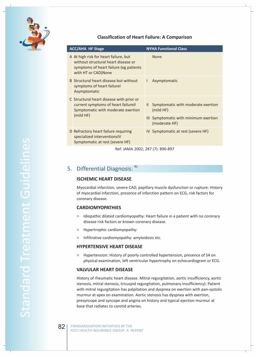
82
Classification of Heart Failure: A Comparison
ACC/AHA HF Stage
A At high risk for heart failure, but without structural heart disease or symptoms of heart failure (eg patients with HT or CAD)None
B Structural heart disease but without symptoms of heart failureIAsymptomatic
C Structural heart disease with prior or current symptoms of heart failureIISymptomatic with moderate exertion (mild HF)
D Refractory heart failure requiring specialized interventionsIVSymptomatic at rest (severe HF)
NYHA Functional Class
None
I Asymptomatic
II Symptomatic with moderate exertion (mild HF)
III Symptomatic with minimum exertion (moderate HF)
IV Symptomatic at rest (severe HF)
Ref: JAMA 2002; 287 (7): 890-897
5. Differential Diagnosis: B1
ISCHEMIC HEART DISEASE
Myocardial infarction, severe CAD, papillary muscle dysfunction or rupture: History
of myocardial infarction, presence of infarction pattern on ECG, risk factors for
coronary disease.
CARDIOMYOPATHIES
Idiopathic dilated cardiomyopathy: Heart failure in a patient with no coronary
disease risk factors or known coronary disease.
Hypertrophic cardiomyopathy:
Infiltrative cardiomyopathy: amyloidosis etc.
HYPERTENSIVE HEART DISEASE
Hypertension: History of poorly controlled hypertension, presence of S4 on
physical examination, left ventricular hypertrophy on echocardiogram or ECG.
VALVULAR HEART DISEASE
History of rheumatic heart disease. Mitral regurgitation, aortic insufficiency, aortic
stenosis, mitral stenosis, tricuspid regurgitation, pulmonary insufficiency): Patient
with mitral regurgitation has palpitation and dyspnea on exertion with pan-systolic
murmur at apex on examination. Aortic stenosis has dyspnea with exertion,
presyncope and syncope and angina on history and typical ejection murmur at
base that radiates to carotid arteries.
n
n
n
n
83
FICC
I Wo
rking G
rou
p R
epo
rt
MYOCARDITIS
rickettsia, viral, streptococci, staphylococci): Fever, exposure to known agent,
positive blood cultures.
Parastic myocarditis (Trypanosome cruzi-Chagas disease, leishmaniasis,
toxoplasmosis): Travel history to endemic areas, fever, peripheral stigmata of
infection.
Collagen vascular disease (SLE, polyarteritis nodosa, scleroderma,
dermatomyositis): History of collagen vascular disease, positive serology
results for a collagen vascular disease, other stigmata of collagen vascular
disease.
PERICARDIAL DISEASES:
Precordial chest pain and presence of rub suggests pericardial disease.
Constrictive pericarditis and pericardial effusion etc.
DRUGS and TOXINS:
Alcohol, adriamycin and many anti cancer drugs.
HIGH OUTPUT STATES:
History of anemia, thyroid disease and other high output states to be obtained.
The objective of treatment is to:
Increase Survival
Reduce Morbidity
Improve Exercise capacity
Improve Quality of life
Reduce Neurohormonal changes
Reduce Progression of CHF
Manage Symptoms
6.1. Situation 1:
The goal of treatment / management in a non metro clinic or small hospital is to
evaluate HF and early stabilization. In mild and moderate HF – investigations and
both pharmacological and non pharmacological treatment should be initiated.
Depending upon available facilities and severity of presentation or need for
surgical intervention, severe HF should be referred to higher centers.
6.1.1.1 Reasons for hospitalization:
• Symptomatic HF
• Evaluation & therapy of new HF
• Severity of congestion may warrant hospitalization
• Anasarca (collection of fluid)
n
n
n
n
n
n
v
v
v
v
v
v
v
Bacterial myocarditis (Borrelia burgdorferi or Lyme disease, diphtheria,
6. Management
STANDARDISATION INITIATIVES BY THE FICCI HEALTH INSURANCE GROUP- A REPORT
STANDARDISATION INITIATIVES BY THE FICCI HEALTH INSURANCE GROUP- A REPORT
Stan
dar
d T
reat
men
t G
uid
elin
es
82
Classification of Heart Failure: A Comparison
ACC/AHA HF Stage
A At high risk for heart failure, but without structural heart disease or symptoms of heart failure (eg patients with HT or CAD)None
B Structural heart disease but without symptoms of heart failureIAsymptomatic
C Structural heart disease with prior or current symptoms of heart failureIISymptomatic with moderate exertion (mild HF)
D Refractory heart failure requiring specialized interventionsIVSymptomatic at rest (severe HF)
NYHA Functional Class
None
I Asymptomatic
II Symptomatic with moderate exertion (mild HF)
III Symptomatic with minimum exertion (moderate HF)
IV Symptomatic at rest (severe HF)
Ref: JAMA 2002; 287 (7): 890-897
5. Differential Diagnosis: B1
ISCHEMIC HEART DISEASE
Myocardial infarction, severe CAD, papillary muscle dysfunction or rupture: History
of myocardial infarction, presence of infarction pattern on ECG, risk factors for
coronary disease.
CARDIOMYOPATHIES
Idiopathic dilated cardiomyopathy: Heart failure in a patient with no coronary
disease risk factors or known coronary disease.
Hypertrophic cardiomyopathy:
Infiltrative cardiomyopathy: amyloidosis etc.
HYPERTENSIVE HEART DISEASE
Hypertension: History of poorly controlled hypertension, presence of S4 on
physical examination, left ventricular hypertrophy on echocardiogram or ECG.
VALVULAR HEART DISEASE
History of rheumatic heart disease. Mitral regurgitation, aortic insufficiency, aortic
stenosis, mitral stenosis, tricuspid regurgitation, pulmonary insufficiency): Patient
with mitral regurgitation has palpitation and dyspnea on exertion with pan-systolic
murmur at apex on examination. Aortic stenosis has dyspnea with exertion,
presyncope and syncope and angina on history and typical ejection murmur at
base that radiates to carotid arteries.
n
n
n
n
83
FICC
I Wo
rking G
rou
p R
epo
rtMYOCARDITIS
rickettsia, viral, streptococci, staphylococci): Fever, exposure to known agent,
positive blood cultures.
Parastic myocarditis (Trypanosome cruzi-Chagas disease, leishmaniasis,
toxoplasmosis): Travel history to endemic areas, fever, peripheral stigmata of
infection.
Collagen vascular disease (SLE, polyarteritis nodosa, scleroderma,
dermatomyositis): History of collagen vascular disease, positive serology
results for a collagen vascular disease, other stigmata of collagen vascular
disease.
PERICARDIAL DISEASES:
Precordial chest pain and presence of rub suggests pericardial disease.
Constrictive pericarditis and pericardial effusion etc.
DRUGS and TOXINS:
Alcohol, adriamycin and many anti cancer drugs.
HIGH OUTPUT STATES:
History of anemia, thyroid disease and other high output states to be obtained.
The objective of treatment is to:
Increase Survival
Reduce Morbidity
Improve Exercise capacity
Improve Quality of life
Reduce Neurohormonal changes
Reduce Progression of CHF
Manage Symptoms
6.1. Situation 1:
The goal of treatment / management in a non metro clinic or small hospital is to
evaluate HF and early stabilization. In mild and moderate HF – investigations and
both pharmacological and non pharmacological treatment should be initiated.
Depending upon available facilities and severity of presentation or need for
surgical intervention, severe HF should be referred to higher centers.
6.1.1.1 Reasons for hospitalization:
• Symptomatic HF
• Evaluation & therapy of new HF
• Severity of congestion may warrant hospitalization
• Anasarca (collection of fluid)
n
n
n
n
n
n
v
v
v
v
v
v
v
Bacterial myocarditis (Borrelia burgdorferi or Lyme disease, diphtheria,
6. Management
STANDARDISATION INITIATIVES BY THE FICCI HEALTH INSURANCE GROUP- A REPORT
STANDARDISATION INITIATIVES BY THE FICCI HEALTH INSURANCE GROUP- A REPORT
Stan
dar
d T
reat
men
t G
uid
elin
es
84
• Class III - IV with no response to higher dose of diuretics
• Clinical cold & wet profile (hypotension and edema)
• Dysarrhythmia
• Syncope
• Sustained VT
• New onset AF
• Other CV events
• Unstable angina
• CVA
• Embolic events
• Non-cardiac events
• Severe anemia
• COPD exacerbation
• New onset renal failure in HF patient
• Septicemia or severe infection
6.1.1.2 Criteria for Discharge
• Stable fluid balance/renal function
• More than 24 hours on oral regimen Off short acting i.v. agents >24 hours Off
long acting i.v. agents >48 hours Stable BP >90 mmHg without postural fall
• Ambulation without dyspnea/dizziness
• Patient education & comprehension of Na & fluid intake, Weight monitoring
and Symptoms of fluid overload
6.1.2. Investigations:
All management must include some mandatory investigations to confirm the
diagnosis:
• CBC
• U routine
• Sugar
• KFT
• LFT
• Na/K
• Ca/Mg
• Lipids
• Thyroid tests
• ECG
• X-ray Chest
• Echo-doppler
85
FICC
I Wo
rking G
rou
p R
epo
rt
6.1.3. Treatment
Patient education, non pharmacological management and pharmacological
treatment should be initiated.
6.1.3.1 Non pharmacological management:
• Dietary sodium 2-3 gms/day
• Fluids <2 lts/day
• Multivitamins recommended
• Calcium supplements recommended
• Protein supplements recommended
• Oxygen supplementation not as a routine
• Alcohol intake to be restricted
• Pneumonia vaccine recommended
• Annual influenza vaccine recommended
• Smoking cessation advice
6.1.3.2 Pharmacotherapy
• Beta-blockers
• Angiotensin Converting enzyme inhibitors (ACEI)
• Angiotensin Receptor Blockers (ARB)
• Diuretics
• Digitalis
• Vasodilators (Nitrates and Hydralazine)
• Intravenous Inotropes in Acute Decompensated HF
• Anticoagulants: Low Molecular Weight Heparin and oral drugs
Evidence-Based Treatment Across the Continuum of Systolic LVD and HF
Control Volume Improve Clinical Outcomes
DiureticsRenal ReplacementTherapy*
Digoxin
-BlockerACEIor ARB
AldosteroneAntagonist
or ARB
Treat Residual Symptoms
CRT an ICD*
HDZN/ISDN**In selected patients
STANDARDISATION INITIATIVES BY THE FICCI HEALTH INSURANCE GROUP- A REPORT
STANDARDISATION INITIATIVES BY THE FICCI HEALTH INSURANCE GROUP- A REPORT
Stan
dar
d T
reat
men
t G
uid
elin
es
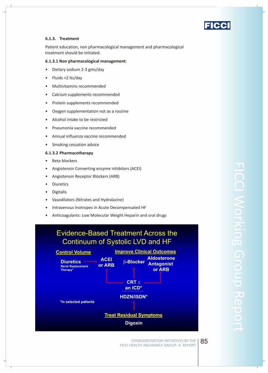
84
• Class III - IV with no response to higher dose of diuretics
• Clinical cold & wet profile (hypotension and edema)
• Dysarrhythmia
• Syncope
• Sustained VT
• New onset AF
• Other CV events
• Unstable angina
• CVA
• Embolic events
• Non-cardiac events
• Severe anemia
• COPD exacerbation
• New onset renal failure in HF patient
• Septicemia or severe infection
6.1.1.2 Criteria for Discharge
• Stable fluid balance/renal function
• More than 24 hours on oral regimen Off short acting i.v. agents >24 hours Off
long acting i.v. agents >48 hours Stable BP >90 mmHg without postural fall
• Ambulation without dyspnea/dizziness
• Patient education & comprehension of Na & fluid intake, Weight monitoring
and Symptoms of fluid overload
6.1.2. Investigations:
All management must include some mandatory investigations to confirm the
diagnosis:
• CBC
• U routine
• Sugar
• KFT
• LFT
• Na/K
• Ca/Mg
• Lipids
• Thyroid tests
• ECG
• X-ray Chest
• Echo-doppler
85
FICC
I Wo
rking G
rou
p R
epo
rt6.1.3. Treatment
Patient education, non pharmacological management and pharmacological
treatment should be initiated.
6.1.3.1 Non pharmacological management:
• Dietary sodium 2-3 gms/day
• Fluids <2 lts/day
• Multivitamins recommended
• Calcium supplements recommended
• Protein supplements recommended
• Oxygen supplementation not as a routine
• Alcohol intake to be restricted
• Pneumonia vaccine recommended
• Annual influenza vaccine recommended
• Smoking cessation advice
6.1.3.2 Pharmacotherapy
• Beta-blockers
• Angiotensin Converting enzyme inhibitors (ACEI)
• Angiotensin Receptor Blockers (ARB)
• Diuretics
• Digitalis
• Vasodilators (Nitrates and Hydralazine)
• Intravenous Inotropes in Acute Decompensated HF
• Anticoagulants: Low Molecular Weight Heparin and oral drugs
Evidence-Based Treatment Across the Continuum of Systolic LVD and HF
Control Volume Improve Clinical Outcomes
DiureticsRenal ReplacementTherapy*
Digoxin
-BlockerACEIor ARB
AldosteroneAntagonist
or ARB
Treat Residual Symptoms
CRT an ICD*
HDZN/ISDN**In selected patients
STANDARDISATION INITIATIVES BY THE FICCI HEALTH INSURANCE GROUP- A REPORT
STANDARDISATION INITIATIVES BY THE FICCI HEALTH INSURANCE GROUP- A REPORT
Stan
dar
d T
reat
men
t G
uid
elin
es
86
6.1.3.3 Specific indications for pharmacotherapy
Beta blockers indications
• Symptomatic heart failure
• Asymptomatic ventricular dysfunction LVEF < 35 - 40 %
• After AMI
Commonly used drugs- Carvedilol, metaprolol or bisoprodolol
• BB are recommended in all pts with HF even if concomitant diseases like DM,
COPD and PVD are present.
• Use with caution in unstable DM and asthma. Use extreme caution if HR <55
and SBP <80 mmHg.
• Not recommended in acute asthma and limb ischemia
(HFSA 2006 Practice Guidelines BB Recommendations)
Angiotensin Converting Enzyme Inhibitors indications
• ACEI are recommended for all symptomatic patients and asymptomatic
patients with LVEF of <40
(HFSA 2006 Practice Guidelines)
Angiotensin Receptor Blockers (ARB) indications
• ARB are recommended for routine use in symptomatic and asymptomatic
patients of HF with EF of <40 if they are intolerant to ACEI or reasons other
than hyperkalemia and renal insufficiency
(HFSA 2006 Practice Guidelines ARB Recommendations)
Diuretics indications
• Diuretic therapy is recommended to restore and maintain volume status in
patients with volume overload
• Loop diuretics rather than thiazide diuretics are generally needed to restore
volume control
(HFSA 2006 Practice Guidelines Diuretic Therapy Recommendations)
Aldosterone antagonists indications
• Aldosterone antagonists are recommended for patients on standard therapy
who have Class III or Class IV symptoms
• Considered in post MI with clinical HF or diabetic and an EF <40% and who are
on standard therapy
(HFSA 2006 Practice Guidelines Aldosterone Antagonist Recommendations)
Digoxin indications
• When no adequate response to ACEI + diuretics + beta-blockers (AHA / ACC
Guidelines 2001)
• In combination with ACEI + diuretics if persisting symptoms and in patients
with AF- to slow AV conduction
(ESC Guidelines 2001)
Nitrates indications
• CHF with myocardial ischemia
87
FICC
I Wo
rking G
rou
p R
epo
rt
• Orthopnea and paroxysmal nocturnal dyspnea
• In acute CHF and pulmonary edema: nitroglycerine iv
• Oral Nitrates + Hydralazine in patients with intolerance to ACE-I (hypotension,
renal insufficiency)
(HFSA 2006 Practice Guidelines)
Hydralazine and ISDN indications
• Combination of nitrates and hydralazine is recommended in addition to ACEI
and BB in patients with LV systolic dysfunction in Class III & Class IV.
LMWH indications:
• As a therapeutic agent in patients with poor LV systolic dysfunction with atrial
fibrillation & in presence of documented LV thrombus.
• As a prophylactic agent in hospitalized patients, since patients with heart
failure are at high risk of venous thromboembolism.
Oral Anticoagulant (warfarin, acitrome) Indications:
• Long term oral anticoagulants in patients with LV dysfunction & atrial
fibrillation.
Drugs to Avoid in patients of HF
• Inotropes, long term / intermittent
• Antiarrhythmics (except amiodarone)
• Calcium antagonists (except amlodipine)
• Non-steroidal antiinflammatory drugs (NSAIDS)
• Tricyclic antidepressants
• Corticosteroids
• Lithium
(ESC HF guidelines 2001, HFSA 2006 Practice Guidelines)
6.1.4. Referral criteria for a specialist center if:
• Worsening symptoms, acute decompensating HF
• Severe HF
• Cardiac vascularization indicated
• Need for CRT/ventricular assist devices/implantable cardiac devices
6.2. Situation 2:
• The goal of treatment in a metro specialized setup may include further
evaluation, medical management or cardiac vascularization e.g. Implantable
Cardiovertor Defibrillators (ICD) or biventricular pacing, if indications are met.
Alternatively, medical management may include all treatment principles of
situation 1 and additional investigations and drug therapy may be indicated.
6.2.1. Investigation:
All investigations of situation 1 and preanesthetic / preoperative investigations:
BNP ( B type natriuretic peptide)
STANDARDISATION INITIATIVES BY THE FICCI HEALTH INSURANCE GROUP- A REPORT
STANDARDISATION INITIATIVES BY THE FICCI HEALTH INSURANCE GROUP- A REPORT
Stan
dar
d T
reat
men
t G
uid
elin
es
86
6.1.3.3 Specific indications for pharmacotherapy
Beta blockers indications
• Symptomatic heart failure
• Asymptomatic ventricular dysfunction LVEF < 35 - 40 %
• After AMI
Commonly used drugs- Carvedilol, metaprolol or bisoprodolol
• BB are recommended in all pts with HF even if concomitant diseases like DM,
COPD and PVD are present.
• Use with caution in unstable DM and asthma. Use extreme caution if HR <55
and SBP <80 mmHg.
• Not recommended in acute asthma and limb ischemia
(HFSA 2006 Practice Guidelines BB Recommendations)
Angiotensin Converting Enzyme Inhibitors indications
• ACEI are recommended for all symptomatic patients and asymptomatic
patients with LVEF of <40
(HFSA 2006 Practice Guidelines)
Angiotensin Receptor Blockers (ARB) indications
• ARB are recommended for routine use in symptomatic and asymptomatic
patients of HF with EF of <40 if they are intolerant to ACEI or reasons other
than hyperkalemia and renal insufficiency
(HFSA 2006 Practice Guidelines ARB Recommendations)
Diuretics indications
• Diuretic therapy is recommended to restore and maintain volume status in
patients with volume overload
• Loop diuretics rather than thiazide diuretics are generally needed to restore
volume control
(HFSA 2006 Practice Guidelines Diuretic Therapy Recommendations)
Aldosterone antagonists indications
• Aldosterone antagonists are recommended for patients on standard therapy
who have Class III or Class IV symptoms
• Considered in post MI with clinical HF or diabetic and an EF <40% and who are
on standard therapy
(HFSA 2006 Practice Guidelines Aldosterone Antagonist Recommendations)
Digoxin indications
• When no adequate response to ACEI + diuretics + beta-blockers (AHA / ACC
Guidelines 2001)
• In combination with ACEI + diuretics if persisting symptoms and in patients
with AF- to slow AV conduction
(ESC Guidelines 2001)
Nitrates indications
• CHF with myocardial ischemia
87
FICC
I Wo
rking G
rou
p R
epo
rt• Orthopnea and paroxysmal nocturnal dyspnea
• In acute CHF and pulmonary edema: nitroglycerine iv
• Oral Nitrates + Hydralazine in patients with intolerance to ACE-I (hypotension,
renal insufficiency)
(HFSA 2006 Practice Guidelines)
Hydralazine and ISDN indications
• Combination of nitrates and hydralazine is recommended in addition to ACEI
and BB in patients with LV systolic dysfunction in Class III & Class IV.
LMWH indications:
• As a therapeutic agent in patients with poor LV systolic dysfunction with atrial
fibrillation & in presence of documented LV thrombus.
• As a prophylactic agent in hospitalized patients, since patients with heart
failure are at high risk of venous thromboembolism.
Oral Anticoagulant (warfarin, acitrome) Indications:
• Long term oral anticoagulants in patients with LV dysfunction & atrial
fibrillation.
Drugs to Avoid in patients of HF
• Inotropes, long term / intermittent
• Antiarrhythmics (except amiodarone)
• Calcium antagonists (except amlodipine)
• Non-steroidal antiinflammatory drugs (NSAIDS)
• Tricyclic antidepressants
• Corticosteroids
• Lithium
(ESC HF guidelines 2001, HFSA 2006 Practice Guidelines)
6.1.4. Referral criteria for a specialist center if:
• Worsening symptoms, acute decompensating HF
• Severe HF
• Cardiac vascularization indicated
• Need for CRT/ventricular assist devices/implantable cardiac devices
6.2. Situation 2:
• The goal of treatment in a metro specialized setup may include further
evaluation, medical management or cardiac vascularization e.g. Implantable
Cardiovertor Defibrillators (ICD) or biventricular pacing, if indications are met.
Alternatively, medical management may include all treatment principles of
situation 1 and additional investigations and drug therapy may be indicated.
6.2.1. Investigation:
All investigations of situation 1 and preanesthetic / preoperative investigations:
BNP ( B type natriuretic peptide)
STANDARDISATION INITIATIVES BY THE FICCI HEALTH INSURANCE GROUP- A REPORT
STANDARDISATION INITIATIVES BY THE FICCI HEALTH INSURANCE GROUP- A REPORT
Stan
dar
d T
reat
men
t G
uid
elin
es
88
6.2.2. Special Investigation Special Investigations required in some persons with
co-morbidities.
• Holter test if arrhythmia
• Stress tests ( stress ECG, stress Echo, stress Thallium ) in suspected CAD
• Electrophysiological studies to evaluate arrhythmia
• Endo myocardial biopsy in infiltrative cardiomyopathies
• Coronary angiography if suspected CAD
6.2.2. Treatment: All treatment of situation 1 and:
Overview of Treatment
6.2.2.1 Coronary Revascularization
• Implantable Cardiovertor Defibrillators (ICD)
• Bi Ventricular Pacing
• Ventricular Assist Devices
• Cardiac transplantation
80% of patients with heart failure have coronary disease. Patients should be
evaluated for the presence of myocardial ischemia and the potential benefit of
revascularization.
Survival was improved by revascularization compared with medical therapy, even
in the absence of angina pectoris (Duke database)
• Implantable Cardiovertor Defibrillators (ICD) indications
• An ICD is recommended as secondary prevention to prolong survival in
patients with current or prior symptoms of HF and reduced LVEF who have
a history of cardiac arrest, ventricular fibrillation, or hemodynamically
destabilizing ventricular tachycardia.
• ICD therapy is recommended for primary prevention to reduce total
mortality by a reduction in sudden cardiac death in patients with ischemic
heart disease who are at least 40 days post-MI, have an LVEF less than or
equal to 30%, with NYHA functional class II or III symptoms while
undergoing chronic optimal medical therapy, and have reasonable
expectation of survival with a good functional status for more than 1 year.
(HFSA 2006 Practice Guidelines)
• Bi-ventricular pacing(Cardiac Resynchronization Therapy) indications
Consider Bi-ventricular pacing for patients with
• Sinus rhythm
• Wide QRR complex (>120 ms)
• LVEF <35%
• Persistent NYHA Class III despite optimal treatment
• Cardiac Transplantation Indications
Consider cardiac transplant for patients
89
FICC
I Wo
rking G
rou
p R
epo
rt
• Less than 65 years
• Class III-IV HF
• Lack of other medical or surgical options
• Limited comorbidities
• Expected survival less than 12 months
Fluid and salt restriction
Diuretics- loop diuretics
Ultrafiltration in some patients
Parenteral vasodilators- nitroglycerine, nitroprusside, nesiritide
Inotropes- dobutamine,milrinone
HFSA practice guidelines
ESC HF guidelines
ACC/ AHA practice recommendations
Gottdiener J et al. JACC 2000;35:1628
Haldeman GA et al. Am Heart J 1999;137:352
Kannel WB et al. Am Heart J 1991;121:951
O'Connell JB et al. J Heart Lung Transplant 1993;13:S107
Braunwald's Heart Disease – a textbook of cardiovascular medicine. Libby,
Bonow Mann Zipes editors, Saunders Publishers 2008
7. Acute Decompensated HF
8 References
v
v
v
v
v
v
v
v
v
v
v
v
v
STANDARDISATION INITIATIVES BY THE FICCI HEALTH INSURANCE GROUP- A REPORT
STANDARDISATION INITIATIVES BY THE FICCI HEALTH INSURANCE GROUP- A REPORT
Stan
dar
d T
reat
men
t G
uid
elin
es
88
6.2.2. Special Investigation Special Investigations required in some persons with
co-morbidities.
• Holter test if arrhythmia
• Stress tests ( stress ECG, stress Echo, stress Thallium ) in suspected CAD
• Electrophysiological studies to evaluate arrhythmia
• Endo myocardial biopsy in infiltrative cardiomyopathies
• Coronary angiography if suspected CAD
6.2.2. Treatment: All treatment of situation 1 and:
Overview of Treatment
6.2.2.1 Coronary Revascularization
• Implantable Cardiovertor Defibrillators (ICD)
• Bi Ventricular Pacing
• Ventricular Assist Devices
• Cardiac transplantation
80% of patients with heart failure have coronary disease. Patients should be
evaluated for the presence of myocardial ischemia and the potential benefit of
revascularization.
Survival was improved by revascularization compared with medical therapy, even
in the absence of angina pectoris (Duke database)
• Implantable Cardiovertor Defibrillators (ICD) indications
• An ICD is recommended as secondary prevention to prolong survival in
patients with current or prior symptoms of HF and reduced LVEF who have
a history of cardiac arrest, ventricular fibrillation, or hemodynamically
destabilizing ventricular tachycardia.
• ICD therapy is recommended for primary prevention to reduce total
mortality by a reduction in sudden cardiac death in patients with ischemic
heart disease who are at least 40 days post-MI, have an LVEF less than or
equal to 30%, with NYHA functional class II or III symptoms while
undergoing chronic optimal medical therapy, and have reasonable
expectation of survival with a good functional status for more than 1 year.
(HFSA 2006 Practice Guidelines)
• Bi-ventricular pacing(Cardiac Resynchronization Therapy) indications
Consider Bi-ventricular pacing for patients with
• Sinus rhythm
• Wide QRR complex (>120 ms)
• LVEF <35%
• Persistent NYHA Class III despite optimal treatment
• Cardiac Transplantation Indications
Consider cardiac transplant for patients
89
FICC
I Wo
rking G
rou
p R
epo
rt• Less than 65 years
• Class III-IV HF
• Lack of other medical or surgical options
• Limited comorbidities
• Expected survival less than 12 months
Fluid and salt restriction
Diuretics- loop diuretics
Ultrafiltration in some patients
Parenteral vasodilators- nitroglycerine, nitroprusside, nesiritide
Inotropes- dobutamine,milrinone
HFSA practice guidelines
ESC HF guidelines
ACC/ AHA practice recommendations
Gottdiener J et al. JACC 2000;35:1628
Haldeman GA et al. Am Heart J 1999;137:352
Kannel WB et al. Am Heart J 1991;121:951
O'Connell JB et al. J Heart Lung Transplant 1993;13:S107
Braunwald's Heart Disease – a textbook of cardiovascular medicine. Libby,
Bonow Mann Zipes editors, Saunders Publishers 2008
7. Acute Decompensated HF
8 References
v
v
v
v
v
v
v
v
v
v
v
v
v
STANDARDISATION INITIATIVES BY THE FICCI HEALTH INSURANCE GROUP- A REPORT
STANDARDISATION INITIATIVES BY THE FICCI HEALTH INSURANCE GROUP- A REPORT
Stan
dar
d T
reat
men
t G
uid
elin
es
90
Content developed by
Dr A K Sood
MD (Medicine), DM (cardiology), AIIMS
Head -Cardiology department &
Chief of non-invasive cardiology
Rockland Hospital
New Delhi
Dr A K Sood is presently Head of cardiology department at Rockland Hospital and chief
of non-invasive cardiology. He has been in-charge of non-invasive cardiology
departments of Sitaram Bhartia Institute of science and Research, New Delhi and Escort
Hospital, Faridabad in the past. He has several publications to his credit and has been
invited as guest speaker on various topics in cardiology and allied areas in various
scientific fora and meetings
Peer reviewed by
Dr Manoj Kuar Agarwala
MD, DM, FACC
Senior Consultant Cardiologist
Apollo Health City
Hyderabad
Dr Manoj Kuar Agarwala is working as Senior Consultant Cardiologist at Apollo Health
City, Hyderabad. His area of interest includes Coronary, Peripheral & Carotid
intervention. He did his MD (Medicine) , DM (Cardiology) from PGIMER Chandigrah.
Dr Ved PrakashYadav
Consultant Cardiologist
Artemis Hospital
Gurgaon
Dr Ved PrakashYadav is working as Consultant Cardiologist in Artemis Hospital, Gurgaon.
He is having more than a decade of experience in field of cardiology. He has expertise in
Interventional cardiology and been actively involved in aggressive preventive cardiology
as well. His areas of interest includes Coronary PTCA, Stenting & pacemaker. He
graduated from Karnataka Medical College and did his post graduation from PGIMS,
Haryana and D. M in Cardiology from K.G.M.C Lucknow
He has been associated with leading corporate hospitals/Clinics in NCR
91
FICC
I Wo
rking G
rou
p R
epo
rt
Standard Treatment Guidelines forInguinal Hernia
1. Introduction/ Definition/ Description:
2. Incidence of the condition
3. Differential Diagnosis
An inguinal hernia is a protrusion of a sac of peritoneum (often containing
intestine or other abdominal contents) through a weakness in the abdominal wall
in the groin area. It usually presents as a lump, with or without some discomfort
that may limit daily activities and the ability to work. Inguinal hernias can
occasionally be life-threatening if the bowel within the peritoneal sac strangulates
and/or becomes obstructed.
In India Inguinal hernia occurs in 3.8% of population and accounts for
approximately 12.5% of all surgical admission [Source: DGAFMS Medical
memorandum on Inguinal hernia and their disposal]. According to the
American Academy of Pediatrics, about 5 out of 100 children have inguinal
hernias.
Around 98% of inguinal hernias are found in men because of the vulnerability
of the male anatomy to the formation of hernias in this region.
An inguinal hernia may be indirect or direct. An indirect inguinal hernia, which
is more common, may develop at any age, is more common in males, and is
especially prevalent in infants younger than age 1.
Globally, about 10% of people develop some type of hernia during their
lifetime, and more than 500,000 hernia operations are performed in the
United States each year. Hernias are seven times more common in males than
in females.
Most hernia repairs are undertaken as elective procedures. However, 4.8% of
primary repairs and 8.6% of recurrent hernias present as an emergency with a
complication. Some individuals present with bilateral hernias, which may be
repaired during the same operation or at a later date, and up to 30% of people
with a primary unilateral hernia subsequently develop a hernia on the opposite
side.
3.1 Classification
Irrespective of the site a hernia can be classified into five different types.
Reducible- contents can be returned to the abdomen
Irreducible- contents cannot be returned but there are no other complications,
Obstructed – bowel in the hernia has good blood supply but bowel is
obstructed.
Strangulated- blood supply of the bowel is obstructed.
Inflamed – contents of the sac are inflamed
v
v
v
v
v
n
n
n
n
n
STANDARDISATION INITIATIVES BY THE FICCI HEALTH INSURANCE GROUP- A REPORT
STANDARDISATION INITIATIVES BY THE FICCI HEALTH INSURANCE GROUP- A REPORT
Stan
dar
d T
reat
men
t G
uid
elin
es
90
Content developed by
Dr A K Sood
MD (Medicine), DM (cardiology), AIIMS
Head -Cardiology department &
Chief of non-invasive cardiology
Rockland Hospital
New Delhi
Dr A K Sood is presently Head of cardiology department at Rockland Hospital and chief
of non-invasive cardiology. He has been in-charge of non-invasive cardiology
departments of Sitaram Bhartia Institute of science and Research, New Delhi and Escort
Hospital, Faridabad in the past. He has several publications to his credit and has been
invited as guest speaker on various topics in cardiology and allied areas in various
scientific fora and meetings
Peer reviewed by
Dr Manoj Kuar Agarwala
MD, DM, FACC
Senior Consultant Cardiologist
Apollo Health City
Hyderabad
Dr Manoj Kuar Agarwala is working as Senior Consultant Cardiologist at Apollo Health
City, Hyderabad. His area of interest includes Coronary, Peripheral & Carotid
intervention. He did his MD (Medicine) , DM (Cardiology) from PGIMER Chandigrah.
Dr Ved PrakashYadav
Consultant Cardiologist
Artemis Hospital
Gurgaon
Dr Ved PrakashYadav is working as Consultant Cardiologist in Artemis Hospital, Gurgaon.
He is having more than a decade of experience in field of cardiology. He has expertise in
Interventional cardiology and been actively involved in aggressive preventive cardiology
as well. His areas of interest includes Coronary PTCA, Stenting & pacemaker. He
graduated from Karnataka Medical College and did his post graduation from PGIMS,
Haryana and D. M in Cardiology from K.G.M.C Lucknow
He has been associated with leading corporate hospitals/Clinics in NCR
91
FICC
I Wo
rking G
rou
p R
epo
rtStandard Treatment Guidelines forInguinal Hernia
1. Introduction/ Definition/ Description:
2. Incidence of the condition
3. Differential Diagnosis
An inguinal hernia is a protrusion of a sac of peritoneum (often containing
intestine or other abdominal contents) through a weakness in the abdominal wall
in the groin area. It usually presents as a lump, with or without some discomfort
that may limit daily activities and the ability to work. Inguinal hernias can
occasionally be life-threatening if the bowel within the peritoneal sac strangulates
and/or becomes obstructed.
In India Inguinal hernia occurs in 3.8% of population and accounts for
approximately 12.5% of all surgical admission [Source: DGAFMS Medical
memorandum on Inguinal hernia and their disposal]. According to the
American Academy of Pediatrics, about 5 out of 100 children have inguinal
hernias.
Around 98% of inguinal hernias are found in men because of the vulnerability
of the male anatomy to the formation of hernias in this region.
An inguinal hernia may be indirect or direct. An indirect inguinal hernia, which
is more common, may develop at any age, is more common in males, and is
especially prevalent in infants younger than age 1.
Globally, about 10% of people develop some type of hernia during their
lifetime, and more than 500,000 hernia operations are performed in the
United States each year. Hernias are seven times more common in males than
in females.
Most hernia repairs are undertaken as elective procedures. However, 4.8% of
primary repairs and 8.6% of recurrent hernias present as an emergency with a
complication. Some individuals present with bilateral hernias, which may be
repaired during the same operation or at a later date, and up to 30% of people
with a primary unilateral hernia subsequently develop a hernia on the opposite
side.
3.1 Classification
Irrespective of the site a hernia can be classified into five different types.
Reducible- contents can be returned to the abdomen
Irreducible- contents cannot be returned but there are no other complications,
Obstructed – bowel in the hernia has good blood supply but bowel is
obstructed.
Strangulated- blood supply of the bowel is obstructed.
Inflamed – contents of the sac are inflamed
v
v
v
v
v
n
n
n
n
n
STANDARDISATION INITIATIVES BY THE FICCI HEALTH INSURANCE GROUP- A REPORT
STANDARDISATION INITIATIVES BY THE FICCI HEALTH INSURANCE GROUP- A REPORT
Stan
dar
d T
reat
men
t G
uid
elin
es
92
3.2 Differential Diagnosis in the male
In males the differential diagnosis includes the following:
Vaginal Hydrocele
Encysted Hydrocele of the cord.
Spermatocele
Femoral hernia
Incompletely descended testes in the inguinal canal- an inguinal hernia are
often associated with the condition.
Lipoma of the cord- this is often a difficult diagnosis and it is usually not settled
until the parts are displayed in operation.
3.3 Differential Diagnosis in the female
In females the differential diagnosis includes the following:
Hydrocele of the canal of Nuck-this is the most common differential diagnostic
problem.
Femoral hernia
Classical historical aspects, presenting complaints and careful clinical examination
are all that are required in the diagnosis of an inguinal hernia.
An indirect inguinal hernia, the more common form, results from weakness in
the fascial margin of the internal inguinal ring. In an indirect hernia, abdominal
viscera leave the abdomen through the inguinal ring and follow the spermatic
cord (in males) or round ligament (in females); they emerge at the external ring
and extend down into the scrotum or labia.
A direct inguinal hernia results from a weakness in the fascial floor of the
inguinal canal. Instead of entering the canal through the internal ring, the
hernia passes through the posterior inguinal wall, protrudes directly through
the transverse fascia of the canal (in an area known as Hesselbach's triangle),
and comes out at the external ring.
Inguinal Hernia would require surgical management at some stage.
Situation I: At a secondary hospital/ Non-metro situation where technology and
resources may be limited
6.1 Investigations
Routine Pre-anesthetic Investigations
Some cases require a USG Abdomen or an X-ray Abdomen Erect.
6.2 Treatment
Recommendations for Type of mesh for Inguinal Hernia repair
n
n
n
n
n
n
n
n
v
v
n
n
n
4. Clinical Diagnosis:
5. Causes
6. Management
93
FICC
I Wo
rking G
rou
p R
epo
rt
n
n
n
n
cost effective choice for use in the repair of inguinal hernia. A light weight
prolene mesh may be justified in select cases especially younger adults as
they produce only desired fibrosis, but an additional cost is involved.
Recommendations for Prolene Hernia System
nThe Prolene hernia system (PHS) is a three-dimensional mesh device that
combines three approaches to hernia repair, but its high cost precludes its
widespread use in developing countries.
nThe Authors believe that hernia repair with the P.H.S. is a valid choice
comparable to the other common techniques but they recommend its use
particularly in primary hernias with major relaxation of the posterior
inguinal wall of the inguinal canal or of the entire myopectineal
orifice.
nPMID: 15038658 [PubMed - indexed for MEDLINE]
Recommendations for Contralateral Hernia Repair for Occult hernia
nThe endoscopic approach to inguinal hernia repair is an excellent tool to
detect and treat occult contralateral hernias. The incidence of hernia
occurring at the contralateral side after a previous bilateral exploration is
low, hence a prophylactic repair on the contralateral side is not
recommended on a routine basis.
nPMID: 17006623 [PubMed - indexed for MEDLINE]
Recommendations for Laparoscopic hernia Repair
nAs per the NICE guidelines for the comparison of laparoscopic hernia vs.
open mesh repair for inguinal hernia the current consensus is that the
choice of laparoscopic hernia repair is non controversial in:
- patients requiring a bilateral hernia repair and
- those with recurrence following previous open repair.
(www.nice.org.uk/TA083guidance).
6.3 Referral criteria
Most Hernias can be managed at the first situation wherever the services of a
general surgeon are available. Laparoscopic surgery is to be tried by surgeons
trained to practice it at centers equipped with good quality laparoscopic
equipment. In addition, some of these patients with very high risk factors may be
referred to a tertiary centre, as adequate ICU care or capability to manage
complicated cases may not be available in non-metro situations.
6.4 Situation II: At a superspeciality facility in a metro where higher-end
technology and resources are available
Exactly as shown for Situation I. These centers are better suited to manage
patients with complicated hernias, especially those with high risk factors.
Current Evidence suggests that a good quality prolene mesh is the ideal
STANDARDISATION INITIATIVES BY THE FICCI HEALTH INSURANCE GROUP- A REPORT
STANDARDISATION INITIATIVES BY THE FICCI HEALTH INSURANCE GROUP- A REPORT
Stan
dar
d T
reat
men
t G
uid
elin
es
94
7. Complications
8. Admitted with Strangulated Hernia
9. References
10. Additional Information
The possible complications include:
Intestinal Obstruction
Strangulation of hernia
Testicular dysfunction etc.
* Additional cost of Intestinal Resection and Anastamosis, if gut non viable.
Technology Appraisal Guidance 83. Laparoscopic surgery for inguinal hernia repair.
Issue date: September 2004Review date: September 2007
( ). An abridged version of this guidance (a 'quick
reference guide') is also available from the NICE website
(www.nice.org.uk/TA083quickrefguide).
*Additional Cost of Disposables:
Tacker = upto Rs 15,000 ,
Mesh- Prolene = 15cmx15cm = upto Rs 4500
*Additional cost of Comorbities, ICU Care if required.
In males, during the seventh month of gestation, the testicle normally descends
into the scrotum, preceded by the peritoneal sac. If the sac closes improperly, it
leaves an opening through which the intestine can slip. In either sex, a hernia can
result from weak abdominal muscles (caused by congenital malformation, trauma,
or aging) which may be exaggerated by increased intra-abdominal pressure (due to
heavy lifting, pregnancy, obesity, or straining).
In England, there were approximately 70,000 surgical repairs of inguinal hernia in
2001/02, affecting 0.14% of the population and utilizing over 100,000NHS bed-
days of hospital resources. Of these procedures, 62,969 were for the repair of
primary hernias and 4939 for the repair of recurrent hernias.
Inguinal hernia usually causes a lump to appear over the herniated area when the
patient stands or strains. The lump disappears when the patient is supine. Tension
on the herniated contents may cause a sharp, steady pain in the groin, which fades
when the hernia is reduced. Strangulation produces severe pain and may lead to
partial or complete bowel obstruction and even intestinal necrosis. Partial bowel
obstruction may cause anorexia, vomiting, pain and tenderness in the groin, an
irreducible mass, and diminished bowel sounds. Complete obstruction may cause
shock, high fever, absent bowel sounds, and bloody stools. In an infant, an inguinal
hernia commonly coexists with an undescended testicle or may manifest only as
congenital hydrocele.
In a patient with a large hernia, physical examination reveals an obvious swelling
or lump in the inguinal area. In a patient with a small hernia, the affected area may
v
v
v
www.nice.org.uk/TA083guidance
95
FICC
I Wo
rking G
rou
p R
epo
rt
simply appear full. Palpation of the inguinal area while the patient is performing
Valsalva's maneuver confirms the diagnosis. To detect a hernia in a male patient,
the patient is asked to stand with his ipsilateral leg slightly flexed and his weight
resting on the other leg. The examiner inserts an index finger into the lower part of
the scrotum and invaginates the scrotal skin so the finger advances through the
external inguinal ring to the internal ring (about 1 ½" to 2" [4 cm to 5 cm] through
the inguinal canal). The patient is then told to cough. If the examiner feels pressure
against the fingertip, an indirect hernia exists; if pressure is felt against the side of
the finger, a direct hernia exists.
A patient history of sharp or “catching” pain when lifting or straining may help
confirm the diagnosis. Suspected bowel obstruction requires X-rays and a white
blood cell count (may be elevated).
STANDARDISATION INITIATIVES BY THE FICCI HEALTH INSURANCE GROUP- A REPORT
STANDARDISATION INITIATIVES BY THE FICCI HEALTH INSURANCE GROUP- A REPORT
Stan
dar
d T
reat
men
t G
uid
elin
es
94
7. Complications
8. Admitted with Strangulated Hernia
9. References
10. Additional Information
The possible complications include:
Intestinal Obstruction
Strangulation of hernia
Testicular dysfunction etc.
* Additional cost of Intestinal Resection and Anastamosis, if gut non viable.
Technology Appraisal Guidance 83. Laparoscopic surgery for inguinal hernia repair.
Issue date: September 2004Review date: September 2007
( ). An abridged version of this guidance (a 'quick
reference guide') is also available from the NICE website
(www.nice.org.uk/TA083quickrefguide).
*Additional Cost of Disposables:
Tacker = upto Rs 15,000 ,
Mesh- Prolene = 15cmx15cm = upto Rs 4500
*Additional cost of Comorbities, ICU Care if required.
In males, during the seventh month of gestation, the testicle normally descends
into the scrotum, preceded by the peritoneal sac. If the sac closes improperly, it
leaves an opening through which the intestine can slip. In either sex, a hernia can
result from weak abdominal muscles (caused by congenital malformation, trauma,
or aging) which may be exaggerated by increased intra-abdominal pressure (due to
heavy lifting, pregnancy, obesity, or straining).
In England, there were approximately 70,000 surgical repairs of inguinal hernia in
2001/02, affecting 0.14% of the population and utilizing over 100,000NHS bed-
days of hospital resources. Of these procedures, 62,969 were for the repair of
primary hernias and 4939 for the repair of recurrent hernias.
Inguinal hernia usually causes a lump to appear over the herniated area when the
patient stands or strains. The lump disappears when the patient is supine. Tension
on the herniated contents may cause a sharp, steady pain in the groin, which fades
when the hernia is reduced. Strangulation produces severe pain and may lead to
partial or complete bowel obstruction and even intestinal necrosis. Partial bowel
obstruction may cause anorexia, vomiting, pain and tenderness in the groin, an
irreducible mass, and diminished bowel sounds. Complete obstruction may cause
shock, high fever, absent bowel sounds, and bloody stools. In an infant, an inguinal
hernia commonly coexists with an undescended testicle or may manifest only as
congenital hydrocele.
In a patient with a large hernia, physical examination reveals an obvious swelling
or lump in the inguinal area. In a patient with a small hernia, the affected area may
v
v
v
www.nice.org.uk/TA083guidance
95
FICC
I Wo
rking G
rou
p R
epo
rtsimply appear full. Palpation of the inguinal area while the patient is performing
Valsalva's maneuver confirms the diagnosis. To detect a hernia in a male patient,
the patient is asked to stand with his ipsilateral leg slightly flexed and his weight
resting on the other leg. The examiner inserts an index finger into the lower part of
the scrotum and invaginates the scrotal skin so the finger advances through the
external inguinal ring to the internal ring (about 1 ½" to 2" [4 cm to 5 cm] through
the inguinal canal). The patient is then told to cough. If the examiner feels pressure
against the fingertip, an indirect hernia exists; if pressure is felt against the side of
the finger, a direct hernia exists.
A patient history of sharp or “catching” pain when lifting or straining may help
confirm the diagnosis. Suspected bowel obstruction requires X-rays and a white
blood cell count (may be elevated).
STANDARDISATION INITIATIVES BY THE FICCI HEALTH INSURANCE GROUP- A REPORT
STANDARDISATION INITIATIVES BY THE FICCI HEALTH INSURANCE GROUP- A REPORT
Stan
dar
d T
reat
men
t G
uid
elin
es
96
Content developed by
Dr Sudhir Kalhan
M. S
Senior Consultant Surgeon, Minimal Access Surgery
B L Kapur Memorial Hospital
New Delhi
Dr Sudhir Kalhan is Practicing General Surgery and Laparoscopic Surgery at B L Kapur
Hospital, New Delhi. He has been actively involved in the training of young Surgeons
and Gynaecologists who want to practice Minimal Access Surgery through training
programmes approved by Indian Academy of Medical Specialties and FOGSI. He is also
Course Cordinator for the operative workshops on' Laparoscopy beyond
Cholecystectomy' held half yearly in association with IMA –AMS. He is Member Advisory
Council, Max Institute of Medical Excellence. He was also the Course Director for Jointly
organizing a training workshop with the “ Chicago Colorectal Society – USA “ on
Laparoscopic Colon and Rectal Surgery , Nov2006 at Max Superspeciality Hospital ,Saket,
New Delhi.
Earlier he has worked with Max Healthcare, Sita Ram Bhartia Institute and Jeewan Mala
Hospital. He is member of Indian Association of Gastroendoscopic Surgeons, Endoscopic
and Lap. Surgeons of Asia, Gasless International, Tokyo, European Association of
Endoscopic Surgeons, Member – Asia Pacific Hernia Society, Secretary- Association of
Endoscopic Surgeons, New Delhi
Peer reviewed by
Dr Yogesh Agarwal
Head Department of Surgery
Minimal Access Surgery & Bariatric Surgery Centre
Fortis Hospital, New Delhi
Dr Yogesh Agarwal is presently working as Head Department of Surgery, Minimal Access
Surgery & Bariatric Surgery Centre at Fortis Hospital. He did his graduation and post
graduation from J N Medical College, Aligarh. In 1991 – 1992 he underwent an extensive
training in Laparoscopic Surgery at Singapore and Japan. He did a fellowship in Minimal
Invasive Surgery from Kieo University, Tokyo Japan. Since 1993 he is into active surgical
practice. His main interest and focus area is Minimal Invasive surgery. He has developed a
very active and structured Bariatric Surgery Programme at group hospitals in Delhi & NCR
.
He is a member of a number of surgical associations both nationally and internationally.
He has been an invited faculty in over a hundred conferences and workshops on
Laparoscopic Surgery and is on the faculty of EISE at Delhi for Advanced Laparoscopic
and Bariatric Surgery training.
He has over a dozen papers and publications in national and international journals. He is
presently secretary of East Delhi Surgeon's Forum.
97
FICC
I Wo
rking G
rou
p R
epo
rt
Dr Parveen Bhatia
MS, FICS, FIAGES, FMAS, FIMSA
Medical Director & Consultant Laparoscopic Surgeon
Global Hospital & Endosurgery Institute
New Delhi
Dr Parveen Bhatia is currently the Medical Director & Consultant Laparoscopic Surgeon,
Global Hospital & Endosurgery Institute, New Delhi. He is a member of Obesity Surgery
Society of India, A.S.I, IAGES, AMASI, ELSA, SAGES and has been Honored with
International Federation for the Surgery of Obesity (IFSO-2005) scholarship at
Maastricht, Netherland on Sept. 2, 2005. His field of interest lies in Laproscopic Hernia
Repair.
To his credit he has authored many books and is on the Editorial board of Journal of
Minimal Access Surgery (JMAS). He has participated in 200 National & International
Conferences & Workshops on Laparoscopic Surgery as invited Faculty (including
Operative Faculty).
Dr Meenakshi Sharma
Consultant in surgery and Laparoscopic surgery
Paras Hospitals
Gurgaon
Dr Meenakshi Sharma is currently with Paras hospitals since July 2006 and has been
performing all general, laproscopic & trauma surgeries. Prior to this she was working
with the department of minimal access surgery in Gangaram Hospital as a clinical
research fellow and was doing private practice. Dr Sharma did her post graduation from
Government Medical College, Nagpur and senior residency from Moolchand Hospital
and Batra hospital.Her focus is on Minimal access and Gastrointestinal (GI) surgery and
to her credit has performed many general and laproscopic surgeries independently. She
has attended many conferences in India and abroad
STANDARDISATION INITIATIVES BY THE FICCI HEALTH INSURANCE GROUP- A REPORT
STANDARDISATION INITIATIVES BY THE FICCI HEALTH INSURANCE GROUP- A REPORT
Stan
dar
d T
reat
men
t G
uid
elin
es
96
Content developed by
Dr Sudhir Kalhan
M. S
Senior Consultant Surgeon, Minimal Access Surgery
B L Kapur Memorial Hospital
New Delhi
Dr Sudhir Kalhan is Practicing General Surgery and Laparoscopic Surgery at B L Kapur
Hospital, New Delhi. He has been actively involved in the training of young Surgeons
and Gynaecologists who want to practice Minimal Access Surgery through training
programmes approved by Indian Academy of Medical Specialties and FOGSI. He is also
Course Cordinator for the operative workshops on' Laparoscopy beyond
Cholecystectomy' held half yearly in association with IMA –AMS. He is Member Advisory
Council, Max Institute of Medical Excellence. He was also the Course Director for Jointly
organizing a training workshop with the “ Chicago Colorectal Society – USA “ on
Laparoscopic Colon and Rectal Surgery , Nov2006 at Max Superspeciality Hospital ,Saket,
New Delhi.
Earlier he has worked with Max Healthcare, Sita Ram Bhartia Institute and Jeewan Mala
Hospital. He is member of Indian Association of Gastroendoscopic Surgeons, Endoscopic
and Lap. Surgeons of Asia, Gasless International, Tokyo, European Association of
Endoscopic Surgeons, Member – Asia Pacific Hernia Society, Secretary- Association of
Endoscopic Surgeons, New Delhi
Peer reviewed by
Dr Yogesh Agarwal
Head Department of Surgery
Minimal Access Surgery & Bariatric Surgery Centre
Fortis Hospital, New Delhi
Dr Yogesh Agarwal is presently working as Head Department of Surgery, Minimal Access
Surgery & Bariatric Surgery Centre at Fortis Hospital. He did his graduation and post
graduation from J N Medical College, Aligarh. In 1991 – 1992 he underwent an extensive
training in Laparoscopic Surgery at Singapore and Japan. He did a fellowship in Minimal
Invasive Surgery from Kieo University, Tokyo Japan. Since 1993 he is into active surgical
practice. His main interest and focus area is Minimal Invasive surgery. He has developed a
very active and structured Bariatric Surgery Programme at group hospitals in Delhi & NCR
.
He is a member of a number of surgical associations both nationally and internationally.
He has been an invited faculty in over a hundred conferences and workshops on
Laparoscopic Surgery and is on the faculty of EISE at Delhi for Advanced Laparoscopic
and Bariatric Surgery training.
He has over a dozen papers and publications in national and international journals. He is
presently secretary of East Delhi Surgeon's Forum.
97
FICC
I Wo
rking G
rou
p R
epo
rtDr Parveen Bhatia
MS, FICS, FIAGES, FMAS, FIMSA
Medical Director & Consultant Laparoscopic Surgeon
Global Hospital & Endosurgery Institute
New Delhi
Dr Parveen Bhatia is currently the Medical Director & Consultant Laparoscopic Surgeon,
Global Hospital & Endosurgery Institute, New Delhi. He is a member of Obesity Surgery
Society of India, A.S.I, IAGES, AMASI, ELSA, SAGES and has been Honored with
International Federation for the Surgery of Obesity (IFSO-2005) scholarship at
Maastricht, Netherland on Sept. 2, 2005. His field of interest lies in Laproscopic Hernia
Repair.
To his credit he has authored many books and is on the Editorial board of Journal of
Minimal Access Surgery (JMAS). He has participated in 200 National & International
Conferences & Workshops on Laparoscopic Surgery as invited Faculty (including
Operative Faculty).
Dr Meenakshi Sharma
Consultant in surgery and Laparoscopic surgery
Paras Hospitals
Gurgaon
Dr Meenakshi Sharma is currently with Paras hospitals since July 2006 and has been
performing all general, laproscopic & trauma surgeries. Prior to this she was working
with the department of minimal access surgery in Gangaram Hospital as a clinical
research fellow and was doing private practice. Dr Sharma did her post graduation from
Government Medical College, Nagpur and senior residency from Moolchand Hospital
and Batra hospital.Her focus is on Minimal access and Gastrointestinal (GI) surgery and
to her credit has performed many general and laproscopic surgeries independently. She
has attended many conferences in India and abroad
STANDARDISATION INITIATIVES BY THE FICCI HEALTH INSURANCE GROUP- A REPORT
STANDARDISATION INITIATIVES BY THE FICCI HEALTH INSURANCE GROUP- A REPORT
Stan
dar
d T
reat
men
t G
uid
elin
es
98
Standard Treatment Guidelines for Total Joint Replacement
1. Introduction/ Definition/ Description
2. Incidence of the condition
3. Differential Diagnosis of Arthritis causing Joint Pain
4. Clinical Diagnosis
Joint replacement is a common orthopedic procedure, originally used to relieve
severe pain and disability due to degenerative hip disease (Rheumatoid and
Osteoarthritis) where non operative treatment was unsuccessful. However over
the years, with improved technology, improved prosthetic design and metallurgy,
and considerable reduction in mortality and morbidity, its indications have
broadened.
Arthritis is the leading cause of disability in people older than 55 yrs
Osteoarthritis of the knee and hip is one of the five leading causes of disability
among elderly men and women. The disability from Osteoarthritis is as great as
that from cardiovascular disease.
It is estimated that osteoarthritis of hip and knee would outnumber cases of HTN /
CAD / DM
Articular causes:
Bursitis
Faciitis
Tendonitis
Ligament Injury
Synovitis
Myofacial Pain / Fibromyalgia
Nonarticular causes of joint pain
Tumors of Bone
Radiculopathy
Osteomyelitis
Neuroma
Nerve Entrapment
Vasculopathy
Referred pain
History taking, clinical examination and radiology are usual modalities for
diagnosis. For rheumatoid or other inflammatory arthritis additional blood tests
(ESR, Rh factor and culture) may be required. Common presentation of a
worsening arthritis is:
1,2
v
v
v
n
n
n
n
n
n
n
n
n
n
n
n
n
99
FICC
I Wo
rking G
rou
p R
epo
rt
Stiffness in joint limits ability to move
Impaired mobility and balance which increases the risk of falling and suffering
a serious injury such as a hip fracture
Degenerative condition frequently associated with age, obesity, previous injury,
family history and occupational stress etc.
6.1 Situation 1
The goal of treatment / management in a non metro clinic or small hospital is pain
control, maintenance of joint structures and activity.
6.1.1 Investigations:
All management must include some mandatory investigations to confirm the
diagnosis:
lX-ray (as applicable)
lBoth knees AP and lateral
lPelvis with Both Hips AP
l Lumbo-sacral spine- AP/ lateral
Depending upon the underlying arthritis additional blood tests may be needed.
Blood Investigations
l ESR
lCRP
lR.A factor
lURIC ACID
lASLo
lT3, T4, TSH
6.1.2 Treatment
Patient education, pain management through analgesia, anti-inflammatory
medication, posture/ weight management and physiotherapy may be used as
needed.
6.1.3 Referral criteria for a specialist center if:
lWorsening arthritis or pain/ inflammation unresponsive to medication and
physiotherapy
lSurgical indications met
6.2 Situation 2
The goal of treatment in a metro specialized setup may include further medical
management or surgical intervention e.g. joint replacement if indications for
surgery are met. Alternatively, medical management may include all treatment
n
n
n
Joint pain limits everyday activities even walking
5. Causes
6. Management
STANDARDISATION INITIATIVES BY THE FICCI HEALTH INSURANCE GROUP- A REPORT
STANDARDISATION INITIATIVES BY THE FICCI HEALTH INSURANCE GROUP- A REPORT
Stan
dar
d T
reat
men
t G
uid
elin
es
98
Standard Treatment Guidelines for Total Joint Replacement
1. Introduction/ Definition/ Description
2. Incidence of the condition
3. Differential Diagnosis of Arthritis causing Joint Pain
4. Clinical Diagnosis
Joint replacement is a common orthopedic procedure, originally used to relieve
severe pain and disability due to degenerative hip disease (Rheumatoid and
Osteoarthritis) where non operative treatment was unsuccessful. However over
the years, with improved technology, improved prosthetic design and metallurgy,
and considerable reduction in mortality and morbidity, its indications have
broadened.
Arthritis is the leading cause of disability in people older than 55 yrs
Osteoarthritis of the knee and hip is one of the five leading causes of disability
among elderly men and women. The disability from Osteoarthritis is as great as
that from cardiovascular disease.
It is estimated that osteoarthritis of hip and knee would outnumber cases of HTN /
CAD / DM
Articular causes:
Bursitis
Faciitis
Tendonitis
Ligament Injury
Synovitis
Myofacial Pain / Fibromyalgia
Nonarticular causes of joint pain
Tumors of Bone
Radiculopathy
Osteomyelitis
Neuroma
Nerve Entrapment
Vasculopathy
Referred pain
History taking, clinical examination and radiology are usual modalities for
diagnosis. For rheumatoid or other inflammatory arthritis additional blood tests
(ESR, Rh factor and culture) may be required. Common presentation of a
worsening arthritis is:
1,2
v
v
v
n
n
n
n
n
n
n
n
n
n
n
n
n
99
FICC
I Wo
rking G
rou
p R
epo
rtStiffness in joint limits ability to move
Impaired mobility and balance which increases the risk of falling and suffering
a serious injury such as a hip fracture
Degenerative condition frequently associated with age, obesity, previous injury,
family history and occupational stress etc.
6.1 Situation 1
The goal of treatment / management in a non metro clinic or small hospital is pain
control, maintenance of joint structures and activity.
6.1.1 Investigations:
All management must include some mandatory investigations to confirm the
diagnosis:
lX-ray (as applicable)
lBoth knees AP and lateral
lPelvis with Both Hips AP
l Lumbo-sacral spine- AP/ lateral
Depending upon the underlying arthritis additional blood tests may be needed.
Blood Investigations
l ESR
lCRP
lR.A factor
lURIC ACID
lASLo
lT3, T4, TSH
6.1.2 Treatment
Patient education, pain management through analgesia, anti-inflammatory
medication, posture/ weight management and physiotherapy may be used as
needed.
6.1.3 Referral criteria for a specialist center if:
lWorsening arthritis or pain/ inflammation unresponsive to medication and
physiotherapy
lSurgical indications met
6.2 Situation 2
The goal of treatment in a metro specialized setup may include further medical
management or surgical intervention e.g. joint replacement if indications for
surgery are met. Alternatively, medical management may include all treatment
n
n
n
Joint pain limits everyday activities even walking
5. Causes
6. Management
STANDARDISATION INITIATIVES BY THE FICCI HEALTH INSURANCE GROUP- A REPORT
STANDARDISATION INITIATIVES BY THE FICCI HEALTH INSURANCE GROUP- A REPORT
Stan
dar
d T
reat
men
t G
uid
elin
es
100
principles of situation 1 and additional investigations and drug therapy (Disease
modification drugs for rheumatoid arthritis). In worsening arthritis joint
replacement may be indicated.
Indications of Total Joint Replacement:
Total Joint replacement is indicated in
lPain not responding to medical treatment or analgesic requirement is high
lSevere restriction of daily activities
lProgressive & severe deformity
1,2,3Hip Joint replacement may also be conducted in other conditions e.g.
lIdiopathic avascular necrosis
lFailed osteosynthesis
lFracture neck of femur (#NOF)
lFailed hemiarthroplasty
6.2.1 Investigation
All investigations of situation 1 and preanesthetic / preoperative investigations:
lECG
lBlood Investigations
lComplete Blood count
lLiver and renal function tests
lBlood sugar fasting and post-prandial
lCoagulation profile
lBlood Group and cross matching
lViral Markers
lUrine routine and microscopic
6.2.2 Special Investigation Special Investigations required in some persons with
co-morbidities.
lECHO including stress ECHO / CT Angio / Angiogram
Indications: Previous IHD, Valvular disease, long standing hypertension or diabetes
lMRI lower spine
Indications: Co-existing spinal stenosis
lScanogram or CT scan
Indications: for limb length discrepancy
Monitoring tests for blood sugar, lipids and other parameters may be needed to
manage co-morbidities.
6.2.3 Treatment: All treatment of situation 1 and
lSevere knee arthritis is treated with Total Knee Replacement
lSevere hip arthritis is treated with Total hip Replacement
101
FICC
I Wo
rking G
rou
p R
epo
rt
6.2.3.1 Type of knee prostheses to be implanted may be guided by Surgeon
Preference and Patient needs after Surgery
For example:
lAge > 70 years- All polyethylene Tibia
lAge 60-70 years- Fixed bearing modular knee prosthesis
lAge < 60 years- Rotating platform
lUse of rotating platform in rheumatoid patients needs caution.
lUnicondylar prosthesis can be used at any age if only one half of the joint is
destroyed.
6.2.3.2 Indications for Preferred Hip Replacement prosthesis for different ages
Preferred Hip Replacement prosthesis for different ages
lFor all age groups: Uncommented Total hip replacement
lFor weak osteoporotic bones- Cemented Total Hip Replacement
lFor <60 years of age- Metal-on-metal, Ceramic-on-ceramic and surface
resurfacing prosthesis
6.2.3.3 Indications for special prosthesis for Hip Replacement
Special prosthesis with longer stems and more constraint are required in cases of
associated fractures with joint degeneration, revision of previously replaced joints
that have failed as a result of normal wear, malposition, subsequent fractures and
infection
6.2.4 Complications
Possible Complications after Joint Replacement
lInfection
lAccepted incidence less than 2%
lPersistent hemorrhage
lBlood Clots (DVT and PE) 3% after hip replacements and 2% after knee
replacements
lLoosening
lDislocation Primarily in Total Hip replacements (Incidence of 2 to 3%”: Early
dislocation if happens <3 months, Late dislocation if happens >3 months)
lNerve Injury (Incidence is between 0.3% and 4% in primary procedures)
Length of Hospital Stay
10 days for bilateral THR and TKR
5-7 days stay for unilateral THR, TKR and Uni-condylar knee replacement
Post-operative Regime consists of -
Pain control
Antibiotics- IV and oral
7. Rehabilitation measures
n
n
n
n
n
STANDARDISATION INITIATIVES BY THE FICCI HEALTH INSURANCE GROUP- A REPORT
STANDARDISATION INITIATIVES BY THE FICCI HEALTH INSURANCE GROUP- A REPORT
Stan
dar
d T
reat
men
t G
uid
elin
es
100
principles of situation 1 and additional investigations and drug therapy (Disease
modification drugs for rheumatoid arthritis). In worsening arthritis joint
replacement may be indicated.
Indications of Total Joint Replacement:
Total Joint replacement is indicated in
lPain not responding to medical treatment or analgesic requirement is high
lSevere restriction of daily activities
lProgressive & severe deformity
1,2,3Hip Joint replacement may also be conducted in other conditions e.g.
lIdiopathic avascular necrosis
lFailed osteosynthesis
lFracture neck of femur (#NOF)
lFailed hemiarthroplasty
6.2.1 Investigation
All investigations of situation 1 and preanesthetic / preoperative investigations:
lECG
lBlood Investigations
lComplete Blood count
lLiver and renal function tests
lBlood sugar fasting and post-prandial
lCoagulation profile
lBlood Group and cross matching
lViral Markers
lUrine routine and microscopic
6.2.2 Special Investigation Special Investigations required in some persons with
co-morbidities.
lECHO including stress ECHO / CT Angio / Angiogram
Indications: Previous IHD, Valvular disease, long standing hypertension or diabetes
lMRI lower spine
Indications: Co-existing spinal stenosis
lScanogram or CT scan
Indications: for limb length discrepancy
Monitoring tests for blood sugar, lipids and other parameters may be needed to
manage co-morbidities.
6.2.3 Treatment: All treatment of situation 1 and
lSevere knee arthritis is treated with Total Knee Replacement
lSevere hip arthritis is treated with Total hip Replacement
101
FICC
I Wo
rking G
rou
p R
epo
rt6.2.3.1 Type of knee prostheses to be implanted may be guided by Surgeon
Preference and Patient needs after Surgery
For example:
lAge > 70 years- All polyethylene Tibia
lAge 60-70 years- Fixed bearing modular knee prosthesis
lAge < 60 years- Rotating platform
lUse of rotating platform in rheumatoid patients needs caution.
lUnicondylar prosthesis can be used at any age if only one half of the joint is
destroyed.
6.2.3.2 Indications for Preferred Hip Replacement prosthesis for different ages
Preferred Hip Replacement prosthesis for different ages
lFor all age groups: Uncommented Total hip replacement
lFor weak osteoporotic bones- Cemented Total Hip Replacement
lFor <60 years of age- Metal-on-metal, Ceramic-on-ceramic and surface
resurfacing prosthesis
6.2.3.3 Indications for special prosthesis for Hip Replacement
Special prosthesis with longer stems and more constraint are required in cases of
associated fractures with joint degeneration, revision of previously replaced joints
that have failed as a result of normal wear, malposition, subsequent fractures and
infection
6.2.4 Complications
Possible Complications after Joint Replacement
lInfection
lAccepted incidence less than 2%
lPersistent hemorrhage
lBlood Clots (DVT and PE) 3% after hip replacements and 2% after knee
replacements
lLoosening
lDislocation Primarily in Total Hip replacements (Incidence of 2 to 3%”: Early
dislocation if happens <3 months, Late dislocation if happens >3 months)
lNerve Injury (Incidence is between 0.3% and 4% in primary procedures)
Length of Hospital Stay
10 days for bilateral THR and TKR
5-7 days stay for unilateral THR, TKR and Uni-condylar knee replacement
Post-operative Regime consists of -
Pain control
Antibiotics- IV and oral
7. Rehabilitation measures
n
n
n
n
n
STANDARDISATION INITIATIVES BY THE FICCI HEALTH INSURANCE GROUP- A REPORT
STANDARDISATION INITIATIVES BY THE FICCI HEALTH INSURANCE GROUP- A REPORT
Stan
dar
d T
reat
men
t G
uid
elin
es
102
Stitch removal after 14 days
Physiotherapy
Where appropriate, the following can be considered for substitution for cost-
reduction
1. Space suits
2. Prolonged stay of more than 7 days after surgery (unless complications compel
further stay)
However, no compromise should be made in following to get best results from
joint replacement
1. Pulsatile Lavage
2. Antibiotic cement for a certain group of patients
3. Dedicated orthopedic OT for clean cases
4. Proper Disposable gowns and drapes, preferably disposables
5. Laminar air flow
6. Implants of proven clinical record and standard instrumentation set
7. Use of costly implants like TC-3, LCCK, if indicated
1. Siwach RC, Kadyan Virender Singh, Sangwan SS, Gupta Rajiv. A retrospective study
of total hip arthroplasty. Indian Journal of Orthopaedics, Year 2007, Volume 41,
Issue 1.
2. Dhaon BK, Jaiswal Anuj, Nigam Vishal, Jain Vineet. Noncemented total hip
replacement in various disorders of the hip. Indian Journal of Orthopaedics, Year
2005, Volume 39, Issue 4.
3. Bhan S, Pankaj A, Malhotra R. One- or two-stage bilateral total hip arthroplasty: a
prospective, randomized, controlled study in an Asian population J Bone Joint Surg
Br. 2006 Mar;88(3):298-303.
Knee Arthritis
Severe knee arthritis is treated with Total Knee Replacement
The operation of joint replacement consists of shaving the destroyed articular
ends of femur and tibia, which are then capped with suitably sized metallic
implants. The metal components are glued to bone surface with special glue
like material called methyl methacrylate which hardens in about 10 minutes
and this fixation can last for 10-15 years.
To achieve most effective and long lasting bond between bone suface, cement
and metal implant it is necessary to clean, wash and dry the cut bone surface
n
n
n
n
n
n
LMWH and aspirin for 6 weeks
8. Balancing Costs and Infrastructure
9. References
Annexure (Additional Information)
103
FICC
I Wo
rking G
rou
p R
epo
rt
using high pressure saline lavage system which costs about Rs 3500 and is a
disposable item so that a new device is to be used for each case.
Types of Knee joints
The femoral component of all knee prostheses is made of specialized, highly
polished cobalt chrome material and the differences in the design of the tibial
component are incorporated into the femoral component. According to the design
variations of tibial component different make of knee prostheses are
available.
The commonest prosthesis is in which the tibial component is metallic on to
which ultra high molecular weight polyethylene (type of plastic) insert is fixed
by locking mechanism.
The next variation is in which the tibial component is made completely of
plastic and this is called all poly ethylene tibia.
Another variation is in which the plastic and metallic parts of tibial component
can have mobility between them and this is called rotating platform prosthesis.
This is supposed to reduce long term wear of plastic component. A variety of
this rotating platform is known as High Flex Knee.
Least commonly used prosthesis is to replace one side of the knee joint in
special situations and this is called Uni-condylar replacement
Hip Arthritis
Severe hip arthritis is treated with Total hip Replacement
The operation of joint replacement consists of shaving the destroyed articular
ends of femur and acetabulum, which are then capped with suitably sized
metallic implants. The metal components are glued to bone surface with
special glue like material called methyl methacrylate which hardens in about
10 minutes and this fixation can last for 10-15 years. The recent and most
common method of fixation of hip prosthesis to bone is coating of a material
into which the bone trabeculae can grow. This does not need the use of bone
cement, gives equally good fixation and revision is also easy.
Types of Hip Prostheses
Most commonly used prosthesis is non-cemented prosthesis and the inner surface
of acetabular component is fitted with a special plastic material (Ultra high
molecular weight polyethylene).
Next common prosthesis is in which acetabular component is made entirely of
plastic and this along with femoral component is fixed to bone using special
glue as in knee prosthesis. Here again cleaning of bone surfaces by high
pressure lavage system is a must to obtain long lasting bond between
prosthesis and bone.
The least commonly used prosthesis is in which the acetabular component has
a lining of metal instead to plastic or lining of ceramic. These are respectively
called metal-on-metal and ceramic-on-ceramic bearing hips
In certain situations the femoral prosthesis can be of a rounded bulb like
structure of metal that fits onto the head of femur. This is named surface hip
replacement
n
n
n
n
n
n
n
n
n
STANDARDISATION INITIATIVES BY THE FICCI HEALTH INSURANCE GROUP- A REPORT
STANDARDISATION INITIATIVES BY THE FICCI HEALTH INSURANCE GROUP- A REPORT
Stan
dar
d T
reat
men
t G
uid
elin
es
102
Stitch removal after 14 days
Physiotherapy
Where appropriate, the following can be considered for substitution for cost-
reduction
1. Space suits
2. Prolonged stay of more than 7 days after surgery (unless complications compel
further stay)
However, no compromise should be made in following to get best results from
joint replacement
1. Pulsatile Lavage
2. Antibiotic cement for a certain group of patients
3. Dedicated orthopedic OT for clean cases
4. Proper Disposable gowns and drapes, preferably disposables
5. Laminar air flow
6. Implants of proven clinical record and standard instrumentation set
7. Use of costly implants like TC-3, LCCK, if indicated
1. Siwach RC, Kadyan Virender Singh, Sangwan SS, Gupta Rajiv. A retrospective study
of total hip arthroplasty. Indian Journal of Orthopaedics, Year 2007, Volume 41,
Issue 1.
2. Dhaon BK, Jaiswal Anuj, Nigam Vishal, Jain Vineet. Noncemented total hip
replacement in various disorders of the hip. Indian Journal of Orthopaedics, Year
2005, Volume 39, Issue 4.
3. Bhan S, Pankaj A, Malhotra R. One- or two-stage bilateral total hip arthroplasty: a
prospective, randomized, controlled study in an Asian population J Bone Joint Surg
Br. 2006 Mar;88(3):298-303.
Knee Arthritis
Severe knee arthritis is treated with Total Knee Replacement
The operation of joint replacement consists of shaving the destroyed articular
ends of femur and tibia, which are then capped with suitably sized metallic
implants. The metal components are glued to bone surface with special glue
like material called methyl methacrylate which hardens in about 10 minutes
and this fixation can last for 10-15 years.
To achieve most effective and long lasting bond between bone suface, cement
and metal implant it is necessary to clean, wash and dry the cut bone surface
n
n
n
n
n
n
LMWH and aspirin for 6 weeks
8. Balancing Costs and Infrastructure
9. References
Annexure (Additional Information)
103
FICC
I Wo
rking G
rou
p R
epo
rtusing high pressure saline lavage system which costs about Rs 3500 and is a
disposable item so that a new device is to be used for each case.
Types of Knee joints
The femoral component of all knee prostheses is made of specialized, highly
polished cobalt chrome material and the differences in the design of the tibial
component are incorporated into the femoral component. According to the design
variations of tibial component different make of knee prostheses are
available.
The commonest prosthesis is in which the tibial component is metallic on to
which ultra high molecular weight polyethylene (type of plastic) insert is fixed
by locking mechanism.
The next variation is in which the tibial component is made completely of
plastic and this is called all poly ethylene tibia.
Another variation is in which the plastic and metallic parts of tibial component
can have mobility between them and this is called rotating platform prosthesis.
This is supposed to reduce long term wear of plastic component. A variety of
this rotating platform is known as High Flex Knee.
Least commonly used prosthesis is to replace one side of the knee joint in
special situations and this is called Uni-condylar replacement
Hip Arthritis
Severe hip arthritis is treated with Total hip Replacement
The operation of joint replacement consists of shaving the destroyed articular
ends of femur and acetabulum, which are then capped with suitably sized
metallic implants. The metal components are glued to bone surface with
special glue like material called methyl methacrylate which hardens in about
10 minutes and this fixation can last for 10-15 years. The recent and most
common method of fixation of hip prosthesis to bone is coating of a material
into which the bone trabeculae can grow. This does not need the use of bone
cement, gives equally good fixation and revision is also easy.
Types of Hip Prostheses
Most commonly used prosthesis is non-cemented prosthesis and the inner surface
of acetabular component is fitted with a special plastic material (Ultra high
molecular weight polyethylene).
Next common prosthesis is in which acetabular component is made entirely of
plastic and this along with femoral component is fixed to bone using special
glue as in knee prosthesis. Here again cleaning of bone surfaces by high
pressure lavage system is a must to obtain long lasting bond between
prosthesis and bone.
The least commonly used prosthesis is in which the acetabular component has
a lining of metal instead to plastic or lining of ceramic. These are respectively
called metal-on-metal and ceramic-on-ceramic bearing hips
In certain situations the femoral prosthesis can be of a rounded bulb like
structure of metal that fits onto the head of femur. This is named surface hip
replacement
n
n
n
n
n
n
n
n
n
STANDARDISATION INITIATIVES BY THE FICCI HEALTH INSURANCE GROUP- A REPORT
STANDARDISATION INITIATIVES BY THE FICCI HEALTH INSURANCE GROUP- A REPORT
Stan
dar
d T
reat
men
t G
uid
elin
es
104
Wear of Artificial Joints
In artificial joints two moving surfaces wear out with time and this wear mainly
affects the plastic interposed between two metallic components both in the
hip and knee. Now a days, an improved version of plastic material has become
available which is supposed to last longer and it also costs slightly more. This
specialized plastic is called cross-linked polyethylene.
Complications in Joint replacement:
Infection: Infection is the most serious complication of joint replacement and
revision surgery with eradication of infection is a tedious, long drawn
procedure involving multiple operations. Therefore thorough preoperative
investigations to detect infection, usually urine infection are necessary. After
joint replacement if any invasive procedure is done like TURP etc. the antibiotic
cover is essential. Quite often infection occurs due to bad OT environment and
imperfect sterilization of equipment. Therefore it must be ensured that joint
replacement is performed in a well equipped facility
Performance Indicators which should be monitored in relation to joint replacement to
give best results
Operating room & Equipment sterilization method and record
Perioperative antibiotic protocol & administration
Deep Venous Thrombosis prophylaxis
What is the short & medium term complication rate
n
n
n
n
n
n
105
FICC
I Wo
rking G
rou
p R
epo
rt
Content developed by
Prof Surya Bhan
MS, FRCS, FAMS
Director of Orthopaedics & Chief Joint Replacement Surgeon
Primus Superspeciality Hospital
New Delhi
Prof Bhan is a leading joint replacement surgeon and has performed large number of
complex and revision joint replacements. He has worked as a faculty for 27 years at
AIIMS and was Head Department of Orthopaedics, Officer in charge Emergency Services
and Chief of Trauma Centre of AIIMS, before taking up the present assignment.
He introduced modern techniques of fracture fixation and arthroplasty and has also
developed teaching modules and has been forerunner in teaching and propagating
science and technique of arthropalsty. He established India's first “Bone Bank” at AIIMS
with facilities for allograft from living and nonliving donors. He has large series of
successful allograft reconstruction in arthroplasty, tumours and varied bone defects.
Dr. Bhan is an excellent academician and has 157 published papers with distinction of 6
papers abstracted in Year Book Series. He was awarded the First 'V.K.Pillay Lecturership'
of Singapore University. He has 29 years experience of postgraduate teaching and as
examiner and is also a visiting professor to medical facility of Kuwait Oil Company.
Prof. Bhan was in team of CSIR to develop indigenous implants and was Chairman of
Stem Cell Committee of DBT. He has been contributing in shaping and developing
research in India as member of Project Review Committees of DST, DBT, ICMR and
member of many prestigious organizations .Dr. Bhan has contributed to public
awareness programmes through various television channels Doordarshan and
Telemedicine Outreach Programme of AIIMS.
Peer reviewed by
Dr Jairamchander Pingle
PG (UK), FRCS (UK)
Senior Orthopedic Surgeon
Apollo HospitalHyderabad
Dr Jairamchander Pingle is Senior Orthopedic Surgeon in Apollo Hospital, Hyderabad.
After completing his FRCS in 1970 he returned to India in 1972 and joined NIMS,
Hyderabad. He was the first one to do Total Hip Replacement in 1976 and Scoliosis
Surgery and Arthroscopy in 1984. He joined Apollo since its inception in 1988. He holds
the position of Vice President of OASIS (Orthopadic Association of South Indian States)
for Andhra Pradesh.
STANDARDISATION INITIATIVES BY THE FICCI HEALTH INSURANCE GROUP- A REPORT
STANDARDISATION INITIATIVES BY THE FICCI HEALTH INSURANCE GROUP- A REPORT
Stan
dar
d T
reat
men
t G
uid
elin
es
104
Wear of Artificial Joints
In artificial joints two moving surfaces wear out with time and this wear mainly
affects the plastic interposed between two metallic components both in the
hip and knee. Now a days, an improved version of plastic material has become
available which is supposed to last longer and it also costs slightly more. This
specialized plastic is called cross-linked polyethylene.
Complications in Joint replacement:
Infection: Infection is the most serious complication of joint replacement and
revision surgery with eradication of infection is a tedious, long drawn
procedure involving multiple operations. Therefore thorough preoperative
investigations to detect infection, usually urine infection are necessary. After
joint replacement if any invasive procedure is done like TURP etc. the antibiotic
cover is essential. Quite often infection occurs due to bad OT environment and
imperfect sterilization of equipment. Therefore it must be ensured that joint
replacement is performed in a well equipped facility
Performance Indicators which should be monitored in relation to joint replacement to
give best results
Operating room & Equipment sterilization method and record
Perioperative antibiotic protocol & administration
Deep Venous Thrombosis prophylaxis
What is the short & medium term complication rate
n
n
n
n
n
n
105
FICC
I Wo
rking G
rou
p R
epo
rtContent developed by
Prof Surya Bhan
MS, FRCS, FAMS
Director of Orthopaedics & Chief Joint Replacement Surgeon
Primus Superspeciality Hospital
New Delhi
Prof Bhan is a leading joint replacement surgeon and has performed large number of
complex and revision joint replacements. He has worked as a faculty for 27 years at
AIIMS and was Head Department of Orthopaedics, Officer in charge Emergency Services
and Chief of Trauma Centre of AIIMS, before taking up the present assignment.
He introduced modern techniques of fracture fixation and arthroplasty and has also
developed teaching modules and has been forerunner in teaching and propagating
science and technique of arthropalsty. He established India's first “Bone Bank” at AIIMS
with facilities for allograft from living and nonliving donors. He has large series of
successful allograft reconstruction in arthroplasty, tumours and varied bone defects.
Dr. Bhan is an excellent academician and has 157 published papers with distinction of 6
papers abstracted in Year Book Series. He was awarded the First 'V.K.Pillay Lecturership'
of Singapore University. He has 29 years experience of postgraduate teaching and as
examiner and is also a visiting professor to medical facility of Kuwait Oil Company.
Prof. Bhan was in team of CSIR to develop indigenous implants and was Chairman of
Stem Cell Committee of DBT. He has been contributing in shaping and developing
research in India as member of Project Review Committees of DST, DBT, ICMR and
member of many prestigious organizations .Dr. Bhan has contributed to public
awareness programmes through various television channels Doordarshan and
Telemedicine Outreach Programme of AIIMS.
Peer reviewed by
Dr Jairamchander Pingle
PG (UK), FRCS (UK)
Senior Orthopedic Surgeon
Apollo HospitalHyderabad
Dr Jairamchander Pingle is Senior Orthopedic Surgeon in Apollo Hospital, Hyderabad.
After completing his FRCS in 1970 he returned to India in 1972 and joined NIMS,
Hyderabad. He was the first one to do Total Hip Replacement in 1976 and Scoliosis
Surgery and Arthroscopy in 1984. He joined Apollo since its inception in 1988. He holds
the position of Vice President of OASIS (Orthopadic Association of South Indian States)
for Andhra Pradesh.
STANDARDISATION INITIATIVES BY THE FICCI HEALTH INSURANCE GROUP- A REPORT
STANDARDISATION INITIATIVES BY THE FICCI HEALTH INSURANCE GROUP- A REPORT
Stan
dar
d T
reat
men
t G
uid
elin
es
106
Dr Srinivas J V
MBBS, MS (Orthopedics)
Consultant- Orthopedics
Wockhardt Hospital
Bangalore
Dr Srinivas J V is working as Consultant- Orthopedics in Wockhardt Hospital, Bangalore
since December 2006. Earlier he worked with PIMS, Pondicherry from 2002 to 2006. He
has received training in Joint Replacement & Arthroscopy , Germany & International
Training Fellowship, France He has a publication on “Osteoarthritis” in Text Book of
Geriatric Medicine-Help Age India. He has given many presentations on national and
international platforms.
Dr Harpal S Selhi
MS, Associate Professor
Department of Orthopedic Surgery
Dayanand Medical College and Hospital
Ludhiana
Dr Harpal Singh Selhi, is currently working as an Orthopedic Surgeon and Associate
Professor at Department of Orthopedic Surgery in Dayanand Medical College &
Hospital, India since 2001. He is also into private practice and specializes in adult joint
reconstructive surgery, sports medicine, pelvic-acetabular trauma, hand surgery as well
as knee/hip surgery. Dr. Selhi is an academician with interest in clinical research. He has
published various papers in International journals and is a certified post -graduate
teacher for degree programs recognized by Medical Council of India.
Dr S K Yadav
MBBS and D.Orth, Gandhi Medical College
Consultant Orthopeadic,
Gokuldas Hospital and Research Center, Indore
Dr S K Yadav is working a Consultant Orthopeadic in Gokuldas Hospital and Research
Center, Indore. After completing his D.Orth from Gandhi Medical College, he is
practicing as orthopedic surgeon and been attached to different private hospitals in
Indore as honorary consultant
107
FICC
I Wo
rking G
rou
p R
epo
rt
Standard Treatment Guidelines forFixation of Long Bone Fractures
1. Introduction/ Definition/ Description
2. Incidence of the condition
3. Differential Diagnosis
4. Clinical Diagnosis
5. Causes
6. Management
Fracture is a break in the continuity of the bone. Common Bones to Fractures are
femur, tibia, humerus, forearm bones and hip
Fractures are fairly common in both children and adults. Trauma is the usual
cause, however morbidities like malignancy, osteoporosis can lead to pathological
fractures.
Fracture is usually apparent by clinical presentation and confirmed by an X-ray.
Differential diagnosis may be pertinent for pathological fractures to identify and
treat the underlying condition.
Pain, swelling and deformity are typical signs of a fracture. Radiology confirms the
type, location, stability and displacement of a fracture.
Fractures are classified on the basis of 5 Part Code 'Bone':
Location: Proximal (upper), Diaphyseal (middle), Distal (lower) name of bone
Type A=simple fracture, B=wedge fracture, C=complex fracture
Closed or Open
Line of Split Transverse, oblique, spiral, or segmental
Displacement: Displacement, Angulation and Shortening
Leading Causes of Fractures are high force impact or stress, osteoporosis,
malignancy
Aim
To ensure the best possible function of the injured part after healing
Typical principles of fracture management are:
Stable fracture
Likely to stay in a good (functional) position while it heals
These can be treated in plaster
Treatment of long bone fracture in plaster is disabling
v
v
v
v
v
v
v
v
STANDARDISATION INITIATIVES BY THE FICCI HEALTH INSURANCE GROUP- A REPORT
STANDARDISATION INITIATIVES BY THE FICCI HEALTH INSURANCE GROUP- A REPORT
Stan
dar
d T
reat
men
t G
uid
elin
es
106
Dr Srinivas J V
MBBS, MS (Orthopedics)
Consultant- Orthopedics
Wockhardt Hospital
Bangalore
Dr Srinivas J V is working as Consultant- Orthopedics in Wockhardt Hospital, Bangalore
since December 2006. Earlier he worked with PIMS, Pondicherry from 2002 to 2006. He
has received training in Joint Replacement & Arthroscopy , Germany & International
Training Fellowship, France He has a publication on “Osteoarthritis” in Text Book of
Geriatric Medicine-Help Age India. He has given many presentations on national and
international platforms.
Dr Harpal S Selhi
MS, Associate Professor
Department of Orthopedic Surgery
Dayanand Medical College and Hospital
Ludhiana
Dr Harpal Singh Selhi, is currently working as an Orthopedic Surgeon and Associate
Professor at Department of Orthopedic Surgery in Dayanand Medical College &
Hospital, India since 2001. He is also into private practice and specializes in adult joint
reconstructive surgery, sports medicine, pelvic-acetabular trauma, hand surgery as well
as knee/hip surgery. Dr. Selhi is an academician with interest in clinical research. He has
published various papers in International journals and is a certified post -graduate
teacher for degree programs recognized by Medical Council of India.
Dr S K Yadav
MBBS and D.Orth, Gandhi Medical College
Consultant Orthopeadic,
Gokuldas Hospital and Research Center, Indore
Dr S K Yadav is working a Consultant Orthopeadic in Gokuldas Hospital and Research
Center, Indore. After completing his D.Orth from Gandhi Medical College, he is
practicing as orthopedic surgeon and been attached to different private hospitals in
Indore as honorary consultant
107
FICC
I Wo
rking G
rou
p R
epo
rtStandard Treatment Guidelines forFixation of Long Bone Fractures
1. Introduction/ Definition/ Description
2. Incidence of the condition
3. Differential Diagnosis
4. Clinical Diagnosis
5. Causes
6. Management
Fracture is a break in the continuity of the bone. Common Bones to Fractures are
femur, tibia, humerus, forearm bones and hip
Fractures are fairly common in both children and adults. Trauma is the usual
cause, however morbidities like malignancy, osteoporosis can lead to pathological
fractures.
Fracture is usually apparent by clinical presentation and confirmed by an X-ray.
Differential diagnosis may be pertinent for pathological fractures to identify and
treat the underlying condition.
Pain, swelling and deformity are typical signs of a fracture. Radiology confirms the
type, location, stability and displacement of a fracture.
Fractures are classified on the basis of 5 Part Code 'Bone':
Location: Proximal (upper), Diaphyseal (middle), Distal (lower) name of bone
Type A=simple fracture, B=wedge fracture, C=complex fracture
Closed or Open
Line of Split Transverse, oblique, spiral, or segmental
Displacement: Displacement, Angulation and Shortening
Leading Causes of Fractures are high force impact or stress, osteoporosis,
malignancy
Aim
To ensure the best possible function of the injured part after healing
Typical principles of fracture management are:
Stable fracture
Likely to stay in a good (functional) position while it heals
These can be treated in plaster
Treatment of long bone fracture in plaster is disabling
v
v
v
v
v
v
v
v
STANDARDISATION INITIATIVES BY THE FICCI HEALTH INSURANCE GROUP- A REPORT
STANDARDISATION INITIATIVES BY THE FICCI HEALTH INSURANCE GROUP- A REPORT
Stan
dar
d T
reat
men
t G
uid
elin
es
108
Unstable fracture
the long term -these fractures must be fixed
Management Options
Closed reduction and external stabilization/ fixation
Closed Reduction & Internal Fixation,
Open Reduction and Internal Fixation
6.1. Situation 1:
Usually in situation 1 stable fractures can be managed conservatively by closed
reduction and external stabilization and pain management.
Unstable fractures or multiple fractures which require fixation should be
referred to higher care centers.
6.1.1. Investigations
lRadiographs of the affected limb should be obtained in at least 2 planes
lAdditional views of the affected limb are occasionally needed to determine the
extent of the comminution and the fracture anatomy.
lAdditional radiographs may be needed to assess for other injuries.
6.1.2. Treatment
Stable fractures which can be managed conservatively are managed by closed
reduction and external stabilization (plaster or external fixation devices), pain
management and careful mobilization.
6.1.3. Referral criteria for a specialist center if:
lUnstable fractures
lMultiple injuries
lFractures requiring internal fixation
lPatients with multiple medical complications
6.2. Situation 2:
Internal fixation, open reduction or multiple fractures may require higher level of
care and services.
6.2.1. Investigation:
lRadiographs as indicated in situation 1
lLab studies may be indicated in cases of co morbidities or advanced age
6.2.2. Special Investigation
lComputed tomography (CT) scanning
lAdditional radiological investigations have role if articular extension is present
6.2.3. Treatment
lTreatment as in situation 1 and
v
v
v
v
n
n
Likely to shorten, angulate or rotate before healing and lead to poor function in
109
FICC
I Wo
rking G
rou
p R
epo
rt
l
lInternal fixation with closed reduction (Dynamic hip screws) or open reduction
6.2.3.1. Indications for closed reduction and external stabilization (plaster or
external fixation devices)
lDistal Radius fractures
lMost Pediatric fractures
Indications for closed reduction internal fixation
lDynamic hip screw for trochanteric fractures
lIntramedullary nailing for fracture shaft of femur & Tibia
6.2.3.2. Indications for Open Reduction Internal Fixation (ORIF):
Open reduction refers to open surgery to set bone which has fractured, while
internal fixation refers to fixation of nails, screws and plates to maintain alignment
& length & facilitate healing in anatomical or near anatomical position to restore
full function of injured limb. Indications for
ORIF include:
lConservative treatment has failed or is very likely to fail
lUnstable fractures
lIntra-articular fractures
Advantages
lAnatomical reduction: especially intra-articular fracture
lStable internal fixation: to fulfill the local biomechanical requirements
lEarly active pain-free mobilization
lPrevents & Minimizes Complications like Malunion, Delayed union, Non-union,
Deep Vein Thrombosis
6.2.3.3. Indications for Dynamic Compression Plates (DCP)
lRequire compression of the plate to the bone and rely on friction at the bone-
plate interface
l3.5 mm or 4.5 mm thick depending on bone.
lCommonly Stainless Steel plates are used
lTitanium plates are better but expensive.
lBetter modulus of elasticity & MRI compatible
6.2.3.4 Indications for Anatomically Pre-Shaped Plates
lfracture at proximal and distal parts of the femur
lfracture at proximal and distal parts of the tibia
lfracture at proximal and distal parts of the humerus
lfracture Calcaneus
Prophylactic immunization against tetanus and gas gangrene
STANDARDISATION INITIATIVES BY THE FICCI HEALTH INSURANCE GROUP- A REPORT
STANDARDISATION INITIATIVES BY THE FICCI HEALTH INSURANCE GROUP- A REPORT
Stan
dar
d T
reat
men
t G
uid
elin
es
108
Unstable fracture
the long term -these fractures must be fixed
Management Options
Closed reduction and external stabilization/ fixation
Closed Reduction & Internal Fixation,
Open Reduction and Internal Fixation
6.1. Situation 1:
Usually in situation 1 stable fractures can be managed conservatively by closed
reduction and external stabilization and pain management.
Unstable fractures or multiple fractures which require fixation should be
referred to higher care centers.
6.1.1. Investigations
lRadiographs of the affected limb should be obtained in at least 2 planes
lAdditional views of the affected limb are occasionally needed to determine the
extent of the comminution and the fracture anatomy.
lAdditional radiographs may be needed to assess for other injuries.
6.1.2. Treatment
Stable fractures which can be managed conservatively are managed by closed
reduction and external stabilization (plaster or external fixation devices), pain
management and careful mobilization.
6.1.3. Referral criteria for a specialist center if:
lUnstable fractures
lMultiple injuries
lFractures requiring internal fixation
lPatients with multiple medical complications
6.2. Situation 2:
Internal fixation, open reduction or multiple fractures may require higher level of
care and services.
6.2.1. Investigation:
lRadiographs as indicated in situation 1
lLab studies may be indicated in cases of co morbidities or advanced age
6.2.2. Special Investigation
lComputed tomography (CT) scanning
lAdditional radiological investigations have role if articular extension is present
6.2.3. Treatment
lTreatment as in situation 1 and
v
v
v
v
n
n
Likely to shorten, angulate or rotate before healing and lead to poor function in
109
FICC
I Wo
rking G
rou
p R
epo
rtl
lInternal fixation with closed reduction (Dynamic hip screws) or open reduction
6.2.3.1. Indications for closed reduction and external stabilization (plaster or
external fixation devices)
lDistal Radius fractures
lMost Pediatric fractures
Indications for closed reduction internal fixation
lDynamic hip screw for trochanteric fractures
lIntramedullary nailing for fracture shaft of femur & Tibia
6.2.3.2. Indications for Open Reduction Internal Fixation (ORIF):
Open reduction refers to open surgery to set bone which has fractured, while
internal fixation refers to fixation of nails, screws and plates to maintain alignment
& length & facilitate healing in anatomical or near anatomical position to restore
full function of injured limb. Indications for
ORIF include:
lConservative treatment has failed or is very likely to fail
lUnstable fractures
lIntra-articular fractures
Advantages
lAnatomical reduction: especially intra-articular fracture
lStable internal fixation: to fulfill the local biomechanical requirements
lEarly active pain-free mobilization
lPrevents & Minimizes Complications like Malunion, Delayed union, Non-union,
Deep Vein Thrombosis
6.2.3.3. Indications for Dynamic Compression Plates (DCP)
lRequire compression of the plate to the bone and rely on friction at the bone-
plate interface
l3.5 mm or 4.5 mm thick depending on bone.
lCommonly Stainless Steel plates are used
lTitanium plates are better but expensive.
lBetter modulus of elasticity & MRI compatible
6.2.3.4 Indications for Anatomically Pre-Shaped Plates
lfracture at proximal and distal parts of the femur
lfracture at proximal and distal parts of the tibia
lfracture at proximal and distal parts of the humerus
lfracture Calcaneus
Prophylactic immunization against tetanus and gas gangrene
STANDARDISATION INITIATIVES BY THE FICCI HEALTH INSURANCE GROUP- A REPORT
STANDARDISATION INITIATIVES BY THE FICCI HEALTH INSURANCE GROUP- A REPORT
Stan
dar
d T
reat
men
t G
uid
elin
es
110
Implant costs depending Stainless Titanium Anatomically
upon the length of plate Steel DCP Locking plates Pre-Shaped Plates
DCP plates are most cost-effective.
6.2.3.5 Indications for LCP
lParticular fractures
lCommunited fractures
lFractures extending into or near the joint
Compound fracture: Aims of treatment
lSoft tissue management
lExternal fixation/ Internal fixation
lFlap cover when required
lFree microsurgical flaps
Fractures needing partial or total joint replacements
lFracture neck femur in elderly with physiological age above 60 years
l4 part fracture of proximal humerus in elderly
6.2.4. Complications
lNerve Injury
lCompartment syndrome
lInfection
lImplant failure- If non-union occurs
Post surgery advice, physiotherapy as necessary, and careful mobilization is
indicated.
Implant Costs:
1. Nadkarni B, Srivastav S, Mittal V, Agarwal S.
Use of locking compression plates for long bone nonunions without removing
existing intramedullary nail: review of literature and our experience.
J Trauma. 2008 Aug;65(2):482-6.
2. Anup K, Mehra MM.
Retrograde femoral interlocking nail in complex fractures.
J Orthop Surg (Hong Kong). 2002 Jun;10(1):17-21.
3. M Poduval and SC Kale, Diaphyseal Tibial Fractures, emedicine 2008
4. Maheshwari textbook of orthopedics
7. Rehabilitation measures
8. Additional Information
9. References
111
FICC
I Wo
rking G
rou
p R
epo
rt
Content developed by
Dr Sourav Shukla
MBBS, MS (Orth), MRCS
Senior Consultant
Primus Super Speciality Hospital
New Delhi
Dr Sourav Shukla is working as Senior consultant in Primus Super Specialty Hospital. He
has a number of National and International publications to his credit.
Some of them are
Incidence of MRSA surgical-site infection in MRSA carriers. JBJS Br 2009.
Outcome of traumatic subtrochanteric femoral fractures fixed using cephalo
medullary nails. Injury 2007
A Comparison of Fixed-Bearing and Mobile-Bearing Total Knee Arthroplasty JBJS
Am 2005
Results of fresh frozen Allograft in Orthopaedics. IJO 2003
n
n
n
n
Peer reviewed by
Dr Srinivas J V
MBBS, MS (Orthopedics)
Consultant- Orthopedics
Wockhardt Hospital
Bangalore
Dr Srinivas J V is working as Consultant- Orthopedics in Wockhardt Hospital, Bangalore
since December 2006. Earlier he worked with PIMS, Pondicherry from 2002 to 2006. He
has received training in Joint Replacement & Arthroscopy, Germany & International
Training Fellowship, France He has a publication on “Osteoarthritis” in Text Book of
Geriatric Medicine-Help Age India. He has given many presentations on national and
international platforms
STANDARDISATION INITIATIVES BY THE FICCI HEALTH INSURANCE GROUP- A REPORT
STANDARDISATION INITIATIVES BY THE FICCI HEALTH INSURANCE GROUP- A REPORT
Stan
dar
d T
reat
men
t G
uid
elin
es
110
Implant costs depending Stainless Titanium Anatomically
upon the length of plate Steel DCP Locking plates Pre-Shaped Plates
DCP plates are most cost-effective.
6.2.3.5 Indications for LCP
lParticular fractures
lCommunited fractures
lFractures extending into or near the joint
Compound fracture: Aims of treatment
lSoft tissue management
lExternal fixation/ Internal fixation
lFlap cover when required
lFree microsurgical flaps
Fractures needing partial or total joint replacements
lFracture neck femur in elderly with physiological age above 60 years
l4 part fracture of proximal humerus in elderly
6.2.4. Complications
lNerve Injury
lCompartment syndrome
lInfection
lImplant failure- If non-union occurs
Post surgery advice, physiotherapy as necessary, and careful mobilization is
indicated.
Implant Costs:
1. Nadkarni B, Srivastav S, Mittal V, Agarwal S.
Use of locking compression plates for long bone nonunions without removing
existing intramedullary nail: review of literature and our experience.
J Trauma. 2008 Aug;65(2):482-6.
2. Anup K, Mehra MM.
Retrograde femoral interlocking nail in complex fractures.
J Orthop Surg (Hong Kong). 2002 Jun;10(1):17-21.
3. M Poduval and SC Kale, Diaphyseal Tibial Fractures, emedicine 2008
4. Maheshwari textbook of orthopedics
7. Rehabilitation measures
8. Additional Information
9. References
111
FICC
I Wo
rking G
rou
p R
epo
rtContent developed by
Dr Sourav Shukla
MBBS, MS (Orth), MRCS
Senior Consultant
Primus Super Speciality Hospital
New Delhi
Dr Sourav Shukla is working as Senior consultant in Primus Super Specialty Hospital. He
has a number of National and International publications to his credit.
Some of them are
Incidence of MRSA surgical-site infection in MRSA carriers. JBJS Br 2009.
Outcome of traumatic subtrochanteric femoral fractures fixed using cephalo
medullary nails. Injury 2007
A Comparison of Fixed-Bearing and Mobile-Bearing Total Knee Arthroplasty JBJS
Am 2005
Results of fresh frozen Allograft in Orthopaedics. IJO 2003
n
n
n
n
Peer reviewed by
Dr Srinivas J V
MBBS, MS (Orthopedics)
Consultant- Orthopedics
Wockhardt Hospital
Bangalore
Dr Srinivas J V is working as Consultant- Orthopedics in Wockhardt Hospital, Bangalore
since December 2006. Earlier he worked with PIMS, Pondicherry from 2002 to 2006. He
has received training in Joint Replacement & Arthroscopy, Germany & International
Training Fellowship, France He has a publication on “Osteoarthritis” in Text Book of
Geriatric Medicine-Help Age India. He has given many presentations on national and
international platforms
STANDARDISATION INITIATIVES BY THE FICCI HEALTH INSURANCE GROUP- A REPORT
STANDARDISATION INITIATIVES BY THE FICCI HEALTH INSURANCE GROUP- A REPORT
Stan
dar
d T
reat
men
t G
uid
elin
es
112
Dr Jairamchander Pingle
PG (UK), FRCS (UK)
Senior Orthopedic Surgeon
Apollo Hospital
Hyderabad
Dr Jairamchander Pingle is Senior Orthopedic Surgeon in Apollo Hospital, Hyderabad.
After completing his FRCS in 1970 he returned to India in 1972 and joined NIMS,
Hyderabad. He was the first one to do Total Hip Replacement in 1976 and Scoliosis
Surgery and Arthroscopy in 1984. He joined Apollo since its inception in 1988. He holds
the position of Vice President of OASIS (Orthopadic Association of South Indian States)
for Andhra Pradesh.
Dr Santosh Rath
Bhubaneswar
Dr Santosh Rath trained in Orthopaedics in AIIMS and persueded his specialization in
Hand Surgery & Reconstructive Microsurgery in Australia and the UK. He is an associate
professor of Orthopaedics and practices in Bhubaneswar. He firmly believes that the
way forward to health care in India is community health insurance & is now developing
a community Orthopaedics Hospital in Bhubaneswar.
113
FICC
I Wo
rking G
rou
p R
epo
rt
Standard Treatment Guidelines for Breast Cancer
1. Introduction/ Definition/ Description
2. Incidence of the condition
3. Differential Diagnosis
4. Clinical Diagnosis
Breast cancer has a major impact on the health of women. It is the leading cause
of mortality in women aged 40-55 years.
ICMR data- in metropolitan cities of Mumbai/Chennai/Delhi-1 in 22 females is
likely to develop breast cancer in India.
It varies between 8-27% between urban and rural areas. Rare before 20 years
of age and increases with the increase of age.
Incidence in males is less than 5%
Fibroadenoma
ANDI (aberrations of normal development and involution)
Hematoma
Traumatic fat necrosis
Chronic intramammary abscess
Cysto sarcoma phylloides
Tuberculosis
Phylloides tumor
(Inflammatory carcinoma may be confused with acute mastitis)
Clinical diagnosis is based on history, clinical examinations and investigations.
History taking includes identification of risk factors / family history
4.1 A history of (painless) breast lump, heaviness ,distortion of breast, eczema like
allergy seen in Paget's disease, nipple discharge, recent retraction of nipple are the
common reported complaints.
4.2 Clinical examination will include physical examination, inspection and palpation of
skin, breast and lymph nodes particularly axillary and supraclavicular nodes.
Clinical examination will include careful examination of lump size, consistency,
shape, fixity, tenderness and quadrant.
4.3 Pathological examination establishes the diagnosis and imaging is a useful adjunct.
Histopathology is usually needed for confirmation – Fine Needle Aspiration
Cytology (FNAC), Core Biopsy, and Excision Biopsy.
v
v
v
v
v
v
v
v
v
v
v
STANDARDISATION INITIATIVES BY THE FICCI HEALTH INSURANCE GROUP- A REPORT
STANDARDISATION INITIATIVES BY THE FICCI HEALTH INSURANCE GROUP- A REPORT
Stan
dar
d T
reat
men
t G
uid
elin
es
112
Dr Jairamchander Pingle
PG (UK), FRCS (UK)
Senior Orthopedic Surgeon
Apollo Hospital
Hyderabad
Dr Jairamchander Pingle is Senior Orthopedic Surgeon in Apollo Hospital, Hyderabad.
After completing his FRCS in 1970 he returned to India in 1972 and joined NIMS,
Hyderabad. He was the first one to do Total Hip Replacement in 1976 and Scoliosis
Surgery and Arthroscopy in 1984. He joined Apollo since its inception in 1988. He holds
the position of Vice President of OASIS (Orthopadic Association of South Indian States)
for Andhra Pradesh.
Dr Santosh Rath
Bhubaneswar
Dr Santosh Rath trained in Orthopaedics in AIIMS and persueded his specialization in
Hand Surgery & Reconstructive Microsurgery in Australia and the UK. He is an associate
professor of Orthopaedics and practices in Bhubaneswar. He firmly believes that the
way forward to health care in India is community health insurance & is now developing
a community Orthopaedics Hospital in Bhubaneswar.
113
FICC
I Wo
rking G
rou
p R
epo
rtStandard Treatment Guidelines for Breast Cancer
1. Introduction/ Definition/ Description
2. Incidence of the condition
3. Differential Diagnosis
4. Clinical Diagnosis
Breast cancer has a major impact on the health of women. It is the leading cause
of mortality in women aged 40-55 years.
ICMR data- in metropolitan cities of Mumbai/Chennai/Delhi-1 in 22 females is
likely to develop breast cancer in India.
It varies between 8-27% between urban and rural areas. Rare before 20 years
of age and increases with the increase of age.
Incidence in males is less than 5%
Fibroadenoma
ANDI (aberrations of normal development and involution)
Hematoma
Traumatic fat necrosis
Chronic intramammary abscess
Cysto sarcoma phylloides
Tuberculosis
Phylloides tumor
(Inflammatory carcinoma may be confused with acute mastitis)
Clinical diagnosis is based on history, clinical examinations and investigations.
History taking includes identification of risk factors / family history
4.1 A history of (painless) breast lump, heaviness ,distortion of breast, eczema like
allergy seen in Paget's disease, nipple discharge, recent retraction of nipple are the
common reported complaints.
4.2 Clinical examination will include physical examination, inspection and palpation of
skin, breast and lymph nodes particularly axillary and supraclavicular nodes.
Clinical examination will include careful examination of lump size, consistency,
shape, fixity, tenderness and quadrant.
4.3 Pathological examination establishes the diagnosis and imaging is a useful adjunct.
Histopathology is usually needed for confirmation – Fine Needle Aspiration
Cytology (FNAC), Core Biopsy, and Excision Biopsy.
v
v
v
v
v
v
v
v
v
v
v
STANDARDISATION INITIATIVES BY THE FICCI HEALTH INSURANCE GROUP- A REPORT
STANDARDISATION INITIATIVES BY THE FICCI HEALTH INSURANCE GROUP- A REPORT
Stan
dar
d T
reat
men
t G
uid
elin
es
114
4.4 Radiological Investigations - Mammography, USG, MRI, PET SCAN
Primary tumor is usually identified through mammogram or ultrasound.
4.4.1 Specific indications of Ultrasonography (USG)
lTo differentiate solid from cystic
lEarly invasive Ca-pretreatment axillary USG is advisable
4.4.2 Specific indications of MRI
lIn breast conservative surgery to assess tumor size in invasive lobular
carcinoma.
lIf clinical examination and mammography and USG falls short of defining the
extent of disease.
lDensity of breast affects accurate assessment
lScar tissue.
4.4.3 Specific indications for PET Scan are
lResponse to neo adjuvant therapy
lFollow up for recurrence/Metastasis
lStaging in advanced Cancers
Causation is linked to many risk factors including:
Family history
Age
Exposure to female reproductive hormones(endogenous and exogenous),
Past history of breast cancer or ovarian cancer in the same patient, which has
been cured
Proliferate benign breast disorders like atypical hyperplasia of breast
Breast Conservation Surgery is the gold standard for early breast cancer. Modified
radical mastectomy remains the standard of treatment when disease is multi-
centric or compliance to postoperative radiotherapy is doubtful.
Indications of hospitalization:
For surgical intervention
For chemotherapy and its complications
6.1 Situation I: At a secondary hospital/ Non-metro situation where
technology and resources may be limited
6.1.1 Investigations:
6.1.1.1 Diagnostic investigations
As described in 4.3 and 4.4
5. Causes
6. Management
v
v
v
v
v
v
v
115
FICC
I Wo
rking G
rou
p R
epo
rt
6.1.1.2 Excisional biopsy
lAt times an FNAC may be negative but due to strong clinical suspicion or
radiological proof, an excision biopsy is done.
lSpecific indication if Core Needle biopsy is non confirmatory or prior to
neoadjuvant chemotherapy in conservative breast surgery.
6.1.1.3 In case of early cancer breast
lCBC
lX-ray chest
lKFT
lLFT
lHbsAg
lECG
lBlood Sugar
lreceptor status
6.1.1.4 In case of advanced cancer
lCBC
lX-ray chest
lKFT
lLFT
lHbsAg
lECG
lBlood Sugar
lUSG abdomen
lBone scan
lDEXA scan
lTumor markers
lReceptor status
6.1.2 Treatment
Surgery:
6.1.2.1 Excision biopsy, Needle Localization and biopsy.
6.1.2.2 Wide Excision with Axillary / Sentinel Lymph Node Biopsy (SLNB)/ lymph
node dissection
6.1.2.3 Breast conservation Surgery (BCS) with Axillary Lymph Node Dissection
(ALND)
6.1.2.4 Simple Mastectomy
6.1.2.5 Modified Radical Mastectomy (MRM) with reconstruction
6.1.2.6 Modified Radical mastectomy
6.1.2.7 Radical mastectomy
Chemotherapy
lADJUVANT CHEMOTHERAPY
STANDARDISATION INITIATIVES BY THE FICCI HEALTH INSURANCE GROUP- A REPORT
STANDARDISATION INITIATIVES BY THE FICCI HEALTH INSURANCE GROUP- A REPORT
Stan
dar
d T
reat
men
t G
uid
elin
es
114
4.4 Radiological Investigations - Mammography, USG, MRI, PET SCAN
Primary tumor is usually identified through mammogram or ultrasound.
4.4.1 Specific indications of Ultrasonography (USG)
lTo differentiate solid from cystic
lEarly invasive Ca-pretreatment axillary USG is advisable
4.4.2 Specific indications of MRI
lIn breast conservative surgery to assess tumor size in invasive lobular
carcinoma.
lIf clinical examination and mammography and USG falls short of defining the
extent of disease.
lDensity of breast affects accurate assessment
lScar tissue.
4.4.3 Specific indications for PET Scan are
lResponse to neo adjuvant therapy
lFollow up for recurrence/Metastasis
lStaging in advanced Cancers
Causation is linked to many risk factors including:
Family history
Age
Exposure to female reproductive hormones(endogenous and exogenous),
Past history of breast cancer or ovarian cancer in the same patient, which has
been cured
Proliferate benign breast disorders like atypical hyperplasia of breast
Breast Conservation Surgery is the gold standard for early breast cancer. Modified
radical mastectomy remains the standard of treatment when disease is multi-
centric or compliance to postoperative radiotherapy is doubtful.
Indications of hospitalization:
For surgical intervention
For chemotherapy and its complications
6.1 Situation I: At a secondary hospital/ Non-metro situation where
technology and resources may be limited
6.1.1 Investigations:
6.1.1.1 Diagnostic investigations
As described in 4.3 and 4.4
5. Causes
6. Management
v
v
v
v
v
v
v
115
FICC
I Wo
rking G
rou
p R
epo
rt6.1.1.2 Excisional biopsy
lAt times an FNAC may be negative but due to strong clinical suspicion or
radiological proof, an excision biopsy is done.
lSpecific indication if Core Needle biopsy is non confirmatory or prior to
neoadjuvant chemotherapy in conservative breast surgery.
6.1.1.3 In case of early cancer breast
lCBC
lX-ray chest
lKFT
lLFT
lHbsAg
lECG
lBlood Sugar
lreceptor status
6.1.1.4 In case of advanced cancer
lCBC
lX-ray chest
lKFT
lLFT
lHbsAg
lECG
lBlood Sugar
lUSG abdomen
lBone scan
lDEXA scan
lTumor markers
lReceptor status
6.1.2 Treatment
Surgery:
6.1.2.1 Excision biopsy, Needle Localization and biopsy.
6.1.2.2 Wide Excision with Axillary / Sentinel Lymph Node Biopsy (SLNB)/ lymph
node dissection
6.1.2.3 Breast conservation Surgery (BCS) with Axillary Lymph Node Dissection
(ALND)
6.1.2.4 Simple Mastectomy
6.1.2.5 Modified Radical Mastectomy (MRM) with reconstruction
6.1.2.6 Modified Radical mastectomy
6.1.2.7 Radical mastectomy
Chemotherapy
lADJUVANT CHEMOTHERAPY
STANDARDISATION INITIATIVES BY THE FICCI HEALTH INSURANCE GROUP- A REPORT
STANDARDISATION INITIATIVES BY THE FICCI HEALTH INSURANCE GROUP- A REPORT
Stan
dar
d T
reat
men
t G
uid
elin
es
116
Indications: Post MRM and BCS (Estimated total Cost: Rs. 12000-15000)
(Cost of drug + admission)
adjuvant chemotherapy now a days also includes herceptin that costs
much higher
lNEO-ADJUVANT CHEMOTHERAPY
Indications: locally advanced, stage 111B
lPALLIATIVE CHEMOTHERAPY
Indications: Metastatic (Estimated costs for first line Rs.17000-20000,
second line Rs.1 lakh and above)
Radiotherapy
lAfter Breast conservative surgery
lLocally advanced tumors.
lMetastatic lesions.
6.1.3 Referral criteria
Ideally all Cancer Breast Proved cases should be referred to a specialist for
initial proper treatment
6.2 Situation II: At a super specialty facility in a metro where higher-end
technology and resources are available
As in situation I (6.1) and in addition,
6.2.1 Needle localization and excision biopsy
6.2.2 BCS with SLNB, 4 node biopsy
6.2.3 Modified radical mastectomy with primary reconstruction
6.2.4 Secondary reconstruction
Cachexia
Metastasis to Brain, Lung, Liver, Kidney, Bones and other organs/tissues
Neuropathy
Mastalgia
Pleural effusion etc.
1. Devita
2. LIppincot-Diseases of the breast.
3. NICE-UK- Guide Lines
4. Tata memorial hospital: Breast Cancer guidelines available at
5. Shah SN, Anand MP, editors. API text book of medicine. 7th ed. Mumbai (India):
The association of physicians of India, Mumbai; 2003.
7. Complications
8. References
v
v
v
v
v
http://www.tatamemorialcentre.com/clinicalguidelines/breastcan.htm
117
FICC
I Wo
rking G
rou
p R
epo
rt
Content developed by
Dr Loraine Kalra
MBBS, MS, MBA, FMS
Oncologist
Columbia Asia Hospital
Gurgaon
Dr. Loraine Kalra started as a junior resident at King Georges' Medical College Lucknow,
India. She has worked across various departments from the casualty to trauma ward
during her tenure in King Georges' Medical College. She had held independent charge of
all the surgeries from Diagnosis, treatment till Discharge. Apart from that she has
independently performed all Electric & Emergency major and minor surgical
procedures.
Her current role provides her opportunity to giving a medical direction and advice to all
medical personnel in the facility. She has worked in various capacities in her long tenure
and importantly her background in a teaching Medical College provides her the
understanding of the necessity of creating and following ethical processes and an in-
depth knowledge and understanding of governmental protocols.
Dr. Loraine Kalra has worked as Medical Director & Co-Coordinator Oncology Services in
Fortis Hospital New Delhi. Prior to this assignment Dr. Loraine Kalra took up the Medical
Directorship of JessaRam Hospital, Pusa Road New Delhi. She had earlier worked as a
Director in Consultants Combine & managed Sahi Hospital Jungpura, New Delhi in the
year 1999-2000. She also helped initiate & installed Radiology Department at Mellinium
Hospital Pusa road.
Content reviewed by
Dr M S Ganesh
Head of Oncology
Vydehi Institute of Oncology & Research centre
Bangalore
Dr.M.S.Ganesh is a qualified surgical oncologist who is presently the Head of Oncology
at Vydehi Institute of Oncology and research centre, Bangalore, India. He did his
oncology super specialisation from Cancer Institute, Adayar, Chennai, the premier
teaching cancer centre of India. After working as Assistant Professor at the same
centre, he moved to Delhi and was the Head of surgical Oncology at Dharamshila
cancer centre as well as Fortis group of Hospitals. Dr Ganesh has rich experience in the
management of diverse group of cancers and holds special interest in head and neck
and breast cancer management. He has experience with International trials and is a
member of European society of Surgical Oncology
STANDARDISATION INITIATIVES BY THE FICCI HEALTH INSURANCE GROUP- A REPORT
STANDARDISATION INITIATIVES BY THE FICCI HEALTH INSURANCE GROUP- A REPORT
Stan
dar
d T
reat
men
t G
uid
elin
es
116
Indications: Post MRM and BCS (Estimated total Cost: Rs. 12000-15000)
(Cost of drug + admission)
adjuvant chemotherapy now a days also includes herceptin that costs
much higher
lNEO-ADJUVANT CHEMOTHERAPY
Indications: locally advanced, stage 111B
lPALLIATIVE CHEMOTHERAPY
Indications: Metastatic (Estimated costs for first line Rs.17000-20000,
second line Rs.1 lakh and above)
Radiotherapy
lAfter Breast conservative surgery
lLocally advanced tumors.
lMetastatic lesions.
6.1.3 Referral criteria
Ideally all Cancer Breast Proved cases should be referred to a specialist for
initial proper treatment
6.2 Situation II: At a super specialty facility in a metro where higher-end
technology and resources are available
As in situation I (6.1) and in addition,
6.2.1 Needle localization and excision biopsy
6.2.2 BCS with SLNB, 4 node biopsy
6.2.3 Modified radical mastectomy with primary reconstruction
6.2.4 Secondary reconstruction
Cachexia
Metastasis to Brain, Lung, Liver, Kidney, Bones and other organs/tissues
Neuropathy
Mastalgia
Pleural effusion etc.
1. Devita
2. LIppincot-Diseases of the breast.
3. NICE-UK- Guide Lines
4. Tata memorial hospital: Breast Cancer guidelines available at
5. Shah SN, Anand MP, editors. API text book of medicine. 7th ed. Mumbai (India):
The association of physicians of India, Mumbai; 2003.
7. Complications
8. References
v
v
v
v
v
http://www.tatamemorialcentre.com/clinicalguidelines/breastcan.htm
117
FICC
I Wo
rking G
rou
p R
epo
rtContent developed by
Dr Loraine Kalra
MBBS, MS, MBA, FMS
Oncologist
Columbia Asia Hospital
Gurgaon
Dr. Loraine Kalra started as a junior resident at King Georges' Medical College Lucknow,
India. She has worked across various departments from the casualty to trauma ward
during her tenure in King Georges' Medical College. She had held independent charge of
all the surgeries from Diagnosis, treatment till Discharge. Apart from that she has
independently performed all Electric & Emergency major and minor surgical
procedures.
Her current role provides her opportunity to giving a medical direction and advice to all
medical personnel in the facility. She has worked in various capacities in her long tenure
and importantly her background in a teaching Medical College provides her the
understanding of the necessity of creating and following ethical processes and an in-
depth knowledge and understanding of governmental protocols.
Dr. Loraine Kalra has worked as Medical Director & Co-Coordinator Oncology Services in
Fortis Hospital New Delhi. Prior to this assignment Dr. Loraine Kalra took up the Medical
Directorship of JessaRam Hospital, Pusa Road New Delhi. She had earlier worked as a
Director in Consultants Combine & managed Sahi Hospital Jungpura, New Delhi in the
year 1999-2000. She also helped initiate & installed Radiology Department at Mellinium
Hospital Pusa road.
Content reviewed by
Dr M S Ganesh
Head of Oncology
Vydehi Institute of Oncology & Research centre
Bangalore
Dr.M.S.Ganesh is a qualified surgical oncologist who is presently the Head of Oncology
at Vydehi Institute of Oncology and research centre, Bangalore, India. He did his
oncology super specialisation from Cancer Institute, Adayar, Chennai, the premier
teaching cancer centre of India. After working as Assistant Professor at the same
centre, he moved to Delhi and was the Head of surgical Oncology at Dharamshila
cancer centre as well as Fortis group of Hospitals. Dr Ganesh has rich experience in the
management of diverse group of cancers and holds special interest in head and neck
and breast cancer management. He has experience with International trials and is a
member of European society of Surgical Oncology
STANDARDISATION INITIATIVES BY THE FICCI HEALTH INSURANCE GROUP- A REPORT
STANDARDISATION INITIATIVES BY THE FICCI HEALTH INSURANCE GROUP- A REPORT
Stan
dar
d T
reat
men
t G
uid
elin
es
118
Dr S Hukku
MD
Chairman Roentgen Oncologic Solutions Pvt Ltd
& Senior Consultant
Indraprastha Apollo Hospital
New Delhi
Dr S Hukku MD is the Chairman Roentgen Oncologic Solutions Pvt Ltd which has
established Roentgen-BLK Radiation Oncology Centre at BL Kapur Memorial Hospital,
Pusa Road, New Delhi and Senior Consultant in Radiation Oncology at Indraprastha
Apollo Hospital, New Delhi.
An alumnus of PGI Chandigarh, he has an established a number of cancer centers in the
country.
119
FICC
I Wo
rking G
rou
p R
epo
rt
Standard Treatment Guidelines
for Lung Cancer
1. Introduction/ Definition/ Description
2. Incidence of the condition
3. Differential Diagnosis
4. Clinical Diagnosis
Lung cancer is one of the commonest malignant neoplasms all over the world. It
accounts for more cancer deaths than any other cancer. It is increasingly being
recognized in India.
Four major cell types make up 88% of all primary lung cancers
1. Squamous cell = 20-30%
2. Adenocarcinoma = 30-40%
3. Large cell = 10%
4 Small cell = 20%
Remainder include undifferentiated, carcinoids, bronchial gland tumors.
Each different type has different natural histories and responses to therapy.
The most common cancer among men is lung & bronchus in Mumbai, Delhi &
Bhopal (14.4); stomach cancer in Bangalore & Chennai & hypopharygeal cancer in
Barshi. (Indian cancer registry)
Tuberculosis of lung
Fungal Infection (Aspergillosis)
Various causes of pleural effusion
Majority are symptomatic at presentation (> 85%)
4.1. Symptoms related to lung lesion
4.2. Symptoms from intrathoracic spread
4.3. Symptoms from distant mets
4.4. Symptoms from paraneoplastic syndrome
4.1 Symptoms of lung lesion
Cough with or without sputum
Dyspnea
Hemoptysis
Chest pain
v
v
v
n
n
n
n
STANDARDISATION INITIATIVES BY THE FICCI HEALTH INSURANCE GROUP- A REPORT
STANDARDISATION INITIATIVES BY THE FICCI HEALTH INSURANCE GROUP- A REPORT
Stan
dar
d T
reat
men
t G
uid
elin
es
118
Dr S Hukku
MD
Chairman Roentgen Oncologic Solutions Pvt Ltd
& Senior Consultant
Indraprastha Apollo Hospital
New Delhi
Dr S Hukku MD is the Chairman Roentgen Oncologic Solutions Pvt Ltd which has
established Roentgen-BLK Radiation Oncology Centre at BL Kapur Memorial Hospital,
Pusa Road, New Delhi and Senior Consultant in Radiation Oncology at Indraprastha
Apollo Hospital, New Delhi.
An alumnus of PGI Chandigarh, he has an established a number of cancer centers in the
country.
119
FICC
I Wo
rking G
rou
p R
epo
rtStandard Treatment Guidelines
for Lung Cancer
1. Introduction/ Definition/ Description
2. Incidence of the condition
3. Differential Diagnosis
4. Clinical Diagnosis
Lung cancer is one of the commonest malignant neoplasms all over the world. It
accounts for more cancer deaths than any other cancer. It is increasingly being
recognized in India.
Four major cell types make up 88% of all primary lung cancers
1. Squamous cell = 20-30%
2. Adenocarcinoma = 30-40%
3. Large cell = 10%
4 Small cell = 20%
Remainder include undifferentiated, carcinoids, bronchial gland tumors.
Each different type has different natural histories and responses to therapy.
The most common cancer among men is lung & bronchus in Mumbai, Delhi &
Bhopal (14.4); stomach cancer in Bangalore & Chennai & hypopharygeal cancer in
Barshi. (Indian cancer registry)
Tuberculosis of lung
Fungal Infection (Aspergillosis)
Various causes of pleural effusion
Majority are symptomatic at presentation (> 85%)
4.1. Symptoms related to lung lesion
4.2. Symptoms from intrathoracic spread
4.3. Symptoms from distant mets
4.4. Symptoms from paraneoplastic syndrome
4.1 Symptoms of lung lesion
Cough with or without sputum
Dyspnea
Hemoptysis
Chest pain
v
v
v
n
n
n
n
STANDARDISATION INITIATIVES BY THE FICCI HEALTH INSURANCE GROUP- A REPORT
STANDARDISATION INITIATIVES BY THE FICCI HEALTH INSURANCE GROUP- A REPORT
Stan
dar
d T
reat
men
t G
uid
elin
es
120 121
FICC
I Wo
rking G
rou
p R
epo
rt
Weight loss
4.2. Symptoms from intrathoracic spread
Dysphagia
Pancoast’s syndrome
Hoarseness
4.3 Symptoms of distant mets (may occur in almost every organ system)
Bone mets (vertebrae, ribs, pelvis most popular)
Hepatic mets (usually indicate poor prognosis)
Brain mets (can have Headache, nausea/vomiting, seizures, confusion,
personality changes)
4.4 Symptoms of paraneoplastic syndromes
Production of parathyroid hormone-related peptide (squamous cell)
SIADH (small cell)
Ectopic ACTH production (small cell)
Peripheral neuropathy, cortical cerebellar degeneration, Eaton-Lambert
syndrome (small cell)
Migratory venous thrombophlebitis
Digital clubbing (non-small cell)
Hypertrophic palm osteoarthopathy (adenocarcinoma)
4.5 Classification of Lung Carcinoma
4.5.1 TNM Classification in Non small cell lung carcinoma (LSCLC)
T1: < 3cm, surr by lung
T2: > 3cm / main bronchus /visceral pleura
T3: any size / invades chest wall / diaph / mediast pleura / parietal
pericard
T4: any size / invades mediastinum /malignant effusion
N1: intrapulm / peribronch / hilar
N2: ipsilateral mediastinal / subcarinal
N3: ipsilateral or contralateral scalene / supraclavic / contralateral /
mediastinal / contralateral
M0: No distant mets
M1: Distant mets
4.5.2 Staging in small cell lung cancer
• Limited stage: Disease limited to single hemithorax / encompassable by single
radiation port
• Extensive stage: Extrathoracic disease
n
n
n
n
n
n
n
n
n
n
n
n
n
n
n
Wheezing 5. Causes
6. Management
Causation is linked to many risk factors.
Cigarette smoking
Second hand smoke (passive smoking)
Asbestos
Radon
Arsenic
Ionizing radiation
Haloethers
Polycyclic aromatic
hydrocarbons
Nickel
Treatment options for Lung cancer are dependent on stage of cancer. The
treatment options are surgery, chemotherapy (CT), radiotherapy (RT), usually in
combination.
Surgery: Basic principle of surgical management are Lobectomy,
pneumonectomy, segmentectomy with a goal to resect all disease and
preserve maximum normal lung function
Palliative interventions: thoracoscopy and pleurodesis; chest tube insertion
and pleurodesis; stenting or endobrachial laser
Treatment goals in unresectable disease are usually palliation/symptom
control.
The other intervention options used may be:
o Adjuvant treatment with chemotherapy or radiotherapy or both after
surgery
o Neoadjuvant chemotherapy followed by surgery
o Sequential/ concomitant chemo / radiotherapy combinations
o Palliative chemotherapy and palliative radiation therapy
Indications of hospitalization:
o For surgical intervention
o For chemotherapy and its complications
6.1 Situation I: At a secondary hospital/ Non-metro situation where
technology and resources may be limited
6.1.1 Investigations:
General investigations:
lHemogram
v
v
v
v
v
v
v
v
v
v
v
v
v
v
STANDARDISATION INITIATIVES BY THE FICCI HEALTH INSURANCE GROUP- A REPORT
STANDARDISATION INITIATIVES BY THE FICCI HEALTH INSURANCE GROUP- A REPORT
Stan
dar
d T
reat
men
t G
uid
elin
es
120 121
FICC
I Wo
rking G
rou
p R
epo
rtWeight loss
4.2. Symptoms from intrathoracic spread
Dysphagia
Pancoast’s syndrome
Hoarseness
4.3 Symptoms of distant mets (may occur in almost every organ system)
Bone mets (vertebrae, ribs, pelvis most popular)
Hepatic mets (usually indicate poor prognosis)
Brain mets (can have Headache, nausea/vomiting, seizures, confusion,
personality changes)
4.4 Symptoms of paraneoplastic syndromes
Production of parathyroid hormone-related peptide (squamous cell)
SIADH (small cell)
Ectopic ACTH production (small cell)
Peripheral neuropathy, cortical cerebellar degeneration, Eaton-Lambert
syndrome (small cell)
Migratory venous thrombophlebitis
Digital clubbing (non-small cell)
Hypertrophic palm osteoarthopathy (adenocarcinoma)
4.5 Classification of Lung Carcinoma
4.5.1 TNM Classification in Non small cell lung carcinoma (LSCLC)
T1: < 3cm, surr by lung
T2: > 3cm / main bronchus /visceral pleura
T3: any size / invades chest wall / diaph / mediast pleura / parietal
pericard
T4: any size / invades mediastinum /malignant effusion
N1: intrapulm / peribronch / hilar
N2: ipsilateral mediastinal / subcarinal
N3: ipsilateral or contralateral scalene / supraclavic / contralateral /
mediastinal / contralateral
M0: No distant mets
M1: Distant mets
4.5.2 Staging in small cell lung cancer
• Limited stage: Disease limited to single hemithorax / encompassable by single
radiation port
• Extensive stage: Extrathoracic disease
n
n
n
n
n
n
n
n
n
n
n
n
n
n
n
Wheezing 5. Causes
6. Management
Causation is linked to many risk factors.
Cigarette smoking
Second hand smoke (passive smoking)
Asbestos
Radon
Arsenic
Ionizing radiation
Haloethers
Polycyclic aromatic
hydrocarbons
Nickel
Treatment options for Lung cancer are dependent on stage of cancer. The
treatment options are surgery, chemotherapy (CT), radiotherapy (RT), usually in
combination.
Surgery: Basic principle of surgical management are Lobectomy,
pneumonectomy, segmentectomy with a goal to resect all disease and
preserve maximum normal lung function
Palliative interventions: thoracoscopy and pleurodesis; chest tube insertion
and pleurodesis; stenting or endobrachial laser
Treatment goals in unresectable disease are usually palliation/symptom
control.
The other intervention options used may be:
o Adjuvant treatment with chemotherapy or radiotherapy or both after
surgery
o Neoadjuvant chemotherapy followed by surgery
o Sequential/ concomitant chemo / radiotherapy combinations
o Palliative chemotherapy and palliative radiation therapy
Indications of hospitalization:
o For surgical intervention
o For chemotherapy and its complications
6.1 Situation I: At a secondary hospital/ Non-metro situation where
technology and resources may be limited
6.1.1 Investigations:
General investigations:
lHemogram
v
v
v
v
v
v
v
v
v
v
v
v
v
v
STANDARDISATION INITIATIVES BY THE FICCI HEALTH INSURANCE GROUP- A REPORT
STANDARDISATION INITIATIVES BY THE FICCI HEALTH INSURANCE GROUP- A REPORT
Stan
dar
d T
reat
men
t G
uid
elin
es
122 123
FICC
I Wo
rking G
rou
p R
epo
rt
l
lECG
lECHO
lPulmonary function test
lX rays of suspicious bony lesions
lX-ray chest PA view
lBarium swallow if dysphagia history
lAdvanced cardiopulmonary workup before surgery
Pathological diagnosis
lSputum cytology
lBronchoscopy with biopsy/ brush cytology
lBronchoscopy with transbronchial/ transtracheal aspiration
lCT guided FNAC or biopsy
lMediastonoscopy and biopsy
lThoracoscopy and biopsy
lLymph node biopsy from neck or axilla
lDiagnostic thoracoscopy may also be useful for some patients
For staging
lCECT scan thorax/ abdomen
lMRI brain
lBone scan
lWhole body PET scan is also a very useful investigation.
6.1.2 Treatment
6.1.2.1 Treatment principles NSCLC (Non small cell lung carcinoma)
lStage I A & IB
lSurgery- Thoracotomy, resection, mediastinal lymph node sampling/
dissection
lPost surgery
+ve margin resection- Concurrent CT+RT
-ve margin resection- Observe Or Chemotherapy (Category 2b)
lStage IIA, IIB (T1-2, N1) or Stage IIIA (T1-2, N2)
lSurgery- Thoracotomy, resection, mediastinal lymph node sampling/
dissection
lPost surgery
+ve margin resection- Concurrent CT+RT
-ve margin resection- Chemotherapy
Biochemistry l
lSuperior Sulcus Tumour-
lResectable- Preop Concurrent CT+RT followed by surgery ->
Chemotherapy
lMarginally resectable- preop Concurrent CT+RT-> Reevaluation for
surgery
lUnresectable- Definitive concurrent chemoradiation
lChest wall invasion-
lSurgery (Preferred)-> Chemotherapy
lChemoradiation
lResectable satellite lesion ( Stage IIIB- T4, No-1)
lSurgery-> Chemotherapy
lUnresectable IIIB- Chemoradiation
lUnresectable IIIB- Chemoradiation / Chemotherapy (Pleural effusion)
lStage IV disease
lPerformance Status 0-2- Chemotherapy/ Immunotherapy
lPerformance status 3-4- Best supportive care
lpalliative RT – selected sites
Unresectable stage III disease: radiation therapy alone or concurrent
chemoradiation therapy
Stage IV disease: palliative chemotherapy alone +/- radiation to palliate select
sites (i.e. bone, brain)
6.1.2.2 Treatment Principles SCLC (small cell lung carcinoma)
lExtensive stage (extrathoracic / not encompassable by single radiation port):
palliative chemotherapy alone with palliative radiation to selected sites.
lLimited stage: Chemoradiation
New : Disease progressed to Ist line treatment: Second line chemo if PS ≤ 2 /
Target therapy (Erlotinib)
Prognosis:
NSCLC:
1. Stage at presentation
2. Performance score
3. Weight loss
SCLC:
1. Stage at presentation
2. Performance score
3. Weight loss
Stage IIB (T3 N0), IIIA, IIIB (T3-4, N1)
STANDARDISATION INITIATIVES BY THE FICCI HEALTH INSURANCE GROUP- A REPORT
STANDARDISATION INITIATIVES BY THE FICCI HEALTH INSURANCE GROUP- A REPORT
Stan
dar
d T
reat
men
t G
uid
elin
es
122 123
FICC
I Wo
rking G
rou
p R
epo
rtl
lECG
lECHO
lPulmonary function test
lX rays of suspicious bony lesions
lX-ray chest PA view
lBarium swallow if dysphagia history
lAdvanced cardiopulmonary workup before surgery
Pathological diagnosis
lSputum cytology
lBronchoscopy with biopsy/ brush cytology
lBronchoscopy with transbronchial/ transtracheal aspiration
lCT guided FNAC or biopsy
lMediastonoscopy and biopsy
lThoracoscopy and biopsy
lLymph node biopsy from neck or axilla
lDiagnostic thoracoscopy may also be useful for some patients
For staging
lCECT scan thorax/ abdomen
lMRI brain
lBone scan
lWhole body PET scan is also a very useful investigation.
6.1.2 Treatment
6.1.2.1 Treatment principles NSCLC (Non small cell lung carcinoma)
lStage I A & IB
lSurgery- Thoracotomy, resection, mediastinal lymph node sampling/
dissection
lPost surgery
+ve margin resection- Concurrent CT+RT
-ve margin resection- Observe Or Chemotherapy (Category 2b)
lStage IIA, IIB (T1-2, N1) or Stage IIIA (T1-2, N2)
lSurgery- Thoracotomy, resection, mediastinal lymph node sampling/
dissection
lPost surgery
+ve margin resection- Concurrent CT+RT
-ve margin resection- Chemotherapy
Biochemistry l
lSuperior Sulcus Tumour-
lResectable- Preop Concurrent CT+RT followed by surgery ->
Chemotherapy
lMarginally resectable- preop Concurrent CT+RT-> Reevaluation for
surgery
lUnresectable- Definitive concurrent chemoradiation
lChest wall invasion-
lSurgery (Preferred)-> Chemotherapy
lChemoradiation
lResectable satellite lesion ( Stage IIIB- T4, No-1)
lSurgery-> Chemotherapy
lUnresectable IIIB- Chemoradiation
lUnresectable IIIB- Chemoradiation / Chemotherapy (Pleural effusion)
lStage IV disease
lPerformance Status 0-2- Chemotherapy/ Immunotherapy
lPerformance status 3-4- Best supportive care
lpalliative RT – selected sites
Unresectable stage III disease: radiation therapy alone or concurrent
chemoradiation therapy
Stage IV disease: palliative chemotherapy alone +/- radiation to palliate select
sites (i.e. bone, brain)
6.1.2.2 Treatment Principles SCLC (small cell lung carcinoma)
lExtensive stage (extrathoracic / not encompassable by single radiation port):
palliative chemotherapy alone with palliative radiation to selected sites.
lLimited stage: Chemoradiation
New : Disease progressed to Ist line treatment: Second line chemo if PS ≤ 2 /
Target therapy (Erlotinib)
Prognosis:
NSCLC:
1. Stage at presentation
2. Performance score
3. Weight loss
SCLC:
1. Stage at presentation
2. Performance score
3. Weight loss
Stage IIB (T3 N0), IIIA, IIIB (T3-4, N1)
STANDARDISATION INITIATIVES BY THE FICCI HEALTH INSURANCE GROUP- A REPORT
STANDARDISATION INITIATIVES BY THE FICCI HEALTH INSURANCE GROUP- A REPORT
Stan
dar
d T
reat
men
t G
uid
elin
es
124 125
FICC
I Wo
rking G
rou
p R
epo
rt
4. Elevated LDH
5. Male sex
6. Hyponatremia
7. Elevated alkaline phosphatase
Estimated 5-year survival rates are as follows:
o Stage IA - 75%
o Stage IB - 55%
o Stage IIA - 50%
o Stage IIB - 40%
o Stage IIIA - 10-35%
o Stage IIIB - Less than 5%
o Stage IV - Less than 5%
6.1.3 Referral criteria
Ideally all Cancer Lung established cases should be referred for appropriate
treatment to a specialty facility.
6.2 Situation II: At a super specialty facility in a metro where higher-end technology
and resources are available
As in situation I (6.1)
Pleural effusion
Hemoptysis
Pneumothorax
Bronchial obstruction
Pneumonia
Pericardial effusion
Metastasis
NCCN practice guide lines in Oncology 2009
Devita cancer- Textbook
7. Complications
8 . References
v
v
v
v
v
v
v
v
v
Content developed by
Dr Anshuman Kumar
M.B.B.S (Hons), M.S. (Surgery), MRCS (Edinburgh, UK),
M.Ch (Oncosurgery)
Consultant Oncosurgeon
Dharamshila Hospital and Research Centre
New Delhi
Dr Anshuman Kumar is working as Consultant Oncosurgeon at Dharamshila Hospital &
Research Centre. He is member of the Royal College of Surgeons of Edinburgh,
European Society of Surgical Oncology and Indian Association of Surgical Oncology. His
research experience includes Clinical study of management of hand injury with special
reference to skin cover, Role of CRP in monitoring postoperative infection in children,
Study of management of osteosarcoma. He has a number of publications and
presentations to his credit.
Content reviewed by
Dr T P S Bhandari
MS, DNB, FISO, Mch
Consultant- Surgical Oncologist
Apollo Hospitals
Hyderabad
Dr Bhandari is currently working as a Consultant-Surgical Oncologist in Apollo Hospital,
Hyderabad.
His specialization is Oncoplastic and Breast Reconstruction Surgeon and
Musculoskeletal and Limb Salvage Surgery.
Dr Arun Kumar Goel
Director
Oncology Services & Senior Consultant, Surgical Oncology
Galaxy Cancer Institute
Pushpanjali Crosslay Hospital
Ghaziabad
Dr Arun Kumar Goel is an alumnus of the prestigious All India Institute of Medical
Sciences, New Delhi. He joined the institute in 1981 and continued till 1997. He has
nearly 20 years of experience in surgical specialties after post graduation of which 17
years is in the field of Surgical Oncology, He worked in the Department of Surgical
Oncology at Institute Rotary Cancer Hospital, AIIMS from 1993 to 1997.
To his credit he has more than 70 publications in various national and international
journals. He has contributed chapters to many textbooks including Textbook of
Radiation Oncology commissioned by NCERT. He has been participating in various
national and international conferences.
He has also has extensive experience in the surgical management of all solid cancers
including Breast cancer, Head & Neck Cancer, lung and esophageal cancer,
gastrointestinal & hepato-biliary cancers as well as gynecological cancers.
STANDARDISATION INITIATIVES BY THE FICCI HEALTH INSURANCE GROUP- A REPORT
STANDARDISATION INITIATIVES BY THE FICCI HEALTH INSURANCE GROUP- A REPORT
Stan
dar
d T
reat
men
t G
uid
elin
es
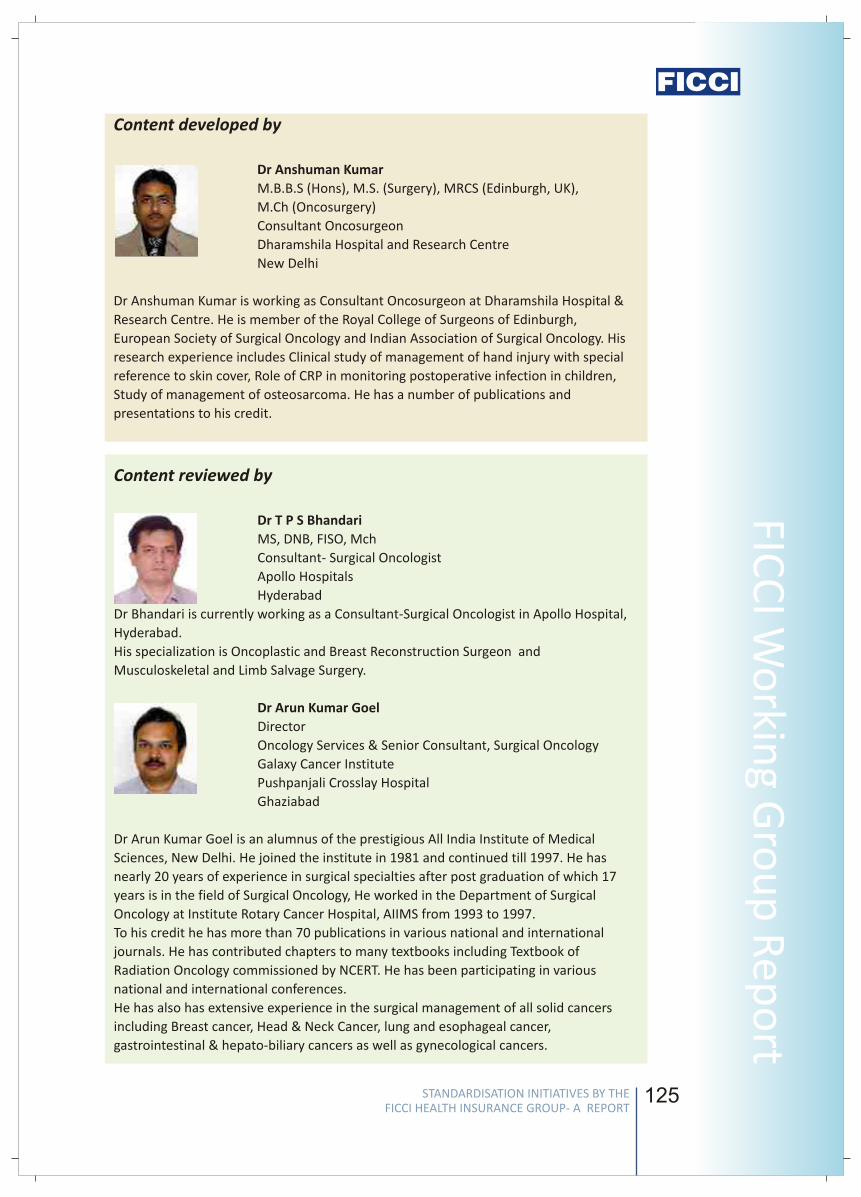
124 125
FICC
I Wo
rking G
rou
p R
epo
rt4. Elevated LDH
5. Male sex
6. Hyponatremia
7. Elevated alkaline phosphatase
Estimated 5-year survival rates are as follows:
o Stage IA - 75%
o Stage IB - 55%
o Stage IIA - 50%
o Stage IIB - 40%
o Stage IIIA - 10-35%
o Stage IIIB - Less than 5%
o Stage IV - Less than 5%
6.1.3 Referral criteria
Ideally all Cancer Lung established cases should be referred for appropriate
treatment to a specialty facility.
6.2 Situation II: At a super specialty facility in a metro where higher-end technology
and resources are available
As in situation I (6.1)
Pleural effusion
Hemoptysis
Pneumothorax
Bronchial obstruction
Pneumonia
Pericardial effusion
Metastasis
NCCN practice guide lines in Oncology 2009
Devita cancer- Textbook
7. Complications
8 . References
v
v
v
v
v
v
v
v
v
Content developed by
Dr Anshuman Kumar
M.B.B.S (Hons), M.S. (Surgery), MRCS (Edinburgh, UK),
M.Ch (Oncosurgery)
Consultant Oncosurgeon
Dharamshila Hospital and Research Centre
New Delhi
Dr Anshuman Kumar is working as Consultant Oncosurgeon at Dharamshila Hospital &
Research Centre. He is member of the Royal College of Surgeons of Edinburgh,
European Society of Surgical Oncology and Indian Association of Surgical Oncology. His
research experience includes Clinical study of management of hand injury with special
reference to skin cover, Role of CRP in monitoring postoperative infection in children,
Study of management of osteosarcoma. He has a number of publications and
presentations to his credit.
Content reviewed by
Dr T P S Bhandari
MS, DNB, FISO, Mch
Consultant- Surgical Oncologist
Apollo Hospitals
Hyderabad
Dr Bhandari is currently working as a Consultant-Surgical Oncologist in Apollo Hospital,
Hyderabad.
His specialization is Oncoplastic and Breast Reconstruction Surgeon and
Musculoskeletal and Limb Salvage Surgery.
Dr Arun Kumar Goel
Director
Oncology Services & Senior Consultant, Surgical Oncology
Galaxy Cancer Institute
Pushpanjali Crosslay Hospital
Ghaziabad
Dr Arun Kumar Goel is an alumnus of the prestigious All India Institute of Medical
Sciences, New Delhi. He joined the institute in 1981 and continued till 1997. He has
nearly 20 years of experience in surgical specialties after post graduation of which 17
years is in the field of Surgical Oncology, He worked in the Department of Surgical
Oncology at Institute Rotary Cancer Hospital, AIIMS from 1993 to 1997.
To his credit he has more than 70 publications in various national and international
journals. He has contributed chapters to many textbooks including Textbook of
Radiation Oncology commissioned by NCERT. He has been participating in various
national and international conferences.
He has also has extensive experience in the surgical management of all solid cancers
including Breast cancer, Head & Neck Cancer, lung and esophageal cancer,
gastrointestinal & hepato-biliary cancers as well as gynecological cancers.
STANDARDISATION INITIATIVES BY THE FICCI HEALTH INSURANCE GROUP- A REPORT
STANDARDISATION INITIATIVES BY THE FICCI HEALTH INSURANCE GROUP- A REPORT
Stan
dar
d T
reat
men
t G
uid
elin
es
126 127
FICC
I Wo
rking G
rou
p R
epo
rt
Dr Ashish Kaushal
MBBS, MD (Internal Medicine), DM (Medical Oncology)
Consultant-Medical Oncology
HCG Medisurge Hospital
Ahmedabad
Dr Ashish Kaushal is presently working as Consultant, Medical Oncology, HCG Medisurge
Hospital, Ahmedabad. His Post Graduate training experience is from NSCB Medical
College Jabalpur. His special skills and procedures are Management of various solid and
hematological malignancies in adult and pediatric patients, High dose chemotherapy
administration and toxicity management and Oncological procedures like –Lumbar
puncture & intrathecal chemotherapy administration, Bone Marrow spiration and
Biopsy, Intravenous line, central line & femoral catheterization, Ascitic tapping and
Pleural Fluid tapping , FNAC of lymph node, lung mass etc. He has a number of
Research Papers to his credit.
Notes
STANDARDISATION INITIATIVES BY THE FICCI HEALTH INSURANCE GROUP- A REPORT
STANDARDISATION INITIATIVES BY THE FICCI HEALTH INSURANCE GROUP- A REPORT
Stan
dar
d T
reat
men
t G
uid
elin
es

126 127
FICC
I Wo
rking G
rou
p R
epo
rtDr Ashish Kaushal
MBBS, MD (Internal Medicine), DM (Medical Oncology)
Consultant-Medical Oncology
HCG Medisurge Hospital
Ahmedabad
Dr Ashish Kaushal is presently working as Consultant, Medical Oncology, HCG Medisurge
Hospital, Ahmedabad. His Post Graduate training experience is from NSCB Medical
College Jabalpur. His special skills and procedures are Management of various solid and
hematological malignancies in adult and pediatric patients, High dose chemotherapy
administration and toxicity management and Oncological procedures like –Lumbar
puncture & intrathecal chemotherapy administration, Bone Marrow spiration and
Biopsy, Intravenous line, central line & femoral catheterization, Ascitic tapping and
Pleural Fluid tapping , FNAC of lymph node, lung mass etc. He has a number of
Research Papers to his credit.
Notes
STANDARDISATION INITIATIVES BY THE FICCI HEALTH INSURANCE GROUP- A REPORT
STANDARDISATION INITIATIVES BY THE FICCI HEALTH INSURANCE GROUP- A REPORT
Stan
dar
d T
reat
men
t G
uid
elin
es
128
Standard Treatment Guidelines
for Peptic Ulcer requiring hospitalisation
1. Introduction/ Definition/ Description
2. Incidence of the condition
. Differential Diagnosis
4. Clinical Diagnosis
The term peptic ulcer disease relates to ulcers which develop in the stomach or
duodenal mucosa and other ectopic areas vulnerable for acid-peptic digestion. It is
secondary to mucosal cell injury resulting from high hydrochloric acid secretion
and/or decreased mucosal defence mechanism. Peptic ulcers are of two types,
gastric ulcers (GU) and duodenal ulcers (DU).
The ratio duodenal ulcer: gastric ulcer was estimated to vary from 0.8:1 in Japan to
19:1 in Africa and as high as 32:1 in India [1]. The male to female ratio also varies
geographically, for example from 1:1 in USA to 18:1 in India [2].
There appears to be a change in the prevalence of peptic ulcer now in India,
especially in the urban areas. In Chandigarh, the point prevalence of active peptic
ulcer was reported to be 3.4% and the life time prevalence 8.8%. The duodenal-to-
gastric ulcer ratio is 12:1[3]. As per study by Khuroo et al the point prevalence of
peptic ulcer in Srinagar was 4.72% and the lifetime prevalence was 11.22%. The
duodenal to gastric ulcer ratio was 17.1:1 [4]. Duodenal and gastric ulcer were
common in men. The prevalence of peptic ulcer increased with age, with a peak
prevalence of 28.8% in the 5th decade of life [4].
3
For both the duodenal and gastric ulcers when present with recurrent episodes
of abdominal pain:
Biliary colic, chronic pancreatitis, subacute intestinal obstruction
When presenting only with acute abdominal pain
Billiary colic, acute pancreatitis, hollow viscus perforation, mesenteric
ischaemia
When presenting with haemetemesis
Portal hypertension with gastro-esophageal variceal bleeding, Mallory-Weiss
syndrome, bleeding from benign and malignant tumours of stomach
When presenting with repeated minor GI bleeding or occult bleeding
All causes of recurrent GI bleeding
The most common symptom is abdominal pain which is mainly described as
burning though it may be perceived as cutting, lancinating, scalding or dull aching.
The pain usually appears on empty stomach, often waking the patient in the early
hours of the day and characteristically relieved by ingestion of food in the majority.
In patients with severe acute duodenal ulcers and prepyloric ulcers, pylorospasm
v
v
v
v
v
v
v
v
129
FICC
I Wo
rking G
rou
p R
epo
rt
may result in vomiting immediately after food. Gastric ulcers tend to occur in
relatively older patients and generally above the age of 40 years with equal
distribution in both genders. Abdominal pain is the common symptom and unlike
the duodenal ulcer it does not have diagnostic characteristics.
Classical historical aspects, presenting complaints and clinical examination are very
useful in the diagnosis of peptic ulcer, especially when complications supervene.
H pylori infection
Nonsteroidal anti-inflammatory drugs or those using corticosteroids with
NSAIDs
Severe physiologic stress
Diseases associated with an increased risk of PUD include cirrhosis, chronic
obstructive pulmonary disease, Chronic renal failure, and organ transplantation
It is necessary for either H pylori infection or usage of nonsteroidal anti-
inflammatory or both be present to produce duodenal ulcer. In other words, in
an endoscopy proved duodenal ulcer, in the absence of NSAID usage, it is safe
to assume that the patient has H pylori infection.
However, no such precondition is applicable for gastric ulcer
Indications for Hospitalization
Acute exacerbation of pain in a known case- for observation and management
Acute abdominal pain in a patient not previously known to have peptic ulcer –
for diagnosis and appropriate therapy
Upper GI bleeding
Perforation of peptic ulcer
Features of gastric outlet obstruction
6.1. Situation 1:
Peptic ulcer and its complications can be managed in most non-metro situations
where infrastructure facilities exist.
6.1.1. Investigations: (Based on Clinical Presentation)
lAcute exacerbation of pain in a known case
lCBC, X-ray chest + abdomen
lAcute abdominal pain in a patient not previously known to have peptic ulcer
lCBC, X-ray chest + abdomen, USG abdomen, Serum amylase, Upper GI
endoscopy with H pylori detection
lUpper GI bleeding
lCBC, Coagulation profile, Upper GI endoscopy with H pylori detection, U/S
Abdomen
5. Causes
6. Management
v
v
v
v
v
v
v
v
v
v
v
STANDARDISATION INITIATIVES BY THE FICCI HEALTH INSURANCE GROUP- A REPORT
STANDARDISATION INITIATIVES BY THE FICCI HEALTH INSURANCE GROUP- A REPORT
Stan
dar
d T
reat
men
t G
uid
elin
es

128
Standard Treatment Guidelines
for Peptic Ulcer requiring hospitalisation
1. Introduction/ Definition/ Description
2. Incidence of the condition
. Differential Diagnosis
4. Clinical Diagnosis
The term peptic ulcer disease relates to ulcers which develop in the stomach or
duodenal mucosa and other ectopic areas vulnerable for acid-peptic digestion. It is
secondary to mucosal cell injury resulting from high hydrochloric acid secretion
and/or decreased mucosal defence mechanism. Peptic ulcers are of two types,
gastric ulcers (GU) and duodenal ulcers (DU).
The ratio duodenal ulcer: gastric ulcer was estimated to vary from 0.8:1 in Japan to
19:1 in Africa and as high as 32:1 in India [1]. The male to female ratio also varies
geographically, for example from 1:1 in USA to 18:1 in India [2].
There appears to be a change in the prevalence of peptic ulcer now in India,
especially in the urban areas. In Chandigarh, the point prevalence of active peptic
ulcer was reported to be 3.4% and the life time prevalence 8.8%. The duodenal-to-
gastric ulcer ratio is 12:1[3]. As per study by Khuroo et al the point prevalence of
peptic ulcer in Srinagar was 4.72% and the lifetime prevalence was 11.22%. The
duodenal to gastric ulcer ratio was 17.1:1 [4]. Duodenal and gastric ulcer were
common in men. The prevalence of peptic ulcer increased with age, with a peak
prevalence of 28.8% in the 5th decade of life [4].
3
For both the duodenal and gastric ulcers when present with recurrent episodes
of abdominal pain:
Biliary colic, chronic pancreatitis, subacute intestinal obstruction
When presenting only with acute abdominal pain
Billiary colic, acute pancreatitis, hollow viscus perforation, mesenteric
ischaemia
When presenting with haemetemesis
Portal hypertension with gastro-esophageal variceal bleeding, Mallory-Weiss
syndrome, bleeding from benign and malignant tumours of stomach
When presenting with repeated minor GI bleeding or occult bleeding
All causes of recurrent GI bleeding
The most common symptom is abdominal pain which is mainly described as
burning though it may be perceived as cutting, lancinating, scalding or dull aching.
The pain usually appears on empty stomach, often waking the patient in the early
hours of the day and characteristically relieved by ingestion of food in the majority.
In patients with severe acute duodenal ulcers and prepyloric ulcers, pylorospasm
v
v
v
v
v
v
v
v
129
FICC
I Wo
rking G
rou
p R
epo
rtmay result in vomiting immediately after food. Gastric ulcers tend to occur in
relatively older patients and generally above the age of 40 years with equal
distribution in both genders. Abdominal pain is the common symptom and unlike
the duodenal ulcer it does not have diagnostic characteristics.
Classical historical aspects, presenting complaints and clinical examination are very
useful in the diagnosis of peptic ulcer, especially when complications supervene.
H pylori infection
Nonsteroidal anti-inflammatory drugs or those using corticosteroids with
NSAIDs
Severe physiologic stress
Diseases associated with an increased risk of PUD include cirrhosis, chronic
obstructive pulmonary disease, Chronic renal failure, and organ transplantation
It is necessary for either H pylori infection or usage of nonsteroidal anti-
inflammatory or both be present to produce duodenal ulcer. In other words, in
an endoscopy proved duodenal ulcer, in the absence of NSAID usage, it is safe
to assume that the patient has H pylori infection.
However, no such precondition is applicable for gastric ulcer
Indications for Hospitalization
Acute exacerbation of pain in a known case- for observation and management
Acute abdominal pain in a patient not previously known to have peptic ulcer –
for diagnosis and appropriate therapy
Upper GI bleeding
Perforation of peptic ulcer
Features of gastric outlet obstruction
6.1. Situation 1:
Peptic ulcer and its complications can be managed in most non-metro situations
where infrastructure facilities exist.
6.1.1. Investigations: (Based on Clinical Presentation)
lAcute exacerbation of pain in a known case
lCBC, X-ray chest + abdomen
lAcute abdominal pain in a patient not previously known to have peptic ulcer
lCBC, X-ray chest + abdomen, USG abdomen, Serum amylase, Upper GI
endoscopy with H pylori detection
lUpper GI bleeding
lCBC, Coagulation profile, Upper GI endoscopy with H pylori detection, U/S
Abdomen
5. Causes
6. Management
v
v
v
v
v
v
v
v
v
v
v
STANDARDISATION INITIATIVES BY THE FICCI HEALTH INSURANCE GROUP- A REPORT
STANDARDISATION INITIATIVES BY THE FICCI HEALTH INSURANCE GROUP- A REPORT
Stan
dar
d T
reat
men
t G
uid
elin
es
130
lPerforation of peptic ulcer
lCBC, serum electrolytes, Blood urea, creatinine, blood culture, X-ray chest
+ abdomen, USG abdomen
lFeatures of gastric outlet obstruction
lCBC, serum electrolytes, Blood urea, creatinine, USG abdomen, Upper GI
endoscopy, barium meal studies
6.1.2. Treatment (Based on Clinical Presentation)
lAcute exacerbation of pain in a known case
lObservation and management with administration of proton Pump
Inhibitors (PPIs)- both oral & parenteral preparations, sucralfate suspension
orally, parenteral analgesics if required, IV fluids if oral intake is not
permitted or accepted
lAcute abdominal pain in a patient not previously known to have peptic ulcer
lOnce diagnosis of peptic ulcer is made, therapy as shown under 6.2.1.
lUpper GI bleeding
lStabilization with IV fluids and blood/ packed cells/ FFP etc
lStoppage of aspirins and other anti-platelet agents
lDetection and correction of coagulopathies, if any
lParenteral high dose administration of PPIs
lEndoscopic diagnosis and control of bleeding with Injection of adrenaline
to reduce bleeding from the ulcers, application of thermal probes, clips etc
for continued control of bleeding
lAnti H pylori therapy, if H pylori was tested and found positive in gastric
mucosal biopsy
lIn case the bleeding is not controlled by endoscopic methods, or the
bleeding site is not seen or there has been a need for > 6 units of blood to
maintain homeostasis, or there has been re-bleeding while in hospital,
such patients may need surgery as a method to control bleeding. The
operative procedure in such case would be duodenotomy/ pylorotomy
with under running of the bleeding vessel.
lPerforation of peptic ulcer
lResuscitation, antibiotics, analgesics and preparation for surgery
lLaparotomy with closure of perforation with or without biopsy with
Graham's omental patch
lDefinitive surgery for duodenal ulcer not recommended, but acceptable
for gastric ulcer
lLaparoscopic management of perforation is a viable alternative, if
facilities and skill levels exist
lInstitution of anti H pylori therapy in post op period, if H pylori was
tested and found positive in gastric mucosal biopsy
131
FICC
I Wo
rking G
rou
p R
epo
rt
l
lCorrection of fluid and electrolyte imbalance
lRyle's tube aspiration and preparation for surgery
lRecommended surgery is truncal vagotomy with a drainage procedure-
Heinecke- Mickulicz or Finney pyloroplasty or alternatively a
gastrojejunostomy
lAntrectomy not recommended as a definitive surgical procedure for
duodenal ulcer, but permissible for gastric ulcer
lAnti H pylori therapy, if H pylori was tested and found positive in gastric
mucosal biopsy
lCurrently there is no definite evidence that endoscopic balloon
dilatation has success rates equivalent to surgery, and hence cannot be
recommended
6.1.3. Referral criteria for a specialist center if
As peptic ulcer and its complications are manageable in most non-metro
situations, the need for referral arises only when the facilities are not available or
when endoscopic attempts at control of bleeding have been unsuccessful. In
addition, some of these patients with very high risk factor may be referred to a
tertiary centre, as adequate ICU care or capability to manage complicated cases
may not be available in non-metro situations.
6.2. Situation 2:
Exactly as shown for Situation I. These centers are better suited to manage
patients with peptic ulcer especially those with high risk factors or requiring critical
care.
6.2.3. Complications
Complications of peptic ulcers may be acute or chronic. The common complication
is bleeding from the ulcer which may present as life threatening exsanguinating
hemetemesis or as slow bleeding or as recurrent bleeding manifest only as
intermittent malena. It is not uncommon for some of these patients to present as
GI bleeding without any pain and some ulcers are detected only when a patient is
investigated for anemia due to chronic blood loss.
The other acute complication is perforation and such a patient may present with
severe excruciating upper abdominal pain. Secondary to perforation of the ulcer
there is efflux of gastric and duodenal contents into the free peritoneal cavity
causing peritonitis. Such patients exhibit signs of hypovolemia and abdominal signs
such as distension, tenderness, guarding and rigidity. A characteristic finding of
hollow viscus perforation is the absence of liver dullness on percussion.
Such patients would need immediate admission, resuscitation, correction of fluid
and electrolyte disturbances and surgery. Untreated peritonitis may be fatal and in
some the infection may be contained by the peritoneal defences as intra-
abdominal abscesses.
Gastric outlet obstruction
STANDARDISATION INITIATIVES BY THE FICCI HEALTH INSURANCE GROUP- A REPORT
STANDARDISATION INITIATIVES BY THE FICCI HEALTH INSURANCE GROUP- A REPORT
Stan
dar
d T
reat
men
t G
uid
elin
es
130
lPerforation of peptic ulcer
lCBC, serum electrolytes, Blood urea, creatinine, blood culture, X-ray chest
+ abdomen, USG abdomen
lFeatures of gastric outlet obstruction
lCBC, serum electrolytes, Blood urea, creatinine, USG abdomen, Upper GI
endoscopy, barium meal studies
6.1.2. Treatment (Based on Clinical Presentation)
lAcute exacerbation of pain in a known case
lObservation and management with administration of proton Pump
Inhibitors (PPIs)- both oral & parenteral preparations, sucralfate suspension
orally, parenteral analgesics if required, IV fluids if oral intake is not
permitted or accepted
lAcute abdominal pain in a patient not previously known to have peptic ulcer
lOnce diagnosis of peptic ulcer is made, therapy as shown under 6.2.1.
lUpper GI bleeding
lStabilization with IV fluids and blood/ packed cells/ FFP etc
lStoppage of aspirins and other anti-platelet agents
lDetection and correction of coagulopathies, if any
lParenteral high dose administration of PPIs
lEndoscopic diagnosis and control of bleeding with Injection of adrenaline
to reduce bleeding from the ulcers, application of thermal probes, clips etc
for continued control of bleeding
lAnti H pylori therapy, if H pylori was tested and found positive in gastric
mucosal biopsy
lIn case the bleeding is not controlled by endoscopic methods, or the
bleeding site is not seen or there has been a need for > 6 units of blood to
maintain homeostasis, or there has been re-bleeding while in hospital,
such patients may need surgery as a method to control bleeding. The
operative procedure in such case would be duodenotomy/ pylorotomy
with under running of the bleeding vessel.
lPerforation of peptic ulcer
lResuscitation, antibiotics, analgesics and preparation for surgery
lLaparotomy with closure of perforation with or without biopsy with
Graham's omental patch
lDefinitive surgery for duodenal ulcer not recommended, but acceptable
for gastric ulcer
lLaparoscopic management of perforation is a viable alternative, if
facilities and skill levels exist
lInstitution of anti H pylori therapy in post op period, if H pylori was
tested and found positive in gastric mucosal biopsy
131
FICC
I Wo
rking G
rou
p R
epo
rtl
lCorrection of fluid and electrolyte imbalance
lRyle's tube aspiration and preparation for surgery
lRecommended surgery is truncal vagotomy with a drainage procedure-
Heinecke- Mickulicz or Finney pyloroplasty or alternatively a
gastrojejunostomy
lAntrectomy not recommended as a definitive surgical procedure for
duodenal ulcer, but permissible for gastric ulcer
lAnti H pylori therapy, if H pylori was tested and found positive in gastric
mucosal biopsy
lCurrently there is no definite evidence that endoscopic balloon
dilatation has success rates equivalent to surgery, and hence cannot be
recommended
6.1.3. Referral criteria for a specialist center if
As peptic ulcer and its complications are manageable in most non-metro
situations, the need for referral arises only when the facilities are not available or
when endoscopic attempts at control of bleeding have been unsuccessful. In
addition, some of these patients with very high risk factor may be referred to a
tertiary centre, as adequate ICU care or capability to manage complicated cases
may not be available in non-metro situations.
6.2. Situation 2:
Exactly as shown for Situation I. These centers are better suited to manage
patients with peptic ulcer especially those with high risk factors or requiring critical
care.
6.2.3. Complications
Complications of peptic ulcers may be acute or chronic. The common complication
is bleeding from the ulcer which may present as life threatening exsanguinating
hemetemesis or as slow bleeding or as recurrent bleeding manifest only as
intermittent malena. It is not uncommon for some of these patients to present as
GI bleeding without any pain and some ulcers are detected only when a patient is
investigated for anemia due to chronic blood loss.
The other acute complication is perforation and such a patient may present with
severe excruciating upper abdominal pain. Secondary to perforation of the ulcer
there is efflux of gastric and duodenal contents into the free peritoneal cavity
causing peritonitis. Such patients exhibit signs of hypovolemia and abdominal signs
such as distension, tenderness, guarding and rigidity. A characteristic finding of
hollow viscus perforation is the absence of liver dullness on percussion.
Such patients would need immediate admission, resuscitation, correction of fluid
and electrolyte disturbances and surgery. Untreated peritonitis may be fatal and in
some the infection may be contained by the peritoneal defences as intra-
abdominal abscesses.
Gastric outlet obstruction
STANDARDISATION INITIATIVES BY THE FICCI HEALTH INSURANCE GROUP- A REPORT
STANDARDISATION INITIATIVES BY THE FICCI HEALTH INSURANCE GROUP- A REPORT
Stan
dar
d T
reat
men
t G
uid
elin
es
132
7. References
1. Lam SK. Peptic ulcer: from epidemiology to cause. J Gastroenterol Hepatol. 1989;4
Suppl 2:1-6.
2. Lam SK Epidemiology and genetics of peptic ulcer. Gastroenterol Jpn. 1993;28
Suppl 5:145-57.
3. Singh V, Trikha B, Nain CK, Singh K, Vaiphei K. Epidemiology of Helicobacter pylori
and peptic ulcer in India. J Gastroenterol Hepatol. 2002;17:659-65.
4. M S Khuroo, R Mahajan, S A Zargar, G Javid, and S Munshi. Prevalence of peptic
ulcer in India: an endoscopic and epidemiological study in urban Kashmir.Gut.
1989; 30: 930–934.
5. Jais M, Barua S. Seroprevalence of anti Helicobacter pylori IgG/IgA in
asymptomatic population from Delhi. J Commun Dis. 2004;36:132-5.
6. Scheiman JM, Yeomans ND, Talley NJ, Vakil N, Chan FK, Tulassay Z, Rainoldi JL,
Szczepanski L, Ung KA, Kleczkowski D, Ahlbom H, Naesdal J, Hawkey C. Prevention
of ulcers by esomeprazole in at-risk patients using non-selective NSAIDs and COX-2
inhibitors. Am J Gastroenterol. 2006;101:701-10.
7. Ahuja V. The case for Helicobacter pylori eradication in India: sensationalism,
skepticism and scientific salesmanship. Indian J Gastroenterol 2006;25:20-24.
8. Ford A, Delaney B, Forman D, Moayyedi P. Eradication therapy for peptic ulcer
disease in Helicobacter pylori positive patients. Cochrane Database Syst Rev. 2006;
19:CD003840.
9. Thyagarajan SP, Ray P, Das BK, Ayyagari A, Khan AA, Dharmalingam S, Rao UA,
Rajasambandam P, Ramathilagam B, Bhasin D, Sharma MP, Naik SR, Habibullah CM.
Geographical difference in antimicrobial resistance pattern of Helicobacter pylori
clinical isolates from Indian patients: Multicentric study. J Gastroenterol Hepatol.
2003;18:1373-8.
10. Zargar SA, Javid G, Khan BA, Yattoo GN, Shah AH, Gulzar GM, Sodhi JS, Mujeeb SA,
Khan MA, Shah NA, Shafi HM.Pantoprazole infusion as adjuvant therapy to
endoscopic treatment in patients with peptic ulcer bleeding: prospective
randomized controlled trial. J Gastroenterol Hepatol. 2006;21:716-21.
11. Bhogal RH, Athwal R, Durkin D, Deakin M, Cheruvu CN. Comparison between open
and laparoscopic repair of perforated peptic ulcer disease. World J Surg.
2008;32:2371-4.
133
FICC
I Wo
rking G
rou
p R
epo
rt
Content developed by
Dr V Baskaran
MS, PhD, FACS, FICS,FIAGES
Senior Consultant
Department of Surgical Gastroenterology
Dr BL Kapur Memorial Hospital
New Delhi
A dynamic gastrointestinal and advanced laparoscopic surgeon, with immense body of
surgical skills and academic credentials, including a PhD in gastrointestinal surgery. Has
enormous scientific publications to his credit. Now in practice after 29 years in the army,
this former Professor of Surgery, AFMC, Pune practices evidence based surgery.
Content reviewed by
Dr S C Samal
DM (Gastroenterology)
Senior Consultant Gastroenterologist
Apollo Hospital
Hyderabad
Dr S C Samal is Senior Consultant Gastroenterologist in Apollo Hospital, Hyderabad. He
completed his DM in Gastroenterology from CMC, Vellore in 1995 and worked there as
faculty till 2001. He has more than 15 publications in national and international
journals.
STANDARDISATION INITIATIVES BY THE FICCI HEALTH INSURANCE GROUP- A REPORT
STANDARDISATION INITIATIVES BY THE FICCI HEALTH INSURANCE GROUP- A REPORT
Stan
dar
d T
reat
men
t G
uid
elin
es
132
7. References
1. Lam SK. Peptic ulcer: from epidemiology to cause. J Gastroenterol Hepatol. 1989;4
Suppl 2:1-6.
2. Lam SK Epidemiology and genetics of peptic ulcer. Gastroenterol Jpn. 1993;28
Suppl 5:145-57.
3. Singh V, Trikha B, Nain CK, Singh K, Vaiphei K. Epidemiology of Helicobacter pylori
and peptic ulcer in India. J Gastroenterol Hepatol. 2002;17:659-65.
4. M S Khuroo, R Mahajan, S A Zargar, G Javid, and S Munshi. Prevalence of peptic
ulcer in India: an endoscopic and epidemiological study in urban Kashmir.Gut.
1989; 30: 930–934.
5. Jais M, Barua S. Seroprevalence of anti Helicobacter pylori IgG/IgA in
asymptomatic population from Delhi. J Commun Dis. 2004;36:132-5.
6. Scheiman JM, Yeomans ND, Talley NJ, Vakil N, Chan FK, Tulassay Z, Rainoldi JL,
Szczepanski L, Ung KA, Kleczkowski D, Ahlbom H, Naesdal J, Hawkey C. Prevention
of ulcers by esomeprazole in at-risk patients using non-selective NSAIDs and COX-2
inhibitors. Am J Gastroenterol. 2006;101:701-10.
7. Ahuja V. The case for Helicobacter pylori eradication in India: sensationalism,
skepticism and scientific salesmanship. Indian J Gastroenterol 2006;25:20-24.
8. Ford A, Delaney B, Forman D, Moayyedi P. Eradication therapy for peptic ulcer
disease in Helicobacter pylori positive patients. Cochrane Database Syst Rev. 2006;
19:CD003840.
9. Thyagarajan SP, Ray P, Das BK, Ayyagari A, Khan AA, Dharmalingam S, Rao UA,
Rajasambandam P, Ramathilagam B, Bhasin D, Sharma MP, Naik SR, Habibullah CM.
Geographical difference in antimicrobial resistance pattern of Helicobacter pylori
clinical isolates from Indian patients: Multicentric study. J Gastroenterol Hepatol.
2003;18:1373-8.
10. Zargar SA, Javid G, Khan BA, Yattoo GN, Shah AH, Gulzar GM, Sodhi JS, Mujeeb SA,
Khan MA, Shah NA, Shafi HM.Pantoprazole infusion as adjuvant therapy to
endoscopic treatment in patients with peptic ulcer bleeding: prospective
randomized controlled trial. J Gastroenterol Hepatol. 2006;21:716-21.
11. Bhogal RH, Athwal R, Durkin D, Deakin M, Cheruvu CN. Comparison between open
and laparoscopic repair of perforated peptic ulcer disease. World J Surg.
2008;32:2371-4.
133
FICC
I Wo
rking G
rou
p R
epo
rtContent developed by
Dr V Baskaran
MS, PhD, FACS, FICS,FIAGES
Senior Consultant
Department of Surgical Gastroenterology
Dr BL Kapur Memorial Hospital
New Delhi
A dynamic gastrointestinal and advanced laparoscopic surgeon, with immense body of
surgical skills and academic credentials, including a PhD in gastrointestinal surgery. Has
enormous scientific publications to his credit. Now in practice after 29 years in the army,
this former Professor of Surgery, AFMC, Pune practices evidence based surgery.
Content reviewed by
Dr S C Samal
DM (Gastroenterology)
Senior Consultant Gastroenterologist
Apollo Hospital
Hyderabad
Dr S C Samal is Senior Consultant Gastroenterologist in Apollo Hospital, Hyderabad. He
completed his DM in Gastroenterology from CMC, Vellore in 1995 and worked there as
faculty till 2001. He has more than 15 publications in national and international
journals.
STANDARDISATION INITIATIVES BY THE FICCI HEALTH INSURANCE GROUP- A REPORT
STANDARDISATION INITIATIVES BY THE FICCI HEALTH INSURANCE GROUP- A REPORT
Stan
dar
d T
reat
men
t G
uid
elin
es
134
Standard Treatment Guidelines for
Management of Renal Stones
1. Introduction/ Definition/ Description
2. Incidence of the condition
3. Causes/ risk factors
A kidney stone is a hard mass developed from crystals that separate from the urine
within the urinary tract. Kidney stones may contain various combinations of
chemicals. The most common type of stone contains calcium in combination with
either oxalate or phosphate. These chemicals are part of a person's normal diet
and make up important parts of the body, such as bones and muscles.
Renal stones are commonly found in three different sites
Kidney
Bladder
Ureter
Stones in urinary system almost always develop first in kidney but stones can go
and grow in ureter and urinary bladder.
Prevalence 2-3%
Two to three times more common in males
More common in adults than in elderly pts and lesser in children
Climatic factors: Hot , Arid areas and in temperate regions
3.1. Kidney stone:
Genetic predisposition, underlying metabolic diseases like renal tubular
acidosis or hyperparathyroidism and some dietary factors (water fluoridation)
have been associated with development of renal stones.
Hypercalciuria is inherited, and it may be the cause of stones in more than half
of patients. Calcium is absorbed from food in excess and is lost into the urine.
This high level of calcium in the urine causes crystals of calcium oxalate or
calcium phosphate to form in the kidneys or elsewhere in the urinary tract.
Other causes of kidney stones are hyperuricosuria, which is a disorder of uric
acid metabolism; gout; excess intake of vitamin D; urinary tract infections; and
blockage of the urinary tract. Certain diuretics, commonly called water pills
and calcium-based antacids may increase the risk of forming kidney stones by
increasing the amount of calcium in the urine.
Calcium oxalate stones may also form in people who have chronic
inflammation of the bowel or who have had an intestinal bypass operation, or
ostomy surgery. As mentioned earlier, struvite stones can form in people who
have had a urinary tract infection. People who take the protease inhibitor
v
v
v
v
v
v
v
n
n
n
n
135
FICC
I Wo
rking G
rou
p R
epo
rt
indinavir, a medicine used to treat HIV infection, may also be at increased risk
of developing kidney stones.
3.2. Ureter stone:
Majority of Ureteric stones are formed in the kidney and migrate into the
ureter
Ureteric stones may be formed in the ureter secondary to the following:
nUreterocele
nNeoplasms
nUreter with blind endings
nDilated segments of ureter proximal to stricture
3.3. Bladder stones:
Primary bladder stones were seen in children. However with the improvement
in nutritional status the incidence has decreased
Secondary bladder stones
nBPH
nBladder neck obstruction
nStricture urethra
nNeurogenic bladder
nPosterior urethral valves
nUreteric stones
Symptoms similar to renal colic may be elicited due to non calculus conditions or
other causes of abdominal pain, such as appendicitis, cholecystitis, diverticulitis,
colitis, constipation, hernias or severe constipation. These conditions should be
ruled out.
Uretero or ureteropelvic obstruction may present as renal calculus as well.
In women, ovarian torsion, cyst or ectopic pregnancy should be ruled out. In men
testicular inflammation (prostatitis or epididymitis) may mimic the presentation of
ureteral stone.
Kidney stones often do not cause any symptoms. Usually, the first symptom of a
kidney stone is extreme pain, which begins suddenly when a stone moves in the
urinary tract and blocks the flow of urine. Typically, a person feels a sharp,
cramping pain in the back and side in the area of the kidney or in the lower
abdomen. Sometimes nausea and vomiting occur. Later, pain may spread to the
groin.
If the stone is too large to pass easily, pain continues as the muscles in the wall of
the narrow ureter try to squeeze the stone into the bladder. As the stone moves
and the body try to push it out, blood may appear in the urine, making the urine
n
n
n
n
4. Differential diagnosis
5. Symptoms of urinary stones
STANDARDISATION INITIATIVES BY THE FICCI HEALTH INSURANCE GROUP- A REPORT
STANDARDISATION INITIATIVES BY THE FICCI HEALTH INSURANCE GROUP- A REPORT
Stan
dar
d T
reat
men
t G
uid
elin
es
134
Standard Treatment Guidelines for
Management of Renal Stones
1. Introduction/ Definition/ Description
2. Incidence of the condition
3. Causes/ risk factors
A kidney stone is a hard mass developed from crystals that separate from the urine
within the urinary tract. Kidney stones may contain various combinations of
chemicals. The most common type of stone contains calcium in combination with
either oxalate or phosphate. These chemicals are part of a person's normal diet
and make up important parts of the body, such as bones and muscles.
Renal stones are commonly found in three different sites
Kidney
Bladder
Ureter
Stones in urinary system almost always develop first in kidney but stones can go
and grow in ureter and urinary bladder.
Prevalence 2-3%
Two to three times more common in males
More common in adults than in elderly pts and lesser in children
Climatic factors: Hot , Arid areas and in temperate regions
3.1. Kidney stone:
Genetic predisposition, underlying metabolic diseases like renal tubular
acidosis or hyperparathyroidism and some dietary factors (water fluoridation)
have been associated with development of renal stones.
Hypercalciuria is inherited, and it may be the cause of stones in more than half
of patients. Calcium is absorbed from food in excess and is lost into the urine.
This high level of calcium in the urine causes crystals of calcium oxalate or
calcium phosphate to form in the kidneys or elsewhere in the urinary tract.
Other causes of kidney stones are hyperuricosuria, which is a disorder of uric
acid metabolism; gout; excess intake of vitamin D; urinary tract infections; and
blockage of the urinary tract. Certain diuretics, commonly called water pills
and calcium-based antacids may increase the risk of forming kidney stones by
increasing the amount of calcium in the urine.
Calcium oxalate stones may also form in people who have chronic
inflammation of the bowel or who have had an intestinal bypass operation, or
ostomy surgery. As mentioned earlier, struvite stones can form in people who
have had a urinary tract infection. People who take the protease inhibitor
v
v
v
v
v
v
v
n
n
n
n
135
FICC
I Wo
rking G
rou
p R
epo
rtindinavir, a medicine used to treat HIV infection, may also be at increased risk
of developing kidney stones.
3.2. Ureter stone:
Majority of Ureteric stones are formed in the kidney and migrate into the
ureter
Ureteric stones may be formed in the ureter secondary to the following:
nUreterocele
nNeoplasms
nUreter with blind endings
nDilated segments of ureter proximal to stricture
3.3. Bladder stones:
Primary bladder stones were seen in children. However with the improvement
in nutritional status the incidence has decreased
Secondary bladder stones
nBPH
nBladder neck obstruction
nStricture urethra
nNeurogenic bladder
nPosterior urethral valves
nUreteric stones
Symptoms similar to renal colic may be elicited due to non calculus conditions or
other causes of abdominal pain, such as appendicitis, cholecystitis, diverticulitis,
colitis, constipation, hernias or severe constipation. These conditions should be
ruled out.
Uretero or ureteropelvic obstruction may present as renal calculus as well.
In women, ovarian torsion, cyst or ectopic pregnancy should be ruled out. In men
testicular inflammation (prostatitis or epididymitis) may mimic the presentation of
ureteral stone.
Kidney stones often do not cause any symptoms. Usually, the first symptom of a
kidney stone is extreme pain, which begins suddenly when a stone moves in the
urinary tract and blocks the flow of urine. Typically, a person feels a sharp,
cramping pain in the back and side in the area of the kidney or in the lower
abdomen. Sometimes nausea and vomiting occur. Later, pain may spread to the
groin.
If the stone is too large to pass easily, pain continues as the muscles in the wall of
the narrow ureter try to squeeze the stone into the bladder. As the stone moves
and the body try to push it out, blood may appear in the urine, making the urine
n
n
n
n
4. Differential diagnosis
5. Symptoms of urinary stones
STANDARDISATION INITIATIVES BY THE FICCI HEALTH INSURANCE GROUP- A REPORT
STANDARDISATION INITIATIVES BY THE FICCI HEALTH INSURANCE GROUP- A REPORT
Stan
dar
d T
reat
men
t G
uid
elin
es
136
pink. As the stone moves down the ureter, closer to the bladder, a person may feel
the need to urinate more often or feel a burning sensation during urination.
If fever and chills accompany any of these symptoms, an infection may be present,
requiring urgent medical attention.
Renal Colic
Hematuria
Recurrent UTI
Fever and Sepsis
Chronic renal failure
Gastro-intestinal symptoms
Back ache
Lower urinary tract symptoms
Surgery may not be necessary and most kidney stones can pass through the
urinary system with plenty of water.
A simple and most important lifestyle change to prevent stones is to drink more
liquids—water being the best. Someone who tends to form stones should try to
drink enough liquids throughout the day to produce at least 2 liters of urine in
every 24-hour period.
Patients may be told to avoid food with added vitamin D and certain types of
antacids that have a calcium base. Someone who has highly acidic urine may need
to eat less meat, fish, and poultry. These foods increase the amount of acid in the
urine.
To prevent cystine stones, a person should drink enough water each day to dilute
the concentration of cystine that escapes into the urine, which may be difficult.
More than a gallon of water may be needed every 24 hours, and a third of that
must be drunk during the night.
Medical Management
A doctor may prescribe certain medications to help prevent calcium and uric acid
stones. These medicines control the amount of acid or alkali in the urine, key
factors in crystal formation. The medicine allopurinol may also be useful in some
cases of hyperuricosuria.
Doctors usually try to control hypercalciuria, and thus prevent calcium stones, by
prescribing certain diuretics, such as hydrochlorothiazide. These medicines
decrease the amount of calcium released by the kidneys into the urine by favoring
calcium retention in bone. They work best when sodium intake is low.
Rarely, patients with hypercalciuria are given the medicine sodium cellulose
phosphate, which binds calcium in the intestines and prevents it from leaking into
the urine.
6. Clinical Diagnosis
7. Management / Modalities of treatment
v
v
v
v
v
v
v
v
n
137
FICC
I Wo
rking G
rou
p R
epo
rt
If cystine stones cannot be controlled by drinking more fluids, a doctor may
prescribe medicines such as Thiola and Cuprimine, which help reduce the amount
of cystine in the urine.
For struvite stones that have been totally removed, the first line of prevention is to
keep the urine free of bacteria that can cause infection. A patient's urine will be
tested regularly to ensure no bacteria are present.
If struvite stones cannot be removed, a doctor may prescribe a medicine called
acetohydroxamic acid (AHA). AHA is used with long-term antibiotic medicines to
prevent the infection that leads to stone growth.
People with hyperparathyroidism sometimes develop calcium stones. Treatment in
these cases is usually surgery to remove the parathyroid glands, which are located
in the neck. In most cases, only one of the glands is enlarged. Removing the glands
cures the patient's problem with hyperparathyroidism and kidney stones.
Surgery may be needed to remove a kidney stone if it
ldoes not pass after a reasonable period of time and causes constant pain
lis too large to pass on its own or is caught in a difficult place
lblocks the flow of urine
lcauses an ongoing urinary tract infection
ldamages kidney tissue or causes constant bleeding
lhas grown larger, as seen on follow-up x rays
Surgical removal of Stone
lOpen Pyelolithotomy involves surgical removal of stones from the renal pelvis
via an abdominal incision.
lPercutaneous nephrolithotomy (PCNL) is a surgical procedure by which stones
in the kidney or the upper ureter are removed by making a small incision in the
flank.
lExtracorporeal shock wave lithotripsy (ESWL) is a non-invasive technique used
for the treatment of kidney stones by generation of acoustic shock waves
produced outside the body that are focused on the stone via a coupling
medium to shatter it to pieces. The pieces are then allowed to pass out
naturally through the urinary tract.
lRetrograde intra- renal surgery: involves use of flexible ureteroscope to locate
the stone in the kidney, and use of lasers to break stones and baskets and
forceps are used to remove the stones
lLaparoscopic surgery: has limited benefit over PCNL. However the role is
coming up in many new situations. In carefully selected patients, laparoscopic
and endourological techniques can be successfully combined in a one
procedure solution that deals with complex stone disease and repairs
underlying urinary anomalies.
lLaser lithotripsy: It breaks the stones into smaller pieces and can break all type
of stones. It has a low retropulsion risk
nSurgical Management
STANDARDISATION INITIATIVES BY THE FICCI HEALTH INSURANCE GROUP- A REPORT
STANDARDISATION INITIATIVES BY THE FICCI HEALTH INSURANCE GROUP- A REPORT
Stan
dar
d T
reat
men
t G
uid
elin
es
136
pink. As the stone moves down the ureter, closer to the bladder, a person may feel
the need to urinate more often or feel a burning sensation during urination.
If fever and chills accompany any of these symptoms, an infection may be present,
requiring urgent medical attention.
Renal Colic
Hematuria
Recurrent UTI
Fever and Sepsis
Chronic renal failure
Gastro-intestinal symptoms
Back ache
Lower urinary tract symptoms
Surgery may not be necessary and most kidney stones can pass through the
urinary system with plenty of water.
A simple and most important lifestyle change to prevent stones is to drink more
liquids—water being the best. Someone who tends to form stones should try to
drink enough liquids throughout the day to produce at least 2 liters of urine in
every 24-hour period.
Patients may be told to avoid food with added vitamin D and certain types of
antacids that have a calcium base. Someone who has highly acidic urine may need
to eat less meat, fish, and poultry. These foods increase the amount of acid in the
urine.
To prevent cystine stones, a person should drink enough water each day to dilute
the concentration of cystine that escapes into the urine, which may be difficult.
More than a gallon of water may be needed every 24 hours, and a third of that
must be drunk during the night.
Medical Management
A doctor may prescribe certain medications to help prevent calcium and uric acid
stones. These medicines control the amount of acid or alkali in the urine, key
factors in crystal formation. The medicine allopurinol may also be useful in some
cases of hyperuricosuria.
Doctors usually try to control hypercalciuria, and thus prevent calcium stones, by
prescribing certain diuretics, such as hydrochlorothiazide. These medicines
decrease the amount of calcium released by the kidneys into the urine by favoring
calcium retention in bone. They work best when sodium intake is low.
Rarely, patients with hypercalciuria are given the medicine sodium cellulose
phosphate, which binds calcium in the intestines and prevents it from leaking into
the urine.
6. Clinical Diagnosis
7. Management / Modalities of treatment
v
v
v
v
v
v
v
v
n
137
FICC
I Wo
rking G
rou
p R
epo
rtIf cystine stones cannot be controlled by drinking more fluids, a doctor may
prescribe medicines such as Thiola and Cuprimine, which help reduce the amount
of cystine in the urine.
For struvite stones that have been totally removed, the first line of prevention is to
keep the urine free of bacteria that can cause infection. A patient's urine will be
tested regularly to ensure no bacteria are present.
If struvite stones cannot be removed, a doctor may prescribe a medicine called
acetohydroxamic acid (AHA). AHA is used with long-term antibiotic medicines to
prevent the infection that leads to stone growth.
People with hyperparathyroidism sometimes develop calcium stones. Treatment in
these cases is usually surgery to remove the parathyroid glands, which are located
in the neck. In most cases, only one of the glands is enlarged. Removing the glands
cures the patient's problem with hyperparathyroidism and kidney stones.
Surgery may be needed to remove a kidney stone if it
ldoes not pass after a reasonable period of time and causes constant pain
lis too large to pass on its own or is caught in a difficult place
lblocks the flow of urine
lcauses an ongoing urinary tract infection
ldamages kidney tissue or causes constant bleeding
lhas grown larger, as seen on follow-up x rays
Surgical removal of Stone
lOpen Pyelolithotomy involves surgical removal of stones from the renal pelvis
via an abdominal incision.
lPercutaneous nephrolithotomy (PCNL) is a surgical procedure by which stones
in the kidney or the upper ureter are removed by making a small incision in the
flank.
lExtracorporeal shock wave lithotripsy (ESWL) is a non-invasive technique used
for the treatment of kidney stones by generation of acoustic shock waves
produced outside the body that are focused on the stone via a coupling
medium to shatter it to pieces. The pieces are then allowed to pass out
naturally through the urinary tract.
lRetrograde intra- renal surgery: involves use of flexible ureteroscope to locate
the stone in the kidney, and use of lasers to break stones and baskets and
forceps are used to remove the stones
lLaparoscopic surgery: has limited benefit over PCNL. However the role is
coming up in many new situations. In carefully selected patients, laparoscopic
and endourological techniques can be successfully combined in a one
procedure solution that deals with complex stone disease and repairs
underlying urinary anomalies.
lLaser lithotripsy: It breaks the stones into smaller pieces and can break all type
of stones. It has a low retropulsion risk
nSurgical Management
STANDARDISATION INITIATIVES BY THE FICCI HEALTH INSURANCE GROUP- A REPORT
STANDARDISATION INITIATIVES BY THE FICCI HEALTH INSURANCE GROUP- A REPORT
Stan
dar
d T
reat
men
t G
uid
elin
es
138
8. Indications for surgery
8.1 Indications ESWL
Absence of obstruction
Stone burden< than 2 cm
Superior and middle calyceal stones
or Mixed uric acid stones
Favorable anatomy allowing easy passage of stone particles
Note: Stones in upper ureter may be pushed back into the kidney by a
ureteroscope and subjected to ESWL. Repeated ESWL interventions may be2
indicated for larger stones.
Failure of ESWL may require alternative treatment SOS, like PCNL
8.2 Indications PCNL
Large stones (bigger than 2 cm)
Staghorn calculus or Anatomic abnormalities like horse shoe kidney, calyceal
diverticulum, scoliosis etc.
Stones unresponsive to ESWL
More radio-opaque(hard) stones
Coexisting obstructive uropathy
8.3 Indications Open pyelolithotomy
Large stones that are inaccessible to ESWL or PCNL or failed ESWL or PCN
leading to conversion to open pyelolithotomy
Obstruction with impending parenchymal renal loss
· In presence of morbid obesity
9.1 Situation 1:
9.1.1 Investigations
lUrine analysis and urine culture
lPlain X ray KUB
lAbdominal ultrasonography
9.1.2 Treatment:
Diagnosis
lConservative management
lReferral
lSurgical intervention (if skills/ resources available)
9.1.3 Referral criteria to a specialist centre if:
lSurgical indications met for PCNL or ESWL surgical intervention
lFor optimal investigation and management
n
n
n
n
n
n
n
n
n
n
n
n
n
Kidney are functioning well
139
FICC
I Wo
rking G
rou
p R
epo
rt
9.2 Situation 2:
9.2.1 Investigations:
9.2.1.1 Routine Investigations:
lUrine analysis and urine culture
lAbdominal ultrasonography:
Note: accessible, sensitive investigation (detects renal calculi and hydronephrosis)
although limited application for ureteral stone.
lPlain X–ray abdomen KUB: Detects the size and location of calcium
containing stones but may not show pure uric acid, cystine magnesium
ammonium phosphate stones or stones over bones
9.2.1.2 Additional investigations (with specific indications)
lIntra-venous pyelogram
lRetrograde Ureterography (if ureter can not be seen on IVP)
lHelical CT scan (Sensitivity 95-100%, Specificity 94-96%)
lDTPA Scan
lDMSA Scan
lMRI Scan
9.2.1.3 Pre Operative-Investigations:
lHemogram
lBlood Sugar
lBlood Urea
lSerum Creatinine
lSodium & Potassium
lECG
lCXR
lBT-CT
lPlatelet count
A physician consultation for older patients, patients with history of diabetes,
hypertension, asthma, IHD or other medical co-morbidity for fitness to a
procedure/surgery is recommended.
9.3 Admission criteria:
Surgical intervention confirmed and indicated
Pre-operative care
Urine C/S and Antibiotics especially for Staghorn Renal Stones
Antibiotics, pain management and drainage monitoring.
10. Post Operative Care
STANDARDISATION INITIATIVES BY THE FICCI HEALTH INSURANCE GROUP- A REPORT
STANDARDISATION INITIATIVES BY THE FICCI HEALTH INSURANCE GROUP- A REPORT
Stan
dar
d T
reat
men
t G
uid
elin
es
138
8. Indications for surgery
8.1 Indications ESWL
Absence of obstruction
Stone burden< than 2 cm
Superior and middle calyceal stones
or Mixed uric acid stones
Favorable anatomy allowing easy passage of stone particles
Note: Stones in upper ureter may be pushed back into the kidney by a
ureteroscope and subjected to ESWL. Repeated ESWL interventions may be2
indicated for larger stones.
Failure of ESWL may require alternative treatment SOS, like PCNL
8.2 Indications PCNL
Large stones (bigger than 2 cm)
Staghorn calculus or Anatomic abnormalities like horse shoe kidney, calyceal
diverticulum, scoliosis etc.
Stones unresponsive to ESWL
More radio-opaque(hard) stones
Coexisting obstructive uropathy
8.3 Indications Open pyelolithotomy
Large stones that are inaccessible to ESWL or PCNL or failed ESWL or PCN
leading to conversion to open pyelolithotomy
Obstruction with impending parenchymal renal loss
· In presence of morbid obesity
9.1 Situation 1:
9.1.1 Investigations
lUrine analysis and urine culture
lPlain X ray KUB
lAbdominal ultrasonography
9.1.2 Treatment:
Diagnosis
lConservative management
lReferral
lSurgical intervention (if skills/ resources available)
9.1.3 Referral criteria to a specialist centre if:
lSurgical indications met for PCNL or ESWL surgical intervention
lFor optimal investigation and management
n
n
n
n
n
n
n
n
n
n
n
n
n
Kidney are functioning well
139
FICC
I Wo
rking G
rou
p R
epo
rt9.2 Situation 2:
9.2.1 Investigations:
9.2.1.1 Routine Investigations:
lUrine analysis and urine culture
lAbdominal ultrasonography:
Note: accessible, sensitive investigation (detects renal calculi and hydronephrosis)
although limited application for ureteral stone.
lPlain X–ray abdomen KUB: Detects the size and location of calcium
containing stones but may not show pure uric acid, cystine magnesium
ammonium phosphate stones or stones over bones
9.2.1.2 Additional investigations (with specific indications)
lIntra-venous pyelogram
lRetrograde Ureterography (if ureter can not be seen on IVP)
lHelical CT scan (Sensitivity 95-100%, Specificity 94-96%)
lDTPA Scan
lDMSA Scan
lMRI Scan
9.2.1.3 Pre Operative-Investigations:
lHemogram
lBlood Sugar
lBlood Urea
lSerum Creatinine
lSodium & Potassium
lECG
lCXR
lBT-CT
lPlatelet count
A physician consultation for older patients, patients with history of diabetes,
hypertension, asthma, IHD or other medical co-morbidity for fitness to a
procedure/surgery is recommended.
9.3 Admission criteria:
Surgical intervention confirmed and indicated
Pre-operative care
Urine C/S and Antibiotics especially for Staghorn Renal Stones
Antibiotics, pain management and drainage monitoring.
10. Post Operative Care
STANDARDISATION INITIATIVES BY THE FICCI HEALTH INSURANCE GROUP- A REPORT
STANDARDISATION INITIATIVES BY THE FICCI HEALTH INSURANCE GROUP- A REPORT
Stan
dar
d T
reat
men
t G
uid
elin
es
140
Prevention of renal stones: A person who has had more than one kidney stone
may be likely to form another; so, if possible, prevention is important. To help
determine their cause, the doctor will order laboratory tests, including urine and
blood tests. The doctor will also ask about the patient's medical history,
occupation, and eating habits. If a stone has been removed, or if the patient has
passed a stone and saved it, a stone analysis by the laboratory may help the doctor
in planning treatment.
The doctor may ask the patient to collect urine for 24 hours after a stone has
passed or been removed. The collection is used to measure urine volume and
levels of acidity, calcium, sodium, uric acid, oxalate, citrate, and creatinine - a
product of muscle metabolism. The doctor will use this information to determine
the cause of the stone. A second 24-hour urine collection may be needed to
determine whether the prescribed treatment is working.
11.1 Complications of ESWL
Renal / ureteric colic due to passage of stone fragments or Steinstrasse
(column of stone fragments in the ureter)
Urinary tract infection or Hematuria
Renal parenchymal damage or Obstructive uropathy
Surrounding organ injury such as lung contusion, pancreatitis, splenic
hematoma, intestinal injury etc.
11.2 Complications of retrograde intra renal surgery
Reported complications are minor.
Postoperative colic rates are reported in 3.5-9%. Postoperative pyelonephritis
and gross hematuria, occur in less than 3% of the cases.
Major complications are extremely rare. Major perforation is reported in
approximately 1% of the cases. The risk of postoperative stricture of the ureter
is under 1%
11.3 Complications of open surgery
Superficial wound infection
Urinary tract infection or Pyelonephritis
Retained stone fragments or obstruction
Ureteral / renal pelvic scarring
Urine leak or urinary fistula (to skin or bowel)
Bleeding or Arteriovenous malformations
Injury to pleura or lung with pneumothorax
11.4 Complications of PCNL
Septicemia
Hemorrhage which may require blood transfusion(s)
11. Complications
n
n
n
n
n
n
n
n
n
n
n
n
n
n
n
141
FICC
I Wo
rking G
rou
p R
epo
rt
Hemopneumothorax
11.5 Complications of laparoscopic surgery
Similar to any other laparoscopic surgery
11.6 Complications of laser lithotripsy
Bleeding (1-10%).AV fistula or pseudo-aneurysm requires emergency
embolization <0.5%
Pneumothorax or pleural effusion (4-12%)
Injury to colon or spleen
Fluid absorption
Infection and septicemia
1. Kumar A, Verma BS, Gogoi S, Kapoor R, Srivastava A, Mandhani A. A prospective
randomized trial of open surgery versus endourological stone removal in patients
of staghorn stones with chronic renal failure. Indian J Urol 2001;18:14-9
2. S Das. A concise textbook of surgery. 4th edition. 2007;
3. Achleshwar Dayal, Karthikeyan Selvaraju, G.G. Laxman Prabhu: Prospective study
of Percutaneous Nephrolithotripsy as monotherapy in treatment of renal calculi .
The Internet Journal of Urology.2008;5 (2)
4. U G Oza, Hema K, EXTRACORPOREAL SHOCK WAVE LITHOTRIPSY (ESWL) Removal
of Stones without Surgery bhj July 1999
5. Rao, et al, The relative cost-effectiveness of PCNL and ESWL for medium sized ( < 2
cms) renal calculi in a tertiary care urological referral centre. Indian Journal of
Urology, 2001, Volume 17 issue 2: 121-123
Significant costs for high end equipment and its maintenance required for Laser
surgeries
Cost of laser machine is approximate Rs 30 lacs
Laser fiber Rs 25000 to 35000/
Life of laser fiber 10 - 15 cases �
AMC 1.5 TO 2.0 LAC Rupees
n
n
n
n
n
n
n
n
n
n
n
Retained Stone Fragments
12. References
13. Additional Information
STANDARDISATION INITIATIVES BY THE FICCI HEALTH INSURANCE GROUP- A REPORT
STANDARDISATION INITIATIVES BY THE FICCI HEALTH INSURANCE GROUP- A REPORT
Stan
dar
d T
reat
men
t G
uid
elin
es
140
Prevention of renal stones: A person who has had more than one kidney stone
may be likely to form another; so, if possible, prevention is important. To help
determine their cause, the doctor will order laboratory tests, including urine and
blood tests. The doctor will also ask about the patient's medical history,
occupation, and eating habits. If a stone has been removed, or if the patient has
passed a stone and saved it, a stone analysis by the laboratory may help the doctor
in planning treatment.
The doctor may ask the patient to collect urine for 24 hours after a stone has
passed or been removed. The collection is used to measure urine volume and
levels of acidity, calcium, sodium, uric acid, oxalate, citrate, and creatinine - a
product of muscle metabolism. The doctor will use this information to determine
the cause of the stone. A second 24-hour urine collection may be needed to
determine whether the prescribed treatment is working.
11.1 Complications of ESWL
Renal / ureteric colic due to passage of stone fragments or Steinstrasse
(column of stone fragments in the ureter)
Urinary tract infection or Hematuria
Renal parenchymal damage or Obstructive uropathy
Surrounding organ injury such as lung contusion, pancreatitis, splenic
hematoma, intestinal injury etc.
11.2 Complications of retrograde intra renal surgery
Reported complications are minor.
Postoperative colic rates are reported in 3.5-9%. Postoperative pyelonephritis
and gross hematuria, occur in less than 3% of the cases.
Major complications are extremely rare. Major perforation is reported in
approximately 1% of the cases. The risk of postoperative stricture of the ureter
is under 1%
11.3 Complications of open surgery
Superficial wound infection
Urinary tract infection or Pyelonephritis
Retained stone fragments or obstruction
Ureteral / renal pelvic scarring
Urine leak or urinary fistula (to skin or bowel)
Bleeding or Arteriovenous malformations
Injury to pleura or lung with pneumothorax
11.4 Complications of PCNL
Septicemia
Hemorrhage which may require blood transfusion(s)
11. Complications
n
n
n
n
n
n
n
n
n
n
n
n
n
n
n
141
FICC
I Wo
rking G
rou
p R
epo
rtHemopneumothorax
11.5 Complications of laparoscopic surgery
Similar to any other laparoscopic surgery
11.6 Complications of laser lithotripsy
Bleeding (1-10%).AV fistula or pseudo-aneurysm requires emergency
embolization <0.5%
Pneumothorax or pleural effusion (4-12%)
Injury to colon or spleen
Fluid absorption
Infection and septicemia
1. Kumar A, Verma BS, Gogoi S, Kapoor R, Srivastava A, Mandhani A. A prospective
randomized trial of open surgery versus endourological stone removal in patients
of staghorn stones with chronic renal failure. Indian J Urol 2001;18:14-9
2. S Das. A concise textbook of surgery. 4th edition. 2007;
3. Achleshwar Dayal, Karthikeyan Selvaraju, G.G. Laxman Prabhu: Prospective study
of Percutaneous Nephrolithotripsy as monotherapy in treatment of renal calculi .
The Internet Journal of Urology.2008;5 (2)
4. U G Oza, Hema K, EXTRACORPOREAL SHOCK WAVE LITHOTRIPSY (ESWL) Removal
of Stones without Surgery bhj July 1999
5. Rao, et al, The relative cost-effectiveness of PCNL and ESWL for medium sized ( < 2
cms) renal calculi in a tertiary care urological referral centre. Indian Journal of
Urology, 2001, Volume 17 issue 2: 121-123
Significant costs for high end equipment and its maintenance required for Laser
surgeries
Cost of laser machine is approximate Rs 30 lacs
Laser fiber Rs 25000 to 35000/
Life of laser fiber 10 - 15 cases �
AMC 1.5 TO 2.0 LAC Rupees
n
n
n
n
n
n
n
n
n
n
n
Retained Stone Fragments
12. References
13. Additional Information
STANDARDISATION INITIATIVES BY THE FICCI HEALTH INSURANCE GROUP- A REPORT
STANDARDISATION INITIATIVES BY THE FICCI HEALTH INSURANCE GROUP- A REPORT
Stan
dar
d T
reat
men
t G
uid
elin
es
142
Content developed by
Dr Atul Goswami
MBBS, MS (General Surgery) M.Ch (Urology)
Senior Consultant Urologist & Andrologist
Sunder Lal Jain Hospital
Delhi
Dr Atul Goswami is Senior Consultant Urologist & Andrologist at Sunder Lal Jain
Hospital, Delhi and Medical Director, Northex Stone Clinic. Prior to this he has worked in
with various private hospitals as consultant urologist like Sant Parmanand Hospital ,
Maharaja Agarsen Hospital , Max Hospital, Pitam Pura, Kalra Hospital, Kirti Nagar, Gouri
Hospital, Malka Ganj, Sir Ganga Ram Hospital, Delhi since 1995. He was a Registrar for 3
years (1989-1992) at - LNJPN & GB Pant Hospital in department of surgery.
He is a member of Urological society of India, Societe international d urologie, American
urological association and Association of surgeons of India.
He also imparted Endo-urological training in shija hospital Imphal for eight months and
trained them in the intricacies of Endo-urology and Training to resident doctor of Adyar
cancer hospital Chennai for TUR- Bladder tumors.
Peer reviewed by
Dr Deepak Dubey
MCh(Urol), FRCS(Urol)
Consultant in Urology and Renal transplant
Manipal Hospital
Bangalore
Dr Deepak Dubey is currently working as Consultant in Urology and Renal transplant in
Manipal Hospital, Bangalore. Prior to this he was working as Associate Professor,
Urology and Renal transplant, SGPGIMS, Lucknow. His Sunspecialisation is in
Laparoscopic Urology, Renal Transplantation, Reconstructive Urology. His research
interests lie in Urethral Stricture disease, BXO.
143
FICC
I Wo
rking G
rou
p R
epo
rt
Dr Vivek Jha
Senior Consultant Urologist
Bombay Hospital
Indore
Dr Vivek Jha is Senior Consultant Urologist in Bombay Hospital Indore since February
2006. Before joining Bombay Hospital Indore he was Associate consultant Urology in
R.G. Stone Urological Research Institute, Mumbai from August 2005 to January 2006. He
has 12 years of experience in Urology and has held various positions in the organizations
like Prince Aly Khan Hospital & Jaslok Hospital & Research centre, Mumbai, Sir H. N.
Hospital & Research centre, Mumbai, Gandhi Medical College, Bhopal, M.G.M. Medical
College, Indore.
Dr V Rajagopal
Senior Consultant Urologist
Apollo Hospital
Hyderabad
Dr V Rajagopal is Senior Consultant Urologist in Apollo Hospital, Hyderabad since 1993.
He is trained in General Surgery and Urology from AIIMS. Dr Rajagopal has worked as
faculty in Osmania and Gandia Hospitals. He has done his training from UK and worked
there for 6 years
STANDARDISATION INITIATIVES BY THE FICCI HEALTH INSURANCE GROUP- A REPORT
STANDARDISATION INITIATIVES BY THE FICCI HEALTH INSURANCE GROUP- A REPORT
Stan
dar
d T
reat
men
t G
uid
elin
es
142
Content developed by
Dr Atul Goswami
MBBS, MS (General Surgery) M.Ch (Urology)
Senior Consultant Urologist & Andrologist
Sunder Lal Jain Hospital
Delhi
Dr Atul Goswami is Senior Consultant Urologist & Andrologist at Sunder Lal Jain
Hospital, Delhi and Medical Director, Northex Stone Clinic. Prior to this he has worked in
with various private hospitals as consultant urologist like Sant Parmanand Hospital ,
Maharaja Agarsen Hospital , Max Hospital, Pitam Pura, Kalra Hospital, Kirti Nagar, Gouri
Hospital, Malka Ganj, Sir Ganga Ram Hospital, Delhi since 1995. He was a Registrar for 3
years (1989-1992) at - LNJPN & GB Pant Hospital in department of surgery.
He is a member of Urological society of India, Societe international d urologie, American
urological association and Association of surgeons of India.
He also imparted Endo-urological training in shija hospital Imphal for eight months and
trained them in the intricacies of Endo-urology and Training to resident doctor of Adyar
cancer hospital Chennai for TUR- Bladder tumors.
Peer reviewed by
Dr Deepak Dubey
MCh(Urol), FRCS(Urol)
Consultant in Urology and Renal transplant
Manipal Hospital
Bangalore
Dr Deepak Dubey is currently working as Consultant in Urology and Renal transplant in
Manipal Hospital, Bangalore. Prior to this he was working as Associate Professor,
Urology and Renal transplant, SGPGIMS, Lucknow. His Sunspecialisation is in
Laparoscopic Urology, Renal Transplantation, Reconstructive Urology. His research
interests lie in Urethral Stricture disease, BXO.
143
FICC
I Wo
rking G
rou
p R
epo
rtDr Vivek Jha
Senior Consultant Urologist
Bombay Hospital
Indore
Dr Vivek Jha is Senior Consultant Urologist in Bombay Hospital Indore since February
2006. Before joining Bombay Hospital Indore he was Associate consultant Urology in
R.G. Stone Urological Research Institute, Mumbai from August 2005 to January 2006. He
has 12 years of experience in Urology and has held various positions in the organizations
like Prince Aly Khan Hospital & Jaslok Hospital & Research centre, Mumbai, Sir H. N.
Hospital & Research centre, Mumbai, Gandhi Medical College, Bhopal, M.G.M. Medical
College, Indore.
Dr V Rajagopal
Senior Consultant Urologist
Apollo Hospital
Hyderabad
Dr V Rajagopal is Senior Consultant Urologist in Apollo Hospital, Hyderabad since 1993.
He is trained in General Surgery and Urology from AIIMS. Dr Rajagopal has worked as
faculty in Osmania and Gandia Hospitals. He has done his training from UK and worked
there for 6 years
STANDARDISATION INITIATIVES BY THE FICCI HEALTH INSURANCE GROUP- A REPORT
STANDARDISATION INITIATIVES BY THE FICCI HEALTH INSURANCE GROUP- A REPORT
Stan
dar
d T
reat
men
t G
uid
elin
es
144
Standard Treatment Guidelines
for Tonsillectomy
1. Introduction/ Definition/ Description
2. Incidence of the condition
3. Causes/ risk factors
4. Differential diagnosis
5. Clinical Diagnosis
1
Tonsillectomy is a surgical procedure in which the tonsils are removed. Sometimes
the adenoids are removed at the same time (adenoidectomy). If adenoids are
enlarged or become a source of infection or for the treatment of secretary otitis
media.
Tonsillectomy used to be the most commonly performed surgical procedure in the
world, however today its incidence has fallen drastically. Its incidence in tier 1 & 2
cities is, however, different from its incidence in metro cities. Tonsillectomy may or
may not be combined with adenoidectomy.
1,2,3
Usually results from Streptococcus ("strep throat"), but some may be due to other
bacteria, such as Staphylococcus, or viruses. Recurrent infection may cause
enlargement and hypertrophy of tonsil glands and persistent lymphadinopathy.
Most surgeries are performed in children although they may also be conducted in
adults .
The incidence of tonsillectomy is reducing both due to better medical management
and stringent criteria developed for surgical intervention.
1,2,3
Differential Diagnosis of tonsillitis would involve eliminating other causes of sore
throat or recurrent infections:
Lymphomas of the Head and Neck
Malignant Tumors of the Tonsil
Other reasons of chronic or recurrent throat infections:
Pharyngitis
Gastroesophageal reflux disease (GERD)
Obstructive sleep apnea
Leukemia
Fungal infections
1
Clinical examination will include examination of signs of infection, abcess and
airway compromise. Detailed history taking of recurrent attacks of Acute Tonsillitis
and treatment response. The symptoms of acute tonsillitis include sore throat,
fever and painful swallowing. In cases of chronic tonsillitis, cardinal signs of the
same viz. pus in the crypts, flushing of pillars, enlarged tonsillar lymph nodes. In
cases of OSA gross hypertrophy is seen.
v
v
v
v
v
v
v
v
145
FICC
I Wo
rking G
rou
p R
epo
rt
6. Indications for surgery
7. Management
v
v
v
v
v
v
v
v
v
Recurrent attacks of acute tonsillitis, this is a cause for a lot of subjectivity.
Gross enlargement causing symptoms
As part of another procedure such as UPP for snoring / obstructive sleep
apnoea.
Secretory Otitis Media
Attack of Acute tonsillitis with acute otitis media
Suspected growth/Unilateral Enlargement of Tonsils
Cysts
Tonsillolith
Quinsy
The indications have certain element of subjectivity. The most common indication
is chronic and recurring attacks (more than 3 episodes in a year, presence of sleep
apnea 4)
As of now enough reports do not exist so as to indicate the superiority of one
technique over another.
In view of this there is no need for differential pricing for different techniques in
tonsillectomy. Surgical and anesthetic facilities with appropriate surgical
experience are prerequisite to surgical intervention.
7.1. Situation 1 1,2
7.1.1. Investigations
CBC, Urine, PT,PTT, Blood Sugar, Blood Urea, ECG, X-Ray Chest or further
investigations as per the institutional protocols/requirement in a particular patient. 1,2
7.1.2. Treatment:
1 Diagnosis
2 Medical treatment
3 Analgesics/ Antipyretics/ Antiinflammatories
4 Antibiotics
5 Referral for surgery (if surgical facilities not available)4
7.1.3. Referral criteria to a specialist centre if:
1 High risk patients (low body weight, failure to thrive and severe obstructive
sleep apnea)
2 Very young patients with obstructive sleep apnoea, who may require post
operative intensive care.
3 Clinical suspicion of neoplasm
4 Poor response to medical treatment
7.2. Situation 2:
7.2.1. Investigations:
Clearance to undergo surgery from physician/paediatrician
STANDARDISATION INITIATIVES BY THE FICCI HEALTH INSURANCE GROUP- A REPORT
STANDARDISATION INITIATIVES BY THE FICCI HEALTH INSURANCE GROUP- A REPORT
Stan
dar
d T
reat
men
t G
uid
elin
es
144
Standard Treatment Guidelines
for Tonsillectomy
1. Introduction/ Definition/ Description
2. Incidence of the condition
3. Causes/ risk factors
4. Differential diagnosis
5. Clinical Diagnosis
1
Tonsillectomy is a surgical procedure in which the tonsils are removed. Sometimes
the adenoids are removed at the same time (adenoidectomy). If adenoids are
enlarged or become a source of infection or for the treatment of secretary otitis
media.
Tonsillectomy used to be the most commonly performed surgical procedure in the
world, however today its incidence has fallen drastically. Its incidence in tier 1 & 2
cities is, however, different from its incidence in metro cities. Tonsillectomy may or
may not be combined with adenoidectomy.
1,2,3
Usually results from Streptococcus ("strep throat"), but some may be due to other
bacteria, such as Staphylococcus, or viruses. Recurrent infection may cause
enlargement and hypertrophy of tonsil glands and persistent lymphadinopathy.
Most surgeries are performed in children although they may also be conducted in
adults .
The incidence of tonsillectomy is reducing both due to better medical management
and stringent criteria developed for surgical intervention.
1,2,3
Differential Diagnosis of tonsillitis would involve eliminating other causes of sore
throat or recurrent infections:
Lymphomas of the Head and Neck
Malignant Tumors of the Tonsil
Other reasons of chronic or recurrent throat infections:
Pharyngitis
Gastroesophageal reflux disease (GERD)
Obstructive sleep apnea
Leukemia
Fungal infections
1
Clinical examination will include examination of signs of infection, abcess and
airway compromise. Detailed history taking of recurrent attacks of Acute Tonsillitis
and treatment response. The symptoms of acute tonsillitis include sore throat,
fever and painful swallowing. In cases of chronic tonsillitis, cardinal signs of the
same viz. pus in the crypts, flushing of pillars, enlarged tonsillar lymph nodes. In
cases of OSA gross hypertrophy is seen.
v
v
v
v
v
v
v
v
145
FICC
I Wo
rking G
rou
p R
epo
rt6. Indications for surgery
7. Management
v
v
v
v
v
v
v
v
v
Recurrent attacks of acute tonsillitis, this is a cause for a lot of subjectivity.
Gross enlargement causing symptoms
As part of another procedure such as UPP for snoring / obstructive sleep
apnoea.
Secretory Otitis Media
Attack of Acute tonsillitis with acute otitis media
Suspected growth/Unilateral Enlargement of Tonsils
Cysts
Tonsillolith
Quinsy
The indications have certain element of subjectivity. The most common indication
is chronic and recurring attacks (more than 3 episodes in a year, presence of sleep
apnea 4)
As of now enough reports do not exist so as to indicate the superiority of one
technique over another.
In view of this there is no need for differential pricing for different techniques in
tonsillectomy. Surgical and anesthetic facilities with appropriate surgical
experience are prerequisite to surgical intervention.
7.1. Situation 1 1,2
7.1.1. Investigations
CBC, Urine, PT,PTT, Blood Sugar, Blood Urea, ECG, X-Ray Chest or further
investigations as per the institutional protocols/requirement in a particular patient. 1,2
7.1.2. Treatment:
1 Diagnosis
2 Medical treatment
3 Analgesics/ Antipyretics/ Antiinflammatories
4 Antibiotics
5 Referral for surgery (if surgical facilities not available)4
7.1.3. Referral criteria to a specialist centre if:
1 High risk patients (low body weight, failure to thrive and severe obstructive
sleep apnea)
2 Very young patients with obstructive sleep apnoea, who may require post
operative intensive care.
3 Clinical suspicion of neoplasm
4 Poor response to medical treatment
7.2. Situation 2:
7.2.1. Investigations:
Clearance to undergo surgery from physician/paediatrician
STANDARDISATION INITIATIVES BY THE FICCI HEALTH INSURANCE GROUP- A REPORT
STANDARDISATION INITIATIVES BY THE FICCI HEALTH INSURANCE GROUP- A REPORT
Stan
dar
d T
reat
men
t G
uid
elin
es
146
Pre anesthetic checks 1,4
7.2.2. Additional investigations (with specific indications)
lCoagulation profile (if bleeding disorder suspected)
lCT/ MRI (if cancer is suspected)
lBlood urea, Urine R&M, Blood sugar, ECG (if comorbidities/ risks suspected)
Note: Biopsy is not essential except when suspecting a tumor
7.2.3. Treatment:
Surgical removal with or without adenoidectomy if indications met.4
7.2.3.1. Procedures for Tonsillectomy:
lDissection - Removal of the tonsils by use of a scalpel or dissector is the most
common method. The tonsils are completely removed under general
anesthesia with minimal post-operative bleeding.
lLASER - Laser tonsil ablation (LTA) uses a hand-held CO2 or KTP laser to
vaporize and remove tonsil tissue.
lRadio Frequency - Monopolar radiofrequency thermal ablation transfers
radiofrequency energy to the tonsil tissue through probes inserted in the
tonsil.
lCoblation - This mechanism can be used for removal of all or only part of the
tonsil using ionic dissociation.
lEndoscope Assisted Adenoidectomy
lMicrodebrider- removal of enlarged tissue using a powered rotary shaving
device with continuous suction
1,47.3. Admission criteria:
While some patients can be discharged as a day patient, an overnight admission
is preferred in young patients. Two nights may be indicated for high risk patients,
very young patients and in case of post operative complications.
1,4
Pain management, infection control and gradual return to normal diet
1,2,3,4
Hemorrhage, pain, respiratory distress or dysphagia
1. PL Dhingra Diseases of ear, nose and throat 4th edition. Elsevier puplisher 2007
2. Ghai OP, Essential pediatrics, 6th edition, CBS Publishers New Delhi 2005
3 The American Academy of Otolaryngology—Head and Neck Surgery
Tonsillectomy factsheet
2008
4. Wong et al, A 10 year Review of Tonsillectomy in a Tertiary Centre, HK J Paediatr
(new series)
2007;12:297-299
8. Post Operative Care
9. Complications
10. References
147
FICC
I Wo
rking G
rou
p R
epo
rt
Content developed by
Dr Rajeev Puri
Senior Consultant
ORL & HNS
Indrasprastha Apollo Hospital
New Delhi
Dr Rajeev Puri has held various faculty positions in the department of ORL and HNS at
Maulana Azad Medical College for 20 years, before taking voluntary retirement as a
Professor and Unit Head. He has been a WHO Fellow at MD Anderson Cancer Center,
Houston, University of Texas, U.S.A.
He has been recognized as an examiner for the undergraduate and postgraduate
examination in E.N.T in various universities. At present, working as a senior consultant in
ORL and Head and Neck Surgery at Indraprastha Apollo Hospitals since 2003. Special
interests include Head & Neck Cancer Surgery and Microsurgery of the Ear and Larynx.
Peer reviewed by
Dr Vijay Giridher
MBBS, DLO, MS (ENT)
Senior Consultant & Head of Department of ENT
Jaipur Golden Hospital
New Delhi
Currently working as a Senior Consultant in Jaipur Golden since 1999, September. He
started his career as head ENT department in a Regional Referral Hospital in the
Sultanate of Oman in 1985 till 1999.He underwent training in Endoscopic Surgery of
Nose & Sinuses (FESS) to Graz Austria in 1993. He has been Guide & Co-guide to DNB
students at Jaipur Golden Hospital & Railway Hospital, New Delhi.
Dr K Rambabu
Senior Consultant
Apollo Hospital
Hyderabad
Dr K Rambabu is Senior Consultant with Apollo Hospital, Hyderabad. He has been an
ENT Surgeon for last 22 years. He is experienced in Cochlear Implants, Endoscopic Sinus
Surgery and Phonosurgery.
STANDARDISATION INITIATIVES BY THE FICCI HEALTH INSURANCE GROUP- A REPORT
STANDARDISATION INITIATIVES BY THE FICCI HEALTH INSURANCE GROUP- A REPORT
Stan
dar
d T
reat
men
t G
uid
elin
es
146
Pre anesthetic checks 1,4
7.2.2. Additional investigations (with specific indications)
lCoagulation profile (if bleeding disorder suspected)
lCT/ MRI (if cancer is suspected)
lBlood urea, Urine R&M, Blood sugar, ECG (if comorbidities/ risks suspected)
Note: Biopsy is not essential except when suspecting a tumor
7.2.3. Treatment:
Surgical removal with or without adenoidectomy if indications met.4
7.2.3.1. Procedures for Tonsillectomy:
lDissection - Removal of the tonsils by use of a scalpel or dissector is the most
common method. The tonsils are completely removed under general
anesthesia with minimal post-operative bleeding.
lLASER - Laser tonsil ablation (LTA) uses a hand-held CO2 or KTP laser to
vaporize and remove tonsil tissue.
lRadio Frequency - Monopolar radiofrequency thermal ablation transfers
radiofrequency energy to the tonsil tissue through probes inserted in the
tonsil.
lCoblation - This mechanism can be used for removal of all or only part of the
tonsil using ionic dissociation.
lEndoscope Assisted Adenoidectomy
lMicrodebrider- removal of enlarged tissue using a powered rotary shaving
device with continuous suction
1,47.3. Admission criteria:
While some patients can be discharged as a day patient, an overnight admission
is preferred in young patients. Two nights may be indicated for high risk patients,
very young patients and in case of post operative complications.
1,4
Pain management, infection control and gradual return to normal diet
1,2,3,4
Hemorrhage, pain, respiratory distress or dysphagia
1. PL Dhingra Diseases of ear, nose and throat 4th edition. Elsevier puplisher 2007
2. Ghai OP, Essential pediatrics, 6th edition, CBS Publishers New Delhi 2005
3 The American Academy of Otolaryngology—Head and Neck Surgery
Tonsillectomy factsheet
2008
4. Wong et al, A 10 year Review of Tonsillectomy in a Tertiary Centre, HK J Paediatr
(new series)
2007;12:297-299
8. Post Operative Care
9. Complications
10. References
147
FICC
I Wo
rking G
rou
p R
epo
rtContent developed by
Dr Rajeev Puri
Senior Consultant
ORL & HNS
Indrasprastha Apollo Hospital
New Delhi
Dr Rajeev Puri has held various faculty positions in the department of ORL and HNS at
Maulana Azad Medical College for 20 years, before taking voluntary retirement as a
Professor and Unit Head. He has been a WHO Fellow at MD Anderson Cancer Center,
Houston, University of Texas, U.S.A.
He has been recognized as an examiner for the undergraduate and postgraduate
examination in E.N.T in various universities. At present, working as a senior consultant in
ORL and Head and Neck Surgery at Indraprastha Apollo Hospitals since 2003. Special
interests include Head & Neck Cancer Surgery and Microsurgery of the Ear and Larynx.
Peer reviewed by
Dr Vijay Giridher
MBBS, DLO, MS (ENT)
Senior Consultant & Head of Department of ENT
Jaipur Golden Hospital
New Delhi
Currently working as a Senior Consultant in Jaipur Golden since 1999, September. He
started his career as head ENT department in a Regional Referral Hospital in the
Sultanate of Oman in 1985 till 1999.He underwent training in Endoscopic Surgery of
Nose & Sinuses (FESS) to Graz Austria in 1993. He has been Guide & Co-guide to DNB
students at Jaipur Golden Hospital & Railway Hospital, New Delhi.
Dr K Rambabu
Senior Consultant
Apollo Hospital
Hyderabad
Dr K Rambabu is Senior Consultant with Apollo Hospital, Hyderabad. He has been an
ENT Surgeon for last 22 years. He is experienced in Cochlear Implants, Endoscopic Sinus
Surgery and Phonosurgery.
STANDARDISATION INITIATIVES BY THE FICCI HEALTH INSURANCE GROUP- A REPORT
STANDARDISATION INITIATIVES BY THE FICCI HEALTH INSURANCE GROUP- A REPORT
Stan
dar
d T
reat
men
t G
uid
elin
es
148
Dr Vidit Tripathi
Senior ENT Surgeon
Fortis Escort Hospital
New Delhi
Dr Vidit Tripathi is an ENT surgeon who specializes in Endoscopic sinus surgery and
Phonosurgery along with routine Microear surgery and cosmetic Rhinology. He has been
in practice in various hospitals in Delhi after having finished his Graduation & Post-
Graduation from Maulana Azad Medical College in 1998. He was also the Director of an
ENT Diagnostic centre by the name of Vertigo Hearing and Speech Centre. He has
organized various workshops and has presented papers at various national &
International fora.
149
FICC
I Wo
rking G
rou
p R
epo
rt
Standard Treatment Guidelines for Typhoid and Parartyphoid Fevers
1. Introduction/ Definition/ Description
. Incidence of the condition
3. Differential Diagnosis
4. Clinical Diagnosis
Enteric fever or typhoid fever is a communicable disease, found only in humans
and includes both typhoid fever caused by S.Typhi and paratyphoid fever caused by
S.Paratyphi A, B and C . It is a bacteremic condition affecting the reticulo
endothelial system, intestinal lymphoid tissue, and the gall bladder. It may also
occasionally affect organs like heart, nervous system, kidney, eyes etc.
2
The incidence of this disease in UK is reported to be just one case per 1, 00,000
population.
The mean incidence of typhoid fever in developing countries is estimated
between 150 cases/million population/year in Latin America to
1000cases/million population/year in some Asian countries.
This disease is endemic in India with a tendency for outbreaks. Studies in this
millennium for all age groups indicate an incidence of between 136 to 241 per
100,000 population.
Case fatality rate due to typhoid has been varying between 1.1% to 2.5 % in
last few years.
Recent studies suggest that the disease is not uncommon even in infants and
toddlers. Among childhood typhoid cases upto 40% cases may be occurring in
children below 5 year of age.
Malaria
Hepatitis
Bacterial Enteritis
Dengue
Leptospirosis
Ricketsial Infections
Tuberculosis
Fever Of Unknown Origin
Patient presents with high grade fever lasting more than 5-7 days with
abdominal pain (20-40%) with or without diarrhea and /or constipation.
Relative bradycardia, unexplained deterioration of consciousness, mild
jaundice may also be present.
v
v
v
v
v
v
v
v
v
v
v
v
v
v
v
requiring hospitalisation
STANDARDISATION INITIATIVES BY THE FICCI HEALTH INSURANCE GROUP- A REPORT
STANDARDISATION INITIATIVES BY THE FICCI HEALTH INSURANCE GROUP- A REPORT
Stan
dar
d T
reat
men
t G
uid
elin
es
148
Dr Vidit Tripathi
Senior ENT Surgeon
Fortis Escort Hospital
New Delhi
Dr Vidit Tripathi is an ENT surgeon who specializes in Endoscopic sinus surgery and
Phonosurgery along with routine Microear surgery and cosmetic Rhinology. He has been
in practice in various hospitals in Delhi after having finished his Graduation & Post-
Graduation from Maulana Azad Medical College in 1998. He was also the Director of an
ENT Diagnostic centre by the name of Vertigo Hearing and Speech Centre. He has
organized various workshops and has presented papers at various national &
International fora.
149
FICC
I Wo
rking G
rou
p R
epo
rtStandard Treatment Guidelines for Typhoid and Parartyphoid Fevers
1. Introduction/ Definition/ Description
. Incidence of the condition
3. Differential Diagnosis
4. Clinical Diagnosis
Enteric fever or typhoid fever is a communicable disease, found only in humans
and includes both typhoid fever caused by S.Typhi and paratyphoid fever caused by
S.Paratyphi A, B and C . It is a bacteremic condition affecting the reticulo
endothelial system, intestinal lymphoid tissue, and the gall bladder. It may also
occasionally affect organs like heart, nervous system, kidney, eyes etc.
2
The incidence of this disease in UK is reported to be just one case per 1, 00,000
population.
The mean incidence of typhoid fever in developing countries is estimated
between 150 cases/million population/year in Latin America to
1000cases/million population/year in some Asian countries.
This disease is endemic in India with a tendency for outbreaks. Studies in this
millennium for all age groups indicate an incidence of between 136 to 241 per
100,000 population.
Case fatality rate due to typhoid has been varying between 1.1% to 2.5 % in
last few years.
Recent studies suggest that the disease is not uncommon even in infants and
toddlers. Among childhood typhoid cases upto 40% cases may be occurring in
children below 5 year of age.
Malaria
Hepatitis
Bacterial Enteritis
Dengue
Leptospirosis
Ricketsial Infections
Tuberculosis
Fever Of Unknown Origin
Patient presents with high grade fever lasting more than 5-7 days with
abdominal pain (20-40%) with or without diarrhea and /or constipation.
Relative bradycardia, unexplained deterioration of consciousness, mild
jaundice may also be present.
v
v
v
v
v
v
v
v
v
v
v
v
v
v
v
requiring hospitalisation
STANDARDISATION INITIATIVES BY THE FICCI HEALTH INSURANCE GROUP- A REPORT
STANDARDISATION INITIATIVES BY THE FICCI HEALTH INSURANCE GROUP- A REPORT
Stan
dar
d T
reat
men
t G
uid
elin
es
150
lower gastrointestinal bleed may occur.
The pattern of fever is classic step ladder fashion as described below:
First week: The disease classically presents with step-ladder fashion rise in
temperature (40 - 41°C) over 4 to 5 days, accompanied by headache, vague
abdominal pain, and constipation.
Second week: Between the 7 th -10 th day of illness, mild hepato-splenomegally
occurs in majority of patients. Relative bradycardia may occur and rose-spots may
be seen.
Third week: The patient will appear in the "typhoid state" which is a state of
prolonged apathy, toxaemia, delirium, disorientation and/or coma. (“enteric
encephalopathy”) Diarrhoea will then become apparent. If left untreated by this
time, there is a high risk (5-10%) of intestinal hemorrhage and perforation.
Typhoid fever may present as Clinical Syndromes
Enteritis (acute gastroenteritis)
Enteric fever (prototype is typhoid fever and less severe paratyphoid fever)
Septicemia (particularly S. choleraesuis, S. typhi, and S. paratyphi)
Asymptomatic carriage (gall bladder is the reservoir for Salmonella typhi)
Diagnostic criteria
Clinician's analysis of interpretation and test results is the key
Two strong indicators of Typhoid fever may be:
o Leucopenia 15-25%
o Elevated liver enzymes
Serological tests like Widal. Results of Widal test needs to be carefully
interpreted. Rsing titre in repeat Widal is the most reliable positive test. Widal
test may be negative in 30% culture positive patients.
It is a disease of poor environmental sanitation and hence occurs in parts of the
world where water supply is unsafe and sanitation is substandard. Contaminated
food supplies like poultry etc may be major source of Salmonella outbreaks.
It is linked with various risk factors like work or travel to areas where typhoid fever
is endemic, weakened immune system by systemic diseases (e.g. HIV/AIDS) or
prolonged use of steroids.
Study from Safdarjung hospital, Delhi shows that 71% can be managed on OPD
basis
Most studies show that defervescence of fever can take 5-7 days after
admission
Can be managed where technology and resources are limited
v
v
v
v
v
v
v
v
v
v
v
Skin rash with fever may be presented in about 30% of Patients, sometimes
5. Causes
6. Management
151
FICC
I Wo
rking G
rou
p R
epo
rt
Indications for hospital admission:
Fever > 101 degree F for more than 5-7 days despite antibiotics on an OPD
basis
Marked toxemia
If not responding to outpatient treatment
Need for ICU management
Evidence of Complication like GI perforation, GI hemorrhage, Myocarditis,
encephalopathy etc.
6.1. Situation 1:
Typhoid and Paratyphoid fevers can be first managed on outpatient basis.
6.1.1. Investigations:
Routine investigations:
§All routine blood tests SGOT,SGPT
§Investigations to rule out malaria, dengue and hepatitis
§Serology and cultures to confirm diagnosis
§Blood culture is the gold standard for diagnosis
§ Widal test and blood c/s for < 10 days of fever
§Stool & Urine c/s for IIIrd – IVth wk of fever
Xray Chest and abdominal USG may be occasionally required to exclude other
causes and to look for abdominal complications.
§A combination of investigations are important to identify the disease as
different tests may have higher sensitivity / specificity at different stages of
typhoid fever:
§Blood culture-high yield in first week (70-80%)
§Widal agglutination reaction of the serum raised in second week
§Stool and urine culture-third week (45-75%)
§Rapid serological test for diagnosing Typhoid (comparatively less reliable)–
options available -
§Typhidot test that detects presence of IgM and IgG in one hour
(sensitivity>95%, Specificity 75%)
§Typhidot-M that detects IgM only (sensitivity 90% and specificity 93%)
§Typhidot rapid (sensitivity 85% and Specificity 99%) is a rapid 15 minute
immunochromatographic test to detect IgM.
§ IgM dipstick test
6.1.2. Treatment
§General: Supportive care includes
§Maintenance of adequate hydration.
§Antipyretics.
v
v
v
v
v
STANDARDISATION INITIATIVES BY THE FICCI HEALTH INSURANCE GROUP- A REPORT
STANDARDISATION INITIATIVES BY THE FICCI HEALTH INSURANCE GROUP- A REPORT
Stan
dar
d T
reat
men
t G
uid
elin
es
150
lower gastrointestinal bleed may occur.
The pattern of fever is classic step ladder fashion as described below:
First week: The disease classically presents with step-ladder fashion rise in
temperature (40 - 41°C) over 4 to 5 days, accompanied by headache, vague
abdominal pain, and constipation.
Second week: Between the 7 th -10 th day of illness, mild hepato-splenomegally
occurs in majority of patients. Relative bradycardia may occur and rose-spots may
be seen.
Third week: The patient will appear in the "typhoid state" which is a state of
prolonged apathy, toxaemia, delirium, disorientation and/or coma. (“enteric
encephalopathy”) Diarrhoea will then become apparent. If left untreated by this
time, there is a high risk (5-10%) of intestinal hemorrhage and perforation.
Typhoid fever may present as Clinical Syndromes
Enteritis (acute gastroenteritis)
Enteric fever (prototype is typhoid fever and less severe paratyphoid fever)
Septicemia (particularly S. choleraesuis, S. typhi, and S. paratyphi)
Asymptomatic carriage (gall bladder is the reservoir for Salmonella typhi)
Diagnostic criteria
Clinician's analysis of interpretation and test results is the key
Two strong indicators of Typhoid fever may be:
o Leucopenia 15-25%
o Elevated liver enzymes
Serological tests like Widal. Results of Widal test needs to be carefully
interpreted. Rsing titre in repeat Widal is the most reliable positive test. Widal
test may be negative in 30% culture positive patients.
It is a disease of poor environmental sanitation and hence occurs in parts of the
world where water supply is unsafe and sanitation is substandard. Contaminated
food supplies like poultry etc may be major source of Salmonella outbreaks.
It is linked with various risk factors like work or travel to areas where typhoid fever
is endemic, weakened immune system by systemic diseases (e.g. HIV/AIDS) or
prolonged use of steroids.
Study from Safdarjung hospital, Delhi shows that 71% can be managed on OPD
basis
Most studies show that defervescence of fever can take 5-7 days after
admission
Can be managed where technology and resources are limited
v
v
v
v
v
v
v
v
v
v
v
Skin rash with fever may be presented in about 30% of Patients, sometimes
5. Causes
6. Management
151
FICC
I Wo
rking G
rou
p R
epo
rtIndications for hospital admission:
Fever > 101 degree F for more than 5-7 days despite antibiotics on an OPD
basis
Marked toxemia
If not responding to outpatient treatment
Need for ICU management
Evidence of Complication like GI perforation, GI hemorrhage, Myocarditis,
encephalopathy etc.
6.1. Situation 1:
Typhoid and Paratyphoid fevers can be first managed on outpatient basis.
6.1.1. Investigations:
Routine investigations:
§All routine blood tests SGOT,SGPT
§Investigations to rule out malaria, dengue and hepatitis
§Serology and cultures to confirm diagnosis
§Blood culture is the gold standard for diagnosis
§ Widal test and blood c/s for < 10 days of fever
§Stool & Urine c/s for IIIrd – IVth wk of fever
Xray Chest and abdominal USG may be occasionally required to exclude other
causes and to look for abdominal complications.
§A combination of investigations are important to identify the disease as
different tests may have higher sensitivity / specificity at different stages of
typhoid fever:
§Blood culture-high yield in first week (70-80%)
§Widal agglutination reaction of the serum raised in second week
§Stool and urine culture-third week (45-75%)
§Rapid serological test for diagnosing Typhoid (comparatively less reliable)–
options available -
§Typhidot test that detects presence of IgM and IgG in one hour
(sensitivity>95%, Specificity 75%)
§Typhidot-M that detects IgM only (sensitivity 90% and specificity 93%)
§Typhidot rapid (sensitivity 85% and Specificity 99%) is a rapid 15 minute
immunochromatographic test to detect IgM.
§ IgM dipstick test
6.1.2. Treatment
§General: Supportive care includes
§Maintenance of adequate hydration.
§Antipyretics.
v
v
v
v
v
STANDARDISATION INITIATIVES BY THE FICCI HEALTH INSURANCE GROUP- A REPORT
STANDARDISATION INITIATIVES BY THE FICCI HEALTH INSURANCE GROUP- A REPORT
Stan
dar
d T
reat
men
t G
uid
elin
es
152
§
§Specific:
§Antimicrobial therapy is the mainstay treatment. Selection of antibiotic
should be based on its efficacy, availability and cost.
§Chloramphenicol , Ampicillin , Amoxicillin , Trimethoprim &
Sulphamethoxazole, Fluroquinolones (choice)
§In case of quinolone resistance – Azithromycin, 3rd generation
cephalosporins (ceftriaxone)
Antibiotic therapy for enteric fever in adults*
§Empirical treatment-
§Ceftriaxone,2gm /day for 7-14 days
§Azithromycin ,1gm/day for 5 days
§Fully susceptible-
§Ciprofloxacin , 500 mg BD for 5-7 days or
§Amoxicillin,1gm tid, orally or 2gm,6hrly for 14 days or
§Chloramphenicol, 25mg/kg tds for 14 days or
§Trimethoprim-sulfamethoxazole, 160-800 mg BD orally for 14 days
§Multidrug resistant-
§Ciprofloxacin**, 500 mg BD for 5-7*** days or
§Ceftriaxone, 2-4 gm/day for 7-14* days or
§Azithromycin, 1 gm/day for 5 days may be added if not responding to
first
line(flouroquinolones)
§Nalidixic acid resistant-
§Ceftriaxone ,1-2 gm/day for 7-14 days
§Azithromycin, 1 gm/day for 5 days
§High dose Ciprofloxacin, 750 mg BD/day for 10-14 days
*For Children, same antibiotics in appropriate doses may be used.
Fluoroquinolones should be avoided as far as possible except in life threatening
situation.
** Other fluoroquinolones like Ofloxacin, Peflox etc can also be used
***Duration of treatment may be longer if the patient takes longer to respond.
Generally it may be wiser to give antimicrobial for 5-7 days after the patient
becomes afebrile.
Outpatient treatment
§Ciprofloxacin 750 mg bd* 10 days,
Appropriate nutrition.
153
FICC
I Wo
rking G
rou
p R
epo
rt
§
Inpatient management
Antibiotics on admission
§Ceftriaxone 2g I/u b.d * 5-7 days
§If no response in 72 hrs, Add
§Amikacin
§Chloramphenicol
§Azithromycin
§May be guided by c/s reports
§Resistance to quinolones and even third generation cephalosporins is being
increasingly recognized necessitating use of amikacin and chloramphenicol
6.1.3. Referral criteria for a specialist center if:
Referral criteria for tertiary centers
§Poor response to treatment
§For further investigations in case of complications
§Complications like pancreatitis, encephalopathy, perforation, renal failure
and persistent fever may warrant the need of I.C.U setting
§To manage complications
6.2. Situation 2:
6.2.1. Investigations:
All investigations as highlighted in situation 1 and special investigations described
below.
6.2.2. Special Investigation
§Bone marrow culture-highly sensitive despite antibiotics (85-95%)
§Polymerase chain reaction (PCR) can be performed on peripheral mononuclear
cells. The test is more sensitive than blood culture alone (92% compared with
50-70%) but requires significant technical expertise
Additional investigations to monitor complications
§CT-Scan /MRI Brain and lumbar puncture may be required to rule out other
causes of encephalopathy
§Colonoscopy may be required in Lower GI bleeding
§CT-scan Abdomen may be indicated for complications like Pancreatitis
6.2.3. Treatment:
§In addition to treatment described in situation 1
§Oxygen therapy, IV fluids and electrolyte replacement or blood transfusion as
indicated
§Surgical review if complications
§Parenteral antibiotics Ceftriaxone 2g I/u b.d * 5-7 days
Cefixime/Cefuroxime 200 mg bd* 7-10 days
STANDARDISATION INITIATIVES BY THE FICCI HEALTH INSURANCE GROUP- A REPORT
STANDARDISATION INITIATIVES BY THE FICCI HEALTH INSURANCE GROUP- A REPORT
Stan
dar
d T
reat
men
t G
uid
elin
es
152
§
§Specific:
§Antimicrobial therapy is the mainstay treatment. Selection of antibiotic
should be based on its efficacy, availability and cost.
§Chloramphenicol , Ampicillin , Amoxicillin , Trimethoprim &
Sulphamethoxazole, Fluroquinolones (choice)
§In case of quinolone resistance – Azithromycin, 3rd generation
cephalosporins (ceftriaxone)
Antibiotic therapy for enteric fever in adults*
§Empirical treatment-
§Ceftriaxone,2gm /day for 7-14 days
§Azithromycin ,1gm/day for 5 days
§Fully susceptible-
§Ciprofloxacin , 500 mg BD for 5-7 days or
§Amoxicillin,1gm tid, orally or 2gm,6hrly for 14 days or
§Chloramphenicol, 25mg/kg tds for 14 days or
§Trimethoprim-sulfamethoxazole, 160-800 mg BD orally for 14 days
§Multidrug resistant-
§Ciprofloxacin**, 500 mg BD for 5-7*** days or
§Ceftriaxone, 2-4 gm/day for 7-14* days or
§Azithromycin, 1 gm/day for 5 days may be added if not responding to
first
line(flouroquinolones)
§Nalidixic acid resistant-
§Ceftriaxone ,1-2 gm/day for 7-14 days
§Azithromycin, 1 gm/day for 5 days
§High dose Ciprofloxacin, 750 mg BD/day for 10-14 days
*For Children, same antibiotics in appropriate doses may be used.
Fluoroquinolones should be avoided as far as possible except in life threatening
situation.
** Other fluoroquinolones like Ofloxacin, Peflox etc can also be used
***Duration of treatment may be longer if the patient takes longer to respond.
Generally it may be wiser to give antimicrobial for 5-7 days after the patient
becomes afebrile.
Outpatient treatment
§Ciprofloxacin 750 mg bd* 10 days,
Appropriate nutrition.
153
FICC
I Wo
rking G
rou
p R
epo
rt§
Inpatient management
Antibiotics on admission
§Ceftriaxone 2g I/u b.d * 5-7 days
§If no response in 72 hrs, Add
§Amikacin
§Chloramphenicol
§Azithromycin
§May be guided by c/s reports
§Resistance to quinolones and even third generation cephalosporins is being
increasingly recognized necessitating use of amikacin and chloramphenicol
6.1.3. Referral criteria for a specialist center if:
Referral criteria for tertiary centers
§Poor response to treatment
§For further investigations in case of complications
§Complications like pancreatitis, encephalopathy, perforation, renal failure
and persistent fever may warrant the need of I.C.U setting
§To manage complications
6.2. Situation 2:
6.2.1. Investigations:
All investigations as highlighted in situation 1 and special investigations described
below.
6.2.2. Special Investigation
§Bone marrow culture-highly sensitive despite antibiotics (85-95%)
§Polymerase chain reaction (PCR) can be performed on peripheral mononuclear
cells. The test is more sensitive than blood culture alone (92% compared with
50-70%) but requires significant technical expertise
Additional investigations to monitor complications
§CT-Scan /MRI Brain and lumbar puncture may be required to rule out other
causes of encephalopathy
§Colonoscopy may be required in Lower GI bleeding
§CT-scan Abdomen may be indicated for complications like Pancreatitis
6.2.3. Treatment:
§In addition to treatment described in situation 1
§Oxygen therapy, IV fluids and electrolyte replacement or blood transfusion as
indicated
§Surgical review if complications
§Parenteral antibiotics Ceftriaxone 2g I/u b.d * 5-7 days
Cefixime/Cefuroxime 200 mg bd* 7-10 days
STANDARDISATION INITIATIVES BY THE FICCI HEALTH INSURANCE GROUP- A REPORT
STANDARDISATION INITIATIVES BY THE FICCI HEALTH INSURANCE GROUP- A REPORT
Stan
dar
d T
reat
men
t G
uid
elin
es
154
If no response in 72 hrs, add
§Amikacin
§Chloramphenicol
§Azithromycin
§High dose steroids (dexamethasone-3mg/kg single dose followed by 8 doses of
1mg/kg given every six hours) may be given in enteric encephalopathy
§Appropriate radiological and surgical intervention will be required in
pancreatitis, perforation and osteomyelitis
§Additional CT / MRI investigations may be indicated in cases of pancreatitis/
encephalopathy
§Colonoscopy may be indicated for LGI Bleed
§Hepatic abscesses may need drainage
§Chronic carriers will require six weeks of quinolones or ampicillin/amoxycillin
§Cholecystectomy will be required in patients with gall stones
6.2.4. Complications
§Pancreatitis, encephalopathy, perforation, renal failure, hepatic abcess, LGI
Bleed, cholecystitis and persistent fever
§Rare complications (30%): Typhoid hepatitis, Empyema, Osteomyelitis,
pancreatitis, myocarditis, endocarditis, pericarditis, arthritis, orchitis, parotitis,
splenic abscess and Psychosis.
§Relapse rate is 10% after 2 weeks of termination of fever
§2-5% patients may become Gall-bladder carriers
1. Text book of Microbiology by CKJ Panicker
2. K.PARK ( PREVENTIVE AND SOCIAL MEDICINE)
3. Text book of community medicine (A.P.KULKARNI)
4. TEXT OF COMMUNITY MEDICINE (T.BHASKAR RAO)
5. www.cdc.gov/ncidod/dbmd/diseaseinfo/typhoidfever_
6. www.netdoctor.co.uk/travel/diseases/typhoid.htm
7. www.who.int/mediacentre/factsheets/
8. en.wikipedia.org/wiki/Typhoid_fever –
9. history1900s.about.com/od/1900s/a/typhoidmary.htm
10. Thakur B B, Enteric Fever, Shah's API Textbook Of Medicicne, 7th Edition, Published
by Association of Physicians of India, 49-54
11. WHO Background document: The diagnosis, treatment and prevention of Typhoid
fever
7. References
155
FICC
I Wo
rking G
rou
p R
epo
rt
8. Additional Information
Emerging trends for Immunization against typhoid.
§Live oral attenuatedTy21a vaccine-given as a capsule on day 1,3 and 5 before
meals
§Purified Vi polysaccharide vaccine as a single injection given at or after 2 years
of age. To be repeated every three years.
§Both offer 65-75% immunity and require booster after 3 years
§Ty21a attenuated S.typhi vaccine has been found to decrease the incidence of
typhoid fever by 493.5/1,00,000 person years in Calcutta
STANDARDISATION INITIATIVES BY THE FICCI HEALTH INSURANCE GROUP- A REPORT
STANDARDISATION INITIATIVES BY THE FICCI HEALTH INSURANCE GROUP- A REPORT
Stan
dar
d T
reat
men
t G
uid
elin
es
154
If no response in 72 hrs, add
§Amikacin
§Chloramphenicol
§Azithromycin
§High dose steroids (dexamethasone-3mg/kg single dose followed by 8 doses of
1mg/kg given every six hours) may be given in enteric encephalopathy
§Appropriate radiological and surgical intervention will be required in
pancreatitis, perforation and osteomyelitis
§Additional CT / MRI investigations may be indicated in cases of pancreatitis/
encephalopathy
§Colonoscopy may be indicated for LGI Bleed
§Hepatic abscesses may need drainage
§Chronic carriers will require six weeks of quinolones or ampicillin/amoxycillin
§Cholecystectomy will be required in patients with gall stones
6.2.4. Complications
§Pancreatitis, encephalopathy, perforation, renal failure, hepatic abcess, LGI
Bleed, cholecystitis and persistent fever
§Rare complications (30%): Typhoid hepatitis, Empyema, Osteomyelitis,
pancreatitis, myocarditis, endocarditis, pericarditis, arthritis, orchitis, parotitis,
splenic abscess and Psychosis.
§Relapse rate is 10% after 2 weeks of termination of fever
§2-5% patients may become Gall-bladder carriers
1. Text book of Microbiology by CKJ Panicker
2. K.PARK ( PREVENTIVE AND SOCIAL MEDICINE)
3. Text book of community medicine (A.P.KULKARNI)
4. TEXT OF COMMUNITY MEDICINE (T.BHASKAR RAO)
5. www.cdc.gov/ncidod/dbmd/diseaseinfo/typhoidfever_
6. www.netdoctor.co.uk/travel/diseases/typhoid.htm
7. www.who.int/mediacentre/factsheets/
8. en.wikipedia.org/wiki/Typhoid_fever –
9. history1900s.about.com/od/1900s/a/typhoidmary.htm
10. Thakur B B, Enteric Fever, Shah's API Textbook Of Medicicne, 7th Edition, Published
by Association of Physicians of India, 49-54
11. WHO Background document: The diagnosis, treatment and prevention of Typhoid
fever
7. References
155
FICC
I Wo
rking G
rou
p R
epo
rt8. Additional Information
Emerging trends for Immunization against typhoid.
§Live oral attenuatedTy21a vaccine-given as a capsule on day 1,3 and 5 before
meals
§Purified Vi polysaccharide vaccine as a single injection given at or after 2 years
of age. To be repeated every three years.
§Both offer 65-75% immunity and require booster after 3 years
§Ty21a attenuated S.typhi vaccine has been found to decrease the incidence of
typhoid fever by 493.5/1,00,000 person years in Calcutta
STANDARDISATION INITIATIVES BY THE FICCI HEALTH INSURANCE GROUP- A REPORT
STANDARDISATION INITIATIVES BY THE FICCI HEALTH INSURANCE GROUP- A REPORT
Stan
dar
d T
reat
men
t G
uid
elin
es
156
Content developed by
Dr Seema Dhir
(MBBS), MD(Internal Medicine)
Consultant- Department of Medicine
Holy Family Hospital
New Delhi
Dr Seema Dhir is currently working as consultant in Department of Medicine, Holy
Family Hospital, Delhi from 2005 till date. Prior to this she was working as junior
consultant in St. Stephen's Hospital, New Delhi from 2001-2004 . She did her MBBS from
Lady Hardinge Medical College and MD (Internal Medicine).
Peer reviewed by
Dr Pankaj Kumar
MD (Medicine)
Consultant In charge Critical Care
Maharaja Agrasen Hospital
New Delhi
Dr Pankaj Kumar is working as Consultant and In-charge Critical Care medicine at
Maharaja Agrasen Hospital, Delhi. He has worked as Senior Consultant in Internal
Medicine and Critical Care at St Stephen's Hospital Delhi. He is registered as specialist in
GMC, UK, and has worked as Consultant Acute Medicine in NHS Hospital in UK. He
completed his MD (Medicine) from AIIMS and received training in Critical Care from
CMC Vellore
157
FICC
I Wo
rking G
rou
p R
epo
rt
Standard Treatment Guidelines for Ischemic Stroke
1. Introduction/ Definition/ Description:
2. Differential Diagnosis
3. Management
v
v
v
v
v
v
v
v
n
n
n
v
v
v
v
v
obstruction of blood supply to brain
TIA (transient Ischemic attack) is defined occurrence of focal neurological signs
and symptoms with symptom duration < 1 hour
Craniocerebral / cervical trauma
Meningitis/encephalitis
Intracranial mass/ Space Occupying Lesion
Seizure with persistent neurological signs
Migraine with persistent neurological signs
Metabolic disorders like
Hyperglycemia (nonketotic hyperosmolar coma)
Hypoglycemia
Drug/narcotic overdose
Goals of therapy
Observe changes in clinical status which may require urgent medical/surgical interventions
Facilitate medical/surgical interventions to improve neurological outcome
Begin measures to prevent complications
Rehabilitation
Secondary prevention of stroke
3.1. Situation 1:
Minimum requirement at admitting centre have been identified and described in
point 8. Some patients may require specialist care, ICU or HDU.
3.1.1 Reasons for hospitalization
lAdmission to Hospital
l25% patients may worsen suddenly in first 48 hours
lDifficult to predict which patients will deteriorate
lSo all patients should be admitted to the hospital
Ischemic stroke is defined as occurrence of focal neurological symptoms due to
STANDARDISATION INITIATIVES BY THE FICCI HEALTH INSURANCE GROUP- A REPORT
STANDARDISATION INITIATIVES BY THE FICCI HEALTH INSURANCE GROUP- A REPORT
Stan
dar
d T
reat
men
t G
uid
elin
es
156
Content developed by
Dr Seema Dhir
(MBBS), MD(Internal Medicine)
Consultant- Department of Medicine
Holy Family Hospital
New Delhi
Dr Seema Dhir is currently working as consultant in Department of Medicine, Holy
Family Hospital, Delhi from 2005 till date. Prior to this she was working as junior
consultant in St. Stephen's Hospital, New Delhi from 2001-2004 . She did her MBBS from
Lady Hardinge Medical College and MD (Internal Medicine).
Peer reviewed by
Dr Pankaj Kumar
MD (Medicine)
Consultant In charge Critical Care
Maharaja Agrasen Hospital
New Delhi
Dr Pankaj Kumar is working as Consultant and In-charge Critical Care medicine at
Maharaja Agrasen Hospital, Delhi. He has worked as Senior Consultant in Internal
Medicine and Critical Care at St Stephen's Hospital Delhi. He is registered as specialist in
GMC, UK, and has worked as Consultant Acute Medicine in NHS Hospital in UK. He
completed his MD (Medicine) from AIIMS and received training in Critical Care from
CMC Vellore
157
FICC
I Wo
rking G
rou
p R
epo
rtStandard Treatment Guidelines for Ischemic Stroke
1. Introduction/ Definition/ Description:
2. Differential Diagnosis
3. Management
v
v
v
v
v
v
v
v
n
n
n
v
v
v
v
v
obstruction of blood supply to brain
TIA (transient Ischemic attack) is defined occurrence of focal neurological signs
and symptoms with symptom duration < 1 hour
Craniocerebral / cervical trauma
Meningitis/encephalitis
Intracranial mass/ Space Occupying Lesion
Seizure with persistent neurological signs
Migraine with persistent neurological signs
Metabolic disorders like
Hyperglycemia (nonketotic hyperosmolar coma)
Hypoglycemia
Drug/narcotic overdose
Goals of therapy
Observe changes in clinical status which may require urgent medical/surgical interventions
Facilitate medical/surgical interventions to improve neurological outcome
Begin measures to prevent complications
Rehabilitation
Secondary prevention of stroke
3.1. Situation 1:
Minimum requirement at admitting centre have been identified and described in
point 8. Some patients may require specialist care, ICU or HDU.
3.1.1 Reasons for hospitalization
lAdmission to Hospital
l25% patients may worsen suddenly in first 48 hours
lDifficult to predict which patients will deteriorate
lSo all patients should be admitted to the hospital
Ischemic stroke is defined as occurrence of focal neurological symptoms due to
STANDARDISATION INITIATIVES BY THE FICCI HEALTH INSURANCE GROUP- A REPORT
STANDARDISATION INITIATIVES BY THE FICCI HEALTH INSURANCE GROUP- A REPORT
Stan
dar
d T
reat
men
t G
uid
elin
es
158
Admit Where
lAdmit stroke patients to stroke unit where available, in absence of stroke unit
admit HDU
lAdmission to stroke units increases independent survivors (6%) and decreases
mortality by 3% and nursing home care 3%
lSome patients with impaired sensorium/ large strokes/ accelerated
hypertension may require admission to ICU
3.1.2. Investigations:
3.1.2.1 Brain Imaging
lNon contrast CT head is the first and foremost test required to establish the
diagnosis of stroke if MRI imaging is not available / cannot be done rapidly and
reliably
lMRI wherever available is better in stroke evaluation than CT and diffusion and
gradient ECHO imaging should be apart of all stroke MRI protocols
lHowever it should not preclude emergent treatment
3.1.2.2 Ancillary tests (All patients)
lDuplex sonography/ MR angiography : MR angiography is better as it evaluates
intracranial vessels and vertebrobasilar system
lECG/ Trans thoracic ECHO
lBlood Glucose
lSerum electrolytes
lKFT
lCBC
lPT, APTT
3.1.2.3 Ancillary tests (Selected patients, where indicated)
lPregnancy test
lLFT
lToxicology
lAlcohol
lABG
lChest X ray
lLP if infection/SAH suspected
lEEG if seizures occur
3.1.3 Medical management
3.1.3.1 Management :Airway
lIntubation is required if airway threatened, diminished gag, raised ICT .
lIntubation means 50% 30 day mortality
lOxygen inhalation; Only if hypoxia is documented, routine use not indicated
159
FICC
I Wo
rking G
rou
p R
epo
rt
l
lHyperbaric oxygen in air embolism , decompression sickness
3.1.3.2 Fever
lTreat Fever with antipyretics round the clock , cooling
lMay be found in upto 25% patients
lInvestigate the cause
lHypothermia Benefit is questionable moderate hypothermia may be used in
selected patients with raised intracranial pressure Routine use not
recommended
3.1.3.3 Cardiac Monitoring
lArrythmia and myocardial infarction can occur as complications of stroke
lAtrial Fibrillation and MI are also a cause of stroke
lMonitoring cardiac rhythm is useful
3.1.3.4 General Care
lBed rest
lCheck BP, vitals , neurological status
lOnce stable Mobilisation
lAlimentation
lMaintaining nutrition improves outcome
lMany patients may not be able to swallow' due to large stroke, brainstem stroke
lDrowsiness, impaired orolabial closure, high stroke severity score , may
require Ryle's tube medication/ feeding
lIn long term PEG may be better than Ryle's tube.
lParentral nutrition not routinely indicated
nArterial Hypertension
lBP Systolic < 220/ diastolic < 120 observe .Treat agitation , pain retention ,
hypoxia, raised ICT hypoglycemia
lIf target organ damage , aortic dissection, MI, pulmonary odema, hypertensive
encephalopathy treat .
lSytolic >220/diastolic > 120 treat with labetalol/ nicardipine / nitroprusside
lDiastolic > 140 use nitroprusside
nArterial hypotension
lCauses : MI , arrythmia , decreased oral intake , drug induced , aortic dissection
lCorrect cause , use i/v fluids
lVasopressors
lAugmentation of BP as a routine in hypotensive patients does not show clear
efficacy so not recommended routinely
Pulse oximetry maintain saturation > 95%
STANDARDISATION INITIATIVES BY THE FICCI HEALTH INSURANCE GROUP- A REPORT
STANDARDISATION INITIATIVES BY THE FICCI HEALTH INSURANCE GROUP- A REPORT
Stan
dar
d T
reat
men
t G
uid
elin
es
158
Admit Where
lAdmit stroke patients to stroke unit where available, in absence of stroke unit
admit HDU
lAdmission to stroke units increases independent survivors (6%) and decreases
mortality by 3% and nursing home care 3%
lSome patients with impaired sensorium/ large strokes/ accelerated
hypertension may require admission to ICU
3.1.2. Investigations:
3.1.2.1 Brain Imaging
lNon contrast CT head is the first and foremost test required to establish the
diagnosis of stroke if MRI imaging is not available / cannot be done rapidly and
reliably
lMRI wherever available is better in stroke evaluation than CT and diffusion and
gradient ECHO imaging should be apart of all stroke MRI protocols
lHowever it should not preclude emergent treatment
3.1.2.2 Ancillary tests (All patients)
lDuplex sonography/ MR angiography : MR angiography is better as it evaluates
intracranial vessels and vertebrobasilar system
lECG/ Trans thoracic ECHO
lBlood Glucose
lSerum electrolytes
lKFT
lCBC
lPT, APTT
3.1.2.3 Ancillary tests (Selected patients, where indicated)
lPregnancy test
lLFT
lToxicology
lAlcohol
lABG
lChest X ray
lLP if infection/SAH suspected
lEEG if seizures occur
3.1.3 Medical management
3.1.3.1 Management :Airway
lIntubation is required if airway threatened, diminished gag, raised ICT .
lIntubation means 50% 30 day mortality
lOxygen inhalation; Only if hypoxia is documented, routine use not indicated
159
FICC
I Wo
rking G
rou
p R
epo
rtl
lHyperbaric oxygen in air embolism , decompression sickness
3.1.3.2 Fever
lTreat Fever with antipyretics round the clock , cooling
lMay be found in upto 25% patients
lInvestigate the cause
lHypothermia Benefit is questionable moderate hypothermia may be used in
selected patients with raised intracranial pressure Routine use not
recommended
3.1.3.3 Cardiac Monitoring
lArrythmia and myocardial infarction can occur as complications of stroke
lAtrial Fibrillation and MI are also a cause of stroke
lMonitoring cardiac rhythm is useful
3.1.3.4 General Care
lBed rest
lCheck BP, vitals , neurological status
lOnce stable Mobilisation
lAlimentation
lMaintaining nutrition improves outcome
lMany patients may not be able to swallow' due to large stroke, brainstem stroke
lDrowsiness, impaired orolabial closure, high stroke severity score , may
require Ryle's tube medication/ feeding
lIn long term PEG may be better than Ryle's tube.
lParentral nutrition not routinely indicated
nArterial Hypertension
lBP Systolic < 220/ diastolic < 120 observe .Treat agitation , pain retention ,
hypoxia, raised ICT hypoglycemia
lIf target organ damage , aortic dissection, MI, pulmonary odema, hypertensive
encephalopathy treat .
lSytolic >220/diastolic > 120 treat with labetalol/ nicardipine / nitroprusside
lDiastolic > 140 use nitroprusside
nArterial hypotension
lCauses : MI , arrythmia , decreased oral intake , drug induced , aortic dissection
lCorrect cause , use i/v fluids
lVasopressors
lAugmentation of BP as a routine in hypotensive patients does not show clear
efficacy so not recommended routinely
Pulse oximetry maintain saturation > 95%
STANDARDISATION INITIATIVES BY THE FICCI HEALTH INSURANCE GROUP- A REPORT
STANDARDISATION INITIATIVES BY THE FICCI HEALTH INSURANCE GROUP- A REPORT
Stan
dar
d T
reat
men
t G
uid
elin
es
160
n
l
lAvoid only dextrose containing fluids
lUse insulin to titrate blood sugars
nRehabilitation
lPhysiotherapy, occupational therapy , mobilisation are important cornerstones
of therapy
nPrevent DVT/Pulmonary Embolism
l10% deaths due to pulmonary thromboembolism. 1% patients may have PE
l1/3 patients may have some thrombosis in proximal veins
lAnticoagulation with LMWH/unfractionated heparin at preventive doses is
useful to prevent DVT in immobilized patients.
lAspirin and stockings may be useful
nInfection
lPneumonia; in all patients who develop fever CXr Pa view is to be done.
Treat early with antibiotics
l5% patients specially ones catherized may develop UTI
lAvoid catheterisation, anticholinrgics may be used, CIC may be used
3.1.3.5 Stroke specific interventions :
lIntravenous Thrombolysis
lStandard of care for acute ischemic stroke in eligible patients
lThrombolysis with i/v TPA (0.9mg/kg over 1hour ) (NINDS)
l3-4.5 hours 50% increase in independent functioning (ECASS 3)
lStreptokinase contraindicated
lOther agents are under investigations
lAll patients within time window of upto 4.5 hours should be proactively
transferred to a centre where thrombolysis can be done
lCareful selection of patients according to NINDS protocol important
lNeeds Experience, expertise imaging facilities and readily available TPA with
predefined protocol
nIntrarterial Thrombolysis
lUseful for selected patients with ICA/ MCA large artery stroke ( prourokinase)
lTime window upto 6hours with diffussion /perfusion mismatch on MRI
lLimitations are small time window and logistics of organizing cath facility in the
same
lIntrarterial thrombolysis should not preclude I/v thrombolysis where indicated.
Hypoglycaemia
Avoid hypoglycaemia
161
FICC
I Wo
rking G
rou
p R
epo
rt
n
l
bleeding negated this benefit
lLow molecular weight heparins are not found useful even in cardioembolic
strokes
lUrgent anti coagulation is not recommended
nAntiplatelet Medication
lAspirin is the only proven antiplatelet agent in acute stroke so should started
within 24-48 hours of onset .
l4Clopidogrel, dypiridamole , ticlopidine abxicimab not proven to be efficacious
lVolume expansion and haemodilution
lUse of colloids / dextrans rheological agents currently not proven to be useful
nNeuroprotective agents
lMost neuroprotective agents not found useful
lCiticholine is the only neuroprotective agent that shows some benefit in poled
data analysis
lMay be used but confirmatory robust data for efficacy lacking
nPrevent DVT/Pulmonary Embolism
l10% deaths due to pulmonary thromboembolism . 1% patients may have PE
l1/3 patients may have some thrombosis in proximal veins
lAnticoagulation with LMWH/unfractionated heparin at preventive doses is
useful to prevent DVT in immobilized patients.
lAspirin and stockings may be useful
nInfection
lPneumonia ; in all patients who develop fever CXr Pa view is to be done.
Treat early with antibiotics
l5% patients specially ones catherized may develop UTI
lAvoid catheterisation, anticholinrgics may be used, CIC may be used
3.1.3.6 Acute neurological complications
nBrain odema and raised ICT
lPeaks at 3-5 days
lUpto 20% patients may develop significant odema
lRestrict fluids , avoid plain dextrose
lRaise head end
lHyperventilation , osmotic diuresis with mannitol/glycerol may be used
lSteroids not recommended
lBarbiturates may be used
Anticoagulation
Unfractionated Heparin did show benefit in reducing the severity but increased
STANDARDISATION INITIATIVES BY THE FICCI HEALTH INSURANCE GROUP- A REPORT
STANDARDISATION INITIATIVES BY THE FICCI HEALTH INSURANCE GROUP- A REPORT
Stan
dar
d T
reat
men
t G
uid
elin
es
160
n
l
lAvoid only dextrose containing fluids
lUse insulin to titrate blood sugars
nRehabilitation
lPhysiotherapy, occupational therapy , mobilisation are important cornerstones
of therapy
nPrevent DVT/Pulmonary Embolism
l10% deaths due to pulmonary thromboembolism. 1% patients may have PE
l1/3 patients may have some thrombosis in proximal veins
lAnticoagulation with LMWH/unfractionated heparin at preventive doses is
useful to prevent DVT in immobilized patients.
lAspirin and stockings may be useful
nInfection
lPneumonia; in all patients who develop fever CXr Pa view is to be done.
Treat early with antibiotics
l5% patients specially ones catherized may develop UTI
lAvoid catheterisation, anticholinrgics may be used, CIC may be used
3.1.3.5 Stroke specific interventions :
lIntravenous Thrombolysis
lStandard of care for acute ischemic stroke in eligible patients
lThrombolysis with i/v TPA (0.9mg/kg over 1hour ) (NINDS)
l3-4.5 hours 50% increase in independent functioning (ECASS 3)
lStreptokinase contraindicated
lOther agents are under investigations
lAll patients within time window of upto 4.5 hours should be proactively
transferred to a centre where thrombolysis can be done
lCareful selection of patients according to NINDS protocol important
lNeeds Experience, expertise imaging facilities and readily available TPA with
predefined protocol
nIntrarterial Thrombolysis
lUseful for selected patients with ICA/ MCA large artery stroke ( prourokinase)
lTime window upto 6hours with diffussion /perfusion mismatch on MRI
lLimitations are small time window and logistics of organizing cath facility in the
same
lIntrarterial thrombolysis should not preclude I/v thrombolysis where indicated.
Hypoglycaemia
Avoid hypoglycaemia
161
FICC
I Wo
rking G
rou
p R
epo
rtn
l
bleeding negated this benefit
lLow molecular weight heparins are not found useful even in cardioembolic
strokes
lUrgent anti coagulation is not recommended
nAntiplatelet Medication
lAspirin is the only proven antiplatelet agent in acute stroke so should started
within 24-48 hours of onset .
l4Clopidogrel, dypiridamole , ticlopidine abxicimab not proven to be efficacious
lVolume expansion and haemodilution
lUse of colloids / dextrans rheological agents currently not proven to be useful
nNeuroprotective agents
lMost neuroprotective agents not found useful
lCiticholine is the only neuroprotective agent that shows some benefit in poled
data analysis
lMay be used but confirmatory robust data for efficacy lacking
nPrevent DVT/Pulmonary Embolism
l10% deaths due to pulmonary thromboembolism . 1% patients may have PE
l1/3 patients may have some thrombosis in proximal veins
lAnticoagulation with LMWH/unfractionated heparin at preventive doses is
useful to prevent DVT in immobilized patients.
lAspirin and stockings may be useful
nInfection
lPneumonia ; in all patients who develop fever CXr Pa view is to be done.
Treat early with antibiotics
l5% patients specially ones catherized may develop UTI
lAvoid catheterisation, anticholinrgics may be used, CIC may be used
3.1.3.6 Acute neurological complications
nBrain odema and raised ICT
lPeaks at 3-5 days
lUpto 20% patients may develop significant odema
lRestrict fluids , avoid plain dextrose
lRaise head end
lHyperventilation , osmotic diuresis with mannitol/glycerol may be used
lSteroids not recommended
lBarbiturates may be used
Anticoagulation
Unfractionated Heparin did show benefit in reducing the severity but increased
STANDARDISATION INITIATIVES BY THE FICCI HEALTH INSURANCE GROUP- A REPORT
STANDARDISATION INITIATIVES BY THE FICCI HEALTH INSURANCE GROUP- A REPORT
Stan
dar
d T
reat
men
t G
uid
elin
es
162
n
l
lHemicraniectomy for malignant MCA infarcts
lVentriculostomy and suboccipital craniectomy in cerebellar strokes
nSeizures
l4-43% patients
lwithin 24 hours mostly
lIntermittent seizures do not worsen prognosis
lStatus epilepticus may
lRoutine prophylaxis not recommended
lHaemmorhagic transformation
lMay occur in 5% patients on CT
lTreatment guided by size and location of hemorrhage
3.1.3.7 Secondary prevention
nHypertension
lLowering of BP by 10/5 mm HG is useful in all patients
lTarget 120/80
lLifestyle modifications
lAntihypertensives: mainly ACE inhibitors and diuretics
nDiabetes
lOptimal control of blood sugar
lTarget HbAIC , 7%
lUse ACE and diuretics for BP
nCholesterol
lTarget LDL , 100mg/dl in most patients and 70mg/dl in high risk patients
lTriglycerides maintain < 200mg/dl
lDiet and statins
lLow HDL cholesterol target >35 : Niacin , gemfibrozil
lTriglycerides ; Fibrates, Ezetimibe
nHyperhomocysteinemia
lSupplementation with B vitamins folate, pyridoxine , B 12 may be used in
view of their safety to lower homocysteine
nLifestyle Modification
lPhysical exercise
lDiet
lSmoking cessation
Raised ICT
Extravenricular drainage if acute hydrocephalus
163
FICC
I Wo
rking G
rou
p R
epo
rtn
l
lControl of obesity
3.1.4 Surgical management
lCarotid endartrectomy, angioplasty EC/IC bypass
lNot proven to be useful
lHemicraniectomy
lHemicraniectomy is removal of bone flap to allow brain to expand
lFound to be useful in large malignant MCA infarcts specially non
dominant side.
lMay decrease mortality and morbidity
lEndartrectomy and stenting in Internal carotid artery stenosis
lSymptomatic disease in ICA 70-99% stenosis (within 6 months) Carotid
Endartrectomy/ stenting is recommended
lModerate stenosis (50-69%) it is recommended in certain patients
l>50% stenosis surgical intervention is not recommended
lEarly intervention , less then 2weeks is better
lSymptomatic vertebro basilar disease may be treated with
endovascular treatment
lIntracranial disease the benefit is questionable in general may be
dependent on patient factors
3.1.5 Specific indications for pharmacotherapy
lAnticoagulation
lAtrial fibrilation
lAcute MI with LV thrombus
lMitral stenosis
lCardiomyopathy
lProsthtic heart valves
lAntiplatelet with aspirin may be added if recurrent strokes
lAntiplatelet Therapy
lAspirin 50-325 mg/day is treatment of choice
lAspirin with sustained release dypiridamole may be more efficacious and
equally safe
lClopidogrel is as safe as aspirin and slightly more efficacious
lCombination of clopidogrel and aspirin is not recommended due to
increased risk for hemorrhagic complications
3.2 Complications
Brain edema
Alcohol
STANDARDISATION INITIATIVES BY THE FICCI HEALTH INSURANCE GROUP- A REPORT
STANDARDISATION INITIATIVES BY THE FICCI HEALTH INSURANCE GROUP- A REPORT
Stan
dar
d T
reat
men
t G
uid
elin
es
164 165
FICC
I Wo
rking G
rou
p R
epo
rt
Content Developed by
Dr Praveen Gupta
MBBS, M.D (Internal Medicine), D. M (Neurology)
Consultant Neurologist
Artemis Hospital
Gurgaon
Dr Praveen Gupta completed his MBBS with a Distinction in Physiology, Biochemistry and Microbiology and completed his MD from Maulana Azad Medical college, University of Delhi and D.M from All India Institute of Medical Sciences. Delhi and was awarded Best Senior resident in Neurology
Dr. Praveen Gupta is currently empanelled as Consultant in Neurology at Artemis Health Institute which is a tertiary care hospital in Gurgaon since February 2009. Prior to this he served as consultant Neurology and Electro physiology at Paras Hospitals, Gurgaon. He was instrumental in setting up neurology department at Paras Hospitals and organized the stroke thrombolysis programme in Gurgaon.
He has published India's first randomized trial of topiramate in migraine and has publication in various journals and chapters in books. He has taken lectures in various national and international conferences.
Peer reviewed by
Dr Satish Jain
MBBS, MD (Medicine), DM (Neurology), FRCP (Glassgow)
Senior Consultant & Head of Neurology, Primus Hospital &
Director, Indian Epilepsy Centre
New Delhi
He is currently Senior Consultant & Head of Neurology, Primus Hospital, New Delhi. In
the past he has held various positions in a number of hospitals. He has been honored
with a number of Academic Honors and member of various professional societies. He
has also been on the Editorial Board of many journals and has many publications to his
credit.
Dr Vibhor Pardasani
MBBS, MD (Internal Medicine), DM (Neurology)
Assistant Profesor
Department of Neurology
I nstitute of Human Behaviour And Allied Sciences
New Delhi
Dr Pardasani has done his MBBS from Maulana Azad Medical College, MD from Lady
Hardinge Medical College and DM from AIIMS, New Delhi. Currently he is working as
an Assistant Professor in Institute of Human Behavior And Allied Sciences. He has
experience in stroke management and research. He has been part of the Stroke unit at
AIIMS, New Delhi involving assessment & management of acute cerebrovascular
emergencies including thrombolysis and has conducted one year randomized trial
comparing aspirin & clopidogrel in acute ischemic stroke.
Bed sores
Deep venous thrombosis
Clinical depression
EUSI writing committee ; Cerebrovascular disease 2003 :16;311-37
Adams Et al Stroke 2003 :34;1018-56
Adams Et al Stroke 2007 :38;1655-1711
Sacco Et al Stroke 2006:37; 577-617
Sacco Et al Stroke 2009:40; 2276-2293
Meyers Et al Circulation 2009 : 119; 2235-2249
• All patients should be admitted
• Urgent Brain imaging is corner stone of stroke evaluation .
• MRI brain better though non Contrast CT is useful
• Intravenous Thrombolysis is standard of care in acute stroke upto 4.5 hrs
• Intraarterial Thrombolysis is useful in selected large artery strokes in less than 6
hours
• Aspirin is drug of choice and only approved antiplatelet agent in acute stroke
• Routine use of anticoagulation with heparin/LMWX is not indicated
• Blood pressure control to very low levels is not required
• Supportive care like oxygenation , management sugars , temperature , nutrition
cannot be overemphasized
• Urgent surgical therapies are not useful apart from hemicraniectomy , EVD for
hydrocephalus
• Utility of rehabilitation and team work cannot be over emphasized
n
n
n
n
v
v
v
v
v
Seizures
4. References
5. Key conclusions
STANDARDISATION INITIATIVES BY THE FICCI HEALTH INSURANCE GROUP- A REPORT
STANDARDISATION INITIATIVES BY THE FICCI HEALTH INSURANCE GROUP- A REPORT
Stan
dar
d T
reat
men
t G
uid
elin
es
164 165
FICC
I Wo
rking G
rou
p R
epo
rtContent Developed by
Dr Praveen Gupta
MBBS, M.D (Internal Medicine), D. M (Neurology)
Consultant Neurologist
Artemis Hospital
Gurgaon
Dr Praveen Gupta completed his MBBS with a Distinction in Physiology, Biochemistry and Microbiology and completed his MD from Maulana Azad Medical college, University of Delhi and D.M from All India Institute of Medical Sciences. Delhi and was awarded Best Senior resident in Neurology
Dr. Praveen Gupta is currently empanelled as Consultant in Neurology at Artemis Health Institute which is a tertiary care hospital in Gurgaon since February 2009. Prior to this he served as consultant Neurology and Electro physiology at Paras Hospitals, Gurgaon. He was instrumental in setting up neurology department at Paras Hospitals and organized the stroke thrombolysis programme in Gurgaon.
He has published India's first randomized trial of topiramate in migraine and has publication in various journals and chapters in books. He has taken lectures in various national and international conferences.
Peer reviewed by
Dr Satish Jain
MBBS, MD (Medicine), DM (Neurology), FRCP (Glassgow)
Senior Consultant & Head of Neurology, Primus Hospital &
Director, Indian Epilepsy Centre
New Delhi
He is currently Senior Consultant & Head of Neurology, Primus Hospital, New Delhi. In
the past he has held various positions in a number of hospitals. He has been honored
with a number of Academic Honors and member of various professional societies. He
has also been on the Editorial Board of many journals and has many publications to his
credit.
Dr Vibhor Pardasani
MBBS, MD (Internal Medicine), DM (Neurology)
Assistant Profesor
Department of Neurology
I nstitute of Human Behaviour And Allied Sciences
New Delhi
Dr Pardasani has done his MBBS from Maulana Azad Medical College, MD from Lady
Hardinge Medical College and DM from AIIMS, New Delhi. Currently he is working as
an Assistant Professor in Institute of Human Behavior And Allied Sciences. He has
experience in stroke management and research. He has been part of the Stroke unit at
AIIMS, New Delhi involving assessment & management of acute cerebrovascular
emergencies including thrombolysis and has conducted one year randomized trial
comparing aspirin & clopidogrel in acute ischemic stroke.
Bed sores
Deep venous thrombosis
Clinical depression
EUSI writing committee ; Cerebrovascular disease 2003 :16;311-37
Adams Et al Stroke 2003 :34;1018-56
Adams Et al Stroke 2007 :38;1655-1711
Sacco Et al Stroke 2006:37; 577-617
Sacco Et al Stroke 2009:40; 2276-2293
Meyers Et al Circulation 2009 : 119; 2235-2249
• All patients should be admitted
• Urgent Brain imaging is corner stone of stroke evaluation .
• MRI brain better though non Contrast CT is useful
• Intravenous Thrombolysis is standard of care in acute stroke upto 4.5 hrs
• Intraarterial Thrombolysis is useful in selected large artery strokes in less than 6
hours
• Aspirin is drug of choice and only approved antiplatelet agent in acute stroke
• Routine use of anticoagulation with heparin/LMWX is not indicated
• Blood pressure control to very low levels is not required
• Supportive care like oxygenation , management sugars , temperature , nutrition
cannot be overemphasized
• Urgent surgical therapies are not useful apart from hemicraniectomy , EVD for
hydrocephalus
• Utility of rehabilitation and team work cannot be over emphasized
n
n
n
n
v
v
v
v
v
Seizures
4. References
5. Key conclusions
STANDARDISATION INITIATIVES BY THE FICCI HEALTH INSURANCE GROUP- A REPORT
STANDARDISATION INITIATIVES BY THE FICCI HEALTH INSURANCE GROUP- A REPORT
Stan
dar
d T
reat
men
t G
uid
elin
es
166 167
FICC
I Wo
rking G
rou
p R
epo
rt
the insurer or the TPA, the same is subject to review by medical audit and monitoring
purposes. It is expected that the onus of providing a justification will itself minimize the
injudicious use of these antibiotics.
We recommend that hospitals adopt the use of this format as a standard practice even
for patients who are not paying through insurance.Annexure I A
Reserve antibiotics list
The following drugs are the recommended antibiotics listed as 'Restricted antibiotics'.
The list can be updated from time to time based on industry inputs and newer
antibiotics being launched.
The following drugs should not be used routinely and if any of these drugs are
prescribed, a justification is required by using the request form in annexure 2.
1. Amphotericin-B
2. Artesunate
3. Aztreonam
4. Caspofungin
5. Colistin
6. Ertapenem
7. Fluconazole
8. Imipenum
9. Linezolid
10. Meropenum
11. Piperacillin + Tazobactam
12. Polymyxin B
13. Teicoplanin
14. Tigecycline
15. Vancomycin,
16. Voriconazole
Annexure
Concept of Reserved Antibiotics/ Restricted
Antibiotics
Concept by - Dr R. K Mani, Director-Critical Care, Pulmonology and Sleep Medicine,
Artemis Health Institute, Gurgaon
Content Developed by Dr Somil Nagpal, Special Officer- Health Insurance, IRDA,
Hyderabad
& Mr Lalit Baveja, Senior Healthcare Consultant, Milliman, Gurgaon
The FICCI STGs recommend a restricted antibiotic policy as an important strategy
primarily aimed at reducing irrational or injudicious use of antibiotics and the
consequent costs in terms of antibiotic resistance and higher cost of treatment. The
group was concerned that antibiotic resistance and the cost of treatment has been
growing recently because of: injudicious use of newer generations of antibiotics, which
are also fairly expensive.
• Restricted antibiotics are those antimicrobial agents, which should not be routinely
used and which are restricted to be used in the empirical therapy of any infection.
• The purpose of enlisting such restricted antibiotic is to keep certain antibiotics in
reserve only to be used in case of proven resistance to other available options, and
where culture and cross sensitivity reports are positive for that specific antibiotic.
• These restricted antibiotics are mainly the newer molecules in the market and
certain old molecules which have been specified for certain specific uses only.
• Improve patient care by promoting the best practice in antibiotic prophylaxis and
therapy
• Reduce the rise and spread of multiple antibiotic-resistant bacteria
• Optimize the use of resources
• Improve understanding of healthcare providers by providing guidelines for
appropriate therapy.
• Prevent the use of unnecessary or ineffective antibiotics and restrict the use of
expensive or unnecessarily powerful ones.
The list of 'Reserved Antibiotics' or 'Restricted Antibiotics' are enclosed in this
document for reference in annexure 1.
Implementation: It is proposed to implement the Restricted Antibiotics concept on a
self-certification basis. Thus, a physician will need to fill out a prescribed format and
provide due justification for his decision of prescribing any of the antibiotics on this list
by completing the request form attached in annexure 2 which will also be enclosed with
the claim documents. While such a format will not specifically require prior approval of
Introduction
Description
Objectives
STANDARDISATION INITIATIVES BY THE FICCI HEALTH INSURANCE GROUP- A REPORT
STANDARDISATION INITIATIVES BY THE FICCI HEALTH INSURANCE GROUP- A REPORT
Stan
dar
d T
reat
men
t G
uid
elin
es
166 167
FICC
I Wo
rking G
rou
p R
epo
rtthe insurer or the TPA, the same is subject to review by medical audit and monitoring
purposes. It is expected that the onus of providing a justification will itself minimize the
injudicious use of these antibiotics.
We recommend that hospitals adopt the use of this format as a standard practice even
for patients who are not paying through insurance.Annexure I A
Reserve antibiotics list
The following drugs are the recommended antibiotics listed as 'Restricted antibiotics'.
The list can be updated from time to time based on industry inputs and newer
antibiotics being launched.
The following drugs should not be used routinely and if any of these drugs are
prescribed, a justification is required by using the request form in annexure 2.
1. Amphotericin-B
2. Artesunate
3. Aztreonam
4. Caspofungin
5. Colistin
6. Ertapenem
7. Fluconazole
8. Imipenum
9. Linezolid
10. Meropenum
11. Piperacillin + Tazobactam
12. Polymyxin B
13. Teicoplanin
14. Tigecycline
15. Vancomycin,
16. Voriconazole
Annexure
Concept of Reserved Antibiotics/ Restricted
Antibiotics
Concept by - Dr R. K Mani, Director-Critical Care, Pulmonology and Sleep Medicine,
Artemis Health Institute, Gurgaon
Content Developed by Dr Somil Nagpal, Special Officer- Health Insurance, IRDA,
Hyderabad
& Mr Lalit Baveja, Senior Healthcare Consultant, Milliman, Gurgaon
The FICCI STGs recommend a restricted antibiotic policy as an important strategy
primarily aimed at reducing irrational or injudicious use of antibiotics and the
consequent costs in terms of antibiotic resistance and higher cost of treatment. The
group was concerned that antibiotic resistance and the cost of treatment has been
growing recently because of: injudicious use of newer generations of antibiotics, which
are also fairly expensive.
• Restricted antibiotics are those antimicrobial agents, which should not be routinely
used and which are restricted to be used in the empirical therapy of any infection.
• The purpose of enlisting such restricted antibiotic is to keep certain antibiotics in
reserve only to be used in case of proven resistance to other available options, and
where culture and cross sensitivity reports are positive for that specific antibiotic.
• These restricted antibiotics are mainly the newer molecules in the market and
certain old molecules which have been specified for certain specific uses only.
• Improve patient care by promoting the best practice in antibiotic prophylaxis and
therapy
• Reduce the rise and spread of multiple antibiotic-resistant bacteria
• Optimize the use of resources
• Improve understanding of healthcare providers by providing guidelines for
appropriate therapy.
• Prevent the use of unnecessary or ineffective antibiotics and restrict the use of
expensive or unnecessarily powerful ones.
The list of 'Reserved Antibiotics' or 'Restricted Antibiotics' are enclosed in this
document for reference in annexure 1.
Implementation: It is proposed to implement the Restricted Antibiotics concept on a
self-certification basis. Thus, a physician will need to fill out a prescribed format and
provide due justification for his decision of prescribing any of the antibiotics on this list
by completing the request form attached in annexure 2 which will also be enclosed with
the claim documents. While such a format will not specifically require prior approval of
Introduction
Description
Objectives
STANDARDISATION INITIATIVES BY THE FICCI HEALTH INSURANCE GROUP- A REPORT
STANDARDISATION INITIATIVES BY THE FICCI HEALTH INSURANCE GROUP- A REPORT
Stan
dar
d T
reat
men
t G
uid
elin
es
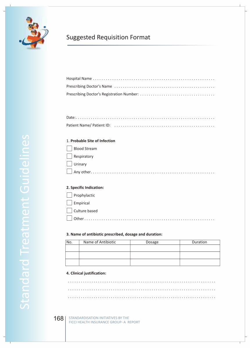
168
Suggested Requisition Format
Hospital Name . . . . . . . . . . . . . . . . . . . . . . . . . . . . . . . . . . . . . . . . . . . . . . . . . . . . . . . . .
Prescribing Doctor's Name . . . . . . . . . . . . . . . . . . . . . . . . . . . . . . . . . . . . . . . . . . . . . . .
Prescribing Doctor's Registration Number: . . . . . . . . . . . . . . . . . . . . . . . . . . . . . . . . . . .
Date:. . . . . . . . . . . . . . . . . . . . . . . . . . . . . . . . . . . . . . . . . . . . . . . . . . . . . . . . . . . . . . . . .
Patient Name/ Patient ID: . . . . . . . . . . . . . . . . . . . . . . . . . . . . . . . . . . . . . . . . . . . . . . .
1. Probable Site of Infection
Blood Stream
Respiratory
Urinary
Any other. . . . . . . . . . . . . . . . . . . . . . . . . . . . . . . . . . . . . . . . . . . . . . . . . . . . . . . . . .
2. Specific Indication:
Prophylactic
Empirical
Culture based
Other . . . . . . . . . . . . . . . . . . . . . . . . . . . . . . . . . . . . . . . . . . . . . . . . . . . . . . . . . . . . .
3. Name of antibiotic prescribed, dosage and duration:
No. Name of Antibiotic Dosage Duration
4. Clinical justification:
. . . . . . . . . . . . . . . . . . . . . . . . . . . . . . . . . . . . . . . . . . . . . . . . . . . . . . . . . . . . . . . . . . . . .
. . . . . . . . . . . . . . . . . . . . . . . . . . . . . . . . . . . . . . . . . . . . . . . . . . . . . . . . . . . . . . . . . . . . .
. . . . . . . . . . . . . . . . . . . . . . . . . . . . . . . . . . . . . . . . . . . . . . . . . . . . . . . . . . . . . . . . . . . . .
169
FICC
I Wo
rking G
rou
p R
epo
rt
Annexure : TEMPLATE FOR DEVELOPMENT OF STGs
1. WHEN TO SUSPECT/ RECOGNIZE?
For both situations of care:
Situation 1 – Secondary / Non Metro
Situation 2 - Super Specialty / Metro
2. INCIDENCE OF THE CONDITION IN OUR COUNTRY
3. DIFFERENTIAL DIAGNOSIS
4. DIFFERENTIAL DIAGNOSIS
OPTIMAL DIAGNOSTIC CRITERIA, INVESTIGATIONS, TREATMENT &
REFERRAL CRITERIA
Situation 1: Secondary Hospital/ Non-Metro situation: Optimal
Standards of Treatment in Situations where technology and resources
are limited
Clinical Diagnosis :
Investigations:
Treatment
Referral criteria
Situation 2: At Super Specialty Facility in Metro location where higher-
end technology is available
Clinical Diagnosis :
Investigations:
Treatment:
Referral criteria :
5. FURTHER READING / REFERENCES
Introduction:
Case definition:
STANDARDISATION INITIATIVES BY THE FICCI HEALTH INSURANCE GROUP- A REPORT
STANDARDISATION INITIATIVES BY THE FICCI HEALTH INSURANCE GROUP- A REPORT
Stan
dar
d T
reat
men
t G
uid
elin
es
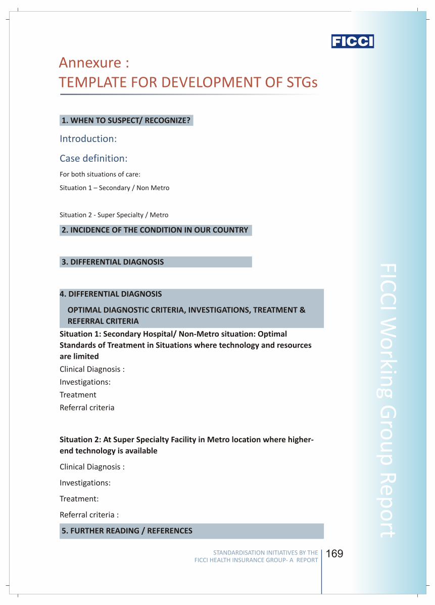
168
Suggested Requisition Format
Hospital Name . . . . . . . . . . . . . . . . . . . . . . . . . . . . . . . . . . . . . . . . . . . . . . . . . . . . . . . . .
Prescribing Doctor's Name . . . . . . . . . . . . . . . . . . . . . . . . . . . . . . . . . . . . . . . . . . . . . . .
Prescribing Doctor's Registration Number: . . . . . . . . . . . . . . . . . . . . . . . . . . . . . . . . . . .
Date:. . . . . . . . . . . . . . . . . . . . . . . . . . . . . . . . . . . . . . . . . . . . . . . . . . . . . . . . . . . . . . . . .
Patient Name/ Patient ID: . . . . . . . . . . . . . . . . . . . . . . . . . . . . . . . . . . . . . . . . . . . . . . .
1. Probable Site of Infection
Blood Stream
Respiratory
Urinary
Any other. . . . . . . . . . . . . . . . . . . . . . . . . . . . . . . . . . . . . . . . . . . . . . . . . . . . . . . . . .
2. Specific Indication:
Prophylactic
Empirical
Culture based
Other . . . . . . . . . . . . . . . . . . . . . . . . . . . . . . . . . . . . . . . . . . . . . . . . . . . . . . . . . . . . .
3. Name of antibiotic prescribed, dosage and duration:
No. Name of Antibiotic Dosage Duration
4. Clinical justification:
. . . . . . . . . . . . . . . . . . . . . . . . . . . . . . . . . . . . . . . . . . . . . . . . . . . . . . . . . . . . . . . . . . . . .
. . . . . . . . . . . . . . . . . . . . . . . . . . . . . . . . . . . . . . . . . . . . . . . . . . . . . . . . . . . . . . . . . . . . .
. . . . . . . . . . . . . . . . . . . . . . . . . . . . . . . . . . . . . . . . . . . . . . . . . . . . . . . . . . . . . . . . . . . . .
169
FICC
I Wo
rking G
rou
p R
epo
rtAnnexure : TEMPLATE FOR DEVELOPMENT OF STGs
1. WHEN TO SUSPECT/ RECOGNIZE?
For both situations of care:
Situation 1 – Secondary / Non Metro
Situation 2 - Super Specialty / Metro
2. INCIDENCE OF THE CONDITION IN OUR COUNTRY
3. DIFFERENTIAL DIAGNOSIS
4. DIFFERENTIAL DIAGNOSIS
OPTIMAL DIAGNOSTIC CRITERIA, INVESTIGATIONS, TREATMENT &
REFERRAL CRITERIA
Situation 1: Secondary Hospital/ Non-Metro situation: Optimal
Standards of Treatment in Situations where technology and resources
are limited
Clinical Diagnosis :
Investigations:
Treatment
Referral criteria
Situation 2: At Super Specialty Facility in Metro location where higher-
end technology is available
Clinical Diagnosis :
Investigations:
Treatment:
Referral criteria :
5. FURTHER READING / REFERENCES
Introduction:
Case definition:
STANDARDISATION INITIATIVES BY THE FICCI HEALTH INSURANCE GROUP- A REPORT
STANDARDISATION INITIATIVES BY THE FICCI HEALTH INSURANCE GROUP- A REPORT
Stan
dar
d T
reat
men
t G
uid
elin
es
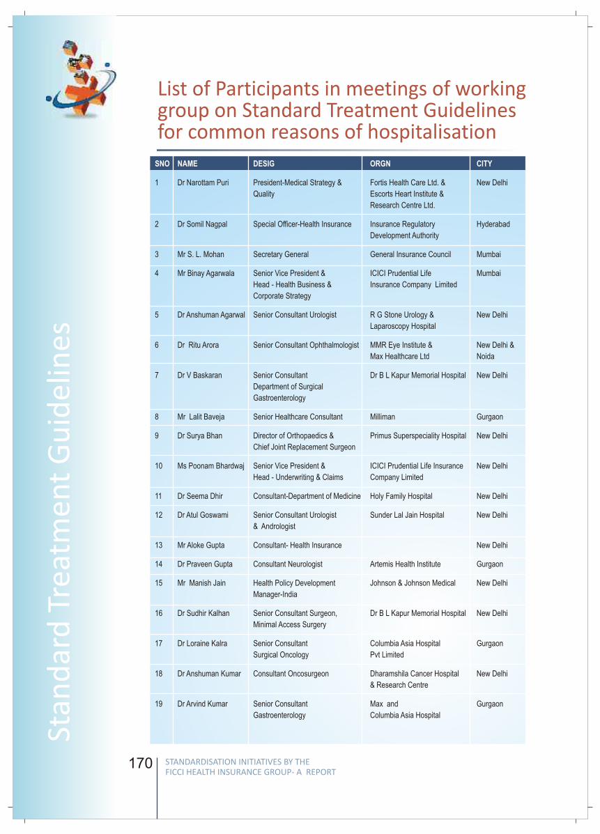
170
List of Participants in meetings of working group on Standard Treatment Guidelines for common reasons of hospitalisation
SNO NAME DESIG ORGN CITY
1
Quality Escorts Heart Institute &
Research Centre Ltd.
2 Dr Somil Nagpal Special Officer-Health Insurance Insurance Regulatory Hyderabad
Development Authority
3 Mr S. L. Mohan Secretary General General Insurance Council Mumbai
4 Mr Binay Agarwala Senior Vice President & ICICI Prudential Life Mumbai
Head - Health Business & Insurance Company Limited
Corporate Strategy
5 Dr Anshuman Agarwal Senior Consultant Urologist R G Stone Urology & New Delhi
Laparoscopy Hospital
6 Dr Ritu Arora Senior Consultant Ophthalmologist MMR Eye Institute & New Delhi &
Max Healthcare Ltd Noida
7 Dr V Baskaran Senior Consultant Dr B L Kapur Memorial Hospital New Delhi
Department of Surgical
Gastroenterology
8 Mr Lalit Baveja Senior Healthcare Consultant Milliman Gurgaon
9 Dr Surya Bhan Director of Orthopaedics & Primus Superspeciality Hospital New Delhi
Chief Joint Replacement Surgeon
10 Ms Poonam Bhardwaj Senior Vice President & ICICI Prudential Life Insurance New Delhi
Head - Underwriting & Claims Company Limited
11 Dr Seema Dhir Consultant-Department of Medicine Holy Family Hospital New Delhi
12 Dr Atul Goswami Senior Consultant Urologist Sunder Lal Jain Hospital New Delhi
& Andrologist
13 Mr Aloke Gupta Consultant- Health Insurance New Delhi
14 Dr Praveen Gupta Consultant Neurologist Artemis Health Institute Gurgaon
15 Mr Manish Jain Health Policy Development Johnson & Johnson Medical New Delhi
Manager-India
16 Dr Sudhir Kalhan Senior Consultant Surgeon, Dr B L Kapur Memorial Hospital New Delhi
Minimal Access Surgery
17 Dr Loraine Kalra Senior Consultant Columbia Asia Hospital Gurgaon
Surgical Oncology Pvt Limited
18 Dr Anshuman Kumar Consultant Oncosurgeon Dharamshila Cancer Hospital New Delhi
& Research Centre
19 Dr Arvind Kumar Senior Consultant Max and Gurgaon
Gastroenterology Columbia Asia Hospital
Dr Narottam Puri President-Medical Strategy & Fortis Health Care Ltd. & New Delhi
171
FICC
I Wo
rking G
rou
p R
epo
rt
SNO NAME DESIG ORGN CITY
20 Dr Sanjeev Malik Executive Director MMR Eye Institute New Delhi
21 Dr Bhabotosh Mishra General Manager-Underwriting Apollo DKV Health Insurance Gurgaon
Company Limited
22 Dr Praphul Misra Consultant-Cardiology Dr B L Kapur Memorial Hospital New Delhi
23 Dr Anil Monga Senior ENT Surgeon Sir Ganga Ram Hospital New Delhi
& Vice Chairman
Department of Otorhinolaryngology
24 Mr Amit Narula Manager-Health Partnerships ICICI Prudential Life Insurance New Delhi
Company Limited
25 Mr Alam Singh Assistant Managing Director Milliman Gurgaon
26 Dr Dinesh Singhal Senior Consultant, Pushpawati Singhania New Delhi
Department of Surgical Research Centre
Gastroenterology
27 Dr Sourav Shukla Senior Consultant Primus Superspeciality Hospital New Delhi
28 Dr A K Sood Head -Cardiology Department Rockland Hospital New Delhi
& Chief of non-invasive cardiology
Rockland Hospital
29 Dr Rakesh Kapoor Gynaecologist R K Hospital Faridabad
30 Dr Vaibhav Junior Consultant Rockland Hospital New Delhi
STANDARDISATION INITIATIVES BY THE FICCI HEALTH INSURANCE GROUP- A REPORT
STANDARDISATION INITIATIVES BY THE FICCI HEALTH INSURANCE GROUP- A REPORT
Stan
dar
d T
reat
men
t G
uid
elin
es
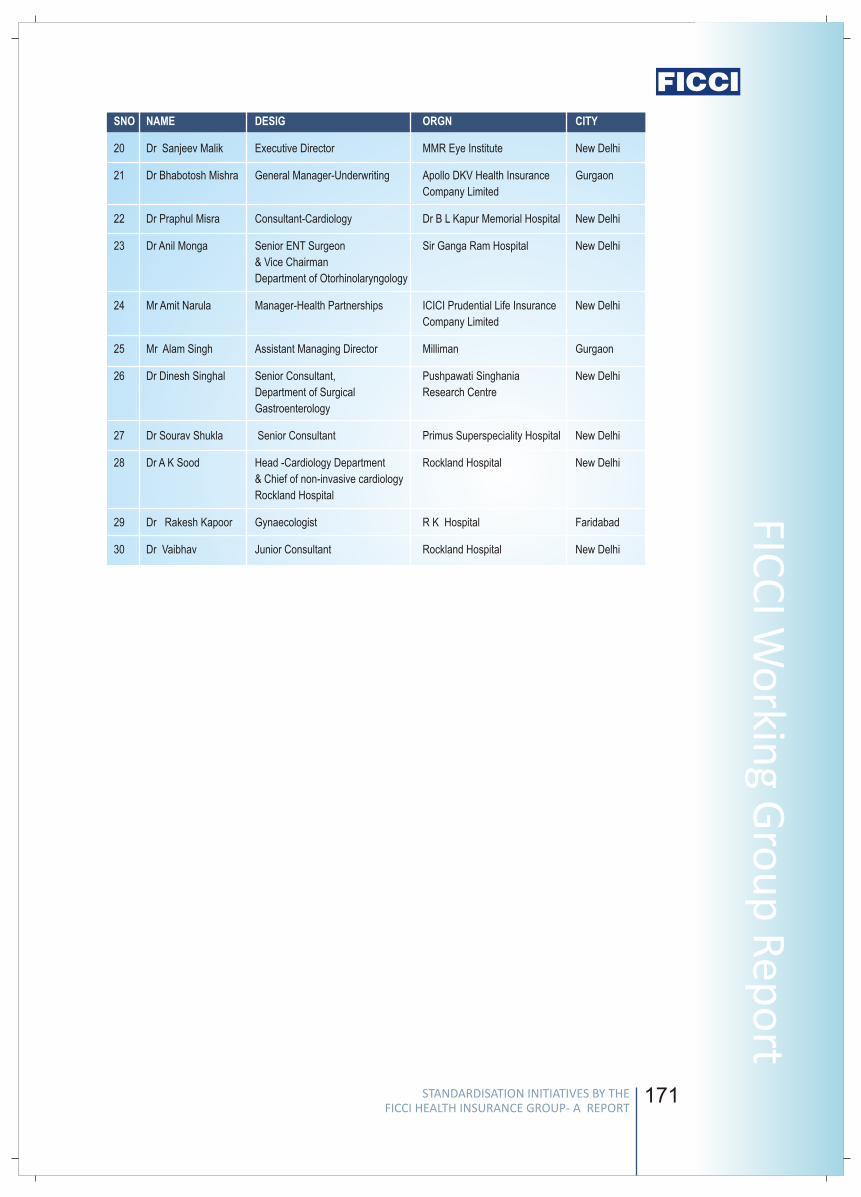
170
List of Participants in meetings of working group on Standard Treatment Guidelines for common reasons of hospitalisation
SNO NAME DESIG ORGN CITY
1
Quality Escorts Heart Institute &
Research Centre Ltd.
2 Dr Somil Nagpal Special Officer-Health Insurance Insurance Regulatory Hyderabad
Development Authority
3 Mr S. L. Mohan Secretary General General Insurance Council Mumbai
4 Mr Binay Agarwala Senior Vice President & ICICI Prudential Life Mumbai
Head - Health Business & Insurance Company Limited
Corporate Strategy
5 Dr Anshuman Agarwal Senior Consultant Urologist R G Stone Urology & New Delhi
Laparoscopy Hospital
6 Dr Ritu Arora Senior Consultant Ophthalmologist MMR Eye Institute & New Delhi &
Max Healthcare Ltd Noida
7 Dr V Baskaran Senior Consultant Dr B L Kapur Memorial Hospital New Delhi
Department of Surgical
Gastroenterology
8 Mr Lalit Baveja Senior Healthcare Consultant Milliman Gurgaon
9 Dr Surya Bhan Director of Orthopaedics & Primus Superspeciality Hospital New Delhi
Chief Joint Replacement Surgeon
10 Ms Poonam Bhardwaj Senior Vice President & ICICI Prudential Life Insurance New Delhi
Head - Underwriting & Claims Company Limited
11 Dr Seema Dhir Consultant-Department of Medicine Holy Family Hospital New Delhi
12 Dr Atul Goswami Senior Consultant Urologist Sunder Lal Jain Hospital New Delhi
& Andrologist
13 Mr Aloke Gupta Consultant- Health Insurance New Delhi
14 Dr Praveen Gupta Consultant Neurologist Artemis Health Institute Gurgaon
15 Mr Manish Jain Health Policy Development Johnson & Johnson Medical New Delhi
Manager-India
16 Dr Sudhir Kalhan Senior Consultant Surgeon, Dr B L Kapur Memorial Hospital New Delhi
Minimal Access Surgery
17 Dr Loraine Kalra Senior Consultant Columbia Asia Hospital Gurgaon
Surgical Oncology Pvt Limited
18 Dr Anshuman Kumar Consultant Oncosurgeon Dharamshila Cancer Hospital New Delhi
& Research Centre
19 Dr Arvind Kumar Senior Consultant Max and Gurgaon
Gastroenterology Columbia Asia Hospital
Dr Narottam Puri President-Medical Strategy & Fortis Health Care Ltd. & New Delhi
171
FICC
I Wo
rking G
rou
p R
epo
rtSNO NAME DESIG ORGN CITY
20 Dr Sanjeev Malik Executive Director MMR Eye Institute New Delhi
21 Dr Bhabotosh Mishra General Manager-Underwriting Apollo DKV Health Insurance Gurgaon
Company Limited
22 Dr Praphul Misra Consultant-Cardiology Dr B L Kapur Memorial Hospital New Delhi
23 Dr Anil Monga Senior ENT Surgeon Sir Ganga Ram Hospital New Delhi
& Vice Chairman
Department of Otorhinolaryngology
24 Mr Amit Narula Manager-Health Partnerships ICICI Prudential Life Insurance New Delhi
Company Limited
25 Mr Alam Singh Assistant Managing Director Milliman Gurgaon
26 Dr Dinesh Singhal Senior Consultant, Pushpawati Singhania New Delhi
Department of Surgical Research Centre
Gastroenterology
27 Dr Sourav Shukla Senior Consultant Primus Superspeciality Hospital New Delhi
28 Dr A K Sood Head -Cardiology Department Rockland Hospital New Delhi
& Chief of non-invasive cardiology
Rockland Hospital
29 Dr Rakesh Kapoor Gynaecologist R K Hospital Faridabad
30 Dr Vaibhav Junior Consultant Rockland Hospital New Delhi
STANDARDISATION INITIATIVES BY THE FICCI HEALTH INSURANCE GROUP- A REPORT
STANDARDISATION INITIATIVES BY THE FICCI HEALTH INSURANCE GROUP- A REPORT
Stan
dar
d T
reat
men
t G
uid
elin
es
172
Editorial Support for Standard
Treatment Guidelines
The Editorial support was provided by Mr Alam Singh, Managing Director,
Milliman and Mr Lalit Baveja, Senior Healthcare Consultant, Milliman in formatting the
content of the STG based on the original content provided by the Clinical Experts.
Alam SinghAssistant Managing Director
Milliman
Alam manages the 42 people health insurance team at Milliman, India. He is a
management professional with more than 12 years of experience in health insurance
and informatics.
Alam supervises project delivery for a wide range of domestic and international projects
which Milliman India undertakes. He focuses on data analysis, product design and
pricing assignments for health insurers and supervises the development of various
products for underwriting and claims management.
Lalit BavejaSenior Healthcare Consultant
Milliman
Lalit is an occupational therapist with over 18 years of experience in clinical practice,
claims management, fraud & abuse detection and project management in India and the
UK.
Lalit leads the Milliman clinical team which has developed evidence based treatment
protocols for Indian healthcare providers and claim processing guidelines for health
insurers. He has also led the development of health risk assessment tools, patient
advisories, hospital order sets and hospital quality reviews.
Assistant
173
FICC
I Wo
rking G
rou
p R
epo
rt
STANDARD DEFINITIONS OF CRITICAL ILLNESSES FOR INDIAN INSURANCE INDUSTRY
STANDARDISATION INITIATIVES BY THE FICCI HEALTH INSURANCE GROUP- A REPORT
STANDARDISATION INITIATIVES BY THE FICCI HEALTH INSURANCE GROUP- A REPORT
Stan
dar
d T
reat
men
t G
uid
elin
es

174 175
FICC
I Wo
rking G
rou
p R
epo
rt
STANDARD DEFINITIONS OF CRITICAL ILLNESSES FOR INDIAN INSURANCE INDUSTRY
BACKGROUND
INTRODUCTION
METHODOLOGY
In addition to hospitalization indemnity products which constitute the predominant
variants of health insurance products in our country, the health insurance market has
also witnessed the introduction of various critical illness products, which cover a list of
designated diseases. Critical Illness cover pays a Lump Sum amount, or benefit, if the
Insured is diagnosed with a specified critical illness or undergoes a specified procedure.
This sum is paid directly to the insured regardless of any other sources of indemnity
(job-related and non-job-related) or the actual expenses incurred (medical and
nonmedical).
However, there do exist differences in the definitions of Critical Illnesses adopted by the
different insurers which can create confusion in the minds of consumers and the
industry especially at the time when insurers and re-insurers have to arrive at a point
where lump sum payment is made. Lack of standard definitions also means that
products are difficult to compare, and the availability of standard definitions would then
ensure better comparability and uniformity in the understanding of critical illness
definitions.
In view of the above problem identified by the health insurance committee, FICCI
undertook an intense exercise on developing Standard Definitions of Critical Illnesses
through its Sub-Group, Chaired by Mr. S.L. Mohan, Secretary General, General
Insurance Council and Co-Chaired by Mr. S.B. Mathur, Secretary General, Life Insurance
Council. This, we believe, will help resolve the confusion arising out of varying
definitions adopted by different companies and will also help enhance consumer
satisfaction significantly. The group had pro-active involvement of senior
representatives from several leading Insurance companies, four large Re-Insurance
Companies as well as representatives from the Health Sector. IRDA has also been an
integral part of the Sub-Group and has continuously guided & supported us in this
endeavor.
Critical Illness Definitions adopted by different insurers, re-insurers as also
standard definitions adopted by bodies like the Association of British Insurers (ABI)
were collated for 11 critical illnesses commonly offered for cover by insurers in
their critical illness policies. The definitions adopted for each such critical illness
were discussed during the interactive meetings in detail by experts in the field and
also examined in detail by their respective organizations.
The members of the Sub-group then attempted to standardize the Critical Illness
definitions, for adoption by the industry.
The final document was again reviewed and vetted by an independent Technical
Board of eminent medical professionals which was constituted by FICCI.
v
v
v
STANDARDISATION INITIATIVES BY THE FICCI HEALTH INSURANCE GROUP- A REPORT
STANDARDISATION INITIATIVES BY THE FICCI HEALTH INSURANCE GROUP- A REPORT
Notes
174 175
FICC
I Wo
rking G
rou
p R
epo
rtSTANDARD DEFINITIONS OF CRITICAL ILLNESSES FOR INDIAN INSURANCE INDUSTRY
BACKGROUND
INTRODUCTION
METHODOLOGY
In addition to hospitalization indemnity products which constitute the predominant
variants of health insurance products in our country, the health insurance market has
also witnessed the introduction of various critical illness products, which cover a list of
designated diseases. Critical Illness cover pays a Lump Sum amount, or benefit, if the
Insured is diagnosed with a specified critical illness or undergoes a specified procedure.
This sum is paid directly to the insured regardless of any other sources of indemnity
(job-related and non-job-related) or the actual expenses incurred (medical and
nonmedical).
However, there do exist differences in the definitions of Critical Illnesses adopted by the
different insurers which can create confusion in the minds of consumers and the
industry especially at the time when insurers and re-insurers have to arrive at a point
where lump sum payment is made. Lack of standard definitions also means that
products are difficult to compare, and the availability of standard definitions would then
ensure better comparability and uniformity in the understanding of critical illness
definitions.
In view of the above problem identified by the health insurance committee, FICCI
undertook an intense exercise on developing Standard Definitions of Critical Illnesses
through its Sub-Group, Chaired by Mr. S.L. Mohan, Secretary General, General
Insurance Council and Co-Chaired by Mr. S.B. Mathur, Secretary General, Life Insurance
Council. This, we believe, will help resolve the confusion arising out of varying
definitions adopted by different companies and will also help enhance consumer
satisfaction significantly. The group had pro-active involvement of senior
representatives from several leading Insurance companies, four large Re-Insurance
Companies as well as representatives from the Health Sector. IRDA has also been an
integral part of the Sub-Group and has continuously guided & supported us in this
endeavor.
Critical Illness Definitions adopted by different insurers, re-insurers as also
standard definitions adopted by bodies like the Association of British Insurers (ABI)
were collated for 11 critical illnesses commonly offered for cover by insurers in
their critical illness policies. The definitions adopted for each such critical illness
were discussed during the interactive meetings in detail by experts in the field and
also examined in detail by their respective organizations.
The members of the Sub-group then attempted to standardize the Critical Illness
definitions, for adoption by the industry.
The final document was again reviewed and vetted by an independent Technical
Board of eminent medical professionals which was constituted by FICCI.
v
v
v
STANDARDISATION INITIATIVES BY THE FICCI HEALTH INSURANCE GROUP- A REPORT
STANDARDISATION INITIATIVES BY THE FICCI HEALTH INSURANCE GROUP- A REPORT
Notes
176
vFICCI has submitted these definitions to IRDA for a wider dissemination to the
Industry so that the feedback of the industry can further enhance the acceptability
of the standard definitions.
RECOMMENDED STANDARD DEFINITIONS
A malignant tumour characterised by the uncontrolled growth & spread of
malignant cells with invasion & destruction of normal tissues. This diagnosis must
be supported by histological evidence of malignancy & confirmed by a pathologist.
The term cancer includes leukemia, lymphoma and sarcoma. The following are
excluded - (1) Tumours showing the malignant changes of carcinoma in situ &
tumours which are histologically described as pre-malignant or non invasive,
including but not limited to: Carcinoma in situ of breasts, Cervical dysplasia CIN-1,
CIN -2 & CIN-3. (2) Any skin cancer other than invasive malignant melanoma (3) All
tumours of the prostate unless histologically classified as having a Gleason score
greater than 6 or having progressed to at least clinical TNM classification
T2N0M0.........(4) Papillary micro - carcinoma of the thyroid less than 1 cm in
diameter (5) Chronic lymphocyctic leukaemia less than RAI stage 3 (6)
microcarcinoma of the bladder (7) All tumours in the presence of HIV infection.
The first occurrence of myocardial infarction which means the death of a portion
of the heart muscle as a result of inadequate blood supply to the relevant area.
The diagnosis for this will be evidenced by all of the following criteria: a) a history
of typical clinical symptoms consistent with the diagnosis of Acute Myocardial
Infarction (for e.g. typical chest pain) b) new characteristic electrocardiogram
changes c) elevation of infarction specific enzymes, Troponins or other specific
biochemical markers. The following are excluded:
(1)Non-ST-segment elevation myocardial infarction (NSTEMI) with elevation of
Troponin I or T; (2)Other acute Coronary Syndromes (3)Any type of angina pectoris
The actual undergoing of open chest surgery for the correction of one or more
coronary arteries, which is/are narrowed or blocked, by coronary artery bypass
graft (CABG). The diagnosis must be supported by a coronary angiography and the
realization of surgery has to be confirmed by a specialist medical practitioner.
Excluded are: (1) Angioplasty and/or any other intra-arterial procedures (2) any
key-hole or laser surgery.
The actual undergoing of open-heart valve surgery to replace or repair one or
more heart valves, as a consequence of defects in, abnormalities of, or disease-
1. CANCER OF SPECIFIED SEVERITY
2. FIRST HEART ATTACK – OF SPECIFIED SEVERITY
3. OPEN CHEST CABG
4. OPEN HEART REPLACEMENT OR REPAIR OF HEART
VALVES
177
FICC
I Wo
rking G
rou
p R
epo
rt
affected cardiac valve(s). The diagnosis of the valve abnormality must be
supported by an echocardiography and the realization of surgery has to be
confirmed by a specialist medical practitioner.
Catheter based techniques including but not limited to, balloon
valvotomy/valvuloplasty are excluded.
A state of unconsciousness with no reaction or response to external stimuli or
internal needs.
This diagnosis must be supported by evidence of all of the following:
Øno response to external stimuli continuously for at least 96 hours;
Ø life support measures are necessary to sustain life; and
Øpermanent neurological deficit which must be assessed at least 30 days after
the onset of the coma.
The condition has to be confirmed by a specialist medical practitioner.
Coma resulting directly from alcohol or drug abuse is excluded.
End stage renal disease presenting as chronic irreversible failure of both kidneys to
function, as a result of which either regular renal dialysis (hemodialysis or
peritoneal dialysis) is instituted or renal transplantation is carried out. Diagnosis
has to be confirmed by a specialist medical practitioner.
Any cerebrovascular incident producing permanent neurological sequelae.This
includes infarction of brain tissue, thrombosis in an intra-cranial vessel,
haemorrhage and embolisation from an extracranial source. Diagnosis has to be
confirmed by a specialist medical practitioner and evidenced by typical clinical
symptoms as well as typical findings in CT Scan or MRI of the brain.
Evidence of permanent neurological deficit lasting for atleast 3 months has to be
produced.
The following are excluded:
ØTransient ischemic attacks (TIA)
ØTraumatic injury of the brain
Ø Vascular disease affecting only the eye or optic nerve or vestibular functions.
The actual undergoing of a transplant of:
ØOne of the following human organs: heart, lung, liver, kidney, pancreas, that
resulted from irreversible end-stage failure of the relevant organ, or
ØHuman bone marrow using haematopoietic stem cells
The undergoing of a transplant has to be confirmed by a specialist medical
practitioner.
The following are excluded:
ØOther stem-cell transplants
ØWhere only islets of langerhans are transplanted
5. COMA OF SPECIFIED SEVERITY
6. KIDNEY FAILURE REQUIRING REGULAR DIALYSIS
7. STROKE RESULTING IN PERMANENT SYMPTOMS
8. MAJOR ORGAN /BONE MARROW TRANSPLANT
STANDARDISATION INITIATIVES BY THE FICCI HEALTH INSURANCE GROUP- A REPORT
STANDARDISATION INITIATIVES BY THE FICCI HEALTH INSURANCE GROUP- A REPORT
Stan
dar
d C
riti
cal I
llnes
s D
efin
itio
ns
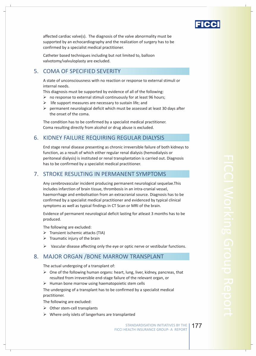
176
vFICCI has submitted these definitions to IRDA for a wider dissemination to the
Industry so that the feedback of the industry can further enhance the acceptability
of the standard definitions.
RECOMMENDED STANDARD DEFINITIONS
A malignant tumour characterised by the uncontrolled growth & spread of
malignant cells with invasion & destruction of normal tissues. This diagnosis must
be supported by histological evidence of malignancy & confirmed by a pathologist.
The term cancer includes leukemia, lymphoma and sarcoma. The following are
excluded - (1) Tumours showing the malignant changes of carcinoma in situ &
tumours which are histologically described as pre-malignant or non invasive,
including but not limited to: Carcinoma in situ of breasts, Cervical dysplasia CIN-1,
CIN -2 & CIN-3. (2) Any skin cancer other than invasive malignant melanoma (3) All
tumours of the prostate unless histologically classified as having a Gleason score
greater than 6 or having progressed to at least clinical TNM classification
T2N0M0.........(4) Papillary micro - carcinoma of the thyroid less than 1 cm in
diameter (5) Chronic lymphocyctic leukaemia less than RAI stage 3 (6)
microcarcinoma of the bladder (7) All tumours in the presence of HIV infection.
The first occurrence of myocardial infarction which means the death of a portion
of the heart muscle as a result of inadequate blood supply to the relevant area.
The diagnosis for this will be evidenced by all of the following criteria: a) a history
of typical clinical symptoms consistent with the diagnosis of Acute Myocardial
Infarction (for e.g. typical chest pain) b) new characteristic electrocardiogram
changes c) elevation of infarction specific enzymes, Troponins or other specific
biochemical markers. The following are excluded:
(1)Non-ST-segment elevation myocardial infarction (NSTEMI) with elevation of
Troponin I or T; (2)Other acute Coronary Syndromes (3)Any type of angina pectoris
The actual undergoing of open chest surgery for the correction of one or more
coronary arteries, which is/are narrowed or blocked, by coronary artery bypass
graft (CABG). The diagnosis must be supported by a coronary angiography and the
realization of surgery has to be confirmed by a specialist medical practitioner.
Excluded are: (1) Angioplasty and/or any other intra-arterial procedures (2) any
key-hole or laser surgery.
The actual undergoing of open-heart valve surgery to replace or repair one or
more heart valves, as a consequence of defects in, abnormalities of, or disease-
1. CANCER OF SPECIFIED SEVERITY
2. FIRST HEART ATTACK – OF SPECIFIED SEVERITY
3. OPEN CHEST CABG
4. OPEN HEART REPLACEMENT OR REPAIR OF HEART
VALVES
177
FICC
I Wo
rking G
rou
p R
epo
rtaffected cardiac valve(s). The diagnosis of the valve abnormality must be
supported by an echocardiography and the realization of surgery has to be
confirmed by a specialist medical practitioner.
Catheter based techniques including but not limited to, balloon
valvotomy/valvuloplasty are excluded.
A state of unconsciousness with no reaction or response to external stimuli or
internal needs.
This diagnosis must be supported by evidence of all of the following:
Øno response to external stimuli continuously for at least 96 hours;
Ø life support measures are necessary to sustain life; and
Øpermanent neurological deficit which must be assessed at least 30 days after
the onset of the coma.
The condition has to be confirmed by a specialist medical practitioner.
Coma resulting directly from alcohol or drug abuse is excluded.
End stage renal disease presenting as chronic irreversible failure of both kidneys to
function, as a result of which either regular renal dialysis (hemodialysis or
peritoneal dialysis) is instituted or renal transplantation is carried out. Diagnosis
has to be confirmed by a specialist medical practitioner.
Any cerebrovascular incident producing permanent neurological sequelae.This
includes infarction of brain tissue, thrombosis in an intra-cranial vessel,
haemorrhage and embolisation from an extracranial source. Diagnosis has to be
confirmed by a specialist medical practitioner and evidenced by typical clinical
symptoms as well as typical findings in CT Scan or MRI of the brain.
Evidence of permanent neurological deficit lasting for atleast 3 months has to be
produced.
The following are excluded:
ØTransient ischemic attacks (TIA)
ØTraumatic injury of the brain
Ø Vascular disease affecting only the eye or optic nerve or vestibular functions.
The actual undergoing of a transplant of:
ØOne of the following human organs: heart, lung, liver, kidney, pancreas, that
resulted from irreversible end-stage failure of the relevant organ, or
ØHuman bone marrow using haematopoietic stem cells
The undergoing of a transplant has to be confirmed by a specialist medical
practitioner.
The following are excluded:
ØOther stem-cell transplants
ØWhere only islets of langerhans are transplanted
5. COMA OF SPECIFIED SEVERITY
6. KIDNEY FAILURE REQUIRING REGULAR DIALYSIS
7. STROKE RESULTING IN PERMANENT SYMPTOMS
8. MAJOR ORGAN /BONE MARROW TRANSPLANT
STANDARDISATION INITIATIVES BY THE FICCI HEALTH INSURANCE GROUP- A REPORT
STANDARDISATION INITIATIVES BY THE FICCI HEALTH INSURANCE GROUP- A REPORT
Stan
dar
d C
riti
cal I
llnes
s D
efin
itio
ns
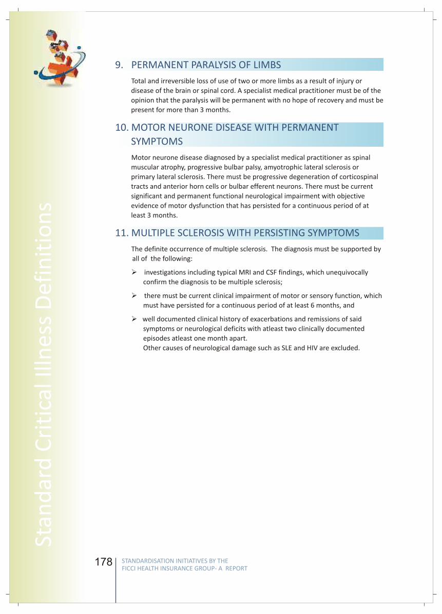
178
9. PERMANENT PARALYSIS OF LIMBS
10. MOTOR NEURONE DISEASE WITH PERMANENT
SYMPTOMS
11. MULTIPLE SCLEROSIS WITH PERSISTING SYMPTOMS
Total and irreversible loss of use of two or more limbs as a result of injury or
disease of the brain or spinal cord. A specialist medical practitioner must be of the
opinion that the paralysis will be permanent with no hope of recovery and must be
present for more than 3 months.
Motor neurone disease diagnosed by a specialist medical practitioner as spinal
muscular atrophy, progressive bulbar palsy, amyotrophic lateral sclerosis or
primary lateral sclerosis. There must be progressive degeneration of corticospinal
tracts and anterior horn cells or bulbar efferent neurons. There must be current
significant and permanent functional neurological impairment with objective
evidence of motor dysfunction that has persisted for a continuous period of at
least 3 months.
The definite occurrence of multiple sclerosis. The diagnosis must be supported by
all of the following:
Ø investigations including typical MRI and CSF findings, which unequivocally
confirm the diagnosis to be multiple sclerosis;
Ø there must be current clinical impairment of motor or sensory function, which
must have persisted for a continuous period of at least 6 months, and
Øwell documented clinical history of exacerbations and remissions of said
symptoms or neurological deficits with atleast two clinically documented
episodes atleast one month apart.
Other causes of neurological damage such as SLE and HIV are excluded.
179
FICC
I Wo
rking G
rou
p R
epo
rt
Chairpersons
S. L. MohanSecretary General
General Insurance Council
Mr. Mohan is the Secretary General of General Insurance Council since November 2008.
In addition to this he is also serving as an Associate of Insurance Institute of India.
Prior to this, he was the Chairman cum Managing Director, The Oriental Insurance Co.
Ltd. (2002-2005), General Manager, United India Insurance Co. Ltd., Chennai (2001-
2002), General Manager, National Insurance Co. Ltd., Kolkata (2000) and Regional
Manager / Asst. General Manager, United India Ins. Co. Ltd., Chennai (1989-2000).
He has also served as a director at Kenindia Assurance Co. Ltd., Nairobi, GIC Housing
Finance Limited, Mumbai, Loss Prevention Association of India Ltd., Mumbai, Member –
Tariff Advisory Committee, Mumbai, Madhura Coats Ltd. to name a few.
He holds a B.Sc. in Mechanical Engineering.
S. B. MathurSecretary General
Life Insurance Council
Mr. Mathur is the Secretary General of Life Insurance Council. In addition to this Mr.
Mathur is the Chairman of National Stock Exchange, an Advisor to National Investment
Fund set up by Government of India and also on Board of some leading Corporates.
He was the Chairman of the Life Insurance Corporation of India (LIC), the largest life
insurance company in India from August 2002 to October 2004. His tenure coincided the
opening of the Insurance sector, which resulted into the entry of 12 domestic players
with strong brands tying up with leading international insurance companies.
Post retirement from LIC, The Government of India appointed him as Administrator of
the Specified Undertaking of the Unit Trust of India (SUUTI), the successor of the
erstwhile Unit Trust of India in December 2004, wherein Government had provided
financial support to meet the liabilities under some of the guaranteed high returns
schemes.
STANDARDISATION INITIATIVES BY THE FICCI HEALTH INSURANCE GROUP- A REPORT
STANDARDISATION INITIATIVES BY THE FICCI HEALTH INSURANCE GROUP- A REPORT
Stan
dar
d C
riti
cal I
llnes
s D
efin
itio
ns
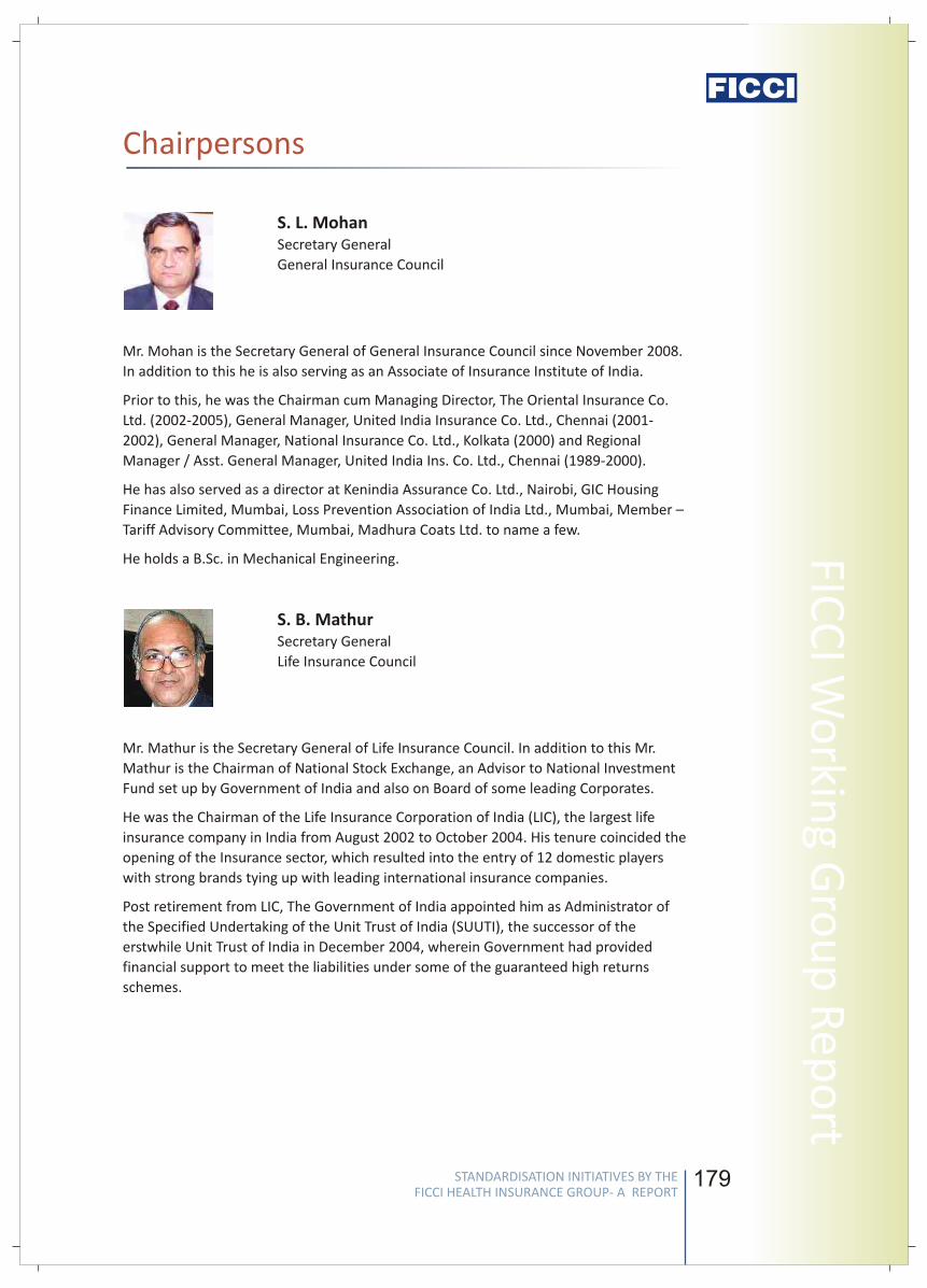
178
9. PERMANENT PARALYSIS OF LIMBS
10. MOTOR NEURONE DISEASE WITH PERMANENT
SYMPTOMS
11. MULTIPLE SCLEROSIS WITH PERSISTING SYMPTOMS
Total and irreversible loss of use of two or more limbs as a result of injury or
disease of the brain or spinal cord. A specialist medical practitioner must be of the
opinion that the paralysis will be permanent with no hope of recovery and must be
present for more than 3 months.
Motor neurone disease diagnosed by a specialist medical practitioner as spinal
muscular atrophy, progressive bulbar palsy, amyotrophic lateral sclerosis or
primary lateral sclerosis. There must be progressive degeneration of corticospinal
tracts and anterior horn cells or bulbar efferent neurons. There must be current
significant and permanent functional neurological impairment with objective
evidence of motor dysfunction that has persisted for a continuous period of at
least 3 months.
The definite occurrence of multiple sclerosis. The diagnosis must be supported by
all of the following:
Ø investigations including typical MRI and CSF findings, which unequivocally
confirm the diagnosis to be multiple sclerosis;
Ø there must be current clinical impairment of motor or sensory function, which
must have persisted for a continuous period of at least 6 months, and
Øwell documented clinical history of exacerbations and remissions of said
symptoms or neurological deficits with atleast two clinically documented
episodes atleast one month apart.
Other causes of neurological damage such as SLE and HIV are excluded.
179
FICC
I Wo
rking G
rou
p R
epo
rtChairpersons
S. L. MohanSecretary General
General Insurance Council
Mr. Mohan is the Secretary General of General Insurance Council since November 2008.
In addition to this he is also serving as an Associate of Insurance Institute of India.
Prior to this, he was the Chairman cum Managing Director, The Oriental Insurance Co.
Ltd. (2002-2005), General Manager, United India Insurance Co. Ltd., Chennai (2001-
2002), General Manager, National Insurance Co. Ltd., Kolkata (2000) and Regional
Manager / Asst. General Manager, United India Ins. Co. Ltd., Chennai (1989-2000).
He has also served as a director at Kenindia Assurance Co. Ltd., Nairobi, GIC Housing
Finance Limited, Mumbai, Loss Prevention Association of India Ltd., Mumbai, Member –
Tariff Advisory Committee, Mumbai, Madhura Coats Ltd. to name a few.
He holds a B.Sc. in Mechanical Engineering.
S. B. MathurSecretary General
Life Insurance Council
Mr. Mathur is the Secretary General of Life Insurance Council. In addition to this Mr.
Mathur is the Chairman of National Stock Exchange, an Advisor to National Investment
Fund set up by Government of India and also on Board of some leading Corporates.
He was the Chairman of the Life Insurance Corporation of India (LIC), the largest life
insurance company in India from August 2002 to October 2004. His tenure coincided the
opening of the Insurance sector, which resulted into the entry of 12 domestic players
with strong brands tying up with leading international insurance companies.
Post retirement from LIC, The Government of India appointed him as Administrator of
the Specified Undertaking of the Unit Trust of India (SUUTI), the successor of the
erstwhile Unit Trust of India in December 2004, wherein Government had provided
financial support to meet the liabilities under some of the guaranteed high returns
schemes.
STANDARDISATION INITIATIVES BY THE FICCI HEALTH INSURANCE GROUP- A REPORT
STANDARDISATION INITIATIVES BY THE FICCI HEALTH INSURANCE GROUP- A REPORT
Stan
dar
d C
riti
cal I
llnes
s D
efin
itio
ns
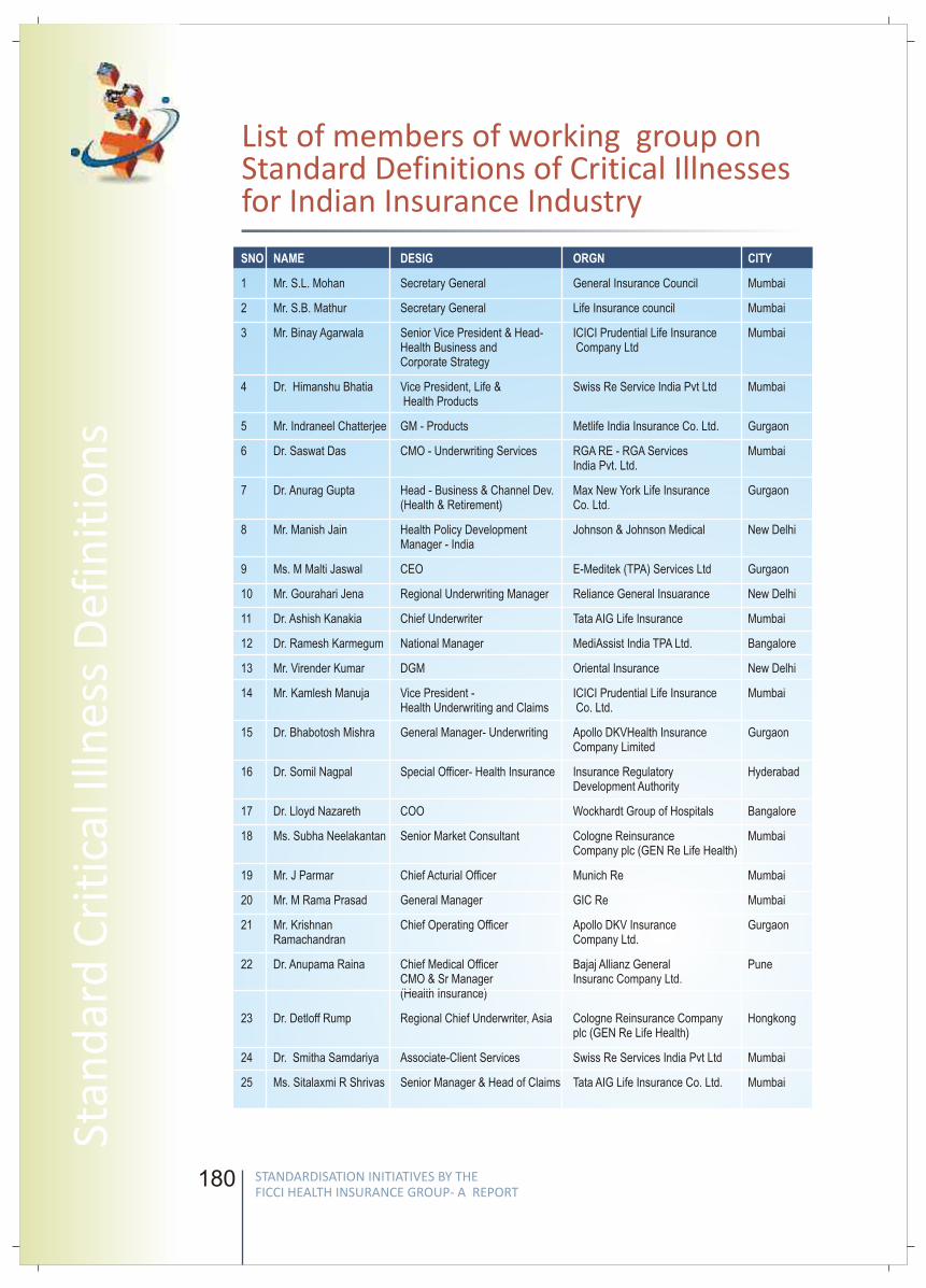
180
List of members of working group on Standard Definitions of Critical Illnesses for Indian Insurance Industry
SNO NAME DESIG ORGN CITY
1 Mr. S.L. Mohan Secretary General General Insurance Council Mumbai
2 Mr. S.B. Mathur Secretary General Life Insurance council Mumbai
3 Mr. Binay Agarwala Senior Vice President & Head- ICICI Prudential Life Insurance MumbaiHealth Business and Company LtdCorporate Strategy
4 Dr. Himanshu Bhatia Vice President, Life & Swiss Re Service India Pvt Ltd Mumbai Health Products
5 Mr. Indraneel Chatterjee GM - Products Metlife India Insurance Co. Ltd. Gurgaon
6 Dr. Saswat Das CMO - Underwriting Services RGA RE - RGA Services MumbaiIndia Pvt. Ltd.
7 Dr. Anurag Gupta Head - Business & Channel Dev. Max New York Life Insurance Gurgaon(Health & Retirement) Co. Ltd.
8 Mr. Manish Jain Health Policy Development Johnson & Johnson Medical New Delhi Manager - India
9 Ms. M Malti Jaswal CEO E-Meditek (TPA) Services Ltd Gurgaon
10 Mr. Gourahari Jena Regional Underwriting Manager Reliance General Insuarance New Delhi
11 Dr. Ashish Kanakia Chief Underwriter Tata AIG Life Insurance Mumbai
12 Dr. Ramesh Karmegum National Manager MediAssist India TPA Ltd. Bangalore
13 Mr. Virender Kumar DGM Oriental Insurance New Delhi
14 Mr. Kamlesh Manuja Vice President - ICICI Prudential Life Insurance MumbaiHealth Underwriting and Claims Co. Ltd.
15 Dr. Bhabotosh Mishra General Manager- Underwriting Apollo DKVHealth Insurance GurgaonCompany Limited
16 Dr. Somil Nagpal Special Officer- Health Insurance Insurance Regulatory HyderabadDevelopment Authority
17 Dr. Lloyd Nazareth COO Wockhardt Group of Hospitals Bangalore
18 Ms. Subha Neelakantan Senior Market Consultant Cologne Reinsurance MumbaiCompany plc (GEN Re Life Health)
19 Mr. J Parmar Chief Acturial Officer Munich Re Mumbai
20 Mr. M Rama Prasad General Manager GIC Re Mumbai
21 Mr. Krishnan Chief Operating Officer Apollo DKV Insurance GurgaonRamachandran Company Ltd.
22 Dr. Anupama Raina Chief Medical Officer Bajaj Allianz General PuneCMO & Sr Manager Insuranc Company Ltd.(Health Insurance)
23 Dr. Detloff Rump Regional Chief Underwriter, Asia Cologne Reinsurance Company Hongkong plc (GEN Re Life Health)
24 Dr. Smitha Samdariya Associate-Client Services Swiss Re Services India Pvt Ltd Mumbai
25 Ms. Sitalaxmi R Shrivas Senior Manager & Head of Claims Tata AIG Life Insurance Co. Ltd. Mumbai
181
FICC
I Wo
rking G
rou
p R
epo
rt
STANDARD LIST OF EXPENSES GENERALLY EXCLUDED (“NON-MEDICAL EXPENSES") IN HOSPITALISATION INDEMNITY POLICIES
STANDARDISATION INITIATIVES BY THE FICCI HEALTH INSURANCE GROUP- A REPORT
STANDARDISATION INITIATIVES BY THE FICCI HEALTH INSURANCE GROUP- A REPORT
Stan
dar
d C
riti
cal I
llnes
s D
efin
itio
ns
182 183
FICC
I Wo
rking G
rou
p R
epo
rt
Standard List of Expenses Generally Excluded (“non-medical expenses") in Hospitalisation Indemnity Policies
BACKGROUND
INTRODUCTION
METHODOLOGY
Insurance companies providing hospitalization indemnity covers generally exclude
certain categories of expenses in their policy terms and conditions. However, as there is
no detailed listing of such excluded expenses, and as the interpretation of these
exclusions is highly varied across different payors in the industry, many a times various
items under the claims filed by hospital providers or individual policyholders are
repudiated by the insurers but are disputed by the claimants. This is, thus, one major
cause of acrimony between Insurance Companies & Healthcare providers and also
causes a lot of confusion in the minds of consumer.
There is, thus, a strong need to minimize the ambiguity on this count through a
collaborative partnership between healthcare providers', health insurance companies
and other important stakeholders. Availability and accessibility of quality healthcare
through affordable and suitable health insurance products is need of the hour. A
consensus between the all the stakeholders of the industry and a uniform
understanding of such ‘exclusions’ would be the key for better understanding of policy
conditions by the policyholders and hospitals, which would in turn facilitate speedier
roll out of health insurance in the country.
FICCI constituted a sub-Group aimed at creating a Standard List of Expenses Generally
Excluded (“Non-Medical Expenses") in Hospitalization Indemnity Policies, as an initiative
under its health insurance working group. The Sub-group is Chaired by Mr. S.L. Mohan,
Secretary General, General Insurance Council and Co-Chaired by Mr. S.B. Mathur,
Secretary General, Life Insurance Council and comprise senior level representatives from
Regulator, TPA's, Healthcare Providers and Insurers.
The aim of the Sub-Group is to arrive at a Standard List of such excluded ('Non Medical')
expenses in order to minimize the ambiguity and subjectivity in deductions from
hospital bills, which will improve the understanding for such expenses amongst patients,
providers and insurers/TPAs.
• As a first step towards Standardization, various Lists of Excluded Expenses were
collated from different Insurers, TPA's etc and compiled in one comprehensive list.
• The members of the sub-group then debated and discussed each of the items in
these lists of excluded expenses, wherein certain items were recommended to be
made admissible under specific situations, in the overall interest of the health
insurance industry, while others which are inadmissible could be standardized to
minimize any friction on this front. (The final list of discussed items with
suggestions and explanations from the group is enclosed).
• The list of various non-medical items was then categorized as per the applicable
exclusions in indemnity policies viz a) toiletries/ cosmetics/ personal comfort or
STANDARDISATION INITIATIVES BY THE FICCI HEALTH INSURANCE GROUP- A REPORT
STANDARDISATION INITIATIVES BY THE FICCI HEALTH INSURANCE GROUP- A REPORT
Stan
dar
d L
ist
of
Excl
ud
ed E
xpen
ses
182 183
FICC
I Wo
rking G
rou
p R
epo
rtStandard List of Expenses Generally Excluded (“non-medical expenses") in Hospitalisation Indemnity Policies
BACKGROUND
INTRODUCTION
METHODOLOGY
Insurance companies providing hospitalization indemnity covers generally exclude
certain categories of expenses in their policy terms and conditions. However, as there is
no detailed listing of such excluded expenses, and as the interpretation of these
exclusions is highly varied across different payors in the industry, many a times various
items under the claims filed by hospital providers or individual policyholders are
repudiated by the insurers but are disputed by the claimants. This is, thus, one major
cause of acrimony between Insurance Companies & Healthcare providers and also
causes a lot of confusion in the minds of consumer.
There is, thus, a strong need to minimize the ambiguity on this count through a
collaborative partnership between healthcare providers', health insurance companies
and other important stakeholders. Availability and accessibility of quality healthcare
through affordable and suitable health insurance products is need of the hour. A
consensus between the all the stakeholders of the industry and a uniform
understanding of such ‘exclusions’ would be the key for better understanding of policy
conditions by the policyholders and hospitals, which would in turn facilitate speedier
roll out of health insurance in the country.
FICCI constituted a sub-Group aimed at creating a Standard List of Expenses Generally
Excluded (“Non-Medical Expenses") in Hospitalization Indemnity Policies, as an initiative
under its health insurance working group. The Sub-group is Chaired by Mr. S.L. Mohan,
Secretary General, General Insurance Council and Co-Chaired by Mr. S.B. Mathur,
Secretary General, Life Insurance Council and comprise senior level representatives from
Regulator, TPA's, Healthcare Providers and Insurers.
The aim of the Sub-Group is to arrive at a Standard List of such excluded ('Non Medical')
expenses in order to minimize the ambiguity and subjectivity in deductions from
hospital bills, which will improve the understanding for such expenses amongst patients,
providers and insurers/TPAs.
• As a first step towards Standardization, various Lists of Excluded Expenses were
collated from different Insurers, TPA's etc and compiled in one comprehensive list.
• The members of the sub-group then debated and discussed each of the items in
these lists of excluded expenses, wherein certain items were recommended to be
made admissible under specific situations, in the overall interest of the health
insurance industry, while others which are inadmissible could be standardized to
minimize any friction on this front. (The final list of discussed items with
suggestions and explanations from the group is enclosed).
• The list of various non-medical items was then categorized as per the applicable
exclusions in indemnity policies viz a) toiletries/ cosmetics/ personal comfort or
STANDARDISATION INITIATIVES BY THE FICCI HEALTH INSURANCE GROUP- A REPORT
STANDARDISATION INITIATIVES BY THE FICCI HEALTH INSURANCE GROUP- A REPORT
Stan
dar
d L
ist
of
Excl
ud
ed E
xpen
ses
184
convenience items, b) items specifically excluded in the policies, c) items which are
elements of Room Charge d) administrative or non-medical charges e) external
durable devices f) items payable if supported by a prescription, and g) Other
exclusions.
• Hospital Providers were also requested to provide the specific indications,
reasonable quantity required and the specific list of certain surgeries where the
use of certain items like Abdominal Binders, belts, braces etc. was warranted &
important and hence could be considered worthy for making payable for the
specific situations.
• The final document has been reviewed and vetted by an independent Technical
Board of eminent medical professionals which was constituted by FICCI.
• FICCI would recommend the Standard List to IRDA and the two Councils for wider
feedback and for a final adoption by the industry.
185
FICC
I Wo
rking G
rou
p R
epo
rt
Recommendations on Commonly Excluded Items
S. No. Items Recommendations
Toiletries/ Cosmetics/ Personal Comfort or Convenience Items
1 Hair removing cream charges Not Payable
2 Baby Charges (unless specified/indicated) Not Payable
3 Baby Food Not Payable
4 Baby Utilites Charges Not Payable
5 Baby Set Not Payable
6 Baby Bottles Not Payable
7 Bottle Not Payable
8 Brush Not Payable
9 Cosy Towel Not Payable
10 Hand Wash Not Payable
11 Moisturiser Paste Brush Not Payable
12 Powder Not Payable
13 Razor Payable
14 Towel Not Payable
15 Shoe Cover Not Payable
16 Beauty Services Not Payable
17 Belts/ Braces Essential andShould be Paid at least Specifically for Caseswho have undergonesurgery of Thoracic orLumbar Spine.
18 Buds Not Payable
19 Barber Charges Not Payable
20 Caps Not Payable
21 Cold Pack/hot Pack Not Payable
22 Carry Bags Not Payable
23 Cradle Charges Not Payable
24 Comb Not Payable
25 Disposable Razor Charges ( For Site Preparations) Payable
26 Eau-De-Cologne / Room Freshners Not Payable
27 Eye Pad Not Payable
28 Eye Sheild Not Payable
29 Email / Internet Charges Not Payable
30 Food Charges (other than Patient's Diet Provided by Hospital) Not Payable
31 Foot Cover Not Payable
32 Gown Not Payable
STANDARDISATION INITIATIVES BY THE FICCI HEALTH INSURANCE GROUP- A REPORT
STANDARDISATION INITIATIVES BY THE FICCI HEALTH INSURANCE GROUP- A REPORT
Stan
dar
d L
ist
of
Excl
ud
ed E
xpen
ses

184
convenience items, b) items specifically excluded in the policies, c) items which are
elements of Room Charge d) administrative or non-medical charges e) external
durable devices f) items payable if supported by a prescription, and g) Other
exclusions.
• Hospital Providers were also requested to provide the specific indications,
reasonable quantity required and the specific list of certain surgeries where the
use of certain items like Abdominal Binders, belts, braces etc. was warranted &
important and hence could be considered worthy for making payable for the
specific situations.
• The final document has been reviewed and vetted by an independent Technical
Board of eminent medical professionals which was constituted by FICCI.
• FICCI would recommend the Standard List to IRDA and the two Councils for wider
feedback and for a final adoption by the industry.
185
FICC
I Wo
rking G
rou
p R
epo
rtRecommendations on Commonly Excluded Items
S. No. Items Recommendations
Toiletries/ Cosmetics/ Personal Comfort or Convenience Items
1 Hair removing cream charges Not Payable
2 Baby Charges (unless specified/indicated) Not Payable
3 Baby Food Not Payable
4 Baby Utilites Charges Not Payable
5 Baby Set Not Payable
6 Baby Bottles Not Payable
7 Bottle Not Payable
8 Brush Not Payable
9 Cosy Towel Not Payable
10 Hand Wash Not Payable
11 Moisturiser Paste Brush Not Payable
12 Powder Not Payable
13 Razor Payable
14 Towel Not Payable
15 Shoe Cover Not Payable
16 Beauty Services Not Payable
17 Belts/ Braces Essential andShould be Paid at least Specifically for Caseswho have undergonesurgery of Thoracic orLumbar Spine.
18 Buds Not Payable
19 Barber Charges Not Payable
20 Caps Not Payable
21 Cold Pack/hot Pack Not Payable
22 Carry Bags Not Payable
23 Cradle Charges Not Payable
24 Comb Not Payable
25 Disposable Razor Charges ( For Site Preparations) Payable
26 Eau-De-Cologne / Room Freshners Not Payable
27 Eye Pad Not Payable
28 Eye Sheild Not Payable
29 Email / Internet Charges Not Payable
30 Food Charges (other than Patient's Diet Provided by Hospital) Not Payable
31 Foot Cover Not Payable
32 Gown Not Payable
STANDARDISATION INITIATIVES BY THE FICCI HEALTH INSURANCE GROUP- A REPORT
STANDARDISATION INITIATIVES BY THE FICCI HEALTH INSURANCE GROUP- A REPORT
Stan
dar
d L
ist
of
Excl
ud
ed E
xpen
ses
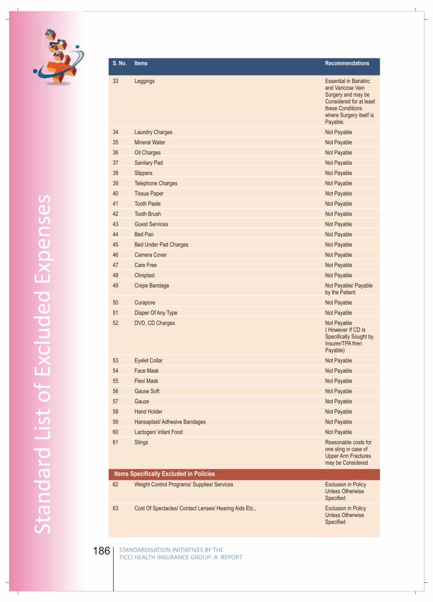
186
S. No. Items Recommendations
Items Specifically Excluded in Policies
33 Leggings Essential in Bariatricand Varicose VeinSurgery and may beConsidered for at leastthese Conditionswhere Surgery itself isPayable.
34 Laundry Charges Not Payable
35 Mineral Water Not Payable
36 Oil Charges Not Payable
37 Sanitary Pad Not Payable
38 Slippers Not Payable
39 Telephone Charges Not Payable
40 Tissue Paper Not Payable
41 Tooth Paste Not Payable
42 Tooth Brush Not Payable
43 Guest Services Not Payable
44 Bed Pan Not Payable
45 Bed Under Pad Charges Not Payable
46 Camera Cover Not Payable
47 Care Free Not Payable
48 Cliniplast Not Payable
49 Crepe Bandage Not Payable/ Payableby the Patient
50 Curapore Not Payable
51 Diaper Of Any Type Not Payable
52 DVD, CD Charges Not Payable ( However If CD IsSpecifically Sought by Insurer/TPA thenPayable)
53 Eyelet Collar Not Payable
54 Face Mask Not Payable
55 Flexi Mask Not Payable
56 Gause Soft Not Payable
57 Gauze Not Payable
58 Hand Holder Not Payable
59 Hansaplast/ Adhesive Bandages Not Payable
60 Lactogen/ Infant Food Not Payable
61 Slings Reasonable costs forone sling in case ofUpper Arm Fracturesmay be Considered
62 Weight Control Programs/ Supplies/ Services Exclusion in PolicyUnless OtherwiseSpecified
63 Cost Of Spectacles/ Contact Lenses/ Hearing Aids Etc., Exclusion in PolicyUnless OtherwiseSpecified
187
FICC
I Wo
rking G
rou
p R
epo
rt
64 Dental Treatment Expenses That Do Not Require Hospitalisation Exclusion In PolicyUnless OtherwiseSpecified
65 Hormone Replacement Therapy Exclusion In PolicyUnless OtherwiseSpecified
66 Home Visit Charges Exclusion In PolicyUnless OtherwiseSpecified
67 Infertility/ Subfertility/ Assisted Conception Procedure Exclusion In PolicyUnless OtherwiseSpecified
68 Obesity (including Morbid Obesity) Treatment Exclusion In PolicyUnless OtherwiseSpecified
69 Psychiatric & Psychosomatic Disorders Exclusion In PolicyUnless OtherwiseSpecified
70 Corrective Surgery For Refractive Error Exclusion In PolicyUnless OtherwiseSpecified
71 Treatment Of Sexually Transmitted Diseases Exclusion In PolicyUnless OtherwiseSpecified
72 Donor Screening Charges Exclusion In PolicyUnless OtherwiseSpecified
73 Admission/registration Charges Exclusion In PolicyUnless OtherwiseSpecified
74 Hospitalisation For Evaluation/ Diagnostic Purpose Exclusion In PolicyUnless OtherwiseSpecified
75 Expenses For Investigation/ Treatment Irrelevant Exclusion In PolicyTo The Disease For Which Admitted Or Diagnosed Not Payable - Unless
Otherwise Specified
76 Any Expenses When The Patient Is Diagnosed Not Payable As PerWith Retro Virus + Or Suffering From /HIV/ Aids HIV/aids ExclusionEtc Is Detected/ Directly Or Indirectly
77 Stem Cell Implantation/ Surgery Not Payable Except Bone MarrowTransplantation WhereCovered By Policy
78 Ward And Theatre Booking Charges Payable Under OTCharges, Not PayableSeparately
79 Arthroscopy & Endoscopy Instruments Rental Charged ByThe Hospital Payable.Purchase ofInstruments NotPayable.
80 Microscope Cover Payable Under OTCharges, NotSeparately
Items Which form Part of Hospital Services where Separate Consumables are not Payable but the Service is
S. No. Items Recommendations
STANDARDISATION INITIATIVES BY THE FICCI HEALTH INSURANCE GROUP- A REPORT
STANDARDISATION INITIATIVES BY THE FICCI HEALTH INSURANCE GROUP- A REPORT
Stan
dar
d L
ist
of
Excl
ud
ed E
xpen
ses
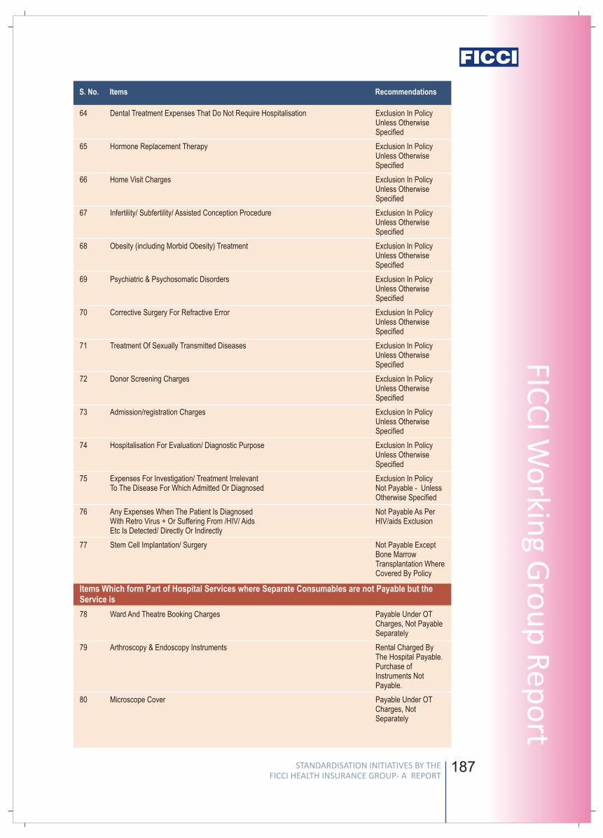
186
S. No. Items Recommendations
Items Specifically Excluded in Policies
33 Leggings Essential in Bariatricand Varicose VeinSurgery and may beConsidered for at leastthese Conditionswhere Surgery itself isPayable.
34 Laundry Charges Not Payable
35 Mineral Water Not Payable
36 Oil Charges Not Payable
37 Sanitary Pad Not Payable
38 Slippers Not Payable
39 Telephone Charges Not Payable
40 Tissue Paper Not Payable
41 Tooth Paste Not Payable
42 Tooth Brush Not Payable
43 Guest Services Not Payable
44 Bed Pan Not Payable
45 Bed Under Pad Charges Not Payable
46 Camera Cover Not Payable
47 Care Free Not Payable
48 Cliniplast Not Payable
49 Crepe Bandage Not Payable/ Payableby the Patient
50 Curapore Not Payable
51 Diaper Of Any Type Not Payable
52 DVD, CD Charges Not Payable ( However If CD IsSpecifically Sought by Insurer/TPA thenPayable)
53 Eyelet Collar Not Payable
54 Face Mask Not Payable
55 Flexi Mask Not Payable
56 Gause Soft Not Payable
57 Gauze Not Payable
58 Hand Holder Not Payable
59 Hansaplast/ Adhesive Bandages Not Payable
60 Lactogen/ Infant Food Not Payable
61 Slings Reasonable costs forone sling in case ofUpper Arm Fracturesmay be Considered
62 Weight Control Programs/ Supplies/ Services Exclusion in PolicyUnless OtherwiseSpecified
63 Cost Of Spectacles/ Contact Lenses/ Hearing Aids Etc., Exclusion in PolicyUnless OtherwiseSpecified
187
FICC
I Wo
rking G
rou
p R
epo
rt64 Dental Treatment Expenses That Do Not Require Hospitalisation Exclusion In Policy
Unless OtherwiseSpecified
65 Hormone Replacement Therapy Exclusion In PolicyUnless OtherwiseSpecified
66 Home Visit Charges Exclusion In PolicyUnless OtherwiseSpecified
67 Infertility/ Subfertility/ Assisted Conception Procedure Exclusion In PolicyUnless OtherwiseSpecified
68 Obesity (including Morbid Obesity) Treatment Exclusion In PolicyUnless OtherwiseSpecified
69 Psychiatric & Psychosomatic Disorders Exclusion In PolicyUnless OtherwiseSpecified
70 Corrective Surgery For Refractive Error Exclusion In PolicyUnless OtherwiseSpecified
71 Treatment Of Sexually Transmitted Diseases Exclusion In PolicyUnless OtherwiseSpecified
72 Donor Screening Charges Exclusion In PolicyUnless OtherwiseSpecified
73 Admission/registration Charges Exclusion In PolicyUnless OtherwiseSpecified
74 Hospitalisation For Evaluation/ Diagnostic Purpose Exclusion In PolicyUnless OtherwiseSpecified
75 Expenses For Investigation/ Treatment Irrelevant Exclusion In PolicyTo The Disease For Which Admitted Or Diagnosed Not Payable - Unless
Otherwise Specified
76 Any Expenses When The Patient Is Diagnosed Not Payable As PerWith Retro Virus + Or Suffering From /HIV/ Aids HIV/aids ExclusionEtc Is Detected/ Directly Or Indirectly
77 Stem Cell Implantation/ Surgery Not Payable Except Bone MarrowTransplantation WhereCovered By Policy
78 Ward And Theatre Booking Charges Payable Under OTCharges, Not PayableSeparately
79 Arthroscopy & Endoscopy Instruments Rental Charged ByThe Hospital Payable.Purchase ofInstruments NotPayable.
80 Microscope Cover Payable Under OTCharges, NotSeparately
Items Which form Part of Hospital Services where Separate Consumables are not Payable but the Service is
S. No. Items Recommendations
STANDARDISATION INITIATIVES BY THE FICCI HEALTH INSURANCE GROUP- A REPORT
STANDARDISATION INITIATIVES BY THE FICCI HEALTH INSURANCE GROUP- A REPORT
Stan
dar
d L
ist
of
Excl
ud
ed E
xpen
ses
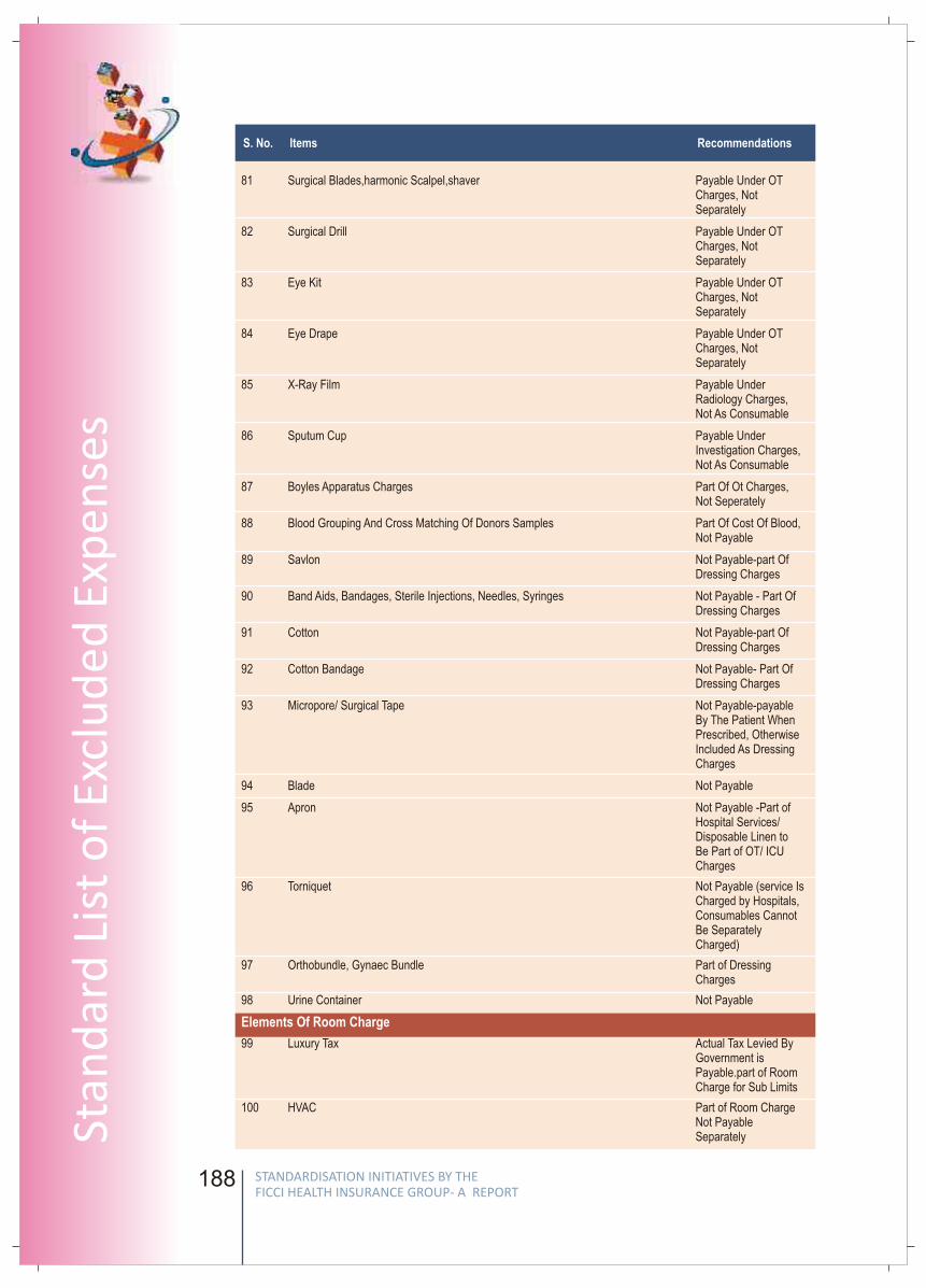
188
81 Surgical Blades,harmonic Scalpel,shaver Payable Under OTCharges, NotSeparately
82 Surgical Drill Payable Under OTCharges, NotSeparately
83 Eye Kit Payable Under OTCharges, NotSeparately
84 Eye Drape Payable Under OTCharges, NotSeparately
85 X-Ray Film Payable UnderRadiology Charges,Not As Consumable
86 Sputum Cup Payable UnderInvestigation Charges,Not As Consumable
87 Boyles Apparatus Charges Part Of Ot Charges,Not Seperately
88 Blood Grouping And Cross Matching Of Donors Samples Part Of Cost Of Blood,Not Payable
89 Savlon Not Payable-part OfDressing Charges
90 Band Aids, Bandages, Sterile Injections, Needles, Syringes Not Payable - Part OfDressing Charges
91 Cotton Not Payable-part OfDressing Charges
92 Cotton Bandage Not Payable- Part OfDressing Charges
93 Micropore/ Surgical Tape Not Payable-payableBy The Patient WhenPrescribed, OtherwiseIncluded As DressingCharges
94 Blade Not Payable
95 Apron Not Payable -Part ofHospital Services/Disposable Linen toBe Part of OT/ ICUCharges
96 Torniquet Not Payable (service IsCharged by Hospitals,Consumables CannotBe SeparatelyCharged)
97 Orthobundle, Gynaec Bundle Part of DressingCharges
98 Urine Container Not Payable
99 Luxury Tax Actual Tax Levied ByGovernment isPayable.part of RoomCharge for Sub Limits
100 HVAC Part of Room ChargeNot PayableSeparately
Elements Of Room Charge
S. No. Items Recommendations
189
FICC
I Wo
rking G
rou
p R
epo
rt
101 House Keeping Charges Part Of Room ChargeNot PayableSeparately
102 Service Charges Where Nursing Charge Also Charged Part Of Room ChargeNot PayableSeparately
103 Television & Air Conditioner Charges Payable Under RoomCharges Not IfSeparately Levied
104 Surcharges Part Of Room Charge,Not PayableSeparately
105 Attendant Charges Not Payable - Part OfRoom Charges
106 IM/ IV Injection Charges Part Of NursingCharges, Not Payable
107 Clean Sheet Part of Laundry/housekeeping Not Payable Separately
108 Extra Diet of Patient(other than that which Forms Part of Bed Charge) Patient Diet Providedby Hospital is Payable
109 Blanket/warmer Blanket Not Payable- Part ofRoom Charges
110 Admission Kit Not Payable
111 Birth Certificate Not Payable
112 Blood Reservation Charges And Ante Natal Booking Charges Not Payable
113 Certificate Charges Not Payable
114 Courier Charges Not Payable
115 Convenyance Charges Not Payable
116 Diabetic Chart Charges Not Payable
117 Documentation Charges / Administrative Expenses Not Payable
118 Discharge Procedure Charges Not Payable
119 Daily Chart Charges Not Payable
120 Entrance Pass / Visitors Pass Charges Not Payable
121 Expenses Related To Prescription On Discharge To Be Claimed ByPatient Under PostHosp WhereAdmissible
122 File Opening Charges Not Payable
123 Incidental Expenses / Misc. Charges (not Explained) Not Payable
124 Medical Certificate Not Payable
125 Maintainance Charges Not Payable
126 Medical Records Not Payable
127 Preparation Charges Not Payable
128 Photocopies Charges Not Payable
129 Patient Identification Band / Name Tag Not Payable
130 Washing Charges Not Payable
131 Medicine Box Not Payable
Administrative or Non-medical Charges
S. No. Items Recommendations
STANDARDISATION INITIATIVES BY THE FICCI HEALTH INSURANCE GROUP- A REPORT
STANDARDISATION INITIATIVES BY THE FICCI HEALTH INSURANCE GROUP- A REPORT
Stan
dar
d L
ist
of
Excl
ud
ed E
xpen
ses
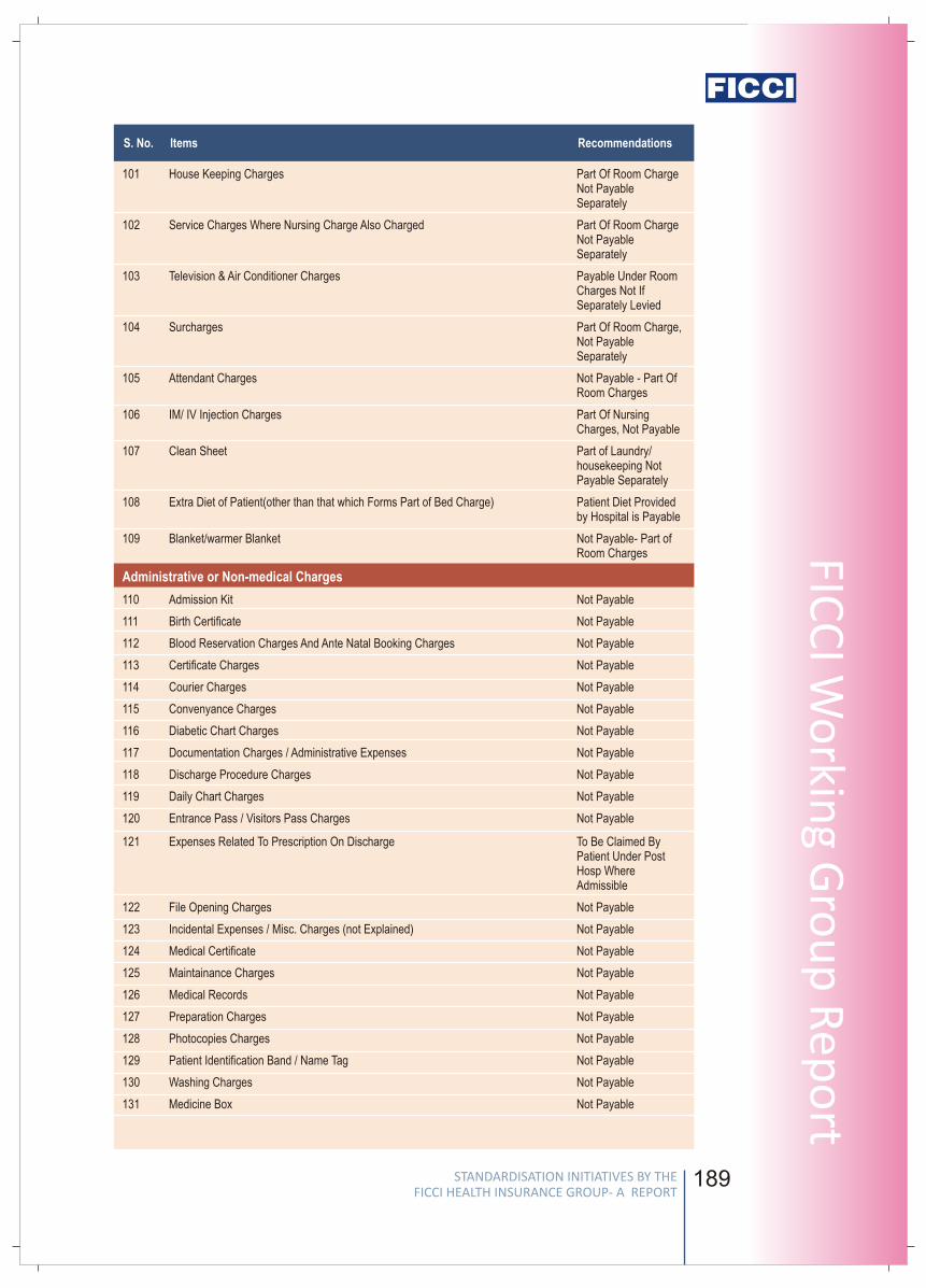
188
81 Surgical Blades,harmonic Scalpel,shaver Payable Under OTCharges, NotSeparately
82 Surgical Drill Payable Under OTCharges, NotSeparately
83 Eye Kit Payable Under OTCharges, NotSeparately
84 Eye Drape Payable Under OTCharges, NotSeparately
85 X-Ray Film Payable UnderRadiology Charges,Not As Consumable
86 Sputum Cup Payable UnderInvestigation Charges,Not As Consumable
87 Boyles Apparatus Charges Part Of Ot Charges,Not Seperately
88 Blood Grouping And Cross Matching Of Donors Samples Part Of Cost Of Blood,Not Payable
89 Savlon Not Payable-part OfDressing Charges
90 Band Aids, Bandages, Sterile Injections, Needles, Syringes Not Payable - Part OfDressing Charges
91 Cotton Not Payable-part OfDressing Charges
92 Cotton Bandage Not Payable- Part OfDressing Charges
93 Micropore/ Surgical Tape Not Payable-payableBy The Patient WhenPrescribed, OtherwiseIncluded As DressingCharges
94 Blade Not Payable
95 Apron Not Payable -Part ofHospital Services/Disposable Linen toBe Part of OT/ ICUCharges
96 Torniquet Not Payable (service IsCharged by Hospitals,Consumables CannotBe SeparatelyCharged)
97 Orthobundle, Gynaec Bundle Part of DressingCharges
98 Urine Container Not Payable
99 Luxury Tax Actual Tax Levied ByGovernment isPayable.part of RoomCharge for Sub Limits
100 HVAC Part of Room ChargeNot PayableSeparately
Elements Of Room Charge
S. No. Items Recommendations
189
FICC
I Wo
rking G
rou
p R
epo
rt101 House Keeping Charges Part Of Room Charge
Not PayableSeparately
102 Service Charges Where Nursing Charge Also Charged Part Of Room ChargeNot PayableSeparately
103 Television & Air Conditioner Charges Payable Under RoomCharges Not IfSeparately Levied
104 Surcharges Part Of Room Charge,Not PayableSeparately
105 Attendant Charges Not Payable - Part OfRoom Charges
106 IM/ IV Injection Charges Part Of NursingCharges, Not Payable
107 Clean Sheet Part of Laundry/housekeeping Not Payable Separately
108 Extra Diet of Patient(other than that which Forms Part of Bed Charge) Patient Diet Providedby Hospital is Payable
109 Blanket/warmer Blanket Not Payable- Part ofRoom Charges
110 Admission Kit Not Payable
111 Birth Certificate Not Payable
112 Blood Reservation Charges And Ante Natal Booking Charges Not Payable
113 Certificate Charges Not Payable
114 Courier Charges Not Payable
115 Convenyance Charges Not Payable
116 Diabetic Chart Charges Not Payable
117 Documentation Charges / Administrative Expenses Not Payable
118 Discharge Procedure Charges Not Payable
119 Daily Chart Charges Not Payable
120 Entrance Pass / Visitors Pass Charges Not Payable
121 Expenses Related To Prescription On Discharge To Be Claimed ByPatient Under PostHosp WhereAdmissible
122 File Opening Charges Not Payable
123 Incidental Expenses / Misc. Charges (not Explained) Not Payable
124 Medical Certificate Not Payable
125 Maintainance Charges Not Payable
126 Medical Records Not Payable
127 Preparation Charges Not Payable
128 Photocopies Charges Not Payable
129 Patient Identification Band / Name Tag Not Payable
130 Washing Charges Not Payable
131 Medicine Box Not Payable
Administrative or Non-medical Charges
S. No. Items Recommendations
STANDARDISATION INITIATIVES BY THE FICCI HEALTH INSURANCE GROUP- A REPORT
STANDARDISATION INITIATIVES BY THE FICCI HEALTH INSURANCE GROUP- A REPORT
Stan
dar
d L
ist
of
Excl
ud
ed E
xpen
ses
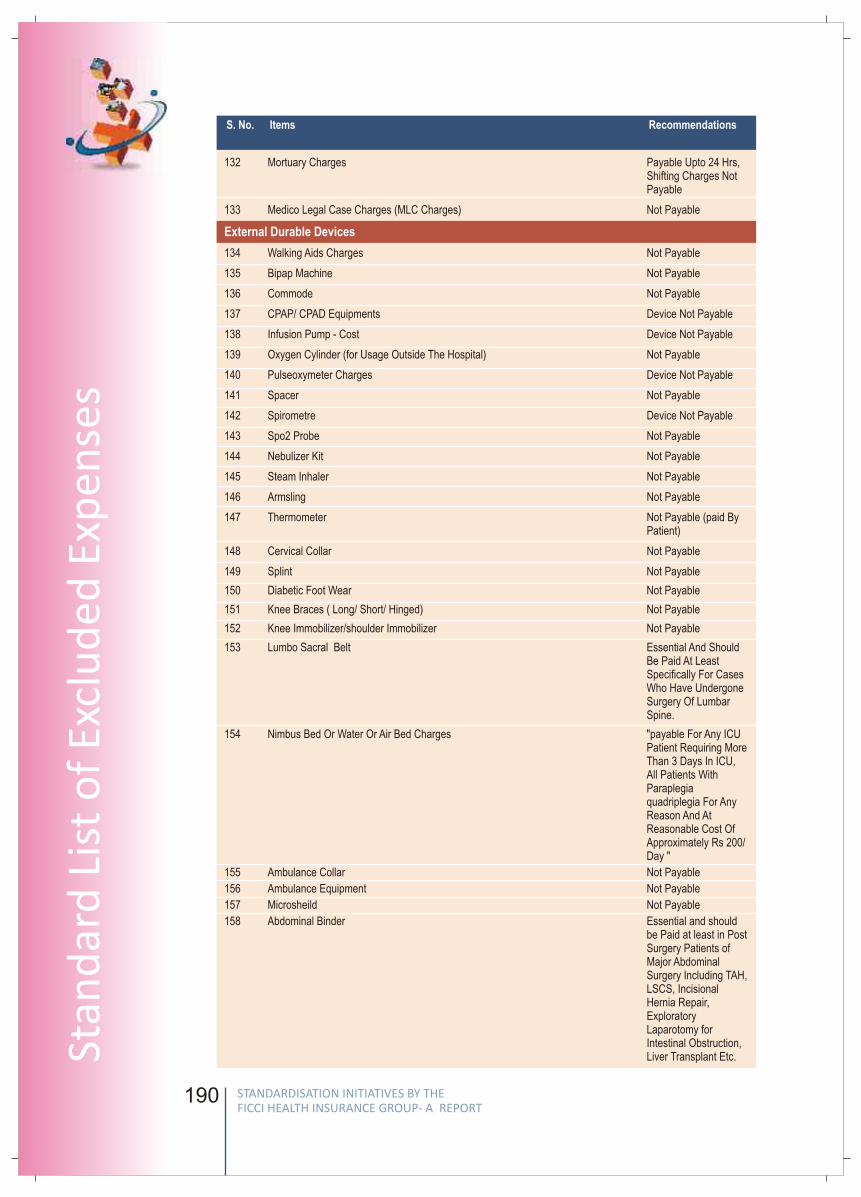
190
S. No. Items Recommendations
132 Mortuary Charges Payable Upto 24 Hrs,Shifting Charges NotPayable
133 Medico Legal Case Charges (MLC Charges) Not Payable
134 Walking Aids Charges Not Payable
135 Bipap Machine Not Payable
136 Commode Not Payable
137 CPAP/ CPAD Equipments Device Not Payable
138 Infusion Pump - Cost Device Not Payable
139 Oxygen Cylinder (for Usage Outside The Hospital) Not Payable
140 Pulseoxymeter Charges Device Not Payable
141 Spacer Not Payable
142 Spirometre Device Not Payable
143 Spo2 Probe Not Payable
144 Nebulizer Kit Not Payable
145 Steam Inhaler Not Payable
146 Armsling Not Payable
147 Thermometer Not Payable (paid ByPatient)
148 Cervical Collar Not Payable
149 Splint Not Payable
150 Diabetic Foot Wear Not Payable
151 Knee Braces ( Long/ Short/ Hinged) Not Payable
152 Knee Immobilizer/shoulder Immobilizer Not Payable
153 Lumbo Sacral Belt Essential And ShouldBe Paid At LeastSpecifically For CasesWho Have UndergoneSurgery Of LumbarSpine.
154 Nimbus Bed Or Water Or Air Bed Charges "payable For Any ICUPatient Requiring MoreThan 3 Days In ICU,All Patients WithParaplegiaquadriplegia For AnyReason And AtReasonable Cost OfApproximately Rs 200/Day "
155 Ambulance Collar Not Payable
156 Ambulance Equipment Not Payable
157 Microsheild Not Payable
158 Abdominal Binder Essential and shouldbe Paid at least in PostSurgery Patients ofMajor AbdominalSurgery Including TAH,LSCS, Incisional Hernia Repair,ExploratoryLaparotomy forIntestinal Obstruction,Liver Transplant Etc.
External Durable Devices
191
FICC
I Wo
rking G
rou
p R
epo
rt
S. No. Items Recommendations
STANDARDISATION INITIATIVES BY THE FICCI HEALTH INSURANCE GROUP- A REPORT
STANDARDISATION INITIATIVES BY THE FICCI HEALTH INSURANCE GROUP- A REPORT
Stan
dar
d L
ist
of
Excl
ud
ed E
xpen
ses
Items Payable If Supported By A Prescription
Part of Hospital's own Costs and not Payable
159 Betadine \ Hydrogen Peroxide\spirit\\dettol \Savlon\ Disinfectants Etc May Be Payable WhenPrescribed For Patient,Not Payable ForHospital use In OT orWard Or For Dressings in Hospital
160 Private Nurses Charges- Special Nursing Charges Post HospitalizationNursing Charges NotPayable
161 Nutrition Planning Charges - Dietician Charges- Diet Charges Patient Diet ProvidedBy Hospital Is Payable
162 Alex Sugar Free Payable -Sugar FreeVariants Of AdmissableMedicines Are NotExcluded
163 Cream Powder Lotion Payable When (Toileteries are Not Payable, only Prescribed Medical Prescribed Pharmaceuticals Payable)
164 Digene Gel/ Antacid Gel Payable WhenPrescribed
165 ECG Electrodes Upto 5 Electrodes AreRequired For EveryCase Visiting OT orICU. For Longer StayIn ICU, May Require AChange And At LeastOne Set Every SecondDay Must Be Payable.
166 Gloves Sterilized GlovesPayable / UnsterilizedGloves Not Payable
167 HIV Kit Payable - PreOperative Screening
168 Listerine/ Antiseptic Mouthwash Payable WhenPrescribed
169 Lozenges Payable WhenPrescribed
170 Mouth Paint Payable WhenPrescribed
171 Nebulisation Kit If Used DuringHospitalization isPayable Reasonably
172 Neosprin Payable WhenPrescribed
173 Novarapid Payable WhenPrescribed
174 Volini Gel/ Analgesic Gel Payable WhenPrescribed
175 Zytee Gel Payable WhenPrescribed
176 Vaccination Charges Routine VaccinationNot Payable / Post BiteVaccination Payable
177 AHD Not Payable - Part ofHospital's Internal Cost
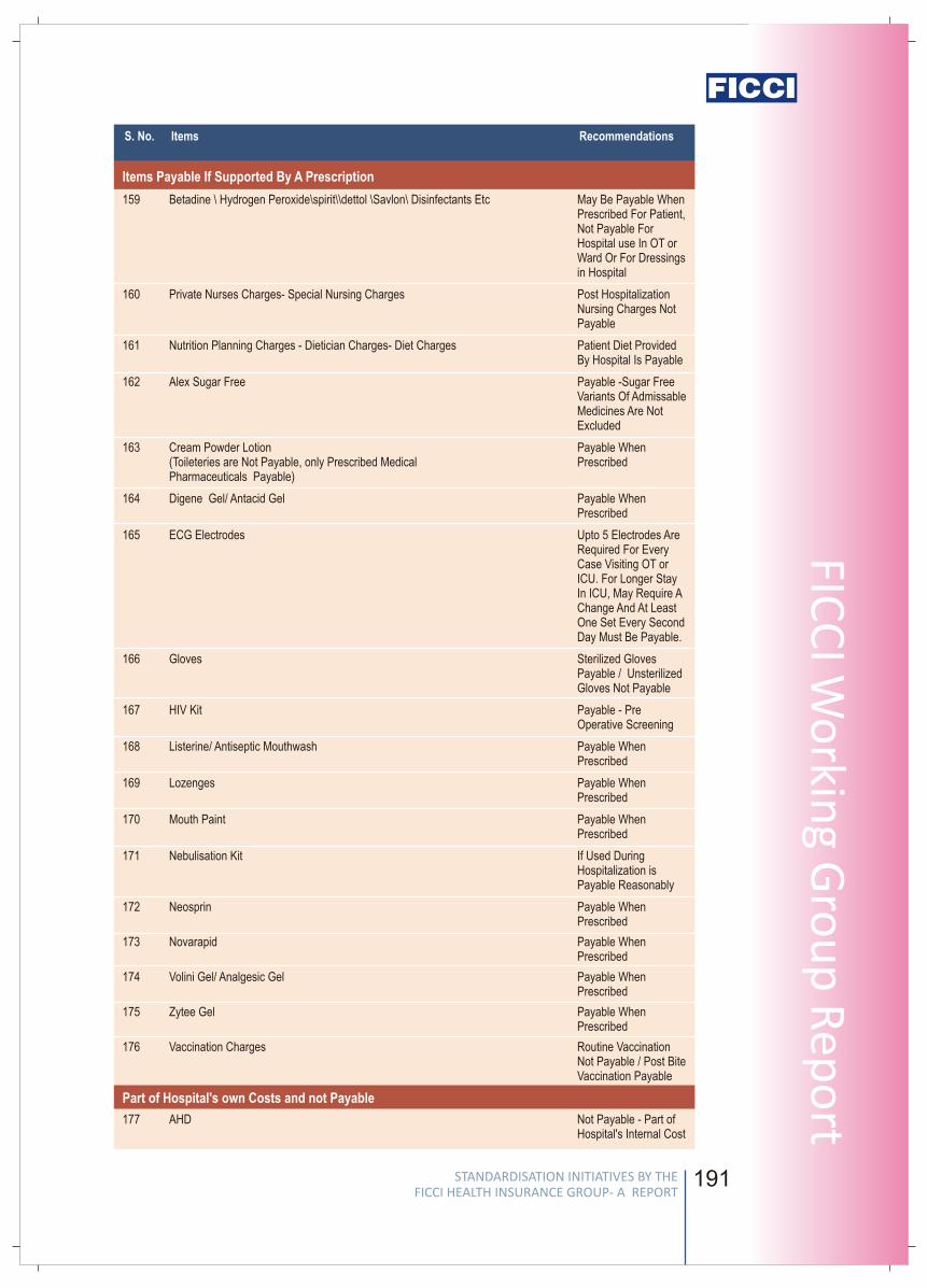
190
S. No. Items Recommendations
132 Mortuary Charges Payable Upto 24 Hrs,Shifting Charges NotPayable
133 Medico Legal Case Charges (MLC Charges) Not Payable
134 Walking Aids Charges Not Payable
135 Bipap Machine Not Payable
136 Commode Not Payable
137 CPAP/ CPAD Equipments Device Not Payable
138 Infusion Pump - Cost Device Not Payable
139 Oxygen Cylinder (for Usage Outside The Hospital) Not Payable
140 Pulseoxymeter Charges Device Not Payable
141 Spacer Not Payable
142 Spirometre Device Not Payable
143 Spo2 Probe Not Payable
144 Nebulizer Kit Not Payable
145 Steam Inhaler Not Payable
146 Armsling Not Payable
147 Thermometer Not Payable (paid ByPatient)
148 Cervical Collar Not Payable
149 Splint Not Payable
150 Diabetic Foot Wear Not Payable
151 Knee Braces ( Long/ Short/ Hinged) Not Payable
152 Knee Immobilizer/shoulder Immobilizer Not Payable
153 Lumbo Sacral Belt Essential And ShouldBe Paid At LeastSpecifically For CasesWho Have UndergoneSurgery Of LumbarSpine.
154 Nimbus Bed Or Water Or Air Bed Charges "payable For Any ICUPatient Requiring MoreThan 3 Days In ICU,All Patients WithParaplegiaquadriplegia For AnyReason And AtReasonable Cost OfApproximately Rs 200/Day "
155 Ambulance Collar Not Payable
156 Ambulance Equipment Not Payable
157 Microsheild Not Payable
158 Abdominal Binder Essential and shouldbe Paid at least in PostSurgery Patients ofMajor AbdominalSurgery Including TAH,LSCS, Incisional Hernia Repair,ExploratoryLaparotomy forIntestinal Obstruction,Liver Transplant Etc.
External Durable Devices
191
FICC
I Wo
rking G
rou
p R
epo
rtS. No. Items Recommendations
STANDARDISATION INITIATIVES BY THE FICCI HEALTH INSURANCE GROUP- A REPORT
STANDARDISATION INITIATIVES BY THE FICCI HEALTH INSURANCE GROUP- A REPORT
Stan
dar
d L
ist
of
Excl
ud
ed E
xpen
ses
Items Payable If Supported By A Prescription
Part of Hospital's own Costs and not Payable
159 Betadine \ Hydrogen Peroxide\spirit\\dettol \Savlon\ Disinfectants Etc May Be Payable WhenPrescribed For Patient,Not Payable ForHospital use In OT orWard Or For Dressings in Hospital
160 Private Nurses Charges- Special Nursing Charges Post HospitalizationNursing Charges NotPayable
161 Nutrition Planning Charges - Dietician Charges- Diet Charges Patient Diet ProvidedBy Hospital Is Payable
162 Alex Sugar Free Payable -Sugar FreeVariants Of AdmissableMedicines Are NotExcluded
163 Cream Powder Lotion Payable When (Toileteries are Not Payable, only Prescribed Medical Prescribed Pharmaceuticals Payable)
164 Digene Gel/ Antacid Gel Payable WhenPrescribed
165 ECG Electrodes Upto 5 Electrodes AreRequired For EveryCase Visiting OT orICU. For Longer StayIn ICU, May Require AChange And At LeastOne Set Every SecondDay Must Be Payable.
166 Gloves Sterilized GlovesPayable / UnsterilizedGloves Not Payable
167 HIV Kit Payable - PreOperative Screening
168 Listerine/ Antiseptic Mouthwash Payable WhenPrescribed
169 Lozenges Payable WhenPrescribed
170 Mouth Paint Payable WhenPrescribed
171 Nebulisation Kit If Used DuringHospitalization isPayable Reasonably
172 Neosprin Payable WhenPrescribed
173 Novarapid Payable WhenPrescribed
174 Volini Gel/ Analgesic Gel Payable WhenPrescribed
175 Zytee Gel Payable WhenPrescribed
176 Vaccination Charges Routine VaccinationNot Payable / Post BiteVaccination Payable
177 AHD Not Payable - Part ofHospital's Internal Cost
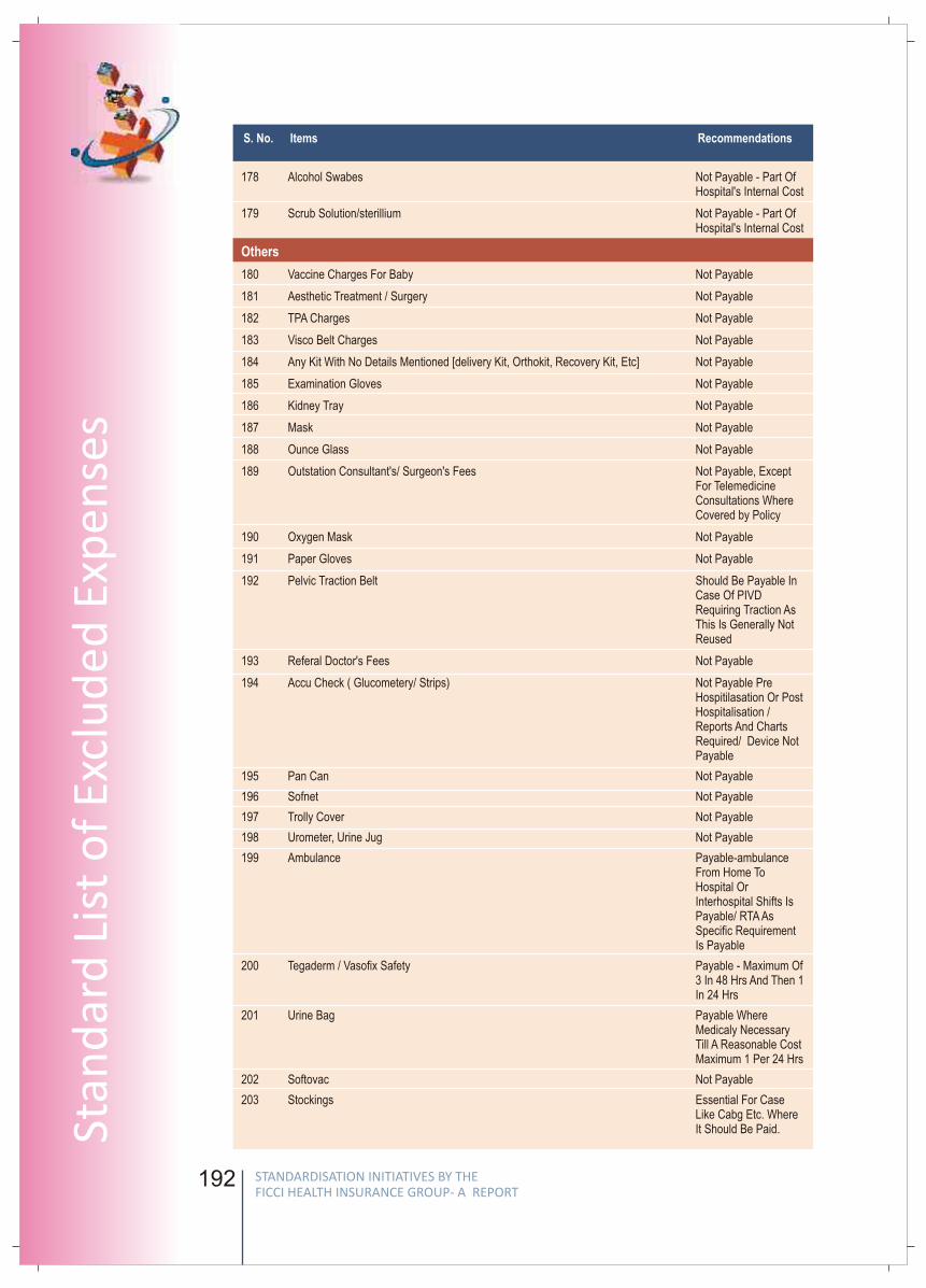
192
178 Alcohol Swabes Not Payable - Part OfHospital's Internal Cost
179 Scrub Solution/sterillium Not Payable - Part OfHospital's Internal Cost
180 Vaccine Charges For Baby Not Payable
181 Aesthetic Treatment / Surgery Not Payable
182 TPA Charges Not Payable
183 Visco Belt Charges Not Payable
184 Any Kit With No Details Mentioned [delivery Kit, Orthokit, Recovery Kit, Etc] Not Payable
185 Examination Gloves Not Payable
186 Kidney Tray Not Payable
187 Mask Not Payable
188 Ounce Glass Not Payable
189 Outstation Consultant's/ Surgeon's Fees Not Payable, ExceptFor TelemedicineConsultations WhereCovered by Policy
190 Oxygen Mask Not Payable
191 Paper Gloves Not Payable
192 Pelvic Traction Belt Should Be Payable InCase Of PIVDRequiring Traction AsThis Is Generally NotReused
193 Referal Doctor's Fees Not Payable
194 Accu Check ( Glucometery/ Strips) Not Payable PreHospitilasation Or PostHospitalisation / Reports And Charts Required/ Device NotPayable
195 Pan Can Not Payable
196 Sofnet Not Payable
197 Trolly Cover Not Payable
198 Urometer, Urine Jug Not Payable
199 Ambulance Payable-ambulanceFrom Home ToHospital OrInterhospital Shifts IsPayable/ RTA AsSpecific RequirementIs Payable
200 Tegaderm / Vasofix Safety Payable - Maximum Of3 In 48 Hrs And Then 1In 24 Hrs
201 Urine Bag Payable WhereMedicaly NecessaryTill A Reasonable CostMaximum 1 Per 24 Hrs
202 Softovac Not Payable
203 Stockings Essential For CaseLike Cabg Etc. WhereIt Should Be Paid.
Others
S. No. Items Recommendations
193
FICC
I Wo
rking G
rou
p R
epo
rt
Chairpersons
S. L. MohanSecretary General
General Insurance Council
Mr. Mohan is the Secretary General of General Insurance Council since November 2008.
In addition to this he is also serving as an Associate of Insurance Institute of India.
Prior to this, he was the Chairman cum Managing Director, The Oriental Insurance Co.
Ltd. (2002-2005), General Manager, United India Insurance Co. Ltd., Chennai (2001-
2002), General Manager, National Insurance Co. Ltd., Kolkata (2000) and Regional
Manager / Asst. General Manager, United India Ins. Co. Ltd., Chennai (1989-2000).
He has also served as a director at Kenindia Assurance Co. Ltd., Nairobi, GIC Housing
Finance Limited, Mumbai, Loss Prevention Association of India Ltd., Mumbai, Member –
Tariff Advisory Committee, Mumbai, Madhura Coats Ltd. to name a few.
He holds a B.Sc. in Mechanical Engineering.
S. B. MathurSecretary General
Life Insurance Council
Mr. Mathur is the Secretary General of Life Insurance Council. In addition to this Mr.
Mathur is the Chairman of National Stock Exchange, an Advisor to National Investment
Fund set up by Government of India and also on Board of some leading Corporates.
He was the Chairman of the Life Insurance Corporation of India (LIC), the largest life
insurance company in India from August 2002 to October 2004. His tenure coincided the
opening of the Insurance sector, which resulted into the entry of 12 domestic players
with strong brands tying up with leading international insurance companies.
Post retirement from LIC, The Government of India appointed him as Administrator of
the Specified Undertaking of the Unit Trust of India (SUUTI), the successor of the
erstwhile Unit Trust of India in December 2004, wherein Government had provided
financial support to meet the liabilities under some of the guaranteed high returns
schemes.
STANDARDISATION INITIATIVES BY THE FICCI HEALTH INSURANCE GROUP- A REPORT
STANDARDISATION INITIATIVES BY THE FICCI HEALTH INSURANCE GROUP- A REPORT
Stan
dar
d L
ist
of
Excl
ud
ed E
xpen
ses
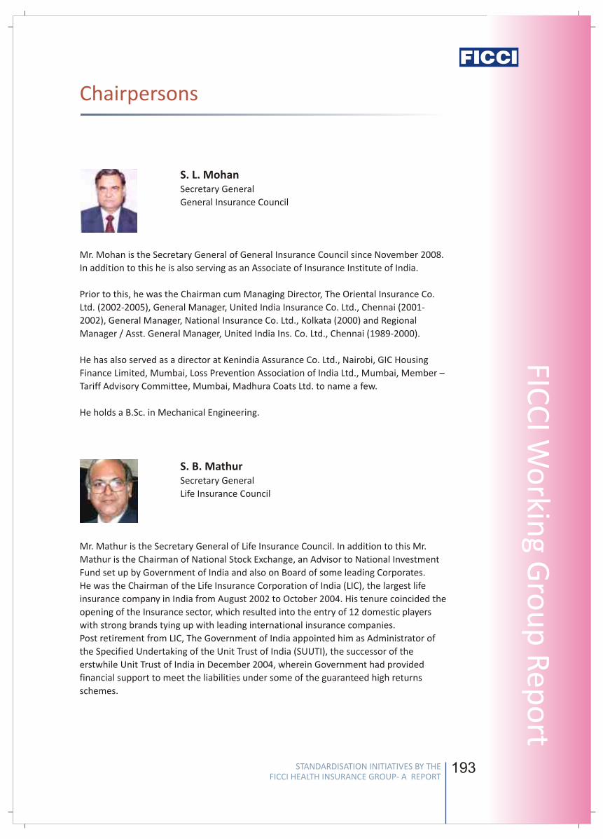
192
178 Alcohol Swabes Not Payable - Part OfHospital's Internal Cost
179 Scrub Solution/sterillium Not Payable - Part OfHospital's Internal Cost
180 Vaccine Charges For Baby Not Payable
181 Aesthetic Treatment / Surgery Not Payable
182 TPA Charges Not Payable
183 Visco Belt Charges Not Payable
184 Any Kit With No Details Mentioned [delivery Kit, Orthokit, Recovery Kit, Etc] Not Payable
185 Examination Gloves Not Payable
186 Kidney Tray Not Payable
187 Mask Not Payable
188 Ounce Glass Not Payable
189 Outstation Consultant's/ Surgeon's Fees Not Payable, ExceptFor TelemedicineConsultations WhereCovered by Policy
190 Oxygen Mask Not Payable
191 Paper Gloves Not Payable
192 Pelvic Traction Belt Should Be Payable InCase Of PIVDRequiring Traction AsThis Is Generally NotReused
193 Referal Doctor's Fees Not Payable
194 Accu Check ( Glucometery/ Strips) Not Payable PreHospitilasation Or PostHospitalisation / Reports And Charts Required/ Device NotPayable
195 Pan Can Not Payable
196 Sofnet Not Payable
197 Trolly Cover Not Payable
198 Urometer, Urine Jug Not Payable
199 Ambulance Payable-ambulanceFrom Home ToHospital OrInterhospital Shifts IsPayable/ RTA AsSpecific RequirementIs Payable
200 Tegaderm / Vasofix Safety Payable - Maximum Of3 In 48 Hrs And Then 1In 24 Hrs
201 Urine Bag Payable WhereMedicaly NecessaryTill A Reasonable CostMaximum 1 Per 24 Hrs
202 Softovac Not Payable
203 Stockings Essential For CaseLike Cabg Etc. WhereIt Should Be Paid.
Others
S. No. Items Recommendations
193
FICC
I Wo
rking G
rou
p R
epo
rtChairpersons
S. L. MohanSecretary General
General Insurance Council
Mr. Mohan is the Secretary General of General Insurance Council since November 2008.
In addition to this he is also serving as an Associate of Insurance Institute of India.
Prior to this, he was the Chairman cum Managing Director, The Oriental Insurance Co.
Ltd. (2002-2005), General Manager, United India Insurance Co. Ltd., Chennai (2001-
2002), General Manager, National Insurance Co. Ltd., Kolkata (2000) and Regional
Manager / Asst. General Manager, United India Ins. Co. Ltd., Chennai (1989-2000).
He has also served as a director at Kenindia Assurance Co. Ltd., Nairobi, GIC Housing
Finance Limited, Mumbai, Loss Prevention Association of India Ltd., Mumbai, Member –
Tariff Advisory Committee, Mumbai, Madhura Coats Ltd. to name a few.
He holds a B.Sc. in Mechanical Engineering.
S. B. MathurSecretary General
Life Insurance Council
Mr. Mathur is the Secretary General of Life Insurance Council. In addition to this Mr.
Mathur is the Chairman of National Stock Exchange, an Advisor to National Investment
Fund set up by Government of India and also on Board of some leading Corporates.
He was the Chairman of the Life Insurance Corporation of India (LIC), the largest life
insurance company in India from August 2002 to October 2004. His tenure coincided the
opening of the Insurance sector, which resulted into the entry of 12 domestic players
with strong brands tying up with leading international insurance companies.
Post retirement from LIC, The Government of India appointed him as Administrator of
the Specified Undertaking of the Unit Trust of India (SUUTI), the successor of the
erstwhile Unit Trust of India in December 2004, wherein Government had provided
financial support to meet the liabilities under some of the guaranteed high returns
schemes.
STANDARDISATION INITIATIVES BY THE FICCI HEALTH INSURANCE GROUP- A REPORT
STANDARDISATION INITIATIVES BY THE FICCI HEALTH INSURANCE GROUP- A REPORT
Stan
dar
d L
ist
of
Excl
ud
ed E
xpen
ses
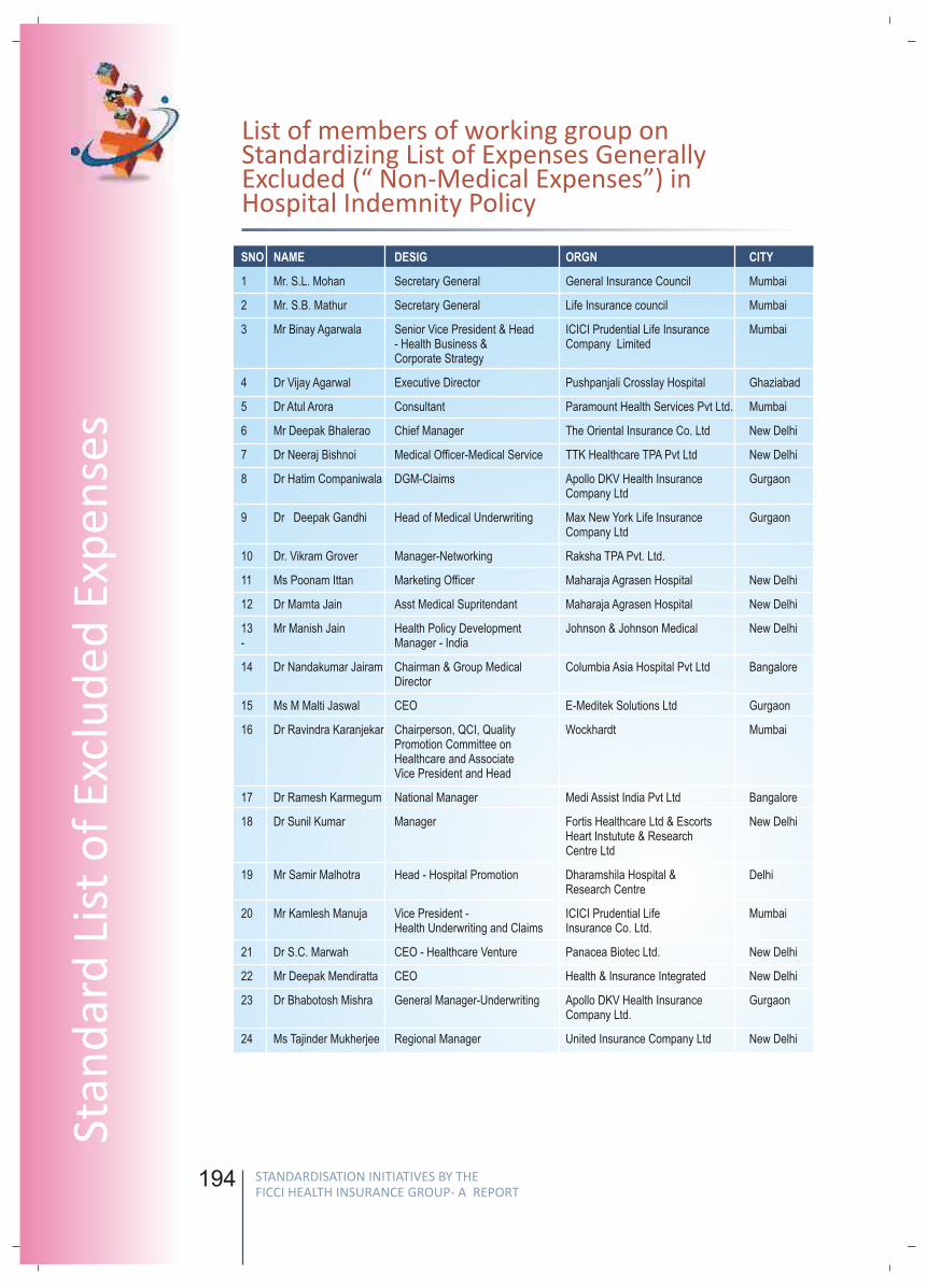
194
SNO NAME DESIG ORGN CITY
1 Mr. S.L. Mohan Secretary General General Insurance Council Mumbai
2 Mr. S.B. Mathur Secretary General Life Insurance council Mumbai
3 Mr Binay Agarwala Senior Vice President & Head ICICI Prudential Life Insurance Mumbai- Health Business & Company Limited Corporate Strategy
4 Dr Vijay Agarwal Executive Director Pushpanjali Crosslay Hospital Ghaziabad
5 Dr Atul Arora Consultant Paramount Health Services Pvt Ltd. Mumbai
6 Mr Deepak Bhalerao Chief Manager The Oriental Insurance Co. Ltd New Delhi
7 Dr Neeraj Bishnoi Medical Officer-Medical Service TTK Healthcare TPA Pvt Ltd New Delhi
8 Dr Hatim Companiwala DGM-Claims Apollo DKV Health Insurance Gurgaon Company Ltd
9 Dr Deepak Gandhi Head of Medical Underwriting Max New York Life Insurance GurgaonCompany Ltd
10 Dr. Vikram Grover Manager-Networking Raksha TPA Pvt. Ltd.
11 Ms Poonam Ittan Marketing Officer Maharaja Agrasen Hospital New Delhi
12 Dr Mamta Jain Asst Medical Supritendant Maharaja Agrasen Hospital New Delhi
13 Mr Manish Jain Health Policy Development Johnson & Johnson Medical New Delhi - Manager - India
14 Dr Nandakumar Jairam Chairman & Group Medical Columbia Asia Hospital Pvt Ltd BangaloreDirector
15 Ms M Malti Jaswal CEO E-Meditek Solutions Ltd Gurgaon
16 Dr Ravindra Karanjekar Chairperson, QCI, Quality Wockhardt MumbaiPromotion Committee on Healthcare and Associate Vice President and Head
17 Dr Ramesh Karmegum National Manager Medi Assist India Pvt Ltd Bangalore
18 Dr Sunil Kumar Manager Fortis Healthcare Ltd & Escorts New DelhiHeart Instutute & Research Centre Ltd
19 Mr Samir Malhotra Head - Hospital Promotion Dharamshila Hospital & DelhiResearch Centre
20 Mr Kamlesh Manuja Vice President - ICICI Prudential Life MumbaiHealth Underwriting and Claims Insurance Co. Ltd.
21 Dr S.C. Marwah CEO - Healthcare Venture Panacea Biotec Ltd. New Delhi
22 Mr Deepak Mendiratta CEO Health & Insurance Integrated New Delhi
23 Dr Bhabotosh Mishra General Manager-Underwriting Apollo DKV Health Insurance GurgaonCompany Ltd.
24 Ms Tajinder Mukherjee Regional Manager United Insurance Company Ltd New Delhi
195
FICC
I Wo
rking G
rou
p R
epo
rt
SNO NAME DESIG ORGN CITY
25 Dr Somil Nagpal Special Officer- Health Insurance Insurance Regulatory Hyderabad Developement Authority
26 Dr Jitender Nagpal Health Insurance Consultant Indraprastha Apollo Hospital New Delhi
27 Dr Surya Prakash Senior Manager - Apollo DKV Insurance CompanyClaim Operations Ltd
28 Ms K. Anita Rajaram Manager United Insurance Company Ltd Chennai
29 Mr Krishnan Chief Operating Officer Apollo DKV Insurance Company Ltd. GurgaonRamachandran
30 Dr Parag Rindani Senior Manager, Medical Services Wockhardt Hospitals Mumbai
31 Dr Amitoj Singh COO E-Meditek Solutions Ltd Faridabad
32 Mr C S Tandon DGM The Oriental Insurance Co. Ltd New Delhi
33 Dr Suman Singh Tilak Assistant General Manager Paramount Health Services Pvt. Ltd. New Delhi
34 Dr. Anupama Verma Manager United Insurance Company Ltd New Delhi
35 Mr Praveen Yadav Chief Administrative Officer, MD India Healthcare Pune Services (TPA) Pvt Ltd
List of members of working group onStandardizing List of Expenses Generally Excluded (“ Non-Medical Expenses”) in Hospital Indemnity Policy
STANDARDISATION INITIATIVES BY THE FICCI HEALTH INSURANCE GROUP- A REPORT
STANDARDISATION INITIATIVES BY THE FICCI HEALTH INSURANCE GROUP- A REPORT
Stan
dar
d L
ist
of
Excl
ud
ed E
xpen
ses
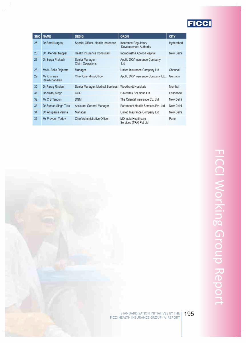
194
SNO NAME DESIG ORGN CITY
1 Mr. S.L. Mohan Secretary General General Insurance Council Mumbai
2 Mr. S.B. Mathur Secretary General Life Insurance council Mumbai
3 Mr Binay Agarwala Senior Vice President & Head ICICI Prudential Life Insurance Mumbai- Health Business & Company Limited Corporate Strategy
4 Dr Vijay Agarwal Executive Director Pushpanjali Crosslay Hospital Ghaziabad
5 Dr Atul Arora Consultant Paramount Health Services Pvt Ltd. Mumbai
6 Mr Deepak Bhalerao Chief Manager The Oriental Insurance Co. Ltd New Delhi
7 Dr Neeraj Bishnoi Medical Officer-Medical Service TTK Healthcare TPA Pvt Ltd New Delhi
8 Dr Hatim Companiwala DGM-Claims Apollo DKV Health Insurance Gurgaon Company Ltd
9 Dr Deepak Gandhi Head of Medical Underwriting Max New York Life Insurance GurgaonCompany Ltd
10 Dr. Vikram Grover Manager-Networking Raksha TPA Pvt. Ltd.
11 Ms Poonam Ittan Marketing Officer Maharaja Agrasen Hospital New Delhi
12 Dr Mamta Jain Asst Medical Supritendant Maharaja Agrasen Hospital New Delhi
13 Mr Manish Jain Health Policy Development Johnson & Johnson Medical New Delhi - Manager - India
14 Dr Nandakumar Jairam Chairman & Group Medical Columbia Asia Hospital Pvt Ltd BangaloreDirector
15 Ms M Malti Jaswal CEO E-Meditek Solutions Ltd Gurgaon
16 Dr Ravindra Karanjekar Chairperson, QCI, Quality Wockhardt MumbaiPromotion Committee on Healthcare and Associate Vice President and Head
17 Dr Ramesh Karmegum National Manager Medi Assist India Pvt Ltd Bangalore
18 Dr Sunil Kumar Manager Fortis Healthcare Ltd & Escorts New DelhiHeart Instutute & Research Centre Ltd
19 Mr Samir Malhotra Head - Hospital Promotion Dharamshila Hospital & DelhiResearch Centre
20 Mr Kamlesh Manuja Vice President - ICICI Prudential Life MumbaiHealth Underwriting and Claims Insurance Co. Ltd.
21 Dr S.C. Marwah CEO - Healthcare Venture Panacea Biotec Ltd. New Delhi
22 Mr Deepak Mendiratta CEO Health & Insurance Integrated New Delhi
23 Dr Bhabotosh Mishra General Manager-Underwriting Apollo DKV Health Insurance GurgaonCompany Ltd.
24 Ms Tajinder Mukherjee Regional Manager United Insurance Company Ltd New Delhi
195
FICC
I Wo
rking G
rou
p R
epo
rtSNO NAME DESIG ORGN CITY
25 Dr Somil Nagpal Special Officer- Health Insurance Insurance Regulatory Hyderabad Developement Authority
26 Dr Jitender Nagpal Health Insurance Consultant Indraprastha Apollo Hospital New Delhi
27 Dr Surya Prakash Senior Manager - Apollo DKV Insurance CompanyClaim Operations Ltd
28 Ms K. Anita Rajaram Manager United Insurance Company Ltd Chennai
29 Mr Krishnan Chief Operating Officer Apollo DKV Insurance Company Ltd. GurgaonRamachandran
30 Dr Parag Rindani Senior Manager, Medical Services Wockhardt Hospitals Mumbai
31 Dr Amitoj Singh COO E-Meditek Solutions Ltd Faridabad
32 Mr C S Tandon DGM The Oriental Insurance Co. Ltd New Delhi
33 Dr Suman Singh Tilak Assistant General Manager Paramount Health Services Pvt. Ltd. New Delhi
34 Dr. Anupama Verma Manager United Insurance Company Ltd New Delhi
35 Mr Praveen Yadav Chief Administrative Officer, MD India Healthcare Pune Services (TPA) Pvt Ltd
List of members of working group onStandardizing List of Expenses Generally Excluded (“ Non-Medical Expenses”) in Hospital Indemnity Policy
STANDARDISATION INITIATIVES BY THE FICCI HEALTH INSURANCE GROUP- A REPORT
STANDARDISATION INITIATIVES BY THE FICCI HEALTH INSURANCE GROUP- A REPORT
Stan
dar
d L
ist
of
Excl
ud
ed E
xpen
ses
196 197
FICC
I Wo
rking G
rou
p R
epo
rt
FICCI HEALTH INSURANCE GROUP
STANDARDISATION INITIATIVES BY THE FICCI HEALTH INSURANCE GROUP- A REPORT
STANDARDISATION INITIATIVES BY THE FICCI HEALTH INSURANCE GROUP- A REPORT
Stan
dar
d L
ist
of
Excl
ud
ed E
xpen
ses
196 197
FICC
I Wo
rking G
rou
p R
epo
rt
FICCI HEALTH INSURANCE GROUP
STANDARDISATION INITIATIVES BY THE FICCI HEALTH INSURANCE GROUP- A REPORT
STANDARDISATION INITIATIVES BY THE FICCI HEALTH INSURANCE GROUP- A REPORT
Stan
dar
d L
ist
of
Excl
ud
ed E
xpen
ses
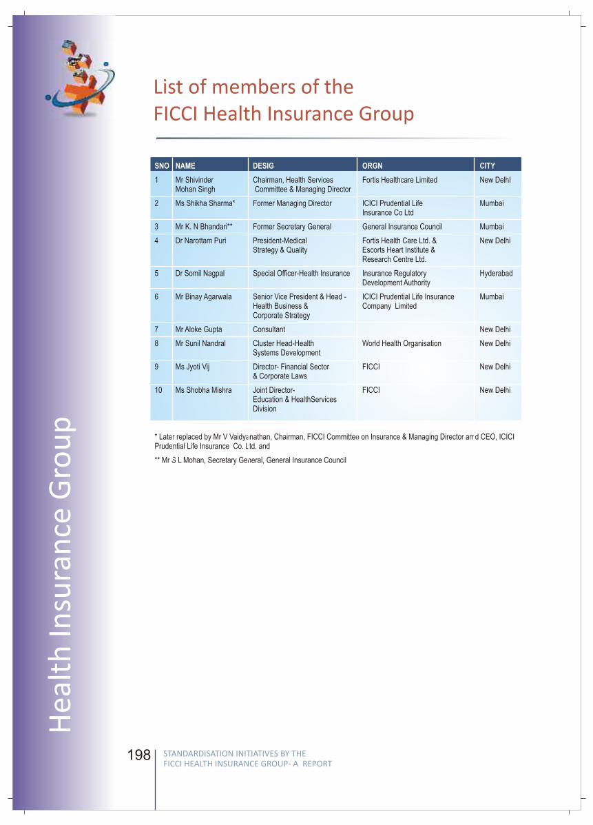
198 199
Technical Board
Chairperson
Dr Narrotam Puri, President- Medical Strategy & Quality, Fortis Healthcare Limited &
Escorts Heart Institute & Research Centre Limited
Members
Dr Somil Nagpal, Special Officer, Health Insurance, IRDA, Hyderabad
Dr Vijay Agarwal, Executive Director, Pushpanjali Crosslay Hospital, Ghaziabad
Dr S. K Mittal, Chairman, Department of Pediatrics, Pushpanjali Crosslay
Hospital, Ghaziabad
Dr S. C Marwah, CEO- Health Care Venture, Panacea Biotech Ltd, New Delhi
Dr Praneet Kumar, COO, Fortis Healthcare Ltd, Shalimar Bagh, New Delhi
Dr Loraine Kalra, Oncologist, Columbia Asia Hospital, New Delhi
Dr P. N Kakar, HOD Anaesthesia, Fortis Hospital, New Delhi
n
n
n
n
n
n
n
SNO NAME DESIG ORGN CITY
1 Mr Shivinder Chairman, Health Services Fortis Healthcare Limited New DelhIMohan Singh Committee & Managing Director
2 Ms Shikha Sharma* Former Managing Director ICICI Prudential Life MumbaiInsurance Co Ltd
3 Mr K. N Bhandari** Former Secretary General General Insurance Council Mumbai
4 Dr Narottam Puri President-Medical Fortis Health Care Ltd. & New DelhiStrategy & Quality Escorts Heart Institute &
Research Centre Ltd.
5 Dr Somil Nagpal Special Officer-Health Insurance Insurance Regulatory HyderabadDevelopment Authority
6 Mr Binay Agarwala Senior Vice President & Head - ICICI Prudential Life Insurance MumbaiHealth Business & Company LimitedCorporate Strategy
7 Mr Aloke Gupta Consultant New Delhi
8 Mr Sunil Nandral Cluster Head-Health World Health Organisation New Delhi Systems Development
9 Ms Jyoti Vij Director- Financial Sector FICCI New Delhi& Corporate Laws
10 Ms Shobha Mishra Joint Director- FICCI New Delhi Education & HealthServices Division
* Later replaced by Mr V Vaidyanathan, Chairman, FICCI Committee on Insurance & Managing Director amd CEO, ICICI Prudential Life Insurance Co. Ltd. and
** Mr S L Mohan, Secretary General, General Insurance Council
List of members of the FICCI Health Insurance Group
STANDARDISATION INITIATIVES BY THE FICCI HEALTH INSURANCE GROUP- A REPORT
STANDARDISATION INITIATIVES BY THE FICCI HEALTH INSURANCE GROUP- A REPORT
Hea
lth
Insu
ran
ce G
rou
p
FICC
I Wo
rking G
rou
p R
epo
rt
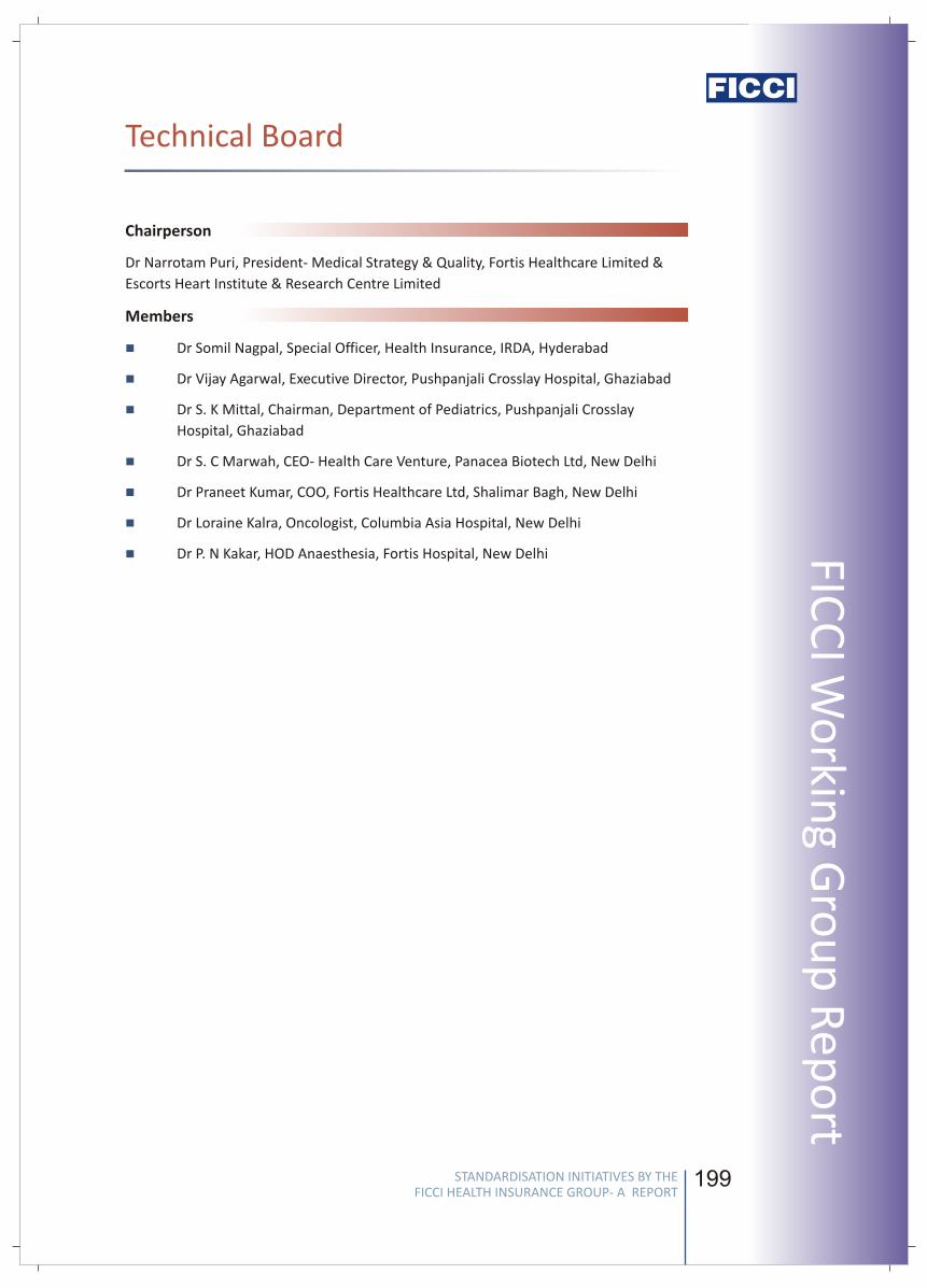
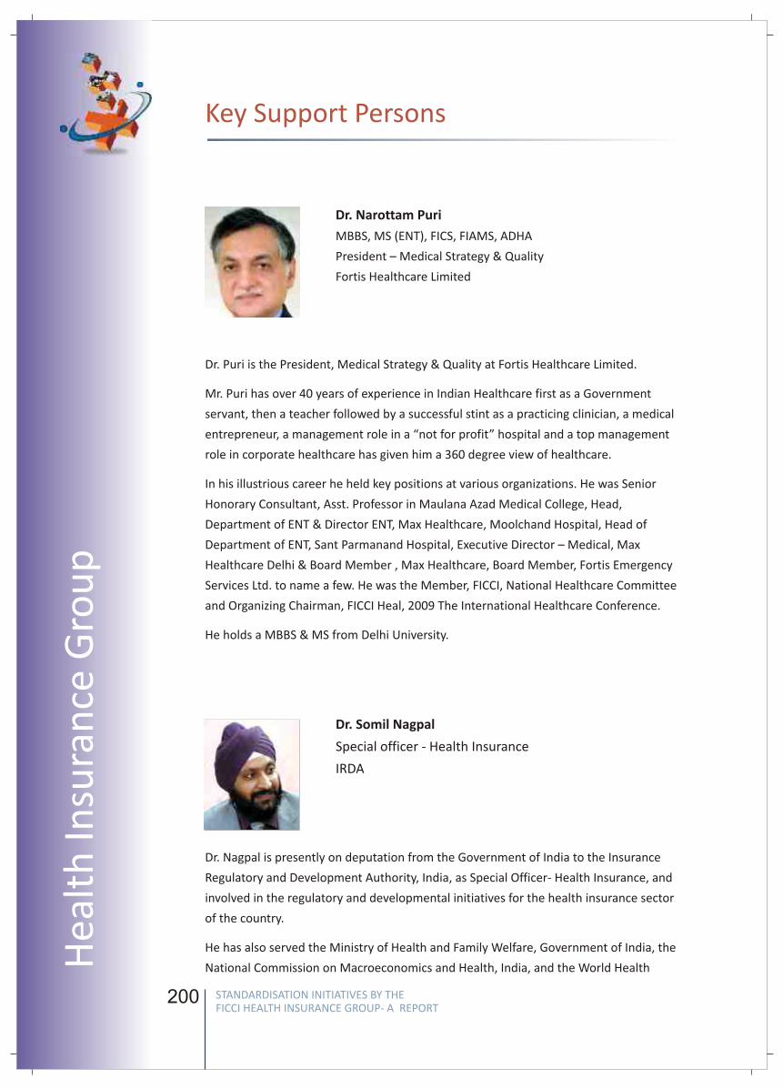
200
Key Support Persons
Dr. Narottam Puri
MBBS, MS (ENT), FICS, FIAMS, ADHA
President – Medical Strategy & Quality
Fortis Healthcare Limited
Dr. Puri is the President, Medical Strategy & Quality at Fortis Healthcare Limited.
Mr. Puri has over 40 years of experience in Indian Healthcare first as a Government
servant, then a teacher followed by a successful stint as a practicing clinician, a medical
entrepreneur, a management role in a “not for profit” hospital and a top management
role in corporate healthcare has given him a 360 degree view of healthcare.
In his illustrious career he held key positions at various organizations. He was Senior
Honorary Consultant, Asst. Professor in Maulana Azad Medical College, Head,
Department of ENT & Director ENT, Max Healthcare, Moolchand Hospital, Head of
Department of ENT, Sant Parmanand Hospital, Executive Director – Medical, Max
Healthcare Delhi & Board Member , Max Healthcare, Board Member, Fortis Emergency
Services Ltd. to name a few. He was the Member, FICCI, National Healthcare Committee
and Organizing Chairman, FICCI Heal, 2009 The International Healthcare Conference.
He holds a MBBS & MS from Delhi University.
Dr. Somil Nagpal
Special officer - Health Insurance
IRDA
Dr. Nagpal is presently on deputation from the Government of India to the Insurance
Regulatory and Development Authority, India, as Special Officer- Health Insurance, and
involved in the regulatory and developmental initiatives for the health insurance sector
of the country.
He has also served the Ministry of Health and Family Welfare, Government of India, the
National Commission on Macroeconomics and Health, India, and the World Health
201
FICC
I Wo
rking G
rou
p R
epo
rt
Organization. He has been involved as a resource person/ expert for the World Health
Organization and for the Indian and Maldivian Ministries of Health in the areas of
Health Insurance, Health Costing and National Health Accounts. He has also been a
resource person/ expert on health insurance, health financing and public finance for
various Government, non-Government and International Organizations, and to
management institutions, both within India and internationally, and has addressed
several national and international conferences, workshops and summits on his areas of
interest. He has been closely associated with the working of several government,
regulatory and industry committees and working groups in the realm of Health
Insurance.
He is a qualified as a medical doctor and has done his postgraduation in healthcare
management. He is also a MBA in Financial Management and is a Fellow of the
Insurance Institute of India.
STANDARDISATION INITIATIVES BY THE FICCI HEALTH INSURANCE GROUP- A REPORT
STANDARDISATION INITIATIVES BY THE FICCI HEALTH INSURANCE GROUP- A REPORT
Hea
lth
Insu
ran
ce G
rou
p
200
Key Support Persons
Dr. Narottam Puri
MBBS, MS (ENT), FICS, FIAMS, ADHA
President – Medical Strategy & Quality
Fortis Healthcare Limited
Dr. Puri is the President, Medical Strategy & Quality at Fortis Healthcare Limited.
Mr. Puri has over 40 years of experience in Indian Healthcare first as a Government
servant, then a teacher followed by a successful stint as a practicing clinician, a medical
entrepreneur, a management role in a “not for profit” hospital and a top management
role in corporate healthcare has given him a 360 degree view of healthcare.
In his illustrious career he held key positions at various organizations. He was Senior
Honorary Consultant, Asst. Professor in Maulana Azad Medical College, Head,
Department of ENT & Director ENT, Max Healthcare, Moolchand Hospital, Head of
Department of ENT, Sant Parmanand Hospital, Executive Director – Medical, Max
Healthcare Delhi & Board Member , Max Healthcare, Board Member, Fortis Emergency
Services Ltd. to name a few. He was the Member, FICCI, National Healthcare Committee
and Organizing Chairman, FICCI Heal, 2009 The International Healthcare Conference.
He holds a MBBS & MS from Delhi University.
Dr. Somil Nagpal
Special officer - Health Insurance
IRDA
Dr. Nagpal is presently on deputation from the Government of India to the Insurance
Regulatory and Development Authority, India, as Special Officer- Health Insurance, and
involved in the regulatory and developmental initiatives for the health insurance sector
of the country.
He has also served the Ministry of Health and Family Welfare, Government of India, the
National Commission on Macroeconomics and Health, India, and the World Health
201
FICC
I Wo
rking G
rou
p R
epo
rtOrganization. He has been involved as a resource person/ expert for the World Health
Organization and for the Indian and Maldivian Ministries of Health in the areas of
Health Insurance, Health Costing and National Health Accounts. He has also been a
resource person/ expert on health insurance, health financing and public finance for
various Government, non-Government and International Organizations, and to
management institutions, both within India and internationally, and has addressed
several national and international conferences, workshops and summits on his areas of
interest. He has been closely associated with the working of several government,
regulatory and industry committees and working groups in the realm of Health
Insurance.
He is a qualified as a medical doctor and has done his postgraduation in healthcare
management. He is also a MBA in Financial Management and is a Fellow of the
Insurance Institute of India.
STANDARDISATION INITIATIVES BY THE FICCI HEALTH INSURANCE GROUP- A REPORT
STANDARDISATION INITIATIVES BY THE FICCI HEALTH INSURANCE GROUP- A REPORT
Hea
lth
Insu
ran
ce G
rou
p
202
About FICCI
Established in 1927, FICCI is the largest and oldest apex business organisation in India.
Its history is closely interwoven with India's struggle for independence and its
subsequent emergence as one of the most rapidly growing economies globally. FICCI
plays a leading role in policy debates that are at the forefront of social, economic and
political change. Through its 400 professionals, FICCI is active in 38 sectors of the
economy. FICCI's stand on policy issues is sought out by think tanks, governments and
academia. Its publications are widely read for their in-depth research and policy
prescriptions. FICCI has joint business councils with 79 countries around the world.
A non-government, not-for-profit organisation, FICCI is the voice of India's business and
industry. FICCI has direct membership from the private as well as public sectors,
including SMEs and MNCs, and an indirect membership of over 83,000 companies from
regional chambers of commerce.
FICCI works closely with the government on policy issues, enhancing efficiency,
competitiveness and expanding business opportunities for industry through a range of
specialised services and global linkages. It also provides a platform for sector specific
consensus building and networking. Partnerships with countries across the world carry
forward our initiatives in inclusive development, which encompass health, education,
livelihood, governance, skill development, etc. FICCI serves as the first port of call for
Indian industry and the international business community.
FICCI Co-ordinators
Financial Sector Division
Ms Jyoti Vij DirectorFinancial Sector & Corporate LawsNew DelhiE-mail : [email protected] : 011 23738760-70 (Ext 306)
Ms Shweta VijResearch AssociateFinancial Sector & Corporate LawsNew DelhiE-mail : [email protected] : 011- 23738760-70 (Ext 413)
Education & HealthServices Division
Ms Shobha MishraJoint DirectorEducation & Healthservices DivisionNew DelhiE-mail : [email protected] : 011-23738760-70 (Ext468)
Ms Debasmita JenaAssistant DirectorEducation & Healthservices DivisionNew DelhiE-mail : [email protected] : 011-23738760-70 (Ext220)
Related Documents