PNL25 Last Updated April 2013 Page 1 Standard Operating Procedure for PulseNet PFGE of Clostridium botulinum Purpose To describe the Standardized Laboratory Protocol for Molecular Subtyping of botulinum toxin producing clostridia by Pulsed-field Gel Electrophoresis (PFGE). Scope To provide the PulseNet participants with the same procedure for performing PFGE of botulinum toxin producing clostridia, thus ensuring inter-laboratory comparability of the generated results. Definitions and Terms 1. PFGE: Pulsed-field Gel Electrophoresis 2. DNA: Deoxyribonucleic acid 3. CDC: Centers for Disease Control and Prevention 4. CLRW: Clinical Laboratory Reagent Water Biosafety Warning All samples received must be considered infectious. Botulinum toxin producing clostridia and/or botulinum toxin may be present in a variety of food products, clinical materials (serum, feces) and environmental samples (soil, surface water). Exposure to botulinum toxin is the primary laboratory hazard. The toxin may be absorbed after ingestion or following contact with the broken skin, eyes, or mucous membranes, including the respiratory tract. Accidental parenteral inoculation may also represent a significant exposure to toxin. Broth cultures grown under optimal conditions for toxin production may contain 2,000,000 mouse LD 50 per ml of toxin. Recommended Precautions: Biosafety Level 2 practices, containment equipment and facilities are recommended for all activities with materials known to or that may potentially contain botulinum toxin. Personal protective equipment (PPE) i.e., gloves, lab coat, safety glasses and/or face shield should be worn at all times when handling anything that has come into contact with the organism or toxin including pipette tips and plastic transfer pipettes used to transfer liquids. All liquids require disinfection using a freshly prepared 10% bleach solution. Solutions of sodium hypochlorite (0.1%) or sodium hydroxide (0.1N) readily inactivate the toxin and are recommended for decontaminating work surfaces and spills of cultures or toxin. Please read all instructions carefully before starting protocol. All plasticware, glassware, pipets, spatulas, etc. that come in contact with the cell suspensions or plugs should be disinfected with 10% bleach for at least 1 hour before they are washed and reused.

Welcome message from author
This document is posted to help you gain knowledge. Please leave a comment to let me know what you think about it! Share it to your friends and learn new things together.
Transcript

PNL25 Last Updated April 2013 Page 1
Standard Operating Procedure for PulseNet PFGE of
Clostridium botulinum
Purpose
To describe the Standardized Laboratory Protocol for Molecular Subtyping of botulinum toxin producing clostridia by
Pulsed-field Gel Electrophoresis (PFGE).
Scope
To provide the PulseNet participants with the same procedure for performing PFGE of botulinum toxin producing clostridia,
thus ensuring inter-laboratory comparability of the generated results.
Definitions and Terms
1. PFGE: Pulsed-field Gel Electrophoresis
2. DNA: Deoxyribonucleic acid
3. CDC: Centers for Disease Control and Prevention
4. CLRW: Clinical Laboratory Reagent Water
Biosafety Warning
All samples received must be considered infectious. Botulinum toxin producing clostridia and/or botulinum toxin may be
present in a variety of food products, clinical materials (serum, feces) and environmental samples (soil, surface water).
Exposure to botulinum toxin is the primary laboratory hazard. The toxin may be absorbed after ingestion or following
contact with the broken skin, eyes, or mucous membranes, including the respiratory tract. Accidental parenteral inoculation
may also represent a significant exposure to toxin. Broth cultures grown under optimal conditions for toxin production may
contain 2,000,000 mouse LD50 per ml of toxin.
Recommended Precautions: Biosafety Level 2 practices, containment equipment and facilities are recommended for all
activities with materials known to or that may potentially contain botulinum toxin. Personal protective equipment (PPE) i.e.,
gloves, lab coat, safety glasses and/or face shield should be worn at all times when handling anything that has come into
contact with the organism or toxin including pipette tips and plastic transfer pipettes used to transfer liquids. All liquids
require disinfection using a freshly prepared 10% bleach solution. Solutions of sodium hypochlorite (0.1%) or sodium
hydroxide (0.1N) readily inactivate the toxin and are recommended for decontaminating work surfaces and spills of cultures
or toxin.
Please read all instructions carefully before starting protocol. All plasticware, glassware, pipets, spatulas, etc. that come in
contact with the cell suspensions or plugs should be disinfected with 10% bleach for at least 1 hour before they are washed
and reused.

PNL25 Last Updated April 2013 Page 2
Select Agent Requirements:
All PFGE materials, including restricted plugs, may contain viable bacteria until the start of electrophoresis. Botulinum toxin
producing clostridia are Select Agents (SA), and according to the APHIS/CDC SA Regulations all parts of the following
procedure until electrophoresis must be performed by SA approved personnel and within SA approved space. In addition,
long term stored plugs are subject to SA inventory requirements, as defined by APHIS/CDC SA Regulations. Please refer to
www.selectagents.gov for additional information.
Day 1
Grow the culture
Streak an isolated colony from test cultures onto Anaerobic Blood Agar Plates – CDC formulation (ANA-BAP). Incubate the
plates in an anaerobic chamber overnight at 35°C±2°C.
Day 2
1. Label small tubes (12-mm x 75-mm Falcon tubes or equivalent) with culture numbers.
2. Prepare Cell Suspension Buffer (100 mM Tris:100 mM EDTA, pH 8.0) as follows:
2.1. 100 ml of 1 M Tris, pH 8.0
2.2. 200 ml of 0.5 M EDTA, pH 8.0
2.3. Dilute to 1000 ml with sterile Ultrapure water (CLRW)
3. Transfer 2 ml of Cell Suspension Buffer (CSB) to small labeled tubes. Use a sterile polyester-fiber or cotton swab that
has been moistened with sterile CSB to remove some of the growth from the ANA-BAP plate; suspend cells in CSB by
spinning swab gently so cells will be evenly dispersed and formation of aerosols is minimized.
4. Adjust concentration of cell suspensions to one of values given below by diluting with sterile CSB or by adding
additional cells.
4.1. Dade Microscan Turbidity Meter: 0.18-0.20 (measured in Falcon 2054 tubes)
4.2. About 1 McFarland
5. Pipette 1000 µl of the cell suspensions into labeled, sterile microcentrifuge tubes and spin for 5 minutes at 5,000 rpm.
6. Remove the supernatants, re-suspend cells in 1000 µl of the cell suspension buffer and spin for 5 minutes at 5,000 rpm.
Decant supernatants into waste container containing bleach.
7. Remove the supernatants and re-suspend cells in 500 µl of the cell suspension buffer. Decant supernatants in waste
container containing bleach.
8. Use these washed cells to inoculate two EYA plates per sample for confluent growth (250 µl per plate), using the Kirby-
Bauer technique.
9. Incubate the plates for 18-72 hours in an anaerobic chamber at 35°C±2°C.
Remove plates from incubation as soon as there is sufficient growth for testing. 18 hours is typically optimal.
Day 3
Making plugs
1. Turn on shaker water bath or incubator (55°C±2°C), stationary water baths (55°C±2°C and 37°C±2°C)
2. Add bottles of sterile Ultrapure water (CLRW) and TE buffer to the 55°C water bath to warm for the washing steps.
PNL25 Last Updated April 2013 Page 3
3. Prepare 2X Cell Lysis Buffer (12mM Tris, 2M NaCl, 200mM EDTA, 1% Brij 58, 0.4% Deoxycholate, 5% Sarcosyl) as
follows:
3.1. 1.2 ml of 1M Tris-HCL, pH 8.0
3.2. 40 ml of 5M NaCl
3.3. 40 ml of 0.5M EDTA, pH 8.0
3.4. 1 g Brij 58
3.5. 0.4 g Deoxycholate
3.6. 5 g Sarkosyl
3.7. Dilute to 100 ml with sterile Ultrapure water (CLRW).
4. Prepare Lysozyme (Sigma L7651 or equivalent) stock solution (20 mg/ml in TE) as follows:
4.1. Weigh out 100 mg Lysozyme (keep container of lysozyme on ice)
4.2. Add 5mL TE buffer, swirl to mix
4.3. Aliquot 250 µl amounts into small eppendorf tubes and freeze for future use.
5. Prepare Mutanolysin (Sigma M9901 or equivalent) stock solution (5 U/µl in TE) as follows:
5.1. Add 1 ml TE buffer to vial of lyophilized Mutanolysin, swirl to mix.
5.2. Aliquot 50 µl amounts into small eppendorf tubes and freeze for future use.
6. Take out aliquots of Lysozyme (20 mg/ml) and Mutanolysin (5 U/µl) from the -20°C freezer and pre-warm 2X Cell Lysis
Buffer to 55°C±2°C.
7. Prepare 1.2% SeaKem Gold agarose in TE Buffer (10 mM Tris:1 mM EDTA, pH 8.0) for PFGE plugs as follows:
7.1. Weigh 0.12 g (or 0.24 g) SeaKem Gold (SKG) agarose into 250 ml screw-cap flask
7.2. Add 10.0 ml (or 20.0 ml) TE Buffer; swirl gently to disperse agarose.
7.3. Loosen cap and microwave for 30 seconds; mix gently and repeat for 10 second intervals until agarose is
completely dissolved.
7.4. Place flask in 55°C ± 2°C water bath and equilibrate the agarose in the water bath for 15 minutes or until ready to
use.
SAFETY WARNING: USE HEAT-RESISTANT GLOVES WHEN HANDLING HOT FLASKS AFTER MICROWAVING.
SeaKem Gold agarose works well for making PFGE plugs because it provides added strength to the plugs minimizing
breakage of plugs during the lysis and washing steps. The time and temperature needed to completely dissolve the agarose
is dependent on the specifications of the microwave used, and will have to be determined empirically in each laboratory.
8. Label small tubes (12mm x 75mm Falcon tubes or equivalent) with culture numbers.
9. Prepare PIV Buffer as follows:
9.1. 5 ml of 1M Tris-HCl, pH 8.0
9.2. 100 ml of 5M NaCl
9.3. Dilute to 500 ml with sterile Ultrapure water (CLRW)
10. Transfer 1.5-2 ml of PIV buffer to the labeled tubes. Use a sterile polyester-fiber or cotton swab that has been
moistened with sterile PIV to remove some of the growth from the agar plate; suspend cells in PIV by spinning swab
gently so cells will be evenly dispersed and formation of aerosols is minimized.
If a large number of samples are being prepared, it is recommended that they are prepared in batches of around 10 samples
at a time.
11. Adjust concentrations of cell suspensions to 0.68-0.72 as measured on the Dade Microscan Turbidity Meter by diluting
with sterile PIV Buffer or by adding additional cells.
These values give satisfactory results at CDC; each laboratory may need to establish the optimal concentration needed for
satisfactory results.
PNL25 Last Updated April 2013 Page 4
Cell suspensions need to be at room temperature when concentration is checked.
12. Pipette 1000 μl of the cell suspensions into microcentrifuge tubes and spin for 5 minutes at 5,000 rpm.
13. Calculate the volume of 2X Cell Lysis Buffer and Proteinase K required per sample as follows:
13.1. Calculation:
(stock conc. of Proteinase K) (X) = (final conc. desired) (final volume)
(stock conc. of Proteinase K) (X) = (0.665 mg/ml) (1 ml)
(0.665 mg/ml) x (1 ml) / (stock conc. of Proteinase K) = volume of Proteinase K needed (ml)
Total volume of 2X Cell Lysis Buffer + Proteinase K = 316 μl
316 μl – volume of Proteinase K = volume of 2X Cell Lysis Buffer needed (μl)
Stock concentrations of Proteinase K may vary by lot and will need to be calculated for each shipment. Adjust total volume
to compensate by changing amount of 2X cell lysis buffer used. The final concentration of Proteinase K for plugs is 0.665
mg/ml.
14. Remove the supernatants and re-suspend cells in the calculated volume of 2X Cell Lysis Buffer that has been pre-
warmed to 55ºC ± 2°C. Then add 80 μl of lysozyme (20 mg/ml) and incubate in the waterbath for 20 minutes at 55°C ±
2°C. Decant supernatants in waste container containing bleach.
15. After removing the samples from the waterbath, add 4 μl of Mutanolysin (5 U/μl) and the calculated volume of
Proteinase K, and incubate another 10 minutes at 37ºC ± 2°C.
Casting Plugs
1. Label wells of PFGE plug molds with culture number. When reusable plug molds are used, put strip of tape on lower
part of reusable plug mold before labeling wells.
Unused plug agarose can be kept at room temperature and reused 1-2 times. Microwave on low-medium power for 10-15
seconds and mix; repeat for 5-10 seconds intervals until agarose is completely melted. This agarose melts rapidly!
2. Add 400 µl melted 1.2% SeaKem Gold agarose to the cell suspensions; mix by gently pipetting mixture up and down a
few times. Over-pipetting can cause DNA shearing. Maintain temperature of melted agarose by keeping flask in
beaker of warm water (55-60°C).
3. Immediately, dispense part of mixture into appropriate well(s) of reusable plug mold. Do not allow bubbles to form.
Two plugs of each sample can be made from these amounts of cell suspension and agarose and are useful if repeat
testing is required. Allow plugs to solidify at room temperature for 10-15 minutes. They can also be placed in the
refrigerator (4°C) for 5 minutes
If large numbers of samples are being prepared, it is recommended that they be processed in batches of about 10 samples at
a time.
Lysis of Cells in Agarose Plugs
Two plugs (reusable molds) of the same strain can be lysed in the same 50 ml tube.
1. Label 50 ml polypropylene screw-cap tubes with culture numbers.
2. Prepare ES Buffer as follows:
2.1. 495 ml of 0.5M EDTA, pH 8.0
2.2. 5 ml of 10% Sarcosyl
3. Add 5 ml of ES Buffer to each labeled 50 ml tube.
4. Calculate the total volume of Cell Lysis/Proteinase K Buffer needed as follows:
PNL25 Last Updated April 2013 Page 5
4.1. Calculation:
(stock conc. of Proteinase K) (X) = (final conc. desired) (final volume)
(stock conc. of Proteinase K) (X) = (0.14 mg/ml) (5 ml)
(0.14 mg/ml) x (5 ml) / (stock conc. of Proteinase K) = volume of Proteinase K needed (ml)
Add the calculated volume of Proteinase K to each labeled 50 ml tube containing ES Buffer.
Stock concentrations of Proteinase K may vary by lot and will need to be calculated for each shipment. The final
concentration of Proteinase K per tube is 0.14 mg/ml
5. Trim excess agarose from top of plugs with scalpel, razor blade or similar instrument. Open reusable plug mold and
transfer plugs from mold with a 6-mm wide spatula to appropriately labeled tube. Be sure plugs are under buffer and
not on side of tube.
Ensure that the green screen caps are in place on conical tubes containing the plugs to prevent the loss or damage of the
plug
6. Place both sections of the plug mold, spatulas, and scalpel in 10% bleach. Soak them for 1 hour before washing them.
7. Place tubes in a rack and incubate in the 55°C ±2°C shaker water bath or incubator for a minimum of 2 hours (4 hours is
optimal) with constant agitation (40-70 rpm). Be sure water level is above level of lysis buffer in tubes if using a water
bath.
Washing of Agarose Plugs After Cell Lysis
Most laboratories will find that their plugs are sufficiently stable to perform the following washing steps at 54-55°C.
However, if you notice that your plugs are nicked along the edges or breaking it will be necessary for your laboratory to
lower the water bath or incubator to 50°C for the following washing steps.
1. Remove tubes from water bath or incubator, and carefully pour off ES buffer into an appropriate discard container;
plugs can be held in tubes with the screened caps.
It is important to remove all of the liquid during this and subsequent wash steps by touching edge of tube or screened cap on
an absorbent paper towel.
2. Add 20 ml sterile Ultrapure water (CLRW) that has been pre-heated to 55ºC ± 2°C to each tube and shake the tubes in a
55°C ± 2°C water bath for 15 minutes at 70 rpm. Decant used distilled water into a waste container containing bleach.
3. Pour off water from the plugs and repeat wash step with pre-heated water (Step 2) one more time.
4. Pour off water, add a minimum of 20 ml pre-heated (55ºC ± 2°C) sterile TE Buffer, and shake the tubes in the 55ºC ±
2°C water bath for 15 minutes at 70 rpm. Decant used TE into a waste container containing bleach
5. Pour off TE and repeat wash step with pre-heated TE five more times.
6. Decant last wash and add 15 ml sterile TE. Continue with step 1 in "Restriction Digestion" section or store plugs in TE
Buffer at 4°C until needed. Plugs can be transferred to smaller tubes for long term storage.
Day 4
Restriction Digestion of DNA in Agarose Plugs
A small slice of the plug should be digested with the primary restriction enzyme because less enzyme is required and other
slices of the plug can be subjected to restriction analysis with secondary or tertiary enzymes, according to the table below.
The use of a secondary enzyme is useful in situations where the PFGE patterns obtained with the primary enzyme from two
or more isolates are indistinguishable.
PNL25 Last Updated April 2013 Page 6
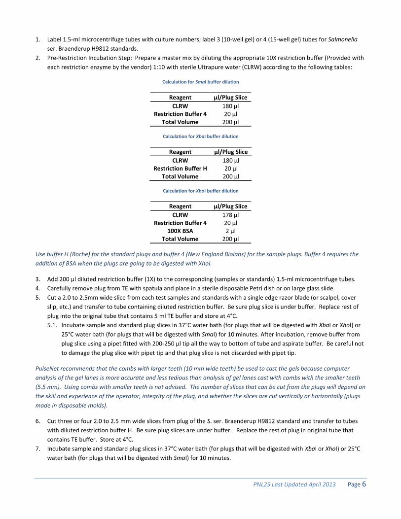
1. Label 1.5-ml microcentrifuge tubes with culture numbers; label 3 (10-well gel) or 4 (15-well gel) tubes for Salmonella
ser. Braenderup H9812 standards.
2. Pre-Restriction Incubation Step: Prepare a master mix by diluting the appropriate 10X restriction buffer (Provided with
each restriction enzyme by the vendor) 1:10 with sterile Ultrapure water (CLRW) according to the following tables:
Calculation for SmaI buffer dilution
Reagent µl/Plug Slice
CLRW 180 µl Restriction Buffer 4 20 µl
Total Volume 200 µl
Calculation for XbaI buffer dilution
Reagent µl/Plug Slice
CLRW 180 µl Restriction Buffer H 20 µl
Total Volume 200 µl
Calculation for XhoI buffer dilution
Reagent µl/Plug Slice
CLRW 178 µl Restriction Buffer 4 20 µl
100X BSA 2 µl Total Volume 200 µl
Use buffer H (Roche) for the standard plugs and buffer 4 (New England Biolabs) for the sample plugs. Buffer 4 requires the
addition of BSA when the plugs are going to be digested with XhoI.
3. Add 200 µl diluted restriction buffer (1X) to the corresponding (samples or standards) 1.5-ml microcentrifuge tubes.
4. Carefully remove plug from TE with spatula and place in a sterile disposable Petri dish or on large glass slide.
5. Cut a 2.0 to 2.5mm wide slice from each test samples and standards with a single edge razor blade (or scalpel, cover
slip, etc.) and transfer to tube containing diluted restriction buffer. Be sure plug slice is under buffer. Replace rest of
plug into the original tube that contains 5 ml TE buffer and store at 4°C.
5.1. Incubate sample and standard plug slices in 37°C water bath (for plugs that will be digested with XbaI or XhoI) or
25°C water bath (for plugs that will be digested with SmaI) for 10 minutes. After incubation, remove buffer from
plug slice using a pipet fitted with 200-250 µl tip all the way to bottom of tube and aspirate buffer. Be careful not
to damage the plug slice with pipet tip and that plug slice is not discarded with pipet tip.
PulseNet recommends that the combs with larger teeth (10 mm wide teeth) be used to cast the gels because computer
analysis of the gel lanes is more accurate and less tedious than analysis of gel lanes cast with combs with the smaller teeth
(5.5 mm). Using combs with smaller teeth is not advised. The number of slices that can be cut from the plugs will depend on
the skill and experience of the operator, integrity of the plug, and whether the slices are cut vertically or horizontally (plugs
made in disposable molds).
6. Cut three or four 2.0 to 2.5 mm wide slices from plug of the S. ser. Braenderup H9812 standard and transfer to tubes
with diluted restriction buffer H. Be sure plug slices are under buffer. Replace the rest of plug in original tube that
contains TE buffer. Store at 4°C.
7. Incubate sample and standard plug slices in 37°C water bath (for plugs that will be digested with XbaI or XhoI) or 25°C
water bath (for plugs that will be digested with SmaI) for 10 minutes.
PNL25 Last Updated April 2013 Page 7
8. Prepare the restriction enzyme master mix according to the following tables. Prepare enough for each plug slice plus
one additional aliquot.
SmaI (20 U/µl stock concentration) master mix calculation
Reagent µl/Plug Slice
CLRW 177.5 µl 10X Restriction Buffer 4 20 µl SmaI enzyme (50 U/sample) 2.5 µl Total Volume 200 µl Incubate 25°C, 4 hours
XbaI (20 U/µl stock concentration) master mix calculation
Reagent µl/Plug Slice
CLRW 175 µl 10X Restriction Buffer H 20 µl XbaI Enzyme (100 U/sample) 5 µl Total Volume 200 µl Incubate 37°C, 2 hours
XhoI (20 U/µl stock concentration) master mix calculation
Reagent µl/Plug Slice
CLRW 173 µl 10X Restriction Buffer 4 20 µl 100X BSA 2 µl XhoI Enzyme (100 U/sample) 5 µl Total Volume 200 µl Incubate 37°C, 3 hours
Keep vial of restriction enzyme on ice or in insulated storage box (-20°C) at all times.
Digest Salmonella standard plugs with Roche XbaI and sample plugs with New England Biolabs SmaI or XhoI. These
restriction enzymes give satisfactory results at CDC. Restriction enzymes provided by other vendors have not been evaluated.
Be certain that the restriction buffer that is used is recommended by the vendor for the corresponding restriction enzyme
and calculate the volume of enzyme needed to achieve the same final concentrations as indicated in the tables.
Addition of Bovine Serum Albumin: Several restriction enzyme vendors specifically recommend the addition of 1X BSA to
enzyme restriction mixtures while others do not. PulseNet Central recommends adding BSA to all enzyme restriction
mixtures to minimize the incidence of incomplete restriction. If BSA is added to the enzyme reaction mixture, the volume of
BSA added should be deducted from the volume of water to maintain the total volume of 200 µl per slice.
9. After incubation, remove buffer from plug slice using a pipet fitted with 200-250 µl tip. Be careful not to damage the
plug slice with the tip and that the plug slice is not discarded with the pipet tip.
10. Add 200 µl restriction enzyme master mix to each tube. Close tube and mix by tapping gently; be sure plug slices are
under enzyme mixture.
11. Incubate the plug slices with restriction enzymes at each of their respective temperatures and times.
PNL25 Last Updated April 2013 Page 8
Loading PFGE Plug Slices and Pouring an Agarose Gel
1. Confirm that water bath is equilibrated to 55°C ±2°C.
2. Make volume of 0.5X Tris-Borate EDTA Buffer (TBE) that is needed for both the gel and electrophoresis running buffer
according to one of the following tables.
Reagent Volume (ml) Volume(ml)
5X TBE Stock 200 220 CLRW 1800 1980 Total Volume 2000 2200
Reagent Volume (ml) Volume (ml)
10X TBE Stock 100 110 CLRW 1900 2090 Total Volume 2000 2200
3. Make 1% SeaKem Gold (SKG) Agarose in 0.5X TBE as follows:
3.1. Weigh appropriate amount of SKG into 500 ml screw-cap flask.
3.1.1. Mix 1.0 g agarose with 100 ml 0.5X TBE for 14cm-wide gel form (10 wells)
3.1.2. Mix 1.5 g agarose with 150 ml 0.5X TBE for 21cm-wide gel form (15 wells)
4. Loosen cap and microwave for 60 seconds; mix gently and repeat for 15 second intervals until agarose is completely
dissolved.
5. Recap flask and return to 55°C ±2°C water bath and equilibrate the agarose in the water bath for 15 minutes or until
ready to use.
SAFETY WARNING: USE HEAT-RESISTANT GLOVES WHEN HANDLING HOT FLASKS AFTER MICROWAVING.
6. Place the gel form on a leveling table and adjust until perfectly leveled. Place the comb holder so the front part (side
with small metal screws) and teeth face the bottom of gel frame and the comb teeth touch the gel platform.
7. Remove restricted plug slices from the water baths. Remove enzyme/buffer mixture and add 200 µl 0.5X TBE. Incubate
at room temperature for 5 minutes.
8. Remove plug slices from tubes; put comb on bench top and load plug slices on the bottom of the comb teeth as
follows:
8.1. Load S. ser. Braenderup H9812 standards on teeth (lanes) 1, 5, 10 (10 well gel) or on teeth 1, 5, 10, 15 (15 well
gel).
8.2. Load samples on remaining teeth and note locations.
9. Remove excess buffer with a kimwipe. Allow plug slices to air dry on the comb for 15 minutes.
10. Position comb in gel form and confirm that the plugs slices are correctly aligned on the bottom of the comb teeth, that
the lower edge of the plug slice is flush against the black platform.
11. Carefully pour the agarose (cooled to 55°C ± 2°C) into the gel form and allow the gel to solidify for a minimum of 15
minutes at room temperature.
12. Put black gel frame in electrophoresis chamber. Add 2.2 L freshly prepared 0.5X TBE. Close cover of unit. (The amount
of buffer needed depends on whether residual buffer was left in tubing or if unit was flushed with water after the last
gel was run).
13. Turn on power supply, pump calibrated to a flow rate of 1 liter/minute (setting of about 70), and cooling module (14°C)
approximately 30 minutes before gel is to be run.
14. After the gel has solidified, add 5-10 ml of 0.5X TBE to the comb and gently remove from the gel.

PNL25 Last Updated April 2013 Page 9
15. Unscrew and remove end gates from the gel mold; remove excess agarose from the sides and bottom of the casting
platform with a tissue. Keep gel on casting platform and carefully place gel inside black gel frame in the electrophoresis
chamber.
16. Add 860 µl of Thiourea (10mg/ml) to 0.5X TBE in the electrophoresis chamber. Close cover of chamber.
SAFETY WARNING: THIOUREA IS A TOXIC CHEMICAL. WEIGH THIOUREA IN A CHEMICAL FUME HOOD; USE GLOVES,
EYE PROTECTION, AND DISPOSABLE SPATULA WHEN HANDLING THIS CHEMICAL. CLEAN UP ANY SPILLS, AND WIPE
DOWN BALANCE AND SURROUNDING AREA WITH A MOISTENED TOWEL. DISCARD GLOVES, SPATULA, WEIGHING
PAPER, ETC. AS HAZARDOUS WASTE, ACCORDING TO THE GUIDELINES OF YOUR INSTITUTION. RE-CAP BOTTLE
TIGHTLY AFTER USE.
Electrophoresis Conditions
Select following conditions on CHEF Mapper
o Auto Algorithm
o 30 kb: low MW
o 600 kb: high MW
o Initial switch time: 0.5 s
o Final switch time: 40 s
o Run time: 18-19 hours (See note below)
Select following conditions on CHEF-DR III
o Initial switch time: 0.5 s
o Final switch time: 40 s
o Voltage: 6 V
o Included Angle: 120°
o Run time: 18-19 hours (See note below)
The electrophoresis running times recommended above are based on the equipment and reagents used at the CDC. Run
times may be different in your laboratory and will have to be optimized for your gels so that the lowest band in the S. ser.
Braenderup H9812 standard migrates within 1.0-1.5 cm of the bottom of the gel.
Make note of the initial milliamp (mA) reading on the instrument. The initial mA should be between 110-150 mA. A reading
outside of this range may indicate that the 0.5X TBE buffer was prepared improperly and the buffer should be remade.
Day 5
Staining and Documentation of an Agarose Gel
The following staining procedure describes the use of ethidium bromide to stain PFGE gels. Alternate DNA stains may be
used. Please see the “Alternate DNA Stains-Results and Recommendations” posting within the Important PulseNet
Documents forum on the SharePoint website for additional information.
1. When electrophoresis run is over, turn off equipment; remove and stain gel with ethidium bromide. Dilute 40 µl of
ethidium bromide stock solution (10 mg/ml) with 400 ml of Ultrapure water (CLRW) (this volume is for a staining box
that is approximately 14-cm x 24-cm; a larger container may require a larger amount of staining solution). Stain gel for
20-30 minutes in covered container.
Ethidium bromide is toxic and a mutagen. Stock solutions of 10 mg/ml Ethidium Bromide (EtBr) in water are available from
several commercial companies (Amresco X328; Bio-Rad, 161-0433; Sigma, E-1510). The diluted solution can be kept in dark

PNL25 Last Updated April 2013 Page 10
bottle and reused 6-8 times before discarding according to your institution's guidelines for hazardous waste. CDC does not
recommend disposing of EtBr down the drain. Aqueous solutions containing EtBr can be filtered through charcoal or
degraded using activated carbon destaining or “tea” bags from Amresco (E732-25 Destaining Bags) or other companies,
which effectively and safely remove EtBr from solutions and gels. Once the EtBr is removed, the treated aqueous solutions
can be discarded down the drain. If you have further questions about EtBr please refer to the Material Safety Data Sheets
(MSDS) provided by the vendor or manufacturer.
2. Destain gel in approximately 500 ml reagent grade water for 60-90 minutes; change water every 20 minutes. Capture
image a Gel Doc 1000, 2000, EQ, or XR, or equivalent documentation system. If too much background is observed de-
stain for an additional 30-60 minutes.
2.1. Alternative Method for PFGE Gel Staining with GelStar: Dilute 40µl of GelStar (10,000X stock solution) into 400 ml
of 1X TBE. Stain gel for 60 minutes in covered container with gentle agitation.
Destaining is not necessary when staining with GelStar. After staining, proceed to capture the image with a gel
documentation system. Stock solutions of GelStar are available from Lonza, #50535. Use the same precautions when
handling and disposing of GelStar as indicated above froe EtBr.
3. Follow directions given with the imaging equipment to save gel image as a *.1sc or *.scn file; convert this file to *.tif file
for analysis with the BioNumerics software program. The gel image should fill the entire window of the imaging
equipment (computer) screen (without cutting off wells or lower bands). Ensure that the image is in focus and that
there is little to no saturation (over-exposure) in the bands. Additional instructions are provided in PNL07 of the
PulseNet QA/QC manual.
4. Drain buffer from electrophoresis chamber and discard. Rinse chamber with 2 L Ultrapure water (CLRW) or, if unit is
not going to be used for several days, flush lines with water by letting pump run for 5-10 minutes before draining water
from chamber and hoses.
5. If the lowest band in the H9812 standard does not migrate within 1 -1.5 cm of the bottom of the gel, the run time will
need to be determined empirically for the conditions in each laboratory.
Please note the following if PFGE results do not have to be available within 24-28 hours:
Plugs can be lysed for longer periods of time (3-16 hours).
The washing steps with TE to remove the lysis buffer from the PFGE plugs can be done for longer periods of time
(30-45 min) and at lower temperatures (37°C or room temperature). They can be started on Day 1 and finished on
Day 2 after overnight refrigeration of the plugs in TE.
USE OF TRADE NAMES AND COMMERCIAL SOURCES IS FOR IDENTIFICATION PURPOSES ONLY AND DOES NOT
IMPLY ENDORSEMENT BY CDC OR THE U.S. DEPARTMENT OF HEALTH AND HUMAN SERVICES.
Alternative Procedure for PFGE of Clostridium botulinum
Some strains may not be typeable by the method described above. The following procedure can be used as an alternative
method to perform PFGE on botulinum toxin producing clostridia.
Alternative Procedure Day 1
Streak each test culture for colony isolation onto Egg Yolk Agar Plate. Incubate the plates at 35± 2°C under anaerobic
conditions until isolated colonies are present (18-48 hours).
PNL25 Last Updated April 2013 Page 11
Alternative Procedure Day 2
Inoculate a single colony from each plate into 10 ml of Trypticase Peptone Glucose Yeast Extract (TPGY) medium.
Incubate the TPGY tubes at 35± 2°C under anaerobic conditions until growth is evident in the TPGY media.
Alternative Procedure Day 3
1. Prepare 2X Cell Lysis Buffer (12mM Tris, 2M NaCl, 200 mM EDTA, 1% Brij 58, 0.4%, Deoxycholate, 5% Sarcosyl) as
follows:
1.1. 1.2 ml of 1 M Tris-HCl, pH 8.0
1.2. 40 ml of 5 M NaCl
1.3. 40 ml of 0.5 M EDTA, pH 8.0
1.4. 1 g Brij 58
1.5. 0.4 g Deoxycholate
1.6. 5 g Sarkosyl
1.7. Dilute to 100 ml with sterile Ultrapure water (CLRW)
2. Prepare PIV Buffer as follows:
2.1. 5 ml of 1 M Tris-HCl, pH 8.0
2.2. 100 ml of 5 M NaCl
2.3. Dilute to 500 ml with sterile Ultrapure water (CLRW)
3. Prepare Lysozyme (Sigma L7651 or equivalent) stock solution (20 mg/mL in TE) as follows:
3.1. Weigh out 100 mg Lysozyme (keep the container of Lysozyme on ice)
3.2. Add 5 mL TE buffer, swirl to mix
3.3. Aliquot 250 uL amounts into small eppendorf tubes and freeze for future use.
4. Prepare Mutanolysin (Sigma M9901 or equivalent) stock solution (5 U/μL in TE) as follows:
4.1. Add 1 mL TE buffer to vial of lyophilized Mutanolysin, swirl to mix
4.2. Aliquot 50 μL amounts into small eppendorf tubes and freeze for future use.
5. Prepare 1.8% SKG Gold Agarose in TE Buffer (10 mM Tris: 1 mM EDTA, pH 8.0) for PFGE plugs as follows:
5.1. Weigh 0.18 g (or 0.36 g) of SeaKem Gold (SKG) agarose into 250 ml screw-cap flask
5.2. Add 10.0 ml (or 20.0 ml) TE Buffer; swirl gently to disperse agarose.
5.3. Loosen cap, and microwave for 30-sec; mix gently and repeat for 10-sec intervals until agarose is completely
dissolved.
5.4. Place flask in 55± 2°C water bath and equilibrate the agarose in the water bath for 15 minutes or until ready to
use.
SAFETY WARNING: USE HEAT-RESISTANT GLOVES WHEN HANDLING HOT FLASKS AFTER MICROWAVING.
SeaKem Gold agarose works well for making PFGE plugs because it provides added strength to the plugs minimizing
breakage of plugs during the lysis and washing steps. The time and temperature needed to completely dissolve the agarose
is dependent on the specifications of the microwave used, and will have to be determined empirically in each laboratory.
6. Take out tubes of Lysozyme (20 mg/ml) and Mutanolysin (5 U/μl) needed from the -20°C freezer.
7. Place the 2X Cell Lysis Buffer in a 37± 2°C water bath and the PIV buffer on ice.
8. Remove the TPGY cultures from the anaerobic incubator and pipet the entire 10 ml into labeled Nalgene 50 ml
centrifuge tubes and keep on ice.
9. Centrifuge at 1500 × g for 15 minutes at 4°C.
10. Prepare a master mix containing diluted formaldehyde according to the following table. Prepare enough for each
sample plus one additional aliquot.
PNL25 Last Updated April 2013 Page 12
Reagent Per Plug Slice
PIV Buffer 3.6 ml Formaldehyde (35-40%) 400 µl
This step should be done using a chemical fume hood.
11. Remove the Nalgene tubes containing the bacterial pellets from the centrifuge and decant the supernatant into a
waste container containing bleach.
12. Gently re-suspend each bacterial pellet with 4 ml of PIV + formaldehyde and place on ice for 1 hour with gentle shaking
every 15 minutes.
SAFETY WARNING: STEPS 11 AND 12 SHOULD BE PERFORMED IN A CLASS 2 BIOLOGICAL SAFETY CABINET.
13. Centrifuge at 1500 × g for 15 minutes at 4°C.
14. Decant the supernatants into a waste container in a chemical fume hood.
15. Re-suspend each bacterial pellet in 4 ml of cold PIV buffer.
16. Repeat step 13 - 15 two more times.
17. Centrifuge at 1500 × g for 15 minutes at 4°C.
18. Decant the supernatants into a waste container in a chemical fume hood.
19. Prepare a master mix containing 2X Cell Lysis Buffer (pre-warmed to 37°C), RNase A, Mutanolysin, and Lysozyme
according to the following table. Prepare enough for each sample plus one additional aliquot.
Reagent µl/Plug Slice
2X Cell Lysis Buffer 500 µl RNase (100 mg/ml) 0.20 µl Mutanolysin 4 µl Lysozyme 50 µl
20. Re-suspend each bacterial pellet in 500 μl of this master mix and transfer to labeled microcentrifuge tubes.
21. Add 500 μl of melted agarose to each cell suspension, mix gently a few times, and immediately transfer to PFGE plug
molds. Agarose suspension is enough to fill two plug molds.
Pre-warm the cell suspensions at 55± 2°C for ≥30 seconds prior to adding the agarose.
22. Allow the plugs to solidify in the plug molds at 4± 2°C for at least 15 minutes.
23. Prepare a master mix containing 1X Cell Lysis Buffer (pre-warmed to 37°C), RNase A, Mutanolysin, and Lysozyme
according to the following table. Prepare enough for each sample plus one additional aliquot.
Reagent Per Plug Slice
2X Cell Lysis Buffer 2 ml Distilled Water 2 ml RNase (100 mg/ml) 0.8 µl Mutanolysin 16 µl Lysozyme 200 µl
24. Pipet 4 ml of this master mix into separate, labeled 50 ml conical tubes for each sample. Fit a green screen cap onto
each 50 ml conical tube and pre-warm at 37± 2°C.
25. Remove the plug molds from the refrigerator and using a spatula remove the sample plugs and place into the
corresponding 50 ml conical tubes containing the pre-warmed lysis solutions.
PNL25 Last Updated April 2013 Page 13
26. Place the conical tubes containing the plugs into the shaking water bath at 37± 2°C and incubate overnight shaking at
approximately 60 rpm overnight.
27. Place both sections of the plug mold, spatulas, and scalpel in 10% bleach. Soak them for 1 hour before washing them.
Alternative Procedure Day 4
1. Prepare ES buffer by mixing 500 ml of 0.5M EDTA and 50 g of Sarkosyl.
2. Place one bottle of distilled water and two bottles of TE buffer in a water bath at 50± 2°C. One glass bottle
containing distilled water and one glass bottle of TE should be equipped with dispensing apparatuses.
3. Decant the lysis solution from each conical tube containing PFGE plugs into a waste container containing bleach.
4. Add 4 ml of pre-warmed TE into each 50 ml conical tube containing plugs and decant the TE into a waste
container containing bleach.
5. Calculate the volume of Proteinase K required to yield 0.1 mg/ml final concentration a s follows:
5.1. Calculation
(stock conc. of Proteinase K) (X) = (final conc. desired) (final volume)
(stock conc. of Proteinase K) (X) = (0.1 mg/ml) (4 ml)
(0.1mg/ml) x (4 ml) / (stock conc. of Proteinase K) = volume of Proteinase K needed (ml)/sample
6. Add 4 ml ES buffer and the calculated volume of Proteinase K into each conical tube. Incubate the conical tubes
containing the plugs at 50± 2°C shaking at approximately 60 rpm for 3 hours.
7. Repeat steps 3, 4, and 6 one more time.
8. Decant the lysis solution from each conical tube containing PFGE plugs into a waste container containing bleach.
9. Add 10 ml of pre-warmed distilled water into each 50 ml conical tube containing plugs and incubate at 50± 2°C
shaking at approximately 60 rpm for 10 minutes. Decant used distilled water into a waste container containing
bleach.
10. Repeat step 9 one more time.
11. Add 20 ml of pre-warmed TE buffer into each 50 ml conical tube containing plugs and incubate at 50± 2°C
shaking at approximately 60 rpm for 10 minutes. Decant used TE buffer into a waste container containing
bleach.
12. Repeat step 11 four more times.
13. Store the PFGE plugs in the 50 ml conical tubes containing ≥10 ml TE at 4± 2°C until ready for digestion.
Continue with step 1 in "Restriction Digestion" section or store plugs in TE Buf fer at 4ºC until needed. Plugs can
be transferred to smaller tubes containing 1 ml of TE for storage.
Alternative Procedure Day 5
Restriction Digestion of DNA in Agarose Plugs
A small slice of the plug should be digested with the primary restriction enzyme because less enzyme is required and other
slices of the plug can be subjected to restriction analysis with secondary or tertiary enzymes. The use of a secondary (or
tertiary) enzyme is useful in situations where the PFGE patterns obtained with the primary enzyme from two or more
isolates are indistinguishable.
1. Label 1.5-ml microcentrifuge tubes with culture numbers; label 3 (10-well gel) or 4 (15-well gel) tubes for Salmonella
ser. Braenderup H9812 standards.
2. Pre-Restriction Incubation Step: Prepare a master mix by diluting the appropriate 10X restriction buffer (Roche Applied
Science or equivalent) 1:10 with sterile Ultrapure water (CLRW) according to the following tables:

PNL25 Last Updated April 2013 Page 14
Calculation for SmaI buffer dilution
Reagent µl/Plug Slice
CLRW 180 µl
Restriction Buffer 4 20 µl
Total Volume 200 µl
Calculation for XhoI buffer dilution
Reagent µl/Plug Slice
CLRW 178 µl
Restriction Buffer 4 20 µl
100X BSA 2 µl
Total Volume 200 µl
Use buffer H (Roche) for the standard plugs and buffer 4 (New England BioLabs) for the sample plugs. Buffer 4 requires the
addition of BSA when the plugs are going to be digested with XhoI.
3. Add 200 µl diluted restriction buffer (1X) to the corresponding (samples or standards) 1.5-ml microcentrifuge tubes.
4. Carefully remove plug from TE with spatula and place in a sterile disposable Petri dish or on large glass slide.
5. Cut a 2.0-2.5 mm wide slice from each test samples with a single edge razor blade (or scalpel, cover slip, etc.) and
transfer to tube containing diluted restriction buffer. Be sure plug slice is under buffer. Replace rest of plug into the
original tube that contains 5 ml TE buffer and store at 4°C.
5.1. Incubate sample and standard plug slices in 37°C water bath (for plugs that will be digested with XhoI) or 25°C
water bath (for plugs that will be digested with SmaI) for 1 hour. After incubation, remove buffer from plug slice
using a pipet fitted with 200-250 µl tip all the way to bottom of tube and aspirate buffer. Be careful not to
damage the plug slice with pipet tip and that plug slice is not discarded with pipet tip.
PulseNet recommends that the combs with larger teeth (10 mm wide teeth) be used to cast the gels because computer
analysis of the gel lanes is more accurate and less tedious than analysis of gel lanes cast with combs with the smaller teeth
(5.5 mm). Using combs with smaller teeth is not advised. The number of slices that can be cut from the plugs will depend on
the skill and experience of the operator, integrity of the plug, and whether the slices are cut vertically or horizontally (plugs
made in disposable molds).
6. Prepare the restriction enzyme master mix according to the following tables.
SmaI (20 U/µl stock concentration) master mix calculation
Reagent µl/Plug Slice
CLRW 177.5 µl
10X Restriction Buffer 4 20 µl
SmaI enzyme (100 U/sample) 5 µl
Total Volume 200 µl
Incubate 25°C, 4 hours
PNL25 Last Updated April 2013 Page 15
XhoI (20 U/µl stock concentration) master mix calculation
Reagent µl/Plug Slice
CLRW 173 µl
10X Restriction Buffer 4 20 µl
100X BSA 2 µl
XhoI Enzyme (100 U/sample) 5 µl
Total Volume 200 µl
Incubate 37°C, 3 hours
Keep vial of restriction enzyme on ice or in insulated storage box (-20°C) at all times.
Digest Salmonella standard plugs with Roche XbaI and sample plugs with New England BioLabs SmaI or XhoI. These
restriction enzymes give satisfactory results at CDC. Restriction enzymes provided by other vendors have not been evaluated.
Be certain that the restriction buffer that is used is recommended by the vendor for the corresponding restriction enzyme
and calculate the volume of enzyme needed to achieve the same final concentrations as indicated in the tables.
Addition of Bovine Serum Albumin: Several restriction enzyme vendors specifically recommend the addition of 1X BSA to
enzyme restriction mixtures while others do not. PulseNet Central recommends adding BSA to all enzyme restriction
mixtures to minimize the incidence of incomplete restriction. If BSA is added to the enzyme reaction mixture, the volume of
BSA added should be deducted from the volume of water to maintain the total volume of 200 µl per slice.
7. Add 200 µl restriction enzyme master mix to each tube. Close tube and mix by tapping gently; be sure plug slices are
under enzyme mixture.
8. Incubate 1.5 ml microcentrifuge tubes containing plugs with restriction buffer SmaI at 25± 2°C and with restriction
buffer XhoI at 37± 2°C overnight.
9. Soak the used green screen caps in 10% bleach for at least one hour and rinse with sterile water prior to storage.
Day 6
Restriction Digestion of DNA in Agarose Plugs : H9812 Standards
1. Label 1.5 ml microcentrifuge tubes for Salmonella ser. Braenderup H9812 standards. One standard plug is needed per
every 3-4 samples.
2. Prepare 1X restriction buffer by diluting the appropriate 10X restriction buffer (provided with each restriction enzyme
by the vendor) 1:10 with distilled water according to the following table:
Calculation for XbaI buffer dilution
Reagent µl/Plug Slice
CLRW 180 µl Restriction Buffer H 20 µl Total Volume 200 µl
3. Add 200 µl diluted restriction buffer (1X) to the corresponding 1.5 ml microcentrifuge tubes.
4. Carefully remove standard plugs from cryovials containing TE and cut 2.0 to 2.5 mm wide slices with a single edge razor
blade.
5. Transfer the plug slices to tubes containing the diluted restriction buffer. Be sure each plug slice is under the buffer.
Replace the rest of the plug into the cryovial and store at 4°C ±2°C.

PNL25 Last Updated April 2013 Page 16
6. Incubate 1.5 ml microcentrifuge tubes containing plugs with restriction buffer for XbaI digestions at 37°C ±2°C for 10
minutes.
7. Prepare a restriction enzyme master mix with the appropriate restriction enzyme per sample according to the following
table. Prepare enough for each plug slice plus one additional aliquot.
XbaII (20 U/µl stock concentration) master mix calculation
Reagent µl/Plug Slice
CLRW 175 µl 10X Restriction Buffer H 20 µl XbaI Enzyme (100 U/sample) 5 µl Total Volume 200 µl Incubate 37°C, 2 hours
8. After incubation, remove buffer from plug slice using a pipet fitted with 200-250 µl tip all the way to the bottom of the
tube and aspirate the buffer. Be careful not to damage the plug slice with pipet tip and that plug slice is not discarded
with pipet tip.
9. Add 200 µl of the XbaI restriction enzyme cocktail to each of the tubes, making sure the plug is completely submerged
in the solution. Close the tubes and mix by gently tapping.
10. Incubate 1.5 ml microcentrifuge tubes containing plugs with XbaI at 37°C ±2°C for 2 hours.
Loading PFGE Plug Slices and Pouring an Agarose Gel
1. Confirm that water bath is equilibrated to 55°C ±2°C.
2. Make volume of 0.5X Tris-Borate EDTA Buffer (TBE) that is needed for both the gel and electrophoresis running buffer
according to one of the following tables.
Reagent Volume (ml) Volume(ml)
5X TBE Stock 200 220 CLRW 1800 1980 Total Volume 2000 2200
Reagent Volume (ml) Volume (ml)
10X TBE Stock 100 110 CLRW 1900 2090 Total Volume 2000 2200
3. Make 1% SeaKem Gold (SKG) Agarose in 0.5X TBE as follows:
3.1. Weigh appropriate amount of SKG into 500 ml screw-cap flask.
3.1.1. Mix 1.0 g agarose with 100 ml 0.5X TBE for 14cm-wide gel form (10 wells)
3.1.2. Mix 1.5 g agarose with 150 ml 0.5X TBE for 21cm-wide gel form (15 wells)
4. Loosen cap and microwave for 60 seconds; mix gently and repeat for 15 second intervals until agarose is completely
dissolved.
5. Recap flask and return to 55°C ±2°C water bath and equilibrate the agarose in the water bath for 15 minutes or until
ready to use.
SAFETY WARNING: USE HEAT-RESISTANT GLOVES WHEN HANDLING HOT FLASKS AFTER MICROWAVING.
6. Place the gel form on a leveling table and adjust until perfectly leveled. Place the comb holder so the front part (side
with small metal screws) and teeth face the bottom of gel frame and the comb teeth touch the gel platform.
PNL25 Last Updated April 2013 Page 17
7. Remove restricted plug slices from the water baths. Remove enzyme/buffer mixture and add 200 µl 0.5X TBE. Incubate
at room temperature for 5 minutes.
8. Remove plug slices from tubes; put comb on bench top and load plug slices on the bottom of the comb teeth as
follows:
8.1. Load S. ser. Braenderup H9812 standards on teeth (lanes) 1, 5, 10 (10 well gel) or on teeth 1, 5, 10, 15 (15 well
gel).
8.2. Load samples on remaining teeth and note locations.
9. Remove excess buffer with a kimwipe. Allow plug slices to air dry on the comb for 15 minutes.
10. Position comb in gel form and confirm that the plugs slices are correctly aligned on the bottom of the comb teeth, that
the lower edge of the plug slice is flush against the black platform.
11. Carefully pour the agarose (cooled to 55°C ± 2°C) into the gel form and allow the gel to solidify for a minimum of 15
minutes at room temperature.
12. Put black gel frame in electrophoresis chamber. Add 2.2 L freshly prepared 0.5X TBE. Close cover of unit. (The amount
of buffer needed depends on whether residual buffer was left in tubing or if unit was flushed with water after the last
gel was run).
13. Turn on power supply, pump calibrated to a flow rate of 1 liter/minute (setting of about 70) and cooling module (14°C)
approximately 30 minutes before gel is to be run.
14. After the gel has solidified, add 5-10 ml of 0.5X TBE to the comb and gently remove from the gel.
15. Unscrew and remove end gates from the gel mold; remove excess agarose from the sides and bottom of the casting
platform with a tissue. Keep gel on casting platform and carefully place gel inside black gel frame in the electrophoresis
chamber.
16. Add 860 µl of Thiourea (10mg/ml) to 0.5X TBE in the electrophoresis chamber. Close cover of chamber.
SAFETY WARNING: THIOUREA IS A TOXIC CHEMICAL. WEIGH THIOUREA IN A CHEMICAL FUME HOOD; USE GLOVES,
EYE PROTECTION, AND DISPOSABLE SPATULA WHEN HANDLING THIS CHEMICAL. CLEAN UP ANY SPILLS, AND WIPE
DOWN BALANCE AND SURROUNDING AREA WITH A MOISTENED TOWEL. DISCARD GLOVES, SPATULA, WEIGHING
PAPER, ETC. AS HAZARDOUS WASTE, ACCORDING TO THE GUIDELINES OF YOUR INSTITUTION. RE-CAP BOTTLE
TIGHTLY AFTER USE.
Electrophoresis Conditions
Select following conditions on CHEF Mapper
o Auto Algorithm
o 30 kb: low MW
o 600 kb: high MW
o Initial switch time: 0.5 s
o Final switch time: 40 s
o Run time: 18-19 hours (See note below)
Select following conditions on CHEF-DR III
o Initial switch time: 0.5 s
o Final switch time: 40 s
o Voltage: 6 V
o Included Angle: 120°
o Run time: 18-19 hours (See note below)

PNL25 Last Updated April 2013 Page 18
The electrophoresis running times recommended above are based on the equipment and reagents used at the CDC. Run
times may be different in your laboratory and will have to be optimized for your gels so that the lowest band in the S. ser.
Braenderup H9812 standard migrates within 1.0-1.5 cm of the bottom of the gel.
Make note of the initial milliamp (mA) reading on the instrument. The initial mA should be between 110-150 mA. A reading
outside of this range may indicate that the 0.5X TBE buffer was prepared improperly and the buffer should be remade.
Day 7
Staining and Documentation of an Agarose Gel
The following staining procedure describes the use of ethidium bromide to stain PFGE gels. Alternate DNA stains may be
used. Please see the “Alternate DNA Stains-Results and Recommendations” posting within the Important PulseNet
Documents forum on the SharePoint website for additional information.
1. When electrophoresis run is over, turn off equipment; remove and stain gel with ethidium bromide. Dilute 40 µl of
ethidium bromide stock solution (10 mg/ml) with 400 ml of Ultrapure water (CLRW) (this volume is for a staining box
that is approximately 14-cm x 24-cm; a larger container may require a larger amount of staining solution). Stain gel for
20-30 minutes in covered container.
Ethidium bromide is toxic and a mutagen. Stock solutions of 10 mg/ml Ethidium Bromide (EtBr) in water are available from
several commercial companies (Amresco X328; Bio-Rad, 161-0433; Sigma, E-1510). The diluted solution can be kept in dark
bottle and reused 6-8 times before discarding according to your institution's guidelines for hazardous waste. CDC does not
recommend disposing of EtBr down the drain. Aqueous solutions containing EtBr can be filtered through charcoal or
degraded using activated carbon destaining or “tea” bags from Amresco (E732-25 Destaining Bags) or other companies,
which effectively and safely remove EtBr from solutions and gels. Once the EtBr is removed, the treated aqueous solutions
can be discarded down the drain. If you have further questions about EtBr please refer to the Material Safety Data Sheets
(MSDS) provided by the vendor or manufacturer.
2. Destain gel in approximately 500 ml reagent grade water for 60-90 minutes; change water every 20 minutes. Capture
image a Gel Doc 1000, 2000, EQ, or XR, or equivalent documentation system. If too much background is observed de-
stain for an additional 30-60 minutes.
2.1. Alternative Method for PFGE Gel Staining with GelStar: Dilute 40 µl of GelStar (10,000X stock solution) into 400
ml of 1X TBE. Stain gel for 60 minutes in covered container with gentle agitation.
Destaining is not necessary when staining with GelStar. After staining, proceed to capture the image with a gel
documentation system. Stock solutions of GelStar are available from Lonza, #50535. Use the same precautions when
handling and disposing of GelStar as indicated above froe EtBr.
3. Follow directions given with the imaging equipment to save gel image as a *.1sc or *.scn file; convert this file to *.tif file
for analysis with the BioNumerics software program. The gel image should fill the entire window of the imaging
equipment (computer) screen (without cutting off wells or lower bands). Ensure that the image is in focus and that
there is little to no saturation (over-exposure) in the bands. Additional instructions are provided in PNL07 of the
PulseNet QA/QC manual.
4. Drain buffer from electrophoresis chamber and discard. Rinse chamber with 2 L Ultrapure water (CLRW) or, if unit is
not going to be used for several days, flush lines with water by letting pump run for 5-10 minutes before draining water
from chamber and hoses.
5. If the lowest band in the H9812 standard does not migrate within 1-1.5 cm of the bottom of the gel, the run time will
need to be determined empirically for the conditions in each laboratory.
PNL25 Last Updated April 2013 Page 19
USE OF TRADE NAMES AND COMMERCIAL SOURCES IS FOR IDENTIFICATION PURPOSES ONLY AND DOES NOT
IMPLY ENDORSEMENT BY CDC OR THE U.S. DEPARTMENT OF HEALTH AND HUMAN SERVICES.
CLIA Laboratory Procedure Manual Requirements
Efforts have been made to assure that the procedures described in this protocol have been written in accordance with the
1988 Clinical Laboratory Improvement Amendments (CLIA) requirements for a procedure manual (42 CFR 493.1211).
However, due to the format required for training, the procedures will require some modifications and additions to customize
them for your particular laboratory operation.
Any questions regarding the CLIA requirements for a procedure manual, quality control, quality assurance, etc., should be
directed to the agency or accreditation organization responsible for performing your laboratory's CLIA inspection. In
addition, some states and accreditation organizations may have more stringent requirements that will need to be
addressed.
Related Documents