6 Abstract. Total necrosis of the femoral head is a common sequelae after septic hip arthritis in infancy. Septic hip dislocation may develop, with severe functional consequences on the growing child: limping and limb length discrepancy. We present our staged surgical treatment of Choi’s type IV A sequelae of septic hip arthritis in a 2-year old boy. Open reduction, followed by various reconstructive procedures, were per- formed. After 8 years of follow-up, the child has a stable, mobile hip, with good functional out- comes. Close follow-up is mandatory to ensure a good long-term outcome. Our technique proved to be successful in the achievement of a functional hip by maintaining the remnants of the femoral head and creating the conditions to remodel them quasi-normal. Key Words Septic hip arthritis, Sequelae, Neonatal, Surgical re- construction. Introduction Hips can suffer severe damage due to un- treated developmental dysplasia, septic arthritis, tuberculosis, a neglected fracture of the neck of femur and neglected hip dislocation 1,2 . Acute septic arthritis involves the hip in 23- 75% of the cases. The main cause is the haematog- enous dissemination of the bacteria and, only in few cases, by direct inoculation. The microorgan- isms most commonly involved in septic osteoar- thritis are Staphylococcus aureus , Streptococcus pneumoniae, and Haemophilus influenza 3,4 . Even in the cases where the diagnosis of septic arthritis is made early, the delays in the institution of treat- ment due to inadequacy of resources, socioeco- nomic reasons and/or ignorance, are frequent 5-7 . When the treatment is delayed, the complications of septic hip arthritis become inevitable 1-3,5,6,8-10 . The most severe complications of the septic arthritis of the hip in infancy and, especially, in newborns include significant joint damage and avascular necrosis of the femoral head. These complications can lead to severe sequelae such as partial or complete destruction of the proximal femoral epiphysis, the growth plate or both 8-10 . The aim of surgical treatment is to restore joint congruency, ensure good stability and hip movement, correct limb length discrepancy, and enable painless gait 3,8 . Our goal in the current case report is to present our staged approach in the treatment of severe sequelae of neonatal septic arthritis of the hip, using various hip reconstructive and salvage procedures. Case report A 2-year-old boy was referred to our service in December, 2010. His legal guardians described painless limp on the left hip. The diagnosis was necrosis of the femoral head due to the hip infec- tion. Child medical history showed sepsis during his first months of life due to infection with Staphylococcus aureus, secondary to intravenous treatment in the first week after birth. On clinical examination, we noticed muscle wasting at the left thigh and hip region. The Trendelenburg sign was positive on the left side, with 25 0 flexion, 20 0 internal rotation and 25 0 adduction contractures of the left hip. Limb length discrepancy mea- surement was done with the patient supine and standing, and the result was 2.7 cm shortening on the left side. Plain radiographs of the pelvis were obtained at the time of referral. Preoperatively, the radio- graphs showed only a small lateral remnant of the femoral head on the left side, and the femur positioned considerably more proximal than nor- European Review for Medical and Pharmacological Sciences A. MAXIM 1 , D. VASILESCU 1 , A. CORBU 1 , M. VALEANU 2 , D. COSMA 1 1 Department of Orthopaedics and Paediatric Orthopaedics, and 2 Department of Medical Informatics and Biostatistics; University of Medicine and Pharmacy, Cluj-Napoca, Romania Corresponding Author: Madalina Valeanu, MD; e-mail: [email protected] Staged surgical management of the sequelae of neonatal septic hip arthritis: a case report and review of the literature 2019; 23(2 Suppl.): 6-11

Welcome message from author
This document is posted to help you gain knowledge. Please leave a comment to let me know what you think about it! Share it to your friends and learn new things together.
Transcript
6
Abstract. Total necrosis of the femoral head is a common sequelae after septic hip arthritis in infancy. Septic hip dislocation may develop, with severe functional consequences on the growing child: limping and limb length discrepancy.
We present our staged surgical treatment of Choi’s type IV A sequelae of septic hip arthritis in a 2-year old boy. Open reduction, followed by various reconstructive procedures, were per-formed. After 8 years of follow-up, the child has a stable, mobile hip, with good functional out-comes. Close follow-up is mandatory to ensure a good long-term outcome.
Our technique proved to be successful in the achievement of a functional hip by maintaining the remnants of the femoral head and creating the conditions to remodel them quasi-normal.
Key WordsSeptic hip arthritis, Sequelae, Neonatal, Surgical re-
construction.
Introduction
Hips can suffer severe damage due to un-treated developmental dysplasia, septic arthritis, tuberculosis, a neglected fracture of the neck of femur and neglected hip dislocation1,2.
Acute septic arthritis involves the hip in 23-75% of the cases. The main cause is the haematog-enous dissemination of the bacteria and, only in few cases, by direct inoculation. The microorgan-isms most commonly involved in septic osteoar-thritis are Staphylococcus aureus, Streptococcus pneumoniae, and Haemophilus influenza3,4. Even in the cases where the diagnosis of septic arthritis is made early, the delays in the institution of treat-ment due to inadequacy of resources, socioeco-nomic reasons and/or ignorance, are frequent5-7. When the treatment is delayed, the complications of septic hip arthritis become inevitable1-3,5,6,8-10.
The most severe complications of the septic arthritis of the hip in infancy and, especially, in newborns include significant joint damage and avascular necrosis of the femoral head. These complications can lead to severe sequelae such as partial or complete destruction of the proximal femoral epiphysis, the growth plate or both8-10.
The aim of surgical treatment is to restore joint congruency, ensure good stability and hip movement, correct limb length discrepancy, and enable painless gait3,8.
Our goal in the current case report is to present our staged approach in the treatment of severe sequelae of neonatal septic arthritis of the hip, using various hip reconstructive and salvage procedures.
Case report
A 2-year-old boy was referred to our service in December, 2010. His legal guardians described painless limp on the left hip. The diagnosis was necrosis of the femoral head due to the hip infec-tion. Child medical history showed sepsis during his first months of life due to infection with Staphylococ cus aureus, secondary to intravenous treatment in the first week after birth. On clinical examination, we noticed muscle wasting at the left thigh and hip region. The Trendelenburg sign was positive on the left side, with 250 flexion, 200 internal rotation and 250 adduction contractures of the left hip. Limb length discrepancy mea-surement was done with the patient supine and standing, and the result was 2.7 cm shortening on the left side.
Plain radiographs of the pelvis were obtained at the time of referral. Preoperatively, the radio-graphs showed only a small lateral remnant of the femoral head on the left side, and the femur positioned considerably more proximal than nor-
European Review for Medical and Pharmacological Sciences
A. MAXIM1, D. VASILESCU1, A. CORBU1, M. VALEANU2, D. COSMA1
1Department of Orthopaedics and Paediatric Orthopaedics, and 2Department of Medical Informatics and Biostatistics; University of Medicine and Pharmacy, Cluj-Napoca, Romania
Corresponding Author: Madalina Valeanu, MD; e-mail: [email protected]
Staged surgical management of the sequelae of neonatal septic hip arthritis: a case report and review of the literature
2019; 23(2 Suppl.): 6-11
Surgical hip reconstruction during childhood for septic hip arthritis sequelae
7
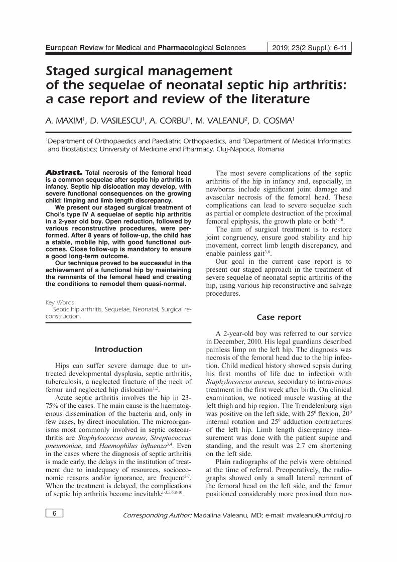
mal, outside of the acetabulum. According to the system suggested by Choi et al8, our patient was grouped under Choi’s type IV A (Figure 1) with severe destruction of the femoral head and dislo-cation of the hip joint.
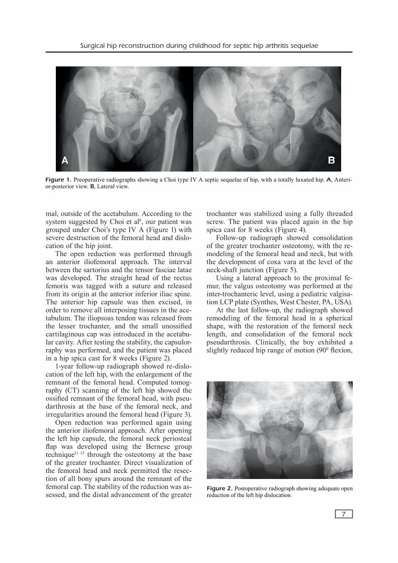
The open reduction was performed through an anterior iliofemoral approach. The interval between the sartorius and the tensor fasciae latae was developed. The straight head of the rectus femoris was tagged with a suture and released from its origin at the anterior inferior iliac spine. The anterior hip capsule was then excised, in order to remove all interposing tissues in the ace-tabulum. The iliopsoas tendon was released from the lesser trochanter, and the small unossified cartilaginous cap was introduced in the acetabu-lar cavity. After testing the stability, the capsulor-raphy was performed, and the patient was placed in a hip spica cast for 8 weeks (Figure 2).
1-year follow-up radiograph showed re-dislo-cation of the left hip, with the enlargement of the remnant of the femoral head. Computed tomog-raphy (CT) scanning of the left hip showed the ossified remnant of the femoral head, with pseu-darthrosis at the base of the femoral neck, and irregularities around the femoral head (Figure 3).
Open reduction was performed again using the anterior iliofemoral approach. After opening the left hip capsule, the femoral neck periosteal flap was developed using the Bernese group technique11–15 through the osteotomy at the base of the greater trochanter. Direct visualization of the femoral head and neck permitted the resec-tion of all bony spurs around the remnant of the femoral cap. The stability of the reduction was as-sessed, and the distal advancement of the greater
trochanter was stabilized using a fully threaded screw. The patient was placed again in the hip spica cast for 8 weeks (Figure 4).
Follow-up radiograph showed consolidation of the greater trochanter osteotomy, with the re-modeling of the femoral head and neck, but with the development of coxa vara at the level of the neck-shaft junction (Figure 5).
Using a lateral approach to the proximal fe-mur, the valgus osteotomy was performed at the inter-trochanteric level, using a pediatric valgisa-tion LCP plate (Synthes, West Chester, PA, USA).
At the last follow-up, the radiograph showed remodeling of the femoral head in a spherical shape, with the restoration of the femoral neck length, and consolidation of the femoral neck pseudarthrosis. Clinically, the boy exhibited a slightly reduced hip range of motion (900 flexion,
Figure 1. Preoperative radiographs showing a Choi type IV A septic sequelae of hip, with a totally luxated hip. A, Anteri-or-posterior view. B, Lateral view.
Figure 2. Postoperative radiograph showing adequate open reduction of the left hip dislocation.

A. Maxim, D. Vasilescu, A. Corbu, M. Valeanu, D. Cosma
8
00 extension, 450 abduction, 100 adduction, 200
external rotation, 150 internal rotation), with a resolution of the Trendelenburg gait, and a limb length discrepancy of 7.6 cm (Figure 6).
The patient is scheduled for future acetabu-loplasty to correct the acetabular dysplasia, prior to the lengthening procedures.
Discussion
Acute septic hip arthritis in neonates rep-resents a surgical emergency, which requires early detection (within 4 days) and adequate treatment (decompression and antibiotics)3,4,16.
Failure in early diagnosis and treatment leads to catastrophic effects on the hip, especially if there are other risk factors: prematurity, age less than 6 months, type of bacteria, concurrent osteomy-elitis of femur, and septic dislocation of the hip joint. The degree of damage and the subsequent sequelae are difficult to predict even after appro-priate treatment3,4.
Radiographic classifications of residual se-quelae after septic hip arthritis have been pro-posed by various authors8,17. Choi et al8 reported the distribution of sequelae severity in septic hip arthritis in children. Nearly 60% of cases were
Figure 3. Preoperative CT-scan of the left hip: dislocated remnants of the left femoral neck, covered by cartilaginous cap.
Figure 4. Postoperative radiograph showing deep-seated femoral head inside the acetabulum, following surgical hip dislocation, osteochondroplasty and distal advancement of the greater trochanter. Figure 5. Development of neck pseudarthrosis and coxa vara.

Surgical hip reconstruction during childhood for septic hip arthritis sequelae
9
less severe type (Choi type I, II, and III) and 38% of patients had the most severe type IV. More re-cently, Forlin et al18 published their report where 41% of children had the severe Choi type IV.
Patients affected by sequelae of the septic hip arthritis type IV have severe anatomical de-formities. The remnant of the head and neck is
covered with a cartilaginous cap and usually is dislocated. The dislocation of the hip in sepsis is different from the developmental hip dysplasia. Intra-articular fibrosis restricts further disloca-tion, and the laxity is due to the misshaped fem-oral head. Moreover, the triradiate physis may be damaged, resulting in loss of remodeling of the acetabulum, and also the capsule is thick, adherent, and fibrous3. Other possible deformities are: flexion-adduction contracture, marked limb length discrepancy, and severe functional limita-tion when walking9.
Before undertaking any reconstructive proce-dure, the infection must have settled down. The desirable aims and objectives of the treatment are (1) restoration of the hip stability; (2) deformity correction of the hip joint; (3) shortening correc-tion; (4) maintain mobility of the hip; (5) relieve pain. Maintenance of the hip joint mobility is very important, and the restoration of the stability at the expense of mobility is not desirable.
Some authors have reported that the patients may do well non-operatively for several years. Fabry et al19 reported on 29 children with hip septic arthritis. They concluded that the desired anatomical and functional improvement was not always obtained by surgical procedures. Wopper-er at al20 and Betz et al21 reported independently that the Harris hip scores were not better in the surgically treated patients and they advised against reconstructive surgery of the hip. They recommended only contralateral epiphysiodesis in adolescence to compensate the limb length discrepancy.
Figure 7. Ossified femoral head, located inside the dysplastic acetabulum, with adequate femoral neck length and inclination angle, 8 years after the initial surgery.
Figure 6. Postoperative radiograph showing consolidation of the pseudarthrosis and restoration of the neck length and head shape after valgisation osteotomy of the proximal femur.

A. Maxim, D. Vasilescu, A. Corbu, M. Valeanu, D. Cosma
10
Later series have shown that late reconstruc-tions and replacement arthroplasty become diffi-cult because of loss of bone stock3. Various forms of reconstructive procedures have been described in the attempt to salvage the hip joint with the objective to obtain a stable painless joint.
Closed reduction can be attempted but, in most cases, this is prone to failure. In our patient, the open reduction was performed at the begin-ning, but even so, the redislocation appeared. Due to the particularity of the septic dislocation, with a lot of fibrous tissues in the hip joint, we advise for open reduction of the hip, with careful debridement of the scary tissues and excision of all redundant capsule. In the presence of redis-location, careful imagistic examination of the hip is recommended. In our case, CT scanning of the hip showed the presence of remnants of the femoral head and neck, with numerous bony spurs. Careful excision of the bony irregularities followed by proximal femoral osteotomies will allow the restoration of the quasi-normal ana-tomical and functional properties of the proximal femur.
Other reports advised for trochanteroplasty as a surgical procedure to restore the stability and mobility of the hip, preparing the area for future prosthetic reconstruction of the hip. Abrishami et al6 achieved satisfactory results in all 9 patients undergoing trochanteroplasty under 3 years of age. Stability of the hip was achieved in all cases with a resolution of abductor lurches, but mobility decreased in variable grades in all patients. The authors concluded that a stable, functional, pain-less hip can be achieved when performing tro-chanteroplasty earlier than 3 years of age. Ferrari et al9 evaluated 11 patients treated by trochan-teroplasty before 10 years of age. They achieved stability and mobility of the hip in 8 out of 11 patients and concluded that satisfactory indepen-dence is generally restored with some inevitable functional limitations. El-Rosasy et al1 reported their results with the pelvic support osteotomy (PSO). 16 adult patients underwent this type of reconstructive surgery with significant improve-ment of Harris hip score in all patients. PSO is a valuable and durable solution for adult patients suffering of sequelae of septic hip arthritis22.
The particularities of the reported case consist of the combined and staged surgical management of Choi type IV A sequelae of septic hip arthri-tis. Initial open reduction restored the normal position of the head remnant in the affected acetabulum, creating the conditions for future
remodeling of the acetabulum. The increase in size and shape of the femoral cap permitted the trimming and the lengthening of the femoral neck through the method described by the Bernese group. This resulted in a deep-seated femoral head, with sufficient length of the femoral neck to allow resolution of the Trendelenburg gait. The stability and mobility of the hip were achieved allowing the future lengthening procedures of the femur (Figure 7).
Conclusions
Careful clinical and imagistic evaluation is recommended preoperatively to asses correctly all the patient particularities. Based on the final evaluation, a reconstructive plan can be devel-oped individually for each patient in order to achieve the hip joint stability and mobility which will allow future prosthetic replacement.
AcknowledgementsThis study was supported by the internal grant for doctor-al students of the University of Medicine and Pharmacy Cluj-Napoca, Romania.
Conflict of InterestsThe authors declare that they have no conflicts of interest.
References
1) El-Rosasy Ma, ayoub Ma. Midterm results of Iliza-rov hip reconstruction for late sequelae of child-hood septic arthritis. Strateg Trauma Limb Re-constr 2014; 9: 149-155.
2) uMER M, Rashid h, uMER hM, Raza h. Hip re-construction osteotomy by Ilizarov method as a salvage option for abnormal hip joints. Biomed Research International, 2014.
3) anil a, aggaRwal an. Pediatric Osteoarticular Infections. Jaypee Brothers Ltd, 2013.
4) Young TP, Maas L, Thorp AW, Brown L. Etiology of septic arthritis in children: An update for the new millennium. Am J Emerg Med 2011; 29: 899-902.
5) baghdadi T, sabERi s, ERaghi as, aRabzadEh a, MaR-dookhpouR s. Late sequelae of hip septic arthritis in children. Acta Med Iran 2012; 50: 463-467.
6) abRishaMi s, kaRaMi M, kaRiMi a, soufali ap, aslani hR, badizadEh k. Greater trochanteric preserving hip ar-throplasty in the treatment of infantile septic arthritis: Long-term results. J Child Orthop 2010; 4: 137-141.
7) McconnEll l, cosMa d, VasilEscu d, MoRcuEndE J. Descriptive epidemiology of clubfoot in Romania: a clinic-based study. Eur Rev Med Pharmacol Sci 2016; 20: 220-224.

Surgical hip reconstruction during childhood for septic hip arthritis sequelae
11
8) choi ih, shin yw, chung cy, cho T-J, yoo wJ, lEE dy. Surgical treatment of the severe sequelae of infantile septic arthritis of the hip. Clin Orthop Relat Res 2005; 434: 102-109.
9) fERRaRi d, libRi R, donzElli o. Trochanteroplasty to treat sequelae of septic arthritis of the hip in infancy. Case series and review of the literature. HIP Int 2011; 21: 653-656.
10) byTyçi c, QoRRaJ h, byTyQi d. Treatment of neona-tal septic arthritis sequelae of hip : a case report. Cases J 2009; 2: 2-3.
11) ziEbaRTh k, zilkEns c, spEncER s, lEunig M, ganz R, kiM y-J. Capital realignment for moderate and severe SCFE using a modified Dunn procedure. Clin Orthop Relat Res 2009; 467: 704-716.
12) Massè a, apRaTo a, gRappiolo g, TuRchETTo l, caMpacci a, ganz R. Surgical hip dislocation for anatomic reorientation of slipped capital femoral epiphysis: preliminary results. Hip Int 2012; 22: 137-144.
13) ganz R, ziEbaRTh k, lEunig M, slongo T, kiM y-J. Anatomical Reconstruction of the Hip with SCFE, Justified by Pathophysiological Findings. In: Bentley G. European Instructional Lectures. Springer Berlin Heidelberg, 2013.
14) cosMa d, VasilEscu dE, coRbu a, ValEanu M, VasilEs-cu d. The modified Dunn procedure for slipped capital femoral epiphysis does not reduce the length of the femoral neck. Pakistan J Med Sci 2016; 32: 379-384.
15) ganz R, huff Tw, lEunig M. Extended retinacu-lar soft-tissue flap for intra-articular hip surgery: surgical technique, indications, and results of application. Instr Course Lect 2009; 58:241-255.
16) bEnuM p. Transposition of the apophysis of the greater trochanter for reconstruction of the fem-oral head after septic hip arthritis in children: 4 children followed for more than 15 years. Acta Orthop 2011; 82: 64-68.
17) hunka l, said sE, MackEnziE da, Rogala EJ, cRuEss Rl. Classification and surgical management of the severe sequelae of septic hips in children. Clin Orthop Relat Res 1982; 171: 30-36.
18) foRlin E, Milani c. Sequelae of septic arthritis of the hip in children: a new classification and a review of 41 hips. J Pediatr Orthop 2008; 28: 524-528.
19) fabRy g, MEiRE E. Septic arthritis of the hip in children: poor results after late and inadequate treatment. J Pediatr Orthop 1983; 3: 461-466.
20) woppERER JM, whiTE JJ, gillEspiE R, oblETz bE. Long-term follow-up of infantile hip sepsis. J Pediatr Orthop 1988; 8: 322-325.
21) bETz RR, coopERMan dR, woppERER JM, suThERland Rd, whiTE JJ, schaaf hw, aschliMan MR, choi ih, bowEn JR, gillEspiE R. Late sequelae of septic arthritis of the hip in infancy and childhood. J Pediatr Orthop 1990; 10: 365-372.
22) chEn z, chEng l, fEng g. Bone inflammation and chronic recurrent multifocal osteomyelitis. Eur Rev Med Pharmacol Sci 2018; 22: 1380-1386.
Related Documents