Stable inheritance of host species-derived microchromosomes in the gynogenetic fish, Poecilia formosa Indrajit Nanda * , Ingo Schlupp ** , Dunja K. Lamatsch ***, § , Kathrin P. Lampert *** , Michael Schmid * , Manfred Schartl *** * Universität Würzburg, Institut für Humangenetik, Biozentrum, D-97074 Würzburg, Germany ** Department of Zoology, University of Oklahoma, Norman, OK 73019, USA *** Universität Würzburg, Physiologische Chemie I, Biozentrum, D-97074 Würzburg, Germany § Present address: Department of Animal and Plant Sciences, University of Sheffield, Sheffield, S10 2TN, UK Genetics: Published Articles Ahead of Print, published on August 24, 2007 as 10.1534/genetics.107.076893

Welcome message from author
This document is posted to help you gain knowledge. Please leave a comment to let me know what you think about it! Share it to your friends and learn new things together.
Transcript

Stable inheritance of host species-derived microchromosomes in the gynogenetic fish,
Poecilia formosa
Indrajit Nanda*, Ingo Schlupp**, Dunja K. Lamatsch***, §, Kathrin P. Lampert***, Michael
Schmid*, Manfred Schartl***
*Universität Würzburg, Institut für Humangenetik, Biozentrum, D-97074 Würzburg, Germany
**Department of Zoology, University of Oklahoma, Norman, OK 73019, USA
***Universität Würzburg, Physiologische Chemie I, Biozentrum, D-97074 Würzburg,
Germany
§Present address: Department of Animal and Plant Sciences, University of Sheffield,
Sheffield, S10 2TN, UK
Genetics: Published Articles Ahead of Print, published on August 24, 2007 as 10.1534/genetics.107.076893
2
Running head: Stable inheritance of B-chromosomes
Key words: clonal diversity, paternal introgression, B-chromosomes, asexual reproduction,
telomeres
Corresponding author: Prof. Dr. Manfred Schartl, Physiological Chemistry I, University of
Würzburg, Biozentrum, Am Hubland, D-97074 Wuerzburg, Germany, Tel: (+49)931-888-
4148, fax: (+49)931-888-4150, email: [email protected]

3
ABSTRACT
B-chromosomes are additional, usually unstable constituents of the genome of many
organisms. Their origin, however, is often unclear and their evolutionary relevance is not well
understood. They may range from being deleterious to neutral or even beneficial. We have
followed the genetic fate of B-chromosomes in the asexual, all-female fish, Poecilia formosa
over eight generations. In this species, B-chromosomes come in the form of one to three tiny
microchromosomes derived from males of the host species that serve as sperm donors for this
gynogenetic species. All microchromosomes have centromeric heterochromatin but usually
only one has a telomere. Such microchromosomes are stably inherited, while the telomere-
less are prone to be lost both in the soma and germline. In some cases the stable
microchromosome carries a functional gene lending support to the hypothesis that the B-
chromosomes in P. formosa could increase the genetic diversity of the clonal lineage in this
ameiotic organism and to some degree counteract the genomic decay that is supposed to be
connected with the lack of recombination.

4
INTRODUCTION
B-chromosomes are supernumerary chromosomes, which do not follow Mendelian
rules of inheritance. To date, they have been found in more than 2000 species of plants,
animals and fungi (CAMACHO et al. 2000; JONES and REES 1982; PALESTIS et al. 2004). B-
chromosomes are considered either to arise from a duplicated or fragmented A-chromosome
within the same genome or to be acquired during a hybridization event from foreign DNA
that evolves into the supernumerary chromosome (CAMACHO et al. 2000; GREEN 1990; JONES
and REES 1982). Within a given species or population, individuals are polymorphic for the
presence of B-chromosomes, because those usually lack a homologous partner to pair with
during meiosis and are therefore distributed unequally to the gametes. There can be one or
several B-chromosomes in one individual. In addition B-chromosomes can also be lost during
an individual’s development due to unequal distribution during cell divisions. Such organisms
then may lack B-chromosomes in certain organs, tissues or cells (PALESTIS et al. 2004).
The maintenance and evolution of B-chromosomes has been explained by several
ways. Traditionally, they have been classified as selfish genetic elements that decrease the
fitness of the “host” genome (CAMACHO et al. 2000; SHAW and HEWITT 1990). Thus they
generate what has been called a ”genetic conflict” between the A- and B-chromosomes. By
virtue of their accumulation mechanisms, they are maintained within populations (JONES
1985; NUR et al. 1988; ÖSTERGREN 1945; THOMSON 1984). In the heterotic model (WHITE
1973), it is assumed that B-chromosomes are maintained because they increase the fitness of
the host when they occur at low frequency. This hypothesis does not require an accumulation
mechanism. An “evolutionary arms race” model (CAMACHO et al. 1997) assumes a non-
stable, dynamic situation. B-chromosomes are considered parasitic and spread through the
population due to an accumulation mechanism. But, as they increase their frequency, they are
5
neutralized by the host genome and begin to disappear slowly, unless a new variant of the B,
which can counteract the elimination mechanism, replaces the neutralized B.
Only in very few cases the B-chromosomes appear to have a beneficial effect on the
host species (BOUGOURD and JONES 1997), while most are considered to be harmful
(PALESTIS et al. 2004). They can, however, escape extinction in outcrossing species because
they can continually “infect” new lineages if they drive. In inbred or asexual species, natural
selection acts among competing lines of descendants or clones, respectively. Lines or clones
without B-chromosomes are expected to outcompete those with B-chromosomes, if B-
chromosomes decrease fitness. In the asexual all-female fish species Poecilia formosa, the
Amazon molly, supernumerary chromosomes have frequently been found in both laboratory-
reared and wild-caught individuals from the Río Purificación/Río Soto la Marina river system,
Mexico (LAMATSCH et al. 2004). The high frequency of B-chromosomes in wild populations
support the idea that the B-chromosomes of P. formosa are not harmful, but rather may be
beneficial (SCHARTL et al. 1995).
The genetic fate of B-chromosomes is usually not well documented and their origin is
mostly unknown. For the B-chromosomes of P. formosa it is clear that they are of hybrid
(allospecific) origin. P. formosa reproduces by gynogenesis meaning that the parthenogenetic
development of the diploid, ameiotic oocytes is triggered by sperm of males from closely
related species. In general, the paternal DNA is excluded from the inseminated oocyte,
however in rare cases parts of the sperm genome persist as tiny B-chromosomes
(microchromosomes) in the karyotype of the developing embryo (SCHARTL et al. 1995). This
process of introgression of paternal genes into the asexual lineage is considered as bringing in
fresh genetic material into the asexual lineage. Organisms that cannot perform recombination
should suffer from genetic decay, because deleterious mutations cannot be purged and are
slow to evolve (KONDRASHOV 1988; MULLER 1932). Thus the B-chromosomes in P. formosa

6
might be ascribed a beneficial effect. However, a precondition for this is that the
microchromosomes can become stable components of the genome of the asexual fish. If not,
their evolutionary impact would only be very transient and not of considerable importance.
We tested this precondition by following the inheritance of microchromosomes from P.
formosa in B-chromosome containing clones from the wild and from recent introgression
events in the laboratory. B-chromosomes were found to be inherited stably over many
generations in the analyzed clones and no fish without B-chromosomes were recorded.
7
MATERIALS AND METHODS
Animals:
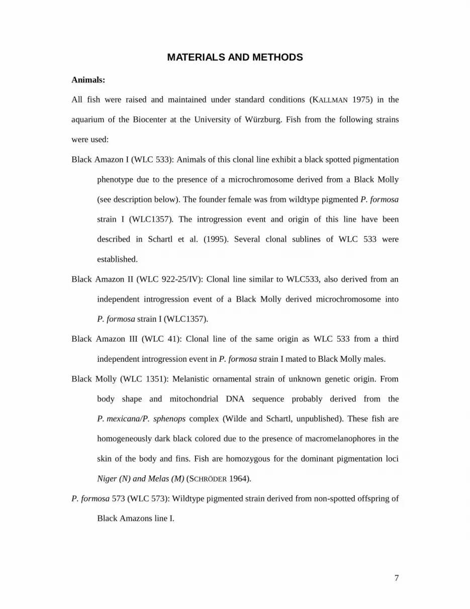
All fish were raised and maintained under standard conditions (KALLMAN 1975) in the
aquarium of the Biocenter at the University of Würzburg. Fish from the following strains
were used:
Black Amazon I (WLC 533): Animals of this clonal line exhibit a black spotted pigmentation
phenotype due to the presence of a microchromosome derived from a Black Molly
(see description below). The founder female was from wildtype pigmented P. formosa
strain I (WLC1357). The introgression event and origin of this line have been
described in Schartl et al. (1995). Several clonal sublines of WLC 533 were
established.
Black Amazon II (WLC 922-25/IV): Clonal line similar to WLC533, also derived from an
independent introgression event of a Black Molly derived microchromosome into
P. formosa strain I (WLC1357).
Black Amazon III (WLC 41): Clonal line of the same origin as WLC 533 from a third
independent introgression event in P. formosa strain I mated to Black Molly males.
Black Molly (WLC 1351): Melanistic ornamental strain of unknown genetic origin. From
body shape and mitochondrial DNA sequence probably derived from the
P. mexicana/P. sphenops complex (Wilde and Schartl, unpublished). These fish are
homogeneously dark black colored due to the presence of macromelanophores in the
skin of the body and fins. Fish are homozygous for the dominant pigmentation loci
Niger (N) and Melas (M) (SCHRÖDER 1964).
P. formosa 573 (WLC 573): Wildtype pigmented strain derived from non-spotted offspring of
Black Amazons line I.

8
P. formosa III/9 (WLC 1612): Wildtype pigmented strain derived from one female with a
single microchromosome of a collection from the Río Purificación near Barretal,
Tamaulipas, Mexico.
P. formosa III/4 (WLC 1588): Wildtype pigmented strain derived from one female with a
single microchromosome of a collection from a canal east of Ciudad Mante,
Tamaulipas, Mexico.
Chromosome analysis and telomere staining
Mitotic chromosomes were prepared directly from pooled organs (spleen, cephalic
kidney, gills) following the standard procedure described elsewhere (NANDA et al. 1995).
Giemsa stained slides were screened under light microscope to check the number of diploid
chromosomes as well as the presence of microchromosomes. To visualize the centromeric
heterochromatin, metaphase chromosomes were subjected to C-banding following the
procedure of Sumner (SUMNER 1972) except that the treatment with alkali was done for 2
min.
To detect the presence of telomere specific (TTAGGG)n repeats at the end of
chromosomes, fluorescence in-situ hybridization (FISH) was performed with a telomeric
PNA oligonucleotide (CCCTAA)3 labeled with FITC (Applied Biosystems). After
pretreatment with pepsin and formaldehyde, slides were denatured at 80oC for 3 min under a
coverslip in presence of the hybridization mixture containing the labeled probe. Hybridization
was performed for 2 h at room temperature after which slides were briefly washed in 70%
formamide (10 min) and further washed in PBS for 5 min. The slides were dehydrated in
ethanol series. Afterwards, slides were mounted in an antifade reagent containing DAPI (4’6-
diamidino-2-phenylindole) as counter-stain. Digital images of metaphases showing
hybridization signals were acquired using a Zeiss epifluorescence microscope coupled with
CCD camera and Applied Spectral Imaging software (Neckerhausen, Germany).
9
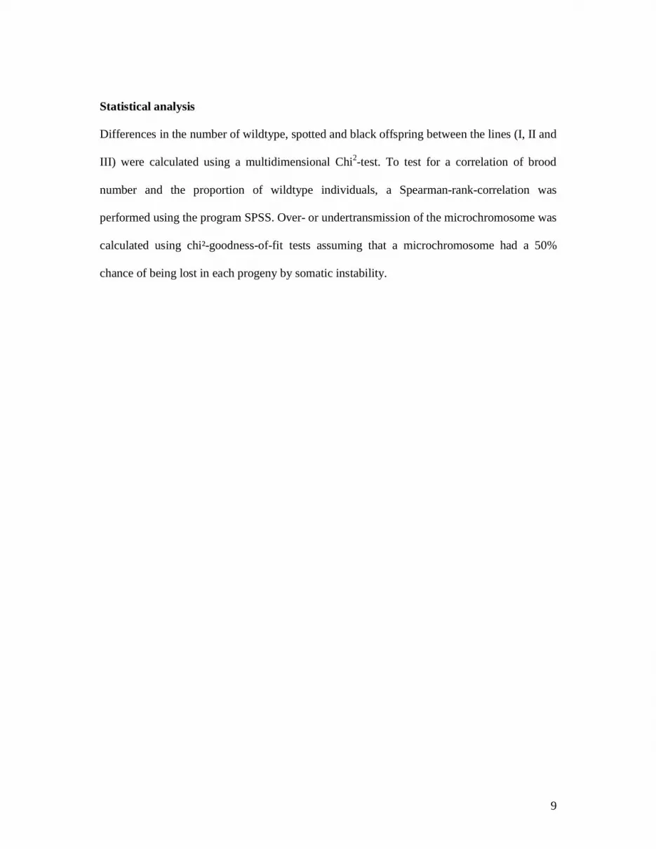
Statistical analysis
Differences in the number of wildtype, spotted and black offspring between the lines (I, II and
III) were calculated using a multidimensional Chi2-test. To test for a correlation of brood
number and the proportion of wildtype individuals, a Spearman-rank-correlation was
performed using the program SPSS. Over- or undertransmission of the microchromosome was
calculated using chi²-goodness-of-fit tests assuming that a microchromosome had a 50%
chance of being lost in each progeny by somatic instability.
10
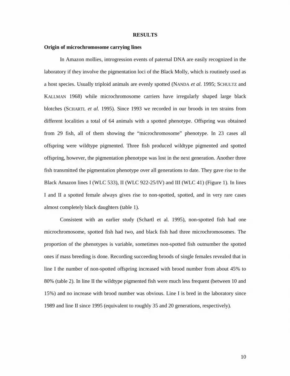
RESULTS
Origin of microchromosome carrying lines
In Amazon mollies, introgression events of paternal DNA are easily recognized in the
laboratory if they involve the pigmentation loci of the Black Molly, which is routinely used as
a host species. Usually triploid animals are evenly spotted (NANDA et al. 1995; SCHULTZ and
KALLMAN 1968) while microchromosome carriers have irregularly shaped large black
blotches (SCHARTL et al. 1995). Since 1993 we recorded in our broods in ten strains from
different localities a total of 64 animals with a spotted phenotype. Offspring was obtained
from 29 fish, all of them showing the “microchromosome” phenotype. In 23 cases all
offspring were wildtype pigmented. Three fish produced wildtype pigmented and spotted
offspring, however, the pigmentation phenotype was lost in the next generation. Another three
fish transmitted the pigmentation phenotype over all generations to date. They gave rise to the
Black Amazon lines I (WLC 533), II (WLC 922-25/IV) and III (WLC 41) (Figure 1). In lines
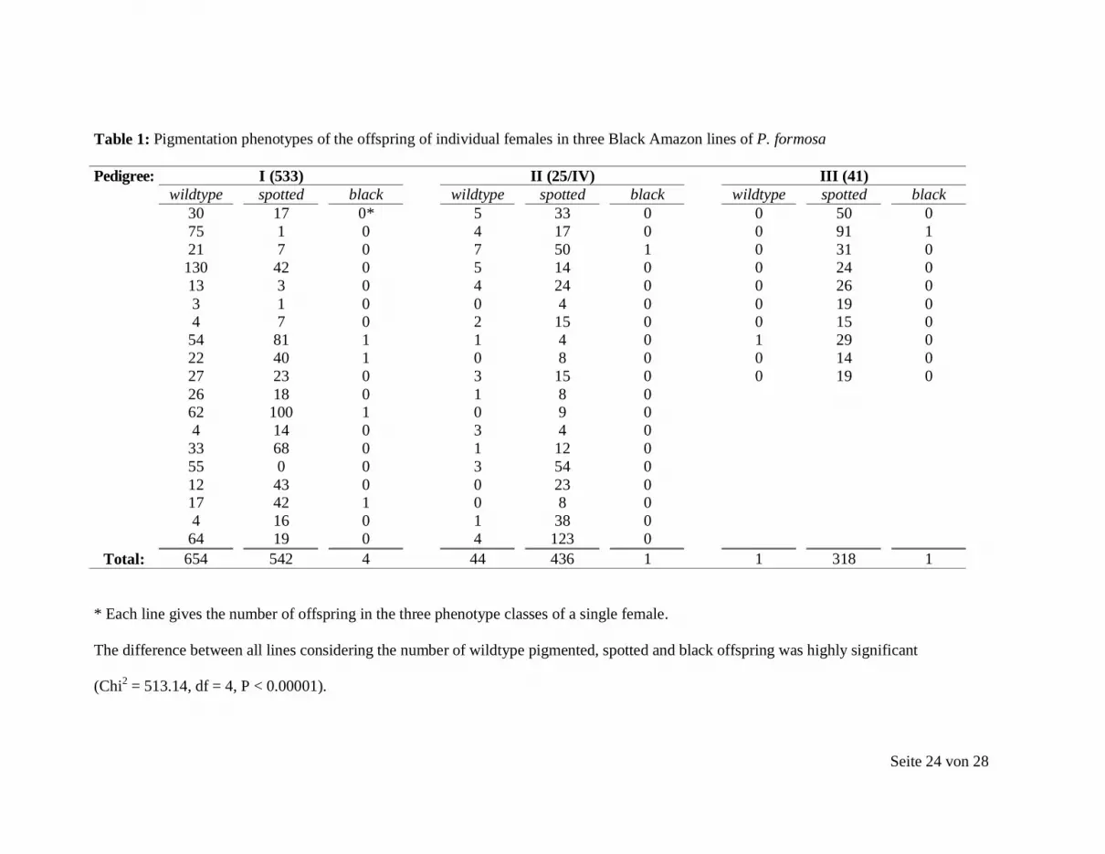
I and II a spotted female always gives rise to non-spotted, spotted, and in very rare cases
almost completely black daughters (table 1).
Consistent with an earlier study (Schartl et al. 1995), non-spotted fish had one
microchromosome, spotted fish had two, and black fish had three microchromosomes. The
proportion of the phenotypes is variable, sometimes non-spotted fish outnumber the spotted
ones if mass breeding is done. Recording succeeding broods of single females revealed that in
line I the number of non-spotted offspring increased with brood number from about 45% to
80% (table 2). In line II the wildtype pigmented fish were much less frequent (between 10 and
15%) and no increase with brood number was obvious. Line I is bred in the laboratory since
1989 and line II since 1995 (equivalent to roughly 35 and 20 generations, respectively).
11
Inheritance of spotted phenotypes
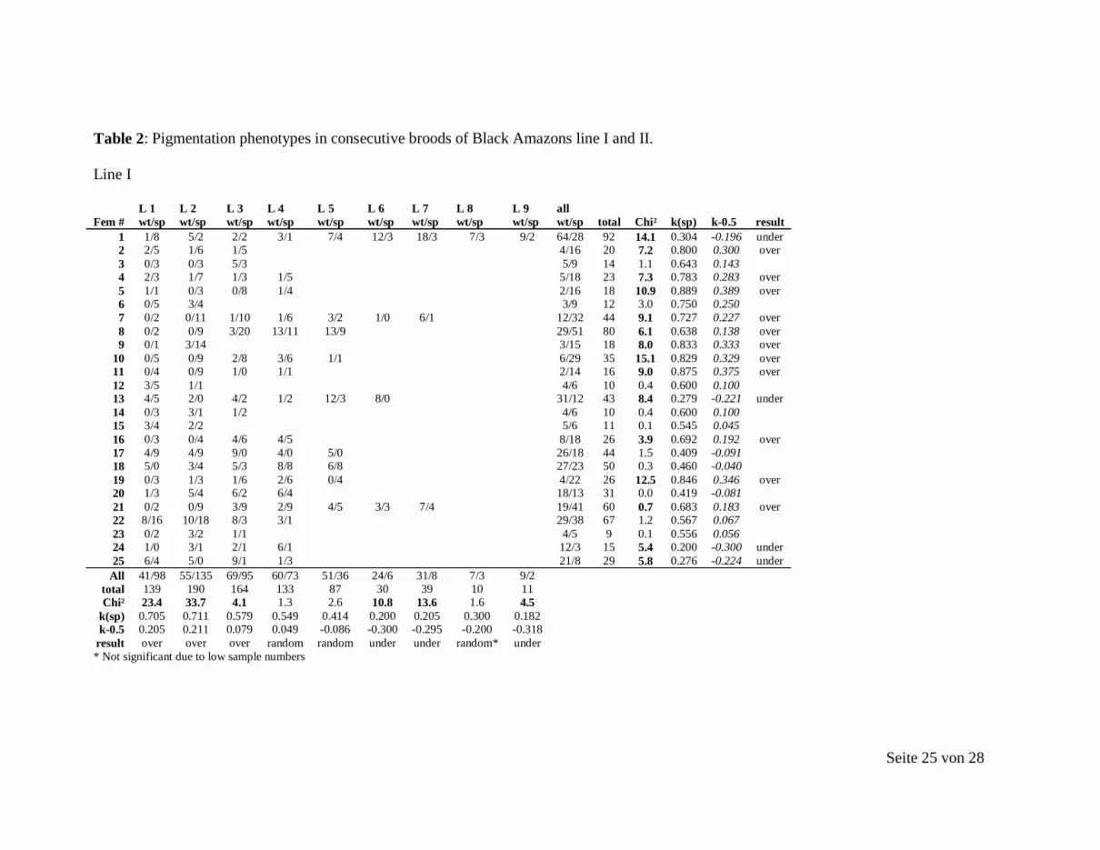
A detailed analysis of microchromosome transmission mode was performed using
spotted females of Black Amazon lines I and II and their broods (table 2). Spotted and black
offspring were grouped as one since both phenotypes did inherit a melanic B chromosome. A
goodness of fit Chi² test was performed under the hypothesis that the melanic B present in the
mother would have a 50% chance of being lost in each progeny by somatic instability.
Significant deviations from this assumption were interpreted as either undertransmission
(transmission rate significantly below 50%) or overtransmission (transmission significantly
above 50%). The analysis was performed for each female (broods had to be pooled due to
otherwise too low sample sizes) and for each litter (females were pooled for this analyses due
to otherwise too low sample sizes).
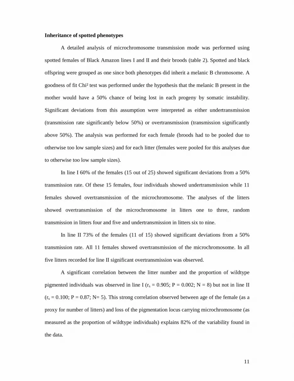
In line I 60% of the females (15 out of 25) showed significant deviations from a 50%
transmission rate. Of these 15 females, four individuals showed undertransmission while 11
females showed overtransmission of the microchromosome. The analyses of the litters
showed overtransmission of the microchromosome in litters one to three, random
transmission in litters four and five and undertransmission in litters six to nine.
In line II 73% of the females (11 of 15) showed significant deviations from a 50%
transmission rate. All 11 females showed overtransmission of the microchromosome. In all
five litters recorded for line II significant overtransmission was observed.
A significant correlation between the litter number and the proportion of wildtype
pigmented individuals was observed in line I (rs = 0.905; P = 0.002; N = 8) but not in line II
(rs = 0.100; P = 0.87; N= 5). This strong correlation observed between age of the female (as a
proxy for number of litters) and loss of the pigmentation locus carrying microchromosome (as
measured as the proportion of wildtype individuals) explains 82% of the variability found in
the data.

12
A considerable degree of somatic instability was noted when in fish of lines I and II
different metaphases of one and the same fish were compared. In each metaphase spread, the
microchromosomes appeared as a separate entity, which was not attached to any
chromosomes. While the number of A-chromosomes was always n=46, either one or two
microchromosomes, or even three (only in totally black animals) were present. However, the
loss of all microchromosomes was never observed. The proportion of one or two
microchromosome-containing metaphases varied between individuals (table 3).
Line III, established in 2003, differs from the two other lines because after eight
generations it had produced only a single non-spotted fish out of a total of 319 offspring (table
1). Karyotype analysis of line III revealed that the spotted fish have only one
microchromosome, which is different from the spotted fish of line I and II. The single
unspotted fish unfortunately died before its karyotype could be analyzed.
The somatic instability observed in lines I and II was absent in fish of Black Amazon
line III. Like in the wildtype pigmented fish from wild populations (n=28) with one
microchromosome (see below), all metaphases consistently had one microchromosome.
Inheritance of microchromosomes
To analyze the inheritance of microchromosomes, females that carried a single
microchromosome were mated to Black Mollies and the karyotype of the offspring over
several consecutive generations was prepared (Figure 2). All animals from a total of nine
generations in both lines established from microchromosome carrying females collected from
natural habitats had the microchromosome (Figure 3). In pedigree III/4 in generations G3 and
G4, spotted fish occurred. They had an additional microchromosome (Figure 2) obviously
paternally derived from the Black Molly that was used for breeding. In pedigree III/9, one
female in G4 and one in G6 also showed an additional microchromosome in the karyotype,
but both fish were wildtype pigmented. This additional microchromosome also should have
13
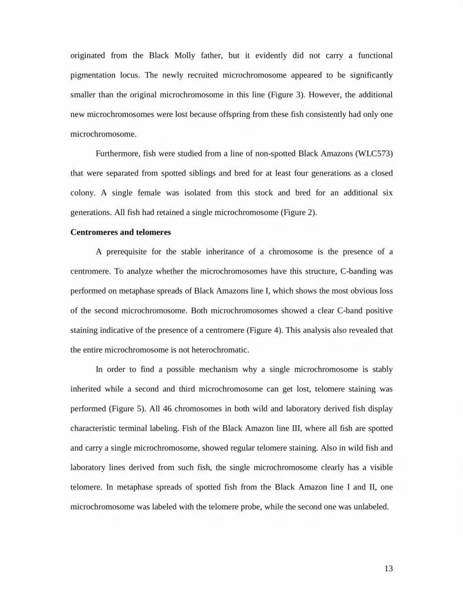
originated from the Black Molly father, but it evidently did not carry a functional
pigmentation locus. The newly recruited microchromosome appeared to be significantly
smaller than the original microchromosome in this line (Figure 3). However, the additional
new microchromosomes were lost because offspring from these fish consistently had only one
microchromosome.
Furthermore, fish were studied from a line of non-spotted Black Amazons (WLC573)
that were separated from spotted siblings and bred for at least four generations as a closed
colony. A single female was isolated from this stock and bred for an additional six
generations. All fish had retained a single microchromosome (Figure 2).
Centromeres and telomeres
A prerequisite for the stable inheritance of a chromosome is the presence of a
centromere. To analyze whether the microchromosomes have this structure, C-banding was
performed on metaphase spreads of Black Amazons line I, which shows the most obvious loss
of the second microchromosome. Both microchromosomes showed a clear C-band positive
staining indicative of the presence of a centromere (Figure 4). This analysis also revealed that
the entire microchromosome is not heterochromatic.
In order to find a possible mechanism why a single microchromosome is stably
inherited while a second and third microchromosome can get lost, telomere staining was
performed (Figure 5). All 46 chromosomes in both wild and laboratory derived fish display
characteristic terminal labeling. Fish of the Black Amazon line III, where all fish are spotted
and carry a single microchromosome, showed regular telomere staining. Also in wild fish and
laboratory lines derived from such fish, the single microchromosome clearly has a visible
telomere. In metaphase spreads of spotted fish from the Black Amazon line I and II, one
microchromosome was labeled with the telomere probe, while the second one was unlabeled.

14
Hybridization with the telomere specific probe labelled the terminal ends of all Black
Molly chromosomes (Figure 5c). This excludes the existence of any specific chromosome end
with undetectable (TTAGGG)n repeats, as the origin of the material giving rise to the
microchromosomes of line I and II without a telomere.
15
DISCUSSION
Our analysis of microchromosome carrying clones of P. formosa revealed that B-
chromosomes were inherited stably over many generations and no fish without B-
chromosomes were recorded.
Microchromosomes seem to be left over from the enzymatic process that normally
clears the diploid ameiotic P. formosa eggs of the sperm nucleus after fertilization has
occurred. The fact that the microchromosomes are regularly inherited in P. formosa pedigrees
suggests that foreign DNA once incorporated into the asexual lineage can become a stable
constituent of the genome and show germline transmission. The fact that a single
microchromosome is found in all offspring despite lacking a homologous partner is obviously
due to the absence of meiotic division in the asexual fish that produces diploid eggs.
The somatic instability of B-chromosomes is obviously something that is due to
mitotic events. It will affect the microchromosomes during the mitotic divisions of the
primordial germs and the oogonia as well.
Telomere staining revealed a possible explanation for the observed stability of the
single microchromosome situation and the genetic instability of the second and third
microchromosome. Cells with one, two or even three microchromosomes exist in the same
individual, but only one microchromosome is ever stained with the telomere probe. This
suggests that the microchromosomes lacking telomere staining have been eliminated in those
cells of the soma carrying only the stable microchromosome.
The absence of functional telomeres is indicated by the inability to stain certain
microchromosomes with the (CCCTAA)3 PNA probe. This may be due to the lack of a
threshold level of telomere repeats (short repeats). It is also likely that telomere repeats may
be completely absent in the microchromosomes, because microchromosomes are chromosome

16
fragments derived from anywhere along the chromosome and are remnants from the
degradation process of the sperm nucleus DNA.
For stable inheritance of a chromosome, functional centromeres and telomeres are
important. C-banding indicated the occurrence of heterochromatin, which is usually
connected to centromeric regions of chromosomes. Although this technique does not prove
the presence of a centromere, the mere presence of microchromosomes in most cells of an
individual indicates that it has passed through many divisions, for which a functional
centromere is indispensable. However, the absence of functional telomeres correlates with
instability and can explain the elimination of certain microchromosomes. In human cancer
cells, it has been found that mitotic instability of chromosomes is correlated with
dysfunctional telomeres (GISSELSSON and HOGLUND 2005). In this context, it is the shortest
telomeres that mostly constitute telomere dysfunction (HEMANN et al. 2001). Studies in
telomerase deficient mice lead to the conclusion that functional telomeres are involved in
mediating metaphase chromosome alignment and maintaining functional spindles (LIU et al.
2002).
Dysfunctional telomeres may also explain the frequent occurrence of unspotted fish in
the offspring of spotted Black Amazon lines. During mitosis of the germ cells, the
microchromosome without functional telomeres may have a tendency to get lost. This would
predict that the pigmentation locus in these lines is on the chromosomes without telomeres.
When the offspring of single females were analyzed for inheritance of the spotted
pigmentation phenotype, there was a remarkable difference in transmission of
microchromosomes of lines I and II, both of which carry microchromosomes with and
without a pigmentation locus. Line I showed a tendency to undertransmit the spotted pigment
pattern, while line II showed a high tendency of overtransmission. This could be interpreted
as a different behavior of melanic B-chromosomes. For example, the melanic B-chromosome

17
in the younger line II transmits much better than in the older line I. Consistent with Camacho
et al. (1997), it is possible that some resistance has evolved in the old line due to a change in
the genotype of the host A-chromosomes, where the melanic B-chromosome transmits poorly.
However, even in line I there were quite a number of females that showed overtransmission.
Line I is a genetic clone with respect to the A chromosomal genome due to the gynogenetic
mode of reproduction, hence a different genetic background cannot explain the different
genetic behavior of the melanic B-chromosome.
An alternative explanation is that the melanic B-chromosomes of the separate lines
show different inheritance because they are actually dissimilar in nature. This dissimilarity
occurs because the microchromosomes originated from different introgressions. The Black
Molly has two non-allelic pigmentation loci from which the melanic B-chromosomes can be
derived and genetic material accompanying the pigmentation locus may be different as well.
A statistically well supported hypothesis is that the inheritance of the melanic B-
chromosome decreases in consecutive broods of the same female. An increase of genotypic
instability with maternal age has been observed in other species with a prolonged meiotic
prophase (DJAHANBAKHCH et al. 2007). In the ameiotic Amazon molly, a different
mechanism might operate. Fish generate primary oocytes throughout adult life. Therefore,
oogonia of older females have had more mitotic divisions and thus a higher chance that a
mitotically instable microchromosome will be lost. Consequently, the proportion of spotted
offspring, which have the pigmentation gene on an instable microchromosome, will decrease.
An age effect of transmission of B-chromosomes was reported in the grasshopper
Myrmeleotettix maculatus (SHAW and HEWITT 1984), but not in the grasshopper,
Eyprepocnemis plorans (BAKKALI et al. 2002).
The presence of telomeres is obviously responsible for the stable inheritance of a
microchromosome. It can be assumed that microchromosomes that show overtransmission in

18
lines I and II are those that have functional telomeres. This may also explain the persistence
of microchromosome carrying clones in certain river systems in the wild (LAMATSCH et al.
2004). So far all microchromosome bearing fish collected from natural populations had only
one microchromosome. The stable lines obtained in the laboratory also carry the single
telomere containing microchromosome, although the original introgression event contributed
two or even three chromosomes. The reason why the fish have only a single stable
microchromosome is completely unclear.
Our study also indicates that further introgression events can happen as seen by the
appearance of a new microchromosome. However, the new introgression became unstable
rapidly. Surprisingly, in both the pedigrees (III/4 and III/9) the unstable marker chromosomes
arising through the latest introgression are extremely tiny. This is consistent with a
comparative study on the stability of minichromosomes in different species that revealed a
lower size as a limiting factor for a stable chromosome transmission (SCHUBERT 2001).
The question whether certain parts of the genome (e.g. telomere or centromere near
regions) are more prone to end up in a microchromosome cannot be answered. From our data
of laboratory-bred fish, it appears that the regions containing the pigmentation gene are more
often represented. However, due to the conspicuous phenotype, fish with such
microchromosomes are much more readily identified. In the Black Amazon lines I and II,
stable microchromosomes do not carry the pigmentation locus, whereas the unstable second
microchromosome leads to pigment spots and is easily maintained in the stock populations
due to selective breeding. In the three pedigrees analyzed, 4 of 39 individuals had additional
microchromosomes of paternal origin. Two of the four new microchromosomes carried a
pigmentation gene. Given the fact that only two of the n=23 chromosomes of the Black Molly
harbor a pigmentation locus, it can be argued that the chromosomal fragments from the
pigmentation regions show a predisposition to escape elimination not shown by other region

19
of the genome. To answer these questions and to understand the relationship between the
pigmentation mosaicism and the number of microchromosomes, isolation of
microchromosomes by microdissection is required. Such material can then be used for
cloning and sequencing as well for in-situ hybridization.
The fact that telomere containing microchromosomes are stably inherited over at least
eight generations (and probably much longer) becomes interesting when using a probabilistic
approach to calculate the genetic contribution of a male to its offspring. In a large sexually
reproducing population, the genetic contribution of a certain individual male is rapidly diluted
out: children share about 50% of their DNA with their father, the grandchildren 25%, great-
grandchildren 12.5% and so on. In G7 certainly less than 1% of a particular male’s genome is
left in any individual offspring. However, the paternally derived microchromosomes in P.
formosa constitute about 0.5 to 1% of the whole genome. Once a microchromosome is stably
integrated into the germline, it may be transmitted unchanged. After G7 theoretically, more
paternal DNA will be present in the offspring of an asexual female with a microchromosome
than in a sexual lineage. Of course, selection cannot favor males that introduce genes into the
Amazon mollies genome, just as this scenario cannot explain why host males court and
copulate with the asexuals being “wrong” females, because there is no evidence of a “genetic
feedback” mechanism into the host species. A microchromosome can be seen as an
introduction of selfish genetic elements, which is in line with a common view that B-
chromosomes are selfish DNA elements (CAMACHO et al. 2000; JONES 1985; MCVEAN 1995;
SHAW and HEWITT 1990). To determine whether microchromosomes increase the genetic
diversity of P. formosa and/or compensate for mutation in genes that cannot be purged due to
the absence of recombination (Muller’s ratchet), analysis of microchromosomal gene content
and expression has to be performed. Whether the microchromosome carrying individuals are
20
under natural selection and may enjoy some selective advantages can, however, be inferred
from comparing fitness components of clones with and without microchromosomes.
Another important point for evaluating the importance of the introgression of paternal
DNA from host species is the frequency of the event. So far it is unknown whether the
microchromosomes found in natural populations of P. formosa in the Río Purificación river
system go back to a few introgression events or if they are the result of many independent
events. The differences in size of the microchromosomes of the lines analyzed here could be
the result of independent introgression events or of clonal diversity due to loss or gain of
sequences on the microchromosome. However, in the laboratory, introgression events are
quite frequent (up to 10%) and the transmission of newly acquired independent
microchromosomes has been observed several times. This speaks for a relatively frequent
introgression of paternal DNA through B-chromosomes.
In summary, other than initially supposed, microchromosomes can be inherited in the
clonal Amazon molly for many generations. We show here that microchromosome mitotic
instability does not tend to decrease its frequency considerably since, in most cases, the
spotted phenotype associated to it showed only a low chance to be lost during reproduction.
However, to complete the scenario on the biological role of these extra elements, the analysis
of possible fitness differences associated to microchromosome presence is necessary in order
to ascertain whether the potential increase in genetic diversity expected from
microchromosome introgression is actually a selective advantage in Amazon molly natural
populations.
ACKNOWLEDGEMENTS
We are grateful to the Mexican government for issuing the permit to collect P.
formosa and to Jakob Parzefall for his important contribution and help in the field. We thank
21
Georg Schneider, Hugo Schwind and Petra Weber for breeding of the fish in the laboratory,
and Monika Niklaus-Ruiz for help in preparing the manuscript. Courtney Fransen kindly
corrected the English. Financial support for this study was granted by the Deutsche
Forschungsgesellschaft (SFB 567 Mechanismen der interspezifischen Interaktion von
Organismen) and Fonds der Chemischen Industrie to Manfred Schartl. Ingo Schlupp was
supported by a Heisenberg fellowship.
22
LITERATURE CITED
BAKKALI, M., F. PERFECTTI and J. P. M. CAMACHO, 2002 The B-chromosome polymorphism of the grasshopper Eyprepocnemis plorans in North Africa: II. Parasitic and neutralized B-1 chromosomes. Heredity 88: 14-18.
BOUGOURD, S. M., and R. N. JONES, 1997 B chromosomes: a physiological enigma. New Phytologist 137: 43-54.
CAMACHO, J. P. M., T. F. SHARBEL and L. W. BEUKEBOOM, 2000 B-chromosome evolution. Philosophical Transactions of the Royal Society of London Series B-Biological Sciences 355: 163-178.
CAMACHO, J. P. M., M. W. SHAW, M. D. LOPEZLEON, M. C. PARDO and J. CABRERO, 1997 Population dynamics of a selfish B chromosome neutralized by the standard genome in the grasshopper Eyprepocnemis plorans. American Naturalist 149: 1030-1050.
DJAHANBAKHCH, O., M. EZZATI and A. ZOSMER, 2007 Reproductive ageing in women. J Pathol 211: 219-231.
GISSELSSON, D., and M. HOGLUND, 2005 Connecting mitotic instability and chromosome aberrations in cancer - can telomeres bridge the gap? Seminars in Cancer Biology 15: 13-23.
GREEN, D. M., 1990 Muller Ratchet and the Evolution of Supernumerary Chromosomes. Genome 33: 818-824.
HEMANN, M. T., M. A. STRONG, L. Y. HAO and C. W. GREIDER, 2001 The shortest telomere, not average telomere length, is critical for cell viability and chromosome stability. Cell 107: 67-77.
JONES, R., 1985 Are B chromosomes selfish?, pp. 397-425 in The Evolution of Genome Size, edited by T. CAVALIER-SMITH. Wiley, London.
JONES, R., and H. REES, 1982 B-chromosomes. Academic Press, New York. KALLMAN, K., 1975 The platyfish, Xiphophorus maculatus, pp. 81-132 in Handbook of
Genetics, edited by K. RC. Plenum Press, New York, N.Y. KONDRASHOV, A. S., 1988 Deleterious Mutations and the Evolution Of Sexual Reproduction.
Nature 336: 435-440. LAMATSCH, D. K., I. NANDA, I. SCHLUPP, J. T. EPPLEN, M. SCHMID et al., 2004 Distribution
and stability of supernumerary microchromosomes in natural populations of the Amazon molly, Poecilia formosa. Cytogenet Genome Res 106: 189-194.
LIU, L., M. A. BLASCO and D. L. KEEFE, 2002 Requirement of functional telomeres for metaphase chromosome alignments and integrity of meiotic spindles. Embo Reports 3: 230-234.
MCVEAN, G. T., 1995 Fractious Chromosomes - Hybrid Disruption and the Origin of Selfish Genetic Elements. Bioessays 17: 579-582.
MULLER, H. J., 1932 Some genetic aspects of sex. Amer. Nat. 66: 118 - 138. NANDA, I., M. SCHARTL, W. FEICHTINGER, I. SCHLUPP, J. PARZEFALL et al., 1995
Chromosomal evidence for laboratory synthesis of a triploid hybrid between the gynogenetic teleost Poecilia formosa and its host species. J. Fish Biol. 47: 619-623.
NUR, U., J. H. WERREN, D. G. EICKBUSH, W. D. BURKE and T. H. EICKBUSH, 1988 A Selfish B-Chromosome That Enhances Its Transmission by Eliminating the Paternal Genome. Science 240: 512-514.
ÖSTERGREN, G., 1945 Parasitic nature of extra fragment chromosomes. ot. Notiser 2: 157-163.
PALESTIS, B. G., R. TRIVERS, A. BURT and R. N. JONES, 2004 The distribution of B chromosomes across species. Cytogenetic and Genome Research 106: 151-158.

23
SCHARTL, M., I. NANDA, I. SCHLUPP, B. WILDE, J. T. EPPLEN et al., 1995 Incorporation of Subgenomic Amounts of DNA as Compensation for Mutational Load in a Gynogenetic Fish. Nature 373: 68-71.
SCHRÖDER, J., 1964 Genetische Untersuchungen an domestizierten Stämmen der Gattung Mollienesia (Poeciliidae). Zoologische Beiträge 10: 369-463.
SCHUBERT, I., 2001 Alteration of chromosome numbers by generation of minichromosomes - Is there a lower limit of chromosome size for stable segregation? Cytogenetics and Cell Genetics 93: 175-181.
SCHULTZ, R. J., and K. D. KALLMAN, 1968 Triploid Hybrids between the All-female Teleost Poecilia formosa and Poecilia sphenops. Nature 219: 280-282.
SHAW, M. W., and G. M. HEWITT, 1984 The Effect of Temperature on Meiotic Transmission Rates of the B-Chromosome of Myrmeleotettix maculatus (Orthoptera, Acrididae). Heredity 53: 259-268.
SHAW, M. W., and G. M. HEWITT, 1990 B chromosomes, selfish DNA and theoretical models: where next? Oxf Surv Evol Biol 7: 197-223.
SUMNER, A. T., 1972 Simple Technique for Demonstrating Centromeric Heterochromatin. Experimental Cell Research 75: 304-&.
THOMSON, R. L., 1984 B-Chromosomes in Rattus fuscipes .2. The Transmission of B-Chromosomes to Offspring and Population Studies - Support for the Parasitic Model. Heredity 52: 363-372.
WHITE, M. J. D., 1973 Animal cytology and evolution. Cambridge University Press, London.
Seite 24 von 28
Table 1: Pigmentation phenotypes of the offspring of individual females in three Black Amazon lines of P. formosa
Pedigree: I (533) II (25/IV) III (41) wildtype spotted black wildtype spotted black wildtype spotted black 30 17 0* 5 33 0 0 50 0 75 1 0 4 17 0 0 91 1 21 7 0 7 50 1 0 31 0 130 42 0 5 14 0 0 24 0 13 3 0 4 24 0 0 26 0 3 1 0 0 4 0 0 19 0 4 7 0 2 15 0 0 15 0 54 81 1 1 4 0 1 29 0 22 40 1 0 8 0 0 14 0 27 23 0 3 15 0 0 19 0 26 18 0 1 8 0 62 100 1 0 9 0 4 14 0 3 4 0 33 68 0 1 12 0 55 0 0 3 54 0 12 43 0 0 23 0 17 42 1 0 8 0 4 16 0 1 38 0 64 19 0 4 123 0
Total: 654 542 4 44 436 1 1 318 1
* Each line gives the number of offspring in the three phenotype classes of a single female.
The difference between all lines considering the number of wildtype pigmented, spotted and black offspring was highly significant
(Chi2 = 513.14, df = 4, P < 0.00001).
Seite 25 von 28
Table 2: Pigmentation phenotypes in consecutive broods of Black Amazons line I and II.
Line I
L 1 L 2 L 3 L 4 L 5 L 6 L 7 L 8 L 9 all Fem # wt/sp wt/sp wt/sp wt/sp wt/sp wt/sp wt/sp wt/sp wt/sp wt/sp total Chi² k(sp) k-0.5 result
1 1/8 5/2 2/2 3/1 7/4 12/3 18/3 7/3 9/2 64/28 92 14.1 0.304 -0.196 under 2 2/5 1/6 1/5 4/16 20 7.2 0.800 0.300 over 3 0/3 0/3 5/3 5/9 14 1.1 0.643 0.143 4 2/3 1/7 1/3 1/5 5/18 23 7.3 0.783 0.283 over 5 1/1 0/3 0/8 1/4 2/16 18 10.9 0.889 0.389 over 6 0/5 3/4 3/9 12 3.0 0.750 0.250 7 0/2 0/11 1/10 1/6 3/2 1/0 6/1 12/32 44 9.1 0.727 0.227 over 8 0/2 0/9 3/20 13/11 13/9 29/51 80 6.1 0.638 0.138 over 9 0/1 3/14 3/15 18 8.0 0.833 0.333 over
10 0/5 0/9 2/8 3/6 1/1 6/29 35 15.1 0.829 0.329 over 11 0/4 0/9 1/0 1/1 2/14 16 9.0 0.875 0.375 over 12 3/5 1/1 4/6 10 0.4 0.600 0.100 13 4/5 2/0 4/2 1/2 12/3 8/0 31/12 43 8.4 0.279 -0.221 under 14 0/3 3/1 1/2 4/6 10 0.4 0.600 0.100 15 3/4 2/2 5/6 11 0.1 0.545 0.045 16 0/3 0/4 4/6 4/5 8/18 26 3.9 0.692 0.192 over 17 4/9 4/9 9/0 4/0 5/0 26/18 44 1.5 0.409 -0.091 18 5/0 3/4 5/3 8/8 6/8 27/23 50 0.3 0.460 -0.040 19 0/3 1/3 1/6 2/6 0/4 4/22 26 12.5 0.846 0.346 over 20 1/3 5/4 6/2 6/4 18/13 31 0.0 0.419 -0.081 21 0/2 0/9 3/9 2/9 4/5 3/3 7/4 19/41 60 0.7 0.683 0.183 over 22 8/16 10/18 8/3 3/1 29/38 67 1.2 0.567 0.067 23 0/2 3/2 1/1 4/5 9 0.1 0.556 0.056 24 1/0 3/1 2/1 6/1 12/3 15 5.4 0.200 -0.300 under 25 6/4 5/0 9/1 1/3 21/8 29 5.8 0.276 -0.224 under
All 41/98 55/135 69/95 60/73 51/36 24/6 31/8 7/3 9/2 total 139 190 164 133 87 30 39 10 11 Chi² 23.4 33.7 4.1 1.3 2.6 10.8 13.6 1.6 4.5
k(sp) 0.705 0.711 0.579 0.549 0.414 0.200 0.205 0.300 0.182 k-0.5 0.205 0.211 0.079 0.049 -0.086 -0.300 -0.295 -0.200 -0.318
result over over over random random under under random* under * Not significant due to low sample numbers

Seite 26 von 28
Line II
L 1 L 2 L 3 L 4 L 5 all Fem # wt/sp wt/sp wt/sp wt/sp wt/sp wt/sp total Chi² k(sp) k-0.5 result
1 2/11 0/4 2/15 17 9.9 0.882 0.382 over 2 0/5 0/3 0/8 8 8’ 1.000 0.500 3 0/1 0/6 1/4 1/3 1/1 3/15 18 8.0 0.833 0.333
4 0/5 1/5 1/10 11 7.4 0.909 0.409 over 5 0/1 0/7 0/1 0/9 9 9’ 1.000 0.500 over
6 2/6 2/11 4/17 21 8.0 0.810 0.310 over
7 0/8 1/7 4/11 0/11 2/14 7/51 58 33.4 0.879 0.379 over 8 0/1 0/5 3/3 2/5 5/14 19 4.3 0.737 0.237 over
9 0/5 1/8 1/3 2/3 0/5 4/24 28 14.3 0.857 0.357 over 10 2/6 1/5 2/22 5/33 38 20.6 0.868 0.368 over
11 1/1 0/2 1/3 4 No statistical test due to low sample size 12 0/6 0/6 2/16 1/13 0/13 3/54 57 45.6 0.947 0.447 over 13 0/5 0/7 0/8 0/3 0/23 23 23’ 1.000 0.500 over
14 0/3 0/5 0/8 8 8’ 1.000 0.500
15 1/5 0/10 0/7 0/14 0/2 1/38 39 35.1 0.974 0.474 over
All 8/69 6/91 13/75 6/52 3/35 total 77 97 88 58 38 Chi² 48.3 74.5 43.7 36.5 27.0 k(sp) 0.896 0.938 0.852 0.897 0.921 k-0.5 0.396 0.438 0.352 0.397 0.421 result over over over over over
L1 –N, litter #; wt, wildtype pigmented; sp, spotted; over, overtransmission; under, undertransmission

Seite 27 von 28
Table 3: Microchromosome heterogeneity in somatic cells
Pedigree/ Specimen Pigmentation pattern Cells with 2 micros Cells with 1 micro
III/4 (WLC 931) spotted 4 22
III/4 (WLC 1043) spotted 6 33
III/9 (WLC 919) wildtype 28 4
III/9 (WLC 1147) wildtype 23 8
Black Amazon II (WLC 922-25/IV) spotted 39 8
Black Amazon I (WLC 533) spotted 9 2

Seite 28 von 28
FIGURE LEGENDS
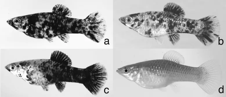
Figure 1. Black Amazons a) line I, b) line II, c) line III, and d) wildtype pigmented
P. formosa from line III/4
Figure 2. Metaphase spreads of P. formosa a) Spotted fish of Black Amazon line I, b)
wildtype pigmented with one microchromosome (specimen #1199) and c) spotted animal with
two different sized microchromosomes (specimen #1043), arrowhead, stable
microchromosome, arrow, “new” microchromosome. b) and c) are from the breeding
experiment to follow the inheritance of microchromosomes.
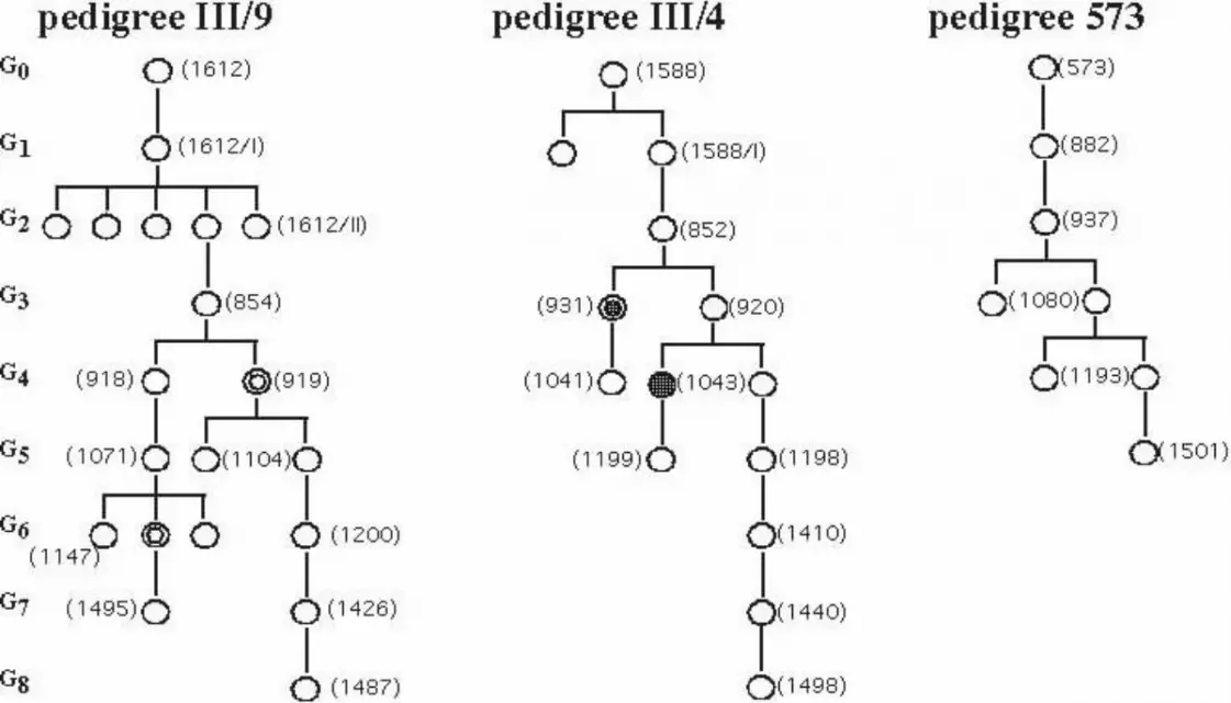
Figure 3. Pedigrees of animals analyzed for inheritance of microchromosomes. Open
circle, wildtype pigmented; filled circle, spotted; double circle, fish with additional
microchromosome. G0 – G8, generation number. Numbers in brackets are specimen codes
from the chromosomes analysis.
Figure 4. C-banding of metaphase chromosomes of a spotted Black Amazon line I (WLC
533) with two microchromosomes. Arrows indicate the dark staining of heterochromatin on
both microchromosomes.
Figure 5. Telomere staining of metaphase chromosomes of Black Amazon line II (WLC
922-25/IV) and the host species (Black Molly) with FITC labelled (CCCTAA)3 PNA
oligonucleotide. a) Metaphase with one microchromosome (arrow). b) Metaphase from the
same fish with two microchromosomes, only one of them being labelled with the telomere
probe. c) Mitotic chromosomes of a Black Molly male displaying specific telomere signals on
each end of all chromosomes.


Related Documents