3D Stabilization software for intravital imaging Iván Gómez Conde, David Olivieri, Carlos Tadokoro ([email protected]) March 1st, 2013

Stabi Tissue Software
Aug 03, 2015
Welcome message from author
This document is posted to help you gain knowledge. Please leave a comment to let me know what you think about it! Share it to your friends and learn new things together.
Transcript
3D Stabilization software for
intravital imaging
Iván Gómez Conde, David Olivieri, Carlos Tadokoro([email protected])
March 1st, 2013
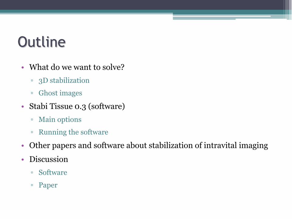
Outline
• What do we want to solve?
▫ 3D stabilization
▫ Ghost images
• Stabi Tissue 0.3 (software)
▫ Main options
▫ Running the software
• Other papers and software about stabilization of intravital imaging
• Discussion
▫ Software
▫ Paper

What do we want to solve?
Two photon microscopeManufacturer: Prairie (Model : Ultima )
Spleen in mice

Doing images of intravital tissue
1
2
3
4
-> 2.19 s
-> 2.19 s
-> 2.19 s
-> 2.19 s
The tissue is static
Stack of images in each cycle(4 slices)
Images captured in 1 cycle(Total time: 8.9 seconds)Cycle 1
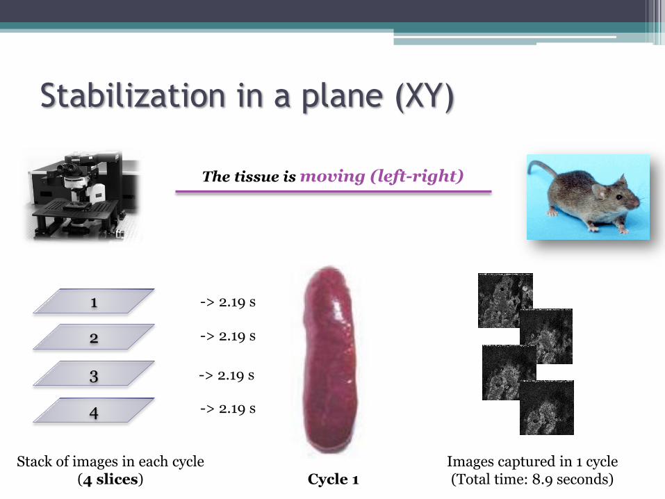
Stabilization in a plane (XY)
1
2
3
4
Stack of images in each cycle(4 slices)
-> 2.19 s
-> 2.19 s
-> 2.19 s
-> 2.19 s
Images captured in 1 cycle(Total time: 8.9 seconds)
The tissue is moving (left-right)
Cycle 1

Stabilization in Z
1
2
3
4
Stack of images in each cycle(4 slices)
-> 2.19 s
-> 2.19 s
-> 2.19 s
-> 2.19 s
Images captured in 1 cycle(Total time: 8.9 seconds)
The tissue is moving (up-down)
Cycle 1
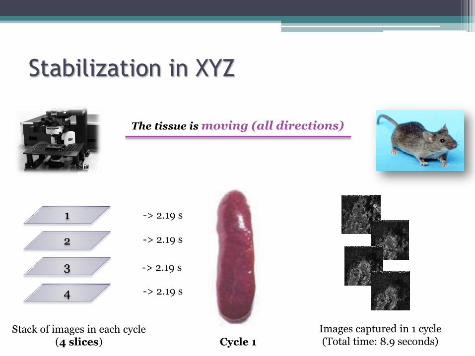
Stabilization in XYZ
1
2
3
4
-> 2.19 s
-> 2.19 s
-> 2.19 s
-> 2.19 s
The tissue is moving (all directions)
Stack of images in each cycle(4 slices)
Images captured in 1 cycle(Total time: 8.9 seconds)Cycle 1

Ghost images
1
2
Cycle 1
The tissue can be contracting
1
Ghost images
Cycle 2

Stabi Tissue Software 0.3
• 3D Stabilization software for intravital imaging of mouse organs
▫ Command-line software (http://sourceforge.net/projects/stabitissue/)
▫ User-interface software (building…)
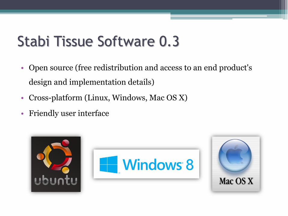
Stabi Tissue Software 0.3
• Open source (free redistribution and access to an end product's
design and implementation details)
• Cross-platform (Linux, Windows, Mac OS X)
• Friendly user interface
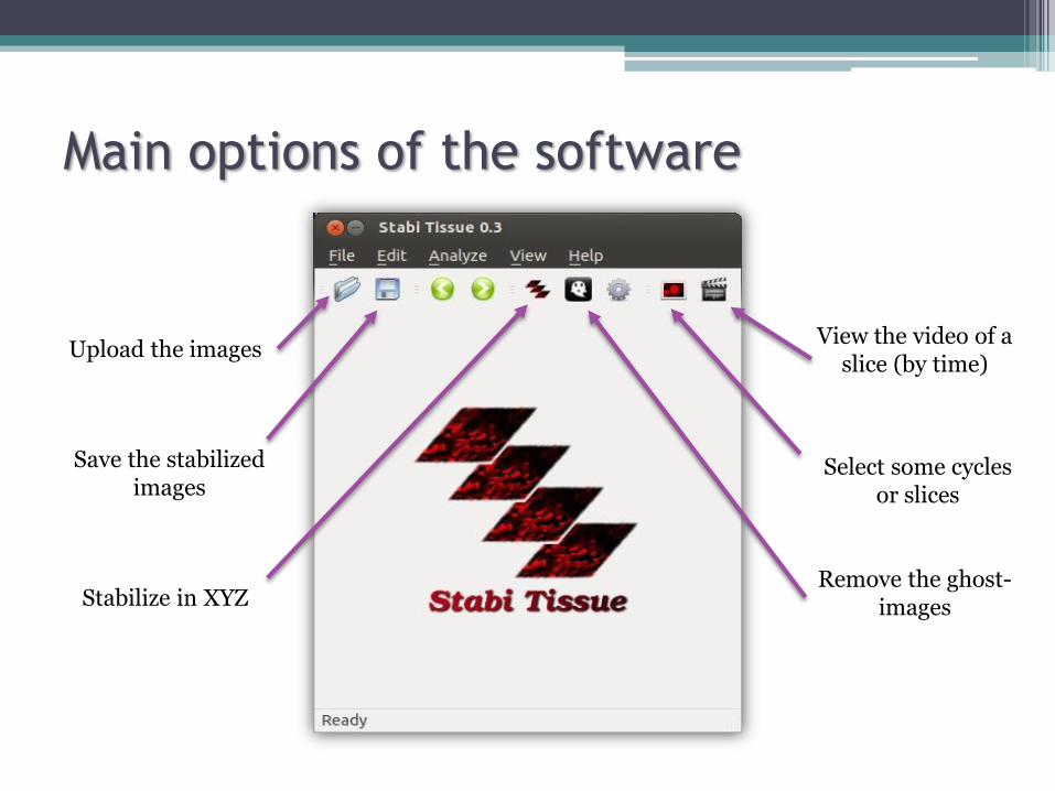
Main options of the software
Upload the images
Select some cyclesor slices
View the video of a slice (by time)
Stabilize in XYZ
Save the stabilizedimages
Remove the ghost-images

Upload images
• The user only needs to introduce:
▫ Folder
▫ Type of images (tif, tiff, jpeg…)
The software uses regular expressions to solve that problem:
"Infected-2x-389_Cycle052_CurrentSettings_Ch5_000022.tif“
"File_Infected-2x-388-1_t001_z004_c0002.tif"

Image formats of microscopes
• LOCI Bio-Formats of University of Wisconsin-Madison lets to
convert propietary microscopy data into a standard format
▫ Problems: written for Java and bindings for C++ (not for Python)
▫ Possible solution: external library or script
http://loci.wisc.edu/software/bio-formats

Select images (cycles/slices)
• The user can choose the images selecting with the mouse
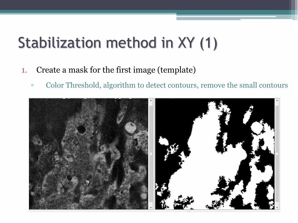
Stabilization method in XY (1)
1. Create a mask for the first image (template)
▫ Color Threshold, algorithm to detect contours, remove the small contours
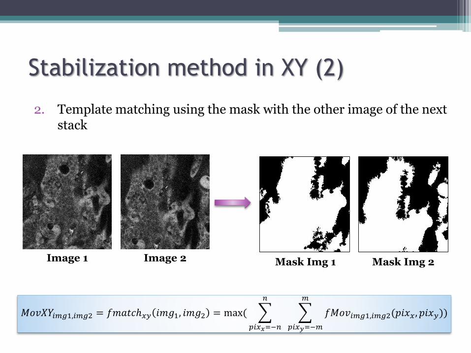
Stabilization method in XY (2)
2. Template matching using the mask with the other image of the next stack
𝑀𝑜𝑣𝑋𝑌𝑖𝑚𝑔1,𝑖𝑚𝑔2 = 𝑓𝑚𝑎𝑡𝑐ℎ𝑥𝑦 𝑖𝑚𝑔1, 𝑖𝑚𝑔2 = max(
𝑝𝑖𝑥𝑥=−𝑛
𝑛
𝑝𝑖𝑥𝑦=−𝑚
𝑚
𝑓𝑀𝑜𝑣𝑖𝑚𝑔1,𝑖𝑚𝑔2(𝑝𝑖𝑥𝑥 , 𝑝𝑖𝑥𝑦))
Image 1 Image 2 Mask Img 2Mask Img 1

Stabilization method in XY (2)
3. Find the maximum likelihood between the mask of the image 1 and the mask of the new image
Mask 1
Mask 2
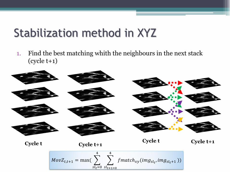
Stabilization method in XYZ
1. Find the best matching whith the neighbours in the next stack (cycle t+1)
Cycle t Cycle t+1Cycle t Cycle t+1
𝑀𝑜𝑣𝑍𝑡,𝑡+1 = max(
𝑠𝑙𝑡=0
4
𝑠𝑙𝑡+1=0
4
𝑓𝑚𝑎𝑡𝑐ℎ𝑥𝑦(𝑖𝑚𝑔𝑠𝑙𝑡 , 𝑖𝑚𝑔𝑠𝑙𝑡+1 ))
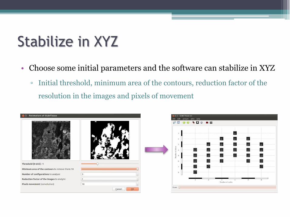
Stabilize in XYZ
• Choose some initial parameters and the software can stabilize in XYZ
▫ Initial threshold, minimum area of the contours, reduction factor of the
resolution in the images and pixels of movement

Ghost images
• Find the images that are similar to choose only one
▫ The software can choose automatically or the user can change the choice
The software will show therecognized ghost images

View video
• Selecting the slice that you want to see by time
▫ Change the speed
▫ See the different channels
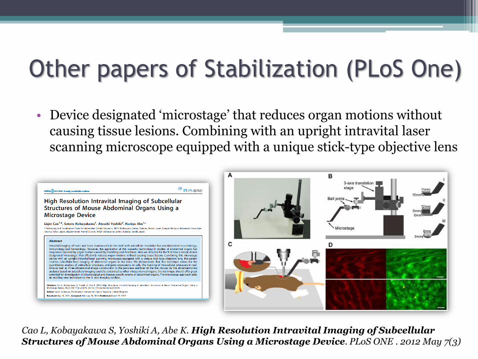
Other papers of Stabilization (PLoS One)
• Device designated ‘microstage’ that reduces organ motions without causing tissue lesions. Combining with an upright intravital laser scanning microscope equipped with a unique stick-type objective lens
Cao L, Kobayakawa S, Yoshiki A, Abe K. High Resolution Intravital Imaging of SubcellularStructures of Mouse Abdominal Organs Using a Microstage Device. PLoS ONE . 2012 May 7(3)
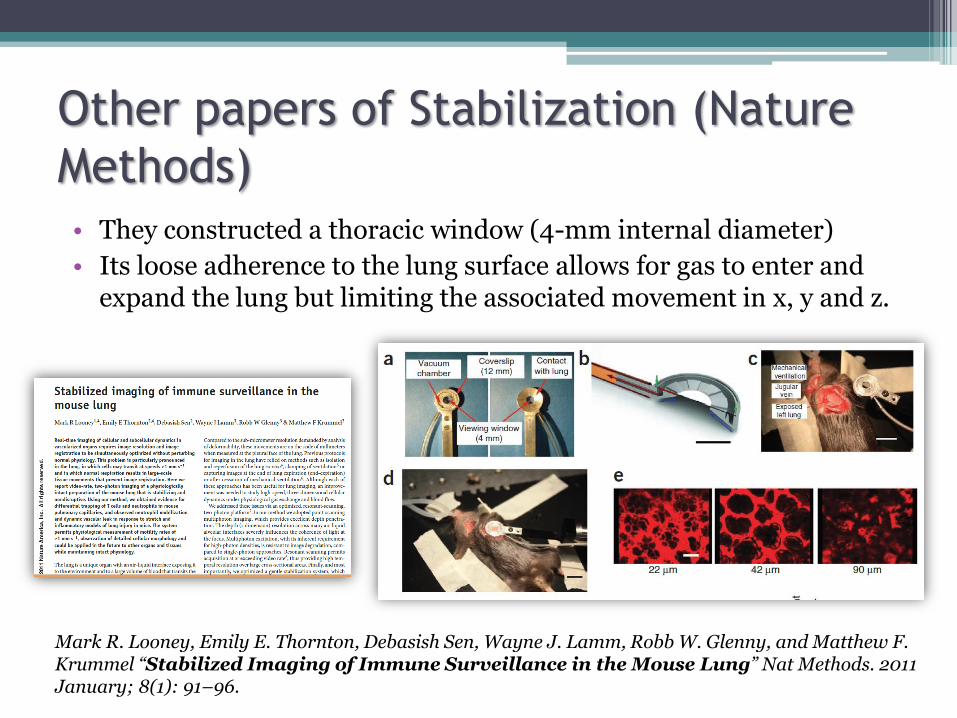
Other papers of Stabilization (Nature
Methods)• They constructed a thoracic window (4-mm internal diameter)
• Its loose adherence to the lung surface allows for gas to enter and expand the lung but limiting the associated movement in x, y and z.
Mark R. Looney, Emily E. Thornton, Debasish Sen, Wayne J. Lamm, Robb W. Glenny, and Matthew F. Krummel “Stabilized Imaging of Immune Surveillance in the Mouse Lung” Nat Methods. 2011 January; 8(1): 91–96.
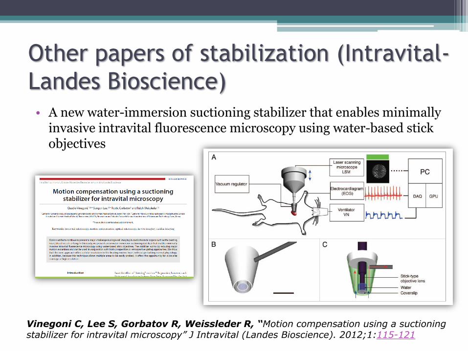
Other papers of stabilization (Intravital-
Landes Bioscience)• A new water-immersion suctioning stabilizer that enables minimally
invasive intravital fluorescence microscopy using water-based stickobjectives
Vinegoni C, Lee S, Gorbatov R, Weissleder R, “Motion compensation using a suctioningstabilizer for intravital microscopy” J Intravital (Landes Bioscience). 2012;1:115-121
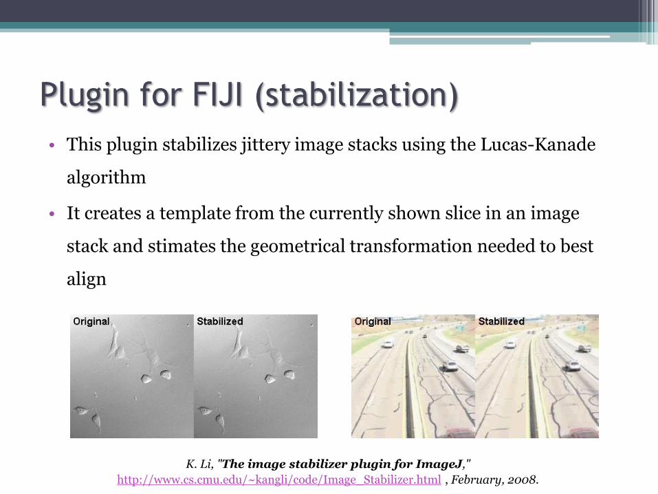
Plugin for FIJI (stabilization)
• This plugin stabilizes jittery image stacks using the Lucas-Kanade
algorithm
• It creates a template from the currently shown slice in an image
stack and stimates the geometrical transformation needed to best
align
K. Li, "The image stabilizer plugin for ImageJ,"
http://www.cs.cmu.edu/~kangli/code/Image_Stabilizer.html , February, 2008.

Image stabilizer FIJI vs Stabi Tissue
• The Stabi Tissue software had been tested with a video against the
plugin of FIJI. (The result is only with a XY stabilization)
Image StabilizerFIJI
Stabi Tissuesoftware
Video withmovement
Luciana Vieira de Moraes, Carlos Eduardo Tadokoro, Iván Gómez-Conde, David N. Olivieri, Carlos Penha-Gonçalves “Intravital Placenta Imaging Reveals Microcirculatory Dynamics Impact on Sequestration
and Phagocytosis of Plasmodium-Infected Erythrocytes” PLoS Pathog. 2013 January; 9(1): e1003154
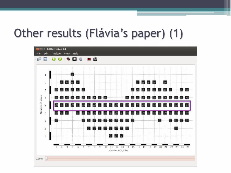
Other results (Flávia’s paper) (1)

Other results (Flávia’s paper) (2)
Stabi Tissue softwareStabilization XYZ
Video withmovement in XYZ
Flávia A Lima, Iván Gómez-Conde, Paula A Videira, Cláudio R Marinho, David N Olivieri, Carlos Eduardo Tadokoro, “Intravital microscopy technique to study parasite dynamics in the labyrinth layer of the mouse
placenta” Submitted to Parasitology International (Elsevier)

Aknowledgements
• University of Vigo
▫ David Olivieri Cecchi
• Instituto Gulbenkian de Ciência
▫ Carlos Tadokoro
▫ Henrique Silva
▫ Flavia Afonso Lima
▫ Susana Caetano
Related Documents