Spontaneous Calcium Oscillations Regulate Human Cardiac Progenitor Cell Growth João Ferreira-Martins 1,* , Carlos Rondon-Clavo 1,* , Derin Tugal 1 , Justin A Korn 1 , Roberto Rizzi 1 , Maria Elena Padin-Iruegas 1 , Sergio Ottolenghi 2 , Antonella De Angelis 1 , Konrad Urbanek 1 , Noriko Iwata 1 , Domenico D’Amario 1 , Toru Hosoda 1 , Annarosa Leri 1 , Jan Kajstura 1 , Piero Anversa 1 , and Marcello Rota 1 1 Departments of Anesthesia and Medicine and Cardiovascular Division, Brigham & Women’s Hospital, Harvard Medical School, Boston, MA 02115, USA 2 Department of Biotechnology and Bioscience, University of Milano-Bicocca, Milan, Italy Abstract Rationale—The adult heart possesses a pool of progenitor cells stored in myocardial niches but the mechanisms involved in the activation of this cell compartment are currently unknown. Objective—Ca 2+ promotes cell growth raising the possibility that changes in intracellular Ca 2+ initiate division of c-kit-positive human cardiac progenitor cells (hCPCs) and determine their fate. Methods and Results—Ca 2+ oscillations were identified in hCPCs and these events occurred independently from coupling with cardiomyocytes or the presence of extracellular Ca 2+ . These findings were confirmed in the heart of transgenic mice in which EGFP was under the control of the c-kit-promoter. Ca 2+ oscillations in hCPCs were regulated by the release of Ca 2+ from the ER through activation of inositol 1,4,5-triphosphate receptors (IP3Rs) and the re-uptake of Ca 2+ by the sarco/ endoplasmic reticulum Ca 2+ pump (SERCA). IP3Rs and SERCA were highly expressed in hCPCs while ryanodine receptors were not detected. Although Na + -Ca 2+ exchanger, store-operated Ca 2+ - channels and plasma membrane Ca 2+ -pump were present and functional in hCPCs, they had no direct effects on Ca 2+ oscillations. Conversely, Ca 2+ oscillations and their frequency markedly increased with ATP and histamine which activated purinoceptors and histamine-1 receptors highly expressed in hCPCs. Importantly, Ca 2+ oscillations in hCPCs were coupled with the entry of cells into the cell cycle and BrdUrd incorporation. Induction of Ca 2+ oscillations in hCPCs prior to their intramyocardial delivery to infarcted hearts was associated with enhanced engraftment and expansion of these cells promoting the generation of a large myocyte progeny. Conclusion—IP3R-mediated Ca 2+ mobilization control hCPC growth and their regenerative potential. Keywords human cardiac progenitor cells; calcium oscillations; cell growth The recognition that the adult heart in animals and humans possesses a pool of stem/progenitor cells 1-3 has raised the critical question concerning the mechanisms involved in the activation Address for Correspondence: Marcello Rota, PhD, Departments of Anesthesia and Medicine, and Cardiovascular Division, Brigham & Women’s Hospital, Harvard Medical School, Boston, MA 02115. Fax: 617- 264-6320; Phone: 617-525-8164; [email protected]. * JFM and CRC contributed equally to this work Disclosures None NIH Public Access Author Manuscript Circ Res. Author manuscript; available in PMC 2010 October 9. Published in final edited form as: Circ Res. 2009 October 9; 105(8): 764–774. doi:10.1161/CIRCRESAHA.109.206698. NIH-PA Author Manuscript NIH-PA Author Manuscript NIH-PA Author Manuscript

Welcome message from author
This document is posted to help you gain knowledge. Please leave a comment to let me know what you think about it! Share it to your friends and learn new things together.
Transcript

Spontaneous Calcium Oscillations Regulate Human CardiacProgenitor Cell Growth
João Ferreira-Martins1,*, Carlos Rondon-Clavo1,*, Derin Tugal1, Justin A Korn1, RobertoRizzi1, Maria Elena Padin-Iruegas1, Sergio Ottolenghi2, Antonella De Angelis1, KonradUrbanek1, Noriko Iwata1, Domenico D’Amario1, Toru Hosoda1, Annarosa Leri1, JanKajstura1, Piero Anversa1, and Marcello Rota11Departments of Anesthesia and Medicine and Cardiovascular Division, Brigham & Women’sHospital, Harvard Medical School, Boston, MA 02115, USA2Department of Biotechnology and Bioscience, University of Milano-Bicocca, Milan, Italy
AbstractRationale—The adult heart possesses a pool of progenitor cells stored in myocardial niches but themechanisms involved in the activation of this cell compartment are currently unknown.
Objective—Ca2+ promotes cell growth raising the possibility that changes in intracellular Ca2+
initiate division of c-kit-positive human cardiac progenitor cells (hCPCs) and determine their fate.
Methods and Results—Ca2+ oscillations were identified in hCPCs and these events occurredindependently from coupling with cardiomyocytes or the presence of extracellular Ca2+. Thesefindings were confirmed in the heart of transgenic mice in which EGFP was under the control of thec-kit-promoter. Ca2+ oscillations in hCPCs were regulated by the release of Ca2+ from the ER throughactivation of inositol 1,4,5-triphosphate receptors (IP3Rs) and the re-uptake of Ca2+ by the sarco/endoplasmic reticulum Ca2+ pump (SERCA). IP3Rs and SERCA were highly expressed in hCPCswhile ryanodine receptors were not detected. Although Na+-Ca2+ exchanger, store-operated Ca2+-channels and plasma membrane Ca2+-pump were present and functional in hCPCs, they had no directeffects on Ca2+ oscillations. Conversely, Ca2+ oscillations and their frequency markedly increasedwith ATP and histamine which activated purinoceptors and histamine-1 receptors highly expressedin hCPCs. Importantly, Ca2+ oscillations in hCPCs were coupled with the entry of cells into the cellcycle and BrdUrd incorporation. Induction of Ca2+ oscillations in hCPCs prior to theirintramyocardial delivery to infarcted hearts was associated with enhanced engraftment and expansionof these cells promoting the generation of a large myocyte progeny.
Conclusion—IP3R-mediated Ca2+ mobilization control hCPC growth and their regenerativepotential.
Keywordshuman cardiac progenitor cells; calcium oscillations; cell growth
The recognition that the adult heart in animals and humans possesses a pool of stem/progenitorcells1-3 has raised the critical question concerning the mechanisms involved in the activation
Address for Correspondence: Marcello Rota, PhD, Departments of Anesthesia and Medicine, and Cardiovascular Division, Brigham &Women’s Hospital, Harvard Medical School, Boston, MA 02115. Fax: 617- 264-6320; Phone: 617-525-8164;[email protected].*JFM and CRC contributed equally to this workDisclosures None
NIH Public AccessAuthor ManuscriptCirc Res. Author manuscript; available in PMC 2010 October 9.
Published in final edited form as:Circ Res. 2009 October 9; 105(8): 764–774. doi:10.1161/CIRCRESAHA.109.206698.
NIH
-PA Author Manuscript
NIH
-PA Author Manuscript
NIH
-PA Author Manuscript
of this cell compartment and the modulation of cardiac homeostasis and repair. During thecourse of life before the manifestations of myocardial aging become apparent,4 dyingparenchymal cells are continuously replaced by newly formed myocytes5,6 through activationand commitment of quiescent cardiac progenitor cells (CPCs) stored in myocardial niches.7,8 However, the signals responsible for the initiation of the cell cycle in CPCs, cardiomyocytegeneration and preservation of the steady state of the organ are currently unknown. Calciumhas two fundamental functions in the heart: it activates growth processes9,10 and modulatesthe mechanical behavior of cardiomyocytes.11,12 Critical for understanding physiological cellturnover and myocardial regeneration following injury is the identification of the mechanismsby which CPCs divide and acquire the myocyte phenotype. Changes of calcium levels in CPCsmay occur and trigger a cascade of events that dictate their ultimate fate. Therefore, theobjectives of the current study were: 1. to determine the pathways that regulate intracellularCa2+ in human CPCs (hCPCs); 2. to establish whether Ca2+ oscillations in hCPCs conditioncell replication; and 3. to assess whether Ca2+ oscillations are intrinsic to the cells or aretriggered by interaction of hCPCs with cardiomyocytes. This cell-to-cell communication mayfavor the translocation of Ca2+ from myocytes to quiescent hCPCs initiating a cellular growthresponse. Moreover, transmembrane Ca2+ fluxes may contribute to rapid and transient rise incytosolic Ca2+ promoting the entry of hCPCs into the cell cycle. These variables include afunctional endoplasmic reticulum (ER) where Ca2+ is stored, the activity of ER channels thatpromote Ca2+ release, and the membrane systems modulating the exchange of Ca2+ with theextracellular compartment.
MethodshCPCs were isolated from myocardial specimens obtained from patients who underwentcardiac surgery. Cytosolic Ca2+ levels in cultured hCPCs were measured utilizing the Ca2+
indicator Fluo-3 and two-photon microscopy. Cell proliferation in vivo and in vitro wasevaluated by BrdUrd incorporation. Results are shown as mean±SEM. An expanded Materialsand Methods section can be found in the online data supplement available athttp://circres.ahajournals.org.
ResultsIntracellular Ca2+ in hCPCs
Changes in [Ca2+]i occur in excitable and non-excitable cells raising the possibility that asimilar phenomenon is present in hCPCs and may have a functional role. Thus, hCPCs wereloaded with the Ca2+ sensitive dye Fluo-3 and the intensity of the fluorescent signal wasmonitored over a period of ~30 minutes. During this interval, 79% hCPCs maintained stablelevels of [Ca2+]i while 21% displayed one or more consecutive Ca2+ oscillations. Repetitiveevents were restricted to a small percentage of cells and were comparable in amplitude andduration (Figure 1A through 1C). The fraction of hCPCs displaying Ca2+ oscillations increasedwith time up to 2 hours although the frequency of these episodes remained low (Figure 1D).These cells were all positive for the stem cell antigen c-kit (Supplemental Figure I). Ca2+
oscillations increased in hCPCs at the G1-S phase transition but decreased at G2-M (Figure 1E and Supplemental Figure II).
Cell-to-Cell Interaction and Ca2+ Oscillations in hCPCsThe next objective was to establish whether Ca2+ oscillations in hCPCs are modulated at thesingle cell level or are mediated by adjacent cells. hCPCs are nested in myocardial niches andconnexins are found between hCPCs and myocytes which operate as supporting cells.3,7 Theseintercellular communications may account for the generation of Ca2+ oscillations in hCPCs, aprocess that is commonly observed in cardiomyocytes (Supplemental Figure III and Movie 1
Ferreira-Martins et al. Page 2
Circ Res. Author manuscript; available in PMC 2010 October 9.
NIH
-PA Author Manuscript
NIH
-PA Author Manuscript
NIH
-PA Author Manuscript
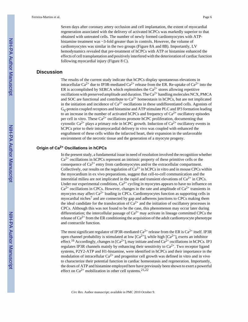
in the online data supplement). Therefore, to identify the origin of Ca2+ oscillations in hCPCs,these cells were cultured alone or together with neonatal cardiomyocytes. Initially, dye transferassays were performed3,7,13 to document the formation of functional gap junctions betweenhCPCs. The fluorescent dye cascade blue was microinjected in individual hCPCs and foundto rapidly migrate to neighboring cells through gap junction channels expressing connexin 43(Figure 2A through 2E). However, the high molecular weight rhodamine-labeled dextran,injected simultaneously with cascade blue, failed to translocate to adjacent hCPCs.Additionally, DiI labeled hCPCs loaded with calcein were cultured with untreated cells. After~12 hours, calcein was detected in unlabeled hCPCs structurally connected to DiI-calcein-positive cells (Supplemental Figure IV). Intracellular Ca2+ was then measured before and afterexposure of hCPCs to the connexin hemi-gap-junction channel blocker octanol. Octanol didnot affect the frequency and properties of Ca2+ oscillations in hCPCs (Figure 2F and 2G).
Subsequently, the effect of Ca2+ cycling in myocytes on hCPC function was assessed by platingtogether DiI-labeled hCPCs and unlabeled cardiomyocytes. In spite of the presence offunctional gap junctions between these two cell populations (Figure 3A and SupplementalFigure V), spontaneous or electrically stimulated Ca2+ transients in myocytes had no detectableconsequence on [Ca2+]i of hCPCs (Figure 3B and 3C). In fact, Ca2+ oscillations in hCPCspersisted in the presence of cadmium which abolished Ca2+ transients in myocytes.
Cell-to-Cell Interaction and Ca2+ Oscillations in Mouse CPCsTo strengthen these in vitro results, a transgenic mouse model in which EGFP was under thecontrol of the c-kit promoter14 was employed to test whether Ca2+ cycling in myocytes triggersCa2+ oscillations in CPCs in situ within the myocardium. Preliminary studies were conductedto evaluate whether EGFP-positive mouse CPCs (mCPCs) in vitro showed spontaneousCa2+ oscillations, mimicking the behavior of hCPCs. The percentage of mCPCs exhibitingCa2+ oscillation was comparable to that measured in human cells. Similarly, the rate of theseevents and their duration did not differ in these two cell classes while the amplitude was largerin mCPCs (Figure 3D and 3E).
The heart of these transgenic mice was then examined ex vivo by two-photon microscopy3,13 following perfusion of the coronary circulation with the Ca2+ indicator Rhod-2. Thepossibility that EGFP may interfere with the detection of Rhod-2 was excluded in preliminarystudies conducted in EGFP-positive mouse myocytes (Supplemental Figure VI). Based onthese observations, the mouse heart was stimulated at 1 Hz and the Ca2+ levels in EGFP-positive mCPCs were found not to be affected by the changes in Ca2+ transients in neighboringcardiomyocytes (Figure 3F and 3G). Thus, human and mouse CPCs appear to possess anintracellular Ca2+ regulatory system which is independent from that of terminally-differentiated parenchymal cells.
Intracellular Ca2+ Control in hCPCsWe then determined whether activation of inositol 1,4,5-triphosphate receptors (IP3Rs) and/or ryanodine receptors (RyRs) resulted in the release of Ca2+ from the ER and Ca2+ oscillatoryevents. Moreover, sarco/endoplasmic reticulum Ca2+-ATPase (SERCA) is responsible for there-uptake of Ca2+ into the ER and restoration of Ca2+ stores.10,11 Before conducting thefunctional studies, quantitative RT-PCR and immunolabeling were employed to document inhCPCs the presence of transcripts and proteins for IP3Rs and SERCA. Both IP3Rs and SERCAwere highly expressed in hCPCs. However, RyRs were not identified in these cells (Figure 4Athrough 4D).
Subsequently, IP3 binding to IP3Rs was enhanced by thimerosal and this intervention markedlyincreased the number of hCPCs displaying Ca2+ oscillations and the frequency of Ca2+
Ferreira-Martins et al. Page 3
Circ Res. Author manuscript; available in PMC 2010 October 9.
NIH
-PA Author Manuscript
NIH
-PA Author Manuscript
NIH
-PA Author Manuscript

oscillatory episodes per cell (Figure 4E and 4F). An opposite response was observed byinhibition of IP3R function with 2-amino-ethoxydiphenyl borate (2-APB) or xestospongin-Cor attenuation of IP3 formation by phospholipase-C (PLC) blockade with U-73122 (Figure 4Gand 4H). Similarly, inhibition of SERCA with cyclopiazonic acid (CPA) reduced the fractionof activated hCPCs (Figure 4I and 4J). Additionally, Ca2+ oscillations were smaller inamplitude and prolonged in duration, pointing to SERCA as a relevant component of hCPCfunction. Consistent with the lack of identify RyRs in hCPCs, their agonists ryanodine andcaffeine had no effects on Ca2+ oscillations; the number of active cells and the frequency,amplitude and duration of Ca2+ events did not differ from baseline (Figure 4K through 4N).These data point to the IP3-IP3R system and SERCA as the predominant modulators ofCa2+ in hCPCs.
ATP-specific P2-purinoceptors (P2Y2) and histamine (H1) receptors are Gq-protein coupledreceptors that are implicated in the release of intracellular Ca2+ from the ER by activation ofPLC, IP3 synthesis and, ultimately, IP3R binding.15,16 P2Y2 and H1 receptors were expressedat the mRNA and protein level in hCPCs (Figure 5A through 5C), suggesting that they maybe implicated in Ca2+ cycling of these progenitor cells. In the presence of ATP or histamine,a more than 3-fold increase in the number of hCPCs exhibiting oscillations was detected.Similarly, the number of events per cell markedly increased while the amplitude and durationof Ca2+ elevations did not change (Figure 5D through 5G). Inhibition of IP3Rs or IP3 formationprevented the effects of ATP and histamine on Ca2+ mobilization (Supplemental Figure VII).
Since PLC-β3 may be modulated by P2Y2 receptor activation,17 the expression of PLC-βsubunits was evaluated in hCPCs. Additionally, the functional role of PLC-β3 in mediatingintracellular Ca2+ mobilization upon ATP stimulation was established. PLC-β3 mRNA inhCPCs was significantly higher than that of the other subunits (Supplemental Figure VIII, Athrough C). Thus, siRNA strategy was employed to downregulate PLC-β3 in hCPCs prior totheir stimulation with ATP. This intervention markedly attenuated the ability of ATP todramatically increase the pool of hCPCs displaying Ca2+ oscillations (Supplemental FigureVIII, D and E).
Although IP3R-mediated Ca2+ mobilization from the ER is responsible for the generation ofCa2+oscillations in hCPCs, transmembrane Ca2+ fluxes maybe operative contributing tointracellular calcium cycling. Exposure of hCPCs to Ca2+ free medium did not alter thefrequency and characteristics of Ca2+ oscillations (Supplemental Figure IX), strengthening therole of intracellular stores as the source of Ca2+ for oscillatory events. Immunolabeling andPCR data, together with patch clamp and cytosolic Ca2+ imaging (Supplemental Figures X andXI), revealed that store operated channels (SOC), Na+-Ca2+ exchanger (NCX) and plasmamembrane Ca2+ pump (PMCA) were functional in hCPCs. However, these systems did notappear to participate in the generation of Ca2+ oscillations in these primitive cells.
Ca2+ Oscillations and hCPC GrowthTo evaluate the functional import of Ca2+ oscillations in hCPC replication, these cells werecultured in serum free medium for 24 hours and were exposed to either ATP or histamine toinduce Ca2+ release from the ER and oscillatory events. BrdUrd was added to the medium andits incorporation in hCPCs was measured 24 hours later. To exclude the potential confoundingeffect of cell death on the evaluation of cell proliferation, apoptosis was also determined. Witheither ATP or histamine, a nearly 2-fold increase in BrdUrd labeling of hCPCs was detected(Figure 6A and 6B). Conversely, inhibition of Ca2+ release from the ER led to a decrease inBrdUrd incorporation in hCPCs to values lower than those seen at baseline (Figure 6A through6C). Moreover, purinergic stimulation failed to promote proliferation of hCPCs when PLC-β3 was downregulated and the P2Y2-IP3R axis was disrupted (Supplemental Figure XII). ATP
Ferreira-Martins et al. Page 4
Circ Res. Author manuscript; available in PMC 2010 October 9.
NIH
-PA Author Manuscript
NIH
-PA Author Manuscript
NIH
-PA Author Manuscript
and histamine had no influence on hCPC apoptosis (Figure 6D and 6E), suggesting thatincreases in Ca2+ oscillations were not coupled with the activation of the cell death program.
To strengthen the possibility of a cause and effect relationship between Ca2+ oscillations andcell cycle activation in hCPCs, the impact of a well-established activator of progenitor celldivision, insulin-like growth factor-1 (IGF-1) was determined. Resident cardiac progenitorsexpress IGF-1 receptors (Figure 6F and 6G) and synthesize and secrete the ligand.6 IGF-1increased the percentage of hCPCs showing Ca2+ oscillations by nearly 3-fold (Figure 6H and6I). Similarly, there was an increase in the number of Ca2+ oscillations per cell while theamplitude and duration of Ca2+ events remained essentially constant. When Ca2+ oscillationswere blocked, the growth promoting effects of IGF-1 on hCPCs were completely prevented.IGF-1 increased hCPC proliferation by ~2-fold and blockade of Ca2+ release from the ERdecreased cell replication to levels below baseline values (Figure 6J). Similarly, Ca2+
oscillations mediated by IGF-1 were abrogated by PLC and IP3R antagonists (supplementalFigure XIII).
To establish whether Ca2+ oscillations in hCPCs favor the acquisition of the myocyte lineage,these cells were cultured in differentiating medium3 in the absence or presence of ATP orhistamine. After one week, the fraction of primitive cells expressing α-sarcomeric actin wascomparable in stimulated and non-stimulated hCPCs (Supplemental Figure XIV), suggestingthat these agents did not impact on the differentiation of hCPCs into the myocyte phenotype.
Ca2+ Oscillations in hCPCs and Myocardial RegenerationIn both animals and humans, shortly after ischemic myocardial injury, there is an increase ofresident progenitors mostly restricted to the border zone of the infarcted heart.1 These cellsrapidly acquire the myocyte lineage and result in small foci of cardiac repair.18 The possibilitythat factors naturally released in the damaged area facilitate this process was confirmed herein our transgenic mouse model in which EGFP-labeling made rather easy the identification ofc-kit-positive CPCs. Myocardial infarction at 2 days led to a 15-fold and 5-fold increase inCPCs in the region bordering and remote from the infarction, respectively (SupplementalFigure XV).
However, spontaneous regeneration is severely limited and only a minimal fraction of a varietyof progenitor cells delivered to the infarcted myocardium survives and integrates in theunfavorable environment of the necrotic tissue.13,19 Activation of hCPCs with ATP andhistamine may enhance their engraftment, growth and formation of a myocyte progeny. EGFP-labeled hCPCs were exposed to ATP or histamine 30 min prior to their injection in the areabordering an acute infarct in immunosuppressed mice. Untreated hCPCs were used as control.All cell preparations were serum-starved for 24 hours before ATP or histamine exposure.Animals were examined 2 days later when cell engraftment is completed, cell death is markedlyattenuated and the number of cells available for cardiac repair is established.13,20 Mice wereexposed to BrdUrd to obtain cumulative values of cell regeneration. Additional groups of micewere sacrificed at 7 days to asses the impact of this protocol on myocardial regeneration andcardiac function.
Of the 60 × 103 hCPCs injected in each heart, 7,000 EGFP-positive cells were found in controlhearts while nearly 19,000 cells (P<0.002) were detected in hearts in which hCPCs were treatedwith ATP or histamine. Cell engraftment was confirmed by the detection of connexin 43 andN-cadherin at the interface of hCPCs and spared myocytes (Supplemental Figure XVI).Moreover, the number of EGFP-positive cells labeled by BrdUrd was 10-12-fold higher ininfarcts injected with ATP or histamine activated hCPCs (Figure 7A through 7C). Mostimportantly, the aggregate number of EGFP-positive cells expressing the myocyte transcriptionfactors Nkx2.5 was ~4-fold larger in hearts treated with cells exposed to ATP or histamine.
Ferreira-Martins et al. Page 5
Circ Res. Author manuscript; available in PMC 2010 October 9.
NIH
-PA Author Manuscript
NIH
-PA Author Manuscript
NIH
-PA Author Manuscript
Seven days after coronary artery occlusion and cell implantation, the extent of myocardialregeneration associated with the delivery of activated hCPCs was markedly superior to thatobtained with untreated cells. The number of newly formed cardiomyocytes with ATP-histamine treatment was ~3-fold greater than in controls. However, the volume ofcardiomyocytes was similar in the two groups (Figure 8A and 8B). Importantly, LVhemodynamics revealed that pre-treatment of hCPCs with ATP or histamine enhanced theeffects of cell transplantation and positively interfered with the deterioration of cardiac functionfollowing myocardial injury (Figure 8 C).
DiscussionThe results of the current study indicate that hCPCs display spontaneous elevations inintracellular Ca2+ due to IP3R-mediated Ca2+ release from the ER. Re-uptake of Ca2+ into theER is accomplished by SERCA which replenishes the Ca2+ stores allowing repetitiveoscillations with preserved amplitude and duration. The Ca2+ handling molecules NCX, PMCAand SOC are functional and contribute to Ca2+ homeostasis in hCPCs, but are not implicatedin the initiation and incidence of Ca2+ oscillations in these undifferentiated cells. Agonists ofGq-protein coupled receptors and histamine and ATP stimulate PLC and IP3 formation leadingto an increase in the number of activated hCPCs and frequency of Ca2+ oscillatory episodesper cell in vitro. These Ca2+ oscillations promote hCPC proliferation, documenting thatcytosolic Ca2+ plays a primary role in hCPC growth. Induction of Ca2+ oscillatory events inhCPCs prior to their intramyocardial delivery in vivo was coupled with enhanced theengraftment of these cells within the infarcted heart, their expansion in the unfavorableenvironment of the necrotic tissue and the generation of a myocyte progeny.
Origin of Ca2+ Oscillations in hCPCsIn the present study, a fundamental issue in need of resolution involved the recognition whetherCa2+ oscillations in hCPCs represent an intrinsic property of these primitive cells or theconsequence of Ca2+ entry from cardiomyocytes and/or the extracellular compartment.Collectively, our results on the regulation of Ca2+ in hCPCs in vitro and in mouse CPCs withinthe myocardium in ex vivo preparations, suggest that cell-to-cell communication and theinterstitial milieu are not implicated in the rapid and transient elevations of Ca2+ in CPCs.Under our experimental conditions, Ca2+ cycling in myocytes appears to have no influence onCa2+ oscillations in CPCs. However, changes in the rate and amplitude of Ca2+ transients inmyocytes may affect Ca2+ loading in CPCs. Cardiomyocytes function as supporting cells inmyocardial niches7 and are connected by gap and adherens junctions to CPCs making themthe ideal candidate for the translocation of Ca2+ and the initiation of oscillatory processes inCPCs. Although this was not found to be the case, this phenomenon may occur later duringdifferentiation; the intercellular passage of Ca2+ may activate in lineage committed CPCs therelease of Ca2+ from the ER conditioning the acquisition of the adult cardiomyocyte phenotypeand contractile function.
The most significant regulator of IP3R-mediated Ca2+ release from the ER is Ca2+ itself. IP3Ropen channel probability is stimulated at low [Ca2+]i while high [Ca2+]i exerts an inhibitoreffect.16 Accordingly, changes in [Ca2+]i may initiate and end Ca2+ oscillations in hCPCs. IP3regulates IP3R channels mainly by enhancing their sensitivity to Ca2+. Two receptor ligandsystems, P2Y2-ATP and H1-histamine, were identified in hCPCs and their importance in themodulation of intracellular Ca2+ and progenitor cell growth was defined in vitro and in vivoto characterize their potential function in cardiac homeostasis and regeneration. Importantly,the doses of ATP and histamine employed here have previously been shown to exert a powerfuleffect on Ca2+ mobilization in other cell systems.21,22
Ferreira-Martins et al. Page 6
Circ Res. Author manuscript; available in PMC 2010 October 9.
NIH
-PA Author Manuscript
NIH
-PA Author Manuscript
NIH
-PA Author Manuscript

In physiological conditions, cell loss by normal wear and tear may result in an increase in thelocal level of ATP15 leading to Ca2+ oscillations in neighboring hCPCs which in turn activatecell replication and expansion. Additionally, the release of ATP from synaptic vesicles interminal nerves15 may produce a comparable role in the hCPC compartment. These twomechanisms of ATP accumulation in the interstitial space are enhanced by prolongedmyocardial ischemia and myocyte death.15,23 Cardiac pathology potentiates the load on thespared myocardium and mechanical stretch further enhances exocytosis of ATP fromcardiomyocytes.15 In humans and animals, myocardial infarction is associated with CPCdivision and the generation of functionally competent cardiomyocytes,1 supporting the notionthat ATP-mediated CPC growth may be critical for cardiac repair.
Mast cells are the predominant source of histamine in the myocardium.24 The number of mastcells and CPCs is comparable in the rodent and human heart.3,7,25,26 Additionally, tissuedamage and inflammation recruit mast cells and CPCs, suggesting that histamine released frommast cells may be implicated in the activation of cardiac progenitors and the creation ofmyocytes. ATP is formed largely by cardiomyocytes that represent ~85% of the myocardiumwhile histamine is synthesized by a very small number of mast cells, ~2-3/mm2 of tissue.15,25,26 However, extracellular ATP is rapidly degraded to inactive ADP. Conversely, histaminehas a longer half-life time,15,27 indicating that these two molecules may have complementaryfunction in the modulation of hCPC function.
As shown here for hCPCs, IP3R-mediated Ca2+ mobilization from the ER has been reportedin human mesenchymal stem cells28 and mouse embryonic stem cells.29,30 In both cases, theincreases in intracellular Ca2+ have been linked to cell growth and lineage specification.Embryonic stem cells and hCPCs differentiate into cardiomyocytes and the characteristics ofCa2+ cycling in these cells is regulated by the Ca2+-induced Ca2+-release mechanism thatcontrols myocyte mechanics and ventricular function in vivo.11 Whether mesenchymal stemcells have a similar capacity is currently debatable.
IGF-1 and Ca2+ Oscillations in hCPCsThe function of IGF-1 is largely mediated by binding to the receptor tyrosine kinase IGF-1R.Phosphorylation of IGF-1R leads to recruitment of the insulin receptor substrate protein 1(IRS-1) that modulates the effects of IGF-1R on cellular responses in the heart. The recruitmentof IRS-1 upregulates PI3 kinase which phosphorylates Akt; Akt activation favors celldifferentiation, hypertrophy or proliferation.31,32 IRS-1 also promotes the interaction of Ras,Raf and ERK which may lead to cellular hypertrophy or division.33 Surprisingly, in the currentstudy, IGF-1 induced Ca2+ oscillatory episodes in hCPCs through the activation of IP3Rs andthe release of Ca2+ from the ER. Whether this was a direct effect or was mediated by thegeneration of IP3 is currently unclear. However, the mitogenic properties of IGF-1 appear tobe mediated, at least in part, by the release of Ca2+ from the ER, strengthening the notion thatCa2+ mobilization via IP3R is involved in cell cycle progression and growth of hCPCs.
Ca2+ Oscillations and hCPC In VivoA critical component of cell therapy is related to the recognition of the variables implicated inthe engraftment and expansion of the delivered cells within the damaged myocardium.34 Thefunction of progenitor cells is determined by causes inherent to the cells and risk factors forcardiovascular diseases. The former includes the telomere-telomerase axis, DNA damage andthe expression of genes implicated in the forced entry of cells into in an irreversible quiescentstate and/or activation of the endogenous cell death program.6,35 The latter involves severalpathologic states such as diabetes, hypertension, coronary artery disease, valvular defects,dilated cardiomyopathy and myocardial aging.34
Ferreira-Martins et al. Page 7
Circ Res. Author manuscript; available in PMC 2010 October 9.
NIH
-PA Author Manuscript
NIH
-PA Author Manuscript
NIH
-PA Author Manuscript

Supplementary MaterialRefer to Web version on PubMed Central for supplementary material.
AcknowledgmentsSources of Funding This work was supported by NIH grants and BWH-BRI. JFM. was supported by the Ministry ofScience of Portugal and DT is the recipient of a Sarnoff fellowship.
References1. Anversa P, Kajstura J, Leri A, Bolli R. Life and death of cardiac stem cells: a paradigm shift in cardiac
biology. Circulation 2006;113:1451–1463. [PubMed: 16549650]2. Smith RR, Barile L, Cho HC, Leppo MK, Hare JM, Messina E, Giacomello A, Abraham MR, Marban
E. Regenerative potential of cardiosphere-derived cells expanded from percutaneous endomyocardialbiopsy specimens. Circulation 2007;115:896–908. [PubMed: 17283259]
3. Bearzi C, Rota M, Hosoda T, Tillmanns J, Nascimbene A, De Angelis A, Yasuzawa-Amano S,Trofimova I, Siggins RW, Lecapitaine N, Cascapera S, Beltrami AP, D’Alessandro DA, Zias E, QuainiF, Urbanek K, Michler RE, Bolli R, Kajstura J, Leri A, Anversa P. Human cardiac stem cells. ProcNatl Acad Sci USA 2007;104:14068–14073. [PubMed: 17709737]
4. Lakatta EG, Levy D. Arterial and cardiac aging: major shareholders in cardiovascular diseaseenterprises: Part II: the aging heart in health: links to heart disease. Circulation 2003;107:346–354.[PubMed: 12538439]
5. Torella D, Rota M, Nurzynska D, Musso E, Monsen A, Shiraishi I, Zias E, Walsh K, Rosenzweig A,Sussman MA, Urbanek K, Nadal-Ginard B, Kajstura J, Anversa P, Leri A. Cardiac stem cell andmyocyte aging, heart failure, and insulin-like growth factor-1 overexpression. Circ Res 2004;94:514–524. [PubMed: 14726476]
6. Gonzalez A, Rota M, Nurzynska D, Misao Y, Tillmanns J, Ojaimi C, Padin-Iruegas ME, Müller P,Esposito G, Bearzi C, Vitale S, Dawn B, Sanganalmath SK, Baker M, Hintze TH, Bolli R, UrbanekK, Hosoda T, Anversa P, Kajstura J, Leri A. Activation of cardiac progenitor cells reverses the failingheart senescent phenotype and prolongs lifespan. Circ Res 2008;102:597–606. [PubMed: 18202313]
7. Urbanek K, Cesselli D, Rota M, Nascimbene A, De Angelis A, Hosoda T, Bearzi C, Boni A, Bolli R,Kajstura J, Anversa P, Leri A. Stem cell niches in the adult mouse heart. Proc Natl Acad Sci USA2006;103:9226–9231. [PubMed: 16754876]
8. Boni A, Urbanek K, Nascimbene A, Hosoda T, Zheng H, Delucchi F, Amano K, Gonzalez A, VitaleS, Ojaimi C, Rizzi R, Bolli R, Yutzey KE, Rota M, Kajstura J, Anversa P, Leri A. Notch1 regulatesthe fate of cardiac progenitor cells. Proc Natl Acad Sci USA 2008;105:15529–15534. [PubMed:18832173]
9. Frey N, McKinsey TA, Olson EN. Decoding calcium signals involved in cardiac growth and function.Nat Med 2000;6:1221–1227. [PubMed: 11062532]
10. Berridge MJ, Lipp P, Bootman MD. The versatility and universality of calcium signalling. Nat RevMol Cell Biol 2000;1:11–21. [PubMed: 11413485]
11. Bers DM. Cardiac excitation-contraction coupling. Nature 2002;415:198–205. [PubMed: 11805843]12. Houser SR, Molkentin JD. Does contractile Ca2+ control calcineurin-NFAT signaling and
pathological hypertrophy in cardiac myocytes? Sci Signal 2008;1:pe31. [PubMed: 18577756]13. Rota M, Kajstura J, Hosoda T, Bearzi C, Vitale S, Esposito G, Iaffaldano G, Padin-Iruegas ME,
Gonzalez A, Rizzi R, Small N, Muraski J, Alvarez R, Chen X, Urbanek K, Bolli R, Houser SR, LeriA, Sussman MA, Anversa P. Bone marrow cells adopt the cardiomyogenic fate in vivo. Proc NatlAcad Sci USA 2007;104:17783–17788. [PubMed: 17965233]
14. Cairns LA, Moroni E, Levantini E, Giorgetti A, Klinger FG, Ronzoni S, Tatangelo L, Tiveron C, DeFelici M, Dolci S, Magli MC, Giglioni B, Ottolenghi S. Kit regulatory elements required forexpression in developing hematopoietic and germ cell lineages. Blood 2003;102:3954–3962.[PubMed: 12907433]
15. Vassort G. Adenosine 5’-triphosphate: a P2-purinergic agonist in the myocardium. Physiol Rev2001;81:767–806. [PubMed: 11274344]
Ferreira-Martins et al. Page 8
Circ Res. Author manuscript; available in PMC 2010 October 9.
NIH
-PA Author Manuscript
NIH
-PA Author Manuscript
NIH
-PA Author Manuscript

16. Foskett JK, White C, Cheung KH, Mak DO. Inositol trisphosphate receptor Ca2+ release channels.Physiol Rev 2007;87:593–658. [PubMed: 17429043]
17. Strassheim D, Williams CL. P2Y2 purinergic and M3 muscarinic acetylcholine receptors activatedifferent phospholipase C-beta isoforms that are uniquely susceptible to protein kinase C-dependentphosphorylation and inactivation. JBiol Chem 2000;275:39767–39772. [PubMed: 10995776]
18. Urbanek K, Torella D, Sheikh F, De Angelis A, Nurzynska D, Silvestri F, Beltrami CA, Bussani R,Beltrami AP, Quaini F, Bolli R, Leri A, Kajstura J, Anversa P. Myocardial regeneration by activationof multipotent cardiac stem cells in ischemic heart failure. Proc Natl Acad Sci USA 2005;102:8692–8697. [PubMed: 15932947]
19. Hofmann M, Wollert KC, Meyer GP, Menke A, Arseniev L, Hertenstein B, Ganser A, Knapp WH,Drexler H. Monitoring of bone marrow cell homing into the infarcted human myocardium.Circulation 2005;111:2198–1202. [PubMed: 15851598]
20. Rota M, Padin-Iruegas ME, Misao Y, De Angelis A, Maestroni S, Ferreira-Martins J, Fiumana E,Rastaldo R, Arcarese ML, Mitchell TS, Boni A, Bolli R, Urbanek K, Hosoda T, Anversa P, Leri A,Kajstura J. Local activation or implantation of cardiac progenitor cells rescues scarred infarctedmyocardium improving cardiac function. Circ Res 2008;103:107–116. [PubMed: 18556576]
21. Jacob R, Merritt JE, Hallam TJ, Rink TJ. Repetitive spikes in cytoplasmic calcium evoked byhistamine in human endothelial cells. Nature 1988;335:40–45. [PubMed: 3412458]
22. Muscella A, Elia MG, Greco S, Storelli C, Marsigliante S. Activation of P2Y2 purinoceptor inhibitsthe activity of the Na+/K+-ATPase in HeLa cells. Cell Signal 2003;15:115–121. [PubMed:12401526]
23. Vial C, Owen P, Opie LH, Posel D. Significance of release of adenosine triphosphate and adenosineinduced by hypoxia or adrenaline in perfused rat heart. J Mol Cell Cardiol 1987;19:187–197.[PubMed: 2883323]
24. Wolff AA, Levi R. Histamine and cardiac arrhythmias. Circ Res 1986;58:1–16. [PubMed: 2417741]25. Olivetti G, Lagrasta C, Ricci R, Sonnenblick EH, Capasso JM, Anversa P. Long-term pressure-
induced cardiac hypertrophy: capillary and mast cell proliferation. Am J Physiol 1989;257:H1766–H1772. [PubMed: 2532477]
26. Pouly J, Bruneval P, Mandet C, Proksch S, Peyrard S, Amrein C, Bousseaux V, Guillemain R, DelocheA, Fabiani JN, Menasche P. Cardiac stem cells in the real world. J Thorac Cardiovasc Surg2008;135:673–678. [PubMed: 18329492]
27. Church, MK.; Caulfield, JP. Mast cell and basophil function. In: Holgate, ST.; Church, MK., editors.Allergy. New York, NY: Raven Press Ltd; 1993.
28. Kawano S, Shoji S, Ichinose S, Yamagata K, Tagami M, Hiraoka M. Characterization of Ca(2+)signaling pathways in human mesenchymal stem cells. Cell Calcium 2002;32:165–174. [PubMed:12379176]
29. Kapur N, Mignery GA, Banach K. Cell cycle-dependent calcium oscillations in mouse embryonicstem cells. Am J Physiol Cell Physiol 2007;292:C1510–C1518. [PubMed: 17092997]
30. Kapur N, Banach K. Inositol-1,4,5-trisphosphate-mediated spontaneous activity in mouse embryonicstem cell-derived cardiomyocytes. J Physiol 2007;581:1113–1127. [PubMed: 17379641]
31. Rota M, Boni A, Urbanek K, Padin-Iruegas ME, Kajstura TJ, Fiore G, Kubo H, Sonnenblick EH,Musso E, Houser SR, Leri A, Sussman MA, Anversa P. Nuclear targeting of Akt enhances ventricularfunction and myocyte contractility. Circ Res 2005;97:1332–1341. [PubMed: 16293788]
32. Nagoshi T, Matsui T, Aoyama T, Leri A, Anversa P, Li L, Ogawa W, del Monte F, Gwathmey JK,Grazette L, Hemmings BA, Kass DA, Champion HC, Rosenzweig A. PI3K rescues the detrimentaleffects of chronic Akt activation in the heart during ischemia/reperfusion injury. J Clin Invest2005;115:2128–2138. [PubMed: 16007268]
33. Coolican SA, Samuel DS, Ewton DZ, McWade FJ, Florini JR. The mitogenic and myogenic actionsof insulin-like growth factors utilize distinct signaling pathways. J Biol Chem 1997;272:6653–6662.[PubMed: 9045696]
34. Dimmeler S, Leri A. Aging and disease as modifiers of efficacy of cell therapy. Circ Res2008;102:1319–1330. [PubMed: 18535269]
35. Serrano M, Blasco MA. Cancer and ageing: convergent and divergent mechanisms. Nat Rev MolCell Biol 2007;8:715–722. [PubMed: 17717516]
Ferreira-Martins et al. Page 9
Circ Res. Author manuscript; available in PMC 2010 October 9.
NIH
-PA Author Manuscript
NIH
-PA Author Manuscript
NIH
-PA Author Manuscript

Non-standard Abbreviations and AcronymsCPA cyclopiazonic acid
CPCs cardiac progenitor cells
H1 histamine receptor-1
hCPCs human cardiac progenitor cells
His histamine
IP3 inositol 1,4,5-triphosphate
IP3Rs inositol 1,4,5-triphosphate receptors
NCX Na+-Ca2+ exchanger
PMCA plasma membrane Ca2+ pump
P2Y2 P2-purinoceptors
RyRs ryanodine receptors
SERCA sarco/endoplasmic reticulum Ca2+ pump
SOC store operated channels
XeC xestospongin-C
2-APB 2-aminoethyl diphenylborinate
BrdUrd 5-bromodeoxyuridine
CPC cardiac progenitor cell
Dil 1,1’-dioctadecyl-3,3,3’,3’-tetramethylindocarbocyanine
EGFP enhanced green fluorescent protein
ER endoplasmic reticulum
IGF insulin-like growth factor
IGF-1R insulin-like growth factor-1 receptor
IRS insulin receptor substrate protein
LV left ventricular
mCPC mouse cardiac progenitor cell
PLC phosphohpase-C
Ferreira-Martins et al. Page 10
Circ Res. Author manuscript; available in PMC 2010 October 9.
NIH
-PA Author Manuscript
NIH
-PA Author Manuscript
NIH
-PA Author Manuscript

Figure 1.Intracellular Ca2+ in hCPCs. A, Cytosolic Ca2+ levels in a quiescent hCPC (upper trace) andin two hCPCs showing single (middle trace) and multiple (lower trace) Ca2+ oscillations. B,Distribution of Ca2+ oscillations, from 1 to more than 8, in hCPCs over a period of 33 min. C,Amplitude and duration of Ca2+ events in hCPCs. D, Ca2+ oscillations in hCPCs (Active Cells)analyzed for a period of 132 minutes. E, Ca2+ oscillations in hCPCs in control condition (Ctrl)and at the G1-S and G2-M transition.
Ferreira-Martins et al. Page 11
Circ Res. Author manuscript; available in PMC 2010 October 9.
NIH
-PA Author Manuscript
NIH
-PA Author Manuscript
NIH
-PA Author Manuscript
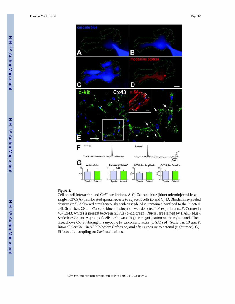
Figure 2.Cell-to-cell interaction and Ca2+ oscillations. A-C, Cascade blue (blue) microinjected in asingle hCPC (A) translocated spontaneously to adjacent cells (B and C). D, Rhodamine-labeleddextran (red), delivered simultaneously with cascade blue, remained confined to the injectedcell. Scale bar: 20 μm. Cascade blue translocation was detected in 6 experiments. E, Connexin43 (Cx43, white) is present between hCPCs (c-kit, green). Nuclei are stained by DAPI (blue).Scale bar: 20 μm. A group of cells is shown at higher magnification on the right panel. Theinset shows Cx43 labeling in a myocyte [α-sarcomeric actin, (α-SA) red]. Scale bar: 10 μm. F,Intracellular Ca2+ in hCPCs before (left trace) and after exposure to octanol (right trace). G,Effects of uncoupling on Ca2+ oscillations.
Ferreira-Martins et al. Page 12
Circ Res. Author manuscript; available in PMC 2010 October 9.
NIH
-PA Author Manuscript
NIH
-PA Author Manuscript
NIH
-PA Author Manuscript
Figure 3.Ca2+ cycling in myocytes and CPCs. A, Cx43 (white) between DiI-labeled hCPCs (DiI, red)and myocytes (α-SA, green). Nuclei are identified by DAPI. Scale bars: 10 μm. B, IntracellularCa2+ in hCPCs (blue traces) and adjacent co-cultured neonatal myocytes (black traces). Theeffects of electrical stimulation and cadmium chloride are also shown. Different cells wereused in the three conditions. C, Intracellular Ca2+ in hCPCs and neonatal myocytes in line scanmode. Red identifies hCPCs loaded with DiI and green corresponds to Fluo-3; scale bar: 20μm. D, Cytosolic Ca2+ in a quiescent mCPC (upper trace) and two mCPCs showing a single(middle trace) and multiple (lower trace) Ca2+ oscillations. E, Properties of Ca2+ oscillationsin hCPCs and mCPCs. E, c-kit-EGFP mouse heart loaded with Rhod-2 (red), stimulated at 1Hz and analyzed in line-scan mode (arrows). CPCs were identified by EGFP (green). G,Ca2+ transients in myocytes (red trace) did not affect Ca2+ levels in the neighboring EGFP-positive CPC (green trace). Identical results were obtained in 7 other experiments. Scale bars:20 μm.
Ferreira-Martins et al. Page 13
Circ Res. Author manuscript; available in PMC 2010 October 9.
NIH
-PA Author Manuscript
NIH
-PA Author Manuscript
NIH
-PA Author Manuscript

Figure 4.Ca2+ regulatory proteins in hCPCs. A-D, Expression at the mRNA (A) and protein (B-D) levelsof the components of the ER that are implicated in Ca2+ homeostasis. Myocytes were used aspositive control for RyRs. Human heart (hHeart) was used as positive control. Scale bars: 20μm. E, Repetitive Ca2+ oscillations in hCPCs in the presence of IP3R agonist. F-H, Ca2+
oscillations in hCPCs at baseline (Tyrode) and in the presence of activation (F) and inhibition(G and H) of IP3R function. Xestopsongin-C, XeC. I-N, Ca2+ in hCPCs in the presence ofmodulators of SERCA (I and J) and RyRs (K-N). *P<0.05 vs. Tyrode.
Ferreira-Martins et al. Page 14
Circ Res. Author manuscript; available in PMC 2010 October 9.
NIH
-PA Author Manuscript
NIH
-PA Author Manuscript
NIH
-PA Author Manuscript

Figure 5.Gq-protein coupled receptors and intracellular Ca2+ in hCPCs. A-C, Expression at the mRNA(A) and protein (B and C) levels of P2Y2 and H1 receptors in hCPCs. Human heart (hHeart)was used as positive control. Scale bars: 10 μm. D-G, Ca2+ oscillations in hCPCs in the presenceof ATP (D and E) or histamine (F and G). *P<0.05 vs. Tyrode.
Ferreira-Martins et al. Page 15
Circ Res. Author manuscript; available in PMC 2010 October 9.
NIH
-PA Author Manuscript
NIH
-PA Author Manuscript
NIH
-PA Author Manuscript

Figure 6.hCPC growth and apoptosis. A-C, ATP and histamine increase Ca2+ oscillations andproliferation of hCPCs. Inhibitors of Ca2+ oscillations prevent the effects of ATP andhistamine. Control, Ctrl; histamine, His. *P<0.05 vs. Ctrl, **P<0.05 vs. agonist. D and E,Apoptosis of hCPCs measured by Annexin V labeling and FACS. PI, propidium iodide. Q2,late apoptotic or necrotic cells; Q3, alive cells; Q4, cells undergoing apoptosis. F and G, IGF-1Rtranscript and protein in hCPCs and hHeart. Scale bar: 20 μm. Right panel in G illustratesselected cells at higher magnification. H and I, Intracellular Ca2+ in hCPCs exposed to IGF-1.*P<0.05 vs. Tyrode. J, Proliferation of hCPCs in the presence of IGF-1 alone or in combinationwith inhibitors of Ca2+ oscillations. *P<0.05 vs. Ctrl, ** P<0.05 vs. IGF-1.
Ferreira-Martins et al. Page 16
Circ Res. Author manuscript; available in PMC 2010 October 9.
NIH
-PA Author Manuscript
NIH
-PA Author Manuscript
NIH
-PA Author Manuscript
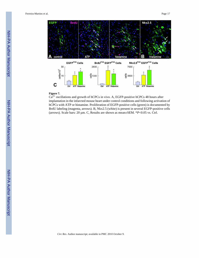
Figure 7.Ca2+ oscillations and growth of hCPCs in vivo. A, EGFP-positive hCPCs 48 hours afterimplantation in the infarcted mouse heart under control conditions and following activation ofhCPCs with ATP or histamine. Proliferation of EGFP-positive cells (green) is documented byBrdU labeling (magenta, arrows). B, Nkx2.5 (white) is present in several EGFP-positive cells(arrows). Scale bars: 20 μm. C, Results are shown as mean±SEM. *P<0.05 vs. Ctrl.
Ferreira-Martins et al. Page 17
Circ Res. Author manuscript; available in PMC 2010 October 9.
NIH
-PA Author Manuscript
NIH
-PA Author Manuscript
NIH
-PA Author Manuscript

Figure 8.Myocardial regeneration by activated hCPC. A, Mouse heart treated with histamine-stimulatedhCPCs. The mid-portion of the infarct is replaced by EGFP-positive (upper panel, green) α-SA-positive cardiomyocytes (lower panel, red). The area in the rectangle is shown at highermagnification in the inset. B, Extent of regeneration mediated by hCPCs non activated (CtrlhCPCs) or exposed to ATP or histamine (ATP-His hCPCs). C, LV function in sham operated(SO), infarcted untreated (MI + PBS) and hCPC-treated (MI + hSCPs) mice 7 days aftercoronary ligation. Ctrl, ATP and His identify non-stimulated, ATP-stimulated and histamine-stimulated hCPCs, respectively. LVEDP, LV end-diastolic pressure; LVDevP and +dP/dt.*P<0.05 vs. SO, **P<0.05 vs. MI + PBS, † P<0.05 vs. MI injected with untreated hCPCs.
Ferreira-Martins et al. Page 18
Circ Res. Author manuscript; available in PMC 2010 October 9.
NIH
-PA Author Manuscript
NIH
-PA Author Manuscript
NIH
-PA Author Manuscript
Related Documents


![Oscillations mécaniques libres non amorties Oscillations ...ww2.cnam.fr/physique/PHR004/04_L08_PHR004.pdf · Leçon n°8 : Oscillations [1] PHR 004 1 Oscillations mécaniques libres](https://static.cupdf.com/doc/110x72/5b968ab509d3f206218b9064/oscillations-mecaniques-libres-non-amorties-oscillations-ww2cnamfrphysiquephr00404l08.jpg)