Splenic Red Pulp Macrophages Produce Type I Interferons as Early Sentinels of Malaria Infection but Are Dispensable for Control Charles C. Kim 1 , Christopher S. Nelson 2 , Emily B. Wilson 2 , Baidong Hou 3,4 , Anthony L. DeFranco 3 , Joseph L. DeRisi 2,5 * 1 Division of Experimental Medicine, Department of Medicine, University of California San Francisco, San Francisco, California, United States of America, 2 Department of Biochemistry and Biophysics, University of California San Francisco, San Francisco, California, United States of America, 3 Department of Microbiology and Immunology, University of California San Francisco, San Francisco, California, United States of America, 4 Institute of Biophysics, Chinese Academy of Sciences, Beijing, China, 5 Howard Hughes Medical Institute, University of California San Francisco, San Francisco, California, United States of America Abstract Type I interferons (T1IFNs) are among the earliest cytokines produced during infections due to their direct regulation by innate immune signaling pathways. Reports have suggested that T1IFNs are produced during malaria infection, but little is known about the in vivo cellular origins of T1IFNs or their role in protection. We have found that in addition to plasmacytoid dendritic cells, splenic red pulp macrophages (RPMs) can generate significant quantities of T1IFNs in response to P. chabaudi infection in a TLR9-, MYD88-, and IRF7-dependent manner. Furthermore, T1IFNs regulate expression of interferon-stimulated genes redundantly with Interferon-gamma (IFNG), resulting in redundancy for resistance to experimental malaria infection. Despite their role in sensing and promoting immune responses to infection, we observe that RPMs are dispensable for control of parasitemia. Our results reveal that RPMs are early sentinels of malaria infection, but that effector mechanisms previously attributed to RPMs are not essential for control. Citation: Kim CC, Nelson CS, Wilson EB, Hou B, DeFranco AL, et al. (2012) Splenic Red Pulp Macrophages Produce Type I Interferons as Early Sentinels of Malaria Infection but Are Dispensable for Control. PLoS ONE 7(10): e48126. doi:10.1371/journal.pone.0048126 Editor: Laurent Re ´nia, Agency for Science, Technology and Research - Singapore Immunology Network, Singapore Received January 16, 2012; Accepted September 27, 2012; Published October 29, 2012 Copyright: ß 2012 Kim et al. This is an open-access article distributed under the terms of the Creative Commons Attribution License, which permits unrestricted use, distribution, and reproduction in any medium, provided the original author and source are credited. Funding: This work was supported by the Howard Hughes Medical Institute (JLD), the Giannini Family Foundation (CCK), and NIAID K99 AI085035 (CCK). The funders had no role in study design, data collection and analysis, decision to publish, or preparation of the manuscript. Competing Interests: The authors have declared that no competing interests exist. * E-mail: [email protected] Introduction Early recognition of infection by innate immune defenses initiates a complex cascade of intra- and intercellular signaling events that ultimately leads to the generation of a systemic immune response. Although detailed analysis of early innate immune events is under way for model organisms such as Listeria [1], relatively little is understood about early detection and responses to Plasmodium spp., the leading parasitic cause of infectious mortality and morbidity in the world. This is despite growing evidence that innate immune responses, particularly of monocytes and macro- phages, play a vital role in the control of malaria infection. For example, inflammatory monocytes contribute to elimination of parasites in P. chabaudi infection in mice [2], and in humans, a subset of peripheral monocytes is associated with control of infection in ex vivo assays [3]. Additionally, adoptive transfer of a recently discovered progenitor cell that primarily generates monocytes enhances clearance of malaria infection [4]. In contrast, B cells are required for elimination of persistent infection but are dispensable for control of the primary parasitemia [5–8]. Similarly, CD8 + T cells are not essential for control of blood stage infection [9]. The dispensability of these major effector arms of adaptive immunity highlights the importance of innate mecha- nisms of anti-parasitic recognition and clearance. Detection of the offending organism is the critical first step in activating innate immune mechanisms. Many microbes are recognized by innate immune sensors such as Toll-like receptors (TLRs), cytosolic nucleic acid sensors such as RIG-I and MDA5, and nucleotide binding domain-leucine-rich repeat (NBD/LRR) receptors, which can activate downstream production of immu- nomodulatory cytokines such as the type I interferons alpha and beta (T1IFNs, IFNA, IFNB), tumor necrosis factor (TNF), and interleukin 12 (IL12). In the case of malaria, TLR9 has emerged as a major sensor of infection, although the identity of the ligand remains controversial [4,10–13]. Studies implicating TLR9 in recognition of malaria were conducted using in vitro-differentiated plasmacytoid dendritic cells (pDCs), suggesting that pDCs may play a role in in vivo recognition of Plasmodium infection. This was recently demonstrated to be the case in a report of TLR9- dependent expression of Ifna in pDCs during P. chabuadi infection of mice [14]. However, it is well known that other innate leukocyte populations, such as conventional dendritic cells (cDCs) and macrophages, also express and signal through TLR9, but the role of these populations in recognition of malaria infection remains largely unexplored. Although it is clear that detection of malaria infection occurs through TLRs and likely also through other innate immune receptors, the mechanisms through which innate cells contribute to defense against Plasmodium parasites are poorly characterized. PLOS ONE | www.plosone.org 1 October 2012 | Volume 7 | Issue 10 | e48126

Welcome message from author
This document is posted to help you gain knowledge. Please leave a comment to let me know what you think about it! Share it to your friends and learn new things together.
Transcript

Splenic Red Pulp Macrophages Produce Type IInterferons as Early Sentinels of Malaria Infection but AreDispensable for ControlCharles C. Kim1, Christopher S. Nelson2, Emily B. Wilson2, Baidong Hou3,4, Anthony L. DeFranco3,
Joseph L. DeRisi2,5*
1 Division of Experimental Medicine, Department of Medicine, University of California San Francisco, San Francisco, California, United States of America, 2 Department of
Biochemistry and Biophysics, University of California San Francisco, San Francisco, California, United States of America, 3 Department of Microbiology and Immunology,
University of California San Francisco, San Francisco, California, United States of America, 4 Institute of Biophysics, Chinese Academy of Sciences, Beijing, China, 5 Howard
Hughes Medical Institute, University of California San Francisco, San Francisco, California, United States of America
Abstract
Type I interferons (T1IFNs) are among the earliest cytokines produced during infections due to their direct regulation byinnate immune signaling pathways. Reports have suggested that T1IFNs are produced during malaria infection, but little isknown about the in vivo cellular origins of T1IFNs or their role in protection. We have found that in addition to plasmacytoiddendritic cells, splenic red pulp macrophages (RPMs) can generate significant quantities of T1IFNs in response to P. chabaudiinfection in a TLR9-, MYD88-, and IRF7-dependent manner. Furthermore, T1IFNs regulate expression of interferon-stimulatedgenes redundantly with Interferon-gamma (IFNG), resulting in redundancy for resistance to experimental malaria infection.Despite their role in sensing and promoting immune responses to infection, we observe that RPMs are dispensable forcontrol of parasitemia. Our results reveal that RPMs are early sentinels of malaria infection, but that effector mechanismspreviously attributed to RPMs are not essential for control.
Citation: Kim CC, Nelson CS, Wilson EB, Hou B, DeFranco AL, et al. (2012) Splenic Red Pulp Macrophages Produce Type I Interferons as Early Sentinels of MalariaInfection but Are Dispensable for Control. PLoS ONE 7(10): e48126. doi:10.1371/journal.pone.0048126
Editor: Laurent Renia, Agency for Science, Technology and Research - Singapore Immunology Network, Singapore
Received January 16, 2012; Accepted September 27, 2012; Published October 29, 2012
Copyright: � 2012 Kim et al. This is an open-access article distributed under the terms of the Creative Commons Attribution License, which permits unrestricteduse, distribution, and reproduction in any medium, provided the original author and source are credited.
Funding: This work was supported by the Howard Hughes Medical Institute (JLD), the Giannini Family Foundation (CCK), and NIAID K99 AI085035 (CCK). Thefunders had no role in study design, data collection and analysis, decision to publish, or preparation of the manuscript.
Competing Interests: The authors have declared that no competing interests exist.
* E-mail: [email protected]
Introduction
Early recognition of infection by innate immune defenses
initiates a complex cascade of intra- and intercellular signaling
events that ultimately leads to the generation of a systemic immune
response. Although detailed analysis of early innate immune events
is under way for model organisms such as Listeria [1], relatively
little is understood about early detection and responses to
Plasmodium spp., the leading parasitic cause of infectious mortality
and morbidity in the world. This is despite growing evidence that
innate immune responses, particularly of monocytes and macro-
phages, play a vital role in the control of malaria infection. For
example, inflammatory monocytes contribute to elimination of
parasites in P. chabaudi infection in mice [2], and in humans, a
subset of peripheral monocytes is associated with control of
infection in ex vivo assays [3]. Additionally, adoptive transfer of a
recently discovered progenitor cell that primarily generates
monocytes enhances clearance of malaria infection [4]. In
contrast, B cells are required for elimination of persistent infection
but are dispensable for control of the primary parasitemia [5–8].
Similarly, CD8+ T cells are not essential for control of blood stage
infection [9]. The dispensability of these major effector arms of
adaptive immunity highlights the importance of innate mecha-
nisms of anti-parasitic recognition and clearance.
Detection of the offending organism is the critical first step in
activating innate immune mechanisms. Many microbes are
recognized by innate immune sensors such as Toll-like receptors
(TLRs), cytosolic nucleic acid sensors such as RIG-I and MDA5,
and nucleotide binding domain-leucine-rich repeat (NBD/LRR)
receptors, which can activate downstream production of immu-
nomodulatory cytokines such as the type I interferons alpha and
beta (T1IFNs, IFNA, IFNB), tumor necrosis factor (TNF), and
interleukin 12 (IL12). In the case of malaria, TLR9 has emerged as
a major sensor of infection, although the identity of the ligand
remains controversial [4,10–13]. Studies implicating TLR9 in
recognition of malaria were conducted using in vitro-differentiated
plasmacytoid dendritic cells (pDCs), suggesting that pDCs may
play a role in in vivo recognition of Plasmodium infection. This was
recently demonstrated to be the case in a report of TLR9-
dependent expression of Ifna in pDCs during P. chabuadi infection
of mice [14]. However, it is well known that other innate leukocyte
populations, such as conventional dendritic cells (cDCs) and
macrophages, also express and signal through TLR9, but the role
of these populations in recognition of malaria infection remains
largely unexplored.
Although it is clear that detection of malaria infection occurs
through TLRs and likely also through other innate immune
receptors, the mechanisms through which innate cells contribute
to defense against Plasmodium parasites are poorly characterized.
PLOS ONE | www.plosone.org 1 October 2012 | Volume 7 | Issue 10 | e48126
During viral and bacterial infections, signaling through TLRs and
other innate sensing pathways frequently results in the immediate
downstream production of cytokines such as T1IFNs. With regard
to malaria, Plasmodium ligands have been reported to stimulate
T1IFN production in in vitro systems [10,13,15], experimentally
infected mice [14], and Plasmodium-infected individuals [10,16].
However, in contrast to IFNG, which has been shown to be an
important activator of anti-malarial mechanisms, the role of
T1IFNs in protection against malaria infection is not well
characterized.
In order to address these gaps in our knowledge, we conducted
a systematic investigation of T1IFN production during malaria
infection using the P. chabaudi model of uncomplicated malaria.
Here we present evidence that in addition to pDCs, splenic red
pulp macrophages (RPMs) are an important contributor to
systemic T1IFN during early malaria infection. Additionally, we
have found that T1IFNs regulate gene expression and contribute
to control of infection in a manner that is largely redundant with
IFNG signaling. However, despite the role of RPMs in T1IFN
production, mice lacking RPMs exhibit no deficiencies in their
ability to control infection. Our findings demonstrate that T1IFNs
play an important immunomodulatory role during in vivo malaria
infection and provide us with a basic understanding of the
molecular and cellular machinery involved in innate immune
recognition of malaria parasites. We also demonstrate that RPMs
are not essential for control of infection despite their role in early
sensing of infection and their key location in contact with
circulating parasites.
Results
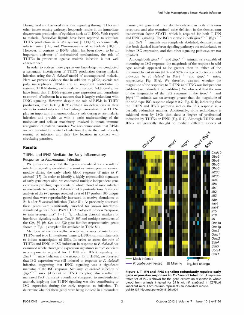
T1IFNs and IFNG Mediate the Early InflammatoryResponse to Plasmodium Infection
We previously reported that genes stimulated as a result of
interferon signaling constitute the most extensive gene expression
module during the early whole blood response of mice to P.
chabaudi [17]. In order to identify a highly reproducible signature
of early gene expression, we conducted multiple independent gene
expression profiling experiments of whole blood of mice infected
or mock-infected with P. chabaudi at 24 h post-infection. Statistical
analysis of the two groups revealed a set of 117 probes (103 unique
genes) that were reproducibly increased in relative abundance at
24 h after P. chabaudi infection (Table S1). As previously observed,
these genes were significantly enriched for known interferon-
stimulated genes (ISGs; PANTHER biological process ‘‘response
to interferon-gamma’’ p = 1029), including classical markers of
interferon signaling such as Cxcl10, Il6, and multiple members of
the Gbp, Ifi, Ifit, Oas, and Slfn gene families (representative genes
shown in Fig. 1; complete list available in Table S1).
Members of the two well-characterized classes of interferons,
T1IFNs and type II interferon (namely, IFNG), can stimulate cells
to induce transcription of ISGs. In order to assess the role of
T1IFNs and IFNG in ISG induction in response to P. chabaudi, we
examined whole blood gene expression signatures in mice deficient
in components required for T1IFN and IFNG signaling. In
Ifnar12/2 mice (deficient in the receptor for T1IFNs), we observed
that ISG expression was still induced in response to P. chabaudi
infection, suggesting that IFNG signaling was a significant
mediator of the ISG response. Similarly, P. chabaudi infection of
Ifngr12/2 mice (deficient in IFNG receptor) also resulted in
increased ISG transcript abundance compared to mock-infected
animals, implying that T1IFN signaling was also contributing to
ISG expression during the early response to infection. To
determine whether these genes were being induced in a redundant
manner, we generated mice doubly deficient in both interferon
receptors, and also examined mice deficient in the downstream
transcription factor STAT1, which is required for both T1IFN
and IFNG signaling. The ISG response in both Ifnar12/2 Ifngr12 /
2 and Stat12/2 animals was completely abolished, demonstrating
that both classical interferon signaling pathways act redundantly to
induce ISG expression, and that other signaling pathways are not
involved.
Although both Ifnar12/2 and Ifngr12/2 animals were capable of
mounting an ISG response, the magnitude of the response in wild
type animals appeared to be greater than in either of the
immunodeficient strains (41% and 32% average reductions in fold
induction by P. chabaudi in Ifnar12/2 and Ifngr12/2 mice,
respectively; Fig. S1A). We therefore assessed whether the
magnitude of the responses to T1IFNs and IFNG was independent
(additive) or redundant (sub-additive). We observed that the sum
of the magnitudes of the ISG response in the Ifnar12/2 and
Ifngr12/2 animals was on average greater than the magnitude of
the wild type ISG response (slope = 0.7; Fig. S1B), indicating that
the T1IFN and IFNG pathways induce the ISG response in a
partially redundant manner. Additionally, some redundancy is
exhibited even by ISGs that show a degree of preferential
induction by T1IFNs or IFNG (Fig. S1C). Although T1IFNs and
IFNG are generally thought to mediate different aspects of
Figure 1. T1IFN and IFNG signaling redundantly regulate earlygene expression responses to P. chabaudi infection. A represen-tative set of ISG is shown for the gene expression response in wholeblood from animals infected for 24 h with P. chabaudi in C57BL/6knockout mice. Each column represents an individual mouse.doi:10.1371/journal.pone.0048126.g001
Red Pulp Macrophages Sense Malaria Infection
PLOS ONE | www.plosone.org 2 October 2012 | Volume 7 | Issue 10 | e48126

immune activation, these results demonstrate that at least in the
context of early malaria infection, the majority of genes regulated
by one type of interferon are also regulated by the other.
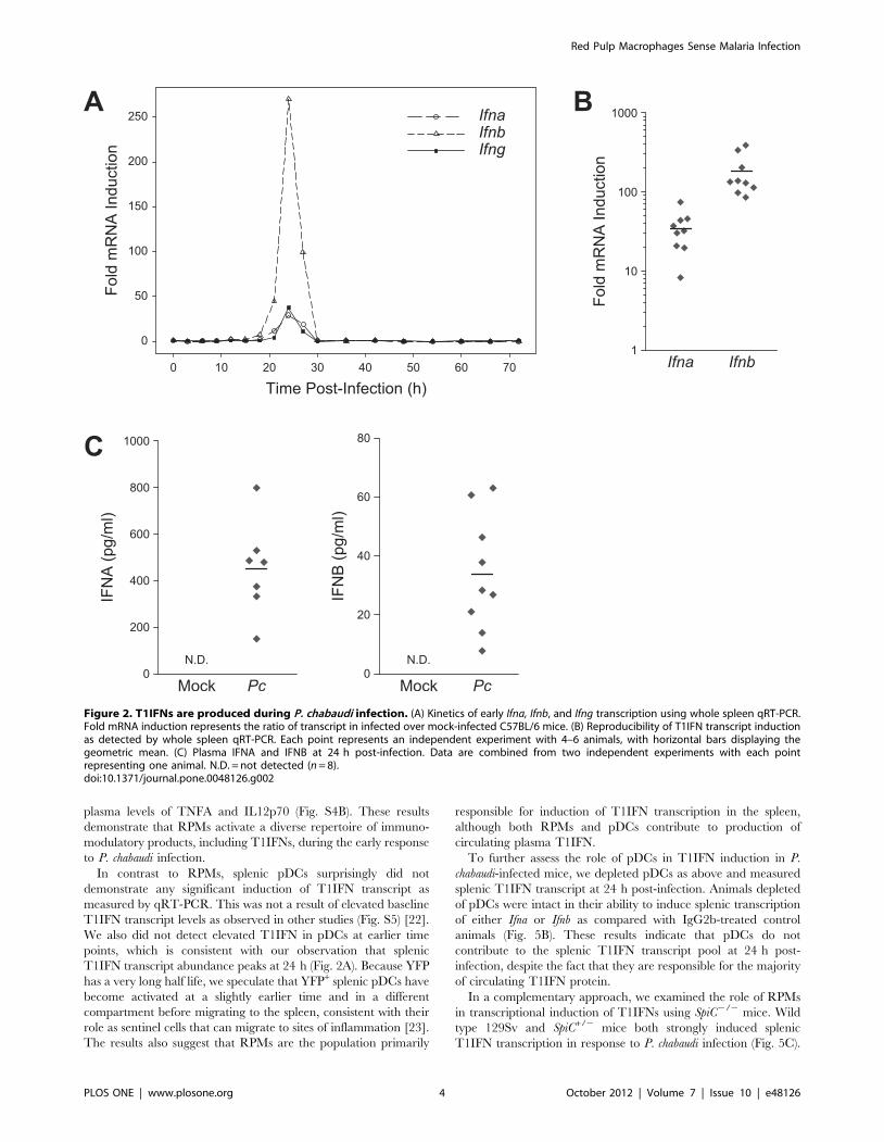
In order to directly measure T1IFN production, we
performed quantitative reverse transcription PCR (qRT-PCR)
to estimate relative transcript abundance for Ifna and Ifnb in the
spleens of mice infected with P. chabaudi. Examination of splenic
transcripts every 3 h for the first 30 h post-infection revealed
that both Ifna and Ifnb transcripts, as well as Ifng, exhibited a
peak of increased abundance centered around 24 h (Fig. 2A).
Upon return to baseline levels, splenic T1IFN transcripts were
not induced again within the first three days of infection
(measured in 6 h intervals after 30 h). Detection of elevated Ifna
and Ifnb in spleens of infected animals at 24 h post-infection was
highly reproducible across independent experiments (Fig. 2B),
and IFNA and IFNB were reproducibly detected in the plasma
of infected animals (Fig. 2C). Together, these findings provide
evidence that a burst of T1IFNs is produced during the early
response to P. chabaudi infection and contributes to induction of
ISG expression.
T1IFNs and IFNG Redundantly Promote Control ofParasitemia
Our results show early production of T1IFNs during P.
chabaudi infection, but the contribution of T1IFNs to the control
of malaria parasite replication is not well characterized. The
normal course of P. chabaudi infection in C57BL/6 mice
develops as an exponentially increasing load of parasites in
the blood that typically peaks at 7–10 days post-infection
followed by control and resolution of the primary parasitemia
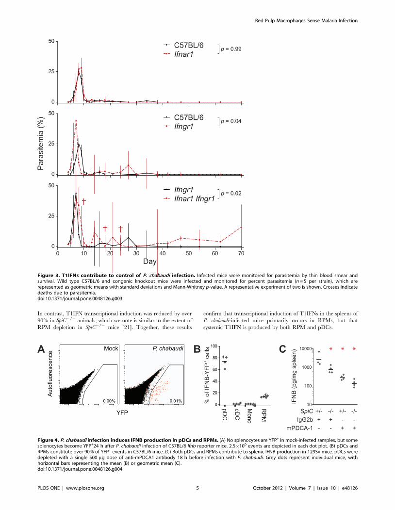
over the next 2–4 days (Fig. 3). A recent study reported a slight
increase in the magnitude of peak P. chabaudi parasitemia in
Ifnar12/2 mice, but resolution occurred with kinetics identical to
wild type (129Sv) animals [14]. In contrast, we observed no
significant differences in the magnitude or times to manifesta-
tion of any of the ascending, descending, or clearance phases of
parasitemia in Ifnar12/2 animals as compared to infection of
C57BL/6 mice (Fig. 3). The discrepancy between our findings
and those of Voisine et al. could be a result of the different
backgrounds used, since 129Sv mice produce higher levels of
T1IFNs (Fig. S3 and [18]).
Although our results would appear to suggest that T1IFNs do
not contribute to control of malaria infection, we considered the
possibility that the redundancy between T1IFNs and IFNG in
the regulation of ISG expression could confer redundancy in
control of infection. We therefore examined the course of
parasitemia in Ifngr12/2 animals as compared to Ifnar12/2
Ifngr12/2 animals in order to assess the function of T1IFNs in
the absence of IFNG signaling. We observed that Ifngr12/2
animals exhibited defects in their ability to resolve parasitemia
as compared to wild type animals; although most animals
controlled the primary and secondary peaks, peak parasitemias
were higher in Ifngr12/2 animals, and a tertiary peak of
parasitemia occurred in most animals (Fig. 3). Despite the
increased severity of infection in Ifngr12/2 mice, parasites were
controlled in all mice by 40 days post-infection. In contrast,
Ifnar12/2 Ifngr12/2 animals exhibited mortality, multiple late
peaks of high parasitemia, and an inability to completely clear
parasites from the bloodstream within the duration of the 70
day study, indicating that T1IFNs and IFNG signaling exhibit
redundancy in the regulation of anti-parasitic mechanisms that
are essential to the control of malaria infection.
Plasmacytoid Dendritic Cells and Red Pulp MacrophagesProduce T1IFNs in Response to P. chabaudi
Previous in vitro studies have provided conflicting measurements
of production of IFNA by pDCs after stimulation with malaria
ligands [10–13]. Another study recently reported Ifna expression in
pDCs during P. chabaudi infection [14], but this observation was
made at a time after the peak of C57BL/6 T1IFN production and
did not assess other potential cellular sources. We therefore took
an unbiased approach to identify the cellular origins of T1IFN
production in response to physiologically relevant stimuli during
in vivo infection with P. chabaudi.
In order to achieve single-cell resolution of T1IFN expression,
transgenic Ifnb-Yfp reporter mice [19] were mock- or P. chabaudi-
infected and analyzed for splenic Ifnb expression by flow
cytometry. Animals infected with P. chabaudi contained a small
but highly reproducible population of YFP+ cells, whereas no
YFP+ events were detected in any of the spleens of mock-infected
animals (Fig. 4A). Lineage marker analysis of the YFP+ popula-
tions demonstrated that approximately 75% of the YFP+ events
were CD11cint Siglec-H+, consistent with markers of pDCs
(Fig. 4B). In contrast, conventional dendritic cells (cDCs; CD11chi
Siglec H2) and CD11bhi F4/80int-hi SSClo monocytes (Mono)
constituted none of the YFP+ events. Interestingly, a small but
reproducible fraction (,15%) of the total YFP+ events was F4/80hi
CD11blo/2, consistent with markers of splenic RPMs. Similar
frequencies of YFP+ and lineage markers were observed using
Ifna6-Gfp reporter mice (Fig. S2) [20]. Notably, the pDCs and
RPMs together account for nearly all the YFP+ and GFP+ cells,
indicating that, together, they are the major populations respon-
sible for splenic T1IFN induction during P. chabaudi infection.
Because T1IFN can be produced at low levels by other cell
types, we assessed whether pDCs and RPMs measurably
contribute to systemic T1IFN levels. In order to examine the role
of RPMs in T1IFN production, we employed SpiC2/2 mice [21],
which lack a transcription factor required for development of
RPMs but not other myeloid populations (Fig. S3A). pDCs were
depleted 18 h pre-infection with P. chabaudi using the anti-
mPDCA-1 antibody, which reproducibly depleted 85% of splenic
pDCs with no impact on RPM frequency (Fig. S3B). After 24 h
infection with P. chabaudi, SpiC2/2 mice produced roughly half the
splenic IFNB of SpiC+/2mice (Fig. 4C), with similar results also
observed in plasma (Fig. S3C–D). pDCs were also required for
T1IFN production, with depletion resulting in over 80% reduction
of splenic and plasma IFNB levels in SpiC+/2 mice (Fig. 4C and
S4D) and C57BL/6 mice (Fig. S4E). The absence of both
populations resulted in over 90% reduction of splenic IFNB
(Fig. 4C), with the residual levels likely reflecting incompletely
depleted pDCs. Together with the reporter data, these results
demonstrate that pDCs and RPMs are the primary sources of
T1IFN during experimental malaria infection.
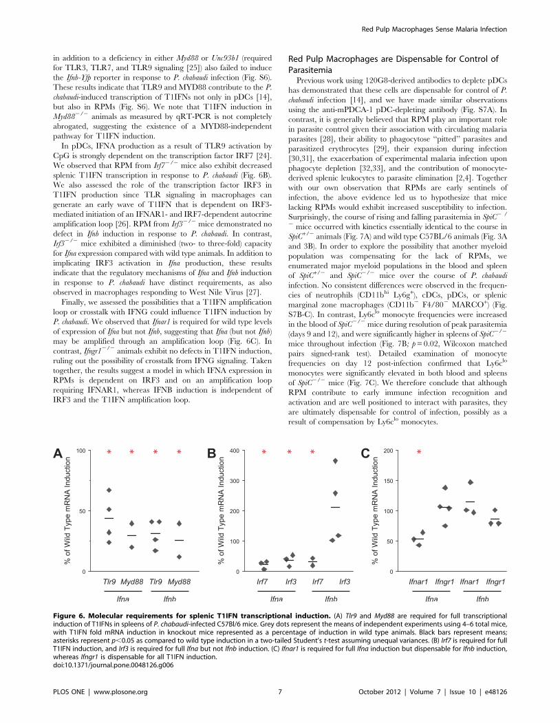
Red Pulp Macrophages are the Primary Source of SplenicT1IFN Transcripts during P. chabaudi Infection
In order to corroborate our observations with the Ifnb reporter
mice, we isolated the same splenic leukocyte subsets by FACS and
assessed T1IFN transcriptional induction by qRT-PCR. Consis-
tent with our observations in Ifnb-Yfp reporter mice, RPMs
strongly induced both Ifna and Ifnb transcript post-infection with P.
chabaudi (Fig. 5A). Similarly, microarray analysis of isolated RPMs
from mock- and P. chabaudi-infected mice demonstrated induction
of multiple members of the Ifna family along with a variety of other
cytokines and chemokines, including Tnf, Il1b, Il6, Il10, Cxcl1, and
Cxcl2 (Fig. S4A), and RPM-deficient mice exhibited decreased
Red Pulp Macrophages Sense Malaria Infection
PLOS ONE | www.plosone.org 3 October 2012 | Volume 7 | Issue 10 | e48126
plasma levels of TNFA and IL12p70 (Fig. S4B). These results
demonstrate that RPMs activate a diverse repertoire of immuno-
modulatory products, including T1IFNs, during the early response
to P. chabaudi infection.
In contrast to RPMs, splenic pDCs surprisingly did not
demonstrate any significant induction of T1IFN transcript as
measured by qRT-PCR. This was not a result of elevated baseline
T1IFN transcript levels as observed in other studies (Fig. S5) [22].
We also did not detect elevated T1IFN in pDCs at earlier time
points, which is consistent with our observation that splenic
T1IFN transcript abundance peaks at 24 h (Fig. 2A). Because YFP
has a very long half life, we speculate that YFP+ splenic pDCs have
become activated at a slightly earlier time and in a different
compartment before migrating to the spleen, consistent with their
role as sentinel cells that can migrate to sites of inflammation [23].
The results also suggest that RPMs are the population primarily
responsible for induction of T1IFN transcription in the spleen,
although both RPMs and pDCs contribute to production of
circulating plasma T1IFN.
To further assess the role of pDCs in T1IFN induction in P.
chabaudi-infected mice, we depleted pDCs as above and measured
splenic T1IFN transcript at 24 h post-infection. Animals depleted
of pDCs were intact in their ability to induce splenic transcription
of either Ifna or Ifnb as compared with IgG2b-treated control
animals (Fig. 5B). These results indicate that pDCs do not
contribute to the splenic T1IFN transcript pool at 24 h post-
infection, despite the fact that they are responsible for the majority
of circulating T1IFN protein.
In a complementary approach, we examined the role of RPMs
in transcriptional induction of T1IFNs using SpiC2/2 mice. Wild
type 129Sv and SpiC+/2 mice both strongly induced splenic
T1IFN transcription in response to P. chabaudi infection (Fig. 5C).
Figure 2. T1IFNs are produced during P. chabaudi infection. (A) Kinetics of early Ifna, Ifnb, and Ifng transcription using whole spleen qRT-PCR.Fold mRNA induction represents the ratio of transcript in infected over mock-infected C57BL/6 mice. (B) Reproducibility of T1IFN transcript inductionas detected by whole spleen qRT-PCR. Each point represents an independent experiment with 4–6 animals, with horizontal bars displaying thegeometric mean. (C) Plasma IFNA and IFNB at 24 h post-infection. Data are combined from two independent experiments with each pointrepresenting one animal. N.D. = not detected (n = 8).doi:10.1371/journal.pone.0048126.g002
Red Pulp Macrophages Sense Malaria Infection
PLOS ONE | www.plosone.org 4 October 2012 | Volume 7 | Issue 10 | e48126
In contrast, T1IFN transcriptional induction was reduced by over
90% in SpiC2/2 animals, which we note is similar to the extent of
RPM depletion in SpiC2/2 mice [21]. Together, these results
confirm that transcriptional induction of T1IFNs in the spleens of
P. chabaudi-infected mice primarily occurs in RPMs, but that
systemic T1IFN is produced by both RPM and pDCs.
Figure 3. T1IFNs contribute to control of P. chabaudi infection. Infected mice were monitored for parasitemia by thin blood smear andsurvival. Wild type C57BL/6 and congenic knockout mice were infected and monitored for percent parasitemia (n = 5 per strain), which arerepresented as geometric means with standard deviations and Mann-Whitney p-value. A representative experiment of two is shown. Crosses indicatedeaths due to parasitemia.doi:10.1371/journal.pone.0048126.g003
Figure 4. P. chabaudi infection induces IFNB production in pDCs and RPMs. (A) No splenocytes are YFP+ in mock-infected samples, but somesplenocytes become YFP+24 h after P. chabaudi infection of C57BL/6 Ifnb reporter mice. 2.56106 events are depicted in each dot plot. (B) pDCs andRPMs constitute over 90% of YFP+ events in C57BL/6 mice. (C) Both pDCs and RPMs contribute to splenic IFNB production in 129Sv mice. pDCs weredepleted with a single 500 mg dose of anti-mPDCA1 antibody 18 h before infection with P. chabaudi. Grey dots represent individual mice, withhorizontal bars representing the mean (B) or geometric mean (C).doi:10.1371/journal.pone.0048126.g004
Red Pulp Macrophages Sense Malaria Infection
PLOS ONE | www.plosone.org 5 October 2012 | Volume 7 | Issue 10 | e48126
In a final approach to characterizing the cellular origins of
splenic T1IFN transcriptional induction, we employed mice
homozygous for a floxed allele of Myd88 (Myd88fl/fl), which
encodes an adaptor molecule required for TLR signaling.
Myd88f l/fl mice that are hemizygous for the CD11c-Cre or Lyz2-
Cre transgene efficiently delete Myd88 from the genomes of
dendritic cells and macrophages, respectively [22]. Consistent with
the model that RPMs are responsible for transcriptional induction
of T1IFNs in spleens, we observed normal levels of splenic Ifna and
Ifnb transcription in CD11c-Cre animals, whereas Lyz2-Cre animals
exhibited a nearly 90% reduction in T1IFN transcription (Fig. 5D–
E). These data indicate that Myd88 is required in macrophages,
but not in dendritic cells, for transcriptional induction of splenic
T1IFNs. Taken together, our data lead us to conclude that RPMs,
and not pDCs, are the primary source of splenic T1IFN transcripts
at 24 h after P. chabaudi infection, despite the fact that both
populations contribute IFNB to the circulating plasma pool.
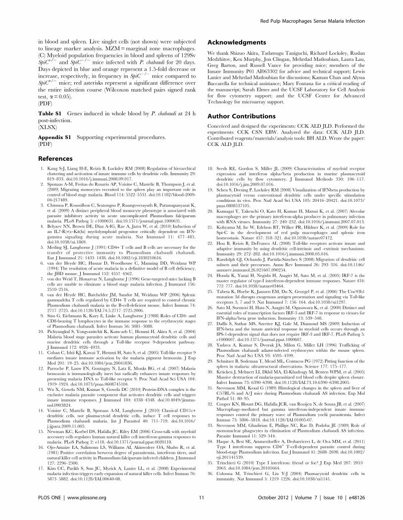
TLR9 Signaling is Required for Full Induction of T1IFNsActivation of TLR9 by A-type CpG DNA leads to induction of
IFNA in pDCs through a MYD88- and IRF7-dependent
mechanism [24], and previous work similarly found TLR9-
dependence of IFNA production in pDCs during in vitro infection
with malaria parasites [14]. To characterize the molecular
mechanisms by which RPMs respond to Plasmodium infection, we
took advantage of the fact that splenic T1IFN transcript is almost
exclusively derived from RPMs to examine the role of several
signaling molecules. We measured the induction of splenic T1IFN
transcript by qRT-PCR in wild type and knockout mice infected
with P. chabaudi for 24 h. In Tlr92/2 mice, we consistently
observed a two- to four-fold decrease in production of Ifna and Ifnb
transcript compared to wild type mice (Fig. 6A). Consistent with
an important role for TLR9 signaling in T1IFN production, Ifna
and Ifnb transcripts failed to be induced to wild type levels in
Myd882/2 animals, similar to our results in Lyz2-Cre Myd88fl/fl
mice (Fig. 6A and 5D). Mice harboring the Ifnb-Yfp reporter gene
Figure 5. Cellular requirements for splenic T1IFN transcriptional induction. (A) RPMs, but not other macrophage or dendritic cell subsets,induce Ifna and Ifnb at 24 h post-infection with P. chabaudi as detected by qRT-PCR in C57BL/6 mice. Fold mRNA induction represents fold inductionof transcript in infected versus mock-infected normalized to beta-actin. Grey dots represent independent experiments conducted on different days;black bars denote the geometric means of the fold inductions. (B) pDCs are not required for splenic Ifna or Ifnb transcriptional induction in responseto P. chabaudi in C57BL/6 mice. (C) Genetic deletion of RPMs in 129Sv mice results in diminished T1IFN transcriptional induction. (D) Genetic deletionof Myd88 from dendritic cells does not impact transcriptional induction of T1IFNs in spleens of C57BL/6 mice. (E) Genetic deletion of Myd88 frommacrophages/neutrophils decreases transcriptional induction of T1IFNs by an order of magnitude in C57Bl/6 mice. Grey bars represent geometricmeans with 95% confidence intervals. Asterisks represent p,0.05 in a Student’s t-test against control samples.doi:10.1371/journal.pone.0048126.g005
Red Pulp Macrophages Sense Malaria Infection
PLOS ONE | www.plosone.org 6 October 2012 | Volume 7 | Issue 10 | e48126
in addition to a deficiency in either Myd88 or Unc93b1 (required
for TLR3, TLR7, and TLR9 signaling [25]) also failed to induce
the Ifnb-Yfp reporter in response to P. chabaudi infection (Fig. S6).
These results indicate that TLR9 and MYD88 contribute to the P.
chabaudi-induced transcription of T1IFNs not only in pDCs [14],
but also in RPMs (Fig. S6). We note that T1IFN induction in
Myd882/2 animals as measured by qRT-PCR is not completely
abrogated, suggesting the existence of a MYD88-independent
pathway for T1IFN induction.
In pDCs, IFNA production as a result of TLR9 activation by
CpG is strongly dependent on the transcription factor IRF7 [24].
We observed that RPM from Irf72/2 mice also exhibit decreased
splenic T1IFN transcription in response to P. chabaudi (Fig. 6B).
We also assessed the role of the transcription factor IRF3 in
T1IFN production since TLR signaling in macrophages can
generate an early wave of T1IFN that is dependent on IRF3-
mediated initiation of an IFNAR1- and IRF7-dependent autocrine
amplification loop [26]. RPM from Irf32/2 mice demonstrated no
defect in Ifnb induction in response to P. chabaudi. In contrast,
Irf32/2 mice exhibited a diminished (two- to three-fold) capacity
for Ifna expression compared with wild type animals. In addition to
implicating IRF3 activation in Ifna production, these results
indicate that the regulatory mechanisms of Ifna and Ifnb induction
in response to P. chabaudi have distinct requirements, as also
observed in macrophages responding to West Nile Virus [27].
Finally, we assessed the possibilities that a T1IFN amplification
loop or crosstalk with IFNG could influence T1IFN induction by
P. chabaudi. We observed that Ifnar1 is required for wild type levels
of expression of Ifna but not Ifnb, suggesting that Ifna (but not Ifnb)
may be amplified through an amplification loop (Fig. 6C). In
contrast, Ifngr12/2 animals exhibit no defects in T1IFN induction,
ruling out the possibility of crosstalk from IFNG signaling. Taken
together, the results suggest a model in which IFNA expression in
RPMs is dependent on IRF3 and on an amplification loop
requiring IFNAR1, whereas IFNB induction is independent of
IRF3 and the T1IFN amplification loop.
Red Pulp Macrophages are Dispensable for Control ofParasitemia
Previous work using 120G8-derived antibodies to deplete pDCs
has demonstrated that these cells are dispensable for control of P.
chabaudi infection [14], and we have made similar observations
using the anti-mPDCA-1 pDC-depleting antibody (Fig. S7A). In
contrast, it is generally believed that RPM play an important role
in parasite control given their association with circulating malaria
parasites [28], their ability to phagocytose ‘‘pitted’’ parasites and
parasitized erythrocytes [29], their expansion during infection
[30,31], the exacerbation of experimental malaria infection upon
phagocyte depletion [32,33], and the contribution of monocyte-
derived splenic leukocytes to parasite elimination [2,4]. Together
with our own observation that RPMs are early sentinels of
infection, the above evidence led us to hypothesize that mice
lacking RPMs would exhibit increased susceptibility to infection.
Surprisingly, the course of rising and falling parasitemia in SpiC2 /
2 mice occurred with kinetics essentially identical to the course in
SpiC+/2 animals (Fig. 7A) and wild type C57BL/6 animals (Fig. 3A
and 3B). In order to explore the possibility that another myeloid
population was compensating for the lack of RPMs, we
enumerated major myeloid populations in the blood and spleen
of SpiC+/2 and SpiC2/2 mice over the course of P. chabaudi
infection. No consistent differences were observed in the frequen-
cies of neutrophils (CD11bhi Ly6g+), cDCs, pDCs, or splenic
marginal zone macrophages (CD11b2 F4/802 MARCO+) (Fig.
S7B-C). In contrast, Ly6clo monocyte frequencies were increased
in the blood of SpiC2/2 mice during resolution of peak parasitemia
(days 9 and 12), and were significantly higher in spleens of SpiC2/2
mice throughout infection (Fig. 7B; p = 0.02, Wilcoxon matched
pairs signed-rank test). Detailed examination of monocyte
frequencies on day 12 post-infection confirmed that Ly6clo
monocytes were significantly elevated in both blood and spleens
of SpiC2/2 mice (Fig. 7C). We therefore conclude that although
RPM contribute to early immune infection recognition and
activation and are well positioned to interact with parasites, they
are ultimately dispensable for control of infection, possibly as a
result of compensation by Ly6clo monocytes.
Figure 6. Molecular requirements for splenic T1IFN transcriptional induction. (A) Tlr9 and Myd88 are required for full transcriptionalinduction of T1IFNs in spleens of P. chabaudi-infected C57Bl/6 mice. Grey dots represent the means of independent experiments using 4–6 total mice,with T1IFN fold mRNA induction in knockout mice represented as a percentage of induction in wild type animals. Black bars represent means;asterisks represent p,0.05 as compared to wild type induction in a two-tailed Student’s t-test assuming unequal variances. (B) Irf7 is required for fullT1IFN induction, and Irf3 is required for full Ifna but not Ifnb induction. (C) Ifnar1 is required for full Ifna induction but dispensable for Ifnb induction,whereas Ifngr1 is dispensable for all T1IFN induction.doi:10.1371/journal.pone.0048126.g006
Red Pulp Macrophages Sense Malaria Infection
PLOS ONE | www.plosone.org 7 October 2012 | Volume 7 | Issue 10 | e48126

Discussion
We previously found that P. chabaudi infection of mice induced
robust expression of an interferon-induced gene signature as the
earliest detectable expression response in blood [17]. Here, we
demonstrate that this ISG response is the combined result of
T1IFNs and IFNG, acting in a largely redundant fashion.
Although the prominent involvement of IFNG in responses to
malaria infection is well established, much less is understood about
production of T1IFNs. Studies have shown that malaria extracts
can induce IFNA from human pDCs in vitro [10,13], and have
documented IFNA induction in P. chabaudi- [14] and P. berghei-
infected mice [34]. Using a variety of approaches, we have
demonstrated that T1IFNs are indeed produced during in vivo
infection with P. chabaudi, and that both pDCs and RPMs are the
key cellular sources that contribute to the systemic T1IFN pool.
Although the protective role of T1IFNs in viral infections is well
established, in some bacterial infections and autoimmune disor-
ders, T1IFNs appear to exacerbate disease [35]. Similar to viral
infections, our functional studies indicate that T1IFNs act
redundantly with IFNG to activate mechanisms that protect
Figure 7. Mice lacking RPMs exhibit wild type infection kinetics. (A) Parasitemia courses in 129Sv SpiC+/2 (n = 4) and SpiC2/2 (n = 5) mice arerepresented as geometric means with standard deviations and Mann-Whitney p-value. (B) Ly6clo monocyte (CD11b+ F4/80+ Ly6g2 SSClo Ly6clo)frequencies in blood and spleen of 129Sv SpiC+/2 (white bars) and SpiC2/2 mice (black bars) during the course of infection. Days depicted in blue andorange represent a 1.5-fold decrease or increase, respectively, in Ly6clo monocyte frequencies in blood and spleen of mice infected with P. chabaudi.(C) Ly6clo monocyte frequencies on day 12 post-infection. Means are presented with standard errors; p-values represent a two-tailed t-test assumingunequal variances. Data represent three independent experiments (n = 6–7 mice per group total).doi:10.1371/journal.pone.0048126.g007
Red Pulp Macrophages Sense Malaria Infection
PLOS ONE | www.plosone.org 8 October 2012 | Volume 7 | Issue 10 | e48126

against malaria disease. Together, our findings reveal redundan-
cies at several different levels: first, at the level of multiple
molecular sensing pathways in RPMs feeding into T1IFN
production; second, at the level of multiple leukocyte populations
generating systemically available T1IFNs; and finally, at the level
of T1IFNs conferring protection that is redundant with IFNG. We
suggest that this tiered redundancy is widespread in immunolog-
ical systems but has been overlooked due to absent or mild
phenotypes in organism-level assays.
T1IFNs frequently originate from pDCs, which are also known
as ‘‘interferon producing cells’’ due to their ability to produce
more T1IFNs than any other cell type in human blood [36]. Our
observation that pDCs produce IFNA and IFNB during malaria
infection is in line with the general function of pDCs and similar
findings from Voisine et al. [14]. However, we have demonstrated
that RPMs also contribute significantly to total T1IFN production
during the response to P. chabaudi, indicating that these macro-
phages play a role in early immune activation during malaria
infection. We estimate that roughly 3000 pDCs and 1000 RPMs
per spleen produce high levels of T1IFN, and the comparable
fluorescence levels of these populations in Ifnb-Yfp reporter animals
suggest that pDC and RPM are capable of transcribing similar
levels of Ifnb. Whether or not this corresponds to similar levels of
IFNB production on a per-cell basis remains to be determined;
regardless, our findings contribute to the increasing body of
literature indicating that macrophages and other non-pDC
populations are significant sources of T1IFNs in vivo [27,37–41].
It is likely that the localization of the infections at the tissue,
cellular, and sub-cellular levels defines in part which leukocytes
respond and in what manner. This is likely to be the case for
T1IFN production by RPMs in malaria infection: ultrastructural
studies have demonstrated that RPMs are capable of phagocytosis
of both whole infected erythrocytes and parasites that have been
‘‘pitted’’ from infected erythrocytes in the spleen [29], and
trafficking studies using stained infected erythrocytes have
demonstrated localization to the splenic red pulp [28]. Although
these studies only examined splenic organization during the time
of peak parasitemia, it was reasonable to expect that RPMs would
also function as early detectors of malaria parasites due to their
inherent role in filtering parasites from the blood. We have
demonstrated that this is indeed the case, despite the low parasite
load during early sub-patent infection, and that RPMs respond by
producing T1IFNs and a host of additional chemokines and
cytokines. To the best of our knowledge, this is the first
demonstration of production of an immunomodulatory cytokine
by RPMs during early malaria infection.
We have found that TLR9-MYD88-IRF7 signaling is required
for full T1IFN expression in RPMs, similar to the role of this
pathway in pDC [14]. This is at odds with the fact that no in vitro
studies of TLR9 activation have reported IFNA production in
mouse pDCs or macrophages, but it is possible that malaria
ligands may be less potent than synthetic ligands and therefore
require additional activating signals from other leukocyte popu-
lations present in vivo. Obvious candidates for such signals include
cytokines that signal through the MAP kinase and NF-kappa B
pathways, which participate in Ifnb induction through the
heterodimeric transcription factors ATF-2/c-Jun and p50/RelA
[42]. Consistent with this possibility, inhibition of NF-kappa B
signaling in mice infected with West Nile Virus decreases IFNB
production [27]. Further studies will be required to understand the
relative contributions of these pathways in vitro and in vivo, and also
to identify the pathway(s) responsible for residual levels of T1IFN
production in the absences of TLR9 and MYD88.
T1IFNs can augment their own expression through a feed-
forward signaling loop, but for P. chabaudi, only Ifna, not Ifnb,
induction appears to rely on IFNAR1-dependent amplification.
This result is similar to observations from Listeria infection, in
which IFNB generation is essentially unaffected by the absence of
IFNAR1 whereas IFNA production is severely diminished [40].
Similarly, expression of Ifna by cDCs during West Nile Virus
infection was diminished in mice lacking IFNAR1, whereas Ifnb
expression was not [27]. Thus, our data extend the paradigm of
IFNB being induced prior to amplification loop-dependent
production of IFNA, as demonstrated in viral and bacterial
systems, to infection with a protozoan parasite. With regard to Ifna
induction by P. chabaudi, we observed that Ifnar12/2and Irf32/2
mice exhibit similar levels of reduction, consistent with observa-
tions from other systems that these molecules are both required for
T1IFN amplification [26].
Although RPMs produce T1IFNs and other cytokines during
early infection, mice lacking RPMs clear parasites with kinetics
identical to control animals. This result was surprising given the
general belief that RPMs contribute to control of parasitemia
through phagocytic mechanisms [28,29,33,43]. Furthermore, we
observed that RPMs act as early sentinels of infection and produce
cytokines that ultimately contribute to elimination of infection.
Given our observation that pDCs also produce T1IFNs, it is
possible that all of the important functions of RPMs are redundant
with other leukocyte subsets. For example, splenic monocytes are
capable of phagocytosis of P. chabaudi [2], and this population
undergoes expansion near the time of peak parasitemia in both
SpiC+/2 and SpiC2/2 mice (Fig. S7C). Our data indicate that the
Ly6clo monocytes are also significantly increased in frequency in
RPM-deficient mice, suggesting the possibility that this subset
could be providing redundancy with RPMs. Although the exact
mechanism requires further investigation, our data indicate that
the important role of the spleen in clearance of malaria infection is
due to functions that are not specific to RPMs.
In summary, our results demonstrate that T1IFNs play a
redundant but important protective role during experimental
malaria infection. These T1IFNs are derived from both pDCs and
RPMs, which are thus identified as the major populations
responsible for early innate recognition of malaria infection.
Future work will reveal how these innate populations and T1IFNs
promote the development of an integrated immune response that
can ultimately resolve malaria infection.
Materials and Methods
MiceC57BL/6 9–14 week old female mice (Jackson Laboratories or
National Cancer Institute) were maintained on a 12 h light cycle
(on from 0600 to 1800 h). All mice used in this study (Ifnar12/2,
Ifngr12/2, Ifnar12/2 Ifngr12/2, Stat12/2, Ifnb-Yfp+/+, Ifna6-gfp+/2,
Tlr92/2, Myd882/2, Irf72/2) were .95% C57BL/6 by micro-
satellite genotyping at 94 loci (UCSF genomics core) with the
exceptions of Irf32/2 (80% C57BL/6) and SpiC2/2 mice (129Sv).
This study was conducted in strict accordance with the guidelines
of the Office of Laboratory Animal Welfare and with the approval
of the UCSF Institutional Animal Care and Use Committee.
ParasitesP. chabaudi AS (MRA-429) was maintained in C57BL/6 mice.
Blood was harvested by cardiac puncture from an infected mouse
just prior to peak parasitemia and 106 infected erythrocytes were
introduced by intraperitoneal injection. All infections were
Red Pulp Macrophages Sense Malaria Infection
PLOS ONE | www.plosone.org 9 October 2012 | Volume 7 | Issue 10 | e48126

initiated at 1400 h. Blood was harvested by cardiac puncture, and
spleens were harvested for analysis at specified times.
RNASamples for RNA preparation were immersed in RNAlater
(Ambion) upon harvest and stored at -80uC. RNA from blood was
isolated by using the Mouse Ribopure-Blood kit (Ambion) and
amplified in a single round using the Amino Allyl MessageAmp II
aRNA Amplification Kit (Ambion). RNA from spleens was
isolated using Trizol as per the manufacturer’s protocol, followed
by two rounds of treatment with Turbo DNase (Ambion). RNA
from FACS-sorted leukocyte subsets was isolated and treated with
DNase using the RNAqueous Micro Kit (Ambion).
MicroarraysAll microarray methods used in this study were as previously
described [17]. Further details are provided as supplementary
material. Data are available through the Gene Expression
Omnibus (GSE23565).
qRT-PCRFor splenic RNA analysis by qRT-PCR, 3 mg of RNA was
reverse transcribed, diluted, and amplified with Quantitect SYBR
Green (Qiagen) on an Opticon thermal cycler (MJ Research).
Sorted leukocyte RNA was processed similarly except the entire
RNA sample was used in the RT. ‘‘Universal’’ primers were
designed to target multiple Ifna variants (GTGAGGAAA-
TACTTCCACAG, GGCTCTCCAGACTTCTGCTC). Primers
for Act (GGCTGTATTCCCCTCCATCG, CCAGTTGGTAA-
CAATGCCATGT) and Ifnb (CAGCTCCAAGAAAGGAC-
GAAC, GGCAGTGTAACTCTTCTGCAT) were from Primer-
Bank [44]. T1IFN transcript levels were normalized to beta-actin
levels and fold-inductions calculated using the Pfaffl method.
ELISAAssays for IFNA and IFNB were performed as per the
manufacturer’s instructions (Pestka Biomedical Laboratories) on
K2EDTA plasma or spleens homogenized in PBS with a protease
inhibitor cocktail (Roche) using a TissueLyzer II (Qiagen).
Flow CytometrySpleens were mechanically homogenized in FACS buffer.
Erythrocytes were lysed in 1x RBC lysis solution. Fc receptors
on the leukocytes were blocked with anti-CD16/CD32 antibody
(2.4G2; UCSF hybridoma core), stained with specific antibodies,
and analyzed/sorted on an LSR II or FACSAria II. Antibodies
used for leukocyte subset identification included those targeting
Siglec H (eBio440c), Ly6c (HK1.4), CD11c (N418), and rat IgG1
staining control (eBioscience); F4/80 (BM8), CD11b (M1/70),
Ly6g (1A8), and rat IgG2a staining control (2A3) (UCSF
hybridoma core); and MARCO (ED31; Thermo Fisher).
Supporting Information
Figure S1 Redundancy and specificty in interferonsignaling. (A) The distribution of percent reduction in fold-
induction for individual ISG in IFN receptor knockout mice. (B)
The sum of the average magnitudes of T1IFN and IFNG gene
induction amount to more than the whole observed in wild type
mice, indicating redundancy in gene expression. Each point
represents a different probe, and lines represent the linear
regression and 95% confidence interval. (C) A subset of ISG
exhibit preferential induction by either T1IFN or IFNG. The log2
fold induction of the 117 early response genes is plotted for
Ifnar12/2 and Ifngr12/2 mice to identify preferentially induced
genes. Residuals from identity (x = y) were calculated, and an
arbitrary cutoff of 1.4 was chosen to highlight the most distant
genes (i.e. the most preferentially induced genes). Green points
represent genes preferentially induced by IFNG, and red points
denote genes preferentially induced by T1IFN.
(PDF)
Figure S2 Induction of Ifna6-Gfp expression in splenicleukocytes by P. chabaudi. GFP+ events were analyzed for
lineage markers 24 h after infection as in Fig. 4.
(PDF)
Figure S3 Both pDCs and RPMs are required for fullT1IFN production during P. chabaudi infection. (A) Live
singlet cells were subjected to lineage marker analysis for myeloid
populations, demonstrating that SpiC2/2 mice exhibit reduced
RPM frequency compared to SpiC+/2 animals, but otherwise have
intact splenic macrophage and dendritic cell populations. (B)
Treatment of C57BL/6 mice with mPDCA-1 antibody depletes
splenic pDC populations but does not affect red pulp macrophag-
es. Data represents frequencies measured after 18 h depletion plus
24 h infection with P. chabaudi. (C) Plasma IFNB levels are
diminished in 129Sv SpiC2/2 compared to 129Sv SpiC+/2 mice.
(D) Deficiencies in RPM and pDCs diminish the plasma IFNB
response to P. chabaudi in 129Sv SpiC2/2 mice. (E) Depletion of
pDCs in C57BL/6 mice decreases the plasma IFNB response to P.
chabaudi. Asterisks represent p,0.05 in a two-tailed t-test assuming
unequal variances compared with intact controls.
(PDF)
Figure S4 RPMs induce expression of Ifna and othercytokines and chemokines in response to P. chabaudiinfection. (A) RNA was harvested from FACS-isolated RPMs
from mock- or P. chabaudi-infected C57BL/6 animals, amplified,
and hybridized to microarrays. A representative set of cytokines
and chemokines induced upon infection are shown with fold
change in transcript abundance. (B) Plasma cytokines of 129Sv
SpiC+/2 and SpiC2/2 mice infected for 24 h with P. chabaudi were
measured using Milliplex analysis (Millipore) on a MagPix
instrument (Luminex). Differences between SpiC+/2 and SpiC2/2
mice are significant by a two-tailed t-test assuming unequal
variances (a= 0.05; red asterisks).
(PDF)
Figure S5 Basal C(t) values for leukocyte subsets. FACS-
sorted populations from mock-infected animals were subjected to
qRT-PCR for T1IFN transcripts. The data were aggregated from
4 independent experiments, with means and 95% confidence
intervals represented. No significant differences were observed for
any populations.
(PDF)
Figure S6 MYD88 is required for Ifnb-Yfp induction.Mice were inoculated with 106 infected erythrocytes or mock-
infected with uninfected erythrocytes and spleens were harvested
and processed for flow cytometry 24 h later.
(PDF)
Figure S7 pDC and RPM are both dispensable for thecontrol of P. chabaudi parasitemia. (A) C57BL/6 mice were
intraperitoneally infected with 106 parasites. On day 4 post-
infection, 500 mg of anti-mPDCA-1 antibody (Miltenyi Biotec) or
IgG2b isotype control antibody (LTF2, UCSF hybridoma core)
was administered intraperitoneally. Parasitemias are presented as
geometric means with standard deviations and Mann-Whitney p-
value. (B) Gating strategy for identification of myeloid populations
Red Pulp Macrophages Sense Malaria Infection
PLOS ONE | www.plosone.org 10 October 2012 | Volume 7 | Issue 10 | e48126
in blood and spleen. Live singlet cells (not shown) were subjected
to lineage marker analysis. MZM = marginal zone macrophages.
(C) Myeloid population frequencies in blood and spleens of 129Sv
SpiC+/2 and SpiC2/2 mice infected with P. chabaudi for 20 days.
Days depicted in blue and orange represent a 1.5-fold decrease or
increase, respectively, in frequency in SpiC2/2 mice compared to
SpiC+/2 mice; red asterisks represent a significant difference over
the entire infection course (Wilcoxon matched pairs signed rank
test, a= 0.05).
(PDF)
Table S1 Genes induced in whole blood by P. chabaudi at 24 h
post-infection.
(XLSX)
Appendix S1 Supporting experimental procedures.
(PDF)
Acknowledgments
We thank Shizuo Akira, Tadatsugu Taniguchi, Richard Locksley, Ruslan
Medzhitov, Ken Murphy, Jon Clingan, Mehrdad Matloubian, Laura Lau,
Greg Barton, and Russell Vance for providing mice; members of the
Innate Immunity P01 AI063302 for advice and technical support; Lewis
Lanier and Mehrdad Matloubian for discussions; Kaman Chan and Alyssa
Baccarella for technical assistance; Mary Fontana for a critical reading of
the manuscript; Sarah Elmes and the UCSF Laboratory for Cell Analysis
for flow cytometry support; and the UCSF Center for Advanced
Technology for microarray support.
Author Contributions
Conceived and designed the experiments: CCK ALD JLD. Performed the
experiments: CCK CSN EBW. Analyzed the data: CCK ALD JLD.
Contributed reagents/materials/analysis tools: BH ALD. Wrote the paper:
CCK ALD JLD.
References
1. Kang S-J, Liang H-E, Reizis B, Locksley RM (2008) Regulation of hierarchicalclustering and activation of innate immune cells by dendritic cells. Immunity 29:
819–833. doi:10.1016/j.immuni.2008.09.017.
2. Sponaas A-M, Freitas do Rosario AP, Voisine C, Mastelic B, Thompson J, et al.
(2009) Migrating monocytes recruited to the spleen play an important role incontrol of blood stage malaria. Blood 114: 5522–5531. doi:10.1182/blood-2009-
04-217489.
3. Chimma P, Roussilhon C, Sratongno P, Ruangveerayuth R, Pattanapanyasat K,
et al. (2009) A distinct peripheral blood monocyte phenotype is associated with
parasite inhibitory activity in acute uncomplicated Plasmodium falciparummalaria. PLoS Pathog 5: e1000631. doi:10.1371/journal.ppat.1000631.
4. Belyaev NN, Brown DE, Diaz A-IG, Rae A, Jarra W, et al. (2010) Induction of
an IL7-R(+)c-Kit(hi) myelolymphoid progenitor critically dependent on IFN-
gamma signaling during acute malaria. Nat Immunol 11: 477–485.doi:10.1038/ni.1869.
5. Meding SJ, Langhorne J (1991) CD4+ T cells and B cells are necessary for the
transfer of protective immunity to Plasmodium chabaudi chabaudi.
Eur J Immunol 21: 1433–1438. doi:10.1002/eji.1830210616.
6. van der Heyde HC, Huszar D, Woodhouse C, Manning DD, Weidanz WP(1994) The resolution of acute malaria in a definitive model of B cell deficiency,
the JHD mouse. J Immunol 152: 4557–4562.
7. von der Weid T, Honarvar N, Langhorne J (1996) Gene-targeted mice lacking B
cells are unable to eliminate a blood stage malaria infection. J Immunol 156:2510–2516.
8. van der Heyde HC, Batchelder JM, Sandor M, Weidanz WP (2006) Splenicgammadelta T cells regulated by CD4+ T cells are required to control chronic
Plasmodium chabaudi malaria in the B-cell-deficient mouse. Infect Immun 74:
2717–2725. doi:10.1128/IAI.74.5.2717–2725.2006.
9. Suss G, Eichmann K, Kury E, Linke A, Langhorne J (1988) Roles of CD4- andCD8-bearing T lymphocytes in the immune response to the erythrocytic stages
of Plasmodium chabaudi. Infect Immun 56: 3081–3088.
10. Pichyangkul S, Yongvanitchit K, Kum-arb U, Hemmi H, Akira S, et al. (2004)
Malaria blood stage parasites activate human plasmacytoid dendritic cells andmurine dendritic cells through a Toll-like receptor 9-dependent pathway.
J Immunol 172: 4926–4933.
11. Coban C, Ishii KJ, Kawai T, Hemmi H, Sato S, et al. (2005) Toll-like receptor 9
mediates innate immune activation by the malaria pigment hemozoin. J ExpMed 201: 19–25. doi:10.1084/jem.20041836.
12. Parroche P, Lauw FN, Goutagny N, Latz E, Monks BG, et al. (2007) Malariahemozoin is immunologically inert but radically enhances innate responses by
presenting malaria DNA to Toll-like receptor 9. Proc Natl Acad Sci USA 104:
1919–1924. doi:10.1073/pnas.0608745104.
13. Wu X, Gowda NM, Kumar S, Gowda DC (2010) Protein-DNA complex is theexclusive malaria parasite component that activates dendritic cells and triggers
innate immune responses. J Immunol 184: 4338–4348. doi:10.4049/jimmu-
nol.0903824.
14. Voisine C, Mastelic B, Sponaas A-M, Langhorne J (2010) Classical CD11c+dendritic cells, not plasmacytoid dendritic cells, induce T cell responses to
Plasmodium chabaudi malaria. Int J Parasitol 40: 711–719. doi:10.1016/
j.ijpara.2009.11.005.
15. Newman KC, Korbel DS, Hafalla JC, Riley EM (2006) Cross-talk with myeloidaccessory cells regulates human natural killer cell interferon-gamma responses to
malaria. PLoS Pathog 2: e118. doi:10.1371/journal.ppat.0020118.
16. Ojo-Amaize EA, Salimonu LS, Williams AI, Akinwolere OA, Shabo R, et al.
(1981) Positive correlation between degree of parasitemia, interferon titers, and
natural killer cell activity in Plasmodium falciparum-infected children. J Immunol127: 2296–2300.
17. Kim CC, Parikh S, Sun JC, Myrick A, Lanier LL, et al. (2008) Experimental
malaria infection triggers early expansion of natural killer cells. Infect Immun 76:
5873–5882. doi:10.1128/IAI.00640-08.
18. Seeds RE, Gordon S, Miller JL (2009) Characterisation of myeloid receptor
expression and interferon alpha/beta production in murine plasmacytoid
dendritic cells by flow cytomtery. J Immunol Methods 350: 106–117.
doi:10.1016/j.jim.2009.07.016.
19. Scheu S, Dresing P, Locksley RM (2008) Visualization of IFNbeta production by
plasmacytoid versus conventional dendritic cells under specific stimulation
conditions in vivo. Proc Natl Acad Sci USA 105: 20416–20421. doi:10.1073/
pnas.0808537105.
20. Kumagai Y, Takeuchi O, Kato H, Kumar H, Matsui K, et al. (2007) Alveolar
macrophages are the primary interferon-alpha producer in pulmonary infection
with RNA viruses. Immunity 27: 240–252. doi:10.1016/j.immuni.2007.07.013.
21. Kohyama M, Ise W, Edelson BT, Wilker PR, Hildner K, et al. (2009) Role for
Spi-C in the development of red pulp macrophages and splenic iron
homeostasis. Nature 457: 318–321. doi:10.1038/nature07472.
22. Hou B, Reizis B, DeFranco AL (2008) Toll-like receptors activate innate and
adaptive immunity by using dendritic cell-intrinsic and -extrinsic mechanisms.
Immunity 29: 272–282. doi:10.1016/j.immuni.2008.05.016.
23. Randolph GJ, Ochando J, Partida-Sanchez S (2008) Migration of dendritic cell
subsets and their precursors. Annu Rev Immunol 26: 293–316. doi:10.1146/
annurev.immunol.26.021607.090254.
24. Honda K, Yanai H, Negishi H, Asagiri M, Sato M, et al. (2005) IRF-7 is the
master regulator of type-I interferon-dependent immune responses. Nature 434:
772–777. doi:10.1038/nature03464.
25. Tabeta K, Hoebe K, Janssen EM, Du X, Georgel P, et al. (2006) The Unc93b1
mutation 3d disrupts exogenous antigen presentation and signaling via Toll-like
receptors 3, 7 and 9. Nat Immunol 7: 156–164. doi:10.1038/ni1297.
26. Sato M, Suemori H, Hata N, Asagiri M, Ogasawara K, et al. (2000) Distinct and
essential roles of transcription factors IRF-3 and IRF-7 in response to viruses for
IFN-alpha/beta gene induction. Immunity 13: 539–548.
27. Daffis S, Suthar MS, Szretter KJ, Gale M, Diamond MS (2009) Induction of
IFN-beta and the innate antiviral response in myeloid cells occurs through an
IPS-1-dependent signal that does not require IRF-3 and IRF-7. PLoS Pathog 5:
e1000607. doi:10.1371/journal.ppat.1000607.
28. Yadava A, Kumar S, Dvorak JA, Milon G, Miller LH (1996) Trafficking of
Plasmodium chabaudi adami-infected erythrocytes within the mouse spleen.
Proc Natl Acad Sci USA 93: 4595–4599.
29. Schnitzer B, Sodeman T, Mead ML, Contacos PG (1972) Pitting function of the
spleen in malaria: ultrastructural observations. Science 177: 175–177.
30. Krucken J, Mehnert LI, Dkhil MA, El-Khadragy M, Benten WPM, et al. (2005)
Massive destruction of malaria-parasitized red blood cells despite spleen closure.
Infect Immun 73: 6390–6398. doi:10.1128/IAI.73.10.6390–6398.2005.
31. Stevenson MM, Kraal G (1989) Histological changes in the spleen and liver of
C57BL/6 and A/J mice during Plasmodium chabaudi AS infection. Exp Mol
Pathol 51: 80–95.
32. Couper KN, Blount DG, Hafalla JCR, van Rooijen N, de Souza JB, et al. (2007)
Macrophage-mediated but gamma interferon-independent innate immune
responses control the primary wave of Plasmodium yoelii parasitemia. Infect
Immun 75: 5806–5818. doi:10.1128/IAI.01005-07.
33. Stevenson MM, Ghadirian E, Phillips NC, Rae D, Podoba JE (1989) Role of
mononuclear phagocytes in elimination of Plasmodium chabaudi AS infection.
Parasite Immunol 11: 529–544.
34. Haque A, Best SE, Ammerdorffer A, Desbarrieres L, de Oca MM, et al. (2011)
Type I interferons suppress CD4+ T-cell-dependent parasite control during
blood-stage Plasmodium infection. Eur J Immunol 41: 2688–2698. doi:10.1002/
eji.201141539.
35. Trinchieri G (2010) Type I interferon: friend or foe? J Exp Med 207: 2053–
2063. doi:10.1084/jem.20101664.
36. Colonna M, Trinchieri G, Liu Y-J (2004) Plasmacytoid dendritic cells in
immunity. Nat Immunol 5: 1219–1226. doi:10.1038/ni1141.
Red Pulp Macrophages Sense Malaria Infection
PLOS ONE | www.plosone.org 11 October 2012 | Volume 7 | Issue 10 | e48126
37. Eloranta ML, Alm GV (1999) Splenic marginal metallophilic macrophages and
marginal zone macrophages are the major interferon-alpha/beta producers in
mice upon intravenous challenge with herpes simplex virus. Scand J Immunol
49: 391–394.
38. Dalod M, Salazar-Mather TP, Malmgaard L, Lewis C, Asselin-Paturel C, et al.
(2002) Interferon alpha/beta and interleukin 12 responses to viral infections:
pathways regulating dendritic cell cytokine expression in vivo. J Exp Med 195:
517–528.
39. Ciavarra RP, Taylor L, Greene AR, Yousefieh N, Horeth D, et al. (2005)
Impact of macrophage and dendritic cell subset elimination on antiviral
immunity, viral clearance and production of type 1 interferon. Virology 342:
177–189. doi:10.1016/j.virol.2005.07.031.
40. Stockinger S, Kastner R, Kernbauer E, Pilz A, Westermayer S, et al. (2009)
Characterization of the interferon-producing cell in mice infected with Listeriamonocytogenes. PLoS Pathog 5: e1000355. doi:10.1371/journal.ppat.1000355.
41. Swiecki M, Colonna M (2010) Unraveling the functions of plasmacytoid
dendritic cells during viral infections, autoimmunity, and tolerance. ImmunolRev 234: 142–162. doi:10.1111/j.0105–2896.2009.00881.x.
42. Panne D, Maniatis T, Harrison SC (2007) An atomic model of the interferon-beta enhanceosome. Cell 129: 1111–1123. doi:10.1016/j.cell.2007.05.019.
43. Engwerda CR, Beattie L, Amante FH (2005) The importance of the spleen in
malaria. Trends Parasitol 21: 75–80. doi:10.1016/j.pt.2004.11.008.44. Spandidos A, Wang X, Wang H, Seed B (2010) PrimerBank: a resource of
human and mouse PCR primer pairs for gene expression detection andquantification. Nucleic Acids Res 38: D792–799. doi:10.1093/nar/gkp1005.
Red Pulp Macrophages Sense Malaria Infection
PLOS ONE | www.plosone.org 12 October 2012 | Volume 7 | Issue 10 | e48126
Related Documents