CYSTS OF THE SPLEEN. A PATHOLOGICAL AND SURGICAL STUDY. BY ROYALE HAMILTON FOWLER, M.D., OF BROOKLYN, NEW YORK. COMPARED with the very extensive observations which have been made upon cysts of the ovary and kidney, their mode of origin, pathology, symptoms, and treatment, our knowledge of cysts of the spleen is very meagre. Cysts of the spleen have not been made the subject of exhaustive study because they are of relatively rare occurrence. Contributions to the mode of origin and pathology have been published by Aschoff, Beneke, Bottcher, Coenen, Fink, Otto, Renggli, Ramdhor and Schmidt, the writer, and others, but it was not until 1904 when cysts of the spleen became the subject of surgical interest that a comprehensive clinical as well as pathological study was made of this lesion. This year marked a distinct advance in our knowledge of cysts of the spleen, when noteworthy contributions were made in Germany by Heinricius, Monnier and Lasperes. Heinricius and Mon- nier wrote from the surgical stand-point. Lasperes wrote upon splenectomy in general and devoted a portion of his paper to cysts of the spleen which had received this treatment. The subject was entirely ignored in English and American litera- ture until I905, when Bryan's monograph appeared in the Journal of the American Medical Association. This article was overlooked as well as a few isolated cases which ap- peared somewhat earlier by Powers of Denver, who analyzed 32 cases in I906. During the past five years cases have been reported by Bircher, Coenen, Darling, Harnett, Huntington, Johnson, Kiistner, Musser, myself, and others. Bircher's article, which appeared in Germany in I908, is the most ex- haustive. Since I906 and up to I9I2 no complete study has been undertaken by Anerican or English authors. 658

Welcome message from author
This document is posted to help you gain knowledge. Please leave a comment to let me know what you think about it! Share it to your friends and learn new things together.
Transcript
CYSTS OF THE SPLEEN.
A PATHOLOGICAL AND SURGICAL STUDY.
BY ROYALE HAMILTON FOWLER, M.D.,OF BROOKLYN, NEW YORK.
COMPARED with the very extensive observations whichhave been made upon cysts of the ovary and kidney, theirmode of origin, pathology, symptoms, and treatment, ourknowledge of cysts of the spleen is very meagre. Cysts of thespleen have not been made the subject of exhaustive studybecause they are of relatively rare occurrence.
Contributions to the mode of origin and pathology havebeen published by Aschoff, Beneke, Bottcher, Coenen, Fink,Otto, Renggli, Ramdhor and Schmidt, the writer, and others,but it was not until 1904 when cysts of the spleen became thesubject of surgical interest that a comprehensive clinical aswell as pathological study was made of this lesion. This yearmarked a distinct advance in our knowledge of cysts of thespleen, when noteworthy contributions were made in Germanyby Heinricius, Monnier and Lasperes. Heinricius and Mon-nier wrote from the surgical stand-point. Lasperes wrote uponsplenectomy in general and devoted a portion of his paper tocysts of the spleen which had received this treatment. Thesubject was entirely ignored in English and American litera-ture until I905, when Bryan's monograph appeared in theJournal of the American Medical Association. This articlewas overlooked as well as a few isolated cases which ap-peared somewhat earlier by Powers of Denver, who analyzed32 cases in I906. During the past five years cases have beenreported by Bircher, Coenen, Darling, Harnett, Huntington,Johnson, Kiistner, Musser, myself, and others. Bircher'sarticle, which appeared in Germany in I908, is the most ex-haustive. Since I906 and up to I9I2 no complete study hasbeen undertaken by Anerican or English authors.
658

CYSTS OF THE SPLEEN.
Cysts of the spleen may be classified as dermoid cysts,parasitic cysts, and non-parasitic cysts. There is a singlerecorded case of a dermoid of the spleen. It was reported byAndlral in I829. Full details of the case are lacking, but itwas said to contain hair and sebaceous material. The occur-rence of a dermoid in this organ may be explained by implan-tation from an ovarian tumor of this nature. It may also beexplained by the inclusion of these epithelial elements in thespleen during fetal life.
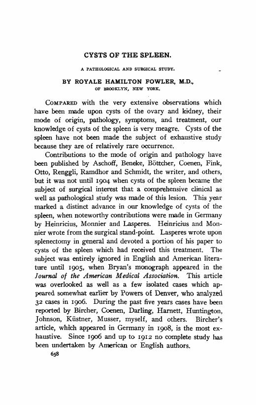
Parasitic cysts are uncommon. Echinococcus is the mostfrequent variety, and occurs in regions where hydatid diseaseprevails. The spleen was involved in 2 per cent. of casesof hydatid disease which Thomas studied. He collected 88cases in which the spleen was affected. In 45 of these thespleen alone was involved. The cysts may be unilocular ormultilocular, single or multiple (Fig. i). They may be sit-uated anywhere in the organ. The mode of entrance of echi-nococci into the spleen is of interest. Leukart has found themin the portal circulation, which explains their entrance into theliver. This is the site of predilection, but to gain entranceinto the spleen by this route the embryos would have to goagainst the blood strean. A possible explanation is that afterbeing freed from the capsule by the action of the digestivejuices they traverse the gastric and intestinal walls by boringor pushing their way directly into the spleen where this organis in relation with the stomach and colon. They may pushtheir way through actual or potential spaces, or they maytravel in the lymphatics. The extreme rarity with which theembryos are found in the lymph-glands and vessels speaksagainst lymphatic transference.
Cysts of non-parasitic origin are of especial interest, andit is this variety with which we are chiefly concerned. Thepresent contribution is an analysis of all the available cases,products of both the autopsy table and surgical clinics whichhave been published up to I9I2. Abstracts of these cases areto be found at the conclusion of the monograph. Subdivisionsof this variety have been classified as hemorrhagic, serous, and
659
ROYALE HAMILTON FOWLER.
lymphatic cysts. Such a classification is unsatisfactory. Theterms are misleading. Grouping based upon the character ofthe wall of the cyst or its lining is equally unsatisfactory.Some are of undoubted traumatic origin, others are possiblyinflammatory, still others represent true neoplastic processes.A distinction should be made between (i) haematomas, (2)cysts arising from the disintegration of splenic tissue (theresult of occlusion of vessels by emboli, endarteritis or amyloidchanges), and (3) genuine cysts of the spleen. True cystsare exceedingly rare. Pseudocysts will be considered as theyhave an important bearing upon the general subject. Cystsmay be situated in the peritoneal covering of the spleen (peri-toneal cysts) beneath the capsule (subcapsular), or deep inthe splenic tissue (intraparenchymatous). There may be asingle cavity (unilocular cysts) or many intercommunicatingpockets (multilocular cysts). They may be found in groupsor singly, isolated or confluent. When the cyst is solitaryit is uswally large and when multiple cysts are present they areusually small. Classification according to contents does notafford an explanation of the mode of origin. The writeroffers the following which suggests this:
(i) Traumatic cysts (haematoma, large unilocular cysts,secondary serous cysts).
(2) Infoliation cysts (traumatic or inflammatory inclu-sions of peritoneum). Small multiple superficial and deep.
(3) Dilatation cysts (ectasis of splenic sinuses).(4) Disintegration cysts (arising from arterial degenera-
tion and occlusion or other arterial occlusion as from emboli,and resulting in infarction and necrosis of parenchyma).
(5) Neoplastic types (hsemangioma and lynmphangioma).(6) Degeneration cysts (arising from secondary changes
in 5).Further investigation is essential to a satisfactory classifi-
cation of genuine cysts of the spleen. We may include underthis heading tentatively, infoliation cysts and dilatation cysts.
Etiology.-The age was stated in 66 cases in this series.In the majority the disease has affected persons in middle adult
66o
CYSTS OF THE SPLEEN.
life. In but two cases were cysts found in infants under onieyear. This would speak against their congenital origin, butwould not exclude the possibility. One case occurred betweenthe ages of one and ten years, 8 cases between the ages of tenand twenty, I5 between the ages of thirty and forty, I4 casesbetween the ages of forty and fifty, 3 cases between fifty andsixty years. In four cases cysts were found between theages of sixty and seventy years. Of those which were autopsycases, all occurred in individuals over thirty.
Females are predisposed to this condition. Of 65 cases nwhidc the sex was stated, 38 occurred in this sex. The ma-jority occurred between the ages of twenty and forty duringthe child-bearing period. Of the I4 cases which were studiedat autopsy, i occurred in a female. Monnier explains thepredilection for the occurrence during the reproductive periodof woman by the fact that the spleen becomes congested inpregnancy, during menstruation, and at the menopause, andsubsequently relaxes. In six cases in this series there seemedto be some relation between pregnancy and cyst formation.The contents of the cysts which showed relation to pregnancyare stated in five cases. Of these three were blood cysts andtwo lymphatic. In Bircher's case " something " was discov-ered in the course of pregnancy alongside of the uterus, whichsubsequent operation showed to be a displaced cystic spleen.Routier's case showed rapid increase in growth followingpregnancy and normal labor, which occurred one year pre-viously. In Well's case an already existing tumor was foundto be stimulated to increased growth by pregnancy. Conenoperated in the course of pregnancy and excised a spleen theseat of lymphangiectasis. The splenic tumor was first ob-served following previous childbirth. In the case obiserved bythe writer the abdominal tumor was noticed three monthsafter childbirth. Downs reports the development of a cystof the spleen, which he incised following confinement. Thewalls were necrotic. "Almost the entire spleen came awayin sloughs." The most rational explanation of the develop-ment of cysts of the spleen following pregnancy is that
66I

ROYALE HAMILTON FOWLER.
embolism which may occur in this state, leading to in-farction and secondary hemorrhage, may eventuate in cystformation. This explanation would seem to hold good in thecases of Bircher and Downs. Congestion of the spleen mayexplainthe increase in size of an already existing tumor. Theinfluence of pregnancy upon the formation of lymphatic cystsis not so apparent. Mechanical dilatation from pressure is apossible explanation.
Antecedent diseases of the spleen may exert an influence,especially malaria and syphilis. Hypertrophied spleens more-over are more likely to be recipients of injury, consequentlyhaematomas could readily form. Perisplenic adhesions areprone to occur in hyperplastic spleens. There seems to besome relation between perisplenitis and the formation of cysts.Brunswig-le-Bihan states that in Arabs, hemorrhagic cysts, dueto ruptures restricted by adhesions around the spleen, are notuncommon. Adhesions it must be remembered may be theresult of cyst formation as well. Malaria was stated to havebeen present in ten cases. The spleen itself was found to beenlarged in those cases giving a malarial history (in four casesclinical evidence of enlargement only). They were all uni-locular blood cysts except two. One specimen showed small,superficial multiple cysts with serous contents. A second hadserosanguineous contents. It is conceivable that spontaneousrupture of a distended capsule from congestion of the pulpincident to typhoid fever, etc., might predispose to cyst forma-tion. The relaxation and shrinking upon subsidence ofhyperaemia leaves the capsule in a wrinkled state with countlessfurrows which might easily become sealed over by inflamma-tion and result in infoliation cysts. The relation of peritonealcysts to spontaneous rupture of the capsule of an overdistendedspleen the seat of perisplenitis may be intimate. Subboticobserved small multiple surface cysts in malarial splenomegalywhich possibly originated in this way. Syphilis was men-tioned in two history records. In one the initial lesionoccurred six years previously (Garcia), in the other case thehistory of the infection was lacking, but there was evidence of
662
CYSTS OF THE SPLEEN.
the disease in the blood-vessels and viscera (Harnett). Asidefrom the greater danger to exposure to trauma to which anenlarged syph:ilitic spleen might be subjected, resulting in anhaemnatoma, its liability to perisplenic adhesions with the sub-sequent development of infoliation cysts, cysts may occurwithin the parenchyma of the spleen the result of specficendarteritis. Harnett considered that the spontaneous rup-ture of an intrasplenic blood-vessel was responsible for a casewhich he observed occurrng in a man who gave postmortemevidence of syphilis, although neither the spleen nor its ves-vels showed evidence of this disease. Bednar states that cystsof the spleen are frequently associated with pemphigus, anddescribes a small cyst occurring in a child of one week bornwith this disease. In one case observed by Fereal a largeabdominal tumor formed during an attack of mumps. Opera-tion subsequently revealed it to be a cyst of the spleen. Asidefrom malaria and syphilis there does not seem to be any rela-tion noted in histories which have been studied between dis-eases causing splenic enlargement and cysts of the spleen.
In the etiology of large cysts of the spleen the one mostgenerally acknowledged factor is trauma. Injury was statedto have preceded the development of cysts of the spleen in I7cases. The interval between injury and development wasstated in I3 cases. In 5 cases the cyst developed within iyear or less, in 7 cases within io years or less and in I casethe cyst did not develop until 40 years had elapsed. Such along period of tiEne would seem to exclude any relationship.Of those cases which showed a relationship to trauma in whichthe contents of the cyst was stated, I2 were hemorrhagic, iwas lymphatic and i serous. The injury has usually been adirect blow or a fall upon the abdomen. Long-continuedpressure of the left lower chest against a machine was men-tioned in one case and a penetrating wound of the abdomeninvolving the spleen was mentioned in another case. Thehistory of trauma cannot be taken as proof of the origin ofany particular kind of cyst although hemorrhagic cysts seemto develop most frequently. The relationship of cysts arising
663

ROYALE HAMILTON FOWLER.
from inclusions of peritoneal endothelium in consequence ofgross ihijury (traumatic infoliation cysts) is a theory whichshould be mentioned.
Among these etiologic factors must be considered t;he in-fluence of a twisted pedicle. This is suggested by its occur-rence in two cases. In Bircher's case the cyst was not heldresponsible. The pedicle was twisted upon itself about 360degrees. The cyst did not represent a large central area ofnecrosis which would follow the arrest of blood at the mainsource of supply. Under such an influence it is likely that theentire spleen and not a part of it would be involved. Aperipherally located cavity in a spleen otherwise intact couldhardly result from torsion of the main vessels. In Kiistner'scase the gastrolienal ligament showed undoubted evidenceof strangulation. He believes that this was tlhe cause of thecyst formation. The cyst occupied four-fifths of the spleen.The parenchyma was reduced to a small, narrow, wedge-shaped area. He is of the opinion that hemorrhage occurredfrom stasis and that destruction of splenic tissue resulted in thecavity. One can hardly reconcile such a cyst as this writerdescribes with such a degenerative process.
Pathogenesis.-Little has been added in recent years in ex-planation of the more intimate mode of origin. In no otherorgan can the pathogenesis be explained in such a variety ofways. This is proved by histologic findings. Each varietyoriginates in quite a distinctive manner. The genesis of cysticspleen seems to have no points of analogy to the genesis ofcysts of the ovary or kidney. In the absence of a tubularstructure true retention cysts do not occur. An association hasbeen demonstrated between non-parasitic cysts of the liver andcongenital anomalies, especially cystic kidney. There wouldappear to be no such association in the case of the cysts underdiscussion.
In 30 of the 43 cases in this series which showed hem-orrhagic contents, the history or the pathologic findings indi-cated a mode of origin. Seventeen resulted from trauma, 3from disintegration (2 from infarction and 2 from arterial
664

CYSTS OF THE SPLEEN.
degeneration). Eight were cysts into which secondary hem-orrhage had occurred (4 serous, 4 lymphatic). Two were tobe regarded as neoplasms (angioana). In this class, then,rupture of blood-vessels, spontaneous or traumratic, figuresmost conspicuously. Spontaneous rupture of diseased blood-vessels and hemorrhagic infarction are more rarely the factorsconcerned. Secondary hemorrhage is of frequent occurrence.It is difficult to determine the frequency with which disinte-gration cysts occur, inasmuch as evidence of ruptured intra-splenic aneurism, diseased blood-vessels, splenic infarction, andembolism would rarely be present after such a process hadformed a cyst.
In 22 cases analysis showed contents to be serous. ITelarge unilocular single cyst may be said to originate second-arily from the subcapsular haematoma or intraparenchyma-tous hemorrhage, the fluid contents of these becoming clearthrough the deposit of cellular elements upon the walls. Eightcases of large, single, isolated u.nilocular cysts were studied,of which two were thought to develop from hemorrhagic cysts,one from a twisted pedidle and one was thought to be trans-fonned from a multilocular lymphatic cyst through destruc-tion of the interior network. In two cases large, single, uni-Jocular cysts were associated with small multiple cysts. Inthree specimens cysts were, small, single, and situated deep inthe organ. Small deep multiple cysts, however, originate in adifferent manner. According to Boettcher they may arise inconsequence of amyloid changes which occur in the blood-vessels of the spleen, causing degeneration of the area sup-plied by them. This investigator claims that these degeneratedareas subsequently develop into serous cysts. He traced anarterial branch to the wall of such a cyst and to a point in theparenchyma which was involved where it suddenly stopped.
Beneke is of the opinion that small multiple surface cysts originatefrom the infoliation of peritoneal endothelium, in consequence of trau-matic rupture of the splenic capsule. It was claimed by him and hispupil Ramdhor that the splenic tissue protrudes through these ruptures,which he terms " hernias of splenic tissue," becomes snared off between
066
666 ROYALE HAMILTON FOWLER.
neighboring areas of parenchyma and forms cystic cavities. He con-siders the cells lining the cysts and the endothelium of the peritoneumidentical. M. B. Schmidt also holds Beneke's view and states that thesecysts commence as little red excrescences which extend over the surfaceand appear through the rents in the capsule. Schmidt states that almostall small superficial cysts originate from tissue hernias. He has foundmany such instances at autopsy in which there had been swelling ofthe spleen. Wohlwill believes that trauma is not essential to the develop-ment of infoliation cysts, but that rupture of the splenic capsule andhernia of the splenic tissue ensue from a rapid swelling of the organ.As the spleen continues to enlarge, rupture takes place at various points,peritoneuTn becomes turned in and snared off after the manner of adermoid.
Renggli describes cysts of the spleen lined with cubical epitheliumand offers the following explanation in regard to their development. Healso attempts to explain the occurrence of deep multiple cysts fromsnared off peritoneal endothelium. He substitutes inflammation fortrauma as a necessary precursor. Renggli believes that in consequenceof the fusion of proliferated areas on the surface due to inflammatoryprocesses the endothelium becomes snared off and in situations wheresuch areas have been pressed deep into splenic tissue is stimulated toenergetic growth and cyst formation, the cyst coming to lie deep in theparenchyma. He considers that the single layer of cuboidal epitheliumlining these cysts is the same as that which composes the peritoneum,except that the peritoneal endothelium of the surface has become flat-tened from the pressure of neighboring organs. He places the originof these cysts in the period of embryonic development, when the endo-thelium of the peritoneal cavity shows its original cuboidal character.
Otto is not of the opinion that deep cysts could arise after themanner of Renggli, but considers that small superficial cysts possessinga flat layer of cells unsurrounded by splenic tissue may have this origin.Ziegler in his text-book of pathology mentions the Renggli theory butstates that such cysts are rare. Kiihne has dealt with the subject andreports three cases. In investigating the cause he concludes that thetheory of Renggli is quite reasonable and holds good in one of his cases.
Efforts to determine the mode of origin through a studyof the lining have not proved successful, for as Aschoff has-pointed out the character of the cellular lining is not proof oforigin. Endothelium of lymph vessels could be transformedinto cuboidal epithelium. It would seem, however, that thereis some evidence to substantiate the peritoneal origin in a fewindividual cases. The theory of Beneke, his pupil Ranidlhor,and Schmidt would seem to present a more forced aspect thanthat of Renggli.

CYSTS OF THE SPLEEN.
Explanations thus far offered do not make clear the occur-rence of extensive multilocular cyst formations. Renggli'stheory of inclusions of peritoneum from superficial inflamma-tion would not seem to hold good. A different explanationmust be offered for the occurrence of countless fused cystsscattered throughout the organ. These are best explained bythe dilatation of the blood or lymph sinuses. Although theintimate natuire of these channels is not dearly understood,the origin of multiple, multilocular cysts has been attributedto the lymphatic system. We are indebted to Fink who firstrecognized this relation. He described several cases. Opinionsin support of this origin are held by Kiihne, Aschoff, andSchmidt. The cause of lymphangiectasis is not dearly un-derstood. In twelve cases which were thought to have origi-nated in the lymphatic system various explanations have beenoffered. Lymphangiectasis may be due to mechanical conditionsor inflamnatory processes. In no cases however has an inter-ference with the return flow of lymph been demonstrated nor-has there been any reason to suppose that an increased supplyof lymph had resulted in this condition. Lymph stasis alonewould not offer a rational explanation. The conglomerateand multilocular form which these cysts assume is to be ex-plained by the fusion of individual dilatations. Some of theseformations have been reported to be lymphangiomas, butwhether these are actually new growths or not is an openquestion. Misplaced lymph vessels or their cells of originmight become snared off during fetal life, proliferate and formsuch a cystic growth. We know that the spleen is an im-portant if not vital blood-forming organ during the develop-mental period, and it would appear that extensive changesoccurring at this time necessarily interfering with functionwould hardly permit of life prolonged to an adult age. Thiswould speak against the congenital lymphangioma of thespleen. Fink was successful in proving histologically thetransition of dilated lymphatic chann-els to large cystic cavi-ties. No sharp distinction can be drawn between lymphan-giectasis and lymphangiotna. The one possibly represents a
667

ROYALE HAMILTON FOWLER.
sliding scale into the other without a marked transition stage.It has been suggested by M. B. Schmidt that certain pre-formed canals existing in the peritoneal capsule and trabeculae,which he believes are lymphatic spaces, may form cysts.Schmidt holds iin part in this connection to the Beneke theoryof traumatic hernia of splenic tissue but believed that lymphcysts develop in this manner from snared off peritoneal endo-thelium, as well as multiple serous cysts. He has adoptedthis theory on the ground that some of the little superficialcysts show on their cut surface a cavenous structuire and anetwork of interlacing bands. Kiihne and Jamischita believethat cysts may develop from these lymphatic spaces in peri-toneal endothelium and trabecuiae.
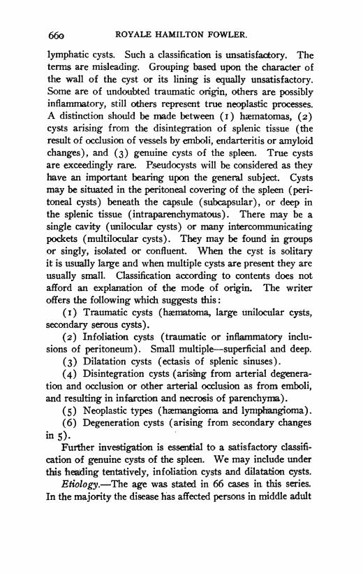
Pathology.-In 43 cases in this series the contents werestated to be hemorrhagic. Seventeen were subcapsular haema-tomas. These are usually large, single, and unilocular (Fig. 2).There is rarely any evidence of a recent or active hemorrhage.The wall of the subperitoneal haematoma is composed super-ficially of the capsule and upon the remaining sides the cyst issurrounded by parenchyma. There is no distinct epithelial lin-ing. The wall is composed of connective tissue if organizationhas taken place and contents vary according to the age of thecyst. These cysts may contain as much as ten litres of blood,which may. be coagulated or fluid, of a black or brown color,depending upon the stage of alteration. The contents usuallybecome fluid, thin, clear, or remain thick. The interior maybe irregular and show no definite encapsulating wall, or itmay present the appearance of an aneurism. Disintegrationcysts into which hemorrhage has occurred may present ne-crotic walls and show no evidence of embolism or infarction.Other cysts into which secondary hemorrhage has occurredand angioma need no description. Hamatoidin and choles-terin crystals are usually present in old cysts. Intraparen-chymatous cysts ariising from arterial disease may present thech,aracters of the above.
Of 22 cases of serous cysts eight were small superficialmultiple cysts (four of these were multilocular). Two cases
668

CYSTS OF THE SPLEEN.
of large isolated cysts were associated with small multiplecysts. Eight were large solitary cysts with a single cavity.
Small superficial cysts with serous contents as describedby M. B. Schmidt are frequently found on the anterior borderof the spleen, seldom upon the posterior border or convexsurface, and rarely upon the concave surface. These littlecysts give the organ a beaded appearance. They do not ex-tend into the parenchyma more than i cm. They are about0.5 cm. in height and seldom more than i cm. in diameter.Schmidt states that he has observed many cases post mortem.Mueller found these cysts of the spleen in i i per cent. in hisseries of autopsies. These small surface cysts occasionallyshow bloody contents. They are usually isolated and uni-locular. Ramdhor describes in addition deep furrows -in thespleen and at the anterior border dense groups of cysts thesize of a small pea, tense, and filled with clear fluid and occa-sionally withl mucous, colloid-like substance. He also de-scribes numerous excrescences scattered over the capsule re-sembling splenic pulp. These hernial projections were coveredwith a layer of epithelium. In rare instances an interlacingnetwork has been present in the interior of small superficialmultiple cysts.
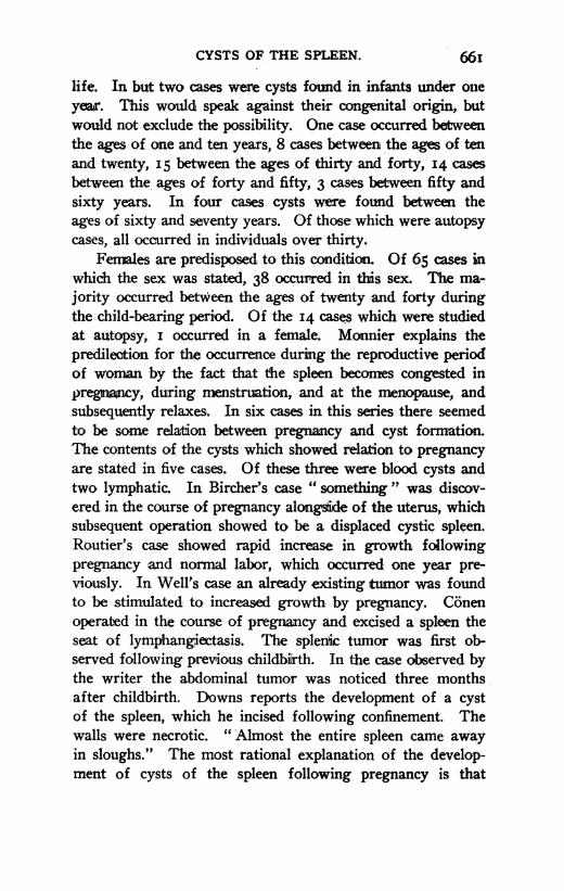
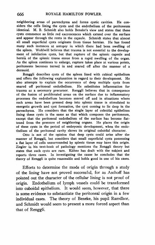
Solitary unilocular cysts with serous contents may be enor-mous and have been described as large as a foot-ball (Fig. 3).They project as a globular mass from the surface and aresmooth except for the occasional presence of adhesions. Theyare usually limited superficially by the splenic capsule beneathwhich thev are situated and partly surrounded by parenchyma.The smaller unilocular cysts may be entirely surrounded bysplenic tissue. This is true of those deep lying serous cystsoriginating from intraparenchymous hemorrhage and pulp de-generation. Those which have been transformed into theserous variety from the subcapsular haematoma may show asmooth interior of organized connective tissue from the de-posit of cellular material and fibrin. The walls nay appearsmooth, laminated, rough, or irregular. The interior nevershows an epithelial lining but occasionally a layer of fibro-
669

ROYALE HAMILTON FOWLER.
blasts or round cells. Boetcher describes multiple serous cystswithin the parenchyma, from the size of poppy to that ofhemp seed. He states that the pulp in his case was infiltratedand the capsule thickened. The vessels and connective tissueshowed amyloid degeneration. The cysts were close together.The smailler ones contained in addition to serum fat andfibrin. The walls showed an endothelial lining separated bysplenic tissue and a fibrinous septum. In places cavitiesmerged into each other. The vessel walls were greatly thick-ened by amyloid changes and constricted.
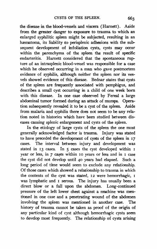
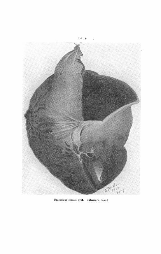
In twelve cases the diagnosis of lymphatic cysts or lymphan-gioma has been made in this series. In some instances findingsare at variance with our conception of these cysts. In the caseof lymphatic cysts, resulting from lymphangiectasis, the organis enlarged as a whole. Coenen has designated this conditionpolycystic degeneration( Fig. 4). Lymphangiectatic cysts havebeen described by Coenen and myself. The surface is irregularand the seat of countless projections. The cut surface showsinnumerable cavities, some isolated, others confluent. Thelatter in their interior present an interlacing network givingthe characteristic appearance of a multilocular cavity. Thecyst walls in places may be in immediate contact or there maybe a small amount of splenic tissue between them. Aspirationof these cavities before fixation of the organ and coagulationof their contents shows a fluid presenting the characteristicsof lymph. It is usuaUly a clear fluid of high specific gravityand high percentage of albumin. After fixation this materialcoagulates into homogeneous masses which stain a pink colorwith eosin. M-icroscopic examination shows lymph or bloodsinuses in various degrees of dilatation, which range in sizefrom the slightly dilated channel to large cystic cavities. Thesmaller cavities usually show no endothelial lining. The largershow a delicate connective-tissue wall and an endothelial liningwhich may show degeneration at various points. In someplaces it would appear that this lining has become retractedfrom the wall and adherent to the coagulated lymph. Cellularcontents are usually present. Hemorrhages are prone to occur
670

FIG. I.
Hydatid cyst of spleen. The spleen of a man from whose abdominal cavity severallarge hydatid cysts were taken. On its surface and partly imbedded in it are several thick-walled cysts of tough texture which contained acephalocyst hydatids. The largest cysthas been emptied; its internal surface is uneven and has flakes of yellowish lymph depositedon it. In another cyst near it and thickly lined with lymph there is a mass, composed ofrolled-up membranes of collapsed hydatids; and in another (at the back of the preparation)similarly rolled-up membranes have a deep amber color. In a fourth one several hydatidspresenting their ordinary appearances. (Museum Royal Coll. Surg., Eng., Hunterianspecimen, No. 2890.)

FIG. 2.
Hemorrhagic cyst of spleen. A spleen the lower part of which is replaced by an ovalcyst, measuring 6 in. in its longest diameter. The outer surface of the cyst is covered forthe most part by a layer of peritoneum, continuous with that on the spleen, and has shredsof great omentum adhering to it.
At the seat of attachment to the spleen, the substance of that organ is flattened outand forms part of the wall of the cyst. The inner surface is marked by numerous interlacingbands of fibrous tissue, which appear to be remnants of the splenic stroma. In recentstate the cyst contained 3 pints of brown, thick fluid like broken-down blood. Microscopicexaminations shows that the wall is composed of fibrous tissue permeated with large ves-sels. Masses of partially organized clot and granular material containing blood crystalsare attached to the inner surface of cyst wall, but there is no distinct lining membrane.
From a woman aged thirty-seven, who had noticed a swelling in the abdomen for eightyears. It was very elastic, and as it appeared to fill the abdominal cavity as high as theumbilicus it was considered to be an ovarian tumor. For three months previous to operationfor its removal the tumor had enlarged rapidly. The patient made a good recovery andwas in excellent health a year after the operation. (Museum Royal Coll. Surg., Eng., No.2887-A.).
1"1IG. 3.
-~~~~~~~~~~~~~~~~~~~~~~kP~~~~~~~~~~~~~~~~~~ p -1 i I|Unilocular serous cyst. (Musser's case.)
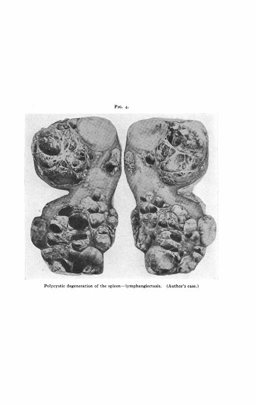
FIG. 4.
I
Polycystic degeneration of the spleen-lymphangiectasis. (Author's case.)
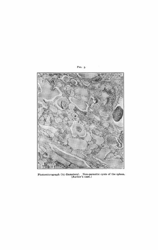
FIG. 5.
Photomicrograph (75 diameters). Non-parasitic cysts of the spleen.(Author's case.)
I
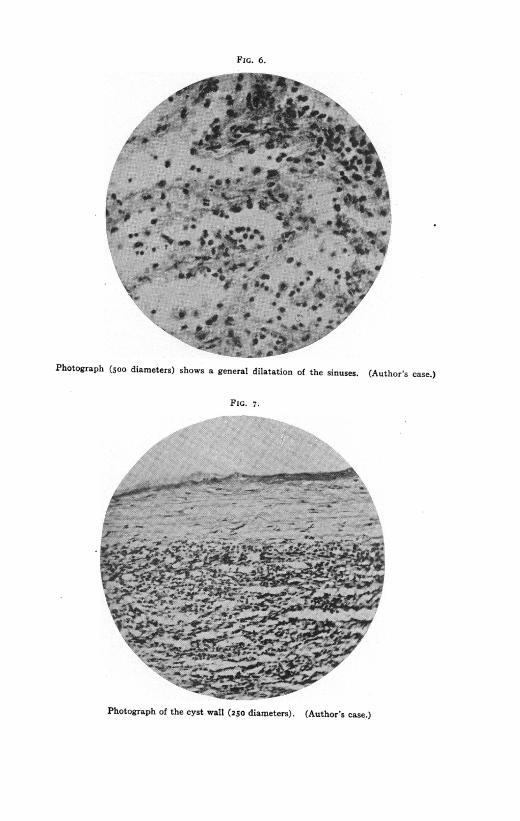
FIG. 6.
Photograph (5oo diameters) shows a general dilatation of the sinuses. (Author's case.)
FIG. 7.
Photograph of the cyst wall (250 diameters). (Author's case.)

FIG. 8.
Photomicrograph, showing section of the wall of a cystic spleen. (Author's case.)
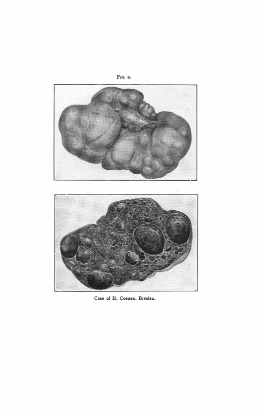
FiG. 9.
Case of H. Coenen, Breslau.
---

CYSTS OF THE SPLEEN.
into the larger cavities. The above description would hold fora lymphangioma were the process a localized conglomeratemass such as Fink has described at the hilum of the spleen.In these cases the organ itself is not materially enlarged.
Symptomatology.-Clinically, the most frequently recog-nized cyst is the large unilocular variety of hemorrhagic orserous type containing from one to ten litres. Intermittentor continuous extravasations of blood are never sufficient tocause symptoms of internal hemorrhage.
In general terns symptomatology is simple. In the ab-sence of any apparent function of the adult spleen there areno symptoms which are present as the direct result of the in-volvement of the splenic tissue per se. When cysts are smallthere are no symptoms. The small cysts have been accidentalpost-mortem findings. Large cysts usually cause symptomsfrom pressure. A low grade inflammation causes adhesions,which in turn may also result in symptoms. Intense peritonealreaction may excite severe pain, vomiting, and fever. In nota few cases have acute symptoms brought the patient to thephysician. The predominant symptom is pain. It may ex-press itself as a feeling of heaviness and be of a draggingcharacter. It is located in the left hypochondrium or referredto the epigastrium. A number of these cases have been mis-taken for movable kidney, so closely have the symptoms imi-tated this condition. If the spleen is dislocated pain may beexperienced in any part of the abdomen and radiate to thehypochondrium. The chief symptoms are from pressure.Digestive disturbances have been very frequently observed asa result of this. The stomach and intestines suffer from dis-turbed function by being crowded downward and to the right.Dyspncea and circulatory disturbances are more rarely presentfrom upward pressure upon the diaphragm. Obstinate consti-pation has been observed, more rarely diarrhcea. Frequentlythe patient has first observed the presence of an abdominaltumor which has gradually increased in size without pain(Heautreaux, Monnier, Routier, Wells). Growth is usuallyslow. In other cases reported mild repeated attacks of pain
67I
ROYALE HAMILTON FOWLER.
or dyspepsia have been the rule, or a long latent period hasresulted in a sudden severe outbreak. Occasionally symptomsof collapse have occurred frotn a twisted pedicle. Loss ofweight and strength have not been observed until the growthis large.
The tumor is usually located to the left of the umbilicus.A cyst of large size may extend from the lower border of thenibs to the median line and below the navel. Percussion re-veals a mass continuous with splenic dulness. The mass maybe freely movable or fixed. The surface may be smooth, irreg-ular, of doughy or elastic consistency. Fluctuation is notalways present. Monnier has drawn attention to a frictionsound which results from perisplenitis. It indicates thepresence of adhesions and is of no especial diagnostic value.There is usually no ascites except in the case of new growths.
Diagnosis of cyst of the spleen has rarely been madeclinically. Many have been recognized post mortem. Theprevious history of the patient is of the utmnost importance.Careful inquiry must be made in regard to trauma, therapidity of growth, 'its situation when first observed, the direc-tion of growth, and character and site of pain. The labora-tory, X-ray and ureteral catheterization may be useful in ex-cluding other conditions. The most decisive symptom is thediscovery of a tumor in the left hypochondrium which presentsthe characteristics of a cyst. Other abdominal cysts are ofmuch more frequent occurrence, and must be excluded beforea cyst of the spleen may be diagnosed. The diagnosis of en-largement of the spleen only may be made. It is usually notdifficult. It may not be possible to appreciate the fact that thetumor is cystic. Puncture through the abdominal wall fordiagnosis is never justifiable unless the abdomen is to be imme-diately opened thereafter. All other more common causes forsplenic enlargement must first be ruled out. A more refineddiagnosis than that of abdominal cyst or splenic enlargementcan rarely be made. Cysts of the spleen must be differentiatedfrom cysts of the ome , pancreas, mesentery and left lobeof the liver. If the cystic spleen is displaced ilnto the pelvis
672

CYSTS OF THE SPLEEN.
it may easily be mistaken for an ovarian cyst or floating kid-ney. Adhesions may occur between the uterus and the dislo-cated spleen and further simulate tumors of adnexal origin.
Treatrnent.-Cysts of the spleen have been treated sur-gically by (i) puncture, (2) incision and drainage, (3) ex-cision, and by (4) splenrectomy. Eight cases received treat-ment by puncture. In this series diagnosis of the character ofthe cyst was dependent upon examination of the aspiratedfluid. Four of these were said to have recovered. One deathwas caused by the operation. It was due to peritonitis andfollowed the injection of iodline. Reaccumulation of fluid tookplace in two cases. The fourth case which remlained uncuredresulted in a persistent sinus. Puncture and subsequent in-jection does not afford a complete cure and is a dangerous,unsurgical procedure. Within recent years it has not been per-formed. The last time was in I898, by Reimann. His patientrefused more radical treatment.
Fourteen cases were treated by incision and drainage.The result,is unstated in five cases, seven recovered, two died.Healing has been protracted and occurred in from threemonths to one year. Causes of death were sepsis (Powers)and peritonitis and splenitis (Quintard and Pean). Thismethod has been preceded in a number of cases by exploratorypuncture before laparotomy. Various methods of drainagehave been used, tampons in two cases, marsupialization inthree. A two-stage operation has been performeed. Follow-ing laparotomy and the establishment of adhesions betweenparietal peritoneum and cyst wall, incision is made and con-tents drained. This is an advisable procedure in case of para-sitic cysts if no more radical treatment is possible.
Excision of the cyst has been practised six times. Fourrecovered, one died, result unstated in one. The cause of deathin Bircher's case was post-operative intestinal obstruction frompressure by the spleen. No difficulty may be encountered inexcising the walls of the cyst if it is subcapsular. Radicaltreatment of the floor of the cyst may be effected by cauteri-zation (Bardenheuer). Hemorrhage may be controlled by
673

ROYALE HAMILTON FOWLER.
clamp at the hilum. Partial-splenectomy (excision of the cyst-bearing tissue) is rarely justifiable. This procedure is at-tended with grave danger from hemorrhage. It has beenaccomplished by Bircher, who controlled hemorrhage by in-terrupted sutures placed around the cyst. It has been success-fully performed by Gussenbauer. It must be regarded as avery formidable operation as these cysts are rarely if everpedunculated.
Splenectomy is the treatment of choice in cases in whichthe operation is not contraindicated by the presence of exten-sive adhesions. If there is polycystic degeneration (Coenen,Fowler), or if the spleen is otherwise extensively affected bypre-existing disease (malaria), or if the spleen is materiallyenlarged as a result of the cyst, if there is displacement, elonga-tion of the pedicle with danger of torsion, excision of thespleen is the best treatment.
Pean was the first to perform splenectomy for cyst (I867).Johnson in an admirable monograph upon splenectomy hascollected from various sources I9 cases of cysts which weretreated in this way without mortality. All these cases oc-curred prior to January, I908. Three cases in Johnson'sseries are not included in my list of splenectomy cases (Bac-celli, Israel and Gerard). The case of Hedinger, reported inI906, was overlooked by Johnson. In all I have been able toobtain data of 27 cases of cystic spleen treated by splenectomyup to 19I2. The operative result is not stated in two cases(Royster, Boechelman). One died, Homans (angio-sar-coma?), 24 recovered.
In selected cases permanent recovery may be secured byless radical means. The subsequent changes, glandular swell-ing, lymphatic and thyroid, leucocytosis, and other transientpost-operative phenomena are, however, not of sufficientgravity to contraindicate the removal of the spleen. The lessradical procedures were undertaken at a time when splenec-tomy was considered a grave operation and before it wasdemonstrated that excision was not followed by serious im-pairment of the general health.
674

CYSTS OF ;T ,$LE;N.
ABSTRACTS OF CASES OF NON-PARASITI CrST$X Q iT.HE .SPLW iWHIHAVE BEEN FOUND POST MORTEM.
ANDRAL (I829): Spleen contained smiAl e-0tgiltd With' gef6isfluidscattered throughout the organ, isolated arid Pflui4t. ee iib1d f~rmations seen in cystic cervicitis of the uterus. /
BARBACCI (I89I): Cyst of the spleen the size of`h'46sl egJ cbiithihietransparent, viscid fluid the color of gold, fibrinout cdakgiltu, aid ilbit'min. This was diagnosed a lymphatic cyst.
BEDNAR (I850): Small cyst in a child of one we<be&*n withpemphigus.
BOETTCHER (I870): Multiple serous cysts the size ofa l-Ritsks^t-that of a pea. Some traversed by trabeculae, lined with end6thdliiiri.
CHAVIER (I902): Male. Seventeen months previously ieelvedblow in the region of the spleen. Digestive disturbances followod,-lat6tan attack of sudden severe abdominal pain with tympanites, diagoidsddintestinal obstruction. Death in two days. At autopsy a tumor the §iteof a Savoy biscuit was found in the left hypochondrium which originatedfrom the spleen and rested upon the anterior wall of the stomach. Thecyst was compressed, and in contact with the abdominal wall, extendedforward under the false ribs, inward three inches beyond the medianline and downward to the level of the navel. There were no adhesions.Cyst thought to originate from subcapsular hemorrhage. ContainedI87o Grams of reddish-brown fluid. Spleen weighed 500 Grams. Theinterior of the sac was covered with reddish fibrinous deposit, com-mencing organization. The spleen pulp was very soft. Rupture of thestomach.
FINK (I890): Three cases. (a) Male aged 48. Spleen was occupiedupon its convex surface by countless projections of varied shape andsize. Spleen slightly enlarged. Section showed cystic cavities inter-mingled with apparently normal splenic tissue. Most of the spaces werefilled with yellowish-red masses. The walls of the cavities possessednumerous projecting ridges. Cavities were separated by bands of con-nective tissue. The interior of the cavities was lined with endothelium.Contents consisted of homogeneous masses, fine and coarse granulardetritus, and here and there a lymphoid cell. (b) In the museum of thePathologic Institute in Prague. Spleen enlarged a trifle. Numerousprojections the size of a cherry pit and larger were scattered over theconvex surface, especially on the borders. The cut surface is similarto Case I. In but few places could the character of the lining be recog-nized. Contents similar to Case I. (c) Upon the anterior portion ofthe spleen was found a cyst as large as a hen's egg. Contents clear,cholesterin crystals. Walls consisted of thin fibrous membrane (epi-thelial lining washed off by the alcohol ?). In the region of the cystthe endothelium lining the lymph spaces was slightly degenerated. Inthis case destruction of the connective-tissue bands of the interior of thecavity had occurred, resulting in a single cavity. In the splenic tissuewhich showed little change there was a sharp demarcation between cystand parenchyma. The convexity of the cyst is formed by the capsule

676 ROYALE HAMILTON FOWLER.
of the spleen. A boundary of splenic tissue cannot be demonstrated inthis situation.
FozRTs: Spleen in the Wiirtzburg collection. Serous cyst size ofa hazel-nut with thick cartilaginous walls.
GAUGET: Male, aged 46. Died in collapse. History of alcoholism,dyspncea on exertion. Spleen enlarged, contained yeUowish fluid inrounded regular pockets, size of a hen's egg and walnut.
Guio and VINARDI: Calcified cyst of the spleen the size of a smallnut containing albuminous fluid which effervesced in hydrochloric acid.
HARNETT (I907): Male, aged 40, history of malaria. Lues negative.Suppurating aneurism of the popliteal artery, arteriosclerosis. Enormousunilocular cyst IoX6 inches containing a pint of dark altered blood.Unsuccessful attempts to locate a branch of the splenic artery enteringthe cyst cavity were made. Slight fibrosis, large amount of free ironin pigment. Slight amyloid degeneration of trabecula. Outer layer ofcyst wall fibromuscular, middle composed of splenic tissue, inner fibrous,no cell lining.
HASWELL: Male, aged 57. Died of cancer. Sense of fulness andfluctuation noticed in the left hypochondrium before death. Spleen andcyst weighed three and three-quarters pounds. On opening same, pul-taceous matter escaped of dirty yellow color in rounded masses. Amongthem were round jelly-like globules the size of a walnut which con-tained a clear fluid (echinococcus?). Diagnosis, mucus cyst.
HUNTERIAN MUSEUM of the Royal College of Surgeons. One speci-men of hemorrhagic cyst of the spleen, one serous cyst of spleen of an ox.
K.Fm and LEFAS (i898): Male, aged 72. Death from chronicmeningitis and myelitis. Cysts in the kidneys. Senile alterations in thevascular system. Spleen weighed 235 Grams. At the anterior border acyst the size of a large nut, containing clear, yellow fluid. Inner surfacewhitish, in places ecchymotic. The cut surface showed an appearancesimilar to the ventricles of the heart. Within the parenchyma was anothercyst the size of a small nut which was easily enucleated. Its walls werethin and greyish-white. Contents clear, light red. Microscopic exami-nation showed walls to' be composed of connective tissue i mm. inthickness, small round cells in places. No endothelial lining. Splenicparenchyma normal.
KUHNE: Three cases. In two cases the cysts were situated beneaththe capsule. In one case the cyst lay in a trabeculum and its wall waslined with endothelium. The third case resembled that described byRenggli. On the capsule were found countless projections of connec-tive tissue rich in blood-vessels. The small cysts lay close together. Thecysts were lined with high epithelium, contents consisted of homogeneous,granular material with a few round cells. The first two were diagnosedlymphatic cysts, the third he believed originated from snared off peri-toneal endothelium.
LANGHANS (I879): Pulsating, cavernous hemangioma. Male, aged30. Stroma fibrous. Interstices lined with endothelium. Contents con-sisted of blood.

CYSTS OF THE SPLEEN.
LEUDET (I853): Female, aged 6o. Mitral affection. Volume andsurface of spleen normal. Large cyst in the parenchyma separated intofour or five cavities. Wall of fibrous tissue lined with endothelium.Contents, serous.
Livois (I838): Male, aged 46. Tertian malaria. Greatly enlargedspleen containing cyst the size of an ostrich egg. Spleen and cystweighed seven pounds. Contents serosanguineous.
MATTEi (i885): Male, aged 68. Spleen enlarged, weight 370 Grams.Four-fifths of the organ occupied by a tumor the size of an orange,covered in part by the capsule of the spleen. The wall was hard, car-tilaginous in some places, calcified in others. Contents, clear albuminousmaterial. Surrounding this cyst were I7 others, ranging in size fromthat of a hemp-seed to that of a large nut. Most of them were filledwith serous fluid, chlosterin, granular material, and detritus. Leucocyteswere found in the smaller cavities. The cyst walls of the larger werecalcified. The walls of the smaller were twice as thick and composed ofconnective tissue. The interior of the large cyst showed two semilunarfolds partly calcified. This cavity communicated with another mediumsized one.
PICCINELLI: Male, aged 67. Digestive disturbances for thirty years.Pain in the left hypochondrium. Spleen contained a hard tumor thesize of a hen's egg, situated at the hilum and full of knobs; incised withdifficulty, escape of viscid gelatinous fluid. Diagnosis, ossified, cystictumor.
PORGE: Puro-sanguineous, size of a walnut. Easily enucleated.Contained yellow, grumous, odorless fluid, red blood-cells, pus, and choles-terin.
SPILLMAN (1876): Male, aged 51. Tumor the size of a fetal head,which projected from the anterior border and inner surface of the spleen.Weight 655 Grams. The walls were about 2 mm. thick and consisted ofspleen substance, connective tissue, calcareous areas, and endothelium.Vessels normal. Contents 300 Grams of fluid of yellow color containingcholesterin crystals and red blood-cells, deformed epithelial cells, andheematoidin crystals.
WILKS observed a cyst of the spleen the size of a walnut in an oldman who died of chronic nephritis. Few small cysts on the surface.Contents serous, lining smooth and membranous.
Similar specimen to Wilks in Guy's Hospital Museum found in apatient who died of chronic nephritis.
VIRCHOW demonstrated a small cyst with tough walls the size of alarge cherry in i898. The contents was cholesterin. The spleen was notenlarged. He expressed no opinion in regard to its mode of origin. Ina second specimen observed by him in I873 multilocular cysts werepresent.
NON-PARASITIC CYSTS OF THE SPLEEN WHICH HAVE BEEN TREATEDBY PUNCTURE.
BACCELII (I897): Female, aged 27. Came in contact with a cupboardand injured her left side. A few days later upon arising she experi-

678!dnced-'pain,' whihiwas'! suddeti "and 'se4eve.- Thig piassed -dff quickly butreturred in few 'days wifth igreater everity." PiinIti9sed in twomonthsil*t"Xtpatient hotkedi't this titi* g'gtoth idr the left 'iiy#ochon-drium which slowly increased in size. The surface of the growth wassmanotls, coiaistency soft, elastic, b.nd 'dis6tintly `flictuatitg. The; iass*as freey' miovable,: 'pain1ese, and adherett tb thel lo*t';pi6le of 'thespleen. Two exploratoey "paticturesl libeattd blood.;' 'The swelling 'dis-appeared' £oar 6ne month,t When, fltii re-acicutilatel. Thoi'httbi S 'sub-capsukir hemorrhage. " 'C-: MxIn.-MaLe, aged 46. " Interhiittent fever for fourmttith. 'En-
larged'spleen. 'Acute' hypothondriac pain 'and swelling utider the 'ibs onthe left' sk",of',twelve days'- durationw, Tumor hard, ti6t teider. iAteefflutituationr. 'Puictdre-lileratedl'degenerated biood. Reovery.;'"GARCA: Male;-aged '36.'> S*philis'sixt'years'peviously.'i iTerian' feverctrred by"qfiini1e.' Fil -frbrh * horse -'a&tsedd7 i ' bdbmintal ntohtusifn,followedlini onden;month by 'local'pain, idigi§tibn,;'ad' constipatioin. Timor'i'i the left hyp6chondrium.- Punttuie libetat;&ed Iodbess gas, and; tu±ti6rditnihished iii' Size."iOnn the 'f4olowing* day a seond pn'ctur& 'liibratedone quart of clear, transparent, white wine-colored fluid, specific igravityI43O4.'SConitimed albuminifid sodiuni' chldride. A third puncture inth'+e 'mobnthS, 300 Grams Of fluiid iob',tained; "Rec0ver5r ,i! ii*MARCAt;o and 7FEd.A Mle, aged 33. Miliri` fot some years. Atthis 'time 'spIenic' hl1dt'e&ient' 'Dyspepia*. '-Ti years atg, nausea add'vomiting after meals. Fluctuating tumor in the epigastrium and left'hipbltiotidrium` Punctre'tesultdd in 'the-'Withdratal 'of `3oo Grais ofch,oidatC,'tolorede Autid, 'ttaitiing' 'blood-cell.-Treited by 'injections -of,iodine and potassium permanganate. Recovery. Three months latet 4asniall"fIula'about 'cm. deep dtill persisted.i;! --
"LIALI : Mal,t ag*d 33'' Malria. Six' 'm'Othg'-orevieusly injutyover'ther left hypochb:dti. Punctureliberated 'degenetated blood.rPkAPi 'nid Rousr' (i863')': Femate,' aged Y. -' Serosangtineous cyt
dilLgdtbkd'b undttctke,' treated;bjr catstict. 'Death ih two mtonths fromperitonitis .' " ''
REIMANN (I9oI): Female, aged 33. Weakness and pain ih the leftside and ' lubar region. 'No infectious diseages. Tumor correspondedwith tbieen*arged stlen.!';Heartpushed tothe right.' In' I898 exploratorypuncture liberated choc6lat&' OlcAred 'fluid which 'cohtained' albumifn,'detritus,'red biood-&ells,'Whddvaskfih 'in chlaksterin.' Secnd putnctureten days later, 2400c.c. withdrawn. '-Weing etittiry 'disappeated, 'heattrttlrned 'to the 'left.' Cyt 6sbn"'-efil1ed.! Repetted punctures.; Opera-tidoii efufsdd. PatiSnt disdiard uktiueit-ud.,
V!tktkitr ste'thftthe'1h` sen 'fofircases of' htMorrhagccyst ofthe ' spleen,jthree' ofihich'- hiave Ibeeficred by trocar. -
ABSTRACTS OF CASES WHICH HAVE BEEN TREATED BY EXCISION''' [ts'>1I "'T* ' ''AN D '* GAND D AI&G'
BAGINSKY (I898): Female, aged 13. Swelling of the left sides'iortl' after she' 'had fallen' directly' upon "the abdomen. 'In; the lefthy*lhondu'iuni art' eldstie flactdathig7 growt extended''ovei th- median
ROYAILP, 44AMILT(J9 #OWLIER.
CYSTS OF THE SPLEEN.
line to the right, the lower edge a little below the umbilicus. Explora-tory puncture, turbid brown fluid containing numerous red blood-cor-puscles, partly degenerated, a colorless stroma, sparkling globules, andcholesterin. Blood examination normal. Operation by Gluck. Incisionmade over the cyst, contents emptied and iodoform gauze tampon placedin the cavity. Cure in i6 weeks. Chemical and microscopic analysisshowed contents to be altered blood. When wound had healed spleenwas still enlarged and firm. A fragment from the cyst wall showedconnective tissue and splenic parenchyma.
BARADULIN (i9o6): Male, aged 34. Trauma four years previously.Diagnosis, hemorrhagic cyst. Incision liberated 3000 Grams of fluidblood. Healing in five months.
BoccHIA: Male, aged 24. Traumatism over the spleen six yearsbefore. Splenic enlargement for three and one half years. Punctureresulted in the removal of dense chocolate colored alkaline fluid. Nextday cyst was evacuated by incision, when 2I40 Grams of fluid wereremoved. Recovery.
DARLING, C. G.: Female, aged i8. When I5 years of age suddensevere pain in the left side. Temperature, sweating for one week.Splenic enlargement. Operation. Sac of cyst adherent to the diaphragmand omentum, which prevented excision of spleen. Cyst opened anddrained, space packed.
DowNs, A. J.: Female, aged 29. April, i886, pain in the left side.Confined in I888. Operation. Trocar introduced and large quantity ofblood obtained. Sac incised, walls of cavity necrotic. Cavity drained.From time to time portions of necrotic tissue were discharged. Dis-charge of sloughs continued until almost the entire spleen came away.Microscopic examination of spleen tissue showed hyperplasia and degen-eration of follicles.
HEURTAUX (I898): Female, aged 27. Injury eight years previously,later fluctuating tumor. Operation. Large cyst from which 9q4 litres ofchocolate colored fluid were removed. Contained cholesterin. Cystopened and cavity curetted, irrigated, and marsupialized. Injection ofiodoform-ether and chloride of zinc. Suppuration. Healing in one year.Recovery.
LEONTE in igoi reported two cases, both females. Treated by mar-supialization; both unilocular, serosanguineous cysts. Capacity, I4oo and2000 c.c. Recovery in both.
LEDDERHOSE (I887): Aged i6. Fluctuating tumor of the spleen.Operation by Riedel in two stages. It had been decided not to open thecyst until adhesions had formed to the parietal peritoneum. Spontaneousrupture occurred, liberating a quantity of yellow serous fluid containingcholesterin. Wound healed in three months.
LEJARs (igoi): Female, aged 43. Para vi. In I897 patient fell onthe stairs, striking upon the left side. May, Igoo, severe pain in theepigastrium, vomiting, and diarrhcea. Du-ring the next six months swell-ing appeared, pain continued in epigastrium and left hypochondrium.December 22, I900, puncture in the splenic region, brown fluid obtained.
679

680 ROYALE HAMILTON FOWLER.
Two dais later puncture in the epigastrium, fluid of same character.January 3, I90I, chill, fever, and vomiting. Tumor tense and sensitive.January 5, incision in the lumbar region. One and one half litres offluid obtained. Wall of sac covered with dark, brittle, regular forma-tion, and particles of fibrin. Sac irrigated and drained. Lejars believedthat hemorrhage was responsible for the formation of the cyst and thatthe walls were composed of splenic capsule.
PowmEs, C. A.: Male, aged I8. Large left-sided abdominal cyst offour years' growth. Gradual loss of weight and strength, general pres-sure symptoms, and headache. Operation. Wall of tumor was one-halfinch in thickness, semicartilaginous and adherent to adjoining structures.Removal impossible. Incision and drainage. Walls did not collapse.Patient died of sepsis on the twelfth day. Anatomical diagnosis, uni-locular, hemorrhagic cyst of the spleen.
QUINTARD and PEAN (I879): Female, aged 53. Hemorrhagic cysttreated by caustic. Death following opening of the cyst from splenitisand peritonitis.
SUBBOTIC: Two cases. (a) Male, aged 30. Beneath the ribs upon theleft side protruded a growth the size of a man's head. Incised and I500c.c. of fluid obtained. Adhesions between the tumor and abdominal wall.Peritoneal cavity was not opened. Contents composed only of alteredblood-cells. Interior of cyst cavity was smooth, walls tough. The cyst wassituated upon the spleen and occupied a portion of the interior of theorgan. (b) Male, aged 21. Growth larger than a man's head. Situatedbeneath the left costal arch and extended to the right mammary line.Incision, bloody fluid evacuated. The anterior wall of the cyst was aboutan inch and a half in thickness. There were many hard adherent coagulain the cavity.
ABSTRACTS OF CASES WHICH HAVE BEEN TREATED BY INCISIONAND PARTIAL SPLENECTOMY.
BARDENHEUER (I8go): Male, aged 47. Traumatism at seven years.Symptoms of low abdominal pain referred to the left hypochondrium,marked digestive disturbances, severe gastric pain, eructations, vomiting,a feeling of violent tearing in the epigastrium, and obstinate constipa-tion. At the time of operation the cyst was the size of a child's head,adherent in the pelvis. Spleen incised, hemorrhage controlled by clampat the hilum and by cauterization in the parenchyma. Excision of cyst.Spleen returned to normal position. Recovery. The convex surface ofthe spleen was atrophied and merged into the wall of the cyst imme-diately under the spleen, which was 2 mm. thick. The surface of thecyst was smooth except in a few places where there evidently had beenadhesions to the surrounding tissues. In the upper portion of the cystwhere it came in contact with the atrophied parenchyma of the spleenthere was hard calcium deposit in the wall. The interior of the cystshowed many strands of connective tissue in the form of flat bands whichinterlaced to form a network. The wall consisted of connective tissue
CYSTS OF THE SPLEEN.
in which there was embedded blood pigment and calcium. No epithelia!lining was evident. The splenic parenchyma was more than half atrophiedand merged gradually into the cyst wall. Contents thin and of dirtybrown color. Microscopic examination showed considerable blood pig-ment and cholesterin crystals.
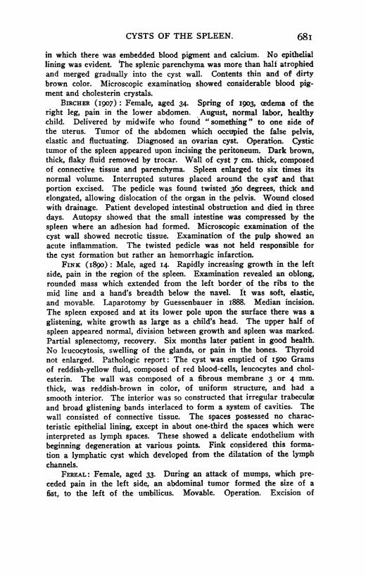
BIRCHER (I907): Female, aged 34. Spring of I903, cedema of theright leg, pain in the lower abdomen. August, normal labor, healthychild. Delivered by midwife who found " something" to one side ofthe uterus. Tumor of the abdomen which occupied the false pelvis,elastic and fluctuating. Diagnosed an ovarian cyst. Operation. Cystictumor of the spleen appeared upon incising the peritoneum. Dark brown,thick, flaky fluid removed by trocar. Wall of cyst 7 cm. thick, composedof connective tissue and parenchyma. Spleen enlarged to six times itsnormal volume. Interrupted sutures placed around the cyst and thatportion excised. The pedicle was found twisted 360 degrees, thick andelongated, allowing dislocation of the organ in the pelvis. Wound closedwith drainage. Patient developed intestinal obstruttion and died in threedays. Autopsy showed that the small intestine was compressed by thespleen where an adhesion had formed. Microscopic examination of thecyst wall showed necrotic tissue. Examination of the pulp showed anacute inflammation. The twisted pedicle was not held responsible forthe cyst formation but rather an hemorrhagic infarction.
FINK (I890): Male, aged I4. Rapidly increasing growth in the leftside, pain in the region of the spleen. Examination revealed an oblong,rounded mass which extended from the left border of the ribs to themid line and a hand's breadth below the navel. It was soft, elastic,and movable. Laparotomy by Guessenbauer in i888. Median incision.The spleen exposed and at its lower pole upon the surface there was aglistening, white growth as large as a child's head. The upper half ofspleen appeared normal, division between growth and spleen was marked.Partial splenectomy, recovery. Six months later patient in good health.No leucocytosis, swelling of the glands, or pain in the bones. Thyroidnot enlarged. Pathologic report: The cyst was emptied of I5oo Gramsof reddish-yellow fluid, composed of red blood-cells, leucocytes and chol-esterin. The wall was composed of a fibrous membrane 3 or 4 mm.thick, was reddish-brown in color, of uniform structure, and had asmooth interior. The interior was so constructed that irregular trabeculaand broad glistening bands interlaced to form a system of cavities. Thewall consisted of connective tissue. The spaces possessed no charac-teristic epithelial lining, except in about one-third the spaces which wereinterpreted as lymph spaces. These showed a delicate endothelium withbeginning degeneration at various points. Fink considered this forma-tion a lymphatic cyst which developed from the dilatation of the lymphchannels.
FEREAL: Female, aged 33. During an attack of mumps, which pre-ceded pain in the left side, an abdominal tumor formed the size of afist, to the left of the umbilicus. Movable. Operation. Excision of
68I
ROYALE HAMILTON FOWLER.
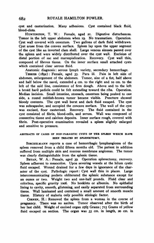
cyst and cauterization. Many adhesions. Cyst contained black fluid,blood-clots.
HUNTINGTON, T. W.: Female, aged 21. Digestive disturbances.Tumor in the left upper abdomen when ig. No traumatism. Operation.Cyst wall covered with omentum. Two gallons of dark fluid withdrawn.Cyst arose from the convex surface. Spleen lay upon the upper segmentof the cyst like an inverted clam shell. Large venous sinuses passed overthe spleen and were widely distributed over the cyst wall. Excision ofdistal portion of sac and marsupialization. Recovery. Cyst wall thin,composed of fibrous tissue. On the inner surface small attached cystswhich contained clear serous fluid.
Anatomic diagnosis: serous lymph variety, secondary hemorrhage.TmiRER (i892): Female, aged 33. Para iii. Pain in left side of
abdomen, enlargement of the abdomen. Tumor, size of a fist, half aboveand half below the navel, extended 4 cm. to the right and io cm. to theleft of the mid line, consistence of firm dough. Above and to the lefta brodd hara pedicle could be felt extending toward the ribs. Operation.Median incision. Small intestine, stomach, omentum being pushed to oneside, a round reddish-brown tumor became visible. Puncture showedbloody contents. The cyst wall burst and dark fluid escaped. The cystwas subcapsular, and occupied the concave surface. The wall of the cystwas excised, floor cauterized. Recovery. The fluid contained in thecyst consisted of fibrin, blood-cells, and serum. Wall was composed ofconnective tissue and calcium deposits. Inner surface rough, covered withfibrin. Post-operative examination revealed a spleen slightly enlargedand sensitive to pressure.
ABSTRACTS OF CASES OF NON-PARASITIC CYSTS OF THE SPLEEN WHICH HAVEBEEN TREATED BY SPLENECTOMY.
BOECKELMANN reports a case of hemorrhagic lymphangioma of thespleen removed from a child fifteen months old. The patient in additionsuffered from multiple skin and mucous membrane angiomas. The tumorwas clearly distinguishable from the splenic tissue.
BRYAN, W. A.: Female, aged 39. Operation splenectomy, recovery.Spleen adherent to mesocolon. Upon severing vessels at the hilum cysticfluid escaped. Wound drained for a few days in ignorance of the char-acter of the cyst. Pathologic report: Cyst wall thin in places. Largeintercommunicating pockets obliterated the splenic substance except foran ounce or two. Weight two and one-half pounds. Fluid clear andcolorless, specific gravity Ioo6. No hooklets or albumin. No epitheliallining to cavity, smooth, glistening, and easily separated from surroundingtissue. Wall laminated and contained a small amount of smooth muscletissue. History of malaria only possible etiologic factor.
COENEN, H.: Removed the spleen from a woman in the course ofpregnancy. There was no ascites. Tumor observed after the birth ofher last child. Weight of excised organ 2565 Grams; 7I5 Grams of yellowfluid escaped on section. The organ was 33 cm. in length, 2o cm. in
682

CYSTS OF THE SPLEEN. 683breadth, io cm. in thickness, of grayish-blue color, and distinctly knobbed.The projections were largest at the poles and on the concave surface.They proved to be thin walled cysts which in places were composed ofthe capsule only. On section the organ presented a honeycombed appear-ance. It was riddled with smooth walled cysts which varied in size. Theinterior of the cysts was gray, smooth and glistening, and traversed bytrabeculae. A few cavities contained fresh blood. Most of them con-tained a clear watery fluid. By pressure upon the organ the fluid indistant lying cysts was moved so that we might assume that all were incommunication. In the sections little normal splenic tissue could bedemonstrated. Where pulp remained it was in a state of chronic inflam-mation. Here and there were to be seen a few follicles. Except forthis the entire organ was transformed into cystic cavities. The cysts inplaces were separated only by a thin connective-tissue septum. The con-tents were homogeneous masses staining well with eosin, in which werescattered a few lymphocytes. Coenen speaks of this condition as one oflymphangiectasis.
CREDE (i883): Male, aged 44. Injury ten years previously. Suf-fered pain for five days. Later, swelling size of a fist in the left hypo-chondrium rapid increase in size to that of a fetal head. Operation, leftrectus incision; I350 Grams of fluid drawn off by puncture, scant supplyof albumin, numerous cholesterin crystals. Profuse hemorrhage fol-lowed incision into the cyst which was the size of a fifty-cent piece. Wallconsisted of a membrane which gradually thickened into splenic tissue.Pedicle ligated, spleen excised. Weight of spleen without cystic fluid,380 -Grams. Parenchyma normal upon microscopic examination. Theinterior of the cyst showed a structural network interlaced with trabeculaand lined with endothelium. Four weeks after operation swelling of thethyroid. One week after operation leucocytosis. No swelling oflymphatics or pains in the bones. Thyroid swelling disappeared in fourand one-half months. Patient in excellent condition ten and one-halfmonths after operation.
DALINGER: Female, aged 44. Had malaria for one year. Three daysbefore admission to hospital had an attack of syncope. At operation athin-walled cyst disclosed extending from the epigastrium to the lefthypochondrium. It was adherent to the abdominal wall and diaphragm.Beneath the capsule was a considerable amount of fluid and coagulatedblood. Recovery followed splenectomy.
FoWLER, R. H. (igio): Female, aged 22. One year ago the patientfell backward a distance of two and one-half feet. She experiencedlocalized pain upon exertion for a period of three months in the lefthypochondrium which was relieved for a time by an abdominal belt. Shehad not complained of pain for the past six months. Three monthspreviously she was delivered of twins and since that time she complainedof experiencing a sensation of something moving during exertion. Aprobable diagnosis of movable kidney was made and operation advised.Upon opening the abdomen a much enlarged spleen presented. A leuco-cyte count was made while the patient was under ether and showed

684 ROYALE HAMILTON FOWLER.
22,ooo leucocytes; differential count gave 59 per cent. polynuclears, 41per cent. lymphocytes (large and small). Splenectomy by Dr. B. F.Curtis. The patient made an uneventful recovery. Following splenec-tomy the leucocytes were as follows: Two days after operation, 23,600;polynuclears, 72.5 per cent.; lymphocytes, 27.5 per cent. Eight days after,2o,20o leucocytes, polynuclears 63 per cent.; lymphocytes 37 per cent.Seventeen days after, I8,9oo leucocytes, polynuclears 74 per cent.; lympho-cytes 26 per cent. Twenty-eights days after operation, 14,200 leucocytes;polynuclears 55 per cent.; lymphocytes 41 per cent.; eosinophiles 2.5 percent.; basophiles I.5 per cent.
Pathologic report (Fowler): The gross specimen consists of an en-larged spleen 20 cm. in length. It is broadest at the poles, where itmeasures ii cm. The thickness at these points varies between 6 and 7cm. The surface is lobulated, especially at the poles, and there are manysmall cysts projecting from the surface. At the centre of the organ is aconstriction, separating the spleen into two quite distinct lobes. Theseare raised 2-Dr 3 cm. above the central constriction. The capsule stretchesover the entire organ intact. The protuberances situated at the extremi-ties are thin walled, project prominently outward, and fluctuate. Thelarger is 7 cm. in diameter. Upon aspiration a fluid of yellow color,slightly turbid, and of io3o specific gravity is obtained. The fluid showed6 per cent. albumin by weight, no reduction of Fehling's solution andmicroscopically many red blood-cells. The specimen was hardened informalin and preserved in Kaiserling solution before longitudinal sectionwas made. The cut surface shows that the entire organ is cystic. Thelarger cysts are situated at the poles. The cysts contain whitish, homo-geneous, semisolid masses, the result of coagulation by the fixative.When the jelly-like material is removed, the larger cysts are seen to besubdivided. The larger protuberance at the upper pole is seen to becomposed of two large cysts. The smaller of these is smooth in itsinterior, the other has many projecting edges or septa separating it intomany small compartments, giving the appearance of the formation fromthe fusion of many smaller cysts. The other extremity is riddled withintercommunicating pockets of various sizes. There is an area of firmtissue fairly free from cysts in the region of the central constriction.The cyst wall is extremely thin in places and appears to be composedonly of the serosa covering the organ. In the interior of the organsplenic tissue is seen to separate the cysts in some cases, while in othersthe lining membrane of the one appears to be in contact with another.The interior of the smaller cysts is for the most part smooth and glisten-ing, and free from falciform projections. The weight of the organexclusive of cystic contents is 385 Grams. The vessels at the hilumdo not appear thickened. There is no demonstrable lymphatic trunk.
Microscopic examination: The tissue removed was embedded inparaffin and stained with hamatoxylin and eosin. Slide No. I. Thispiece of tissue was removed from the region of the central constrictionand shows no macroscopic cysts. The general appearance is that ofa dilatation of the lymph sinuses. There are many spaces of various size

CYSTS OF THE SPLEEN. 685
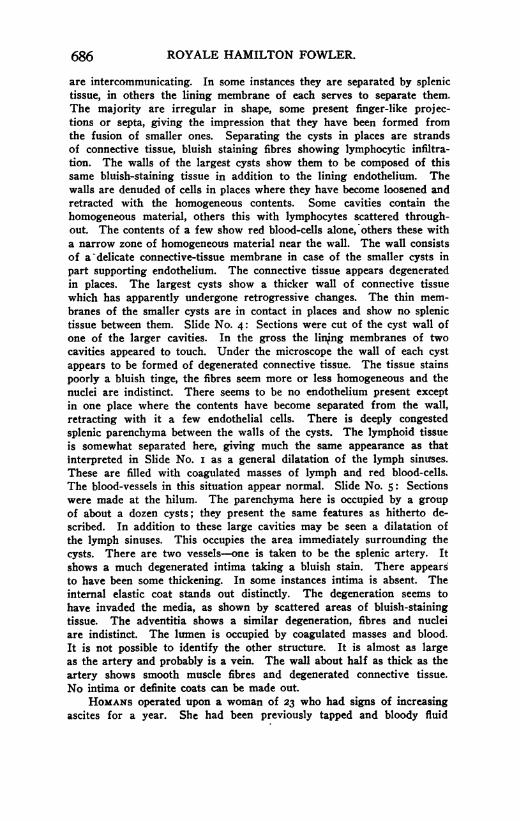
and shape, some are tortuous and intercommunicating, others are longand arranged in parallel, some are round and others are oval. The entiresection is seen to be made up of these spaces surrounded by splenicparenchyma. They possess a delicate membrane of connective tissuewhich composes the wall, supporting in some instances endothelial cells.In other cases it is not possible to positively identify an endothelial lining.Lymphoid tissue is seen to touch directly upon the membrane in somecases. The contents show for the most part large and small mono-nuclear leucocytes, with a preponderance of the latter, shreds of fibrin,an occasional eosinophilic and neutrophilic leucocyte and here and therered blood-cells. There are some scattered areas of round cells whichappear to represent Malpighian bodies. These follicles show some hyper-plasia, otherwise no change. The arterioles show normal walls. In somefollicles they are situated eccentrically, others show two vessels periph-erally located. The blood-vessels in other situations in this slide appearnormal. The connective-tissue trabecula are somewhat increased. Insome situations they are seen to contain dilated spaces lined with endo-thelium and which contain lymphocytes and fibrin. These are taken tobe dilated lymphatic vessels. Some of the larger spaces located in theparenchyma show in addition to the delicate membrane of connectivetissue alluded to, a wall of connective tissue which appears thicker thanthat composing the wall of the smaller spaces. The wall is infiltratedwith leucocytes and red blood-cells. One or two of the larger spaces,in addition to the cellular contents, are partially occupied by homogeneous,pink staining masses. Slide No. 2: This was also removed from theregion of the central constriction and shows macroscopic cysts half thesize of a split pea. Microscopically there is less dilatation of the lymphsinuses and more cavities. The parenchyma is denser and shows a greaterdegree of congestion. The several large cysts visible to the naked eye ap-pear under the microscope to possess a definite wall of connective tissue andin some instances an endothelial lining. They are occupied by a smooth,uniform, pink staining mass which has become slightly retracted fromthe walls. The cells lining these cavities have become separated inplaces, adhering to the homogeneous material. The walls of some of thelarger cysts appear to be made up of connective tissue which has becomechanged through hyalin-like degeneration. It stains poorly a bluish color.These are to be considered secondary regressive changes. Scattered inand among the lymphocytes are deposits of finely grained blood pigment.The small dilated spaces which correspond to dilated lymph sinuses showcellular contents as described in the first slide and not coagulated massesof lymph. Some of the larger cysts are seen to be surrounded by moreconnective tissue which appears in elongated bundles and resemblesobliquely cut trabeculae. These are to be considered dilated lymphaticvessels lying in trabeculae, they contain homogeneous pink staining masses,which in part have become retracted from the walls, the lining endothe-lium having become detached and adherent to the homogeneou-s material.Slide No. 3 shows a greater number of small cysts of variable size.Under the microscope countless cavities are seen, large and small; some
686 ROYALE HAMILTON FOWLER.
are intercommunicating. In some instances they are separated by splenictissue, in others the lining membrane of each serves to separate them.The majority are irregular in shape, some present finger-like projec-tions or septa, giving the impression that they have been formed fromthe fusion of smaller ones. Separating the cysts in places are strandsof connective tissue, bluish staining fibres showing lymphocytic infiltra-tion. The walls of the largest cysts show them to be composed of thissame bluish-staining tissue in addition to the lining endothelium. Thewalls are denuded of cells in places where they have become loosened andretracted with the homogeneous contents. Some cavities contain thehomogeneous material, others this with lymphocytes scattered through-out. The contents of a few show red blood-cells alone, others these witha narrow zone of homogeneous material near the wall. The wall consistsof a delicate connective-tissue membrane in case of the smaller cysts inpart supporting endothelium. The connective tissue appears degeneratedin places. The largest cysts show a thicker wall of connective tissuewhich has apparently undergone retrogressive changes. The thin mem-branes of the smaller cysts are in contact in places and show no splenictissue between them. Slide No. 4: Sections were cut of the cyst wall ofone of the larger cavities. In the gross the lining membranes of twocavities appeared to touch. Under the microscope the wall of each cystappears to be formed of degenerated connective tissue. The tissue stainspoorly a bluish tinge, the fibres seem more or less homogeneous and thenuclei are indistinct. There seems to be no endothelium present exceptin one place where the contents have become separated from the wall,retracting with it a few endothelial cells. There is deeply congestedsplenic parenchyma between the walls of the cysts. The lymphoid tissueis somewhat separated here, giving much the same appearance as thatinterpreted in Slide No. i as a general dilatation of the lymph sinuses.These are filled with coagulated masses of lymph and red blood-cells.The blood-vessels in this situation appear normal. Slide No. 5: Sectionswere made at the hilum. The parenchyma here is occupied by a groupof about a dozen cysts; they present the same features as hitherto de-scribed. In addition to these large cavities may be seen a dilatation ofthe lymph sinuses. This occupies the area immediately surrounding thecysts. There are two vessels-one is taken to be the splenic artery. Itshows a much degenerated intima taking a bluish stain. There appearsto have been some thickening. In some instances intima is absent. Theinternal elastic coat stands out distinctly. The degeneration seems tohave invaded the media, as shown by scattered areas of bluish-stainingtissue. The adventitia shows a similar degeneration, fibres and nucleiare indistinct. The lumen is occupied by coagulated masses and blood.It is not possible to identify the other structure. It is almost as largeas the artery and probably is a vein. The wall about half as thick as theartery shows smooth muscle fibres and degenerated connective tissue.No intima or definite coats can be made out.
HOMANS operated upon a woman of 23 who had signs of increasingascites for a year. She had been previously tapped and bloody fluid

CYSTS OF THE SPLEEN. 687obtained. At operation an abdominal tumor was removed from the trans-verse colon. It was covered with omentum, and represented a spongy,fibrous mass diagnosed by Whitney as a cavernous argioma possiblyoriginating in the omentum. The fluid re-accumulated and four monthslater another operation was performed. The spleen was removed. Itwas enlarged and of normal shape, weight 4I5 Grams. There werenumerous purple elevated areas upon the surface. Section showed afibrous network filled with reddish and light colored fluid. Microscopicexamination showed areas of blood and transparent coagulated massesin a cavernous structure. The walls were formed of thin connectivetissue with an endothelial lining.
HEDINGER (i9o6) demonstrated a specimen before the German Patho-logical Society. The young girl was thought to suffer from hydronephro-sis. Trauma two years previous to operation. Splenectomy. Tumorof the spleen contained fluid, no free bodies. Wall of connective tissue.Recovery.
HEINIRICIUS: Male, aged I4. Hard, abdominal tumor in the umbilicalregion for one month. Operation. Large blue-gray mass adherent todisplaced spleen. It arose from the outer side. Splenectomy. Pathologicreport: Cyst contained 8oo c.c. brownish-yellow, flaky, sticky fluid, chol-esterin crystals, leucocytes, red blood-cells, and detritus. Wall of cysttoward the lower pole thin as paper, pale yellow, 3 cm. in thickness, andincreased in size and thickness to a point where it arose from the spleen.At this point the color resembled that of the spleen. The size of the cystwas that of the head of a new-born child. The spleen retained its originalshape, cyst arose from the outer and under portion. Wall of the cystlined with endothelium. Considered a lymphatic cyst.
JODAN (I903): Female, aged 46. Operation splenectomy. Haema-toma, weight four kilograms. Recovery.
KUSTNER, Otto (I9II): Female, aged 39. Noticed a tumor the sizeof a head in the left hypochondriac region which had caused considerablepain. This condition had existed for two years, when Kustner decidedto extirpate what he considered a cyst of the spleen. Recovery. Patientdischarged on the sixteenth day after operation. Blood count 23,000 leu-cocytes, 6,ooo,ooo erythrocytes, hamoglobin 72 per cent. Three and one-quarter years after operation the patient is well and able to carry on herwork. No nutritional disturbances or gland swellings. Four-fifths ofthe spleen in this case was occupied by the cyst, which contained asulphur-yellow fluid. Echinococcus was ruled out. It was suggested thata twisted pedicle was responsible for the formation.
LEONTE: Female, aged 55. Tumor in the epigastric region diagnoseda cyst of the gastrosplenic omentutm. At operation a unilocular cyst ofthe spleen was found. Spleen much shrunken. Splenectomy.
MINERVINI: Female, aged I4. Immovable fluctuating tumor largerthan a head. Splenectomy. Serosanguineous. Recovery.
MONNIER: Female, aged 21. General and local symptoms. Bulgingtumor of the hypochondrium, moved with respiration. Friction murmur.Operation by Kronlein. Splenectomy. Cyst of the upper part of the

688 ROYALE HAMILTON FOWLER.
spleen adherent to surrounding parts. Capacity of cyst 3Y/2 litres ofbloody fluid. Recovery.
McMuRTRY: Female, aged 30. A freely movable abdominal tumorcould be displaced to any part of the cavity. Receded toward the dia-phragm upon lying down, descended to the pubis upon assuming an erectposture. Diagnosis ovarian cyst or movable kidney. Operation. Splen-ectomy. Pathologic report: Splenic tissue comparatively healthy in ap-pearance. Large cyst chief departure from the normal.
MICHAILOWSKY (igoo): Female, aged I2. History of trauma andmalaria. Splenectomy. Spleen seat of a large unilocular cyst.
MORESCHI and GHmTIx: Female, aged 42. Indefinite pain in the leftside for more than a year. Prong of a pitchfork pierced the left side, wherea painful swelling appeared which gradually increased in size. In amonth the spleen was found to be enlarged, displaced, and fluctuating.Operation. Adhesions between ascending colon and cyst. Splenectomy,recovery. Pathologic report: Organ 29 x 23 cm. Weight, 13I5 Grams.Cyst occupied the anterior border of the spleen and contained 95o c.c. ofdark fluid. Spleen tissue normal. Wall of cyst composed of connectivetissue with epithelial lining.
MussER (I9II): Female, aged 25. First noticed abdominal swellingin the splenic region when i8 years of age. Since this time mass hasgrown downward and into the pelvis. Rapid growth for past year. Noinjury. Soreness over tumor, slight nausea. No change in general healthup to one year ago, when she became weak and bowels constipated.Mitral systolic murmur at the cardiac apex. Tumor largest and mostprominent in the epigastrium. Hard, fluctuating, moved with respiration.A few small discrete lymph-nodes in the cervical chain and in the in-guinal region. Splenectomy by Gibbon. Recovery. Left upper rectusincision. Large cyst adherent to the diaphragm and surrounded exceptin this situation by splenic tissue. Freed and tapped, Ioo ounces of clear,yellow fluid obtained. Considerable bleeding after freeing adhesions.Pathologic report: Weight of specimen 400 Grams. Upper posteriorregion, huge cyst the size of a foot-ball, single cavity, open and col-lapsed. Surrounded on three sides by splenic tissue. Spleen is enlarged,surface smooth. Microscopic examination: Sinuses show some increasein connective tissue in places. Tissue very cellular. Malpighian bodieslarge, germinal centres swollen. An occasional uninuclear giant-cell atthe periphery. No cellular lining in the wall. Anatomical diagnosis,unilocular serous cyst arising in the capsule or splenic pulp.
PkAN (I88o): Female, aged 2o. Two years ago swelling and painin the hypogastrium. Acute pains for two months. Hard, fixed, partlyfluctuating tumor of the abdomen. Diagnosed an ovarian cyst. Opera-tion, median incision, tumor adherent to omentum. Brownishyellowfluid from puncture. Adhesions freed, spleen excised. Recovery. Spleenpresented the characteristic color of an hypertrophied spleen, weight II40Grams. Cyst wall of variable thickness, thin in places, where it consistedof connective tissue. In thicker places it was composed of splenic tissue.Cyst unilocular. The interior of the wall was smooth and composed of
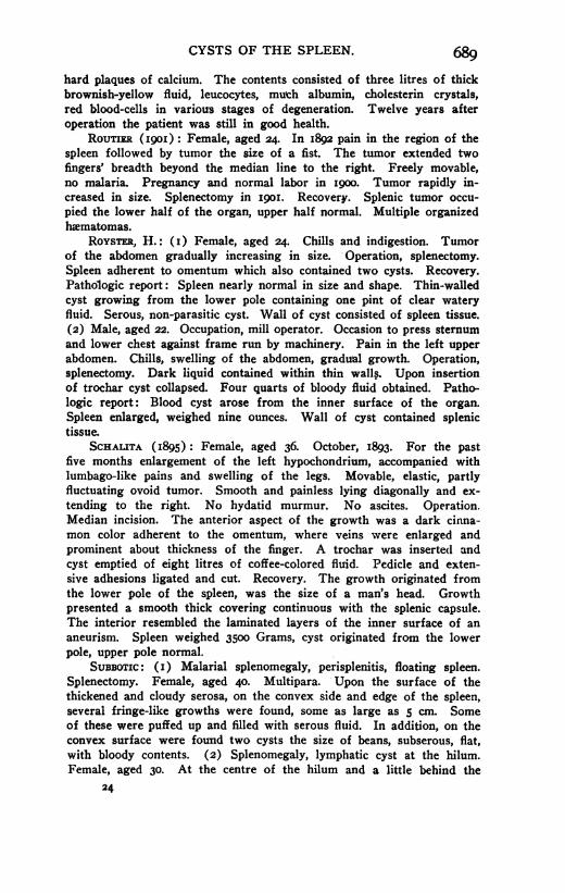
CYSTS OF THE SPLEEN. 689hard plaques of calcium. The contents consisted of three litres of thickbrownish-yellow fluid, leucocytes, much albumin, cholesterin crystals,red blood-cells in various stages of degeneration. Twelve years afteroperation the patient was still in good health.
RouTizR (igoi): Female, aged 24. In I892 pain in the region of thespleen followed by tumor the size of a fist. The tumor extended twofingers' breadth beyond the median line to the right. Freely movable,no malaria. Pregnancy and normal labor in igoo. Tumor rapidly in-creased in size. Splenectomy in I90I. Recovery. Splenic tumor occu-pied the lower half of the organ, upper half normal. Multiple organizedhamatomas.
RoysmR, H.: (i) Female, aged 24. Chills and indigestion. Tumorof the abdomen gradually increasing in size. Operation, splenectomy.Spleen adherent to omentum which also contained two cysts. Recovery.Pathologic report: Spleen nearly normal in size and shape. Thin-walledcyst growing from the lower pole containing one pint of clear wateryfluid. Serous, non-parasitic cyst. Wall of cyst consisted of spleen tissue.(2) Male, aged 22. Occupation, mill operator. Occasion to press sternumand lower chest against frame run by machinery. Pain in the left upperabdomen. Chills, swelling of the abdomen, gradual growth. Operation,splenectomy. Dark liquid contained within thin walls. Upon insertionof trochar cyst collapsed. Four quarts of bloody fluid obtained. Patho-logic report: Blood cyst arose from the inner surface of the organ.Spleen enlarged, weighed nine ounces. Wall of cyst contained splenictissue.
SCHALITA (I895): Female, aged 36. October, I893. For the pastfive months enlargement of the left hypochondrium, accompanied withlumbago-like pains and swelling of the legs. Movable, elastic, partlyfluctuating ovoid tumor. Smooth and painless lying diagonally and ex-tending to the right. No hydatid murmur. No ascites. Operation.Median incision. The anterior aspect of the growth was a dark cinna-mon color adherent to the omentum, where veins were enlarged andprominent about thickness of the finger. A trochar was inserte(d andcyst emptied of eight litres of coffee-colored fluid. Pedicle and exten-sive adhesions ligated and cut. Recovery. The growth originated fromthe lower pole of the spleen, was the size of a man's head. Growthpresented a smooth thick covering continuous with the splenic capsule.The interior resembled the laminated layers of the inner surface of ananeurism. Spleen weighed 3500 Grams, cyst originated from the lowerpole, upper pole normal.
SuBBoTIc: (I) Malarial splenomegaly, perisplenitis, floating spleen.Splenectomy. Female, aged 40. Multipara. Upon the surface of thethickened and cloudy serosa, on the convex side and edge of the spleen,several fringe-like growths were found, some as large as 5 cm. Someof these were puffed up and filled with serous fluid. In addition, on theconvex surface were found two cysts the size of beans, subserous, flat,with bloody contents. (2) Splenomegaly, lymphatic cyst at the hilum.Female, aged 30. At the centre of the hilum and a little behind the
24
ROYALE HAMILTON FOWLER.
pedicle there were fused spaces the size of a hen's egg. The interiorlining was composed of a delicate, smooth white membrane which showednumerous strands of fan-like septa separating small cavities. The mem-brane was easily peeled off. The wall of the cyst where it was in directcontact with the spleen could not be differentiated from the organ andcould only be separated from it with difficulty.
THORNTON (i886): Female, aged I9. Fluctuating movable tumor inthe left side for two years. Operation. Adhesions between omentumand under portion of the spleen. Patient collapsed during ligation of thepedicle. Splenectomy, recovery. Pathologic report: Tumor consisted ofa large cyst which contained serum, red blood-cells, and cholesterin. Inthe upper half of the spleen above the main cysts there were other smallercysts. The walls were thin, almost transparent in places. Blood exami-nation during convalescence showed leucocytosis. No glandular or thyroidswelling.
WELLS, SPENCER (i889): Female, aged 2I. At the age of four hadintermittent fever for two years, leaving spleen enlarged. Later tumorevident in ovarian region, round, hard, size of a fist, movable, not ad-herent to the uterus. Pregnant. Tumor pushed to the left hypochon-drium by pregnant uterus. Normal labor. Tumor extended from thenavel to the pubes. Patient became very ill, abdomen enlarged, tumorincreased in size. PunctuFe. Five litres of thick, brownish red fluidwithdrawn, containing red and white blood-cells. Fluid reaccumulatedalmost immediately. Operation. Cyst burst during manipulation allow-ing the escape of four or five litres of fluid similar to that obtained bypuncture. Numerous adhesions to the uterus and intestine. A smallportion of the cyst wall had to be left behind. Spleen removed. Woundclosed without drainage. Wound infection. Recovery.
690
Related Documents