Acta Zoologica (Stockholm) 85: 211– 221 (October 2004) © 2005 The Royal Swedish Academy of Sciences Abstract Guidi, L., Pierboni, L., Ferraguti, M., Todaro, M.A., Balsamo, M. Spermatology of the genus Lepidodasys Remane, 1926 (Gastrotricha, Macrodasyida): towards a revision of the family Lepidodasyidae Remane 1927. — Acta Zoologica (Stockholm) 85: 211–221 The spermatozoa of Lepidodasys unicarenatus and Lepidodasys sp. are filiform and composed of a cork-screw shaped acrosome, a helical nucleus surrounding a mitochondrial axis, and a 9 × 2 + 2 flagellum as in the basic structural model of the macrodasyidan sperm. The genus Lepidodasys has a debated phylogenetic position and has been linked in turn to the family Lepidodasyidae and the family Thaumastodermatidae. The sperm features of the two Lepidodasys species examined are distinct from those typical of the two families: the absence of the periaxonemal cylinder, a character shared only with Turbanellidae among Macrodasyida, could be considered as a symplesiomorphy, suggesting a basal position of the genus along the Macrodasyida clade. Moreover, a comparison of the spermatogenic process of Lepidodasys sp. with those of Acanthodasys aculeatus ( Thaumastodermatidae) and Cephalodasys maximus (Lepidodasyidae) has revealed that the process of acrosome formation and nuclear morphology during spermatogenesis are peculiar in Lepidodasys sp. and differences are evident especially in the late stages of spermatogenesis. Penetrated spermatozoa were observed in the oocytes at all maturation stages in L. unicarenatus. Dr L. Guidi, Istituto di Scienze Morfologiche, Università di Urbino, Località Crocicchia, I-61029 Urbino, Italy. E-mail: [email protected] Blackwell Publishing, Ltd. Spermatology of the genus Lepidodasys Remane, 1926 (Gastrotricha, Macrodasyida): towards a revision of the family Lepidodasyidae Remane, 1927 Loretta Guidi, 1 Lara Pierboni, 1 Marco Ferraguti, 2 M. Antonio Todaro 3 and Maria Balsamo 1 1 Istituto di Scienze Morfologiche, I-Università di Urbino, Loc. Crocicchia, 61029 Urbino, Italy; 2 Dipartimento di Biologia, Università di Milano, Via Celoria 26, I-20133 Milano, Italy; 3 Dipartimento di Biologia Animale, Università di Modena e Reggio Emilia, Via Università 213/d, I-41100 Modena, Italy Keywords: Lepidodasys, Gastrotricha, spermatogenesis, spermatozoa, ultrastructure, phylogeny Accepted for publication: 1 June 2004 Introduction Gastrotricha Macrodasyida includes 240 protandric or simultaneous hermaphroditic species distributed in six families and 30 genera (Kisielewski 1987, 1991; Ruppert 1988; Evans and Hummon 1991; Hummon et al. 1993). The evo- lutionary relationships among families are little known and uncertainty also persists within group alliances because spe- cies descriptions are often incomplete and there is a general lack of knowledge about the microscopic anatomy of taxa perceived to be ‘primitive’, or at least possessing plesiomor- phic features. These problems are also highlighted by two recent phylogenetic reconstructions of Gastrotricha based on morphological traits ( Hochberg and Litvaitis 2000, 2001) as well as one based on the 18S rRNA gene sequences (Todaro et al. 2003).These studies confirm previous doubts about the monophyly of some families of Macrodasyida, such as Lepi- dodasyidae and Planodasyidae (Travis 1983; Ruppert 1991). The family Lepidodasyidae is especially problematic because it contains seven morphologically and biologically disparate genera with no known synapomorphies. Ironically, species of the type genus Lepidodasys Remane, 1926 share more potentially homologous characters with the species of Thaumastodermatidae than with the species of Lepidodasy- idae, including: Y cells with myofilaments, absence of circular muscles in the lateral body regions, and structural complexity of the cuticle (Rieger and Rieger 1977; Ruppert 1978). Still, species of Lepidodasys are exceptional among Macrodasyida because of two unique characters: a non-striated pharyngeal myo-epitelium (all other species of Gastrotricha possess cross-striated myoepithelia), and the absence of pharyngeal pores (apomorphic to Macrodasyida) (Ruppert 1978).

Welcome message from author
This document is posted to help you gain knowledge. Please leave a comment to let me know what you think about it! Share it to your friends and learn new things together.
Transcript
Acta Zoologica
(Stockholm)
85
: 211–221 (October 2004)
© 2005 The Royal Swedish Academy of Sciences
Abstract
Guidi, L., Pierboni, L., Ferraguti, M., Todaro, M.A., Balsamo, M. Spermatologyof the genus
Lepidodasys
Remane, 1926 (Gastrotricha, Macrodasyida): towardsa revision of the family
Lepidodasyidae
Remane 1927. —
Acta Zoologica
(Stockholm)
85
: 211–221
The spermatozoa of
Lepidodasys unicarenatus
and
Lepidodasys
sp. are filiformand composed of a cork-screw shaped acrosome, a helical nucleus surroundinga mitochondrial axis, and a 9
×
2 + 2 flagellum as in the basic structural modelof the macrodasyidan sperm. The genus
Lepidodasys
has a debated phylogeneticposition and has been linked in turn to the family Lepidodasyidae and thefamily Thaumastodermatidae. The sperm features of the two
Lepidodasys
species examined are distinct from those typical of the two families: the absenceof the periaxonemal cylinder, a character shared only with Turbanellidaeamong Macrodasyida, could be considered as a symplesiomorphy, suggestinga basal position of the genus along the Macrodasyida clade. Moreover, acomparison of the spermatogenic process of
Lepidodasys
sp. with those of
Acanthodasys aculeatus
(Thaumastodermatidae) and
Cephalodasys maximus
(Lepidodasyidae) has revealed that the process of acrosome formation andnuclear morphology during spermatogenesis are peculiar in
Lepidodasys
sp.and differences are evident especially in the late stages of spermatogenesis.Penetrated spermatozoa were observed in the oocytes at all maturation stagesin
L. unicarenatus.
Dr L. Guidi, Istituto di Scienze Morfologiche, Università di Urbino, Località Crocicchia, I-61029 Urbino, Italy. E-mail: [email protected]
Blackwell Publishing, Ltd.
Spermatology of the genus
Lepidodasys
Remane, 1926 (Gastrotricha, Macrodasyida): towards a revision of the family Lepidodasyidae Remane, 1927
Loretta Guidi,
1
Lara Pierboni,
1
Marco Ferraguti,
2
M. Antonio Todaro
3
and Maria Balsamo
1
1
Istituto di Scienze Morfologiche, I-Università di Urbino, Loc. Crocicchia, 61029 Urbino, Italy;
2
Dipartimento di Biologia, Università di Milano, Via Celoria 26, I-20133 Milano, Italy;
3
Dipartimento di Biologia Animale, Università di Modena e Reggio Emilia, Via Università 213/d, I-41100 Modena, Italy
Keywords:
Lepidodasys
, Gastrotricha, spermatogenesis, spermatozoa, ultrastructure, phylogeny
Accepted for publication:
1 June 2004
Introduction
Gastrotricha Macrodasyida includes 240 protandric orsimultaneous hermaphroditic species distributed in sixfamilies and 30 genera (Kisielewski 1987, 1991; Ruppert 1988;Evans and Hummon 1991; Hummon
et al
. 1993). The evo-lutionary relationships among families are little known anduncertainty also persists within group alliances because spe-cies descriptions are often incomplete and there is a generallack of knowledge about the microscopic anatomy of taxaperceived to be ‘primitive’, or at least possessing plesiomor-phic features. These problems are also highlighted by tworecent phylogenetic reconstructions of Gastrotricha based onmorphological traits (Hochberg and Litvaitis 2000, 2001) aswell as one based on the 18S rRNA gene sequences (Todaro
et al
. 2003). These studies confirm previous doubts about the
monophyly of some families of Macrodasyida, such as Lepi-dodasyidae and Planodasyidae (Travis 1983; Ruppert 1991).
The family Lepidodasyidae is especially problematicbecause it contains seven morphologically and biologicallydisparate genera with no known synapomorphies. Ironically,species of the type genus
Lepidodasys
Remane, 1926 sharemore potentially homologous characters with the species ofThaumastodermatidae than with the species of Lepidodasy-idae, including: Y cells with myofilaments, absence of circularmuscles in the lateral body regions, and structural complexityof the cuticle (Rieger and Rieger 1977; Ruppert 1978). Still,species of
Lepidodasys
are exceptional among Macrodasyidabecause of two unique characters: a non-striated pharyngealmyo-epitelium (all other species of Gastrotricha possesscross-striated myoepithelia), and the absence of pharyngealpores (apomorphic to Macrodasyida) (Ruppert 1978).

Spermatology of the genus
Lepidodasys
•
Guidi
et al.
Acta Zoologica
(Stockholm)
85
: 211–221 (October 2004)
© 2005 The Royal Swedish Academy of Sciences
Recently, comparative studies of the gastrotrich spermatozoahave provided useful information on taxonomic and phylo-genetic relationships within the phylum (Balsamo 1992;Balsamo
et al
. 1999). In this study, we compare the sperma-tozoa of two species of
Lepidodasys
,
L. unicarenatus
Balsamo,Fregni & Tongiorgi, 1994 and
Lepidodasys
sp. (cf Todaro
et al
.1995), with those of other macrodasyidans to gain insighton the phylogenetic position of the genus. We also examinethe development of the spermatogenesis of
Lepidodasys
sp.compared to that of
Cephalodasys maximus
(Lepidodasyidae;Fischer 1994) and
A. aculeatus
(Thaumastodermatidae; Guidi
et al
. 2003), because the latter species are representative ofthe two families to which
Lepidodasys
has been linked.Finally, because specimens of
L. unicarenatus
also show anumber of oocytes at different maturation stages, eachcontaining several spermatozoa within the ooplasm, the finemorphology of the penetrated spermatozoa has been surveyedand compared with that of the testicular spermatozoa.
Materials and methods
Adult specimens of
Lepidodasis unicarenatus
were collectedon 8 June 2002 from Porthu de la Rena, Castelsardo, Sardinia(Italy) at 13 m water depth (Balsamo and Todaro, unpub-lished data), while those of
Lepidodasys
sp. were collected on25 September 1994 from the shallow sublittoral of PanamaCity beach, Florida, US (cf. Todaro
et al
. 1995). In laboratory,living gastrotrich were extracted by means of the narcot-ization and decantation technique (Higgins and Thiel 1988);thereafter, the Italian specimens were fixed in sucrose para-formaldehyde glutaraldehyde (SPAFG) (Ermak and Eakin1976) while the American ones were fixed in 2% glutaralde-hyde. All the animals were then washed in 0.1
phosphatebuffer (PBS) at pH 7.2 and post fixed in 2% osmium tetroxidesolution for one hour at room temperature. All fixatives werein PBS with 10% sucrose. After a rinsing in PBS, the speci-mens were dehydrated in a graded acetone series, stained
enbloc
in uranyl acetate in 70% acetone, and embedded inaraldite. Ultra thin sections were cut with an LKB Ultratome2088 V, contrasted with lead citrate, and observed under aPhilips 300 and a Zeiss 902 transmission electron micro-scope. Additional specimens of both species were whole-mounted on slides and observed using Nomarski differentialinterference contrast (DIC) optics under a Leitz Dialux 20microscope. These specimens were photographed with aNikon 995 Coolpix digital camera, and measured with anocular micrometer; location of the testes along the body wasreported in percentage units (U) from anterior to posterior.
Results
Mature spermatozoon of
Lepidodasis unicarenatus
The mature spermatozoon of
L. unicarenatus
is a filiformcell, 83.8
µ
m long, composed of an elongated acrosome, a
nucleus surrounding a single mitochondrion and a tail(Fig. 1G,H). The acrosome, 19.6
µ
m long, is clearly dis-tinct in three regions. The apical one is 13.4
µ
m long and0.08
µ
m in diameter, cork screw-shaped, and shows alter-nating dark and light thin streaks (Fig. 1A,B). The middleregion, 2.6
µ
m long and 0.5
µ
m wide, is weakly twisted andfull of moderately electron-dense material (Fig. 1B,C). Thebasal one, 3.6
µ
m long and 0.35
µ
m in diameter, is weaklytwisted and contains a more electron-dense material;numerous, tiny electron-dense vesicles close to one anotherare located between the acrosomal membrane and the innermaterial (Fig. 1C,D). The spring-shaped nucleus, 12.2
µ
min length and 0.35
µ
m in diameter, is composed of fully con-densed chromatin organized into well-spaced coils and sur-rounding a single, long and twisted mitochondrion (Fig. 1E, F).A simple clasp-like structure (
sensu
Teuchert 1976) con-nects the nucleus to the 52
µ
m long flagellum. The 9
×
2 + 2axoneme is devoid of any accessory structure. The axonemedoublets run parallel to each other for the entire length of theaxoneme (Fig. 1F).
Mature spermatozoon of
Lepidodasys
sp.
The mature spermatozoon of
Lepidodasys
sp. is filiform andcomposed of an elongated acrosome, a nucleus surroundinga single mitochondrion, and a tail (Fig. 2E). The acrosomeis clearly distinct in two regions. The apical one is 0.15
µ
min diameter, cork screw-shaped, made up of at least 10 coils,and full of a very electron-dense material, whereas the basalone, is 5
µ
m long and 0.38 in diameter, slightly twisted andcomposed of a high number of disks (89 counted in one spec-imen) stacked on top of each other. Each disk is moderatelyelectron-dense and is separated from each adjacent one byan electron-dark septum, which is obliquely arranged withrespect to the main cell axis. The disks show a rectangular ortrapezoidal shape in longitudinal section generating acro-some twisting (Fig. 2A,B). The nucleus (10
µ
m in length,0.53
µ
m in diameter) is made of fully condensed chromatinorganized into 30 coils (Fig. 2C). The inner nuclear cavityincludes a single, thin and straight mitochondrion (Fig. 2B).The nucleus-flagellum connection is a simple clasp-likestructure. The basal, short, tract of the 9
×
2 + 2 axonemeshows a parallel arrangement of the external microtubules,which then coil around the central doublets giving the tailan obliquely striated appearance in longitudinal sections(Fig. 2D). The axoneme is devoid of accessory structures.
Spermatogenesis in
Lepidodasys
sp.
Two very elongated, lateral testes lie close to the anteriorregion of the gut. They appear slightly asymmetrical, the leftposterior (U43.5) being smaller than the anterior one (U41)(Fig. 3A). Each testis is entirely wrapped by a basal laminaand consists of a thin band of germ cells maturing in a caudo-cephalic direction so that spermatogonia, spermatocytes,

Acta Zoologica
(Stockholm)
85
: 211–221 (October 2004)
Guidi
et al.
•
Spermatology of the genus
Lepidodasys
© 2005 The Royal Swedish Academy of Sciences
and spermatids are linearly arranged (Fig. 3B). At the caudalend of each testis, two types of spermatogonia are recog-nizable, some showing signs of degeneration (Fig. 4A) and theothers showing a conventional morphology (Fig. 4B). The
first cells are characterized by a strongly condensed chromatinand a crowding of cytoplasmic organules, two markers ofthe apoptotic processes (Fig. 4A). The conventional sperma-togonia, about 2.5
×
4
µ
m in diameter, are irregular in shape,
Fig. 1—Mature spermatozoa of Lepidodasys unicarenatus. —A. High magnification of the acrosomal apical portion. —B. Apical and middle portion of the acrosome. —C. Middle and basal portion of the acrosome. —D. Basal portion of the acrosome and the beginning of the nucleus. —E. The nucleus surrounding the mitochondrion. —F. Nucleus and proximal portion of the flagellum. —G. Schematic drawing of the mature spermatozoon: 1. The acrosome; 2. the helical nucleus and the mitocondrion 3. the flagellum. —H. A single spermatozoon as seen by differential interference contrast optics. Scale bars: A = 0.25 µm; B, C, D, E, F = 0.5 µm; H = 5 µm. ma – middle region of the acrosome, f – flagellum, ba – basal region of the acrosome, aa – apical region of the acrosome, n – nucleus, m – mitochondrion.

Spermatology of the genus
Lepidodasys
•
Guidi
et al.
Acta Zoologica
(Stockholm)
85
: 211–221 (October 2004)
© 2005 The Royal Swedish Academy of Sciences
with an eccentric, round nucleus and dense chromatin patches.Many thin, elongated, mitochondria, some lysosome-likebodies and many Golgi cisternae filled with electron-lucentmaterial are visible in the cytoplasm (Fig. 4B). Primary lepto-
tenic spermatocytes, about 5.2
×
3.1
µ
m, are connected bycytoplasmic bridges (Fig. 4C). Their nucleus is eccentric andovoid, about 2.8
×
2
µ
m in size. A large number of roundvesicles are grouped at the cell pole opposite to the nuclear one
Fig. 2—Mature spermatozoa of Lepidodasys sp. —A. Apical and basal regions of the acrosome. The acrosomal base is formed by a pile of disks (arrow) stacked on top of each other. —B. Acrosomal base and apical portion of the nucleus inside which the mitochondrion is located. —C. Tangentially sectioned nucleus showing the fully compacted chromatin organized into coils. —D. Basal portion of the nucleus and proximal portion of the flagellum (arrow). The axoneme, with a 9 × 2 + 2 microtubular array, shows an obliquely striated appearance because the external doublets coil around the central microtubules. —E. Schematic drawing of the mature spermatozoon: 1. The acrosome; 2. the nucleus surrounding the mitochondrion; 3. the flagellum. Scale bars: A, B, C, D, E, F = 0.5 µm.
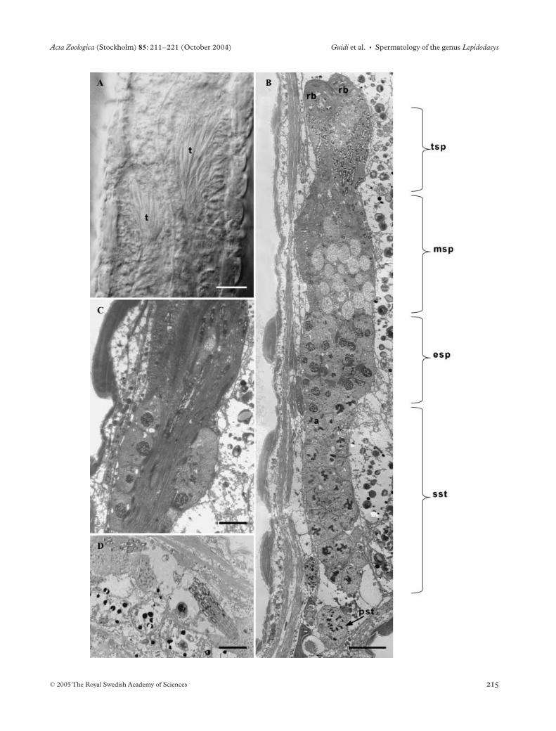
Fig. 3—Testes. —A. Mid portion of the body of Lepidodasys sp. showing the two asymmetrical testes. —B. Longitudinal section of L. sp. testis formed by a band of germ cells developing in a caudo-cephalic direction. —C. Group of spermatids sliding ventrally towards the caudal region of the testis. —D. Mature spermatozoa located between the youngest germinal cells and the basal lamina. Scale bars: A, C = 2 µm; B = 4 µm; D = 6 µm. t – testis, rb – residual body, tsp – terminal spermatids, msp – middle spermatids, esp – early spermatids, pst – primary spermatocyte, sst – secondary spermatocytes.
Acta Zoologica (Stockholm) 85: 211–221 (October 2004) Guidi et al. • Spermatology of the genus Lepidodasys
© 2005 The Royal Swedish Academy of Sciences

Spermatology of the genus Lepidodasys • Guidi et al. Acta Zoologica (Stockholm) 85: 211–221 (October 2004)
© 2005 The Royal Swedish Academy of Sciences
(4D), close to numerous Golgi cisternae, the contents of whichappear with the same electron-density as that of the vesicles(Fig. 4E). Duplicated diplosomes have also been observed inseveral cases (Fig. 4D). In primary pachytenic spermatocytes,synaptonemal complexes appear and become more visible asthe chromatin gradually condenses at their sides (Fig. 4D).Secondary spermatocytes have an irregular shape, about3.15 × 2.05 µm, and an eccentric, round nucleus, about
1.2 µm in diameter, with electron-dense chromatin patches.In the examined specimens, most of them appeared in inter-phase, some were binucleated, and only one was in anaphase(Figs 3B and 4F). Many thin elongated mitochondria andseveral ovoid vesicles, derived from the fusion of the smallerones at the previous stage, are visible in the cytoplasm(Fig. 4F). Three successive spermatid stages can be distin-guished. The early spermatids already show developing
Fig. 4—Spermatogenesis of Lepidodasys sp. —A. Spermatogonia showing signs of apoptotis. —B. Spermatogonia with normal morphology. In the cytoplasm mitochondria, Golgi apparatus (arrow) and lysosome like bodies (arrow-head) are visible — C. Primary leptotenic spermatocytes connected by a cytoplasmic bridge (arrow). —D. Primary spermatocytes in leptotene and in pachytene. In the cytoplasm of the leptotenic spermatocytes duplicated diplosomes are visible (arrow); in the nucleus of pachytenic ones synaptonemal complexes are present (arrow-head). —E. Detail of a leptotenic spermatocyte showing a group of vesicles, located close to a Golgi apparatus (arrow). —F. Secondary spermatocyte. In the cytoplasm, large ovoidal vesicles are present. Scale bars: A, B, C, D, E, F = 1 µm. dsg – spermatogonia with degenerating signs, sg – spermatogonia with normal morphology, ls – leptotenic spermatocyte, ps – pachytenic spermatocytes, ve – vesicles.

Acta Zoologica (Stockholm) 85: 211–221 (October 2004) Guidi et al. • Spermatology of the genus Lepidodasys
© 2005 The Royal Swedish Academy of Sciences
Fig. 5—Spermiogenesis of Lepidodasys sp. —A. Early spermatids. The nucleus is filled with a condensed chromatin network; an elongated mitochondrion is close to the nuclear envelope on the cytoplasm side. Large cytoplasmatic vesicles, which represent the pro-twisted acrosomal region, are visible. Some of them have a more electron-dense extremity (arrows). —B. Two early spermatids: the one on the right side shows the axoneme (arrows). —C. Mid spermatids: the elongated convoluted nuclei, with highly compacted chromatin, are located in the cellular body. —D. Cytoplasmic region of mid spermatids with large vesicles full of granular and fibrillar material. Other vesicles (arrows), with the same appearance as the basal tract of the acrosome of the mature spermatozoon are visible among the large ones. These vesicles will fuse with the twisted apical acrosomal region, as may be seen in one case (arrowhead). —E. Terminal spermatids: the mitochondrion is inside the completely condensed nucleus. —F. Terminal spermatids with many vesicles derived from the condensation of the ones visible in Fig. 5(D). Four kinds of vesicles, in intermediate stages of condensation, are visible (arrows). —G. Magnification of a terminal spermatid nucleus surrounded by a manchette of microtubules (arrow). —H. Terminal, U-shaped, spermatid with nucleus and axoneme in a parallel arrangement. Scale bars: A, B, C, D, F, H = 1 µm; E = 2 µm; G = 0.5 µm. lve – large vesicles.
Spermatology of the genus Lepidodasys • Guidi et al. Acta Zoologica (Stockholm) 85: 211–221 (October 2004)
© 2005 The Royal Swedish Academy of Sciences
acrosomes and flagella (Figs 3B and 5B). The nucleus isconvoluted, round in transversal sections (about 1 µm indiameter), and characterized by a condensed chromatinnetwork. Some large, irregular vesicles are present in thecytoplasm (Figs 3B and 5A,B). These vesicles, arising fromthe fusion of the smaller ones of secondary spermatocytes,show a homogeneous content of moderate electron-density, inwhich a denser region can sometimes be recognized (Fig. 5A).These vesicles represent the ‘pro-twisted acrosomal region’,as they will give rise to the twisted, apical region of the acro-some. A long mitochondrion formed by the merging of manysmaller ones sticks to the nuclear envelope surrounding itprogressively; then the ‘pro-twisted acrosomal region’ startsadhering to the mitochondrion (Fig. 5A). Mid spermatidsare large cells with a nuclear and a cytoplasmic pole. Theyhave an elongated, convoluted nucleus with highly con-densed chromatin (Fig. 5C) and many cytoplasmic vesicleswith different morphology: most of them are large and full ofa granular and fibrillar content (Figs 3B and 5D), whereasothers, in a more limited number, look like the basal regionof the acrosome of the mature spermatozoon. The twistedapical region of the acrosome is already formed (Fig. 5D).The long mitochondrion, coiled around the nucleus, pro-gressively sinks into its chromatin (Fig. 5C,E), while theaxoneme lengthens further on. Terminal spermatids have acompletely condensed nucleus, surrounding the mitochon-drion (Fig. 5E,F), and a high number of cytoplasmic vesiclesprobably derived from the condensation of the large onesvisible in the previous stages. These gradually decrease inwidth through a condensation process developing in fourmorphologically distinct stages to form the smallest vesicles,0.5 µm wide, which look like that of the acrosomal base(Fig. 5F). These vesicles finally fuse with each other, length-ening the acrosomal base. At the same time the nucleuslengthening is completed and it appears surrounded by amanchette of microtubules (Fig. 5G): it first turns mediallyand then posteriorly, so giving the spermatid a clear U shape(Figs 3C and 5H). The axoneme lengthening is completed,and the centriole localizes in a hollow at the posterior end ofthe nucleus (Fig. 5H). Cytoplasmic debris are removed fromterminal spermatids by extruding droplets, which gather ina large residual body at the most anterior part of each testis(Fig. 3B). At this point, spermatids start sliding ventro-laterallyalong each testis moving to its posterior end (Fig. 3C) to reacha location just ventral to the primary spermatocytes. Heresperm maturation ends with the rotation of the flagellum, whichat the end lines up with the nucleus-acrosome. Mature sper-matozoa migrate into the narrow space defined by the youngestgerminal cells at the caudal region of each testis and its basallamina: this space actually acts as a sperm duct (Fig. 3D).
Penetrated spermatozoa of L. unicarenatus
Several spermatozoa were observed between the basallamina of the ovary and the oocytes membrane (Fig. 6A,D)
and also in the ooplasm of all oocytes, at any maturing stage(Fig. 6B,C,E). The spermatozoa still external to the oocytesshowed the same structure as the testicular ones, with a com-plete, elongated acrosome, a nucleus and a tail (Fig. 6A); onthe contrary, those penetrated into the ooplasm showed onlythe nucleus and the basal acrosomal region (Fig. 6B,C,E).Moreover, several spermatozoa were observed in the secretorycells forming the wall of an accessory organ, which extendsfor the entire length of the ovary, of unclear function(Fig. 6A,B,C,D,E).
Discussion
Spermatozoon ultrastructure
Both the morphology and ultrastructure of the spermatozoaof Lepidodasis unicarenatus and Lepidodasys sp. agree with thebasic structural model of the Macrodasyida sperm (Ferragutiand Balsamo 1994–1995; Balsamo et al. 2002): a filiformsperm with cork-screw shaped acrosome, helical nucleus sur-rounding a mitochondrial axis, and a 9 × 2 + 2 flagellum,and thus clearly places the genus among the GastrotrichaMacrodasyida. However, a comparison with the other spe-cies of Lepidodasyidae (Mesodasys laticaudatus, Cephalo-dasys maximus) and Thaumastodermatidae (Acanthodasysaculeatus, Diplodasys ankeli, Pseudostomella etrusca, Tetran-chyroderma papii, T. sp. 1, T. sp. 2) studied so far in detail showsseveral different, ultrastructural details. The spermatozoaof both Lepidodasys species have a simple acrosome likeM. laticaudatus, but unlike C. maximus and all the Thaumasto-dermatidae, which are provided with a hollow striated acro-somal tube. The absence of a striated cylinder surroundingthe axoneme in the spermatozoa of Lepidodasys species is acharacter shared only by Turbanellidae among the Macro-dasyida; even if it does not prove a close relationship of thetwo taxa because it can be considered a possible simple-siomorphy, however, it may indicate their basal position alongthe Macrodasyida clade.
Spermatogenesis in Lepidodasys sp.
The structure of the testes and spermatogenesis developmentin Macrodasyida is well documented from three species:Lepidodasys sp. (the present study), C. maximus, Lepidodasy-idae, and A. aculeatus, Thaumastodermatidae (Fischer 1994;Guidi et al. 2003). Among these species, only Lepidodasys sp. ischaracterized by strip-shaped testes lacking both an internallumen and a true sperm duct, and showing germinal cellsarranged into a band. This condition contrasts with the moretypical club-shaped and hollow testes of the other species ofMacrodasyida. Moreover, no mitotic spermatogonial divi-sions have been seen in Lepidodasys sp. contrary to A. aculeatus,in which the observation of duplicated diplosomes in thespermatogonia proved that mitoses occur in the male germcells of Gastrotricha, a phylum known for its euthely. Because

Acta Zoologica (Stockholm) 85: 211–221 (October 2004) Guidi et al. • Spermatology of the genus Lepidodasys
© 2005 The Royal Swedish Academy of Sciences
apoptosis has been recognized as an important mechanismfor the removal of unwanted cells (Hetts 1998; Shinoda et al.2000), the presence of numerous spermatogonia showingapoptotic markers both in the cytoplasm and in the nucleussuggests that an apoptotic process plays this role in the testisof Lepidodasys sp. Detailed observations of the spermatogenesisin this species imply a two-step process in sperm formation:first the fusion of mitochondria into a single element, then thepenetration of the mitochondrial element into the nucleus.This process is similar to that observed for C. maximus (Fischer1994), whereas in A. aculeatus, the two phases follow each
other in an opposite sequence. Both the acrosome formationand the nucleus morphology during spermatogenesis appearto be peculiar in Lepidodasys sp. the distal and the proximalregions of the acrosome derive from the fusion of many vesicles,not from a single one as in the other macrodasyidans, and thewhole process completes later. Moreover, the nucleus keeps aconvoluted shape throughout nearly the whole spermatogenesislength, and becomes straight and parallel to the axonemeonly at the final spermatid stage, contrary to the other macro-dasyid species in which the spermatids show a U-shapedmorphology just from the beginning of the spermatogenesis.
Fig. 6—Penetrated spermatozoa of Lepidodasys unicarenatus. —A. Some spermatozoa with acrosome (arrows), nucleus (arrowheads) and tail are visible externally to the cytoplasmic membranes of a previtellogenic and a pre-previtellogenic oocyte. —B. Pre-previtellogenic oocyte with several spermatozoa (arrowheads) into the cytoplasm. —C. Oocyte in early vitellogenesis: on the top a group of spermatozoa (arrowhead) is visibile. —D. Two spermatozoa (arrows) located between the basal lamina of the ovary and the cytoplasmic membrane of the oocyte in middle vitellogenesis are visible. —E. A spermatozoon (arrowhead) is present in the cytoplasm of an oocyte in late vitellogenesis – another sperm in the cytoplasm of a secretory cell is visible (arrow). Scale bars: A, B, C, D, E = 2 µm. po – previtellogenic oocytes, ppo – pre-previtellogenic oocytes, sc – secretory cell.

Spermatology of the genus Lepidodasys • Guidi et al. Acta Zoologica (Stockholm) 85: 211–221 (October 2004)
© 2005 The Royal Swedish Academy of Sciences
The removal of the residual cytoplasm is also carried out indifferent ways: in Lepidodasys by the expulsion of a residualbody, whereas in A. aculeatus by lysosome intracellular diges-tion, and consequent reabsorbing (Guidi et al. 2003).
Penetrated spermatozoa of L. unicarenatus
The observation of penetrated spermatozoa not only betweenthe ovary and its basal lamina, but even in the oocytes at anydevelopment stage, is new for Gastrotricha; in fact, Fischer(1996) reported sperm in Dactylopodola baltica only in post-vitellogenetic oocytes. The spread presence of penetratedspermatozoa in all the oocytes implies that fertilization mayoccur in oocytes at any development stage, and not only inthe mature ones, as generally believed for Macrodasyida: thatis also suggested by the presence of a sexual accessory organclose to the large, fully grown oocyte and storing allosperm(Ruppert 1991).
Conclusions
To summarize, a number of morphological features of thetestis structure and of the spermatozoa appear to be exclusiveof Lepidodasys species with respect to all the other Macro-dasyida species studied so far. A comparison of the spermato-genesis in Lepidodasys and in other Macrodasyida shows thatthe general process occurs in a similar way, but differencesare evident especially in the late stages of the process. Thus,from a spermatological point of view, the genus Lepidodasysclearly fits within the Gastrotricha Macrodasyida, but standsout among all the other members of the order. It appears tobelong to a separate, basal clade, according to the results ofrecent molecular analyses based on 18S rRNA gene whichsuggest various locations of the genus in the phylogenetic treeof Gastrotricha, but in any case on a separate clade (Todaroet al. 2003). On account of these observations, the study ofspermatozoa of additional species of Lepidodasyidae family aswell as of potential out-group species will be of great importancein order to make the Macrodasyida basic sperm plan clearerand to understand the in-group relationships of the phylum.
Acknowledgements
We would like to thank Prof Gilberto Grandi (Dipartimentodi Biologia, Università di Ferrara) for his critical revision ofthe manuscript, Mr Federico Bastianelli (Centro di Citome-tria, Università di Urbino) for his valuable help with the pho-tographic material and Mrs Anna Bernard for her Englishrevision. We are also grateful to Mr Oliviero Rusciadelli forhis technical help with electron microscopes. This researchwas supported by grants from MIUR (Rome) to M.B andM.F. (Cofin Project 2002/2003 ‘Evoluzione molecolare emarcatori morfologici nello studio dei rapporti filogenetici emeccanismi adattativi in taxa chiave di protisti e invertebrati)and to M.B. (Scientific research funds).
References
Balsamo, M. 1992. Hermaphroditism and parthenogenesis in lowerBilateria: Gnathostomulida and Gastrotricha. In Dallai, R. (Ed.):Sex Origin and Evolution, Selected Symposia and Monographs UZI,Vol. 6, pp. 309–327. Mucchi, Modena.
Balsamo, M., Ferraguti, M., Guidi, L., Todaro, M. A. andTongiorgi, P. 2002. Reproductive system and spermatozoa ofParaturbanella tessieri (Gastrotricha, Macrodasyida): implicationsfor the sperm transfer modality in Turbanellidae. – Zoomorphology121: 235–241.
Balsamo, M., Fregni, E. and Ferraguti, M. 1999. Gastrotricha. InAdiyodi, K. G. and Adiyodi, R. G. (Eds): Reproductive Biology ofInvertebrates, Vol. IX Part A, pp. 171–191. John Wiley & Sons,Chichester.
Ermak, T. H. and Eakin, R. M. 1976. Fine structure of the cerebralpygidial ocelli in Chone ecaudata (Polychaeta: Sabellidae). – Journalof Ultrastructure Research 54: 243–260.
Evans, W. A. and Hummon, W. D. 1991. A new genus and speciesof Gastrotricha from the Atlantic coast of Florida, USA. – Trans-action of the American Microscopical Society 110: 321–327.
Ferraguti, M. and Balsamo, M. 1994. Sperm morphology and anat-omy of the genital organs in Mesodasys laticaudatus Remane, 1951(Gastrotricha: Macrodasyida). – Journal of Submicroscopic Cytologyand Pathology 26: 21–28.
Ferraguti, M. and Balsamo, M. 1995. Comparative Spermatology ofGastrotricha. In Jamieson, B. G. M., Ausio, J. and Justine, J.-L.(Eds): Advances in Spermatozoal Phylogeny and Taxonomy, Vol.166, pp. 105–117. Mémoires du Muséum National D’HistoireNaturelle, Paris.
Fischer, U. 1994. Ultrastructure of spermiogenesis and spermatozoaof Cephalodasys maximus (Gastrotricha, Macrodasyida). – Zoomor-phology 114: 213–225.
Fischer, U. 1996. Ultrastructure of penetrated spermatozoa, ovary,and oogenesis of Dactylopodola baltica (Gastrotricha, Macrodasyida).– Invertebrate Reproduction and Development 29: 71–78.
Guidi, L., Ferraguti, M., Pierboni, L. and Balsamo, M. 2003.Spermiogenesis and spermatozoa in Acanthodasys aculeatus(Gastrotricha, Macrodasyida): an ultrastructural study. – ActaZoologica 84: 77–85.
Hetts, S. W. 1998. To die or not to die: an overview of apoptosis andits role in disease. – Journal of American Medical Association 279:300–307.
Higgins, R. P. and Thiel, H. (Ed.) 1988. – Introduction to the Study ofMeiofauna. Smithsonian Institute Press, Washington.
Hochberg, R. and Litvaitis, M. K. 2000. Phylogeny of Gastrotricha:a morphology-based framework of gastrotrich relationships. – Bio-logical Bulletin 198: 299–305.
Hochberg, R. and Litvaitis, M. K. 2001. Macrodasyida (Gastro-tricha): a cladistic analysis of morphology. – Invertebrate Biology120: 124–135.
Hummon, W. D., Todaro, M. A. and Tongiorgi, P. 1993. ItalianMarine Gastrotricha. II. One new genus and ten new species ofMacrodasyida. – Bollettino Di Zoologia 60: 109–127.
Kisielewski, J. 1987. Two new interesting genera of Gastrotricha(Macrodasyida and Chetonotida) from the Brazilian freshwaterpsammon. – Hydrobiologia 153: 23–30.
Kisielewski, J. 1991. Inland-water Gastrotricha from Brazil. –Annales Zoologici (Warsavia) 43 Suppl. 2: 1–168.
Rieger, G. E. and Rieger, R. M. 1977. Comparative fine structurestudy of the gastrotrich cuticle and aspects of cuticle evolutionwithin the Aschelminthes. – Zeitschrift für Zoologische Systematikund Evolutionsforschung 15: 81–124.

Acta Zoologica (Stockholm) 85: 211–221 (October 2004) Guidi et al. • Spermatology of the genus Lepidodasys
© 2005 The Royal Swedish Academy of Sciences
Ruppert, E. E. 1978. The reproductive system of gastrotrichs.III. Genital organs of Thaumastodermatinae subfam. n. andDiplodasyinae subfam. n. with discussion of reproduction inMacrodasyida. – Zoologica Scripta 7: 93–114.
Ruppert, E. E. 1988. Gastrotricha. In Higgins, R. P. and Thiel, H.(Eds): Introduction to the Study of Meiofauna, pp. 302–311. Smith-sonian Institute Press, Washington.
Ruppert, E. E. 1991. Gastrotricha. In Harrison, F. W. and Ruppert,E. E. (Eds): Microscopic Anatomy of Invertebrates. Aschelminthes,Vol. 4, pp. 41–109. Wiley-Liss, New York.
Shinoda, K., Mitsumori, K., Uneyama, C. and Uehara, M. 2000.Induction and inhibition of testicular germ cell apoptosis by fluoro-acetate in rats. – Organ Toxicity and Mechanisms 74: 33–39.
Teuchert, G. 1976. Elektronenmikroskopische Untersuchung über
die Spermatogenese und Spermatohistogenese von Turbanella cor-nuta Remane (Gastrotricha). – Journal of Ultrastructure Research56: 1–14.
Todaro, M. A., Fleeger, J. W. and Hummon, W. D. 1995. Marinegastrotrichs from the sand beaches of the northern Gulf of Mex-ico: species list and distribution. – Hydrobiologia 310: 107–117.
Todaro, M. A., Littlewood, D. T. J., Balsamo, M., Herniou, E. A.,Cassanelli, S., Manicardi, G., Wirz, A. and Tongiorgi, P. 2003.The interrelationships of the Gastrotricha using nuclear smallrRNA subunit sequence data, with an interpretation based onmorphology. – Zoologischer Anzeiger 242: 145–156.
Travis, P. B. 1983. Ultrastructural study of body wall organizationand Y-cell composition in the Gastrotricha. – Zeitschrift für Zool-ogische Systematik und Evolutionsforschung 21: 52–68.
Related Documents