Sperm from Hyh Mice Carrying a Point Mutation in aSNAP Have a Defect in Acrosome Reaction Luis Federico Ba ´ tiz 1. , Gerardo A. De Blas 2. , Marcela A. Michaut 2. , Alfredo R. Ramı´rez 3 , Facundo Rodrı´guez 2 , Marcelo H. Ratto 3 , Cristian Oliver 1 , Claudia N. Tomes 2 , Esteban M. Rodrı ´guez 1 , Luis S. Mayorga 2 * 1 Instituto de Anatomı ´a, Histologı ´a y Patologı ´a, Facultad de Medicina, Universidad Austral de Chile (UACh), Valdivia, Chile, 2 Laboratorio de Biologı ´a Celular y Molecular, Instituto de Histologı ´a y Embriologı ´a (IHEM-CONICET), Facultad de Ciencias Me ´ dicas, Universidad Nacional de Cuyo, Mendoza, Argentina, 3 Unidad de Reproduccio ´n, Instituto de Ciencia Animal, Facultad de Ciencias Veterinarias, UACh, Valdivia, Chile Abstract Hydrocephalus with hop gait (hyh) is a recessive inheritable disease that arose spontaneously in a mouse strain. A missense mutation in the Napa gene that results in the substitution of a methionine for isoleucine at position 105 (M105I) of aSNAP has been detected in these animals. aSNAP is a ubiquitous protein that plays a key role in membrane fusion and exocytosis. In this study, we found that male hyh mice with a mild phenotype produced morphologically normal and motile sperm, but had a strongly reduced fertility. When stimulated with progesterone or A23187 (a calcium ionophore), sperm from these animals had a defective acrosome reaction. It has been reported that the M105I mutation affects the expression but not the function of the protein. Consistent with an hypomorphic phenotype, the testes and epididymides of hyh mice had low amounts of the mutated protein. In contrast, sperm had aSNAP levels indistinguishable from those found in wild type cells, suggesting that the mutated protein is not fully functional for acrosomal exocytosis. Corroborating this possibility, addition of recombinant wild type aSNAP rescued exocytosis in streptolysin O-permeabilized sperm, while the mutant protein was ineffective. Moreover, addition of recombinant aSNAP. M105I inhibited acrosomal exocytosis in permeabilized human and wild type mouse sperm. We conclude that the M105I mutation affects the expression and also the function of aSNAP, and that a fully functional aSNAP is necessary for acrosomal exocytosis, a key event in fertilization. Citation: Ba ´tiz LF, De Blas GA, Michaut MA, Ramı ´rez AR, Rodrı ´guez F, et al. (2009) Sperm from Hyh Mice Carrying a Point Mutation in aSNAP Have a Defect in Acrosome Reaction. PLoS ONE 4(3): e4963. doi:10.1371/journal.pone.0004963 Editor: Alfred Lewin, University of Florida, United States of America Received November 19, 2008; Accepted February 2, 2009; Published March 23, 2009 Copyright: ß 2009 Ba ´tiz et al. This is an open-access article distributed under the terms of the Creative Commons Attribution License, which permits unrestricted use, distribution, and reproduction in any medium, provided the original author and source are credited. Funding: This work was supported by grants Fondecyt 1070241 to EMR, ANPCyT and CONICET (Argentina) to LSM, DID-UACh D-2005-12 to LFB, and Fundacion Florencio Fiorini (Argentina), Institut Pasteur-Montevideo (Uruguay), and International Brain Research Organization (IBRO, France) to GADB. The work of GADB and FR was supported by CONICET fellowships (Argentina). LFB was supported by a CONICYT fellowship (Chile) and by Escuela de Graduados, Facultad de Ciencias, UACh. MAM received support from Programa de Integracion, Universidad Nacional de Cuyo, Argentina. The funders had no role in study design, data collection and analysis, decision to publish, or preparation of the manuscript. Competing Interests: The authors have declared that no competing interests exist. * E-mail: [email protected] . These authors contributed equally to this work Introduction Intracellular transport of macromolecules is an essential process in cell physiology. Most steps of this process require apposition and fusion of membrane-bound compartments. Soluble N-ethylmalei- mide sensitive factor attachment protein a (aSNAP) is a ubiquitous protein present in all eukaryotic cells playing a key role in membrane fusion [1]. It participates in the activation of SNAP receptors (SNAREs), which are membrane associated proteins necessary for membrane fusion [2]. SNAREs localizing in the same compartment form cis SNARE complexes, which are inactive [3]. aSNAP binds to these complexes and recruits N- ethylmaleimide-sensitive factor (NSF), an ATPase that catalyzes the disruption of the complexes rendering active monomeric SNARE proteins. Activated SNAREs in the compartments that are going to fuse form trans complexes (i.e., SNAREs in one membrane bound to complementary SNAREs in the opposite membrane) bringing the two membranes in close proximity and promoting lipid mixing and membrane fusion. Regulated secretion requires the fusion of exocytic granules with the plasma membrane and depends on aSNAP [4–6]. In particular we have documented that this protein is necessary for acrosomal exocytosis in human sperm [7]. The acrosome is a large membrane-limited granule that overlies the nucleus of mature sperm [8]. When in contact with the extracellular matrix surrounding the oocyte -named zona pellucida-, the spermatozoon undergoes acrosomal exocytosis. This secretory process releases a set of enzymes that facilitates the penetration of the zona pellucida and exposes membrane domains in the sperm that are important for fertilization. Interestingly, in resting sperm SNAREs are engaged in cis complexes [9]. Upon initiation of the acrosomal exocytosis, aSNAP -in association with NSF- disassembles cis SNARE complexes that can then form trans complexes and drive membrane fusion. Many diseases have been associated to mutations in proteins involved in intracellular transport [10]. In particular, hydroceph- alus with hop gait (hyh) is a recessive mouse disease that arose spontaneously in the C57BL/10J strain [11]. Affected mice exhibit PLoS ONE | www.plosone.org 1 March 2009 | Volume 4 | Issue 3 | e4963

Welcome message from author
This document is posted to help you gain knowledge. Please leave a comment to let me know what you think about it! Share it to your friends and learn new things together.
Transcript
Sperm from Hyh Mice Carrying a Point Mutation inaSNAP Have a Defect in Acrosome ReactionLuis Federico Batiz1., Gerardo A. De Blas2., Marcela A. Michaut2., Alfredo R. Ramırez3, Facundo
Rodrıguez2, Marcelo H. Ratto3, Cristian Oliver1, Claudia N. Tomes2, Esteban M. Rodrıguez1, Luis S.
Mayorga2*
1 Instituto de Anatomıa, Histologıa y Patologıa, Facultad de Medicina, Universidad Austral de Chile (UACh), Valdivia, Chile, 2 Laboratorio de Biologıa Celular y Molecular,
Instituto de Histologıa y Embriologıa (IHEM-CONICET), Facultad de Ciencias Medicas, Universidad Nacional de Cuyo, Mendoza, Argentina, 3 Unidad de Reproduccion,
Instituto de Ciencia Animal, Facultad de Ciencias Veterinarias, UACh, Valdivia, Chile
Abstract
Hydrocephalus with hop gait (hyh) is a recessive inheritable disease that arose spontaneously in a mouse strain. A missensemutation in the Napa gene that results in the substitution of a methionine for isoleucine at position 105 (M105I) of aSNAPhas been detected in these animals. aSNAP is a ubiquitous protein that plays a key role in membrane fusion and exocytosis.In this study, we found that male hyh mice with a mild phenotype produced morphologically normal and motile sperm, buthad a strongly reduced fertility. When stimulated with progesterone or A23187 (a calcium ionophore), sperm from theseanimals had a defective acrosome reaction. It has been reported that the M105I mutation affects the expression but not thefunction of the protein. Consistent with an hypomorphic phenotype, the testes and epididymides of hyh mice had lowamounts of the mutated protein. In contrast, sperm had aSNAP levels indistinguishable from those found in wild type cells,suggesting that the mutated protein is not fully functional for acrosomal exocytosis. Corroborating this possibility, additionof recombinant wild type aSNAP rescued exocytosis in streptolysin O-permeabilized sperm, while the mutant protein wasineffective. Moreover, addition of recombinant aSNAP. M105I inhibited acrosomal exocytosis in permeabilized human andwild type mouse sperm. We conclude that the M105I mutation affects the expression and also the function of aSNAP, andthat a fully functional aSNAP is necessary for acrosomal exocytosis, a key event in fertilization.
Citation: Batiz LF, De Blas GA, Michaut MA, Ramırez AR, Rodrıguez F, et al. (2009) Sperm from Hyh Mice Carrying a Point Mutation in aSNAP Have a Defect inAcrosome Reaction. PLoS ONE 4(3): e4963. doi:10.1371/journal.pone.0004963
Editor: Alfred Lewin, University of Florida, United States of America
Received November 19, 2008; Accepted February 2, 2009; Published March 23, 2009
Copyright: � 2009 Batiz et al. This is an open-access article distributed under the terms of the Creative Commons Attribution License, which permitsunrestricted use, distribution, and reproduction in any medium, provided the original author and source are credited.
Funding: This work was supported by grants Fondecyt 1070241 to EMR, ANPCyT and CONICET (Argentina) to LSM, DID-UACh D-2005-12 to LFB, and FundacionFlorencio Fiorini (Argentina), Institut Pasteur-Montevideo (Uruguay), and International Brain Research Organization (IBRO, France) to GADB. The work of GADB andFR was supported by CONICET fellowships (Argentina). LFB was supported by a CONICYT fellowship (Chile) and by Escuela de Graduados, Facultad de Ciencias,UACh. MAM received support from Programa de Integracion, Universidad Nacional de Cuyo, Argentina. The funders had no role in study design, data collectionand analysis, decision to publish, or preparation of the manuscript.
Competing Interests: The authors have declared that no competing interests exist.
* E-mail: [email protected]
. These authors contributed equally to this work
Introduction
Intracellular transport of macromolecules is an essential process
in cell physiology. Most steps of this process require apposition and
fusion of membrane-bound compartments. Soluble N-ethylmalei-
mide sensitive factor attachment protein a (aSNAP) is a ubiquitous
protein present in all eukaryotic cells playing a key role in
membrane fusion [1]. It participates in the activation of SNAP
receptors (SNAREs), which are membrane associated proteins
necessary for membrane fusion [2]. SNAREs localizing in the
same compartment form cis SNARE complexes, which are
inactive [3]. aSNAP binds to these complexes and recruits N-
ethylmaleimide-sensitive factor (NSF), an ATPase that catalyzes
the disruption of the complexes rendering active monomeric
SNARE proteins. Activated SNAREs in the compartments that
are going to fuse form trans complexes (i.e., SNAREs in one
membrane bound to complementary SNAREs in the opposite
membrane) bringing the two membranes in close proximity and
promoting lipid mixing and membrane fusion.
Regulated secretion requires the fusion of exocytic granules with
the plasma membrane and depends on aSNAP [4–6]. In
particular we have documented that this protein is necessary for
acrosomal exocytosis in human sperm [7]. The acrosome is a large
membrane-limited granule that overlies the nucleus of mature
sperm [8]. When in contact with the extracellular matrix
surrounding the oocyte -named zona pellucida-, the spermatozoon
undergoes acrosomal exocytosis. This secretory process releases a
set of enzymes that facilitates the penetration of the zona pellucida
and exposes membrane domains in the sperm that are important
for fertilization. Interestingly, in resting sperm SNAREs are
engaged in cis complexes [9]. Upon initiation of the acrosomal
exocytosis, aSNAP -in association with NSF- disassembles cis
SNARE complexes that can then form trans complexes and drive
membrane fusion.
Many diseases have been associated to mutations in proteins
involved in intracellular transport [10]. In particular, hydroceph-
alus with hop gait (hyh) is a recessive mouse disease that arose
spontaneously in the C57BL/10J strain [11]. Affected mice exhibit
PLoS ONE | www.plosone.org 1 March 2009 | Volume 4 | Issue 3 | e4963
dilatation of the cerebral ventricles at birth and develop hopping
gait. However, heterogeneous phenotype expression has been
recently described [12]. It has been shown that Napa -the gene
encoding for aSNAP- is mutated in hyh mice [13,14]. A GRA
missense mutation in exon 4 causes the substitution of a highly
conserved methionine for isoleucine at position 105 (M105I) in
one of the a-helical domains of the protein.
The aim of this study was to asses whether the M105I mutation
in aSNAP could affect the sperm acrosome reaction, and hence,
the fertility of hyh male mice. Our results show that animals
displaying a slowly progressive phenotype produce morphologi-
cally normal and motile sperm, but have strongly reduced fertility.
We demonstrate that these cells have a defective acrosomal
exocytosis due to a functional deficiency of aSNAP. Moreover, we
show that aSNAP carrying the M105I mutation is not fully
functional for the acrosome reaction.
Results
Male hyh mice have a strongly reduced fertilityWe have previously described that hyh mice present a
heterogenous neuropathological and clinical phenotype [12].
Seventy percent of mutant mice develop a rapidly progressive
(RP) phenotype and most of them die during the first two months
of life. However, 30% of them grow with a mild or slowly
progressive (SP) phenotype and survive for up to 2 years [12]. The
more affected animals (RP) have substantial ventricular dilatations,
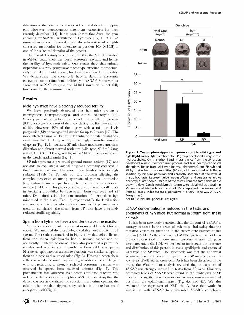
small testes (42.161.1 mg; n = 8), and strongly diminished number
of sperm (Fig. 1). In contrast, SP mice have moderate ventricular
dilatation and almost normal testis size (wild type, 91.662.3 mg,
n = 20; SP, 83.161.8 mg, n = 16; mean6SEM) and sperm count
in the cauda epididymidis (Fig. 1).
SP mice present a preserved general motor activity [12] and
are able to copulate; a vaginal plug was normally observed in
their female partners. However, male fertility was strongly
reduced (Table 1). To rule out any problem affecting the
complex processes occurring upstream of gamete interaction
(i.e., mating behavior, ejaculation, etc.), fertilization was assessed
in vitro (Table 2). This protocol showed a remarkable difference
in fertilizing probability between sperm from wild type and SP
mice. Even duplicating the concentration of sperm from hyh
mice used in the assay (Table 2, experiment B) the fertilization
was not as efficient as when sperm from wild type mice were
used. In conclusion, the sperm from SP mice have a strongly
reduced fertilizing ability.
Sperm from hyh mice have a deficient acrosome reactionSeveral causes can render a spermatozoon unable to fertilize an
oocyte. We analyzed the morphology, viability, and motility of SP
sperm. The results summarized in Fig. 2 show that cells collected
from the cauda epididymidis had a normal aspect and an
apparently unaltered acrosome. They also presented a pattern of
viability and motility undistinguishable from wild type sperm.
Moreover, spontaneous acrosome reaction was similar in sperm
from wild type and mutated mice (Fig. 3). However, when these
cells were incubated under capacitating conditions and challenged
with progesterone, a strongly reduced acrosome reaction was
observed in sperm from mutated animals (Fig. 3). This
phenomenon was observed even when acrosome reaction was
induced with the calcium ionophore A23187, indicating that the
defect was not in the signal transduction mechanism opening the
calcium channels that triggers exocytosis but in the mechanism of
exocytosis itself (Fig. 3).
aSNAP concentration is reduced in the testis andepididymis of hyh mice, but normal in sperm from theseanimals
It has been previously reported that the amount of aSNAP is
strongly reduced in the brain of hyh mice, indicating that the
mutation causes an alteration in the steady state balance of this
protein [13,14]. As the expression of aSNAP protein has not been
previously described in mouse male reproductive tract (except in
spermatogenic cells, [15], we decided to investigate the presence
and distribution of this protein in testis, epididymis and sperm of
wild type and SP mice. The hypothesis was that the abnormal
acrosome reaction observed in sperm from SP mice is caused by
low levels of aSNAP in these cells. As it has been described in the
brain, the Western blot analysis revealed that the amount of
aSNAP was strongly reduced in testes from SP mice. Similarly,
decreased levels of aSNAP were found in the epididymis of SP
mice, a finding that was more evident when sperm were washed
out from the epididymal lumen (Fig. 4A and 4B). We also
evaluated the expression of NSF, the ATPase that works in
association with aSNAP to disassemble SNARE complexes.
Figure 1. Testes phenotype and sperm count in wild type andhyh (hyh) mice. Hyh mice from the RP group developed a very severehydrocephalus. On the other hand, mutant mice from the SP groupdeveloped a mild hydrocephalic process and less neuropathologicalalterations. Brains from wild type (normal phenotype), and SP hyh andRP hyh mice from the same litter (70 day old) were fixed with Bouinsolution by vascular perfusion and coronally sectioned at the level ofthe optic chiasm. Representative images of brain and cerebral ventriclesphenotypes are shown. Images of the testes from the same animals areshown below. Cauda epididymidis sperm were obtained as explain inMaterials and Methods and counted. Data represent the mean6SEMfrom at least 6 independent experiments. * p,0.01 (one way ANOVA,Tukey’s test).doi:10.1371/journal.pone.0004963.g001
aSNAP and Acrosome Reaction
PLoS ONE | www.plosone.org 2 March 2009 | Volume 4 | Issue 3 | e4963

Interestingly, the amount of NSF was not decreased in testes and
epididymides of SP mice; on the contrary, a small but consistent
increase was observed (Fig. 4A and 4C).
Supporting the results obtained by Western blot, a strong
difference in aSNAP immunostaining was observed in the
seminiferous epithelium from wild type and SP animals
(Figure 5). This difference was evident in pre- and post-meiotic
cellular stages, including spermatogonia, primary spermatocytes
and round spermatids. However, the immunoreactive pattern of
elongated spermatids in mutant animals was very similar to that
found in wild type mice. To evaluate if the immunostaining
pattern observed in elongated spermatids was maintained in
mature sperm, the amount of aSNAP was assessed by Western
blot in spermatozoa obtained from cauda epididimydis. The
results showed that the aSNAP protein level in sperm from SP
mice was identical to that found in wild type cells, and that NSF
was -as in the other tissues- slightly increased (Fig. 6A and 6B).
Immunofluorescence for aSNAP also showed a similar pattern in
sperm from wild type and SP mice (Fig. 6C). A distinct acrosomal
labeling was observed (Fig. 6C), which disappeared when the
antibody was absorbed with recombinant aSNAP (data not
shown). In conclusion, the testis and epididymis of SP mice
present a reduced concentration of aSNAP. In contrast, the
amount and distribution of this protein is normal in mature sperm
from SP mice.
Wild type aSNAP rescues acrosomal exocytosis in spermfrom hyh mice
The above results indicate that the acrosomal exocytosis defect
in hyh mice was not due to a diminished amount of aSNAP. A
possible explanation for this observation is that the low
Table 1. Reproductive efficiency of wild type and SP hyh mice.
Male Female Productive matingsa Litter size Number of litters Relative fecundityb
wt wt 90.9% (13/15) 7.5 5.1 34.76
het het 89.7% (113/126) 7.3 5.4 35.35
hyh het 0% 0/24 0.0 0.0 0.0
hyh hyh 1.4% (2/148)c 5.5 4.5 0.33
wt: wild type (Napa(+/+)); het: heterozygous (Napa(hyh/+)); hyh: mutant homozygous (Napa(hyh/hyh)) with the slow progressive phenotype.aMatings are considered ‘‘productive’’ if at least one offspring was born.b‘‘Relative fecundity’’ is obtained as: (productive mating/100)6(litter size)6(number of litters); the value obtained is a measure of the overall fecundity according to the
Handbook of Genetically Standardized JAX Mice [33].cThe same hyh male had two productive matings.doi:10.1371/journal.pone.0004963.t001
Table 2. In vitro fertilization (IVF) assays using sperm fromwild type and SP hyh mice.
sperm(sperm/ml) Fertilized (n) Total (n)
Fertilization(%)
Experiment A wt (26105) 15 29 52
hyh (26105) 5 50 10 **
Experiment B wt (26105) 27 39 69
hyh (46105) 31 67 46 *
Metaphase II eggs from wild type (Napa(+/+)) female mice were incubated withsperm from wild type (Napa(+/+), wt) or mutant homozygous with the slowprogressive phenotype (Napa(hyh/hyh), hyh) mice. Following capacitation, spermfrom wild type and hyh mice were diluted to 2 or 46105 sperm/ml andcoincubated with eggs from wild type female mice. In experiment ‘‘A’’, thesame number of sperm cells from wt and hyh mice was used (26105 sperm/ml).In experiment ‘‘B’’ the number of sperm from hyh mice was duplicated (46105
sperm/ml). (*, p,0.02 or **, p,0.001 for hyh versus wt; Fisher’s exactprobability test).doi:10.1371/journal.pone.0004963.t002
Figure 2. Sperm characteristics in wild type and SP hyh (hyh) mice. (A) Bright field micrographs of sperm stained with Coomassie G-250 [30]from wild type and mutant (hyh) mice showing normal cell and acrosome morphology. Scale bar, 15 mm. (B) Sperm viability for wild type and mutant(hyh) mice was assessed by Eosin Y staining. (C) The percentage of motile sperm (T) and having progressive motility (P) was assessed in wild type andmutant (hyh) mice. Data represent the mean6SEM of at least four independent experiments.doi:10.1371/journal.pone.0004963.g002
aSNAP and Acrosome Reaction
PLoS ONE | www.plosone.org 3 March 2009 | Volume 4 | Issue 3 | e4963

concentrations of aSNAP in the testis and epididymis alter the
normal maturation of the exocytic machinery of the sperm.
Hence, even with a normal quantity of the protein, the sperm may
be unable to exocytose. To test this hypothesis, we assessed
whether we could restore exocytosis by adding wild type aSNAP
to sperm from SP mice. To this end, we set up a plasma
membrane-permeabilized mouse sperm model similar to the one
we routinely use for human sperm [16,17]. The percentage of
permeabilized cells without acrosome under resting conditions was
16% higher than that found in untreated cells (compare Fig. 7 with
Fig. 3), suggesting that some sperm may have lost their acrosome
during the permeabilization procedure. However, upon stimula-
tion with calcium, an additional 18% of the cells reacted (Fig. 7),
indicating that the treatment did not inactivate the exocytic
machinery. A similar percentage of calcium induced exocytosis in
permeabilized sperm has been previously reported by us [16] and
by other authors [18]. In contrast, sperm from mutated mice did
not respond to calcium stimulation, in agreement with the results
obtained with A23187 in intact cells (compare Fig. 7 with Fig 3).
When wild type aSNAP was added to the assay, exocytosis was
completely recovered, suggesting that the primary defect in these
cells is a deficient amount of functional aSNAP and not an abnormal
maturation of sperm in SP mice (Fig. 7). However, the previous
Western blot analysis and immunofluorescence images showed that
these cells have normal levels of aSNAP, suggesting that the M105I
mutation affects directly the function of the protein. To test this
Figure 3. Sperm from SP hyh mice have a deficient acrosomalreaction. Sperm from wild type and SP hyh (hyh) mice were collectedfrom the cauda epididymidis, incubated under capacitation conditions for1 h and stimulated with buffer (control), 10 mM progesterone (Pg) or 10 mMA23187 (A23187) for 15 min at 37uC. The cells were spotted on slides andfixed in ice-cold methanol. Acrosomal status was evaluated in at least 200sperm by staining with TRITC-PNA. The data represent the mean6SEM ofthree independent experiments (*, significant differences between samegroups for wild type and hyh mice, P,0.001, Student’s t test).doi:10.1371/journal.pone.0004963.g003
Figure 4. aSNAP and NSF expression in the reproductive tract of wild type (wt) and SP hyh (hyh) mice. (A) Proteins extracted from testisand cauda epididymidis of wt and SP hyh mice were analyzed by Western blot using an antibody recognizing aSNAP (upper panel) or NSF (middlepanel). Signals detected with an anti-actin antibody served as internal controls for equal protein loading (lower panel). Cauda epididymidis extractswere obtained before (Cauda+sperm) and after (Cauda-sperm) sperm were washed out from the organ. Brain was used as a positive control. Blots arerepresentative of 3 or 4 independent experiments. (B, C) Densitometric analysis of Western blot for aSNAP (B) and NSF (C). Black bars (mean6SEM,N = 3 or 4) refer to the relative amount of each protein in hyh samples compared to wt. * p,0.05 (Student’s t-test).doi:10.1371/journal.pone.0004963.g004
aSNAP and Acrosome Reaction
PLoS ONE | www.plosone.org 4 March 2009 | Volume 4 | Issue 3 | e4963

aSNAP and Acrosome Reaction
PLoS ONE | www.plosone.org 5 March 2009 | Volume 4 | Issue 3 | e4963

possibility, a recombinant aSNAP.M105I protein was produced,
purified, and added to permeabilized sperm from SP mice. The
mutated protein was unable to rescue acrosomal exocytosis in these
cells at 15 nM (Fig. 7), although a partial recovery was observed at
60 nM (data not shown). In conclusion, although carrying normal
levels of aSNAP, hyh sperm cells have a deficient acrosome reaction
because the mutated protein cannot support exocytosis with the
same efficiency than the wild type molecule
Figure 5. Immunolocalization of aSNAP in seminiferous epithelium. Light micrographs of testis sections from wild type (A–D) and SP hyhmice (E–H) showing general and specific patterns of immunoperoxidase staining using an aSNAP-specific antibody. Background staining withhematoxylin. Different stages of epithelial maturation cycle in normal and mutant testis are presented in comparative mode. A’–H’: Highmagnification images of the regions boxed in the corresponding panel (A–H). Epithelial polarization is oriented upwards. The symbols used are:spermatogonium (white pentagon); primary spermatocytes (white stars); round spermatids (white asterisk); elongated spermatids with highpolarization, indicating the residual body or axonemal region (black asterisk) and the heads (white arrowheads). Controls without primary antibodiesare shown in the inserts (A and E). In wild type animals, a strong immunoreaction was observed in the whole ephitelium. In contrast, mutant miceshowed a notably diminished immunoreaction compared to that of wild type mice (compare E–H to A–D panels). This difference was not evident inspermatids undergoing elongation process. Scale bar, 20 mm.doi:10.1371/journal.pone.0004963.g005
Figure 6. aSNAP expression and localization in sperm from wild type (wt) and SP hyh (hyh) mice. (A) Sperm protein extracts obtainedfrom wt and hyh mice were analyzed by Western blot using an antibody recognizing aSNAP (upper panel) or NSF (middle panel). Signals detectedwith an anti b-tubulin antibody served as internal controls for equal protein loading (lower panel). Blots are representative of seven independentexperiments. (B) Densitometric analysis of Western blot for aSNAP and NSF. Black bars (mean6SEM, N = 7) refer to the relative amount of eachprotein in hyh samples compared to wt (gray bars). * p,0.05 (Student’s t-test). (C) aSNAP localizes to the acrosomal region in mouse spermatozoa.Sperm from wild type and hyh mice were fixed, permeabilized and triple-stained with an anti-a/bSNAP antibody (green); TRITC-PNA, a lectin thatrecognizes the intra-acrosomal content (red); and Hoechst 33258 to visualize the nucleus of the cell (blue). Shown are epifluorescence micrographs oftypically stained cells. Scale bar, 10 mm.doi:10.1371/journal.pone.0004963.g006
aSNAP and Acrosome Reaction
PLoS ONE | www.plosone.org 6 March 2009 | Volume 4 | Issue 3 | e4963
aSNAP.M105I inhibits exocytosis in sperm from wild typemice and normal human donors
Although aSNAP is a required factor for intracellular transport,
in some experimental models -including the acrosomal exocytosis-
an excess of the wild type protein inhibits membrane fusion [7,
19–21]. Hence, we tested the effect of wild type and mutated
proteins in the acrosomal exocytosis assay using permeabilized
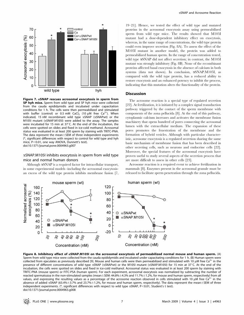
sperm from wild type mice. The results showed that M105I
mutant had a dose-dependent inhibitory effect on exocytosis,
whereas, in the same range of concentrations, the wild type protein
could even improve secretion (Fig. 8A). To assess the effect of the
M105I mutant in another model, the protein was added to
permeabilized human sperm. In the range of concentration tested,
wild type aSNAP did not affect secretion; in contrast, the M105I
mutant was strongly inhibitory (Fig. 8B). None of the recombinant
proteins affected basal exocytosis in the absence of calcium in both
systems (data not shown). In conclusion, aSNAP.M105I, as
compared with the wild type protein, has a reduced ability to
restore exocytosis and an enhanced potency to inhibit the process,
indicating that this mutation alters the functionality of the protein.
Discussion
The acrosome reaction is a special type of regulated secretion
[22]. At fertilization, it is initiated by a complex signal transduction
pathway triggered by the contact of the sperm membrane with
components of the zona pellucida [8]. At the end of this pathway,
cytoplasmic calcium increases and activates the membrane fusion
machinery that opens hundred of pores connecting the acrosomal
lumen with the extracellular medium. The expansion of these
pores promotes the fenestration of the membrane and the
formation of hybrid vesicles. Although with particular character-
istic, acrosome exocytosis is a regulated secretion sharing the same
basic mechanism of membrane fusion that has been described in
other secreting cells, such as neurons and endocrine cells [22].
Moreover, the special features of the acrosomal exocytosis have
proven useful to study several aspects of the secretion process that
are more difficult to assess in other cells [23].
Acrosome reaction is a required event to achieve fertilization in
mammals [8]. Enzymes present in the acrosomal granule must be
released to facilitate sperm penetration through the zona pellucida.
Figure 7. aSNAP rescues acrosomal exocytosis in sperm fromSP hyh mice. Sperm from wild type and SP hyh mice were collectedfrom the cauda epididymidis and incubated under capacitationconditions for 1 h. The cells were then permeabilized and stimulatedwith buffer (control) or 0.5 mM CaCl2 (10 mM free Ca2+). Whenindicated, 15 nM recombinant wild type aSNAP (aSNAPwt) or theM105I mutant (aSNAP.M105I) were added to the assay. The sampleswere incubated for 15 min at 37uC. At the end of the incubation, thecells were spotted on slides and fixed in ice-cold methanol. Acrosomalstatus was evaluated in at least 200 sperm by staining with TRITC-PNA.The data represent the mean6SEM of three independent experiments(*, significant differences with respect to control for wild type and hyhmice, P,0.01, one way ANOVA, Dunnett’s test).doi:10.1371/journal.pone.0004963.g007
Figure 8. Inhibitory effect of aSNAP.M105I on the acrosomal exocytosis of permeabilized normal mouse and human sperm. (A)Sperm from wild type mice were collected from the cauda epididymidis and incubated under capacitating conditions for 1 h. (B) Human sperm werecollected from ejaculates as previously described [9]. Mouse and human cells were then permeabilized and stimulated with 10 mM free Ca2+ in thepresence of different concentrations of wild type aSNAP (aSNAPwt) or the M105I mutant (aSNAP.M105I) for 15 min at 37uC. At the end of theincubation, the cells were spotted on slides and fixed in ice-cold methanol. Acrosomal status was evaluated in at least 200 sperm by staining withTRITC-PNA (mouse sperm) or FITC-PSA (human sperm). For each experiment, acrosomal exocytosis was normalized by subtracting the number ofreacted spermatozoa in the non-stimulated samples (mean6SEM: 44.8%64.3% and 11.7%61.2%, for mouse and human sperm, respectively) from allvalues, and expressing the resulting values as a percentage of the acrosome reaction observed in cells stimulated with 10 mM free Ca2+ in theabsence of added aSNAP (63.4%63.7% and 23.7%61.2%, for mouse and human sperm, respectively). The data represent the mean6SEM of threeindependent experiments. (*, significant differences with respect to wild type aSNAP, P,0.01, Student’s t test).doi:10.1371/journal.pone.0004963.g008
aSNAP and Acrosome Reaction
PLoS ONE | www.plosone.org 7 March 2009 | Volume 4 | Issue 3 | e4963

Hence, it would be expected that malfunctions in factors necessary
for secretion would affect the fertilizing potential of sperm [24,25].
However, since secretion is a key cellular function necessary for
many survival mechanisms, most mutations in these factors render
animals unable to survive to reproductive age. Interestingly, when
they do survive, they are in many cases fertile, suggesting that
compensatory mechanisms may overcome the malfunction or
absence of some factors [26]. Hyh mouse harboring a point
mutation in the Napa gen coding for aSNAP is the first example of
a fertility problem directly related to a factor necessary for the
general mechanism of regulated secretion that is specifically
affecting the acrosome reaction. Mutant mice with a mild clinical
and neuropathological phenotype are able to copulate and the
amount of live and motile sperm that they produce is similar to
that of wild type mice; however, they have a much reduced
fertility. Even when fertilization was assessed in vitro, sperm from
hyh mice behave poorly as compared with sperm from wild type
mice. Notwithstanding, the difference between genotypes was less
dramatic in the in vitro assays than in the mating studies. Thus,
other defects not assessed by in vitro fertilization might contribute
to this phenotype. In this context, although SP mice present a
preserved general motor activity and are able to copulate, they do
have gait and equilibrium impairments [12] that could affect the
frequency and efficiency of copulation.
Acrosome reaction experiments indicate that the basic problem
in sperm from hyh mice is a reduced capacity to undergo
exocytosis upon stimulation with progesterone and even with a
calcium ionophore. The fact that exogenous aSNAP can rescue
acrosomal exocytosis in these cells is a strong evidence that the
principal defect is a malfunction in the endogenous protein. Worth
noticing is that the M105I mutation is in a region of aSNAP that
does not interact with the SNARE complex [27]. Moreover, the
protein can bind to and disassemble SNARE complexes in vitro in
a way similar to the wild type protein [13,14]. These observations,
together with the fact that the hyh mouse presents a diminished
amount of aSNAP in brain, have lead to the conclusion that the
alteration in brain development in these animals is principally due
to an insufficient amount of protein and not to a malfunction of
the mutated molecule [13]. However, a functional defect of the
mutated protein has never been ruled out [13,14]. Our
observations in sperm point to a different mechanism. We
confirmed that the mutated protein has an altered steady state
distribution in several tissues; the levels of aSNAP in testis and
epididymis were significantly lower in the mutated animals. In
contrast, we found normal amount of this protein in sperm.
Therefore, it was unlikely that the defect in acrosomal exocytosis
was due to a decreased amount of aSNAP. These observations
suggested that the mutated protein has some intrinsic malfunction.
This was confirmed by the fact that the mutated protein was less
effective in restoring exocytosis than the wild type protein when
added to permeabilized sperm from hyh mouse. Moreover, the
mutated protein was inhibitory when added to normal mouse and
human sperm. It is worth noticing that an excess of wild type
aSNAP is also inhibitory, but at much higher concentrations [7].
Our results indicate that the M105I mutation alters the normal
steady state balance of aSNAP and also affects its function. The
protein may bind SNARE complexes and may promote their
disassembly as the wild type protein [13,14], but in the complexity
of a cellular environment with several other interacting factors, the
mutant behaves differently than the wild type protein in the
acrosomal secretory processes. In fact, co-immunoprecipitation
studies using brain lysates suggest that the M105I mutation may
affect the aSNAP/SNARE complex interaction [14]. Relatively
high concentrations of NSF -the other key factor for SNARE
complex disassembly- were found in testis, epididymis, and sperm
of hyh mice, likely as a compensatory response to the diminished
amount of functional aSNAP in the cells.
The deletion of aSNAP is embryonically lethal in mice [13];
hence the M105I mutant must conserve a certain degree of
functionality for intracellular transport and regulated exocytosis,
but cannot support acrosomal exocytosis. aSNAP has a well
characterized function in SNARE complex disassembly. In sperm
it is necessary for the activation of SNARE proteins upon sperm
stimulation [7]. However, it may have other still not well
characterized functions [21] that may be altered in the M105I
mutant. The methionine in the 105 position is well preserved
along evolution. This residue is in the convex surface of the
protein, opposite to the one supposed to bind SNARE
complexes; however, mutation analyses indicate that this area
is also important for aSNAP function [27]. All in all, the
molecular mechanism for the mutant malfunction remains to be
determined.
Materials and Methods
Ethics StatementHandling, care and processing of animals were carried out
according to the regulations approved by the Bioethics Committee of
the Universidad Austral de Chile and the Council of the American
Physiological Society. The protocol for handling human sperm
samples has been approved by the Bioethics Committee of the
Medical School of the Universidad Nacional de Cuyo, Argentina.
ReagentsRecombinant streptolysin O (SLO) was obtained from Dr
Bhakdi (University of Mainz, Mainz, Germany). Tetramethylrho-
damine isothiocyanate-labeled peanut agglutinin (TRITC-PNA)
and VECTASTAIN Universal Elite ABC kit (PK-6200) were from
Vector Laboratories Ltd. (Peterborough, UK). Fluorescein iso-
thiocyanate-coupled Pisum sativum (FITC-PSA) was from Sigma
Chemical Co. (St. Louis, Missouri, USA). Hoechst 33258, Sytox
Green, A23187, and Alexa Fluor 488-conjugated goat anti-mouse
immunoglobulin G were from Molecular Probes (Eugene, OR,
USA). Mouse monoclonal anti-a-SNAP (clone 4E4) and anti-a/b-
SNAP (purified IgG, clone 77.1) antibodies were purchased from
Exalpha Biologicals (Maynard, MA, USA) and Synaptic Systems
(Gottingen, Germany), respectively. Mouse anti-NSF monoclonal
antibody (clone 7) was purchased from BD Biosciences Pharmin-
gen. Monoclonal mouse anti-actin antibody (JLA20) was obtained
from the Developmental Studies Hybridoma Bank (Iowa City, IA,
USA) and monoclonal mouse anti-b-tubulin (clone 2-28-33) was
from Sigma Chemical Co. All other chemicals were analytical-
grade and were purchased from Sigma Chemical Co. or ICN
Biochemicals (Aurora, Ohio, USA).
Recombinant proteinsPlasmid pQE9 encoding wild type aSNAP was a kind gift from
Dr S.W. Whiteheart (University of Kentucky, Lexington, KY,
USA). Plasmid pET28 encoding aSNAP.M105I was a generous
gift from Dr. Phillys Hanson (Washington University, St. Louis,
Missouri, USA). Recombinant His6-tagged aSNAP wild type and
aSNAP.M105I were purified from the cytosolic fraction of
Escherichia coli XL-1 blue (Stratagene, Cambridge, UK) on Ni-
NTA-agarose based on previously published methods [7].
AnimalsMice were obtained from The Jackson Laboratory (Bar Harbor,
ME, USA), where the hyh mutation originally arose in the C57BL/
aSNAP and Acrosome Reaction
PLoS ONE | www.plosone.org 8 March 2009 | Volume 4 | Issue 3 | e4963

10J inbred strain and was subsequently placed on a B6C3Fe-a/a
(C57BL/6J female X C3HeB/FeJ-a/a male) hybrid background
[11]. These animals were bred into a colony at Facultad de
Medicina, Universidad Austral de Chile, Valdivia, Chile. Eight
different maternal sub-lines have been developed [12]. This study
included only adult animals (8–20 weeks) from different sub-lines
and generations. Mice were fed ad libitum with rodent food and
kept under a constant photoperiod of light/dark 12:12 h and room
temperature of 22uC.
Mice mating systemMice (8–12 week old) were genotyped and mated using the
monogamous mating system, except in the case of mating
homozygous mutant male with homozygous mutant female were
polygamous mating was utilized (one male with up to three
females). Vaginal plug was used as an indication that mating had
occurred. If no litters were produced after 10 weeks the mating
was considered non-productive.
Gamete collectionMetaphase II eggs were obtained from 6 to 8 week old wild
type female mice superovulated by injection of 5 IU of pregnant
mare serum gonadotrophin (PMSG; Syntex S.A., Argentina)
followed 48 h later by 5 IU of human chorionic gonadotrophin
(hCG; Syntex S.A., Argentina). Eggs were collected in Whitten’s
medium [28] containing 0.4% (w/v) polyvinylpyrrolidone (PVP)
and 20 mM Hepes buffer at pH 7.4. Cumulus cells were
removed using 0.3% hyaluronidase for 1–3 min in the same
solution. Eggs were collected and placed immediately into the
fertilization drop for in vitro fertilization. Mouse sperm were
collected from 12–24 week old wild type and hyh male mice.
Both cauda epididymides from a single male were placed in a
900 ml drop of human tubal fluid (HTF) medium [29]
supplemented with 0.5% BSA. Several incisions were made in
the tissue and the sperm allowed swimming into the medium for
20 min at 37uC. The resultant cell suspension was counted,
adjusted to 7–106106 sperm/ml, and incubated for 1 h at 37uCin an atmosphere of 5% CO2/95% air. Human semen samples
were obtained from normal healthy donors. Highly motile sperm
were recovered following a swim-up separation in HTF
supplemented with 0.5% BSA for 1 h at 37uC in an atmosphere
of 5% CO2/95% air. Concentration was adjusted to 5–106106/
ml, and incubation proceeded for at least 2 h.
In vitro fertilizationAll IVF procedures were performed approximately 14 hours
post hCG injection using HTF medium. Metaphase II eggs were
obtained from 5–6 wild type female mice. Only eggs with visible
polar body were collected and separated in two groups. Sperm
from two wild type male mice and from at least two hyh male
mice were obtained as explained above. Following capacitation,
sperm from wild type and hyh mice were diluted to 2 or 46105
sperm/ml. Eggs and sperm were mixed in a 500 ml fertilization
drop of HTF medium containing 0.5% BSA, and then cultured
at 37uC in an atmosphere of 5% CO2/95% air. After 3 hours,
eggs were washed six times to remove any loosely attached
sperm, and then cultured for 5 h. Finally, embryos were fixed in
3.7% paraformaldehyde in PBS for 15 min, permeabilized with
0.1% Triton X-100 and stained with 10 mM Sytox Green
(Molecular Probes) for DNA staining. Fertilization was evaluated
by the presence of the second polar body and the formation of
both the male and female pronuclei by epifluorescence
microscopy.
Evaluation of mouse sperm morphology, viability andmotility
Wild type and mutant sperm were collected as explained and
adjusted to a concentration of 5–106106 cells/ml in HTF. To
determine morphology of sperm and acrosome, a 5 ml aliquot was
stained with Coomassie G-250 [30] and observed under 1006oil
immersion lens. To assess sperm plasmalemma integrity (i.e.,
viability), 10 ml of sperm suspension and 1 ml of Eosin-Y solution
(5 g/l) were mixed, and the percentage of stained cells was
determined by counting 200 sperm (bright-field microscopy, 1006oil immersion lens). To assess motility in a sample, at least 200
sperm in two independent aliquots were classified as having
progressive or non-progressive motility, or being immotile (bright-
field microscopy, 406).
ImmunohistochemistryTestes were rapidly removed from hyh and wild type mice and
fixed in Bouin fixative solution for 48 h at room temperature.
Paraffin sections were processed for immunohistochemistry as
previously described [7] using mouse monoclonal anti-a-SNAP
(clone 4E4) as primary antibody. Omission of the incubation in the
primary antibody was used as a control of the immunoreaction.
Sections were examined under a Zeiss Axioskop microscope
equipped with a digital camera (Nikon CoolPix 5000).
Indirect ImmunofluorescenceSperm suspensions from hyh and wild type mice were fixed in
2% paraformaldehyde. Indirect immunofluorescence was per-
formed as described [9] using the mouse monoclonal anti-a/b-
SNAP antibody. Coverslips were mounted in Gelvatol, and
examined under a Zeiss Axioskop microscope equipped with
epifluorescence optics and a digital camera (Nikon Coolpix 5000).
Background was subtracted and brightness/contrast were adjusted
in the images to render an all or nothing labelling pattern using
Corel Draw version 12 (Corel, Ottawa, Ontario, Canada).
Protein extraction from tissue samples and spermProteins from hyh and wild type mice brain, testis and
epidydimis were extracted by homogenizing in a modified RIPA
buffer (50 mM Tris-HCl, pH 7.4, 150 mM NaCl, 1% Triton-X-
100, 1% sodium deoxycholate, 0.1% SDS) with a protease
inhibitor cocktail (P2714, Sigma Chemical Co.) plus 1 mM
phenylmethanesulphonylfluoride (PMSF). The homogenates were
centrifuged at 60006g for 10 minutes at 4uC to sediment
unbroken cells and nuclei. Supernatants were stored at 280uCuntil used. Sperm proteins were isolated according to [31].
SDS-PAGE and immunoblottingFor Western blotting, 2% (v/v) 2-mercaptoethanol was added to
the samples. After boiling for 2 min, 15 mg (brain, testis,
epidydimis) or 30 mg (sperm) of total protein were loaded on
10% polyacrylamide gels according to [32]. The proteins were
then transferred to polyvinylidene difluoride (PVDF) membranes
(Millipore, Bedford, MA). Blots were incubated for 1–2 h at 37uCwith the primary antibodies diluted in PBS containing 0.05%
Tween 20 and 1% BSA (anti-a-SNAP, 1:1000; anti-NSF, 1:5000).
Horseradish peroxidase conjugated goat anti-mouse-IgG was used
as secondary antibody (0.25 mg/ml) with 30 min incubations.
Excess antibodies were removed by washing 565 min in 16PBS.
Detection was accomplished with an enhanced chemilumines-
cence system (SuperSignal West Pico Chemiluminescent Sub-
strate, Pierce, Rockford, IL) and subsequent exposure to Kodak
XAR film (Eastman Kodak, Rochester, NY) for 5–30 s. In order
aSNAP and Acrosome Reaction
PLoS ONE | www.plosone.org 9 March 2009 | Volume 4 | Issue 3 | e4963
to confirm equal loading of proteins, blots were stripped and re-
probed with an antibody against actin (1:1000) or b-tubulin
(1:1000).
Acrosome reaction in intact and permeabilized humanand mouse sperm
Motile sperm were obtained as explained above. In some
experiments, spermatozoa without further treatment were used to
study acrosome reaction. Permeabilization of human and mouse
sperm was accomplished as previously described [17]. Briefly,
washed spermatozoa were resuspended in cold PBS containing
0.4–1 units/ml SLO for 15 min at 4uC. Cells were washed once
with PBS and resuspended in ice-cold sucrose buffer (250 mM
sucrose, 0.5 mM EGTA, 20 mM Hepes-K, pH 7.0) containing
2 mM dithiothreitol. Permeabilized sperm were incubated, when
indicated, with the recombinant proteins for 15 min at 37uCbefore adding the exocytosis stimulators. After stimulation of intact
or permeabilized sperm, incubation proceeded at 37uC for
15 min. Acrosomal status was evaluated by staining with
TRITC-PNA (mouse sperm) or FITC-PSA (human sperm). At
least 200 cells were scored using a Zeiss Axioskop microscope
equipped with epifluorescence optics.
Acknowledgments
The authors thank Mr. Genaro Alvial and Marcelo Furlan for their
excellent technical assistance, Dr. Ilona Concha and Dr. Carlos Gonzalez
for epifluorescence microscopy facilities. We also thank Dr. Montserrat
Guerra for critical reading of the manuscript. Plasmids were generously
provided by Dr. P. Hanson and S.W. Whiteheart.
Author Contributions
Conceived and designed the experiments: LFB GADB MAM EMR LSM.
Performed the experiments: LFB GADB MAM ARR FR CO. Analyzed
the data: LFB GADB MAM CNT EMR LSM. Contributed reagents/
materials/analysis tools: LFB GADB MAM ARR FR MHR CO CNT
EMR LSM. Wrote the paper: LSM.
References
1. Whiteheart SW, Griff IC, Brunner M, Clary DO, Mayer T, et al. (1993) SNAP
family of NSF attachment proteins includes a brain-specific isoform. Nature 362:
353–355.
2. Jahn R, Scheller RH (2006) SNAREs–engines for membrane fusion. Nat Rev
Mol Cell Biol 7: 631–643.
3. Wickner W, Schekman R (2008) Membrane fusion. Nat Struct Mol Biol 15:
658–664.
4. Xu T, Ashery U, Burgoyne RD, Neher E (1999) Early requirement for alpha-
SNAP and NSF in the secretory cascade in chromaffin cells. EMBO J 18:
3293–3304.
5. Morgan A, Burgoyne RD (1995) A role for soluble NSF attachment proteins
(SNAPs) in regulated exocytosis in adrenal chromaffin cells. EMBO J 14:
232–239.
6. Barnard RJ, Morgan A, Burgoyne RD (1997) Stimulation of NSF ATPase
activity by alpha-SNAP is required for SNARE complex disassembly and
exocytosis. J Cell Biol 139: 875–883.
7. Tomes CN, De Blas GA, Michaut MA, Farre EV, Cherhitin O, et al. (2005)
alpha-SNAP and NSF are required in a priming step during the human sperm
acrosome reaction. Mol Hum Reprod 11: 43–51.
8. Yanagimachi R (1994) Mammalian Fertilization. In: Knobil E, Neill JD, eds.
The Physiology of Reproduction. New York: Raven Press. pp 189–281.
9. De Blas GA, Roggero CM, Tomes CN, Mayorga LS (2005) Dynamics of
SNARE assembly and disassembly during sperm acrosomal exocytosis. PLoS
Biol 3: 1801–1812.
10. Gissen P, Maher ER (2007) Cargos and genes: insights into vesicular transport
from inherited human disease. J Med Genet 44: 545–555.
11. Bronson RT, Lane PW (1990) Hydrocephalus with hop gait (hyh): a new
mutation on chromosome 7 in the mouse. Brain Res Dev Brain Res 54:
131–136.
12. Batiz LF, Paez P, Jimenez AJ, Rodriguez S, Wagner C, et al. (2006)
Heterogeneous expression of hydrocephalic phenotype in the hyh mice carrying
a point mutation in alpha-SNAP. Neurobiol Dis 23: 152–168.
13. Chae TH, Kim S, Marz KE, Hanson PI, Walsh CA (2004) The hyh mutation
uncovers roles for alpha-SNAP in apical protein localization and control of
neural cell fate. Nat Genet 36: 264–270.
14. Hong HK, Chakravarti A, Takahashi JS (2004) The gene for soluble N-
ethylmaleimide sensitive factor attachment protein alpha is mutated in
hydrocephaly with hop gait (hyh) mice. Proc Natl Acad Sci U S A 101:
1748–1753.
15. Ramalho-Santos J, Moreno RD, Wessel GM, Chan EK, Schatten G (2001)
Membrane trafficking machinery components associated with the mammalian
acrosome during spermiogenesis. Exp Cell Res 267: 45–60.
16. Diaz A, Dominguez I, Fornes MW, Burgos MH, Mayorga LS (1996) Acrosome
content release in streptolysin O permeabilized mouse spermatozoa. Andrologia
28: 21–26.
17. Yunes R, Michaut M, Tomes C, Mayorga LS (2000) Rab3A triggers the
acrosome reaction in permeabilized human spermatozoa. Biol Reprod 62:1084–1089.
18. Kitamura K, Tanaka H, Nishimune Y (2003) Haprin, a novel haploid germ cell-specific RING finger protein involved in the acrosome reaction. J Biol Chem
278: 44417–44423.
19. Wang L, Ungermann C, Wickner W (2000) The docking of primed vacuoles canbe reversibly arrested by excess Sec17p (alpha-SNAP). J Biol Chem 275:
22862–22867.20. Babcock M, Macleod GT, Leither J, Pallanck L (2004) Genetic analysis of
soluble N-ethylmaleimide-sensitive factor attachment protein function in
Drosophila reveals positive and negative secretory roles. J Neurosci 24:3964–3973.
21. Barszczewski M, Chua JJ, Stein A, Winter U, Heintzmann R, et al. (2008) Anovel site of action for alpha-SNAP in the SNARE conformational cycle
controlling membrane fusion. Mol Biol Cell 19: 776–784.22. Mayorga LS, Tomes CN, Belmonte SA (2007) Acrosomal exocytosis, a special
type of regulated secretion. IUBMB Life 59: 286–292.
23. Tomes C (2006) Acrosomal exocytosis. In: Regazzi R, ed. Molecularmechanisms of exocytosis. Georgetown, TX, USA: Landes Bioscience. pp
117–147.24. Zhao L, Reim K, Miller DJ (2008) Complexin-I-deficient sperm are subfertile
due to a defect in zona pellucida penetration. Reproduction 136: 323–334.
25. Zhao L, Burkin HR, Shi X, Li L, Reim K, et al. (2007) Complexin I is requiredfor mammalian sperm acrosomal exocytosis. Dev Biol 309: 236–244.
26. Schluter OM, Schmitz F, Jahn R, Rosenmund C, Sudhof TC (2004) A completegenetic analysis of neuronal Rab3 function. J Neurosci 24: 6629–6637.
27. Marz KE, Lauer JM, Hanson PI (2003) Defining the SNARE complex bindingsurface of alpha-SNAP: implications for SNARE complex disassembly. J Biol
Chem 278: 27000–27008.
28. Whitten WK (1971) Nutrient requirements for the culture of preimplantationembryos in vitro. In: Gaspe R, ed. Advances in Biosciences, Vol 6. Oxford:
Pergamon Press. pp 129–141.29. Quinn P, Kerin JF, Warnes GM (1985) Improved pregnancy rate in human in
vitro fertilization with the use of a medium based on the composition of human
tubal fluid. Fertil Steril 44: 493–498.30. Larson JL, Miller DJ (1999) Simple histochemical stain for acrosomes on sperm
from several species. Mol Reprod Dev 52: 445–449.31. Baker MA, Hetherington L, Ecroyd H, Roman SD, Aitken RJ (2004) Analysis of
the mechanism by which calcium negatively regulates the tyrosine phosphor-ylation cascade associated with sperm capacitation. J Cell Sci 117: 211–222.
32. Laemmli UK (1970) Cleavage of structural proteins during the assembly of the
head of bacteriophage T4. Nature 227: 680–685.33. Green MC, Witham BA (1991) Handbook on Genetically Standardized JAX
Mice. Green MC, Witham BA, eds. Bar Harbor, 4th Edition. Bar Harbor: TheJackson Laboratory.
aSNAP and Acrosome Reaction
PLoS ONE | www.plosone.org 10 March 2009 | Volume 4 | Issue 3 | e4963
Related Documents

![QHS LMGRX S HYH]PRX MH W]Y GRGDYDWHOp SRVOHGQt …](https://static.cupdf.com/doc/110x72/624a569f97caeb58034e0ddd/qhs-lmgrx-s-hyhprx-mh-wy-grgdydwhop-srvohgqt-.jpg)

![hyh-kmbtIcfw - Department of Industries · hyh-kmbtIcfw vyavasayakeralam 2 Pqsse 2013 Pqsse 2013 hyh-kmbtIcfw 3 D≈S°w Pqsse 2013 thmfyw ˛ 46 e°w ˛2 ]w‡n-Iƒ sI´nS\n¿ΩmWkma-{Kn-I-fpsS](https://static.cupdf.com/doc/110x72/5c9f4b7788c993552d8d4f06/hyh-kmbticfw-department-of-hyh-kmbticfw-vyavasayakeralam-2-pqsse-2013-pqsse.jpg)