Spectroscopy of Biopolymers (III) Carbonhydrates

Spectroscopy of Biopolymers (III) Carbonhydrates.
Dec 20, 2015
Welcome message from author
This document is posted to help you gain knowledge. Please leave a comment to let me know what you think about it! Share it to your friends and learn new things together.
Transcript

Spectroscopy of Biopolymers
(III) Carbonhydrates
Monosaccharides
Number of
Carbons
Category Name
Examples
4 Tetrose Erythrose, Threose
5 Pentose Arabinose, Ribose, Ribulose, Xylose, Xylulose, Lyxose
6 Hexose Allose, Altrose, Fructose, Galactose, Glucose, Gulose, Idose, Mannose, Sorbose, Talose, Tagatose
7 Heptose Sedoheptulose

• Many saccharide structures differ only in the orientation of the hydroxyl groups (-OH). This slight structural difference makes a big difference in the biochemical properties, organoleptic properties (e.g., taste), and in the physical properties such as melting point and Specific Rotation (how polarized light is distorted).
• A chain-form monosaccharide that has a carbonyl group (C=O) on an end carbon forming an aldehyde group (-CHO) is classified as an aldose.
• When the carbonyl group is on an inner atom forming a ketone, it is classified as a ketose.

Monosaccharides-Tetroses
D-Erythrose D-Threose

Monosaccharides - Pentoses
The ring form of ribose is a component of ribonucleic acid (RNA). Deoxyribose, which is missing an oxygen at position 2, is a component of deoxyribonucleic acid (DNA). In nucleic acids, the hydroxyl group attached to carbon number 1 is replaced with nucleotide bases.
D-Ribose D-Arabinose D-Xylose D-Lyxose
Ribose Deoxyribose

Monosaccharides Hexoses C6H12
O6
D-Allose D-Altrose D-Glucose D-Mannose
D-Gulose D-Idose D-Galactose D-Talose

• Structures that have opposite configurations of a hydroxyl group at only one position, such as glucose and mannose, are called epimers.
• Glucose, also called dextrose, is the most widely distributed sugar in the plant and animal kingdoms and it is the sugar present in blood as "blood sugar".
• The chain form of glucose is a polyhydric aldehyde, meaning that it has multiple hydroxyl groups and an aldehyde group.
• Fructose, also called levulose or "fruit sugar", is shown here in the chain and ring forms. Fructose and glucose are the main carbohydrate constituents of honey.
D-Fructose Fructose

Monosaccharides - Heptoses
D-Sedoheptulose

Chain and Ring forms• Many simple sugars can exist in a chain form or a ring form. The ring f
orm is favored in aqueous solutions, and the mechanism of ring formation is similar for most sugars.
• The glucose ring form is created when the oxygen on carbon number 5 links with the carbon comprising the carbonyl group (carbon number 1) and transfers its hydrogen to the carbonyl oxygen to create a hydroxyl group.
• The rearrangement produces alpha glucose when the hydroxyl group is on the opposite side of the -CH2OH group, or beta glucose when the hydroxyl group is on the same side as the -CH2OH group.
• Isomers, such as these, which differ only in their configuration about their carbonyl carbon atom are called anomers. The little D in the name derives from the fact that natural glucose is dextrorotary, i.e., it rotates polarized light to the right, but it now denotes a specific configuration.
• Monosaccharides forming a five-sided ring, like ribose, are called furanoses. Those forming six-sided rings, like glucose, are called pyranoses.

Stereochemistry
β-D-Glucose β-L-Glucose β-D-Glucose(chair form)
β-D-Glucose β-L-Glucose β-D-Glucose(boat form)
Saccharides with identical functional groups but with different spatial configurations have different chemical and biological properties. Compounds that are mirror images of each other but are not identical, comparable to left and right shoes, are called enantiomers. The following structures illustrate the difference between β-D-Glucose and β-L-Glucose. Identical molecules can be made to correspond to each other by flipping and rotating. However, enantiomers cannot be made to correspond to their mirror images by flipping and rotating. Glucose is sometimes illustrated as a "chair form" because it is a more accurate representation of the bond angles of the molecule. The "boat" form of glucose is unstable.

Disaccharides
Disaccharide Description Component monosaccharides
sucrose common table sugar glucose + fructose
lactose main sugar in milk galactose + glucose
maltose product of starch hydrolysis
glucose + glucose
trehalose found in fungi glucose + glucose

Sucrose, also called saccharose, is ordinary table sugar refined from sugar cane or sugar beets. It is the main ingredient in turbinado sugar, evaporated or dried cane juice, brown sugar, and confectioner's sugar.

Lactose has a molecular structure consisting of galactose and glucose. It is of interest because it is associated with lactose intolerance which is the intestinal distress caused by a deficiency of lactase, an intestinal enzyme needed to absorb and digest lactose in milk. Undigested lactose ferments in the colon and causes abdominal pain, bloating, gas, and diarrhea. Yogurt does not cause these problems because lactose is consumed by the bacteria that transform milk into yogurt.

Maltose consists of two α-D-glucose molecules with the alpha bond at carbon 1 of one molecule attached to the oxygen at carbon 4 of the second molecule. This is called a 1α→4 glycosidic linkage. Trehalose has two α-D-glucose molecules connected through carbon number one in a 1α→1 linkage. Cellobiose is a disaccharide consisting of two β-D-glucose molecules that have a 1β→4 linkage as in cellulose. Cellobiose has no taste, whereas maltose and trehalose are about one-third as sweet as sucrose.

Polysaccharides
Many polysaccharides, unlike sugars, are insoluble in water. Dietary fiber includes polysaccharides and oligosaccharides that are resistant to digestion and absorption in the human small intestine but which are completely or partially fermented by microorganisms in the large intestine. The polysaccharides play important roles in nutrition, biology, or food preparation.

StarchStarch is the major form of stored carbohydrate in plants. Starch is composed of a mixture of two substances: amylose, an essentially linear polysaccharide, and amylopectin, a highly branched polysaccharide. Both forms of starch are polymers of α-D-Glucose. Natural starches contain 10-20% amylose and 80-90% amylopectin. Amylose forms a colloidal dispersion in hot water (which helps to thicken gravies) whereas amylopectin is completely insoluble.

• Amylose molecules consist typically of 200 to 20,000 glucose units which form a helix as a result of the bond angles between the glucose units.
•
•Amylose

• Amylopectin differs from amylose in being highly branched. Short side chains of about 30 glucose units are attached with 1α→6 linkages approximately every twenty to thirty glucose units along the chain. Amylopectin molecules may contain up to two million glucose units.
Amylopectin
The side branching chains are clustered together within the amylopectin molecule
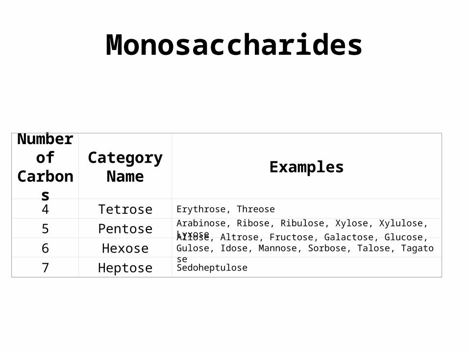
Glycogen• Glucose is stored as glycogen in animal tissues by the process of
glycogenesis. When glucose cannot be stored as glycogen or used immediately for energy, it is converted to fat. Glycogen is a polymer of α-D-Glucose identical to amylopectin, but the branches in glycogen tend to be shorter (about 13 glucose units) and more frequent. The glucose chains are organized globularly like branches of a tree originating from a pair of molecules of glycogenin, a protein with a molecular weight of 38,000 that acts as a primer at the core of the structure. Glycogen is easily converted back to glucose to provide energy.
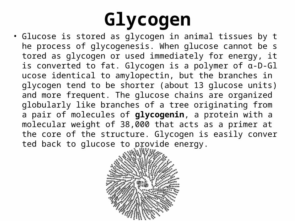
Dextran• Dextran is a polysaccharide similar to amylopectin, but the main chains are fo
rmed by 1α→6 glycosidic linkages and the side branches are attached by 1α→3 or 1α→4 linkages. Dextran is an oral bacterial product that adheres to the teeth, creating a film called plaque. It is also used commercially in confection
s, in lacquers, as food additives, and as plasma volume expanders.

Inulin• Some plants store carbohydrates in the form of inulin as an a
lternative, or in addition, to starch. Inulins are polymers consisting of fructose units that typically have a terminal glucose. Inulins have a sweet taste and are present in many vegetables and fruits, including onions, leeks, garlic, bananas, asparag
us, chicory, and Jerusalem artichokes.

CelluloseCellulose is a polymer of β-D-Glucose, which in contrast to starch, is oriented with -CH2OH groups alternating above and below the plane of the cellulose molecul
e thus producing long, unbranched chains. The absence of side chains allows cellulose molecules to lie close together and form rigid structures. Cellulose is the major structural material of plants. Wood is largely cellulose, and cotton is almost pure cellulose. Cellulose can be hydrolyzed to its constituent glucose units by microorganisms that inhabit the digestive tract of termites and ruminants. Cellulose may be modified in the laboratory by treating it with nitric acid (HNO3) to replace all t
he hydroxyl groups with nitrate groups (-ONO2) to produce cellulose nitrate (nitr
ocellulose or guncotton) which is an explosive component of smokeless powder. Partially nitrated cellulose, known as pyroxylin, is used in the manufacture of coll
odion, plastics, lacquers, and nail polish.

ChitinChitin is an unbranched polymer of N-Acetyl-D-glucosamine. It is found in fungi and is the principal component of arthropod and lower animal exoskeletons, e.g., insect, crab, and shrimp shells. It may be regarded as a derivative of cellulose, in which the hydroxyl groups of the second carbon of each glucose unit have been replaced with acetamido (-NH(C=O)CH3) groups.
Chitin

Beta-Glucan• Beta-glucans consist of linear unbranched polysaccharides of β-
D-Glucose like cellulose, but with one 1β→3 linkage for every three or four 1β→4 linkages. Beta-glucans form long cylindrical molecules containing up to about 250,000 glucose units. Beta-glucans occur in the bran of grains such as barley and oats, and they are recognized as being beneficial for reducing heart disease by lowering cholesterol and reducing the glycemic response. They are used comercially to modify food texture and as fat substitutes.
Beta-Glucan

Glycosaminoglycans• Glycosaminoglycans are found in the lubricating fluid of the joints and as c
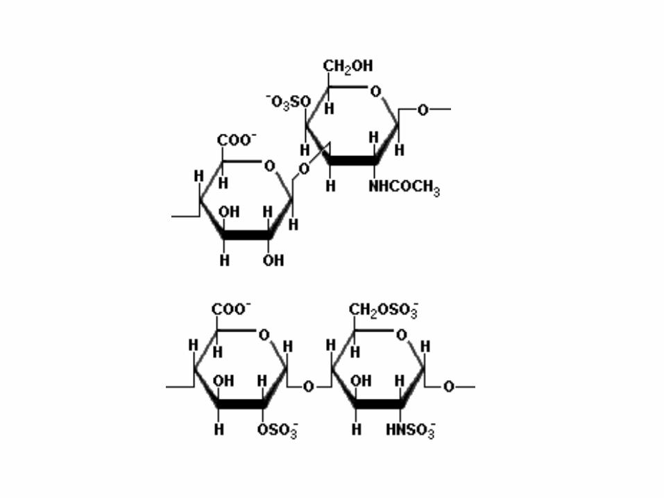
omponents of cartilage, synovial fluid, vitreous humor, bone, and heart valves. Glycosaminoglycans are long unbranched polysaccharides containing repeating disaccharide units that contain either of two amino sugar compounds -- N-acetylgalactosamine or N-acetylglucosamine, and a uronic acid such as glucuronate (glucose where carbon six forms a carboxyl group). Glycosaminoglycans are negatively charged, highly viscous molecules sometimes called mucopolysaccharides. The physiologically most important glycosaminoglycans are hyaluronic acid, dermatan sulfate, chondroitin sulfate, heparin, heparan sulfate, and keratan sulfate. Chondroitin sulfate is composed of β-D-glucuronate linked to the third carbon of N-acetylgalactosamine-4-sulfate as illustrated here. Heparin is a complex mixture of linear polysaccharides that have anticoagulant properties and vary in the degree of sulfation of the saccharide units.
Related Documents