Spectroscopic studies of Ti 3+ ions speciation inside MgAl 2 O 4 spinels P. Lombard a , B. Boizot a , N. Ollier a , A.Jouin b , A.Yoshikawa c a Laboratoire des Solides Irradiés, UMR7642 CEA - CNRS - EcolePolytechnique, EcolePolytechnique, 91128 Palaiseau Cedex, France b BerlinSolar GmbH, Magnusstrasse 11, 12489 Berlin, Germany c IMRAM, Tohoku University,2-1-1 Katahira, Aoba-ku, Sendai 980-8577, Japan Abstract: An electron paramagnetic resonance (EPR) investigation on Ti-doped MgAl 2 O 4 spinels has been made in order to study the Ti 3+ sites. The study we present here concerns the angular dependencies of the Ti 3+ EPR lines and the variations of the EPR spectra due to a modification of the TiO 2 content or of the chemical composition from MgAl 2 O 4 to MgAl 4 O 7 and MgAl 6 O 10 . In all the studied samples except one, we observe the presence of both Mn 2+ and Ti 3+ ions. No correlation was observed between Ti 3+ amount and TiO 2 content; the titanium ions are located in three different sites : the octahedral B site of the spinel structure; the tetrahedral A site and a last site which remains unclear. On the contrary, the major part of the Mn 2+ ions is assumed to be in the tetrahedral A site of the spinel structure and a minor part in the octahedral B site. Our work demonstrates the overall interest of EPR spectroscopy in the study of the paramagnetic optically active ions inside optical materials. 1. Introduction Among the crystalline compounds, the spinel oxides present a large range of interesting electrical, magnetic and optical properties. The magnesium–aluminum spinel MgAl 2 O 4 in particular has been comprehensively studied and previous works point out a high melting point (2135°C [1]), a high hardness(7.5–8 Mohs [2]), and interesting thermal and optical properties [3]. MgAl 2 O 4 can crystallize in the ‘‘normal’’ or ‘‘inverse’’ spinel structure. This structure may be described as a cubic closest-packed array of oxygen atoms, with one-eighth of the tetrahedral sites and one-half of the octahedral sites filled. The space group is Fd3 m (O ) and the lattice parameter a equals to 8.08435 Å [4]. In the case of a ‘‘normal’’ spinel structure, Mg 2+ ions are hal-00512719, version 1 - 1 Sep 2010 Author manuscript, published in "N/P" DOI : 10.1016/j.jcrysgro.2008.09.131

Welcome message from author
This document is posted to help you gain knowledge. Please leave a comment to let me know what you think about it! Share it to your friends and learn new things together.
Transcript
Spectroscopic studies of Ti3+ ions speciation inside
MgAl2O4 spinels
P. Lombarda, B. Boizota, N. Olliera, A.Jouinb, A.Yoshikawac aLaboratoire des Solides Irradiés, UMR7642 CEA - CNRS - EcolePolytechnique, EcolePolytechnique,
91128 Palaiseau Cedex, France bBerlinSolar GmbH, Magnusstrasse 11, 12489 Berlin, Germany
cIMRAM, Tohoku University,2-1-1 Katahira, Aoba-ku, Sendai 980-8577, Japan
Abstract: An electron paramagnetic resonance (EPR) investigation on Ti-doped MgAl2O4 spinels
has been made in order to study the Ti3+ sites. The study we present here concerns the angular
dependencies of the Ti3+ EPR lines and the variations of the EPR spectra due to a modification of
the TiO2 content or of the chemical composition from MgAl2O4 to MgAl4O7 and MgAl6O10. In
all the studied samples except one, we observe the presence of both Mn2+ and Ti3+ ions. No
correlation was observed between Ti3+ amount and TiO2 content; the titanium ions are located in
three different sites : the octahedral B site of the spinel structure; the tetrahedral A site and a last
site which remains unclear. On the contrary, the major part of the Mn2+ ions is assumed to be in
the tetrahedral A site of the spinel structure and a minor part in the octahedral B site. Our work
demonstrates the overall interest of EPR spectroscopy in the study of the paramagnetic optically
active ions inside optical materials.
1. Introduction
Among the crystalline compounds, the spinel oxides present a large range of interesting
electrical, magnetic and optical properties. The magnesium–aluminum spinel MgAl2O4 in
particular has been comprehensively studied and previous works point out a high melting point
(2135°C [1]), a high hardness(7.5–8 Mohs [2]), and interesting thermal and optical properties [3].
MgAl2O4 can crystallize in the ‘‘normal’’ or ‘‘inverse’’ spinel structure. This structure may be
described as a cubic closest-packed array of oxygen atoms, with one-eighth of the tetrahedral
sites and one-half of the octahedral sites filled. The space group is Fd3�m (O��) and the lattice
parameter a equals to 8.08435 Å [4]. In the case of a ‘‘normal’’ spinel structure, Mg2+ ions are
hal-0
0512
719,
ver
sion
1 -
1 Se
p 20
10Author manuscript, published in "N/P" DOI : 10.1016/j.jcrysgro.2008.09.131
located in tetrahedral sites and the Al3+ ions are in octahedral sites. Moreover, this compound can
be easily doped by transition metal ions such as Ti, Mn [5], Cr [6], Fe [3] or V [7]. In the
particular case of Ti-doped MgAl2O4 spinel, a strong visible blue emission around 455–490 nm
under excitation at 280 nm was observed and attributed to a charge transfer between the Ti4+ and
Ti3+ ions which replaced some of the Al3+ ions in octahedral sites [8,9]. This large Stokes shift (�
40,000 cm-1 from 280 to � 500 nm) was understood as the consequence of a large lattice
distortion around the titanium ion [8]. Nevertheless, the precise distortions of the environment of
the Ti3+ ion and the presence of trivalent titanium remain to be explained. Thus, the aim of this
work is to determine the nature and the site of the different paramagnetic ions present in Ti-doped
MgAl2O4 spinel and to study the distortions of the Ti3+ ion environment. For that purpose, we
used electron paramagnetic resonance (EPR), a very sensitive tool for the analysis of
paramagnetic species in crystals and glasses.
2. Experimental procedure
Samples were prepared by Jouini et al. [5] using the modified µ-pulling down method (µ-PD)
which has been detailed elsewhere [5, 8]. Synthesis was conducted in a reducing Ar atmo sphere
to avoid all oxidation of the special Iridium crucible of the experimental setup and the different
samples were Ti-doped at 0.1, 0.4, 0.5, 0.6, 0.8 and 1 mol%. The samples are cylindrical with the
c-axis parallel to the direction of the sample. After synthesis, X-band EPR spectra have been
recorded at 5K using an OXFORD He cryostat on a EMX Brücker EPR spectrometer. The
microwave power used for the experiments is 0.01mW in order to avoid saturation of Ti3+ EPR
lines. This power was determined in relation with the saturation properties of Ti3+ EPR lines at
5K. Angular dependencies of the different Ti-dopedMgAl2O4 samples EPR spectra were recorded
by rotating the samples from a reference EPR spectrum (0°) corresponding to a magnetic field B0
parallel to the c-axis to a B0 in the (ab) plane (90° sample). The different EPR spectra presented
in this report have been normalized to the receiver gain and to the sample weight in order to
obtain quantitative comparison of the EPR lines intensities between the different samples.
Moreover, it is worth reminding that EPR spectroscopy allows the absorbance spectra (i.e. the
experimental spectrum integrated once) of all EPR lines corresponding to one paramagnetic ion
hal-0
0512
719,
ver
sion
1 -
1 Se
p 20
10
in a specific site to coincide directly with the number of spins of this paramagnetic ion inside the
sample [10].
3. Results
3.1. Influence of TiO2 content
Fig. 1 presents the reference (c-axis parallel to B0) X-band EPR spectrum of 0.1TiO2–MgAl2O4
sample recorded at 5K with a power of 0.01mW and a frequency of 9.490 GHz. We observe a set
of six strong and 10 weak lines correlated to the Mn2+ ions (electronic spin S= �
� , nuclear spin I
=�
�) diluted inside the spinel structure. This spectrum is very similar to the spectrum obtained by
Tomita et al. [11] into crystal manganese-doped spinel Mn:MgAl2O4. By calculating the spin
Hamiltonian of the Mn2+ ion into MgAl2O4 powders, Shaffer et al. [12] predicted the presence of
six sharp main lines corresponding to the hyperfine components of the ∆MS = ±1, ∆MI = 0
allowed transitions and 10 sharp ∆MS = ±1, ∆MI = ± 1 forbidden transitions. So as a result, the
different lines observed in Fig. 1 may be correlated either to a small zero field splitting (ZFS) due
to high symmetry around Mn2+ ions or to three different Mn2+ environments inside the MgAl2O4
spinel structure. As Tomita et al. have observed before, we do not find any angular dependency
of the EPR lines attributed to Mn2+ ions (cf. Fig. 4). Tomita et al. concluded to the existence of
Mn2+ ions into two different spinels sites, a tetragonal one and an octahedral one. In our case,
further EPR studies and simulation on Mn-doped MgAl2O4 spinels are required to discriminate
between these two hypotheses. Fig. 2 presents the EPR spectra recorded with the c-axis parallel
to the applied magnetic field B0 of different spinels samples (MgAl2O4, MgAl4O7, MgAl6O10)
doped with different amounts of titanium from 0.1 to 1mol%. We observe the presence of Mn2+
ion EPR lines in all studied samples. Moreover, there is no correlation between the increase of
the TiO2 content introduced inside the crystals and the Mn2+ EPR lines intensity. This result
could be correlated to two different points. First, Mn2+ is mainly an impurity of the MgO powders
used for the synthesis. Second, the TiO2 doping content plays a role upon the redox couple
between Mn2+ and Mn3+ ions inside the spinel crystal. But Mn3+ ions are diamagnetic and cannot
be studied by EPR spectroscopy; however, optical measurements could be used to analyze Mn3+
hal-0
0512
719,
ver
sion
1 -
1 Se
p 20
10

ions inside these samples. In addition to Mn2+ impurity EPR lines, for all samples except the
0.1TiO2–MgAl2O4 sample, we observe in Fig. 2 different EPR lines attributed to Ti3+ ions diluted
inside these samples. The spin Hamiltonian of Ti3+ ion (S=
� is only described by a Zeeman
effect without influence of crystalline field observed for paramagnetic species with electronic
spin strictly higher than
� like Mn2+ ions [13]. This result means that each line observed on the
spectrum can be directly attributed to a different Ti3+ environment inside the spinel structure. We
will go further later in this study on the different environment around Ti3+ ions as a function of Ti
content and the nature of the spinel. However, Fig.2 clearly shows that there is no correlation
between the Ti3+ content and the amount of TiO2 inserted inside the spinel matrix. Moreover, we
observe that the higher Ti3+ content in these samples are always associated with the lower Mn2+
impurity content in the EPR spectra. This result could show the influence of the different ion
redox couples in a reducing atmosphere synthesis.
3.2. Calculated EPR powder spectra from mono crystal experiments
These experiments make it difficult to directly compare EPR spectra because the shape of the
Ti3+ EPR lines (position and intensity) depends on the orientation of the samples relatively to the
applied magnetic field. The cylindrical shape in the (ab) plane of the different samples limits
therefore the direct comparison of the EPR spectra on one orientation. A convenient way to
compare the nature of different Ti3+ sites is to average the different EPR spectra recorded at
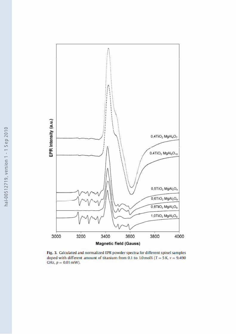
different orientations. The resulting spectrum is equivalent to an EPR powder spectrum. In Fig. 3,
we present the resulting calculated and normalized powder EPR spectra of different Ti-doped
spinel samples. In this figure, two different types of EPR powder spectra of Ti3+ can be observed.
The first Ti3+ powder EPR spectrum corresponds to the Ti-dopedMgAl2O4 samples. As a function
of TiO2 doping content, the Ti3+ spectra seems to be equivalent even if the EPR spectra are the
sum of two components related to Ti3+ and Mn2+ ions. On the contrary, calculated EPR powder
spectra of 0.4TiO2–MgAl6O10 and 0.4TiO2–MgAl4O7 are similar and clearlydifferentfromTiO2–
MgAl2O4 samples. This result shows the presence of different Ti3+ sites and can be correlated
either to the appearance of a new site or to a modification of the Ti3+ site speciation when the
chemical composition is modified from MgAl2O4 to MgAl4O7 and MgAl6O10.
hal-0
0512
719,
ver
sion
1 -
1 Se
p 20
10

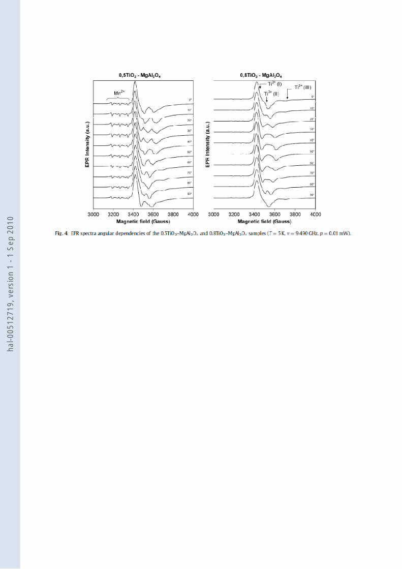
3.3. Angular dependencies of Ti3+ and Mn
2+ EPR lines
Fig. 4 shows the EPR spectra of the 0.5TiO2–MgAl2O4 and 0.8TiO2–MgAl2O4 samples recorded
under the same conditions as before but at different orientations (from0° to 90°) of the sample’s
c-axis in regard to the magnetic field B0. First, we observe that the EPR lines of the Mn2+ ions
show no angular dependencies. But as mentioned earlier, we observe on the spectra of 0.5TiO2–
MgAl2O4 the presence of a combination of EPR lines corresponding to both Mn2+ and Ti3+ ions.
Due to this combination, it is difficult to follow the angular dependencies of the differentTi3+
EPR lines. By contrast, the most Ti-doped samples present the lowest Mn2+ impurities content
(cf.Section3.1).Thus, it is easier to follow the evolution of the Ti3+ EPR lines during the rotation
of the sample on the spectra of 0.8TiO2–MgAl2O4. We observe here at least three different Ti3+
sites inside the spinel structure.
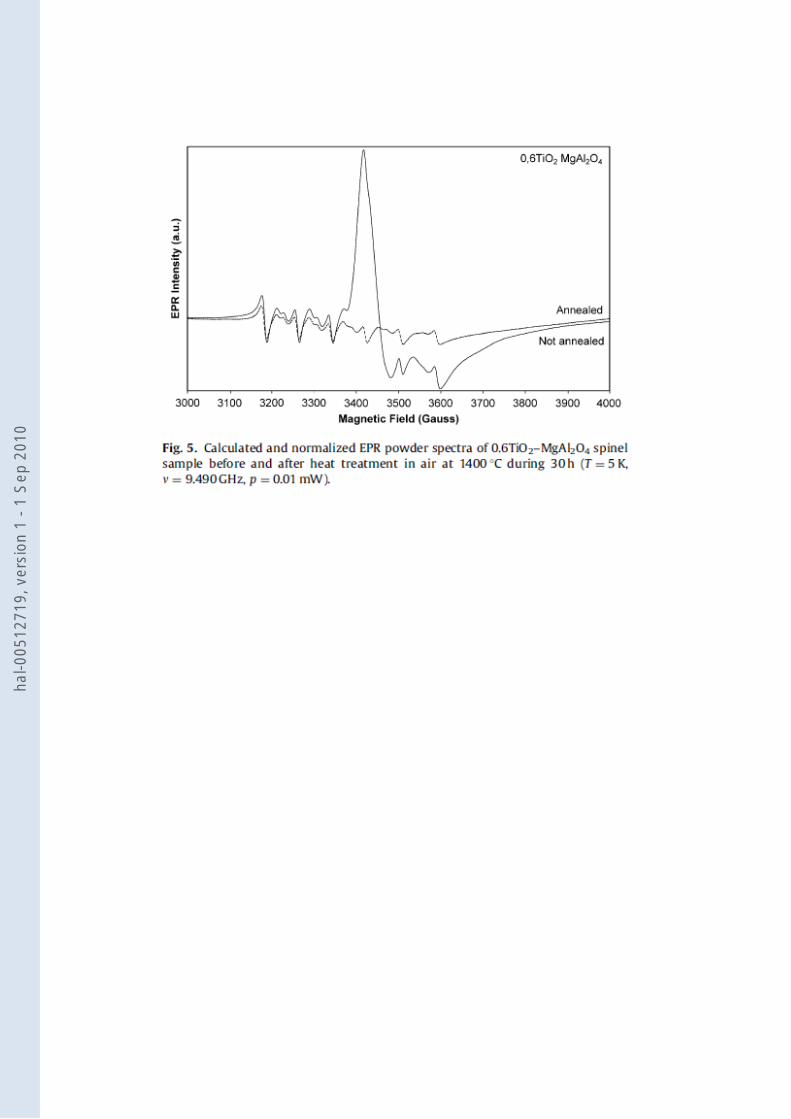
3.4. EPR spectra of the 0.6TiO2–MgAl2O4 sample annealed at high temperature
The influence of annealing at 1400°C in air for 30h is shown for the 0.6TiO2–MgAl2O4 sample in
Fig.5. With this heat treatment, we observe by EPR spectroscopy a complete disap- pearance of
the EPR lines attributed to Ti3+ lines. By contrast we only observe a slight decrease of the Mn2+
EPR lines. This confirms the results of Jouini et al. [8] who observe a complete disappearance of
the bluish color of the same sample. They attribute this disappearance to (i) a redistribution of the
cation sites in the spinel crystal and/or (ii) to the oxidation of the transition metal ions (Ti3+-Ti4+)
[8]. As will be discussed in the next section, we attribute part of the different Mn2+ EPR lines to a
redistribution of Mn2+ ions in the different sites in the spinel structure. Since we do not observe
any modification of the EPR lines attributed to the Mn2+ ions we conclude that the disappearance
of the bluish color of the sample and the disappearance of the Ti3+ EPR line is due to the
oxidation of the transition metal ions.
4. Discussion
4.1. Sites of Ti3+ ions
hal-0
0512
719,
ver
sion
1 -
1 Se
p 20
10

We observed by EPR spectroscopy in 0.8TiO2–MgAl2O4 and 0.4 TiO2–MgAl4O7 samples the
presence of, at least, three different sites of Ti3+ into the spinel structure. However, previous
studies by optical absorption [5], luminescence [6,8] or optically detected magnetic resonance
(ODMR) [9] on Ti-dopedMgAl2O4 samples attributed the observed optical properties to a
trivalent titanium in an unique site which consists of a Ti3+ in the Al3+ octahedral site of the spinel
structure (B site). To explain this apparent inconsistency between our EPR observations and the
results of previous studies we will consider first the spinel structure and second the possible
unusual position of some atoms in the crystal structure. First, as previously mentioned, only one-
eighth of the tetrahedral and one-half of the octahedral sites in the spinel structure are occupied
respectively by Mg2+ or Al3+ ions. When heated, some Al3+ and Mg2+ ions may change their sites,
giving rise to a more random distribution of the cations. The fraction of Al3+ ions in the Mg2+
sublattice is usually denoted by i and it was demonstrated by nuclear magnetic resonance (NMR)
and infrared absorption that i takes values from 0.1 to 0.6 in synthetic MgAl2O4 spinels [14].
Thus, we speculate that one of the Ti3+ sites observed by EPR spectroscopy is linked with a Ti3+
in the octahedral B site while another one is linked with an‘‘inverted’’Ti3+ in a tetrahedral A site
of the spinel structure. Second, the spinel structure may contain a part of ions in insertion site
which corresponds to unoccupied sites in a pure spinel structure. Due to the different neighbor’s
nature and position, the environment’s distortions of theses ions into insertion sites will be very
different from the distortions of the spinels A or B sites. Thus, we speculate that the third EPR
lines attributed to Ti3+ originate from trivalent titanium ions in such insertion sites. In that case
the important variations of the EPR spectra between 0.5TiO2–MgAl2O4, 0.4TiO2–MgAl4O7 and
0.4TiO2–MgAl6O10 samples results from O2- or Mg2+ vacancies and will constitute another type
of Ti3+ site. Sato et al. [9] have already attributed an emission band at 720 nm observed in Ti-
doped MgAl2O4 spinel under excitation at 280 nm to Mg2+ vacancy. However, to confirm theses
hypotheses and to analyze the difference in the site population as a function of the spinel matrix,
more detailed EPR experiments and simulations are required.
4.2. Sites of Mn2+ ions
In this work, the observed EPR signal due to Mn2+ ions maybe attributed either to three different
Mn2+ environments inside the MgAl2O4 spinel structure or to a ZFS due to high symmetry around
hal-0
0512
719,
ver
sion
1 -
1 Se
p 20
10
Mn2+ ions. Considering absorption [5], luminescence [6, 15] or EPR measurements [15], previous
research has concluded to the presence of the Mn2+ ion in the A site of the spinel structure, i.e. in
the tetrahedral site. However, as said before, Tomita et al. [11] pointed out that the spinel has two
types of sites for a metal ion (the tetragonal A site and the octahedral B site). They assign a major
part of the Mn2+ ions in the A site (tetrahedral) and a minor part in anti sites, i.e. in the B site
(octahedral). As with titanium ions we assume that one of the EPR lines is linked to the Mn2+
ions in the A site and another one to the Mn2+ ions in the B site. However, we only observe a
reduction of the intensity of the EPR signal when the number of oxygen and magnesium
vacancies increase (Figs.2 and 4). To this day, some general characteristics of Mn2+ site linked
with the third observed EPR line remain unspecified.
4.3. Relation between the TiO2 and Ti3+ concentrations.
As mentioned in Section 3, we saw no correlation between the amount of TiO2 inserted into the
spinel matrix and the Ti3+ content observed by EPR spectroscopy. This inconsistency requires
more studies to be fully understood. Electron microprobe analysis will determine the repartition
and the concentration of trivalent titanium in the sample and EPR, in addition to a reference
sample with a known spin concentration, will be useful to study the number of Ti3+ in each
different sites.
5. Conclusion
Thanks to EPR spectroscopy, we observed the presence in Ti-doped MgAl2O4 spinels of different
sites of trivalent titanium and manganese ions. To attribute these different lines to different
environments, we considered some previous works based on luminescence, absorption and
ODMR experiment and we also considered the variations of the EPR spectra when the chemistry
of the sample is modified. For Ti3+ ions, we conclude that the major part of the ions is located in
the octahedral B site of the spinel structure. A minor part of the Ti3+ ions are in tetrahedral sites
and one of the sites is assumed to be correlated to some ions in insertion sites. Concerning the
Mn2+ ions, we conclude that a major part of the ions are in the tetrahedral A site of the structure
while a minor are in the octahedral B site. Nevertheless more EPR experiments and simulations
hal-0
0512
719,
ver
sion
1 -
1 Se
p 20
10

are required to confirm theses attributions. However, this work demonstrates the interest of the
EPR measurement in the study of the environment of the active ions in optical materials.
Acknowledgements
We are indebted to Dr. A. Jouini and Dr. A. Yoshikawa who furnished us with the samples we
have studied. We sincerely acknowledge Mr. T. Clément for critically reading this article.
References
[1] Robert C. Weast, Handbook of Chemistry and Physics, CRC Press, Cleveland, 1973, 105.
[2] Robert C. Weast, Handbook of Chemistry and Physics, CRC Press, Cleveland, 1973, 196.
[3] G.Slack, Phys. Rev. A 134 (1964) A1268.
[4] R.C. Peterson, G.A. Lager, R.L. Hitterman, Am. Miner. 76 (1991) 1455.
[5] A. Jouini,H. Sato,A. Yoshikawa,A. Fukuda,G. Boulon,K. Kato,E. Hanamura, J. Crystal
Growth 287 (2006) 313.
[6] K. Izumi, S. Miyazaki, S. Yoshida, T. Mizokawa, E. Hanamura, Phys. Rev. B 76 (2007)
075111.
[7] Y. Fujimoto, H. Tanno, K. Izumi, S. Yoshida, S. Shiori, M. Shirai, K. Tanaka, Y. Kawabe, E.
Hanamura, J. Lumin. 128 (2008) 282.
[8] A. Jouini, H. Sato, A. Yoshikawa, T. Fukuda, G. Boulon, G. Panczer, K. Kato, E. Hanamura,
J. Mater. Res. 21 (2006) 2337.
[9] T. Sato, M. Shirai, K. Tanaka, Y. Kawabe, E. Hanamura, J. Lumin. 114 (2005) 155.
[10] L.Ursu, La Resonance Paramagnetique Electronique, Meridiane, Bucarest, 1968.
[11] A. Tomita, T. Sato, K. Tanaka, Y. Kawabe, M. Shirai, K. Tanaka, E. Hanamura, J. Lumin.
109 (2004) 19.
[12] J. Shaffer, H. Farach, C. Poole, Phys. Rev. B 13 (1976) 1869.
[13] A. Abragam, B. Bleaney, Electron Paramagnetic Resonance of Transition Ions, Clarendon
Press, Oxford, 1970.
[14] K.E. Sickafus, J.M. Wills, N.W. Grimes, J. Am. Ceram. Soc. 82 (1999) 3279.
[15] V. Singh, R. Chakradhar, J. Rao, J. Kim, J. Solid State Chem. 180 (2007) 2067.
hal-0
0512
719,
ver
sion
1 -
1 Se
p 20
10

hal-0
0512
719,
ver
sion
1 -
1 Se
p 20
10

hal-0
0512
719,
ver
sion
1 -
1 Se
p 20
10
hal-0
0512
719,
ver
sion
1 -
1 Se
p 20
10
hal-0
0512
719,
ver
sion
1 -
1 Se
p 20
10
hal-0
0512
719,
ver
sion
1 -
1 Se
p 20
10
Related Documents