Hindawi Publishing Corporation Journal of Ophthalmology Volume 2011, Article ID 285296, 5 pages doi:10.1155/2011/285296 Case Report Spectral Domain Optical Coherence Tomography in Diffuse Unilateral Subacute Neuroretinitis Carlos Alexandre de A. Garcia Filho, 1, 2 Ana Claudia Medeiros de A. G. Soares, 3 Fernando Marcondes Penha, 2 and Carlos Alexandre de Amorim Garcia 1 1 Departamento de Oftalmolog´ ıa, Universidade Federal do Rio Grande do Norte, 59072-970 Natal, RN, Brazil 2 Departamento de Oftalmolog´ ıa, Universidade Federal de S˜ ao Paulo, 04021-001 S˜ ao Paulo, SP, Brazil 3 Departamento de Oftalmolog´ ıa, Santa Casa de Misericordia de S˜ ao Paulo, 01221-020 S˜ ao Paulo, SP, Brazil Correspondence should be addressed to Carlos Alexandre de A. Garcia Filho, [email protected] Received 28 February 2011; Accepted 20 June 2011 Academic Editor: Eduardo Rodrigues Copyright © 2011 Carlos Alexandre de A. Garcia Filho et al. This is an open access article distributed under the Creative Commons Attribution License, which permits unrestricted use, distribution, and reproduction in any medium, provided the original work is properly cited. Purpose. To describe the SD-OCT findings in patients with diffuse unilateral subacute neuroretinitis (DUSN) and evaluate CRT and RNFL thickness. Methods. Patients with clinical diagnosis of DUSN who were submitted to SD-OCT were included in the study. Complete ophthalmologic examination and SD-OCT were performed. Cirrus scan strategy protocols used were 200 × 200 macular cube, optic nerve head cube, and HD-5 line raster. Results. Eight patients with DUSN were included. Mean RNFL thickness was 80.25 μm and 104.75 μm for affected and normal eyes, respectively. Late stage had mean RNFL thickness of 74.83 μm compared to 96.5 μm in early stage. Mean CMT was 205.5 μm for affected eyes and 255.13 μm for normal fellow eyes. Conclusion. RNFL and CMT were thinner in DUSN eyes compared to normal eyes. Late-stage disease had more pronounced thinning compared to early-stage patients. This thinning in RNFL and CMT may reflect the low visual acuity in patients with DUSN. 1. Introduction Diffuse unilateral subacute neuroretinitis (DUSN) is an inflammatory and infectious disease characterized by insid- ious and usually severe loss of peripheral and central vision [1]. Clinical features are manifested in early and late stages [1]. The acute phase is characterized by swelling of the optic disc, vitritis, and recurrent crops of evanescent, multifocal, white-yellowish lesions at the outer retina and choroid [2]. The chronic phase presents with optic nerve atrophy, narrowing of retinal vessels and focal or diffuse pigmentary changes [3]. Parasites of different sizes and several species of nematodes have been reported as the etiology of DUSN, including Toxocara canis, Baylisascaris procyonis, and Ancylostoma caninum, but most of these reports do not present conclusive evidence [4, 5]. Optical coherence tomography (OCT) is a noncontact, noninvasive diagnostic technique that allows measurement of central retinal thickness (CRT) retinal nerve fiber layer (RNFL) thickness and provides important information about the anatomy of the retina and choroid. The devel- opment of spectral domain OCT (SD OCT) considerably improved retinal imaging. The purpose of this study is to describe SD-OCT findings in patients with DUSN and evaluate CRT and RNFL thick- ness with this image device. 2. Patients and Methods This is a retrospective study in which a medical record review was performed at the Department of Ophthalmology, Federal University of Rio Grande do Norte, Brazil between January 2010 and January 2011. The study was approved by the institutional Review Board, and informed consent was obtained from all patients. Subjects with a diagnosis of DUSN were identified. Eyes included in the study had a minimum of 3 months followup. Patients had clinical diag- nosis of DUSN based on Gass and Scelfo criteria, and both

Welcome message from author
This document is posted to help you gain knowledge. Please leave a comment to let me know what you think about it! Share it to your friends and learn new things together.
Transcript
Hindawi Publishing CorporationJournal of OphthalmologyVolume 2011, Article ID 285296, 5 pagesdoi:10.1155/2011/285296
Case Report
Spectral Domain Optical Coherence Tomography in DiffuseUnilateral Subacute Neuroretinitis
Carlos Alexandre de A. Garcia Filho,1, 2 Ana Claudia Medeiros de A. G. Soares,3
Fernando Marcondes Penha,2 and Carlos Alexandre de Amorim Garcia1
1 Departamento de Oftalmologıa, Universidade Federal do Rio Grande do Norte, 59072-970 Natal, RN, Brazil2 Departamento de Oftalmologıa, Universidade Federal de Sao Paulo, 04021-001 Sao Paulo, SP, Brazil3 Departamento de Oftalmologıa, Santa Casa de Misericordia de Sao Paulo, 01221-020 Sao Paulo, SP, Brazil
Correspondence should be addressed to Carlos Alexandre de A. Garcia Filho, [email protected]
Received 28 February 2011; Accepted 20 June 2011
Academic Editor: Eduardo Rodrigues
Copyright © 2011 Carlos Alexandre de A. Garcia Filho et al. This is an open access article distributed under the CreativeCommons Attribution License, which permits unrestricted use, distribution, and reproduction in any medium, provided theoriginal work is properly cited.
Purpose. To describe the SD-OCT findings in patients with diffuse unilateral subacute neuroretinitis (DUSN) and evaluate CRT andRNFL thickness. Methods. Patients with clinical diagnosis of DUSN who were submitted to SD-OCT were included in the study.Complete ophthalmologic examination and SD-OCT were performed. Cirrus scan strategy protocols used were 200×200 macularcube, optic nerve head cube, and HD-5 line raster. Results. Eight patients with DUSN were included. Mean RNFL thickness was80.25 µm and 104.75 µm for affected and normal eyes, respectively. Late stage had mean RNFL thickness of 74.83 µm comparedto 96.5 µm in early stage. Mean CMT was 205.5 µm for affected eyes and 255.13 µm for normal fellow eyes. Conclusion. RNFLand CMT were thinner in DUSN eyes compared to normal eyes. Late-stage disease had more pronounced thinning compared toearly-stage patients. This thinning in RNFL and CMT may reflect the low visual acuity in patients with DUSN.
1. Introduction
Diffuse unilateral subacute neuroretinitis (DUSN) is aninflammatory and infectious disease characterized by insid-ious and usually severe loss of peripheral and centralvision [1]. Clinical features are manifested in early and latestages [1]. The acute phase is characterized by swelling ofthe optic disc, vitritis, and recurrent crops of evanescent,multifocal, white-yellowish lesions at the outer retina andchoroid [2]. The chronic phase presents with optic nerveatrophy, narrowing of retinal vessels and focal or diffusepigmentary changes [3]. Parasites of different sizes andseveral species of nematodes have been reported as theetiology of DUSN, including Toxocara canis, Baylisascarisprocyonis, and Ancylostoma caninum, but most of thesereports do not present conclusive evidence [4, 5].
Optical coherence tomography (OCT) is a noncontact,noninvasive diagnostic technique that allows measurementof central retinal thickness (CRT) retinal nerve fiber layer
(RNFL) thickness and provides important informationabout the anatomy of the retina and choroid. The devel-opment of spectral domain OCT (SD OCT) considerablyimproved retinal imaging.
The purpose of this study is to describe SD-OCT findingsin patients with DUSN and evaluate CRT and RNFL thick-ness with this image device.
2. Patients and Methods
This is a retrospective study in which a medical recordreview was performed at the Department of Ophthalmology,Federal University of Rio Grande do Norte, Brazil betweenJanuary 2010 and January 2011. The study was approvedby the institutional Review Board, and informed consentwas obtained from all patients. Subjects with a diagnosisof DUSN were identified. Eyes included in the study had aminimum of 3 months followup. Patients had clinical diag-nosis of DUSN based on Gass and Scelfo criteria, and both
2 Journal of Ophthalmology
Table 1: Clinical data of patients with DUSN and SD-OCT findings.
Stage ofdisease
Age Gender EyeRNFL
thicknessRNFL
fellow eyeCMT CMT fellow eye
Fovealaspect
InitialVA
FinalVA
Worm
Case 1 Early 14 Male OS 87 µ 105 µ 201 µ 246 µNo foveal
depressionCF 20/60 Present
Case 2 Early 15 Male OD 106 µ 112 µ 214 µ 249 µNormalfoveal
depression20/200 20/25 Present
Case 3 Late 10 Female OS 88 µ 113 µ 184 µ 259 µNo foveal
depressionCF CF Absent
Case 4 Late 25 Male OD 67 µ 92 µ 217 µ 247 µNo foveal
depressionCF CF Absent
Case 5 Late 19 Male OD 82 µ 102 µ 228 µ 253 µNo foveal
depression20/200 20/60 Absent
Case 6 Late 17 Male OS 87 µ 114 µ 256 µ 285 µNormalfoveal
depressionHM HM Absent
Case 7 Late 23 Male OS 49 µ 100 µ 163 µ 253 µNo foveal
depressionCF CF Present
Case 8 Late 13 Male OD 76 µ 100 µ 181 µ 249 µNo foveal
depressionCF CF Absent
Mean NA 17 NA NA 80.250 µ 104.75 µ 205.5 µ 255.13 µ NA NA NA NA
RNFL: retinal nerve fiber layer.CMT: central macular thickness.CF: counting fingers.HM: hand motion.NA: not applicable.
early-stage and late-disease patients who underwent SD-OCT (Carl Zeiss Meditec, Dublin, Calif) were included. Anyother ocular disease was considered exclusion criteria.
All patients were submitted to complete ophthalmologicexamination, including best corrected visual acuity (BCVA),slit lamp examination, tonometry, fundoscopy, and opticalcoherence tomography with cirrus. Strategy protocols usedto obtain images were macular cube 200×200 for the centralretinal thickness map, optic nerve head cube for retinal nervefiber layer (RNFL) analysis, and HD-5 line raster to observemacular and foveal aspects.
The following data were collected and recorded: age, sex,initial and final best correct visual acuity (BCVA), affectedeye, disease stage, and presence of the worm. Statisticalanalysis was performed using Paired Student’s t-test.
3. Results
A total of 8 patients with clinical diagnosis of DUSN wereincluded in the study. Mean age of affected patients was 17years (13–25 yr). Out of 8 patients, 7 were male. Late-stagedisease was found in 6 patients. The subretinal worm wasidentified in only 3 patients, 2 were in early-stage disease and1 in the chronic stage. All patients in whom the worm wasidentified were treated with photocoagulation to destroy it.
Table 1 summarizes clinical and OCT findings for allpatients included in the study.
Mean RNFL thickness of the affected eyes was 80.25 µmcompared to 104.75 µm in the normal contralateral eyes
(P = 0.0005). There was a difference in RNFL thicknessmeasurements when the early-stage disease (mean RNFLthickness = 96.5 µm) was compared to late-stage disease(mean RNFL thickness = 74.83 µm).
Central macular thickness was assessed using macularcube strategy. Mean central macular thickness of affectedeyes was 205.5 µm compared to 255.13 µm in the normalcontralateral eyes (P = 0.0004). Macular thickness was sim-ilar in early- and late-stage disease (207.5 µm and 204.8 µm,resp.).
With respect to foveal anatomy, 6 out of 8 patients hadalterations in foveal contour with a loss of foveal depression,despite diminished central retinal thickness. Neither theearly- or late-stage disease patients presented focal or diffusedefects at the junction of the inner and outer segments ofphotoreceptors (IS/OS junction).
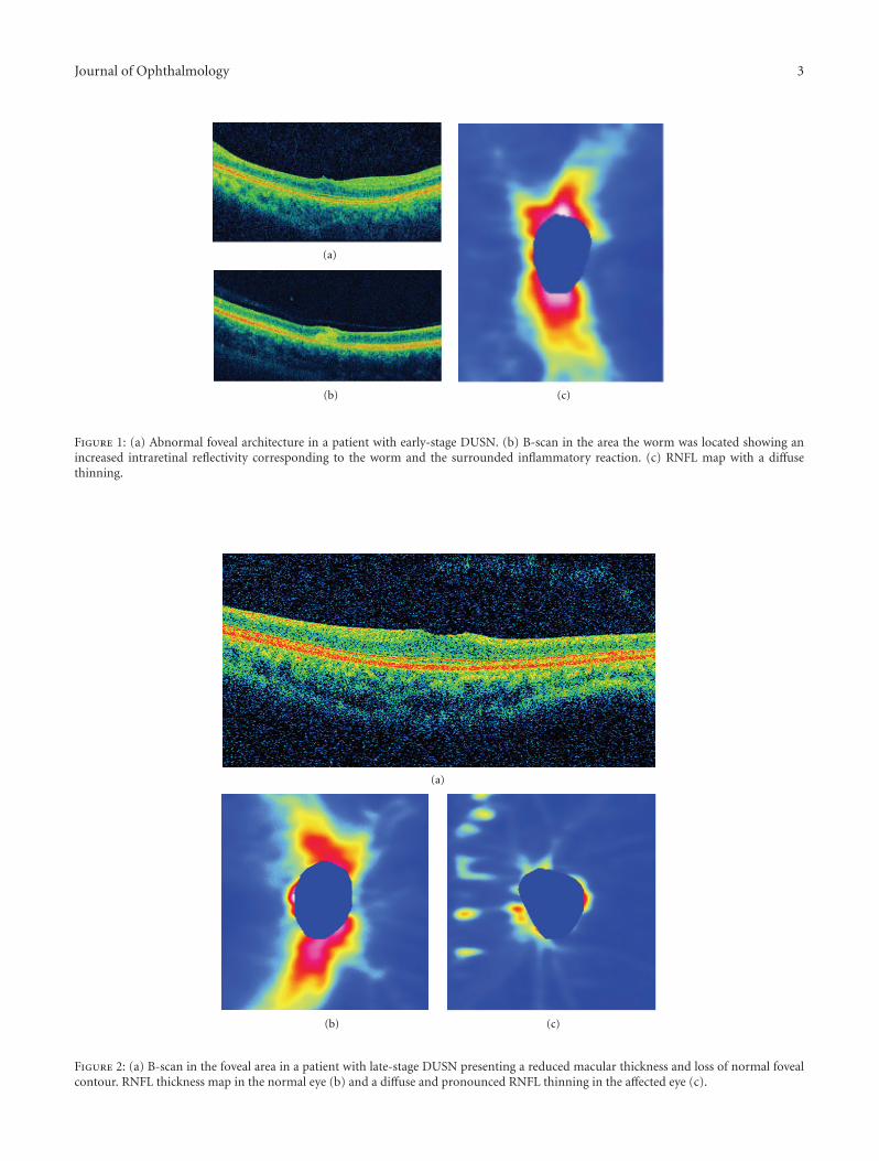
3.1. Cases Report. Case 1 is a 14-year-old male patient inearly-stage disease with a 15-day history of low visual acuityin his left eye. Visual acuity was 20/20 in the right eye andcounting fingers in the left eye. Biomicroscopy revealed mildvitritis. Fundus examination revealed multifocal, evanescent,white-yellowish lesions near the superior temporal arcadeand the presence of a subretinal worm adjacent to the lesions.The worm was destroyed using photocoagulation and visualacuity improved to 20/60. Figure 1 illustrates SD-OCTfindings for this patient. Figure 1(a) shows abnormal fovealarchitecture with a thinning in central macular thickness(201 µ compared to 246 µ in the fellow eye). Figure 1(b)
Journal of Ophthalmology 3
(a)
(b) (c)
Figure 1: (a) Abnormal foveal architecture in a patient with early-stage DUSN. (b) B-scan in the area the worm was located showing anincreased intraretinal reflectivity corresponding to the worm and the surrounded inflammatory reaction. (c) RNFL map with a diffusethinning.
(a)
(b) (c)
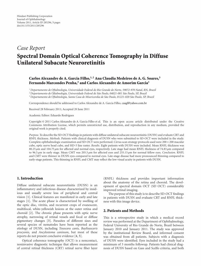
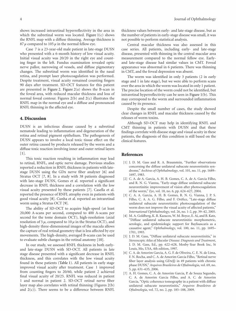
Figure 2: (a) B-scan in the foveal area in a patient with late-stage DUSN presenting a reduced macular thickness and loss of normal fovealcontour. RNFL thickness map in the normal eye (b) and a diffuse and pronounced RNFL thinning in the affected eye (c).
4 Journal of Ophthalmology
shows increased intraretinal hyperreflectivity in the area inwhich the subretinal worm was located. Figure 1(c) showsthe RNFL map with a diffuse thinning. Average thickness is87 µ compared to 105 µ in the normal fellow eye.
Case 7 is a 23-year-old male patient in late-stage DUSNwho presented with a 6-month history of low visual acuity.Initial visual acuity was 20/20 in the right eye and count-ing finger in the left. Fundus examination revealed opticnerve pallor, narrowing of vessels, and diffuse pigmentarychanges. The subretinal worm was identified in the nasalretina, and prompt laser photocoagulation was performed.Despite treatment, visual acuity remained counting fingers90 days after treatment. SD-OCT features for this patientare presented in Figure 2. Figure 2(a) shows the B-scan inthe foveal area, with reduced macular thickness and loss ofnormal foveal contour. Figures 2(b) and 2(c) illustrates theRNFL map in the normal eye and a diffuse and pronouncedRNFL thinning in the affected eye.
4. Discussion
DUSN is an infectious disease caused by a subretinalnematode leading to inflammation and degeneration of theretina and retinal pigment epithelium. The pathogenesis ofDUSN appears to involve a local toxic tissue effect on theouter retina caused by products released by the worm and adiffuse toxic reaction involving inner and outer retinal layers[1].
This toxic reaction resulting in inflammation may leadto retinal, RNFL, and optic nerve damage. Previous studiesreported a reduction in RNFL thickness in patients with late-stage DUSN using the GDx nerve fiber analyzer [6] andStratus OCT [7, 8]. In a study with 38 patients diagnosedwith late-stage DUSN, Gomes et al. reported a significantdecrease in RNFL thickness and a correlation with the lowvisual acuity presented by these patients [7]. Casella et al.reported the presence of RNFL atrophy even in patients withgood visual acuity [8]. Cunha et al. reported an intraretinalworm using a Stratus OCT [9].
The ability of SD-OCT to acquire high-speed (at least20,000 A-scans per second, compared to 400 A-scans persecond for the tome domain OCT), high-resolution (axialresolution of 5 µ, compared to 10 µ in the Stratus OCT), andhigh-density three-dimensional images of the macula allowsthe capture of real retinal geometry that is less affected by eyemovements. The high-density, averaged B-scans can be usedto evaluate subtle changes in the retinal anatomy [10].
In our study, we assessed RNFL thickness in both early-and late-stage DUSN with SD-OCT. All patients in latestage disease presented with a significant decrease in RNFLthickness, and this correlates with the low visual acuityfound in these patients (Table 1). All patients in early stageimproved visual acuity after treatment. Case 1 improvedfrom counting fingers to 20/60, while patient 2 achievedfinal visual acuity of 20/25. RNFL was reduced in patient1 and normal in patient 2. SD-OCT retinal nerve fiberlayer map also correlates with retinal thinning (Figures 2(b)and 2(c)). There seems to be a difference between RNFL
thickness values between early- and late-stage disease, but asthe number of patients in early-stage disease was small, it wasnot possible to perform statistical analysis.
Central macular thickness was also assessed in thiscase series. All patients, including early- and late-stagedisease, presented with thinning in the central macular areameasurement compared to the normal fellow eye. Early-and late-stage disease had similar values in CMT. Fovealappearance was abnormal in 6 patients. There was thinningin CMT, and the foveal depression was absent.
The worm was identified in only 3 patients (2 in earlystage and 1 in late stage), but we were able to perform scansover the area in which the worm was located in only 1 patient.The precise location of the worm could not be identified, butintraretinal hyperreflectivity can be seen in Figure 1(b). Thismay correspond to the worm and surrounded inflammationcaused by its presence.
Despite the small number of cases, the study showedclear changes in RNFL and macular thickness caused by thereleases of worm toxins.
Although SD-OCT may help in identifying RNFL andCMT thinning in patients with DUSN and that thesefindings correlate with disease stage and visual acuity in thesepatients, the diagnosis of this condition is still based on theclinical features.
References
[1] J. D. M. Gass and R. A. Braunstein, “Further observationsconcerning the diffuse unilateral subacute neuroretinitis syn-drome,” Archives of Ophthalmology, vol. 101, no. 11, pp. 1689–1697, 1983.
[2] C. A. de A. Garcia, A. H. B. Gomes, C. A. de A. Garcia Filho,and R. N. G. Vianna, “Early-stage diffuse unilateral subacuteneuroretinitis: improvement of vision after photocoagulationof the worm,” Eye, vol. 18, no. 6, pp. 624–627, 2004.
[3] C. A. A. Garcia, A. H. B. Gomes, R. N. G. Vianna, J. P. S.Filho, C. A. A. G. Filho, and F. Orefice, “Late-stage diffuseunilateral subacute neuroretinitis: photocoagulation of theworm does not improve the visual acuity of affected patients,”International Ophthalmology, vol. 26, no. 1-2, pp. 39–42, 2005.
[4] M. A. Goldberg, K. R. Kazacos, W. M. Boyce, E. Ai, and B. Katz,“Diffuse unilateral subacute neuroretinitis: morphometric,serologic, and epidemiologic support for Baylisascaris as acausative agent,” Ophthalmology, vol. 100, no. 11, pp. 1695–1701, 1993.
[5] J. D. M. Gass, “Diffuse unilateral subacute neuroretinitis,” inStereoscopic Atlas of Macular Disease: Diagnosis and Treatment,J. D. M. Gass, Ed., pp. 622–628, Mosby-Year Book Inc, StLouis, Mo, USA, 4th edition, 1997.
[6] C. A. de Amorim Garcia, A. G. F. de Oliveira, C. E. N. de Lima,F. N. Rocha, and C. A. de Amorim Garcia Filho, “Retinal nervefiber layer analysis using GDx� in 49 patients with chronicphase DUSN,” Arquivos Brasileiros de Oftalmologia, vol. 69, no.5, pp. 631–635, 2006.
[7] A. H. Gomes, C. A. de Amorim Garcia, P. de Souza Segundo,C. A. de Amorim Garcia Filho, and A. C. de AmorimGarcia, “Optic coherence tomography in a patient with diffuseunilateral subacute neuroretinitis,” Arquivos Brasileiros deOftalmologia, vol. 72, no. 2, pp. 185–188, 2009.
Journal of Ophthalmology 5
[8] A. M. B. Casella, M. E. Farah, E. C. de Souza, R. Belfort,and A. P. Miyagusko Taba Oguido, “Retinal nerve fiber layeratrophy as relevant feature for diffuse unilateral subacuteneuroretinitis (DUSN): case series,” Arquivos Brasileiros deOftalmologia, vol. 73, no. 2, pp. 182–185, 2010.
[9] L. P. Cunha, L. V. F. Costa-Cunha, E. C. de Souza, and M.L. R. Monteiro, “Intraretinal worm documented by opticalcoherence tomography in a patient with diffuse unilateralsubacute neuroretinitis: case report,” Arquivos Brasileiros deOftalmologia, vol. 73, no. 5, pp. 462–463, 2010.
[10] M. Wojtkowski, V. Srinivasan, J. G. Fujimoto et al., “Three-dimensional retinal imaging with high-speed ultrahigh-reso-lution optical coherence tomography,” Ophthalmology, vol.112, no. 10, pp. 1734–1746, 2005.

Submit your manuscripts athttp://www.hindawi.com
Stem CellsInternational
Hindawi Publishing Corporationhttp://www.hindawi.com Volume 2014
Hindawi Publishing Corporationhttp://www.hindawi.com Volume 2014
MEDIATORSINFLAMMATION
of
Hindawi Publishing Corporationhttp://www.hindawi.com Volume 2014
Behavioural Neurology
EndocrinologyInternational Journal of
Hindawi Publishing Corporationhttp://www.hindawi.com Volume 2014
Hindawi Publishing Corporationhttp://www.hindawi.com Volume 2014
Disease Markers
Hindawi Publishing Corporationhttp://www.hindawi.com Volume 2014
BioMed Research International
OncologyJournal of
Hindawi Publishing Corporationhttp://www.hindawi.com Volume 2014
Hindawi Publishing Corporationhttp://www.hindawi.com Volume 2014
Oxidative Medicine and Cellular Longevity
Hindawi Publishing Corporationhttp://www.hindawi.com Volume 2014
PPAR Research
The Scientific World JournalHindawi Publishing Corporation http://www.hindawi.com Volume 2014
Immunology ResearchHindawi Publishing Corporationhttp://www.hindawi.com Volume 2014
Journal of
ObesityJournal of
Hindawi Publishing Corporationhttp://www.hindawi.com Volume 2014
Hindawi Publishing Corporationhttp://www.hindawi.com Volume 2014
Computational and Mathematical Methods in Medicine
OphthalmologyJournal of
Hindawi Publishing Corporationhttp://www.hindawi.com Volume 2014
Diabetes ResearchJournal of
Hindawi Publishing Corporationhttp://www.hindawi.com Volume 2014
Hindawi Publishing Corporationhttp://www.hindawi.com Volume 2014
Research and TreatmentAIDS
Hindawi Publishing Corporationhttp://www.hindawi.com Volume 2014
Gastroenterology Research and Practice
Hindawi Publishing Corporationhttp://www.hindawi.com Volume 2014
Parkinson’s Disease
Evidence-Based Complementary and Alternative Medicine
Volume 2014Hindawi Publishing Corporationhttp://www.hindawi.com
Related Documents


![ScreeningforStereopsisofChildrenUsingan ...downloads.hindawi.com/journals/joph/2019/1570309.pdfanimage-splittersysteminalmost20yearsago.Breyeretal. [24]establishedarandom-dotstereotestbasedontheuseof](https://static.cupdf.com/doc/110x72/60d13f23af69a13bcf505548/screeningforstereopsisofchildrenusingan-animage-splittersysteminalmost20yearsagobreyeretal.jpg)
![ComparisonofIndividualRetinalLayerThicknessesafter ...downloads.hindawi.com/journals/joph/2018/1256781.pdf[10,11].ILMremoval,therefore,inhibitsfibrousmembrane formation by removing](https://static.cupdf.com/doc/110x72/5f0eecaa7e708231d4419c6c/comparisonofindividualretinallayerthicknessesafter-1011ilmremovalthereforeinhibitsibrousmembrane.jpg)