Hindawi Publishing Corporation Pathology Research International Volume 2013, Article ID 570526, 6 pages http://dx.doi.org/10.1155/2013/570526 Research Article Specimens from Biopsies of Colorectal Polyps Often Harbor Additional Diagnoses Shefali Chopra and Mark Li-cheng Wu Department of Pathology and Laboratory Medicine, University of California Irvine School of Medicine, Irvine, CA 92868, USA Correspondence should be addressed to Mark Li-cheng Wu; [email protected] Received 26 July 2013; Accepted 13 November 2013 Academic Editor: Oscar Cummings Copyright © 2013 S. Chopra and M. L.-c. Wu. is is an open access article distributed under the Creative Commons Attribution License, which permits unrestricted use, distribution, and reproduction in any medium, provided the original work is properly cited. Objectives. e utility of examining specimens from colorectal biopsies of polyps for nonneoplastic diseases is currently unknown. Our objectives were to characterize such additional diagnoses that could be rendered. Methods. We retrospectively and prospectively reviewed specimens from endoscopic biopsies of colorectal polyps obtained during routine screening or surveillance. Results. 17 of 168 specimens (10.1%) contained additional diagnoses, including schistosomiasis, eosinophilic colitis, intestinal spirochetosis, melanosis coli, and other entities. ese findings were easily overlooked because they oſten affected mucosa that was spared by the polyps or were oſten evident only at high magnification. Schistosomiasis, eosinophilic colitis, and intestinal spirochetosis were clinically occult. Conclusions. Specimens from biopsies of colorectal polyps oſten harbor other diagnoses, in addition to polyps, and can be simultaneously screened for polyps and examined for nonneoplastic diseases. Detection of other diagnoses in addition to polyps requires awareness, examination at high magnification, and examination of areas spared by the polyps. 1. Introduction Specimens from endoscopic biopsies of putative colorectal lesions usually show traditional nonserrated adenomas, ser- rated polyps, or variants of normal mucosa. ese entities generally are easily and rapidly diagnosed at low magnifi- cation. Consequently, pathologists are tempted to examine these specimens quickly and only at low magnification, to assume that these specimens will harbor only polyps or variants of normal mucosa, and to refrain from examining these specimens further at high magnification once polyps are diagnosed. Directed review of these specimens might reveal other diagnoses, in addition to polyps. Pathologists might overlook such second diagnoses for various reasons. However, such second diagnoses might be significant. We reviewed specimens from biopsies of colorectal polyps to characterize second diagnoses that could be rendered in this setting and to demonstrate that these specimens could be examined for nonneoplastic diseases. 2. Materials and Methods e study was approved by the Institutional Review Board of the University of California, Irvine, USA, on November 7, 2005 as protocol HS number: 2005-4646. e study was carried out in 2 phases. e first phase was designed to determine prevalence, and the second phase was designed to determine incidence. During the first phase, consecutive specimens from endoscopic colorectal biopsies of polyps accessioned at our institution during a 1-month interval, from July 1, 2003 to August 1, 2003, were retrospectively reviewed by both of us, who are gastrointestinal pathologists, with special effort to render second diagnoses in addition to polyps. Only specimens that had polyps, broadly defined as protruding pathologic mucosal lesions [1], confirmed histologically were eligible for review. All original diagnoses were made by 5 other pathologists, who were general surgical pathologists. ese pathologists were unaware of our study. During the second phase, specimens from 100 consecutive histologically

Welcome message from author
This document is posted to help you gain knowledge. Please leave a comment to let me know what you think about it! Share it to your friends and learn new things together.
Transcript
Hindawi Publishing CorporationPathology Research InternationalVolume 2013, Article ID 570526, 6 pageshttp://dx.doi.org/10.1155/2013/570526
Research ArticleSpecimens from Biopsies of Colorectal Polyps OftenHarbor Additional Diagnoses
Shefali Chopra and Mark Li-cheng Wu
Department of Pathology and Laboratory Medicine, University of California Irvine School of Medicine,Irvine, CA 92868, USA
Correspondence should be addressed to Mark Li-cheng Wu; [email protected]
Received 26 July 2013; Accepted 13 November 2013
Academic Editor: Oscar Cummings
Copyright © 2013 S. Chopra and M. L.-c. Wu. This is an open access article distributed under the Creative Commons AttributionLicense, which permits unrestricted use, distribution, and reproduction in any medium, provided the original work is properlycited.
Objectives. The utility of examining specimens from colorectal biopsies of polyps for nonneoplastic diseases is currently unknown.Our objectiveswere to characterize such additional diagnoses that could be rendered.Methods.We retrospectively and prospectivelyreviewed specimens from endoscopic biopsies of colorectal polyps obtained during routine screening or surveillance. Results. 17of 168 specimens (10.1%) contained additional diagnoses, including schistosomiasis, eosinophilic colitis, intestinal spirochetosis,melanosis coli, and other entities. These findings were easily overlooked because they often affected mucosa that was spared bythe polyps or were often evident only at high magnification. Schistosomiasis, eosinophilic colitis, and intestinal spirochetosis wereclinically occult.Conclusions. Specimens from biopsies of colorectal polyps often harbor other diagnoses, in addition to polyps, andcan be simultaneously screened for polyps and examined for nonneoplastic diseases. Detection of other diagnoses in addition topolyps requires awareness, examination at high magnification, and examination of areas spared by the polyps.
1. Introduction
Specimens from endoscopic biopsies of putative colorectallesions usually show traditional nonserrated adenomas, ser-rated polyps, or variants of normal mucosa. These entitiesgenerally are easily and rapidly diagnosed at low magnifi-cation. Consequently, pathologists are tempted to examinethese specimens quickly and only at low magnification, toassume that these specimens will harbor only polyps orvariants of normal mucosa, and to refrain from examiningthese specimens further at high magnification once polypsare diagnosed. Directed review of these specimens mightreveal other diagnoses, in addition to polyps. Pathologistsmight overlook such second diagnoses for various reasons.However, such second diagnoses might be significant. Wereviewed specimens from biopsies of colorectal polyps tocharacterize second diagnoses that could be rendered in thissetting and to demonstrate that these specimens could beexamined for nonneoplastic diseases.
2. Materials and Methods
The study was approved by the Institutional Review Boardof the University of California, Irvine, USA, on November7, 2005 as protocol HS number: 2005-4646. The study wascarried out in 2 phases. The first phase was designed todetermine prevalence, and the second phase was designed todetermine incidence.
During the first phase, consecutive specimens fromendoscopic colorectal biopsies of polyps accessioned at ourinstitution during a 1-month interval, from July 1, 2003 toAugust 1, 2003, were retrospectively reviewed by both ofus, who are gastrointestinal pathologists, with special effortto render second diagnoses in addition to polyps. Onlyspecimens that had polyps, broadly defined as protrudingpathologic mucosal lesions [1], confirmed histologically wereeligible for review. All original diagnoses were made by 5other pathologists, who were general surgical pathologists.These pathologists were unaware of our study. During thesecond phase, specimens from 100 consecutive histologically

2 Pathology Research International
confirmed polyps obtained from endoscopic colorectal biop-sies accessioned to 1 of us (M. L.-c. Wu) at our institutionduring the course of normal signout were prospectivelyaccrued during the interval July 1, 2007–September 11, 2007.These specimens were examinedwith knowledge of our studyandwith special effort to render second diagnoses in additionto polyps. For both phases, this special effort consisted ofexamining all sections on each slide, at low magnificationand high magnification, and examining the entire surfacearea of each section including mucosa affected by polypsand mucosa spared by polyps. Specimens from both phaseswith additional diagnoses were reviewed by both of us forconfirmation.
Pathology reports and requisitions for all specimenswere reviewed. Corresponding reports from endoscopy werereviewed for each specimen, in either phase, for which addi-tional diagnoses were rendered. Diagnoses were consideredclinically occult if the requisitions or corresponding reportsfrom endoscopy lacked the mention of the diagnoses orlacked the mention of findings that could be reasonablyattributed to the diagnoses. Excluded from the entire studywere specimens from biopsies of putative lesions that lackedpolyps histologically. All specimens were processed routinely,formalin-fixed, and paraffin-embedded. All histologic sec-tions were cut 4 microns thick and stained with hematoxylinand eosin. Microscopy was performed with a conventionalmultiheaded optical microscope (BX45, Olympus, Melville,NY,USA). All diagnoseswere rendered according to standardcriteria [1–4].
3. Results
For the first phase, 78 specimens were diagnosed as polyps byoriginal pathologists in the 1-month interval. We reclassified3 hyperplastic polyps and 1 tubular adenoma as normalmucosa and excluded these specimens from further study.We reclassified 1mucosal prolapse, 1 hyperplastic polyp, and 1inflammatory polyp, as leiomyoma, sessile serrated adenoma,and tubular adenoma, respectively. We confirmed all otheroriginal diagnoses. A few specimens hadmultiple polyps.Thefirst phase therefore eventually consisted of 74 specimens,with 78 histologically confirmed polyps. These polyps werefrom 52 patients, ages 43 to 74, including 31 men and 21women. The 78 polyps included 48 polyps in men and 30polyps in women.
Of the 74 specimens, additional diagnoses were retro-spectively detected by us in 7 specimens (9.5%). These diag-noses included the following: mucosal prolapse (Figure 1) (in2 specimens with hyperplastic polyps and 1 specimen withtubular adenoma), melanosis coli (Figures 2(a) and 2(b)) (in2 specimens with tubular adenomas), hyperplastic polyp (in1 specimen with tubular adenoma), and eosinophilic colitis(Figure 3) (in 1 specimen with tubular adenoma).
The patient for whom we diagnosed eosinophilic colitiswas asymptomatic. With this caveat, we rendered the diag-nosis of eosinophilic colitis based on compellingmorphologythat satisfied recently proposed criteria [3]. Microscopyshowed the entire specimen, including areas remote from
Figure 1: Mucosal prolapse in specimen with hyperplastic polyp.Only small focus of smooth muscle present (left of center) (H&E,×200).
the polyp, to have an extremely high density of eosinophilswith greater than 120 eosinophils per 600x field, frequenteosinophilic cryptitis and crypt abscesses, degranulatedeosinophils, submucosal eosinophils, and absence of neu-trophils in nonpolypoid mucosa.
Mention of all of these additional diagnoses, except foreosinophilic colitis, was absent from the original pathologyreports. Therefore, 6 of 7 additional diagnoses were poten-tially overlooked by original pathologists.
For the second phase, the 100 polyps were accrued afterexamining 94 specimens. A few specimens had multiplepolyps.The 94 specimens were from 55 patients, including 27men and 28 women, ages 45 to 82. The 100 polyps included57 polyps in men and 43 polyps in women.
Of the 94 specimens, additional diagnoses were prospec-tively detected in 10 specimens (10.6%) by 1 of us (M. L.-c.Wu). These diagnoses included the following: melanosis coli(in 4 specimens with tubular adenomas and 1 specimen withhyperplastic polyp), mucosal prolapse (in 2 specimens withhyperplastic polyps), schistosomiasis (Figures 4(a), 4(b), and4(c)) (in 1 specimen with adenocarcinoma and 1 specimenwith hyperplastic polyp), and intestinal spirochetosis (Fig-ure 5) (in 1 specimenwith sessile serrated adenoma), and theywere confirmed retrospectively by both of us.
For the entire study, a final total of 168 specimens wereincluded, and 17 additional diagnoses were rendered, for arate of 10.1%. Clinical information obtained from pathol-ogy requisitions and corresponding notes from endoscopyindicated that all endoscopies were performed for screeningor surveillance for polyps and that all patients were asymp-tomatic.
Several factors potentially contributed to difficulty inrendering additional diagnoses. The additional diagnosticfindings were easily overlooked in many scenarios (Figures1, 2(a), 2(b), 3, 4(a), 4(b), 4(c), and 5), including whenthe findings were small, focal, in nonpolypoid mucosa, inotherwise normal mucosa, or clinically occult.
Pathology Research International 3
(a) (b)
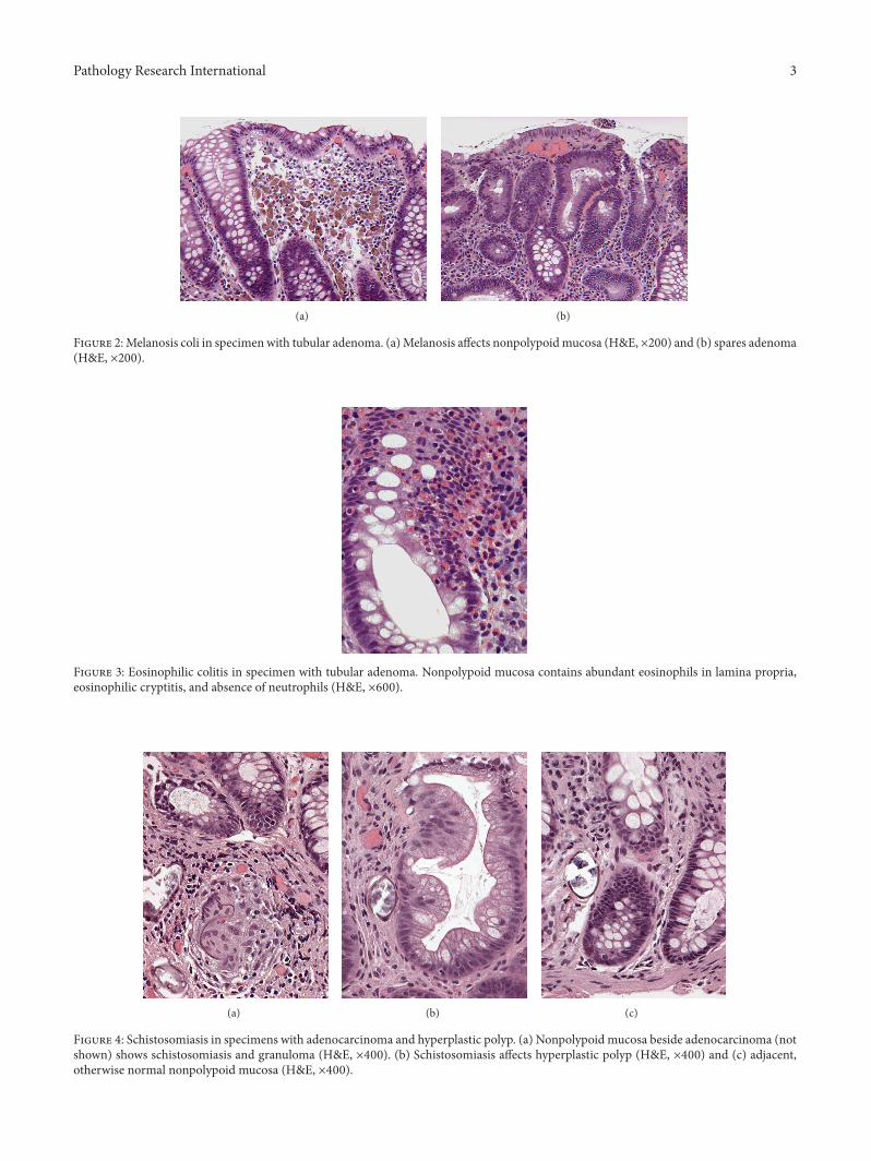
Figure 2:Melanosis coli in specimenwith tubular adenoma. (a)Melanosis affects nonpolypoidmucosa (H&E, ×200) and (b) spares adenoma(H&E, ×200).
Figure 3: Eosinophilic colitis in specimen with tubular adenoma. Nonpolypoid mucosa contains abundant eosinophils in lamina propria,eosinophilic cryptitis, and absence of neutrophils (H&E, ×600).
(a) (b) (c)
Figure 4: Schistosomiasis in specimens with adenocarcinoma and hyperplastic polyp. (a) Nonpolypoid mucosa beside adenocarcinoma (notshown) shows schistosomiasis and granuloma (H&E, ×400). (b) Schistosomiasis affects hyperplastic polyp (H&E, ×400) and (c) adjacent,otherwise normal nonpolypoid mucosa (H&E, ×400).

4 Pathology Research International
Figure 5: Intestinal spirochetosis in specimen with sessile serratedadenoma. Spirochetes colonize surface of otherwise normal mucosapresent beside polyp (top right) and present focally between serratedcrypts (top left) (Warthin-Starry, ×200).
Table 1: Histology of polyps.
Histology Phase 1 Phase 2Tubular adenomas 49 34Tubulovillous adenomas 2 10Adenocarcinomas 2 2Hyperplastic polyps 22 46Sessile serrated adenomas 2 7Leiomyomas 1 0Neuroendocrine tumors 0 1
Table 2: Distribution of polyps.
Site Phase 1 Phase 2Cecum 9 11Ascending colon 15 11Hepatic flexure 1 3Transverse colon 1 11Splenic flexure 2 1Descending colon 9 9Sigmoid colon 12 28Rectum 15 24Unspecified 14 2
Tables 1, 2, and 3 summarize the histology and distribu-tion of the polyps and the additional diagnoses.
4. Discussion
Specimens taken for the purpose of evaluating 1 diseasemay contain additional, incidental diagnostic findings, whichmay be related or unrelated to the primary disease. Theseadditional diagnoses are easily overlooked, because of manyreasons: (1) additional diagnoses may be associated withmorphologic changes that are considerably smaller thanthose of the primary disease and might only be appreciatedat high magnification or by purposefully examining the slidefor incidental findings; (2) attention of pathologists is usually
Table 3: Additional diagnoses detected at colorectal biopsy forpolyps.
Additional diagnoses Factors potentially contributing todiagnostic difficulty
Mucosal prolapse Only small focus affected
Melanosis coliNonpolypoid mucosa preferentiallyaffectedPolyps relatively or completely spared
Eosinophilic colitis Confusion with reaction to adenomaClinically occult
SchistosomiasisMinute ovaAffected nonpolypoid mucosa otherwisenormal or focally normalClinically occult
Intestinal spirochetosis
Minute bacteriaConfined to nonpolypoid mucosaAffected nonpolypoid mucosa otherwisenormalClinically occult
directed toward the primary disease due to habit and due tobias from the clinical request, and hence attention is divertedaway from incidental findings; (3) incidental findings mayoccupy seemingly extraneous tissue, from the same organor unrelated organs, that usually is histologically normal[5]; (4) even if incidental findings are detected, pathologiststhat lack expertise in a particular specialty may fail torecognize incidental findings as significant. It is importantfor pathologists to report such additional diagnoses becausethese additional diagnoses may have pathologic, endoscopic,or clinical significance or may be clinically occult. Thisprocess transforms these specimens into invaluable, cost-effective tools to detect unexpected diagnoses. Examining foradditional diagnoses detects disease early, guides therapy, andeliminates morbidity and financial burden associated withrepeat biopsies and advanced disease. Colonoscopy carriesa 0.07% incidence of iatrogenic perforation [6] and costsapproximately $3081 by itself but costs significantlymorewithbiopsy, anesthesia, or immunohistochemistry or if performedon an inpatient [7].
The concept of purposefully examining specimens todetect other, secondary diagnoses has been studied in thesetting of genitourinary pathology and hepatic pathology.Specimens from nephrectomies performed to stage primaryrenal neoplasia were recently shown to occasionally containincidental nonneoplastic disease [8]. Specimens from pro-static core biopsies often contain incidental pieces of rectalmucosa that occasionally harbors clinically significant rectalpathology or causes diagnostic difficulty [5]. Specimens fromhepatic core biopsies obtained for the purpose of grading andstaging chronic viral hepatitis often contain other diagnoses[9]. To our knowledge, our study is the first to apply thisconcept to colorectal biopsies.
Our results show that the rate of additional diagnosesin specimens from biopsies of colorectal polyps in asymp-tomatic patients at time of screening or surveillance isapproximately 10%. This rate is predictably similar to that
Pathology Research International 5
found in a recent study, which detected abnormal histologyin 11% of specimens from colorectal biopsies of patients withnormal endoscopy and without diarrhea [10]. Furthermore,this rate is predictablymuch less than that found in a previousstudy, which detected abnormal histology in 32.1% of patientswith normal colonoscopy and with chronic diarrhea [11].
Although some additional diagnoses had minor clini-cal impact, all had pathologic or endoscopic significance.For reasons yet to be elucidated, melanosis coli generallyavoids adenomas, as our study demonstrated. Adenomasspared by melanosis coli appear endoscopically as palepatches, which help endoscopists locate adenomas [12].Our study also demonstrated that intestinal spirochetosisand schistosomiasis may be asymptomatic, involve normalor abnormal mucosa, and involve polyps [13–19]. To ourknowledge, our study is the first to report schistosomiasiscoincidentally affecting a hyperplastic polyp. Although ourcase of eosinophilic colitis was easily diagnosed, cautionmust be exercised to avoid overdiagnosing colitis, becausepolyps commonly attract eosinophils and lymphocytes [20–22]. Mucosal prolapse may mimic hyperplastic polyps, ses-sile serrated adenomas, traditional nonserrated adenomas,inflammatory bowel disease, and other entities [4, 23].
Specimens from colorectal biopsies of polyps can andshould be simultaneously screened for polyps and examinedfor additional diagnoses. Awareness of the possibility ofsecond diagnoses, examination of nonpolypoid areas, andexamination at high magnification are necessary to avoidoverlooking these diagnoses.
Conflict of Interests
The authors declare that there is no conflict of interestsregarding the publication of this paper.
Acknowledgment
This research was performed without specific financial sup-port and was presented in part in abstract form at the Collegeof American Pathologists ’08 annual meeting, poster exhibitssession, San Diego, CA, USA, September 25, 2008.
References
[1] K. J. Lewin, R. H. Riddell, andW. M.Weinstein, Eds., Gastroin-testinal Pathology and Its Clinical Implications, Igaku-Shoin,New York, NY, USA, 1992.
[2] D. C. Snover, J. R. Jass, C. Fenoglio-Preiser, and K. P. Batts,“Serrated polyps of the large intestine: a morphologic andmolecular review of an evolving concept,”TheAmerican Journalof Clinical Pathology, vol. 124, no. 3, pp. 380–391, 2005.
[3] J. M. Hurrell, R. M. Genta, and S. D. Melton, “Histopathologicdiagnosis of eosinophilic conditions in the gastrointestinaltract,” Advances in Anatomic Pathology, vol. 18, no. 5, pp. 335–348, 2011.
[4] B. Singh, N. J. M.Mortensen, and B. F.Warren, “Histopatholog-ical mimicry in mucosal prolapse,” Histopathology, vol. 50, no.1, pp. 97–102, 2007.
[5] A. Ashton-Sager and M. L. Wu, “Incidental rectal mucosaobtained via transrectal ultrasound-guided prostatic core biop-sies,” International Journal of Surgical Pathology, vol. 15, no. 1,pp. 26–30, 2007.
[6] N. E. Samalavicius, D. Kazanavicius, R. Lunevicius et al.,“Incidence, risk, management, and outcomes of iatrogenic full-thickness large bowel injury associated with 56, 882 colono-scopies in 14 Lithuanian hospitals,” Surgical Endoscopy, vol. 27,pp. 1628–1635, 2013.
[7] “Cost of a Colonoscopy: Consumer information and pricespaid,” 2008, http://health.costhelper.com/colonoscopy.html.
[8] B. Sarsık, A. Simsir,M. Yılmaz, K. Yorukoglu, and S. Sen, “Spec-trum of nontumoral renal pathologies in tumor nephrectomies:nontumoral renal parenchyma changes,” Annals of DiagnosticPathology, vol. 17, pp. 176–182, 2013.
[9] V. Nair, S. E. Fischer, and O. A. Adeyi, “Non-viral-relatedpathologic findings in liver needle biopsy specimens frompatients with chronic viral hepatitis,” The American Journal ofClinical Pathology, vol. 133, no. 1, pp. 127–132, 2010.
[10] O. H. O’Mahony, M. Burgoyne, and J. J. Going, “Specifichistological abnormalities are more likely in biopsies of endo-scopically normal large bowel after the age of 60 years,”Histopathology, vol. 61, pp. 1209–1213, 2012.
[11] J. G. N. Da Silva, T. De Brito, A. O. M. Cintra Damiao, A.A. Laudanna, and A. M. Sipahi, “Histologic study of colonicmucosa in patients with chronic diarrhea and normal colono-scopic findings,” Journal of Clinical Gastroenterology, vol. 40, no.1, pp. 44–48, 2006.
[12] S. J. van Weyenberg, F. Hoentjen, F. Thunnissen, and C.J. Mulder, “Pseudomelanosis coli and adenomatous polyps,”Journal of Gastrointestinal and Liver Diseases, vol. 20, no. 3, p.233, 2011.
[13] J. J. Godyn, R. Siderits, and A. Hazra, “Schistosoma mansoni incolon and liver,”Archives of Pathology and Laboratory Medicine,vol. 129, no. 4, pp. 544–545, 2005.
[14] X. Yu, P. Chen, J. Xu, X. S. Xiao Sen, and Z. Shan, “Histologicalclassification of schistosomal egg induced polyps of colon andtheir clinical significance: An analysis of 272 cases,” ChineseMedical Journal, vol. 104, no. 1, pp. 64–70, 1991.
[15] M. Korner and J.-O. Gebbers, “Clinical significance of humanintestinal spirochetosis: amorphologic approach,” Infection, vol.31, no. 5, pp. 341–349, 2003.
[16] A. Kansagra, N. Nagaria, and S. Ahlawat, “Asymptomatic colonadenoma associated with Schistosoma mansoni,” Digestive andLiver Disease, vol. 42, no. 7, pp. 526–527, 2010.
[17] A. R. E.-S. Mohamed, M. A. Al Karawi, and M. I. Yasawy,“Schistosomal colonic disease,” Gut, vol. 31, no. 4, pp. 439–442,1990.
[18] A. Calderaro, C. Gorrini, S. Montecchini et al., “Intestinalspirochaetosis associated with hyperplastic and adenomatouscolonic polyps,” Pathology Research and Practice, vol. 208, no.3, pp. 177–180, 2012.
[19] W.-C. Li, Z.-G. Pan, and Y.-H. Sun, “Sigmoid colonic carcinomaassociated with deposited ova of Schistosoma japonicum: a casereport,” World Journal of Gastroenterology, vol. 12, no. 37, pp.6077–6079, 2006.
[20] J. Moezzi, N. Gopalswamy, R. J. Haas Jr., R. J. Markert, S.Suryaprasad, and M. S. Bhutani, “Stromal eosinophilia incolonic epithelial neoplasms,” The American Journal of Gas-troenterology, vol. 95, no. 2, pp. 520–523, 2000.
6 Pathology Research International
[21] S. Kiziltas, S. Sezgin Ramadan, A. Topuzoglu, and S. Kullu,“Does the severity of tissue eosinophilia of colonic neoplasmsreflect theirmalignancy potential?”Turkish Journal of Gastroen-terology, vol. 19, pp. 239–244, 2008.
[22] A. D. Polydorides, B. Mukherjee, S. B. Gruber, B. J. McKenna,H. D. Appelman, and J. K. Greenson, “Adenoma-infiltratingLymphocytes (AILs) are a potential marker of hereditary non-polyposis colorectal cancer,” The American Journal of SurgicalPathology, vol. 32, no. 11, pp. 1661–1666, 2008.
[23] C. C. Huang, W. L. Frankel, T. Doukides, X. P. Zhou, W. Zhao,and M. M. Yearsley, “Prolapse-related changes are a confound-ing factor in misdiagnosis of sessile serrated adenomas in therectum,” Human Pathology, vol. 44, pp. 480–486, 2013.

Submit your manuscripts athttp://www.hindawi.com
Stem CellsInternational
Hindawi Publishing Corporationhttp://www.hindawi.com Volume 2014
Hindawi Publishing Corporationhttp://www.hindawi.com Volume 2014
MEDIATORSINFLAMMATION
of
Hindawi Publishing Corporationhttp://www.hindawi.com Volume 2014
Behavioural Neurology
EndocrinologyInternational Journal of
Hindawi Publishing Corporationhttp://www.hindawi.com Volume 2014
Hindawi Publishing Corporationhttp://www.hindawi.com Volume 2014
Disease Markers
Hindawi Publishing Corporationhttp://www.hindawi.com Volume 2014
BioMed Research International
OncologyJournal of
Hindawi Publishing Corporationhttp://www.hindawi.com Volume 2014
Hindawi Publishing Corporationhttp://www.hindawi.com Volume 2014
Oxidative Medicine and Cellular Longevity
Hindawi Publishing Corporationhttp://www.hindawi.com Volume 2014
PPAR Research
The Scientific World JournalHindawi Publishing Corporation http://www.hindawi.com Volume 2014
Immunology ResearchHindawi Publishing Corporationhttp://www.hindawi.com Volume 2014
Journal of
ObesityJournal of
Hindawi Publishing Corporationhttp://www.hindawi.com Volume 2014
Hindawi Publishing Corporationhttp://www.hindawi.com Volume 2014
Computational and Mathematical Methods in Medicine
OphthalmologyJournal of
Hindawi Publishing Corporationhttp://www.hindawi.com Volume 2014
Diabetes ResearchJournal of
Hindawi Publishing Corporationhttp://www.hindawi.com Volume 2014
Hindawi Publishing Corporationhttp://www.hindawi.com Volume 2014
Research and TreatmentAIDS
Hindawi Publishing Corporationhttp://www.hindawi.com Volume 2014
Gastroenterology Research and Practice
Hindawi Publishing Corporationhttp://www.hindawi.com Volume 2014
Parkinson’s Disease
Evidence-Based Complementary and Alternative Medicine
Volume 2014Hindawi Publishing Corporationhttp://www.hindawi.com
Related Documents